Embed Size (px)
Citation preview


1. Membrane Organization and the Plasma Membrane
1a. The lipid bilayer
CYTOPLASM
One layer faces the cytoplasm = cytoplasmic layer (or leaflet)
The other faces either an organelle lumen or the extracellular matrix = non-cytoplasmic layer

Membrane Lipids:
Major lipids are phospholipids and cholesterol
Minor lipids are inositol phospholipids and glycolipids
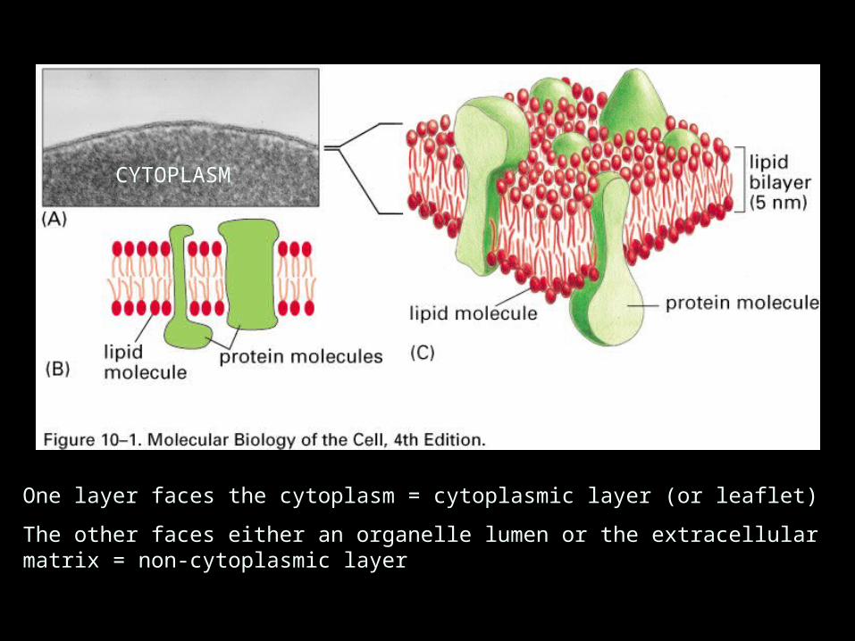
Phospholipids are amphipathic
Example: Phosphatidylcholine
PHOSPHOLIPID STRUCTURE

PHOSPHOLIPID BEHAVIOR
-Can flex, rotate, and are laterally mobile within a leaflet
(with regional restrictions)
-Spontaneous flipping between leaflets rare
-Enzymes (flippases) can flip phospholipids between leaflets
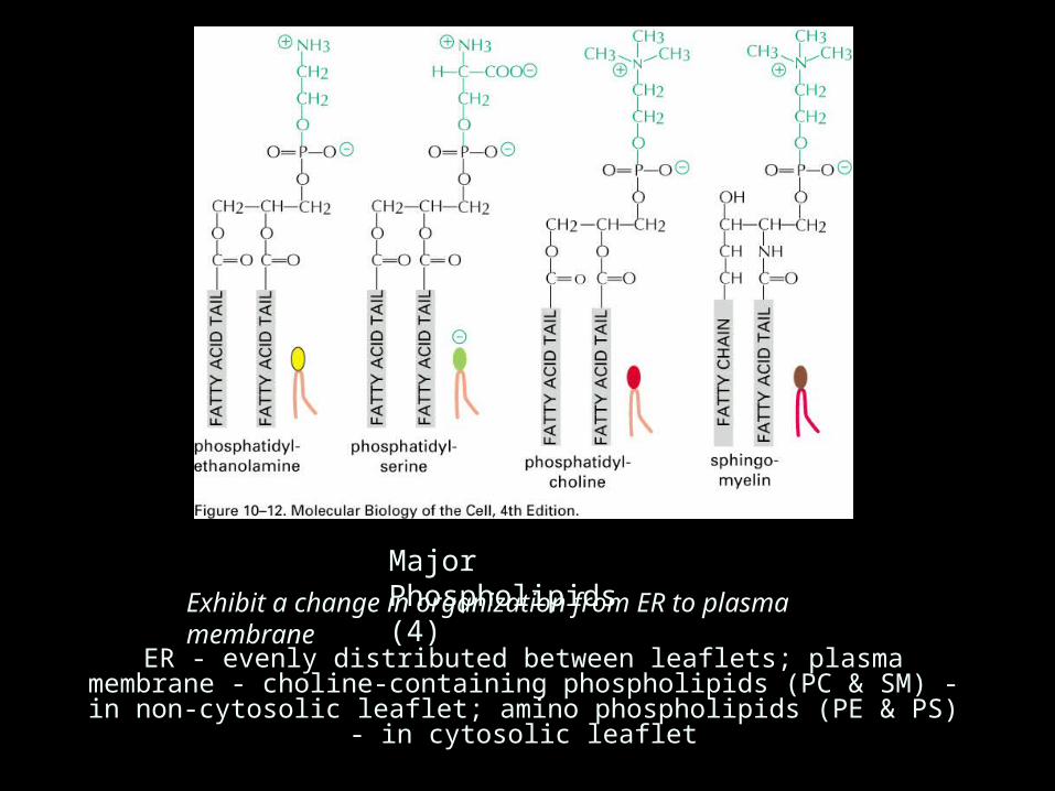
Major Phospholipids (4)
ER - evenly distributed between leaflets; plasma membrane - choline-containing phospholipids (PC & SM) - in non-cytosolic leaflet; amino phospholipids (PE &
PS) - in cytosolic leaflet
Exhibit a change in organization from ER to plasma membrane

Cholesterol; a major membrane lipid

Minor membrane lipids
Inositol phospholipids Glycolipids:
•some are neutral•some are charged•present only on the non-cytosolic leaflet•Lysosomal storage diseases - gangliosides

PE & PS
PC & SM
Cholesterol - about equal in quantity to phospholipid; stiffens membranes, reduces permeability, inhibits phase changes

PE & PS
PC & SM

Lipid Rafts:
Specialized membrane regions
Rich in sphingolipids & cholesterol
Better accommodate certain proteins
Involved in membrane transport & signal transduction

1. Membrane Organization and the Plasma Membrane
1b. Integral and peripheral proteins

1 - Single pass a-helix
2 - Multi-pass a-helix
3 - Rolled-up b-sheet (b-barrel)
4 - a-helix in one layer
5 - lipid anchor
6 - oligosaccharide linker to phosphatidylinositol (non-cytosolic monolayer)
7, 8 - non-covalent interactions with integral membrane proteins (peripheral proteins)

Bacteriorhodopsin
Photosynthetic reaction center (R. viridis)

INTEGRAL PROTEIN BEHAVIOR
-Some exhibit lateral diffusion
-Some are anchored in place
-Orientation is maintained

Self-assembly into aggregates
Tethered to extracellular molecules
Tethered to intracellular molecules
Bind to proteins on adjacent cell
Mechanisms to Organize Proteins in Membranes
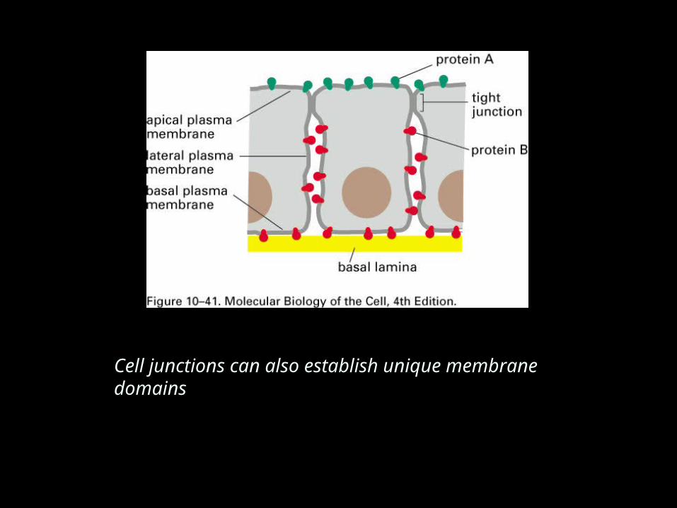
Cell junctions can also establish unique membrane domains

1. Membrane Organization and the Plasma Membrane
1c. Glycocalyx


1. Membrane Organization and the Plasma Membrane
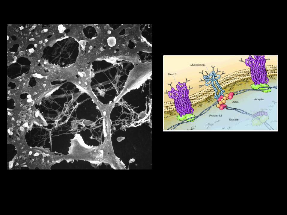
1d. Cytoskeletal associations

Cytoskeletal associations….

1. Membrane Organization and the Plasma Membrane
1e. Functions of the plasma membrane

FUNCTIONS OF THE PLASMA MEMBRANE
1. Protection and identification2. Semi-permeable barrier

FUNCTIONS OF THE PLASMA MEMBRANE
1. Protection and identification2. Semi-permeable barrier3. Transport - Passive and facilitated diffusion, active transport
4. Endocytosis and exocytosis
Endocytosis - transport via membrane flow

FUNCTIONS OF THE PLASMA MEMBRANE
1. Protection and identification2. Semi-permeable barrier3. Transport - Passive and facilitated diffusion, active transport
4. Endocytosis and exocytosis5. Sensing environmental conditions - membrane receptors
a. Ion channel-linked
e.g., acetylcholine receptor at neuromuscular junction
Can activate ion channels or other enzymes(e.g., epinephrine, serotonin, glucagon receptors)
e.g., cytokine and growth factor receptors
b. G protein-linked c. Enzyme-linked

2. Organelles
2a. Ribosomes and the Endoplasmic Reticulum


Membrane, lumenal and secreted
proteins made by RER
Cytoplasmic proteins are made by ‘free’ ribosomes


The oligosaccharide chains of N-linked glycoproteins are assembled on dolichol and transferred to proteins as they spool into the ER lumen

MOST of the proteins made by the RER are glycosylated (branched and N-linked).
FEW cytoplasmic proteins are glycosylated (mostly simple and O-linked)
There is a special class of extraordinarily heavily glycosylated proteins called PROTEOGLYCANS; these proteins are made in the ER but are glycosylated (via an O-linkage - linkage to serine or threonine) either in the Golgi apparatus or outside the cell. Proteoglycans are secreted by cells and make up part of the extracellular matrix.
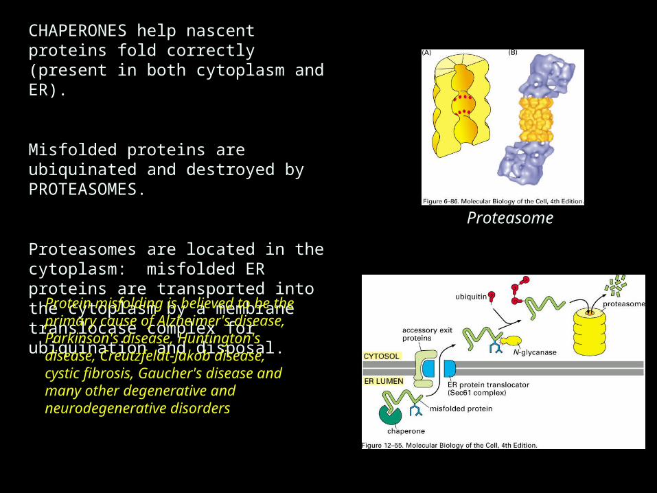
CHAPERONES help nascent proteins fold correctly (present in both cytoplasm and ER).
Misfolded proteins are ubiquinated and destroyed by PROTEASOMES.
Proteasomes are located in the cytoplasm: misfolded ER proteins are transported into the cytoplasm by a membrane translocase complex for ubiquination and disposal.
Proteasome
Protein misfolding is believed to be the primary cause of Alzheimer's disease, Parkinson's disease, Huntington's disease, Creutzfeldt-Jakob disease, cystic fibrosis, Gaucher's disease and many other degenerative and neurodegenerative disorders

OTHER FUNCTIONS OF ENDOPLASMIC RETICULUM:
-Lipid synthesis: delivery to Golgi, lysosomes, endosomes, plasma membrane is by membrane flow; delivery to mitochondria and peroxisomes is via exchange proteins
-Calcium regulation: ER sequesters and releases Ca++
-Detoxification: Cytochrome p450 enzymes