Embed Size (px)
Citation preview
The Permanente Journal/ Winter 2016/ Volume 20 No. 1 e105
CLINICAL MEDICINE
Image Diagnosis: Dental and Skeletal FluorosisNishtha Gupta, MDS; Nikhil Gupta, MD, MBBS; Puneet Chhabra, DM Perm J 2016 Winter;20(1):e105-e106
http://dx.doi.org/10.7812/TPP/15-048
CASE REPORTA 45-year-old man presented to the Department of Medicine
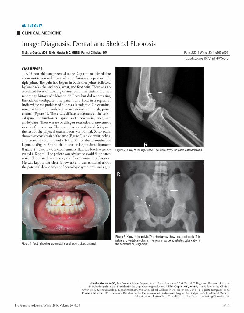
at our institution with 1 year of noninflammatory pain in mul-tiple joints. The pain had begun in both knee joints, followed by low-back ache and neck, wrist, and foot pain. There was no associated fever or swelling of any joint. The patient did not report any history of addiction or illness but did report using fluoridated toothpaste. The patient also lived in a region of India where the problem of fluorosis is endemic. On examina-tion, we found his teeth had brown strains and rough, pitted enamel (Figure 1). There was diffuse tenderness at the cervi-cal spine, the lumbosacral spine, and elbow, wrist, knee, and ankle joints. There was no swelling or restriction of movement in any of these areas. There were no neurologic deficits, and the rest of the physical examination was normal. X-ray scans showed osteosclerosis of the knee (Figure 2), ankle, wrist, pelvis, and vertebral column, and calcification of the sacrotuberous ligament (Figure 3) and the posterior longitudinal ligament (Figure 4). Twenty-four-hour urinary fluoride levels were el-evated (18 ppm). The patient was advised to avoid fluoridated water, fluoridated toothpaste, and foods containing fluoride. He was kept under close follow-up and was educated about the potential development of neurologic symptoms and signs.
Nishtha Gupta, MDS, is a Student in the Department of Endodontics at PDM Dental College and Research Institute in Bahadurgarh, India. E-mail: [email protected]. Nikhil Gupta, MD, MBBS, is a Fellow in the Clinical
Immunology & Rheumatology Department at Christian Medical College in Vellore, India. E-mail: [email protected]. Puneet Chhabra, DM, is a Senior Resident in the Department of Gastroenterology at the Postgraduate Institute of Medical
Education and Research in Chandigarh, India. E-mail: [email protected].
ONLINE ONLY
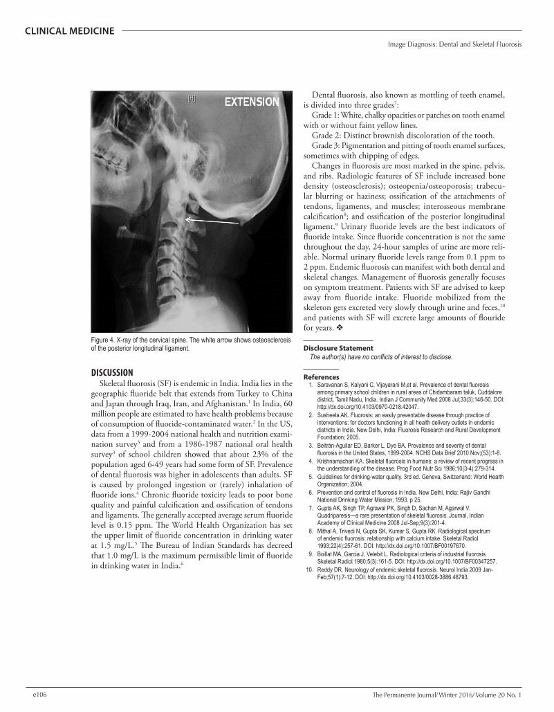
Figure 3. X-ray of the pelvis. The short arrow shows osteosclerosis of the pelvis and vertebral column. The long arrow demonstrates calcification of the sacrotuberous ligament.Figure 1. Teeth showing brown stains and rough, pitted enamel.
Figure 2. X-ray of the right knee. The white arrow indicates osteosclerosis.
The Permanente Journal/ Winter 2016/ Volume 20 No. 1e106
CLINICAL MEDICINEImage Diagnosis: Dental and Skeletal Fluorosis
DISCUSSIONSkeletal fluorosis (SF) is endemic in India. India lies in the
geographic fluoride belt that extends from Turkey to China and Japan through Iraq, Iran, and Afghanistan.1 In India, 60 million people are estimated to have health problems because of consumption of fluoride-contaminated water.2 In the US, data from a 1999-2004 national health and nutrition exami-nation survey3 and from a 1986-1987 national oral health survey3 of school children showed that about 23% of the population aged 6-49 years had some form of SF. Prevalence of dental fluorosis was higher in adolescents than adults. SF is caused by prolonged ingestion or (rarely) inhalation of fluoride ions.4 Chronic fluoride toxicity leads to poor bone quality and painful calcification and ossification of tendons and ligaments. The generally accepted average serum fluoride level is 0.15 ppm. The World Health Organization has set the upper limit of fluoride concentration in drinking water at 1.5 mg/L.5 The Bureau of Indian Standards has decreed that 1.0 mg/L is the maximum permissible limit of fluoride in drinking water in India.6
Dental fluorosis, also known as mottling of teeth enamel, is divided into three grades7:
Grade 1: White, chalky opacities or patches on tooth enamel with or without faint yellow lines.
Grade 2: Distinct brownish discoloration of the tooth.Grade 3: Pigmentation and pitting of tooth enamel surfaces,
sometimes with chipping of edges.Changes in fluorosis are most marked in the spine, pelvis,
and ribs. Radiologic features of SF include increased bone density (osteosclerosis); osteopenia/osteoporosis; trabecu-lar blurring or haziness; ossification of the attachments of tendons, ligaments, and muscles; interosseous membrane calcification8; and ossification of the posterior longitudinal ligament.9 Urinary fluoride levels are the best indicators of fluoride intake. Since fluoride concentration is not the same throughout the day, 24-hour samples of urine are more reli-able. Normal urinary fluoride levels range from 0.1 ppm to 2 ppm. Endemic fluorosis can manifest with both dental and skeletal changes. Management of fluorosis generally focuses on symptom treatment. Patients with SF are advised to keep away from fluoride intake. Fluoride mobilized from the skeleton gets excreted very slowly through urine and feces,10 and patients with SF will excrete large amounts of flouride for years. v
Disclosure StatementThe author(s) have no conflicts of interest to disclose.
References 1. Saravanan S, Kalyani C, Vijayarani M,et al. Prevalence of dental fluorosis
among primary school children in rural areas of Chidambaram taluk, Cuddalore district, Tamil Nadu, India. Indian J Community Med 2008 Jul;33(3):146-50. DOI: http://dx.doi.org/10.4103/0970-0218.42047.
2. Susheela AK. Fluorosis: an easily preventable disease through practice of interventions: for doctors functioning in all health delivery outlets in endemic districts in India. New Delhi, India: Fluorosis Research and Rural Development Foundation; 2005.
3. Beltrán-Aguilar ED, Barker L, Dye BA. Prevalence and severity of dental fluorosis in the United States, 1999-2004. NCHS Data Brief 2010 Nov;(53):1-8.
4. Krishnamachari KA. Skeletal fluorosis in humans: a review of recent progress in the understanding of the disease. Prog Food Nutr Sci 1986;10(3-4):279-314.
5. Guidelines for drinking-water quality. 3rd ed. Geneva, Switzerland: World Health Organization; 2004.
6. Prevention and control of fluorosis in India. New Delhi, India: Rajiv Gandhi National Drinking Water Mission; 1993. p 25.
7. Gupta AK, Singh TP, Agrawal PK, Singh D, Sachan M, Agarwal V. Quadriparesis—a rare presentation of skeletal fluorosis. Journal, Indian Academy of Clinical Medicine 2008 Jul-Sep;9(3):201-4.
8. Mithal A, Trivedi N, Gupta SK, Kumar S, Gupta RK. Radiological spectrum of endemic fluorosis: relationship with calcium intake. Skeletal Radiol 1993;22(4):257-61. DOI: http://dx.doi.org/10.1007/BF00197670.
9. Boillat MA, Garcia J, Velebit L. Radiological criteria of industrial fluorosis. Skeletal Radiol 1980;5(3):161-5. DOI: http://dx.doi.org/10.1007/BF00347257.
10. Reddy DR. Neurology of endemic skeletal fluorosis. Neurol India 2009 Jan-Feb;57(1):7-12. DOI: http://dx.doi.org/10.4103/0028-3886.48793.
Figure 4. X-ray of the cervical spine. The white arrow shows osteosclerosis of the posterior longitudinal ligament.