Embed Size (px)
Citation preview
IEEE TRANSACTIONS ON NEURAL SYSTEMS AND REHABILITATION ENGINEERING, VOL. 16, NO. 4, AUGUST 2008 371
Assistive Control System Using ContinuousMyoelectric Signal in Robot-Aided Arm
Training for Patients After StrokeRong Song, Kai-yu Tong, Senior Member, IEEE, Xiaoling Hu, and Le Li
Abstract—In some stroke rehabilitation programs, robotic sys-tems have been used to aid the patient to train. In this study, amyoelectrically controlled robotic system with 1 degree-of-freedomwas developed to assist elbow training in a horizontal plane with in-tention involvement for people after stroke. The system could pro-vide continuous assistance in extension torque, which was propor-tional to the amplitude of the subject’s electromyographic (EMG)signal from the triceps, and could provide resistive torques duringmovement. This study investigated the system’s effect on restoringthe upper limb functions of eight subjects after chronic stroke ina twenty-session rehabilitation training program. In each session,there were 18 trials comprising different combinations of assistiveand resistive torques and an evaluation trial. Each trial consistedof five cycles of repetitive elbow flexion and extension between 90and 0 at a constant velocity of 10 /s. With the assistive extensiontorque, subjects could reach a more extended position in the firstsession. After 20 sessions of training, there were statistically signif-icant improvements in the modified Ashworth scale, Fugl–Meyerscale for shoulder and elbow, motor status scale, elbow extensionrange, muscle strength, and root mean square error between actualelbow angle and target angle. The results showed that the twenty-session training program improved upper limb functions.
Index Terms—Arm tracking, myoelectric control, robot-assistedrehabilitation, stroke.
I. INTRODUCTION
S TROKE is a leading cause of death and disability in manycountries [1]. Patients after stroke are often reported to
have a lower quality of life (QOL), due to stroke-induced dis-abilities, than normal subjects of similar age [2], [3]. Poststrokedepression is also reported in patients after stroke with impairedQOL [4]. It is important for them, their families, and society thatthey are helped in restoring their lost motor functions to improvetheir QOL.
Rehabilitation training has been shown to have a positive ef-fect on neurological restoration of limb functions [5]. Conven-tionally, rehabilitation training can be conducted by therapists ina one-on-one manual mode in a hospital. In recent years, robotic
Manuscript received August 29, 2007; revised March 18, 2008; acceptedMarch 26, 2008. First published June 3, 2008; last published August 13, 2008(projected). This work was supported in part by the Research Grants Councilof the Hong Kong Special Administrative Region, China under Grant PolyU5271/05E.
The authors are with the Department of Health Technology and informatics,The Hong Kong Polytechnic University, Kowloon, Hong Kong (e-mail: [email protected]; [email protected]; [email protected];[email protected]).
Color versions of one or more of the figures in this paper are available onlineat http://ieeexplore.ieee.org.
Digital Object Identifier 10.1109/TNSRE.2008.926707
systems have been developed as useful complementary units totherapists to manipulate a paretic arm [6]–[12]. MIT-Manus isa robotic system designed for upper limb stroke rehabilitation[5], [8]. The key feature of MIT-MANUS is its impedance con-trol, which can keep a compliant trajectory under perturbation.Its therapeutic effect has been confirmed through a series of ex-periments [12]–[14]. Mirror-image movement enabler (MIME)is 3-D space [10], [15]. Patients can use their unaffected sideto control their affected side to practice mirror-image move-ment by a bimanual position feedback strategy. Daily therapywith MIME in chronic hemiparetic subjects showed a signifi-cant improvement in their muscle strength and motion function[15]. ARM Guide was designed for both training and evalua-tion of upper limb reaching functions in a linear trajectory [9],[16]. Colombo et al. also designed a wrist manipulator with1 degree-of-freedom (DOF) and an elbow–shoulder manipu-lator with 2 DOF for rehabilitation of upper limb movements.They used admittance control to reduce the inertia and facili-tate the movement [6]. Recent developments involving rehabil-itation robots have worked toward interactive control, with therobotic systems reacting to inputs from the subject [11].
Myoelectric control is related to the subject’s intention andcan be used as a control variable since surface electromyo-graphic (EMG) signals reflect the activities of the muscles.EMG signals have been frequently applied in the control ofprosthetics for more than forty years and can be classified in“on–off” control [21], proportional control [22], and a morecomplex form, for distinguishing different kinds of motion[19], [20]. Myoelectric control has also been reported for thecontrol of functional electrical stimulation (FES) systems inrehabilitation [18], [23], [24], since voluntary physical exerciseis important to promote the recovery of brain function in pa-tients after stroke [17]. Recently, many researchers have usedEMG signals to continuously control exoskeleton robots thatcan be worn by the human subject as an assistive device. Theresearchers used EMG signals of selective muscles to estimatethe joint torque, and applied the assistive torque to the joint toprovide additional power. In such studies, the system is underthe control of the subject’s intention, functioning like additionalmuscle groups [26]–[29]. However, Rosen et al. only appliedthe robotic system using continuous EMG control on normalsubjects to share the loading [26], [27]. Cheng et al. appliedtheir system to provide continuous assistive torque for subjectsafter stroke [28]. Their systems could improve the elbow torquecapability of unimpaired subjects and of subjects after strokewithin their voluntary range-of-motion, respectively. However,
1534-4320/$25.00 © 2008 IEEE
Authorized licensed use limited to: Hong Kong Polytechnic University. Downloaded on May 06,2010 at 08:53:33 UTC from IEEE Xplore. Restrictions apply.
372 IEEE TRANSACTIONS ON NEURAL SYSTEMS AND REHABILITATION ENGINEERING, VOL. 16, NO. 4, AUGUST 2008
Fig. 1. Architecture of the myoelectrically controlled robotic system.
it has not been reported if those kinds of devices could helpsubjects after stroke to perform rehabilitation beyond theirvoluntary range, and if those kinds of devices could be appliedas a rehabilitation robot in a robot-aided therapy. The use ofmyoelectrically controlled robot-aided therapy for subjectsafter stroke has so far been studied in an EMG-triggered“on–off” control [25]. A sensorimotor integration theory hasbeen applied to explain that the voluntary efferent output aswell as the afferent sensor input were helpful to promote thereorganization of the brain [18]. Though the subject could onlycontrol the initial action of the external robotic system in theEMG-triggered “on–off” control, the robotic system wouldafterward operate with a predefined trajectory or action for aperiod of time, which had no interaction with the EMG signalduring this period until the time allowed for the next triggerevent. The additional intention control through continuousmyoelectric control could provide more interaction duringthe whole motion, which might be beneficial in promotingthe restoration of motor functions for patients after stroke.Our pilot study had reported promising therapeutic effectsof a myoelectrically controlled robotic system in improvingmuscle strength and extension range in three subjects [30]. Inthis present study, eight subjects after stroke were recruitedfor statistical analysis to evaluate the effects of training witha continuous myoelectrically controlled robotic system. Theoutcome parameters included clinical scales (modified Ash-worth scale, Fugl–Meyer scale, and motor status scale), musclestrength, range-of-motion, and robot-measured parameters.If there was no EMG activation from the subject, the roboticsystem would not generate an assistive torque. Therefore, allof the subjects were encouraged to use their residual voluntaryEMG to actively participate in the training.
II. METHODS
A. System
The structure of the myoelectrically controlled roboticsystem is shown in Fig. 1 and consists of a personal computer
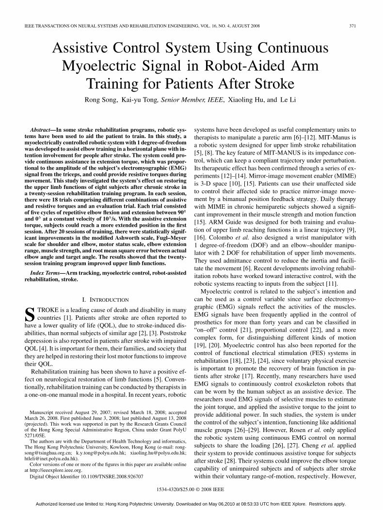
Fig. 2. Diagram of the myoelectrically controlled robotic system.
(PC), a PC-based data acquisition device, an actuated mechan-ical part, and an EMG amplifier. After being captured throughEMG electrodes (Noraxon, Scottsdale, AZ) and amplified bythe EMG amplifier, the EMG signals together with the torquesignal and the angle signal were inputted through the data acqui-sition (DAQ) card (PCI 6036E, National Instruments, Austin,TX) into the computer. The software has three functions: 1) itgenerated a control signal and controlled the motor to providemechanical help through the DAQ card, 2) it provided a task toguide the subject and provided real-time visual feedback to thesubject during the task, displaying both the target and the actualelbow joint angle on a computer screen placed in front of thesubject, and 3) it stored the EMG, torque and angle signals forfurther analysis. The mechanical part of a robotic manipulatorwith 1 DOF was designed and fabricated for assisting the move-ment of elbow flexion and extension (Fig. 2). The two layersof aluminum plates were connected by four aluminum pillars.The lower plate was fixed to a table. The direct drive (DDR)brushless AC servo motor (DM 1045B, Yokogawa, Japan) wasfixed to the lower plate. The motor was connected to a torquesensor (AKC-205, 701st Research Institute of China Aerospace
Authorized licensed use limited to: Hong Kong Polytechnic University. Downloaded on May 06,2010 at 08:53:33 UTC from IEEE Xplore. Restrictions apply.

SONG et al.: ASSISTIVE CONTROL SYSTEM USING CONTINUOUS MYOELECTRIC SIGNAL IN ROBOT-AIDED ARM TRAINING 373
Science and Technology Corporation, China). The other endof the torque sensor was connected to a manipulandum. Anorthosis with a semicircular cross section was attached to themanipulandum. The subject’s forearm was placed inside theorthosis and straps were used to secure the forearm in place.The manipulandum had a handle that the subject grasped forthe experiment. The position of the handle was adjustableaccording to the length of the subject’s forearm. The upper armwas also fastened by a strap to a support mounted on the upperaluminum plate. The orthosis and manipulandum could guidethe forearm to rotate with an axis of rotation in line with themotor and the torque sensor. The torque sensor could measurethe interaction torque between the manipulandum and the servomotor. The DDR motor was driven by a servo driver (SD1045B,Yokogawa, Japan). An optical incremental shaft encoder wasattached to the motor shaft for measuring the joint angle.
For safety reasons, three steps were taken to protect each sub-ject during the experiment. First, two mechanical stops wereused to limit the rotation range of the motor. Second, the soft-ware program limited the output torque to a preset range of 5to 5 Nm, and the operation would be stopped if the motor ex-ceeded this range. Third, an emergency stop could be pressedby the subject to break the power supply to the servo motor ifneeded.
B. EMG-Signal Processing Procedures
Abnormal biceps activation during elbow extension is oftenfound in subjects after stroke, reflecting the impairment of theirability to selectively activate flexors and extensors [44]. Usingthis finding, the present study avoided using the subjects’ elbowflexors for the control signals during elbow extension in order tominimize the interference of abnormal firing patterns from thebiceps in the movement. The EMG signal from medial tricepsbrachii of the affected arm was used as the control signal forproportional control of the robotic system.
The EMG signals were amplified with a custom-made EMGsystem using an instrumentation amplifier (INA126, Texas In-struments, Dallas, TX). The signals were amplified with a gainof 1000 and were band-pass filtered in a 10–400-Hz band. TheEMG signals were all sampled at 1000 Hz. The envelope of theEMG signals was obtained after the signals were full-wave rec-tified and filtered with a moving average window (100 ms).
The processed triceps EMG signals were then normalizedto the range 0–1 for , as in the following [28]:
(1)
where was the amplitude of the processed triceps EMGsignal at rest, and was the maximum amplitude ofthe processed triceps EMG signal during maximum isometricvoluntary extension (MIVE) at 90 elbow flexion. The assistivetorque was estimated based on the normalized EMGsignals, as in the following:
(2)
where was the gain for EMG to torque conversion. The EMG-torque gain was set at 0%, 50%, and 100% in this study.
was the MIVE torque. The resultant torque is shown in thefollowing:
(3)
where . was the MIVE torque duringelbow extension and the maximum isometric voluntary flexion(MIVF) torque during elbow flexion. was a coefficient of theresistive torque, the level of which ranged from 0%, 10%, and20%. The range of the EMG-torque gain (0%–100%) and thecoefficient of the resistive torque (0%–20%) were determinedin a pilot experiment with twelve subjects after chronic strokeand based on the performance of their movements. The 12 sub-jects could manipulate the system easily, with the above-men-tioned parameters during elbow flexion and extension in therobotic system.
C. Experiment Setup
Eight of the 12 subjects in the pilot study (seven males, onefemale) were recruited for this present study consisting of atwenty-session training program, based on their availability toparticipate in the 20 sessions of training. The mean age of theeight subjects was 50 9 years, and they ranged from 39 to62 years. Each subject was to undergo 20 sessions of training,with three to five sessions conducted each week over six con-secutive weeks. The criteria for recruiting the subjects includedthe following: 1) there should be at least six months after uni-lateral stroke in order to minimize the effect of spontaneous re-covery [49] (the mean duration from stroke onset was 5.74.2 years, ranging from 10 months to 13 years), 2) the subjectsshould not have visuospatial, cognitive, or attention deficits thatwould prevent them from following instructions or performingthe experimental procedures, and 3) the subjects should have ameasurable EMG signal from medial triceps brachii (the pro-cessed EMG signal after the moving window should be at leasttwice as large than that at rest). This study was approved by theHuman Subject Ethics Sub-Committee of The Hong Kong Poly-technic University. Prior to the experiment, the subjects wereexplained the experimental procedures and duration before theysigned the consent forms.
During the experiment, the subject was seated beside thesystem. The shoulder was in 90 abduction. The affectedforearm was attached to the manipulandum, and the subjectwas asked to grasp the handle of the manipulandum. Theorthosis and strap were used to secure the forearm in position.A computer screen was placed in front of the subject to providevisual information of the target angle for the subject to follow,and the subject was instructed to complete the following tasks.
1) The MIVE and MIVF torques were measured for the af-fected elbow flexors and extensors when the elbow waspositioned at a 90 angle in the horizontal plane, since themaximum MIVE and MIVF torques across all elbow an-gles could be achieved at nearly 90 [47]. The EMG signalsduring MIVE were captured to normalize the EMG signalof the triceps. Three trials were performed for 5 s each, andthe maximum values of the torque and EMG signal wereused.
Authorized licensed use limited to: Hong Kong Polytechnic University. Downloaded on May 06,2010 at 08:53:33 UTC from IEEE Xplore. Restrictions apply.
374 IEEE TRANSACTIONS ON NEURAL SYSTEMS AND REHABILITATION ENGINEERING, VOL. 16, NO. 4, AUGUST 2008
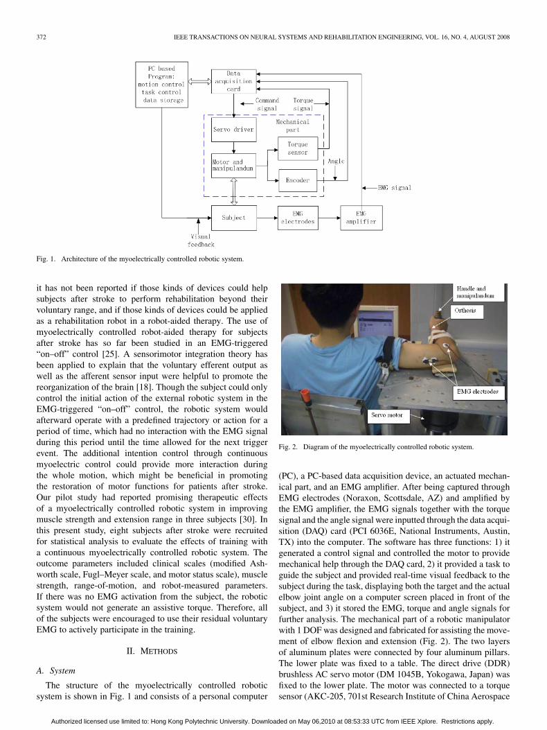
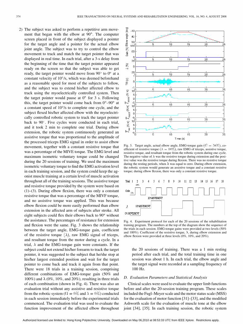
2) The subject was asked to perform a repetitive arm move-ment that began with the elbow at 90 . The computerscreen placed in front of the subject displayed a pointerfor the target angle and a pointer for the actual elbowjoint angle. The subject was to try to control the elbowmovement to track and match the target pointer that wasdisplayed in real time. In each trial, after a 3-s delay fromthe beginning of the time that the target pointer appearedready on the screen so that the subject was able to getready, the target pointer would move from 90 to 0 at aconstant velocity of 10 /s, which was deemed beforehandas a reasonable speed for most of the subjects to follow,and the subject was to extend his/her affected elbow totrack using the myoelectrically controlled system. Thenthe target pointer would pause at 0 for 3 s. Followingthis, the target pointer would come back from 0 –90 ata constant speed of 10 /s to complete one cycle, and thesubject flexed his/her affected elbow with the myoelectri-cally controlled robotic system to track the target pointerback to 90 . Five cycles were conducted in each trial,and it took 2 min to complete one trial. During elbowextension, the robotic system continuously generated anassistive torque that was proportional to the amplitude ofthe processed triceps EMG signal in order to assist elbowmovement, together with a constant resistive torque thatwas a percentage of the MIVE torque. The EMG level andmaximum isometric voluntary torque could be changedduring the 20 sessions of training. We used the maximumisometric voluntary torque to find the EMG activation levelin each training session, and the system could keep the ag-onist muscle training at a certain level of muscle activationthroughout all of the training sessions. The assistive torqueand resistive torque provided by the system were based on(1)–(3). During elbow flexion, there was only a constantresistive torque that was a percentage of the MIVF torque,and no assistive torque was applied. This was becauseelbow flexion could be more easily performed than elbowextension in the affected arm of subjects after stroke. Alleight subjects could flex their elbows back to 90 withoutthe assistance. The percentages of resistance for extensionand flexion were the same. Fig. 3 shows the relationshipbetween the target angle, EMG-torque gain, coefficientof the resistive torque , raw EMG signal of triceps,and resultant torque from the motor during a cycle. In atrial, and the EMG-torque gain were constants. If thesubject could not extend his/her forearm to track the targetpointer, it was suggested to the subject that he/she stop athis/her largest extended position and wait for the targetpointer to come back and track it again from that point.There were 18 trials in a training session, comprisingdifferent combinations of EMG-torque gain (50% and100%) and (0%, 10%, and 20%), resulting in three trialsof each combination (shown in Fig. 4). There was also anevaluation trial without any assistive and resistive torquefrom the robotic system ( and ) conductedin each session immediately before the experimental trialscommenced. The evaluation trial was used to evaluate thefunction improvement of the affected elbow throughout
Fig. 3. Target angle, actual elbow angle, EMG-torque gain (G = 50%), co-efficient of resistive torque (� = 10%), raw EMG of triceps, assistive torque,resistive torque, and resultant torque from the robotic system during one cycle.The negative value of � was the resistive torque during extension and the posi-tive value was the resistive torque during flexion. There was no resistive torqueduring the resting periods, when � was equal to zero. During elbow extension,the robotic system would generate an assistive torque and a constant resistivetorque; during elbow flexion, there was only a constant resistive torque.
Fig. 4. Experiment protocol for each of the 20 sessions of the rehabilitationtraining program. The numbers at the top of the diagram show the sequence ofthe trials in each session. EMG-torque gains were provided at two levels (50%and 100%). Coefficient of the resistive torque, �, during elbow extension andelbow flexion were provided at three levels (0%, 10%, and 20%).
the 20 sessions of training. There was a 1 min restingperiod after each trial, and the total training time in onesession was about 1 h. In each trial, the elbow angle andthe target signal were recorded at a sampling frequency of100 Hz.
D. Evaluation Parameters and Statistical Analysis
Clinical scales were used to evaluate the upper limb functionsbefore and after the 20-session training program. These scalesincluded the Fugl–Meyer scale and the motor status scale (MSS)for the evaluation of motor function [31]–[33], and the modifiedAshworth scale for the evaluation of muscle tone at the elbowjoint [34], [35]. In each training session, the robotic system
Authorized licensed use limited to: Hong Kong Polytechnic University. Downloaded on May 06,2010 at 08:53:33 UTC from IEEE Xplore. Restrictions apply.
SONG et al.: ASSISTIVE CONTROL SYSTEM USING CONTINUOUS MYOELECTRIC SIGNAL IN ROBOT-AIDED ARM TRAINING 375
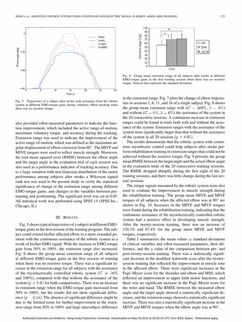
Fig. 5. Trajectories of a subject after stroke with assistance from the roboticsystem at different EMG-torque gains during voluntary elbow tracking whenthere was no resistive torque.
also provided robot-measured parameters to indicate the func-tion improvement, which included the active range-of-motion,maximum voluntary torque, and accuracy during the tracking.Extension range was used to indicate the improvement of theactive range-of-motion, which was defined as the maximum an-gular displacement of elbow extension from 90 . The MIVF andMIVE torques were used to reflect muscle strength. Moreover,the root mean squared error (RMSE) between the elbow angleand the target angle in the evaluation trial of each session wasalso used as a performance indicator of tracking accuracy. Dueto a large variation with non-Gaussian distribution of the motorperformance among subjects after stroke, a Wilcoxon signedrank test was used in the present study to verify the statisticalsignificance of change in the extension range among differentEMG-torque gains, and changes in the variables between pre-training and posttraining. The significant level was set at 0.05.All statistical work was performed using SPSS 14 (SPSS Inc.,Chicago, IL).
III. RESULTS
Fig. 5 shows typical trajectories of a subject at different EMG-torque gains in the first session of the training program. The sub-ject could extend his/her affected elbow to a more extended po-sition with the continuous assistance of the robotic system as aresult of his/her EMG signal. With the increase in EMG-torquegain from 50% to 100%, the extension range also increased.Fig. 6 shows the group mean extension range of all subjectsat different EMG-torque gains in the first session of trainingwhen there was no resistive torque. There was a significant in-crease in the extension range for all subjects with the assistanceof the myoelectrically controlled robotic system (and 100%), compared with that without the assistance of thesystem ( for both comparisons). There was an increasein extension range when the EMG-torque gain increased from50% to 100%, but the results did not show significant differ-ence . The absence of significant difference might bedue to the limited room for further improvement in the exten-sion range from 50% to 100% and large intersubject difference
Fig. 6. Group mean extension range of all subjects after stroke at differentEMG-torque gains in the first training session when there was no resistivetorque. Vertical bars represent the standard deviation.
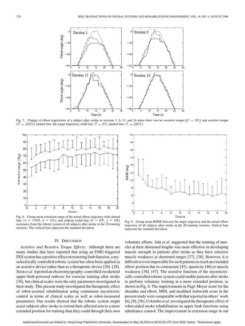
in the extension range. Fig. 7 plots the change of elbow trajecto-ries in sessions 1, 6, 11, and 16 of a single subject. Fig. 8 showsthe group mean extension range with ( , )and without ( , ) the assistance of the system inthe 20 consecutive sessions. A continuous increase in extensionranges could be found in trials both with and without the assis-tance of the system. Extension ranges with the assistance of thesystem were significantly larger than that without the assistanceof the system in all 20 sessions .
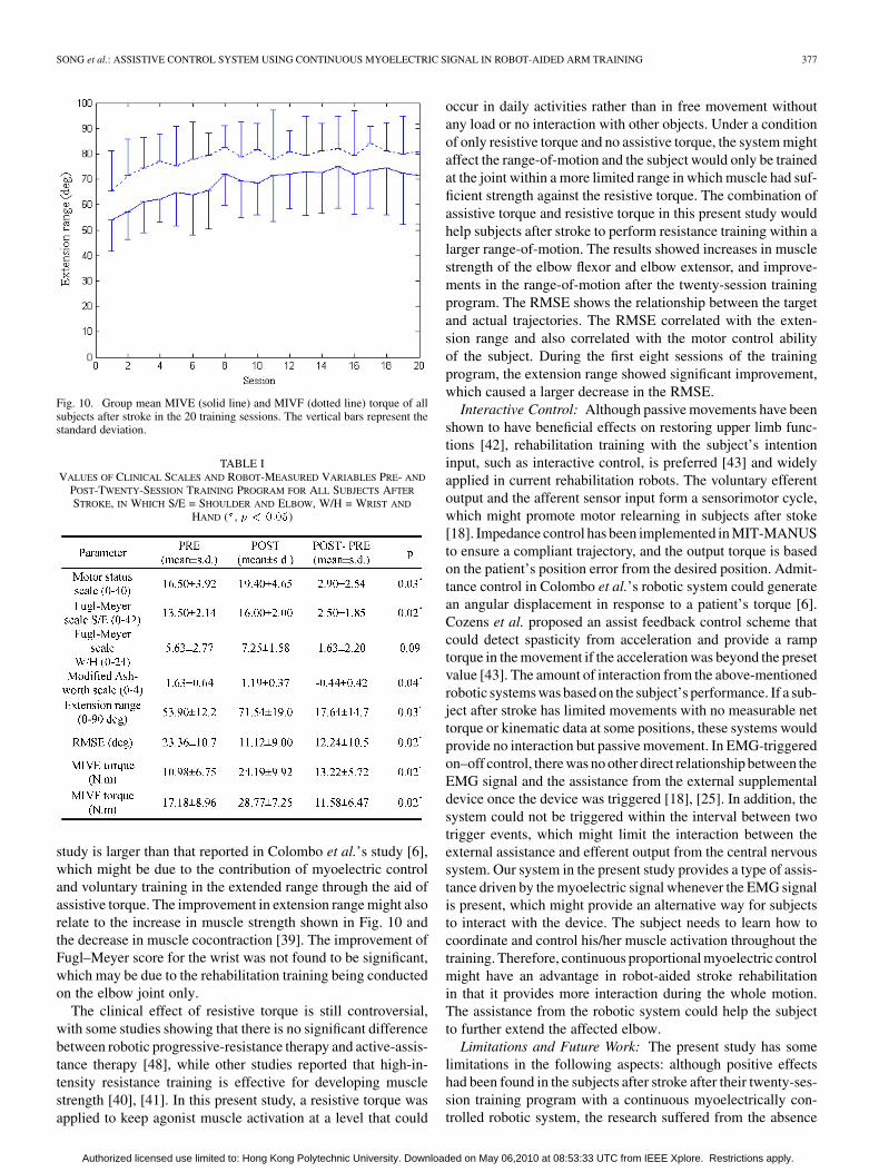
The results demonstrate that the robotic system with contin-uous myoelectric control could help subjects after stroke per-form rehabilitation training in extension ranges that could not beachieved without the assistive torque. Fig. 9 presents the groupmean RMSE between the target angle and the actual elbow anglein the evaluation trials of the 20 consecutive training sessions.The RMSE dropped abruptly during the first eight of the 20training sessions, and there was little change during the last sev-eral sessions.
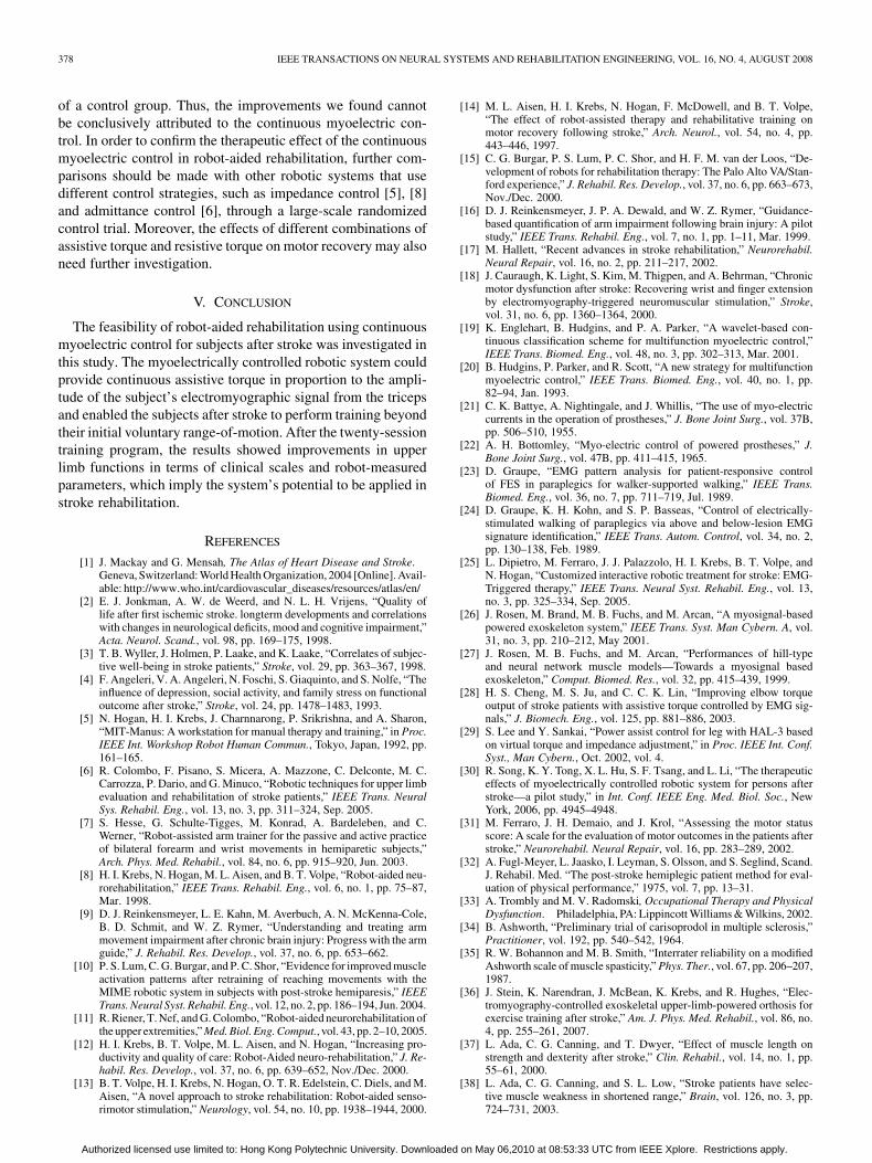
The torque signals measured by the robotic system were alsoused to evaluate the improvement in muscle strength duringthe rehabilitation training. The group mean MIVE and MIVFtorques of all subjects when the affected elbow was at 90 areshown in Fig. 10. Increases in the MIVE and MIVF torqueswere found during the rehabilitation training, indicating that thecontinuous assistance of the myoelectrically controlled roboticsystem had a positive effect in developing muscle strength.After the twenty-session training, there was an increase of120.3% and 67.4% for the group mean MIVE and MIVFtorques, respectively.
Table I summarizes the mean values standard deviationsof clinical variables and robot-measured parameters, their dif-ference, and the value of the comparison between pre- andpost-twenty-session training. There was a statistically signifi-cant decrease in the modified Ashworth score after the twenty-session training that reflected the improvement in muscle tonein the affected elbow. There were significant increases in theFugl–Meyer score for the shoulder and elbow and MSS, whichreflected an improvement in upper limb motor functions, andthere was no significant increase in the Fugl–Meyer score forthe wrist and hand. The RMSE between the measured elbowangle and the target angle showed a statistically significant de-crease, and the extension range showed a statistically significantincrease. There was also a statistically significant increase in theMIVF and MIVE torques when the elbow angle was at 90 .
Authorized licensed use limited to: Hong Kong Polytechnic University. Downloaded on May 06,2010 at 08:53:33 UTC from IEEE Xplore. Restrictions apply.
376 IEEE TRANSACTIONS ON NEURAL SYSTEMS AND REHABILITATION ENGINEERING, VOL. 16, NO. 4, AUGUST 2008
Fig. 7. Change of elbow trajectories of a subject after stroke in sessions 1, 6, 11, and 16 when there was no assistive torque (G = 0%) and assistive torque(G = 100%) (dotted line: the target trajectory; solid line: G = 0%; dashed line: G = 100%).
Fig. 8. Group mean extension range of the actual elbow trajectory with (dottedline: G = 100%, � = 0%), and without (solid line: G = 0%, � = 0%)assistance from the robotic system of all subjects after stroke in the 20 trainingsessions. The vertical bars represent the standard deviation.
IV. DISCUSSION
Assistive and Resistive Torque Effects: Although there aremany studies that have reported that using an EMG-triggeredFES system has a positive effect on restoring limb function, a my-oelectrically controlled robotic system has often been applied asan assistive device rather than as a therapeutic device [26]–[28].Stein et al. reported an electromyography-controlled exoskeletalupper-limb-powered orthosis for exercise training after stroke[36], but clinical scales were the only parameters investigated intheir study. This present study investigated the therapeutic effectof robot-assisted rehabilitation using continuous myoelectriccontrol in terms of clinical scales as well as robot-measuredparameters. Our results showed that the robotic system mightassist subjects after stroke to extend their affected arm to a moreextended position for training than they could through their own
Fig. 9. Group mean RMSE between the target trajectory and the actual elbowtrajectory of all subjects after stroke in the 20 training sessions. Vertical barsrepresent the standard deviation.
voluntary efforts. Ada et al. suggested that the training of mus-cles at their shortened lengths was more effective in developingmuscle strength in patients after stroke as they have selectivemuscle weakness at shortened ranges [37], [38]. However, it isdifficult or even impossible for such patients to reach an extendedelbow position due to contracture [45], spasticity [46] or muscleweakness [38], [47]. The assistive function of the myoelectri-cally controlled robotic system could enable patients after stroketo perform voluntary training at a more extended position, asshown in Fig. 8. The improvements in Fugl–Meyer score for theshoulder and elbow, MSS, and modified Ashworth score in thepresent study were comparable with that reported in others’ work[6], [9], [36]. Colombo et al. investigated the therapeutic effect ofrobot-aided stroke rehabilitation on upper limb functions usingadmittance control. The improvement in extension range in our
Authorized licensed use limited to: Hong Kong Polytechnic University. Downloaded on May 06,2010 at 08:53:33 UTC from IEEE Xplore. Restrictions apply.
SONG et al.: ASSISTIVE CONTROL SYSTEM USING CONTINUOUS MYOELECTRIC SIGNAL IN ROBOT-AIDED ARM TRAINING 377
Fig. 10. Group mean MIVE (solid line) and MIVF (dotted line) torque of allsubjects after stroke in the 20 training sessions. The vertical bars represent thestandard deviation.
TABLE IVALUES OF CLINICAL SCALES AND ROBOT-MEASURED VARIABLES PRE- AND
POST-TWENTY-SESSION TRAINING PROGRAM FOR ALL SUBJECTS AFTER
STROKE, IN WHICH S/E = SHOULDER AND ELBOW, W/H = WRIST AND
HAND ( , p < 0:05)
study is larger than that reported in Colombo et al.’s study [6],which might be due to the contribution of myoelectric controland voluntary training in the extended range through the aid ofassistive torque. The improvement in extension range might alsorelate to the increase in muscle strength shown in Fig. 10 andthe decrease in muscle cocontraction [39]. The improvement ofFugl–Meyer score for the wrist was not found to be significant,which may be due to the rehabilitation training being conductedon the elbow joint only.
The clinical effect of resistive torque is still controversial,with some studies showing that there is no significant differencebetween robotic progressive-resistance therapy and active-assis-tance therapy [48], while other studies reported that high-in-tensity resistance training is effective for developing musclestrength [40], [41]. In this present study, a resistive torque wasapplied to keep agonist muscle activation at a level that could
occur in daily activities rather than in free movement withoutany load or no interaction with other objects. Under a conditionof only resistive torque and no assistive torque, the system mightaffect the range-of-motion and the subject would only be trainedat the joint within a more limited range in which muscle had suf-ficient strength against the resistive torque. The combination ofassistive torque and resistive torque in this present study wouldhelp subjects after stroke to perform resistance training within alarger range-of-motion. The results showed increases in musclestrength of the elbow flexor and elbow extensor, and improve-ments in the range-of-motion after the twenty-session trainingprogram. The RMSE shows the relationship between the targetand actual trajectories. The RMSE correlated with the exten-sion range and also correlated with the motor control abilityof the subject. During the first eight sessions of the trainingprogram, the extension range showed significant improvement,which caused a larger decrease in the RMSE.
Interactive Control: Although passive movements have beenshown to have beneficial effects on restoring upper limb func-tions [42], rehabilitation training with the subject’s intentioninput, such as interactive control, is preferred [43] and widelyapplied in current rehabilitation robots. The voluntary efferentoutput and the afferent sensor input form a sensorimotor cycle,which might promote motor relearning in subjects after stoke[18]. Impedance control has been implemented in MIT-MANUSto ensure a compliant trajectory, and the output torque is basedon the patient’s position error from the desired position. Admit-tance control in Colombo et al.’s robotic system could generatean angular displacement in response to a patient’s torque [6].Cozens et al. proposed an assist feedback control scheme thatcould detect spasticity from acceleration and provide a ramptorque in the movement if the acceleration was beyond the presetvalue [43]. The amount of interaction from the above-mentionedrobotic systems was based on the subject’s performance. If a sub-ject after stroke has limited movements with no measurable nettorque or kinematic data at some positions, these systems wouldprovide no interaction but passive movement. In EMG-triggeredon–off control, there was no other direct relationship between theEMG signal and the assistance from the external supplementaldevice once the device was triggered [18], [25]. In addition, thesystem could not be triggered within the interval between twotrigger events, which might limit the interaction between theexternal assistance and efferent output from the central nervoussystem. Our system in the present study provides a type of assis-tance driven by the myoelectric signal whenever the EMG signalis present, which might provide an alternative way for subjectsto interact with the device. The subject needs to learn how tocoordinate and control his/her muscle activation throughout thetraining. Therefore, continuous proportional myoelectric controlmight have an advantage in robot-aided stroke rehabilitationin that it provides more interaction during the whole motion.The assistance from the robotic system could help the subjectto further extend the affected elbow.
Limitations and Future Work: The present study has somelimitations in the following aspects: although positive effectshad been found in the subjects after stroke after their twenty-ses-sion training program with a continuous myoelectrically con-trolled robotic system, the research suffered from the absence
Authorized licensed use limited to: Hong Kong Polytechnic University. Downloaded on May 06,2010 at 08:53:33 UTC from IEEE Xplore. Restrictions apply.
378 IEEE TRANSACTIONS ON NEURAL SYSTEMS AND REHABILITATION ENGINEERING, VOL. 16, NO. 4, AUGUST 2008
of a control group. Thus, the improvements we found cannotbe conclusively attributed to the continuous myoelectric con-trol. In order to confirm the therapeutic effect of the continuousmyoelectric control in robot-aided rehabilitation, further com-parisons should be made with other robotic systems that usedifferent control strategies, such as impedance control [5], [8]and admittance control [6], through a large-scale randomizedcontrol trial. Moreover, the effects of different combinations ofassistive torque and resistive torque on motor recovery may alsoneed further investigation.
V. CONCLUSION
The feasibility of robot-aided rehabilitation using continuousmyoelectric control for subjects after stroke was investigated inthis study. The myoelectrically controlled robotic system couldprovide continuous assistive torque in proportion to the ampli-tude of the subject’s electromyographic signal from the tricepsand enabled the subjects after stroke to perform training beyondtheir initial voluntary range-of-motion. After the twenty-sessiontraining program, the results showed improvements in upperlimb functions in terms of clinical scales and robot-measuredparameters, which imply the system’s potential to be applied instroke rehabilitation.
REFERENCES
[1] J. Mackay and G. Mensah, The Atlas of Heart Disease and Stroke.Geneva, Switzerland: World Health Organization, 2004 [Online]. Avail-able: http://www.who.int/cardiovascular_diseases/resources/atlas/en/
[2] E. J. Jonkman, A. W. de Weerd, and N. L. H. Vrijens, “Quality oflife after first ischemic stroke. longterm developments and correlationswith changes in neurological deficits, mood and cognitive impairment,”Acta. Neurol. Scand., vol. 98, pp. 169–175, 1998.
[3] T. B. Wyller, J. Holmen, P. Laake, and K. Laake, “Correlates of subjec-tive well-being in stroke patients,” Stroke, vol. 29, pp. 363–367, 1998.
[4] F. Angeleri, V. A. Angeleri, N. Foschi, S. Giaquinto, and S. Nolfe, “Theinfluence of depression, social activity, and family stress on functionaloutcome after stroke,” Stroke, vol. 24, pp. 1478–1483, 1993.
[5] N. Hogan, H. I. Krebs, J. Charnnarong, P. Srikrishna, and A. Sharon,“MIT-Manus: A workstation for manual therapy and training,” in Proc.IEEE Int. Workshop Robot Human Commun., Tokyo, Japan, 1992, pp.161–165.
[6] R. Colombo, F. Pisano, S. Micera, A. Mazzone, C. Delconte, M. C.Carrozza, P. Dario, and G. Minuco, “Robotic techniques for upper limbevaluation and rehabilitation of stroke patients,” IEEE Trans. NeuralSys. Rehabil. Eng., vol. 13, no. 3, pp. 311–324, Sep. 2005.
[7] S. Hesse, G. Schulte-Tigges, M. Konrad, A. Bardeleben, and C.Werner, “Robot-assisted arm trainer for the passive and active practiceof bilateral forearm and wrist movements in hemiparetic subjects,”Arch. Phys. Med. Rehabil., vol. 84, no. 6, pp. 915–920, Jun. 2003.
[8] H. I. Krebs, N. Hogan, M. L. Aisen, and B. T. Volpe, “Robot-aided neu-rorehabilitation,” IEEE Trans. Rehabil. Eng., vol. 6, no. 1, pp. 75–87,Mar. 1998.
[9] D. J. Reinkensmeyer, L. E. Kahn, M. Averbuch, A. N. McKenna-Cole,B. D. Schmit, and W. Z. Rymer, “Understanding and treating armmovement impairment after chronic brain injury: Progress with the armguide,” J. Rehabil. Res. Develop., vol. 37, no. 6, pp. 653–662.
[10] P. S. Lum, C. G. Burgar, and P. C. Shor, “Evidence for improved muscleactivation patterns after retraining of reaching movements with theMIME robotic system in subjects with post-stroke hemiparesis,” IEEETrans. Neural Syst. Rehabil. Eng., vol. 12, no. 2, pp. 186–194, Jun. 2004.
[11] R. Riener, T. Nef, and G. Colombo, “Robot-aided neurorehabilitation ofthe upper extremities,” Med. Biol. Eng. Comput., vol. 43, pp. 2–10, 2005.
[12] H. I. Krebs, B. T. Volpe, M. L. Aisen, and N. Hogan, “Increasing pro-ductivity and quality of care: Robot-Aided neuro-rehabilitation,” J. Re-habil. Res. Develop., vol. 37, no. 6, pp. 639–652, Nov./Dec. 2000.
[13] B. T. Volpe, H. I. Krebs, N. Hogan, O. T. R. Edelstein, C. Diels, and M.Aisen, “A novel approach to stroke rehabilitation: Robot-aided senso-rimotor stimulation,” Neurology, vol. 54, no. 10, pp. 1938–1944, 2000.
[14] M. L. Aisen, H. I. Krebs, N. Hogan, F. McDowell, and B. T. Volpe,“The effect of robot-assisted therapy and rehabilitative training onmotor recovery following stroke,” Arch. Neurol., vol. 54, no. 4, pp.443–446, 1997.
[15] C. G. Burgar, P. S. Lum, P. C. Shor, and H. F. M. van der Loos, “De-velopment of robots for rehabilitation therapy: The Palo Alto VA/Stan-ford experience,” J. Rehabil. Res. Develop., vol. 37, no. 6, pp. 663–673,Nov./Dec. 2000.
[16] D. J. Reinkensmeyer, J. P. A. Dewald, and W. Z. Rymer, “Guidance-based quantification of arm impairment following brain injury: A pilotstudy,” IEEE Trans. Rehabil. Eng., vol. 7, no. 1, pp. 1–11, Mar. 1999.
[17] M. Hallett, “Recent advances in stroke rehabilitation,” Neurorehabil.Neural Repair, vol. 16, no. 2, pp. 211–217, 2002.
[18] J. Cauraugh, K. Light, S. Kim, M. Thigpen, and A. Behrman, “Chronicmotor dysfunction after stroke: Recovering wrist and finger extensionby electromyography-triggered neuromuscular stimulation,” Stroke,vol. 31, no. 6, pp. 1360–1364, 2000.
[19] K. Englehart, B. Hudgins, and P. A. Parker, “A wavelet-based con-tinuous classification scheme for multifunction myoelectric control,”IEEE Trans. Biomed. Eng., vol. 48, no. 3, pp. 302–313, Mar. 2001.
[20] B. Hudgins, P. Parker, and R. Scott, “A new strategy for multifunctionmyoelectric control,” IEEE Trans. Biomed. Eng., vol. 40, no. 1, pp.82–94, Jan. 1993.
[21] C. K. Battye, A. Nightingale, and J. Whillis, “The use of myo-electriccurrents in the operation of prostheses,” J. Bone Joint Surg., vol. 37B,pp. 506–510, 1955.
[22] A. H. Bottomley, “Myo-electric control of powered prostheses,” J.Bone Joint Surg., vol. 47B, pp. 411–415, 1965.
[23] D. Graupe, “EMG pattern analysis for patient-responsive controlof FES in paraplegics for walker-supported walking,” IEEE Trans.Biomed. Eng., vol. 36, no. 7, pp. 711–719, Jul. 1989.
[24] D. Graupe, K. H. Kohn, and S. P. Basseas, “Control of electrically-stimulated walking of paraplegics via above and below-lesion EMGsignature identification,” IEEE Trans. Autom. Control, vol. 34, no. 2,pp. 130–138, Feb. 1989.
[25] L. Dipietro, M. Ferraro, J. J. Palazzolo, H. I. Krebs, B. T. Volpe, andN. Hogan, “Customized interactive robotic treatment for stroke: EMG-Triggered therapy,” IEEE Trans. Neural Syst. Rehabil. Eng., vol. 13,no. 3, pp. 325–334, Sep. 2005.
[26] J. Rosen, M. Brand, M. B. Fuchs, and M. Arcan, “A myosignal-basedpowered exoskeleton system,” IEEE Trans. Syst. Man Cybern. A, vol.31, no. 3, pp. 210–212, May 2001.
[27] J. Rosen, M. B. Fuchs, and M. Arcan, “Performances of hill-typeand neural network muscle models—Towards a myosignal basedexoskeleton,” Comput. Biomed. Res., vol. 32, pp. 415–439, 1999.
[28] H. S. Cheng, M. S. Ju, and C. C. K. Lin, “Improving elbow torqueoutput of stroke patients with assistive torque controlled by EMG sig-nals,” J. Biomech. Eng., vol. 125, pp. 881–886, 2003.
[29] S. Lee and Y. Sankai, “Power assist control for leg with HAL-3 basedon virtual torque and impedance adjustment,” in Proc. IEEE Int. Conf.Syst., Man Cybern., Oct. 2002, vol. 4.
[30] R. Song, K. Y. Tong, X. L. Hu, S. F. Tsang, and L. Li, “The therapeuticeffects of myoelectrically controlled robotic system for persons afterstroke—a pilot study,” in Int. Conf. IEEE Eng. Med. Biol. Soc., NewYork, 2006, pp. 4945–4948.
[31] M. Ferraro, J. H. Demaio, and J. Krol, “Assessing the motor statusscore: A scale for the evaluation of motor outcomes in the patients afterstroke,” Neurorehabil. Neural Repair, vol. 16, pp. 283–289, 2002.
[32] A. Fugl-Meyer, L. Jaasko, I. Leyman, S. Olsson, and S. Seglind, Scand.J. Rehabil. Med. “The post-stroke hemiplegic patient method for eval-uation of physical performance,” 1975, vol. 7, pp. 13–31.
[33] A. Trombly and M. V. Radomski, Occupational Therapy and PhysicalDysfunction. Philadelphia, PA: Lippincott Williams & Wilkins, 2002.
[34] B. Ashworth, “Preliminary trial of carisoprodol in multiple sclerosis,”Practitioner, vol. 192, pp. 540–542, 1964.
[35] R. W. Bohannon and M. B. Smith, “Interrater reliability on a modifiedAshworth scale of muscle spasticity,” Phys. Ther., vol. 67, pp. 206–207,1987.
[36] J. Stein, K. Narendran, J. McBean, K. Krebs, and R. Hughes, “Elec-tromyography-controlled exoskeletal upper-limb-powered orthosis forexercise training after stroke,” Am. J. Phys. Med. Rehabil., vol. 86, no.4, pp. 255–261, 2007.
[37] L. Ada, C. G. Canning, and T. Dwyer, “Effect of muscle length onstrength and dexterity after stroke,” Clin. Rehabil., vol. 14, no. 1, pp.55–61, 2000.
[38] L. Ada, C. G. Canning, and S. L. Low, “Stroke patients have selec-tive muscle weakness in shortened range,” Brain, vol. 126, no. 3, pp.724–731, 2003.
Authorized licensed use limited to: Hong Kong Polytechnic University. Downloaded on May 06,2010 at 08:53:33 UTC from IEEE Xplore. Restrictions apply.
SONG et al.: ASSISTIVE CONTROL SYSTEM USING CONTINUOUS MYOELECTRIC SIGNAL IN ROBOT-AIDED ARM TRAINING 379
[39] X. L. Hu, K. Y. Tong, R. Song, V. S. Tsang, P. O. Leung, and L.Li, “Variation of muscle coactivation patterns in chronic stroke duringrobot-assisted elbow training,” Arch. Phys. Med. Rehabil., vol. 88, pp.1022–1029, 2007.
[40] A. Weiss, T. Suzuki, J. Bean, and R. A. Fielding, “High intensitystrength training improves strength and functional performance afterstroke,” Am. J. Phys. Med. Rehabil., vol. 79, no. 4, pp. 369–376, 2000.
[41] M. M. Ouellette, N. K. LeBrasseur, J. F. Bean, E. Phillips, J. Stein, W.R. Frontera, and R. A. Fielding, “High-intensity resistance training im-proves muscle strength, self-reported function, and disability in long-term stroke survivors,” Stroke, vol. 35, no. 6, pp. 1404–1409, 2004.
[42] G. Nelles, W. Jentzen, M. Jueptner, S. Muller, and H. C. Diener, “Armtraining induced brain plasticity in stroke studied with serial positronemission tomography,” NeuroImage, vol. 13, pp. 1146–1154, 2001.
[43] J. A. Cozens, “Robotic assistance of an active upper limb exercise inneurologically impaired patients,” IEEE Trans. Rehab. Eng., vol. 7, pp.254–256, Jul. 1999.
[44] C. G. Canning, L. Ada, and N. J. O’Dwyer, “Abnormal muscle acti-vation characteristics associated with loss of dexterity after stroke,” J.Neurol. Sci., vol. 176, no. 1, pp. 45–56, 2000.
[45] N. J. O’Dwyer, L. Ada, and P. D. Neilson, “Spasticity and muscle con-tracture following stroke,” Brain, vol. 119, no. 5, pp. 1737–1749, 1996.
[46] R. T. Katz and W. Z. Rymer, “Spastic hypertonia: Mechanisms andmeasurement,” Arch. Phys. Med. Rehabil., vol. 70, pp. 144–155, 1989.
[47] T. K. Koo, A. F. Mak, L. K. Hung, and J. P. Dewald, “Joint position de-pendence of weakness during maximum isometric voluntary contrac-tions in subjects with hemiparesis,” Arch. Phys. Med. Rehabil., vol. 84,pp. 1380–1386, 2003.
[48] N. Hogan, H. I. Krebs, B. Rohrer, J. J. Palazzolo, L. Dipietro, S. E.Fasoli, J. Stein, W. R. Frontera, and B. T. Volpe, “Motions or mus-cles? Some behavioral factors underlying robotic assistance of motorrecovery,” J. Rehabil. Res. Develop., vol. 43, no. 5, pp. 605–618, 2006.
[49] J. W. Krakauer, “Motor learning: Its relevance to stroke recovery andneurorehabilitation,” Curr. Opin. Neurol., vol. 19, pp. 84–90, 2006.
Rong Song received the B.S. degree in electrical en-gineering from Tsinghua University, Beijing, China,in 1999, the M.S. degree in electronic engineeringfrom Shantou University, Shantou, China, in 2002,and the Ph.D. degree in biomedical engineering fromthe Hong Kong Polytechnic University, Kowloon,Hong Kong, in 2006.
He is currently Research Scientist in Philips Re-search Asia Shanghai. His research interests includemusculoskeletal modeling, biomedical signal pro-cessing, human motion analysis, and robot-assisted
stroke rehabilitation.
Kai-yu Tong received the Ph.D. degree in bioengi-neering from the University of Strathclyde, Glasgow,U.K., in 1998.
He spent four months as a Research Fellow atStrathclyde University and participated in a jointproject with the Spinal Cord Injury Unit, SouthernGeneral Hospital, Glasgow, U.K. He joined theHong Kong Polytechnic University in 1999 and asan Associate Professor in the Department of HealthTechnology and Informatics in 2008. His researchinterests include rehabilitation robot, the control
of functional electrical stimulation for upper and lower extremity functions,sensor development, stroke rat model and gait training rehabilitation on personsafter stroke.
Xiaoling Hu received the Ph.D. degree in biomed-ical engineering from the Department of ElectronicEngineering, the Chinese University of Hong Kong,in 2002.
She joined the Department of Health Technologyand Informatics, the Hong Kong Polytechnic Univer-sity, for postdoctoral training. She is currently an In-structor in the Department of Health Technology andInformatics with research interests in the neuromus-cular modeling, bio-signal processing, and design ofstroke rehabilitation programs.
Le Li received the B.E. degree and M.E. degree inbiomedical engineering, in 2000 and 2003, respec-tively, from Xi’an JiaoTong University, Xi’an, China,and Ph.D. degree in the Department of Health Tech-nology and Informatics, The Hong Kong PolytechnicUniversity, in 2007. He is currently Research Asso-ciate in The Hong Kong Polytechnic University.
His research interests include neuromuscu-loskeletal modeling of normal and spastic subjects,biosignal processing, and functional electricalstimulation.
Authorized licensed use limited to: Hong Kong Polytechnic University. Downloaded on May 06,2010 at 08:53:33 UTC from IEEE Xplore. Restrictions apply.


![IEEE TRANSACTIONS ON NEURAL NETWORKS AND LEARNING …xiaopingwu.cn/assets/paper/tnnls2019_spbl.pdf · 2020-04-20 · 2 IEEE TRANSACTIONS ON NEURAL NETWORKS AND LEARNING SYSTEMS [19],](https://img.pdfslide.us/doc/110x75/5f0ffba07e708231d446db9c/ieee-transactions-on-neural-networks-and-learning-2020-04-20-2-ieee-transactions.jpg)