Embed Size (px)
Citation preview
Identification of Proliferating Dendritic Cell Precursors in Mouse Blood By Kayo Inaba,* Ralph M. Steinman,~ Margit Witmer Pack,~ Hikeki Aya,* Muneo Inaba,$ Tesuo Sudo,* Stephen Wolpe, ll and Gerold Schuler�82
From the *Department of Zoology, K1/oto University, Sakyo Kyoto 606, Japan; the *Laboratory of Cellular Physiology and Immunology, The Rockefeller University, New York, New York 10021; the SDepartment of Pathology, Kansai Medical University, Moriguchi, 560 Osaka, Japan; IIGenetics Institute, Cambridge, Massachusetts 02140; and the �82 of Dermatology, University of Innsbruck, A-6020 Innsbruck, Austria
Summary While it has been known that dendritic cells arise from proliferating precursors in situ, it has been difficult to identify progenitors in culture. We find that aggregates of growing dendritic cells develop in cultures of mouse blood that are supplemented with granulocyte/macrophage colony-stimulating factor (GM-CSF) but not other CSFs. The dendritic cell precursor derives from the Ia-negative and nonadherent fraction. The aggregates of developing dendritic cells appear at about 1 wk of culture, with 100 or more such clusters being formed per 106 blood leukocytes. The aggregates can be dislodged and subcultured as expanding clusters that are covered with cells having the motile sheet-like processes ("veils") of dendritic cells. By about 2 wk, large numbers of single, major histocompatibility complex (MHC) class II-rich dendritic cells begin to be released into the medium. Combined immunoperoxidase and [3H]thymidine autoradio- graph}, show that the cells that proliferate within the aggregate lack certain antigenic markers that are found on mature dendritic cells. However, in pulse-chase protocols, the [3H]thymidine- labeled progeny exhibit many typical dendritic cell features, including abundant MHC class II and a cytoplasmic granular antigen identified by monodonal antibody 2A1. The progeny dendritic cells are potent stimulators of the mixed leukocyte reaction and can home to the T-dependent areas of lymph node after injection into the footpads. We conclude that mouse blood contains GM-CSF-dependent, proliferating progenitors that give rise to large numbers of dendritic cells with characteristic morphology, mobility, phenotype, and strong T cell stimulatory function.
T he immune system contains a system of dendritic cells that is specialized to present antigens and initiate several
T-dependent immune responses. A good deal is known about the tissue distribution of dendritic cells (reviewed in refer- ence 1). Dendritic cells are found in nonlymphoid organs ei- ther close to body surfaces, as in the skin and airways, or in interstitial regions of organs like heart and liver. Dendritic cells, possibly under the control of the cytokine GM-CSF, can undergo a maturation process that does not entail cell proliferation (2, 3). Initially, the cells process and present an- tigens most likely on abundant, newly synthesized MHC class II molecules, and then strong accessory and cell-cell adhe- sion functions are acquired (4-7). Dendritic cells can migrate via the blood and lymph to lymphoid organs (8-10). There, presumably as the "interdigitating" cells of the T area (8, 11-13), antigens can be presented to T cells in the recirculating pool (14). However, little is known about the progenitors of dendritic cells in the different compartments outlined above.
Dendritic cells in spleen (15) and afferent lymph (16, 17) are not in cell cycle but arise from a proliferating precursor. Ultimately, dendritic cells emanate from the bone marrow (15, 16, 18, 19), yet it has been difficult to generate these cells in marrow culture, except for two reports describing their formation in small numbers (20, 21). Here we describe the formation from mouse blood of proliferating cell ag- gregates that in turn release large numbers of mature den- dritic cells. We will outline this tissue culture system, the importance of GM-CSF, and the identification of the progeny as dendritic cells on the basis of cytology, phenotype, and functional properties.
Materials and Methods M/c~ We purchased BALB/c, (BALB/c • DBA/2)F1, (BALB/c
x C57BL/6)Ft, (C57BL/6 • DBA/2)F1, and C57BL/6 males and females, 6-8 wk of age, from Japan SIC Inc. (Shizuoka, Japan),
1157 J. Exp. Med. �9 The Rockefeller University Press �9 0022-1007/92/05/1157/11 $2.00 Volume 175 May 1992 1157-1167
the Trudeau Institute (Saranac Lake, NY), and Charles River Wiga (Sulzberg, FRG).
Blood. Blood was obtained by cardiac puncture or from the carotid artery. The blood was diluted in, or allowed to drip into, R.PMI 1640 with 100 U/m1 heparin (~2 ml/mouse). Blood cells were pelleted at 1,000 rpm at 4~ resuspended in RPMI 1640, and sedimented again. The pellet was suspended in 1 ml RPMI 1640 per mouse and mixed with an equal volume of 1.66% ammo- nium chloride in distilled water to lyse the red cells. After 2 min at room temperature, the suspension was spun at 1,000 rpm at 4~ The pellet, which still contained red cells, was resuspended again in 0.5 ml RPMI and 0.5 ml NH4C1 for 2 rain, diluted in RPMI, and sedimented again. After two more washes, most platelets and red cells had been depleted.
Blood Cultures. The cells were cultured in 24-well dishes (25820; Costar, Cambridge, MA)in I ml of medium per well. The medium was RPMI 1640 supplemented with 5% FCS (JRH Biosciences, Lenexa, KA), 50/~M 2-ME, 20/~g/ml gentamicin, and recombinant mouse (r)GM-CSF. Four preparations of rGM-CSF were evaluated with similar results, the yield of dendritic cells reaching a plateau with 30-100 U/ml. The preparations were from Dr. S. Gillis (Im- munex Corp., Seattle, WA); Genetics Institute (supernatant from COS cells transfected with mGM-CSF; used at 30 U/ml or greater); and Dr. T. Sudo (supernatant from CHO cells transfected with the expression vector, pHSmGM-CSF [22]), and Escherichia coli-ex- pressed material). The protocol is described in detail in Results. Cultures were fed first at day 6-7, and then every 3 d by aspirating 0.5-0.75 ml of the medium and adding back an equal volume of fresh medium with GM-CSF. To subculture the wells, we dislodged by pipetting most of the aggregates of growing dendritic cells as well as some cells in the monolayer of growing macrophages and fibroblasts. Pipetting usually disrupted the aggregate, particularly the peripheral cells that were more mature. With time in culture, e.g., at 2 wk, the aggregates of the growing dendritic cells became more stable and it was possible to dislodge the aggregates for sepa- ration by 1-g sedimentation. Typically, we applied the contents of five wells to a 6-ml column of 50% FCS-RPMI 1640 in a 15-ml conical tube (62.553.002 PS; Sarstedt, Inc., Princeton, NJ). After at least 20 min, the applied medium and top 1 ml of the column were removed. RPMI was added, the aggregates were pelleted at 1,000 rpm at 4~ for 5 min, and the cells were suspended gently for subculture in fresh medium.
Phenotyping with mAbs. A panel ofmAbs was used to identify and characterize the cells in the GM-CSF-expanded blood cultures. The mAbs are reviewed elsewhere (23, 24) and are cited in Results. Cytospin preparations were made in a cytocentrifuge (Shandon Southern Instruments Inc., Sewickley, PA) using 3-10 x 104 cells. The slides were stored with dessicant before fixation in acetone and staining with mAbs followed by peroxidase mouse anti-rat Ig (605-545; Boehringer Mannheim Biochemicals, Indianapolis, IN) or rabbit anti-hamster Ig (JZY-036-003; Accurate Chemical & Scientific Corp., Westbury, NY). The preparations were stained with Giemsa and mounted in Permount for bright field analysis. For cytofluorography (FACScan| Becton Dickinson & Co., Moun- tain View, CA) aliquots of cells were stained with primary rat or hamster mAb followed by FITC mouse anti-rat Ig (605-540; Boehringer Mannheim Biochemicals) or biotin rabbit anti-ham- ster Ig (JZY-066-003; Accurate Chemical & Scientific Corp.) and FITC-avidin.
Autoradiography. Cultures were labeled with [3H]TdR to iden- tify and phenotype the proliferating cells and their progeny. For pulse labeling, [3H]TdR. was added to the cultures (6 Ci/mM, 1 #Ci/ml final). 2 h later, the medium was replaced with [3H]TdR.
free medium, and the cultures were separated into nonadherent released cells and residual adherent aggregates for examination on cytospin preparations (59900102; Shandon Southern Instruments Inc.). The cytospun cells were stained for specific antigens with mAb and immunoperoxidase as above. Also, the slides were dipped in photographic emulsion (Kodak autoradiography emulsion type NTB2 #165-4433) for exposure (5 d) before development, staining with Giemsa, and mounting in Permount. For pulse-chase experi- ments, a lower dose of [3H]TdR was used to maintain cell via- bility, but the cells were handled similarly otherwise. The pulse was applied at 0.1/~Ci/ml for 2 or 16 h, the latter to provide higher initial labeling indices. The cells were washed and chased for 1-3 d before harvesting, and analysis was as above with immunoperoxi- dase, autoradiography, and Giemsa staining.
Mixed Leukocyte Reactions. Cells from the blood cultures were exposed to 1,500 tad ('TCs) and applied in graded doses to 3 x 10⁵ purified syngeneic or allogeneic T cells in 96-well, flat- bottomed microtest wells. The T cells were nylon wool-nonad- herent spleen and lymph node suspensions that were treated with anti-Ia plus Jlld mAbs and complement to remove residual APC. [3H]TdR uptake was measured at 72-86 h (6 Ci/mM, 4/~Ci/ml final).
Dendritic Cell Homing. Dendritic cells or other cell types were labeled at 2-10 x l&/ml with carboxyfluorescein for 10 min on ice (C-1157; Molecular Probes, Eugene, OK) (30 #M final concen- tration in HBSS with 5% FCS), washed in RPMI 1640, and in- jected in a volume of 50/~1 RPMI 1640 into the foot pads. 1 d later, the draining popliteal lymph nodes were removed, frozen in OCT medium, and sectioned (10/~M) in a cryostat. To sample the entire node, we took duplicate specimens at regular intervals. The sections were applied to multiwell slides (111006; Carlson Scientific, Peotone, IL), stored at -20~ dried in a dessicator 30 rain before use (or left at room temperature overnight), fixed in acetone, and stained with a pernxidase-conjugated rabbit anti-FITC antibody (P404; Dakopatts, Carpinteria, CA). To verify that the dendritic cells in the lymph node were in the T-dependent areas as described (8), we added appropriate mAbs to B cells, T cells, macrophages, or dendritic cells, and visualized the latter with alka- line phosphatase-conjugated mouse anti-rat Ig (605-5357; Boehringer Mannheim Biochemicals) plus a chromogen kit (S04; Biomeda Corp., Foster City, CA). We then blocked endogenous peroxidase with "Endo Blocker" (M69; Biomeda Corp.) followed by the peroxidase anti-FITC as above.
Results
When mouse blood leukocytes were cultured in GM-CSF at 30 U/ml, the cultures developed a large number of ag- gregates from which typical dendritic cells were eventually released. In the absence of GM-CSF, no colonies developed. Initially, we used cytologic criteria to detect the dendritic cells that characteristically extend large, sheet-like processes or veils (25-27). The procedure for expanding the aggregates of developing dendritic cells is described first (Fig. 1). Then, we will consider the phenotype and functional data that iden- tify the progeny as typical mature dendritic cells.
Aggregates of Proliferating Dendritic Cells in Blood Supplemented with GM-CSE Blood leukocytes, usually from (C x D2)F1 mice, were cultured in 16-ram tissue culture weUs in medium supplemented with GM-CSF at 30 U/ml and at 1.5 x 10⁶ cells/well. After overnight culture, many monocytes adhered and the nonadherent cells were transferred to new 16-mm
1158 Identification of Proliferating Dendritic Cell Precursors in Mouse Blood
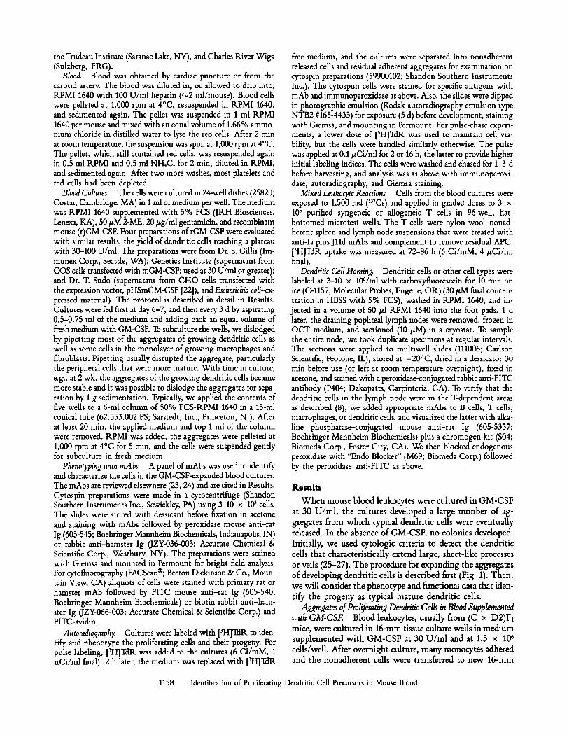
BLOOD LEUKOCYTES IN GM-CSF
Culture ld to adhere most monocytes Culture nonadherent cells 6d Feed with GM-CSF, culture 3d Rinse loose cells away
AGGREGATES AFFIXED TO A DENSE ADHERENT MONOLAYER
Dislodge aggregates with Pasteur pipettes Subculture one well to three in GM-CSF Culture 4-10d feeding q 3d
DENDRITIC CELL AGGREGATES AFFIXED TO SCATTERED ADHERENT CELLS
Subculture as above at 2-3 weeks
AGGREGATES ATTACHED TO A FEW ADHERENT CELLS PLUS RELEASED DENDRITIC CELLS Figure 1. "colonies."
Flow plan for inducing dendritic cell
wells. The adherent cells did not develop dendritic cell colo- nies, but during the next week, the nonadherent populations exhibited three changes. First, most of the lymphocytes and granulocytes died or could be removed by washing. Second, the surface of the well became covered with a monolayer of tightly adherent cells that included macrophages and fibrob- lasts. Third, affixed to scattered sites on the monolayer, there developed small aggregates of cells. The cultures were fed with GM-CSF at day 6-7. The aggregates continued to ex- pand in number and size. At about day 10, the cells were ready to be subcuhured. Any residual loose cells could be rinsed offbefore dislodging the aggregates into fresh medium and GM-CSF. About 0.8-1 x 10⁶ dislodged cells per original well were divided into three subculture wells.
Most of the aggregates disassembled during this first sub- culture, while the bulk of the adherent monolayer remained attached to the original well. Upon transfer, most of the cells
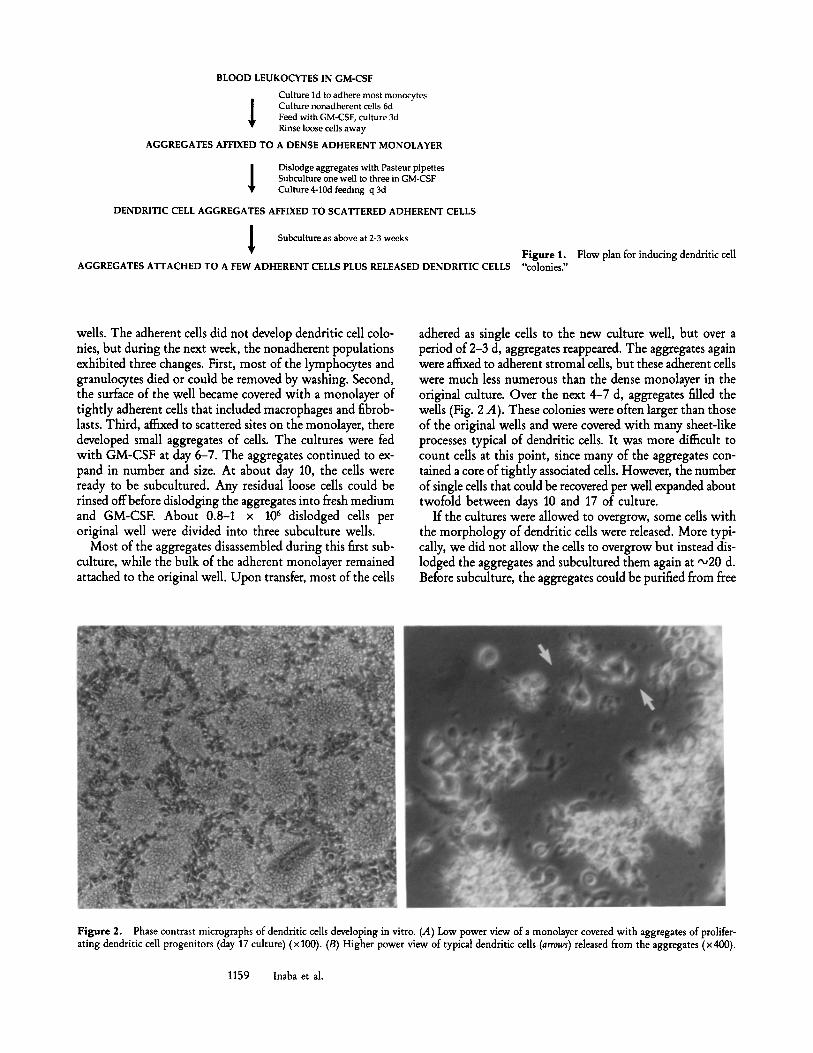
adhered as single cells to the new cuhure well, but over a period of 2-3 d, aggregates reappeared. The aggregates again were affixed to adherent stromal cells, but these adherent cells were much less numerous than the dense monolayer in the original culture. Over the next 4-7 d, aggregates filled the wells (Fig. 2 A). These colonies were often larger than those of the original wells and were covered with many sheet-like processes typical of dendritic cells. It was more difficult to count cells at this point, since many of the aggregates con- tained a core of tightly associated cells. However, the number of single cells that could be recovered per well expanded about twofold between days 10 and 17 of culture.
If the cultures were allowed to overgrow, some cells with the morphology of dendritic cells were released. More typi- cally, we did not allow the cells to overgrow but instead dis- lodged the aggregates and subcuhured them again at ,,o20 d. Before subculture, the aggregates could be purified from free
Figure 2. Phase contrast micrographs of dendritic cells developing in vitro. (A) Low power view of a monolayer covered with aggregates of prolifer- ating dendritic cell progenitors (day 17 culture) (• 100). (B) Higher power view of typical dendritic cells (arrows) released from the aggregates (• 400).
1159 Inaba et al.
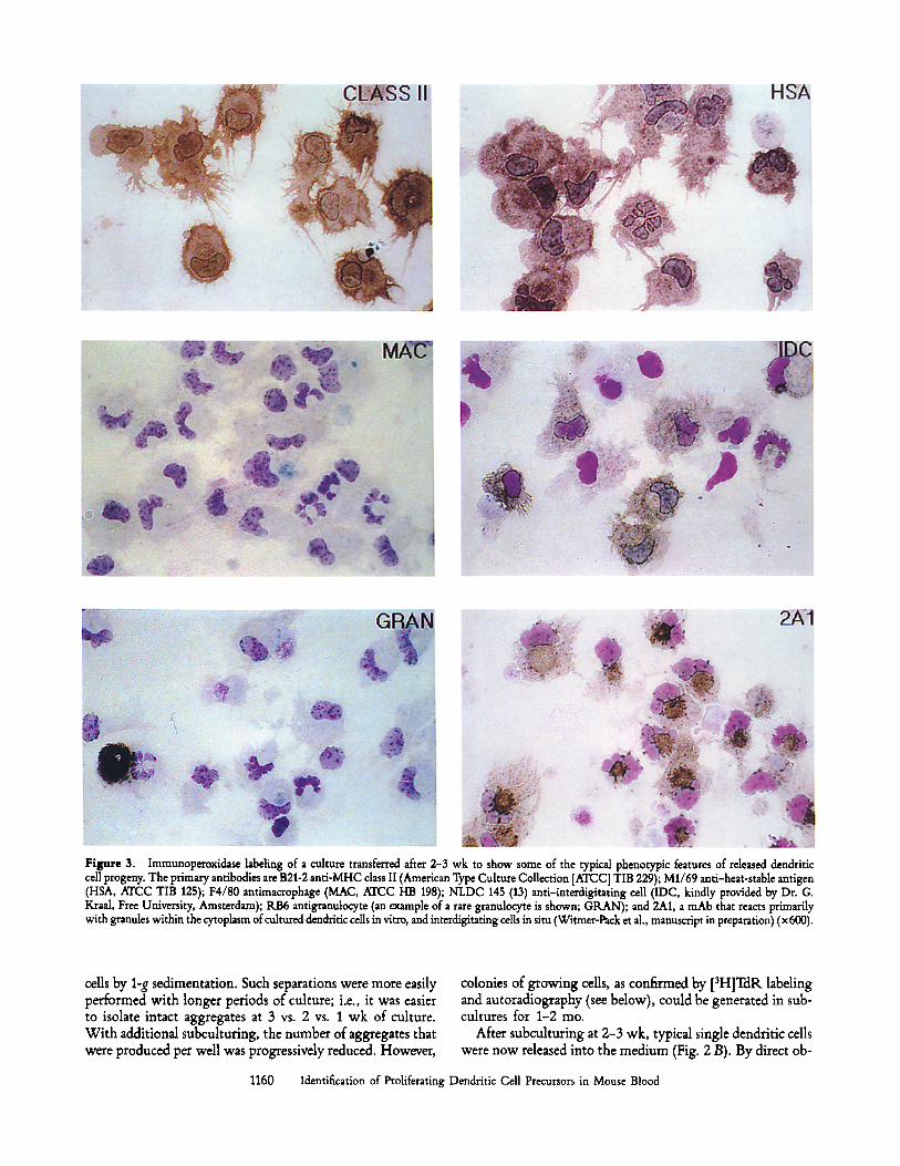
Figure 3. Immunoperoxidase labeling of a culture transferred after 2-3 wk to show some of the typical phenotypic features of released dendritic cell progeny. The primary antibodies are B21-2 anti-MHC class II (American Type Culture Collection [ATCC] TIB 229); M1/69 anti-heat-stable antigen (HSA, ATCC TIB 125); F4/80 antimacrophage (MAC, ATCC HB 198); NLDC 145 (13) anti-interdigitating cell (IDC, kindly provided by Dr. G. Kraal, Free University, Amsterdam); RB6 antigranulocyte (an example of a rare granulocyte is shown; GRAN); and 2A1, a mAb that reacts primarily with granules within the cytoplasm of cultured dendritic cells in vitro, and interdigitating ceUs in situ (Witmer-Pack et al., manuscript in preparation) (x 600).
cells by 1-g sedimentation. Such separations were more easily performed with longer periods of culture; i.e., it was easier to isolate intact aggregates at 3 vs. 2 vs. 1 wk of culture. Wi th additional subculturing, the number of aggregates that were produced per well was progressively reduced. However,
colonies of growing cells, as confirmed by [3H]TdR labeling and autoradiography (see below), could be generated in sub- cultures for 1-2 mo.
After subculturing at 2-3 wk, typical single dendritic cells were now released into the medium (Fig. 2 B). By direct ob-
1160 Identification of Proliferating Dendritic Cell Precursors in Mouse Blood
RB6, GRAN.
J ::i ]
SER-4, ~M1/69,
. . . . . . , , t . . . . . . "1 . . . . . . . . i . . . . . . . .
o> z U . I
0 U J ee" L.L .
CD45R F4/80, M1/42, II~ ,~ MAC CLASS I
M5/114,1 2(211, Ir
,d" .... ~;' .... i '-~= .... i;3 ........ , ' . .... i- ~, .... '/;= .... i-~ FLUORESCENCE INTENSITY
2A1 ~ 3C7, / 2E6, ~ C D 1 8
'1
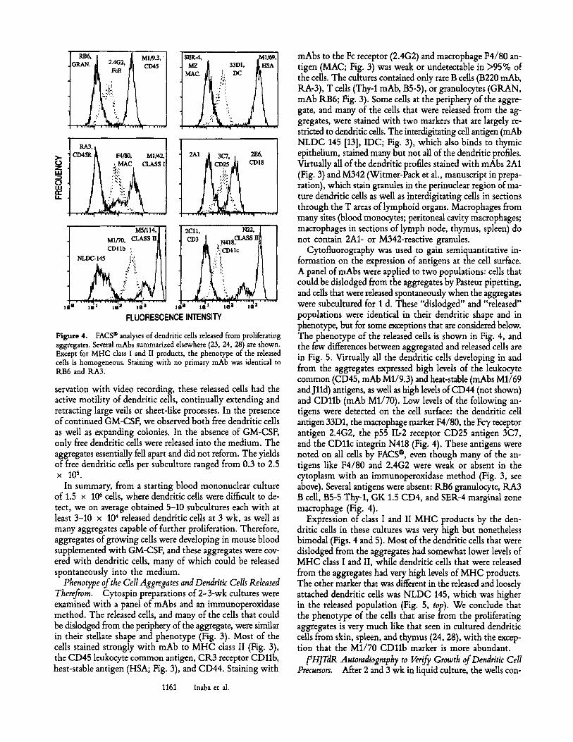
Figure 4. FACS | analyses of dendritic cells released from proliferating aggregates. Several mAbs summarized elsewhere (23, 24, 28) are shown. Except for MHC class I and II products, the phenotype of the released cells is homogeneous. Staining with no primary mAb was identical to RB6 and RA3.
servation with video recording, these released cells had the active motility of dendritic cells, continually extending and retracting large veils or sheet-like processes. In the presence of continued GM-CSF, we observed both free dendritic cells as well as expanding colonies. In the absence of GM-CSF, only free dendritic cells were released into the medium. The aggregates essentially fell apart and did not reform. The yields of free dendritic ceils per subculture ranged from 0.3 to 2.5 x lO s.
In summary, from a starting blood mononuclear culture of 1.5 x 10 6 cells, where dendritic cells were difficult to de- tect, we on average obtained 5-10 subcultures each with at least 3-10 x 104 released dendritic cells at 3 wk, as well as many aggregates capable of further proliferation. Therefore, aggregates of growing cells were developing in mouse blood supplemented with GM-CSF, and these aggregates were cov- ered with dendritic cells, many of which could be released spontaneously into the medium.
Phenotype of the Cell Aggregates and Dendritic Cells Released Therefrom. Cytospin preparations of 2-3-wk cultures were examined with a panel of mAbs and an immunoperoxidase method. The released cells, and many of the cells that could be dislodged from the periphery of the aggregate, were similar in their stellate shape and phenotype (Fig. 3). Most of the cells stained strongly with mAb to MHC class II (Fig. 3), the CD45 leukocyte common antigen, CR3 receptor CD11b, heat-stable antigen (HSA; Fig. 3), and CD44. Staining with
1161 Inaba et al.
mAbs to the Fc receptor (2.4G2) and macrophage F4/80 an- tigen (MAC; Fig. 3) was weak or undetectable in >95% of the cells. The cultures contained only rare B cells (B220 mAb, RA-3), T cells (Thy-1 mAb, B5-5), or granulocytes (GRAN, mAb RB6; Fig. 3). Some cells at the periphery of the aggre- gate, and many of the cells that were released from the ag- gregates, were stained with two markers that are largely re- stricted to dendritic cells. The interdigitating cell antigen (mAb NLDC 145 [D], IDC; Fig. 3), which also binds to thymic epithelium, stained many but not all of the dendritic profiles. Virtually all of the dendritic profiles stained with mAbs 2A1 (Fig. 3) and M342 (Witmer-Pack et al., manuscript in prepa- ration), which stain granules in the perinuclear region of ma- ture dendritic cells as well as interdigitating cells in sections through the T areas of lymphoid organs. Macrophages from many sites (blood monocytes; peritoneal cavity macrophages; macrophages in sections of lymph node, thymus, spleen) do not contain 2A1- or M342-reactive granules.
Cytofluorography was used to gain semiquantitative in- formation on the expression of antigens at the cell surface. A panel of mAbs were applied to two populations: cells that could be dislodged from the aggregates by Pasteur pipetting, and cells that were released spontaneously when the aggregates were subcultured for 1 d. These "dislodged" and "released" populations were identical in their dendritic shape and in phenotype, but for some exceptions that are considered below. The phenotype of the released cells is shown in Fig. 4, and the few differences between aggregated and released cells are in Fig. 5. Virtually all the dendritic cells developing in and from the aggregates expressed high levels of the leukocyte common (CD45, mAb M1/9.3) and heat-stable (mAbs M1/69 and Jlld) antigens, as well as high levels of CD44 (not shown) and CD11b (mAb M1/70). Low levels of the following an- tigens were detected on the cell surface: the dendritic cell antigen 33D1, the macrophage marker F4/80, the Fc3, receptor antigen 2.4G2, the p55 IL2 receptor CD25 antigen 3C7, and the CD11c integrin N418 (Fig. 4). These antigens were noted on all cells by FACS | even though many of the an- tigens like F4/80 and 2.4G2 were weak or absent in the cytoplasm with an immunoperoxidase method (Fig. 3, see above). Several antigens were absent: RB6 granulocyte, RA3 B cell, B5-5 Thy-1, GK 1.5 CD4, and SER-4 marginal zone macrophage (Fig. 4).
Expression of class I and II MHC products by the den- dritic cells in these cultures was very high but nonetheless bimodal (Figs. 4 and 5). Most of the dendritic cells that were dislodged from the aggregates had somewhat lower levels of MHC class I and II, while dendritic cells that were released from the aggregates had very high levels of MHC products. The other marker that was different in the released and loosely attached dendritic cells was NLDC 145, which was higher in the released population (Fig. 5, top). We conclude that the phenotype of the cells that arise from the proliferating aggregates is very much like that seen in cultured dendritic cells from skin, spleen, and thymus (24, 28), with the excep- tion that the M1/70 CDllb marker is more abundant.
[3H]TdR Autoradiography to Verify Growth of Dendritic Cell Precursors. After 2 and 3 wk in liquid culture, the wells con-
i No 1 ~ M H C
�9 ~ Class II
:.r
33D1
..':. MI-IC
i ~ ! Class I
, , , ; ; ; , , i
: thy- I
":i
I I I I I l l l I I 1 1 1 1 1 1 1 1 1 I 111111 |
' " ,.q
o re"
No 1 ~ M H C o
' : Class II k
: ' ~
z -2 :
o
I I I I l l l l | I l I 1 1 1 1 1 | I I I I I I I I 1 I I
33D1 M H C
i
Class I
Ir
| I1"11111~ I | I ' I i , I i 1 , 1 1
t thy-1
: :
.; z, .
r
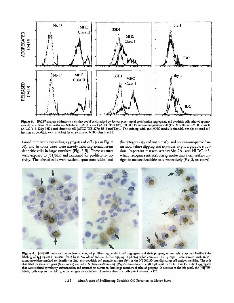
Figure 5. FACS | analyses of dendritic cells that could be dislodged by Pasteur pipetting of proliferating aggregates, and dendritic cells released sponta- neously in culture. The mAbs are M1/42 anti-MHC class I (ATCC TIB 126); NLDC145 anti-interdigitating cell (13); M5/114 anti-MHC class II (ATCC TIB 120); 33D1 anti-dendritic cell (ATCC TIB 227); B5-5 anti-Thy-1. The staining with anti-MHC mAbs is bimodal, but the released cell fraction of dendritic cells is richest in expression of MHC class I and II.
tained numerous expanding aggregates of cells (as in Fig. 2 A) , and in some cases were already releasing nonadherent dendrit ic cells in large numbers (Fig. 2 B). These cultures were exposed to [3H]TdR and examined for proliferative ac- tivity. T h e labeled cells were washed, spun onto slides, and
the cytospins stained w i th m A b s and an immunoperox idase m e t h o d before d ipping and exposure to pho tograph ic emul- sion. I m p o r t a n t markers were m A b s 2A1 and N L D C - 1 4 5 , which recognize intracellular granules and a cell surface an- t igen in mature dendritic cells, respectively (Fig. 3, see above).
Figure 6. [3H]TdR pulse and pulse-chase labeling of proliferating dendritic cell aggregates and their progeny, respectively. (Left and Middle) Pulse labeling of aggregates (1 #Ci/ml for 2 h) at ~2 wk of culture. Before dipping in photographic emulsion, the cytospins were stained with an im- munoperoxidase method to identify the 2A1 anti-dendritic cell granule antigen (/eft) or the NLDC145 interdigitating cell antigen (middle). The cells that label for these antigens (black arrows) are not in S phase (white arrows). (Right) Pulse-chase label (0.2 #Ci/ml for 16 h, chase for 2 d) of aggregates that were isolated by velocity sedimentation and returned to culture to form large numbers of released progeny. In contrast to the left panel, the [3H]TdR- labeled cells express the 2A1 granule antigen characteristic of mature dendritic cells (black arrows), x 425.
1162 Identification of Proliferating Dendritic Cell Precursors in Mouse Blood
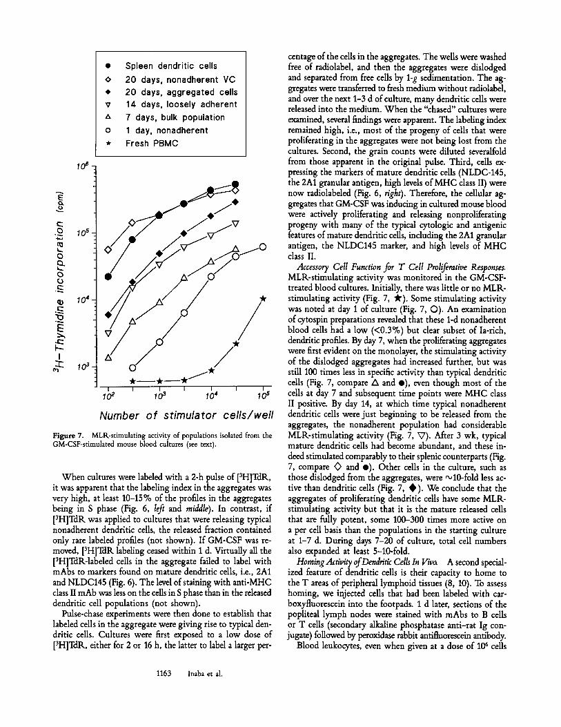
�9 Spleen dendrit ic cells
<> 20 days, nonadherent VC # 20 days, aggregated cells v 14 days, loosely adherent
A 7 days, bulk population
o 1 day, nonadherent
* Fresh PBMC
106
E o
0 105
0
0
10 +
E
I
:~ 10 3
.,, .. / / V .+"
I I I I I I I
1o 2 l o 3 ~ o' ~ o s
N u m b e r o f s t i m u l a t o r c e l l s ~ w e l l
Figure 7. MLK-stimulating activity of populations isolated from the GM-CSF-stimulated mouse blood cultures (see text).
When cultures were labeled with a 2-h pulse of [3H]TdR., it was apparent that the labeling index in the aggregates was very high, at least 10-15 % of the profiles in the aggregates being in S phase (Fig. 6, left and middle). In contrast, if [3H]TdR was applied to cultures that were releasing typical nonadherent dendritic cells, the released fraction contained only rare labeled profiles (not shown). If GM-CSF was re- moved, [3H]TdR labeling ceased within 1 d. Virtually all the [3H]TdR-labeled cells in the aggregate failed to label with mAbs to markers found on mature dendritic cells, i.e., 2A1 and NLDC145 (Fig. 6). The level of staining with anti-MHC class II mAb was less on the cells in S phase than in the released dendritic cell populations (not shown).
Pulse-chase experiments were then done to establish that labeled cells in the aggregate were giving rise to typical den- dritic cells. Cultures were first exposed to a low dose of [3H]TdR, either for 2 or 16 h, the latter to label a larger per-
centage of the cells in the aggregates. The wells were washed free of radiolabel, and then the aggregates were dislodged and separated from free cells by 1-g sedimentation. The ag- gregates were transferred to fresh medium without radiolabel, and over the next 1-3 d of culture, many dendritic cells were released into the medium. When the "chased" cultures were examined, several findings were apparent. The labeling index remained high, i.e., most of the progeny of cells that were proliferating in the aggregates were not being lost from the cultures. Second, the grain counts were diluted severalfold from those apparent in the original pulse. Third, cells ex- pressing the markers of mature dendritic cells (NLDC-145, the 2A1 granular antigen, high levels of MHC class II) were now radiolabeled (Fig. 6, right). Therefore, the cellular ag- gregates that GM-CSF was inducing in cultured mouse blood were actively proliferating and releasing nonproliferating progeny with many of the typical cytologic and antigenic features of mature dendritic cells, including the 2A1 granular antigen, the NLDC145 marker, and high levels of MHC class II.
Accessory Cell Function for T Cell Proliferative Responses. MLR-stimulating activity was monitored in the GM-CSF- treated blood cultures. Initially, there was little or no MLR- stimulating activity (Fig. 7, ~'). Some stimulating activity was noted at day 1 of culture (Fig. 7, O). An examination of cytospin preparations revealed that these 1-d nonadherent blood cells had a low (<0.3%) but clear subset of Ia-rich, dendritic profiles. By day 7, when the proliferating aggregates were first evident on the monolayer, the stimulating activity of the dislodged aggregates had increased further, but was still 100 times less in specific activity than typical dendritic cells (Fig. 7, compare A and e), even though most of the cells at day 7 and subsequent time points were MHC class II positive. By day 14, at which time typical nonadherent dendritic cells were just beginning to be released from the aggregates, the nonadherent population had considerable MLR-stimulating activity (Fig. 7, V). After 3 wk, typical mature dendritic cells had become abundant, and these in- deed stimulated comparably to their splenic counterparts (Fig. 7, compare <> and e). Other cells in the culture, such as those dislodged from the aggregates, were "+10-fold less ac- tive than dendritic cells (Fig. 7, � 9 We conclude that the aggregates of proliferating dendritic cells have some MLR- stimulating activity but that it is the mature released cells that are fully potent, some 100-300 times more active on a per cell basis than the populations in the starting culture at 1-7 d. During days 7-20 of culture, total cell numbers also expanded at least 5-10-fold.
Homing Activity of Dendritic Cells In Viva A second special- ized feature of dendritic cells is their capacity to home to the T areas of peripheral lymphoid tissues (8, 10). To assess homing, we injected cells that had been labeled with car- boxyftuorescein into the footpads. 1 d later, sections of the popliteal lymph nodes were stained with mAbs to B cells or T cells (secondary alkaline phosphatase anti-rat Ig con- jugate) followed by peroxidase rabbit antifluorescein antibody.
Blood leukocytes, even when given at a dose of 106 cells
1163 Inaba et al.
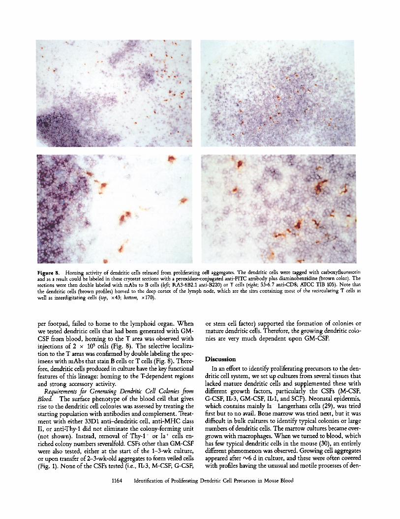
Figure 8. Homing activity of dendritic cells released from proliferating cell aggregates. The dendritic cells were tagged with carboxyfluorescein and as a result could be labeled in these cryostat sections with a peroxidase-conjugated anti-FITC antibody plus diaminobenzidine (brown color). The sections were then double labeled with mAbs to B cells (left; RA3-6B2.1 anti-B220) or T cells (right; 53-6.7 anti-CD8; ATCC TIB 105). Note that the dendritic cells (brown profiles) homed to the deep cortex of the lymph node, which are the sites containing most of the recirculating T cells as well as interdigitating cells (top, x43; bottom, x170).
per footpad, failed to home to the lymphoid organ. When we tested dendritic cells that had been generated with GM- CSF from blood, homing to the T area was observed with injections of 2 x 10⁵ cells (Fig. 8). The selective localiza- tion to the T areas was confirmed by double labeling the spec- imens with mAbs that stain B cells or T cells (Fig. 8). There- fore, dendritic cells produced in culture have the key functional features of this lineage: homing to the T-dependent regions and strong accessory activity.
Requirements for Generating Dendritic Cell Colonies from Blood. The surface phenotype of the blood cell that gives rise to the dendritic cell colonies was assessed by treating the starting population with antibodies and complement. Treat- ment with either 33D1 anti-dendritic cell, anti-MHC class II, or anti-Thyol did not eliminate the colony-forming unit (not shown). Instead, removal of Thy-1 + or Ia + cells en- riched colony numbers severalfold. CSFs other than GM-CSF were also tested, either at the start of the 1-3-wk culture, or upon transfer of 2-3-wk-old aggregates to form veiled cells (Fig. 1). None of the CSFs tested (i.e., IL-3, M-CSF, G-CSF,
or stem cell factor) supported the formation of colonies or mature dendritic cells. Therefore, the growing dendritic colo- nies are very much dependent upon GM-CSF.
Discuss ion
In an effort to identify proliferating precursors to the den- dritic cell system, we set up cultures from several tissues that lacked mature dendritic cells and supplemented these with different growth factors, particularly the CSFs (M-CSF, G-CSF, IL-3, GM-CSF, II.,1, and SCF). Neonatal epidermis, which contains mainly Ia- Langerhans cells (29), was tried first but to no avail. Bone marrow was tried next, but it was difficult in bulk cultures to identify typical colonies or large numbers of dendritic cells. The marrow cultures became over- grown with macrophages. When we turned to blood, which has few typical dendritic cells in the mouse (30), an entirely different phenomenon was observed. Growing cell aggregates appeared after ,,o6 d in culture, and these were often covered with profiles having the unusual and motile processes of den-
1164 Identification of Proliferating Dendritic Cell Precursors in Mouse Blood
dritic cells. With time, typical nonadherent dendritic cells were released. The latter had the morphology and movement of dendritic cells as previously described in cultured mouse spleen, mouse skin, lymph from several species, and human blood (25-27). Therefore, to identify proliferating dendritic cells, it seems critical to begin with an appropriate starting population (in this case, blood) and to supplement the cul- ture with GM-CSF.
We think that the initial aggregates that appeared in the cultures represented clones, since small groups of four to six cells were observed early on, e.g., day 5. We tried to prove that the aggregates were clonal by mixing blood cells from strains that were distinguished with markers to polymorphic antigens like CD44 and MHC class II. However, we could not complete the experiments since we found that mouse strains differed in the number and speed with which colonies developed. BALB/c and DBA (and FI strains derived there- from) were the most active; B6 and B10 were several times less active; and strains like CBA/J, C3H/He, and A/J were poor sources of proliferating, dendritic cell aggregates.
The precursors to the aggregates of proliferating dendritic cells were not typical monocytes or dendritic cells, because the number of aggregates that developed could be increased substantially if one depleted monocytes by adherence or Ia- positive cells with antibody and complement. We tentatively conclude that blood contains an Ia- precursor that forms a proliferating aggregate. In the aggregate, dendritic cells ma- ture and are released as nonproliferating progeny.
The formation of aggregates of dendritic cells required ex- ogenous GM-CSF. If the aggregates were placed in macro- phage- or granulocyte-restricted CSFs (M-CSF, G-CSF), proliferation ceased and neither macrophages nor granulo- cytes were formed. Because the cultures contained macro- phages and some stromal cells, in addition to the dendritic cell aggregates, it was possible that other cytokines were being produced that were critical to the formation of dendritic cells. It appears, however, that the cells in the aggregates have lost responsiveness to M- and G-CSF, and that dendritic cells rep- resent a distinct myeloid pathway of development. Perhaps the pathway originates from a common precursor in which the dendritic cell lineage is an offshoot that no longer responds to macrophage- and granulocyte-restricted CSFs.
Labeling with [3H]thymidine, using pulse and pulse-chase protocols, was important in establishing the precursor-product relationships that were taking place in these liquid cultures (Fig. 6). In a 2-h pulse, virtually every labeled cell lacked two typical markers of mature dendritic cells, i.e., the NLDC- 145 interdigitating cell surface antigen (13) and the recently identified 2A1/M342 granular cytoplasmic antigens (Witmer- Pack et al., manuscript in preparation). These mAbs do not stain most macrophage populations that we have examined either as isolated cells (blood, spleen, peritoneal macrophages)
or in sections (thymus cortex, spleen red pulp, lymph node medulla). In pulse-chase protocols, large numbers of labeled progeny were released from the aggregates, and these released cells were nonadherent, motile, and strongly stimulatory in the MLR. After combined autoradiography and immuno- peroxidase labeling, the labeled progeny carried the granular antigens, the NLDC-145 antigen, and very high levels of MHC class II. Each of these cytologic and antigenic markers are largely restricted to dendritic cells.
We believe that maturation to typical nonproliferating den- dritic cells occurred within the aggregate. The aggregates were covered with cells with the sheet-like or veiled processes of dendritic cells. Cells with markers of mature dendritic cell markers (high MHC class II, 2Al-positive granules, NLDC antigen) were also observed at the periphery of the cell ag- gregates (Fig. 6, left and middle). However, it was difficult to isolate the aggregate intact, without dislodging these more mature cells. The mechanism whereby dendritic cells matured and left the aggregate was not dear. Maturation was enhanced in older cultures (>2 wk) or by removing adherent stroma cells. Both proliferation and maturation was blocked if the cultures contained too many fibroblasts.
The functional maturation that occurred in the prolifer- ating aggregate is striking. The dendritic cells that were gener- ated in culture were potent MLR stimulators. 102 dendritic cells induced a much stronger primary MLR than 10 s blood leukocytes (Fig. 7). The increase in stimulating activity per Ia + cell was at least 2 logs between the time that the ag- gregates first appeared and the time that typical dendritic cells were released in large numbers. Over this time period, cell recovery increased 5-10-fold. Also, the dendritic cell progeny homed in a precise way to the T cell area of lymph node (Fig. 8), another functional property that was not detectable in blood cells (data not shown).
Given the criteria that have become evident in this paper, it will be feasible to look for proliferating progenitors of den- dritic cells in other organs. It is known that proliferating precursors are giving rise to the rapidly turning over popula- tions of dendritic cells in spleen (15) and afferent lymph (16). The proliferation of leukocytes (other than T cells) occurs in the bone marrow, but it may be that for dendritic cells, the marrow also seeds the blood and other tissues with pro- genitors that then proliferate extensively as shown here. By being able to prepare the otherwise trace dendritic cell in large numbers, it should be more feasible to pursue previously un- explored areas of dendritic cell function. Specifically, growing dendritic cells will facilitate molecular and clinical studies on the mechanism of action of these APCs, including their ca- pacities to capture and retain antigens in an immunogenic form (4, 6, 14) and act as adjuvants for the generation of immunity in vivo (1, 14, 31).
We thank Mrs. Bettina Trokenbacher for valuable technical help, and Judy Adams and Stuart Gezelter for assistance with the illustrative material.
1165 lnaba et al.

This work was supported by grants from the Ministry of Education, Science, and Culture of Japan (03670245 for Scientific Research, and 03044086 for Joint Research in International Scientific Research [K. Inaba]), National Institutes of Health grant AI-13013 (R. M. Steinman), and the Austrian National Bank (Jubilaeums- fonds project 3872 [C. Schuler]). G. Schuler was also supported by the Max Kade Foundation.
Address correspondence to Ralph M. Steinman, Laboratory of Cellular Physiology and Immunology, Box 280, The Rockefeller University, 1230 York Avenue, New York, NY 10021. T. Sudo's present address is Basic Research Labs, Toray Industries, Kamakura, Kanagawa 248, Japan.
Received for publication 27 November 1991.
References 1. Steinman, lL.M. 1991. The dendritic cell system and its role
in immunogenicity. Annu. Rev. Immunot. 9:271. 2. Witmer-Pack, M.D., W. Olivier, J. Valinsky, G. Schuler, and
R.M. Steinman. 1987. Granulocyte/macrophage colony-stimu- lating factor is essential for the viability and function of cul- tured murine epidermal Langerhans cells.J. Exja Med. 166:1484.
3. Heufler, C., F. Koch, and G. Schuler. 1987. Granulocyte- macrophage colony-stimulating factor and interleukin I mediate the maturation of murine epidermal Langerhans ceUs into po- tent immunostimulatory dendritic ceUs.J. Exp. Med. 167:700.
4. Romani, N., S. Koide, M. Crowley, M. Witmer-Pack, A.M. Livingstone, C.G. Fathman, K. Inaba, and lL.M. Steinman. 1989. Presentation of exogenous protein antigens by dendritic cells to T cell clones: intact protein is presented best by imma- ture, epidermal Langerhans ceils. J. Extx Med. 169:1169.
5. Inaba, K., N. lLomani, and lL.M. Steinman. 1989. An antigen- independent contact mechanism as an early step in T cell- proliferative responses to dendritic cells.J. Ext~ Med. 170:527.
6. Pur~, E., K. Inaba, M.T. Crowley, L. Tardelli, M.D. Witmer- Pack, G. lLuberti, G. Fathman, and R.M. Steinman. 1990. An- tigen processing by epidermal Langerhans cells correlates with the level of biosynthesis of major histocompatibility complex class II molecules and expression of invariant chain, j. Exp. Med. 172:1459.
7. Kampgen, E., N. Koch, F. Koch, P. Stoger, C. Heufler, G. Schuler, and N. Romani. 1991. Class II major histocompati- bility complex molecules of murine dendritic cells: synthesis, sialylation of invariant chain, and antigen processing capacity are downregulated upon culture. Proc. Natl. Acad. Sci, USA. 88:3014.
8. Austyn, J.M., M.W. Kupiec-Weglinski, D.F. Hankins, and P.J. Morris. 1988. Migration pattern of dendritic cells in the mouse. Homing to T ceU-dependent areas of spleen, and binding within marginal zone. J. Exp. Med. 167:646.
9. Larsen, C.P., P.J. Morris, and J.M. Austyn. 1990. Migration of dendritic leukocytes from cardiac allografts into host spleens: a novel pathway for initiation of rejection.J. Ext~ Med. 171:307.
10. Austyn, J.M., and C.P. Larsen. 1990. Migration patterns of dendritic leukocytes. Transplantation (Baltimore). 49:1.
11. Veldman, J.E., and E. Kaiserling. 1980. Interdigitating cells. In The Reticuloendothelial System, Morphology. I. Carr, and W.T. Daems, editors. Plenum Publishing Corporation, New York. 381-416.
12. Witmer, M.D., and R.M. Steinman. 1984. The anatomy of peripheral lymphoid organs with emphasis on accessory ceils: light microscopic, immunocytochemical studies of mouse spleen, lymph node and Peyer's patch. Am. J. Anat. 170:465.
13. Kraal, G., M. Breel, M. Janse, and G. Bruin. 1986. Langer-
hans cells, veiled cells, and interdigitating ceils in the mouse recognized by a monoclonat antibody. J. Extx Med. 163:981.
14. Inaba, K., J.P. Metlay, M.T. Crowley, and R.M. Steinman. 1990. Dendritic cells pulsed with protein antigens in vitro can prime antigen-specific, MHC-restricted T cells in situ.j. Exp. Med. 172:631.
15. Steinman, R.M., D.S. Lustig, and Z.A. Cohn. 1974. Iden- tification of a novel cell type in peripheral lymphoid organs of mice. III. Functional properties in vivo.j. Exp Med. 139:1431.
16. Pugh, C.W., G.G. MacPherson, and H.W. Steer. 1983. Char- acterization of nonlymphoid cells derived from rat peripheral lymph, j. Exla Med. 157:1758.
17. Fossum, S. 1989. The life history of dendritic leukocytes [DL]. In Current Topics in Pathology. O.H. Ivesson, editor. Springer- Verlag, Berlin. 101-124.
18. Katz, S.I., K. Tamaki, and D.H. Sachs. 1979. Epidermal Lang- erhans cells are derived from cells originating in bone marrow. Nature (Lond.). 282:324.
19. Hart, D.N.J., and J.W. Fabre. 1981. Demonstration and char- acterization of Ia-positive dendritic cells in the interstitial con- nective tissues of rat heart and other tissues, but not brain. J. Exp. Med. 154:347.
20. Bowers, W.E., and M.R. Berkowitz. 1986. Differentiation of dendritic cells in cultures of rat bone marrow cells.J. Exp. Med. 163:872.
21. Reid, C.D.L., P.R. Fryer, C. Clifford, A. Kirk, J. Tikerpae, and S.C. Knight. 1990. Identification of hematopoietic pro- genitors of macrophages and dendritic Langerhans cells (DL- CFU) in human bone marrow and peripheral blood. Blood. 76:1139.
22. Kajigaya, S., T. Suda, J. Suda, M. Saito, Y. Miura, M. Iizuka, S. Kobayashi, N. Minato, and T. Sudo. 1986. A recombinant murine granulocyte/macrophage (GM) colony-stimulating factor derived from an inducer T cell line (IH5.5). Functional restriction to GM progenitor cells. J. Exp. Med. 164:1102.
23. Agger, R., M.T. Crowley, and M.D. Witmer-Pack. 1990. The surface of dendritic cells in the mouse as studied with mono- clonal antibodies. Int. Rev. lmmunol. 6:89.
24. Crowley, M., K. Inaba, H. Witmer-Pack, and IL.M. Steinman. 1989. The cell surface of mouse dendritic cells: FACS analyses of dendritic cells from different tissues including thymus. Cell. ImmunoL 118:108.
25. Freudenthal, P.S., and lL.M. Steinman. t990. The distinct sur- face of human blood dendritic cells, as observed after an im- proved isolation method. Proc. NatL Acad. Sci. USA. 87:7698.
26. Drexhage, H.A., H. Mullink, J. de Groot, J. Clarke, and B.M. Balfour. 1979. A study of cells present in peripheral lymph of pigs with special reference to a type of cell resembling the
1166 Identification of Proliferating Dendritic Cell Precursors in Mouse Blood
Langerhans cells. Cell Tissue Res. 202:407. 27. Schuler, G., and R.M. Steinman. 1985. Murine epidermal Lang-
erhans cells mature into potent immunostimulatory dendritic cells in vitro, j . Exp. Med. 161:526.
28. Romani, N., W. Witmer-Pack, M. Crowley, S. Koide, G. Schuler, K. Inaba, and R.M. Steinman. 1991. Langerhans Cells as Immature Dendritic Cells. CRC Press, Inc., Boca Raton, FL. 191-216.
29. Romani, N., G. Schuler, and P. Fritsch. 1986. Ontogeny of
Ia-positive and Thy-1 positive leukocytes of murine epidermis. J. Invest. Dermatol. 86:129.
30. Nussenzweig, M.C., R.M. Steinman, M.D. Witmer, and B. Gutchinov. 1982. A monoclonal antibody specific for mouse dendritic cells. Proc. Natl. Acad. Sci. USA. 79:161.
31. Inaba, K., J.P. Metlay, M.T. Crowley, M. Witmer-Pack, and R.M. Steinman. 1990. Dendritic cells as antigen presenting cells in vivo. Int. Rev. Immunol. 6:197.
1167 Inaba et al.