Embed Size (px)
Citation preview

REVIEW Open Access
Biomaterial-based platforms for in situdendritic cell programming and their use inantitumor immunotherapyJoão Calmeiro1,2, Mylène Carrascal2,3, Célia Gomes4,5, Amílcar Falcão1,6, Maria Teresa Cruz1,2 andBruno Miguel Neves7*
Abstract
Dendritic cells (DCs) are central players in the immune system, with an exquisite capacity to initiate and modulateimmune responses. These functional characteristics have led to intense research on the development of DC-basedimmunotherapies, particularly for oncologic diseases. During recent decades, DC-based vaccines have generatedvery promising results in animal studies, and more than 300 clinical assays have demonstrated the safety profile ofthis approach. However, clinical data are inconsistent, and clear evidence of meaningful efficacy is still lacking. Oneof the reasons for this lack of evidence is the limited functional abilities of the used ex vivo-differentiated DCs.Therefore, alternative approaches for targeting and modulating endogenous DC subpopulations have emerged asan attractive concept. Here, we sought to revise the evolution of several strategies for the in situ mobilization andmodulation of DCs. The first approaches using chemokine-secreting irradiated tumor cells are addressed, andspecial attention is given to the cutting-edge injectable bioengineered platforms, programmed to releasechemoattractants, tumor antigens and DC maturating agents. Finally, we discuss how our increasing knowledge ofDC biology, the use of neoantigens and their combination with immune checkpoint inhibitors can leverage therefinement of these polymeric vaccines to boost their antitumor efficacy.
Keywords: Biomaterial-based scaffolds, Dendritic cells, In situ mobilization, Antitumor immunotherapy
Dendritic cell-based approaches in antitumorimmunotherapyApproaches to enhance or restore the immune systemaptitude to identify and destroy malignant cells havelong been viewed as a central goal in cancer treatment[1–3]. The use of dendritic cells (DCs), powerful modu-lators of immune responses, in immunotherapy has beenextensively scrutinized and has been highly desirable forclinical application since the early 1990s. There are morethan 300 completed or ongoing registered clinical trialsusing these cells as antitumor vaccines [4]. Currently,there are mainly two approaches for exploring DCs inoncologic treatments: 1) vaccines constituted by ex vivo-generated DCs matured and loaded with tumor antigensand 2) in vivo direct targeting of antigens to DCs [5].
Manipulation of DCs ex vivo followed by their injectionback into the patient is the most common approach,which is being used in 97% of referenced clinical trials[4]. In this approach, blood precursors (CD14+ mono-cytes or CD34+ hematopoietic stem cells) are collectedfrom patients, differentiated into DCs, loaded withantigens and matured. The resultant cellular product iscryopreserved and then released for administrationaccording to the defined vaccination schedule.These types of vaccines present exceptional tolerabil-
ity, but the procedure is highly expensive and laboriousas result of the required manipulation in GMPconditions and notwithstanding the good safety profile,the rate of success is inconsistent [4]. In fact, objectivetumor responses using standard oncologic criteria areusually low, with reports ranging from 3.3 to 15% [6–8].Furthermore, promising vaccines in early phase studies[9–12] often fail to present clear beneficial clinicaloutputs in phase III trials [13]. So far, only sipuleucel-T,
© The Author(s). 2019 Open Access This article is distributed under the terms of the Creative Commons Attribution 4.0International License (http://creativecommons.org/licenses/by/4.0/), which permits unrestricted use, distribution, andreproduction in any medium, provided you give appropriate credit to the original author(s) and the source, provide a link tothe Creative Commons license, and indicate if changes were made. The Creative Commons Public Domain Dedication waiver(http://creativecommons.org/publicdomain/zero/1.0/) applies to the data made available in this article, unless otherwise stated.
* Correspondence: [email protected] of Medical Sciences and Institute of Biomedicine – iBiMED,University of Aveiro, Agra do Crasto - Edifício 30, 3810-193 Aveiro, PortugalFull list of author information is available at the end of the article
Calmeiro et al. Journal for ImmunoTherapy of Cancer (2019) 7:238 https://doi.org/10.1186/s40425-019-0716-8

an autologous antigen-presenting cell vaccine for thetreatment of asymptomatic metastatic hormone refrac-tory prostate cancer, has demonstrated satisfactoryefficacy in phase III trials and was approved by the Foodand Drug Administration (FDA) in 2010. The lack ofrobustness of DC-antitumor immunotherapies was at-tributed in part to low numbers of injected cells that areable to migrate to the lymph nodes and to prime T lym-phocytes [14, 15] and also to functional limitations ofthe ex vivo-differentiated DCs. These DCs, which aredifferentiated from hematopoietic precursors, have beenshown to be less efficient than endogenous DC subpopu-lations, specifically in their competence to cross-presentantigens to CD8+ T cells [16, 17]. The lack of definitionof immunogenic neoantigens, the use of shared antigens,the induction of low levels of CD8+ T cell responses andthe inexistence of standardized production and manufac-turing protocols are other reasons to explain the poorefficacy of DC vaccines.To overcome the limitations of ex vivo manipulated DC
vaccines, several strategies aiming to directly target antigensto endogenous DCs have been developed in recent years[18, 19]. These strategies encompass antigen coupling tomonoclonal antibodies specific to DC surface molecules, in-cluding XCR1, DCIR, Cleac9A, CD40, DC-SIGN DEC-205and the mannose receptor. Preclinical and clinical studiesdemonstrated encouraging results, with the establishmentof effective antitumor CD8+ and CD4+ T cell responses andhumoral immunity [20–28]. However, clinical implementa-tion has been struggling with several challenges: theapproach demands the co-administration of DC maturationagents; otherwise, it is prone to induce tolerance to thevehiculated antigen [29]; it is limited to immunization withone known tumor antigen at a time; and the targeted recep-tor needs to be unequivocally expressed by the selected DCsubpopulation.Another way to explore the immunogenic power of
endogenous DC populations in cancer therapies relies onstrategies for their in situ mobilization and modulation.They consist of implantable or injectable biomaterial-based scaffolds providing a specific microenvironmentthat allows the recruitment of desired DC populations andpotentiates their interaction with other immune effectors.Seminal and promising applications of this approach,which encompass both biotechnology and immunology,have gradually appeared in the cancer immunotherapyfield and will be the focus of the present review.
Strategies for in situ DC mobilization and antigenloadingGM-CSF-secreting tumor cellsOne of the first approaches used for in situ mobilizationand activation of endogenous DCs was the use of irradi-ated tumor cells that were genetically altered to secrete
cytokines/chemokines [30, 31]. Among these strategies,GM-CSF-secreting tumor cell vaccines attracted particu-lar interest [32]. GM-CSF is a hematopoietic cytokinewith multiple effects on the immune system: it directlyinfluences hematopoiesis and expansion of granulocytes,macrophages, DCs, eosinophils and neutrophils [33, 34]and indirectly modulates T cell activation and prolifera-tion [35]. In the context of DC-based antitumor vac-cines, GM-CSF is particularly appealing, given that it is apowerful DC chemoattractant and a maturation inducer[36–38]. Furthermore, GM-CSF also presents immune-independent effects by directly inhibiting cancer cellproliferation [39, 40].Seminal studies by Glenn Dranoff and colleagues,
performed with the B16 melanoma mouse model,demonstrated that intradermal injection of irradiatedGM-CSF-secreting tumor cells efficiently inducesstrong, specific and prolonged antitumor immunity[30]. The main action of the approach is due to thegeneration of a local inflammatory reaction with re-cruitment and activation of DCs, macrophages andgranulocytes [30, 41–43]. Briefly, GM-CSF secreted bymodified tumor cells attracts DCs to the injection site.Recruited DCs engulf apoptotic tumor cells and ma-ture via the effect of released GM-CSF. Then, matureDCs migrate to draining lymph nodes to efficientlypresent processed tumor antigens to T cells, resultingin lymphocyte activation and expansion with the con-sequent boost of the antitumor immune response.Clinically, several phase I/II clinical trials exploringthis type of vaccine have shown a coherent inductionof humoral and cellular immunity in several cancers,such as melanoma [44, 45]; pancreatic [46–48], pros-tate [49, 50], kidney [51] cancer; and chronic myeloidleukemia [52].However, these vaccines present some drawbacks.
The sustained GM-CSF release by injected tumor cellscan paradoxically lead to disease progression due to theprovocation of immune tolerance via the differentiationof tolerogenic DCs and the recruitment of myeloidsuppressor cells [53–55]. Moreover, clinical trial out-comes are often variable, with tumor regressions beinginconsistent within patients and with phase III trialsthat continuously failed [32, 56]. Hence, despite initialpromising results, the GVAX vaccine - a whole cellpancreatic cancer vaccine plus GM-CSF-expressingtumor cells - failed due to lack of efficacy [57]. How-ever, we are currently in an exciting era of scientificachievements in cancer immunotherapy, supported by agrowing knowledge on the precise interactions oftumors and the different immune players. Thus, newvaccine designs accommodating this information andexploring novel biotechnological solutions are requiredand highly anticipated.
Calmeiro et al. Journal for ImmunoTherapy of Cancer (2019) 7:238 Page 2 of 11
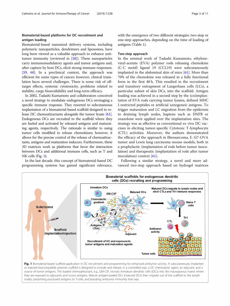
Biomaterial-based platforms for DC recruitment andantigen loadingBiomaterial-based nanosized delivery systems, includingpolymeric nanoparticles, dendrimers and liposomes, havelong been viewed as a valuable approach to enhance anti-tumor immunity (reviewed in [58]). These nanoparticlescarry immunomodulatory agents and tumor antigens and,after capture by host DCs, elicit strong immune responses.[59, 60]. In a preclinical context, the approach wasefficient for some types of cancer; however, clinical trans-lation faces several challenges. There is some risk of off-target effects, systemic cytotoxicity, problems related tostability, cargo bioavailability and long-term efficacy.In 2002, Tadashi Kumamoto and collaborators conceived
a novel strategy to modulate endogenous DCs envisaging aspecific immune response. They resorted to subcutaneousimplantation of a biomaterial-based scaffold designed to re-lease DC chemoattractants alongside the tumor lysate [61].Endogenous DCs are recruited to the scaffold where theyare fueled and activated by released antigens and maturat-ing agents, respectively. The rationale is similar to usingtumor cells modified to release chemokines; however, itallows for the precise control of the release of chemoattrac-tants, antigens and maturation inducers. Furthermore, these3D matrices work as platforms that favor the interactionbetween DCs and additional immune cells, such as T andNK cells (Fig. 1).In the last decade, this concept of biomaterial-based DC
programming systems has gained significant relevance,
with the emergence of two different strategies: two-step orone-step approaches, depending on the time of loading ofantigens (Table 1).
Two-step approachIn the seminal work of Tadashi Kumamoto, ethylene-vinyl-acetate (EVA) polymer rods releasing chemokine(C-C motif) ligand 19 (CCL19) were subcutaneouslyimplanted in the abdominal skin of mice [61]. More than70% of the chemokine was released in a fully functionalform in the first 48 h. This resulted in the recruitmentand transitory entrapment of Langerhans cells (LCs), aparticular subset of skin DCs, into the scaffold. Antigenloading was achieved in a second step by the (co)implan-tation of EVA rods carrying tumor lysates, defined MHCI-restricted peptides or artificial xenogeneic antigens. Totrigger maturation and LC migration from the epidermisto draining lymph nodes, haptens such as DNFB oroxazolone were applied over the implantation sites. Thestrategy was as effective as conventional ex vivo DC vac-cines in eliciting tumor-specific Cytotoxic T-lymphocyte(CTL) activities. Moreover, the authors demonstratedthe efficacy of the approach in fibrosarcoma, E. G7-OVAtumor and Lewis lung carcinoma mouse models, both ina prophylactic (implantation of rods before tumor inocu-lation) and therapeutic (implantation of rods after tumorinoculation) context [61].Following a similar strategy, a novel and more ad-
vanced two-step approach based on hydrogel matrices
Fig. 1 Biomaterial-based scaffold application in DC recruitment and programming for enhanced antitumor activity. A subcutaneously implantedor injected biocompatible polymer scaffold is designed to include and release, in a controlled way, a DC chemotactic agent, an adjuvant, and asource of tumor antigens. The loaded chemoattractant, e.g., GM-CSF, recruits immature dendritic cells (iDCs) into the macroporous matrix wherethey are exposed to adjuvants and tumor antigens. Mature antigen-loaded DCs (matured DCs) then migrate out of the scaffold to the lymphnodes, presenting processed antigens to T-cells, and boosting antitumor immunity that way
Calmeiro et al. Journal for ImmunoTherapy of Cancer (2019) 7:238 Page 3 of 11

was developed [62]. First, DCs are attracted to an inject-able thermosensitive monomethoxypoly(ethylene gly-col)-co-poly(lactic-co-glycolic acid) copolymer (mPEG-PLGA) hydrogel via continuous and controlled release ofGM-CSF [62, 75]. In a second phase, viral and nonviralvectors were used to deliver cancer antigens and to pro-gram recruited DCs. The hydrogel scaffold was able torelease GM-CSF and recruit DCs and macrophages. Thisstrategy resulted in the production of strong tumor-specific immune responses in therapeutic and prophylac-tic settings of murine melanoma models [62].
One-step approachImplantable structuresAs an evolution of the two-step system, in the last dec-ade, David Mooney and collaborators conceived severalbiomaterial-based implantable or injectable platforms forendogenous DC recruitment and antigen loading, all in asingle step. Biocompatible polymers were designed toinclude and release, in a controlled way, a DC chemotac-tic agent, adjuvants and tumor antigens [63]. Several ofthese approaches are based on an extremely porousscaffold composed of poly(lactide-coglycolide) (PLG).
Table 1 Overview of different existing anti-cancer biomaterial-based vaccines for DC recruitment and antigen loading
Approach Scaffold biomaterial Load Administration Target/tumor model
Two step Ethylene-vinyl-acetate(EVA) polymers rods
1st CCL192nd Tumor lysate
Coimplantation E.G7-OVA tumor cellsinjected mice [61]
Hydrogel - Thermosensitivemonomethoxypoly(ethylene glycol)-co-poly(lactic-co-glycolic acid)copolymer (mPEG-PLGA)
1st - GM-CSF2nd - Tumor antigens
2 injections(injection of viral or nonviralvectors in a 2nd step)
Murine melanomamodel [62]
One step Poly(lactide-co-glycolide) GM-CSF, CpG, autologoustumor lysate
Implantation Human melanomaPhase I clinical trialNCT01753089
Poly(lactide-co-glycolide) GM-CSF, CpG, tumor lysate Implantation Murine melanomamodel [41, 63–66]
Poly(lactide-co-glycolide) GM-CSF, CpG, tumor lysate Implantation Murine Lewis lungcarcinoma (LLC) [64]
Poly(lactide-co-glycolide) GM-CSF, CpG, tumor lysate Implantation Rat gliomamodel [67, 68]
Poly(lactide-co-glycolide) GM-CSF, CpG, Tumor lysate Implantation Murine melanomamodel; combinationwith anti PD-1 orCTLA-4 mAb [69]
Poly(lactide-co-glycolide) CCL20, CpG, tumor lysate Implantation Murine melanomamodel [66]
Poly(lactide-co-glycolide) Flt3L, CpG, tumor lysate Implantation Murine melanomamodel [66]
Poly(lactide-co-glycolide) GM-CSF, MPLA, tumor lysate Implantation Murine melanomamodel [64]
Poly(lactide-co-glycolide) GM-CSF, Poly-I:C, Tumor lysate Implantation Murine melanomamodel [64]
Poly(lactide-co-glycolide) GM-CSF, Poly-I:C, tumor lysate Implantation Murine Lewis lungcarcinoma (LLC) [64]
Hydrogel/cryogel–alginate polymer GM-CSF, CpG, irradiatedtumor cells
Injection Murine breastcancer [70]
Hydrogel/cryogel–alginate polymer GM-CSF, CpG, irradiatedtumor cells
Injection Murine melanomamodel [71]
Covalent and ionic crosslinkedcryogel–alginate polymer
GM-CSF, CpG, irradiated-tumor cells
Injection Murine breastcancer [72]
Crosslinking hydrogel- dextranvinylsulfone and tetra-thiolatedpolyethyleneglycol
CCL20 + PLGA microparticlesencapsulating IL-10,siRNA and DNA antigen
Injection Murine A20 B celllymphoma [73]
Mesoporous silica rods (MSRs) -synthetic amorphous silica
GM-CSF, CpG, OVA Injection Prophylactic action ina murine model,injected with EG7-OVA lymphomacells [74]
Calmeiro et al. Journal for ImmunoTherapy of Cancer (2019) 7:238 Page 4 of 11

PLG has multiple applications in the biomedical fieldowing to its specific characteristics: FDA approved forclinical use, prone to surface modification to enhancebiological interactions, high biocompatibility and tailor-able biodegradation rate [76].Using a high-pressure CO2 foaming process, GM-CSF
was encapsulated into macroporous PLG matrices with effi-ciencies above 50% [77, 78]. These scaffolds release up to60% of loaded GM-CSF during the initial 5 days, with theremaining gradually released during an additional 10 days[63]. To strongly activate recruited DCs, CpG-oligonucleotides (CpG-ODN) were also immobilized to thematrices. For this, CpG-ODNs were condensed with poly-ethylenimine to form cationic nanoparticles that electrostati-cally interact with the anionic PLG biomaterial, resulting ina retention higher than 80% over 25 days [63]. The scaffoldscontaining GM-CSF, melanoma tumor lysates and CpG-ODN were assayed in the syngeneic B16-F10 murinemelanoma model across several works. The structures wereable to attract and activate several DC subsets (CD11c+,pDCs and CD8+ DCs) for at least 2 weeks [65]. Importantly,the number of DCs accumulated in the scaffold was of thesame magnitude as that commonly administered in ex vivo-generated DC protocols [63]. Vaccination with these 3Dmacroporous structures elicited robust tumor-specific CTLresponses promoting complete tumor regression in 47% ofmice [41], 50% survival in a therapeutic goal, 33% in a long-term survival goal and a notable 90% in a prophylactic goal[63, 64].In subsequent studies, PLG matrices were used to
supply other chemokines, such as CCL20 and Flt3L, orother adjuvants, such as MPLA and Poly-I:C, ligands forTLR4 and TLR3, respectively [64, 66]. Disregarding theadjuvant used, vaccine efficacy was shown to highlycorrelate to the quantities of recruited CD8+ and pDCsalongside local GM-CSF and IL-12p70 concentrations[64]. PLG scaffolds were also tested in combination withmonoclonal antibodies, targeting the immune checkpointsprogrammed cell death ligand 1 (PD-L1) and cytotoxic T-lymphocyte antigen 4 (CTLA-4). These combinations elic-ited strong CTL activity and tumor regression, reaching aremarkable 75% survival rate in murine models of melan-oma [69]. Finally, in addition to these successful tests inpreclinical melanoma models, DC-recruiting and program-ming PLG scaffolds also showed therapeutic activity in ratglioma models [67, 68] and mouse lung carcinoma [64].The translation of this approach to the clinical context
is presently being evaluated in a phase I clinical trial(NCT01753089) for the treatment of stage IV metastaticmelanoma. It is an open-label interventional study de-signed to address the safety and feasibility of developingand implanting DC activating scaffolds incorporatingautologous melanoma cell lysates in patients with meta-static melanoma. Additionally, as secondary objectives,
the study aims to address the immune response, tumorregression and survival. This vaccine, named WDVAX,is composed of PLGA polymer and includes clinicalgrade GM-CSF, autologous tumor cell lysate and CpG-ODN as a DC maturation agent. The structure is im-planted surgically on the patient’s arm, leg or torso bycutting a small incision into the skin and sliding it intothe “pocket” created between the upper layer of the skinand the tissue underneath.Regarding the clinical trial structure, enrollment consists
of 23 patients who will receive 4 scaffolds by implantation,with skin biopsy being performed after the last vaccine.The study is divided into 3 cohorts of 3–5 patients, witheach one being evaluated in a dose escalation schema,based upon the intervals between scaffold implantation atseparate sites: in cohort 1, the devices are implantedmonthly; in cohort 2, the implantation is performed every3 weeks; in cohort 3, the procedure changes every 2 weeks.CT scan and/or MRI exams are performed to assess thetumor at 3 time points: before the vaccine procedurestarts, halfway through the vaccination schedule and1 month after completion of all 4 vaccines. Finally, theexam will be repeated every 3months after the end of theprotocol. The clinical study is ongoing, with results ex-pected to be out in 2020.
Injectable structuresThe concept of DC-recruiting structures was thenexpanded to other biomaterials, such as hydrogels[70, 71, 73, 79–81], mesoporous silica rods (MSRs)[74] and gelatin [82]. Hydrogel scaffolds have beenapplied in the biomedical field aimed at cell encap-sulation in tissue engineering [83] and for controlledand sustained delivery of drugs [84–87], includingtherapeutic peptide and proteins [88]. Regarding DCprograming platforms, hydrogel-based scaffolds offerthe advantage of being deliverable through conven-tional needle-syringe injection, minimizing the risksand invasiveness associated with surgically implant-able structures. Alginate or gelatin hydrogels devel-oped for this purpose are normally obtained bycryogelation [80, 82]. This technique allows for thedevelopment of cryogels with considerably larger intercon-nected pores [89–93] and augmented mechanical stability[90] when compared to hydrogels obtained by other ap-proaches. Briefly, the reactants are limited to the unfrozen/semi-frozen phases, forming a crosslinked network afterpolymerization. The ice crystals nucleated in the aqueousphase throughout freezing form pores as they melt, creatinginterconnected macroporous networks. Alginate cryogelproduced pore sizes of 150–200 μm, high connectivity ofpores, and shape-memory. These characteristics allow themto regain initial dimensions without considerable deform-ation after injection. Moreover, the open pore structure
Calmeiro et al. Journal for ImmunoTherapy of Cancer (2019) 7:238 Page 5 of 11

confers tissue-like elasticity and creates a favorable micro-environment for cell infiltration. When loaded with GM-CSF, these alginate cryogels were reported to present anencapsulation efficiency of 89%, with 80% of the total en-capsulated cytokine being released within 3 days and acomplete release attained after 4 weeks [80].These scaffolds were preclinically tested as vaccines in
several types of cancer. In mouse breast cancer models,injection of a matrix comprising live attenuated HER-2/neu-overexpressing breast cancer cells, GM-CSF and CpG-ODN resulted in the recruitment and activation of DCsfollowed by a robust antitumor response. The vaccineresulted in 100% survival in vaccinated mice and in a 70-fold enhancement in antibody production when comparedto untreated mice [70]. In another work, alginate cryogelsloaded with irradiated tumor cells and encapsulating andreleasing CpG-ODN and GM-CSF in a controlled mannerwere tested in a mouse melanoma model (Fig. 2) [71]. Thisvaccine efficiently stimulated the recruitment and activationof CD8+ DCs, CD11+ DCs and pDCs. Hence, prophylacticand therapeutic protection against cancer was tested andconfirmed. Specifically, potent antigen-specific T cell re-sponses were detected, conferring long-term prophylacticprotection against melanoma. With this regimen, 80% ofmice survived, and importantly, of these, 100% survived asecond challenge with tumor cells, indicating the inductionof strong immunologic memory. When tested in a thera-peutic context, two vaccination doses at days 3 and 10 posttumor establishment with B16-F10 cells strikingly resultedin complete regression of tumors in 40% of the animals[71]. Recently, the injectability of these cryogels was im-proved by a combination of ionic and covalent crosslinking[72]. The new scaffolds are tougher and allow for the use ofa small caliber needle with no damage after injection. Theseimproved cryogels were shown to avoid tumor develop-ment in 80% of mice injected with HER2/neu-overexpress-ing breast cancer cells [72].In situ crosslinking hydrogels formed via Michael type
addition of dextran vinylsulfone and tetra-thiolated poly-ethylene glycol were also tested as DC programming plat-forms [81]. These synthetic immune priming centers wereloaded with CCL20 and PLGA microparticles carrying IL-10 siRNA and plasmid DNA antigen. They were shown todegrade within 2 to 7 days and to release the chemokine ina sustained manner, which resulted in up to 8-fold moreDCs attracted in vivo compared to blank hydrogels [73].Recruited DCs phagocytose microparticles and mature asobserved by strong expression of CD40 and CD86. Theprophylactic efficacy of these platforms was examined inmice challenged with lymphoma cells. After three immuni-zations separated by 14 days, animals were inoculated withlethal doses of A20-tumor cells and survived until all nega-tive control group mice (PBS-injected) died. Vaccinationresulted in a substantial enhancement in both parameters:
43 days median survival and 40% survival in immunizedmice vs 32 days median survival and 0% survival in PBSgroup. The effect was attributed to DC-induced stimulationof potent Th1 and CTL antitumor responses [73].MSRs are another type of biomaterial that has been
tested as the core of DC programming scaffold vaccines[74]. Synthetic amorphous silica is characterized by greatbiocompatibility [94, 95] and safety [96] and, due to highpore volume and wide surface area, is frequently used asa carrier in controlled drug release devices [97, 98]. TheDC programming scaffolds based on MSRs are synthe-sized with a specific hexagonal mesoporous structure viaa silica sol-gel reaction in the presence of pore-directingagents [99–101]. The formed nanopores provide a highsurface area for payload adsorption and surface modifi-cation [74, 102]. These MSRs spontaneously assemble insitu after injection, forming configurations with interpar-ticle spaces that allow cell infiltration [74]. In in vitrostudies, MSRs loaded with ovalbumin (OVA), CpG-ODNand GM-CSF demonstrated continuous release of thecytokine and of the TLR3 agonist during long periods. Invivo, the scaffolds increased the persistence of OVA anti-gen when compared to a soluble bolus and recruited largenumbers of CD11c+ DCs, B220+ B cells, and CD14+
monocytes to the site of injection [74]. The vaccine in-duced potent Th1 and Th2 immune responses andantigen-specific CD8+ T cells, causing a significant tumorgrowth delay in mice subcutaneously challenged withEG7-OVA lymphoma cells [74]. The physicochemicalproperties of MSRs render these platforms highly tunablethrough modification of surface chemistry. Accordingly,diverse poly(ethylene glycol) (PEG) modifications wereshown to considerably augment DC maturation and invitro production of IL-1β as well as to boost innate im-mune cell infiltration in vivo [102].
Future perspectives and concluding remarksIn recent years, biomaterial-based injectable or implantablescaffolds designed to recruit provide antigens and matur-ation signals to endogenous DCs have emerged as an excit-ing and elegant approach to elicit antitumor responses.These biomaterial-based DC programming platforms pre-sented very promising preclinical results against severaltypes of cancer, and the technology is expected to transitionto the clinic. Accordingly, this approach is now being testedin a phase I trial in metastatic melanoma patients (WDVAXvaccine, trial NCT01753089).The next challenge in this field will be the design of
scaffolds to recruit specific DC subpopulations withsuperior cross-priming abilities, such as Langerhans cellsand cDC1 cells (CD141+ CLEC9A + XCR1+) [103–105].This would be achievable by loading the structures withmore selective chemotactic agents: CX3CL1, CCL2 andCCL7 for Langerhans cells or XCL1/XCL2 for cDC1.
Calmeiro et al. Journal for ImmunoTherapy of Cancer (2019) 7:238 Page 6 of 11

The cDC1 subpopulation, apart from its exquisite cross-presenting capacity, is of particular interest because itwas shown to produce, upon TLR3 engagement, IL-12p70 and IL-15, cytokines with important roles in ad-equate Th1 polarization and CTL and NK cell activation[106]. Moreover, given that the XCR1 ligands are
selectively expressed in NK and CD8+ T cells, the cross-talk of these cells with cDC1 is facilitated, which is ex-pected to result in superior antitumor immunity [107].In fact, several preclinical studies have demonstratedthat targeting antigens to Xcr1+CD8α DCs (mice equiva-lent to human cDC1) induces strong and potent
Fig. 2 Fabrication and imaging of irradiated tumor cell-loaded cryogel sponge vaccines. a Preparation of an alginate-derived active vaccinecontaining viable irradiated B16-F10 cells for the treatment of melanoma in syngeneic C57BL/6 mice. CpG ODN (TLR9-based immune adjuvant)and GM-CSF (cytokine adjuvant)-loaded RGD-containing alginate cryogels were prepared by a cryogelation process at subzero temperature. Thegels were subsequently seeded with irradiated B16-F10 melanoma cells (depicted as round-shaped cells) and incubated for 6 h (depicted assquare-shaped spread cells) before animal vaccination via subcutaneous injection. b SEM showing homogeneous macroporous microstructurethroughout the square-shaped sponge-like gel construct. c SEM cross-sectional image of an alginate cryogel showing the interconnectedmacroporous network. d 2D confocal micrograph displaying immobilization of irradiated B16-F10 cells on a typical RGD-containing cryogel after6 h culture. Actin filaments in cells were visualized by staining with Alexa Fluor 488-phalloidin (green), cell nuclei were stained with DAPI (blue),and polymer walls were stained with polylysine-labeled rhodamine (red). e 3D reconstructed confocal fluorescence micrograph of irradiated B16-F10 cells in cryogel, depicting cell adhesion, spreading and elongation after 6 h culture. Reproduced with permission from Springer Nature,reference [71] https://www.nature.com/articles/ncomms8556 Copyright 2015
Calmeiro et al. Journal for ImmunoTherapy of Cancer (2019) 7:238 Page 7 of 11

antitumor responses [108, 109]. The fast-growing fieldof biomaterials continuously provides new technologicaladvances, allowing the establishment of more efficientand controllable long-term release of the selectedchemotactic agents. A clear example of this is the recentdevelopment of injectable lactic/glycolic copolymer mi-croparticles functioning as pulsatile drug-delivery sys-tems with controlled release from a few days up to 2months [110].Another highly desirable improvement for this vaccine
technology is the loading of DCs with neoantigens encom-passing individual patient tumor mutational heterogeneity.Identifying and targeting patient-specific neoantigens isconsidered a key feature for the development of next-generation immunotherapies [111–113]. Two seminal stud-ies demonstrated the feasibility, safety, and immunogenicityof vaccines consisting of direct injection of melanoma–re-lated neoantigens, either as mRNA (NCT02035956) [114]or as synthetic long peptides (NCT01970358) [115]. Theseworks paved the way in this highly promising area,currently with more than 70 clinical trials testing neoanti-gen immunization. However, the definition of an optimaldelivery strategy to target neoantigens to professionalantigen-presenting cells to elicit potent antitumor CTLresponses remains a challenge [116]. Recently, neo-epitope-loaded DCs were tested in a small phase I trial carried outon patients with advanced melanoma (NCT00683670).This vaccination approach consisted of autologous ex vivo-differentiated DCs loaded with gp100-derived peptides andseven patient-specific neoantigens. The study reported arobust induction of neoantigen-specific CD8+ T cells asearly as 2 weeks after vaccination and the detection ofmemory T cells up to 4months after the final dose [117].Regarding biomaterial-assisted delivery of neoantigens, the
existing data are extremely promising, although still onlycoming from preclinical studies. In one of these works, syn-thetic high density lipoprotein (sHDL) nanodiscs wereshown to markedly improve neoantigen/CpG co-delivery tolymphoid organs and to sustain antigen presentation onDCs [118]. When tested in a murine MC38 colon carcin-oma model, the sHDL structures generated a 47-fold greaterfrequency of neoantigen-specific CTLs when compared withthe soluble neoantigen+CpG immunization. This resulted insubstantially slowed tumor growth and, when combinedwith anti PD-1 treatment, led to complete tumor regressionin 88% of tested mice, compared with only 25% observed inthe soluble neoantigen+CpG+ anti PD-1 treated group[118]. In another exciting work, self-assembled intertwiningDNA-RNA nanocapsules (iDR-NCs) were shown to effi-ciently deliver CpGs, Stat3 short hairpin RNA, and theMC38 tumor neoantigen Adpgk into APCs. Immunizationof C57BL/6 mice with iDR-NC/Adpgk nanovaccines elicitedan 8-fold increase in specific CTLs relative to soluble CpG+Adpgk, induced immunological memory and significantly
inhibited the progression of colorectal tumors [119]. Finally,mesoporous silica micro-rods combined with polyethylenei-mine (PEI), the MSR-PEI vaccine, were also recently testedas a platform for neoantigen delivery [120]. A singleimmunization with MSR-PEI containing a pool of B16F10or CT26 neoantigens significantly increased IFNγ+, TNFα+
and Granzyme B+ TILs. Furthermore, the vaccine controlledtumor growth and eradicated established lung metastases ofrespective tumors, synergizing with anti-CTLA4 therapy.The combination of biomaterials-based platforms for
in situ programming of DCs with other immunother-apies is also expected to contribute to more robust andeffective antitumor immune responses. Due to theirclear clinical effectiveness, immune checkpoint inhibi-tors are promising candidates for these associations[121, 122]. These combinatory therapeutic regimenswill tackle multiple aspects of the tumor immunoedit-ing process: the vaccine boosts the elimination phase byeliciting and expanding effector immune cells, whilecheckpoint inhibitors block major tumor escape mecha-nisms. In fact, numerous clinical trials focused on DCvaccines targeting cancer are currently testing their as-sociation with checkpoint inhibitors [123]. Interestingly,while sipuleucel-T presented moderate clinical outputsas a monotherapy, early observations from recent trialsinvestigating its combination with atezolizumab (Anti-PD-L1) (NCT03024216) or ipilimumab (NCT01804465)show very promising results [124]. Hence, it is also ex-pected that the number of studies exploring the com-bination of biomaterial-based DC programmingvaccines with immune checkpoint inhibitors, such asPDL-1, PD-1 and CTLA-4 mAbs, will strongly increasein the next few years. Indeed, PLG scaffolds combinedwith anti CTLA-4 or anti PD-1 antibodies were alreadytested and reported to elicit strong CTL activity andtumor elimination in murine models of melanoma [69].Follow-up studies of this strategy for a consequenttranslation to clinical trials are needed, allowing the de-velopment of novel and more thrilling paths in cancerimmunotherapy.
AbbreviationsAPC: Antigen-presenting cell;; CAR: Chimeric antigen receptor;CCL19: Chemokine ligand 19; cDC1: Conventional type 1 dendritic cells; CpG-ODN: CpG oligonucleotide; CT: Computed tomography; CTL: Cytotoxic T-lymphocyte; CTLA-4: Cytotoxic T-lymphocyte antigen 4; CXCR3: Chemokinereceptor CXCR3; DC: Dendritic cell; EVA: Ethylene-vinyl-acetate; FDA: Foodand drug administration; GM-CSF: Granulocyte-macrophage colony-stimulating factor; GMP: Good manufacturing practices; HLA: Humanleucocyte antigens; IFN-γ: Interferon gamma; IL: Interleukin; LC: Langerhanscell; LLC: Lewis lung carcinoma; mAb: Monoclonal antibody; MHC: Majorhistocompatibility complex; mPEG-PLGA: monomethoxypoly(ethyleneglycol)-co-poly(lactic-co-glycolic acid); MPLA: Monophosphoryl lipid A;MRI: Magnetic resonance imaging; MSR: Mesoporous silica rod; NK: Naturalkiller; OVA: Ovalbumin; PBMCs: Peripheral blood mononuclear cells;pDC: plasmacytoid dendritic cell; PD-L1: Programmed cell death ligand 1;PEG: Poly(ethylene glycol); PLG: Poly(lactide-co-glycolide); Poly-I:C: Polyinosinic:polycytidylic acid; TAA: Tumor-associated antigens; Th1: T
Calmeiro et al. Journal for ImmunoTherapy of Cancer (2019) 7:238 Page 8 of 11

helper cell type 1; Th2: T helper cell type 2; TIL: Tumor-infiltratinglymphocytes; TLR: Toll-like receptor; TNF: Tumor necrosis factor
AcknowledgementsNot applicable
Authors’ contributionsJC and MC performed the literature search and wrote the first draft of themanuscript. CG, AF, MTC and BMN revised and edited the final version of themanuscript. All authors read and approved the final manuscript.
FundingThis work was financially supported by the Portuguese Science andTechnology Foundation (FCT), European Regional Development Fund(FEDER) Competitiveness and Internationalization Operational Program(COMPETE2020) and own Revenues of the University of Coimbra, projectPOCI-01-0247-FEDER-033532. Thanks are due to FCT/FEDER/COMPETE2020 tothe financial support to iBiMED (UID/BIM/04501/2013 and UID/BIM/04501/2019). João Calmeiro is supported by the FCT through an individual PhDfellowship (PD/BDE/135076/2017).
Availability of data and materialsNot applicable.
Ethics approval and consent to participateNot applicable.
Consent for publicationNot applicable.
Competing interestsThe authors declare that they have no competing interests.
Author details1Faculty of Pharmacy, University of Coimbra, 3000-548 Coimbra, Portugal.2Center for Neuroscience and Cell Biology, University of Coimbra, 3004-504Coimbra, Portugal. 3Tecnimede Group, Sintra, Portugal. 4Coimbra Institute forClinical and Biomedical Research, Faculty of Medicine, University of Coimbra,Coimbra, Portugal. 5Center for Innovation in Biomedicine and Biotechnology,University of Coimbra, Coimbra, Portugal. 6Coimbra Institute for BiomedicalImaging and Translational Research (CIBIT), University of Coimbra, Coimbra,Portugal. 7Department of Medical Sciences and Institute of Biomedicine –iBiMED, University of Aveiro, Agra do Crasto - Edifício 30, 3810-193 Aveiro,Portugal.
Received: 24 October 2018 Accepted: 23 August 2019
References1. Oettgen HF. Immunotherapy of cancer. N Engl J Med. 1977;297:484–91.2. Ngwa W, Irabor OC, Schoenfeld JD, Hesser J, Demaria S, Formenti SC. Using
immunotherapy to boost the abscopal effect. Nat Rev Cancer. 2018;18:313–22.
3. Martin-Liberal J, Ochoa de Olza M, Hierro C, Gros A, Rodon J, Tabernero J.The expanding role of immunotherapy. Cancer Treat Rev. 2017;54:74–86.
4. Constantino J, Gomes C, Falcão A, Cruz MT, Neves BM. Antitumor dendriticcell-based vaccines: lessons from 20 years of clinical trials and futureperspectives. Transl Res. 2016;168:74–95.
5. Palucka K, Banchereau J. Dendritic-cell-based therapeutic cancer vaccines.Immunity. 2013;39:38–48.
6. Klebanoff CA, Acquavella N, Yu Z, Restifo NP. Therapeutic cancer vaccines:are we there yet? Immunol Rev. 2011;239:27.
7. Rosenberg SA, Yang JC, Restifo NP. Cancer immunotherapy: moving beyondcurrent vaccines. Nat Med. 2004;10:909–15.
8. Anguille S, Smits EL, Lion E, van Tendeloo VF, Berneman ZN. Clinical use ofdendritic cells for cancer therapy. Lancet Oncol. 2014;15:e257–67.
9. Vansteenkiste J, Zielinski M, Linder A, Dahabreh J, Gonzalez EE, Malinowski W,et al. Adjuvant MAGE-A3 immunotherapy in resected non–small-cell lungCancer: phase II randomized study results. J Clin Oncol. 2013;31:2396–403.
10. Kruit WHJ, Suciu S, Dreno B, Mortier L, Robert C, Chiarion-Sileni V, et al.Selection of Immunostimulant AS15 for active immunization with MAGE-A3
protein: results of a randomized phase II study of the EuropeanOrganisation for Research and Treatment of Cancer melanoma Group inMetastatic Melanoma. J Clin Oncol. 2013;31:2413–20.
11. Butts C, Maksymiuk A, Goss G, Soulières D, Marshall E, Cormier Y, et al.Updated survival analysis in patients with stage IIIB or IV non-small-cell lungcancer receiving BLP25 liposome vaccine (L-BLP25): phase IIB randomized,multicenter, open-label trial. J Cancer Res Clin Oncol. 2011;137:1337–42.
12. Butts C, Socinski MA, Mitchell PL, Thatcher N, Havel L, Krzakowski M, et al.Tecemotide (L-BLP25) versus placebo after chemoradiotherapy for stage IIInon-small-cell lung cancer (START): a randomised, double-blind, phase 3trial. Lancet Oncol. 2014;15:59–68.
13. Okamoto M, Kobayashi M, Yonemitsu Y, Koido S, Homma S. Dendritic cell-based vaccine for pancreatic cancer in Japan. World J GastrointestPharmacol Ther. 2016;7:133.
14. Verdijk P, Aarntzen EHJG, Lesterhuis WJ, Boullart ACI, Kok E, van RossumMM, et al. Limited amounts of dendritic cells migrate into the T-cell area oflymph nodes but have high immune activating potential in melanomapatients. Clin Cancer Res. 2009;15:2531–40.
15. Fong L, Brockstedt D, Benike C, Wu L, Engleman EG. Dendritic cellsinjected via different routes induce immunity in cancer patients. JImmunol. 2001;166:4254–9.
16. Romano E, Rossi M, Ratzinger G, de Cos M-A, Chung DJ, Panageas KS,et al. Peptide-loaded Langerhans cells, despite increased IL15 secretionand T-cell activation in vitro, elicit antitumor T-cell responsescomparable to peptide-loaded monocyte-derived dendritic cells in vivo.Clin Cancer Res. 2011;17:1984–97.
17. Ratzinger G, Baggers J, de Cos MA, Yuan J, Dao T, Reagan JL, et al. Maturehuman Langerhans cells derived from CD34+ hematopoietic progenitorsstimulate greater cytolytic T lymphocyte activity in the absence of bioactiveIL-12p70, by either single peptide presentation or cross-priming, than dodermal-interstitial or monoc. J Immunol. 2004;173:2780–91.
18. Gilboa E. DC-based cancer vaccines. J Clin Invest. 2007;117:1195–203.19. Steinman RM, Banchereau J. Taking dendritic cells into medicine. Nature.
2007;449:419–26.20. Caminschi I, Maraskovsky E, Heath WR. Targeting dendritic cells in vivo for
Cancer therapy. Front Immunol. 2012;3:13.21. Cohn L, Delamarre L. Dendritic cell-targeted vaccines. Front Immunol.
2014;5:255.22. Bonifaz LC, Bonnyay DP, Charalambous A, Darguste DI, Fujii S-I, Soares H,
et al. In vivo targeting of antigens to maturing dendritic cells via the DEC-205 receptor improves T cell vaccination. J Exp Med. 2004;199:815–24.
23. Kastenmüller W, Kastenmüller K, Kurts C, Seder RA. Dendritic cell-targetedvaccines — hope or hype? Nat Rev Immunol. 2014;14:705–11.
24. Kretz-Rommel A, Qin F, Dakappagari N, Torensma R, Faas S, Wu D, et al. Invivo targeting of antigens to human dendritic cells through DC-SIGN elicitsstimulatory immune responses and inhibits tumor growth in grafted mousemodels. J Immunother. 2007;30:715–26.
25. Tullett KM, Leal Rojas IM, Minoda Y, Tan PS, Zhang J-G, Smith C, et al.Targeting CLEC9A delivers antigen to human CD141(+) DC for CD4(+) andCD8(+)T cell recognition. JCI insight. 2016;1:e87102.
26. Chatterjee B, Smed-Sorensen A, Cohn L, Chalouni C, Vandlen R, Lee B-C,et al. Internalization and endosomal degradation of receptor-boundantigens regulate the efficiency of cross presentation by human dendriticcells. Blood. 2012;120:2011–20.
27. Tel J, Sittig SP, Blom RAM, Cruz LJ, Schreibelt G, Figdor CG, et al.Targeting uptake receptors on human Plasmacytoid dendritic cellstriggers antigen cross-presentation and robust type I IFN secretion. JImmunol. 2013;191:5005–12.
28. Tel J, Benitez-Ribas D, Hoosemans S, Cambi A, Adema GJ, Figdor CG, et al.DEC-205 mediates antigen uptake and presentation by both resting andactivated human plasmacytoid dendritic cells. Eur J Immunol. 2011;41:1014–23.
29. Bonifaz L, Bonnyay D, Mahnke K, Rivera M, Nussenzweig MC, Steinman RM.Efficient targeting of protein antigen to the dendritic cell receptor DEC-205in the steady state leads to antigen presentation on majorhistocompatibility complex class I products and peripheral CD8+ T celltolerance. J Exp Med. 2002;196:1627–38.
30. Dranoff G, Jaffee E, Lazenby A, Golumbek P, Levitsky H, Brose K, et al.Vaccination with irradiated tumor cells engineered to secrete murinegranulocyte-macrophage colony-stimulating factor stimulates potent,specific, and long-lasting anti-tumor immunity. Proc Natl Acad Sci U S A.1993;90:3539–43.
Calmeiro et al. Journal for ImmunoTherapy of Cancer (2019) 7:238 Page 9 of 11

31. Mach N, Dranoff G. Cytokine-secreting tumor cell vaccines. Curr OpinImmunol. 2000;12:571–5.
32. Yan W-L, Shen K-Y, Tien C-Y, Chen Y-A, Liu S-J. Recent progress in GM-CSF-based cancer immunotherapy. Immunotherapy. 2017;9:347–60.
33. Metcalf D. The colony-stimulating factors and cancer. Nat Rev Cancer. 2010;10:425–34.
34. Metcalf D. Hematopoietic cytokines. Blood. 2008;111:485–91.35. Hercus TR, Thomas D, Guthridge MA, Ekert PG, King-Scott J, Parker
MW, et al. The granulocyte-macrophage colony-stimulating factorreceptor: linking its structure to cell signaling and its role in disease.Blood. 2009;114:1289–98.
36. Bhattacharya P, Haddad C, Alharshawi K, Prabhakar B. The role of GM-CSF indendritic cell development in vivo (HEM3P.284). J Immunol. 2014;192(1Supplement):51.13.
37. Zhan Y, Vega-Ramos J, Carrington EM, Villadangos JA, Lew AM, Xu Y. Theinflammatory cytokine, GM-CSF, alters the developmental outcome ofmurine dendritic cells. Eur J Immunol. 2012;42:2889–900.
38. van de Laar L, Coffer PJ, Woltman AM. Regulation of dendritic celldevelopment by GM-CSF: molecular control and implications for immunehomeostasis and therapy. Blood. 2012;119:3383–93.
39. Yamashita Y, Nara N, Aoki N. Antiproliferative and differentiative effect ofgranulocyte-macrophage colony-stimulating factor on a variant humansmall cell lung cancer cell line. Cancer Res. 1989;49:5334–8.
40. Urdinguio RG, Fernandez AF, Moncada-Pazos A, Huidobro C, Rodriguez RM,Ferrero C, et al. Immune-dependent and independent antitumor activity ofGM-CSF aberrantly expressed by mouse and human colorectal tumors.Cancer Res. 2013;73:395–405.
41. Ali OA, Emerich D, Dranoff G, Mooney DJ. In Situ Regulation of DC Subsets andT Cells Mediates Tumor Regression in Mice. Sci Transl Med. 2009;1:8ra19.
42. Zarei S, Schwenter F, Luy P, Aurrand-Lions M, Morel P, Kopf M, et al. Role ofGM-CSF signaling in cell-based tumor immunization. Blood. 2009;113:6658–68.
43. Mach N, Gillessen S, Wilson SB, Sheehan C, Mihm M, Dranoff G. Differencesin dendritic cells stimulated in vivo by tumors engineered to secretegranulocyte-macrophage Colony-stimulating factor or Flt3-ligand 1. CancerRes. 2000;60:3239–46.
44. Lipson EJ, Sharfman WH, Chen S, McMiller TL, Pritchard TS, Salas JT, et al.Safety and immunologic correlates of melanoma GVAX, a GM-CSF secretingallogeneic melanoma cell vaccine administered in the adjuvant setting. JTransl Med. 2015;13:214.
45. Kusumoto M, Umeda S, Ikubo A, Aoki Y, Tawfik O, Oben R, et al.Phase 1 clinical trial of irradiated autologous melanoma cellsadenovirally transduced with human GM-CSF gene. Cancer ImmunolImmunother. 2001;50:373–81.
46. Lutz ER, Wu AA, Bigelow E, Sharma R, Mo G, Soares K, et al. Immunotherapyconverts nonimmunogenic pancreatic tumors into immunogenic foci ofimmune regulation. Cancer Immunol Res. 2014;2:616–31.
47. Laheru D, Lutz E, Burke J, Biedrzycki B, Solt S, Onners B, et al. Allogeneicgranulocyte macrophage Colony-stimulating factor-secreting tumorimmunotherapy alone or in sequence with cyclophosphamide formetastatic pancreatic Cancer: a pilot study of safety, feasibility, and immuneactivation. Clin Cancer Res. 2008;14:1455–63.
48. Lutz E, Yeo CJ, Lillemoe KD, Biedrzycki B, Kobrin B, Herman J, et al. A lethallyirradiated allogeneic granulocyte-macrophage colony stimulating factor-secreting tumor vaccine for pancreatic adenocarcinoma. A phase II trial ofsafety, efficacy, and immune activation. Ann Surg. 2011;253:328–35.
49. Higano CS, Corman JM, Smith DC, Centeno AS, Steidle CP, Gittleman M,et al. Phase 1/2 dose-escalation study of a GM-CSF-secreting, allogeneic,cellular immunotherapy for metastatic hormone-refractory prostate cancer.Cancer. 2008;113:975–84.
50. Le DT, Pardoll DM, Jaffee EM. Cellular vaccine approaches. Cancer J.2010;16:304–10.
51. Tani K, Azuma M, Nakazaki Y, Oyaizu N, Hase H, Ohata J, et al. Phase I studyof autologous tumor vaccines transduced with the GM-CSF gene in fourpatients with stage IV renal cell Cancer in Japan: clinical and immunologicalfindings. Mol Ther. 2004;10:799–816.
52. Smith BD, Kasamon YL, Kowalski J, Gocke C, Murphy K, Miller CB, et al. K562/GM-CSF immunotherapy reduces tumor burden in chronic myeloidleukemia patients with residual disease on imatinib mesylate. Clin CancerRes. 2010;16:338–47.
53. Filipazzi P, Valenti R, Huber V, Pilla L, Canese P, Iero M, et al. Identification ofa new subset of myeloid suppressor cells in peripheral blood of melanoma
patients with modulation by a granulocyte-macrophage colony-stimulationfactor-based antitumor vaccine. J Clin Oncol. 2007;25:2546–53.
54. Sica A, Bronte V. Altered macrophage differentiation and immunedysfunction in tumor development. J Clin Invest. 2007;117:1155–66.
55. Jinushi M, Tahara H. Cytokine gene-mediated immunotherapy: currentstatus and future perspectives. Cancer Sci. 2009;100:1389–96.
56. Melero I, Gaudernack G, Gerritsen W, Huber C, Parmiani G, Scholl S, et al.Therapeutic vaccines for cancer: an overview of clinical trials. Nat Rev ClinOncol. 2014;11:509–24.
57. Arlen PM, Mohebtash M, Madan RA, Gulley JL. Promising novelimmunotherapies and combinations for prostate cancer. Future Oncol.2009;5:187–96.
58. Zang X, Zhao X, Hu H, Qiao M, Deng Y, Chen D. Nanoparticles for tumorimmunotherapy. Eur J Pharm Biopharm. 2017;115:243–56.
59. Diwan M, Tafaghodi M, Samuel J. Enhancement of immune responsesby co-delivery of a CpG oligodeoxynucleotide and tetanus toxoid inbiodegradable nanospheres. J Control Release. 2002;85:247–62.
60. Clawson C, Huang C-T, Futalan D, Martin Seible D, Saenz R, Larsson M,et al. Delivery of a peptide via poly(d,l-lactic-co-glycolic) acidnanoparticles enhances its dendritic cell–stimulatory capacity. NanomedNanotechnol Biol Med. 2010;6:651–61.
61. Kumamoto T, Huang EK, Paek HJ, Morita A, Matsue H, Valentini RF, et al.Induction of tumor-specific protective immunity by in situ Langerhans cellvaccine. Nat Biotechnol. 2002;20:64–9.
62. Liu Y, Xiao L, Joo K-I, Hu B, Fang J, Wang P. In situ modulation of dendriticcells by injectable thermosensitive hydrogels for cancer vaccines in mice.Biomacromolecules. 2014;15:3836–45.
63. Ali OA, Huebsch N, Cao L, Dranoff G, Mooney DJ. Infection-mimickingmaterials to program dendritic cells in situ. Nat Mater. 2009;8:151–8.
64. Ali OA, Verbeke C, Johnson C, Sands RW, Lewin SA, White D, et al.Identification of immune factors regulating antitumor immunity usingpolymeric vaccines with multiple adjuvants. Cancer Res. 2014;74:1670–81.
65. Ali OA, Doherty E, Mooney DJ, Emerich D. Relationship of vaccine efficacyto the kinetics of DC and T-cell responses induced by PLG-based cancervaccines. Biomatter. 2011;1:66–75.
66. Ali OA, Tayalia P, Shvartsman D, Lewin S, Mooney DJ. Inflammatorycytokines presented from polymer matrices differentially generate andactivate DCs in situ. Adv Funct Mater. 2013;23:4621–8.
67. Ali OA, Doherty E, Bell WJ, Fradet T, Hudak J, Laliberte M-T, et al.Biomaterial-based vaccine induces regression of established intracranialglioma in rats. Pharm Res. 2011;28:1074–80.
68. Ali OA, Doherty E, Bell WJ, Fradet T, Hudak J, Laliberte M-T, et al. Theefficacy of intracranial PLG-based vaccines is dependent on directimplantation into brain tissue. J Control Release. 2011;154:249–57.
69. Ali OA, Lewin SA, Dranoff G, Mooney DJ. Vaccines combined withimmune checkpoint antibodies promote cytotoxic T-cell activity andtumor eradication. Cancer Immunol Res. 2016;4:95–100.
70. Bencherif S, Draganov D, Lewin S, Li A, Sands R, Verbeke C, et al.Immunologically active cryogels for breast cancer therapy (P4329). JImmunol. 2013;190(1 Supplement):126.1.
71. Bencherif SA, Warren Sands R, Ali OA, Li WA, Lewin SA, Braschler TM,et al. Injectable cryogel-based whole-cell cancer vaccines. NatCommun. 2015;6:7556.
72. Shih T-Y, Blacklow SO, Li AW, Freedman BR, Bencherif S, Koshy ST, et al.Injectable, Tough Alginate Cryogels as Cancer Vaccines. Adv HealthcMater. 2018;7:1701469.
73. Singh A, Qin H, Fernandez I, Wei J, Lin J, Kwak LW, et al. An injectablesynthetic immune-priming center mediates efficient T-cell classswitching and T-helper 1 response against B cell lymphoma. J ControlRelease. 2011;155:184–92.
74. Kim J, Li WA, Choi Y, Lewin SA, Verbeke CS, Dranoff G, et al.Injectable, spontaneously assembling, inorganic scaffolds modulateimmune cells in vivo and increase vaccine efficacy. Nat Biotechnol.2014;33:64–72.
75. Peng K-T, Chen C-F, Chu I-M, Li Y-M, Hsu W-H, Hsu RW-W, et al.Treatment of osteomyelitis with teicoplanin-encapsulatedbiodegradable thermosensitive hydrogel nanoparticles. Biomaterials.2010;31:5227–36.
76. Gentile P, Chiono V, Carmagnola I, Hatton PV. An overview of poly(lactic-co-glycolic) acid (PLGA)-based biomaterials for bone tissue engineering. Int JMol Sci. 2014;15:3640–59.
Calmeiro et al. Journal for ImmunoTherapy of Cancer (2019) 7:238 Page 10 of 11

77. Mooney DJ, Baldwin DF, Suh NP, Vacanti JP, Langer R. Novel approach tofabricate porous sponges of poly(D,L-lactic-co-glycolic acid) without the useof organic solvents. Biomaterials. 1996;17:1417–22.
78. Harris LD, Kim BS, Mooney DJ. Open pore biodegradable matrices formedwith gas foaming. J Biomed Mater Res. 1998;42:396–402.
79. Hori Y, Winans AM, Huang CC, Horrigan EM, Irvine DJ. Injectable dendriticcell-carrying alginate gels for immunization and immunotherapy.Biomaterials. 2008;29:3671–82.
80. Bencherif SA, Sands RW, Bhatta D, Arany P, Verbeke CS, Edwards DA, et al.Injectable preformed scaffolds with shape-memory properties. Proc NatlAcad Sci U S A. 2012;109:19590–5.
81. Singh A, Suri S, Roy K. In-situ crosslinking hydrogels for combinatorialdelivery of chemokines and siRNA–DNA carrying microparticles to dendriticcells. Biomaterials. 2009;30:5187–200.
82. Koshy ST, Ferrante TC, Lewin SA, Mooney DJ. Injectable, porous, and cell-responsive gelatin cryogels. Biomaterials. 2014;35:2477–87.
83. Nicodemus GD, Bryant SJ. Cell encapsulation in biodegradable hydrogels fortissue engineering applications. Tissue Eng Part B Rev. 2008;14:149–65.
84. Kretlow JD, Klouda L, Mikos AG. Injectable matrices and scaffolds for drugdelivery in tissue engineering. Adv Drug Deliv Rev. 2007;59:263–73.
85. Todd R, Hoare DSK. Hydrogels in drug delivery: Progress and challenges.Polymer (Guildf). 2008;49:1993–2007.
86. Kulkarni CV, Moinuddin Z, Patil-Sen Y, Littlefield R, Hood M. Lipid-hydrogelfilms for sustained drug release. Int J Pharm. 2015;479:416–21.
87. Zhu W, Xiong L, Wang H, Zha G, Du H, Li X, et al. Sustained drug releasefrom an ultrathin hydrogel film. Polym Chem. 2015;6:7097–9.
88. Vermonden T, Censi R, Hennink WE. Hydrogels for Protein Delivery. ChemRev. 2012;112:2853–88.
89. Hwang Y, Zhang C, Varghese S. Poly(ethylene glycol) cryogels as potentialcell scaffolds: effect of polymerization conditions on cryogel microstructureand properties. J Mater Chem. 2010;20:345–51.
90. Lozinsky VI, Galaev IY, Plieva FM, Savina IN, Jungvid H, Mattiasson B.Polymeric cryogels as promising materials of biotechnological interest.Trends Biotechnol. 2003;21:445–51.
91. Henderson TMA, Ladewig K, Haylock DN, McLean KM, O’Connor AJ.Cryogels for biomedical applications. J Mater Chem B. 2013;1:2682.
92. Reichelt S. Introduction to Macroporous Cryogels. Methods Mol Biol. 2015;1286:173–81.
93. Kumar A, YRSB RM. Cryogels: Freezing unveiled by thawing. Mater Today.2010;13:42–4.
94. Akbar N, Mohamed T, Whitehead D, Azzawi M. Biocompatibility ofamorphous silica nanoparticles: size and charge effect on vascular function,in vitro. Biotechnol Appl Biochem. 2011;58:353–62.
95. Petushkov A, Ndiege N, Salem AK, Larsen S. Toxicity of silica nanomaterials:zeolites, mesoporous silica, and amorphous silica nanoparticles. Adv MolToxicol. 2010;4:223–66.
96. Fruijtier-Pölloth C. The safety of nanostructured synthetic amorphous silica(SAS) as a food additive (E 551). Arch Toxicol. 2016;90:2885–916.
97. Xia T, Kovochich M, Liong M, Meng H, Kabehie S, George S, et al.Polyethyleneimine coating enhances the cellular uptake of mesoporoussilica nanoparticles and allows safe delivery of siRNA and DNA constructs.ACS Nano. 2009;3:3273–86.
98. Li Z, Barnes JC, Bosoy A, Stoddart JF, Zink JI. Mesoporous silica nanoparticlesin biomedical applications. Chem Soc Rev. 2012;41:2590.
99. Schmidt-Winkel P, Yang P, Margolese DI, Chmelka BF, Stucky GD. Fluoride-induced hierarchical ordering of mesoporous silica in aqueous acid-syntheses. Adv Mater. 1999;11:303–7.
100. Sundblom A, Palmqvist AEC, Holmberg K. Study of the Pluronic−silicainteraction in synthesis of mesoporous silica under mild acidic conditions.Langmuir. 2010;26:1983–90.
101. Marco A, Martines U, Yeong E, André Larbot EP. Temperaturedependence in the synthesis of hexagonal MSU-3 type mesoporoussilica synthesized with Pluronic P123 block copolymer. MicroporousMesoporous Mater. 2004;74:213–20.
102. Li WA, Lu BY, Gu L, Choi Y, Kim J, Mooney DJ. The effect of surfacemodification of mesoporous silica micro-rod scaffold on immune cellactivation and infiltration. Biomaterials. 2016;83:249–56.
103. Bachem A, Güttler S, Hartung E, Ebstein F, Schaefer M, Tannert A, et al.Superior antigen cross-presentation and XCR1 expression define humanCD11c+CD141+ cells as homologues of mouse CD8+ dendritic cells. J ExpMed. 2010;207:1273–81.
104. Flacher V, Sparber F, Tripp CH, Romani N, Stoitzner P. Targeting of epidermalLangerhans cells with antigenic proteins: attempts to harness their propertiesfor immunotherapy. Cancer Immunol Immunother. 2009;58:1137–47.
105. Klechevsky E, Morita R, Liu M, Cao Y, Coquery S, Thompson-Snipes L, et al.Functional specializations of human epidermal Langerhans cells and CD14+dermal dendritic cells. Immunity. 2008;29:497–510.
106. Cancel J-C, Crozat K, Dalod M, Mattiuz R. Are conventional type 1 dendriticcells critical for protective antitumor immunity and how? Front Immunol.Frontiers. 2019;10:9.
107. Dorner BG, Dorner MB, Zhou X, Opitz C, Mora A, Güttler S, et al. Selectiveexpression of the chemokine receptor XCR1 on cross-presenting dendriticcells determines cooperation with CD8+ T cells. Immunity. 2009;31:823–33.
108. Botelho NK, Tschumi BO, Hubbell JA, Swartz MA, Donda A, Romero P.Combination of synthetic long peptides and XCL1 fusion proteins results insuperior tumor control. Front Immunol. 2019;10:294.
109. Terhorst D, Fossum E, Baranska A, Tamoutounour S, Malosse C, Garbani M,et al. Laser-assisted intradermal delivery of adjuvant-free vaccines targetingXCR1 + dendritic cells induces potent Antitumoral responses. J Immunol.2015;194:5895–902.
110. McHugh KJ, Nguyen TD, Linehan AR, Yang D, Behrens AM, Rose S, et al.Fabrication of fillable microparticles and other complex 3D microstructures.Science. 2017;357:1138–42.
111. Wang R-F, Wang HY. Immune targets and neoantigens for cancerimmunotherapy and precision medicine. Cell Res Publ online 27 December2016; | doi: 101038/cr2016155. Nat Publ Group; 2016;27:11.
112. McGranahan N, Furness AJS, Rosenthal R, Ramskov S, Lyngaa R, Saini SK,et al. Clonal neoantigens elicit T cell immunoreactivity and sensitivity toimmune checkpoint blockade. Science. 2016;351:1463–9.
113. Schumacher TN, Schreiber RD. Neoantigens in cancer immunotherapy.Science. 2015;348:69–74.
114. Sahin U, Derhovanessian E, Miller M, Kloke B-P, Simon P, Löwer M, et al.Personalized RNA mutanome vaccines mobilize poly-specific therapeuticimmunity against cancer. Nature. 2017;547:222–6.
115. Ott PA, Hu Z, Keskin DB, Shukla SA, Sun J, Bozym DJ, et al. Animmunogenic personal neoantigen vaccine for patients with melanoma.Nature. 2017;547:217–21.
116. Zhu G, Zhang F, Ni Q, Niu G, Chen X. Efficient Nanovaccine delivery inCancer immunotherapy. ACS Nano. 2017;11:2387–92.
117. Carreno BM, Magrini V, Becker-Hapak M, Kaabinejadian S, Hundal J, Petti AA,et al. A dendritic cell vaccine increases the breadth and diversity ofmelanoma neoantigen-specific T cells. Science. 2015;348:803–8.
118. Kuai R, Ochyl LJ, Bahjat KS, Schwendeman A, Moon JJ. Designer vaccinenanodiscs for personalized cancer immunotherapy. Nat Mater. 2017;16:489–98.
119. Zhu G, Mei L, Vishwasrao HD, Jacobson O, Wang Z, Liu Y, et al. IntertwiningDNA-RNA nanocapsules loaded with tumor neoantigens as synergisticnanovaccines for cancer immunotherapy. Nat Commun. 2017;8:1482.
120. Li AW, Sobral MC, Badrinath S, Choi Y, Graveline A, Stafford AG, et al. Afacile approach to enhance antigen response for personalized cancervaccination. Nat Mater. 2018;17:528–34.
121. Adachi K, Tamada K. Immune checkpoint blockade opens an avenueof cancer immunotherapy with a potent clinical efficacy. Cancer Sci.2015;106:945–50.
122. Topalian SL, Drake CG, Pardoll DM. Immune checkpoint blockade: acommon denominator approach to Cancer therapy. Cancer Cell. 2015;27:450–61.
123. Saxena M, Balan S, Roudko V, Bhardwaj N. Towards superior dendritic-cellvaccines for cancer therapy. Nat Biomed Eng. 2018;2:341–6.
124. Scholz M, Yep S, Chancey M, Kelly C, Chau K, Turner J, et al. Phase I clinicaltrial of sipuleucel-T combined with escalating doses of ipilimumab inprogressive metastatic castrate-resistant prostate cancer. ImmunoTargetsTher. 2017;6:11–6.
Publisher’s NoteSpringer Nature remains neutral with regard to jurisdictional claims inpublished maps and institutional affiliations.
Calmeiro et al. Journal for ImmunoTherapy of Cancer (2019) 7:238 Page 11 of 11

![BIOMATERIAL [SEM and TEM analysis]nuristianah.lecture.ub.ac.id/files/2016/09/Biomaterial-12.pdf · BIOMATERIAL [SEM and TEM analysis] NurIstianah, ST.,MT.,M.Eng. Scale of Structure](https://img.pdfslide.us/doc/110x75/5e618afba57d6d7f196476ae/biomaterial-sem-and-tem-analysis-biomaterial-sem-and-tem-analysis-nuristianah.jpg)
![BIOMATERIAL [XRD and FTIR analysis]nuristianah.lecture.ub.ac.id/files/2016/09/Biomaterial-13.pdf · BIOMATERIAL [XRD and FTIR analysis] ... • Historical retrospective CHAPTER 3:](https://img.pdfslide.us/doc/110x75/5b0d75de7f8b9a952f8d8c05/biomaterial-xrd-and-ftir-analysis-xrd-and-ftir-analysis-historical-retrospective.jpg)