Embed Size (px)
Citation preview
Proc. Natl. Acad. Sci. USAVol. 93, pp. 10417-10422, September 1996Medical Sciences
Identification of vascular endothelial genes differentiallyresponsive to fluid mechanical stimuli: Cyclooxygenase-2,manganese superoxide dismutase, and endothelial cellnitric oxide synthase are selectively up-regulated bysteady laminar shear stress
(atherosclerosis/vascular endothelium/hemodynamic forces/differential display)
JAMES N. TOPPER*t, JIEXING CAIt, DEAN FALBt, AND MICHAEL A. GIMBRONE, JR.*§*Vascular Research Division, Department of Pathology, and tCardiovascular Division, Department of Medicine, Brigham and Women's Hospital and HarvardMedical School, Boston, MA 02115; and tMillennium Pharmaceuticals, Inc., 640 Memorial Drive, Cambridge, MA 02139
Communicated by Daniel Steinberg, University of California at San Diego, La Jolla, CA, June 17, 1996 (received for review March 13, 1996)
ABSTRACT Early atherosclerotic lesions develop in atopographical pattern that strongly suggests involvement ofhemodynamic forces in their pathogenesis. We hypothesizedthat certain endothelial genes, which exhibit differential re-sponsiveness to distinct fluid mechanical stimuli, may par-ticipate in the atherogenic process by modulating, on a locallevel within the arterial wall, the effects of systemic riskfactors. A differential display strategy using cultured humanendothelial cells has identified two genes, manganese super-oxide dismutase and cyclooxygenase-2, that exhibit selectiveand sustained up-regulation by steady laminar shear stress(LSS). Turbulent shear stress, a nonlaminar fluid mechanicalstimulus, does not induce these genes. The endothelial form ofnitric oxide synthase also demonstrates a similar LSS-selective pattern of induction. Thus, three genes with potentialatheroprotective (antioxidant, antithrombotic, and antiadhe-sive) activities manifest a differential response to distinct fluidmechanical stimuli, providing a possible mechanistic linkbetween endothelial gene expression and early events inatherogenesis. The activities of these and other LSS-responsive genes may have important implications for thepathogenesis and prevention of atherosclerosis.
Vascular endothelium, the single-cell-thick lining of the car-diovascular system, forms a multifunctional, dynamically mu-table interface, that is responsive to a variety of pathophysi-ologic stimuli. Dysfunction of endothelial cells (EC), inducedby systemic biochemical risk factors (e.g., hypercholesterol-emia, hyperhomocysteinemia, and diabetes), is thought to playa critical role in the development of atherosclerotic vasculardisease and its clinical complications (1-3). The strikinglynonrandom distribution of the earliest lesions of atheroscle-rosis in both humans and experimental animals has suggestedto many that hemodynamic forces might be acting as local"biomechanical risk factors"; however, the exact nature of thebiomechanical stimuli involved and their influences on ECpathobiology remain ill-defined (4-6). Arterial bifurcationsand curvatures, where disturbed flow patterns (flow separa-tion, flow reversal, low amplitude, and fluctuating wall shearstresses) occur, typically are "lesion-prone areas," whereasgeometries associated with uniform laminar flow (oscillatorywithout flow reversal) and relatively constant (time-averaged)wall shear stresses, such as the straight tubular portions of theaorta and its primary tributaries, tend to be "lesion-protectedareas" (7-9). Interestingly, these patterns are retained even ingenetically modified animals in which systemic risk factors,
such as markedly elevated levels of atherogenic plasma li-poproteins, are present (10). These observations indicate thatEC may respond differentially to their local fluid mechanicalenvironment, and thus contribute to the characteristic patternof atherosclerotic lesion development.Although the molecular mechanisms responsible for ath-
erosclerotic lesion initiation have yet to be defined, mosthypotheses to date have focused on the up-regulation ofputative pathogenic effector molecules at sites of lesion pre-dilection. Given the ability of vascular EC to elaborate mul-tiple "anti-atherogenic" effector molecules (1, 2), and therecent demonstration by our group and others (11, 12) thatfluid mechanical forces can modulate EC gene expression, wehave formulated an alternative hypothesis: The uniform lam-inar shear stresses (LSSs) characteristically associated withlesion-protected areas selectively induce the expression of oneor more "atheroprotective genes" in EC, which then act locallyto offset the effects of systemic atherogenic factors. To beginto identify these genes, we have applied a PCR-based differ-ential display strategy to examine, in an unbiased fashion, thepattern of genes regulated in cultured EC by distinct fluidmechanical stimuli. Using this experimental approach, we haveidentified three endothelial genes, manganese superoxide dis-mutase (Mn SOD), cyclooxygenase (COX)-2 and the endo-thelial isoform of nitric oxide (NO) synthase (ecNOS, NOS-3),that demonstrate a differential response to laminar versusnonlaminar fluid mechanical stimuli, and whose known activ-ities (antioxidant, antithrombotic, and antiadhesive) are con-sistent with a protective role in the atherosclerotic diseaseprocess. These, and other yet-to-be-characterized, EC genesthat exhibit differential regulation by uniform LSS may pro-vide new insights into the dysregulation of critical protectivefunctions in the arterial wall during atherogenesis.
METHODS
EC Culture. Human umbilical vein ECs (HUVECs) wereisolated from multiple segments of normal-term umbilicalcords, pooled, and cultured in medium 199 supplemented with
Abbreviations: EC, endothelial cell; HUVEC, human umbilical veinEC; LSS, laminar shear stress; TSS, turbulent shear stress; Mn SOD,manganese superoxide dismutase; COX, cyclooxygenase; ICAM-1,intercellular adhesion molecule-1; PDGF-B, the B chain of platelet-derived growth factor; ecNOS, the EC isoform of NO synthase(NOS-3); RT, reverse transcriptase; IL, interleukin; rhIL-1,B, recom-binant human IL-1,B.§To whom reprint requests should be addressed at: Vascular ResearchDivision, Brigham and Women's Hospital, 221 Longwood Avenue,Room 401, Boston, MA 02115. e-mail: [email protected].
10417
The publication costs of this article were defrayed in part by page chargepayment. This article must therefore be hereby marked "advertisement" inaccordance with 18 U.S.C. §1734 solely to indicate this fact.
10418 Medical Sciences: Topper et al.
Endothelial Cell Growth Supplement (50 ,ug/ml, Collabora-tive Research), heparin (50 pLg/ml, Sigma), antibiotics, and20% fetal bovine serum (Sigma). Cells at passage level 2 or 3were replicate-plated on 0.1% gelatin-coated standard Petridishes or specially designed plates fabricated from the same
tissue culture plastic (Costar), and allowed to grow to conflu-ent densities before experimental use.Shear Stress Apparatus. Confluent monolayers grown on
17.8-cm diameter "maxiplates" (=107 cells per plate) were
introduced into a cone-plate flow cuvette (13, 14), consistingof a stainless steel cone rotating over a stationary baseplate.The culture medium present between the cone and the platewas gradually replenished during experiments, and the entireapparatus was maintained in a humidified 5% C02/95% airatmosphere. The equations and calculations for describing theshear stresses generated in this cone-plate apparatus have beenreported in detail (13, 14). For LSS at 10 dyne (1 dyne = 10,uN)/CM2, we used a 0.50 cone at a rotational velocity of 100rpm. As described by Sdougos et al. (14), the parameter R isa function of the local radius from the center of the cone, theangular velocity of the cone, the angle of the cone itself, andthe fluid kinematic viscosity of the media, and at values <1,predicts uniform laminar flow conditions. Equivalent time-averaged shear stresses in turbulent flow can also be generatedin this system by manipulating these variables to achieve R
values of >4. Using a 3.00 cone angle, a rotational velocity of135 rpm, and radii of -3.5 cm, the R values are >5, andturbulence is predicted as well as observed experimentally.
Differential Display and Quantitative Reverse Transcrip-tase (RT)-PCR. Cells were harvested from maxiplate or tissueculture dishes by washing with sterile PBS containing 1 mMEDTA and scraping with a rubber policeman. For the maxi-plates [static, LSS, and turbulent shear stress (TSS)], cellswithin a 3.5-cm radius of the center of the plate were notharvested. Suspended cells were pelleted and lysed in 4 Mguanidinium, 20 mM sodium acetate, 0.1 mM dithiothreitol,0.5% sarkosyl, and RNAwas pelleted through cesium chloride.For some of the experiments, RNA was isolated by guani-dinium/phenol with identical results. Differential display anal-ysis was performed essentially as described (15), using 12poly(dT)-based 3' primers and =40 randomly selected 5'primers. Bands that appeared to be induced were excised andsubcloned. These were then screened by Northern blotting,quantitative RT-PCR, and direct sequencing. For quantitativeRT-PCR analysis, aliquots of equal amounts of RNA from thevarious conditions were annealed with random hexamer prim-ers by heating to 95°C and slowly cooling to 37°C and thenreverse transcribed in 50 mM Tris HCl (pH 8.3), 75 mM KCL,3 mM MgCl2, 0.02 mM DTT, and 200 ,uM of each dNTP for30 min at 37°C. Controls omitting the reverse transcriptasewere routinely performed. These samples were then extractedwith phenol/chloroform and ethanol precipitated twice usingammonium acetate followed by sodium acetate. Equalamounts of the resulting cDNA (between 0.1-0.5 jig) were
subjected to PCR with gene specific primers in a 50 p,l volumecontaining 10 mM Tris (pH 8.4), 50 mM KCl, 1.5 mM MgCl2,100 ,ug/ml gelatin, 25 ,uM each primer, 200 ,tM dNTP, and 2.5units Taq polymerase. A monoclonal antibody against the Taqpolymerase (Taq Start, CLONTECH) that is irreversibly de-natured by the first denaturing cycle of the PCR was used to"hot start" the reactions. Control reactions that varied thenumber of PCR cycles with a fixed amount of starting cDNAwere carried out for every primer pair. We found that 25-30cycles usually yielded a detectable amount of reaction product(see below) and avoided the nonlinear (plateau) phase of thereaction (typically observed at 35-40 cycles) (see Fig. 1B). Allreaction products were analyzed by agarose gel analysis with100-bp ladders as size standards and blotted to nylon mem-branes. These were then hybridized with gene specific probesand visualized by autoradiography. To confirm that this
method yielded a quantitative reflection of the amount ofstarting RNA, aliquots of the cytokine and shear stressed RNApreps were combined, and reverse transcribed as above.Known, relative amounts (i.e., lx, 5x, and 1Ox) of thereaction products were then subjected to PCR with all of thegene specific primers for 25-30 cycles (identical conditions asthe test samples) and analyzed as above.
Probes and Primers. Primers for intercellular adhesionmolecule (ICAM)-1 and glyceraldehyde-3-phosphate dehy-drogenase were purchased from Stratagene. The remainingprimers were as follows: platelet-derived growth factor(PDGF)-B, 5'-CTGTCCAGGTGAGAAAGATCGAGATTGTGCGG-3' and 5'-GCCGTCTTGTCATGCGTGTGCTTGAATTTCCG-3'; the cellular form of transcription factor fos,5'-AAGGAGAATCCGAAGGGAAAGGAATAAGATGGCT-3' and 5'-AGACGAAGGAAGACGTGTAAGCAGTGCAGCT-3'; Mn SOD, 5'-GAGATGTTACAGCCCAGATAGC-3' and 5'-AATCCCCAGCAGTGGAATAAGG-3';COX-1, 5'-GTGCATCAACACAGGCGCCTCTTC-3' and5'-TGCCCAGCTCCTGGCCCGCCGCTT-3'; COX-2, 5'-TTCAAATGAGATTGTGGGAAAATTGCT-3' and 5'-AGATCATCTCTGCCTGAGTATCTT-3'; ecNOS, 5'-ATGGGCAACTTGAAGAGCGTGGCCC-3' and 5'-GCCTGGCCCTTACCTGTGTTCTGGCGCTGGTGGGAGTAG-3'. Theseprimer pairs span introns present in the genomic sequences.Probes consisted of restriction fragments derived from cDNAsor generated by PCR and labeled with random hexamers, oroligonucleotides complementary to the species of interest.
Nuclear Runoff Analysis. Four maxiplates (_ 107 cells perplate) were simultaneously prepared as above. One served asa control, one was treated with 10 units/ml of recombinanthuman interleukin 1(3 (rhIL-1,3) for 3 hr, and the other twowere subjected to 3 hr of LSS or TSS as above. Cells outsidea 3.5-cm radius of the plate center were scrape harvested,washed with PBS, and lysed in 10 mM Tris (pH 8.4), 1.5 mMMgCl2, and 140 mM NaCl, by slowly adding Nonidet P-40 toa final concentration of 1%. The nuclei were pelleted andresuspended in 24 mM Tris (pH 8.0), 24% glycerol, 166 mMKCl, 12 mM MgCl2, 1.2 mM MnCl2, 0.1% 2-mercaptoethanol,1 mM each of GTP, ATP, and CTP, and 500 ,uCi (1 Ci = 37GBq) of [a-32P]UTP (3000 Ci/mmol), and incubated for 30min at 30°C to label nascent RNA. RNA was isolated byguanidinium/phenol and precipitated with isopropanol. Thelabeled RNA was subjected to partial hydrolysis by incubatingwith 75 mM NaOH for 10 min on ice, and reprecipitated withammonium acetate. Aliquots of each sample were counted,and equal numbers of counts were used as probes. Membranescarrying immobilized, denatured plasmids containing thecDNA of interest or a control plasmid (RSV-CAT) wereprepared and probed with the labeled RNA. After 48 hr ofhybridization at 65°C, the membranes were washed in 2x 0.18M NaCl, 10 mM phosphate (pH 7.4), and 1 mM EDTA,followed by 0.2 x 0.18 M NaCl, 10 mM phosphate (pH 7.4) and1 mM EDTA containing 10 ,ug/ml RNase A, and subjected toautoradiography.Western Blot Analysis. Monolayers of HUVEC that had
been subjected to shear stress or cytokine stimuli as describedabove were washed with sterile PBS, and scraped harvested.After pelleting at low speed, the cells were lysed in 150 mMNaCl, 10 mM Tris (pH 8.0), 0.5% deoxycholate, 0.1% SDS, 1%Nonidet P-40, 1 mM phenylmethylsulfonyl fluoride, 1 ,tMleupeptin, and 0.2 unit/ml aprotinin. Insoluble material waspelleted and the lysate was frozen at - 70°C. Proteins wereseparated on 10 or 12% denaturing acrylamide gels andtransferred to nitrocellulose by electroblotting. A monoclonalantibody to COX-2 (Transduction Laboratories, Lexington,KY) was used at 0.1 ,ug/ml final concentration, and a poly-clonal serum generated in sheep against human Mn SOD(Calbiochem) was used at 1/500 dilution. The blots weredeveloped with the appropriate secondary antibody linked to
Proc. Natl. Acad. Sci. USA 93 (1996)
Proc. Natl. Acad. Sci. USA 93 (1996) 10419
horseradish peroxidase and developed with a chemilumines-cent reporter system (Enhanced Chemiluminescence kit, Am-ersham). As a control for protein loading, a monoclonalantibody raised in our laboratory termed El/i (16), nowknown to recognize endoglin, a cell surface type 13 transform-ing growth factor binding protein (17), was used. The level ofthis protein is not significantly altered by cytokine stimulationof HUVEC (16).
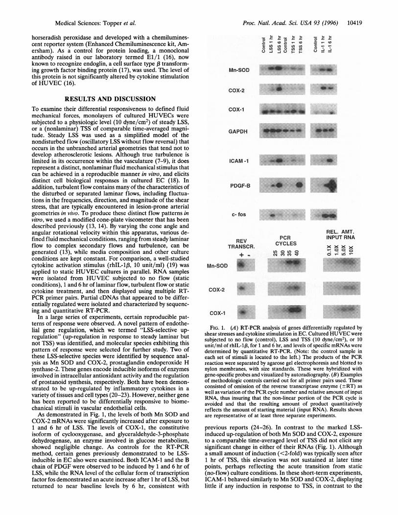
RESULTS AND DISCUSSIONTo examine their differential responsiveness to defined fluidmechanical forces, monolayers of cultured HUVECs weresubjected to a physiologic level (10 dyne/cm2) of steady LSS,or a (nonlaminar) TSS of comparable time-averaged magni-tude. Steady LSS was used as a simplified model of thenondisturbed flow (oscillatory LSS without flow reversal) thatoccurs in the unbranched arterial geometries that tend not todevelop atherosclerotic lesions. Although true turbulence islimited in its occurrence within the vasculature (7-9), it doesrepresent a distinct, nonlaminar fluid mechanical stimulus thatcan be achieved in a reproducible manner in vitro, and elicitsdistinct cell biological responses in cultured EC (18). Inaddition, turbulent flow contains many of the characteristics ofthe disturbed or separated laminar flows, including fluctua-tions in the frequencies, direction, and magnitude of the shearstress, that are typically encountered in lesion-prone arterialgeometries in vivo. To produce these distinct flow patterns invitro, we used a modified cone-plate viscometer that has beendescribed previously (13, 14). By varying the cone angle andangular rotational velocity within this apparatus, various de-fined fluid mechanical conditions, ranging from steady laminarflow to complex secondary flows and turbulence, can begenerated (13), while media composition and other cultureconditions are kept constant. For comparison, a well-studiedcytokine activation stimulus (rhIL-13, 10 unit/ml) (19) wasapplied to static HUVEC cultures in parallel. RNA sampleswere isolated from HUVEC subjected to no flow (staticconditions), 1 and 6 hr of laminar flow, turbulent flow or staticcytokine treatment, and then displayed using multiple RT-PCR primer pairs. Partial cDNAs that appeared to be differ-entially regulated were isolated and characterized by sequenc-ing and quantitative RT-PCR.
In a large series of experiments, certain reproducible pat-terns of response were observed. A novel pattern of endothe-lial gene regulation, which we termed "LSS-selective up-regulation" (up-regulation in response to steady laminar butnot TSS) was identified, and molecular species exhibiting thispattern of response were selected for further study. Two ofthese LSS-selective species were identified by sequence anal-ysis as Mn SOD and COX-2, prostaglandin endoperoxide Hsynthase-2. These genes encode inducible isoforms of enzymesinvolved in intracellular antioxidant activity and the regulationof prostanoid synthesis, respectively. Both have been demon-strated to be up-regulated by inflammatory cytokines in avariety of tissues and cell types (20-23). However, neither genehas been reported to be differentially responsive to biome-chanical stimuli in vascular endothelial cells.As demonstrated in Fig. 1, the levels of both Mn SOD and
COX-2 mRNAs were significantly increased after exposure to1 and 6 hr of LSS. The levels of COX-1, the constitutiveisoform of cyclooxygenase, and glyceraldehyde-3-phosphatedehydrogenase, an enzyme involved in glucose metabolism,showed negligible change. As controls for the RT-PCRmethod, certain genes previously demonstrated to be LSS-inducible in EC also were examined. Both ICAM-1 and the Bchain of PDGF were observed to be induced by 1 and 6 hr ofLSS, while the RNA level of the cellular form of transcriptionfactor fos demonstrated an acute increase after 1 hr of LSS, butreturned to near baseline levels by 6 hr, consistent with
I- X . .x .I..._ - *- b k
_5 m m o6 m s: z
m 0 - (0 W0 cj () C-j
Mn-SOD " _f
COX-2 6*
COX-1 ILa hi.4W
GAPDH E_ _
ICAM-1 *
PDGF-B
c- fos R
REVTRANSCR.
M-nS
Mln-SOD
PCRCYCLES
U) 0 U) 0
cm X c t
*''"A'F
REL. AMT.INPUT RNA
x x x x0- U)(*~.. _ .. ...
W....m&,| l~~~~~~~~~~~~~~~~~~~~~~~~~~~~~~~~~~~~~~~~~~~~~. e... . . . .COX-2
COX-1_
FIG. 1. (A) RT-PCR analysis of genes differentially regulated byshear stresses and cytokine stimulation in EC. Cultured HUVEC weresubjected to no flow (control), LSS and TSS (10 dyne/cm2), or 10unit/ml of rhIL-1,B, for 1 and 6 hr, and levels of specific mRNAs weredetermined by quantitative RT-PCR. (Note: the control sample ineach set of stimuli is located to the left.) The products of the PCRreaction were separated by agarose gel electrophoresis and blotted tonylon membranes, with size standards. These were hybridized withgene-specific probes and visualized by autoradiography. (B) Examplesof methodologic controls carried out for all primer pairs used. Theseconsisted of omission of the reverse transcriptase enzyme (±RT) aswell as variation of the PCR cycle number and relative amount of inputRNA, thus insuring that the non-linear portion of the PCR cycle isavoided and that the resulting amount of product quantitativelyreflects the amount of starting material (input RNA). Results shownare representative of at least three separate experiments.
previous reports (24-26). In contrast to the marked LSS-induced up-regulation of both Mn SOD and COX-2, exposureto a comparable time-averaged level of TSS did not elicit anysignificant change in either of their RNAs (Fig. 1). Althougha small amount of induction (<2-fold) was typically seen after1 hr of TSS, this elevation was not sustained at later timepoints, perhaps reflecting the acute transition from static(no-flow) culture conditions. In these short-term experiments,ICAM-1 behaved similarly to Mn SOD and COX-2, displayinglittle if any induction in response to TSS, in contrast to the
Medical Sciences: Topper et aL
10420 Medical Sciences: Topper et al.
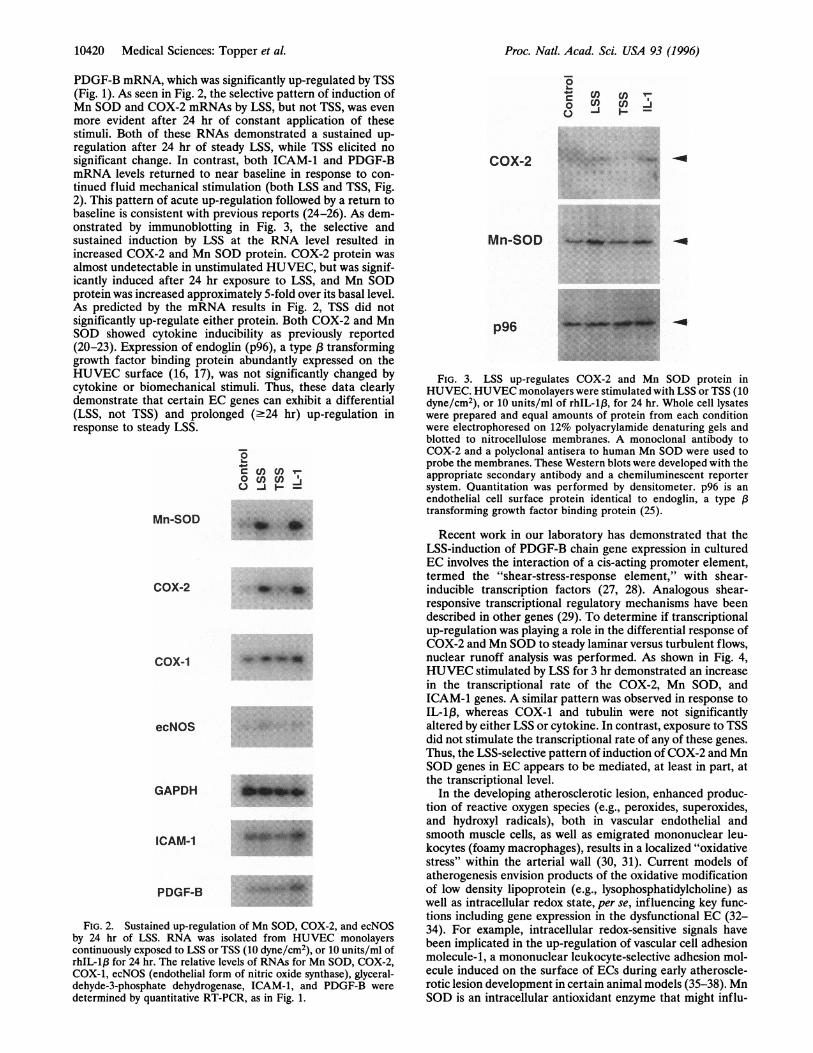
PDGF-B mRNA, which was significantly up-regulated by TSS(Fig. 1). As seen in Fig. 2, the selective pattern of induction ofMn SOD and COX-2 mRNAs by LSS, but not TSS, was evenmore evident after 24 hr of constant application of thesestimuli. Both of these RNAs demonstrated a sustained up-regulation after 24 hr of steady LSS, while TSS elicited nosignificant change. In contrast, both ICAM-1 and PDGF-BmRNA levels returned to near baseline in response to con-tinued fluid mechanical stimulation (both LSS and TSS, Fig.2). This pattern of acute up-regulation followed by a return tobaseline is consistent with previous reports (24-26). As dem-onstrated by immunoblotting in Fig. 3, the selective andsustained induction by LSS at the RNA level resulted inincreased COX-2 and Mn SOD protein. COX-2 protein wasalmost undetectable in unstimulated HUVEC, but was signif-icantly induced after 24 hr exposure to LSS, and Mn SODprotein was increased approximately 5-fold over its basal level.As predicted by the mRNA results in Fig. 2, TSS did notsignificantly up-regulate either protein. Both COX-2 and MnSOD showed cytokine inducibility as previously reported(20-23). Expression of endoglin (p96), a type l3 transforminggrowth factor binding protein abundantly expressed on theHUVEC surface (16, 17), was not significantly changed bycytokine or biomechanical stimuli. Thus, these data clearlydemonstrate that certain EC genes can exhibit a differential(LSS, not TSS) and prolonged (.24 hr) up-regulation inresponse to steady LSS.
0 s.n ua
O J F -
Mn-SOD .
COX-2 1h_
COX-1
ecNOS
GAPDH
ICAM-1
PDGF-B
FIG. 2. Sustained up-regulation of Mn SOD, COX-2, and ecNOSby 24 hr of LSS. RNA was isolated from HUVEC monolayerscontinuously exposed to LSS or TSS (10 dyne/cm2), or 10 units/ml ofrhIL-1f3 for 24 hr. The relative levels of RNAs for Mn SOD, COX-2,COX-1, ecNOS (endothelial form of nitric oxide synthase), glyceral-dehyde-3-phosphate dehydrogenase, ICAM-1, and PDGF-B weredetermined by quantitative RT-PCR, as in Fig. 1.
do Cl) dl)I-
COX-2 MiRm
AWW~~~~~W
Mn-SOD
p96
FIG. 3. LSS up-regulates COX-2 and Mn SOD protein inHUVEC. HUVEC monolayers were stimulated with LSS or TSS (10dyne/cm2), or 10 units/ml of rhIL-1,B, for 24 hr. Whole cell lysateswere prepared and equal amounts of protein from each conditionwere electrophoresed on 12% polyacrylamide denaturing gels andblotted to nitrocellulose membranes. A monoclonal antibody toCOX-2 and a polyclonal antisera to human Mn SOD were used toprobe the membranes. These Western blots were developed with theappropriate secondary antibody and a chemiluminescent reportersystem. Quantitation was performed by densitometer. p96 is anendothelial cell surface protein identical to endoglin, a type f3transforming growth factor binding protein (25).
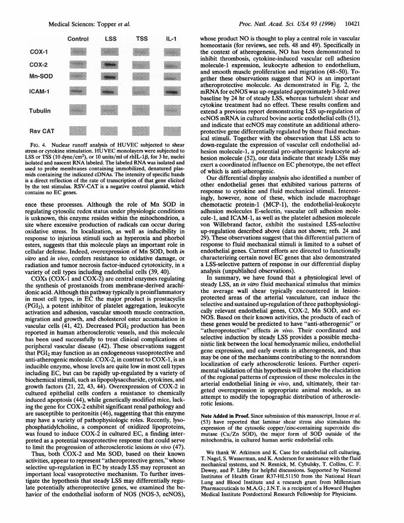
Recent work in our laboratory has demonstrated that theLSS-induction of PDGF-B chain gene expression in culturedEC involves the interaction of a cis-acting promoter element,termed the "shear-stress-response element," with shear-inducible transcription factors (27, 28). Analogous shear-responsive transcriptional regulatory mechanisms have beendescribed in other genes (29). To determine if transcriptionalup-regulation was playing a role in the differential response ofCOX-2 and Mn SOD to steady laminar versus turbulent flows,nuclear runoff analysis was performed. As shown in Fig. 4,HUVEC stimulated by LSS for 3 hr demonstrated an increasein the transcriptional rate of the COX-2, Mn SOD, andICAM-1 genes. A similar pattern was observed in response toIL-13, whereas COX-1 and tubulin were not significantlyaltered by either LSS or cytokine. In contrast, exposure to TSSdid not stimulate the transcriptional rate of any of these genes.Thus, the LSS-selective pattern of induction of COX-2 and MnSOD genes in EC appears to be mediated, at least in part, atthe transcriptional level.
In the developing atherosclerotic lesion, enhanced produc-tion of reactive oxygen species (e.g., peroxides, superoxides,and hydroxyl radicals), both in vascular endothelial andsmooth muscle cells, as well as emigrated mononuclear leu-kocytes (foamy macrophages), results in a localized "oxidativestress" within the arterial wall (30, 31). Current models ofatherogenesis envision products of the oxidative modificationof low density lipoprotein (e.g., lysophosphatidylcholine) aswell as intracellular redox state, per se, influencing key func-tions including gene expression in the dysfunctional EC (32-34). For example, intracellular redox-sensitive signals havebeen implicated in the up-regulation of vascular cell adhesionmolecule-1, a mononuclear leukocyte-selective adhesion mol-ecule induced on the surface of ECs during early atheroscle-rotic lesion development in certain animal models (35-38). MnSOD is an intracellular antioxidant enzyme that might influ-
Proc. Natl. Acad. Sci. USA 93 (1996)
W_I_..i-40 ___..
Proc. Natl. Acad. Sci. USA 93 (1996) 10421
Control LSS TSS IL-1
COX-1
COX-2 A
Mn-SOD
ICAM-1 .
Tubulin
Rsv CAT
FIG. 4. Nuclear runoff analysis of HUVEC subjected to shearstress or cytokine stimulation. HUVEC monolayers were subjected toLSS or TSS (10 dyne/cm2), or 10 units/ml of rhIL-1,8, for 3 hr, nucleiisolated and nascent RNA labeled. The labeled RNA was isolated andused to probe membranes containing immobilized, denatured plas-mids containing the indicated cDNAs. The intensity of specific bandsis a direct reflection of the rate of transcription of that gene elicitedby the test stimulus. RSV-CAT is a negative control plasmid, whichcontains no EC genes.
ence these processes. Although the role of Mn SOD inregulating cytosolic redox status under physiologic conditionsis unknown, this enzyme resides within the mitochondrion, asite where excessive production of radicals can occur duringoxidative stress. Its localization, as well as inducibility inresponse to injurious stimuli such as hyperoxia and phorbolesters, suggests that this molecule plays an important role incellular defense. Indeed, overexpression of Mn SOD, both invitro and in vivo, confers resistance to oxidative damage, orradiation and tumor necrosis factor-induced cytotoxicity, in avariety of cell types including endothelial cells (39, 40).COXs (COX-1 and COX-2) are central enzymes regulating
the synthesis of prostanoids from membrane-derived arachi-donic acid. Although this pathway typically is proinflammatoryin most cell types, in EC the major product is prostacyclin(PGI2), a potent inhibitor of platelet aggregation, leukocyteactivation and adhesion, vascular smooth muscle contraction,migration and growth, and cholesterol ester accumulation invascular cells (41, 42). Decreased PGI2 production has beenreported in human atherosclerotic vessels, and this moleculehas been used successfully to treat clinical complications ofperipheral vascular disease (42). These observations suggestthat PGI2 may function as an endogeneous vasoprotective andanti-atherogenic molecule. COX-2, in contrast to COX-1, is aninducible enzyme, whose levels are quite low in most cell typesincluding EC, but can be rapidly up-regulated by a variety ofbiochemical stimuli, such as lipopolysaccharide, cytokines, andgrowth factors (21, 22, 43, 44). Overexpression of COX-2 incultured epithelial cells confers a resistance to chemicallyinduced apoptosis (44), while genetically modified mice, lack-ing the gene for COX-2 exhibit significant renal pathology andare susceptible to peritonitis (46), suggesting that this enzymemay have a variety of pathophysiologic roles. Recently, lyso-phosphatidylcholine, a component of oxidized lipoproteins,was found to induce COX-2 in cultured EC, a finding inter-preted as a potential vasoprotective response that could serveto limit the progression of atherosclerotic lesions in vivo (47).
Thus, both COX-2 and Mn SOD, based on their knownactivities, appear to represent "atheroprotective genes," whoseselective up-regulation in EC by steady LSS may represent animportant local vasoprotective mechanism. To further inves-tigate the hypothesis that steady LSS may differentially regu-late potentially atheroprotective genes, we examined the be-havior of the endothelial isoform of NOS (NOS-3, ecNOS),
whose product NO is thought to play a central role in vascularhomeostasis (for reviews, see refs. 48 and 49). Specifically inthe context of atherogenesis, NO has been demonstrated toinhibit thrombosis, cytokine-induced vascular cell adhesionmolecule-1 expression, leukocyte adhesion to endothelium,and smooth muscle proliferation and migration (48-50). To-gether these observations suggest that NO is an importantatheroprotective molecule. As demonstrated in Fig. 2, themRNA for ecNOS was up-regulated approximately 3-fold overbaseline by 24 hr of steady LSS, whereas turbulent shear andcytokine treatment had no effect. These results confirm andextend a previous report demonstrating LSS up-regulation ofecNOS mRNA in cultured bovine aortic endothelial cells (51),and indicate that ecNOS may constitute an additional athero-protective gene differentially regulated by these fluid mechan-ical stimuli. Together with the observation that LSS acts todown-regulate the expression of vascular cell endothelial ad-hesion molecule-1, a potential pro-atherogenic leukocyte ad-hesion molecule (52), our data indicate that steady LSSs mayexert a coordinated influence on EC phenotype, the net effectof which is anti-atherogenic.Our differential display analysis also identified a number of
other endothelial genes that exhibited various patterns ofresponse to cytokine and fluid mechanical stimuli. Interest-ingly, however, none of these, which include macrophagechemotactic protein-1 (MCP-1), the endothelial-leukocyteadhesion molecules E-selectin, vascular cell adhesion mole-cule-1, and ICAM-1, as well as the platelet adhesion moleculevon Willebrand factor, exhibit the sustained LSS-selectiveup-regulation described above (data not shown; refs. 24 and29). These observations suggest that this differential pattern ofresponse to fluid mechanical stimuli is limited to a subset ofendothelial genes. Current efforts are directed to functionallycharacterizing certain novel EC genes that also demonstrateda LSS-selective pattern of response in our differential displayanalysis (unpublished observations).
In summary, we have found that a physiological level ofsteady LSS, an in vitro fluid mechanical stimulus that mimicsthe average wall shear typically encountered in lesion-protected areas of the arterial vasculature, can induce theselective and sustained up-regulation of three pathophysiologi-cally relevant endothelial genes, COX-2, Mn SOD, and ec-NOS. Based on their known activities, the products of each ofthese genes would be predicted to have "anti-atherogenic" or"atheroprotective" effects in vivo. Their coordinated andselective induction by steady LSS provides a possible mecha-nistic link between the local hemodynamic milieu, endothelialgene expression, and early events in atherogenesis, and thusmay be one of the mechanisms contributing to the nonrandomlocalization of early atherosclerotic lesions. Further experi-mental validation of this hypothesis will involve the elucidationof the regional patterns of expression of these molecules in thearterial endothelial lining in vivo, and, ultimately, their tar-geted overexpression in appropriate animal models, as anattempt to modify the topographic distribution of atheroscle-rotic lesions.
Note Added in Proof. Since submission of this manuscript, Inoue et al.(53) have reported that laminar shear stress also stimulates theexpression of the cytosolic copper/zinc-containing superoxide dis-mutase (Cu/Zn SOD), the major form of SOD outside of themitochondria, in cultured human aortic endothelial cells.
We thank W. Atkinson and K. Case for endothelial cell culturing,T. Nagel, S. Wasserman, and K. Anderson for assistance with the fluidmechanical systems, and N. Resnick, M. Cybulsky, T. Collins, C. F.Dewey, and P. Libby for helpful discussions. Supported by NationalInstitutes of Health Grant R37-HL51150 from the National HeartLung and Blood Institute and a research grant from MillenniumPharmaceuticals to M.A.G.; J.N.T. is a recipient of a Howard HughesMedical Institute Postdoctoral Research Fellowship for Physicians.
Medical Sciences: Topper et aL
10422 Medical Sciences: Topper et al.
1. Ross, R. (1993) Nature (London) 362, 801-809.2. Gimbrone, M. A., Jr. (1995) in Molecular Cardiovascular Medi-
cine, ed. Haber, E. (Sci. Am. Med., New York), pp. 49-61.3. Gimbrone, M. A., Jr., Cybulsky, M. I., Kume, N., Collins, T. &
Resnick, N. (1995) Ann. N.Y Acad. Sci. 748, 122-132.4. Fry, D. L. (1973) CIBA Found. Symp. 12, 96-118.5. Caro, C. G., Fitzgerald, J. M. & Schroter, R. C. (1969) Nature
(London) 223, 1159-1161.6. Cornhill, J. F. & Roach, M. R. (1976) Atherosclerosis 23, 489-
499.7. Karino, Y. & Motomiya, M. (1983) Biorheology 20, 119-127.8. Zarins, C. K, Giddens, D. P., Bharadvaj, B. K, Sottiurai, V. S.,
Mabon, R. F. & Glagov, S. (1983) Circ. Res. 53, 502-514.9. Ku, D. N., Giddens, D. P., Zarins, C. K. & Glagov, S. (1985)
Arteriosclerosis 5, 293-301.10. Nakashima, Y., Plump, A. S., Raines, E. W., Breslow, J. L. &
Ross, R. (1994) Arterioscler. Thromb. 14, 133-140.11. Davies, P. F. (1995) Physiol. Rev. 75, 519-560.12. Resnick, N. & Gimbrone, M. A., Jr. (1995) FASEB J. 9, 874-882.13. Bussolari, S. R., Dewey, C. F., Jr., & Gimbrone, M. A., Jr. (1982)
Rev. Sci. Instrum. 53, 1851-1854.14. Sdougos, H. P., Bussolari, S. R. & Dewey, C. F. (1984) J. Fluid
Mech. 138, 379-404.15. Liang, P. & Pardee, A. B. (1992) Science 257, 967-971.16. Bevilacqua, M., Stengelin, S., Gimbrone, M. A., Jr., & Seed, B.
(1989) Science 243, 1160-1165.17. Cheifetz, S., Bellon, T., Cales, C., Vera, S., Bernabeus, C.,
Massague, J. & Letarte, M. (1992) J. Biol. Chem. 267, 19027-19030.
18. Davies, P. F., Remuzzi, A., Gordon, E. J., Dewey, C. F., Jr., &Gimbrone, M. A., Jr. (1986) Proc. Natl. Acad. Sci. USA 83,2114-2117.
19. Pober, J. S. & Cotran, R. S. (1990) Physiol. Rev. 70, 427-451.20. Pritchard, K. A., O'Bannion, M. K., Miano, J. M., Vlasic N.,
Bhatia, U. G., Young, D. A. & Stemerman, M. B. (1994) J. Biol.Chem. 269, 8504-8509.
21. Jones, D. A., Carlton, D. P., McIntyre, T. M., Simmerman, G. A.& Prescott, S. M. (1993) J. Biol. Chem. 266, 9049-9054.
22. Ristimaki, A., Garfinkel, S., Wessendorf, J., Maciag, T. & Hla, T.(1994) J. Biol. Chem. 269, 11769-11775.
23. Suzuki, K., Tatsumi, H., Satoh, S., Senda, T., Nakata, T., Fujii, J.& Taniguchi, N. (1993) Am. J. Physiol 265, H1173-H1178.
24. Nagel, T., Resnick, N., Atkinson, W. J., Dewey, C. F. Jr., &Gimbrone, M. A., Jr. (1994) J. Clin. Invest. 94, 885-891.
25. Hsieh, H. J., Li, N. Q. & Frangos, J. (1991) Am. J. Physiol. 260,H642-H646.
26. Hsieh, H., Li, N. & Frangos, J. A. (1992) J. Cell. Physiol. 150,552-558.
27. Resnick, N., Collins, T., Atkinson, W., Bonthron, D. T., Dewey,C. F., Jr., & Gimbrone, M. A., Jr., (1993) Proc. Natl. Acad. Sci.USA 90, 4591-4595.
28. Khachigian, L. M., Resnick, N., Gimbrone, M. A., Jr., & Collins,T. (1995) J. Clin. Invest. 96, 1169-1175.
29. Shyy, J. Y., Lin, M. C., Han, J., Lu, Y., Petrime, M. & Chien, S.(1995) Proc. Natl. Acad. Sci. USA 92, 8069-8073.
30. Steinberg, D., Parthasarathy, S., Carew, T. E., Khoo, J. C. &Witzum, J. L. (1989) N. Engl. J. Med. 320, 915-924.
31. Alexander, R. W. (1995) Hypertension 25, 155-161.32. Kume, N., Cybulsky, M. I. & Gimbrone, M. A., Jr. (1992)J. Clin.
Invest. 90, 1138-1144.33. Kume, N. & Gimbrone, M. A., Jr. (1994) J. Clin. Invest. 93,
907-911.34. Offermann, M. K. & Medford, R. M. (1994) Heart Dis. Stroke 3,
52-57.35. Marui, N., Offermann, M. K, Swerlick, R., Kunsch, C., Rosen,
C. A., Ahmad, M., Alexander, R. W. & Medford, R. M. (1993)J. Clin. Invest. 92, 1866-1874.
36. Cybulsky, M. I. & Gimbrone, M. A., Jr. (1991) Science 251,788-791.
37. Li, H., Cybulsky, M. I., Gimbrone, M. A., Jr., & Libby, P. (1993)Arterioscler. Thromb. 13, 197-204.
38. Collins, T. (1993) Lab Invest. 68, 499-508.39. Wispe, J. R., Warner, B. B., Clark, J. C., Dey, C. R., Nwuman, J.,
Glasser, S. W., Crapo, J. D., Chang, L.-Y. & Whitsett, J. A.(1992) J. Biol. Chem. 267, 23937-23941.
40. Lindau-Shepard, B., Shaffer, J. B. & Vecchio, P. J. D. (1994) J.Cell. Physiol. 161, 237-242.
41. Pomerantz, K. B. & Hajar, D. P. (1989) Arteriosclerosis 9, 413-429.
42. Vane, J. R. & Botting, R. M. (1995)Am. J. Cardiol. 75, 3A-1OA.43. Hla, T. & Neilson, K. (1992) Proc. Natl. Acad. Sci. USA 89,
7384-7388.44. Vane, J. R., Mitchell, J. A., Appleton, I., Tomlinson, A., Bishop-
Bailey, D., Croxtall, J. & Willoughby, D. A. (1994) Proc. Natl.Acad. Sci. USA 91, 2046-2050.
45. Tsujii, M. & Dubois, R. N. (1995) Cell 83, 493-501.46. Morham, S. G., Langenbach, J. R., Loftin, C. D., Tiano, H. F.,
Vouloumaos, N., Jennette, J. C., Mahler, J. F., Kluckman, K. D.,Ledford, A., Lee, C. A. & Smithies, 0. (1995) Cell 83, 473-482.
47. Zembowicz, A., Jones, S. L. & Wu, K. K. (1995) J. Clin. Invest. 96,1688-1692.
48. Dusting, G. (1995) J. Vasc. Res. 32, 143-161.49. Wennmalm, A. (1994) J. Intern. Med. 235, 317-327.50. DeCaterina, R., Libby, P., Thannickal, V. J., Peng, H.-B., Ra-
javashisth, T. B., Gimbrone, M. A., Jr., Shin, W. S. & Liao, J. K.(1995) J. Clin. Invest. 96, 60-68.
51. Nishida, K, Harrison, D. G., Nawas, J. P., Fisher, A. A., Dock-ery, S. P., Uematsu, M., Nerem, R. M., Alexander, R. W. &Murphy, T. J. (1992) J. Clin. Invest. 90, 2092-2096.
52. Ando, J., Tsuboi, H., Korenaga, R., Takada, Y., Toyama-Sori-machi, N., Miyasaka, M. & Kamiya, A. (1994)Am. J. Physiol. 267,C679-C687.
53. Inoe, N., Ramasamy, S., Fukai, T., Nerem, R. M. & Harrison,D. G. (1996) Circ. Res. 79, 32-37.
Proc. Natl. Acad. Sci. USA 93 (1996)