Embed Size (px)
Citation preview
7
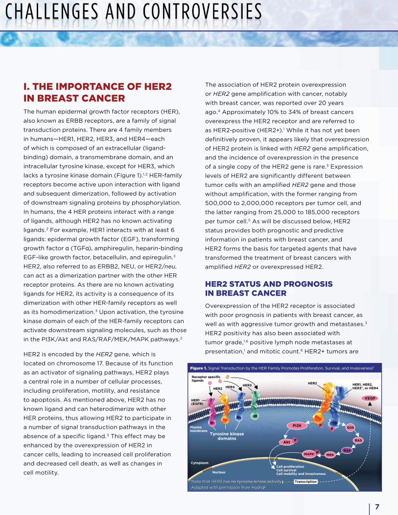
I. THE IMPORTANCE OF HER2 IN BREAST CANCERThe human epidermal growth factor receptors (HER),
also known as ERBB receptors, are a family of signal
transduction proteins. There are 4 family members
in humans—HER1, HER2, HER3, and HER4—each
of which is composed of an extracellular (ligand-
binding) domain, a transmembrane domain, and an
intracellular tyrosine kinase, except for HER3, which
lacks a tyrosine kinase domain (Figure 1).1,2 HER-family
receptors become active upon interaction with ligand
and subsequent dimerization, followed by activation
of downstream signaling proteins by phosphorylation.
In humans, the 4 HER proteins interact with a range
of ligands, although HER2 has no known activating
ligands.2 For example, HER1 interacts with at least 6
ligands: epidermal growth factor (EGF), transforming
growth factor � (TGF�), amphiregulin, heparin-binding
EGF-like growth factor, betacellulin, and epiregulin.3
HER2, also referred to as ERBB2, NEU, or HER2/neu,
can act as a dimerization partner with the other HER
receptor proteins. As there are no known activating
ligands for HER2, its activity is a consequence of its
dimerization with other HER-family receptors as well
as its homodimerization.3 Upon activation, the tyrosine
kinase domain of each of the HER-family receptors can
activate downstream signaling molecules, such as those
in the PI3K/Akt and RAS/RAF/MEK/MAPK pathways.2
HER2 is encoded by the HER2 gene, which is
located on chromosome 17. Because of its function
as an activator of signaling pathways, HER2 plays
a central role in a number of cellular processes,
including proliferation, motility, and resistance
to apoptosis. As mentioned above, HER2 has no
known ligand and can heterodimerize with other
HER proteins, thus allowing HER2 to participate in
a number of signal transduction pathways in the
absence of a specific ligand.3 This effect may be
enhanced by the overexpression of HER2 in
cancer cells, leading to increased cell proliferation
and decreased cell death, as well as changes in
cell motility.
The association of HER2 protein overexpression
or HER2 gene amplification with cancer, notably
with breast cancer, was reported over 20 years
ago.4 Approximately 10% to 34% of breast cancers
overexpress the HER2 receptor and are referred to
as HER2-positive (HER2+).1 While it has not yet been
definitively proven, it appears likely that overexpression
of HER2 protein is linked with HER2 gene amplification,
and the incidence of overexpression in the presence
of a single copy of the HER2 gene is rare.5 Expression
levels of HER2 are significantly different between
tumor cells with an amplified HER2 gene and those
without amplification, with the former ranging from
500,000 to 2,000,000 receptors per tumor cell, and
the latter ranging from 25,000 to 185,000 receptors
per tumor cell.5 As will be discussed below, HER2
status provides both prognostic and predictive
information in patients with breast cancer, and
HER2 forms the basis for targeted agents that have
transformed the treatment of breast cancers with
amplified HER2 or overexpressed HER2.
HER2 STATUS AND PROGNOSIS IN BREAST CANCER
Overexpression of the HER2 receptor is associated
with poor prognosis in patients with breast cancer, as
well as with aggressive tumor growth and metastases.3
HER2 positivity has also been associated with
tumor grade,1,6 positive lymph node metastases at
presentation,1 and mitotic count.6 HER2+ tumors are
Akt
SOS
RAS
RAFMEK
VEGF
MAPK
Receptor specificligands
HER1, HER2,HER3*, or HER4
HER2
HER1(EGFR)
HER2 HER4 HER3
Tyrosine kinasedomains
Plasmamembrane
PI3K
Cell proliferationCell survivalCell mobility and invasiveness
Cytoplasm
Nucleus
Transcription
Figure 1. Signal Transduction by the HER Family Promotes Proliferation, Survival, and Invasiveness2
P
PP
P
*******Not****** e that HER3 has asas aasasas asasassasssssssssssssssssssssssssssssssssasssasssssassasss no tno tno tno tno tno no ttno ttno ttno tnono tnonono tnnnno tnoo to to tnonono tno tto ttnono tnnnnnooonoonoo ttno tnno too to ttnnono tnnnoooo ttttnnnoooo to ooonoo tnoooooo yrosyrosyrosyroyrosyrosyrosrosyrosyyryryrosyrorryrrrosooorosyrosoyrosrosrosyrosyyyyrosrrrosyrosroooooyrososoosssyroyrosyrryrosyroooorooyrosyrosyrosyyroyroyrrroooososyrosyrosy syryyyyyryrosyrosssyrooy osssine ine ine ineneineiineneineineneneineinineinennnineneininineeineineinnnneineneeinennnenneneeineeiinennnnneeeeneiinennineeennnneeneeeenneeneeeenenee kinakinakinakinakinakikinakikkinakkikinakinakinakinakinakkkinakinakkkkinakinakiinakikinankinnnnnkinakinakinkkkikinakinnninaninakinnakinaakininannnanaaanaakkinakinakinkinaikinannaanaakinakiinannnnnkinaakinakkinakkkkininanaakinakkinak nakinankinaaakk aakinase ase ase asese ase ase asee ae ase ase asese ase asse assssssese aeese ase aaaaase ase ase ae aae ase ase assse asese ae ae ae ase aaae ase ase ase ae ae aee aaaaasssese aese aaase ase aseeee ase aseeeesee aseee aeee aae ctivctivtictivctctivcttivctctivtctictivcctivttivtttivivvtivvvctivctictivctivttiictivvvtivctivctivctivctivctivtttiivvtivcctivctivctiivvivvcctivccctivtt vvvvtivvctivcccttttivvctivcttctivvc i ity.itytity.ityty.tty.ttyity.tytyitityityityttytttytytityyyitytyyyitttytyyyityitytytyyityyyy.yyity.iitttyityitytytyityyyyttyyyyityyyyyyyyy
Adapted with permisi sion fro Hm HuH dis.di 2
*Note that HER3 has no tyrosine kinase activity.
Adapted with permission from Hudis.2
8
less likely to be hormone receptor (estrogen receptor
[ER] or progesterone receptor [PgR])–positive.1
HER2 status also correlates with relative response to
various agents. HER2 positivity may result in increased
resistance to endocrine therapy and with a decreased
benefit from non-anthracycline, non-taxane–containing
chemotherapy.7 Conversely, HER2-positive patients
may exhibit improved response to anthracycline
therapy, as well as to paclitaxel.7
Data surrounding the prognostic and predictive value
of HER2 status are continually evolving. In a recent
study, it was shown that higher levels of HER2 gene
amplification were associated with worse outcomes in
patients treated with doxorubicin-based therapy in the
adjuvant setting.8
The association of HER2 gene amplification or HER2
overexpression with some breast cancers has allowed
for the development of agents that specifically target
HER2, altering the treatment landscape for these
cancers. Trastuzumab, which was approved for the
treatment of metastatic breast cancer in 1998, for the
adjuvant treatment of lymph node–positive breast
cancer in 2006, and for the adjuvant treatment of
lymph node–negative breast cancer in 2008, is a
humanized monoclonal antibody to the HER2 protein.
Lapatinib is a selective inhibitor of the tyrosine kinase
activity of HER2 and EGFR. Each of these agents has
shown efficacy in patients whose tumors are HER2+.
HER2 EXPRESSION AND BREAST PATHOLOGY
An association of HER2 status with various breast
pathologies has been noted. For ductal carcinoma
in situ (DCIS), the incidence of HER2+ status is
higher than that seen for invasive breast cancer
(approximately 24%, vs 38% for DCIS). HER2
overexpression in this pathologic subtype is associated
with higher grade and more extensive forms of DCIS.1
HER2 gene amplification is seen less often—in invasive
lobular carcinoma in approximately 10% of cases—
but is linked to adverse outcomes. HER2 positivity is
present less frequently in male breast cancer than in
female breast cancer. The rate of HER2 positivity is also
very low to nonexistent in mucinous, medullary, and
tubular carcinomas; breast sarcomas and Phyllodes
tumors; as well as in hereditary breast cancer, which
is associated with mutations in BRCA1/BRCA2. A low
level of HER2 expression has been observed in some
benign breast disease biopsies and is associated with
a greater risk for subsequent development of invasive
breast cancer.1
HER2 STATUS IN PRIMARY VS METASTATIC BREAST CANCER
Recent studies have shown some discordance between
HER2 status of the primary tumor and metastatic
lesions in the same patient. Discordance rates are
20% to 30%1 and may be at least partly due to issues
of reproducibility of HER2 assays, discussed below.
However, HER2 status may evolve, and HER2 positivity
or negativity may develop between the time of primary
and metastatic disease. The question of the stability of
HER2 status is currently uncertain, but data continue to
emerge to address this issue. A recent study showed a
concordance rate of 66% in HER2 status between the
primary and metastatic tumors.9
II. HER2 TESTINGAs mentioned above, tumor HER2 status can provide
both prognostic and predictive information in patients
with breast cancer. Knowledge of HER2 status is also
essential in the decision of whether to treat with one of
the HER2-targeted agents, trastuzumab or lapatinib.
For this reason, it has been recommended that HER2
testing be performed for all newly diagnosed invasive
breast cancers. Currently, the methods used most often
for HER2 testing are immunohistochemistry (IHC) and
in situ hybridization, described below. These assays
are most often performed on formalin-fixed, paraffin-
embedded (FFPE) tissue samples.
SLIDE-BASED ASSAYS
Immunohistochemistry
Immunohistochemical staining allows for the detection
of protein in tissues and is the most frequently
performed initial test of HER2 status in patients with
9
newly diagnosed invasive breast cancer. Currently,
there are 2 commercially available US Food and Drug
Administration (FDA)-approved HER2 IHC assays:
Dako HercepTest™ (Dako Corporation, Glostrup,
Denmark) and Ventana Pathway™ (Ventana Medical
Systems, Tucson, AZ). Because the HER2 protein is
expressed in normal breast epithelial cells, the HER2
IHC assay is a quantitative, rather than a qualitative,
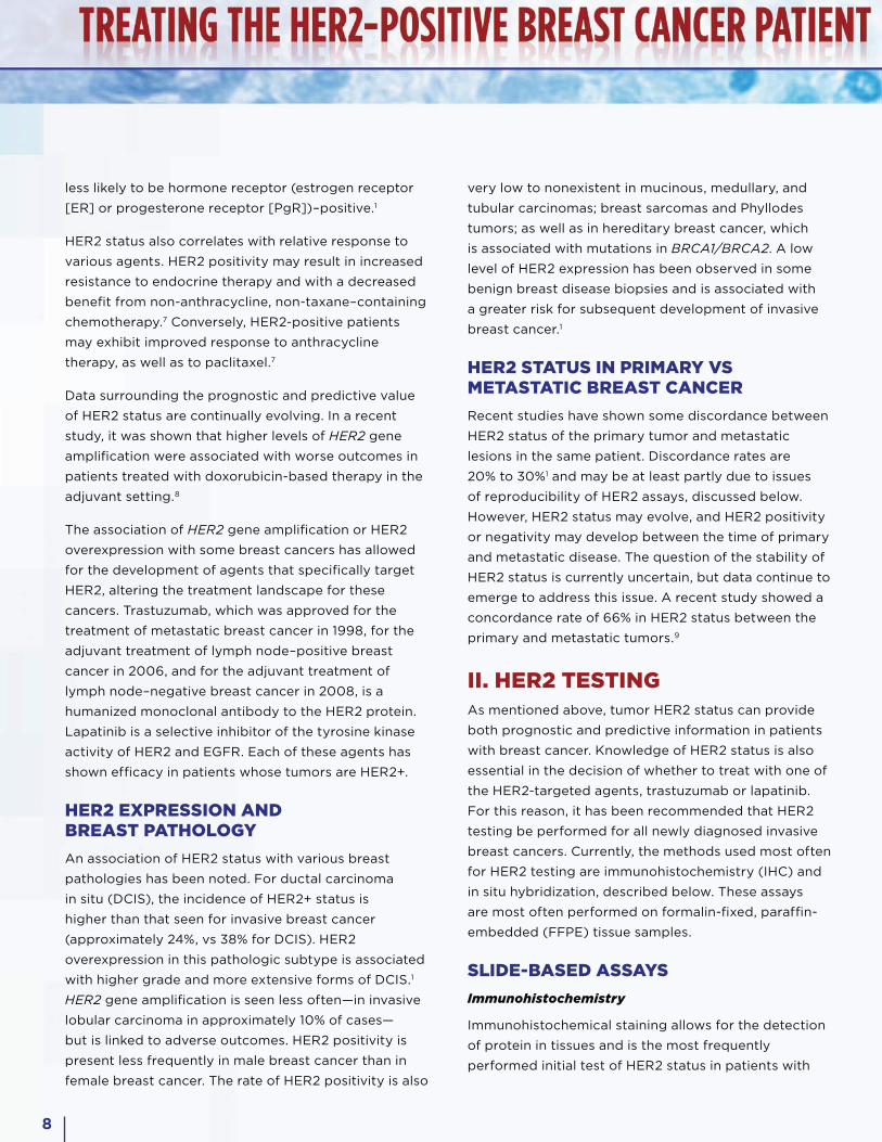
test. One of the most widely used guidelines for
interpretation of IHC results is that developed by the
American Society of Clinical Oncology (ASCO) and
the College of American Pathologists (CAP).7 For
IHC, a positive HER2 test is defined as 3+ cell surface
protein expression (uniform intense staining of >30%
of invasive tumor cells), an equivocal test as 2+ cell
surface protein expression (complete membrane
staining that is variable or weak intensity, but with
cell surface–associated staining in >10% of cells), and
a negative test as 1+ (weak or incomplete staining
in any proportion of tumor cells) or 0 (no staining)
(Figure 210).
While IHC is the most commonly used initial assay
for HER2, this method suffers from a number of
disadvantages. Many of these disadvantages are due
to technical issues, such as variations in tissue fixation,
tissue processing, and embedding of tissue in paraffin.1
One crucial issue is the difficulty in standardization
of signal, which can be at least partly due to variable
fixation and to variation in antigen retrieval.5 Variability
may also be introduced depending on the type of
antibody (monoclonal vs polyclonal) being used.
Another key issue for IHC is that of interpretation, as
this is somewhat subjective and will differ depending
on the slide scorer.
Some of these issues can be addressed; for example
a recent study showed that fixation issues can be
ameliorated.11 This study, in which a standard fixation
time of 6 hours was utilized, showed that there was
a decrease in inconclusive cases (from 10.8% to
3.4%). While improvements can be made, there is
also the option of using the alternative technique of
fluorescence in situ hybridization (FISH),
described in the next column.
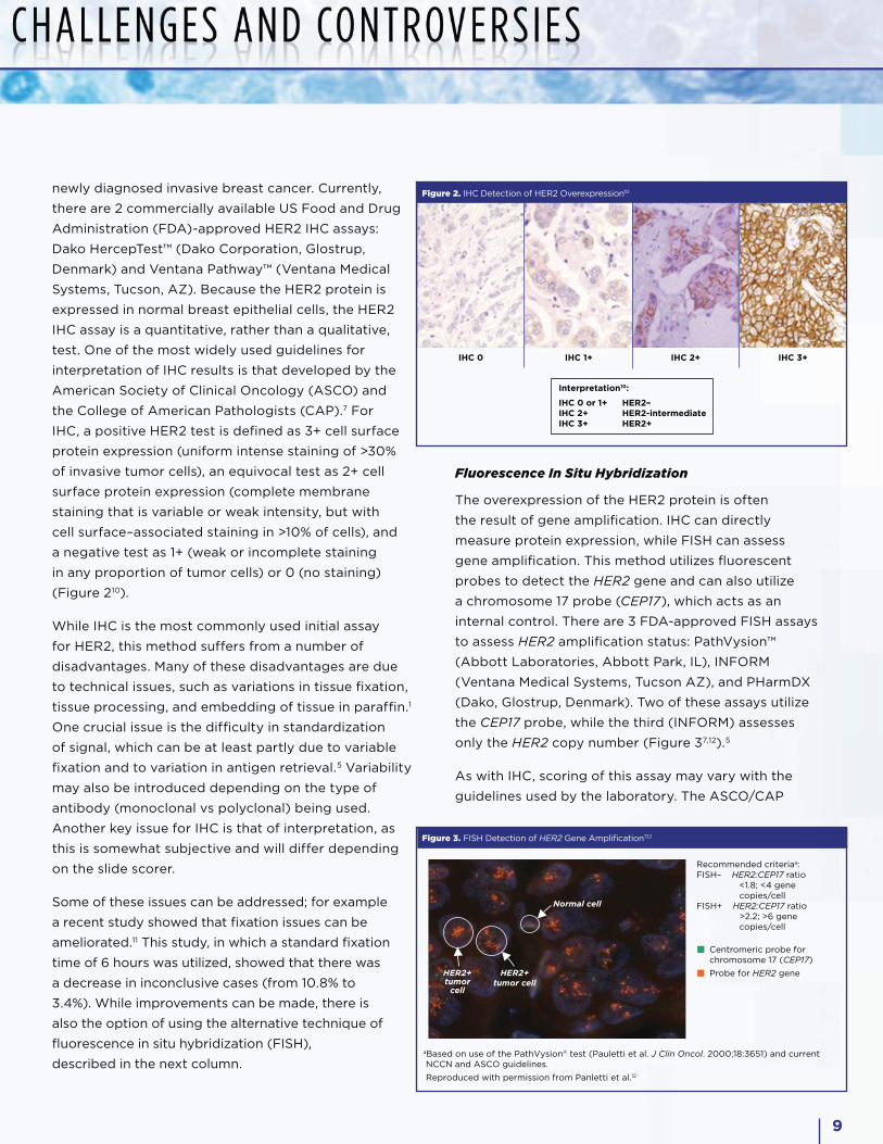
Fluorescence In Situ Hybridization
The overexpression of the HER2 protein is often
the result of gene amplification. IHC can directly
measure protein expression, while FISH can assess
gene amplification. This method utilizes fluorescent
probes to detect the HER2 gene and can also utilize
a chromosome 17 probe (CEP17), which acts as an
internal control. There are 3 FDA-approved FISH assays
to assess HER2 amplification status: PathVysion™
(Abbott Laboratories, Abbott Park, IL), INFORM
(Ventana Medical Systems, Tucson AZ), and PHarmDX
(Dako, Glostrup, Denmark). Two of these assays utilize
the CEP17 probe, while the third (INFORM) assesses
only the HER2 copy number (Figure 37,12).5
As with IHC, scoring of this assay may vary with the
guidelines used by the laboratory. The ASCO/CAP
Figure 2. IHC Detection of HER2 Overexpression10
IHC 0 IHC 1+ IHC 2+
Interpretation10:
IHC 0 or 1+ HER2– IHC 2+ HER2-intermediateIHC 3+ HER2+
IHC 3+
Figure 3. FISH Detection of HER2 Gene Amplification7,12
aBased on use of the PathVysion® test (Pauletti et al. J Clin Oncol. 2000;18:3651) and current NCCN and ASCO guidelines.
Reproduced with permission from Panletti et al.12
HER2+tumor
cell
HER2+tumor cell
Normal cell
Recommended criteriaa:FISH– HER2:CEP17 ratio <1.8; <4 gene copies/cell FISH+ HER2:CEP17 ratio >2.2; >6 gene copies/cell
Centromeric probe forchromosome 17 (CEP17)
Probe for HER2 gene
10
scoring guidelines are as follows7: Results are
reported as positive for HER2 (average of >6 copies
of the HER2 gene per nucleus, or HER2/CEP17 ratio
of >2.2), equivocal for HER2 (average of 4.0 to 6.0
copies of the HER2 gene per nucleus, or HER2/CEP17
ratio of 1.8 to 2.2), and negative for HER2 (average of
<4.0 copies of the HER2 gene per nucleus, or
HER2/CEP17 ratio of <1.8).
Testing and interpretation may be complicated by
the presence of polysomy 17, which can confound
results of the FISH assay.6 The impact of polysomy
17 on response to trastuzumab is still unclear,6 and
the determination of chromosome 17 copy number
should be assessed for patients with breast cancer in
order to make the most informed decisions.13
While FISH is considered the gold standard of HER2
assessment, there are crucial disadvantages associated
with this method, including increased cost relative to
IHC, longer time required for slide scoring, the need
for a fluorescent microscope, and the instability of the
fluorescent signal, which precludes storing slides for
later review.1
IHC vs FISH
IHC and FISH each offer advantages. The key
advantage of IHC is the lower cost, while the key
advantage of FISH is the higher objectivity and
reproducibility. Fixation appears to be less of an issue
with FISH.5 A review of relevant literature showed a
discordance rate between IHC and FISH of 2% to 20%.5
However, FISH has been shown to be superior to IHC
in predicting response to the HER2-targeted agent
trastuzumab.5 As seen below, if an equivocal result is
obtained with IHC, an appropriate follow-up is to carry
out a FISH assay.
CISH and SISH
As discussed above, FISH offers the advantages of
having a chromosome 17 internal control, and being
relatively objective. However, FISH suffers from the key
disadvantages of requiring a fluorescent microscope
and utilizing a signal that fades over time, which
precludes the storage and later reassessment of slides.
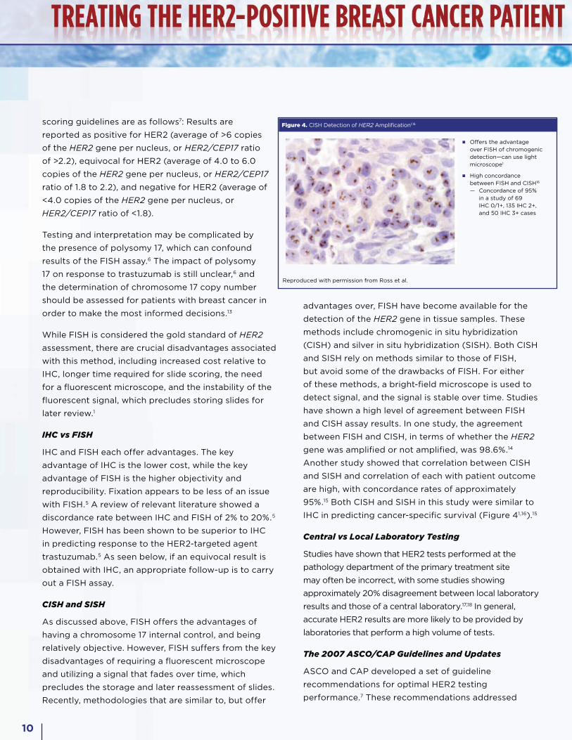
Recently, methodologies that are similar to, but offer
Figure 4. CISH Detection of HER2 Amplification1,16
Reproduced with permission from Ross et al.
■ Offers the advantage over FISH of chromogenic detection—can use light microscope1
■ High concordance between FISH and CISH16
— Concordance of 95% in a study of 69 IHC 0/1+, 135 IHC 2+, and 50 IHC 3+ cases
advantages over, FISH have become available for the
detection of the HER2 gene in tissue samples. These
methods include chromogenic in situ hybridization
(CISH) and silver in situ hybridization (SISH). Both CISH
and SISH rely on methods similar to those of FISH,
but avoid some of the drawbacks of FISH. For either
of these methods, a bright-field microscope is used to
detect signal, and the signal is stable over time. Studies
have shown a high level of agreement between FISH
and CISH assay results. In one study, the agreement
between FISH and CISH, in terms of whether the HER2
gene was amplified or not amplified, was 98.6%.14
Another study showed that correlation between CISH
and SISH and correlation of each with patient outcome
are high, with concordance rates of approximately
95%.15 Both CISH and SISH in this study were similar to
IHC in predicting cancer-specific survival (Figure 41,16).15
Central vs Local Laboratory Testing
Studies have shown that HER2 tests performed at the
pathology department of the primary treatment site
may often be incorrect, with some studies showing
approximately 20% disagreement between local laboratory
results and those of a central laboratory.17,18 In general,
accurate HER2 results are more likely to be provided by
laboratories that perform a high volume of tests.
The 2007 ASCO/CAP Guidelines and Updates
ASCO and CAP developed a set of guideline
recommendations for optimal HER2 testing
performance.7 These recommendations addressed
11
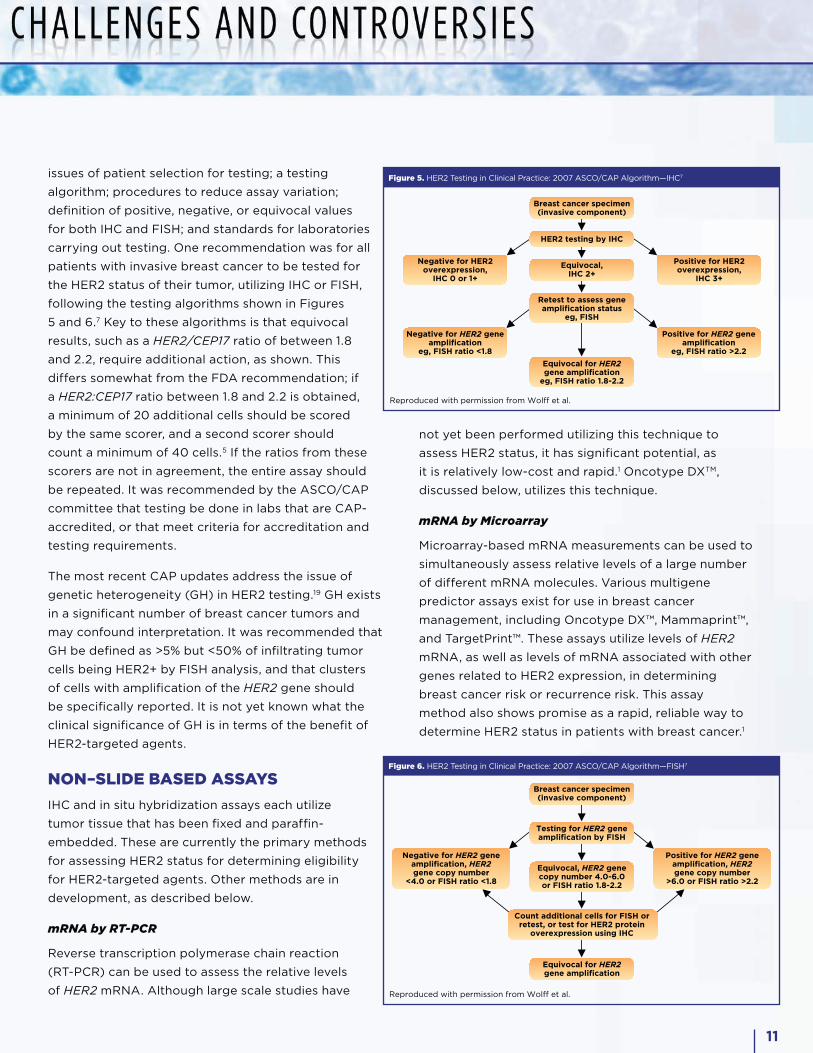
issues of patient selection for testing; a testing
algorithm; procedures to reduce assay variation;
definition of positive, negative, or equivocal values
for both IHC and FISH; and standards for laboratories
carrying out testing. One recommendation was for all
patients with invasive breast cancer to be tested for
the HER2 status of their tumor, utilizing IHC or FISH,
following the testing algorithms shown in Figures
5 and 6.7 Key to these algorithms is that equivocal
results, such as a HER2/CEP17 ratio of between 1.8
and 2.2, require additional action, as shown. This
differs somewhat from the FDA recommendation; if
a HER2:CEP17 ratio between 1.8 and 2.2 is obtained,
a minimum of 20 additional cells should be scored
by the same scorer, and a second scorer should
count a minimum of 40 cells.5 If the ratios from these
scorers are not in agreement, the entire assay should
be repeated. It was recommended by the ASCO/CAP
committee that testing be done in labs that are CAP-
accredited, or that meet criteria for accreditation and
testing requirements.
The most recent CAP updates address the issue of
genetic heterogeneity (GH) in HER2 testing.19 GH exists
in a significant number of breast cancer tumors and
may confound interpretation. It was recommended that
GH be defined as >5% but <50% of infiltrating tumor
cells being HER2+ by FISH analysis, and that clusters
of cells with amplification of the HER2 gene should
be specifically reported. It is not yet known what the
clinical significance of GH is in terms of the benefit of
HER2-targeted agents.
NON–SLIDE BASED ASSAYS
IHC and in situ hybridization assays each utilize
tumor tissue that has been fixed and paraffin-
embedded. These are currently the primary methods
for assessing HER2 status for determining eligibility
for HER2-targeted agents. Other methods are in
development, as described below.
mRNA by RT-PCR
Reverse transcription polymerase chain reaction
(RT-PCR) can be used to assess the relative levels
of HER2 mRNA. Although large scale studies have
Figure 5. HER2 Testing in Clinical Practice: 2007 ASCO/CAP Algorithm—IHC7
Reproduced with permission from Wolff et al.
Breast cancer specimen(invasive component)
Equivocal,IHC 2+
Retest to assess gene amplification status
eg, FISH
Negative for HER2 overexpression,
IHC 0 or 1+
Negative for HER2 gene amplification
eg, FISH ratio <1.8
Positive for HER2 gene amplification
eg, FISH ratio >2.2
Equivocal for HER2 gene amplification
eg, FISH ratio 1.8-2.2
HER2 testing by IHC
Positive for HER2 overexpression,
IHC 3+
Figure 6. HER2 Testing in Clinical Practice: 2007 ASCO/CAP Algorithm—FISH7
Reproduced with permission from Wolff et al.
Breast cancer specimen (invasive component)
Equivocal, HER2 gene copy number 4.0-6.0 or FISH ratio 1.8-2.2
Negative for HER2 gene amplification, HER2 gene copy number
<4.0 or FISH ratio <1.8
Count additional cells for FISH or retest, or test for HER2 protein
overexpression using IHC
Testing for HER2 gene amplification by FISH
Equivocal for HER2 gene amplification
Positive for HER2 gene amplification, HER2 gene copy number
>6.0 or FISH ratio >2.2
not yet been performed utilizing this technique to
assess HER2 status, it has significant potential, as
it is relatively low-cost and rapid.1 Oncotype DXTM,
discussed below, utilizes this technique.
mRNA by Microarray
Microarray-based mRNA measurements can be used to
simultaneously assess relative levels of a large number
of different mRNA molecules. Various multigene
predictor assays exist for use in breast cancer
management, including Oncotype DX™, Mammaprint™,
and TargetPrint™. These assays utilize levels of HER2
mRNA, as well as levels of mRNA associated with other
genes related to HER2 expression, in determining
breast cancer risk or recurrence risk. This assay
method also shows promise as a rapid, reliable way to
determine HER2 status in patients with breast cancer.1
12
Dimerization Assays
Dimerization assays directly measure the level of HER2
homodimers. This method has been commercialized
in the HERMarkTM assay. Results seen thus far show
great promise for this approach in predicting response
to trastuzumab. Data presented at recent congresses
have shown a significant correlation of high levels of
HER2 protein (H2T) and HER2 homodimers (H2D) with
longer time to progression (TTP) after treatment with
trastuzumab.20,21
Phosphorylated HER2
The association of HER2 phosphorylation status
(activated or phosphorylated receptor vs unactivated
or nonphosphorylated receptor) with prognosis or
response to therapy for patients with breast cancer is
not yet well-established, although studies have been
carried out to address this issue. One large study
showed that phosphorylated HER2 was correlated
with a higher number of positive lymph nodes,
cellular proliferation, and poor prognosis.22 Another
study showed that presence of phosphorylated
HER2 was associated with resistance to taxane
therapy for patients with metastatic breast cancer.23
Monoclonal antibodies have been developed to detect
phosphorylated HER2 and could be used in a standard
IHC assay to specifically detect activated HER2 protein.
Tissue and Serum Enzyme-Linked Immunosorbent
Assay (ELISA)
The ELISA can be used to quantitate the concentration
of the extracellular domain (ECD) of the HER2 protein.
The ECD is cleaved from the surface of cells by matrix
metalloproteases and released into the serum.24 The
assessment of ECD in serum avoids many of the
problems associated with IHC and in situ hybridization,
such as those of fixation, embedding, and storage. One
HER2 ELISA is commercially available, the Oncogene
Science HER2/neu ELISA (Oncogene Science,
Cambridge, MA). The use of ELISA to determine HER2
ECD levels has been approved by the FDA for the
monitoring of disease in patients with HER2+ breast
cancer.1 While some studies have shown a correlation
between ECD levels and response to specific therapies,
a recent meta-analysis showed that assessment of
HER2 ECD levels may not be informative. A pooled
analysis of 4 trials showed that baseline ECD levels
were not reliably predictive (positively or negatively)
of response to therapy. There was a trend toward lower
levels of HER2 ECD in patients who achieved better
responses, but this was not significant; and there was
little change in levels of HER2 ECD before disease
progression.24 The potential for the use of serum ELISA
exists in both monitoring breast cancer and in making
treatment decisions, but more research needs to be
carried out in this area before conclusions about the
utility of HER2 ECD assessment can be made.
HER2 Testing of Circulating Tumor Cells
To assess HER2 status by either IHC or FISH, specimens
from tumor tissue are used. There have been a number
of recent studies to investigate the utility of using
circulating tumor cells (CTCs) to predict response
to therapy, as well as to assess HER2 status without
requiring collection of a tissue specimen. It also has
been hypothesized that CTCs may play a role in the
process of metastasis,25 so CTCs could potentially be
used in treatment management decisions for patients
with breast cancer. Counting CTCs as a predictor of
response to therapy has been validated, but the use
of CTCs to assess HER2 status is less well-studied.1
One study showed that 8 of 21 patients whose primary
tumor was HER2 negative (HER2–) or of unknown
HER2 status had detectable CTCs that exhibited
HER2 amplification.26 Another study demonstrated
that HER2+ CTCs are associated with poor outcome
in patients with stage I to stage III breast cancer, with
a significant correlation between the presence and
number of HER2+ CTCs and decreased disease-free
survival and overall survival.25 The authors of this study
state that evaluation of HER2 status in CTCs may
provide for “real time” assessment of HER2 status in
patients with breast cancer. In a study of 24 patients
whose primary tumors were HER2–, 9 patients had
CTCs that were HER2+. Four of these patients were
treated with trastuzumab-containing therapy; 1 had
a complete response, and 2 had a partial response.27
These studies provide intriguing data about the use
of CTCs to assess HER2 status in patients with breast
cancer, and more research is needed to provide answers
concerning the value of assessing CTC HER2 status.
13
III. HER2-TARGETED AGENTS IN BREAST CANCERAs discussed above, it has been shown the HER2
overexpression is associated with a subset of breast
cancers, and that HER2 overexpression (“positivity”)
is generally associated with poor relative prognosis.
Though the poor relative prognosis is problematic,
this association led to the opportunity to develop
treatments for breast cancer that would specifically
target HER2. The first of these was trastuzumab, a
humanized monoclonal antibody to HER2. Initial clinical
trials of trastuzumab utilized this antibody in patients
with metastatic breast cancer, and showed the clinical
activity of this agent alone and then in combination
with chemotherapy. More recent trials have been
carried out in both the adjuvant and neoadjuvant
settings. Trastuzumab is now approved for treatment
of HER2+ metastatic breast cancer, and in the adjuvant
setting for both lymph node–positive and lymph node–
negative breast cancer. Lapatinib is the second HER2
targeted agent to be approved for use in patients with
breast cancer. Lapatinib is a reversible inhibitor of
both HER2 and EGFR tyrosine kinases and has clinical
activity in the treatment of metastatic breast cancer. Its
mechanism of action differs from that of trastuzumab,
and lapatinib is active in patients with progression on
the antibody. Lapatinib was approved by the FDA in
2007 for use in patients with metastatic breast cancer
that overexpresses HER2 and has progressed on prior
chemotherapy and trastuzumab.
HER2-TARGETED AGENTS IN THE METASTATIC SETTING
Trastuzumab Monotherapy
The humanized anti-HER2 monoclonal antibody,
eventually called trastuzumab, was shown to inhibit
the growth of breast cancer cells, indicating that it had
potential as a treatment for breast cancer. In 1996, a
phase 2 study was carried out to assess the efficacy
and safety of this agent in the treatment of HER2-
overexpressing metastatic breast cancer.28 In this
study, 46 patients who had received extensive prior
therapy for metastatic breast cancer were treated with
intravenously administered trastuzumab (a loading
dose of 250 mg, then 100 mg/wk). An overall
response rate of 11.6% (5 of 43 assessable patients)
was obtained, with 1 patient achieving a complete
response. This study was followed by a larger
single-arm trial, in which 222 patients with HER2-
overexpressing metastatic breast cancer were
enrolled.29 In this study, patients received a
loading dose of 4 mg/kg intravenously, followed
by 2 mg/kg, administered weekly. Responses were
once again noted in a number of patients: 8 (4%)
patients achieved a complete response, and 26 (12%)
patients achieved a partial response.
The efficacy of single-agent trastuzumab in patients
with HER2+ breast cancer was confirmed in
subsequent trials, including one in which patients were
randomized to 2 different doses of trastuzumab: a
4-mg/kg loading dose followed by 2 mg/kg weekly,
or an 8-mg/kg loading dose followed by 4 mg/kg
weekly.30 Efficacy was similar between the treatment
arms. However, the results from this trial established
the value of FISH as a method for selecting patients
for therapy with trastuzumab: Responses were seen
in 34% of patients who were HER2+ by FISH, and 7%
in those who were HER2– by FISH.30 A subsequent
study was designed to assess whether trastuzumab
would be effective and safe if administered every 3
weeks, instead of every week.31 Although this was not a
randomized trial, the response rate in this study—19 of
83 (23%) patients—was similar to those of prior studies
of trastuzumab monotherapy. These trials validated the
efficacy of trastuzumab monotherapy and suggested
that a range of doses and either weekly or 3-weekly
dosing were appropriate.
Chemotherapy in Combination With Trastuzumab
The pivotal trial that established the efficacy of
trastuzumab combination regimens in the treatment
of patients with HER2-overexpressing metastatic
breast cancer compared responses in patients who
received chemotherapy plus trastuzumab with those
who received chemotherapy alone.32 For this study,
chemotherapy consisted of an anthracycline plus
cyclophosphamide for patients who had not received
14
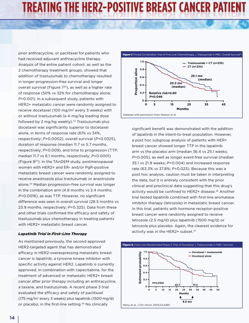
prior anthracycline, or paclitaxel for patients who
had received adjuvant anthracycline therapy.
Analysis of the entire patient cohort, as well as the
2 chemotherapy treatment groups, showed that
addition of trastuzumab to chemotherapy resulted
in longer progression-free survival and longer
overall survival (Figure 732), as well as a higher rate
of response (50% vs 32% for chemotherapy alone;
P<0.001). In a subsequent study, patients with
HER2+ metastatic cancer were randomly assigned to
receive docetaxel (100 mg/m2 every 3 weeks) with
or without trastuzumab (a 4-mg/kg loading dose
followed by 2 mg/kg weekly).33 Trastuzumab plus
docetaxel was significantly superior to docetaxel
alone, in terms of response rate (61% vs 34%,
respectively; P=0.0002), overall survival (P=0.0325),
duration of response (median 11.7 vs 5.7 months,
respectively; P=0.009), and time to progression (TTP;
median 11.7 vs 6.1 months, respectively; P=0.0001)
(Figure 833). In the TAnDEM study, postmenopausal
women with HER2+ and ER- and/or PgR-positive
metastatic breast cancer were randomly assigned to
receive anastrozole plus trastuzumab or anastrozole
alone.34 Median progression-free survival was longer
in the combination arm (4.8 months vs 2.4 months;
P=0.0016), as was TTP. However, no significant
difference was seen in overall survival (28.5 months vs
23.9 months, respectively; P=0.325). Data from these
and other trials confirmed the efficacy and safety of
trastuzumab plus chemotherapy in treating patients
with HER2+ metastatic breast cancer.
Lapatinib Trial in First-Line Therapy
As mentioned previously, the second approved
HER2-targeted agent that has demonstrated
efficacy in HER2-overexpressing metastatic breast
cancer is lapatinib, a tyrosine kinase inhibitor with
specific activity against HER2. Lapatinib is currently
approved, in combination with capecitabine, for the
treatment of advanced or metastatic HER2+ breast
cancer after prior therapy including an anthracycline,
a taxane, and trastuzumab. A recent phase 3 trial
evaluated the efficacy and safety of paclitaxel
(175 mg/m2 every 3 weeks) plus lapatinib (1500 mg/d)
or placebo, in the first-line setting.35 No clinically
significant benefit was demonstrated with the addition
of lapatinib in the intent-to-treat population. However,
a post hoc subgroup analysis of patients with HER+
breast cancer showed longer TTP in the lapatinib
arm vs the placebo arm (median 36.4 vs 25.1 weeks;
P=0.005), as well as longer event-free survival (median
35.1 vs 21.9 weeks; P=0.004) and increased response
rate (63.3% vs 37.8%; P=0.023). Because this was a
post hoc analysis, caution must be taken in interpreting
the data, but it is entirely consistent with the prior
clinical and preclinical data suggesting that this drug’s
activity would be confined to HER2+ disease.36 Another
trial tested lapatinib combined with first-line aromatase
inhibitor therapy (letrozole) in metastatic breast cancer.
In this trial, patients with hormone receptor–positive
breast cancer were randomly assigned to receive
letrozole (2.5 mg/d) plus lapatinib (1500 mg/d) or
letrozole plus placebo. Again, the clearest evidence for
activity was in the HER2+ subset.37
Figure 7. Pivotal Combination Trial of First-Line Chemotherapy ± Trastuzumab in MBC: Overall Survival32
Adapted with permission from Slamon et al.
0
0.2
0.4
0.6
0.8
1.0
5 15 25 35 450
Months
Trastuzumab + CT (n=235)
Relative risk=0.80PP=0.046
Pro
po
rtio
n a
live
20.3 mo (median)
25.1 mo (median)
CT (n=234)
Figure 8. First-Line Randomized Phase 2 Trial of Docetaxel ± Trastuzumab in MBC: Survival
Marty et al. J Clin Oncol. 2005;23:4265.
0
0.2
0.4
0.6
0.8
1.0
0 5 10 15 20 25 30 35 40 45 50
8.5 mo
Est
imate
d p
rob
ab
ilit
y Docetaxel + trastuzumab
Docetaxel alone
Months
P=0.032531.222.7
15
HER2-Targeted Agents After Progression
on Trastuzumab
Despite the significant efficacy of trastuzumab in
patients with HER2+ metastatic breast cancer,
almost all patients will experience disease
progression while on trastuzumab therapy. A
number of clinical studies have been carried out to
evaluate treatment strategies beyond progression
on trastuzumab. In one study, patients who had
progressed on trastuzumab were randomly assigned
to receive capecitabine (2500 mg/m2, days 1 to 14
of a 3-week cycle) with or without trastuzumab
(6 mg/kg, every 3 weeks).38 Although this study was
closed before full patient accrual, treatment with
the combination regimen resulted in a significant
improvement vs capecitabine alone in TTP (8.2 vs
5.6 months; P=0.0338) and response rate (48.1% vs
27.0%; P=0.0115) but no significant change in overall
survival (25.5 vs 20.4 months; P=0.2570). As described
above, lapatinib also has demonstrated efficacy as
a second-line agent in patients with HER2+ breast
cancer, and trials have been carried out to assess the
activity of lapatinib as monotherapy, or in combination
with other agents in patients with HER2+ advanced
or metastatic breast cancer, whose cancer has
progressed on trastuzumab therapy. The study that
led to the approval of lapatinib examined capecitabine
(2000 mg/m2/d, days 1 to 14 of a 3-week cycle) plus
lapatinib (1250 mg/d) vs capecitabine plus placebo
and demonstrated an increase in median TTP (8.4 vs
4.4 months; P<0.001) and median progression-free
survival (8.4 vs 4.1 months; P<0.001) for the lapatinib
combination arm in patients with HER2+ metastatic
or advanced breast cancer that had progressed
after treatment with regimens that included an
anthracycline, a taxane, and trastuzumab.39
In studies of the efficacy of lapatinib monotherapy in
patients who had disease progression on prior therapy,
including chemotherapy and trastuzumab, modest
activity was seen in patients with HER2+ disease.40
In one study, a response rate of 4.3% (by investigator
review, 1.4% by independent review) was achieved. A
more recent study of lapatinib monotherapy (1250 or
1500 mg/d) in patients whose cancer had progressed
on trastuzumab therapy demonstrated the clinical
benefit of lapatinib in this patient population.41
Clinical benefit rates (consisting of complete response,
partial response, or stable disease for �24 weeks)
were 14.1% by investigator review, and 9.0% by
independent review.
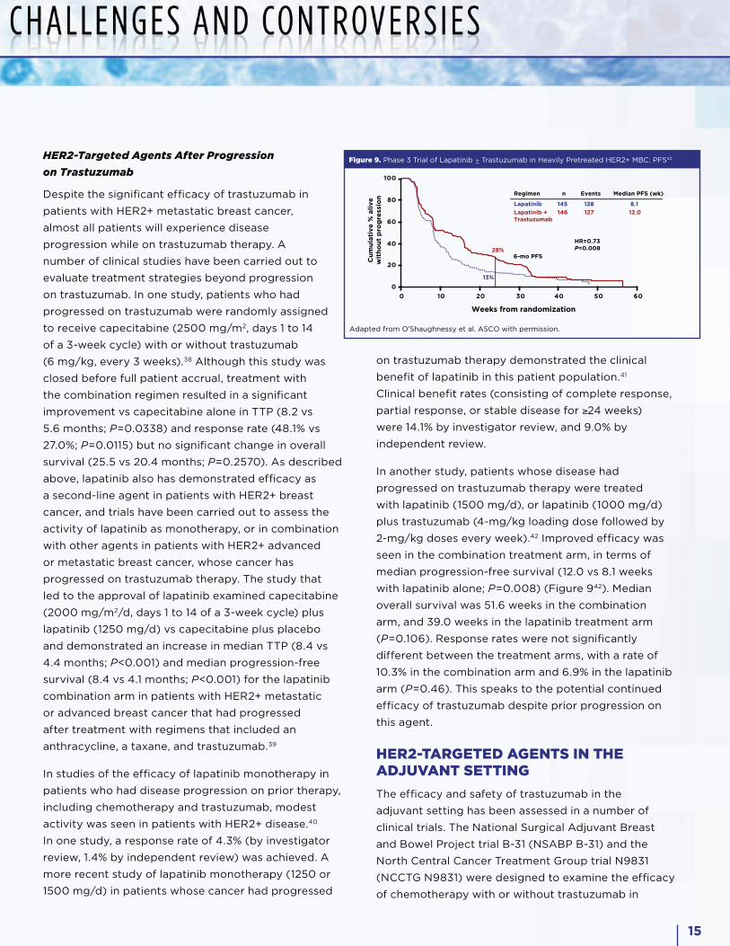
In another study, patients whose disease had
progressed on trastuzumab therapy were treated
with lapatinib (1500 mg/d), or lapatinib (1000 mg/d)
plus trastuzumab (4-mg/kg loading dose followed by
2-mg/kg doses every week).42 Improved efficacy was
seen in the combination treatment arm, in terms of
median progression-free survival (12.0 vs 8.1 weeks
with lapatinib alone; P=0.008) (Figure 942). Median
overall survival was 51.6 weeks in the combination
arm, and 39.0 weeks in the lapatinib treatment arm
(P=0.106). Response rates were not significantly
different between the treatment arms, with a rate of
10.3% in the combination arm and 6.9% in the lapatinib
arm (P=0.46). This speaks to the potential continued
efficacy of trastuzumab despite prior progression on
this agent.
HER2-TARGETED AGENTS IN THE ADJUVANT SETTING
The efficacy and safety of trastuzumab in the
adjuvant setting has been assessed in a number of
clinical trials. The National Surgical Adjuvant Breast
and Bowel Project trial B-31 (NSABP B-31) and the
North Central Cancer Treatment Group trial N9831
(NCCTG N9831) were designed to examine the efficacy
of chemotherapy with or without trastuzumab in
Figure 9. Phase 3 Trial of Lapatinib ± Trastuzumab in Heavily Pretreated HER2+ MBC: PFS42
Adapted from O’Shaughnessy et al. ASCO with permission.
0
20
40
60
80
100
0 10 20 30 40 50 60
Lapatinib +Trastuzumab
Lapatinib
Regimen n Events Median PFS (wk)
145 128 8.1
146 127 12.0
28%6-mo PFS
Weeks from randomization
Cu
mu
lati
ve
% a
live
wit
ho
ut
pro
gre
ssio
n
13%
HR=0.73P=0.008
16
patients with operable HER2+ breast cancer.43 In the
NSABP B-31 trial, patients were randomly assigned
to receive doxorubicin (60 mg/m2 every 3 weeks)
and cyclophosphamide (600 mg/m2 every 3 weeks),
each for 4 cycles, followed by 175 mg/m2 of paclitaxel
every 3 weeks for 4 cycles, or the same regimen
plus trastuzumab (4-mg/kg loading dose, given with
the first dose of paclitaxel, followed by 2-mg/kg
doses given every week for 51 weeks). In the NCCTG
N9831 study, the same regimen of doxorubicin and
cyclophosphamide was used, but it was followed
instead by weekly paclitaxel (80 mg/m2 every week
for 12 weeks) in 1 treatment arm. Patients in a second
treatment arm received the same chemotherapy
regimen followed by trastuzumab (a loading dose of
4 mg/kg followed by 2-mg/kg weekly doses for 51
weeks). Patients in the third treatment arm received
the same chemotherapy regimen plus trastuzumab
(a loading dose of 4 mg/kg followed by 2-mg/kg
weekly doses for 51 weeks) beginning with the first
dose of paclitaxel. A joint analysis of these studies
demonstrated a clinical benefit for patients in the
trastuzumab treatment arms. Disease-free survival at
4 years was 85.3% in the trastuzumab arms vs 67.1% in
the control arms (P<0.0001). Overall survival at 4 years
was 91.4% in the trastuzumab treatment arms vs 86.6%
in the control arms (P=0.015). Significant benefit was
also seen in terms of time to recurrence, time to distant
recurrence, and death from breast cancer.
The BCIRG 006 trial is a randomized study to compare
3 treatment regimens: doxorubicin (60 mg/m2 every
3 weeks) plus cyclophosphamide (600 mg/m2 every
3 weeks) for 4 cycles, followed by docetaxel
(100 mg/m2 every 3 weeks for 4 cycles) (AC�T);
the same chemotherapy regimen followed by 1 year
of trastuzumab (beginning with the first cycle of
docetaxel) (AC�TH); or docetaxel (75 mg/m2 every
3 weeks for 6 cycles) plus carboplatin (AUC6 every 3
weeks for 6 cycles) plus 1 year of trastuzumab (TCH).44
A disease-free survival benefit was seen in both
trastuzumab-containing treatment arms relative to
the AC�T arm (AC�TH: hazard ratio [HR] 0.49; TCH:
HR 0.61).
In the FinHer trial, women with node-positive or
high-risk node-negative breast cancer were randomly
assigned to receive docetaxel (100 mg/m2 on day 1
of a 21-day cycle) or vinorelbine (25 mg/m2, days 1,
8, and 15 of a 21-day cycle), followed by 3 cycles of
5-fluorouracil (600 mg/m2)/epirubicin (60 mg/m2)/
cyclophosphamide (600 mg/m2) (FEC) on day 1 of a
21-day cycle (in both treatment arms).45 Women who
had verified HER2+ cancer were randomly assigned
to receive trastuzumab (9 infusions administered
at 1-week intervals, beginning on day 1 of the first
docetaxel or vinorelbine cycle, a loading dose
of 4 mg/kg, followed by subsequent doses of
2 mg/kg) or no trastuzumab. Trastuzumab was not
given during FEC administration. An analysis of data
from patients with HER2+ cancer showed that the
addition of trastuzumab to these chemotherapy
regimens provided clinical benefit in this patient group.
Kaplan-Meier estimates of survival free of recurrence at
3 years were 89.3% in the trastuzumab arms and 77.6%
in the no-trastuzumab arms, and estimates of overall
survival at 3 years were 96.3% and 89.7%, respectively.
An update of FinHer trial results was presented at the
St. Gallen Oncology Conferences: Primary Therapy of
Early Breast Cancer International Conference. At
that time, it was reported that overall survival was
improved by the addition of trastuzumab to the
docetaxel treatment regimen or the vinorelbine
treatment regimen.
The HERA trial compared 1 or 2 years of trastuzumab
(8-mg/kg loading dose, followed by 6 mg/kg every 3
weeks) treatment with observation, in patients with
HER2+ breast cancer who had received locoregional
therapy and neoadjuvant or adjuvant chemotherapy.46
Results for the observation group of patients and the
1-year trastuzumab treatment group of patients were
reported at a median follow-up of 1 year.46 Kaplan-
Meier curves showed an estimated 2-year disease-free
survival of 85.8% in the trastuzumab arm vs 77.4% in
the observation arm (P<0.0001), and a 2-year time
to disease recurrence of 90.6% vs 82.8% (P<0.0001).
However, overall survival was not significantly different
between the treatment groups.
17
In a trial with a similar design (PACS-04), patients with
node-positive breast cancer were randomly assigned
to receive adjuvant therapy as follows: 6 cycles of FEC
(5-fluorouracil 500 mg/m2; epirubicin 100 mg/m2;
cyclophosphamide 500 mg/m2 every 3 weeks) or
6 cycles of ED (epirubicin 75 mg/m2; docetaxel
75 mg/m2 every 3 weeks), followed by radiotherapy.47
When HER2 status of the tumors became available,
patients with HER2+ tumors were randomly assigned
to receive 1 year of trastuzumab therapy (8-mg/kg
loading dose followed by 6 mg/kg every 3 weeks) or
observation only. At 4 years, disease-free survival was
not significantly different between the trastuzumab
and observation treatment arms.
Overall, these studies demonstrate a consistent
efficacy for trastuzumab in the adjuvant setting,
especially when it is administered concurrently with
chemotherapy. The next-generation trial, ALTTO
(NCT00490139), will assess the relative efficacies of
trastuzumab, lapatinib, and trastuzumab plus lapatinib
in the adjuvant setting. This study consists of 4
treatment arms: paclitaxel plus trastuzumab;
paclitaxel plus lapatinib; paclitaxel plus trastuzumab
for 12 weeks followed by a 6-week washout period,
followed by lapatinib for 34 weeks; and paclitaxel
plus lapatinib plus trastuzumab. All study participants
must have received at least 4 cycles of an approved
anthracycline-containing (neo-)adjuvant chemotherapy
regimen. This study is currently recruiting patients.
HER2-TARGETED AGENTS IN THE NEOADJUVANT SETTING
The use of HER2-targeted agents as part of
a neoadjuvant regimen is less well-studied;
however, encouraging data for HER2-targeted
therapies have emerged in this treatment setting.
A trial investigating the efficacy of neoadjuvant
therapy with paclitaxel (225 mg/m2 every 3
weeks for 4 cycles) followed by FEC (5-fluorouracil
500 mg/m2 on days 1 and 4, epirubicin 75 mg/m2
on day 1, cyclophosphamide 500 mg/m2 on day 1)
randomly assigned patients to receive trastuzumab
(a loading dose of 4 mg/kg followed by weekly doses
of 2 mg/kg, for a total of 24 doses of trastuzumab)
or no trastuzumab.48 Pathologic complete response
(pCR), generally a good correlate of progression-
free and overall survival, was seen in 15 of 23 (65.2%)
patients in the trastuzumab treatment arm, and in 5
of 19 (26.3%) patients in the no-trastuzumab arm. An
updated analysis, which included data on additional
patients, showed that 12 of 22 (54.5%) of the additional
patients, treated with chemotherapy plus trastuzumab,
achieved a pCR.49
The NOAH trial assessed the efficacy of chemotherapy
with or without trastuzumab in the neoadjuvant setting.
Patients who had HER2+ disease were randomized to
1 of 2 treatment arms: doxorubicin (60 mg/m2) and
paclitaxel (150 mg/m2) every 3 weeks, for 3 cycles,
followed by 4 cycles of paclitaxel (175 mg/m2 every
3 weeks) and 3 cycles of CMF (cyclophosphamide
600 mg/m2, methotrexate 40 mg/m2, 5-fluorouracil
600 mg/m2 every 4 weeks, on days 1 and 8),
followed by surgery and radiotherapy; or the same
chemotherapy regimen, with trastuzumab (8-mg/kg
loading dose followed by 6 mg/kg every 3 weeks
for 1 year) for 1 year before surgery, and following
surgery.50 A pCR was achieved in 43% of patients
in the trastuzumab arm vs 23% of those in the
no-trastuzumab arm (P=0.002). Patients in the
trastuzumab arm also benefitted in terms of event-free
survival (HR 0.56) and overall survival (HR 0.65).
The Neo-ALTTO study (NCT00553358) is designed to
assess the efficacies of lapatinib and trastuzumab in
the neoadjuvant setting. In this study, patients will be
randomized to 1 of 3 treatment arms: lapatinib followed
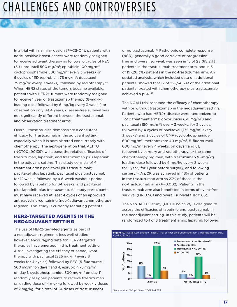
Figure 10. Pivotal Combination Phase 3 Trial of First-Line Chemotherapy ± Trastuzumab in MBC: Cardiac Safety
Slamon et al. N Engl J Med. 2001;344:783.
11%
4%%1%1
28%
19%
7%
3%
0
5
10
15
20
25
30
% o
f p
ati
en
ts
Trastuzumab + paclitaxel (n=91)
Paclitaxel (n=95)
Trastuzumab + AC (n=143)
AC (n=135)
Any CD NYHA class III-IV
18
by lapatinib plus paclitaxel; trastuzumab followed
by trastuzumab plus paclitaxel; lapatinib plus
trastuzumab followed by lapatinib plus trastuzumab
plus paclitaxel. This study is currently enrolling
patients. The CALGB 40601 study (NCT00770809)
also will assess the activities of trastuzumab and
lapatinib in the neoadjuvant setting and will assign
patients to 3 treatment arms: trastuzumab plus
lapatinib plus paclitaxel; trastuzumab plus paclitaxel;
or lapatinib plus paclitaxel. This study is currently
recruiting participants.
HER2-TARGETED AGENTS: CARDIOTOXICITY
A key issue for HER2-targeted agents is that of
cardiotoxicity. It was noted in the pivotal phase 3
trial of trastuzumab and in subsequent trials that
a significant number of patients who received
trastuzumab developed cardiac dysfunction,
particularly when it was given concurrently with
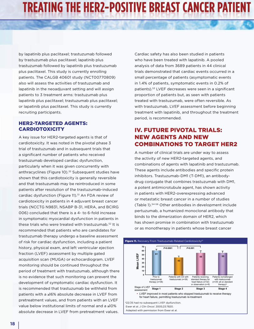
anthracyclines (Figure 10).32 Subsequent studies have
shown that this cardiotoxicity is generally reversible
and that trastuzumab may be reintroduced in some
patients after resolution of the trastuzumab-induced
cardiac dysfunction (Figure 11).51 An FDA review of
cardiotoxicity in patients in 4 adjuvant breast cancer
trials (NCCTG N9831, NSABP B-31, HERA, and BCIRG
006) concluded that there is a 4- to 6-fold increase
in symptomatic myocardial dysfunction in patients in
these trials who were treated with trastuzumab.52 It is
recommended that patients who are candidates for
trastuzumab therapy undergo a baseline assessment
of risk for cardiac dysfunction, including a patient
history, physical exam, and left ventricular ejection
fraction (LVEF) assessment by multiple gated
acquisition scan (MUGA) or echocardiogram. LVEF
monitoring should be continued throughout the
period of treatment with trastuzumab, although there
is no evidence that such monitoring can prevent the
development of symptomatic cardiac dysfunction. It
is recommended that trastuzumab be withheld from
patients with a �16% absolute decrease in LVEF from
pretreatment values, and from patients with an LVEF
value below institutional limits of normal and a �10%
absolute decrease in LVEF from pretreatment values.
Cardiac safety has also been studied in patients
who have been treated with lapatinib. A pooled
analysis of data from 3689 patients in 44 clinical
trials demonstrated that cardiac events occurred in a
small percentage of patients (asymptomatic events
in 1.4% of patients, symptomatic events in 0.2% of
patients).53 LVEF decreases were seen in a significant
proportion of patients but, as seen with patients
treated with trastuzumab, were often reversible. As
with trastuzumab, LVEF assessment before beginning
treatment with lapatinib, and throughout the treatment
period, is recommended.
IV. FUTURE PIVOTAL TRIALS: NEW AGENTS AND NEW COMBINATIONS TO TARGET HER2 A number of clinical trials are under way to assess
the activity of new HER2-targeted agents, and
combinations of agents with lapatinib and trastuzumab.
These agents include antibodies and specific protein
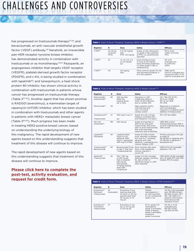
inhibitors. Trastuzumab-DM1 (T-DM1), an antibody-
drug conjugate that combines trastuzumab with DM1,
a potent antimicrotubule agent, has shown activity
in patients with HER2-overexpressing advanced
or metastatic breast cancer in a number of studies
(Table 1).54-56 Other antibodies in development include
pertuzumab, a humanized monoclonal antibody that
binds to the dimerization domain of HER2, which
has shown promise in combination with trastuzumab
or as monotherapy in patients whose breast cancer
Figure 11. Recovery From Trastuzumab-Related Cardiotoxicity51
a22/25 had no subsequent LVEF dysfunction.
Ewer et al. J Clin Oncol. 2005;23:7820.
Adapted with permission from Ewer et al.
• LVEF improved in most patients who stopped trastuzumab to receive therapyfor heart failure, permitting trastuzumab re-treatment
Mea
n LV
EF
Stage of LVEFassessment Stage 1 Stage 2 Stage 3 Stage 4
Prior totrastuzumab
therapy (n=38)
Patients with CD withtrastuzumab (n=38)
Patients receivingstandard therapy forheart failure (n=32)or observation (n=6)
Patients rechallengedwith trastuzumab
(n=25; all on standardtherapy)a
5555
43
61
0
10
20
30
40
50
60
70 P<0.0001 P<0.001
19
has progressed on trastuzumab therapy57,58; and
bevacizumab, an anti–vascular endothelial growth
factor (VEGF) antibody.59 Neratinib, an irreversible
pan-HER receptor tyrosine kinase inhibitor,
has demonstrated activity in combination with
trastuzumab or as monotherapy.60,61 Pazopanib, an
angiogenesis inhibitor that targets VEGF receptor
(VEGFR), platelet-derived growth factor receptor
(PDGFR), and c-Kit, is being studied in combination
with lapatinib62; and tanespimycin, a heat shock
protein 90 inhibitor, has shown clinical activity in
combination with trastuzumab in patients whose
cancer has progressed on trastuzumab therapy
(Table 257-63). Another agent that has shown promise
is RAD001 (everolimus), a mammalian target of
rapamycin (mTOR) inhibitor, which has been studied
in combination with trastuzumab and other agents
in patients with HER2+ metastatic breast cancer
(Table 364,65). Much progress has been made
in treating HER2-positive breast cancer, based
on understanding the underlying biology of
this malignancy. The rapid development of new
agents based on this understanding suggests that
treatment of this disease will continue to improve.
The rapid development of new agents based on
this understanding suggests that treatment of this
disease will continue to improve.
Please click here to complete the post-test, activity evaluation, and request for credit form.
Table 1. Trials of Novel Therapies Targeting HER2 in Breast Cancer: T-DM154-56
Regimen
T-DM154
T-DM155
T-DM156
N
24 (15 pts received MTD)
27
112
Dose
0.3-4.8 mg/kg q3w MTD: 3.6 mg/kg
1.2, 1.6, 2.0, 2.4, or 2.9 mg/kgqw
3.6 mg/kgq3w
Safety
Grade ≥2 AEs infrequent; no grade 4 AEs reported at MTD; no cardiotoxicity reported
Dose-limiting toxicity in 2 pts at 2.9-mg/kg dose (thrombocytopenia); no other grade 4 AEs or cardiotoxicity
Most common grade 3/4 AE was thrombocytopenia, in 8 (7.0%) patients
Efficacy
PR in 4 pts (44%) receiving MTD
PR: 9/15 evaluable pts,8 confirmed (53%)
ORR: 39.3% (42/107 evaluable pts) Confirmed ORR: 27.1% (29/107 evaluable pts)
Table 2. Trials of Novel Therapies Targeting HER2 in Breast Cancer57-63
Regimen
Pertuzumab/trastuzumab57
Pertuzumab58
Tanespimycin63
Trastuzumab
HKI-272 (neratinib)60
Lapatinib62
Pazopanib
Trastuzumab59
Bevacizumab
Neratinib61
Trastuzumab
N
66
29
27
136
141
50
45
Dose
420 mg q3w (840-mg loading dose) qw or q3w
450 mg/m2 qw IV
qw
240 mg qd
Lapatinib 1500 mg/d (n=72), lapatinib 1000 mg/d + pazopanib 400 mg/d (n=69)
Bevacizumab 10 mg on day 7, then q2w + trastuzumab qw
Part 1: 160 mg or 240 mg qdPart 2: neratinib 240 mg qd trastuzumab4-mg/kg loading dose, then 2 mg/kg qw
Safety
Decrease in LVEF(<50% and ≥10%) in 3 pts; no pts withdrew due to cardiac AEs
Major adverse events: mild diarrhea and rash; no clinical cardiac events
Grade 3/4 AEs in 6 pts (fatigue, increased AST, and headache; n=2 each)
Grade 3/4 diarrhea in 30% of pts with prior trastuzumab, 13% of pts with no prior trastuzumab, 16% of pts had dose reduction due to diarrhea
Grade 3/4 AEs in both arms: diarrhea, increased AST and ALT, vomiting; lapatinib arm: rash; lapatinib + pazopanib arm: nausea, fatigue, LVEF
Most common AEs were fever, chills, headache, infusion reaction, 1 severe CE
Grade 3/4 AEs included diarrhea, nausea, vomiting. No AEs of CHF or significant drops in LVEF were reported
Efficacy
CR: 7.6%; PR: 16.7%;SD 6 mo: 25.8%;SD <6 mo: 30%;PD: 50%
Response in 2 patients with monotherapy; response in 2 patients upon addition of trastuzumab
PR: 7/27 pts (26%)
ORR 26% in prior trastuzumab group, 56% in no prior trastuzumab group, 42% overall
Primary end point: PD rateat week 12: 28 (38.9%) in lapatinib arm, 25 (36.2%) in lapatinib + pazopanib arm
ORR 54.1% (37 evaluable patients), CR 2.7%
ORR 28.6% (8/28 evaluable pts), 16-week PFS rate 45%, median PFS 16 weeks
Table 3. Trials of Novel Therapies Targeting HER2 in Breast Cancer: mTOR Inhibitors64,65
Regimen
Everolimus64
PaclitaxelTrastuzumab
Everolimus65
VinorelbineTrastuzumab
N
22 (evaluable)
37
Dose
Everolimus 5-mg starting dose then 10 mg/d, or 30-mg starting dose then 50 mg qw or 70 mg qw
Everolimus 5 mg qd, or 20 mg qw, or 30 mg qw
Safety
Febrile neutropenia in 1 pt, oral mucositis in 1 pt, confusional state in 1 pt
DLTs: grade 3/4 neutropenia, grade 3 stomatitis, grade 3 fatigue, grade 2 dermatitis acneiform, grade 3 anorexia
Efficacy
CR 5% (1/22 evaluable pts), PR 36% (8 pts), SD 50% (11 pts), CR + PR + SD 100% in 9 patients with taxane- and trastuzumab-resistant tumors
CR 3% (1 of 34 evaluable pts), PR 15% (5/34), SD 62% (21/34)
20
REFERENCES 1. Ross JS, Slodkowska EA, Symmans WF, Pusztai L, Ravdin PM, Hortobagyi GN. The HER-2 receptor and breast cancer: ten years of targeted anti-HER-2 therapy and personalized medicine. Oncologist. 2009;14(4):320-368.
2. Hudis CA. Trastuzumab--mechanism of action and use in clinical practice. N Engl J Med. 2007;357(1):39-51.
3. Rubin I, Yarden Y. The basic biology of HER2. Ann Oncol. 2001;12(suppl 1):S3-S8.
4. King CR, Kraus MH, Aaronson SA. Amplification of a novel v-erbB-related gene in a human mammary carcinoma. Science. 1985;229(4717):974-976.
5. Sauter G, Lee J, Bartlett JM, Slamon DJ, Press MF. Guidelines for human epidermal growth factor receptor 2 testing: biologic and methodologic considerations. J Clin Oncol. 2009;27(8):1323-1333.
6. Chibon F, de Mascarel I, Sierankowski G, et al. Prediction of HER2 gene status in Her2 2+ invasive breast cancer: a study of 108 cases comparing ASCO/CAP and FDA recommendations. Mod Pathol. 2009;22(3):403-409.
7. Wolff AC, Hammond ME, Schwartz JN, et al; American Society of Clinical Oncology; College of American Pathologists. American Society of Clinical Oncology/College of American Pathologists guideline recommendations for human epidermal growth factor receptor 2 testing in breast cancer. J Clin Oncol. 2007;25(1):118-145.
8. Tubbs R, Barlow WE, Budd GT, et al. Outcome of patients with early-stage breast cancer treated with doxorubicin-based adjuvant chemotherapy as a function of HER2 and TOP2A status. J Clin Oncol. 2009;27(24):3881-3886.
9. Lower EE, Glass E, Blau R, Harman S. HER-2/neu expression in primary and metastatic breast cancer. Breast Cancer Res Treat. 2009;113(2):301-306.
10. HercepTestTM [package insert]. Carpinteria, California: Dako North America, Inc.; 2009.
11. Middleton LP, Price KM, Puig P, et al. Implementation of American Society of Clinical Oncology/College of American Pathologists HER2 Guideline Recommendations in a tertiary care facility increases HER2 immunohistochemistry and fluorescence in situ hybridization concordance and decreases the number of inconclusive cases. Arch Pathol Lab Med. 2009;133(5):775-780.
12. Pauletti G, Dandekar S, Rong H, et al. Assessment of methods for tissue-based detection of the HER-2/neu alteration in human breast cancer: a direct comparison of fluorescence in situ hybridization and immunohistochemistry. J Clin Oncol. 2000;18(21):3651-3664.
13. Salido M, Tusquets I, Corominas JM, et al. Polysomy of chromosome 17 in breast cancer tumors showing an overexpression of ERBB2: a study of 175 cases using fluorescence in situ hybridization and immunohistochemistry. Breast Cancer Res. 2005;7(2):R267-273.
14. Pedersen M, Rasmussen BB. The correlation between dual-color chromogenic in situ hybridization and fluorescence in situ hybridization in assessing HER2 gene amplification in breast cancer. Diagn Mol Pathol. 2009;18(2):96-102.
15. Francis GD, Jones MA, Beadle GF, Stein SR. Bright-field in situ hybridization for HER2 gene amplification in breast cancer using tissue microarrays: correlation between chromogenic (CISH) and automated silver-enhanced (SISH) methods with patient outcome. Diagn Mol Pathol. 2009;18(2):88-95.
16. Hanna WM, Kwok K. Chromogenic in-situ hybridization: a viable alternative to fluorescence in-situ hybridization in the HER2 testing algorithm. Mod Pathol. 2006;19(4):481-487.
17. Paik S, Bryant J, Tan-Chiu E, et al. Real-world performance of HER2 testing-- National Surgical Adjuvant Breast and Bowel Project experience. J Natl Cancer Inst. 2002;94(11):852-854.
18. Roche PC, Suman VJ, Jenkins RB, et al. Concordance between local and central laboratory HER2 testing in the breast intergroup trial N9831. J Natl Cancer Inst. 2002;94(11):855-857.
19. Vance GH, Barry TS, Bloom KJ, et al; College of American Pathologists. Genetic heterogeneity in HER2 testing in breast cancer: panel summary and guidelines. Arch Pathol Lab Med. 2009;133(4):611-612.
20. Lipton A, Ali SM, Leitzel K, et al. HER2 protein expression predicts response to trastuzumab in FISH-positive patients. Presented at: 31st Annual San Antonio Breast Cancer Symposium; December 10-14, 2008; San Antonio, TX. Abstract 32.
21. Sperinde J, Ali S, Leitzel K, et al. Identification of a subpopulation of metastatic breast cancer patients with very high HER2 expression levels and possible resistance to trastuzumab [abstract]. J Clin Oncol. 2009;27(15s):1059.
22. Thor AD, Liu S, Edgerton S, et al. Activation (tyrosine phosphorylation) of ErbB-2 (HER-2/neu): a study of incidence and correlation with outcome in breast cancer. J Clin Oncol. 2000;18(18):3230-3239.
23. Modi S, DiGiovanna MP, Lu Z, et al. Phosphorylated/activated HER2 as a marker of clinical resistance to single agent taxane chemotherapy for metastatic breast cancer. Cancer Invest. 2005;23(6):483-487.
24. Lennon S, Barton C, Banken L, et al. Utility of serum HER2 extracellular domain assessment in clinical decision making: pooled analysis of four trials of trastuzumab in metastatic breast cancer. J Clin Oncol. 2009;27(10):1685-1693.
25. Wülfing P, Borchard J, Buerger H, et al. HER2-positive circulating tumor cells indicate poor clinical outcome in stage I to III breast cancer patients. Clin Cancer Res. 2006;12(6):1715-1720.
26. Fehm T, Becker S, Duerr-Stoerzer S, et al. Determination of HER2 status using both serum HER2 levels and circulating tumor cells in patients with recurrent breast cancer whose primary tumor was HER2 negative or of unknown HER2 status. Breast Cancer Res. 2007;9(5):R74.
27. Meng S, Tripathy D, Shete S, et al. HER-2 gene amplification can be acquired as breast cancer progresses. Proc Natl Acad Sci U S A. 2004;101(25):9393-9398.
28. Baselga J, Tripathy D, Mendelsohn J, et al. Phase II study of weekly intravenous recombinant humanized anti-p185HER2 monoclonal antibody in patients with HER2/ neu-overexpressing metastatic breast cancer. J Clin Oncol. 1996;14(3):737-744.
29. Cobleigh MA, Vogel CL, Tripathy D, et al. Multinational study of the efficacy and safety of humanized anti-HER2 monoclonal antibody in women who have HER2- overexpressing metastatic breast cancer that has progressed after chemotherapy for metastatic disease. J Clin Oncol. 1999;17(9):2639-2648.
30. Vogel CL, Cobleigh MA, Tripathy D, et al. Efficacy and safety of trastuzumab as a single agent in first-line treatment of HER2-overexpressing metastatic breast cancer. J Clin Oncol. 2002;20(3):719-726.
31. Baselga J, Carbonell X, Castañeda-Soto NJ, et al. Phase II study of efficacy, safety, and pharmacokinetics of trastuzumab monotherapy administered on a 3-weekly schedule. J Clin Oncol. 2005;23(10):2162-2171.
32. Slamon DJ, Leyland-Jones B, Shak S, et al. Use of chemotherapy plus a monoclonal antibody against HER2 for metastatic breast cancer that overexpresses HER2. N Engl J Med. 2001;344(11):783-792.
33. Marty M, Cognetti F, Maraninchi D, et al. Randomized phase II trial of the efficacy and safety of trastuzumab combined with docetaxel in patients with human epidermal growth factor receptor 2-positive metastatic breast cancer administered as first-line treatment: the M77001 study group. J Clin Oncol. 2005;23(19):4265-4274.
34. Mackey JR, Kaufman B, Clemens M, et al. Trastuzumab prolongs progression-free survival in hormone-dependent and HER2-positive metastatic breast cancer. Presented at: 29th Annual San Antonio Breast Cancer Symposium; December 14-17, 2006; San Antonio, TX. Abstract 3.
35. Di Leo A, Gomez HL, Aziz Z, et al. Phase III, double-blind, randomized study comparing lapatinib plus paclitaxel with placebo plus paclitaxel as first-line treatment for metastatic breast cancer. J Clin Oncol. 2008;26(34):5544-5552.
36. Amir E, Seruga B, Freedman O, Tannock I. Lapatinib plus paclitaxel as first-line therapy for patients with human epidermal growth factor receptor 2-positive metastatic breast cancer: inappropriate conclusions from a company-sponsored study? J Clin Oncol. 2009;27(11):1919; author reply 1920-1921.
37. Johnston S, Pegram M, Press M, et al. Lapatinib combined with letrozole vs. letrozole alone for front line postmenopausal hormone receptor positive (HR+) metastatic breast cancer (MBC): first results from the EGF30008 Trial. Presented at: 31st Annual San Antonio Breast Cancer Symposium; December 10-14, 2008; San Antonio, TX. Abstract 46.
38. von Minckwitz G, du Bois A, Schmidt M, et al. Trastuzumab beyond progression in human epidermal growth factor receptor 2-positive advanced breast cancer: a German breast group 26/breast international group 03-05 study. J Clin Oncol. 2009;27(12):1999-2006.
39. Geyer CE, Forster J, Lindquist D, et al. Lapatinib plus capecitabine for HER2-positive advanced breast cancer. N Engl J Med. 2006;355(26):2733-2743.
40. Burstein HJ, Storniolo AM, Franco S, et al. A phase II study of lapatinib monotherapy in chemotherapy-refractory HER2-positive and HER2-negative advanced or metastatic breast cancer. Ann Oncol. 2008;19(6):1068-1074.
41. Blackwell KL, Pegram MD, Tan-Chiu E, et al. Single-agent lapatinib for HER2- overexpressing advanced or metastatic breast cancer that progressed on first- or second-line trastuzumab-containing regimens. Ann Oncol. 2009;20(6):1026-1031.
42. O'Shaughnessy J, Blackwell KL, Burstein H, et al. A randomized study of lapatinib alone or in combination with trastuzumab in heavily pretreated HER2+ metastatic breast cancer progressing on trastuzumab therapy [abstract]. J Clin Oncol. 2008;26(15s):1015.

21
43. Romond EH, Perez EA, Bryant J, et al. Trastuzumab plus adjuvant chemotherapy for operable HER2-positive breast cancer. N Engl J Med. 2005;353(16):1673-1684.
44. Slamon D, Eiermann W, Robert N, et al. BCIRG 006: 2nd interim analysis phase III randomized trial comparing doxorubicin and cyclophosphamide followed by docetaxel (AC�T) with doxorubicin and cyclophosphamide followed by docetaxel and trastuzumab (AC�TH) with docetaxel, carboplatin and trastuzumab (TCH) in Her2neu positive early breast cancer patients. Presented at: 29th Annual San Antonio Breast Cancer Symposium; December 14-17, 2006; San Antonio, TX. Abstract 52.
45. Joensuu H, Kellokumpu-Lehtinen PL, Bono P, et al; FinHer Study Investigators. Adjuvant docetaxel or vinorelbine with or without trastuzumab for breast cancer. N Engl J Med. 2006;354(8):809-820.
46. Piccart-Gebhart MJ, Procter M, Leyland-Jones B, et al; Herceptin Adjuvant (HERA) Trial Study Team. Trastuzumab after adjuvant chemotherapy in HER2-positive breast cancer. N Engl J Med. 2005;353(16):1659-1672.
47. Spielmann M, Roché H, Humblet Y, et al. 3-year follow-up of trastuzumab following adjuvant chemotherapy in node positive HER2-positive breast cancer patients: results of the PACS-04 trial. Presented at: 30th Annual San Antonio Breast Cancer Symposium; December 13-16, 2007; San Antonio, TX. Abstract 72.
48. Buzdar AU, Ibrahim NK, Francis D, et al. Significantly higher pathologic complete remission rate after neoadjuvant therapy with trastuzumab, paclitaxel, and epirubicin chemotherapy: results of a randomized trial in human epidermal growth factor receptor 2-positive operable breast cancer. J Clin Oncol. 2005;23(16):3676-3685.
49. Buzdar AU, Valero V, Ibrahim NK, et al. Neoadjuvant therapy with paclitaxel followed by 5-fluorouracil, epirubicin, and cyclophosphamide chemotherapy and concurrent trastuzumab in human epidermal growth factor receptor 2-positive operable breast cancer: an update of the initial randomized study population and data of additional patients treated with the same regimen. Clin Cancer Res. 2007;13(1):228-233.
50. Gianni L, Eiermann W, Semiglazov V, et al. Neoadjuvant trastuzumab in patients with HER2-positive locally advanced breast cancer: primary efficacy analysis of the NOAH trial. Presented at: 31st Annual San Antonio Breast Cancer Symposium; December 10-14, 2008; San Antonio, TX. Abstract 31.
51. Ewer MS, Vooletich MT, Durand JB, et al. Reversibility of trastuzumab-related cardiotoxicity: new insights based on clinical course and response to medical treatment. J Clin Oncol. 2005;23(31):7820-7826.
52. Fedenko K, Cortazar P, Keegan P, Pazdur R. Trastuzumab cardiotoxicity: FDA review of four adjuvant breast cancer clinical trials leading to trastuzumab marketing approvals [abstract]. J Clin Oncol. 2009;27(15s):e11520.
53. Perez EA, Koehler M, Byrne J, et al. Cardiac safety of lapatinib: pooled analysis of 3689 patients enrolled in clinical trials. Mayo Clin Proc. 2008;83(6):679-686.
54. Beeram M, Burris HA III, Modi S, et al. A phase I study of trastuzumab-DM1 (T-DM1), a first-in-class HER2 antibody-drug conjugate (ADC), in patients (pts) with advanced HER2+ breast cancer (BC) [abstract]. J Clin Oncol. 2008;26(15s):1028.
55. Krop IE, Mita M, Burris HA, et al. A phase I study of weekly dosing of trastuzumab- DM1 (T-DM1) in patients with advanced HER2+ breast cancer. Presented at: 31st Annual San Antonio Breast Cancer Symposium; December 10-14, 2008; San Antonio, TX. Abstract 3136.
56. Vogel CL, Burris HA, Limentani S, et al. A phase II study of trastuzumab-DM1 (T-DM1), a HER2 antibody-drug conjugate (ADC), in patients (pts) with HER2+ metastatic breast cancer (MBC): final results [abstract]. J Clin Oncol. 2009;27(15s):1017.
57. Gelmon KA, Fumoleau P, Verma S, et al. Results of a phase II trial of trastuzumab (H) and pertuzumab (P) in patients (pts) with HER2-positive metastatic breast cancer (MBC) who had progressed during trastuzumab therapy [abstract]. J Clin Oncol. 2008;26(15s):1026.
58. Cortés J, Baselga J, Petrella T, et al. Pertuzumab monotherapy following trastuzumab-based treatment: activity and tolerability in patients with advanced HER2-positive breast cancer [abstract]. J Clin Oncol. 2009;27(15s):1022.
59. Pegram M, Chan D, Dichmann RA. Phase II combined biological therapy targeting the HER2 proto-oncogene and the vascular endothelial growth factor using trastuzumab (T) and bevacizumab (B) as first line therapy of HER2 amplified breast cancer [abstract 301]. Breast Cancer Res Treat. 2006;100(suppl 1):S28.
60. Burstein HJ, Sun Y, Tan AR, et al. Neratinib (HKI-272), an irreversible pan erbB receptor tyrosine kinase inhibitor: phase 2 results in patients with advanced HER2+ breast cancer. Presented at: 31st Annual San Antonio Breast Cancer Symposium; December 10-14, 2008; San Antonio, TX. Abstract 37.
61. Swaby R, Blackwell K, Jiang Z, et al. Neratinib in combination with trastuzumab for the treatment of advanced breast cancer: a phase I/II study [abstract]. J Clin Oncol. 2009;27(15s):1004.
62. Slamon D, Gomez HL, Kabbinavar FF, et al. Randomized study of pazopanib + lapatinib vs. lapatinib alone in patients with HER2-positive advanced or metastatic breast cancer [abstract]. J Clin Oncol. 2008;26(15s):1016.
63. Modi S, Sugarman S, Stopeck A, et al. Phase II trial of the Hsp90 inhibitor tanespimycin (Tan) + trastuzumab (T) in patients (pts) with HER2-positive metastatic breast cancer (MBC) [abstract]. J Clin Oncol. 2008;26(15s):1027.
64. O'Regan R, Andre F, Campone M, et al. RAD001 (everolimus) in combination with weekly paclitaxel and trastuzumab in patients with HER-2-overexpressing metastatic breast cancer with prior resistance to trastuzumab. Presented at: 31st Annual San Antonio Breast Cancer Symposium; December 10-14, 2008; San Antonio, TX. Abstract 3119.
65. Fasolo A, Gianni L, Rorive A, et al. Multicenter phase I clinical trial of daily and weekly RAD001 (everolimus) in combination with vinorelbine and trastuzumab in patients with HER-2-overexpressing metastatic breast cancer with prior resistance to trastuzumab. Presented at: 31st Annual San Antonio Breast Cancer Symposium; December 10-14, 2008; San Antonio, TX. Abstract 406.