Embed Size (px)
Citation preview
Laboratory Testing for Her2 Status in Breast Cancer
May 28, 2015
Assistant Professor, Department of Pathology
Katherine Geiersbach, M.D.
Overview
• Clinical relevance of Her2 status for treatment of breast cancer
• Standard approaches for determining Her2 status in breast cancer
• Current concepts and controversies in Her2 testing
2
Who gets breast cancer?
• Breast cancer is one of the most common malignancies to affect women
• About 1 in 8 women will be diagnosed with breast cancer at some point in her lifetime
• Most cases of breast cancer are sporadic, but a small percentage (5-10%) are related to a heritable gene mutation, most commonly BRCA1 or BRCA2
• Having a first degree relative with breast cancer increases a woman’s chance of developing breast cancer
• Screening mammography is recommended for older women
– US Preventive Services Task Force: Every 2 years starting at age 50
– American Cancer Society, others: Every 2 years starting at age 40
How is breast cancer treated?
• Surgery: excision with or without sentinel lymph node biopsy
– Breast conserving: lumpectomy, partial mastectomy
– Mastectomy
• Chemotherapy: before and/or after surgery
• Radiation
• Targeted therapies
– Hormone therapy: Tamoxifen, aromatase inhibitors
– Her2 targeted therapy for cancers with overexpression of the gene ERBB2, commonly called Her2 or Her2/neu
• Treatment is based on testing for ER, PR, and Her2 status, as well as cancer grade and stage.
4

Her2 targeted therapy • Herceptin (trastuzumab)
• Others: pertuzumab (Perjeta), T-DM1 (Kadcyla), and lapatinib (Tykerb)
• Recent data shows that a combination of pertuzumab, trastuzumab, and docetaxel (PTD) improved progression free survival compared to patients who had only trastuzumab and docetaxel (TD)1,2
5
1. CLEOPATRA trial. Most recent: Swain et al, NEJM 2015 Feb 19;372(8):724-34. 2. NeoSphere trial. Gianni et al, Lancet Oncol 2012 Jan;13(1):25-32.
source: http://www.perjeta.com/hcp/moa
ER, PR, and Her2
• Proteins made by some breast cancers
• ER and PR: Hormone receptors
– ER: estrogen receptor
– PR (PgR): progesterone receptor
– Tested by immunohistochemistry; immunoreactivity in 1% or more cancer cells is considered positive1
• Her2: Growth factor receptor
– Encoded by gene ERBB2, also known as Her2/neu, V-Erb-B2 Avian Erythroblastic Leukemia Viral Oncogene Homolog, etc.
– Tested by immunohistochemistry and/or in situ hybridization
6
1Hammond MEH, Hayes DF, Dowsett M, et al. American Society of Clinical Oncology/ College of American Pathologists guideline recommendations for immunohistochemical testing of estrogen and progesterone receptors in breast cancer. Arch Pathol Lab Med. 2010;134:907–922.
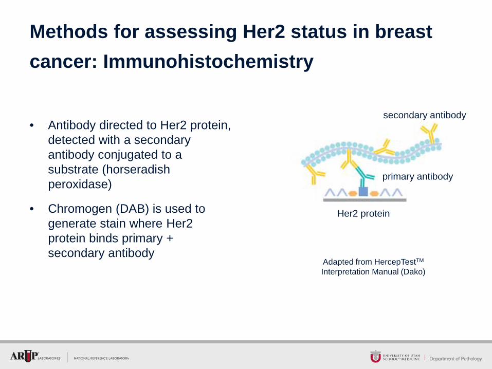
• Antibody directed to Her2 protein, detected with a secondary antibody conjugated to a substrate (horseradish peroxidase)
• Chromogen (DAB) is used to generate stain where Her2 protein binds primary + secondary antibody
Methods for assessing Her2 status in breast cancer: Immunohistochemistry
Adapted from HercepTestTM Interpretation Manual (Dako)
primary antibody
secondary antibody
Her2 protein
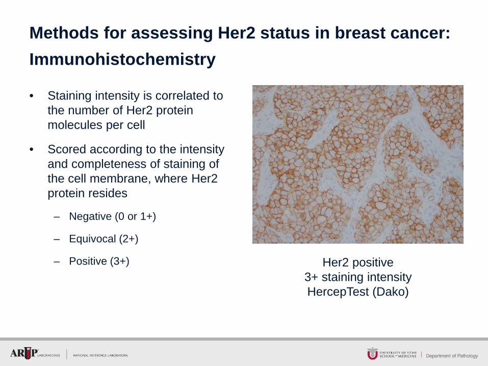
• Staining intensity is correlated to the number of Her2 protein molecules per cell
• Scored according to the intensity and completeness of staining of the cell membrane, where Her2 protein resides
– Negative (0 or 1+)
– Equivocal (2+)
– Positive (3+)
Methods for assessing Her2 status in breast cancer: Immunohistochemistry
Her2 positive 3+ staining intensity HercepTest (Dako)
• Pros
– Inexpensive
– Detects Her2 overexpression regardless of mechanism
– Can visualize with brightfield microscopy under low power, allowing rapid assessment of entire tissue sample tested
• Cons
– False negatives will be undetected due to lack of internal control
– Subjective, semi-quantitative interpretation
Pros and Cons of Her2 Immunohistochemistry
9
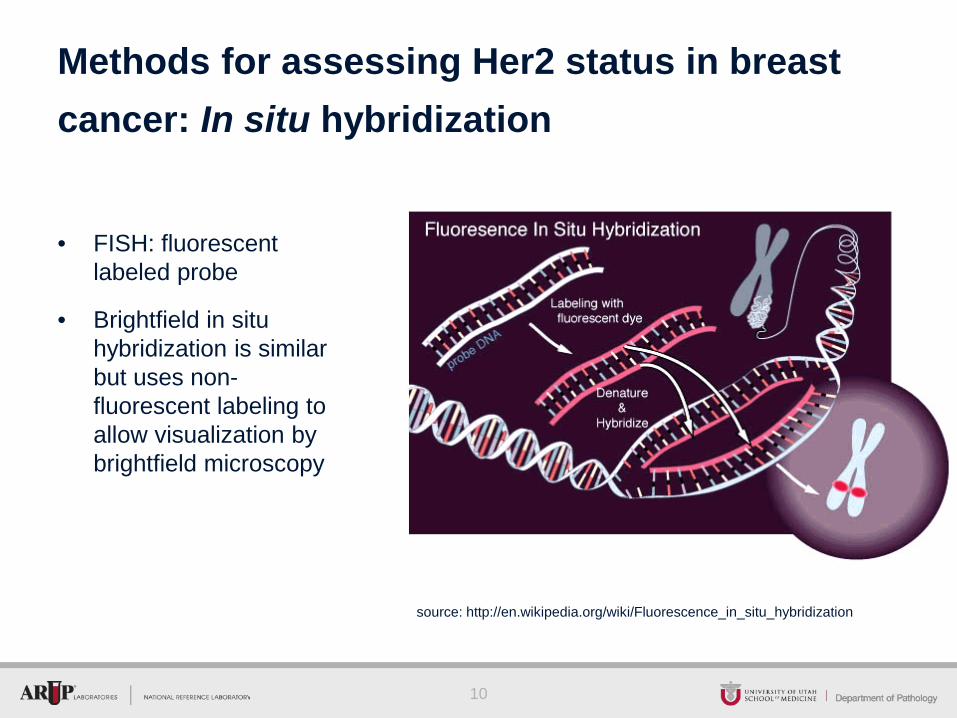
• FISH: fluorescent labeled probe
• Brightfield in situ hybridization is similar but uses non-fluorescent labeling to allow visualization by brightfield microscopy
Methods for assessing Her2 status in breast cancer: In situ hybridization
10
source: http://en.wikipedia.org/wiki/Fluorescence_in_situ_hybridization
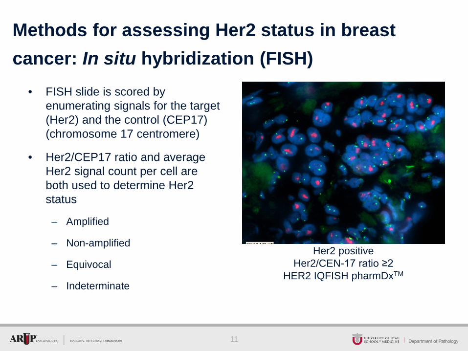
• FISH slide is scored by enumerating signals for the target (Her2) and the control (CEP17) (chromosome 17 centromere)
• Her2/CEP17 ratio and average Her2 signal count per cell are both used to determine Her2 status
– Amplified
– Non-amplified
– Equivocal
– Indeterminate
Methods for assessing Her2 status in breast cancer: In situ hybridization (FISH)
11
Her2 positive Her2/CEN-17 ratio ≥2
HER2 IQFISH pharmDxTM
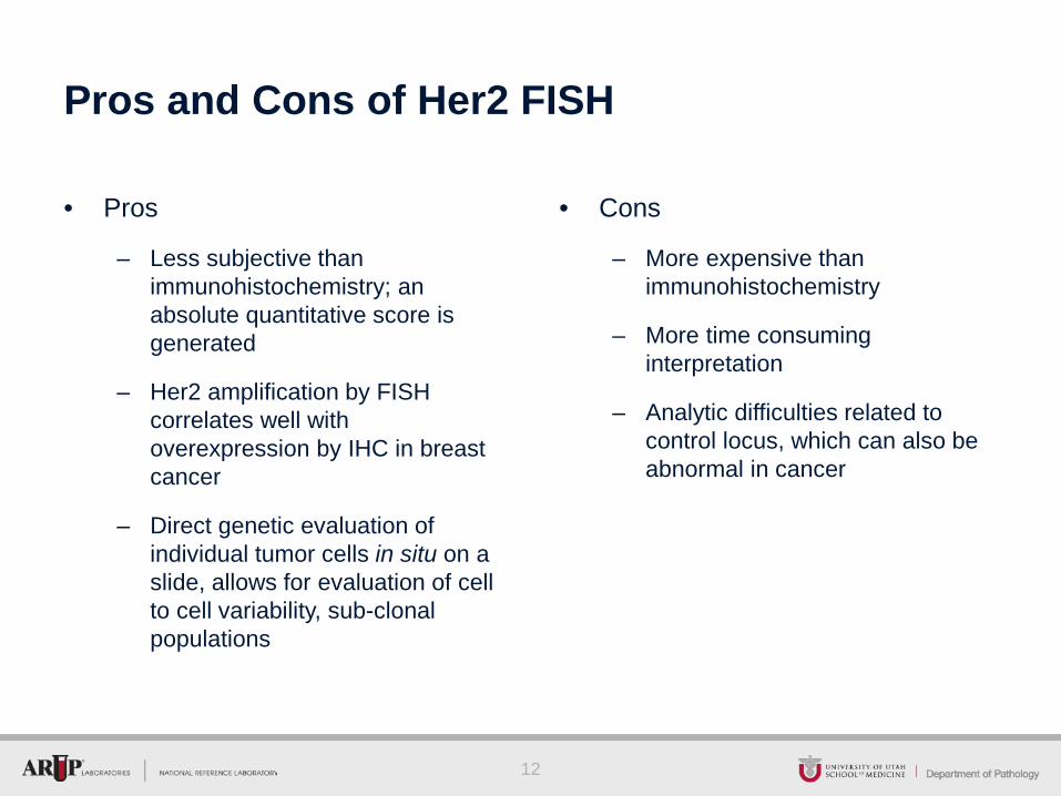
• Pros
– Less subjective than immunohistochemistry; an absolute quantitative score is generated
– Her2 amplification by FISH correlates well with overexpression by IHC in breast cancer
– Direct genetic evaluation of individual tumor cells in situ on a slide, allows for evaluation of cell to cell variability, sub-clonal populations
• Cons
– More expensive than immunohistochemistry
– More time consuming interpretation
– Analytic difficulties related to control locus, which can also be abnormal in cancer
Pros and Cons of Her2 FISH
12
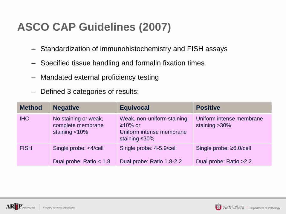
ASCO CAP Guidelines (2007)
– Standardization of immunohistochemistry and FISH assays
– Specified tissue handling and formalin fixation times
– Mandated external proficiency testing
– Defined 3 categories of results:
Method Negative Equivocal Positive IHC No staining or weak,
complete membrane staining <10%
Weak, non-uniform staining ≥10% or Uniform intense membrane staining ≤30%
Uniform intense membrane staining >30%
FISH Single probe: <4/cell Dual probe: Ratio < 1.8
Single probe: 4-5.9/cell Dual probe: Ratio 1.8-2.2
Single probe: ≥6.0/cell Dual probe: Ratio >2.2
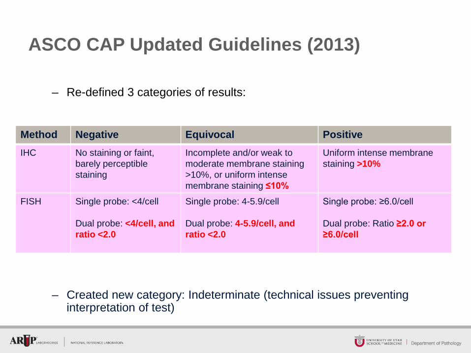
ASCO CAP Updated Guidelines (2013)
– Re-defined 3 categories of results:
Method Negative Equivocal Positive IHC No staining or faint,
barely perceptible staining
Incomplete and/or weak to moderate membrane staining >10%, or uniform intense membrane staining ≤10%
Uniform intense membrane staining >10%
FISH Single probe: <4/cell Dual probe: <4/cell, and ratio <2.0
Single probe: 4-5.9/cell Dual probe: 4-5.9/cell, and ratio <2.0
Single probe: ≥6.0/cell Dual probe: Ratio ≥2.0 or ≥6.0/cell
– Created new category: Indeterminate (technical issues preventing interpretation of test)
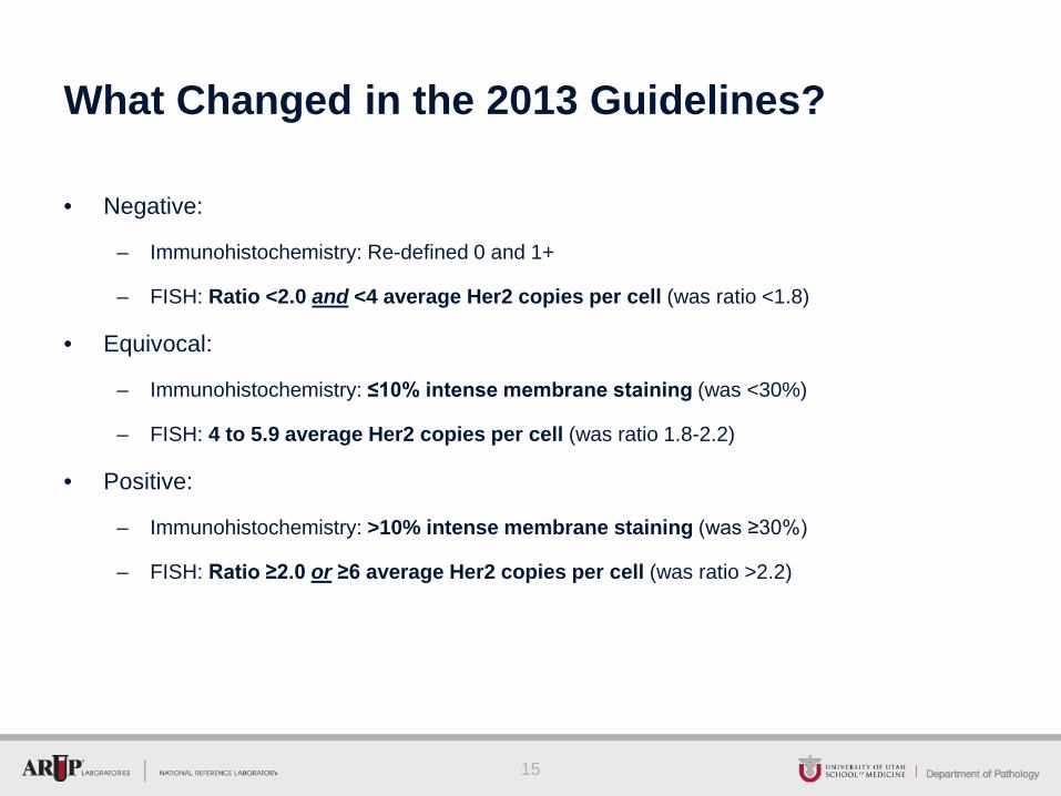
What Changed in the 2013 Guidelines?
• Negative:
– Immunohistochemistry: Re-defined 0 and 1+
– FISH: Ratio <2.0 and <4 average Her2 copies per cell (was ratio <1.8)
• Equivocal:
– Immunohistochemistry: ≤10% intense membrane staining (was <30%)
– FISH: 4 to 5.9 average Her2 copies per cell (was ratio 1.8-2.2)
• Positive:
– Immunohistochemistry: >10% intense membrane staining (was ≥30%)
– FISH: Ratio ≥2.0 or ≥6 average Her2 copies per cell (was ratio >2.2)
15
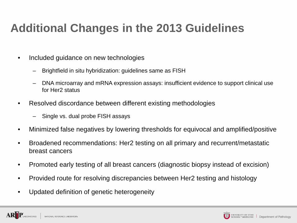
Additional Changes in the 2013 Guidelines
• Included guidance on new technologies
– Brightfield in situ hybridization: guidelines same as FISH
– DNA microarray and mRNA expression assays: insufficient evidence to support clinical use for Her2 status
• Resolved discordance between different existing methodologies
– Single vs. dual probe FISH assays
• Minimized false negatives by lowering thresholds for equivocal and amplified/positive
• Broadened recommendations: Her2 testing on all primary and recurrent/metastatic breast cancers
• Promoted early testing of all breast cancers (diagnostic biopsy instead of excision)
• Provided route for resolving discrepancies between Her2 testing and histology
• Updated definition of genetic heterogeneity
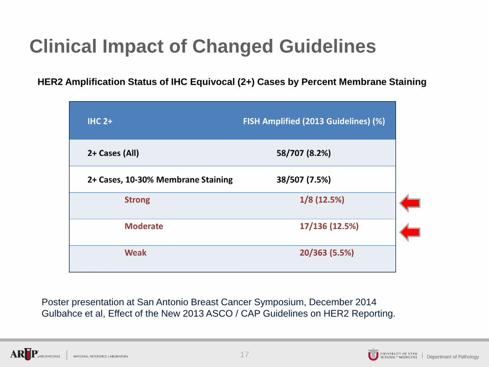
Clinical Impact of Changed Guidelines HER2 Amplification Status of IHC Equivocal (2+) Cases by Percent Membrane Staining
17
Poster presentation at San Antonio Breast Cancer Symposium, December 2014 Gulbahce et al, Effect of the New 2013 ASCO / CAP Guidelines on HER2 Reporting.
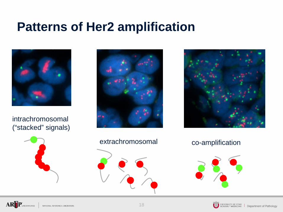
Patterns of Her2 amplification
18
intrachromosomal (“stacked” signals)
extrachromosomal co-amplification
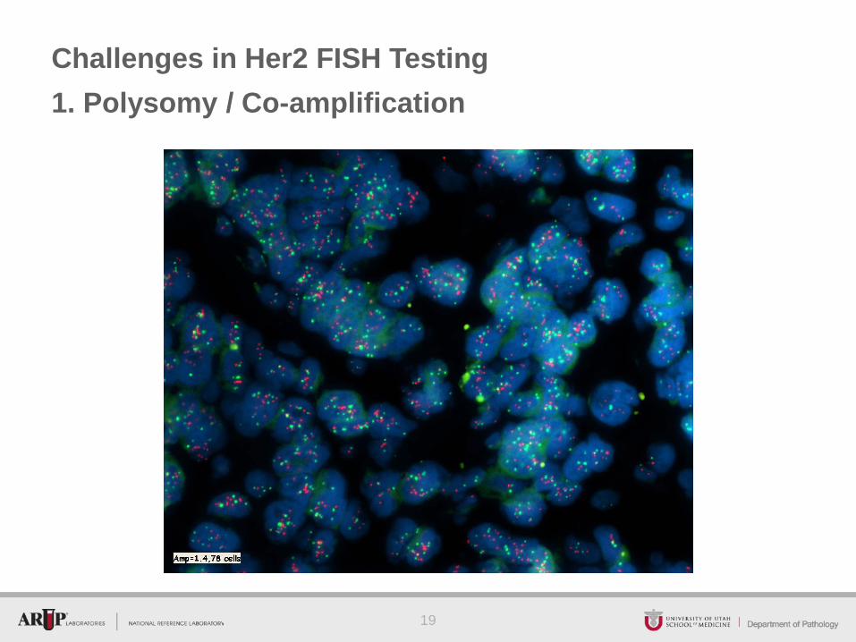
Challenges in Her2 FISH Testing 1. Polysomy / Co-amplification
19
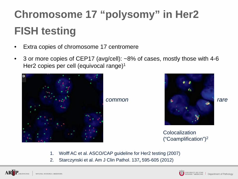
Chromosome 17 “polysomy” in Her2 FISH testing • Extra copies of chromosome 17 centromere
• 3 or more copies of CEP17 (avg/cell): ~8% of cases, mostly those with 4-6 Her2 copies per cell (equivocal range)1
1. Wolff AC et al. ASCO/CAP guideline for Her2 testing (2007) 2. Starczynski et al. Am J Clin Pathol. 137, 595-605 (2012)
Colocalization (“Coamplification”)2
rare common
What is “polysomy”?
• Extra whole copies of a chromosome
• Normal diploid state is 2 copies
• 3 or more copies is polysomy
• Polysomy is harder to define on FFPE sections due to signal truncation
– Average signal count for diploid state is < 2 in FFPE
– Polysomy has been defined in the medical literature as average signal counts as low as 1.861 and ranging up to >3
– Most commonly adopted threshold is mean of ≥3 CEP17 signals per nucleus2
1. Watters et al. Breast cancer research and treatment. 2003 77(2):109-14, 2003. 2. Hanna et al. Modern Pathology 27:4-18, 2014.
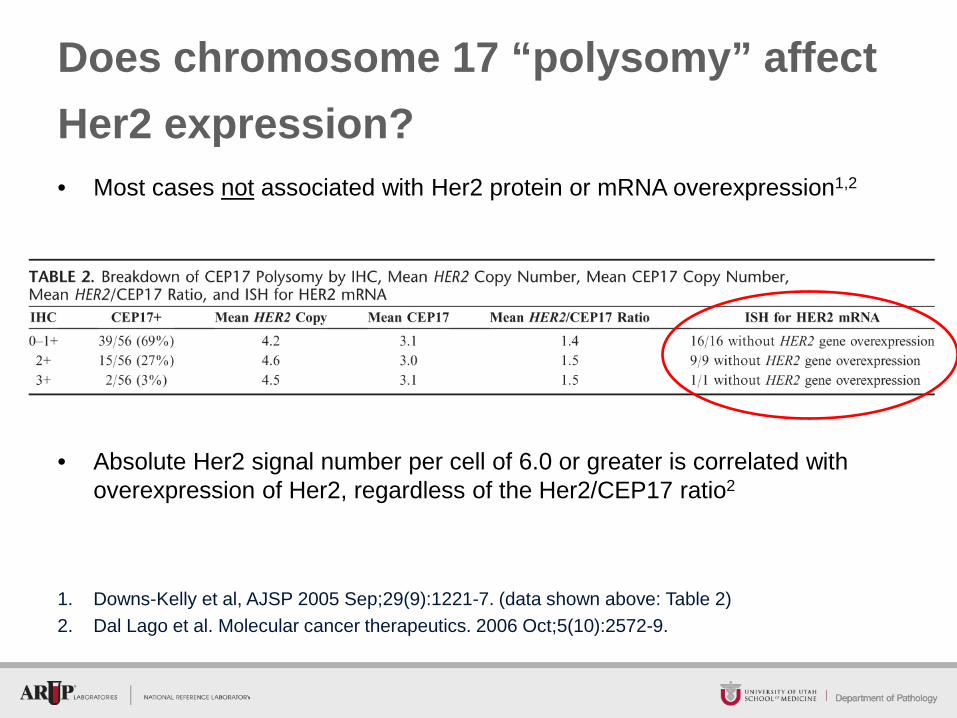
Does chromosome 17 “polysomy” affect Her2 expression? • Most cases not associated with Her2 protein or mRNA overexpression1,2
1. Downs-Kelly et al, AJSP 2005 Sep;29(9):1221-7. (data shown above: Table 2) 2. Dal Lago et al. Molecular cancer therapeutics. 2006 Oct;5(10):2572-9.
• Absolute Her2 signal number per cell of 6.0 or greater is correlated with overexpression of Her2, regardless of the Her2/CEP17 ratio2
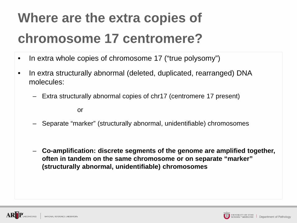
Where are the extra copies of chromosome 17 centromere? • In extra whole copies of chromosome 17 (“true polysomy”)
• In extra structurally abnormal (deleted, duplicated, rearranged) DNA molecules:
– Extra structurally abnormal copies of chr17 (centromere 17 present)
or
– Separate “marker” (structurally abnormal, unidentifiable) chromosomes
– Co-amplification: discrete segments of the genome are amplified together, often in tandem on the same chromosome or on separate “marker” (structurally abnormal, unidentifiable) chromosomes
24
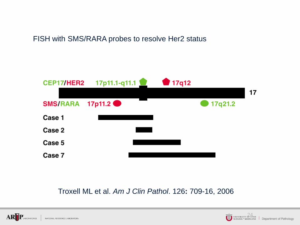
Troxell ML et al. Am J Clin Pathol. 126: 709-16, 2006
FISH with SMS/RARA probes to resolve Her2 status
25
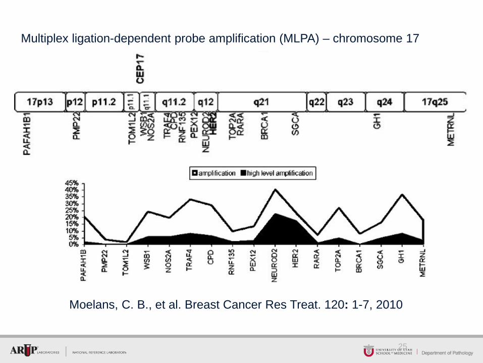
Moelans, C. B., et al. Breast Cancer Res Treat. 120: 1-7, 2010
Multiplex ligation-dependent probe amplification (MLPA) – chromosome 17
26
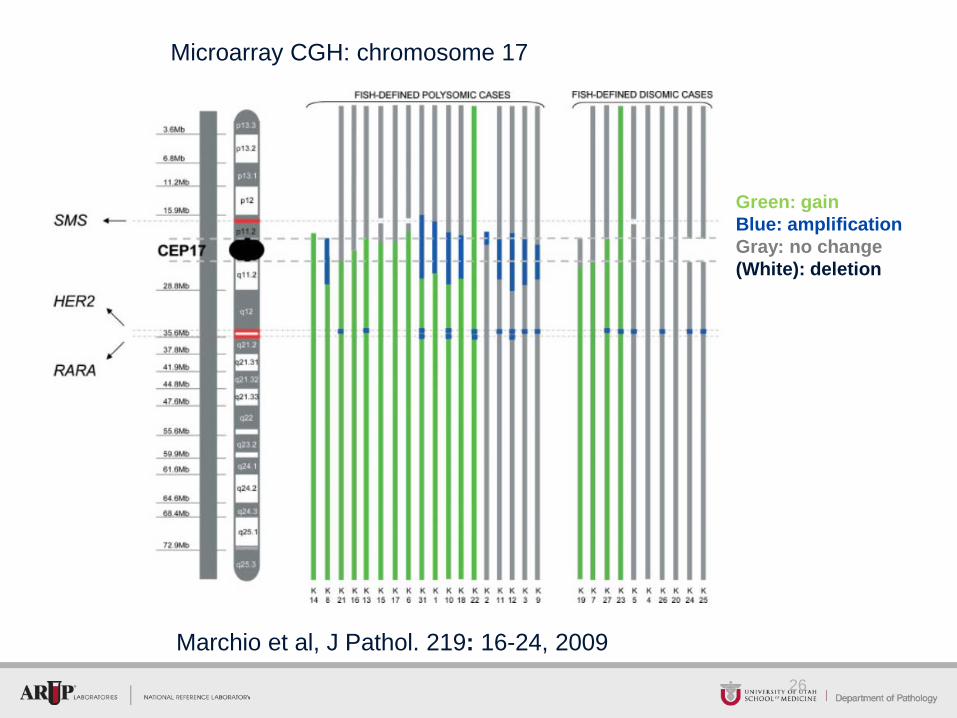
Microarray CGH: chromosome 17
Green: gain Blue: amplification Gray: no change (White): deletion
Marchio et al, J Pathol. 219: 16-24, 2009
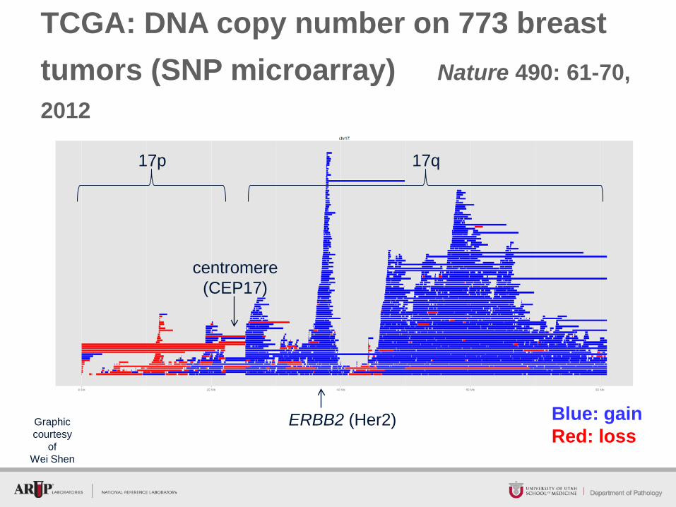
Blue: gain Red: loss
TCGA: DNA copy number on 773 breast tumors (SNP microarray) Nature 490: 61-70, 2012
ERBB2 (Her2)
centromere (CEP17)
17p 17q
Graphic courtesy
of Wei Shen
Chr
omos
ome
1
Chr
omos
ome
2
Chr
omos
ome
3
Chr
omos
ome
4
Chr
omos
ome
5
Chr
omos
ome
6
Chr
omos
ome
7
Chr
omos
ome
8
Chr
omos
ome
9
Chr
omos
ome
10
Chr
omos
ome
11
Chr
omos
ome
12
Chr
omos
ome
13
Chr
omos
ome
14
Chr
omos
ome
15
Chr
omos
ome
16
Chr
omos
ome
17
Chr
omos
ome
18
Chr
omos
ome
19
Chr
omos
ome
20
Chr
omos
ome
21
Chr
omos
ome
22
Chr
omos
ome
X
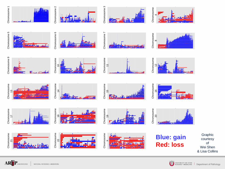
Blue: gain Red: loss
Graphic courtesy
of Wei Shen
& Lisa Collins
Copy number gains in the context of the cancer genome • Entire genome may be present in 3 or more copies (on average), i.e.
“polyploidy,” confounding the definition of “normal” or “control” for the genome
– Polyploidy may not be detected on microarray analysis, depending on the software tools and bioinformatic approach used for analysis
• Adult solid tumors are known to have complex genomes, characterized by gains, losses, allelic imbalances encompassing large portions of the genome
• Absolute copy number per cell can be estimated by some techniques, but not others
– FISH, flow cytometry, cytogenetics: individual cell analysis
• Reference/ “control” region(s) may also be abnormal
(The Search for a Perfect Control)
• CEP17 is co-amplified in a fraction of cases
• Another gene region on chromosome 17 may be used as a control
But…..
• No region of the genome is immune to copy number changes in cancer
And…
• Chromosome 17 is especially prone to copy number changes in breast cancer
30
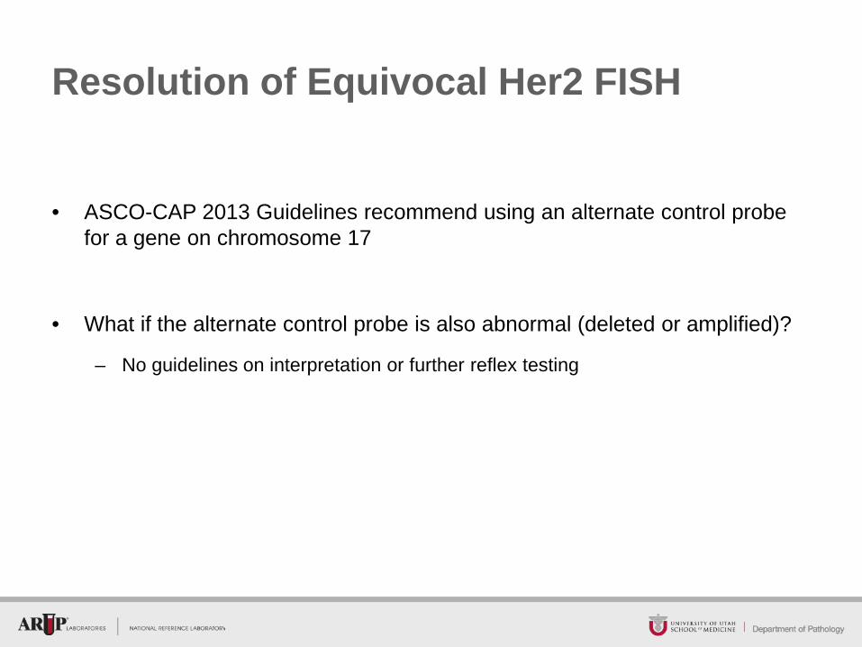
Resolution of Equivocal Her2 FISH
• ASCO-CAP 2013 Guidelines recommend using an alternate control probe for a gene on chromosome 17
• What if the alternate control probe is also abnormal (deleted or amplified)?
– No guidelines on interpretation or further reflex testing
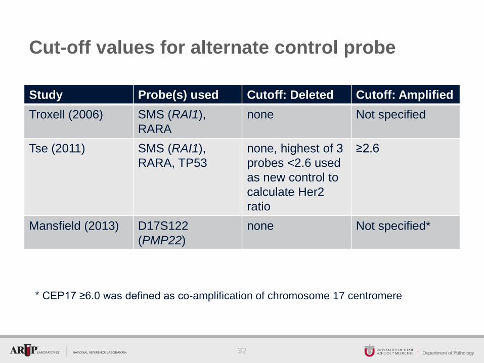
Cut-off values for alternate control probe
Study Probe(s) used Cutoff: Deleted Cutoff: Amplified Troxell (2006) SMS (RAI1),
RARA none Not specified
Tse (2011) SMS (RAI1), RARA, TP53
none, highest of 3 probes <2.6 used as new control to calculate Her2 ratio
≥2.6
Mansfield (2013) D17S122 (PMP22)
none Not specified*
32
* CEP17 ≥6.0 was defined as co-amplification of chromosome 17 centromere
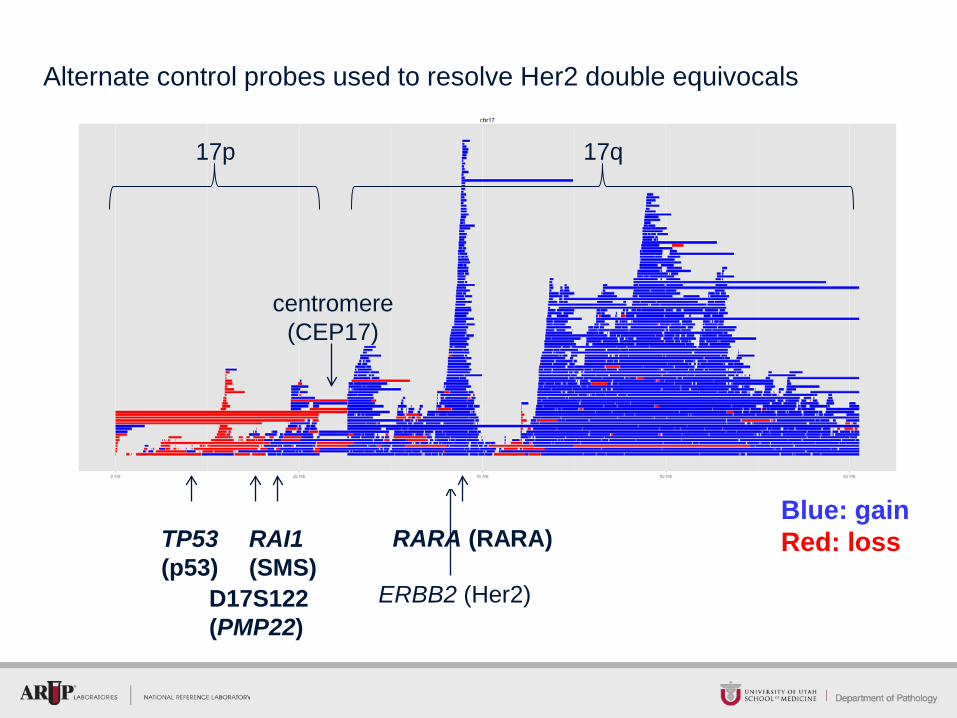
Blue: gain Red: loss
ERBB2 (Her2)
centromere (CEP17)
17p 17q
RAI1 (SMS)
TP53 (p53)
RARA (RARA)
D17S122 (PMP22)
Alternate control probes used to resolve Her2 double equivocals
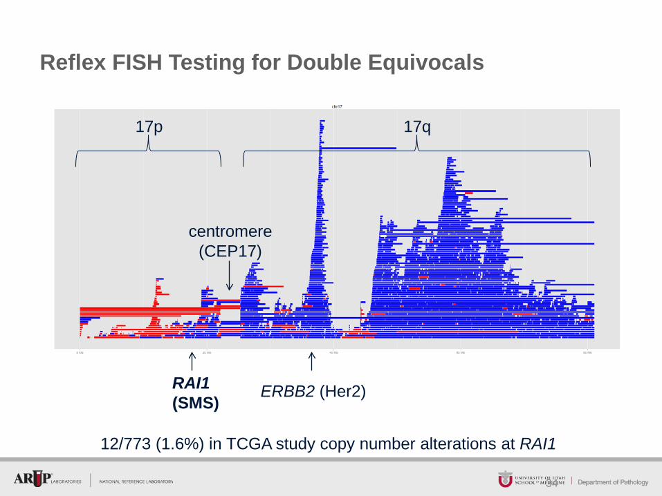
Reflex FISH Testing for Double Equivocals
34
RAI1 (SMS)
ERBB2 (Her2)
centromere (CEP17)
17p 17q
12/773 (1.6%) in TCGA study copy number alterations at RAI1
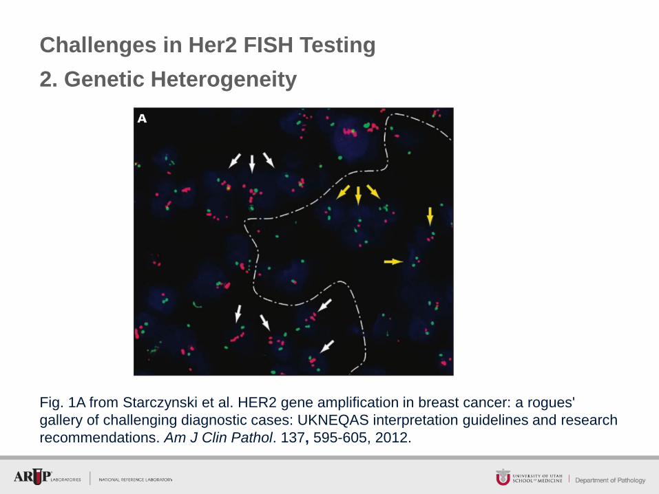
Challenges in Her2 FISH Testing 2. Genetic Heterogeneity
Fig. 1A from Starczynski et al. HER2 gene amplification in breast cancer: a rogues' gallery of challenging diagnostic cases: UKNEQAS interpretation guidelines and research recommendations. Am J Clin Pathol. 137, 595-605, 2012.
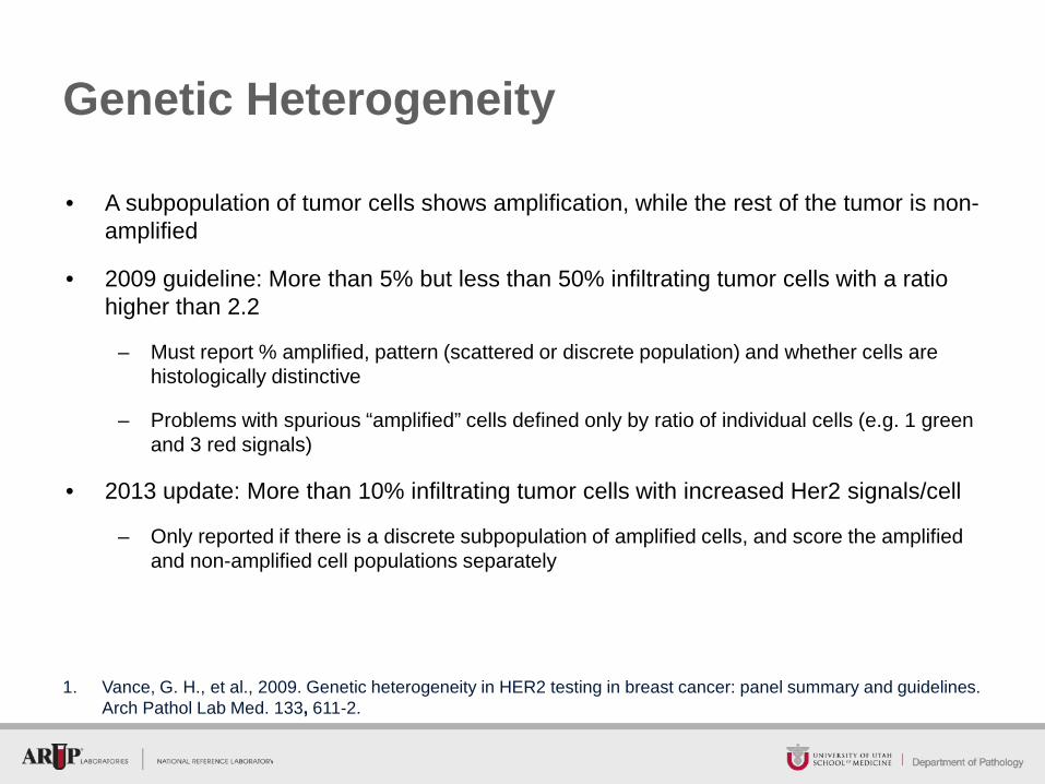
Genetic Heterogeneity
• A subpopulation of tumor cells shows amplification, while the rest of the tumor is non-amplified
• 2009 guideline: More than 5% but less than 50% infiltrating tumor cells with a ratio higher than 2.2
– Must report % amplified, pattern (scattered or discrete population) and whether cells are histologically distinctive
– Problems with spurious “amplified” cells defined only by ratio of individual cells (e.g. 1 green and 3 red signals)
• 2013 update: More than 10% infiltrating tumor cells with increased Her2 signals/cell
– Only reported if there is a discrete subpopulation of amplified cells, and score the amplified and non-amplified cell populations separately
1. Vance, G. H., et al., 2009. Genetic heterogeneity in HER2 testing in breast cancer: panel summary and guidelines. Arch Pathol Lab Med. 133, 611-2.
39
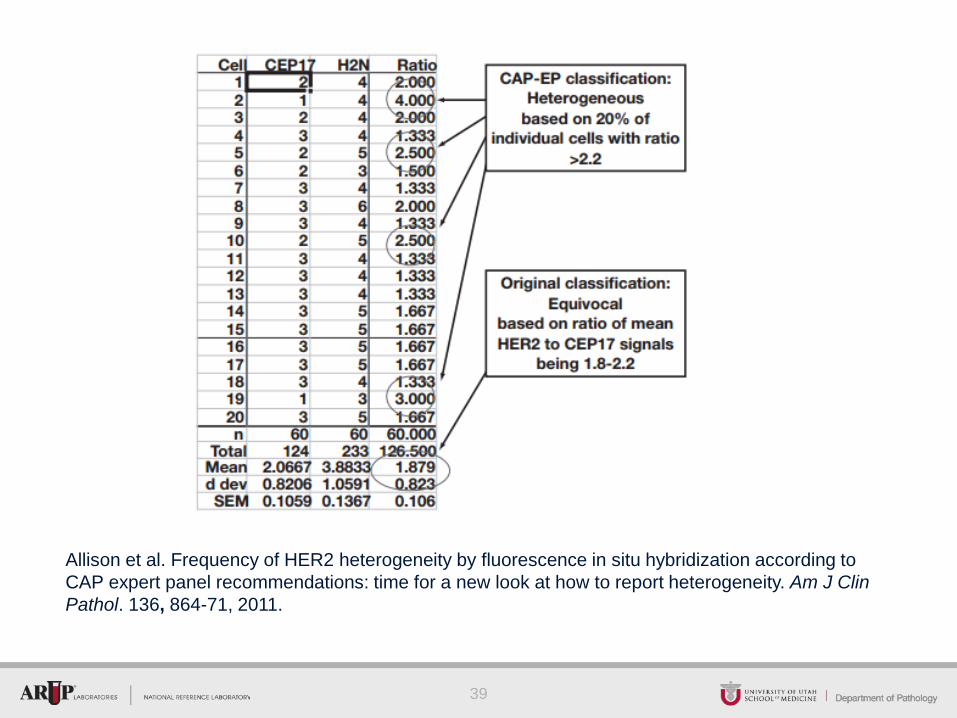
Allison et al. Frequency of HER2 heterogeneity by fluorescence in situ hybridization according to CAP expert panel recommendations: time for a new look at how to report heterogeneity. Am J Clin Pathol. 136, 864-71, 2011.
40
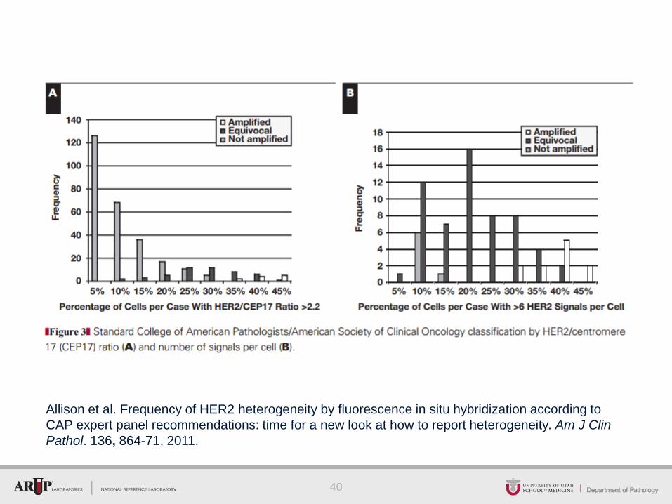
Allison et al. Frequency of HER2 heterogeneity by fluorescence in situ hybridization according to CAP expert panel recommendations: time for a new look at how to report heterogeneity. Am J Clin Pathol. 136, 864-71, 2011.
Summary
• Immunohistochemistry and in situ hybridization (ISH, FISH) are the recommended methods for determining Her2 status for treatment with Her2-targeted therapy
• Neither method is 100% sensitive or specific
• Updated ASCO-CAP (2013) guidelines have resulted in increased proportion of patients being eligible for Her2-targeted therapy
• Her2-positive cases are not a homogeneous group
– Borderline positive cases may not be as responsive to Her2-targeted therapy
• Challenges in Her2 laboratory testing include polysomy / co-amplification, and genetic heterogeneity
41

© ARUP Laboratories 2014