Embed Size (px)
Citation preview
BRITISH MEDICAL JOURNALLONDON SATURDAY NOVEMBER 26 1955
HYPERPARATHYROIDISM WITH RENAL CALCINOSISBY
S. LEONARD SIMPSON, M.D., F.R.C.P.Consultant Endocrinologist to St. Mary's Hospital and to the Paediatric Unit
AND
R. R. WILSON, M.D.Consusltant in Morbid Anatomy, North-western Group Laboratory; formerly Senior Lecturer in Morbid
Anatomy and Honorary Consultant in Morbid Anatomy, St. Mary's Hospital
Clinical knowledge of hyperparathyioidism can be saidto date from the successful removal of a parathyroidadenoma in a case of hyperparathyroidism by Mandl in1926. Since that time Albright and his associates (1929,1934, 1936, 1937, 1938, 1941, 1948) have played a leadingpart in advancing our knowledge of the disorder, parti-cularly in regard to the chemistry and to the involvementof the kidneys. It is from the latter angle that we wishto present an interesting case. The problem is not freefrom complexity, in so far as primary hyperparathyroid-ism (whether due to a parathyroid adenoma or generalparathyroid hyperplasia) may cause renal calcinosis withultimate renal insufficiency; and, on the other hand,renal insufficiency from any cause, associated withretention of phosphorus and with high plasma inorganicphosphorus, may be followed by hyperplasia of the para-thyroid glands, which from a teleological point of viewhas the purpose of an attempted elimination of the(excessively) retained phosphorus, the primary action ofparathyroid hormone being believed to be augmentedelimination of phosphorus.
Further, whatever the nature of the primary renallesion may be, the secondary hyperplasia of parathyroidglands may lead to renal calcinosis and sometimes toosteoporosis. Unless, therefore, the whole history ofany one case is known and the case has been investigatedin its early as well as its late stage, it may be difficult,or impossible, to state in the terminal phase whether thecondition was primary hyperparathyroidism with secon-dary renal involvement or primary renal disease withsecondary parathyroid involvement. Renal calcinosis asa consequence of primary hyperparathyroidism is notonly met with clinically but has also been producedexperimentally (Hueper, 1927; Leberman, 1940). Thecalcium may be deposited profusely in minute particlesboth in the renal parenchyma and in the tubules; and/orrenal calculi of variable size may be visible radio-logically.
In some urological clinics it is an obligatory routine totest for hyperparathyroidism in every case of renal cal-culus, and experience has shown that such calculi neednot be obviously multiple or bilateral. Perhaps the mostcomplex case of all is the one starting with a parathyroidadenoma with secondary renal calcinosis, in which the
adenoma is successfully removed, but, nevertheless, therenal calcium deposits do not disappear and the damagealready done to the kidney is irreversible and even pro-gressive, so that the condition progresses to renal insuffi-ciency, and, secondary to this, there is hyperplasia of theremaining parathyroid glands, the patient ultimatelvdying in uraemia. Such appears to be the sequence ofevents in the case here presented.
Case HistoryA woman of 40 was admitted to a surgical ward of Wil-
lesden General Hospital in September, 1941, under Mr.F. W. M. Pratt, with a firm swelling in the middle of theright tibia, said to have followed slight trauma in that areasix months previously.X-ray examination was carried out on September 4 and
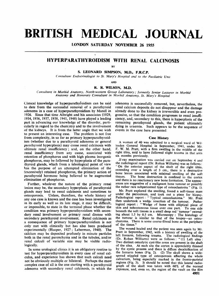
the radiological report (Dr. Rohan Williams) was as follows:"On the anterior aspect of the shaft of the right tibia,extending over a length of 3 cm., there is a destructivebone lesion associated with minimal swelling of the softtissues. The bone destruction is confined to the cortexand there is no reactionary new bone formation. The appear-ances are suggestive of a malignant bone tumour or possiblythe rather rare subperiosteal type of osteoclastoma" (Fig. 1).
Mr. Pratt explored the swelling, found a soft-tissue massunder the periosteum, and took out a piece for biopsy.Pathological report: "Typical osteoclastoma." Mr. Prattthen undertook a wedge resection of the tumour. Patho-logical report: "Wedge of bone with elliptical piece ofskin and subcutaneous tissue over one part. To one sidebeneath the soft tissues is a small deep red 'tumour' extend-ing about 1.5 by 0.5 cm. Microscopy: The histology ofthe tumour is similar to that of the biopsy-an osteo-clastoma. There is some coarse-fibred bone at the peripheryof the tissue."The wound healed and the patient was seen again by Mr.
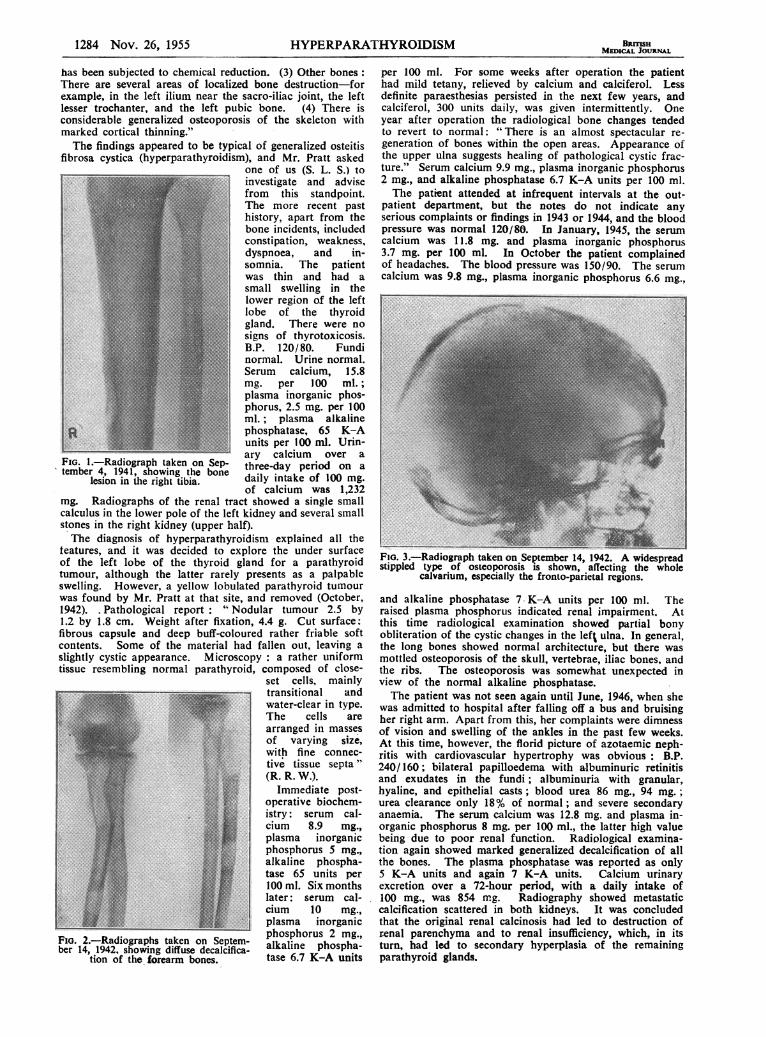
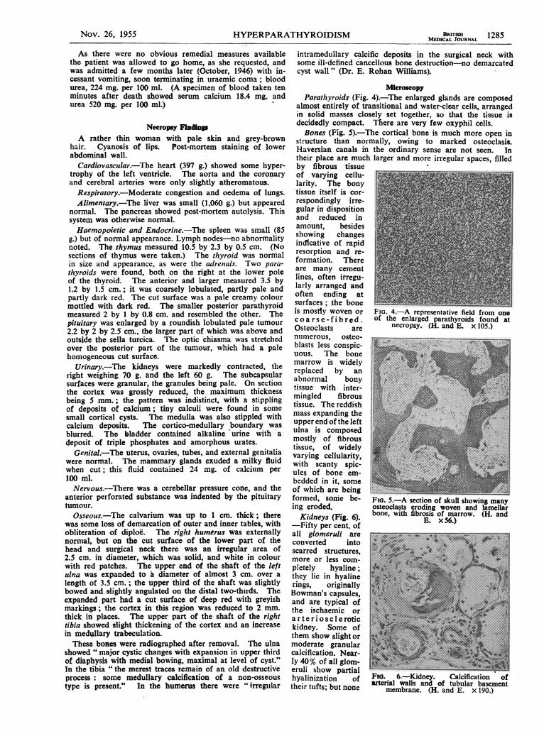
Pratt in September, 1942, with a history of swelling of theleft forearm, following trauma. The radiological findings(Dr. Rohan Williams) were as follows: " (1) Left ulna:Two distinct osteolytic cyst-like areas are present in the shaftof the ulna. At each site the cortex is appreciably thinnedby the cystic process and there is diffuse decalcification ofthe forearm bones (Fig. 2). (2) The skull shows a wide-spread stippled type of osteoporosis affecting the wholecalvarium, being especially marked in the fronto-parietalregions (Fig. 3). It should be noted that this lateral radio-graph of the skull was taken with 60% of the normalexposure, and, even so, the region of the vault on the film
4951
1284 Nov. 26, 1955 HYPERPARATHYROIDISM
has been subjected to chemical reduction. (3) Other bones:There are several areas of localized bone destruction-forexample, in the left ilium near the sacro-iliac joint, the leftlesser trochanter, and the left pubic bone. (4) There isconsiderable generalized osteoporosis of the skeleton withmarked cortical thinning."The findings appeared to be typical of generalized osteitis
fibrosa cystica (hyperparathyroidism), and Mr. Pratt askedone of us (S. L. S.) to
investigate and advisefrom this standpoint.
;iS The more recent pastl history, apart from the
Ia"~~ _bone incidents, includedconstipation, weakness,
i dyspnoea, and in-
l somnia. The patientwas thin and had a
small swelling in the
lower region of the leftlobe of the thyroid
gland. There were no
signs of thyrotoxicosis.B.P. 120/80. Fundi
normal. Urine normal.
Serum calcium, 15.8
mg. per 100 ml.plasma inorganic phos-
phorus, 2.5 mg. per 100
ml. plasma alkalinephosphatase, 65 K-Aunits per 100 ml. Urin-ary calcium over a
FIG. I- Radiograph taken on Sep- three-day period on atember 4, 1941, showing the bone
the-a prido
lesion in the right tibia. daily intake of 100 mg.of calcium was 1,232
mg. Radiographs of the renal tract showed a single smallcalculus in the lower pole of the left kidney and several smallstones in the right kidney (upper half).The diagnosis of hyperparathyroidism explained all the
teatures, and it was decided to explore the under surfaceof the left lobe of the thyroid gland for a parathyroidtumour, although the latter rarely presents as a palpableswelling. However, a yellow lobulated parathyroid tumourwas found by Mr. Pratt at that site, and removed (October,1942). Pathological report : "Nodular tumour 2.5 by1.2 by 1.8 cm. Weight after fixation, 4.4 g. Cut surface:fibrous capsule and deep buff-coloured rather friable softcontents. Some of the material had fallen out, leaving aslightly cystic appearance. Microscopy : a rather uniformtissue resembling normal parathyroid, composed of close-
set cells, mainlyXi.; | transitional and
E-.X_ l water-clear in type.
The cells are
arranged in masses
of varying size,
+S with fine connec-
.I.. ......tive tissue septa
-.t;.$..' - ''(R. R. W.).
Immediate post-
-,............istry: serum cal-
1, >.' -' - ,, f ' - ~~~~~~~~~~~~~~~~................... ..-|~~~~~~~i 9 ,
li:!-- ts : X l phosphorus 5 mg.,
1--; g_ - - - - 1l~~~~~~~~~~~~... latery: serum cal-
Ei-* l .8.9cium mg.,
. .......plasma inorganic
.......phosphorus mg.,
FI. 2.Radiographs taken on Septem alkaline phospha-ber 14, 1942, showing diffuse decalcifica alan phosph
tion of the forearm bones. tase t.7 K-A units
per 100 ml. For some weeks after operation the patienthad mild tetany, relieved by calcium and calciferol. Lessdefinite paraesthesias persisted in the next few years, andcalciferol, 300 units daily, was given intermittently. Oneyear after operation the radiological bone changes tendedto revert to normal: "There is an almost spectacular re-generation of bones within the open areas. Appearance ofthe upper ulna suggests healing of pathological cystic frac-ture." Serum calcium 9.9 mg., plasma inorganic phosphorus2 mg., and alkaline phosphatase 6.7 K-A units per 100 ml.The patient attended at infrequent intervals at the out-
patient department, but the notes do not indicate anyserious complaints or findings in 1943 or 1944, and the bloodpressure was normal 120/80. In January, 1945, the serumcalcium was 11.8 mg. and plasma inorganic phosphorus3.7 mg. per 100 ml. In October the patient complainedof headaches. The blood pressure was 150/90. The serumcalcium was 9.8 mg., plasma inorganic phosphorus 6.6 mg.,
FiG. 3.-Radiograph taken on Sel?tember 14, 1942. A widespreadstippled type of osteoporosis is shown, aflecting the whole
calvarium, especially the fronto-parietal regions.
and alkaline phosphatase 7 K-A units per 100 ml. Theraised plasma phosphorus indicated renal impairment. Atthis time radiological examination showed partial bonyobliteration of the cystic changes in the left ulna. In general,the long bones showed normal architecture, but there wasmottled osteoporosis of the skull, vertebrae, iliac bones, andthe ribs. The osteoporosis was somewhat unexpected inview of the normal alkaline phosphatase.The patient was not seen again until June, 1946, when she
was admitted to hospital after falling off a bus and bruisingher right arm. Apart from this, her complaints were dimnessof vision and swelling of the ankles in the past few weeks.At this time, however, the florid picture of azotaemic neph-ritis with cardiovascular hypertrophy was obvious: B.P.240/160; bilateral papilloedema with albuminuric retinitisand exudates in the fundi; albuminuria with granular,hyaline, and epithelial casts; blood urea 86 mg., 94 mg.;urea clearance only 18% of normal; and severe secondaryanaemia. The serum calcium was 12.8 mg. and plasma in-organic phosphorus 8 mg. per 100 ml., the latter high valuebeing due to poor renal function. Radiological examina-tion again showed marked generalized decalcification of allthe bones. The plasma phosphatase was reported as only5 K-A units and again 7 K-A units. Calcium urinaryexcretion over a 72-hour period, with a daily intake of100 mg., was 854 mg. Radiography showed metastaticcalcification scattered in both kidneys. It was concludedthat the original renal calcinosis had led to destruction ofrenal parenchyma and to renal insufficiency, which, in itsturn, had led to secondary hyperplasia of the remainingparathyroid glands.
BRITISHMEDICAL JOURNAL
- . s-- la,.---w
Nov. 26, 1955HYPERPARATHYROIDISM~~~~~~~~~~~~~~~~~~~~~~~~~~~~~~~~~~~~~~~~~~~~~~~~~~~~~~~~~~~~~~~~~~~~~~~~~~~~~~~~~~~~~~~~~~~~~~~~~~~~~~~~~~~~~~~~~~~~~~~~~~~~~~~~~~~~~~~~~~~~~~~~~~~~~~~~~~~~~~~~~~~~~~~~~~~~~~~~~~~~~~~~~~~~~~~~~~~~~~~~~~~~~~~~~~~~~~~~~~~~BIRURT N 1285MEDICAL JOURNAL
As there were no obvious remedial measures availablethe patient was allowed to go home, as she requested, andwas admitted a few months later (October, 1946) with in-cessant vomiting, soon terminating in uraemic coma; bloodurea, 224 mg. per 100 ml. (A specimen of blood taken tenminutes after death showed serum calcium 18.4 mg. andurea 520 mg. per 100 ml.)
Necropsy FindinpA rather thin woman with pale skin and grey-brown
hair. Cyanosis of lips. Post-mortem staining of lowerabdominal wall.Cardiovascular.-The heart (397 g.) showed some hyper-
trophy of the left ventricle. The aorta and the coronaryand cerebral arteries were only slightly atheromatous.
Respiratory.-Moderate congestion and oedema of lungs.Alimentary.-The liver was small (1,060 g.) but appeared
normal. The pancreas showed post-mortem autolysis. Thissystem was otherwise normal.Haemopoietic and Endocrine.-The spleen was small (85
g.) but of normal appearance. Lymph nodes-no abnormalitynoted. The thymus measured 10.5 by 2.3 by 0.5 cm. (Nosections of thymus were taken.) The thyroid was normalin size and appearance, as were the adrenals. Two para-thyroids were found, both on the right at the lower poleof the thyroid. The anterior and larger measured 3.5 by1.2 by 1.5 cm.; it was coarsely lobulated, partly pale andpartly dark red. The cut surface was a pale creamy colourmottled with dark red. The smaller posterior parathyroidmeasured 2 by 1 by 0.8 cm. and resembled the other. Thepituitary was enlarged by a roundish lobulated pale tumour2.2 by 2 by 2.5 cm., the larger part of which was above andoutside the sella turcica. The optic chiasma was stretchedover the posterior part of the tumour, which had a palehomogeneous cut surface.Urinary.-The kidneys were markedly contracted, the
right weighing 70 g. and the left 60 g. The subcapsularsurfaces were granular, the granules being pale. On sectionthe cortex was grossly reduced, the maximum thicknessbeing 5 mm.; the pattern was indistinct, -with a stipplingof deposits of calcium; tiny calculi were found in somesmall cortical cysts. The medulla was also stippled withcalcium deposits. The cortico-medullary boundary wasblurred. The bladder contained alkaline urine with adeposit of triple phosphates and amorphous urates.
Genital.-The uterus, ovaries, tubes, and external genitaliawere normal. The mammary glands exuded a milky fluidwhen cut; this fluid contained 24 mg. of calcium per100 ml.Nervous.-There was a cerebellar pressure cone, and the
anterior perforated substance was indented by the pituitarytumour.Osseous.-The calvarium was up to 1 cm. thick; there
was some loss of demarcation of outer and inner tables, withobliteration of diploe. The right humerus was externallynormal, but on the cut surface of the lower part of thehead and surgical neck there was an irregular area of2.5 cm. in diameter, which was solid, and white in colourwith red patches. The upper end of the shaft of the leftulna was expanded to h diameter of almost 3 cm. over alength of 3.5 cm.; the upper third of the shaft was slightlybowed and slightly angulated on the distal two-thirds. Theexpanded part had a cut surface of deep red with greyishmarkings; the cortex in this region was reduced to 2 mm.thick in places. The upper part of the shaft of the righttibia showed slight thickening of the cortex and an increasein medullary trabeculation.These bones were radiographed after removal. The ulna
showed "major cystic changes with expansion in upper thirdof diaphysis with medial bowing, maximal at level of cyst."In the tibia "the merest traces remain of an old destructiveprocess: some medullary calcification of a non-osseoustype is present." In the humerus there were "irregular
intramedullary calcific deposits in the surgical neck withsome ill-defined cancellous bone destruction-no demarcatedcyst wall" (Dr. E. Rohan Williams).
MicroscopyParathyroids (Fig. 4).-The enlarged glands are composed
almost entirely of transitional and water-clear cells, arrangedin solid masses closely set together, so that the tissue isdecidedly compact. There are very few oxyphil cells.Bones (Fig. 5).-The cortical bone is much more open in
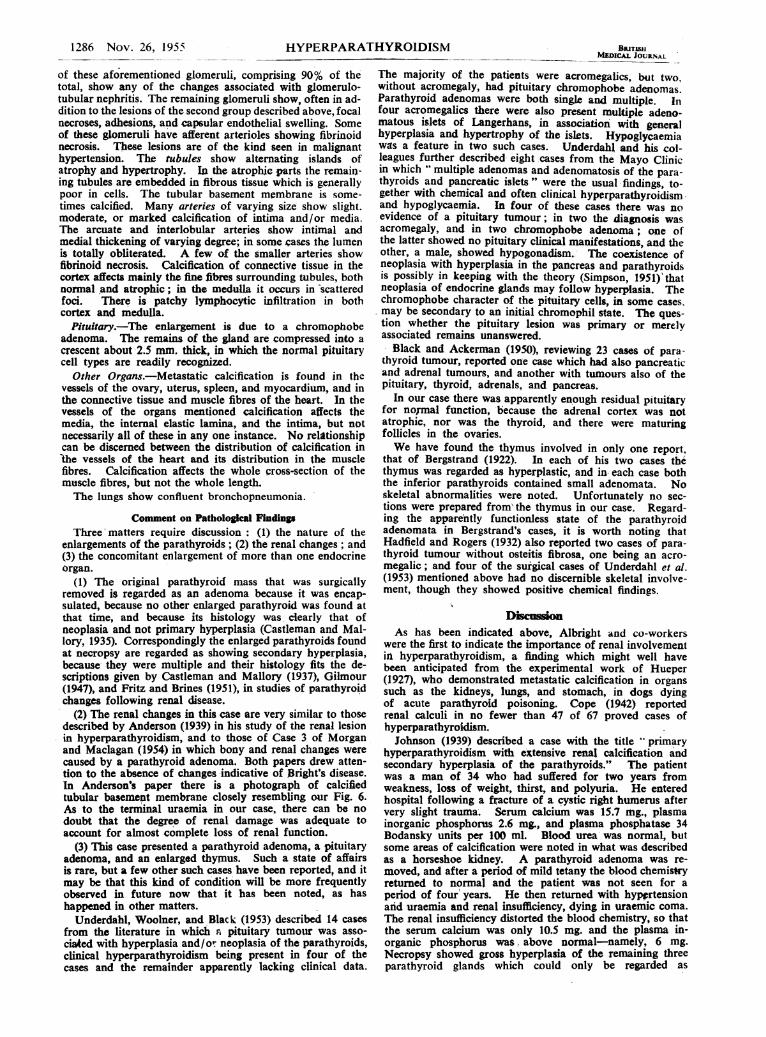
structure than normally, owing to marked osteoclasis.Haversian canals in the ordinary sense are not seen. Intheir place are much larger and more irregular spaces, filledby fibrous tissueof varying cellu-larity. The bony'tissue itself is cor- Irespondingly irre-gular in dispositiontand reduced in S'amount, besidesshowing changesindlcative of rapidresorption and re-formation. Thereare many cementlines, often irregu-larly arranged andEoften ending atsurfaces ; the bone ~is mostly woven or FIG. 4.-A representative field from onec o a r s e - f i b r ed. of the enlarged parathyroids found atOsteoclasts are necropsy. (H. and E. x 105.)numerous, osteo-blasts less conspic- t_UOUS. The bonemarrow is widely ireplaced by anabnormal bony #tissue with inter-mingled fibroustissue. The reddishmass expanding theupper end of the leftulna is composedmostly of fibroustissue, of widelyvarying cellularity,with scanty spic- 2 ~ules of bone em-bedded in it, someof which are beingformed, somie be- FIG. 5 A section of skull showi'ng manying eroded. osteoclasts eroding woven and lamellarKidneys (Fig. 6). bone, with fibrosis of marrow. (H. and
-Fifty per cent, of E. x 56.)all glomeruli areconverted intoscarred structuares,more or less com-pletely hyaline;-they lie in hyalinerings, originallyBowman's capsules,-and are typical ofthe ischaemic orarteriosclerotickidney. Some ofthem show slight ormoderate granularcalcification. Near-ly 40% of all glom-eruli show partial
hyalinization o Fio. 6.-Kidney. Calcification oftheir tufts; but none
aterial walls and of tubular basementtheituts;ut one membrane. (H. and E. x( 190.
Nov. 26, 1955 HYPERPARATHYROIDISM
%-- -*. -e- a %R./
HYPERPARAT.
of these aforementioned glomeruli, comprising 90°% of thetotal, show any of the changes associated with glomerulo-tubular nephritis. The remaining glomeruli show, often in ad-dition to the lesions of the second group described above, focalnecroses, adhesions, and capsular endothelial swelling. Someof these glomeruli have afferent arterioles showing fibrinoidnecrosis. These lesions are of the kind seen in malignanthypertension. The tubules show alternating islands ofatrophy and hypertrophy. In the atrophic parts the remain-ing tubules are embedded in fibrous tissue which is generallypoor in cells. The tubular basement membrane is some-times calcified. Many arteries of varying size show slight.moderate, or marked calcification of intima and/or media.The arcuate and interlobular arteries show intimal andmedial thickening of varying degree; in some rases the lumenis totally obliterated. A few of the smaller arteries showfibrinoid necrosis. Calcification of connective tissue in thecortex affects mainly the fine fbres surrounding tubules, bothnormal and atrophic; in the medulla it occurs in -scatteredfoci. There is patchy lymphocytic infiltration in bothcortex and medulla.Pituitary.-The enlargement is due to a chromophobe
adenoma. The remains of the gland are compressed into acrescent about -2.5 mm. thick, in which the normal pituitarycell types are readily recogized.
Other Organs.-Metastatic calcification is found in thevessels of the ovary, uterus, spleen, and myocardium, and inthe connective tissue and muscle fibres of the heart. In thevessels of the organs mentioned calcification affects themedia, the internal elastic lamina, and the intima, but notnecessarily all of these in any one instance. No relationshipcan be discerned between the distribution of calcification inthe vessels of the heart and its distribution in the musclefibres. Calcification affects the whole cross-section of themuscle fibres, but not the whole length.The lungs show confluent bronchopneumonia.
Comment on Pathological FindingThree matters require discussion: (1) the nature of the
enlargements of the parathyroids; (2) the renal changes; and(3) the concomitant enlargement of more than one endocrineorgan.
(1) The original parathyroid mass that was surgicallyremoved is regarded as an adenoma because it was encap-sulated, because no other enlarged parathyroid was found atthat time, and because its histology was clearly that ofneoplasia and not primary hyperplasia (Castleman and Mal-lory, 1935). Correspondingly the enlarged parathyroids foundat necropsy are regarded as showing secondary hyperplasia,because they were multiple and their histology fits the de-scriptions given by Castleman and Mallory (1937), Gilmour(1947), and Fritz and Brines (1951), in studies of parathyroidchanges following renal disease.
(2) The renal changes in this case are very similar to thosedescribed by Anderson (1939) in his study of the renal lesionin hyperparathyroidism, and to those of Case 3 of Morganand Maclagan (1954) in which bony and renal changes werecaused by a parathyroid adenoma. Both papers drew atten-tion to the absence of changes indicative of Bright's disease.In Anderson's paper there is a photograph of calcifiedtubular basement membrane closely resembling our Fig. 6.As to the terminal uraemia in our case, there can be nodoubt that the degree of renal damage was adequate toaccount for almost complete loss of renal function.
(3) This case presented a parathyroid adenom,a, a pituitaryadenoma, and an enlarged thymus. Such a state of affairsis rare, but a few other such cases have been reported, and itmay be that this kind of condition will be more frequentlyobserved in future now that it has been noted, as hashappened in other matters.
Underdahl, Woolner, and Black- (1953) described 14 casesfrom the literature in which ai pituitary tumour was asso-ciated with hyperplasia and/or neoplasia of the parathyroids,clinical hyperparathyroidism being present in four of thecases and the remainder apparently lacking clinical data.
HYROIDISM BRITISHMWICA.L JOURNAL
The majority of the patients were acromegalics, but two.without acromegaly, had pituitary chromophobe adenomas.Parathyroid adenomas were both single and multiple. Infour acromegalics there were also present multiple adeno-matous islets of Langerhans, in association with generalhyperplasia and hypertrophy of the islets. Hypoglycaemiawas a feature in two such cases. Underdahl and his col-leagues further described eight cases from the Mayo Clinicin which " multiple adenomas and adenomatosis of the para-
thyroids and pancreatic islets" were the usual findings, to-
gether with chemical and often clinical hyperparathyroidismand hypoglycaemia. In four of these cases there was no
evidence of a pituitary tumour; in two the diagnosis was
acromegaly, and in two chromophobe adenoma; one ofthe latter showed no pituitary clinical manifestations, and theother, a male, showed hypogonadism. The coexistence ofneoplasia with hyperplasia in the pancreas and parathyroidsis possibly in keeping with the theory (Simpson, 1951) thatneoplasia of endocrine glands may follow hyperplasia. Thechromophobe character of the pituitary cells, in some cases,may be secondary to an initial chromophil state. The ques-
tion whether the pituitary lesion was primary or merelyassociated remains unanswered.Black and Ackerman (1950), reviewing 23 cases of para-
thyroid tumour, reported one case which had also pancreaticand adrenal tumours, and another with tumours also of thepituitary, thyroid, adrenals, and pancreas.
In our case there was apparently enough residual pituitaryfor noTmal function, because the adrenal cortex was notatrophic, nor was the thyroid, and there were maturingfollicles in the ovaries.We have found the thymus involved in only one report,
that of Bergstrand (1922). In each of his two cases thethymus was regarded as hyperplastic, and in- each case boththe inferior parathyroids contained small adenomata. Noskeletal abnormalities were noted. Unfortunately no sec-tions were prepared from'the thymus in our case. Regard-ing the apparently functionless state of the parathyroidadenomata in Bergstrand's cases, it is worth noting thatHadfield and Rogers (1932) also reported two cases of para-thyroid tumour without osteitis fibrosa, one being an acro-
megalic; and four of the surgical cases of Underdahl et al.(1953) mentioned above had no discernible skeletal involve-ment, though they showed positive chemical findings.
As has been indicated above, Albright and co-workerswere the first to indicate the importance of renal involvementin hyperparathyroidism, a finding which might well havebeen anticipated from the experimental work of Hueper(1927), who demonstrated metastatic calcification in organssuch as the kidneys, lungs, and stomach, in dogs dyingof acute parathyroid poisoning. Cope (1942) reportedrenal calculi in no fewer than 47 of 67 proved cases ofhyperparathyroidism.Johnson (1939) described a case with the title ' primary
hyperparathyroidism with extensive renal calcification andsecondary hyperplasia of the parathyroids." The patientwas a man of 34 who had suffered for two years fromweakness, loss of weight, thirst, and polyuria. He enteredhospital following a fracture of a cystic right humerus aftervery slight trauma. Serum calcium was 15.7 mg., plasmainorganic phosphorus 2.6 mg., and plasma phosphatase 34Bodansky units per 100 ml. Blood urea was normal, butsome areas of calcification were noted in what was describedas a horseshoe kidney. A parathyroid adenoma was re-moved, and after a period of mild tetany the blood chemistryreturned to normal and the patient was not seen for aperiod of four years. He then returned with hyptensionand uraemia and renal insufficiency, dying in uraemic coma.The renal insufficiency distorted the blood chemistry, so thatthe serum calcium was only 10.5 mg. and the plasma in-organic phosphorus was. above normal-namely, 6 mg.Necropsy showed gross hyperplasia of the remaining threeparathyroid glands which could only be regarded as
1286 Nov. 26, 1955
Nov. 26, 1955 HYPERPARATHYROIDISM MEDIALJSHRNAL 1287
secondary to the renal insufficiency, nephrocalcinosis, withhyalinization of the glomeruli and deposition of calcium inthe tubules and interstitial cells.Downs and Scott (1941) reported a similar case with the
title "hyperparathyroidism with adenoma causing renalfailure and secondary hyperparathyroidism." This patient,however, presented in the late stages as a case of renal colicand severe renal insufficiency with terminal uraemia ; andthe necropsy finding of one parathyroid adenoma and hyper-plasia of the remaining parathyroid glands led to the diag-nosis and reconstruction of the disease as indicated by thetitle. The renal insufficiency, in this case also, distortedthe calcium and phosphorus values, which were respectively5.8 mg. and 7.6 mg.-another difficulty in making the diag-nosis when renal insufficiency had led to severe phosphorusretention and secondary depression of an initially raisedserum calcium.In our case, although renal calcinosis was present initially,
there was every indication that renal function was at thattime normal. The blood pressure and the blood urea werenormal and there was no cardiovascular hypertrophy.Further, the removal of the parathyroid adenoma led topost-operative parathyroid insufficiency and mild tetany,which would have been unlike'y if the remaining parathyroidglands were hyperplastic at the time of the operation. How-ever, although we lost touch with the patient there was noevidence that the calcium deposits in both kidneys ever dis-appeared, and the changes that they had already producedin the renal parenchyma were probably of sufficient severityto lead to a progressive involvement of the kidneys andfinally to renal insufficiency. At a certain point along thisroad, the remaining parathyroid glands underwent hyper-plasia in an attempt to eliminate the retained phosphorus.The high serum calcium value and the low plasma inorganicphosphorus value at the time when the patient was firstseen also indicated that the kidneys were then not seriouslyimpaired. In the terminal phase, although parathyroidhyperplasia and hyperfunction were undoubtedly present, theserum calcium was normal, the depression of the raisedserum calcium being secondary to the high plasma inorganicphosphorus.Hellstrom (1950), in a review of this subject, states: "The
prognosis following parathyroidectomy in hyperpara-thyroidism is chiefly dependent upon the degree of renaldamage at the time of operation; the skeletal changes areof less importance. A certain improvement of the renaldamage is possible after parathyroidectomy, but in mostcases the damage is lasting or shows progression." Hefurther states that " evaluation of the prognosis calls for along follow-up period, since the patients may be apparentlyhealthy for many years but ultimately die from sequelae ofhyperparathyroidism, even if the latter has been checked byparathyroidectomy. The renal damage is an early symptomof hyperparathyroidism and is mainly due to calciumdeposition in the tubular epithelium and to vascular changesleading to nephrosclerosis."From our own case and from the study of the literature
we would associate ourselves with the opinion here stated,which was based on a series of 21 cases.
SummaryA case of hyperparathyroidism is described in a
woman of 40, presenting itself initially as an osteo-clastoma of the tibia. There was also radiological evi-dence of renal calcinosis secondary to the primaryhyperparathyroidism. Removal of a causative singleparathyroid adenoma was followed by transient tetanyand subsequent return of the blood chemistry to normal.Four years later, however, the patient returned withsevere renal insufficiency and terminal uraemia.Necropsy showed nephro-sclerosis with multiple cal-cium deposits; and hyperplasia of the remaining para-thyroid glands, secondary to the renal insufficiency.
Primar-y and secondary hyperparathyroidism thereforeoccurred in the one patient at different phases of thedisease. In view of the bad ultimate prognosis ofprimary hyperparathyroidism with secondary renalinvolvement, through multiple calcium deposits, theimportance of diagnosing primary hyperparathyroidismearly, and before such renal involvement, is indicated.The associated finding of a pituitary chromophobe
adenoma is also recorded and discussed.
It is with great pleasure that we express our acknowledgmentsand thanks to Mr. F. W. M. Pratt, who carried out all the surgery,and under whose initial care the patient came; to Dr. E. RohanWilliams for the radiological reports and illustrations, to Dr.G. E. Harre, who carried out the necropsy and initial biopsystudies, and to Dr. G. M. Smith for chemical pathology.
REFERENCESAlbright, F. (1936). Trans. Ass. Amer. Phys., 51, 199.- (1941). J. Amer. med. Ass., 117. 527.- (1948). J. clin. Endocr., 8, 637.- Aub, J. C., and Bauer, W. (1934). J. Amer. med. Ass., 102, 1276.- Sulkowitch, H. W., and Bloomberg, E. (1937). Am1er. J. mtied. Sci.,
193, 800.- and Ellsworth, R. (1929) J. clin. Invest., 7, 183.
-- - (1938). Arch. intern. Med., 62, 199.Anderson. W. A. D. (1939). Endocrinology, 24, 372.'Bergstrand, H. (1922). Ibid., 6, 477.Black, B. K., and Ackerman, L. V. (1950). Cancer, 3, 415.Castleman, B., and Mallory, T. B. (1935). Amer. J. Path., 11, 1.- (1937). Ibid., 13, 553.Cope, 0. (1942). J. Mo. med. Ass., 39, 273.Downs, R. S., and Scott, V. (1941). Arch. intern. Med., 67, 658.Frftz, G. E., and Brines, 0. A. (1951). Amer. J. Pat/i., 27, 265.Gilmour, J. It. (1947). The Parathyroid Glands and Skeleton in Renal
Disease, pp 7-19. London.Hadfield, G., and Rogers, H. (1932). J. Path. Bact., 35, 259.Hellstrom, J. (1950). Acda c-hir. scand., 100, 391.Hueper, W. (1927). Archi. Path/. (Chicago), 3, 14.Johnson, J. W. (1939). Amer. J. Path., 15, 111.Leberman, P. R. (1940). Surg. Gynec. Obstet., 70, 925.Mandl, F. (1926). Arch. klin. Chir., 143, 1.Morgan. A. D., and Maclagan, N. F. (1954). Amer. J. Pat/i., 30, 1141.Simpson, S. L. (1951). Bull. N.Y. Acad. Med., 27, 723.Underdahl, L. O.. Woolner, L. B., and Black, B. M. (1953). J. clin. Endocr..
13, 20.Woolner, L B., Keating, F. R., jun., and Black, B. M. (1952). Catcer,
5. 1069.
DIABETIC AMYOTROPHY*BY
HUGH GARLAND, T.D., M.D., F.R.C.P.Physician-in-Charge, Department of Neurology, General
Infirmary at Leeds
Although neurological complications of diabetes havebeen recognized for almost a hundred years, they havenot received the detailed attention usually giveii toneurological disorders, nor the attention they deserve.That they are common is beyond doubt. Joslin et al.(1952) saw 913 diabetics with neurological syndromesin one hospital during a five-year period; this list in-cluded a number of disorders of the nervous systemwhich were probably coincidental, but 519 consistedof syndromes of areflexia, paraesthesiae, pain, weakness,and wasting, most of which were almost certainly by-products of diabetes. More recently Matthews (1955)has found evidence of " neuropathy " in 37% of 545unselected diabetics. Since the total number of diabeticsin the world is said to be about twenty million it wouldseem that "diabetic neuropathy" may include millionsof victims. The term " neuropathy " is used ratherloosely, sometimes referring to disturbances of peri-pheral nerves only (in the previous sense of " neuritis 7)and sometimes more widely to include motor and sen-sory disorders resulting from lesions of the spinal cord,
*Read in the Section of Neurology at the Joint Annual Meet-ing of the British Medical Association, Canadian MedicalAssociation, and Ontario Medical Association, Toronto, 1955.