Embed Size (px)
DESCRIPTION
IOSR Journal of Dental and Medical Sciences (IOSR-JDMS) volume.14 issue.6 version.7
Citation preview
7/18/2019 Acute Pancreatitis and Hyperparathyroidism
http://slidepdf.com/reader/full/acute-pancreatitis-and-hyperparathyroidism 1/2
IOSR Journal of Dental and Medical Sciences (IOSR-JDMS)
e-ISSN: 2279-0853, p-ISSN: 2279-0861.Volume 14, Issue 6 Ver. VII (Jun. 2015), PP 57-58www.iosrjournals.org
DOI: 10.9790/0853-14675758 www.iosrjournals.org 57 | Page
Acute Pancreatitis and Hyperparathyroidism
Dr. M.G.Jayan Associate Professor of Gastroenterology MOSCMMH Kolenchery Ernakulam Kerala. 682311
Abstract:
Background: Acute pancreatitis is a common and challenging medical emergency seen in Gastroenterology practice. While alcohol, gallstone and viruses are the common causes, one also comes across cases like Primary Hyperparathyroidism(PHPT). It is unusual for PHPT to present with acute pancreatitis as the first
event. In this case report we wish to high light the case of a young male who presented with a severe episode ofacute pancreatitis which was due to PHPT caused by an adenoma in the parathyroid gland .
Acute Pancreatitis and Hyperparathyroidism
I. Introduction Acute Pancreatitis is a common and serious medical emergency.The most common causes are Alcohol,
Gall Stone, Viruses and Drugs.
II. Methods
We present a case which was admitted in our hospital with abdominal pain.In February 2015 a youngmale 25 years of age came to casualty with severe abdominal pain of 2 days duration. He was admitted to the
ICU. His amylase done outside was very high 2463 IU/L. His USG done outside was s/o of bulky pancreas witha small right renal stone.Clinically patient was in ileus.
III. Results
Hemoglobin was 19.8mg/dl PCV was 56 , s/o severe hemo-concentration. Total Count was18600/cumm.Bilirubin was 6.3mg/dl with mild elevation of SGOT/SGPT.Amylase was 1152 U/L and Lipase
was 7272 U/L.
Serum calcium was 14mg/dl and magnesium was 2.4mg/dl. USG of thyroid was done which was s/o“ A Well Defined Lesion Lying Between The Rt. Right Carotid Artery And Lower Pole Of Right,
Lobe Of Thyroid s/o Parathyroid Adenoma with differential diagnosis of “Exophytic Thyroid Nodule”. Surgical consult was taken.
In view of this finding serum PTH was done and was 207.6pg/ml normal range being 15-75 ng/dl.X
rays of Skull Clavicles and Both Hands was taken for any lesions. This was s/o mild abnormal appearance ofcalvarium-? Early “pepper - pot” appearance and doubtful loss of lamina dura of teeth. No lesions in digits orclavicles.
In view of persistent high calcium dialysis was started. Calcitonin nasal spray was also given.Patient
starting taking food orally. He was shifted to the room.One month later still was in hospital slowly recovering.He suddenly developed high grade fever with chills and rigors and hypotension.He was immediately
shifted to ICU and broad spectrum antibiotics was started and necessary supportive measures initiated.Anemergency CT was done s/o large pseudocyst formation with significant spread of gas particles s/o infection
with gas forming organisms. Pig Tail catheter under CT guidance was placed into the abscess cavity by oursurgery colleagues.
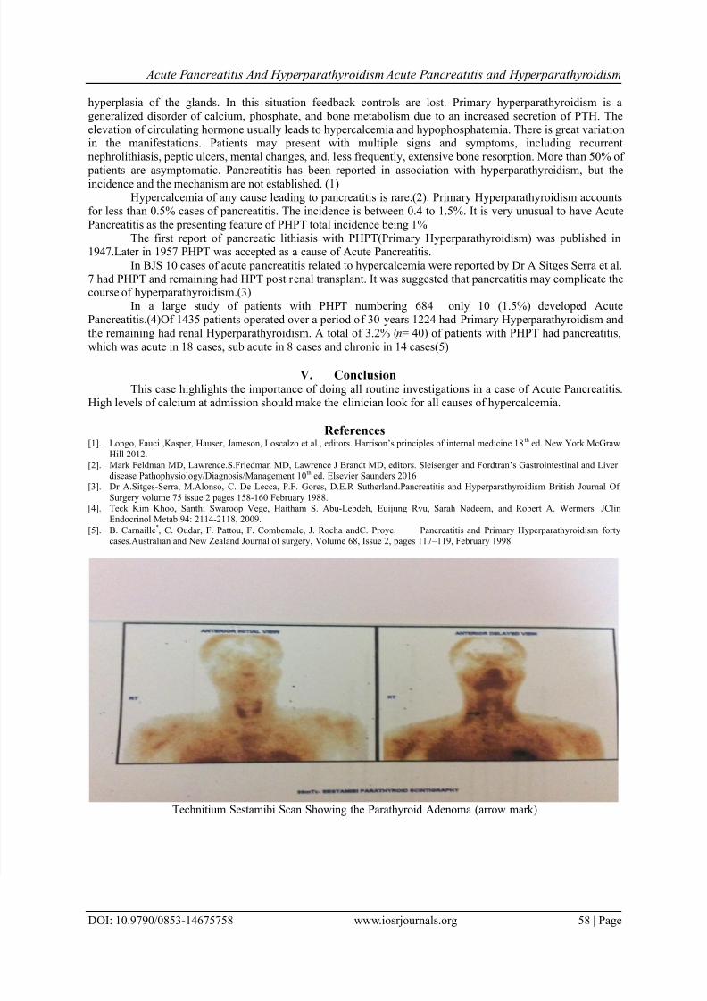
Cinacalcet was started. This drug works by increasing the Parathyroid gland chief cell calcium-sensingreceptor sensitivity to extracellular calcium, thereby decreasing PTH secretion.Parathyroid Scintigraphy was
done with injection of 15mCi of 99 Tc-MIBI and images obtained by SPECTGamma Camera which was s/o of
“Right Lower Parathyroid Gland Adenoma”. Pig tail catheter site continued to drain small quantities of pus. It got dislodged spontaneously.
However the site sealed off.Prior to discharge USG was done showing a large pseudocyst.Patient was advised toundergo MRCPto delineate the pancreatic duct. He was readmitted with fever and abdominal discomfort. Arepeat CT scan was s/o large pseudocysts in the abdomen.
Patient was lost to follow up.
IV. Discussion
Parathyroid glands are four in number and located posterior to the thyroid gland. They produce parathyroid hormone which maintains calcium homeostasis.(1) Hyperparathyroidism is caused by adenoma or
7/18/2019 Acute Pancreatitis and Hyperparathyroidism
http://slidepdf.com/reader/full/acute-pancreatitis-and-hyperparathyroidism 2/2
Acute Pancreatitis And Hyperparathyroidism Acute Pancreatitis and Hyperparathyroidism
DOI: 10.9790/0853-14675758 www.iosrjournals.org 58 | Page
hyperplasia of the glands. In this situation feedback controls are lost. Primary hyperparathyroidism is ageneralized disorder of calcium, phosphate, and bone metabolism due to an increased secretion of PTH. Theelevation of circulating hormone usually leads to hypercalcemia and hypophosphatemia. There is great variationin the manifestations. Patients may present with multiple signs and symptoms, including recurrentnephrolithiasis, peptic ulcers, mental changes, and, less frequently, extensive bone resorption. More than 50% of
patients are asymptomatic. Pancreatitis has been reported in association with hyperparathyroidism, but theincidence and the mechanism are not established. (1)Hypercalcemia of any cause leading to pancreatitis is rare.(2). Primary Hyperparathyroidism accounts
for less than 0.5% cases of pancreatitis. The incidence is between 0.4 to 1.5%. It is very unusual to have Acute
Pancreatitis as the presenting feature of PHPT total incidence being 1%The first report of pancreatic lithiasis with PHPT(Primary Hyperparathyroidism) was published in
1947.Later in 1957 PHPT was accepted as a cause of Acute Pancreatitis.
In BJS 10 cases of acute pancreatitis related to hypercalcemia were reported by Dr A Sitges Serra et al.7 had PHPT and remaining had HPT post renal transplant. It was suggested that pancreatitis may complicate thecourse of hyperparathyroidism.(3)
In a large study of patients with PHPT numbering 684 only 10 (1.5%) developed AcutePancreatitis.(4)Of 1435 patients operated over a period of 30 years 1224 had Primary Hyperparathyroidism andthe remaining had renal Hyperparathyroidism. A total of 3.2% (n= 40) of patients with PHPT had pancreatitis,
which was acute in 18 cases, sub acute in 8 cases and chronic in 14 cases(5)
V. Conclusion
This case highlights the importance of doing all routine investigations in a case of Acute Pancreatitis.High levels of calcium at admission should make the clinician look for all causes of hypercalcemia.
References[1].
Longo, Fauci ,Kasper, Hauser, Jameson, Loscalzo et al., editors. Harrison’s principles of internal medicine 18 th ed. New York McGraw
Hill 2012.
[2].
Mark Feldman MD, Lawrence.S.Friedman MD, Lawrence J Brandt MD, editors. Sleisenger and Fordtran’s Gastrointestinal and Liver
disease Pathophysiology/Diagnosis/Management 10th ed. Elsevier Saunders 2016
[3].
Dr A.Sitges-Serra, M.Alonso, C. De Lecca, P.F. Gores, D.E.R Sutherland.Pancreatitis and Hyperparathyroidism British Journal Of
Surgery volume 75 issue 2 pages 158-160 February 1988.
[4].
Teck Kim Khoo, Santhi Swaroop Vege, Haitham S. Abu-Lebdeh, Euijung Ryu, Sarah Nadeem, and Robert A. Wermers. JClin
Endocrinol Metab 94: 2114-2118, 2009.
[5].
B. Carnaille*, C. Oudar, F. Pattou, F. Combemale, J. Rocha andC. Proye. Pancreatitis and Primary Hyperparathyroidism forty
cases.Australian and New Zealand Journal of surgery, Volume 68, Issue 2, pages 117 – 119, February 1998.
Technitium Sestamibi Scan Showing the Parathyroid Adenoma (arrow mark)