Embed Size (px)
Citation preview
IGFBP-3 RIA
Radioimmunoassay for Quantitative Measurement of
Insulin-like Growth Factor Binding Protein-3
Product Code: IGF-R10
0 DE/CA40/00809/09
For in-vitro diagnostic use only! In the USA: For research use only!
Aspenhaustr. 25 • D-72770 Reutlingen / Germany
Phone: + 49 - (0) 7121 51484-0 • Fax: + 49 - (0) 7121 51484-10 E-mail: [email protected] • http://www.mediagnost.de
Offered in the US by ALPCO – 26G Keewaydin Drive, Salem, NH 03079 www.alpco.com Ph: (800) 592-5726
FOR RESEARCH U
SE ONLY
FOR REFERENCE U
SE ONLY
IGFBP-3 RIA Page 2 IGF-R10 e.22.02.11
Table of contents FEATURES 3 INTRODUCTION 3 INTENDED USE 7 PRECAUTIONS 8
General 8 Radioactivity 9
METHODOLOGY 10 Assay Characteristics and Validation 10 Clinical Validation 10 Validation for Research Purposes 10 Sample Handling and Storage 11
MATERIALS 12 Materials Provided 12 Required Materials Not Provided 13 Reagent Storage and Preparation 13 Sample Preparation 14
ASSAY PROCEDURE 15 Reduction of washing procedure 17
EVALUATION OF RESULTS 17 Establishing the Standard Curve: 17
EVALUATION OF SAMPLE CONCENTRATIONS 18 Concentration of controls 18
EXPECTED VALUES 19 LIMITATIONS 19 REFERENCES 21 Summary of the Assay 24
Offered in the US by ALPCO – 26G Keewaydin Drive, Salem, NH 03079 www.alpco.com Ph: (800) 592-5726
FOR RESEARCH U
SE ONLY
FOR REFERENCE U
SE ONLY
IGFBP-3 RIA Page 3 IGF-R10 e.22.02.11
FEATURES ♦ Quantitative determination of Insulin-like Growth Factor Binding
Protein-3 (IGFBP-3) ♦ Measures growth hormone (GH) dependent IGFBP-3 for
evaluating growth disorders ♦ Use of a high specific antiserum, therefore determination of all
clinically relevant fragments ♦ Stable plasma levels due to absence of circadian variation ♦ Integrates the GH secretory state over days ♦ A single measurement is highly informative for diagnosis of GH
deficiency or GH excess ♦ Superior to IGF-I measurement for diagnosis of GH deficiency
in young children ♦ Small sample requirement, thus ideal for paediatric patients
INTRODUCTION Insulin-like growth factors (IGF)-I and -II are bound to specific binding proteins (IGFBPs) in the circulation. To date, at least six binding proteins can be distinguished on the basis of their amino acid sequence. They are designated as IGFBP-1, IGFBP-2, ... IGPBP-6 (1). Lately the discovery of a new IGFBP-7 has been discussed (2). The predominating IGFBP in blood is IGFBP-3 which largely determines the total IGF-I and IGF-II concentration. In contrast to the other binding proteins, IGFBP-3 has the unique property to associate with an acid-labile non-binding subunit (ALS) after binding of either IGF-I or IGF-II (3-5). Most of the IGFBP-3 in plasma is present as the high molecular weight ternary complex, however, small amounts of free IGFBP-3 are also found (6,7). The development of specific radioimmunoassays for IGFBP-3, that also recognize the complete high molecular weight complex,
Offered in the US by ALPCO – 26G Keewaydin Drive, Salem, NH 03079 www.alpco.com Ph: (800) 592-5726
FOR RESEARCH U
SE ONLY
FOR REFERENCE U
SE ONLY
IGFBP-3 RIA Page 4 IGF-R10 e.22.02.11
provided new in-sights into its regulation (6-9). On the basis of these findings serum IGFBP-3 has proved to be an additional useful test in the repertoire of diagnostic tools for evaluation of growth disorders (7,8). Several factors besides GH influence IGFBP-3 levels: age including sexual development, nutrition, hypothyroidism, diabetes mellitus, liver function and kidney function. IGFBP-3 levels are decreased by malnutrition, although less than IGF-I, in hypothyroidism, in diabetes mellitus and in hepatic failure (6-8), but are increased in chronic renal failure (6,10,11). Measurement over 24 hours revealed constant circadian levels (12,13). For clinical practice, the most important regulatory factor is GH. Single IGFBP-3 measurements correlate significantly with the logarithm of the integrated spontaneous GH secretion (8,14). In patients with GH deficiency, IGFBP-3 levels are subnormal and increase gradually to within the normal range after several days of GH administration (7,8). The slow response to GH and constant circadian levels during chronic daily application of GH (13) suggest that IGFBP-3 reflects the GH secretory state over days. So far, IGF-I serum levels, determined by RIA, have been widely used in screening for GH defiency or acromegaly. However, several limitations are obvious: 1. The normal range of IGF-I is low in young children making
discrimination of subnormal levels difficult at that age. 2. A considerable number of children of small stature have, despite
normal GH secretion, IGF-I levels in the subnormal range. Therefore, the specificity and consequently the accuracy of the test for diagnosis of GH deficiency is limited.
Offered in the US by ALPCO – 26G Keewaydin Drive, Salem, NH 03079 www.alpco.com Ph: (800) 592-5726
FOR RESEARCH U
SE ONLY
FOR REFERENCE U
SE ONLY
IGFBP-3 RIA Page 5 IGF-R10 e.22.02.11
The major advantages of IGFBP-3 over IGF-I are: 1. No extraction step is required prior to measurement thus
improving test accuracy by simplifying the assay procedure. 2. The normal range in young children is comparatively high making
the detection of subnormal levels more reliable. 3. Patients with GH deficiency have subnormal IGFBP-3 levels. In
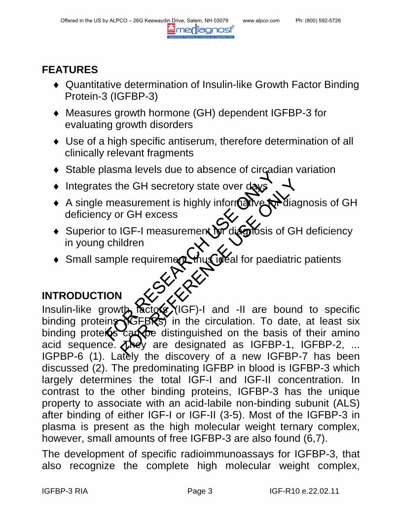
contrast, most of the small statured children with normal GH secretion have levels within the normal range (Figure 1).
Figure 1: Serum IGFBP-3 levels in patients with short stature without GH deficiency (SS:
constitutional delay of growth and adolescence, familial short stature, intra-uterine growth retardation) and in idiopathic or organic GH deficiency (GHD). The normal range is given by the 5th, 50th and 95th percentile.
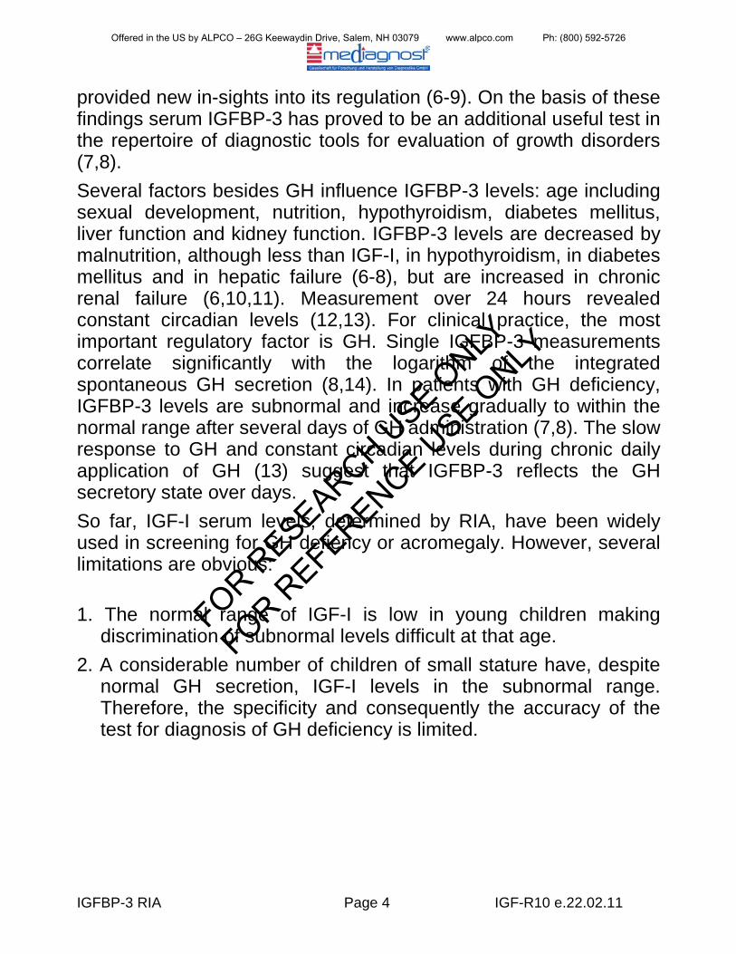
The separation of these two groups is easy. A single measurement of the IGFBP-3 concentration is sufficient for the diagnosis of GH deficiency with high accuracy (7,18). In small statured children IGFBP-3 levels rise to normal range within several days of GH administration and remain normal during continuous GH treatment (Figure 2).
Offered in the US by ALPCO – 26G Keewaydin Drive, Salem, NH 03079 www.alpco.com Ph: (800) 592-5726
FOR RESEARCH U
SE ONLY
FOR REFERENCE U
SE ONLY
IGFBP-3 RIA Page 6 IGF-R10 e.22.02.11
Figure 2: IGFBP-3 levels in GH deficient children before and during GH treatment. Because of the age-dependence, values are given as the mean of standard deviation scores (SDS).
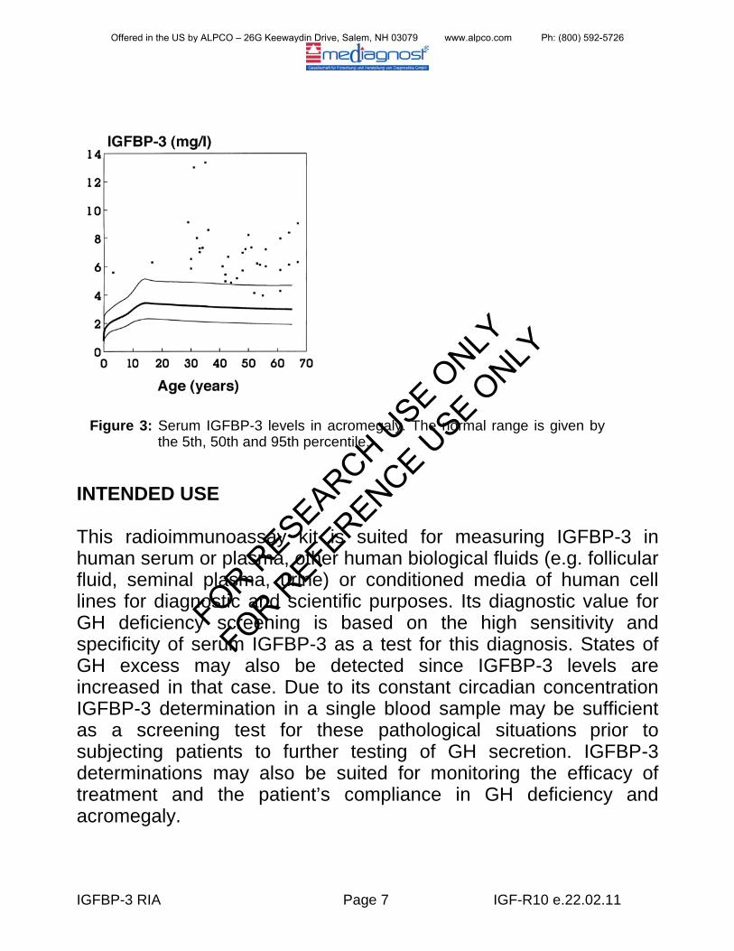
Therefore, serum IGFBP-3 measurements are also suited for evaluating the potential of a patient to respond to GH and for GH therapy monitoring (19). In other patients of severe short stature, e.g. Ullrich-Turner syndrome or Silver-Russell syndrome, IGFBP-3 levels were found normal (8) reflecting normal GH secretion. In normal tall children and adolescents without excessive GH secretion or in patients with Sotos syndrome, IGFBP-3 levels are normal or slightly increased. In contrast, children with pituitary gigantism or adults with acromegaly have clearly elevated levels (Figure 3) (6,15) that normalize on successful treatment. Therefore, IGFBP-3 is also a useful parameter for the detection of excessive GH secretion and monitoring therapy efficacy. In precocious puberty, IGFBP-3 levels are clearly increased by chronological age, whereas patients with premature thelarche have IGFBP-3 levels in the upper normal range (15).
Offered in the US by ALPCO – 26G Keewaydin Drive, Salem, NH 03079 www.alpco.com Ph: (800) 592-5726
FOR RESEARCH U
SE ONLY
FOR REFERENCE U
SE ONLY
IGFBP-3 RIA Page 7 IGF-R10 e.22.02.11
INTENDED USE
This radioimmunoassay kit is suited for measuring IGFBP-3 in human serum or plasma, other human biological fluids (e.g. follicular fluid, seminal plasma, urine) or conditioned media of human cell lines for diagnostic and scientific purposes. Its diagnostic value for GH deficiency screening is based on the high sensitivity and specificity of serum IGFBP-3 as a test for this diagnosis. States of GH excess may also be detected since IGFBP-3 levels are increased in that case. Due to its constant circadian concentration IGFBP-3 determination in a single blood sample may be sufficient as a screening test for these pathological situations prior to subjecting patients to further testing of GH secretion. IGFBP-3 determinations may also be suited for monitoring the efficacy of treatment and the patient’s compliance in GH deficiency and acromegaly.
Figure 3: Serum IGFBP-3 levels in acromegaly. The normal range is given by the 5th, 50th and 95th percentile.
Offered in the US by ALPCO – 26G Keewaydin Drive, Salem, NH 03079 www.alpco.com Ph: (800) 592-5726
FOR RESEARCH U
SE ONLY
FOR REFERENCE U
SE ONLY
IGFBP-3 RIA Page 8 IGF-R10 e.22.02.11
PRECAUTIONS General All reagents are for in vitro use only! In conducting the assay, follow strictly the test protocol. The acquisition, possession and use of the kit is subject to the regulations of the national nuclear regulatory authorities. Reagents with different lot numbers should not be mixed.
Reagents contain Sodium-Azide as preservative, however, highly diluted (0.02%). Sodium-Azide is very toxic, R-Phrases: 28, 32, 50/53 and S-Phrases 28, 45, 60, 61 must be considered. First aid procedures: Scin contact: Wash affected area thoroughly with water at least 15 minutes. Discard contaminated cloths and shoes. See a physician. Eye contact: In case of contact with eyes, rinse immediately with plenty of water at least 15 minutes. In order to assure an effectual rinsing spread the eyelids. See a physician.
Ingestion: If swallowed, wash out mouth throughly with water, provided that the person is conscious. Immediately see a physician. The handling of radioactive and potentially infectious material must comply with the following guidelines: The material should be stored and used in a special designated area. Do not eat, drink or smoke in these areas. Never pipette the materials with the mouth.
Avoid direct contact with these materials by wearing laboratory coats and disposable gloves. Spilled material must be wiped off immediately. Clean contaminated areas and equipment with a suitable detergent.
Offered in the US by ALPCO – 26G Keewaydin Drive, Salem, NH 03079 www.alpco.com Ph: (800) 592-5726
FOR RESEARCH U
SE ONLY
FOR REFERENCE U
SE ONLY
IGFBP-3 RIA Page 9 IGF-R10 e.22.02.11
Unused radioactive material and radioactive waste should be disposed according to the recommendations of the national regulatory authorities.
Radioactivity Before ordering or using radioactive materials, it is necessary to take the appropriate actions to ensure compliance with national regulations governing their use. Local rules in each establishment, which define actions and behaviour in the radioactivity working areas, should also be adhered to. The advice given here does not replace any local rules, instructions or training in the establishment, or advice from the radiation protection advisers. It is important to follow the code of good laboratory practice in addition to the specific precautions relating to the radionuclide I-125 used. Iodine-125 has a radioactive half-life T1/2 of 60 days and emits 35,5 keV gamma radiation, 27 – 32 keV x-rays and no beta radiation. Shielding is effective done by lead, first half value layer is 0.02 mm lead, reduction to 10 % is made by 0.2 mm. To reduce the radiation dose time spent handling radioactivity should be minimized (plan ahead), and distance from source of radiation should be maximized (doubling the distance from the source quarters the radiation dose). Formation of aerosols, e.g. by improper opening and mixing of vials or pipetting of solutions which may cause minute droplets of radioactivity become airborn, is a hazard and should be avoided. Solutions containing iodine should not be made acidic, because this might lead to the formation of volatile elemental iodine. As some iodo-compounds can penetrate rubber gloves, it is advisable to wear two pairs, or polyethylene gloves over rubber. For cleaning of contaminated areas or equipment, the Iodine-125 should be rendered chemically stable by using alkaline sodium thiosulphate solution together with paper or cellulose tissue.
Offered in the US by ALPCO – 26G Keewaydin Drive, Salem, NH 03079 www.alpco.com Ph: (800) 592-5726
FOR RESEARCH U
SE ONLY
FOR REFERENCE U
SE ONLY
IGFBP-3 RIA Page 10 IGF-R10 e.22.02.11
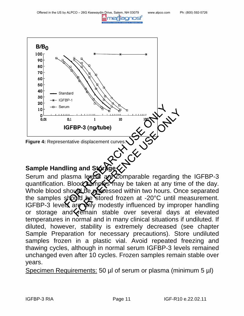
METHODOLOGY Assay Characteristics and Validation The radioimmunoassay for IGFBP-3 utilizes a specific high affinity polyclonal antibody for this protein. It recognizes quantitatively the complete IGFBP-3 complex and is unaffected by excess of IGF-l or IGF-II. Related molecules such as lGFBP-1 or lGFBP-2 show no cross-reaction in the assay (Figure 4) and the antibody is specific for primate IGFBP-3. The sensitivity of the assay is 0.06 ng/ml. Half-maximal displacement occurs at 6 ng/ml. The inter-assay variation coefficient has been found to be less than 9.0%, the intra-assay variation coefficient did not exceed 7.1%. The tracer is prepared by direct radioiodination of pure IGFBP-3 and standards refer to a stable derivative of IGFBP-3 having a molecular weight of 30.5 kDa determined by SDS-PAGE. The high sensitivity of the assay allows measurement of IGFBP-3 in small sample volumes which is limited by pipetting accuracy rather than the amount of IGFBP-3. Serum or plasma samples must be considerably diluted before measurement. No extraction step is required as with conventional IGF-I or IGF-II determinations.
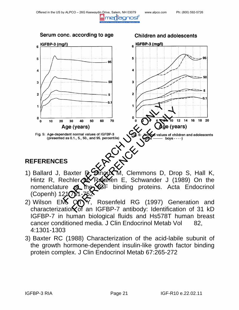
Clinical Validation Clinical validation was achieved by determination of IGFBP-3 levels in a large number of normal children and adults, normal short statured children without GH deficiency, girls with Ullrich-Turner Syndrome, children with Silver-Russell Syndrome, patients with GH deficiency, children with familial tall stature, Sotos-Syndrome, patients with acromegaly, children with premature thelarche and precocious puberty (Tab. 1, Figures 1, 2, 3, 5 and 6). Validation for Research Purposes The presence of IGFBP-3 was shown in a large number of follicular fluid samples, in seminal plasma, in urine, and also in conditioned media from various cell lines of human origin.
Offered in the US by ALPCO – 26G Keewaydin Drive, Salem, NH 03079 www.alpco.com Ph: (800) 592-5726
FOR RESEARCH U
SE ONLY
FOR REFERENCE U
SE ONLY
IGFBP-3 RIA Page 11 IGF-R10 e.22.02.11
Figure 4: Representative displacement curves.
Sample Handling and Storage Serum and plasma levels are comparable regarding the IGFBP-3 quantification. Blood samples may be taken at any time of the day. Whole blood should be processed within two hours. Once separated the samples should be stored frozen at -20°C until measurement. IGFBP-3 levels are only modestly influenced by improper handling or storage and remain stable over several days at elevated temperatures in normal and in many clinical situations if undiluted. If diluted, however, stability is extremely decreased (see chapter Sample Preparation for necessary precautions). Store undiluted samples frozen in a plastic vial. Avoid repeated freezing and thawing cycles, although in normal serum IGFBP-3 levels remained unchanged even after 10 cycles. Frozen samples remain stable over years. Specimen Requirements: 50 µl of serum or plasma (minimum 5 µl)
Offered in the US by ALPCO – 26G Keewaydin Drive, Salem, NH 03079 www.alpco.com Ph: (800) 592-5726
FOR RESEARCH U
SE ONLY
FOR REFERENCE U
SE ONLY
IGFBP-3 RIA Page 12 IGF-R10 e.22.02.11
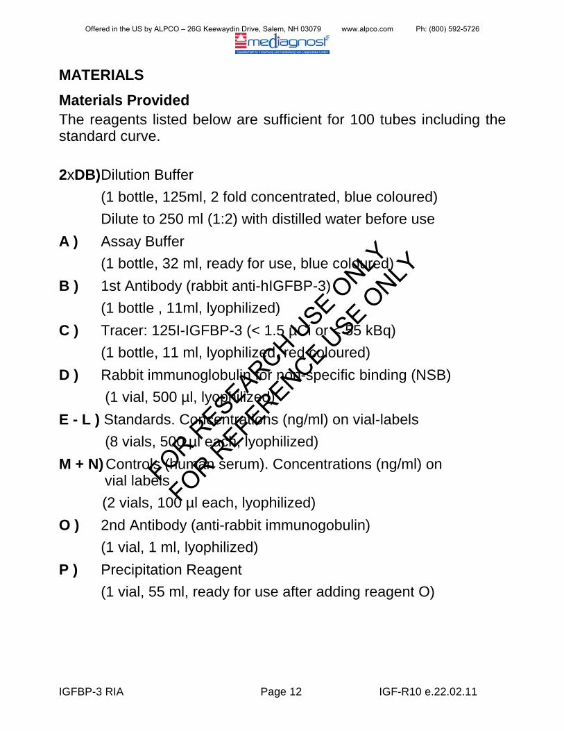
MATERIALS Materials Provided The reagents listed below are sufficient for 100 tubes including the standard curve. 2xDB) Dilution Buffer (1 bottle, 125ml, 2 fold concentrated, blue coloured) Dilute to 250 ml (1:2) with distilled water before use A ) Assay Buffer (1 bottle, 32 ml, ready for use, blue coloured) B ) 1st Antibody (rabbit anti-hIGFBP-3) (1 bottle , 11ml, lyophilized) C ) Tracer: 125I-IGFBP-3 (< 1.5 µCi or < 55 kBq) (1 bottle, 11 ml, lyophilized, red coloured) D ) Rabbit immunoglobulin for non-specific binding (NSB) (1 vial, 500 µl, lyophilized) E - L ) Standards. Concentrations (ng/ml) on vial-labels (8 vials, 500 µl each, lyophilized) M + N) Controls (human serum). Concentrations (ng/ml) on
vial labels (2 vials, 100 µl each, lyophilized) O ) 2nd Antibody (anti-rabbit immunogobulin) (1 vial, 1 ml, lyophilized) P ) Precipitation Reagent (1 vial, 55 ml, ready for use after adding reagent O)
Offered in the US by ALPCO – 26G Keewaydin Drive, Salem, NH 03079 www.alpco.com Ph: (800) 592-5726
FOR RESEARCH U
SE ONLY
FOR REFERENCE U
SE ONLY
IGFBP-3 RIA Page 13 IGF-R10 e.22.02.11
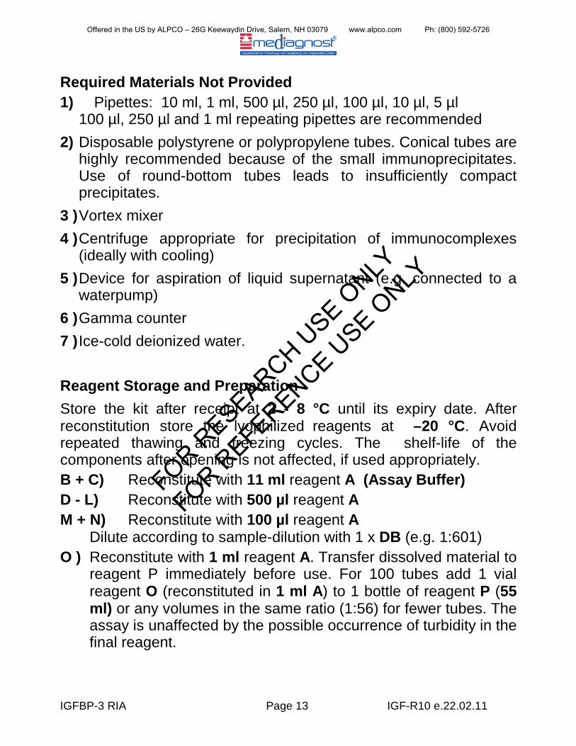
Required Materials Not Provided 1) Pipettes: 10 ml, 1 ml, 500 µl, 250 µl, 100 µl, 10 µl, 5 µl
100 µl, 250 µl and 1 ml repeating pipettes are recommended 2) Disposable polystyrene or polypropylene tubes. Conical tubes are
highly recommended because of the small immunoprecipitates. Use of round-bottom tubes leads to insufficiently compact precipitates.
3 ) Vortex mixer 4 ) Centrifuge appropriate for precipitation of immunocomplexes
(ideally with cooling) 5 ) Device for aspiration of liquid supernatant (e.g. connected to a
waterpump) 6 ) Gamma counter 7 ) Ice-cold deionized water.
Reagent Storage and Preparation Store the kit after receipt at 2 - 8 °C until its expiry date. After reconstitution store the lyophilized reagents at –20 °C. Avoid repeated thawing and freezing cycles. The shelf-life of the components after opening is not affected, if used appropriately. B + C) Reconstitute with 11 ml reagent A (Assay Buffer) D - L) Reconstitute with 500 µl reagent A M + N) Reconstitute with 100 µl reagent A
Dilute according to sample-dilution with 1 x DB (e.g. 1:601) O ) Reconstitute with 1 ml reagent A. Transfer dissolved material to
reagent P immediately before use. For 100 tubes add 1 vial reagent O (reconstituted in 1 ml A) to 1 bottle of reagent P (55 ml) or any volumes in the same ratio (1:56) for fewer tubes. The assay is unaffected by the possible occurrence of turbidity in the final reagent.
Offered in the US by ALPCO – 26G Keewaydin Drive, Salem, NH 03079 www.alpco.com Ph: (800) 592-5726
FOR RESEARCH U
SE ONLY
FOR REFERENCE U
SE ONLY

IGFBP-3 RIA Page 14 IGF-R10 e.22.02.11
Ensure that lyophilized materials are completely dissolved on reconstitution. It is recommended to keep reconstituted reagents at room temperature for half an hour and then to mix them with a Vortexmixer.This is important in particular for the controls M and N!
Sample Preparation Serum or plasma samples should be diluted 1:300 - 1:1000-fold with Dilution Buffer (1 x DB) prior to measurement depending on the expected values. Usually a dilution of 1:600 is appropriate. Important: Because IGFBP-3 is not stable in diluted solutions, please use only, chilled, preferably ice-cold Dilution Buffer DB. The time interval between the sample dilution and incubation should be as short as possible, i.e. the diluted samples should be processed fast as can. Example
IGF-I determination: If you want to determine both the IGF-I concentration and the IGFBP-3 concentration in a sample, use one dilution of the sample for both determinations (provided that IGF-I is determined with the mediagnost IGF-I RIA-Kit IGF-R20):
: Mix 50 µl serum or plasma with 2.5 ml Dilution Buffer (DB, dilution factor: 1:51). Mix 50 µl of this pre-dilution with 500 µl of DB (→1:561). Alternatively, 5 µl of sample may be diluted with 3 ml DB (→1:601). 100 µl of this dilution can be used in the assay.
1) Dilute the samples according to the IGF-I working instruction with acidic
2) For IGFBP-3 determination transfer (without further dilution) 2 x 25µl (double measurement) of the acidic 1:101-dilution in two RIA-tubes. Add 75 µl
Dilution Buffer DB (pH = 2.1) 1:101 (e.g. 10 µl serum + 1 ml DB2.1)
neutral
It is recommended to continue to work on the IGFBP-3 samples at first, because IGFBP-3 is diluted solutions relatively unstable.
IGFBP-3 Dilution Buffer (DB, pH 7.4) to each tube (→neutralisation of the samples) e.g. with a multistep pipette. Dilution factor of the sample: 1:404.
Offered in the US by ALPCO – 26G Keewaydin Drive, Salem, NH 03079 www.alpco.com Ph: (800) 592-5726
FOR RESEARCH U
SE ONLY
FOR REFERENCE U
SE ONLY
IGFBP-3 RIA Page 15 IGF-R10 e.22.02.11
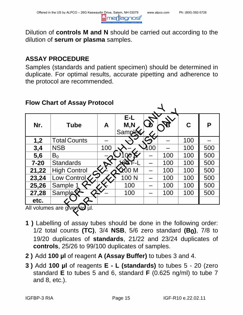
Dilution of controls M and N should be carried out according to the dilution of serum or plasma samples. ASSAY PROCEDURE Samples (standards and patient specimen) should be determined in duplicate. For optimal results, accurate pipetting and adherence to the protocol are recommended.
Flow Chart of Assay Protocol
Nr. Tube A E-L M,N
Samples D B C P
1,2 Total Counts – – – – 100 – 3,4 NSB 100 – 100 – 100 500 5,6 B0 – 100 E – 100 100 500
7-20 Standards – 100 F-L – 100 100 500 21,22 High Control – 100 M – 100 100 500 23,24 Low Control – 100 N – 100 100 500 25,26 Sample 1 – 100 – 100 100 500 27,28 Sample 2 – 100 – 100 100 500 etc.
All volumes are given as µl. 1 ) Labelling of assay tubes should be done in the following order:
1/2 total counts (TC), 3/4 NSB, 5/6 zero standard (B0), 7/8 to 19/20 duplicates of standards, 21/22 and 23/24 duplicates of controls, 25/26 to 99/100 duplicates of samples.
2 ) Add 100 µl of reagent A (Assay Buffer) to tubes 3 and 4. 3 ) Add 100 µl of reagents E - L (standards) to tubes 5 - 20 (zero
standard E to tubes 5 and 6, standard F (0.625 ng/ml) to tube 7 and 8, etc.).
Offered in the US by ALPCO – 26G Keewaydin Drive, Salem, NH 03079 www.alpco.com Ph: (800) 592-5726
FOR RESEARCH U
SE ONLY
FOR REFERENCE U
SE ONLY
IGFBP-3 RIA Page 16 IGF-R10 e.22.02.11
4 ) Add 100 µl of diluted reagent M (high control) to tubes 21 and 22 and diluted reagent N (low control) to tubes 23 and 24.
5 ) Add 100 µl of diluted samples to tubes 25 and 26, etc. 6 ) Add 100 µl reagent D (NSB) to tubes 3 and 4. 7 ) Add 100 µl reagent B (1st Antibody) to each tube beginning
with tube 5. 8 ) Add 100 µl reagent C (tracer) to all tubes9 ) Mark tubes 1 and 2 (total counts), remove or seal with a
stopper.
.
10) Mix tubes with a vortex mixer. 11) Incubate tubes at 2 - 8 °C for 2 days. Incubation for a longer
period (e.g. over the weekend) has no negative effect on the results. Incubation for a shorter period (e.g. overnight) leads to a weaker bondage resulting in a slight loss of sensitivity, irrelevant for most routine measurements.
12) Add 500 µl reagent P (after addition of reagent O !) beginning with tube 3. The reagent-mix should be cold (2 - 8 °C).
13) Mix tubes with a vortex mixer. 14) Incubate tubes at 2 - 8 °C for 1 hour. 15) Centrifuge all tubes except tubes 1 and 2 at least at 2000 x g
for 20 min (if possible, carry out the centrifugation at a temperature of 2 - 8 °C).
16) Decant or aspirate the supernatant (except tubes 1 and 2!). The remaining supernatant should not be higher than 2 mm above the precipitate. Take care that the precipitate remains intact.
17) Add 1 ml ice-cold distilled water. This should be done carefully to avoid destroying the precipitate. Do not mix again!
18) Centrifuge again at least at 2000 x g for 5 min. 19) Decant or aspirate the supernatant (except tubes 1 and 2). 20) Count the activity of the tubes (including tubes 1 and 2) for 1 to
3 min.
Offered in the US by ALPCO – 26G Keewaydin Drive, Salem, NH 03079 www.alpco.com Ph: (800) 592-5726
FOR RESEARCH U
SE ONLY
FOR REFERENCE U
SE ONLY
IGFBP-3 RIA Page 17 IGF-R10 e.22.02.11
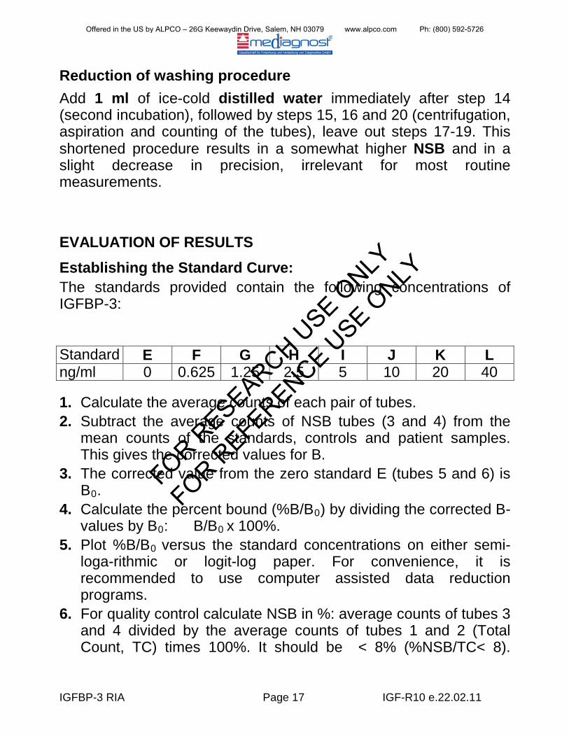
Reduction of washing procedure Add 1 ml of ice-cold distilled water immediately after step 14 (second incubation), followed by steps 15, 16 and 20 (centrifugation, aspiration and counting of the tubes), leave out steps 17-19. This shortened procedure results in a somewhat higher NSB and in a slight decrease in precision, irrelevant for most routine measurements.
EVALUATION OF RESULTS Establishing the Standard Curve: The standards provided contain the following concentrations of IGFBP-3:
Standard E F G H I J K L ng/ml 0 0.625 1.25 2.5 5 10 20 40 1. Calculate the average counts of each pair of tubes. 2. Subtract the average counts of NSB tubes (3 and 4) from the
mean counts of the standards, controls and patient samples. This gives the corrected values for B.
3. The corrected value from the zero standard E (tubes 5 and 6) is B0
4. Calculate the percent bound (%B/B.
0) by dividing the corrected B-values by B0: B/B0
5. Plot %B/Bx 100%.
0
6. For quality control calculate NSB in %: average counts of tubes 3 and 4 divided by the average counts of tubes 1 and 2 (Total Count, TC) times 100%. It should be < 8% (%NSB/TC< 8).
versus the standard concentrations on either semi-loga-rithmic or logit-log paper. For convenience, it is recommended to use computer assisted data reduction programs.
Offered in the US by ALPCO – 26G Keewaydin Drive, Salem, NH 03079 www.alpco.com Ph: (800) 592-5726
FOR RESEARCH U
SE ONLY
FOR REFERENCE U
SE ONLY
IGFBP-3 RIA Page 18 IGF-R10 e.22.02.11
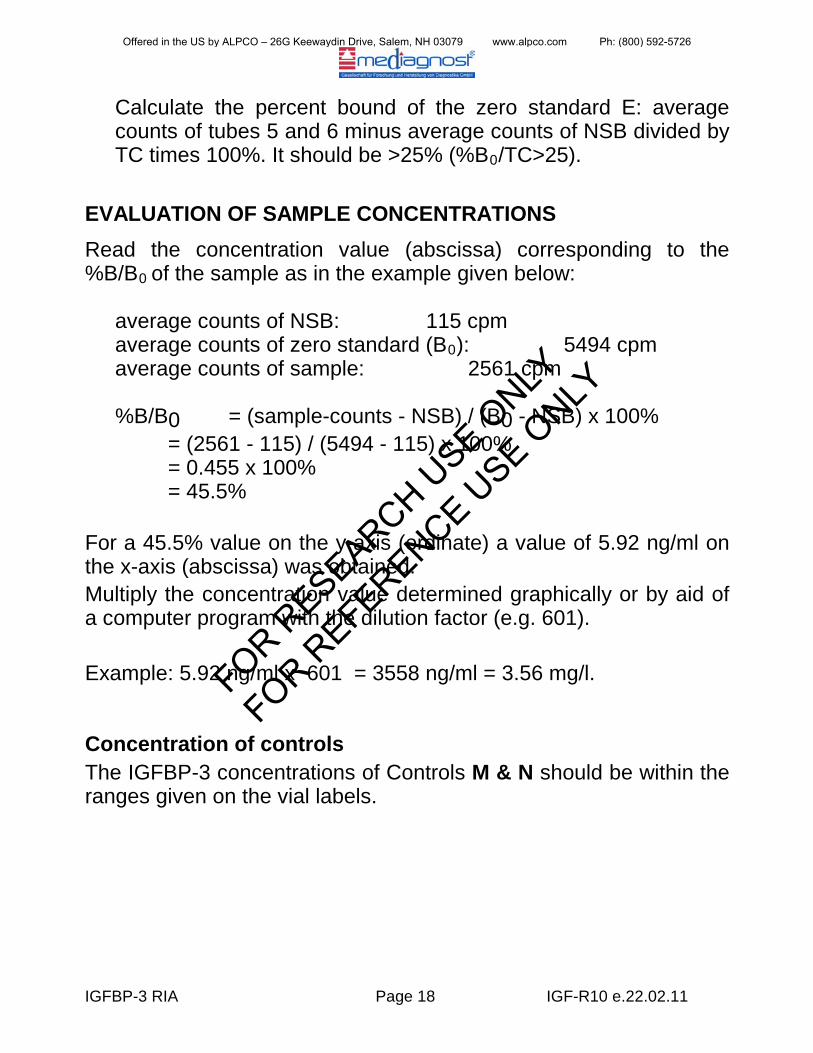
Calculate the percent bound of the zero standard E: average counts of tubes 5 and 6 minus average counts of NSB divided by TC times 100%. It should be >25% (%B0
/TC>25).
EVALUATION OF SAMPLE CONCENTRATIONS Read the concentration value (abscissa) corresponding to the %B/B0
of the sample as in the example given below:
average counts of NSB: 115 cpm average counts of zero standard (B0 average counts of sample: 2561 cpm
): 5494 cpm
%B/B0 = (sample-counts - NSB) / (B0 - NSB) x 100% = (2561 - 115) / (5494 - 115) x 100% = 0.455 x 100% = 45.5% For a 45.5% value on the y-axis (ordinate) a value of 5.92 ng/ml on the x-axis (abscissa) was obtained. Multiply the concentration value determined graphically or by aid of a computer program with the dilution factor (e.g. 601). Example: 5.92 ng/ml x 601 = 3558 ng/ml = 3.56 mg/l.
Concentration of controls The IGFBP-3 concentrations of Controls M & N should be within the ranges given on the vial labels.
Offered in the US by ALPCO – 26G Keewaydin Drive, Salem, NH 03079 www.alpco.com Ph: (800) 592-5726
FOR RESEARCH U
SE ONLY
FOR REFERENCE U
SE ONLY
IGFBP-3 RIA Page 19 IGF-R10 e.22.02.11
EXPECTED VALUES IGFBP-3 levels are strongly age-dependent in children, less so in adults. The normal ranges in various age-groups which were log-normally distributed are given in Table 1 by the percentiles. A graphic presentation is shown in Figures 5 and 6. It is recommended for each laboratory to establish its own normal range.
LIMITATIONS IGFBP-3 levels are strongly dependent on GH secretion. However, a number of factors influence its plasma concentration and should be taken into account for appropriate interpretation. Plasma levels decrease during fasting (more than 1 day), in malnutrition, malabsorption, cachexia, impaired hepatic function, hypothyroidism, and diabetes mellitus. They may also be decreased in chronic inflammatory disease and malignancy. Levels are increased in states of impaired renal function and precocious puberty. In clinical situations with hyperprolactinemia or in patients with craniopharyngeoma, normal levels may be observed despite GH deficiency. In certain physiological (e.g. pregnancy) and pathological states, IGFBP-3 may be degraded to smaller molecular size compounds (16,17) by specific proteases which affect IGFBP patterns seen in Western ligand blotting but have little influence on the outcome of RIA determinations.
Offered in the US by ALPCO – 26G Keewaydin Drive, Salem, NH 03079 www.alpco.com Ph: (800) 592-5726
FOR RESEARCH U
SE ONLY
FOR REFERENCE U
SE ONLY
IGFBP-3 RIA Page 20 IGF-R10 e.22.02.11
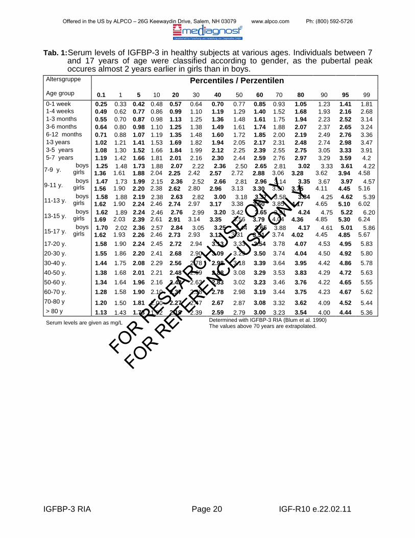
Tab. 1: Serum levels of IGFBP-3 in healthy subjects at various ages. Individuals between 7 and 17 years of age were classified according to gender, as the pubertal peak occures almost 2 years earlier in girls than in boys.
Altersgruppe Percentiles / Perzentilen
Age group 0.1 1 5 10 20 30 40 50 60 70 80 90 95 99 0-1 week 0.25 0.33 0.42 0.48 0.57 0.64 0.70 0.77 0.85 0.93 1.05 1.23 1.41 1.81 1-4 weeks 0.49 0.62 0.77 0.86 0.99 1.10 1.19 1.29 1.40 1.52 1.68 1.93 2.16 2.68 1-3 months 0.55 0.70 0.87 0.98 1.13 1.25 1.36 1.48 1.61 1.75 1.94 2.23 2.52 3.14 3-6 months 0.64 0.80 0.98 1.10 1.25 1.38 1.49 1.61 1.74 1.88 2.07 2.37 2.65 3.24 6-12 months 0.71 0.88 1.07 1.19 1.35 1.48 1.60 1.72 1.85 2.00 2.19 2.49 2.76 3.36 1-3 years 1.02 1.21 1.41 1.53 1.69 1.82 1.94 2.05 2.17 2.31 2.48 2.74 2.98 3.47 3-5 years 1.08 1.30 1.52 1.66 1.84 1.99 2.12 2.25 2.39 2.55 2.75 3.05 3.33 3.91 5-7 years 1.19 1.42 1.66 1.81 2.01 2.16 2.30 2.44 2.59 2.76 2.97 3.29 3.59 4.2
7-9 y. boys
girls 1.25
1.36 1.48
1.61 1.73 1.88
1.88 2.04
2.07 2.25
2.22 2.42
2.36 2.57
2.50 2.72
2.65 2.88
2.81 3.06
3.02 3.28
3.33 3.62
3.61 3.94
4.22 4.58
9-11 y. boys
girls 1.47
1.56 1.73
1.90 1.99 2.20
2.15 2.38
2.36 2.62
2.52 2.80
2.66 2.96
2.81 3.13
2.96 3.30
3.14 3.50
3.35 3.75
3.67 4.11
3.97 4.45
4.57 5.16
11-13 y. boys
girls 1.58
1.62 1.88
1.90 2.19 2.24
2.38 2.46
2.63 2.74
2.82 2.97
3.00 3.17
3.18 3.38
3.37 3.60
3.58 3.85
3.84 4.17
4.25 4.65
4.62 5.10
5.39 6.02
13-15 y. boys
girls 1.62
1.69 1.89
2.03 2.24 2.39
2.46 2.61
2.76 2.91
2.99 3.14
3.20 3.35
3.42 3.56
3.65 3.79
3.91 4.04
4.24 4.36
4.75 4.85
5.22 5.30
6.20 6.24
15-17 y. boys
girls 1.70
1.62 2.02
1.93 2.36 2.26
2.57 2.46
2.84 2.73
3.05 2.93
3.25 3.12
3.44 3.31
3.65 3.51
3.88 3.74
4.17 4.02
4.61 4.45
5.01 4.85
5.86 5.67
17-20 y. 1.58 1.90 2.24 2.45 2.72 2.94 3.13 3.33 3.54 3.78 4.07 4.53 4.95 5.83 20-30 y. 1.55 1.86 2.20 2.41 2.68 2.90 3.09 3.29 3.50 3.74 4.04 4.50 4.92 5.80 30-40 y. 1.44 1.75 2.08 2.29 2.56 2.78 2.98 3.18 3.39 3.64 3.95 4.42 4.86 5.78 40-50 y. 1.38 1.68 2.01 2.21 2.48 2.69 2.88 3.08 3.29 3.53 3.83 4.29 4.72 5.63 50-60 y. 1.34 1.64 1.96 2.16 2.42 2.63 2.83 3.02 3.23 3.46 3.76 4.22 4.65 5.55 60-70 y. 1.28 1.58 1.90 2.10 2.37 2.58 2.78 2.98 3.19 3.44 3.75 4.23 4.67 5.62 70-80 y 1.20 1.50 1.81 2.00 2.27 2.47 2.67 2.87 3.08 3.32 3.62 4.09 4.52 5.44 > 80 y 1.13 1.43 1.73 1.92 2.19 2.39 2.59 2.79 3.00 3.23 3.54 4.00 4.44 5.36 Serum levels are given as mg/L
Determined with IGFBP-3 RIA (Blum et al. 1990) The values above 70 years are extrapolated.
Offered in the US by ALPCO – 26G Keewaydin Drive, Salem, NH 03079 www.alpco.com Ph: (800) 592-5726
FOR RESEARCH U
SE ONLY
FOR REFERENCE U
SE ONLY
IGFBP-3 RIA Page 21 IGF-R10 e.22.02.11
REFERENCES
1) Ballard J, Baxter R, Binoux M, Clemmons D, Drop S, Hall K, Hintz R, Rechler M, Rutanen E, Schwander J (1989) On the nomenclature of the IGF binding proteins. Acta Endocrinol (Copenh) 121:751-752
2) Wilson EM, Oh Y, Rosenfeld RG (1997) Generation and characterization of an IGFBP-7 antibody: Identification of 31 kD IGFBP-7 in human biological fluids and Hs578T human breast cancer conditioned media. J Clin Endocrinol Metab Vol 82, 4:1301-1303
3) Baxter RC (1988) Characterization of the acid-labile subunit of the growth hormone-dependent insulin-like growth factor binding protein complex. J Clin Endocrinol Metab 67:265-272
Offered in the US by ALPCO – 26G Keewaydin Drive, Salem, NH 03079 www.alpco.com Ph: (800) 592-5726
FOR RESEARCH U
SE ONLY
FOR REFERENCE U
SE ONLY
IGFBP-3 RIA Page 22 IGF-R10 e.22.02.11
4) Baxter RC, Martin JL (1989) Structure of the Mr 140,000 growth
hormone dependent insulin-like growth factor binding protein complex: determinati-on by reconstitution and affinity-labeling. Proc Natl Acad Sci USA 86:6898-6902
5) Holman SR, Baxter RC (1996) Insulin-like growth factor-binding protein-3: factors affecting binary and ternary complex formation. Growth Regulation 6: 42-47.
6) Baxter RC, Martin J (1986): Radioimmunassay of growth hormone-dependent insulin-like growth factor binding protein in human plasma. J Clin Invest 78:1504-1512
7) Blum WF, Ranke MB, Kietzmann K, Gauggel E, Zeissel HJ, Bierich JR (1990) A specific radioimmunoassay for the growth hormone (GH)-dependent somatomedin-binding protein: its use for diagnosis of GH deficiency. J Clin Endocrinol Metab 70:1292-1298
8) Blum WF, Ranke MB (1990) Use of insulin-like growth factor binding protein 3 for the evaluation of growth disorders. Horm Res 34 (Suppl):31-37
9) Blum WF (1993) Insulin-like growth factor-binding protein 3: Entwicklung eines Radioimmunoassays und Untersuchungen zur klinischen Bedeutung. Habilitationsschrift, Tübingen.
10) Lee PDK, Hintz RL, Sperry JB, Baxter RC, Powell DR (1989) IGF-binding proteins in growth-retarded children with chronic renal failure. Pediatr Res 26:308-315
11) Blum WF, Ranke MB, Kietzmann K, Tönshoff B, Mehls O (1989) Excess of IGF-binding proteins in chronic renal failure: evidence for relative GH resistence and inhibition of somatomedin activity. In: Drop SLS, Hintz RL (eds) Insulin-like Growth Factor Binding Proteins. Excerpta Medica, Amsterdam, pp 93-101
12) Baxter RC, Cowell CT (1987) Diurnal rhythm of growth hormone-indepen-dent binding protein for insulin-like growth factors in human plasma. J Clin Endocrinol Metab 65:432-440
Offered in the US by ALPCO – 26G Keewaydin Drive, Salem, NH 03079 www.alpco.com Ph: (800) 592-5726
FOR RESEARCH U
SE ONLY
FOR REFERENCE U
SE ONLY
IGFBP-3 RIA Page 23 IGF-R10 e.22.02.11
13) Jorgensen JOL, Blum WF, Moller N, Ranke MB, Christiansen
JS (1990) Circadian patterns of serum insulin-like growth factor (IGF)-II and IGF-binding protein 3 in growth hormone deficient patients and age- and sex-matched normal subjects. Acta Endocrinol (Copenh.) 123:257-262
14) Blum WF, Albertsson-Wikland K, Rosberg S, Jorgensen JOL, Ranke MB (1990) Insulin-like growth factor binding protein 3 (IGFBP-3) reflects spontaneous growth hormone (GH) secretion. Horm Res 33 (Suppl 3): 3(Abstract)
15) Blum WF, Ranke MB (1990) Insulin-like growth factor-binding proteins (IGFBPs) with special reference to IGFBP-3. Acta Paediatr Scand (Suppl) 367:55-62
16) Giudice LC, Farrell EM, Pham H, Lamson G, Rosenfeld RG (1990) Insulin-like growth factor binding proteins in maternal serum throughout gestation and in the puerperium: effects of a pregnancy-associated serum protease activity. J Clin Endocrinol Metab 71:806 816
17) Hossenlopp P, Segovia B, Lassarre C, Roghani M, Bredon M, Binoux M (1990) Evidence of enzymatic degradation of insulin-like growth factor-binding proteins in the 150k complex during pregnancy. J Clin Endocrinol Metab 71:797-805
18) Ranke MB, Schweizer R, Elmlinger MW, Weber K, Binder G, Schwarze CP, Wollmann HA (2000) Significance of Basal IGF-I, IGFBP-3 and IGFBP-2 Measrurements in the diagnostics of short stature in children. Horm Res 2000;54:60-68
19) Ranke MB, Schweizer R, Elmlinger MW, Weber K, Binder G, Schwarze CP, Wollmann HA (2001) Relevance of IGF-I, IGFBP-3, and IGFBP-2 Measurements during GH treatment of GH-deficient and non-GH-deficient children and adolescents. Horm Res 2001;55:115-124
Offered in the US by ALPCO – 26G Keewaydin Drive, Salem, NH 03079 www.alpco.com Ph: (800) 592-5726
FOR RESEARCH U
SE ONLY
FOR REFERENCE U
SE ONLY
IGFBP-3 RIA Page 24 IGF-R10 e.22.02.11
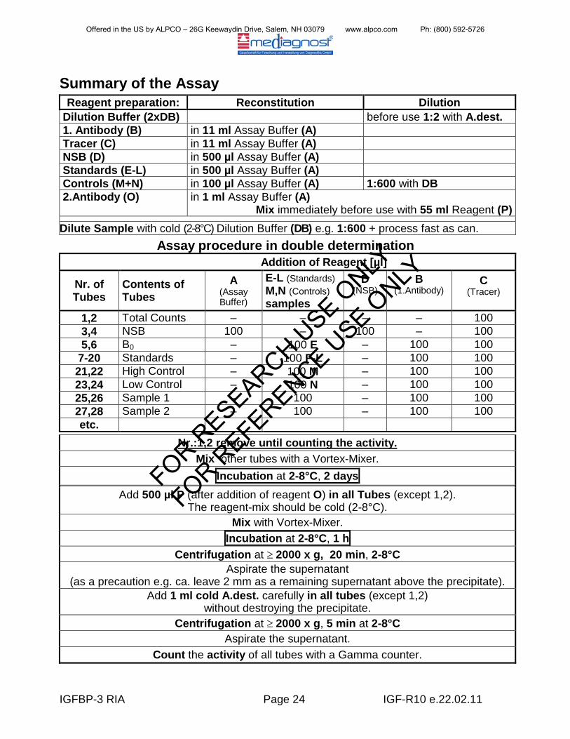
Summary of the Assay Reagent preparation: Reconstitution Dilution
Dilution Buffer (2xDB) before use 1:2 with A.dest. 1. Antibody (B) in 11 ml Assay Buffer (A) Tracer (C) in 11 ml Assay Buffer (A) NSB (D) in 500 µl Assay Buffer (A) Standards (E-L) in 500 µl Assay Buffer (A) Controls (M+N) in 100 µl Assay Buffer (A) 1:600 with DB 2.Antibody (O)
in 1 ml Assay Buffer (A) Mix immediately before use with 55 ml Reagent (P)
Dilute Sample with cold (2-8°C) Dilution Buffer (DB) e.g. 1:600 + process fast as can. Assay procedure in double determination
Addition of Reagent [µl]
Nr. of Tubes
Contents of Tubes
A (Assay Buffer)
E-L (Standards) M,N (Controls) samples
D (NSB)
B (1.Antibody)
C (Tracer)
1,2 Total Counts – – – – 100 3,4 NSB 100 – 100 – 100 5,6 B0 – 100 E – 100 100
7-20 Standards – 100 F-L – 100 100 21,22 High Control – 100 M – 100 100 23,24 Low Control – 100 N – 100 100 25,26 Sample 1 – 100 – 100 100 27,28 Sample 2 – 100 – 100 100 etc.
Nr.:1,2 remove until counting the activity. Mix other tubes with a Vortex-Mixer.
Incubation at 2-8°C, 2 days Add 500 µl P (after addition of reagent O) in all Tubes (except 1,2).
The reagent-mix should be cold (2-8°C). Mix with Vortex-Mixer.
Incubation at 2-8°C, 1 h Centrifugation at ≥ 2000 x g, 20 min, 2-8°C
Aspirate the supernatant (as a precaution e.g. ca. leave 2 mm as a remaining supernatant above the precipitate).
Add 1 ml cold A.dest. carefully in all tubes (except 1,2) without destroying the precipitate.
Centrifugation at ≥ 2000 x g, 5 min at 2-8°C Aspirate the supernatant.
Count the activity of all tubes with a Gamma counter.
Offered in the US by ALPCO – 26G Keewaydin Drive, Salem, NH 03079 www.alpco.com Ph: (800) 592-5726
FOR RESEARCH U
SE ONLY
FOR REFERENCE U
SE ONLY