Embed Size (px)
Citation preview
Original Research DOI: 10.5958/2394-6792.2015.00007.1
Indian Journal of Pathology and Oncology, July – September 2015;2(3);141-149 141
Histopathological study of soft tissue tumors: A three year
experience in tertiary care centre
Preeti Rihal Chakrabarti1,*, Suvadip Chakrabarti2, Ajita Pandit3,
Purti Agrawal4, Shilpi Dosi5, Mukul Raj Jain6
1,4,5,6Assistant Professor, Dept. of Pathology, 2Senior Resident Dept. of Surgical Oncology,
Sri Aurobindo Medical College and PG Institute, Indore, Madhya Pradesh 3Ex Professor Pathology, D Y Patil School of Medicine, Navi Mumbai
*Corresponding Author: Email: [email protected]
ABSTRACT Background: Soft tissue sarcomas compared with carcinomas and other neoplasms are relatively rare and constitute less than
1% of all the cancer. Soft tissue sarcomas may occur anywhere in the body but most arise from large muscle of extremeties, the
chest wall, the mediastinum and the retroperitoneum.
Aims and objectives: To study the macroscopic and microscopic pathology of benign and malignant soft tissue tumour and there
clinicopathological correlation.
Materials and methods: The study was conducted in Dr .D .Y Patil School of Medicine, Navi Mumbai, from 1st of January 2008
to December 2010. A Cross sectional study of 150 cases of soft tissue tumors were carried out in details. A pretested proforma
was used to classify each tumor and details like age, sex, clinical features, gross and microscopic findings were noted.
Results: The most common soft tissue tumor in the study were benign tumors (140 cases), followed by malignant (9 cases) and
one case was reported as borderline. Lipomas were the most common benign tumors accounting for 49 out of 140 cases. Benign
smooth muscle tumors were the next most common tumor accounting for 44 cases.Benign vascular and benign peripheral nerve
sheath tumor accounted for 19 cases in each category. Of 19 cases of benign vascular tumor, 16 cases were of haemangioma, 3
cases were of angiofibroma. Of 19 benign peripheral nerve sheethtumor, 12 cases were reported as neurofibroma and 7
schwannomas. Of 5 cases of fibrohistiocytictumor, 3 cases were benign, 1 case of borderline (Dermatofibrosarcoma
protuberance) and 1 case of malignant fibrous histiocytoma (MFH). Of 7 cases of Fibrous tumor, 6 cases were of Benign and 1
cases of malignant variant (Fibrosarcoma). Out of 6 cases which were reported as benign, comprised of 2 cases each of fibroma,
fibromxyoma, and palmar fibromatosis. 9 out of 150 cases were malignant tumors,3 cases were malignant peripheral nerve
sheath tumor(MPNST) ,whearas Gastrointestinal stromal tumor(GIST),leiomyosarcoma, MFH, atypical lipomatoustumor,
Fibrosarcoma, Embryonal rhabdomyosarcoma accounted for one case each.
Conclusion: Benign soft tissue tumors outnumbered malignant tumors by a ratio of 14:1. Lipomas and leiomyomas of gynaec
origin were the most common benign tumors and MPNST were the most common malignant tumor in the present study. Most
patients with soft tissue neoplasm presented with painless mass. Sarcomas, for the most part develop as deeply located mass.
Hematoxylin and eosin stained sections represented the mainstay of diagnosis, however ancilliary techinque like
immunohistochemistry (IHC) plays a important role in diagnosis.
Keywords: Soft tissue neoplasm, Benign, Malignant
INTRODUCTION
Soft tissue sarcomas compared with
carcinomas and other neoplasms are relatively rare
and constitute less than 1%of all the cancer.1 Soft
tissue sarcomas may occur anywhere in the body but
most arise from large muscle of extremities, the chest
wall, the mediastinum and the retroperitoneum. They
occur at any age and like carcinomas are more
common in older patients, about 15% affect persons
younger than 15 years, and 40 % affect persons 55
years or older.1
Genetic factors, environmental factors,
irradiation, viral infections and immune deficiency
have been found to be associated with development
of unusually malignant soft tissue tumors. There has
also been reports of sarcoma arising from surgical
procedures or thermal or acid burns, a fracture site
and vicinity of plastic or metal implant usually after
latent period of several years.2 Environmental
carcinogens have also been reported to development
of sarcomas eg-asbestos, phenocyacetic acid,
chlorophenols and their contaminants. Radiation
induced soft tissue sarcomas are quite uncommon.
The incidence of post radiation sarcomas is difficult
to estimate but reports generally range from 0.03%to
0.80%. Most incidence estimates are based on breast
cancer patients treated with radiation as adjuvant
therapy. The risk increases with dose. Most patients
have received total dose of 5000 cGY or more. 3
Several benign soft tissue tumors have been
attributed to occur in familial or inherited basis.The
most common examples are probably hereditary
multiple lipomas (often angiolipomas). Desmoid
tumors occur in patients with familial gardner
syndrome. Neurofibromatosis (type 1 and 2) is
associated with multiple benign nerve tumors. Near
around 2% of the patients with neurofibromatosis
type 1 developin malignant peripheral nerve sheath.4

Preeti Rihal Chakrabarti et al. Histopathological study of soft tissue tumors: A three year experience…
Indian Journal of Pathology and Oncology, July – September 2015;2(3);141-149 142
AIMS AND OBJECTIVES
1. To analyse the various types and subtypes of soft
tissue tumors.
2. To find out the incidence of benign and
malignant soft tissue tumours.
3. To estimate the age, sex and location of benign
and malignant soft tissue tumour.
4. To study the macroscopic and microscopic
pathology of benign and malignant soft tissue
tumor
MATERIALS AND METHODS
The study was conducted in Dr. D. Y Patil
School of Medicine, Navi Mumbai, from 1st of
January 2008 to December 2010. Total 150 cases of
soft tissue tumors were analysed in the study period.
Formalin fixed paraffin embedded sections were
stained with Haematoxylin and Eosin and studied (H
& E). The histopathological diagnosis was given and
thus results obtained were analysed. The cases were
further divided into following catogories:1)
lipomatous tumor 2) Smooth muscle tumor 3)Skeletal
muscle tumor 4)Fibrous tumor 5) Fibrohistiocytic
tumor 6) Peripheral nerve sheath tumor 7) Blood
vessel tumor. In each case, tumor were futher
subclassified and details like age, sex, clinical
features, gross and microscopic findings were noted.
The results of IHC were noted wherever possible.
RESULTS
Out of total 150 cases in our study, 140
cases were benign in nature, 1 case was borderline
and 9 were malignant [Table 1]. Of 50 lipomatous
tumors, 49 cases were lipomas and one case was of
liposarcoma. 41/49 cases(83.7%) of lipomas were
classical type and 8/49 cases(16.3%) were
fibrolipoma. The highest number of cases,
15/49(30.61%) were reported in the 4thand 5th decade
of life with trunk being the commonest site with
20/49 cases (40.08%) and 34/ 49cases(69%) were
seen in female[Table 2,3,4]. Benign smooth muscle
tumors were the next most common tumor
accounting for 44 cases with maximum no of cases in
4th and 5th decade. 42/44 cases were reported in
uterine cavity including one each in broad ligament
and cervix.
Benign vascular and peripheral nerve sheath
tumor account for 19 cases in each category. Out of
the 19 cases of benign blood vessel tumors,16
cases(84.2%) were reported as Haemangioma and 3
cases(15.7%) as angiofibroma. 7/19 cases (36.8%)
were reported in the 2nd decade of life, with male
predominance11/ 19 cases (56%) and Head and neck
region was the commonest location 14/19 cases
(73.6%) [Table 2,3,4]. Malignant tumor in this
category was not observed in the present study. Of 19
benign peripheral nerve sheath tumor, 12 cases were
reported as neurofibroma and 7 schwannomas. 5/12
cases (41.7%) of neurofibromas were seen in 2nd
decade, with male predominance 8/12 cases (66.6%)
and head and neck was the commonest location
accounting for 5/12 cases (41.7%). All neurofibromas
in present study had solitary presentation. Of 7
schwannomas, maximum no of cases 5/7 cases
(71.4%) were seen in 4th decade, upper extremity
being commonest location accounting for 4/7 cases
(71.4%), with female predominance 5/7 cases
(71.4%).[Table 2,3,4]
All 3 cases of MPNST were reported in
male patients in the 2nd decade measuring more than
10 cm. Two cases were of classical type and one case
of epithelioid type. Two cases were on back and one
on scapula. 5 cases of fibrohistiocytic tumor, 3/5
cases (60%) were benign and 1 each were borderline
and malignant. Out of the 3 cases of Fibrous
histiocytoma, 2/3 cases (66.6%) were reported in 5th
decade. 2/3 cases (66.6%) were seen in back, male to
female ratio was 2:1[Table2,3,4]. 7 cases of Fibrous
tumor were reported, 6/7 cases (85.7%) were benign
and 1 case of fibrosarcoma. Fibroma, fibromyxoma,
palmar fibromatosis were 2 cases(33.33%) each in
benign fibrous group. 3/ 6 cases (50%) were seen in
3rd decade and were located in head and neck region,
male to female ratio was 2:1 [Table 2,3,4]. Benign
tumors under the category of skeletal muscle tumor
were not seen in the present study. However one case
of embryonal rhabdomyosarcoma was reported in 8
year male patient who presented with mass over a left
thigh since 2 years.
Table 1: Total 150 cases were divided under following category
Type of tumor Benign Malignant
Lipomatous tumor 49 1
Smooth muscle tumor 44 2
Blood vessel tumor 19 0
Fibrohistiocytic tumor * 3 1
Fibrous tumor 6 1
Peripheral nerve sheet tumor 19 3
Skeletal muscle tumor 0 1
*One borderline tumor (DFSP) was reported under fibrohistiocytic category.

Preeti Rihal Chakrabarti et al. Histopathological study of soft tissue tumors: A three year experience…
Indian Journal of Pathology and Oncology, July – September 2015;2(3);141-149 143
Table 2: Age wise distribution of benign soft tissue tumors
Age group
(Yrs)
Lipomatous
Tumor
Vascular
tumors
Nerve sheath tumors Smooth
muscle tumor
Fibrous
tumor
Histiocytic
tumors
Neurofibroma Schwanoma
0-10 1 1 0 1 0 1 0
11-20 3 7 1 5 0 0 1
21-30 11 4 0 2 3 3 0
31-40 14 5 5 2 19 0 0
41-50 15 2 0 1 19 0 0
51-60 6 0 1 0 2 0 2
61-70 2 0 0 1 1 1 0
>71 0 0 0 0 0 1 0
Table 3: Site wise distribution of benign soft tissue neoplasm
Distribution of tumors Head and neck face Trunk Upper extremity Lower extremity
Lipomatous tumors 6 20 12 11
Vascular tumors 14 1 4 0
Nerve sheeth tumor-Schwannoma 2 1 4 0
Nerve sheeth tumor-Nurofibroma 5 2 4 1
Fibrous tumor 3 0 2 1
Fibrohistocytic tumor 1 2 0 0
Smooth muscle tumor 0 44 0 0
Table 4: Histopathological subtype division of benign soft tissue neoplasm
Type cases
Lipomatous tumor
Fibrolipoma 8
Lipoma classical 41
Blood vessel tumor
Haemangioma 16
Angiofibroma 3
Peripheral nerve sheath tumor
Neurofibroma 12
Schwannoma 7
Smooth muscle tumor
Angioleiomyoma 1
Leiomyoma- classical type 39
Leiomyoma- hyaline type 3
Cellular leiomyoma 1
Fibrous tumor
Fibroma 2
Fibromyxoma 2
Palmar fibromatosis 2
Fibrohistocytic tumor
Benign fibrous histiocytoma 3
Preeti Rihal Chakrabarti et al. Histopathological study of soft tissue tumors: A three year experience…
Indian Journal of Pathology and Oncology, July – September 2015;2(3);141-149 144
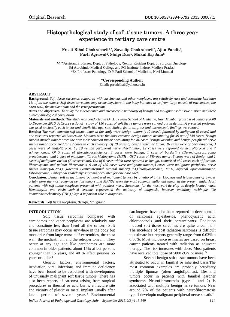
Figure 1: MPNST: photomicrograph showing irregular buckle shaped nuclei characteristic of schwann cell
with mitotic figures. (H & E:400x)
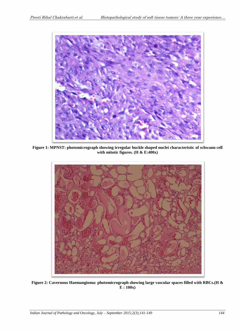
Figure 2: Cavernous Haemangioma: photomicrograph showing large vascular spaces filled with RBCs.(H &
E : 100x)

Preeti Rihal Chakrabarti et al. Histopathological study of soft tissue tumors: A three year experience…
Indian Journal of Pathology and Oncology, July – September 2015;2(3);141-149 145
Figure 3: Angiofibroma: photomicrograph showing ectatic blood vessels surrounded by collagenised stroma.
(H&E: 40x)
Figure 4: DFSP: photomicrograph showing tumor cell arranged in storiform pattern (H & E: 100x)

Preeti Rihal Chakrabarti et al. Histopathological study of soft tissue tumors: A three year experience…
Indian Journal of Pathology and Oncology, July – September 2015;2(3);141-149 146
Figure 5: MFH: photomicrograph showing tumor cells with greater degree of nuclear atypia with giant
cells.(H & E : 100 x)
DISCUSSION
Soft tissue tumors are relatively rare and
constitute less than 1% of all the cancers. Benign
mesenchymal tumors outnumbered sarcomas by the
factor of at least 100.The annual clinical incidence of
benign soft tissue tumors has been estimated up to
3000/million population i.e.-less than 1% of all the
malignant tumors.5 Lipomas are the most common
neoplasm of mesenchymal origin arising in any
location where fat is present. At least one third of
benign tumors are lipomas, one third are
fibrohistiocytic tumors and fibrous tumors, 10% are
vascular and 5 % are nerve sheath tumors.
In present study, there were 140 benign soft
tissue tumours, out of which 49 cases were reported
as lipomas which formed largest group among all
benign soft tissue tumors, with peak incidence in 5th
decade and commonest location being trunk. Two
commonest variants in the study by Ndukweet al6 and
Lin et al7 were conventional and fibrolipomas which
coordinated with our study also. On gross
examination, lipomas presented as encapsulated,
yellow, glistening mass, size ranging from 5-10 cm.
Microscopy examination revealed sheets of
adipocytes admixed with dilated congested blood
vessels and fibrous tissue in few cases. Of the 9
malignant soft tissue tumors in the present study, one
case was reported as atypical lipomatous tumor on
the anterior aspect of thigh of 50 year male patient.
Gross examination showed well circumscribed
yellow, greasy soft tissue, mass with attached muscle
with focal haemorrhagic areas on cut surface. On
microscopic examination tumor composed of sheets
of adipocytes with eccentric placed irregular
hyperchromatic nuclei with indentation and
vacuolated cytoplasm separated by fibrous septae
with focal areas of myxoid change and chronic
inflammatory cell infiltrate.
In a study by Sharon and Weiss8 of 92 cases
of well differentiated liposarcomas, tumor occurred
most frequently in extremity (46 cases), followed by
retroperitoneum (23 cases). Our findings were in
concordance with their study. Second most common
benign soft tissue tumor in our study were smooth
muscle tumor accounting for 44 cases, in which 42
cases were seen in uterine cavity including one in
broad ligament and one in cervix. However, 2 cases
of smooth muscle tumors were reported in males in
which one was in scrotum and other was in hand.
Uterine leiomyomas are extremely common
neoplasm of uterus with estimated rate of 20-40% of
women over 30 years of age.9 Siegel and Gaffey
demonstrated rarity of scrotal leiomyoma as only
they reported 11 cases in a review of 11000 cases of
scrotal tumors.10Grossly, leiomyomas presented as
greyish white firm masses with whorled appearance
on cut surface. Microscopy showed benign spindle
shape cells arranged in interlacing fascicles. One case
showed blood vessels lined by plump endothelial
cells blending with additional smooth muscle, hence

Preeti Rihal Chakrabarti et al. Histopathological study of soft tissue tumors: A three year experience…
Indian Journal of Pathology and Oncology, July – September 2015;2(3);141-149 147
diagnosed as angioleiomyoma in male patient who
presented with painful swelling in the hand. In a
series reported by Hachisuga et al11, 66.6%of
angioleiomyoma were reported in lower extremity
with tumor measuring less than 2 cm and with pain as
the presenting complaint.In our study patient gave the
history of painful swelling and size of tumor was
2x1x0.5 cm. Malignant tumor reported under smooth
muscle cateogory was leiomyosarcoma of uterus in
54 year patient. Grossly, it presented as single
pedunculated sub mucosal greyish yellow mass
10x8x8 cm with stalk protruding through
endocervical canal. Microscopy showed spindle
shaped cells showing plump nuclei and moderate
cytoplasm arranged in interlacing fascicles.
Intracytoplasmic vacuoles along with abnormal
mitosis were also seen.In this study we reported one
case of GIST in 56 y female patient who presented
with history of pain and mass per abdomen since 3-4
months. Grossly, tumor presented as ulcer
proliferative growth 4x3x3 cm surrounded by
numerous fibrous adhesions and necrotic areas.
Microscopy showed a malignant tumor arranged in
fascicles, with tumor cells showing pleomorphism,
round to spindly with hyperchromatic nuclei and
moderate amount of eosinophilic cytoplasm. Hypo
and hypercellular area admixed with giant cells and
area of necrosis were also noted.In a study by
Lakshmi et al12 of 176 cases of mesenchymal origin,
GIST (52.8%) were studied, with smooth muscle
tumor comprising 38.1% forming second largest
group and stomach and small intestine was the
commonest site.95 % of GISTwere in 4th and 5th
decade and male to female ratio 3:1. Most tumor had
spindle cell morphology,with 4 cases had a pure
epithelioid cell in their study.However in this study,
GIST was reported in female patient but age
incidence was in concordance with Lakshmi et
alstudy and it also showed spindle cell morphology.
However immunohistochemistry was not done in this
case.
Following benign smooth muscle tumor,
next most common tumor under benign category
were vascular and peripheral nerve sheath tumours,
19 cases were reported in each category.In benign
peripheral nerve sheath tumor, neurofibroma
were12/19 cases(63.2%), and schwannoma 7
/19(36.8%) cases.Donner et al13 over a period of 22
years revealed 288 benign tumors of major peripheral
nerve reported neurofibromaas the most common
followed by schwannoma which correlated with our
study.Gross examination revealed a characteristic
gelatinous appearance. Microscopy revealed
interlacing bundles of elongated cells with wavy
nuclei. Neurofibromas were commonly seen in 2nd
decade, with male predominance with head and neck
as commonest site which was in accordance with a
study by Lin et al14.In present study, Schwannomas
were commonly seen in 3rd decade with female
predominance and the commonest location was upper
extremity.This was in accordance with Enzinger
findings.15Grossly, schwannoma were encapsulated,
yellow white in appearance. Microscopy revealed
Antony A and Antony B areas.
MPNST account for approximately 5-10 %
of all soft tissue tumor, about one fourth to half occur
in setting of neurofibromatosis. We reported 3 cases
of MPNST with 1 case of epithelioid MPNST and 2
classical MPNST. All the 3 cases were seen in males,
with 2 cases reported on the back and 1 on the
scapula. One case reported recurrence within one
year. All the 3 cases reported in 2nd decade and were
more than 10cm in dimension. Grossly, they
presented as grey white, fleshy, firm to hard with
focal necrotic and haemorrhagic areas. Microscopy
showed malignant tumor composed of slender shaped
cells with wavy nuclei arranged in interlacing
fascicles.Tumor cells showed pleomorphism,
hyperchromatism with scant amount of eosinophilic
cytoplasm. Few hypocellular and hypercellular areas
were also seen with intervening stroma showing
mononuclear cell infiltrate with focal areas of
necrosis [Fig 1]. However 1 case of MPNST also
showed plump epithelioid cells. In a study by
Ducatman et al16 of 120 cases of MPNST, 52 were
male and 68 were female. In our study, male
preponderance was observed, which was in
accordance with their study16.
In our study 19 cases of benign vascular
tumor were reported, 16/19 cases were of
haemangiomas and 3/19 cases of angiofbroma. Head
and neck region was commonest location with peak
incidence in 2nd decade with male predominance.
Gross examination of all showed grey white, soft,
polypoidal bits ranging from 0.5 to 1cm. Microscopy
examination showed vaguely lobular pattern
displaying numerous blood vessels lined by plump
endothelial cells. In few cases large dilated vascular
channels were seen.[Fig 2] Malami et al,17 study
showed vascular tumor as the commonest soft tissue
tumor of childhood with majority hemangiomas
(27.3%), with male predominance and head and neck
as common site which correlated with our findings.
We reported 3 cases of angiofibromas in males in the
2nd decade of life located in the nasal cavity. Gross
examination revealed soft to firm, polypoidalmas
ranging from 2-5cm.Microscopic examination
revealed a tumor composed of numerous ectactic
blood vessels surrounded by pauci cellularstroma.
[Fig 3] Our study was in accordance with the findings
of Enzinger.18
In our study 5 cases of fibrohistiocytictumor
were reported, 3/5 cases were benign fibrohistiocytic
tumor, 1 boderline(DFSP) and 1 MFH. 2/3 cases of
fibrous histocytoma were noted in males. 2/3 cases
were noted in back and 1 noted in the nasal

Preeti Rihal Chakrabarti et al. Histopathological study of soft tissue tumors: A three year experience…
Indian Journal of Pathology and Oncology, July – September 2015;2(3);141-149 148
cavity.Fletcheret al19in their study of benign fibrous
histiocytoma, noted male predominance with lower
limb being the most common site. In ourstudy,
majority were reported in male although the
commonest location was the back.One case of
intermediate malignancy i.e, DFSP was reported in
45 year old male patient over the abdomen. Grossly,
tumor was grey white, firm, appear well
circumscribed and showed a characteristic storiform
pattern on microscopy[Fig 4]. Patients had been
operated for the same lesion 1 year back and
presented to us with recurrence.Our studyfindings
were in accordance with observations of Taylor et
al20. 1 case of MFHwas reported in 50 year female
with history of mass on lateral aspect of thigh.
Histology revealed, well circumscribed, grey white,
fleshy mass measuring -7x7x5 cm and microscopy
revealed a tumor composed of spindle cells arranged
in fascicles and bundles with spindly, hyperchromatic
nuclei and moderate amount of eosinophilic
cytoplasm. At places herring bone pattern was also
seen with large areas of necrosis and giant cell [Fig
5]. IHC of same was done which showed tumor was
vimentin+,desmin-, S100-, SM Actin-and our
diagnosis of MFH was confirmed. Weiss and
Enzinger21 analysed 200 cases of MFH and tumor
occurred mainly as a mass on extremities (lower
followed by upper) with peak incidence between 61
to 70 years with male predominance. The rate of
local recurrences of tumor was 44 % and of
metastasis 42 % with commonest site being lung and
lymph node. In this study, mass was in lower
extremity but we could not comment on local
recurrences as there was no follow up.
Under category of fibrous tumors, 6 cases
were benign and comprised of 2 cases each of
fibroma, fibromyxoma, and palmar fibromatosis.
Palmar fibromatosis were reported in female in the
3rd decade of life presenting with history of swelling
over the wrist of more than 6 months duration
without history of antecedent trauma. Gross
examination revealed grey white, firm masses
ranging from 1-2 cm with whorled cut
surface.Histologically, it showed elongated spindle
cells arranged in patternless pattern separated by
collagen and muscle fibres. Lisset al22in metaanalysis
of 10 previously published studies and found
association between trauma in the form vibration and
development of palmar fibromatosis. Under
malignant category of fibrous tumors, we reported a
case of fibrosracoma in 64 year female patient who
presented with swelling in lumbar region since 1
year.Grossly, it was grey white, fleshy, well
circumscribed mass measuring-14 x8 x7 cm with area
of necrosis and haemorrhage. Microscopy showed an
infiltrative tumor composed of cells arranged in
fascicles and herring bone pattern.In a study by
Prichard et al23 on fibrosarcoma, 60%, 30% and10%
of the study population were noted in lower
extremity, upper extremity and trunk respectively
with male predominance. However in this study we
reported a female patient with mass in trunk.
Rhabdomyosarcoma is not only the most
common soft tissue sarcoma in children under 15
years of age but also one of the most common soft
tissue sarcoma of adolescents and young adults24. We
reported a case of embryonal rhabdomyosarcoma in 8
year male patient who presented with mass over a left
thigh since 2 years. Grossly mass was lobulated, grey
white and cut surface of growth showed a gelatinous
area with hamorrhage and necrosis. Microscopy,
tumor showed hypocellular and hypercellular areas.
Hypercellular area showed round strap like cells,
typical cross striations and classical rhabdomyoblast
were not seen.
CONCLUSION
Painless mass was the most common
presenting symptom in our study. Benign soft tissue
tumor outnumbered malignant tumor by a ratio of
14:1. Lipomas and leiomyomas of gynaecological
origin were the common tumour to be reported.
Vascular tumor were commonly noted in adolescence
male. MPNST were the most common malignant
tumor in the present study.Location provides an
ancillary help in differential diagnosis. Sarcomas, for
the most part develop as deeply located mass. H&E
stained sections represented the mainstay of
diagnosis,however ancilliary techinque like IHC play
a important role in diagnosis.
REFERENCES 1. Jemal A, Seigel R, Ward E, et al.: Cancer
stastistics,2006;CA Cancer J Clin 2006; 56:106-30
2. Lavelle, S.M., PW Walton and Maura MhicLomhair:
Effect of irradiation, asbestos and chemical
carcinogenesis on incidence of sarcomas on implant.
Technology and Health Care2004;12:217-223.
3. Murray EM, Werner D,Greeff EA, Taylor DA:
Postradiation sarcoma: A 20 cases and review of
literature. Int j RadiatOncolBiolPhys 1999:45;951-961.
4. Fletcher CDM ,Unni KK , Mertens F (Eds): WHO
classification of tumors:Pathology and genetics of soft
tissue and bones.IARC press, Lyon 2002;p12-6
5. Fletcher CDM ,Unni KK , Mertens F (Eds): WHO
classification of tumors:Pathology and genetics of soft
tissue and bones.IARC press, Lyon 2002;p12-6
6. Ndukwe K.C,Ugboko VI, Somotun G, Adebiyi, KE
Fatusi OA.Clinical pathology study of lipoma of head
and neck.Nig JSurg Res 2003;5:12-7
7. Lin JJ, Lin F.Two entities in angiolipoma:a study of
459 cases of lipomas with review of literature of
infilteratingangiolipoma.Cancer 1974;34:720-7
8. Weiss SW, Rao VK. Well differentiated liposarcoma
of deep soft tissues of the extremeties,
retroperitoneum, and miscellaneous sites. Afollow-up
study of 92 cases with analysis of incidence of
differentiation. Am J SurgPathol 1992;16:1051-8.

Preeti Rihal Chakrabarti et al. Histopathological study of soft tissue tumors: A three year experience…
Indian Journal of Pathology and Oncology, July – September 2015;2(3);141-149 149
9. Fox H, Wells M. Haines and TaylorObstetric and
Gynaecology,4thed , London Churchill Livingstone, vol
1,p542-3
10. Das AK, Bolick D, Little NA, Walther PJ.
Pedunculated scrotal mass: Leiomyoma of scrotum.
Urology 1992;39:376-9
11. Hachisuga T, Hashimoto H, Enjoji M.
Angioleiomyoma: Aclinicopathological reappraisal of
562 cases.Cancer1984;54:126-130.
12. Lakshmi VA, Chacko RT, Kurian S. Gastrointestinal
stoma tumors: A 7 -year experience from a tertiary
care hospital. Indian J Pathol Microbiol 2010;53:628-
33.
13. Donner TR, Voorhies RM, Kline DG. Neural sheath
tumors of major nerves. J Neurosurg. 1994;81:362-73.
14. Lin BT, Weiss LM,Medeiros LJ. Neurofibroma and
cellular neurofibroma with atypis: A report of 14
tumors. Am J SurgPathol 1997;21:1443
15. Weiss SW, Goldblum JR. Enzinger and Weiss’s Soft
Tissue Tumor. 5thed. Philadelphia: Mosby Elsevier;
2008. P 825-96
16. Ducatman BS, Scheithauer BW, Piepgras DG, Reiman
HM, IIstrup DM. Malignant peripheral nerve sheath
tumors. Aclinicopathologicstudy of 120 cases. Cancer
1986;57:2006-21
17. Malami SA, Banjo AF. Pathological features of
vasculartumors in infants and children in lagos,
Nigeria.Ann Afr Med 2002; 1:92-98.
18. Weiss SW, Goldblum JR. Enzinger and Weiss’s Soft
Tissue Tumor. 5thed.Philadelphia: Mosby Elsevier;
2008. P 633-75
19. Fletcher CD : Benign fibrous histocytoma of
subcutaneous and deep soft tissue a clinicopathological
Analysis of 21 cases .Am J SurgPathol 1990;14:801-9
20. Taylor HB, Helwig E.B. Dermatofibrosarcomaprotub-
erans: Astudy of 115 cases. Cancer 1962;15:717-725.
21. Weiss SW, Enzinger FM.Malignat fibrous
histocytoma: an analysis of 200 cases.Cancer
1978;41:2250-66.
22. Liss, G.M,Stock SR:Can dupuytrens contracture be
work-related? Review of the evidence. Am J Ind Med
1996;29:521-32.
23. Pritchard DJ, Soule EH, Taylor WF et al. Fibrosra-
coma –a clinicopathological and statistical study of
199 tumors of soft tissue of extremities and
trunk.Cancer 1974;33:888-97.
24. Horn RC, Enterline HT. Rhabdomyosarcoma: A
clinicopathologic and statistical study and
classification of 39 cases. Cancer 1958;11:181-99.