Embed Size (px)
Citation preview
C O N T I N U I N G E D U C A T I O N
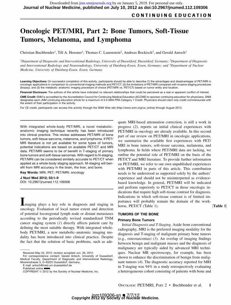
Oncologic PET/MRI, Part 2: Bone Tumors, Soft-TissueTumors, Melanoma, and Lymphoma
Christian Buchbender1, Till A. Heusner1, Thomas C. Lauenstein2, Andreas Bockisch3, and Gerald Antoch1
1Department of Diagnostic and Interventional Radiology, University of Dusseldorf, Dusseldorf, Germany; 2Department of Diagnosticand Interventional Radiology and Neuroradiology, University of Duisburg-Essen, Essen, Germany; and 3Department of NuclearMedicine, University of Duisburg-Essen, Essen, Germany
Learning Objectives: On successful completion of this activity, participants should be able to describe (1) the advantages and disadvantages of PET/MRI inoncologic applications in comparison to conventional imaging methods and PET/CT; (2) the limitations of PET/MRI compared with invasive staging procedures(biopsy); and (3) the metabolic–anatomic imaging procedure of choice (PET/MRI vs. PET/CT) based on tumor entity and location.
Financial Disclosure: The authors of this article have indicated no relevant relationships that could be perceived as a real or apparent conflict of interest.
CME Credit: SNM is accredited by the Accreditation Council for Continuing Medical Education (ACCME) to sponsor continuing education for physicians. SNMdesignates each JNM continuing education article for a maximum of 2.0 AMA PRA Category 1 Credit. Physicians should claim only credit commensurate withthe extent of their participation in the activity.
For CE credit, participants can access this activity through the SNM Web site (http://www.snm.org/ce_online) through August 2013.
With integrated whole-body PET/MRI, a novel metabolic–anatomic imaging technique recently has been introducedinto clinical practice. This review addresses PET/MRI of bonetumors, soft-tissue sarcoma, melanoma, and lymphoma. If PET/MRI literature is not yet available for some types of tumors,potential indications are based on available PET/CT and MRIdata. PET/MRI seems to be of benefit in T-staging of primarybone tumors and soft-tissue sarcomas. With regard to N-staging,PET/MRI can be considered similarly accurate to PET/CT whenapplied as a whole-body staging approach. M-staging will ben-efit from MRI accuracy in the brain, the liver, and bone.
Key Words: MRI; PET; PET/MRI; oncology
J Nucl Med 2012; 53:1–9DOI: 10.2967/jnumed.112.109306
Imaging plays a key role in diagnosis and staging inoncology. Evaluation of local tumor extent and detectionof potential locoregional lymph node or distant metastasesaccording to the periodically revised standardized TNMcancer staging system (1) directly affects patient care bydefining the most suitable therapy. With integrated whole-body PET/MRI, a new metabolic–anatomic imaging mo-dality has been introduced into clinical practice. Despitethe fact that the solution of basic problems, such as ade-
quate MRI-based attenuation correction, is still a work inprogress (2), reports on initial clinical experiences withPET/MRI in oncology are already available. In this secondpart of our review on PET/MRI in oncologic applications,we summarize the available first experiences with PET/MRI in bone tumors, soft-tissue sarcoma, melanoma, andlymphoma. In fields where PET/MRI data are lacking, weoutline the potential role of PET/MRI on the basis of thePET/CT and MRI literature. To provide further informationon PET/MRI, we refer to our own unpublished experienceswith PET/MRI in parts of this article. This contributionneeds to be understood as supported solely by the authors’experience and should not be misinterpreted as evidence-based knowledge. In general, PET/MRI will be indicatedand perform superiorly to PET/CT in those oncologic in-dications that require high soft-tissue contrast for diagnosis.Indications in which soft-tissue contrast is of limited im-portance will probably remain the domain of the work-horse, PET/CT ( ½Table 1�Table 1).
TUMORS OF THE BONE
Primary Bone Tumors
Initial Diagnosis and T-Staging. Aside from conventionalradiography, MRI is the preferred imaging modality for thediagnosis and T-staging of malignant primary bone tumors(e.g., osteosarcomas) (3). An overlap of imaging findingsbetween benign and malignant masses and the diagnosis ofmalignancy are typically aided by advanced MRI techni-ques. Nuclear MR spectroscopy, for example, has beenshown to enhance the discrimination of benign from malig-nant tumors (4). The diagnostic accuracy reported for MRIin T-staging was 94% in a study retrospectively evaluatinga heterogeneous cohort consisting of patients with bone and
Received May 24, 2012; revision accepted Jun. 26, 2012.For correspondence contact: Gerald Antoch, University of Dusseldorf,
Medical Faculty, Department of Diagnostic and Interventional Radiology,Moorenstrasse 5, D-40225 Dusseldorf, Germany.E-mail: [email protected] online nnnn.COPYRIGHT ª 2012 by the Society of Nuclear Medicine, Inc.
ONCOLOGIC PET/MRI, PART 2 • Buchbender et al. 1
jnm109306-sn n 7/7/12
Journal of Nuclear Medicine, published on July 10, 2012 as doi:10.2967/jnumed.112.109306
Copyright 2012 by Society of Nuclear Medicine.
by on January 5, 2019. For personal use only. jnm.snmjournals.org Downloaded from
soft-tissue sarcoma (5). In the same study, the combinedmetabolic–anatomic approach using 18F-FDG PET/CT forT-staging, with 96% accuracy, was reported to exceed eventhe excellent performance of MRI, a result that may beconsidered rather unexpected in view of the inferior soft-tissue contrast of CT to that of MRI and the well-knownlimitations of 18F-FDG PET for T-staging of varioustumors. However, data on integrated PET/MRI for T-stag-ing of malignant primary bone tumors are not yet available.We believe that PET/MRI may not increase the diagnosticaccuracy of T-staging of bone tumors over MRI alone, buta whole-body PET/MRI approach may offer TNM stagingwith high accuracy in a single session. T-staging will ben-efit from the MRI data, N-staging will benefit from PET,and M-staging will benefit from the combination. The PETcomponent of PET/MRI will be able to guide diagnosticbiopsies and help maximize the accuracy of correct stagingand grading, with a consequent impact on treatment andoutcome (3).N-Staging. Another advantage of integrated PET/MRI in
malignant primary bone tumors is that accurate localstaging with high resolution can be paired with a sensitivemetabolic whole-body staging examination. In a retrospec-tive study on 117 patients, 18F-FDG PET/CT had a sensitiv-ity, specificity, positive predictive value (PPV), negativepredictive value (NPV), and accuracy of 88%, 97%, 82%,98%, and 96%, respectively, and was significantly moreaccurate for N-staging in malignant bone tumors than con-ventional staging examinations (MRI of the tumor locationand whole-body CT) (5). Analogously, PET/MRI is ex-pected to be of similar accuracy to PET/CT for the detec-tion of lymph node metastases from malignant bonetumors. Direct comparison to conventional morphologicimaging reveals that 18F-FDG PET foremost contributesto the sensitivity of lymph node metastasis detection(53% vs. 72%) at a high level of specificity (5).
Restaging and Response to Therapy. Patients with re-lapsing malignant bone tumors have a poor prognosis (3).Although there is no guideline-established role for meta-bolic imaging in restaging of bone tumors, some promisingresults on the diagnostic value of 18F-FDG PET/CT can befound in the literature. 18F-FDG PET/CT has high accuracyin restaging, with a sensitivity, specificity, and accuracyof 87%, 97%, and 94%, respectively (6). In a prospectivecomparative study assessing therapy response in pediatricosteosarcomas, 18F-FDG PET was able to discriminate res-ponders from nonresponders, but CT and MRI (measure-ments of the tumor volume) were not (7). Interestingly,a subgroup analysis of the same study revealed that18F-FDG PET was not beneficial for therapy responseevaluation in Ewing sarcoma–type tumors. MRI techni-ques that go beyond tumor size and volume, such asdynamic contrast-enhanced MRI and diffusion-weightedMRI, have proven sensitive to chemotherapy-induced tu-mor necrosis and thus can also be used to evaluate ther-apy response (8). However, both MRI and 18F-FDG PETare of value for restaging and response assessment inprimary bone tumors. The integration of these compo-nents by PET/MRI offers a whole new field for researchon this topic.
Bone Metastases
Approximately 50% of cancer patients are estimated toexperience severe bone pain. Frequently, especially inprostate, breast, and lung cancer, metastases are found tobe causal (9). Imaging plays a pivotal role in the diagnosisof a metastatic osseous spread, and morphologic (CT andMRI) and functional (scintigraphy and PET) imaging mo-dalities are routinely applied in cancer patients. A meta-analysis comparing the diagnostic performance of radiologicand nuclear medicine methods, and their combination, forbone metastasis detection in more than 15,000 patients
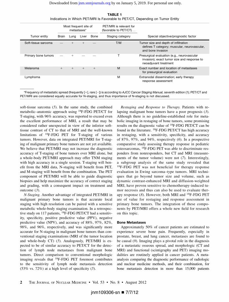
TABLE 1Indications in Which PET/MRI Is Favorable to PET/CT, Depending on Tumor Entity
Most frequent site of
metastases*
PET/MRI is relevant for
(favorable to PET/CT). . .
Tumor entity Brain Lung Liver Bone Staging category Special objective/prognostic factor
Soft-tissue sarcoma — 1 1 — T/M Tumor size and depth of infiltrationdefines T category; muscular, neurovascular,
and bone invasion
Primary bone tumors — 1 — — T Presurgical evaluation (e.g., neurovascular
invasion); exact tumor size and response to
neoadjuvant treatment
Melanoma 1 1 1 1 M Exact number and location of metastases
for presurgical evaluation
Lymphoma M Extranodal dissemination; early therapyresponse assessment
*Frequency of metastatic spread (frequently [1], rare [2]) is according to AJCC Cancer Staging Manual, seventh edition (1); PET/CT and
PET/MRI are considered equally accurate for N-staging, and thus importance of N-staging is not discussed.
2 THE JOURNAL OF NUCLEAR MEDICINE • Vol. 53 • No. 8 • August 2012
jnm109306-sn n 7/7/12
by on January 5, 2019. For personal use only. jnm.snmjournals.org Downloaded from
provided the broadest data so far (10). 18F-FDG PET, witha sensitivity of 90% and specificity of 97%, was as accurateas MRI, at 91% and 95%, respectively. When integrated18F-FDG PET/CT was applied, sensitivity increased to94%, and the same high specificity of 97% was preserved(10). A subanalysis revealed a higher sensitivity (95%)alongside a lower specificity (89%) when studies that in-cluded diffusion-weighted MRI sequences were comparedwith studies that did not (88% and 97%, respectively). 18F-FDG PET, 18F-FDG PET/CT, and MRI were significantlymore accurate for bone metastasis detection than were bonescintigraphy and stand-alone CT (10). A smaller but pro-spectively designed comparative study reported a higheroverall accuracy for whole-body MRI (91%) than for 18F-FDG PET/CT (78%). Whole-body MRI, according to thatstudy, was clearly superior to 18F-FDG PET/CT in sensitiv-ity. Sensitivity was 94% for MRI and 78% for 18F-FDGPET/CT, with comparable specificities (76% and 80%, re-spectively) (11). One reason for the significantly better per-formance of MRI in that study may have been a largernumber of patients with diffuse (but small-volume) osseousmetastases, which may be missed on 18F-FDG PET and onCT. The smallest detectable bone metastasis was 2 mm onMRI, compared with 5 mm on 18F-FDG PET/CT (11). Theresults of these studies are well suited to giving a generalimpression on the diagnostic performance of different im-aging methods. On the other hand, reliable data on diag-nostic accuracy in certain cancer types are of higherpractical relevance. For example, whole-body MRI was lesssensitive (77% vs. 92%) and specific (92% vs. 98%) than18F-FDG PET/18F-FDG PET/CT for the detection of bonemetastases, as well as being less sensitive (77% vs. 86%)but more specific (92% vs. 88%) than bone scintigraphy inlung cancer patients (12). In contrast, MRI was more sen-sitive than 18F-FDG PET and bone scintigraphy (97% vs.83% vs. 87%) for the detection of bone metastases in breastcancer patients (13). These findings indicate that for a rea-sonable judgment on the favored imaging modality in a cer-
tain cancer entity, differentiated studies are indispensible.Both PET/CT and MRI suffer from false-negative resultsrequiring periodic restaging (14).
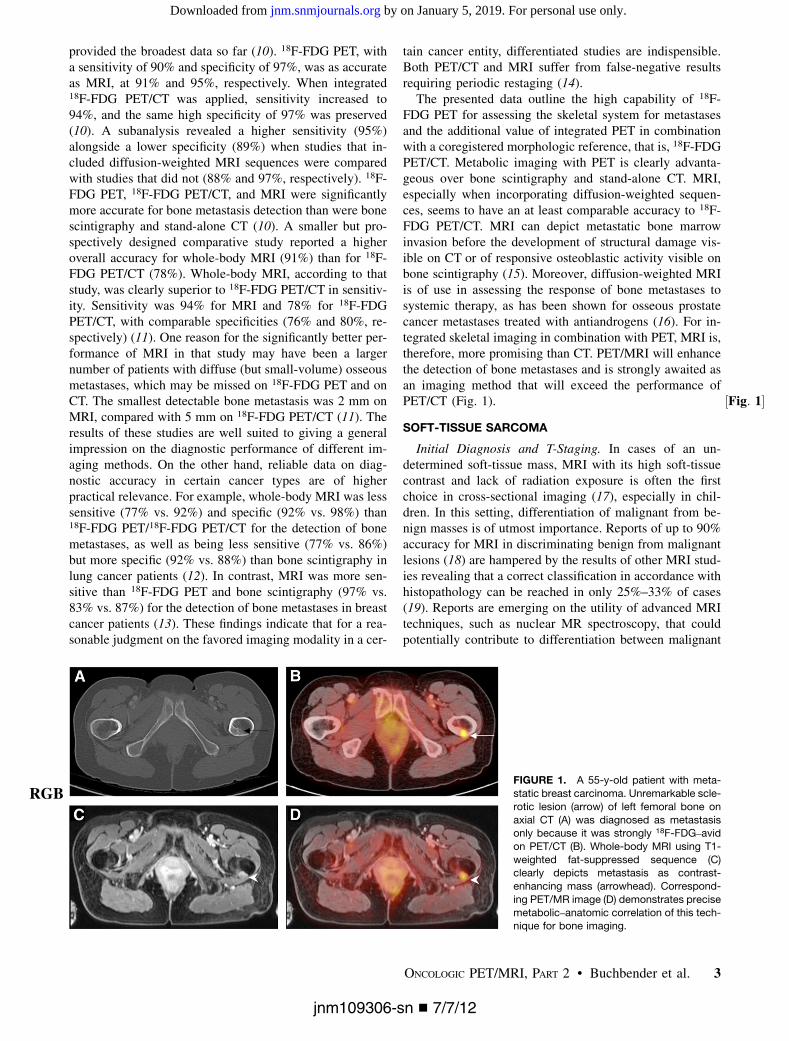
The presented data outline the high capability of 18F-FDG PET for assessing the skeletal system for metastasesand the additional value of integrated PET in combinationwith a coregistered morphologic reference, that is, 18F-FDGPET/CT. Metabolic imaging with PET is clearly advanta-geous over bone scintigraphy and stand-alone CT. MRI,especially when incorporating diffusion-weighted sequen-ces, seems to have an at least comparable accuracy to 18F-FDG PET/CT. MRI can depict metastatic bone marrowinvasion before the development of structural damage vis-ible on CT or of responsive osteoblastic activity visible onbone scintigraphy (15). Moreover, diffusion-weighted MRIis of use in assessing the response of bone metastases tosystemic therapy, as has been shown for osseous prostatecancer metastases treated with antiandrogens (16). For in-tegrated skeletal imaging in combination with PET, MRI is,therefore, more promising than CT. PET/MRI will enhancethe detection of bone metastases and is strongly awaited asan imaging method that will exceed the performance ofPET/CT ( ½Fig: 1�Fig. 1).
SOFT-TISSUE SARCOMA
Initial Diagnosis and T-Staging. In cases of an un-determined soft-tissue mass, MRI with its high soft-tissuecontrast and lack of radiation exposure is often the firstchoice in cross-sectional imaging (17), especially in chil-dren. In this setting, differentiation of malignant from be-nign masses is of utmost importance. Reports of up to 90%accuracy for MRI in discriminating benign from malignantlesions (18) are hampered by the results of other MRI stud-ies revealing that a correct classification in accordance withhistopathology can be reached in only 25%–33% of cases(19). Reports are emerging on the utility of advanced MRItechniques, such as nuclear MR spectroscopy, that couldpotentially contribute to differentiation between malignant
FIGURE 1. A 55-y-old patient with meta-
static breast carcinoma. Unremarkable scle-
rotic lesion (arrow) of left femoral bone on
axial CT (A) was diagnosed as metastasisonly because it was strongly 18F-FDG–avid
on PET/CT (B). Whole-body MRI using T1-
weighted fat-suppressed sequence (C)clearly depicts metastasis as contrast-
enhancing mass (arrowhead). Correspond-
ing PET/MR image (D) demonstrates precise
metabolic–anatomic correlation of this tech-nique for bone imaging.
RGB
ONCOLOGIC PET/MRI, PART 2 • Buchbender et al. 3
jnm109306-sn n 7/7/12
by on January 5, 2019. For personal use only. jnm.snmjournals.org Downloaded from
and benign soft-tissue masses (20), but for a conclusive andsecure diagnosis, patients with indecisive MRI or CT findingswill undergo biopsy in most cases (17). The availability ofadditional PET data will not obviate definite histopathologicdiagnosis by biopsy; thus, PET/MRI is not expected to be ofadditional use in the primary diagnosis of undetermined soft-tissue masses. But analogously to 18F-FDG PET, there mightbe an indication for integrated whole-body PET/MRI in thediagnostic algorithm in cases of sarcoma in which theprimary location is unknown (21).Based on its high soft-tissue contrast, MRI is the
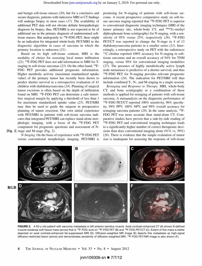
modality of choice for assessing local tumor infiltration(22). 18F-FDG PET does not add information to MRI for T-staging in soft-tissue sarcomas (23). On the other hand, 18F-FDG PET provides additional prognostic information.Higher metabolic activity (maximum standardized uptakevalue) of the primary tumor has recently been shown topredict shorter survival in a retrospective evaluation of 41children with rhabdomyosarcoma (24). Planning of surgicaltumor excisions is often based on the depth of infiltrationfound on MRI. 18F-FDG PET can determine a safe tumor-free surgical margin by applying a threshold of less than 1for maximum standardized uptake value (25). PET/MRImay thus be used to guide the surgeon in preoperativeplanning of tumor resection. Our own initial experiencewith PET/MRI in patients with soft-tissue sarcoma indi-cates that integrated PET/MRI can replace stand-alone mor-phologic imaging, with a focus of the 18F-FDG PETcomponent for prognostic questions and assessment of N-stage and M-stage (½Fig: 2� Fig. 2).N-Staging. On the basis of experience with 18F-FDG PET
versus conventional morphologic imaging, PET/MRI is
promising for N-staging of patients with soft-tissue sar-coma. A recent prospective comparative study on soft-tis-sue sarcoma staging reported that 18F-FDG PET is superiorto conventional diagnostic imaging techniques (MRI of thetumor primary site, whole-body CT, and 99mTc-methyldiphosphonate bone scintigraphy) for N-staging, with a sen-sitivity of 95% versus 25%, respectively (26). 18F-FDGPET/CT was reported to change the N-stage in 4 of 13rhabdomyosarcoma patients in a smaller series (21). Inter-estingly, a retrospective study on PET with the radiotracer11C-choline reported 100% accuracy for N-staging in soft-tissue sarcomas and an overall accuracy of 94% for TNMstaging, versus 50% for conventional imaging modalities(27). The presence of highly metabolically active lymphnode metastases is predictive of a shorter survival, and thus18F-FDG PET for N-staging provides relevant prognosticinformation (24). The indication for PET/MRI will thusinclude combined T-, N-, and M-staging in a single session.
Restaging and Response to Therapy. MRI, whole-bodyCT, and bone scintigraphy or a combination of thesemethods is applied for restaging of patients with soft-tissuesarcoma. A metaanalysis on the diagnostic performance of18F-FDG PET/CT reported 100% sensitivity, 96% specific-ity, 99% PPV, 100% NPV, and 99% overall accuracy forrestaging sarcoma patients (28). In the same analysis, 18F-FDG PET was more accurate than stand-alone CT. Com-parative studies have proven that a side-by-side reading of18F-FDG PET and conventional imaging techniques leadsto a significantly higher number of correct therapeutic deci-sions than does conventional imaging alone (91% vs. 59%)(26). There is evidence that the simple evaluation of tumorsize is inadequate for assessing the response of soft-tissue
FIGURE 2. A 63-y-old patient with sarcoma metastasis in left anterior serratus muscle. Axial contrast-enhanced CT (A) shows ill-defined
muscle-isodense soft-tissue mass (arrow) that is 18F-FDG–avid on 18F-FDG PET (B) and 18F-FDG PET/CT (C). Extent of this mass is betterdepicted on axial contrast-enhanced fat-suppressed MRI (D). Diffusion-weighted MR image (E) depicts this metastasis as high-signal
diffusion-restricted lesion (arrow) and demonstrates sensitivity of diffusion-weighted MRI. 18F-FDG PET/MR image is also shown (F).
RGB
4 THE JOURNAL OF NUCLEAR MEDICINE • Vol. 53 • No. 8 • August 2012
jnm109306-sn n 7/7/12
by on January 5, 2019. For personal use only. jnm.snmjournals.org Downloaded from
sarcomas to therapy (29), and thus markers of tumor bi-ology and metabolism are of increasing importance. Con-ventional MRI, as well as functional MRI, provides a wholebouquet of information on tumor size, perfusion, tissuecomposition, and extent of tumor necrosis and plays animportant role in assessing the response of soft-tissue sar-comas (30). Diffusion-weighted MRI, for example, hasbeen shown to provide a measure of tumor cellularity, witha reverse linear relation between measured adenylate cy-clase values and cellular count in soft-tissue sarcoma his-topathology (31). Moreover, a strong correlation betweena change in tumor adenylate cyclase and a change in tumorvolume under chemotherapy has been demonstrated (32).Diffusion-weighted MRI thus provides a valuable tool forthe assessment of cytotoxic therapy response. Metabolicimaging with 18F-FDG PET is also sensitive for assessingthe therapeutic response of patients with soft-tissue sar-coma. In a prospective study, the reduction of metabolicactivity between 2 consecutive 18F-FDG PET examinationsduring chemotherapy correctly predicted pathologic tumorresponse in 95% of patients (n 5 42) and was more accu-rate for this prediction than was a reduction of tumor size(33). With a threshold of a 60% decrease in maximumstandardized uptake value, 18F-FDG PET had 100% sensi-tivity and 71% specificity for the prediction of pathologictumor response (33). A different study, applying a thresholdof a 35% decrease in maximum standardized uptake valueunder chemotherapy, discriminated therapy respondersfrom nonresponders with 100% sensitivity and 67% speci-ficity as early as after the first chemotherapy cycle (34).Despite these promising reports, recent emerging evidencehas questioned the value of 18F-FDG PET for the evaluationof neoadjuvant therapy in soft-tissue sarcoma (35), indicat-ing that there still is research to be done on this topic.Follow-up data on the diagnostic performance of inte-grated PET/MRI are currently not available. The simulta-neous acquisition of dynamic functional MRI and PETinformation—for example, simultaneous contrast-enhanceddynamic MRI and dynamic PET—offers a new quality ofbioinformation in soft-tissue sarcoma restaging and ther-apy response assessment.
MELANOMA
Initial Diagnosis and T-Staging. Diagnosis of malignantmelanoma of the skin is based on clinical inspection andfull-thickness biopsy, and tumor stage according to theAmerican Joint Committee on Cancer (AJCC) classificationis based on the presence of ulceration and tumor thicknessas confirmed by histopathology (36). Generally, CT, MRI,and 18F-FDG PET depict melanomas with variable sensi-tivity and specificity depending on size and location. How-ever, there is no current indication for imaging in theprimary diagnosis and T-staging of malignant melanomas.N-Staging. The risk for locoregional metastases is
stratified according to the depth of tumor infiltration (AJCCclassification). According to current guidelines, in mela-
noma patients with a tumor thicker than 1 mm, sentinellymph biopsy should be considered, followed by locore-gional lymphadenectomy in cases of positivity for meta-static disease (37). This algorithm is based on the weakperformance of imaging modalities for N-staging in malig-nant melanoma when small metastases and micrometa-stases are considered. A metaanalysis on the diagnosticperformance of different imaging modalities for N-stagingin melanoma patients reported a sensitivity and specificityof 60% and 97% for ultrasonography, which was the high-est accuracy achieved in comparison with the low values forCT (9% and 92%, respectively), 18F-FDG PET (30% and96%, respectively), and 18F-FDG PET/CT (11% and 97%,respectively) (38). The prospectively evaluated sensitivity,specificity, PPV, and NPVof sentinel lymph node biopsy fordetection of occult locoregional lymph node metastases was94%, 100%, 100%, and 99%, respectively (39). The expla-nation for the low sensitivity of imaging studies can poten-tially be found in the small mean tumor volume of lymphnode metastases (,5 mm3) that is regularly found in mel-anoma patients (39,40). These small nests of tumor cells arefrequently missed on morphologic and metabolic scans.However, in patients with positive results on sentinel lymphnode biopsy, imaging is required and recommended to ex-clude further metastatic spread (37). In a prospective studyon 18F-FDG PET in AJCC stage III patients with positivesentinel lymph node biopsy and with CT, MRI, and ultra-sonography negative for disseminated disease, 18F-FDGPET revealed unknown distant metastases and thus up-staged 4 (12%) of 33 patients to AJCC stage IV (41).Whole-body MRI, with a sensitivity, specificity, PPV, NPV,and accuracy of 66%, 77%, 84%, 55%, and 67%, respec-tively, for the detection of lymph node metastases, has beenshown to be equal in accuracy to whole-body CT (42) andinferior to 18F-FDG PET/CT (43). Another comparativestudy on the same topic found whole-body MRI to be atleast as accurate as 18F-FDG PET/CT for N-staging, usinga combination of conventional MRI sequences and diffusion-weighted MRI (44). The rather sobering performance ofwhole-body MRI for N-staging in malignant melanomahas to be seen in the context of major advantages thatwhole-body MRI provides in the detection of subcutaneous,bone, liver, and brain metastases. Integrated PET/MRI will,therefore, be a tool to achieve whole-body melanoma stagingin a single session ( ½Fig: 3�Fig. 3).
Restaging and Response to Therapy. The risk of tumorrecurrence in malignant melanoma depends on the primary-tumor thickness. In low-risk patients (tumor thickness, 1 mm,AJCC stages 0–Ib), surveillance relies solely on clinicalfollow-up and routine self-examinations of the skin andlymph nodes (37). Restaging with ultrasound, CT, MRI,and PET/CT according to current guidelines (37) is re-served to, but not mandatory in, high-risk patients. A ret-rospective study in high-risk melanoma patients found that18F-FDG PET/CT, with a sensitivity, specificity, NPV, andPPVof 97%, was more accurate for the detection of tumor
ONCOLOGIC PET/MRI, PART 2 • Buchbender et al. 5
jnm109306-sn n 7/7/12
by on January 5, 2019. For personal use only. jnm.snmjournals.org Downloaded from
recurrence than the tumor marker S100 (respective values:86%, 45%, 61%, and 76%) (45). The same study revealedthat PET/CT, compared with S100, had a significantlyhigher prognostic value for cancer-related mortality. Ina different study, S100 positivity was used as a pretestfor tumor recurrence detection in asymptomatic high-riskmelanoma patients, with a sensitivity of 100%, a specific-ity of 90%, a PPV of 96%, and an NPV of 100% (46).According to the results of a recent metaanalysis, thePPV of PET/CT for the detection of recurrent metastasesis stage-dependent, yielding a higher PPV in high-riskpatients (80%) than in intermediate-risk patients (63%)and low-risk patients (33%) (38). Besides the reliabledetection, a precise localization of relapsing disease iscrucial for therapeutic, that is, surgical, decisions. Aprospective study on the impact of imaging on surgicaldecision making in melanoma patients reported that in25%–75% of patients, surgery was adapted on the basisof findings on 18F-FDG PET/CT (47). MRI has also beenshown to influence the therapeutic approach in melanomapatients. Therapy was changed in 64% of patients (n5 41)and included adaption of the surgical approach in 10patients after reevaluation of disease spread on whole-body MRI, including diffusion-weighted imaging (43).Moreover MRI, with its higher soft-tissue contrast, hasproven to be clearly superior for the detection of metasta-ses in important organ sites, with an impact on therapystrategies for, for example, liver and brain (42–44). Both18F-FDG PET/CT and MRI are sensitive to changes underlocal and systemic therapy. 18F-FDG PET/CT has been
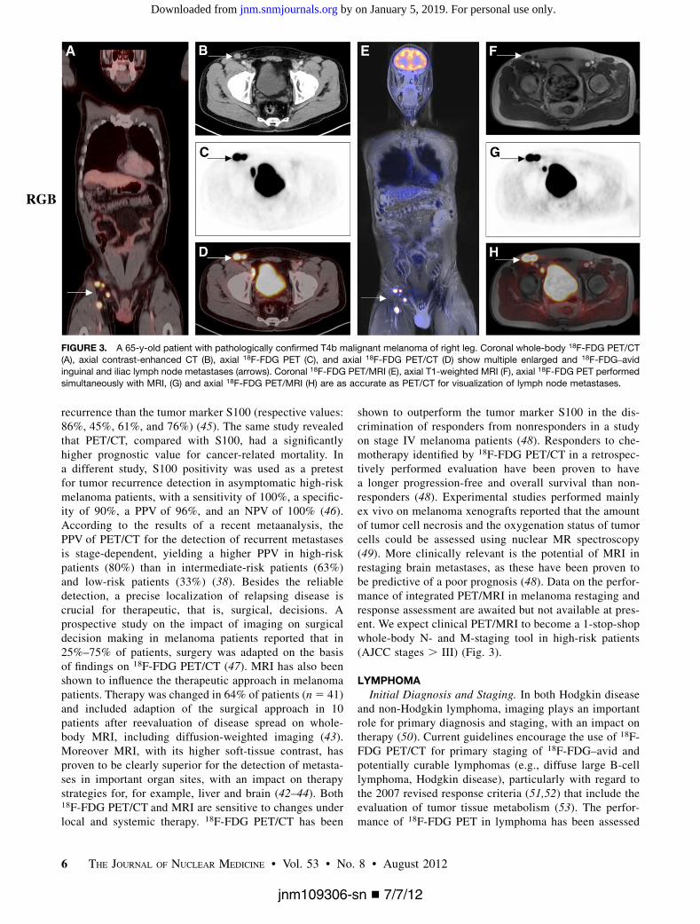
shown to outperform the tumor marker S100 in the dis-crimination of responders from nonresponders in a studyon stage IV melanoma patients (48). Responders to che-motherapy identified by 18F-FDG PET/CT in a retrospec-tively performed evaluation have been proven to havea longer progression-free and overall survival than non-responders (48). Experimental studies performed mainlyex vivo on melanoma xenografts reported that the amountof tumor cell necrosis and the oxygenation status of tumorcells could be assessed using nuclear MR spectroscopy(49). More clinically relevant is the potential of MRI inrestaging brain metastases, as these have been proven tobe predictive of a poor prognosis (48). Data on the perfor-mance of integrated PET/MRI in melanoma restaging andresponse assessment are awaited but not available at pres-ent. We expect clinical PET/MRI to become a 1-stop-shopwhole-body N- and M-staging tool in high-risk patients(AJCC stages . III) (Fig. 3).
LYMPHOMA
Initial Diagnosis and Staging. In both Hodgkin diseaseand non-Hodgkin lymphoma, imaging plays an importantrole for primary diagnosis and staging, with an impact ontherapy (50). Current guidelines encourage the use of 18F-FDG PET/CT for primary staging of 18F-FDG–avid andpotentially curable lymphomas (e.g., diffuse large B-celllymphoma, Hodgkin disease), particularly with regard tothe 2007 revised response criteria (51,52) that include theevaluation of tumor tissue metabolism (53). The perfor-mance of 18F-FDG PET in lymphoma has been assessed
FIGURE 3. A 65-y-old patient with pathologically confirmed T4b malignant melanoma of right leg. Coronal whole-body 18F-FDG PET/CT
(A), axial contrast-enhanced CT (B), axial 18F-FDG PET (C), and axial 18F-FDG PET/CT (D) show multiple enlarged and 18F-FDG–avid
inguinal and iliac lymph node metastases (arrows). Coronal 18F-FDG PET/MRI (E), axial T1-weighted MRI (F), axial 18F-FDG PET performed
simultaneously with MRI, (G) and axial 18F-FDG PET/MRI (H) are as accurate as PET/CT for visualization of lymph node metastases.
RGB
6 THE JOURNAL OF NUCLEAR MEDICINE • Vol. 53 • No. 8 • August 2012
jnm109306-sn n 7/7/12
by on January 5, 2019. For personal use only. jnm.snmjournals.org Downloaded from
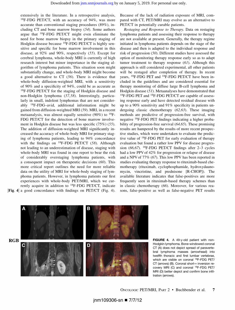
extensively in the literature. In a retrospective analysis,18F-FDG PET/CT, with an accuracy of 94%, was moreaccurate than conventional staging procedures (89%), in-cluding CT and bone marrow biopsy (54). Some authorsargue that 18F-FDG PET/CT might even eliminate theneed for bone marrow biopsy in the primary staging ofHodgkin disease because 18F-FDG PET/CT is highly sen-sitive and specific for bone marrow involvement in thisdisease, at 92% and 90%, respectively (55). Except forcerebral lymphoma, whole-body MRI is currently of highresearch interest but minor importance in the staging al-gorithm of lymphoma patients. This situation soon mightsubstantially change, and whole-body MRI might becomea good alternative to CT (56). There is evidence thatwhole-body diffusion-weighted MRI, with a sensitivityof 90% and a specificity of 94%, could be as accurate as18F-FDG PET/CT for the staging of Hodgkin disease andnon-Hodgkin lymphoma (57,58). Interestingly, particu-larly in small, indolent lymphomas that are not consider-ably 18F-FDG–avid, additional information might begained from diffusion-weighted MRI (59). MRI, in a recentmetaanalysis, was almost equally sensitive (90%) to 18F-FDG PET/CT for the detection of bone marrow involve-ment in Hodgkin disease but was less specific (75%) (55).The addition of diffusion-weighted MRI significantly in-creased the accuracy of whole-body MRI for primary stag-ing of lymphoma patients, leading to 94% concordancewith the findings on 18F-FDG PET/CT (58). Althoughnot leading to an underestimation of disease, staging withwhole-body MRI was found in one report to bear the riskof considerably overstaging lymphoma patients, witha consequent impact on therapeutic decisions (60). Thismore critical report outlines the need for more reliabledata on the utility of MRI for whole-body staging of lym-phoma patients. However, in lymphoma patients our firstexperiences with whole-body PET/MRI, which we cur-rently acquire in addition to 18F-FDG PET/CT, indicatea good concordance with findings on PET/CT (½Fig: 4� Fig. 4).
Because of the lack of radiation exposure of MRI, com-pared with CT, PET/MRI may evolve as an alternative toPET/CT in potentially curable patients.
Restaging and Response to Therapy. Data on restaginglymphoma patients and assessing their response to therapyare not available at present. Generally, the therapy regimeinitiated in lymphoma patients depends on the stage of thedisease and then is adapted to the individual response andrisk of progression (50). Different studies have assessed theoption of monitoring therapy response early so as to adapttumor treatment to therapy response (61). Although thisapproach is still considered experimental, all other patientswill be restaged after completion of therapy. In recentyears, 18F-FDG PET and 18F-FDG PET/CT have been in-cluded in the guidelines and are considered essential fortherapy monitoring of diffuse large B-cell lymphoma andHodgkin disease (51). Metaanalyses have demonstrated that18F-FDG PET and 18F-FDG PET/CT are capable of assess-ing response early and have detected residual disease withup to a 90% sensitivity and 91% specificity in patients un-dergoing classic chemotherapy (62,63). These imagingmethods are predictive of progression-free survival, withnegative 18F-FDG PET findings indicating a higher proba-bility of progression-free survival (64,65). These promisingresults are hampered by the results of more recent prospec-tive studies, which were undertaken to evaluate the predic-tive value of 18F-FDG PET for early evaluation of therapyevaluation but found a rather low PPV for disease progres-sion (66,67). 18F-FDG PET/CT findings after 2–3 cycleshad a low PPVof 42% for progression or relapse of diseaseand a NPVof 77% (67). This low PPV has been reported instudies evaluating therapy response to rituximab-based che-motherapy (rituximab, cyclophosphamide, hydroxydauno-mycin, vincristine, and prednisone [R-CHOP]). Theavailable literature indicates that false-positives are morefrequently seen in rituximab-based therapy schemes thanin classic chemotherapy (66). Moreover, for various rea-sons, false-positive as well as false-negative PET results
FIGURE 4. A 49-y-old patient with non-
Hodgkin lymphoma. Bone-windowed coronal
CT (A) does not depict spread of paraverte-bral lymphoma masses (arrowhead) into
twelfth thoracic and first lumbar vertebrae,
which are visible on coronal 18F-FDG PET/CT (arrows) (B). Coronal short-t inversion re-
covery MRI (C) and coronal 18F-FDG PET/
MRI (D) better depict and confirm bone infil-
tration (arrows).
RGB
ONCOLOGIC PET/MRI, PART 2 • Buchbender et al. 7
jnm109306-sn n 7/7/12
by on January 5, 2019. For personal use only. jnm.snmjournals.org Downloaded from
occur (68). However, the PPV and NPV of 18F-FDG PET/CT for progression-free survival after completion of che-motherapy were 71% and 80%, respectively, even forR-CHOP (67). With regard to these problems, MRI, es-pecially diffusion-weighted imaging, might be of value inrestaging of disease and evaluation of therapy response.MRI, by applying a combination of morphologic (size)and functional (adenylate cyclase measurements) param-eters, has been shown to detect 100% of residual lymphnode sites that were positive on 18F-FDG PET/CT (refer-ence standard) but resulted in 2 false-positive lesions ina comparative prospective pilot study that included 15patients receiving chemotherapy (69). According to a sub-analysis of the same study, the evaluation of morphologicMRI sequences led to a high number of false-positivelesions. The combination of size criteria with visualADC analysis then reduced the number of false-positivefindings to lesions. Lymph nodes with residual disease, aswell as false-positive lymph nodes, demonstrated a signif-icant increase in adenylate cyclase compared with thebaseline scan, indicating that the diagnosis on diffusion-weighted MRI was correct (69). The use of PET/MRI insteadof PET/CT in the surveillance of lymphoma patients under-going chemotherapy may also be helpful for the differentia-tion of thymic rebound from recurrent lymphoma of themediastinum. Chemical shift MRI provides MR images with2 different contrasts (in-phase and opposed-phase images),depending on the fat-to-water ratio. This technique, by thedetection of fat within a tissue mass, allows for the discrim-ination of hyperplastic thymic tissue from tumor tissue (70).Although these potential advantages need to be con-
firmed by larger studies, functional PET/MRI comprisingdiffusion-weighted and chemical shift imaging representsa promising tool for response assessment in lymphomapatients.
CONCLUSION
Literature on integrated PET/MRI in oncologic applica-tions is still limited. According to first experiences with thisimaging technique and the available data on whole-bodyMRI and PET/CT, PET/MRI can be expected to be of benefitin T-staging of primary bone tumors and soft-tissue sarco-mas. For N-staging, PET/MRI seems to provide similaraccuracy to PET/CT. The diagnostic performance of thedifferent imaging modalities for M-staging strongly dependson the location—that is, the organ harboring metastases.Therefore M-staging will benefit from MRI soft-tissue con-trast and accuracy in the brain, the liver, and bone. Thesimultaneous acquisition of functional MRI and PET datapromises to enhance assessments of tumor response to ther-apy. In general, PET/MRI will be indicated and performsuperiorly to PET/CT in those oncologic indications that re-quire high soft-tissue contrast for diagnosis. Indications inwhich soft-tissue contrast is of limited importance will prob-ably remain the domain of the workhorse, PET/CT.
REFERENCES
1. Edge SB, Byrd DR, Compton CC, et al., eds. AJCC Cancer Staging Handbook:
From the AJCC Cancer Staging Manual. 7th ed. New York, NY: Springer; 2009.
2. Hofmann M, Pichler B, Scholkopf B, Beyer T. Towards quantitative PET/MRI:
a review of MR-based attenuation correction techniques. Eur J Nucl Med Mol
Imaging. 2009;36(suppl 1):S93–S104.
3. Hogendoorn PC, Athanasou N, Bielack S, et al. Bone sarcomas: ESMO Clinical
Practice Guidelines for diagnosis, treatment and follow-up. Ann Oncol. 2010;21
(suppl 5):204–213.
4. Zhang J, Cheng K, Ding Y, Liang W, Vanel D, Cheng X. Study of single voxel 1H
MR spectroscopy of bone tumors: differentiation of benign from malignant
tumors. Eur J Radiol. December 12, 2011 [Epub ahead of print].
5. Tateishi U, Yamaguchi U, Seki K, Terauchi T, Arai Y, Kim EE. Bone and soft-
tissue sarcoma: preoperative staging with fluorine 18 fluorodeoxyglucose PET/
CT and conventional imaging. Radiology. 2007;245:839–847.
6. Gerth HU, Juergens KU, Dirksen U, Gerss J, Schober O, Franzius C. Significant
benefit of multimodal imaging: PET/CT compared with PET alone in staging and
follow-up of patients with Ewing tumors. J Nucl Med. 2007;48:1932–1939.
7. Denecke T, Hundsdorfer P, Misch D, et al. Assessment of histological response
of paediatric bone sarcomas using FDG PET in comparison to morphological
volume measurement and standardized MRI parameters. Eur J Nucl Med Mol
Imaging. 2010;37:1842–1853.
8. Uhl M, Saueressig U, van Buiren M, et al. Osteosarcoma: preliminary results of in
vivo assessment of tumor necrosis after chemotherapy with diffusion- and perfu-
sion-weighted magnetic resonance imaging. Invest Radiol. 2006;41:618–623.
9. Smith HS. Painful osseous metastases. Pain Physician. 2011;14:E373–E403.
10. Yang HL, Liu T, Wang XM, Xu Y, Deng SM. Diagnosis of bone metastases:
a meta-analysis comparing 1⁸FDG PET, CT, MRI and bone scintigraphy. Eur
Radiol. 2011;21:2604–2617.
11. Schmidt GP, Schoenberg SO, Schmid R, et al. Screening for bone metastases:
whole-body MRI using a 32-channel system versus dual-modality PET-CT. Eur
Radiol. 2007;17:939–949.
12. Qu X, Huang X, Yan W, Wu L, Dai K. A meta-analysis of 18FDG-PET-CT,18FDG-PET, MRI and bone scintigraphy for diagnosis of bone metastases in
patients with lung cancer. Eur J Radiol. 2012;81:1007–1015.
13. Liu T, Cheng T, Xu W, Yan WL, Liu J, Yang HL. A meta-analysis of 18FDG-
PET, MRI and bone scintigraphy for diagnosis of bone metastases in patients
with breast cancer. Skeletal Radiol. 2011;40:523–531.
14. Heusner T, Golitz P, Hamami M, et al. “One-stop-shop” staging: should we
prefer FDG-PET/CT or MRI for the detection of bone metastases? Eur J Radiol.
2011;78:430–435.
15. Imamura F, Kuriyama K, Seto T, et al. Detection of bone marrow metastases of
small cell lung cancer with magnetic resonance imaging: early diagnosis before
destruction of osseous structure and implications for staging. Lung Cancer.
2000;27:189–197.
16. Reischauer C, Froehlich JM, Koh DM, et al. Bone metastases from prostate
cancer: assessing treatment response by using diffusion-weighted imaging and
functional diffusion maps—initial observations. Radiology. 2010;257:523–531.
17. Casali PG, Blay JY. Soft tissue sarcomas: ESMO Clinical Practice Guidelines for
diagnosis, treatment and follow-up. Ann Oncol. 2010;21(suppl 5):198–203.
18. Berquist TH, Ehman RL, King BF, Hodgman CG, Ilstrup DM. Value of MR
imaging in differentiating benign from malignant soft-tissue masses: study of 95
lesions. AJR. 1990;155:1251–1255.
19. Kransdorf MJ, Murphey MD. Radiologic evaluation of soft-tissue masses: a cur-
rent perspective. AJR. 2000;175:575–587.
20. Doganay S, Altinok T, Alkan A, Kahraman B, Karakas HM. The role of MRS in
the differentiation of benign and malignant soft tissue and bone tumors. Eur J
Radiol. 2011;79:e33–e37.
21. Ricard F, Cimarelli S, Deshayes E, Mognetti T, Thiesse P, Giammarile F. Addi-
tional benefit of F-18 FDG PET/CT in the staging and follow-up of pediatric
rhabdomyosarcoma. Clin Nucl Med. 2011;36:672–677.
22. Sinha S, Peach AH. Diagnosis and management of soft tissue sarcoma. BMJ.
2010;341:c7170.
23. Tateishi U, Hosono A, Makimoto A, et al. Comparative study of FDG PET/CT
and conventional imaging in the staging of rhabdomyosarcoma. Ann Nucl Med.
2009;23:155–161.
24. Baum SH, Fruhwald M, Rahbar K, Wessling J, Schober O, Weckesser M. Con-
tribution of PET/CT to prediction of outcome in children and young adults with
rhabdomyosarcoma. J Nucl Med. 2011;52:1535–1540.
25. Yokouchi M, Terahara M, Nagano S, et al. Clinical implications of determination
of safe surgical margins by using a combination of CT and 18FDG-positron
emission tomography in soft tissue sarcoma. BMC Musculoskelet Disord. 2011;
12:166.
8 THE JOURNAL OF NUCLEAR MEDICINE • Vol. 53 • No. 8 • August 2012
jnm109306-sn n 7/7/12
by on January 5, 2019. For personal use only. jnm.snmjournals.org Downloaded from
26. Volker T, Denecke T, Steffen I, et al. Positron emission tomography for staging
of pediatric sarcoma patients: results of a prospective multicenter trial. J Clin
Oncol. 2007;25:5435–5441.
27. Tateishi U, Yamaguchi U, Maeda T, et al. Staging performance of carbon-11
choline positron emission tomography/computed tomography in patients with
bone and soft tissue sarcoma: comparison with conventional imaging. Cancer
Sci. 2006;97:1125–1128.
28. Piperkova E, Mikhaeil M, Mousavi A, et al. Impact of PET and CT in PET/CT
studies for staging and evaluating treatment response in bone and soft tissue
sarcomas. Clin Nucl Med. 2009;34:146–150.
29. Stacchiotti S, Collini P, Messina A, et al. High-grade soft-tissue sarcomas: tumor
response assessment—pilot study to assess the correlation between radiologic
and pathologic response by using RECIST and Choi criteria. Radiology. 2009;
251:447–456.
30. Wang X, Jacobs MA, Fayad L. Therapeutic response in musculoskeletal soft
tissue sarcomas: evaluation by MRI. NMR Biomed. 2011;24:750–763.
31. Schnapauff D, Zeile M, Niederhagen MB, et al. Diffusion-weighted echo-planar
magnetic resonance imaging for the assessment of tumor cellularity in patients
with soft-tissue sarcomas. J Magn Reson Imaging. 2009;29:1355–1359.
32. Dudeck O, Zeile M, Pink D, et al. Diffusion-weighted magnetic resonance im-
aging allows monitoring of anticancer treatment effects in patients with soft-
tissue sarcomas. J Magn Reson Imaging. 2008;27:1109–1113.
33. Evilevitch V, Weber WA, Tap WD, et al. Reduction of glucose metabolic activity
is more accurate than change in size at predicting histopathologic response to
neoadjuvant therapy in high-grade soft-tissue sarcomas. Clin Cancer Res.
2008;14:715–720.
34. Benz MR, Czernin J, Allen-Auerbach MS, et al. FDG-PET/CT imaging predicts
histopathologic treatment responses after the initial cycle of neoadjuvant chemo-
therapy in high-grade soft-tissue sarcomas. Clin Cancer Res. 2009;15:2856–
2863.
35. Benz MR, Czernin J, Allen-Auerbach MS, et al. 39-deoxy-39-[18F]fluorothymi-
dine positron emission tomography for response assessment in soft tissue sar-
coma: a pilot study to correlate imaging findings with tissue thymidine kinase 1
and Ki-67 activity and histopathologic response. Cancer. 2012;118:3135–3144.
36. Gershenwald JE, Soong SJ, Balch CM, et al. 2010 TNM staging system for
cutaneous melanoma. . .and beyond. Ann Surg Oncol. 2010;17:1475–1477.
37. Dummer R, Hauschild A, Guggenheim M, et al. Melanoma: ESMO clinical
practice guidelines for diagnosis, treatment and follow-up. Ann Oncol. 2010;
21(suppl 5):v194–v197.
38. Xing Y, Bronstein Y, Ross MI, et al. Contemporary diagnostic imaging modal-
ities for the staging and surveillance of melanoma patients: a meta-analysis. J
Natl Cancer Inst. 2011;103:129–142.
39. Wagner JD, Schauwecker D, Davidson D, et al. Prospective study of fluorodeoxy-
glucose-positron emission tomography imaging of lymph node basins in mel-
anoma patients undergoing sentinel node biopsy. J Clin Oncol. 1999;17:
1508–1515.
40. Wagner JD, Davidson D, Coleman JJ, et al. Lymph node tumor volumes in
patients undergoing sentinel lymph node biopsy for cutaneous melanoma. Ann
Surg Oncol. 1999;6:398–404.
41. Horn J, Lock-Andersen J, Sjøstrand H, Loft A. Routine use of FDG-PET scans in
melanoma patients with positive sentinel node biopsy. Eur J Nucl Med Mol
Imaging. 2006;33:887–892.
42. Muller-Horvat C, Radny P, Eigentler TK, et al. Prospective comparison of the
impact on treatment decisions of whole-body magnetic resonance imaging and
computed tomography in patients with metastatic malignant melanoma. Eur J
Cancer. 2006;42:342–350.
43. Pfannenberg C, Aschoff P, Schanz S, et al. Prospective comparison of 18F-fluo-
rodeoxyglucose positron emission tomography/computed tomography and
whole-body magnetic resonance imaging in staging of advanced malignant mel-
anoma. Eur J Cancer. 2007;43:557–564.
44. Laurent V, Trausch G, Bruot O, Olivier P, Felblinger J, Regent D. Comparative
study of two whole-body imaging techniques in the case of melanoma metasta-
ses: advantages of multi-contrast MRI examination including a diffusion-
weighted sequence in comparison with PET-CT. Eur J Radiol. 2010;75:376–383.
45. Essler M, Link A, Belloni B, et al. Prognostic value of [18F]-fluoro-deoxy-glu-
cose PET/CT, S100 or MIA for assessment of cancer-associated mortality in
patients with high risk melanoma. PLoS ONE. 2011;6:324632.
46. Peric B, Zagar I, Novakovic S, Zgajnar J, Hocevar M. Role of serum S100B and
PET-CT in follow-up of patients with cutaneous melanoma. BMC Cancer.
2011;11:328.
47. Gulec SA, Faries MB, Lee CC, et al. The role of fluorine-18 deoxyglucose
positron emission tomography in the management of patients with metastatic
melanoma: impact on surgical decision making. Clin Nucl Med. 2003;28:961–
965.
48. Strobel K, Dummer R, Steinert HC, et al. Chemotherapy response assessment in
stage IV melanoma patients: comparison of 18F-FDG-PET/CT, CT, brain MRI,
and tumor marker S-100B. Eur J Nucl Med Mol Imaging. 2008;35:1786–1795.
49. Olsen DR, Singstad TE, Rofstad EK. Effects of hyperthermia on bioenergetic
status and phosphorus T1S in human melanoma xenografts monitored by 31P-
MRS. Magn Reson Imaging. 1999;17:1049–1056.
50. Hutchings M, Barrington SF. PET/CT for therapy response assessment in lym-
phoma. J Nucl Med. 2009;50(suppl 1):S21–S30.
51. Cheson BD, Pfistner B, Juweid ME, et al. Revised response criteria for malignant
lymphoma. J Clin Oncol. 2007;25:579–586.
52. Juweid ME, Stroobants S, Hoekstra OS, et al. Use of positron emission tomog-
raphy for response assessment of lymphoma: consensus of the Imaging Subcom-
mittee of International Harmonization Project in Lymphoma. J Clin Oncol.
2007;25:571–578.
53. Tilly H, Dreyling M, Group EGW. Diffuse large B-cell non-Hodgkin’s lym-
phoma: ESMO Clinical Practice Guidelines for diagnosis, treatment and fol-
low-up. Ann Oncol. 2010;21(suppl 5):v172–v174.
54. Pelosi E, Pregno P, Penna D, et al. Role of whole-body [18F] fluorodeoxyglucose
positron emission tomography/computed tomography (FDG-PET/CT) and con-
ventional techniques in the staging of patients with Hodgkin and aggressive non
Hodgkin lymphoma. Radiol Med (Torino). 2008;113:578–590.
55. Wu LM, Chen FY, Jiang XX, Gu HY, Yin Y, Xu JR. 18F-FDG PET, combined
FDG-PET/CT and MRI for evaluation of bone marrow infiltration in staging of
lymphoma: a systematic review and meta-analysis. Eur J Radiol. 2012;81:303–311.
56. Kwee TC, van Ufford HM, Beek FJ, et al. Whole-body MRI, including diffusion-
weighted imaging, for the initial staging of malignant lymphoma: comparison to
computed tomography. Invest Radiol. 2009;44:683–690.
57. Punwani S, Taylor SA, Bainbridge A, et al. Pediatric and adolescent lymphoma:
comparison of whole-body STIR half-Fourier RARE MR imaging with an en-
hanced PET/CT reference for initial staging. Radiology. 2010;255:182–190.
58. Lin C, Luciani A, Itti E, et al. Whole-body diffusion-weighted magnetic reso-
nance imaging with apparent diffusion coefficient mapping for staging patients
with diffuse large B-cell lymphoma. Eur Radiol. 2010;20:2027–2038.
59. Abdulqadhr G, Molin D, Astrom G, et al. Whole-body diffusion-weighted im-
aging compared with FDG-PET/CT in staging of lymphoma patients. Acta Ra-
diol. 2011;52:173–180.
60. van Ufford HM, Kwee TC, Beek FJ, et al. Newly diagnosed lymphoma: initial
results with whole-body T1-weighted, STIR, and diffusion-weighted MRI com-
pared with 18F-FDG PET/CT. AJR. 2011;196:662–669.
61. Jochelson M, Mauch P, Balikian J, Rosenthal D, Canellos G. The significance of
the residual mediastinal mass in treated Hodgkin’s disease. J Clin Oncol. 1985;
3:637–640.
62. Zijlstra JM, Lindauer-van der Werf G, Hoekstra OS, Hooft L, Riphagen II,
Huijgens PC. 18F-fluoro-deoxyglucose positron emission tomography for post-
treatment evaluation of malignant lymphoma: a systematic review. Haematolog-
ica. 2006;91:522–529.
63. Isasi CR, Lu P, Blaufox MD. A metaanalysis of 18F-2-deoxy-2-fluoro-D-glucose
positron emission tomography in the staging and restaging of patients with
lymphoma. Cancer. 2005;104:1066–1074.
64. Terasawa T, Lau J, Bardet S, et al. Fluorine-18-fluorodeoxyglucose positron emis-
sion tomography for interim response assessment of advanced-stage Hodgkin’s
lymphoma and diffuse large B-cell lymphoma: a systematic review. J Clin Oncol.
2009;27:1906–1914.
65. Juweid ME, Wiseman GA, Vose JM, et al. Response assessment of aggressive
non-Hodgkin’s lymphoma by integrated International Workshop Criteria and
fluorine-18-fluorodeoxyglucose positron emission tomography. J Clin Oncol.
2005;23:4652–4661.
66. Pregno P, Chiappella A, Bello M, et al. Interim 18-FDG-PET/CT failed to predict
the outcome in diffuse large B-cell lymphoma patients treated at the diagnosis
with rituximab-CHOP. Blood. 2012;119:2066–2073.
67. Cashen AF, Dehdashti F, Luo J, Homb A, Siegel BA, Bartlett NL. 18F-FDG PET/
CT for early response assessment in diffuse large B-cell lymphoma: poor pre-
dictive value of international harmonization project interpretation. J Nucl Med.
2011;52:386–392.
68. Castellucci P, Nanni C, Farsad M, et al. Potential pitfalls of 18F-FDG PET in
a large series of patients treated for malignant lymphoma: prevalence and scan
interpretation. Nucl Med Commun. 2005;26:689–694.
69. Lin C, Itti E, Luciani A, et al. Whole-body diffusion-weighted imaging with
apparent diffusion coefficient mapping for treatment response assessment in
patients with diffuse large B-cell lymphoma: pilot study. Invest Radiol. 2011;
46:341–349.
70. Inaoka T, Takahashi K, Mineta M, et al. Thymic hyperplasia and thymus gland
tumors: differentiation with chemical shift MR imaging. Radiology. 2007;243:
869–876.
ONCOLOGIC PET/MRI, PART 2 • Buchbender et al. 9
jnm109306-sn n 7/7/12
by on January 5, 2019. For personal use only. jnm.snmjournals.org Downloaded from
Doi: 10.2967/jnumed.112.109306Published online: July 10, 2012.J Nucl Med. Christian Buchbender, Till A. Heusner, Thomas C. Lauenstein, Andreas Bockisch and Gerald Antoch LymphomaOncologic PET/MRI, Part 2: Bone Tumors, Soft-Tissue Tumors, Melanoma, and
http://jnm.snmjournals.org/content/early/2012/07/02/jnumed.112.109306This article and updated information are available at:
http://jnm.snmjournals.org/site/subscriptions/online.xhtml
Information about subscriptions to JNM can be found at:
http://jnm.snmjournals.org/site/misc/permission.xhtmlInformation about reproducing figures, tables, or other portions of this article can be found online at:
the manuscript and the final, published version.typesetting, proofreading, and author review. This process may lead to differences between the accepted version of
ahead of print area, they will be prepared for print and online publication, which includes copyediting,JNMthe copyedited, nor have they appeared in a print or online issue of the journal. Once the accepted manuscripts appear in
. They have not beenJNM ahead of print articles have been peer reviewed and accepted for publication in JNM
(Print ISSN: 0161-5505, Online ISSN: 2159-662X)1850 Samuel Morse Drive, Reston, VA 20190.SNMMI | Society of Nuclear Medicine and Molecular Imaging
is published monthly.The Journal of Nuclear Medicine
© Copyright 2012 SNMMI; all rights reserved.
by on January 5, 2019. For personal use only. jnm.snmjournals.org Downloaded from