Embed Size (px)
Citation preview
This article was downloaded by: [University of Winnipeg]On: 08 September 2014, At: 12:29Publisher: RoutledgeInforma Ltd Registered in England and Wales Registered Number: 1072954 Registered office: MortimerHouse, 37-41 Mortimer Street, London W1T 3JH, UK
Neurocase: The Neural Basis of CognitionPublication details, including instructions for authors and subscription information:http://www.tandfonline.com/loi/nncs20
Hemispheric processing of vocal emblem soundsYael Neumann-Werth a , Erika S. Levy b & Loraine K. Obler ca Queens College, City University of New York, Linguistics and CommunicationDisorders , Flushing , NY , USAb Teachers College, Columbia University, Speech and Language Pathology/Biobehavioral Sciences , New York , NY , USAc City University of New York Graduate School and University Center, Speech-Language-Hearing Sciences , New York , NY , USAPublished online: 10 May 2012.
To cite this article: Yael Neumann-Werth , Erika S. Levy & Loraine K. Obler (2013) Hemispheric processing of vocalemblem sounds, Neurocase: The Neural Basis of Cognition, 19:3, 268-281, DOI: 10.1080/13554794.2012.667122
To link to this article: http://dx.doi.org/10.1080/13554794.2012.667122
PLEASE SCROLL DOWN FOR ARTICLE
Taylor & Francis makes every effort to ensure the accuracy of all the information (the “Content”)contained in the publications on our platform. However, Taylor & Francis, our agents, and our licensorsmake no representations or warranties whatsoever as to the accuracy, completeness, or suitabilityfor any purpose of the Content. Any opinions and views expressed in this publication are the opinionsand views of the authors, and are not the views of or endorsed by Taylor & Francis. The accuracy ofthe Content should not be relied upon and should be independently verified with primary sources ofinformation. Taylor and Francis shall not be liable for any losses, actions, claims, proceedings, demands,costs, expenses, damages, and other liabilities whatsoever or howsoever caused arising directly orindirectly in connection with, in relation to or arising out of the use of the Content.
This article may be used for research, teaching, and private study purposes. Any substantial orsystematic reproduction, redistribution, reselling, loan, sub-licensing, systematic supply, or distributionin any form to anyone is expressly forbidden. Terms & Conditions of access and use can be found athttp://www.tandfonline.com/page/terms-and-conditions
Neurocase, 2013Vol. 19, No. 3, 268–281, http://dx.doi.org/10.1080/13554794.2012.667122
Hemispheric processing of vocal emblem sounds
Yael Neumann-Werth1, Erika S. Levy2, and Loraine K. Obler3
1Queens College, City University of New York, Linguistics and Communication Disorders, Flushing,NY, USA2Teachers College, Columbia University, Speech and Language Pathology/Biobehavioral Sciences,New York, NY, USA3City University of New York Graduate School and University Center, Speech-Language-HearingSciences, New York, NY, USA
Vocal emblems, such as shh and brr, are speech sounds that have linguistic and nonlinguistic features; thus, it isunclear how they are processed in the brain. Five adult dextral individuals with left-brain damage and moderate–severe Wernicke’s aphasia, five adult dextral individuals with right-brain damage, and five Controls participated intwo tasks: (1) matching vocal emblems to photographs (‘picture task’) and (2) matching vocal emblems to verbaltranslations (‘phrase task’). Cross-group statistical analyses on items on which the Controls performed at ceilingrevealed lower accuracy by the group with left-brain damage (than by Controls) on both tasks, and lower accuracyby the group with right-brain damage (than by Controls) on the picture task. Additionally, the group with left-braindamage performed significantly less accurately than the group with right-brain damage on the phrase task only.Findings suggest that comprehension of vocal emblems recruits more left- than right-hemisphere processing.
Keywords: Vocal emblems; Aphasia; Left brain-damage; Right brain-damage; Language.
For much of the century and a half ofneurolinguistic study, it has been generallyunderstood that the left hemisphere (LH), andparticularly its perisylvian area, is ‘responsible’ forlanguage. Broca (1861) had noted that his patientLeborgne, called ‘Tan’ because he only spoke thisnonsense word plus a curse, had lost all his lan-guage from a large LH lesion. A series of cases withsimilar LH lesions permitted Broca (1865) to con-clude compellingly that left anterior regions wereresponsible for the production of language. Broca’scontemporary, Hughlings Jackson, considered the
The authors would like to thank Arindam Roy Choudhury, James Jenkins, and Martin Chodorow for statistical consulting, andMarina Faygenbaum, Elaine Fong, Lara Hirner, JungMoon Hyun, and Lauren Liria, for their assistance with references. As well, wethank Martin Albert and colleagues at the Healthcare Boston VA Medical Center and the Harold Goodglass Aphasia Research Centerof the Boston University School of Medicine, and colleagues Marissa Barrera, Marissa Fond, and Natalie Schaefer in New York Cityfor help in recruitment of participants for this study. We very much appreciate the contributions of the participants.
Address correspondence to Yael Neumann-Werth, Queens College, City University of New York, Department of Linguistics andCommunication Disorders, 65-30 Kissena Boulevard, Flushing, NY, 11367 USA. (E-mail: [email protected]).
obverse of Broca’s analysis, reminding his readersthat even patients who had lost their languageabilities, their ability to pantomime, and someunderstanding of symbolism, often retained a smallrepertoire of recurrent utterances. He distinguisheda ‘superior’ level of speech that included ‘propo-sitionizing’ (the use of utterances that state facts)from an ‘inferior’ level that included ‘automatic’and ‘emotional’ utterances. Automatic/emotionalutterances, he claimed, often remained intact inindividuals with aphasia, and included commonexpressions such as ‘yes’, ‘no’, ‘please’, ‘go away’,
c© 2013 Taylor & Francis
Dow
nloa
ded
by [
Uni
vers
ity o
f W
inni
peg]
at 1
2:29
08
Sept
embe
r 20
14
HEMISPHERIC PROCESSING AND VOCAL EMBLEMS 269
‘oh’, oaths, and obscenities (Hughlings Jackson,as cited in Critchley & Critchley, 1998, p. 93). Hebelieved that sites for automatic/emotional speechwere in both hemispheres.
Van Lancker and Cummings (1999) have arguedthat the reason curses (and, presumably, other for-mulae) may be spared in aphasia is that they arenon-analytic speech patterns associated with theright-hemisphere (RH), distinct from the sequen-tial, analytic speech associated with the LH. VanLancker Sidtis (2008) makes reference to the dualprocessing model, which posits that language isgenerally processed in the LH, but that a subcor-tical circuit in the RH facilitates formulaic expres-sion. Support for this model includes observationsof interjections such as wow preserved in individualswith aphasia, while the ability to produce inter-jections is lost by some individuals with right orsubcortical brain damage.
A unique class of communicative sounds, vocalemblems (VEs) (Efron, 1941/1972), such as shhfor ‘Be quiet’ and brr for ‘It’s cold’, seem todefy the LH/RH dichotomy, as such sounds haveboth linguistic and nonlinguistic qualities. They areword-like in that they are short symbolic utteranceswith agreed-upon meanings. However, unlike mostwords, they are usually produced in isolation andnot in a sentence. Moreover, they are distinct inthat they may consist of sounds that are not partof a language’s phonological inventory and do notfollow the language’s phonotactic constraints. Forexample, the vocal emblem tsk, tsk is produced by adental click, which is not an English speech soundand does not contain the vocalic nucleus of a typi-cal syllable. In addition, intonation can be a salientfeature of VEs (e.g., uh-oh). Lastly, physical ges-tures (e.g., putting a finger to one’s lips for shh) andspecific facial expressions (e.g., grimace for pee-uw)conventionally accompany certain VEs.
From these observations, it is not clear howVEs are processed, i.e., whether they are pro-cessed similarly to most words (requiring primarilyLH processing) or to nonlinguistic intonation (seeWong, 2002, for an overview) and formulaic utter-ances (requiring primarily RH processing; e.g., VanLancker and Cummings, 1999). As there is littleliterature precisely on the items we consider, wereview studies that have examined the processing ofsymbolic gestures (unaccompanied by sound) andvarieties of communicative sounds.
In a functional Magnetic Resonance Imaging(fMRI) study, Xu, Gannon, Emmorey, Smith, andBraun (2009) examined whether two categories of
symbolic gestures: (1) emblem gestures (such asraising one’s finger and producing the facial expres-sion for ‘I‘ve got it’, rather than ones includingvocalizations like those studied here) and (2) pan-tomimes (e.g., threading a needle), are processedby the same cerebral system as spoken language.Twenty healthy dextral native English-speakingadults observed video clips of an actor perform-ing pantomimes, emblem gestures, and clips of heruttering spoken glosses corresponding to the stim-uli. Common areas of activation when participantsprocessed the symbolic gestures and spoken lan-guage were more abundant than areas uniquelyactivated by symbolic gestures or spoken language.Symbolic gestures, including emblems, were pro-cessed overall similarly to speech, primarily in theLH, although some areas of activation unique togestures were evident in both hemispheres. Theauthors suggest that the left perisylvian networkmaps symbols and meanings in a universal sense,whether this occurs in the vocal-auditory or in thegestural-visual domains.1
It remains possible that, to some degree, differ-ent symbols and gestures may activate brain regionsdifferentially, as the data of Xu et al. (2009) sug-gest. In their fMRI study, Gallagher and Frith(2004) considered two categories of symbolic ges-tures: instrumental gestures, i.e., those calling for anaction (e.g., ‘be quiet’); and expressive gestures (e.g.,‘it’s cold’). Watching instrumental gestures acti-vated the LH in regions associated with languageand imitation in healthy adult listeners. However,watching expressive gestures activated the anteriorparacingulate cortex, the amygdala and temporalpoles bilaterally, as well as the right superior tem-poral sulcus. Knutson, McClellan, and Grafman(2008) further found that watching more provoca-tive gestures (such as a fascist salute) activatedmore prefrontal and limbic areas than did sociallymeaningful gestures that are less provocative, suchas waving. Furthermore, Montgomery and Haxby(2008) reported that hand gestures and facialexpressions activated the mirror neuron systemdifferentially, with social hand gestures showing
1They note that signs used in American Sign Language andin all sign languages that have been studied are unlike emblemsin that these conform to lexical, phonological and syntacticrules similar to spoken language and thus their comprehen-sion and production understandably elicit activity patterns inthe perisylvian areas mostly indistinguishable from those acti-vated by the comprehension and production of spoken language(MacSweeney, Capek, Campbell, and Woll, 2008).
Dow
nloa
ded
by [
Uni
vers
ity o
f W
inni
peg]
at 1
2:29
08
Sept
embe
r 20
14
270 NEUMANN-WERTH, LEVY, OBLER
bilateral representation with greater activation ofthe left inferior parietal lobule and facial expres-sions showing greater bilateral activation of thefrontal operculum.
In summary, processing of symbolic gesturesnot accompanied by sound appears to show bilat-eral representation of some regions associatedwith language and other areas not associatedwith language. However, less is known about theprocessing of vocal emblems (e.g., shh, brr) –sounds that can be understood without use of thevisual modality by healthy individuals. A body ofresearch related to vocal emblem sounds has beenreported in the realm of nonverbal environmen-tal sound recognition, e.g., the sound of a cowmooing or a bell ringing. In three studies involv-ing participants with various lesion sites post-CVA(Saygin, Dick, Wilson, Dronkers, & Bates 2003;Schnider, Benson, Alexander, & Schnider-Klaus,1994; Spinnler &Vignolo, 1966), findings suggestedthat the LH is heavily involved in processing envi-ronmental sounds. However, there is controversy asto whether the RH is also involved. For example,Schnider et al. (1994) supported bilateral involve-ment and Spinnler and Vignolo (1966) reportedthat individuals with RBD performed similarly toControls on recognizing environmental sounds.
Onomatopoeias, another class of symbolicsounds with characteristics of words and environ-mental sounds, are used frequently in Japanesespeech, particularly in conversations with youngchildren. In an fMRI study, Hashimoto et al.(2006) found that nouns (i.e., types of animals, e.g.,Zou [elephant]) activated the left anterior supe-rior temporal gyrus (STG), while animal sounds(e.g., owl’s hoot) activated the left inferior frontalgyrus and the bilateral superior temporal gyrus.However, onomatopoeic sounds (e.g., gwa-gwa[onomatopoeia for sound made by duck]) resultedin activation of several brain regions, including theleft inferior frontal gyrus and the bilateral superiortemporal sulcus. The authors suggest that theseonomatopoeic sounds may be a ‘bridge’ betweenthe processing of animal sounds and of nouns(p. 1762).
In the only fMRI study that has includedvocal emblem sounds, Dietrich, Hertrich, Alter,Ischebeck, and Ackermann (2008) examined nativeGerman listeners’ processing of German interjec-tions. These interjections involved sounds consid-ered to have different lexical and prosodic loads.An example of an interjection with a high lexicalload was pfui (yuck), which is a word found in
dictionaries and one whose meaning can be under-stood even when uttered in a neutral tone. An inter-jection with a low lexical load was a (ah), which,when uttered in a neutral tone is simply a vocalicsound, but when uttered in a prolonged man-ner with a rising-falling intonation, can signal apleasant experience. Both were uttered in neutral-affect and in high-affect modes, distinguishedby ‘neutral’ vs. ‘affective/emotional’ prosody.Overlapping bilateral activation was revealed forheavily lexical and prosodic interjections, althougha stronger prosodic load activated the righttemporal lobe.
Evidence of RH dominance has been found fortasks involving non-linguistic intonation such aspitch discrimination (Robin, Tranel, & Demasio,1990) and affective prosody (Heilman, Scholes, &Watson, 1975). Van Lancker (1980) proposed ascale of LH to RH specialization associated withintonation, with lexical tone being most linguisticand affect/voice quality being least linguistic. Themore linguistic the intonational contrast is (e.g.,‘This is your pencil’ as opposed to someone else’s),the more it is lateralized to the LH and the less lin-guistic the contrast (e.g., higher pitch to indicatejoy), the more it is associated with the RH.
As most of the limited evidence points toLH processing for emblem-like sounds, environ-mental sounds, and even pantomimes, it seemslikely that vocal emblems, which include agreed-upon phonological forms, would be processedprimarily in the language areas, consistent withDietrich et al.’s (2008) findings. On the otherhand, if bilateral or RH representation charac-terizes onomatopoetic sounds (Hashimoto et al.,2006), and utterances that are dependent onintonation (Dietrich et al., 2008) and interjec-tions such as wow may be preserved in indi-viduals with aphasia (Van Lancker Sidtis, 2008),processing VEs may require more extensive RHactivation.
The aim of this study was to address the questionof whether processing of VEs is analogous to theprocessing of words (relying primarily on the LH)or whether more RH participation is evident (as isthe case for nonlinguistic intonation). Our goal wasto employ classic neurolinguistic subtractive rea-soning to further elucidate hemispheric responsibil-ities for processing such category-defying sounds.
The comprehension of VEs by two groups ofparticipants with brain damage was investigated:individuals with exclusively LH damage affect-ing the language areas, and individuals with
Dow
nloa
ded
by [
Uni
vers
ity o
f W
inni
peg]
at 1
2:29
08
Sept
embe
r 20
14
HEMISPHERIC PROCESSING AND VOCAL EMBLEMS 271
predominantly RH damage.2 Their comprehensionwas compared to that of a Control group of age-matched participants with no brain damage. Theproject consisted of two tasks to determine partic-ipants’ abilities to comprehend VEs. The picturetask investigated whether participants could indi-cate the appropriate setting in which they wouldexpect to hear each emblem. The response sheetconsisted of pictorial (photographic) materials, aswe assumed that the ability to point to photographswould be spared for participants with left braindamage (LBDs). The phrase task evaluated partic-ipants’ comprehension via verbal translation of theVEs, as we assumed that the required verbal skillswould be spared for individuals with right braindamage (RBDs).
We hypothesized, based on the research reviewedabove, that RBDs would score more accuratelyoverall than the LBDs and less accurately thanControls. That is, regardless of the modality ofresponse (pictures or phrases), the processing ofVEs is likely to be more robustly represented in theLH than in the RH and therefore to be most dif-ficult to comprehend for LBDs – especially thosewith lesions compromising their verbal comprehen-sion. However, because bilateral representation mayalso occur with VEs, and processing emblems thathave stronger affect might recruit regions in theRH, RBDs were expected to perform less accu-rately than Controls on both tasks. As well, wepredicted that RBDs would perform more accu-rately on the phrase task than on the picture taskand that LBDs would perform more accuratelyon the picture task than the phrase task, simplybecause verbal responses are less difficult for indi-viduals with RBD than individuals with LBD, andthe reverse is true for pictorial material.
We asked whether specific items would differenti-ate the two brain-damage groups, either differenti-ating between those VEs with vs. without emotionas Dietrich et al. (2008) had, or between VEs thatare instrumental vs. expressive (Gallagher & Frith,2004) or provocative vs. not (Knutson et al., 2008)or social vs. facial, as Montgomery and Haxby(2008) suggested. As well, we considered whethererror patterns would distinguish the two groups.
2All RBD participants except #5 had exclusively right dam-age; Participant 5 had a minor stroke six years prior to ourtesting, which had resulted in temporary right-sided weaknessbut had no consequences for his language abilities. We includedhim in the study when our analyses indicated his scores werewithin the range of the other RBDs.
We predicted that the errors of the LBDs wouldbe more different from any made by Controls thanwould those of the RBDs.
METHODS
Participants
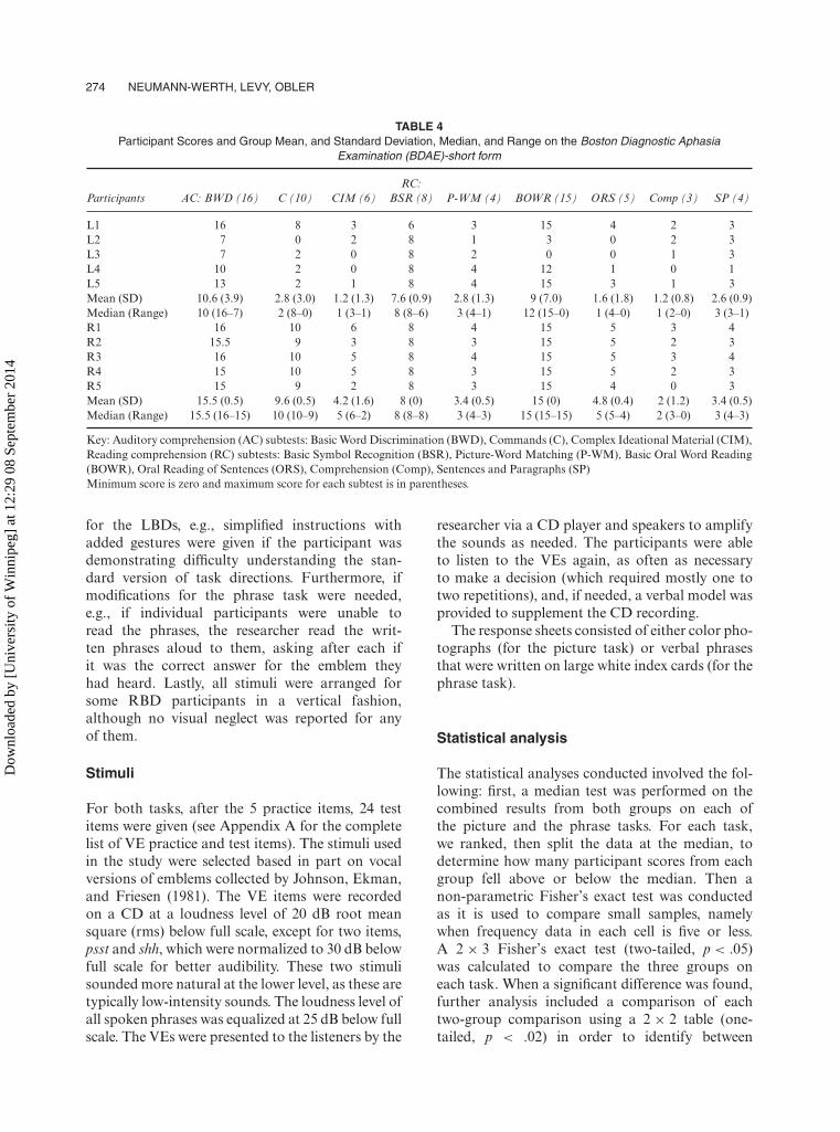
Two experimental groups with focal hemisphericdamage (left or right), namely, five dextral LBDswith moderate–severe Wernicke’s aphasia, five dex-tral RBDs and a control group of five normalhealthy adults were included in the study (seeTables 1 and 2 for their individual and groupdemographic data and Table 3 for the stroke andneurological information). Table 4 lists the BostonDiagnostic Aphasia Examination (BDAE)-ShortForm (Goodglass, Kaplan, & Barresi, 2001) scoresof the participants with brain-damage on the com-prehension and other subtests, to demonstrate thedeficits in comprehension in the LBD relative tothose of the RBDs, who performed very accuratelyon almost all of the subtests.
Participants were included based on the fol-lowing criteria: having spoken American Englishfor most of their life,3 demonstrating normalhearing for conversational purposes,4 good vision(or corrected), no prior neurological or psychiatrichistory (apart from RBD 5; see footnote 3),and sufficient comprehension to follow directions(based on a recent speech-language pathologistreport and performance on the BDAE-ShortForm [Goodglass et al., 2001] comprehensionsubtests). Additionally, all control participants
3 Four of our RBDs (though none of our LBDs) were multi-lingual. Because their performance as individuals and as a groupwas significantly superior to that of the LBDs, we have to assumethat their non-monolingual status did not impair their perfor-mance on the VEs. One of these multilingual patients in the RBDgroup (#5) had not spoken substantial English until he immi-grated to the U.S. at age 30. Recall that as well, he had hada minor stroke with mild right-sided weakness, which resolvedcompletely. We included him as his data fell within the range ofthe other RBD participants.
4 Two or three participants in each of the three groups did notpass the hearing screening at 50dB at all frequencies in the speechrange. When addressing within-group comparisons, we deter-mined that these individuals did not perform the least accuratelyin their group. Crucially, poor hearing scores did not precludeexcellent performance on the VE tasks by Controls 3 and 4, norfor RBDs 1, 2 and 3, so we conclude that poor hearing cannotbe the cause of the poor performance by LBDs 2 and 3.
Dow
nloa
ded
by [
Uni
vers
ity o
f W
inni
peg]
at 1
2:29
08
Sept
embe
r 20
14
272 NEUMANN-WERTH, LEVY, OBLER
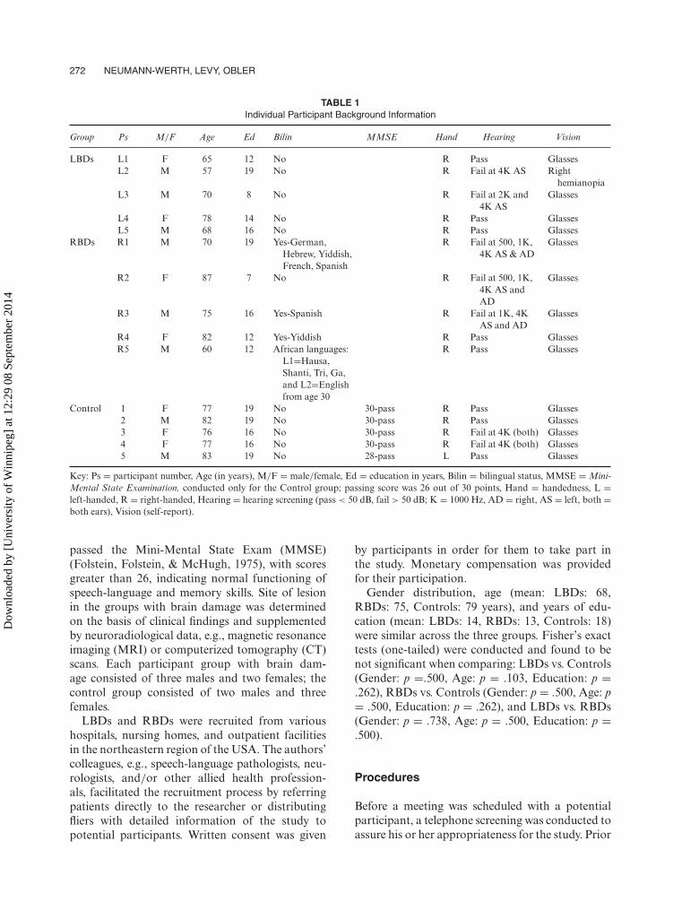
TABLE 1Individual Participant Background Information
Group Ps M/F Age Ed Bilin MMSE Hand Hearing Vision
LBDs L1 F 65 12 No R Pass GlassesL2 M 57 19 No R Fail at 4K AS Right
hemianopiaL3 M 70 8 No R Fail at 2K and
4K ASGlasses
L4 F 78 14 No R Pass GlassesL5 M 68 16 No R Pass Glasses
RBDs R1 M 70 19 Yes-German,Hebrew, Yiddish,French, Spanish
R Fail at 500, 1K,4K AS & AD
Glasses
R2 F 87 7 No R Fail at 500, 1K,4K AS andAD
Glasses
R3 M 75 16 Yes-Spanish R Fail at 1K, 4KAS and AD
Glasses
R4 F 82 12 Yes-Yiddish R Pass GlassesR5 M 60 12 African languages:
L1=Hausa,Shanti, Tri, Ga,and L2=Englishfrom age 30
R Pass Glasses
Control 1 F 77 19 No 30-pass R Pass Glasses2 M 82 19 No 30-pass R Pass Glasses3 F 76 16 No 30-pass R Fail at 4K (both) Glasses4 F 77 16 No 30-pass R Fail at 4K (both) Glasses5 M 83 19 No 28-pass L Pass Glasses
Key: Ps = participant number, Age (in years), M/F = male/female, Ed = education in years, Bilin = bilingual status, MMSE = Mini-Mental State Examination, conducted only for the Control group; passing score was 26 out of 30 points, Hand = handedness, L =left-handed, R = right-handed, Hearing = hearing screening (pass < 50 dB, fail > 50 dB; K = 1000 Hz, AD = right, AS = left, both =both ears), Vision (self-report).
passed the Mini-Mental State Exam (MMSE)(Folstein, Folstein, & McHugh, 1975), with scoresgreater than 26, indicating normal functioning ofspeech-language and memory skills. Site of lesionin the groups with brain damage was determinedon the basis of clinical findings and supplementedby neuroradiological data, e.g., magnetic resonanceimaging (MRI) or computerized tomography (CT)scans. Each participant group with brain dam-age consisted of three males and two females; thecontrol group consisted of two males and threefemales.
LBDs and RBDs were recruited from varioushospitals, nursing homes, and outpatient facilitiesin the northeastern region of the USA. The authors’colleagues, e.g., speech-language pathologists, neu-rologists, and/or other allied health profession-als, facilitated the recruitment process by referringpatients directly to the researcher or distributingfliers with detailed information of the study topotential participants. Written consent was given
by participants in order for them to take part inthe study. Monetary compensation was providedfor their participation.
Gender distribution, age (mean: LBDs: 68,RBDs: 75, Controls: 79 years), and years of edu-cation (mean: LBDs: 14, RBDs: 13, Controls: 18)were similar across the three groups. Fisher’s exacttests (one-tailed) were conducted and found to benot significant when comparing: LBDs vs. Controls(Gender: p =.500, Age: p = .103, Education: p =.262), RBDs vs. Controls (Gender: p = .500, Age: p= .500, Education: p = .262), and LBDs vs. RBDs(Gender: p = .738, Age: p = .500, Education: p =.500).
Procedures
Before a meeting was scheduled with a potentialparticipant, a telephone screening was conducted toassure his or her appropriateness for the study. Prior
Dow
nloa
ded
by [
Uni
vers
ity o
f W
inni
peg]
at 1
2:29
08
Sept
embe
r 20
14
HEMISPHERIC PROCESSING AND VOCAL EMBLEMS 273
TABLE 2Group Background Information
Participants LBDs RBDs Controls
Gender 3 M, 2 F 3 M, 2 F 2 M, 3 FAge
Mean (SD) 67.6 (7.6) 74.8 (10.5) 79 (3.2)Median
(Range)68 (78–57) 75 (87–60) 77 (83–76)
EducationMean (SD) 13.8 (4.1) 13.2 (4.5) 17.8 (1.6)Median
(Range)14 (19–8) 12 (19–7) 19 (19–16)
MMSEMean (SD) NA NA 29.6 (0.9)Median
(Range)30 (30–28)
Key: Standard deviations in parentheses, LBDs = participantswith left brain damage, RBDs = participants with right braindamage, M = male, F = female, Age (in years), Education(in years), MMSE, Mini-Mental State Examination (for con-trol group only, passing score was 26 out of 30 points), NA =not applicable).
to the experiment, the first author gathered fur-ther information by speaking with the participantor family member and conducting a hearing screen-ing, using a Welch-Allyn audioscope. Participantswith brain damage were then administered the audi-tory and reading comprehension subtests of theBDAE-Short Form (Goodglass et al., 2001), toassess their level of severity and type of aphasia andto ensure adequate comprehension at the word andphrase level. The control group was administeredthe MMSE (Folstein et al., 1975) to ensure nor-mal functioning of speech-language and memoryskills.
For the experimental testing, we explained whatwe meant by the term ‘emblem’ and gave severalexamples that were not included among the stimuli.
Task 1: Picture task
In Task 1, the picture task (emblem-to-photograph matching), the researcher presented anemblem sound (e.g., shh) that was then followed byfour photographs: a photograph of an appropriatesetting (e.g., a library) and three other photographs(e.g., different vocal emblem settings such as snow,which would serve as the photograph for brr).Participants were given the following instructions:‘You will hear an emblem. Then you will see fourpictures. Please point to the picture that shows asituation in which you would expect to hear theemblem’. The researcher recorded participants’choices on a scoring sheet.
Task 2: Phrase task
In Task 2, the phrase task (emblem-to-writtenstimulus matching), presentation of an emblemsound (e.g., shh) was followed by four phrases writ-ten on separate cards (e.g., ‘Be quiet’, and threeother foils of phrases describing different VEs, e.g.,‘I’m thinking’, ‘That stinks’, ‘That’s disgusting’).The researcher gave the following instructions: ‘Youwill hear an emblem. I will then show you four cardswith different phrases. Please point to the phrasethat best describes what the emblem means’.
For both tasks, five practice items were first givenbefore the test items, in order to familiarize theparticipant with the experimental task. As well,appropriate modifications were made, particularly
TABLE 3Participant Stroke/Neurological Information
Group Ps Severity TPO Localizing Information Prior Neurological History
LBDs (Wernicke): L1 Mild-Moderate 1 LH NoL2 Severe 12 L-MCA NoL3 Severe 5 LH NoL4 Severe 24 L-craniotomy Yes-tumor left temporalL5 Severe 32 L-frontal No
RBDs: R1 Moderate 160 RH NoR2 Mild-Moderate 3 R-frontal NoR3 Mild 1.5 R-MCA NoR4 Mild-Mod 60 R-perisylvian NoR5 Mild 2 RH Mini-stroke
Key: Ps = participants, LBDs = participants with left brain damage, RBDs = participants with right brain damage, L = left, R = right),Severity = of aphasia (mild, moderate, severe), TPO, time post onset in months, localizing information (left hemisphere (LH) or righthemisphere (RH); middle cerebral artery (MCA).
Dow
nloa
ded
by [
Uni
vers
ity o
f W
inni
peg]
at 1
2:29
08
Sept
embe
r 20
14
274 NEUMANN-WERTH, LEVY, OBLER
TABLE 4Participant Scores and Group Mean, and Standard Deviation, Median, and Range on the Boston Diagnostic Aphasia
Examination (BDAE)-short form
RC:Participants AC: BWD (16) C (10) CIM (6) BSR (8) P-WM (4) BOWR (15) ORS (5) Comp (3) SP (4)
L1 16 8 3 6 3 15 4 2 3L2 7 0 2 8 1 3 0 2 3L3 7 2 0 8 2 0 0 1 3L4 10 2 0 8 4 12 1 0 1L5 13 2 1 8 4 15 3 1 3Mean (SD) 10.6 (3.9) 2.8 (3.0) 1.2 (1.3) 7.6 (0.9) 2.8 (1.3) 9 (7.0) 1.6 (1.8) 1.2 (0.8) 2.6 (0.9)Median (Range) 10 (16–7) 2 (8–0) 1 (3–1) 8 (8–6) 3 (4–1) 12 (15–0) 1 (4–0) 1 (2–0) 3 (3–1)R1 16 10 6 8 4 15 5 3 4R2 15.5 9 3 8 3 15 5 2 3R3 16 10 5 8 4 15 5 3 4R4 15 10 5 8 3 15 5 2 3R5 15 9 2 8 3 15 4 0 3Mean (SD) 15.5 (0.5) 9.6 (0.5) 4.2 (1.6) 8 (0) 3.4 (0.5) 15 (0) 4.8 (0.4) 2 (1.2) 3.4 (0.5)Median (Range) 15.5 (16–15) 10 (10–9) 5 (6–2) 8 (8–8) 3 (4–3) 15 (15–15) 5 (5–4) 2 (3–0) 3 (4–3)
Key: Auditory comprehension (AC) subtests: Basic Word Discrimination (BWD), Commands (C), Complex Ideational Material (CIM),Reading comprehension (RC) subtests: Basic Symbol Recognition (BSR), Picture-Word Matching (P-WM), Basic Oral Word Reading(BOWR), Oral Reading of Sentences (ORS), Comprehension (Comp), Sentences and Paragraphs (SP)Minimum score is zero and maximum score for each subtest is in parentheses.
for the LBDs, e.g., simplified instructions withadded gestures were given if the participant wasdemonstrating difficulty understanding the stan-dard version of task directions. Furthermore, ifmodifications for the phrase task were needed,e.g., if individual participants were unable toread the phrases, the researcher read the writ-ten phrases aloud to them, asking after each ifit was the correct answer for the emblem theyhad heard. Lastly, all stimuli were arranged forsome RBD participants in a vertical fashion,although no visual neglect was reported for anyof them.
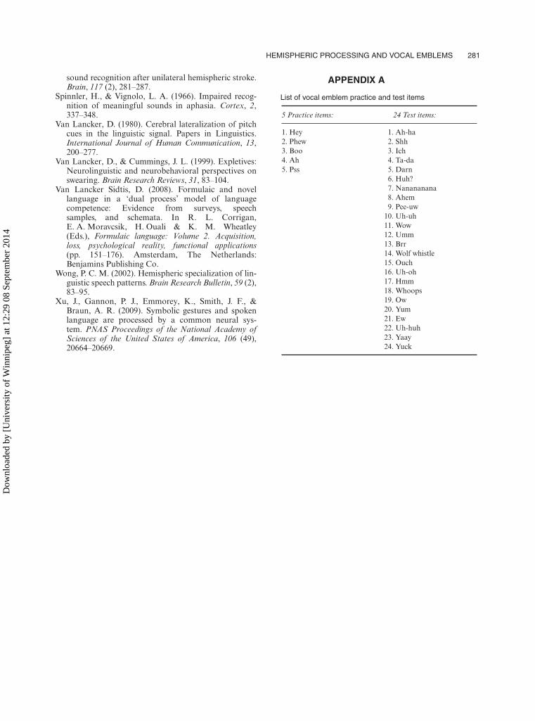
Stimuli
For both tasks, after the 5 practice items, 24 testitems were given (see Appendix A for the completelist of VE practice and test items). The stimuli usedin the study were selected based in part on vocalversions of emblems collected by Johnson, Ekman,and Friesen (1981). The VE items were recordedon a CD at a loudness level of 20 dB root meansquare (rms) below full scale, except for two items,psst and shh, which were normalized to 30 dB belowfull scale for better audibility. These two stimulisounded more natural at the lower level, as these aretypically low-intensity sounds. The loudness level ofall spoken phrases was equalized at 25 dB below fullscale. The VEs were presented to the listeners by the
researcher via a CD player and speakers to amplifythe sounds as needed. The participants were ableto listen to the VEs again, as often as necessaryto make a decision (which required mostly one totwo repetitions), and, if needed, a verbal model wasprovided to supplement the CD recording.
The response sheets consisted of either color pho-tographs (for the picture task) or verbal phrasesthat were written on large white index cards (for thephrase task).
Statistical analysis
The statistical analyses conducted involved the fol-lowing: first, a median test was performed on thecombined results from both groups on each ofthe picture and the phrase tasks. For each task,we ranked, then split the data at the median, todetermine how many participant scores from eachgroup fell above or below the median. Then anon-parametric Fisher’s exact test was conductedas it is used to compare small samples, namelywhen frequency data in each cell is five or less.A 2 × 3 Fisher’s exact test (two-tailed, p < .05)was calculated to compare the three groups oneach task. When a significant difference was found,further analysis included a comparison of eachtwo-group comparison using a 2 × 2 table (one-tailed, p < .02) in order to identify between
Dow
nloa
ded
by [
Uni
vers
ity o
f W
inni
peg]
at 1
2:29
08
Sept
embe
r 20
14
HEMISPHERIC PROCESSING AND VOCAL EMBLEMS 275
which two groups the difference lay. A strictercriterion for significance was set (p < .02) forthe follow-up 2 × 2 calculations in order toexclude Type I errors. Additionally, within-groupdifferences, of performance on the two tasks, werecalculated for each group using a 2 × 2 Fisher’sexact test.
RESULTS
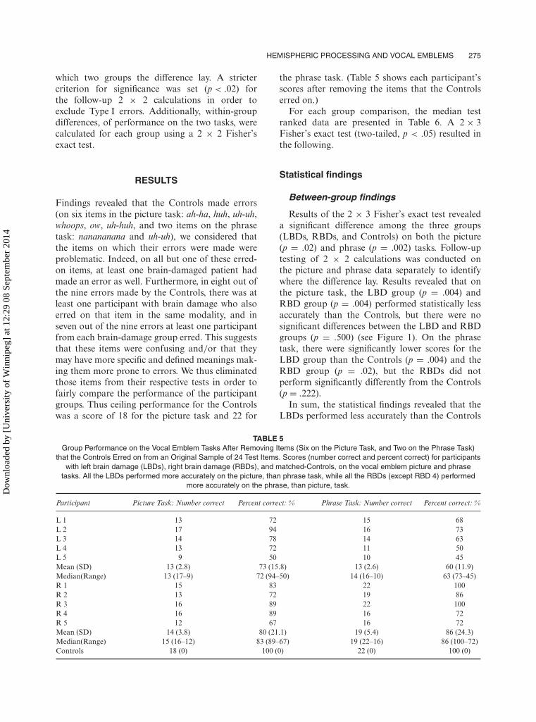
Findings revealed that the Controls made errors(on six items in the picture task: ah-ha, huh, uh-uh,whoops, ow, uh-huh, and two items on the phrasetask: nanananana and uh-uh), we considered thatthe items on which their errors were made wereproblematic. Indeed, on all but one of these erred-on items, at least one brain-damaged patient hadmade an error as well. Furthermore, in eight out ofthe nine errors made by the Controls, there was atleast one participant with brain damage who alsoerred on that item in the same modality, and inseven out of the nine errors at least one participantfrom each brain-damage group erred. This suggeststhat these items were confusing and/or that theymay have more specific and defined meanings mak-ing them more prone to errors. We thus eliminatedthose items from their respective tests in order tofairly compare the performance of the participantgroups. Thus ceiling performance for the Controlswas a score of 18 for the picture task and 22 for
the phrase task. (Table 5 shows each participant’sscores after removing the items that the Controlserred on.)
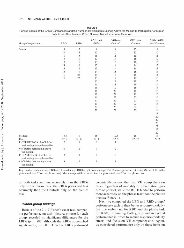
For each group comparison, the median testranked data are presented in Table 6. A 2 × 3Fisher’s exact test (two-tailed, p < .05) resulted inthe following.
Statistical findings
Between-group findings
Results of the 2 × 3 Fisher’s exact test revealeda significant difference among the three groups(LBDs, RBDs, and Controls) on both the picture(p = .02) and phrase (p = .002) tasks. Follow-uptesting of 2 × 2 calculations was conducted onthe picture and phrase data separately to identifywhere the difference lay. Results revealed that onthe picture task, the LBD group (p = .004) andRBD group (p = .004) performed statistically lessaccurately than the Controls, but there were nosignificant differences between the LBD and RBDgroups (p = .500) (see Figure 1). On the phrasetask, there were significantly lower scores for theLBD group than the Controls (p = .004) and theRBD group (p = .02), but the RBDs did notperform significantly differently from the Controls(p = .222).
In sum, the statistical findings revealed that theLBDs performed less accurately than the Controls
TABLE 5Group Performance on the Vocal Emblem Tasks After Removing Items (Six on the Picture Task, and Two on the Phrase Task)
that the Controls Erred on from an Original Sample of 24 Test Items. Scores (number correct and percent correct) for participantswith left brain damage (LBDs), right brain damage (RBDs), and matched-Controls, on the vocal emblem picture and phrase
tasks. All the LBDs performed more accurately on the picture, than phrase task, while all the RBDs (except RBD 4) performedmore accurately on the phrase, than picture, task.
Participant Picture Task: Number correct Percent correct: % Phrase Task: Number correct Percent correct: %
L 1 13 72 15 68L 2 17 94 16 73L 3 14 78 14 63L 4 13 72 11 50L 5 9 50 10 45Mean (SD) 13 (2.8) 73 (15.8) 13 (2.6) 60 (11.9)Median(Range) 13 (17–9) 72 (94–50) 14 (16–10) 63 (73–45)R 1 15 83 22 100R 2 13 72 19 86R 3 16 89 22 100R 4 16 89 16 72R 5 12 67 16 72Mean (SD) 14 (3.8) 80 (21.1) 19 (5.4) 86 (24.3)Median(Range) 15 (16–12) 83 (89–67) 19 (22–16) 86 (100–72)Controls 18 (0) 100 (0) 22 (0) 100 (0)
Dow
nloa
ded
by [
Uni
vers
ity o
f W
inni
peg]
at 1
2:29
08
Sept
embe
r 20
14
276 NEUMANN-WERTH, LEVY, OBLER
TABLE 6Ranked Scores of the Group Comparisons and the Number of Participants Scoring Above the Median (5 Participants/Group) on
Both Tasks, After Items on Which Controls Made Errors were Removed
LBDs and LBDs and RBDs and LBDs, RBD,s,Group Comparisons: LBDs RBDs RBDs Controls Controls and Controls
Scores: 9 12 9 9 12 910 13 10 10 13 1011 15 11 11 15 1113 16 12 13 16 1213 16 13 13 16 1314 16 13 14 16 1314 16 13 14 16 1315 19 14 15 18 1416 22 14 16 18 1417 22 15 17 18 15
15 18 18 1516 18 18 1616 19 18 1616 22 18 1616 22 18 1616 22 22 1617 22 22 1719 22 22 1822 22 22 1822 22 22 18
18222222222222
Median: 13.5 16 15 17.5 18 16Range: 17–9 22–12 22–9 22–9 22–12 22–9PICTURE TASK: # of LBDs 2 1 0 1
performing above the median# of RBDs performing above 0 2 0 2
the medianPHRASE TASK: # of LBDs 3 1 0 0
performing above the median# of RBDs performing above 3 5 3 3
the median
Key: bold = median scores, LBD=left brain damage, RBD=right brain damage; The Controls performed at ceiling (Score of 18 on thepicture task and 22 on the phrase task). Maximum possible score is 18 on the picture task and 22 on the phrase task.
on both tasks and less accurately than the RBDsonly on the phrase task; the RBDs performed lessaccurately than the Controls only on the picturetask.
Within-group findings
Results of the 2 × 2 Fisher’s exact test, compar-ing performance on task (picture, phrase) for eachgroup, revealed no significant differences for theLBDs (p = .897) although the RBDs approachedsignificance (p = .080). Thus the LBDs performed
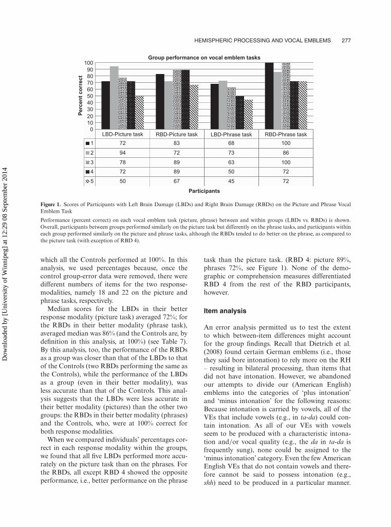
consistently across the two VE comprehensiontasks, regardless of modality of presentation (pic-ture or phrase), while the RBDs tended to performmore accurately on the phrase task than the pictureone (see Figure 1).
Next, we compared the LBD and RBD groups’performance each in their better response-modality(i.e., the verbal task for RBD and the phrase taskfor RBD), examining both group and individualperformance in order to reduce response-modalityeffects and focus on VE comprehension. Again,we considered performance only on those items on
Dow
nloa
ded
by [
Uni
vers
ity o
f W
inni
peg]
at 1
2:29
08
Sept
embe
r 20
14
HEMISPHERIC PROCESSING AND VOCAL EMBLEMS 277
1009080706050
Per
cen
t co
rrec
t
403020100
1
2
3
4
5
LBD-Picture task RBD-Picture task LBD-Phrase task RBD-Phrase task
72 83 68 100
86
100
72
72
73
63
50
45
Participants
72
89
89
67
94
78
72
50
Group performance on vocal emblem tasks
Figure 1. Scores of Participants with Left Brain Damage (LBDs) and Right Brain Damage (RBDs) on the Picture and Phrase VocalEmblem Task
Performance (percent correct) on each vocal emblem task (picture, phrase) between and within groups (LBDs vs. RBDs) is shown.Overall, participants between groups performed similarly on the picture task but differently on the phrase tasks, and participants withineach group performed similarly on the picture and phrase tasks, although the RBDs tended to do better on the phrase, as compared tothe picture task (with exception of RBD 4).
which all the Controls performed at 100%. In thisanalysis, we used percentages because, once thecontrol group-error data were removed, there weredifferent numbers of items for the two response-modalities, namely 18 and 22 on the picture andphrase tasks, respectively.
Median scores for the LBDs in their betterresponse modality (picture task) averaged 72%; forthe RBDs in their better modality (phrase task),averaged median was 86% (and the Controls are, bydefinition in this analysis, at 100%) (see Table 7).By this analysis, too, the performance of the RBDsas a group was closer than that of the LBDs to thatof the Controls (two RBDs performing the same asthe Controls), while the performance of the LBDsas a group (even in their better modality), wasless accurate than that of the Controls. This anal-ysis suggests that the LBDs were less accurate intheir better modality (pictures) than the other twogroups: the RBDs in their better modality (phrases)and the Controls, who, were at 100% correct forboth response modalities.
When we compared individuals’ percentages cor-rect in each response modality within the groups,we found that all five LBDs performed more accu-rately on the picture task than on the phrases. Forthe RBDs, all except RBD 4 showed the oppositeperformance, i.e., better performance on the phrase
task than the picture task. (RBD 4: picture 89%,phrases 72%, see Figure 1). None of the demo-graphic or comprehension measures differentiatedRBD 4 from the rest of the RBD participants,however.
Item analysis
An error analysis permitted us to test the extentto which between-item differences might accountfor the group findings. Recall that Dietrich et al.(2008) found certain German emblems (i.e., thosethey said bore intonation) to rely more on the RH– resulting in bilateral processing, than items thatdid not have intonation. However, we abandonedour attempts to divide our (American English)emblems into the categories of ‘plus intonation’and ‘minus intonation’ for the following reasons:Because intonation is carried by vowels, all of theVEs that include vowels (e.g., in ta-da) could con-tain intonation. As all of our VEs with vowelsseem to be produced with a characteristic intona-tion and/or vocal quality (e.g., the da in ta-da isfrequently sung), none could be assigned to the‘minus intonation’ category. Even the few AmericanEnglish VEs that do not contain vowels and there-fore cannot be said to possess intonation (e.g.,shh) need to be produced in a particular manner.
Dow
nloa
ded
by [
Uni
vers
ity o
f W
inni
peg]
at 1
2:29
08
Sept
embe
r 20
14
278 NEUMANN-WERTH, LEVY, OBLER
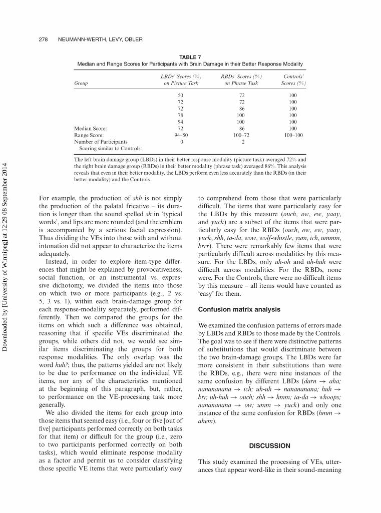
TABLE 7Median and Range Scores for Participants with Brain Damage in their Better Response Modality
LBDs’ Scores (%) RBDs’ Scores (%) Controls’Group on Picture Task on Phrase Task Scores (%)
50 72 10072 72 10072 86 10078 100 10094 100 100
Median Score: 72 86 100Range Score: 94–50 100–72 100–100Number of Participants 0 2
Scoring similar to Controls:
The left brain damage group (LBDs) in their better response modality (picture task) averaged 72% andthe right brain damage group (RBDs) in their better modality (phrase task) averaged 86%. This analysisreveals that even in their better modality, the LBDs perform even less accurately than the RBDs (in theirbetter modality) and the Controls.
For example, the production of shh is not simplythe production of the palatal fricative – its dura-tion is longer than the sound spelled sh in ‘typicalwords’, and lips are more rounded (and the emblemis accompanied by a serious facial expression).Thus dividing the VEs into those with and withoutintonation did not appear to characterize the itemsadequately.
Instead, in order to explore item-type differ-ences that might be explained by provocativeness,social function, or an instrumental vs. expres-sive dichotomy, we divided the items into thoseon which two or more participants (e.g., 2 vs.5, 3 vs. 1), within each brain-damage group foreach response-modality separately, performed dif-ferently. Then we compared the groups for theitems on which such a difference was obtained,reasoning that if specific VEs discriminated thegroups, while others did not, we would see sim-ilar items discriminating the groups for bothresponse modalities. The only overlap was theword huh?; thus, the patterns yielded are not likelyto be due to performance on the individual VEitems, nor any of the characteristics mentionedat the beginning of this paragraph, but, rather,to performance on the VE-processing task moregenerally.
We also divided the items for each group intothose items that seemed easy (i.e., four or five [out offive] participants performed correctly on both tasksfor that item) or difficult for the group (i.e., zeroto two participants performed correctly on bothtasks), which would eliminate response modalityas a factor and permit us to consider classifyingthose specific VE items that were particularly easy
to comprehend from those that were particularlydifficult. The items that were particularly easy forthe LBDs by this measure (ouch, ow, ew, yaay,and yuck) are a subset of the items that were par-ticularly easy for the RBDs (ouch, ow, ew, yaay,yuck, shh, ta-da, wow, wolf-whistle, yum, ich, ummm,brrr). There were remarkably few items that wereparticularly difficult across modalities by this mea-sure. For the LBDs, only uh-oh and uh-huh weredifficult across modalities. For the RBDs, nonewere. For the Controls, there were no difficult itemsby this measure – all items would have counted as‘easy’ for them.
Confusion matrix analysis
We examined the confusion patterns of errors madeby LBDs and RBDs to those made by the Controls.The goal was to see if there were distinctive patternsof substitutions that would discriminate betweenthe two brain-damage groups. The LBDs were farmore consistent in their substitutions than werethe RBDs, e.g., there were nine instances of thesame confusion by different LBDs (darn → aha;nanananana → ich; uh-uh → nanananana; huh →brr; uh-huh → ouch; shh → hmm; ta-da → whoops;nanananana → ow; umm → yuck) and only oneinstance of the same confusion for RBDs (hmm →ahem).
DISCUSSION
This study examined the processing of VEs, utter-ances that appear word-like in their sound-meaning
Dow
nloa
ded
by [
Uni
vers
ity o
f W
inni
peg]
at 1
2:29
08
Sept
embe
r 20
14
HEMISPHERIC PROCESSING AND VOCAL EMBLEMS 279
correspondence, but non-word-like in that they defylanguages’ phonological, morphological, and syn-tactic constraints, and rely more on intonation andon physical gestures than do typical words. RH vs.LH involvement in processing VEs was examinedby the participation of LBDs, RBDs, and Controlsin a picture task and a phrase task in which theywere asked to match VEs (presented auditorily)with photographs depicting settings where theywould expect to hear the emblem and with ver-bal translations respectively. The LBDs performedsignificantly less accurately than the Controls onboth tasks and significantly less accurately thanthe RBDs only on the phrase task. The poor per-formance (73% on the pictures, and 60% on thephrases) by the LBDs was consistent even on thetask in their better modality (i.e., 73% accuracyon the picture task). This suggests that the LBDs’lower performance is an indication of their diffi-culty in processing the VEs overall, and not simplyan indication of difficulty with a response modal-ity. Thus, it may be concluded that processing ofVEs recruits primarily the LH, consistent withthe findings of Dietrich et al. (2008) for Germaninterjections and Xu et al. (2009) for emblemgestures.
The RBD group, however, performed signifi-cantly less accurately than the Controls only on thepicture task, the task with which they were expectedto have more difficulty. This is likely due to themore holistic processing requirements of the picturetask, as such processing is an impairment character-istic of individuals with RBD. Their performancein their better modality, the phrase task, was notsignificantly different from Controls, as expected.Taken together, this suggests that the RBDs’ lowerperformance on the picture task might be due totheir difficulty with the response modality and notto VE processing, per se.
Van Lancker Sidtis’s (2008) observation of wowproduction preserved in individuals with LBDbut not by individuals with RBD or subcorticallesions, and other evidence pointing to RH andbilateral representation for emblem-like utterances,suggested that individual VEs might have differ-ent lateralization from regular words. However, wefind no evidence for this, for comprehension, atleast. That is, our item analysis revealed no spe-cific characteristics of VEs associated particularlywith LBDs or RBDs, but, rather, that the LBDsmade more consistent substitutions and ones that
truly indicated lack of comprehension than did theRBDs. Thus, on the continuum proposed by VanLancker (1980), VEs appear to be markedly moretoward the language end than the non-languagecommunication end.
With respect to heterogeneity of hemisphericlateralization for specific items, or item clusters(instrumental vs. expressive; social vs. provocative;high vs. low affect) as proposed by various authorsdiscussed in the Introduction, recall that our item-analysis (in which we divided the items into thoseon which two or more participants had performeddifferently) revealed that only one item occurredon both lists. This suggests that our findings arosenot because certain VE items were differently pro-cessed in the two hemispheres, but rather becausethe LBDs generally have more difficulty processingVEs than do RBDs.
Furthermore, the results from the confusionmatrix revealed a sole RBD error: substitutionof ahem for hmm. This might be considered anexpected confusion – these two VEs have similarphonetic properties and are not as clearly associ-ated with delineated specific meanings as are VEssuch as shh. In contrast, the confusions made by theLBDs are ones that seem less ‘confusable’, e.g., uh-huh (meaning ‘yes’) with ouch (meaning ‘it hurts’).This could bolster our argument that the LBDstruly did not understand the meaning of these VEs,whereas this likely cannot be said about the RBDsbased on their confusion pattern.
In working with patients with moderate–severeWernicke’s aphasia, clinicians often focus on stim-ulating language comprehension and expressionto their maximal potential, while simultaneouslycounseling family members regarding the best wayto encourage successful communicative interac-tions. The research presented here aimed at dis-covering whether LBD patients with language-comprehension difficulties would comprehend thecommunicative meaning of VEs, despite theirmoderate–severe comprehension deficits. It wasanticipated that if success in understanding VEs,such as shh and brr, was demonstrated on eitherof the experimental tasks, then this would sug-gest that the linguistic information was being pro-cessed through the brain’s nonlinguistic channelsand thus VEs would prove a valuable comple-ment for activities of daily living communicativeexchanges. Our research findings are consistentwith the notion that patients with moderate–severe
Dow
nloa
ded
by [
Uni
vers
ity o
f W
inni
peg]
at 1
2:29
08
Sept
embe
r 20
14
280 NEUMANN-WERTH, LEVY, OBLER
Wernicke’s aphasia process VEs as they do lan-guage, regardless of task (linguistic vs. nonlinguis-tic). Thus, clinicians using compensatory strategiesto facilitate patients’ understanding of linguisticinformation would likely need to do the samefor facilitating comprehension of these very basicforms of symbolic sounds. It is possible, though,that presenting ‘provocative’ emblems (Knutsonet al., 2008) with exaggerated intonation (Dietrichet al., 2008) would render them easier to under-stand.
A limitation of our study is the small number ofparticipants in each group and the relatively widerange of scores within the groups. To the extent thatour item- and individual-analyses converge withour group analyses, however, our findings are sup-ported. Further limitations of this study are thatsome of our participants had hearing losses andwere not monolingual, as described in footnotes3 and 4. Replication with more normal-hearingmonolinguals would be desirable.
Neuroimaging techniques, too, could provideconverging evidence of performance on vocal-emblem tasks and should circumscribe regionswithin the LH – which may or may not overlap fullywith those for regular lexicon – that are involved incomprehending VEs. Studies of production of VEsby patients with brain damage should complementour study of comprehension of this class of com-munication items. Additionally, the exploration ofVEs cross-linguistically would prove interesting inseveral regards. Different languages employ differ-ent sets of VEs, and it is possible that these reflectdifferent subtypes in languages other than English.Languages with tone, too, might integrate into-nation into their VEs somewhat differently fromthe way non-tone languages do, and studying VEsin such languages would, thus, further our under-standing of processing of this class of sounds basedon intonation characteristics.
Original manuscript received 15 April 2011Revised manuscript accepted 5 December 2011
First published online 10 May 2012
REFERENCES
Broca, P. P. (1861). Remarques sur le siège de la faculté dulangage articulé, suivies d’une observation d’aphémie(perte de la parole). Bulletins de la Société d’anatomie(Paris), 2 (6), 330–357.
Broca, P. P. (1865). Sur le siège de la faculté du lan-gage articulé. Bulletin de la Société d’anthropologie, 6,337–393.
Critchley, M., & Critchley, E. A. (1998). John HughlingsJackson: Father of English neurology. New York, NY:Oxford University Press.
Dietrich, S., Hertrich, I., Alter, K., Ischebeck, A., &Ackermann, H. (2008). Understanding the emotionalexpression of verbal interjections: A functional MRIstudy. NeuroReport, 19 (18), 1751–1755.
Efron, D. (1972). Gesture, race and culture. The Hague,The Netherlands: Mouton. (Original work publishedas Gesture and Environment, 1941. New York: King’sCrown.)
Folstein, M. F., Folstein, S. E., & McHugh, P. R. (1975).Mini-Mental State: A practical method for gradingthe cognitive state of patients for the clinician. Journalof Psychiatric Research, 12, 189–198.
Gallagher, H. L., & Frith, C. D. (2004). Dissociableneural pathways for the perception and recog-nition of expressive and instrumental gestures.Neuropsychologia, 42, 1725–1736.
Goodglass, H., Kaplan, E., & Barresi, B. (2001). BostonDiagnostic Aphasia Examination-Short Form, 3rd edi-tion. Austin, TX: Pro-Ed.
Hashimoto, T., Usui, N., Taira, M., Nose, I., Haji, T.,& Kojima, S. (2006). The neural mechanism asso-ciated with the processing of onomatopoeic sounds.NeuroImage, 31 (4), 1762–1770.
Heilman, K., Scholes, R., & Watson, R. (1975). Auditoryaffective agnosia: Disturbed comprehension of affec-tive speech. Journal of Neurology, Neurosurgery andPsychiatry, 38, 69–72.
Johnson, H., Ekman, P., & Friesen, W. (1981).Communicative body movements: Americanemblems. In T. A. Sebeok & D. J. Umiker-Sebeok(Eds.), Nonverbal communication, interaction, andgesture: Selections from Semiotica (pp. 401–419). TheHague, The Netherlands: Mouton.
Knutson, K. M., McClellan, E. M., & Grafman, J.(2008) Observing social gestures: An fMRI study.Experimental Brain Research, 188, 187–198.
MacSweeney, M., Capek, C. M., Campbell, R., &Woll, B. (2008). The signing brain: The neurobiol-ogy of sign language. Trends in Cognitive Sciences, 12,432–440.
Montgomery, K. J., & Haxby, J. V. (2008). Mirrorneuron system differentially activated by facialexpressions and social hand gestures: A func-tional magnetic resonance imaging study Journal ofCognitive Neuroscience, 20, 1866–1877.
Robin, D., Tranel, D., & Damasio, H. (1990). Auditoryperception of temporal and spectral events in patientswith focal left and right cerebral lesions. Brain andLanguage, 39, 539–555.
Saygin, A. P., Dick, F., Wilson, S. M., Dronkers, N. F.,& Bates, E. (2003). Neural resources for processinglanguage and environmental sounds: Evidence fromaphasia. Brain, 126, 928–945.
Schnider, A., Benson, D. F., Alexander, D. N., &Schnider-Klaus, A. (1994) Non-verbal environmental
Dow
nloa
ded
by [
Uni
vers
ity o
f W
inni
peg]
at 1
2:29
08
Sept
embe
r 20
14
HEMISPHERIC PROCESSING AND VOCAL EMBLEMS 281
sound recognition after unilateral hemispheric stroke.Brain, 117 (2), 281–287.
Spinnler, H., & Vignolo, L. A. (1966). Impaired recog-nition of meaningful sounds in aphasia. Cortex, 2,337–348.
Van Lancker, D. (1980). Cerebral lateralization of pitchcues in the linguistic signal. Papers in Linguistics.International Journal of Human Communication, 13,200–277.
Van Lancker, D., & Cummings, J. L. (1999). Expletives:Neurolinguistic and neurobehavioral perspectives onswearing. Brain Research Reviews, 31, 83–104.
Van Lancker Sidtis, D. (2008). Formulaic and novellanguage in a ‘dual process’ model of languagecompetence: Evidence from surveys, speechsamples, and schemata. In R. L. Corrigan,E. A. Moravcsik, H. Ouali & K. M. Wheatley(Eds.), Formulaic language: Volume 2. Acquisition,loss, psychological reality, functional applications(pp. 151–176). Amsterdam, The Netherlands:Benjamins Publishing Co.
Wong, P. C. M. (2002). Hemispheric specialization of lin-guistic speech patterns. Brain Research Bulletin, 59 (2),83–95.
Xu, J., Gannon, P. J., Emmorey, K., Smith, J. F., &Braun, A. R. (2009). Symbolic gestures and spokenlanguage are processed by a common neural sys-tem. PNAS Proceedings of the National Academy ofSciences of the United States of America, 106 (49),20664–20669.
APPENDIX A
List of vocal emblem practice and test items
5 Practice items: 24 Test items:
1. Hey 1. Ah-ha2. Phew 2. Shh3. Boo 3. Ich4. Ah 4. Ta-da5. Pss 5. Darn
6. Huh?7. Nanananana8. Ahem9. Pee-uw
10. Uh-uh11. Wow12. Umm13. Brr14. Wolf whistle15. Ouch16. Uh-oh17. Hmm18. Whoops19. Ow20. Yum21. Ew22. Uh-huh23. Yaay24. Yuck
Dow
nloa
ded
by [
Uni
vers
ity o
f W
inni
peg]
at 1
2:29
08
Sept
embe
r 20
14