Embed Size (px)
Citation preview

© 2004 Nature Publishing Group
(Accurate Chemical and Scientific Corp.) to destroy inhibitors of influenza virus
replication. After inactivation of the RDE by treatment at 56 8C for 1 h, WSN/HspNsp and
K233 were each incubated with twofold serial dilutions of each serum at 37 8C for 1 h.
Remaining infectivity was determined by titration of the samples in a plaque assay on
MDCK cells.
Received 28 July; accepted 10 August 2004; doi:10.1038/nature02951.
1. Johnson, N. P. & Mueller, J. Updating the accounts: global mortality of the 1918–1920 “Spanish”
influenza pandemic. Bull. Hist. Med. 76, 105–115 (2002).
2. Reid, A. H., Fanning, T. G., Hultin, J. V. & Taubenberger, J. K. Origin and evolution of the 1918
“Spanish” influenza virus hemagglutinin gene. Proc. Natl Acad. Sci. USA 96, 1651–1656 (1999).
3. Reid, A. H., Fanning, T. G., Janczewski, T. A. & Taubenberger, J. K. Characterization of the 1918
“Spanish” influenza virus neuraminidase gene. Proc. Natl Acad. Sci. USA 97, 6785–6790 (2000).
4. Tumpey, T. M. et al. Existing antivirals are effective against influenza viruses with genes from the 1918
pandemic virus. Proc. Natl Acad. Sci. USA 99, 13849–13854 (2002).
5. Tumpey, T. M. et al. Pathogenicity and immunogenicity of influenza viruses with genes from the 1918
pandemic virus. Proc. Natl Acad. Sci. USA 101, 3166–3171 (2004).
6. Oxford, J. S. Influenza A pandemics of the 20th century with special reference to 1918: virology,
pathology and epidemiology. Rev. Med. Virol. 10, 119–133 (2000).
7. Lamb, R. A. & Krug, R. M. Fields Virology (eds Knipe, D. M. et al.) 1487–1531 (Lippincott-Raven,
Philadelphia, Pennsylvania, 2001).
8. Wright, P. F. & Webster, R. G. Fields Virology (eds Knipe, D. M. et al.) 1533–1579 (Lippincott-Raven,
Philadelphia, Pennsylvania, 2001).
9. Oxford, J. S. et al. World War I may have allowed the emergence of “Spanish” influenza. Lancet Infect.
Dis. 2, 111–114 (2002).
10. Basler, C. F. et al. Sequence of the 1918 pandemic influenza virus nonstructural gene (NS) segment and
characterization of recombinant viruses bearing the 1918 NS genes. Proc. Natl Acad. Sci. USA 98,
2746–2751 (2001).
11. Chen, W. et al. A novel influenza A virus mitochondrial protein that induces cell death. Nature Med. 7,
1306–1312 (2001).
12. Neumann, G. et al. Generation of influenza Aviruses entirely from cloned cDNAs. Proc. Natl Acad. Sci.
USA 96, 9345–9350 (1999).
13. Goto, H., Wells, K., Takada, A. & Kawaoka, Y. Plasminogen-binding activity of neuraminidase
determines the pathogenicity of influenza A virus. J. Virol. 75, 9297–9301 (2001).
14. Guan, Y. et al. H5N1 influenza: a protean pandemic threat. Proc. Natl Acad. Sci. USA 101, 8156–8161
(2004).
15. Cheung, C. Y. et al. Induction of proinflammatory cytokines in human macrophages by influenza A
(H5N1) viruses: a mechanism for the unusual severity of human disease? Lancet 360, 1831–1837
(2002).
16. Hayden, F. G. et al. Local and systemic cytokine responses during experimental human influenza
A virus infection. Relation to symptom formation and host defense. J. Clin. Invest. 101, 643–649
(1998).
17. Fritz, R. S. et al. Nasal cytokine and chemokine responses in experimental influenza A virus infection:
results of a placebo-controlled trial of intravenous zanamivir treatment. J. Infect. Dis. 180, 586–593
(1999).
18. Kaiser, L., Fritz, R. S., Straus, S. E., Gubareva, L. & Hayden, F. G. Symptom pathogenesis during acute
influenza: interleukin-6 and other cytokine responses. J. Med. Virol. 64, 262–268 (2001).
19. Abraham, E. Neutrophils and acute lung injury. Crit. Care Med. 31 ( 4 suppl.), S195–S199
(2003).
20. Lee, W. L. & Downey, G. P. Neutrophil activation and acute lung injury. Curr. Opin. Crit. Care 7, 1–7
(2001).
21. Connor, J. S., Kawaoka, Y., Webster, R. G. & Paulson, J. C. Receptor specificity in human, avian and
equine H2 and H3 influenza virus isolates. Virology 15, 17–23 (1994).
22. Matrosovich, M. N., Matrosovich, T. Y., Gray, T., Roberts, N. A. & Klenk, H. D. Human and avian
influenza viruses target different cell types in cultures of human airway epithelium. Proc. Natl Acad.
Sci. USA 101, 4620–4624 (2004).
23. Gamblin, S. J. et al. The structure and receptor binding properties of the 1918 influenza
hemagglutinin. Science 303, 1838–1842 (2004).
24. Stevens, J. et al. Structure of the uncleaved human H1 hemagglutinin from the extinct 1918 influenza
virus. Science 303, 1866–1870 (2004).
25. Francis, T. Jr Influenza: The newe acquayantance. Ann. Intern. Med. 39, 203–221 (1953).
26. Fazekas De St Groth, S. & Webster, R. G. Disquisitions on original antigenic sin. I. Evidence in man.
J. Exp. Med. 124, 331–345 (1966).
27. Geiss, G. K. et al. Cellular transcriptional profiling in influenza A virus-infected lung epithelial
cells: the role of the nonstructural NS1 protein in the evasion of the host innate defense and
its potential contribution to pandemic influenza. Proc. Natl Acad. Sci. USA 99, 10736–10741
(2002).
28. Gambaryan, A. S. & Matrosovich, M. N. A solid-phase enzyme-linked assay for influenza virus
receptor-binding activity. J. Virol. Methods 39, 111–123 (1992).
29. Totani, K. et al. Chemoenzymatic synthesis and application of glycopolymers containing multivalent
sialyloligosaccharides with a poly(L-glutamic acid) backbone for inhibition of infection by influenza
viruses. Glycobiology 13, 315–326 (2003).
30. Reed, L. J. & Muench, H. A simple method of estimating fifty per cent endpoints. Am. J. Hyg. 27,
493–497 (1938).
Supplementary Information accompanies the paper on www.nature.com/nature.
Acknowledgements We thank D. Dick, A. Grolla, M. Garbutt and S. Jones for assistance with
BL4 procedures, M. McGregor and K. Wells for technical assistance, J. Gilbert for editing the
manuscript, and Y. Kawaoka for illustrations. This work was supported by a grant-in-aid from
the Japanese Ministry of Education, Culture, Sports, Science and Technology, CREST (Japan
Science and Technology Corporation), NIAID Public Health Service research grants and
Health Canada.
Competing interests statement The authors declare that they have no competing financial
interests.
Correspondence and requests for materials should be addressed to Y.K.
..............................................................
Hedgehog signalling in prostateregeneration, neoplasia andmetastasisSunil S. Karhadkar1,2, G. Steven Bova2–4, Nadia Abdallah2, Surajit Dhara2,Dale Gardner5, Anirban Maitra2, John T. Isaacs3,4, David M. Berman1–4
& Philip A. Beachy1,4
1Department of Molecular Biology and Genetics and the Howard Hughes MedicalInstitute, 2Departments of Pathology, 3Urology and 4Oncology, The Johns HopkinsUniversity School of Medicine, Baltimore, Maryland 21205, USA5USDA ARS, Poisonous Plant Research Laboratory, Logan, Utah 84341, USA.............................................................................................................................................................................
Metastatic cancers adopt certain properties of normal cellsin developing or regenerating organs, such as the ability toproliferate and alter tissue organization. We find here thatactivity of the Hedgehog (Hh) signalling pathway, which hasessential roles in developmental patterning1–6, is required forregeneration of prostate epithelium, and that continuous path-way activation transforms prostate progenitor cells and rendersthem tumorigenic. Elevated pathway activity furthermore dis-tinguishes metastatic from localized prostate cancer, and path-way manipulation can modulate invasiveness and metastasis.Pathway activity is triggered in response to endogenousexpression of Hh ligands, and is dependent upon the expressionof Smoothened, an essential Hh response component1,2,7 that isnot expressed in benign prostate epithelial cells. Monitoring andmanipulating Hh pathway activity may thus offer significantimprovements in diagnosis and treatment of prostate cancerswith metastatic potential.
Hedgehog signalling influences development and homeostasis ofmany gut-derived organs, some of which give rise to Hh-dependentcancers. In the foregut, for example, ligand-dependent pathwayactivity is required for growth of a significant proportion of small-cell lung cancers and of carcinomas of the stomach, oesophagus,pancreas and biliary tract8–10. We consider here the prostate, the siteof origin for the second most lethal malignancy in men. Theprostate derives from embryonic endoderm that is caudal to theintestinal portal, and which also gives rise to organs such asthe colon. Although tumorigenesis in the colon invariably involvesWnt pathway activation, the prostate has recently been found toexhibit a developmental patterning role for Hh signalling4–6 similarto that in the lung3.
We examined expression of Hh pathway ligands and endogenoustargets in human prostate cancer cell lines by measuring levels ofmessenger RNA that encode the pathway components GLI andPATCHED (PTCH). Both GLI and PTCH are transcriptional targetsof pathway activation with opposite roles in pathway response; GLIserves as a positive transcriptional effector and PTCH functions torestrain pathway activity by suppressing the action of Smoothened
letters to nature
NATURE | VOL 431 | 7 OCTOBER 2004 | www.nature.com/nature 707

© 2004 Nature Publishing Group
(SMO). This negative function of PTCH is blocked by the binding ofHh ligands, and the pathway can thus be activated through Hh-mediated or mutational inactivation of PTCH; either form ofpathway activation requires SMO (reviewed in refs 1, 2, 7).
All four prostate cancer cell lines examined (PC3, DU145,CWR22RV1 and LnCAP) expressed transcripts encoding Sonic(SHH) and Indian (IHH) hedgehog ligands (Fig. 1a). Tumour cellsalso expressed PTCH and GLI transcripts, and pathway activity wasconfirmed by quantitative real-time analysis of polymerase chainreaction with reverse transcription (RT–PCR), which revealedPTCH mRNA levels to be elevated ,200–400-fold in cancer cells,relative to benign prostate epithelial cells (PrE cells; see below)(Fig. 1b). Introduction of a Hh-responsive GLI-luciferase reporter11
also revealed high luciferase activity in tumour cells, which wasfurther augmented by addition of exogenous Shh ligand (ShhNp)and was blocked by treatment with a ligand-neutralizing mono-clonal antibody (5E1) (ref. 8) (Supplementary Fig. 1a). Activity wasalso fully suppressible by treatment with cyclopamine, whichspecifically inhibits the Hh pathway response by binding toand stabilizing the inactive conformation of SMO11,12. As seen in22RV1-GLI cells, cyclopamine blockade of SMO was bypassed bystable overexpression of GLI, demonstrating the specificity of thecyclopamine effect in the Hh pathway.
Cyclopamine treatment inhibited growth of PC3, DU145 and22RV1 cells (Fig. 1c) and expression of c-Myc and cyclin D1(Supplementary Fig. 1c, d), as compared with treatment with thestructurally related but inactive compound, tomatidine8,13. Thisanti-proliferative effect of cyclopamine again was bypassed byoverexpression of GLI, but not by an altered inactive form, GLIzfd
(ref. 14) (Fig. 1c), and growth inhibition was confirmed in PC3 cellsby treatment with 5E1 antibody (Supplementary Fig. 1b).
We further tested the role of Hh pathway activity in vivo bytreating established subcutaneous PC3 and 22RV1 xenografttumours in athymic mice with daily subcutaneous injections ofcyclopamine (10 or 50 mg kg21) or of vehicle alone. We observedsuppression of tumour growth at 10 mg kg21 cyclopamine, andactual regression at 50 mg kg21. Animals treated at the intermediatedose of 10 mg kg21 were killed and a 90% reduction in staining forthe proliferation antigen Ki67 was noted (Supplementary Fig. 1f).Animals receiving treatment at 50 mg kg21 showed completeregression of the tumours within 20–24 days (d) of treatment(Fig. 1d, e; Supplementary Fig. 1g). Cessation of treatment didnot result in re-growth of tumours, even after observation periodsof 72 d (PC3) and 148 d (22RV1). Cyclopamine-treated 22RV1-GLItumours grew faster than vehicle-treated 22RV1 tumours (Fig. 1e),and this acceleration of tumour growth by GLI overexpression evenwith cyclopamine treatment confirms in vivo that the rate of tumourcell growth corresponds to the degree of Hh pathway activity(Fig. 1c, e; Supplementary Fig. 1a).
Complete and durable tumour regression suggests that cellscapable of renewing the tumour, that is, of functioning as tumourstem cells2,15,16, require Hh pathway activity for their maintenance.In this regard, it is notable that cyclopamine suppressed transcrip-tion of the gene encoding nestin, an intermediate filament proteinwhose expression has not been described previously in the prostate,but which marks progenitor cell populations elsewhere in theendoderm as well as in neural tissues and muscle17–19 (Supplemen-tary Fig. 1e). In addition to nestin, the Polycomb group protein
Figure 1 Hh pathway activity in growth of human prostate cancer cells. a, RT–PCR
assay indicating expression of transcripts encoding IHH and SHH ligands and the Hh
pathway targets PTCH and GLI in all prostate cancer cell lines examined. b, Quantitative
real-time RT–PCR for PTCH mRNA relative to levels in benign prostate epithelial cells.
c, Dose-dependent inhibition of growth in prostate cancer cells cultured with
cyclopamine. 22RV1-GLI cells maintained Hh pathway activity and growth, whereas
22RV1-GLIzfd cells remained susceptible. Results show the average of three
experiments. d, e, PC3 (d) and 22RV1 (e) prostate cancer xenograft tumours (median size
155 mm3) completely regressed after 22–28 d of cyclopamine treatment
(50 mg kg21 d21) and did not recur during 58 (PC3) or 148 d (22RV1) of subsequent
observation. Vehicle-treated tumours (22RV1-Ctl) and cyclopamine-treated
22RV1-GLI tumours grew rapidly. Error bars indicate standard error of the mean
(s.e.m.)
letters to nature
NATURE | VOL 431 | 7 OCTOBER 2004 | www.nature.com/nature708
© 2004 Nature Publishing Group
Bmi-1, required for self-renewal of somatic stem cells in haemato-poietic and neural lineages20, is also expressed in a Hedgehog-pathway-dependent manner (data not shown).
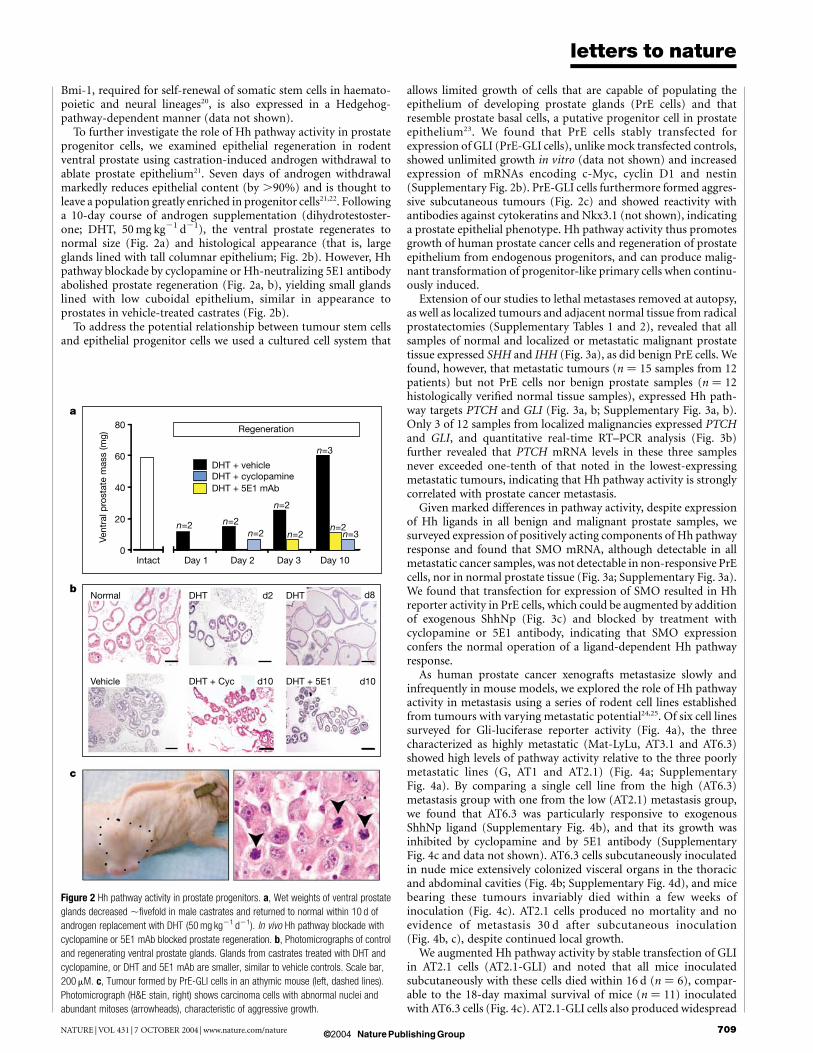
To further investigate the role of Hh pathway activity in prostateprogenitor cells, we examined epithelial regeneration in rodentventral prostate using castration-induced androgen withdrawal toablate prostate epithelium21. Seven days of androgen withdrawalmarkedly reduces epithelial content (by .90%) and is thought toleave a population greatly enriched in progenitor cells21,22. Followinga 10-day course of androgen supplementation (dihydrotestoster-one; DHT, 50 mg kg21 d21), the ventral prostate regenerates tonormal size (Fig. 2a) and histological appearance (that is, largeglands lined with tall columnar epithelium; Fig. 2b). However, Hhpathway blockade by cyclopamine or Hh-neutralizing 5E1 antibodyabolished prostate regeneration (Fig. 2a, b), yielding small glandslined with low cuboidal epithelium, similar in appearance toprostates in vehicle-treated castrates (Fig. 2b).
To address the potential relationship between tumour stem cellsand epithelial progenitor cells we used a cultured cell system that
allows limited growth of cells that are capable of populating theepithelium of developing prostate glands (PrE cells) and thatresemble prostate basal cells, a putative progenitor cell in prostateepithelium23. We found that PrE cells stably transfected forexpression of GLI (PrE-GLI cells), unlike mock transfected controls,showed unlimited growth in vitro (data not shown) and increasedexpression of mRNAs encoding c-Myc, cyclin D1 and nestin(Supplementary Fig. 2b). PrE-GLI cells furthermore formed aggres-sive subcutaneous tumours (Fig. 2c) and showed reactivity withantibodies against cytokeratins and Nkx3.1 (not shown), indicatinga prostate epithelial phenotype. Hh pathway activity thus promotesgrowth of human prostate cancer cells and regeneration of prostateepithelium from endogenous progenitors, and can produce malig-nant transformation of progenitor-like primary cells when continu-ously induced.
Extension of our studies to lethal metastases removed at autopsy,as well as localized tumours and adjacent normal tissue from radicalprostatectomies (Supplementary Tables 1 and 2), revealed that allsamples of normal and localized or metastatic malignant prostatetissue expressed SHH and IHH (Fig. 3a), as did benign PrE cells. Wefound, however, that metastatic tumours (n ¼ 15 samples from 12patients) but not PrE cells nor benign prostate samples (n ¼ 12histologically verified normal tissue samples), expressed Hh path-way targets PTCH and GLI (Fig. 3a, b; Supplementary Fig. 3a, b).Only 3 of 12 samples from localized malignancies expressed PTCHand GLI, and quantitative real-time RT–PCR analysis (Fig. 3b)further revealed that PTCH mRNA levels in these three samplesnever exceeded one-tenth of that noted in the lowest-expressingmetastatic tumours, indicating that Hh pathway activity is stronglycorrelated with prostate cancer metastasis.
Given marked differences in pathway activity, despite expressionof Hh ligands in all benign and malignant prostate samples, wesurveyed expression of positively acting components of Hh pathwayresponse and found that SMO mRNA, although detectable in allmetastatic cancer samples, was not detectable in non-responsive PrEcells, nor in normal prostate tissue (Fig. 3a; Supplementary Fig. 3a).We found that transfection for expression of SMO resulted in Hhreporter activity in PrE cells, which could be augmented by additionof exogenous ShhNp (Fig. 3c) and blocked by treatment withcyclopamine or 5E1 antibody, indicating that SMO expressionconfers the normal operation of a ligand-dependent Hh pathwayresponse.
As human prostate cancer xenografts metastasize slowly andinfrequently in mouse models, we explored the role of Hh pathwayactivity in metastasis using a series of rodent cell lines establishedfrom tumours with varying metastatic potential24,25. Of six cell linessurveyed for Gli-luciferase reporter activity (Fig. 4a), the threecharacterized as highly metastatic (Mat-LyLu, AT3.1 and AT6.3)showed high levels of pathway activity relative to the three poorlymetastatic lines (G, AT1 and AT2.1) (Fig. 4a; SupplementaryFig. 4a). By comparing a single cell line from the high (AT6.3)metastasis group with one from the low (AT2.1) metastasis group,we found that AT6.3 was particularly responsive to exogenousShhNp ligand (Supplementary Fig. 4b), and that its growth wasinhibited by cyclopamine and by 5E1 antibody (SupplementaryFig. 4c and data not shown). AT6.3 cells subcutaneously inoculatedin nude mice extensively colonized visceral organs in the thoracicand abdominal cavities (Fig. 4b; Supplementary Fig. 4d), and micebearing these tumours invariably died within a few weeks ofinoculation (Fig. 4c). AT2.1 cells produced no mortality and noevidence of metastasis 30 d after subcutaneous inoculation(Fig. 4b, c), despite continued local growth.
We augmented Hh pathway activity by stable transfection of GLIin AT2.1 cells (AT2.1-GLI) and noted that all mice inoculatedsubcutaneously with these cells died within 16 d (n ¼ 6), compar-able to the 18-day maximal survival of mice (n ¼ 11) inoculatedwith AT6.3 cells (Fig. 4c). AT2.1-GLI cells also produced widespread
Figure 2 Hh pathway activity in prostate progenitors. a, Wet weights of ventral prostate
glands decreased ,fivefold in male castrates and returned to normal within 10 d of
androgen replacement with DHT (50 mg kg21 d21). In vivo Hh pathway blockade with
cyclopamine or 5E1 mAb blocked prostate regeneration. b, Photomicrographs of control
and regenerating ventral prostate glands. Glands from castrates treated with DHT and
cyclopamine, or DHT and 5E1 mAb are smaller, similar to vehicle controls. Scale bar,
200 mM. c, Tumour formed by PrE-GLI cells in an athymic mouse (left, dashed lines).
Photomicrograph (H&E stain, right) shows carcinoma cells with abnormal nuclei and
abundant mitoses (arrowheads), characteristic of aggressive growth.
letters to nature
NATURE | VOL 431 | 7 OCTOBER 2004 | www.nature.com/nature 709

© 2004 Nature Publishing Group
visceral metastases (Fig. 4b; data not shown), and activation of Hhpathway targets thus seems sufficient for conferral of a lethalmetastatic phenotype. To determine whether the metastatic pheno-type of AT6.3 cells could be reversed by Hh pathway blockade, weadministered cyclopamine by intraperitoneal injection. This routeof administration is less effective in providing continuous pathwayinhibition (Supplementary Fig. 4e) and permitted growth of sub-cutaneous AT6.3 tumours, unlike subcutaneous administration(data not shown), but nevertheless improved survival to a medianof 19 d at 10 mg kg21 d21, and inhibited metastasis and preventeddeath throughout a 50-day treatment period at 50 mg kg21 d21
(Fig. 4b, c; Supplementary Fig. 4d).Although the primary AT6.3 subcutaneous tumours continued to
grow under the 50 and 10 mg kg21 d21 intraperitoneal treatmentregimens, the rate of growth was reduced from that of vehicle-treated tumours (Supplementary Table 3). In addition, conversionof AT2.1 to a metastatic phenotype by overexpression of GLI alsoincreased growth rate, raising the possibility that growth rate maydetermine metastatic potential. We therefore measured the ability ofcells to traverse a Matrigel-coated membrane with 8-mm pores andcolonize the side of the membrane opposite that on which they areseeded (modified Boyden chamber assay), a correlate of metastaticpotential in vivo26–28. AT2.1-GLI and AT6.3 cells readily penetratedthe matrix and populated the bottom surface of the membrane (theside opposite seeding), whereas AT6.3 cells treated with cyclo-pamine and AT2.1 cells rarely did so (Fig. 4d, e). The 22RV1human cell line also exhibited cyclopamine-sensitive invasiveness,and cyclopamine effects in both human and rodent cells werebypassed by expression of Gli (Fig. 4e; Supplementary Fig. 4f).Growth rate of cells was not a significant factor in these assays owingto the short timescale (20 h) and to the normalization of invadingcells to total viable cell mass.
Metastasis-associated invasiveness of epithelial tumours isthought to involve a transition from epithelial to mesenchymalcharacter (EMT), associated with expression of the transcriptionfactor Snail and consequent reduction in levels of proteins such as
E-cadherin that maintain epithelial organization28. We found thatSnail mRNA expression, strongly stimulated by GLI expression inAT2.1 cells, is constitutive in AT6.3 cells and can be suppressed bycyclopamine (Fig. 4f). The levels of E-cadherin mRNA are low inAT2.1-GLI and AT6.3 cells, and are highest in AT2.1 cells andcyclopamine-treated AT6.3 cells (Supplementary Fig. 4g), in corre-lation with low metastatic potential and reciprocal to the expressionlevels of Snail mRNA. The Ndrg1 gene, specifically associated withsuppression of the metastatic phenotype without appreciable effectson proliferation in prostate and colon cancer27,29, is also expressed inbenign and non-metastatic tumour cells, but not in metastatictumour cells unless Hh pathway blockade is imposed with cyclo-pamine (Supplementary Fig. 4h).
Approximately one in six human prostate cancers manifests theability to metastasize and cause death. The occurrence of Hhpathway activity in all metastases and the ability of pathwayactivation to promote cell invasiveness, EMT and a metastasis-associated programme of gene expression suggest that pathwayactivity may be a prerequisite for metastasis. This activitycould arise as indolent tumours progress or could be determinedat the outset of tumour initiation30 as a result of distinct initiatinglesions affecting particular prostate cell types. The pathway activityevident in a minority of apparently localized prostate tumours (seeabove) might then indicate unrealized metastatic potential, apossibility that could be tested in prospective studies assessing thelikelihood of metastasis as a function of pathway activity in prostatetumours.
Our findings that Hh pathway activity is required for regener-ation of prostate epithelium, propagation of prostate cancer inxenografts and expression of the stem cell renewal factors nestin andBmi-1 (refs 17–20) in cancer cells, and the additional finding thatforced Hh pathway activity can produce malignant transformationof primitive prostate epithelial progenitor cells, together suggestthat prostate cancer may be initiated by trapping of a normal stemcell in a Hh-dependent state of continuous renewal. The limitingfactor for pathway activation seems to be SMO, suggesting that
Figure 3 Hh pathway activation in metastatic prostate cancer and determination of Hh
pathway responsiveness by SMO. a, RT–PCR assays indicating expression of IHH and
SHH ligands in benign PrE cells (far left), and in localized (n ¼ 12) and metastatic (n ¼ 15
samples from 12 patients) prostate tumours. PTCH and GLI expression indicated Hh
pathway activity in all metastases, but was undetectable in benign PrE cells or in benign
prostate tissue (see Supplementary Fig. 3). Three of twelve localized tumours expressed
detectable PTCH and GLI, and one of these also showed SMO expression. b, Quantitative
real-time RT–PCR shows PTCH levels in metastases $ tenfold higher than levels in
localized tumours (note change of scale in y axis). c, Hh ligand responsiveness is
conferred by SMO. Unlike control (LacZ) transfected PrE cells, cells transfected to express
SMO exhibit endogenous Hh reporter activity that is augmented by exogenous ShhNp and
abolished by cyclopamine or Hh neutralizing antibody (5E1 mAb). Error bars indicate
s.e.m.
letters to nature
NATURE | VOL 431 | 7 OCTOBER 2004 | www.nature.com/nature710

© 2004 Nature Publishing Group
SMO expression may be a focal point of regulation in tissueregeneration and tumorigenesis. The dual roles of Hh pathwayactivity in promoting growth and metastasis of prostate cancersuggest that assessment and manipulation of Hh pathway activitymay provide an important clinical avenue for the diagnosis andtreatment of potentially lethal cancers.Note added in proof: During the final revision of our manuscript,two new publications appeared, implicating Hh signalling inprostate cancer growth31,32. A
MethodsCells and tissuesPC3, CWR22RV1, DU145 and LnCAP (ATCC) cells, were cultured in growth media(RPMI-1640 supplemented with 10% fetal bovine serum). AT6.3 and AT2.1 cells werecultured in growth media supplemented with 250 nM dexamethasone. PrE cells (CambrexBiochemicals) were cultured according to the vendor’s instructions. Human tissuesamples are described in Supplementary Tables 1 and 2.
RNA isolation and analysisTotal cellular RNA was isolated and used to synthesize random primed first strandcomplementary DNA for analysis by conventional and quantitative real-time (SYBR green
stain) PCR (qRT–PCR) as described13. Amplification of Hh pathway components wasnormalized in qRT–PCR experiments to that of endogenous phosphoglycerate kinase ineach sample. Oligonucleotide primers are listed in Supplementary Table 4.
Reporter assaysGli-luciferase reporter assays were performed as described11.
Stable transfectionsCells were transfected in 100-mm dishes with 15 ml of Fugene6 transfection reagent(Roche) and 5 mg of plasmid DNA, consisting of pKO-Neo (Invitrogen) alone or in a 1:19ratio with either pSRa-FLAG-Gli1, pSRa-FLAG-Gli1ZFD (ref. 14), pGEM-mSMO-EANor pIC-LacZ. Transfectants were selected with Geneticin (500 mg ml21; Gibco) andsubcloned.
Viability assaysViable cell mass was assayed using the CellTiter96 reagent (Promega) as described8.
XenograftsAll animal studies were carried out using approved institutional protocols. CWR22RV1(n ¼ 14) and PC3 tumour xenografts (n ¼ 20) were inoculated subcutaneously (s.c.) with2.5 £ 106 cells at two sites per female athymic mouse, treated and measured as described8.For Ki67 staining, groups of animals bearing 400–500-mm3 tumours (average volume)were injected s.c. with vehicle alone or with cyclopamine (10 mg kg21 d21) for 9 (PC3) or
Figure 4 Hh pathway activity determines metastatic potential. a, Hh reporter activity in
rodent prostate cancer lines with high (Mat-LyLu, AT3.1, AT6.3) and low (G, AT1, AT2)
metastatic potential. Subsequent experiments used AT2.1 and AT6.3 (arrows). b, Lung
metastases (arrows) in control mice subcutaneously inoculated with AT6.3 cells.
Cyclopamine prevented lung metastasis (AT6.3 CYC). AT2.1 tumours did not
metastasize, unless Hh pathway activity was augmented by GLI overexpression (AT2.1
GLI). c, Survival of nude mice bearing subcutaneous Dunning prostate carcinoma
xenografts. AT6.3 tumours were treated intraperitoneally with vehicle alone or with 10 or
50 mg kg21 d21 cyclopamine. Median survival times in controls and animals treated with
10 mg kg21 were 13.5 and 22 d, respectively. No lethality was observed with the
50 mg kg21 dose. Untreated AT2.1 tumours were not lethal, whereas mice bearing
AT2.1-GLI tumours showed a median survival of 13 d. d–f, Hh pathway activation drives a
metastasis-promoting program of cell invasiveness and gene expression. d,
Photomicrographs of a modified Boyden chamber assay show numerous AT2.1-GLI
cells (top), but few AT2.1 cells (bottom) that have traversed a Matrigel-coated membrane
after 20 h. Scale bar, 100 mM. e, Control-treated AT6.3 cells readily invaded the
membrane, but cyclopamine treatment blocked invasion. The number of cells that have
invaded is normalized to total viable cell mass on both sides of the membrane. f,
Quantitative real-time RT–PCR showing decreased expression of Snail mRNA in AT6.3
cells treated with cyclopamine and enhanced expression in 1AT2.1-GLI. Error bars
indicate s.e.m.
letters to nature
NATURE | VOL 431 | 7 OCTOBER 2004 | www.nature.com/nature 711

© 2004 Nature Publishing Group
10 d (CWR22RV1). In tumour regression studies, CWR22RV1 (n ¼ 20), CWR22RV1GLI(n ¼ 8) and PC3 (n ¼ 12) tumours were grown to an average volume of 195 mm3 andtreated with 50 mg kg21 d21 cyclopamine or vehicle. Treatment was stopped after 28 d(PC3) or 22 d (22RV1), 7 d after all tumours appeared to have completely regressed. Formetastasis studies, AT6.3, AT 2.1 and AT2.1-GLI rat prostate cancer cells in PBS wereinoculated s.c. but without Matrigel, and treatment was started the next day with dailyinjections. Injections comprised intraperitoneal (i.p.) corn oil vehicle (Sigma) alone(AT2.1, n ¼ 5; AT6.3, n ¼ 6; and AT2.1-GLI, n ¼ 5) or with cyclopamine (10 mg kg21 d21
or 50 mg kg21 d21; AT6.3; n ¼ 12 per dose), or s.c. cyclopamine at 50 mg kg21 d21 (AT6.3;n ¼ 5).
Prostate regenerationC57Bl6/J mice were castrated, rested for 7 d and treated with daily s.c. vehicle (80%glycerol trioleate in ethanol) alone, with DHT (50 mg kg21), with separate injections ofDHT (s.c.) and 5E1mAb (150 mg d21 i.p.) or of DHT (s.c.) and cyclopamine (s.c.)(50 mg kg21) for 10 d. Ventral prostates were collected, weighed and processed forhistology.
In vitro invasion assaysCells were pre-treated with 3 mM cyclopamine or tomatidine for 24 h, and 2 £ 105 cellswere loaded into the top of a 24-well Matrigel invasion chamber assay plate (BD Biocoat).Cells reaching the lower chamber were counted and results were normalized to viable cellmass assayed as described above.
Ki-67 stainingSections prepared from control- and cyclopamine-treated tumours were incubated withrabbit polyclonal antisera against Ki-67 (NovoCastra). Immunodetection was performedwith the VectaStain ABC kit (Vector Laboratories). The ratio of Ki-67-positive to totalnuclei was calculated in at least 300 cells examined in each of five randomly selected regions.
Received 1 February; accepted 23 August 2004; doi:10.1038/nature02962.
Published online 12 September 2004.
1. Ingham, P. W. & McMahon, A. P. Hedgehog signaling in animal development: paradigms and
principles. Genes Dev. 15, 3059–3087 (2001).
2. Taipale, J. & Beachy, P. A. The Hedgehog and Wnt signalling pathways in cancer. Nature 411, 349–354
(2001).
3. Litingtung, Y., Lei, L., Westphal, H. & Chiang, C. Sonic hedgehog is essential to foregut development.
Nature Genet. 20, 58–61 (1998).
4. Lamm, M. L. et al. Sonic hedgehog activates mesenchymal Gli1 expression during prostate ductal bud
formation. Dev. Biol. 249, 349–366 (2002).
5. Freestone, S. H. et al. Sonic hedgehog regulates prostatic growth and epithelial differentiation. Dev.
Biol. 264, 352–362 (2003).
6. Berman, D. M. et al. Roles for Hedgehog signaling in androgen production and prostate ductal
morphogenesis. Dev. Biol. 267, 387–398 (2004).
7. Lum, L. & Beachy, P. A. The Hedgehog response network: sensors, switches, and routers. Science 304,
1755–1759 (2004).
8. Berman, D. M. et al. Widespread requirement for Hedgehog ligand stimulation in growth of digestive
tract tumours. Nature 425, 846–851 (2003).
9. Thayer, S. P. et al. Hedgehog is an early and late mediator of pancreatic cancer tumorigenesis. Nature
425, 851–856 (2003).
10. Watkins, D. N. et al. Hedgehog signalling within airway epithelial progenitors and in small-cell lung
cancer. Nature 422, 313–317 (2003).
11. Taipale, J. et al. Effects of oncogenic mutations in Smoothened and Patched can be reversed by
cyclopamine. Nature 406, 1005–1009 (2000).
12. Chen, J. K., Taipale, J., Cooper, M. K. & Beachy, P. A. Inhibition of Hedgehog signaling by direct
binding of cyclopamine to Smoothened. Genes Dev. 16, 2743–2748 (2002).
13. Berman, D. M. et al. Medulloblastoma growth inhibition by hedgehog pathway blockade. Science 297,
1559–1561 (2002).
14. Park, H. L. et al. Mouse Gli1 mutants are viable but have defects in SHH signaling in combination with
a Gli2 mutation. Development 127, 1593–1605 (2000).
15. Al-Hajj, M., Wicha, M. S., Benito-Hernandez, A., Morrison, S. J. & Clarke, M. F. Prospective
identification of tumorigenic breast cancer cells. Proc. Natl Acad. Sci. USA 100, 3983–3988 (2003).
16. Reya, T., Morrison, S. J., Clarke, M. F. & Weissman, I. L. Stem cells, cancer, and cancer stem cells.
Nature 414, 105–111 (2001).
17. Kachinsky, A. M., Dominov, J. A. & Miller, J. B. Myogenesis and the intermediate filament protein,
nestin. Dev. Biol. 165, 216–228 (1994).
18. Lendahl, U., Zimmerman, L. B. & McKay, R. D. CNS stem cells express a new class of intermediate
filament protein. Cell 60, 585–595 (1990).
19. Zulewski, H. et al. Multipotential nestin-positive stem cells isolated from adult pancreatic islets
differentiate ex vivo into pancreatic endocrine, exocrine, and hepatic phenotypes. Diabetes 50,
521–533 (2001).
20. Molofsky, A. V. et al. Bmi-1 dependence distinguishes neural stem cell self-renewal from progenitor
proliferation. Nature 425, 962–967 (2003).
21. English, H. F., Santen, R. J. & Isaacs, J. T. Response of glandular versus basal rat ventral prostatic
epithelial cells to androgen withdrawal and replacement. Prostate 11, 229–242 (1987).
22. Meeker, A. K., Sommerfeld, H. J. & Coffey, D. S. Telomerase is activated in the prostate and seminal
vesicles of the castrated rat. Endocrinology 137, 5743–5746 (1996).
23. Garraway, L. A. et al. Intermediate basal cells of the prostate: in vitro and in vivo characterization.
Prostate 55, 206–218 (2003).
24. Isaacs, J. T., Isaacs, W. B., Feitz, W. F. & Scheres, J. Establishment and characterization of seven
Dunning rat prostatic cancer cell lines and their use in developing methods for predicting metastatic
abilities of prostatic cancers. Prostate 9, 261–281 (1986).
25. Dong, J. T. et al. KAI1, a metastasis suppressor gene for prostate cancer on human chromosome
11p11.2. Science 268, 884–886 (1995).
26. Albini, A. et al. A rapid in vitro assay for quantitating the invasive potential of tumor cells. Cancer Res.
47, 3239–3245 (1987).
27. Guan, R. J. et al. Drg-1 as a differentiation-related, putative metastatic suppressor gene in human
colon cancer. Cancer Res. 60, 749–755 (2000).
28. Cano, A. et al. The transcription factor snail controls epithelial-mesenchymal transitions by repressing
E-cadherin expression. Nature Cell Biol. 2, 76–83 (2000).
29. Bandyopadhyay, S. et al. The Drg-1 gene suppresses tumor metastasis in prostate cancer. Cancer Res.
63, 1731–1736 (2003).
30. Bernards, R. & Weinberg, R. A. A progression puzzle. Nature 418, 823 (2002).
31. Fan, L. et al. Hedgehog signaling promotes prostate xenograft tumor growth. Endocrinology 145,
3961–3970 (2004).
32. Sanchez, P. et al. Inhibition of prostate cancer proliferation by interference with SONIC
HEDGEHOG-GLI1 signaling. Proc. Natl Acad. Sci. USA 101, 12561–12566 (2004).
Supplementary Information accompanies the paper on www.nature.com/nature.
Acknowledgements We thank W. Bushman for sharing results before publication, S. Dalrymple,
H. Fedor, M. Gerstenblith, T. Harcke, W. Kleeberger, R. Montes de Oca, E. Traband, Y. Xin and
K. Young for help with experiments; M. Eisenberger, M. Carducci and W. Isaacs for support of
autopsy studies; C. Sawyers, W. Nelson and D. Neil Watkins for discussions and A. Joyner,
D. Coffey and J. Chen for reagents. We are very grateful to A. De Marzo and the Johns Hopkins
Prostate Specimen Repository for generously providing tissue samples. This research was
supported by NIH and the Prostate Cancer Foundation. P.A.B. is an investigator of the Howard
Hughes Medical Institute.
Competing interests statement The authors declare competing financial interests: details
accompany the paper on www.nature.com/nature.
Correspondence and requests for materials should be addressed to D.M.B. ([email protected])
or P.A.B. ([email protected]).
..............................................................
p19ARF directly and differentiallycontrols the functions of c-Mycindependently of p53Ying Qi, Mark A. Gregory, Zhaoliang Li, Jeffrey P. Brousal,Kimberly West & Stephen R. Hann
Department of Cell and Developmental Biology, Vanderbilt University School ofMedicine, Nashville, Tennessee 37232-2175, USA.............................................................................................................................................................................
Increased expression of the oncogenic transcription factor c-Myccauses unregulated cell cycle progression1. c-Myc can also causeapoptosis, but it is not known whether the activation and/orrepression of c-Myc target genes mediates these diverse functionsof c-Myc. Because unchecked cell cycle progression leads tohyperproliferation and tumorigenesis, it is essential for tumoursuppressors, such as p53 and p19ARF (ARF), to curb cell cycleprogression in response to increased c-Myc (refs 2, 3). Increasedc-Myc has previously been shown to induce ARF expression,which leads to cell cycle arrest or apoptosis through the acti-vation of p53 (ref. 4). Here we show that ARF can inhibit c-Myc bya unique and direct mechanism that is independent of p53. Whenc-Myc increases, ARF binds with c-Myc and dramatically blocksc-Myc’s ability to activate transcription and induce hyperproli-feration and transformation. In contrast, c-Myc’s ability torepress transcription is unaffected by ARF and c-Myc-mediatedapoptosis is enhanced. These differential effects of ARF on c-Mycfunction suggest that separate molecular mechanisms mediatec-Myc-induced hyperproliferation and apoptosis. This directfeedback mechanism represents a p53-independent checkpointto prevent c-Myc-mediated tumorigenesis.
When ARF was examined as a nucleolar marker during studiesexamining c-Myc localization in immortalized mouse embryofibroblasts (MEF) lacking p53, we observed that ARF shiftedlocalization in response to increased c-Myc. In untransfected cells,endogenous ARF protein was found in nucleoli and colocalized with
letters to nature
NATURE | VOL 431 | 7 OCTOBER 2004 | www.nature.com/nature712