Embed Size (px)
Citation preview

7/23/2019 Hand Rehabilitation Support System Based on Self-motion Control, With a Clinical Case Report
http://slidepdf.com/reader/full/hand-rehabilitation-support-system-based-on-self-motion-control-with-a-clinical 1/6
HAND REHABILITATION SUPPORT SYSTEM
BASED ON SELF-MOTION CONTROL, WITH A CLINICAL CASE REPORT
Haruhisa KAWASAKI, Faculty of Engineering, Gifu University, Japan,
[email protected] KIMURA, Faculty of Engineering, Gifu University, Japan
Satoshi ITO, Faculty of Engineering, Gifu University, Japan, [email protected]
Yutaka NISHIMOTO, School of Medicine, Gifu University, Japan, [email protected]
Hiroyuki HAYASHI, Gifu University Hospital, Japan, [email protected]
Hirohumi SAKAEDA, Gifu Red Cross Hospital, Japan, [email protected]
ABSTRACT
This paper presents a virtual reality-enhanced hand rehabilitation support system with a
symmetric master-slave motion assistant for independent rehabilitation therapies. This system
consists of a hand exoskeleton device and a lateral symmetric master-slave motion assistant
system joined with a virtual reality (VR) environment. Since most disabilities caused by cerebralvascular accidents or bone fractures are hemiplegic, we adopted a symmetric master-slave motion
assistant system in which the impaired hand is driven by the healthy hand on the opposite side.
Furthermore, a VR environment displaying an enjoyable exercise was introduced. To verify the
effectiveness of this system, a clinical trial was executed using one subject.
KEYWORDS: hand rehabilitation, exoskeleton device, master-slave, virtual reality
1. INTRODUCTION
The number of patients with a disability in a certain part of the body as a result of a CVA
(cerebral vascular accident) or bone fracture has increased in recent years. These patients need
timely and persistent rehabilitation to recover their lost ability and regain their normal daily lives.Given the relative shortage of therapists, long rehabilitation training sessions with them are not
always possible for patients to obtain. A solution to this problem would be a rehabilitation system
that allows the patient to carry out rehabilitation exercises independently.
Many systems for hand rehabilitation have been studied. Functional electrical stimulation
(FES) [1][2] has been proven to be a valuable tool in the restoration of hand function to patients,
but this approach is not suitable for self-performing rehabilitation therapy. Virtual reality-based
stroke rehabilitation [3] has shown the effectiveness of virtual reality technology for hand
rehabilitation therapy, but the proposed exoskeleton device cannot support an individual patient’s
finger joint motion. Most disabilities caused by CVAs are hemiplegic; that is, only one hand is
impaired. Arm rehabilitation therapy with the aid of a robot [4], providing for bimanual, mirror-
image, patient-controlled therapeutic exercise, is one type of self-performed rehabilitation. These
therapies, however, are limited to hand motions such as gripping and tapping and arm motions.To enhance the quality of life of patients with hand impairments, a rehabilitation therapy for
manipulation function and fine motion is needed [5]. Moreover, evaluation of movable joint angle,
speed, fractionation, and finger strength is needed for hand rehabilitation therapies. Some devices
to measure finger joint motion [6][7] have been presented, but it is preferable that anyrehabilitation support system also be able to measure the motion.
This paper presents a virtual reality-enhanced hand rehabilitation support system with
symmetric master-slave motion assistance. A concept of the proposed rehabilitation system, the

7/23/2019 Hand Rehabilitation Support System Based on Self-motion Control, With a Clinical Case Report
http://slidepdf.com/reader/full/hand-rehabilitation-support-system-based-on-self-motion-control-with-a-clinical 2/6
design of the exoskeleton device, the VR environment, and the evaluation results of the subject’s
functional recovery are presented.
2. FINGER MOTION ASSISTANT DEVICE
2.1 Requirements
During hand rehabilitation, the therapist extends and flexes each finger joint independentlywithin its movable area many times. What is required instead is a device to assist such
independent finger motions. In light of this need from the field of hand rehabilitation, we heredeveloped a rehabilitation instrument to assist in the flexion/extension as well as the adduction/
abduction of both the MP and PIP joints of each finger. In addition to the independent finger
motion assistant, we took three requests into consideration in the design of the device:
• Flexibility to allow patients with different hand sizes to use the device
• Safety
• Ease of attachmentRegarding flexibility, an exoskeleton device constructed of closed loops with the finger is
adopted. To address the safety issue, the power of the actuators is carefully designed so that the
device does not cause injury. Finally, Velcro straps are utilized to fix the fingers to the device.
Although there are three joints in the figure, i.e., the MP, PIP, and DIP (distal interphlangeal) joints, we studied only the MP and PIP joint, since the DIP joint moves indirectly together with
the PIP joint.
2.2 Exoskeleton Device
To avoid mechanical interference, the rotation axis of the finger should coincide with that of
the exoskeleton device. This requirement will be achievable for the DIP joints and PIP joints by
arranging the mechanisms parallel to the finger. However, this is impossible for the MP joint,
because no space is available for the mechanisms at the side of an MP joint. So we here propose
an exoskeleton device as shown in Fig. 1 to maintain a wide movable range of the finger’s joints
without interference. One feature of this exoskeleton device is that the device forms the closed
loop with the human finger as shown in Fig. 2. Two joints, whose angles are denoted by 1θ and 5θ ,
are active, while the others are passive. Although it is notshown in the figure, an active joint is prepared for the
adduction/abduction. Therefore, three actuators are
necessary to drive this device. These actuators employ DC
motors because they are compact and easily controlled.
According to the geometrical properties of the parallel
mechanisms, the MP and PIP joint angles are uniquely
determined by 1θ and 5θ , respectively. For example, we
discuss the relationship between 1θ and 1 f θ . In Fig. 2, two
equations are satisfied on the Cartesian coordinate,
( ) ( )
( ) ( )⎩
⎨⎧
−+=++=
−+=++=
11121211
11121211
sinsinsin
coscoscos
φ θ θ θ θ
φ θ θ θ θ
f
f
LY aa y
L X aa x
where, 1φ and 1 L satisfy 131tan f a=φ and 2
1
2
3
2
1 f a L += .
Here, the lengths of links a1 and a2 and the half finger
length between joints f 1 are a constant and 1θ , 2θ , 1 f θ are
variables. The device has only one degree of freedom
(DOF) because there are three variables and two
constraints. By eliminating 2θ , 1θ is uniquely determined
by 1 f θ and vice versa while considering the continuity of
Fig.1 Image of exoskeleton device
Fig.2 Device model
X
Y
a1
a2a4
a3
a5
a6
a7
L1
L2
θ1
θ2
θ5
θ3
θ6
θ7
f 1f 2
θf1
θf2
(0,0)
(x,y) (x’,y’)
φ1
φ2
x
y
+
θ0
MPPIP
DIP
Active Joint
Passive Joint
X
Y
a1
a2a4
a3
a5
a6
a7
L1
L2
θ1
θ2
θ5
θ3
θ6
θ7
f 1f 2
θf1
θf2
(0,0)
(x,y) (x’,y’)
φ1
φ2
x
y
+ x
y
+
θ0
MPPIP
DIP
Active Joint
Passive Joint
Active Joint
Passive Joint

7/23/2019 Hand Rehabilitation Support System Based on Self-motion Control, With a Clinical Case Report
http://slidepdf.com/reader/full/hand-rehabilitation-support-system-based-on-self-motion-control-with-a-clinical 3/6
the configuration changes, which is achieved by avoiding
the singular configurations. We can obtain a similar
relationship between 5θ and 2 f θ . This property allows us to
detect the finger joint angles without any other measureme
nt devices.
To avoid singular configurations, as shown in Fig. 3, in
which the configulation of a quadrangle turns into a
triangle, we adequately determined the length of each link
ai (i=1-7). The singular configuratio n is avoided if and
only if 1θ monotonically varies with the MP joint angle
( 1 f θ ) and so do 5θ with 2 f θ . To adequately select ai’s, the
monotonic relationships between them can be obtained as
shown in Fig. 4, where the MP joint angle changes from -
45 to 90 [deg]. To accommodate different hand sizes, we
made lengths X and Y adjustable.
During hand rehabilitation, therapists pay attention
especially to the magnitude of their assisting forces on
flexion or extension of the patient’s fingers. The acceptabletorque for assistance is determined from the experiences of
the therapists. The maximum safe joint torques to extend or
flex, according to two therapists, are measured by using a
torque gauge as a substitute for a patient finger. The
results of the measurements are summarized in Table
1. According to these data, the adequate torques in
the device are designed as follows; extension/flexion
MP and PIP joints is 11 and 20 [Nm], respectively,
and adduction/abduction of MP joint is 22 [Nm].
Our prototype device is shown in Fig. 5. It assists
the flexion/extension of the MP and PIP joints, as
well as adduction/abduction of the MP joint. Thisdevice contains “asymmetric differential gears”
developed in our laboratory for an anthropomorphic
robot hand [8]. These gears make the device compact
while keeping two rotation-axes (flexion/extension
and abduction/ adduction) orthogonal.
3. SELF-MOTION CONTROL
3.1 Concepts
Most patients who need hand rehabilitation are disabled
only on one side of the body. With that in mind, we
propose a lateral-symmetric master-slave system usingself-motion control as a motion assistant device. The
normal side of the patient’s hand produces the reference
motion for the exercise as the master system, while the
slave system, i.e., the motion assistant device attached to
the disabled hand, reproduces the motions, thus enabling
the impaired hand to make the reference motions
symmetrically, as shown in Fig. 6. The symmetric master-
slave control for arm motions [4] has already been
Table 1 Acceptable torque for
fingers as estimated by therapists’
experiments
Joint Thumb
[Ncm]
Index
[Ncm
]
Extension 29.3
Flexion 29.0
CM
Abduction 32.8
Extension 13.0 24.7
Flexion 26.0 29.3
MP
Abduction - 16.7
PIP Extension 28.7
Extension 22.3 17.7IP/DIP Flexion 24.8 19.7
Fig. 5 Finger motion assistant device
-80
-60
-40
-20
0
-50 0 50 100
θf1 [deg]
θ
1
[ d e g
Fig. 4 Relation 1θ and 1 f θ
xy
+θ1
θ1θ1
xy
+ xy
xy
+θ1
θ1θ1
Fig. 3 Singular configuration

7/23/2019 Hand Rehabilitation Support System Based on Self-motion Control, With a Clinical Case Report
http://slidepdf.com/reader/full/hand-rehabilitation-support-system-based-on-self-motion-control-with-a-clinical 4/6
presented, and the actual recovery of shoulder and elbow
functions in clinical tests has been reported. However, this
control method has not been realized for fine finger
movements with many DOFs, which we are considering in
this paper.
The concept of the lateral-symmetric master-slave
control will bring the following advantages to handrehabilitation:
• Patients control the motion assist device by themselves.Thus, they can stop the device assistant whenever theywant, e.g., if they feel pain during the exercise.
• The motion assistant device is unlikely to force theimpaired hand to extend or flex beyond the movable ranges.This is because the reference motions for the impaired handare constructed from the actual joint angles of the healthy hand, since the two hands will be similarin size and structure.
• Patients can imagine the training motions for the impaired hand because such motions aregenerated by their own hand on the opposite side. This ability is expected to facilitate the
recovery of the disabled function.• The master motion of the normal side prevents the atrophy of disused muscle on that side;such atrophy would occur even in the normal hand if not used sufficiently.
4. EEERSIZE WITH VIRTUAL REALITY
Virtual reality (VR) simulation is utilized to incorporate amusement into the rehabilitation
system. This could prevent patients from getting bored during painful exercise. The VR
environments are constructed using OpenGL. Graphical images of both hands are displayed. The
fingers in the healthy hand are depicted based on the actual angles of that hand as measured by the
data glove. On the other hand, the fingers in the impaired hand are calculated from the joint angle
of the motion assistant device by solving the forward kinematics of the mechanism. We provide
some VR environments that contain gaming elements. Fig. 7(a) shows pinching trainingsimulation, i.e., flexion of finger simulation. In
this simulation, when the tips of the impaired
hand’s thumb and index finger come close to
each other, the color of an object to be grasped
(a piece of fruit) will turn translucent. When
the tips become closer still, the object will
disappear and the trial is completed. Fig. 7(b)
shows an extension training simulation. In all
these simulations, when a trial is completed,
the subject is awarded a point. The
accumulation of points will help motivate the patient.
5. DEVICE PERFORMANCE
The self-motion control of the motion assist device is implemented. To measure the joint angles
of the healthy hand’s fingers, a data glove (Cyber Glove, by Immersion Inc.) is utilized. On the
other hand, the joint angles of the mechanism are detected from the rotary encoders installed in the
DC motors. The system’s performance is evaluated experimentally by normal healthy subjects. In
this experiment, the left hand is used as the master side, while the right is the slave side. The data
of the left hand motion are measured every 15[ms] by the data glove, while those of the right hand
Support device for
rehabilitation
HealthyImpaired
Measuring
instrument
Support device for
rehabilitation
HealthyImpaired
Measuring
instrument
Fig. 6 Symmetric master-slave
system
Fig.7 Training simulation(a) Pinching training (b) Extension training(a) Pinching training (b) Extension training
7/23/2019 Hand Rehabilitation Support System Based on Self-motion Control, With a Clinical Case Report
http://slidepdf.com/reader/full/hand-rehabilitation-support-system-based-on-self-motion-control-with-a-clinical 5/6
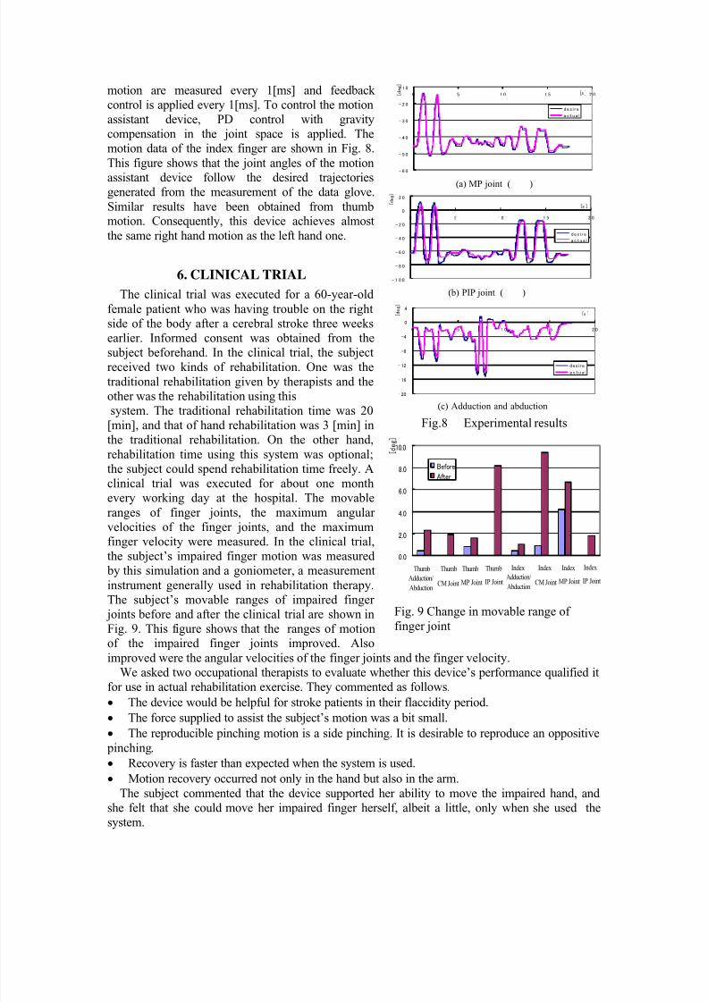
motion are measured every 1[ms] and feedback
control is applied every 1[ms]. To control the motion
assistant device, PD control with gravity
compensation in the joint space is applied. The
motion data of the index finger are shown in Fig. 8.
This figure shows that the joint angles of the motion
assistant device follow the desired trajectoriesgenerated from the measurement of the data glove.
Similar results have been obtained from thumbmotion. Consequently, this device achieves almost
the same right hand motion as the left hand one.
6. CLINICAL TRIAL
The clinical trial was executed for a 60-year-old
female patient who was having trouble on the right
side of the body after a cerebral stroke three weeks
earlier. Informed consent was obtained from the
subject beforehand. In the clinical trial, the subjectreceived two kinds of rehabilitation. One was the
traditional rehabilitation given by therapists and the
other was the rehabilitation using this
system. The traditional rehabilitation time was 20
[min], and that of hand rehabilitation was 3 [min] in
the traditional rehabilitation. On the other hand,
rehabilitation time using this system was optional;
the subject could spend rehabilitation time freely. A
clinical trial was executed for about one month
every working day at the hospital. The movable
ranges of finger joints, the maximum angular
velocities of the finger joints, and the maximumfinger velocity were measured. In the clinical trial,
the subject’s impaired finger motion was measured
by this simulation and a goniometer, a measurement
instrument generally used in rehabilitation therapy.
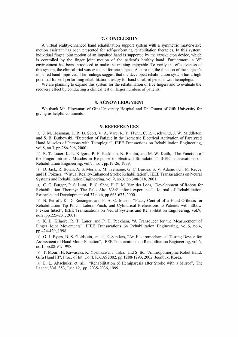
The subject’s movable ranges of impaired finger
joints before and after the clinical trial are shown in
Fig. 9. This figure shows that the ranges of motion
of the impaired finger joints improved. Also
improved were the angular velocities of the finger joints and the finger velocity.
We asked two occupational therapists to evaluate whether this device’s performance qualified it
for use in actual rehabilitation exercise. They commented as follows.
• The device would be helpful for stroke patients in their flaccidity period.• The force supplied to assist the subject’s motion was a bit small.
• The reproducible pinching motion is a side pinching. It is desirable to reproduce an oppositive
pinching.
• Recovery is faster than expected when the system is used.
• Motion recovery occurred not only in the hand but also in the arm.
The subject commented that the device supported her ability to move the impaired hand, and
she felt that she could move her impaired finger herself, albeit a little, only when she used the
system.
Fig.8 Experimental results
- 6 0
- 5 0
- 4 0
- 3 0
- 2 0
- 1 0
0 5 1 0 1 5 2 0[s ] [ d e g ]
des i r e
a c t u a l
- 1 0 0
- 8 0
- 6 0
- 4 0
- 2 0
0
2 0
0 5 1 0 1 5 2 0
[s ] [ d e g ]
des i r e
a c t u a l
- 20
- 16
- 12
-8
-4
0
4
0 5 1 0 1 5 2 0
[s ] [ d e g ]
des i re
a c t u a l
(a) MP joint ( )
(b) PIP joint ( )
(c) Adduction and abduction
0.0
2.0
4.0
6.0
8.0
10.0
1 2 3 4 5 6 7 8
[ d
e g ]
Before
After
Adduction/
Abduction
Thumb
CM Joint
Thumb
MP Joint
Thumb
IP Joint
Thumb
Adduction/
Abduction
Index
CM Joint
Index
MP Joint
Index
IP Joint
Index
Fig. 9 Change in movable range of
finger joint
7/23/2019 Hand Rehabilitation Support System Based on Self-motion Control, With a Clinical Case Report
http://slidepdf.com/reader/full/hand-rehabilitation-support-system-based-on-self-motion-control-with-a-clinical 6/6
7. CONCLUSION
A virtual reality-enhanced hand rehabilitation support system with a symmetric master-slave
motion assistant has been presented for self-performing rehabilitation therapies. In this system,
individual finger joint motion of an impaired hand is supported by the exoskeleton device, which
is controlled by the finger joint motion of the patient’s healthy hand. Furthermore, a VRenvironment has been introduced to make the training enjoyable. To verify the effectiveness of
this system, the clinical trial was executed for one subject. As a result, the function of the subject’s
impaired hand improved. The findings suggest that the developed rehabilitation system has a high
potential for self-performing rehabilitation therapy for hand-disabled persons with hemiplegia.
We are planning to expand this system for the rehabilitation of five fingers and to evaluate the
recovery effect by conducting a clinical test on larger numbers of patients.
8. ACNOWLEDGMENT
We thank Mr. Hirowatari of Gifu University Hospital and Dr. Osama of Gifu University forgiving us helpful comments.
9. REFEFRENCES
[1] J. M. Heasman, T. R. D. Scott, V. A. Vare, R. Y. Flynn, C. R. Gschwind, J. W. Middleton,
and S. B. Butkowski, “Detection of Fatigue in the Isometric Electrical Activation of Paralyzed
Hand Muscles of Persons with Tetraplegia”, IEEE Transactions on Rehabilitation Engineering,
vol.8, no.3, pp.286-296, 2000.
[2] R. T. Lauer, K. L. Kilgore, P. H. Peckham, N. Bhadra, and M. W. Keith, “The Function of
the Finger Intrinsic Muscles in Response to Electrical Stimulation”, IEEE Transacations on
Rehabilitation Engineering, vol.7, no.1, pp.19-26, 1999.
[3] D. Jack, R. Boian, A. S. Merians, M. Tremaine, G. C. Burdea, S. V. Adamovich, M. Recce,
and H. Poizner, “Virtual Reality-Enhanced Stroke Rehabilitation”, IEEE Transacations on Neural
Systems and Rehabilitation Engineering, vol.9, no.3, pp.308-318, 2001.
[4] C. G. Burger, P. S. Lum, P. C. Shor, H. F. M. Van der Loos, “Development of Robots for
Rehabilitation Therapy: The Palo Alto VA/Stanford experience”, Journal of Rehabilitation
Research and Development vol.37 no.6, pp.663-673, 2000.
[5] N. Petroff, K. D. Reisinger, and P. A. C. Mason, “Fuzzy-Control of a Hand Orthosis for
Rehabilitation Tip Pinch, Lateral Pinch, and Cylindrical Prehensions to Patients with Elbow
Flexion Intact”, IEEE Transacations on Neural Systems and Rehabilitation Engineering, vol.9,
no.2, pp.225-231, 2001.
[6] K. L. Kilgore, R. T. Lauer, and P. H. Peckham, “A Transducer for the Measurement of
Finger Joint Movements”, IEEE Transacations on Rehabilitation Engineering, vol.6, no.4,
pp.424-429, 1998.
[7] G. J. Byers, B. S. Goldstein, and J. E. Sanders, “An Electromechanical Testing Device forAssessment of Hand Motor Function”, IEEE Transacations on Rehabilitation Engineering, vol.6,
no.1, pp.88-94, 1998.
[8] T. Mouri, H. Kawasaki, K. Yoshikawa, J. Takai, and S. Ito, "Anthropomorphic Robot Hand:
Gifu Hand III", Proc. of Int. Conf. ICCAS2002, pp.1288-1293, 2002, Jeonbuk, Korea.
[9] E. L. Altschuler, et. al., “Rehabilitation of Hemiparesis after Stroke with a Mirror”, The
Lancet, Vol. 353, June 12, pp. 2035-2036, 1999.