Embed Size (px)
Citation preview

Hand InjuriesHilary Lee R2 EM
May 24, 2015Hilary Lee 2015

Overview• Review relevant anatomy
• Physical exam maneuvers
• Lacerations
• Fractures
• Dislocations
• Special fingers
• Crush, injection injuries
Hilary Lee 2015

Objectives
• Perform an appropriate evaluation of a hand injury
• Know when to consult a hand surgeon immediately and with delay
• Be familiar with the management of common hand trauma complaints
Hilary Lee 2015
Hand Injuries
• 30-40% of all trauma visits (major and minor) involve a significant hand complaint
• Highly functional body part with multiple superficial tissues
• Loss of productivity - 3rd highest reason for missed work days
Hilary Lee 2015

Immediate/Prehospital Care
• Control bleeding • Pressure, elevation • Tourniquet
• Remove jewelry
• Splint deformities
• Preserve amputations • Cooling procedures
Hilary Lee 2015

History• SAMPLE hx
• Pain, ROM, function, strength, paralysis, numbness/tingling, cold fingers, timeline, tetanus
• Handedness, occupation
• Prior injuries/surgeries
• Immunocompromise, smoking, DM
Hilary Lee 2015

PhysicalInspection
• Deformity, bleeding, rotation, amputations, avulsions, holding of limb
• Skin integrity - lacerations, edema, scars
• Fist and open
Palpation
• Tenderness, crepitus, AROM, PROM
Ligaments, tendons
• Varus/valgus stress (DIP, PIP, MCP)
• Explore wound for tendinous injury
Vascular
• Warmth, colour, cap refill
• Explore wounds for vascular injury (+/- tourniquet)
Neurological
• Ulnar, median, radial
Hilary Lee 2015
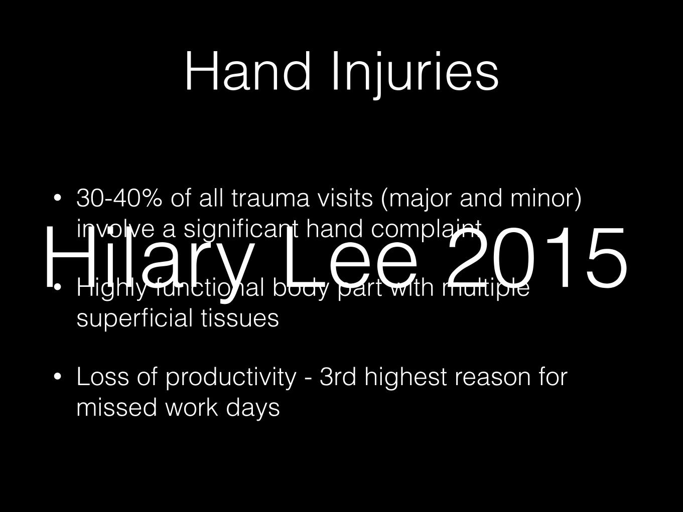
Sensory
46% have crossover between median and ulnar nerves
Hilary Lee 2015

Motor
Hilary Lee 2015

Tendons
Emergency Medicine Practice © 2011 6 ebmedicine.net • June 2011
(such as plastics and wood) may not show up on x-rays. A 1998 prospective study concluded that mech-anisms most likely to have retained glass included motor vehicle collisions and puncture wounds.12
Computed TomographyComputed tomography (CT) scanning is rarely used in the evaluation of hand trauma, as it usually does not add significant information to that already obtained by conventional radiography. While no studies have specifically addressed this matter, the following are important exceptions where CT scan-ning can be considered: complex and/or intra-artic-ular fractures, clinical scenarios highly suspicious of fracture with absence of fracture on x-ray, and at the request of a subspecialist for surgical planning. The emergency clinician should keep in mind that CT scans can provide high resolution of the bony struc-tures of the hand but provide limited information regarding soft tissues such as ligaments, tendons, and muscles.
Magnetic Resonance ImagingLike CT, magnetic resonance imaging (MRI) is only used in specific circumstances in hand trau-ma. It is important to remember that while MRI is not suited for evaluation of bony structures, it does offer visualization of soft tissues such as ligaments, tendons, muscles, and nerves. Magnetic resonance angiography (MRA) can be used to evaluate vascular structures.
UltrasonographyIn the hands of a skilled operator, ultrasound can be used to visualize soft tissue structures. The small structures of the hand are, however, difficult to scan and usually require high-frequency linear probes and an experienced ultrasonographer.
be considered in patients with difficult-to-control bleeding or bleeding out of proportion to injury. This is particularly true for patients known to be taking warfarin or other anticoagulants. Because hand surgeries are typically low-blood-loss pro-cedures (thanks to intraoperative tourniquet use), baseline or preoperative coagulation studies are not typically indicated.
Imaging In Hand TraumaImaging is, by far, the most useful diagnostic tool in traumatic hand injury after history and physical examination. More often than not in the ED, the diagnosis and management hinges on the results of an imaging study. Diagnostic imaging should be tailored to confirm or exclude suspected injuries based on the history and physical examination findings. Any attempt at closed reduction in the ED (with the exception of a distal phalanx fracture) requires a set of postreduction films to assess align-ment of bony structures.
Conventional RadiographyPlain x-ray is the most useful tool for the emergency clinician in assessment of traumatic hand injury. Un-like the Ottawa rules for ankle and knee injuries, no decision rules exist for when to order x-rays of the hand. According to the American Society of Radiol-ogy’s published guidelines, any clinically suspected fracture or dislocation in the hand should be evalu-ated with at least posteroanterior and lateral views, and an oblique view should be strongly considered.6
Conventional radiography can also be used to evaluate lacerations that are suspected to contain a retained foreign body. The current gold standard for detecting radio-opaque materials (such as glass and metal) is careful scrutiny of multi-view x-rays.10,11 Clinicians must keep in mind that some materials
Figure 3. Physical Examination Of Hand Tendons
A, Extensor digitorum. B, Flexor digitorum superficialis. C, Flexor digitorum profundus.
Used with permission of Aaron Andrade, MD.
A B C
Extensor digitorum
Flexor digitorum superficialis
Flexor digitorum profundus
Hilary Lee 2015

Imaging
• X ray • AP, lateral, oblique
• CT
• Ultrasound
Hilary Lee 2015

Regional Anesthesia• Digital block
• Disinfect
• Volar approach
• In skin crease
• Pinch finger
• Hit bone
• Back 1-2mm
• Inject 2-3cc
7 Emergency Medicine Practice © 2011June 2011 • ebmedicine.net
should be fully explored to their base to assess the extent of tissue injury and to search for any foreign bodies. Particularly deep lacerations to the palm of the hand should not be explored aggressively, for fear of further damage to deep structures and risk of infection. All lacerations that involve tissue deep to the dermis or those that have continued bleeding should be repaired. A 2002 randomized controlled trial suggests that simple hand lacerations (< 2 cm in length and without associated nerve, tendon, joint, or bony involvement) can be managed conservative-ly (irrigation, ointment, and dressing) with similar cosmetic and functional outcomes.15
Lacerations should be thoroughly irrigated to remove any debris, and devitalized tissue should be carefully debrided. Classically, sterile saline has been the preferred irrigation solution. However, a 2008 Cochrane systematic review of local wound irrigation demonstrates that irrigation with potable tap water has identical rates of wound infection as sterile saline.16 Solutions of iodine, peroxide, or detergents should be avoided, as they have been shown to be toxic to fibroblasts.17
The ideal time interval between injury and laceration repair has not been fully elucidated in the literature. Several factors must be weighed when considering wound closure: location, depth, degree of contamination, and patient health. A classic emer-gency medicine prospective study of 204 patients concluded that uncontaminated wounds can be repaired by primary intention up to 12 hours after the time of injury, though it is believed that many can be closed even later. Contaminated wounds can be cleaned, packed, and reexamined for infection 3-5 days post injury. If no signs of infection exist, delayed primary closure is a reasonable option. Infected wounds should be allowed to close by sec-ondary intention.18
The vast majority of hand lacerations are best repaired with nonabsorbable monofilament suture material using a simple interrupted technique. Al-though there are no current trials supporting timing of suture removal, traditional practice dictates that sutures should be removed in 10-14 days except for those on the palm, which require 14-21 days. There has been a recent push towards using materials that do not require a repeat visit for suture removal, especially in children. A well-designed 2004 ran-domized controlled trial of 147 children showed no long-term cosmetic or functional difference between the use of plain gut and nylon sutures.19 Absorbable sutures are also useful in repairing deep structures of the hand. Additionally, a 2002 randomized con-trolled trial of 814 patients concluded that low skin tension on the dorsum of the hand allows it to be repaired successfully with tissue adhesives.20
Treatment
Local And Regional AnesthesiaThe classic methods for obtaining hand anesthesia in the ED are local anesthetic infiltration, digital nerve blocks, and anatomic forearm nerve blocks using lidocaine without epinephrine. (Refer to the “Epinephrine In Digital Nerve Blocks” section on page 19 for further discussion on epinephrine use.) When longer anesthesia is desired, bupivacaine or other longer-acting local anesthetics without epi-nephrine may be substituted. The traditional teaching for digital nerve blocks includes a 2-injection dorsal approach that is quite painful to patients. A 2006 randomized trial of 27 patients concluded that a volar single-injection tech-nique produces similar anesthetic results (except for patients who require very proximal dorsal anesthe-sia) with less patient discomfort.13 (See Figure 4.) Classically, anatomic landmark injections and ring wrist blocks have been used for regional anes-thesia to the hand. With the ever-increasing ubiquity of bedside ultrasound in the ED, ultrasound-guided nerve blocks are emerging in the literature. (See Figure 5, page 8.) A 2006 prospective study of 11 patients concluded that ultrasound-guided forearm nerve blocks are feasible for emergency clinicians to perform, with high patient satisfaction.14 More randomized clinical trials in this area may acceler-ate ultrasound-guided nerve blocks to become the standard of care for regional anesthesia for the hand and elsewhere in the body.
Skin LacerationsAfter hemostasis is achieved and appropriate local or regional anesthesia provided, all skin lacerations
Figure 4. The Volar Single-Injection Method For Digital Nerve Anesthesia
Note the injection site is at the volar MCP skin crease and the clinician is lightly pinching the digit. A 25-gauge needle should be advanced until it hits bone, backed up 1-2 mm, and 2-3 cc of anesthetic in-jected. Used with permission of Aaron Andrade, MD.
Hilary Lee 2015

Regional Anesthesia• Local infiltration
• Forearm nerve blocks - ultrasound guided
Emergency Medicine Practice © 2011 8 ebmedicine.net • June 2011
onstrated no outcome difference at 2 years between operative and trephination groups regardless of presence of underlying fracture or mechanism of injury. Furthermore, the study showed a substantial cost benefit in the nail trephination group.31 There-fore, current literature supports the recommenda-tion that subungual hematomas caused by nail bed lacerations do not require nail removal and direct re-pair if the nail and its margins are intact.32 If the nail itself is significantly disrupted, the nail bed matrix should be exposed and repaired with fine absorbable sutures. Patients and parents should be warned of potential for infection and permanent nail deformity.
Fingertip AmputationsManagement of fingertip amputations must be approached on a case-by-case basis, as there are no current guidelines and little supporting evidence in the literature. Amputations distal to the DIP can usually heal by secondary intention if less than 1 cm in diameter. If there is a small amount of exposed bone, the bone can be trimmed back in the ED with a rongeur until it is underneath the surrounding soft tissue and allowed to heal by secondary intention. Follow-up with a hand surgeon is advised. Imme-diate consultation of a hand surgeon is required in cases of wounds larger than 1 cm in diameter, per-sistently exposed bone, or amputation of the volar pad.33 Additionally, surgeons subclassify fingertip injuries into zones I, II, and III. (See Figure 7.) Zone I injuries are managed conservatively as described above. Zone II injuries may require rongeuring of ex-posed bone. Zone III injuries generally require distal phalanx amputation and warrant follow-up with a hand specialist.34
FracturesPerhaps the most important job of an emergency clinician in hand fractures is proper reduction and
Fingertip InjuriesFingertip injuries are those that involve any struc-ture distal to the DIP. These injuries are very com-mon, occurring most frequently in young males as a crush/jam injury.21,22
Distal Phalanx FracturesFractures of the distal phalanx can be subcategorized into 3 types: tuft (distal) fractures, shaft fractures, and intra-articular fractures. Though no research exists, reference books generally agree that for the purposes of the emergency clinician, tuft and shaft fractures (see Figure 6) can usually be managed conservatively with repair of soft tissues (as necessary) and splinting in extension for 2-3 weeks. Splinting of the entire fin-ger is unnecessary and may cause stiffness. Severely angulated shaft fractures can be reduced in the ED after digital block and should be splinted for 3 weeks. A hand surgeon should evaluate open fractures or severe crush injuries with large losses of soft tissue. Intra-articular fractures require thorough examination to rule out associated tendon avulsions and should always be evaluated by a hand specialist.23
Subungual Hematoma/Nail Bed LacerationsIn crush injuries of the finger, nail bed lacerations causing subungual hematomas are common. They are characterized by throbbing pain and purple discoloration under the nail. Two management strategies are commonly used in the ED: removal of the nail, with direct repair of nail bed laceration; and nail trephination with a heated paperclip, a cautery device, or a twirled 18-g needle. A review of the classic literature yields a long-standing debate about which management strategy is superior. The commonly taught “consensus” is that nail bed repair should be considered for subungual hematomas covering greater than 25% to 50% of the nail bed.25-30 However, a 1999 prospective study in children dem-
Figure 5. Ultrasound Visualization Of Nerves And Arteries In The Forearm
Ulnar, medial, and radial nerves are shown by the arrow across the bottom of the images. Arrowheads show arteries, A (ulnar), B (medial), and C (radial).
Liebmann O, Price D, Mills C et al. Ann Emerg Med. 2006;48(5)558-562. Used with permission of Mosby, Inc.
A B C
Hilary Lee 2015

Lidocaine +/- Epinephrine• Digital ischemia secondary to epinephrine in nerve
block extremely rare
• 17 cases worldwide
• Multiple large safety studies over last 10 years
• Phentolamine -> injectable antidote
• Epinephrine improves anesthesia, decreases bleeding, decreases systemic anesthetic absorption
Hilary Lee 2015

Lacerations
Hilary Lee 2015

Lacerations
• Hemostasis
• Local anesthesia
• Exploration
• Irrigation
Hilary Lee 2015

Primary Closure• Noncontaminated wounds - within 12 hours?
• Delayed primary closure
• Contaminated wounds
• Irrigate
• Pack
• Reinspect after 3-5 days
Hilary Lee 2015

Primary Closure
• Nonabsorbable vs absorbable
• Monofilament
• Simple interrupted
• 10-14 days (14-21 days palmar)
Hilary Lee 2015

Distal Phalanx
9 Emergency Medicine Practice © 2011June 2011 • ebmedicine.net
Unstable phalangeal fractures include oblique fractures, malrotated fractures, and angulated frac-tures. After anesthesia with either a digital block or hematoma block, the emergency clinician should attempt to reduce the fracture with gentle manipu-lation. Adequate alignment should be confirmed with postreduction x-rays as well as examination of the fingers for evidence of malrotation. Successfully reduced phalangeal fractures should be splinted in extension and referred for outpatient follow-up. Immediate surgical consultation is required for open fractures, unsuccessful reduction, malrotation, and intra-articular fractures involving more than 30% of the joint surface.36
Metacarpal II-V Fractures And Boxer’s FractureManagement of fractures of the II-V metacarpals varies based on the location of the fracture. Meta-carpal head and base fractures are relatively rare and require little management in the ED. A volar splint should be applied in a neutral position and the patient referred to a hand surgeon. The emer-gency clinician can reduce metacarpal shaft frac-tures after adequate anesthesia with a hematoma block or regional nerve block. Reduction goal is less than 10° of angulation in metacarpals II and III, less than 20° of angulation in metacarpals IV and V, less than 3 mm of digit length loss, and no rotational deformity.35 All metacarpal shaft fractures should be splinted and referred to a hand surgeon. Open fractures and those that fail reduction should re-ceive immediate surgical consultation.35
splinting. While specific reduction techniques can vary widely, there is one universal “safe position” for splinting of hand fractures, called the “intrinsic plus” position. The thumb is extended and abduct-ed, while the other fingers are flexed to 90° at the MCP and fully extended at the PIP and DIP. Addi-tionally, the wrist is extended 15° to 30°. The actual location of the splint varies depending on the loca-tion of the fracture: thumb spica for thumb injuries, volar for digits II and III, and ulnar gutter for digits IV and V. Traditionally, orthopedists prefer plaster splints over fiberglass due to their durability and ability to be molded. Refer to Figure 8 (page 10) for an example of the “safe position.” The thumb is extended and abducted and fingers II-V have MCPs flexed to 90° and interphalangeal (IP) joints fully extended. This position prevents shortening of ten-dons and ligaments while the hand is immobilized, reducing stiffness.
Proximal And Middle Phalanx FracturesUnlike distal phalanx fractures, proximal and middle phalanx fractures require precise alignment. That said, the majority of phalangeal fractures do not require reduction, as they are stable and nondis-placed (usually transverse).35 These stable fractures are managed by “buddy-taping” the affected finger to the adjacent finger to promote early mobilization and reduce stiffness. (See Figure 9, page 10.)
Figure 6. Radiograph Demonstrating Phalanx Fractures
Left arrow notes a tuft fracture of digit IV. Right arrow notes a shaft fracture of the distal phalanx of digit III.
Used with permission of John D. Lubahn, MD.
Figure 7. Zones Of Fingertip Amputation
© 2001. Renee L. Cannon. Used with permission.
Zone I II III
Zone I II III
Hilary Lee 2015

Distal Phalanx
• Fractures
• Tuft
• Shaft
• Intraarticular
Hilary Lee 2015

Distal Phalanx
• Fractures
• Tuft
• Shaft
• Intraarticular
Reduce and repair soft tissues prn
Splint in extensionHilary Lee 2015
Distal Phalanx
• Fractures
• Tuft
• Shaft
• Intraarticular
Reduce and repair soft tissues prn
Splint in extension
Carefully assess tendons, +/- hand surgeon
Hilary Lee 2015

Distal Phalanx
• Amputations • Zone I - secondary intention
• Zone II - file down bone, secondary intention, follow up
• Zone III - distal phalanx amputation, immediate consult
9 Emergency Medicine Practice © 2011June 2011 • ebmedicine.net
Unstable phalangeal fractures include oblique fractures, malrotated fractures, and angulated frac-tures. After anesthesia with either a digital block or hematoma block, the emergency clinician should attempt to reduce the fracture with gentle manipu-lation. Adequate alignment should be confirmed with postreduction x-rays as well as examination of the fingers for evidence of malrotation. Successfully reduced phalangeal fractures should be splinted in extension and referred for outpatient follow-up. Immediate surgical consultation is required for open fractures, unsuccessful reduction, malrotation, and intra-articular fractures involving more than 30% of the joint surface.36
Metacarpal II-V Fractures And Boxer’s FractureManagement of fractures of the II-V metacarpals varies based on the location of the fracture. Meta-carpal head and base fractures are relatively rare and require little management in the ED. A volar splint should be applied in a neutral position and the patient referred to a hand surgeon. The emer-gency clinician can reduce metacarpal shaft frac-tures after adequate anesthesia with a hematoma block or regional nerve block. Reduction goal is less than 10° of angulation in metacarpals II and III, less than 20° of angulation in metacarpals IV and V, less than 3 mm of digit length loss, and no rotational deformity.35 All metacarpal shaft fractures should be splinted and referred to a hand surgeon. Open fractures and those that fail reduction should re-ceive immediate surgical consultation.35
splinting. While specific reduction techniques can vary widely, there is one universal “safe position” for splinting of hand fractures, called the “intrinsic plus” position. The thumb is extended and abduct-ed, while the other fingers are flexed to 90° at the MCP and fully extended at the PIP and DIP. Addi-tionally, the wrist is extended 15° to 30°. The actual location of the splint varies depending on the loca-tion of the fracture: thumb spica for thumb injuries, volar for digits II and III, and ulnar gutter for digits IV and V. Traditionally, orthopedists prefer plaster splints over fiberglass due to their durability and ability to be molded. Refer to Figure 8 (page 10) for an example of the “safe position.” The thumb is extended and abducted and fingers II-V have MCPs flexed to 90° and interphalangeal (IP) joints fully extended. This position prevents shortening of ten-dons and ligaments while the hand is immobilized, reducing stiffness.
Proximal And Middle Phalanx FracturesUnlike distal phalanx fractures, proximal and middle phalanx fractures require precise alignment. That said, the majority of phalangeal fractures do not require reduction, as they are stable and nondis-placed (usually transverse).35 These stable fractures are managed by “buddy-taping” the affected finger to the adjacent finger to promote early mobilization and reduce stiffness. (See Figure 9, page 10.)
Figure 6. Radiograph Demonstrating Phalanx Fractures
Left arrow notes a tuft fracture of digit IV. Right arrow notes a shaft fracture of the distal phalanx of digit III.
Used with permission of John D. Lubahn, MD.
Figure 7. Zones Of Fingertip Amputation
© 2001. Renee L. Cannon. Used with permission.
Zone I II III
Zone I II IIIHilary Lee 2015

Nail
• Subungual hematoma
• Trephination
• Nail removal and nail bed repair
• Nail fold preservation
Hilary Lee 2015

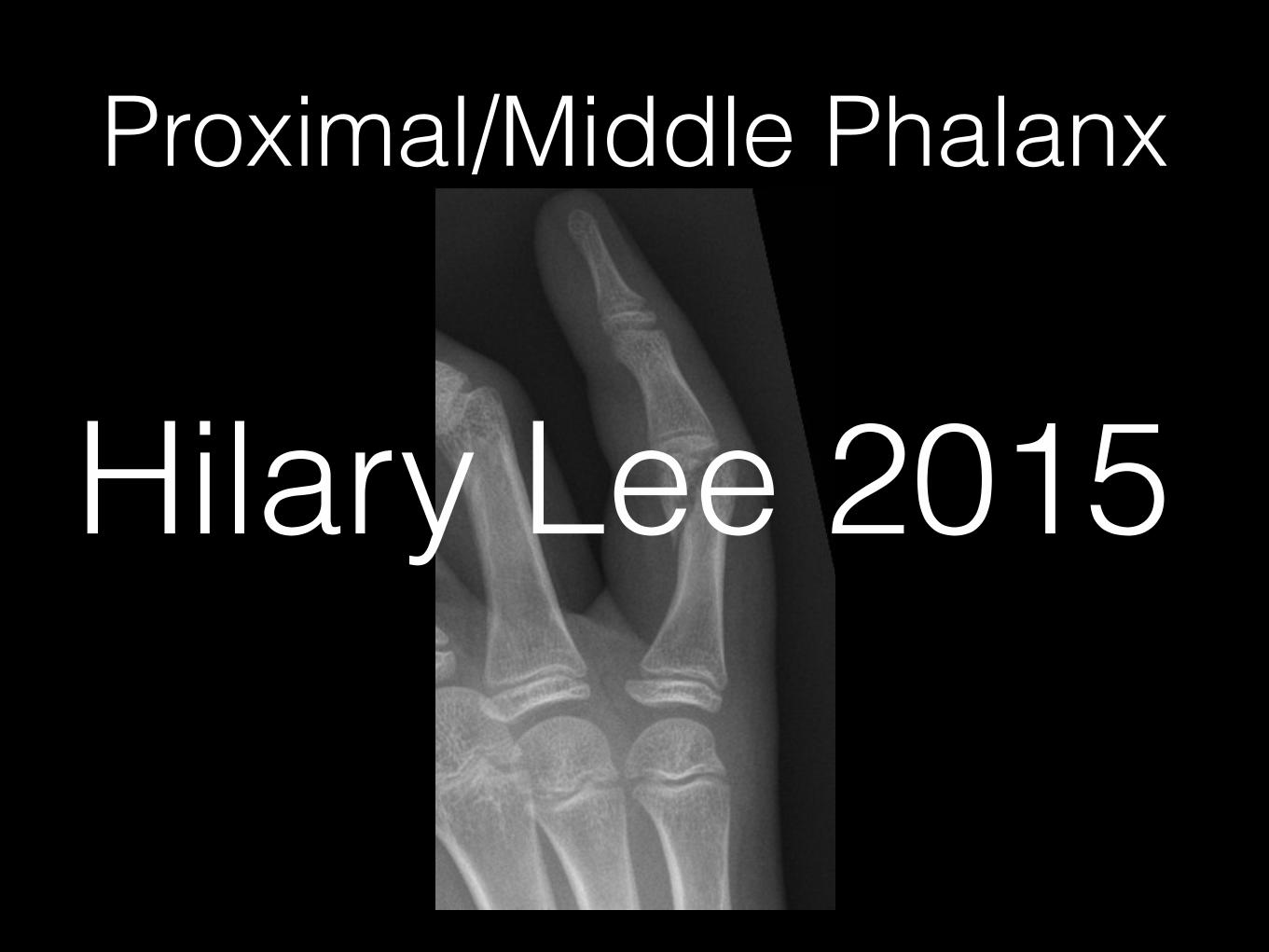
Proximal/Middle Phalanx• Require precise alignment
• Stable, nondisplaced
• Buddy tape
• Oblique, angulated, malrotated
• Digital block / hematoma block
• Reduction
• Post-reduction film
• Splint in extension
Hilary Lee 2015

Proximal/Middle Phalanx• Require precise alignment
• Stable, nondisplaced
• Buddy tape
• Oblique, angulated, malrotated
• Digital block / hematoma block
• Reduction
• Post-reduction film
• Splint in extension
Repeat if not properly aligned
Hilary Lee 2015

Proximal/Middle Phalanx
296 Practical Plastic Surgery for Nonsurgeons
3. However you immobilize the joint, be sure to allow motion of theproximal interphalangeal (PIP) joint.
4. When the finger is no longer tender, the patient should start movingthe joint both passively and actively in hope of regaining functionalrange of motion at the DIP joint.
Middle and Proximal Phalangeal FracturesMiddle and proximal phalangeal fractures are classified according towhether they involve the joint surface.
Extraarticular FracturesExtraarticular fractures affect the part of the bone that is not involvedwith the joint surface. The main concern is whether a rotational deformityis present when the patient attempts to bend the fingers. See chapter 26,“Normal Hand Exam,” for discussion of rotational finger alignment.
If no rotational deformity is present, the finger can be treated bybuddy taping for 2–3 weeks until the finger is no longer tender. Buddytaping is used to initiate gentle movement of the injured finger whilemaintaining proper bone alignment. The injured finger is taped to theadjacent finger, and the patient is instructed to use the hand as nor-mally as possible.
Any malrotation associated with metacarpal or phalangeal fractures must becorrected. Left, Normally all fingers point toward the region of the scaphoidwhen a fist is made. Right, Malrotation at the fracture site causes the affectedfinger to deviate. (From Crenshaw AH (ed): Campbell’s OperativeOrthopaedics, 7th ed. St. Louis, Mosby, 1987, with permission.)
Hilary Lee 2015

Proximal/Middle Phalanx
Emergency Medicine Practice © 2011 10 ebmedicine.net • June 2011
of angulations up to 70° to 75°.39,40 The emergency clinician should not forget that boxer’s fractures are often a consequence of violent and intentional be-havior and patients are at risk for recurrent injury.41 In addition, boxer’s fracture patients have higher rates of anxiety, borderline personality disorder, and antisocial personality disorder.42 As such, patients with boxer’s fractures should receive in their ED evaluation psychiatric questioning as well as pre-vention strategies. Thumb Metacarpal Fractures: Bennett And Rolando FracturesFractures of the first metacarpal are less common than those of the remaining metacarpals. They can be subdivided into extra-articular and intra-articular fractures. Extra-articular fractures follow the same conservative management principles as other meta-carpal fractures, namely, closed reduction with an angulation goal of less than 20° to 30° followed by thumb spica splinting for 4 weeks. Oblique fractures are unstable and require prompt consultation by a hand surgeon.43
Intra-articular fractures of the first metacarpal involve the CMC joint and generally occur due to an axial injury to a partially flexed metacarpal. A Bennett fracture is an intra-articular fracture and dislocation; a Rolando fracture is a comminuted intra-articular fracture.44 (See Figure 11.) While debate exists regarding the specific type of surgical correction each fracture requires, the available litera-ture supports that emergency management should consist of closed reduction (Bennett fracture only),
Metacarpal neck fractures deserve special men-tion as they are among the most common fractures of the hand. Nondisplaced, nonangulated fractures should be treated with a gutter splint that immobi-lizes the CMC and MCP joints for 3-4 weeks, with surgical clinic follow-up. Unstable fractures of the II and III metacarpals generally require immediate consultation by a hand surgeon for surgical correc-tion. Unstable fractures in the IV and/or V meta-carpals, also known as a boxer’s fracture, can be reduced in the ED after adequate anesthesia. In the author’s experience, a forearm ulnar nerve block in conjunction with a hematoma block using 1% lido-caine without epinephrine provides excellent results. Reduction is achieved by traction decompression followed by the “90-90 method.” (See Figure 10.) The MCP, PIP, and DIP joints are flexed at 90° and volar-ward pressure is applied to the dorsum of the metacarpal shaft. An ulnar gutter splint should be applied with prompt clinic follow-up within 1 week. Much controversy exists in the literature regard-ing the goal of boxer’s fracture reduction. Classic literature supports acceptable angulation between 20° and 70°.37 More-recent studies are incongruent. A 1999 cadaveric study concluded that angulation greater than 30° resulted in measurable functional impairment.38 Two more recent prospective stud-ies, however, found good outcomes with 1 week of soft wrap followed by immediate buddy-wrapping
Figure 8. The “Intrinsic Plus” Splinting Position
Used with permission of Aaron Andrade, MD.
Figure 9. “Buddy-Taping” An Injured Finger
Used with permission of Aaron Andrade, MD.
Hilary Lee 2015

Proximal/Middle Phalanx
• Refer immediately if:
• Open
• Unsuccessful reduction
• Malrotation
• Intraarticular >30% joint surface
Hilary Lee 2015
Proximal/Middle Phalanx
Hilary Lee 2015

Fractures
• Safe position / intrinsic plus
Emergency Medicine Practice © 2011 10 ebmedicine.net • June 2011
of angulations up to 70° to 75°.39,40 The emergency clinician should not forget that boxer’s fractures are often a consequence of violent and intentional be-havior and patients are at risk for recurrent injury.41 In addition, boxer’s fracture patients have higher rates of anxiety, borderline personality disorder, and antisocial personality disorder.42 As such, patients with boxer’s fractures should receive in their ED evaluation psychiatric questioning as well as pre-vention strategies. Thumb Metacarpal Fractures: Bennett And Rolando FracturesFractures of the first metacarpal are less common than those of the remaining metacarpals. They can be subdivided into extra-articular and intra-articular fractures. Extra-articular fractures follow the same conservative management principles as other meta-carpal fractures, namely, closed reduction with an angulation goal of less than 20° to 30° followed by thumb spica splinting for 4 weeks. Oblique fractures are unstable and require prompt consultation by a hand surgeon.43
Intra-articular fractures of the first metacarpal involve the CMC joint and generally occur due to an axial injury to a partially flexed metacarpal. A Bennett fracture is an intra-articular fracture and dislocation; a Rolando fracture is a comminuted intra-articular fracture.44 (See Figure 11.) While debate exists regarding the specific type of surgical correction each fracture requires, the available litera-ture supports that emergency management should consist of closed reduction (Bennett fracture only),
Metacarpal neck fractures deserve special men-tion as they are among the most common fractures of the hand. Nondisplaced, nonangulated fractures should be treated with a gutter splint that immobi-lizes the CMC and MCP joints for 3-4 weeks, with surgical clinic follow-up. Unstable fractures of the II and III metacarpals generally require immediate consultation by a hand surgeon for surgical correc-tion. Unstable fractures in the IV and/or V meta-carpals, also known as a boxer’s fracture, can be reduced in the ED after adequate anesthesia. In the author’s experience, a forearm ulnar nerve block in conjunction with a hematoma block using 1% lido-caine without epinephrine provides excellent results. Reduction is achieved by traction decompression followed by the “90-90 method.” (See Figure 10.) The MCP, PIP, and DIP joints are flexed at 90° and volar-ward pressure is applied to the dorsum of the metacarpal shaft. An ulnar gutter splint should be applied with prompt clinic follow-up within 1 week. Much controversy exists in the literature regard-ing the goal of boxer’s fracture reduction. Classic literature supports acceptable angulation between 20° and 70°.37 More-recent studies are incongruent. A 1999 cadaveric study concluded that angulation greater than 30° resulted in measurable functional impairment.38 Two more recent prospective stud-ies, however, found good outcomes with 1 week of soft wrap followed by immediate buddy-wrapping
Figure 8. The “Intrinsic Plus” Splinting Position
Used with permission of Aaron Andrade, MD.
Figure 9. “Buddy-Taping” An Injured Finger
Used with permission of Aaron Andrade, MD.
Hilary Lee 2015

Metacarpal Fractures
Hilary Lee 2015

Metacarpal Fractures• Base or head
• Rare
• Volar splint and consultation
• Shaft
• Common
• Reducible in ED
Hilary Lee 2015

Metacarpal Shaft Fractures• Local anesthesia
• Regional block
• Hematoma block
• Reduction goals
• <10 degrees
• <20 degrees
• <3mm impaction
• No rotation
Hilary Lee 2015

Metacarpal Shaft Fractures
• Splint (ulnar or radial gutter)
• Refer
Hilary Lee 2015
Metacarpal Neck Fracture
• Nondisplaced, nonangulated
• Gutter splint - immobilize CMP and MCP
• Refer in 3-4 weeks
• Unstable, MCP 2 or 3
• Immediate consultation for OR
Hilary Lee 2015

Fractures
Hilary Lee 2015

Fractures
Hilary Lee 2015

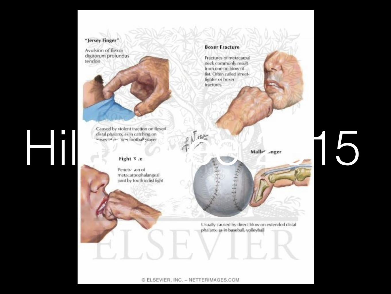
Boxer’s Fracture
• 5th (or 4th) metacarpal neck
• Volar angulation of distal segment
• Inexperienced punch -> axial load
• >95% young adult males
Hilary Lee 2015

Boxer’s Fracture• Local anesthesia
• Hematoma block plus ulnar nerve block
• Traction decompression
• 90 / 90 method
• Volar pressure to dorsum
• Ulnar gutter splint, refer
• 20-70 degrees angulation accepted 11 Emergency Medicine Practice © 2011June 2011 • ebmedicine.net
extension for at least 6 weeks.54,55 Strict compliance is necessary, which can prove difficult for patients due to hygiene and comfort issues. Because of this, many different types of splints are available for com-mercial use.56,57 (See Figure 12.) Note that the PIP is not splinted. The few randomized trials comparing splints demonstrate equal efficacy as long as patients follow strict compliance.58-60 One study in cadavers has shown that PIP motion does not affect structural integrity of the DIP tendon, and therefore splint-ing of the entire finger is not recommended, as it may cause unnecessary stiffness.61 Furthermore, no difference in outcome has been measured between early and delayed splinting of mallet finger.62 In spite of the high success rates of conservative management, some debate remains over which cases of mallet finger require surgical management. Clas-sically, all open injuries and those with greater than
thumb spica immobilization, and early consultation with a hand specialist.45-52 The emergency clinician should also warn patients that both Bennett and Rolando fractures carry a high risk of future compli-cations such as degenerative arthritis, with Rolando fractures being particularly vulnerable.
Tendon InjuriesInjuries to hand tendons most often occur due to laceration, crush, or forceful hyperextension/hyper-flexion injuries. Regardless of the mechanism, ten-don injuries share the following common manage-ment strategies: (1) radiographs should be obtained to rule out associated fractures and avulsions, (2) surgical consultants should evaluate open tendon lacerations immediately for surgical repair, and (3) closed tendon injuries require splinting and surgical follow-up. Clinicians in the ED should remember that tendons often run close to peripheral nerves and vascular structures, so the presence of a tendon injury should raise suspicion for possible neurovas-cular injury.53
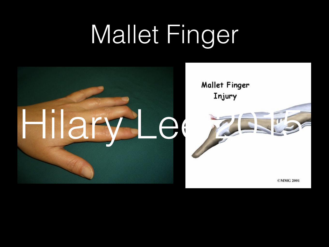
Mallet FingerA mallet finger is a very common injury of the exten-sor tendon insertion into the distal phalanx, usually caused by forced flexion of the DIP joint. It is so named because the flexed DIP cannot be extended and looks like a mallet. The injury can sometimes be associated with an avulsion fracture of the dorsal base of the distal phalanx. The classic strategy for treating closed mallet finger injuries with less than one-third of the joint surface disrupted is continuous splinting of the DIP joint in full extension to hyper-
Figure 10. The “90-90” Method
Used with permission of Aaron Andrade, MD.
Figure 11. Radiographs Of A Bennett Fracture (Left) And A Rolando Fracture (Right)
From Carsen BT, Moran SL. J Hand Surg. 2009;34A:945-953. Used with permission from Elsevier.
Figure 12. Splinting The DIP In Full To Hyperextension
Used with permission, Aaron Andrade, MD.
Hilary Lee 2015

Thumb Metacarpal Fractures
• Less common
• Extraarticular
• Closed reduction
• Goal 20-30 degrees angulation
• Thumb spica splint x 4 weeks
Hilary Lee 2015

Thumb Metacarpal Fractures
Intraarticular
Hilary Lee 2015

Thumb Metacarpal Fractures
Bennett fracture
• Intraarticular fracture / dislocation of 1st MCP
• Fragment continues to articulate
• Axial load on flexed digit
• ORIF if >3mm displacement
• Closed reduction, refer
Hilary Lee 2015

Thumb Metacarpal Fractures
• Rolando fracture
• Comminuted intraairticular 1st metacarpal fracture with dislocation
• Unstable, requires ORIF
Hilary Lee 2015

Tendon Injuries
• Laceration, crush, hyperflexion, hyperextension
• XR - r/o associated avulsion or #
• Closed tendon injuries - splint, close follow up
• Open tendon injuries….
Hilary Lee 2015

Tendon Injuries
• Laceration, crush, hyperflexion, hyperextension
• XR - r/o associated avulsion or #
• Closed tendon injuries - splint, close follow up
• Open tendon injuries….
Hilary Lee 2015

Tendon Injuries
• Laceration, crush, hyperflexion, hyperextension
• XR - r/o associated avulsion or #
• Closed tendon injuries - splint, close follow up
• Open tendon injuries….
Hilary Lee 2015

Tendon Injuries
• Laceration, crush, hyperflexion, hyperextension
• XR - r/o associated avulsion or #
• Closed tendon injuries - splint, close follow up
• Open tendon injuries….
Hilary Lee 2015
Tendon Injuries• Extensor
• Generally reparable in ED
• Few associated structures
• Flexor
• Wound closure and referral
• Many associated structures, evaluate carefully for nerve or vascular injury
Hilary Lee 2015

Tendon Injuries• Extensor
• Generally reparable in ED
• Few associated structures
• Flexor
• Wound closure and referral
• Many associated structures, evaluate carefully for nerve or vascular injury
Hilary Lee 2015
Mallet Finger
Hilary Lee 2015

Mallet Finger
Hilary Lee 2015
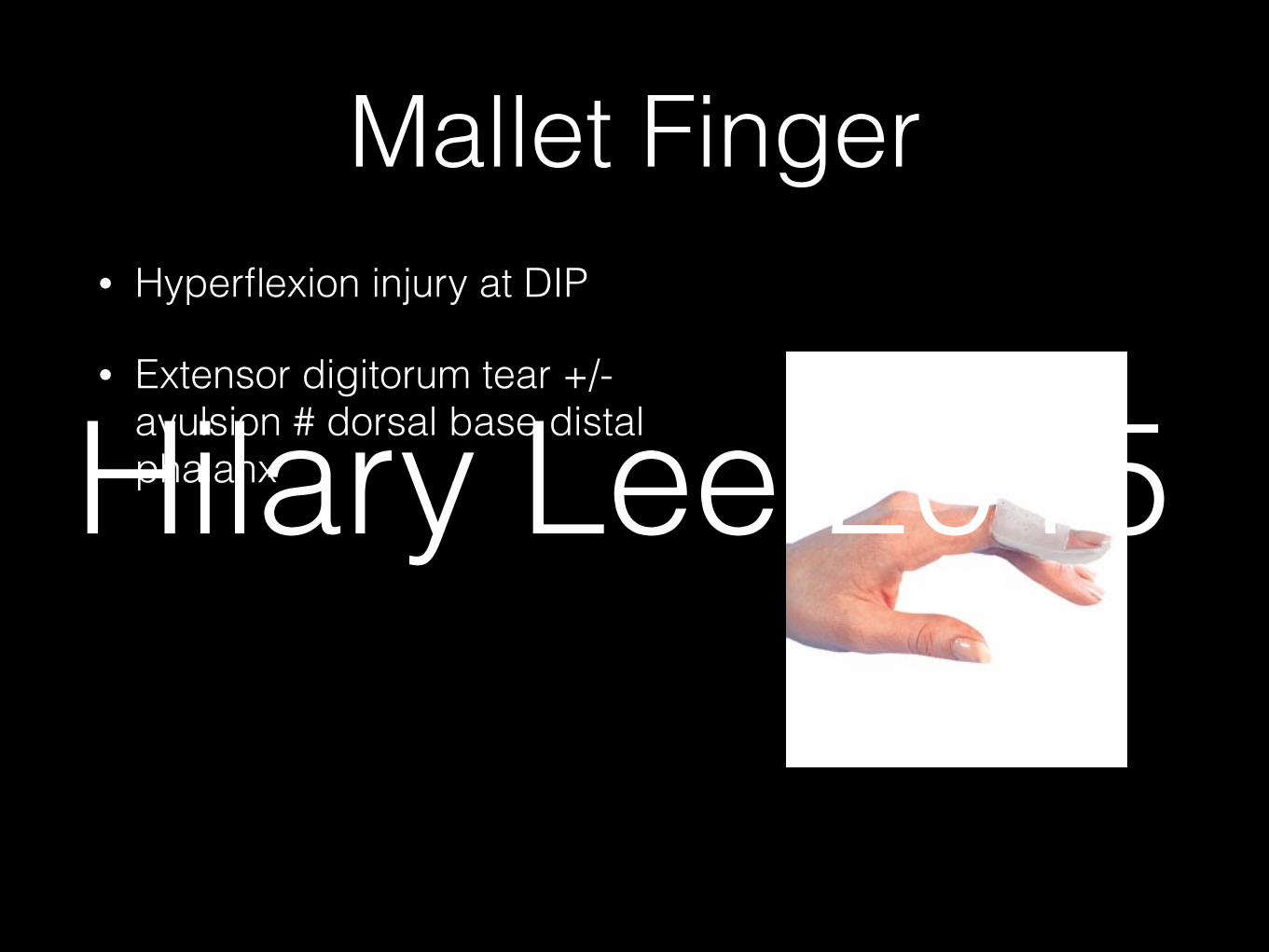
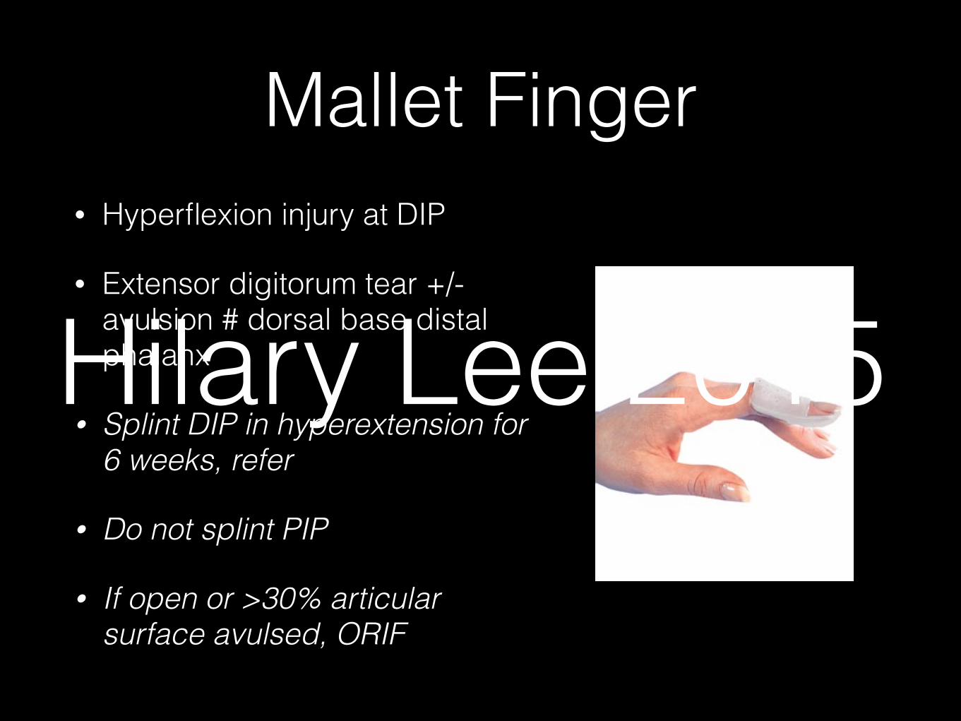
Mallet Finger• Hyperflexion injury at DIP
• Extensor digitorum tear +/- avulsion # dorsal base distal phalanx
• Splint DIP in hyperextension for 6 weeks, refer
• Do not splint DIP
• If open or >30% articular surface avulsed, ORIF
Hilary Lee 2015
Mallet Finger• Hyperflexion injury at DIP
• Extensor digitorum tear +/- avulsion # dorsal base distal phalanx
• Splint DIP in hyperextension for 6 weeks, refer
• Do not splint PIP
• If open or >30% articular surface avulsed, ORIF
Hilary Lee 2015

Jersey Finger
• Extension against resistance injury
• Flexor digitorum profundus tear
• Named after jersey tugging at a sports match….
Hilary Lee 2015

Jersey Finger
Hilary Lee 2015

Jersey Finger
• Isolated or plus avulsion fracture
• Immediate consultation for surgical intervention
Hilary Lee 2015

Hilary Lee 2015

Hilary Lee 2015

Fight Bite
• Laceration to MCP
• High rate of associated tendon, joint or bone injury - 75%
• Clenched fist mechanism - with relaxation, oral bacteria on extensor tendon sheath track back
Hilary Lee 2015
Fight Bite
• Aggressive early management
• XR r/o foreign body or fracture
• Irrigate, elevate, immobilize
• Early surgical consultation
Hilary Lee 2015

Fight Bite• Polymicrobial
• alpha-hemolytic streptococcus (s viridans)
• staphylococcus aureus
• eikonella corrodens (25%)
• gram negatives, anaerobes
• If joint or tendon sheath involved
• Admit for IV antibiotics (ampicillin/sulbactam, cefoxitin, carbapenem)
• OR for I&D
• PO amox-clav at discharge for 5-7 days
• Complications: osteomyelitis, tenosynovitis, septic arthritis
Hilary Lee 2015
Hilary Lee 2015

Dislocations/Subluxations
• Closed reduction with local anesthesia
• XR r/o avulsion #, confirm post-reduction alignment • Avulsion >30% articular surface = immediate consult
• Splint in slight flexion x 2-3 weeks
• Consultation as outpatient
Hilary Lee 2015

Hilary Lee 2015

Skiier’s/Gamekeeper’s Thumb
• Ulnar collateral ligament tear
• Forceful radial deviation of the thumb
• Pain, swelling on ulnar aspect of 1st MCP joint
• Valgus stress / radial deviation on 1st MCP with thumb fully extended and in 30 degrees flexion
• Distal deviation >35 degrees or 15 degrees more than unaffected thumb = complete tear
Hilary Lee 2015

15 Emergency Medicine Practice © 2011June 2011 • ebmedicine.net
stuck between ruptured ends of the UCL), resulting in poor healing. This requires prompt hand spe-cialist follow-up so that surgical repair may occur within 3 weeks of injury.87 Partial tears usually heal well with conservative management and should be immobilized in a thumb spica cast for 4 weeks, with outpatient follow-up. In cases where it is difficult to distinguish partial versus complete UCL tears, additional imaging is very useful. In a 1999 double-blind prospec-tive study of 34 patients using surgical findings as the gold standard, MRI was shown to have a 96% sensitivity and 95% specificity in detecting complete tears.88 Because repair is not required for 3 weeks, MRI may be scheduled as an outpatient and should not delay ED disposition. More recent literature assesses the utility of ultrasound in these cases, showing a sensitivity of 83% and specificity of 75% compared to surgical and cadaveric gold standards.89,90 A 1997 retrospective study concluded that the most common ultrasound error is misdiagnosing a complete tear as a partial tear, while the opposite is rarely true.91 Therefore, partial tears diagnosed on ultrasound should be con-firmed with an MRI as an outpatient before opera-tive management is entirely abandoned. While ultra-sound is dependent on technical ability, it should be considered as a useful alternative or adjunct to MRI.
Vascular InjuriesSignificant morbidity from vascular injuries of the hand is actually quite rare due to the dual sup-ply from the radial and ulnar arteries. Initial ED
Subluxation/DislocationDisruption of the IP joints or the MCP joints gener-ally warrants closed reduction in the ED, splinting in slight flexion for 2-3 weeks, and outpatient hand specialist follow-up. Plain films are necessary to assess presence of associated avulsion fractures and to confirm postreduction alignment. While a lack of literature exists on ED management, traditional practice suggests that avulsion fractures that involve more than one-third of the joint surface require im-mediate hand surgeon consultation and operative management. Closed reduction of IP joints and MCP joints are fairly similar and require gentle traction following appropriate regional nerve block. If a dislocation is irreducible, this may be due to entrap-ment of a bony fragment, a tendon, or the volar plate in the joint space. Such cases require immediate hand surgeon consultation.9,84
Gamekeeper’s Thumb And Skier’s ThumbLigament tears and ruptures can occur anywhere in the hand, but the most common ligament to be injured is the ulnar collateral ligament (UCL) of the thumb, as seen in the patient in the first clinical vi-gnette. Traditionally, this injury was given the name “gamekeeper’s thumb” because it most commonly affected English gamekeepers from the repetitive motion of breaking rabbit necks. Today, this injury is seen more acutely after skiing accidents and as such has been named “skier’s thumb.” The injury itself occurs due to a forceful radial deviation of the thumb, causing pain and swelling on the ulnar aspect of the first MCP joint.85
The examination of joint laxity is often difficult due to pain, usually requiring median and radial nerve blocks. The emergency clinician should place valgus stress (radial deviation) on the first MCP joint while the thumb is in full extension and in 30° of flexion. (See Figure 15.) Thumb deviation greater than 35° or 15° further than the unaffected thumb is indicative of a complete ligament tear.86 Complete tears have a high incidence of associated Stener lesion (the adductor pollicis aponeurosis becomes
Figure 15. Valgus Stress Testing Of The First MCP Joint
Ulnar deviation by greater than 35° or 15° more than the unaffected side is diagnostic of a complete UCL rupture.
From Rhee S, Cobiella C. Trauma. 2007;9:163-170, copyright © 2007 by Sage Publications. Reprinted by permission of SAGE.
Figure 14. Boutonniere Deformity
Volar migration of the lateral bands
Central slip disruption Hilary Lee 2015

Skiier’s/Gamekeeper’s Thumb
• High incidence of Stener lesion (adductor pollicis aponeurosis sticks between ends of UCL) -> poor healing
• Consult, requires ORIF within 3 weeks
• Partial tear -> thumb spica x 4 weeks, outpatient follow up
Hilary Lee 2015

Vascular Injuries
• Rarely causing significant morbidity -> ulnar and radial collaterals
• Direct pressure, tourniquet
• Distal ischemia -> immediate consultation
• Doppler u/s, pulse oximetry
Hilary Lee 2015

Nerve Injuries• Splint to prevent further damage
• Closed -> likely neurapraxia or axonotmesis
• Endoneurium intact, will spontaneously regenerate
• Outpatient referral for repeat physical exams
• Open -> likely severed
• Without endoneurium, regeneration not possible
• Immediate referral for repair
Hilary Lee 2015
High Pressure Injection• Nondominant hand of industrial workers
• Paint, oil, water, air, solvents, metal, cement
• Cannot estimate tissue damage with superficial signs or imaging
• High risk of infection, vascular compromise, amputation
• Immediate consultation for debridement
• Splint, elevate, pain control, broad spectrum abx
• Nerve blocks contraindicated
Hilary Lee 2015

Amputations
• Wrap amputated part in saline-moistened gauze, place in sealed plastic bag, in insulated container with sealed bag of ice
• Proper cooling -> viable for 12-24 hrs
• Control hemorrhage, pain
• Immediate consultation
Hilary Lee 2015

Compartment Syndrome• Rare
• Difficult to identify, 10 compartments in the hand
• Pain -> parasthesias -> paresis -> pallor -> pulselessness
• Suspect if…
• Crush
• Circumferential burn with eschar
• Pain out of proportion or rapidly increasing
• Palpably tense tissues
• Nerve/vascular injuries
• Immediate consultation, limb elevation, removal of constricting materials
Hilary Lee 2015

Summary• Impossible
• Extensive, highly specific knowledge that is not generalizable
• If in doubt, discuss case with a hand surgeon for appropriate guidance
• Always manage pain, provide immobilization, thoroughly investigate functional components
• Practice defensively - hands are the most occupationally significant limb and disability will have lasting consequences
Hilary Lee 2015

Hilary Lee 2015