Embed Size (px)
Citation preview

Hall, Jane and Harrison, Simon and Cohen, Helen and McCabe,Candy and Harris, N and Blake, David R. (2010) Pain and othersymptoms of CRPS can be increased by ambiguous visual stimuli –an exploratory study. European Journal of Pain . ISSN 1090-3801(In Press)
We recommend you cite the published version.The publisher’s URL is http://dx.doi.org/10.1016/j.ejpain.2010.04.009
Refereed: No
Disclaimer
UWE has obtained warranties from all depositors as to their title in the materialdeposited and as to their right to deposit such material.
UWE makes no representation or warranties of commercial utility, title, or fit-ness for a particular purpose or any other warranty, express or implied in respectof any material deposited.
UWE makes no representation that the use of the materials will not infringeany patent, copyright, trademark or other property or proprietary rights.
UWE accepts no liability for any infringement of intellectual property rightsin any material deposited but will remove such material from public view pend-ing investigation in the event of an allegation of any such infringement.
PLEASE SCROLL DOWN FOR TEXT.
1
Pain and other symptoms of CRPS can be increased by ambiguous visual stimuli – an
exploratory study
Jane Hall1,3
, Simon Harrison2, Helen Cohen
1, Candida S McCabe
1,3, N Harris
3,4, David R
Blake1,3
1Royal National Hospital for Rheumatic Diseases, Upper Borough Walls, Bath, UK;
2
Maudsley Hospital, Denmark Hill, London SE5 8AZ; 3School for Health, University of Bath,
Bath, BA2 7AY; 4Bath Institute of Medical Engineering, Royal United Hospital, Combe
Park,Bath, BA1 3NG.
Corresponding Author:
Dr. Jane Hall, PhD, MPhil, MCSP
Senior Clinical Research Therapist
Centre for Pain Services
Royal National Hospital for Rheumatic Diseases
Upper Borough Walls
Bath BA1 1RL
Tel:+44 (0) 1225 787043 x.348
Fax: +44 (0) 1225 473461
Submission category: original article
Key words- Complex Regional Pain Syndrome, somatosensory system, visual illusion,
visual stimulus
2
Abstract
Background: Visual disturbance, visuo-spatial difficulties, and exacerbations of pain
associated with these, have been reported by some patients with Complex Regional Pain
Syndrome (CRPS).
Aims: We investigated the hypothesis that some visual stimuli (i.e. those which produce
ambiguous perceptions) can induce pain and other somatic sensations in people with CRPS.
Methods: Thirty patients with CRPS, 33 with rheumatology conditions and 45 healthy
controls viewed 2 images: a bistable spatial image and a control image. For each image
participants recorded the frequency of percept change in 1 minute and reported any changes
in somatosensation.
Results: 73% of patients with CRPS reported increases in pain and /or sensory disturbances
including changes in perception of the affected limb, temperature and weight changes and
feelings of disorientation after viewing the bistable image. Additionally, 13% of the CRPS
group responded with striking worsening of their symptoms which necessitated task
cessation. Subjects in the control groups did not report pain increases or somatic sensations.
Conclusions: It is possible to worsen the pain suffered in CRPS, and to produce other
somatic sensations, by means of a visual stimulus alone. This is a newly described finding.
As a clinical and research tool, the experimental method provides a means to generate and
exacerbate somaesthetic disturbances, including pain, without moving the affected limb and
causing nociceptive interference. This may be particularly useful for brain imaging studies.
3
Introduction
Complex Regional Pain Syndrome (CRPS) is characterised by sensory, motor and autonomic
abnormalities with pain as the central and most distressing feature (de Mos et al., 2009). The
signs and symptoms of CRPS demonstrate volatility to a wide range of endogenous and
exogenous stimuli (McCabe and Blake, 2008) and pain can be manipulated via cognitive,
sensory and motor challenges (Moseley.et al., 2008a,b; McCabe et al., 2003).
Therapies that aim to restore movement via active or cognitive means and sensory
desensitisation are reported to give therapeutic relief (McCabe and Moseley,2005; McCabe et
al., 2003,2008a,b; Moseley et al., 2008b,c; Harden et al., 2006; Moseley, 2005). Similarly,
visual manipulation of the affected part has been shown to induce, exacerbate and ameliorate
pain in chronic pain and CRPS patients (Ramachandran and Altschuler, 2009; McCabe et al.,
2008a; McCabe et al., 2007; Moseley et al., 2008c). The majority of studies have assessed
the sensory consequences of visual manipulation on a moving affected limb. In this
experimental pilot study we examined the effects of visual conflict alone using a Necker
cube, a well known example of a reversible figure without any obvious contextual features
which could evoke an emotional response (Long and Topino, 2004; Gregory, 1998). This
was prompted by our patients‟ reports of visual disturbances which were not related to
objective changes in visual performance when assessed by an optometrist. Typically, these
visual disturbances occur during normal daily life and include bizarre illusions of seeing tall
buildings „jumping‟ or „shimmering,‟ and difficulties in reading and watching television.
Furthermore, and of clinical significance, the visual discord provokes an increase in
symptoms (eg, pain, paresthesia, temperature changes). Thus, our exploratory pilot study
attempted to establish whether the sensory disturbances reported by our CRPS patients could
be replicated in the laboratory setting. We hypothesised that pain and other symptoms would
4
be exacerbated when people with CRPS viewed the Necker cube; (i) compared to viewing an
unambiguous, non-reversible figure and (ii) compared to people with other chronic pain
conditions, or healthy controls
Method
The perception of pain is a complex construct, thus a mixed methods study was employed to
generate deeper insights than would be possible with a quantitative method alone.
A purposive sample of adult patients who met the IASP diagnostic criteria for CRPS type 1
(Bruehl et al., 1999) were recruited from patients attending the CRPS service at the Royal
National Hospital for Rheumatic Diseases (RNHRD), Bath. There were two control groups;
patients with general chronic rheumatology conditions recruited from the hospital clinics, and
a group of healthy volunteers recruited from staff and visitors. Participants were excluded if
they had a concurrent neurological diagnosis, loss of vision or visual disturbances (eg,
blurred/double vision) or had viewed the Necker cube during clinical practice. No limitations
were placed on routine medication prior to testing. The sample size was based on the number
of subjects available within the 6-month time period.
Participants were informed that the purpose of the study was to explore the effects of visual
tasks in chronic pain conditions. The rationale provided was that people with chronic pain
may differ from healthy controls in processing visual signals due to the attentional demands
of pain. Importantly, the participant information sheet stressed that the focus of the study was
quantitative (relating to the number of times the percept altered) and there was no mention of
potential sensory changes. This explanation met the criteria for informed consent as outlined
5
by the approving ethics committee (Bath Local Ethics committee, UK) but was considered
sufficiently vague not to induce a source of bias.
Procedure
A sequence of three images, each printed on an A4 sized card were shown: these consisted of
an ambiguous Duck/Rabbit image, a Necker Cube (RF) and a non reversible figure (NRF)
depicting the inner rectangle and dot from the Necker figure, which represented the control
condition (Figure 1). The images were placed on a table and the participants seated in a
chair at a distance that approximated reading distance, thus the images subtended
approximately 4°-10° of the visual arc. This figure is only approximate as the participants
were invited to view the images from a comfortable, rather than precisely determined, read
position.
Informed consent was taken, in accordance with the Declaration of Helsinki guidance and a
short interview to establish demographic and current health status. The Duck/Rabbit was used
to illustrate the experimental task and was chosen for this purpose because it lacks the spatial
percept inherent in the Necker cube. The bistable nature of the image was pointed out to the
participants, who were then asked to confirm that they could see both the Duck and the
Rabbit by pressing a digital counter, using their preferred hand. Participants were then
informed that they would be shown a series of pictures and again, asked to indicate changes
in percept. All participants were cued to focus on the frequency of percept change. Care was
taken not to specify the number of pictures to be shown.
The RF and NRF were then shown in sequence, with sequential alteration between
participants, for a maximum of 60 seconds each. A rest period of two minutes was given
6
between each of the figures. At the end of 2 minutes subjects who did not feel ready to
continue were given up to 10 minutes before continuing. Before and after each viewing
condition patients verbally rated their pain intensity using an eleven point numerical rating
pain scale (NRS - where 0 was equivalent to no pain at all, and 10 to the worst pain
imaginable (Williamson and Hoggart,2005), and to describe what they saw and what, if
anything different they felt, during the viewing. These descriptions were recorded verbatim
by the researcher.
Data Analysis and Management
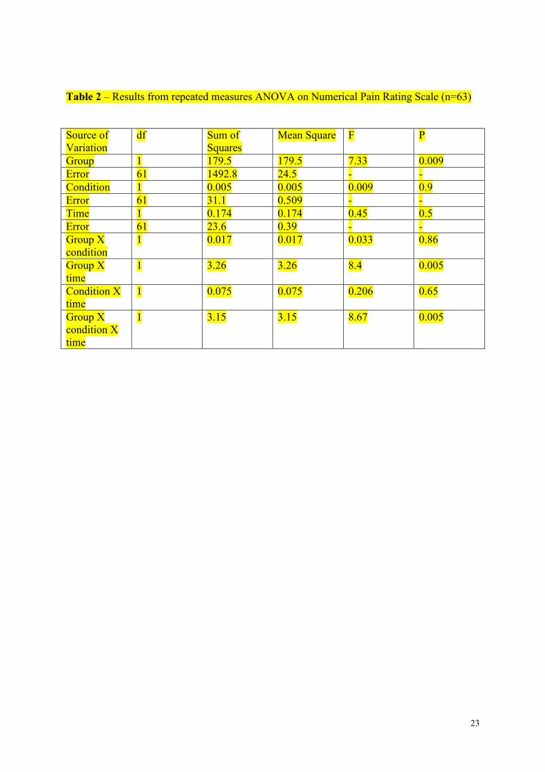
In order to test the hypothesis that the RF condition would increase pain intensity (Numerical
Rating Scale) more than the NRF in the CRPS group we undertook repeated measures
ANOVA with group as between subjects factor and condition (RF and NRF) and time (pre
and post) as within subjects factors. Given the wide variance in pain intensity between
patients and healthy controls and the assumption of similar variability between groups
required by ANOVA, only the CRPS and rheumatology group data were entered into this
analysis. The number of percept changes was analysed by one-way ANOVA to examine for
group differences; a paired t-test was used to examine differences between figures.
Qualitative data, generated from the subjects‟ responses to the open questions was tabulated
on MS-Excel and analysed using content analysis (Holsti, 1968; Frankfort-Nachmias and
Nachmias, 1992). A process of iterative inductive generation of categories from the
descriptive responses generated a number of themes, which corresponded to the diagnostic
criteria for CRPS vasomotor, sudomotor and motor symptoms (sensory, pain and paresthesia
7
were coded separately). Other categories included body perception disturbances
(weight/pressure changes, altered sensitivity, loss of limb), affective (feelings of frustration,
anxiety, tension) and miscellaneous (disorientation, nausea, eye fatigue, dry mouth). A
second independent coder verified category generation; the interrater reliability was found to
be Kappa=0.69 (p <.0.001).
For statistical testing, these data were summarised by determining the frequency of report of
a particular sensation at each stage of the protocol and its reported change. The data were
analysed in three categories according to reported change (worse, same, better), and two
categories according to type (pain / other somatic sensations).The data were examined using
Chi-square test. All statistical tests were performed using SPPS Version 16, a p value ≤0.05
was considered significant.
Results
Thirty patients with CRPS (Type I), thirty-three with general rheumatology disease and forty-
five healthy controls were studied. Table 1 details the demographics of the participants
recruited.
Statistical Analysis
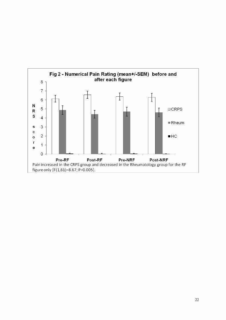
There was main effect on Numerical Pain Rating of group (F (1, 61=7.33, p=0.009) (Table
2). There were significant “group by time” (F (1, 61) =8.4; p=0.005) and “condition by time
by group interactions” (F (1, 61) =8.67; P=0.005). This means that pain intensity differed
significantly between condition and group such that pain increased in the CRPS group and
decreased in the Rheumatology group after viewing the RF (Fig 2). There was, similarly, a
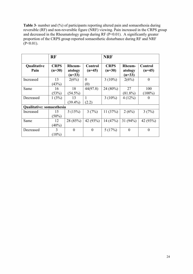
8
significant difference between the CRPS and Rheumatology groups, for frequency of
qualitative pain (x2=19.9, P<0.01) and of other bodily sensation change (x2=17.5; P<0.01)
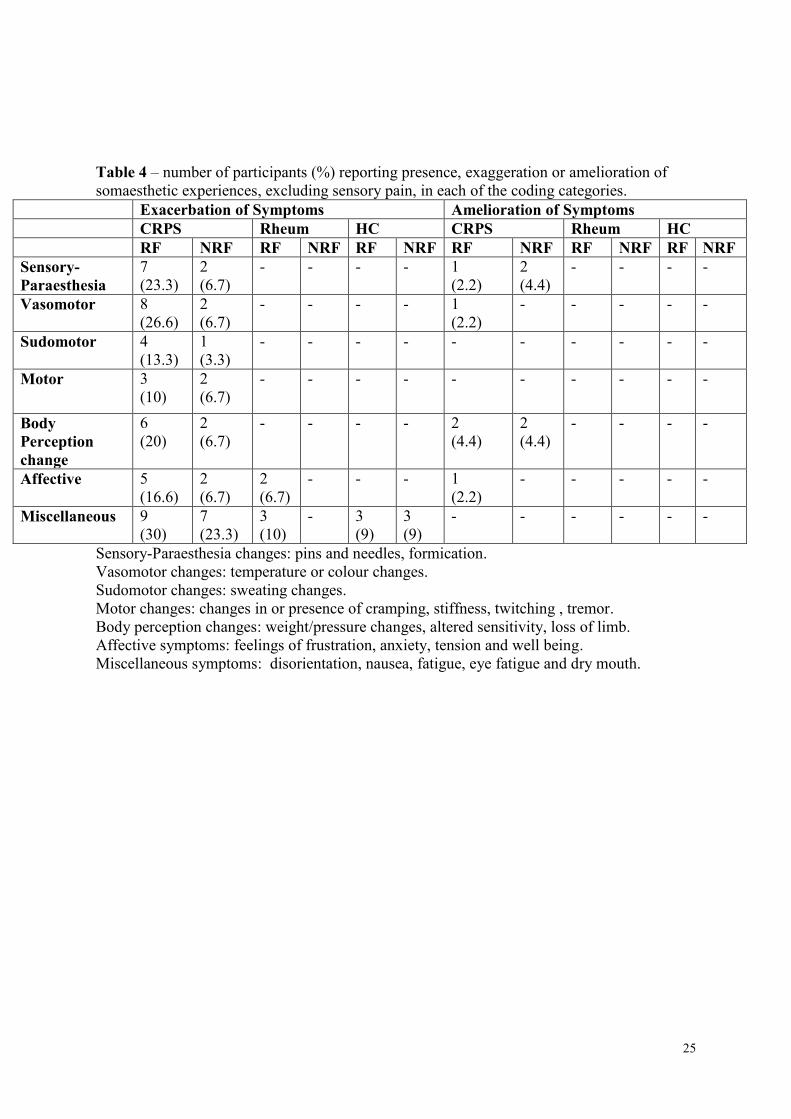
(Table 3). Table 4 defines the type of somaesthetic changes experienced by each of the
groups. In addition 37% of patients with CRPS reported increasing somaesthesia during NRF
(x2=17.5; P<0.01).
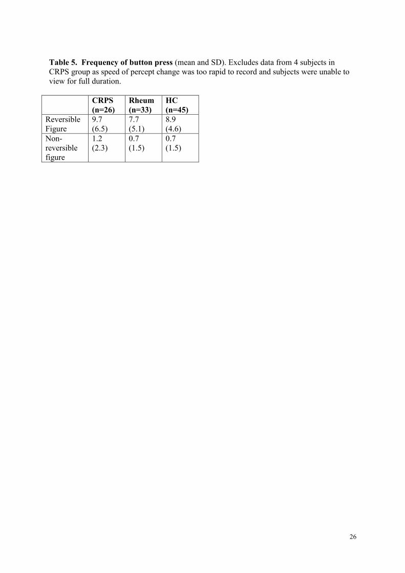
While there was, as expected, a significant difference between the two visual stimuli in the
number of percept changes, there was no difference between groups (Table 5). However, of
interest is that four participants in the CRPS group found the RF changed more frequently
than the button could be pressed and were unable to complete the task for the full minute.
Qualitative Results
Participants in the rheumatology and healthy control groups reported fewer and more minor
sensory disturbances when viewing the figures compared to the CRPS group (Table 4). Most
of these sensory effects related to eye fatigue and feelings of slight disorientation (“almost
felt a bit wobbly”, and “I felt agitated /squiffy”). When viewing the RF and NRF
respectively, some participants (12 for RF, 2 for NRF) in the rheumatology group described
that their pain diminished and attributed this to “my mind is concentrating on something other
than pain”.
In the CRPS group eight subjects responded in a similar way to the healthy volunteers in that
six reported no change in any sensations to either figure. Of the remaining two subjects, one
reported amelioration of their symptoms - attributed to distraction via attention to the figures
- and one noted that they felt “a little bit giddy, probably because I was staring at it”. The
remaining 22 subjects (73%) responded in ways that were different from the healthy and
9
rheumatology groups. These responses ranged from the mildly to the extremely distressing.
Responses at the mild end of the spectrum were isolated or temporary sensations. For
example, one subject reported that their pain became more „nagging‟; another reported the
onset of „tingling‟ on their (affected) forearm, which ceased when the stimulus was removed.
Four subjects with CRPS reported being extremely distressed and were unable to look at the
RF for the full minute (mean: 40s, SD: 15.7) due to increases in pain. Furthermore, two of
these patients were unable to view the NRF for the full minute (14s and 28s). The symptoms
reported included paresthesia (“the tingling has changed to a very deep and irritating
feeling”), dizziness, nausea ( “don‟t feel too good……sweaty, feel flushed, sickly, hot,
bothered”), temperature changes (“foot is flushed hot now”), perceived weight changes (“
right shoulder feels heavy”, “entire arm heavy”) and disorientation (“feel almost trance-
like”). Observation of these subjects showed a characteristic pattern of extreme blinking,
looking away, and finally shutting their eyes with head averted and asking for the picture to
be removed. No differences between these 4 subjects and others in the CRPS group could be
found in relation to baseline pain (NRS), location, symptom duration or medication.
All sensory changes reported by the rheumatology and healthy control groups disappeared
rapidly on removal of the stimulus. Whilst this was similar for most of the CRPS group, four
of the participants had not returned to their baseline pain values by the end of the 2 minutes
rest period and all required the maximum recovery time before proceeding to the next stage
of the protocol.
Discussion
10
The data supports the hypothesis that pain and other symptoms are exacerbated in patients
with CRPS, when viewing the RF compared to viewing the NRF but not in other chronic pain
patients or healthy controls. Both qualitative and quantitative data supported the hypothesis
but the qualitative data was particularly strong with 73% of the CRPS group reporting
exacerbation of their pain or other CRPS symptoms on viewing the RF. Furthermore, we
identified a small group (13%) who responded with striking worsening of their symptoms,
and consequent inability to complete the experimental task. These results differ significantly
from rheumatology patients and healthy controls who reported only minor symptoms; in the
former group almost half reported improvement in their pain due to attending to the picture.
The experimental design does not allow us to attribute the results to the RF alone but it would
appear that symptom increase is confined to patients with CRPS.
Our study is limited by the lack of quantitative measures as well as several possible
confounding factors. Expectation, suggestion and arousal are possible sources of
confounding. The investigators were not blind to the study‟s hypotheses; however, every
attempt was made to use neutral language and to draw subjects‟ attention to the button count
rather than their experiences. Medication may have had an effect on our outcomes as all of
the „severe‟ responders were on anticonvulsant medication. However, half of the CRPS
sample group with mild response were also on similar dosages of psychotropic medication.
Furthermore, 24% of the rheumatology group were taking antidepressants also and none of
these patients exhibited more than minor symptoms. Nevertheless future studies should
include medication and dose as covariates.
Participants with CRPS demonstrated a spectrum of response ranging from none to severe
distress which in some cases necessitated task cessation. These participants reported reversal
11
rates which were too fast to record, suggesting that the speed of reversal was linked to
symptom exacerbation. This might be further tested by using two images that are highlighted
to draw attention to only one of the two potential visual precepts and these images being
alternated in a graded, accelerating manner whilst the subjects‟ symptom responses are
recorded (e.g. a single session of fixed time with the rate of image change controlled at a
fixed speed. Escalation of speed of image change in subsequent sessions would allow
systematic responses to be measured). Objective evidence of autonomic changes within the
affected limb could be captured in this paradigm by adding measures of galvanic skin
response and laser Doppler flowmetry.
The switching rate of RF has been used to elucidate the processes underlying perceptual
instability and using fMRI, Lumer et al (1998) showed that activity in the visual and
frontoparietal cortices was heightened during perceptual transitions (Sterzer et al., 2002;
Long and Topino, 2004). It is of note that superior and inferior parietal lobes and anterior
intraparietal sulcus are involved in switching as these areas are also activated during hand
laterality tasks. The response to such tasks is slower for the affected side in CRPS and
suggests disruption of the body schema, the neural correlates of which reside in the parietal
cortices (de Lange et al., 2006; Moseley, 2004). Our clinical experience has shown that some
patients respond to laterality training in a similar manner to a RF and therefore using a
bistable image might provide a rapid clinical assessment to establish suitability for hand
laterality training. That perceptual transitions activate the parietal cortices is of particular
interest in CRPS aetiology, firstly because of its afferent role in movement via sensory
integration and secondly, because it maps for the body schema, disruption of which is
reported in CRPS (Lewis et al., 2007; Moseley, 2005; Förderreuther et al, 2004; Galer and
Jensen, 1999). Future studies in which the relationships between the response to perceptual
12
instability, Body Perception Disturbance and vulnerability to sensorimotor disruption are
examined may add to the body of literature suggestive of parietal dysfunction in CRPS
(Cohen et al., 2009; Schwenkreis et al., 2009; Schwoebel et al., 2001)
Conclusion
This exploratory mixed methods study has shown that the symptoms of CRPS can be
exacerbated by viewing ambiguous figures. This phenomenon corroborates anecdotal
accounts of the visual disturbances reported by some patients with CRPS. However, further
studies which include objective markers of symptom change are required to confirm these
initial findings. Our experimental method provides a means to generate or exacerbate
somaesthetic disturbances, including pain, without nociceptive interference which could be a
useful technique for future studies, particularly those involving imaging. .
Acknowledgements
The authors gratefully acknowledge the support of our research participants and the Gwen
Bush Foundation who funded this research. Particular thanks too to Professors Richard
Gregory, Ian Gilchrist and Drs Peter Tucker and Trevor Day for their helpful comments when
writing this paper.
13
References
Bruehl S, Harden RN, Galer BS, Saltz S, Bertram M, Backonja M, Gayles R, Rudin N,
Bhugra MK, Stanton-Hicks M. External validation of IASP diagnostic criteria for Complex
Regional Pain Syndrome and proposed research diagnostic criteria. International Association
for the Study of Pain. Pain. 1999; 81: 147-54.
Cohen H, McCabe CS, Harris N, Blake DR. Clinical evidence of parietal lobe dysfunction in
patients with CRPS Type 1. Rheumatology. 2009; 48 (S1):i95
de Lange FP, Helmich RC, Toni I. Posture influences motor imagery: an fMRI study.
Neuroimage. 2006 Nov 1; 33(2):609-17.
Declaration of Helsinki (1964). Br Med J 1996; 313:1448–9.
de Mos M, Sturkenboom MC, Huygen FJ. Current understandings on complex regional pain
syndrome. Pain Pract. 2009 Mar-Apr;9(2):86-99.
Förderreuther S, Sailer U, Straube A. Impaired self-perception of the hand in complex
regional pain syndrome (CRPS). Pain. 2004 Aug; 110(3):756-61.
Frankfort-Nachmias C, Nachmias D. Research methods in the social sciences, 4th
edn. London. Edward Arnold.1992.
14
Galer BS, Jensen M. Neglect-like symptoms in complex regional pain syndrome: results of a
self-administered survey. J Pain Symptom Manage. 1999 Sep; 18(3):213-7.
Gregory RL. Eye and Brain. The Psychology of Seeing. (Oxford:Oxford University Press
1998), 5th
ed, p205.
Harden RN, Swan M, King A, Costa B, Barthel J. Treatment of complex regional pain
syndrome: functional restoration. Clin J Pain. 2006 Jun; 22(5):420-4
Holsti OR. Content analysis. G.Lindzey and E. Aronson (eds).The handbook of Social
Psychology. Reading, MA:Addison-Wesley, 1968.
Lewis JS, Kersten P, McCabe CS, McPherson KM, Blake DR. Body perception disturbance:
a contribution to pain in complex regional pain syndrome (CRPS). Pain. 2007 Dec 15;
133(1-3):111-9
Long GM, Toppino TC. Enduring interest in perceptual ambiguity: alternating views of
reversible figures. Psychol Bull. 2004 Sep; 130(5):748-68
Lumer ED, Friston KJ, Rees G. Neural correlates of perceptual rivalry in the human brain.
Science. 1998 Jun 19; 280(5371):1930-4.
McCabe CS, Haigh RC, Ring EF, Halligan PW, Wall PD, Blake DR.
15
A controlled pilot study of the utility of mirror visual feedback in the treatment of complex
regional pain syndrome (type 1). Rheumatology (Oxford). 2003 Jan; 42(1):97-101.
McCabe C, Moseley L. Functional strategies of restoration of complex regional pain
syndrome. In: Justins D (Ed) PAIN 2005 - An Updated Review: Refresher Course Syllabus.
IASP Press, Seattle 2005: 317-328.
McCabe CS, Cohen H, Blake DR. Somaesthetic disturbances in fibromyalgia are exaggerated
by sensory motor conflict: implications for chronicity of the disease? Rheumatology
(Oxford). 2007 Oct; 46(10):1587-92
McCabe CS, Blake DR. An embarrassment of pain perceptions? Towards an understanding
of and explanation for the clinical presentation of CRPS type 1. Rheumatology (Oxford).
2008, Nov; 47(11):1612-6.
McCabe CS, Haigh RC, Blake DR. Mirror visual feedback for the treatment of complex
regional pain syndrome (type 1).Curr Pain Headache Rep. 2008a Apr;12(2):103-7.
McCabe CS, Haigh RC, Blake DR. Mirror visual feedback for the treatment of complex
regional pain syndrome (type 1). Curr Pain Headache Rep. 2008b, Apr; 12(2):103-7
Moseley GL. Why do people with complex regional pain syndrome take longer to recognize
their affected hand? Neurology. 2004 Jun 22; 62(12):2182-6.
16
Moseley GL. Is successful rehabilitation of complex regional pain syndrome due to sustained
attention to the affected limb? A randomised clinical trial. Pain. 2005 Mar; 114(1-2):54-61
Moseley GL, Zalucki N, Birklein F, Marinus J, van Hilten JJ, Luomajoki H. Thinking about
movement hurts: the effect of motor imagery on pain and swelling in people with chronic arm
pain. Arthritis Rheum. 2008a May 15; 59(5):623-31.
Moseley GL, Zalucki NM, Wiech K. Tactile discrimination, but not tactile stimulation alone,
reduces chronic limb pain. Pain. 2008b Jul 31; 137(3):600-8.
Moseley GL, Parsons TJ, Spence C. Visual distortion of a limb modulates the pain and
swelling evoked by movement. Curr Biol. 2008c Nov 25; 18(22):R1047-8Moseley L.
Distorted body image in complex regional pain syndrome. Neurology 2005;6 5:773,
Ramachandran VS, Altschuler EL. The use of visual feedback, in particular mirror visual
feedback, in restoring brain function. Brain. 2009 Jul; 132(Pt 7):1693-710
Schwenkreis P, Maier C, Tegenthoff M. Functional imaging of central nervous system
involvement in complex regional pain syndrome. AJNR Am J Neuroradiol. 2009 Aug;
30(7):1279-84
Schwoebel J, Friedman R, Duda N, Coslett H. Pain and the body schema. Evidence for
peripheral effects on mental representations of movement. Brain 2001; 124:2098-2104.
17
Sterzer P, Russ MO, Preibisch C, Kleinschmidt A. Neural correlates of spontaneous direction
reversals in ambiguous apparent visual motion. Neuroimage. 2002 Apr; 15(4):908-16
Williamson A, Hoggart B. Pain: a review of three commonly used pain rating scales. J Clin
Nurs. 2005 Aug; 14(7):798-804.
Figure/table Legends
Figure 1. Flow diagram illustrating the sequence of images
Table 1- Participant characteristics.
Figure 2 – Pain intensity on a 10cm Numerical rating scale (mean and SD) before and after
each figure
Table 2 – Results from repeated measures ANOVA on Numerical Pain Rating Scale (n=63)
Table 3- number and (%) of participants reporting altered pain and somaesthesia during
reversible (RF) and non-reversible figure (NRF) viewing. Pain increased in the CRPS group
and decreased in the Rheumatology group during RF (P<0.01). A significantly greater
proportion of the CRPS group reported somaesthetic disturbance during RF and NRF
(P<0.01).
Table 4 – number of participants (%) reporting presence, exaggeration or amelioration of
somaesthetic experiences, excluding sensory pain, in each of the coding categories.
Table 5. Frequency of button press (mean and SD). Excludes data from 4 subjects in
CRPS group as speed of percept change was too rapid to record and subjects were unable to
view for full duration.
18
Figure 1. Flow diagram illustrating the sequence of images
19
Rest 2-10 min
Rest 2 mins
Rest 2-10 min
Time
Familiarisation
Until 3 button
presses
I minute
Press when see
change
I minute
Press when see
change
20
Table 1- Participant characteristics.
Subject
characteristics
CRPS
(n=30)
Rheumatology
(n=33)
Healthy
Controls
(n=45)
Age (years) [mean&
range]
42 (20-63) 63.6(24-88) 46 (22-64)
Male 9 10 4
Distribution of
CRPS/
Rheumatology
conditions
(n=)
UL=13
LL=10
ULLs=4
LT=3
RA=16
OA =13
V=2
AS =1
PsA =1
Time since diagnosis
[median (range)]
16 months
(0-9 years)
Numerical Pain
Rating scale
(mean&SD)
6.17(2) 5.1(2.7)* 0.044
(0.2)
Frequency of
comorbidity
46.6% 59.9% 28.8%
Medication (n)
DMARDs 0 15 0
Non-opioid analgesic 11 12 1
NSAID 9 13 2
Weak opioid
analgesic
17 16 0
Strong opioid 7 5 0
Steroid 0 9 2
Osteoporosis
prophylaxis
treatment
2 13 0
Gastroprotection 3 16 1
Cardiovascular 6 22 4
Anti-epileptic/anti-
depressant
18 8 3
Oral hypoglcaemics 0 5 1
Lipid lowering drugs 0 7 3
HRT 4 2 3
Respiratory
medication
5 1 3
Miscellaneous 12 10 3
UL-upper limb, LL-Lower lib, ULLs-Upper and lower limbs, LT-limb and trunk.
RA-Rheumatoid arthritis, OA-osteoarthritis, V- vasculitis, AS-ankylosing spondylitis, PSa-
psoriatic arthritis
DMARDs- disease modifying anti-rheumatic drugs
NSAID - Non steroidal anti-inflammatory drug
Cardiovascular (inc. anti-hypertensives, anti-arhythmics, anti-anginal drugs, anti-platelet
agents, anticoagulants)
21
HRT - hormone replacement (inc. thyroxine)
Miscellaneous (vitamin/iron supplementation, antihistamine, night sedation, prostate/bladder
medications, antibiotics, antiemetics, quinine )
*There were no significant differences in pain at baseline between patients in the CRPS and
rheumatology groups (unpaired t-test).
22
23
Table 2 – Results from repeated measures ANOVA on Numerical Pain Rating Scale (n=63)
Source of
Variation
df Sum of
Squares
Mean Square F P
Group 1 179.5 179.5 7.33 0.009
Error 61 1492.8 24.5 - -
Condition 1 0.005 0.005 0.009 0.9
Error 61 31.1 0.509 - -
Time 1 0.174 0.174 0.45 0.5
Error 61 23.6 0.39 - -
Group X
condition
1 0.017 0.017 0.033 0.86
Group X
time
1 3.26 3.26 8.4 0.005
Condition X
time
1 0.075 0.075 0.206 0.65
Group X
condition X
time
1 3.15 3.15 8.67 0.005
24
Table 3- number and (%) of participants reporting altered pain and somaesthesia during
reversible (RF) and non-reversible figure (NRF) viewing. Pain increased in the CRPS group
and decreased in the Rheumatology group during RF (P<0.01). A significantly greater
proportion of the CRPS group reported somaesthetic disturbance during RF and NRF
(P<0.01).
RF NRF
Qualitative
Pain
CRPS
(n=30)
Rheum-
atology
(n=33)
Control
(n=45)
CRPS
(n=30)
Rheum-
atology
(n=33)
Control
(n=45)
Increased 13
(43%)
2(6%) 0
(0)
3 (10%) 2(6%) 0
Same 16
(53%)
18
(54.5%)
44(97.8) 24 (80%) 27
(81.8%)
100
(100%)
Decreased 1 (3%) 13
(39.4%)
1
(2.2)
3 (10%) 4 (12%) 0
Qualitative: somaesthesia
Increased 15
(50%)
5 (15%) 3 (7%) 11 (37%) 2 (6%) 3 (7%)
Same 12
(40%)
28 (85%) 42 (93%) 14 (47%) 31 (94%) 42 (93%)
Decreased 3
(10%)
0 0 5 (17%) 0 0
25
Table 4 – number of participants (%) reporting presence, exaggeration or amelioration of
somaesthetic experiences, excluding sensory pain, in each of the coding categories.
Exacerbation of Symptoms Amelioration of Symptoms
CRPS Rheum HC CRPS Rheum HC
RF NRF RF NRF RF NRF RF NRF RF NRF RF NRF
Sensory-
Paraesthesia
7
(23.3)
2
(6.7)
- - - - 1
(2.2)
2
(4.4)
- - - -
Vasomotor 8
(26.6)
2
(6.7)
- - - - 1
(2.2)
- - - - -
Sudomotor 4
(13.3)
1
(3.3)
- - - - - - - - - -
Motor 3
(10)
2
(6.7)
- - - - - - - - - -
Body
Perception
change
6
(20)
2
(6.7)
- - - - 2
(4.4)
2
(4.4)
- - - -
Affective 5
(16.6)
2
(6.7)
2
(6.7)
- - - 1
(2.2)
- - - - -
Miscellaneous 9
(30)
7
(23.3)
3
(10)
- 3
(9)
3
(9)
- - - - - -
Sensory-Paraesthesia changes: pins and needles, formication.
Vasomotor changes: temperature or colour changes.
Sudomotor changes: sweating changes.
Motor changes: changes in or presence of cramping, stiffness, twitching , tremor.
Body perception changes: weight/pressure changes, altered sensitivity, loss of limb.
Affective symptoms: feelings of frustration, anxiety, tension and well being.
Miscellaneous symptoms: disorientation, nausea, fatigue, eye fatigue and dry mouth.
26
Table 5. Frequency of button press (mean and SD). Excludes data from 4 subjects in
CRPS group as speed of percept change was too rapid to record and subjects were unable to
view for full duration.
CRPS
(n=26)
Rheum
(n=33)
HC
(n=45)
Reversible
Figure
9.7
(6.5)
7.7
(5.1)
8.9
(4.6)
Non-
reversible
figure
1.2
(2.3)
0.7
(1.5)
0.7
(1.5)