Embed Size (px)
DESCRIPTION
Human Amyloid Imaging 2014
Citation preview

w w w . w o r l d e v e n t s f o r u m . c o m / h a i
8th Human Amyloid Imaging
January 15-17, 2014Miami, Florida
Co-Organizers:Keith A. Johnson, MD ● William J. Jagust, MD ● William E. Klunk, MD, PhD ● Chester Mathis, PhD

To locate a poster by presenter’s last name, view Table of Contents at page 3.
To locate a poster by number, view Program at page 10.
To view program, posters and abstracts on your mobile or tablet, please scan the QR code
below or visit http://my.yapp.us/HAI2014
1
TABLE OF CONTENTS
PROGRAM (all sessions will be held at the Miami Beach Resort)..................................... 8
ORAL PRESENTATIONS ................................................................................................. 18
SESSION 1: Tau PET I .......................................................................................................................................... 18
PET imaging of tau deposits in Alzheimer's disease patients using 18F-THK5105 and 18F-THK5117 ................ 18
Presented by: Okamura, Nobuyuki ................................................................................................................... 18
Tau deposition estimated by [11C]PBB3 PET in Alzheimer’s disease, MCI with and without amyloid deposition,
and cognitive healthy subjects .............................................................................................................................. 19
Presented by: Shimada, Hitoshi ....................................................................................................................... 19
Tau PET: Initial experience with F18 T807 ......................................................................................................... 20
Presented by: Johnson, Keith ........................................................................................................................... 20
SESSION 2: Tau PET II ........................................................................................................................................ 21
Detection of PHF-Tau pathology with T557, T726 and [18F]-T807 in brain sections from Alzheimer’s and non-
Alzheimer’s tauopathy patients ............................................................................................................................ 21
Presented by: Skovronsky, Daniel M. ............................................................................................................... 21
Imaging tau pathology in vivo in FTLD: initial experience with [18F] T807 PET .............................................. 22
Presented by: Dickerson, Brad ......................................................................................................................... 22
PET Tau imaging with [F-18]-T807 (AV-1451) in normal subjects and patients with cognitive impairment due
to Alzheimer’s disease: Review of initial analyses ............................................................................................... 23
Presented by: Mintun, Mark A. ......................................................................................................................... 23
SESSION 3: KEYNOTE LECTURE .................................................................................................................... 24
Tau protein - from native unfolding to pathological folding: structural aspects of Tau, Tau aggregates, and Tau
interacting compounds .......................................................................................................................................... 24
Presented by: Eckhard Mandelkow .................................................................................................................. 24
SESSION 4: Abeta PET: Assessment of Cognition and Disease Progression ................................................... 25
Amyloid change early in disease is related to increased glucose metabolism and episodic memory decline ...... 25
Presented by: Landau, Susan............................................................................................................................ 25
Amyloid-β deposition in mild cognitive impairment is associated with hippocampal hyperactivation, atrophy
and clinical progression ........................................................................................................................................ 26
Presented by: Huijbers, Willem ........................................................................................................................ 26
Gene-Environment interactions over the lifecourse: Cognitive activity, apolipoprotein E genotype, and brain
beta-amyloid ......................................................................................................................................................... 27
Presented by: Wirth, Miranka .......................................................................................................................... 27
Preclinical effects of Aβ deposition on episodic memory and disease progression ............................................. 28
Presented by: Villemagne, Victor L .................................................................................................................. 28
SESSION 5: Abeta PET: Relation to Co-morbid Conditions ............................................................................. 29
Vascular and amyloid pathologies are independent predictors of cognitive decline in cognitively normal elderly
.............................................................................................................................................................................. 29
Presented by: Vemuri, Prashanthi .................................................................................................................... 29
2 8th Human Amyloid Imaging – Miami, 2014
Multiple brain markers contribute to age-related variation in cognition .............................................................. 30
Presented by: Hedden, Trey.............................................................................................................................. 30
Cerebral amyloid related alterations in neuronal metabolism and the contribution of multimodal measures of
vascular function ................................................................................................................................................... 31
Presented by: McDade, Eric ............................................................................................................................. 31
Binding of Pittsburgh Compound B to both normal and abnormal white matter in elderly cognitively normal
controls. ................................................................................................................................................................ 32
Presented by: Cohen, Ann ................................................................................................................................ 32
SESSION 6: INVITED LECTURE ....................................................................................................................... 33
The Centiloid method for quantifying Amyloid PET studies: Great illuminator or master or illusion ................ 33
Presented by: Koeppe, Robert .......................................................................................................................... 33
SESSION 7: Technical Emphasis .......................................................................................................................... 34
Modeling the influence of white matter contamination on detectability of brain amyloid changes in longitudinal
studies of Alzheimer’s progression: Segmentation analyses using the PET β-Amyloid tracer 18F NAV4694 ... 34
Presented by: Seibyl, John ................................................................................................................................ 34
Amyloid PET screening results by APOE ε4 status from a Phase 1b Clinical Study (221AD103) in patients with
prodromal to mild Alzheimer’s disease ................................................................................................................ 35
Presented by: Chiao, Ping ................................................................................................................................ 35
Liberal thresholds for PIB-positivity optimally capture high-burden postmortem amyloid pathology ................ 36
Presented by: Cohn-Sheehy, Brendan I. ........................................................................................................... 36
Existing thresholds for PIB positivity are too high ............................................................................................... 37
Presented by: Villeneuve, Sylvia ....................................................................................................................... 37
SESSION 8: Neuropathologic Correlations ......................................................................................................... 38
PiB PET and FDG PET quantitative analysis methods with autopsy correlation ................................................ 38
Presented by: Lowe, Val ................................................................................................................................... 38
Evaluation of cotton wool plaques using amyloid binding compounds and Aβ immunohistochemistry:
implications for PiB PET imaging ........................................................................................................................ 39
Presented by: Ikonomovic, Milos ...................................................................................................................... 39
[18F]Flutemetamol amyloid PET detection of Aβ plaque phases 4 and 5 and significant diffuse plaque burden 40
Presented by: Buckley, Chris ............................................................................................................................ 40
Diagnostic accuracy of amyloid and FDG PET in pathologically-confirmed dementia ...................................... 41
Presented by: Rabinovici, Gil D. ...................................................................................................................... 41
Senile plaques: Classification, distribution, clinical correlation and Amyloid imaging ...................................... 42
Presented by: Beach, Thomas G.. ..................................................................................................................... 42
SESSION 10: Memory Complaints/JADNI ......................................................................................................... 43
Subjective memory complaints are related to default network disruption in clinically normal older adults with
high amyloid burden ............................................................................................................................................. 43
Presented by: Vannini, Patrizia ........................................................................................................................ 43
3
Amyloid burden and neurodegeneration independently contribute to greater subjective cognitive concerns in
clinically normal older individuals ....................................................................................................................... 44
Presented by: Amariglio, Rebecca .................................................................................................................... 44
Longitudinal amyloid deposition with 11C-PiB in Japanese ADNI study ............................................................. 45
Presented by: Ishii, Kenji.................................................................................................................................. 45
Higher Aβ burden in subjective memory complainers: A flutemetamol sub-study in AIBL ............................... 46
Presented by: Rowe, Christopher C ................................................................................................................. 46
Poster Presentations (Alphabetized by presenting author)* .............................................. 47
POSTER SESSION ................................................................................................................................................ 47
Early functional changes in the language network in response to increased amyloid beta deposition in healthy
older adults ............................................................................................................................................................ 47
Presented by: Adamczuk, Katarzyna ................................................................................................................ 47
*To locate a poster by number, view Program at page 10. .............................................................................. 47
Patterns of Av-45 uptake using VOIs based multivariate analysis in elderly peoples .......................................... 48
Presented by: Adel, Djilali................................................................................................................................ 48
Prevalence of amyloid burden in elderly subjects in a PET-AV45 multicenter study.......................................... 49
Presented by: Adel, Djilali................................................................................................................................ 49
Imaging amyloid in adults with Down’s syndrome using PiB-PET ..................................................................... 50
Presented by: Annus, Tiina ............................................................................................................................... 50
Exploring the best methods to detect longitudinal change in amyloid imaging ................................................... 51
Presented by: Baker, Suzanne .......................................................................................................................... 51
Comparison of reference regions for [18F]Florbetapir PET SUVR computation ................................................ 52
Presented by: Bedell, Barry .............................................................................................................................. 52
Statistically-driven, automated subject classification based on amyloid PET scans ............................................ 53
Presented by: Bedell, Barry .............................................................................................................................. 53
Body mass index is associated with verbal episodic memory in cognitively normal older individuals with low
fibrillar amyloid-beta measured by [11C]-PiB ....................................................................................................... 54
Presented by: Bilgel, Murat .............................................................................................................................. 54
[18F]Flutemetamol blinded image interpretation: assessment of the electronic training program for Japanese
readers ................................................................................................................................................................... 55
Presented by: Buckley, Chris ............................................................................................................................ 55
Disclosure of amyloid imaging results in cognitively normal individuals ........................................................... 56
Presented by: Burns, Jeffrey ............................................................................................................................. 56
Differential temporal patterns of Amyloid-β and functional imaging markers across mutation types in autosomal
dominant Alzheimer’s disease: Findings from the DIAN Study .......................................................................... 57
Presented by: Chhatwal, Jasmeer .................................................................................................................... 57
Comparison of PIB-PET data to Florbetapir-PET data acquired from different patient cohorts and the role of age
in the discriminative power of amyloid imaging .................................................................................................. 58
Presented by: Chiotis, Konstantinos ................................................................................................................. 58
4 8th Human Amyloid Imaging – Miami, 2014
The value of early F18-Florbetapir scan information as an estimate of regional cerebral blood flow and
comparison to F18-FDG measures of cerebral metabolism.................................................................................. 59
Presented by: Devous, Michael ........................................................................................................................ 59
Amyloid imaging changes diagnosis and treatment in patients with progressive cognitive impairment:
Multicenter evaluation of 3-month post-scan outcomes ....................................................................................... 60
Presented by: Doraiswamy, P. Murali ............................................................................................................. 60
Practice effects relate to flutemetamol uptake, but not FDG or hippocampal volume: Moving cognition earlier in
Jack’s curves ......................................................................................................................................................... 61
Presented by: Duff, Kevin ................................................................................................................................. 61
Alpha-synuclein imaging: Identifying small molecules that bind to aggregated Alpha-synuclein ...................... 62
Presented by: Eberling, Jamie .......................................................................................................................... 62
Evaluation of F-18 radiolabeled cromolyn as a potential Aβ polymerization inhibitor and PET tracer............... 63
Presented by: Elmaleh, David R. ...................................................................................................................... 63
Effects of age and amyloid on the encoding of visual scene detail ...................................................................... 64
Presented by: Elman, Jeremy ........................................................................................................................... 64
Clinical presentation and imaging findings in complex ALS with AD-like cognitive impairment ...................... 65
Presented by: FARID, Karim ............................................................................................................................ 65
Striatal 11C-PiB retention in AD, MCI and HC: the pathotololgical significance of high PIB retention in the
putamen................................................................................................................................................................. 66
Presented by: FARID, Karim ............................................................................................................................ 66
Advanced education mediates the impact of amyloid burden on reasoning in healthy older adult ...................... 67
Presented by: Farrell, Michelle E. ................................................................................................................... 67
Early-frame PiB PET perfusion and FDG PET glucose metabolism comparison in cognitively normal persons at
three levels of genetic risk for Alzheimer’s disease ............................................................................................. 68
Presented by: Fleisher, Adam ........................................................................................................................... 68
Florbetapir-PET has greater clinical impact than FDG-PET in the differential diagnosis of AD and FTD ......... 69
Presented by: Ghosh, Pia M. ............................................................................................................................ 69
Source of cognitive complaint and amyloid binding in mild cognitive impairment ............................................ 70
Presented by: Gifford, Katherine ...................................................................................................................... 70
Striatal and extrastriatal dopamine transporter levels relate to cognition in Lewy body diseases ........................ 71
Presented by: Gomperts, Stephen ..................................................................................................................... 71
Is there a binary relationship of mean brain Amyloid concentrations to cognitive, functional, metabolic and
volumetric MRI variables in the ADNI cohort ..................................................................................................... 72
Presented by: Goryawala, Mohammed ............................................................................................................ 72
Relationship of regional cerebral volumes to mean brain Amyloid concentration, APOE genotype and disease
stage in the ADNI 2 cohort ................................................................................................................................... 73
Presented by: Goryawala, Mohammed ............................................................................................................ 73
Genetic resilience to Tau- and Amyloid-related neurodegeneration .................................................................... 74
Presented by: Hohman, Timothy ...................................................................................................................... 74

5
Specificity of [3H]T808, [3H]THK-5105 and [3H]AV-45 (florbetapir) binding to aggregated tau and amyloid
plaques in human AD tissue in vitro ..................................................................................................................... 75
Presented by: Honer, Michael .......................................................................................................................... 75
Arterial stiffness is associated with amyloid accumulation over two years in very elderly adults ....................... 76
Presented by: Hughes, Timothy ........................................................................................................................ 76
Estimation of the number of physicians specializing in treating adult dementia patients in the US: Applying
appropriate use criteria to medicare claims data ................................................................................................... 77
Presented by: Hunter, Craig A. ........................................................................................................................ 77
Modeling intra-brain Amyloid-ß propagation via an epidemic spreading framework ......................................... 78
Presented by: Iturria Medina, Yasser ............................................................................................................... 78
The potential of florbetapir F 18 and an early scan PET protocol (0-20 minutes after injection) to evaluate for
the presence of amyloid ........................................................................................................................................ 79
Presented by: Joshi, Abhinay D. ....................................................................................................................... 79
Comparing concordance of different SUVR calculation methods with florbetapir visual reads .......................... 80
Presented by: Klein, Gregory ........................................................................................................................... 80
Early age of onset is associated with greater neuroinflammation in Alzheimer’s disease ................................... 81
Presented by: Kreisl, William Charles ............................................................................................................. 81
The association between glucose metabolism in Alzheimer’s disease-vulnerable regions and cognitive reserve is
modified by amyloid status within clinically normal individuals ......................................................................... 82
Presented by: LaPoint, Molly ........................................................................................................................... 82
Quantification and accuracy of clinical [11C]-PiB PET/MRI: The effect of MR-based attenuation correction .. 83
Presented by: Law, Ian ..................................................................................................................................... 83
Relationship between Amyloid burden and clinical course across AD pathophysiologic stages ......................... 84
Presented by: Margolin, Richard ..................................................................................................................... 84
Aβ accumulation is associated with hippocampal hypometabolism in cognitively normal older adults regardless
of Aβ level ............................................................................................................................................................ 85
Presented by: Marks, Shawn ............................................................................................................................ 85
Is there an asymmetric distribution of in vivo Amyloid in primary progressive aphasia? ................................... 86
Presented by: Martersteck, Adam ..................................................................................................................... 86
CogState computerized testing and neurodegenerative and amyloid imaging: Implications for secondary
preventive trials .................................................................................................................................................... 87
Presented by: Mielke, Michelle ........................................................................................................................ 87
Longitudinal Amyloid measurement for clinical trials: A new approach to overcome variability....................... 88
Presented by: Matthews, Dawn ........................................................................................................................ 89
Prediction of Amyloid-beta hepatic clearance using sandwich cultured primary rat hepatocytes........................ 90
Presented by: Mohamed, Loqman .................................................................................................................... 90
Identifying cost-effective predictive rules of Amyloid-β level by integrating neuropsychological tests and
plasma-based marker ............................................................................................................................................ 91
Presented by: Morgan, Dave ............................................................................................................................ 91
6 8th Human Amyloid Imaging – Miami, 2014
Associations between beta-amyloid, markers of neurodegeneration, and cognition in clinically normal
individuals from the Harvard Aging Brain Study ................................................................................................. 92
Presented by: Mormino, Elizabeth ................................................................................................................... 92
Beta-amyloid has a greater impact on memory in females than males across the clinical spectrum .................... 93
Presented by: Mormino, Elizabeth ................................................................................................................... 93
PiB-PET significantly differs across neuropathologic classification of Alzheimer-type pathology and tangle
predominant dementia........................................................................................................................................... 94
Presented by: Murray, Melissa E. .................................................................................................................... 94
Brain Amyloid deposition influences the Cognition-Mobility Interface (COMBINE) in cognitively normal older
adults ..................................................................................................................................................................... 95
Presented by: Nadkarni, Neelesh ...................................................................................................................... 95
Astrocytosis, amyloid deposition and gray matter density in parahippocampus of MCI patients ........................ 96
Presented by: Nordberg, Agneta ...................................................................................................................... 96
Regional brain activity and functional connectivity during memory encoding are differentially affected by age
and β-amyloid deposition in cognitively normal older adults .............................................................................. 97
Presented by: Oh, Hwamee .............................................................................................................................. 97
Prevalence of Amyloid in cognitively normal, MCI and demented subjects – A meta-analysis of Amyloid PET
studies ................................................................................................................................................................... 98
Presented by: Ossenkoppele, Rik ...................................................................................................................... 98
Longitudinal quantification of brain amyloidosis in a rat model of Alzheimer’s disease using [18F]NAV4694 99
Presented by: Parent, Maxime J. ...................................................................................................................... 99
Potential value of an interpretation method that incorporates quantitative estimate of cortical to cerebellar SUVr
as an adjunct to visual interpretation of florbetapir PET scans........................................................................... 100
Presented by: Pontecorvo, Michael ................................................................................................................ 100
The Alzheimer’s structural connectome: Patterns of cortical reorganization with increasing neuritic amyloid
plaque burden ...................................................................................................................................................... 101
Presented by: Prescott, Jeff ............................................................................................................................ 101
Mathematical modeling of amyloid-β disposition by brain endothelial cells ..................................................... 102
Presented by: Qosa, Hisham .......................................................................................................................... 102
Amyloid burden influences the relationship between hippocampal volume and default mode network
connectivity in cognitively normal elderly ......................................................................................................... 103
Presented by: Schultz, Aaron .......................................................................................................................... 103
Update on the multicenter phase 3 histopathology study for ß-amyloid brain PET imaging ............................. 104
Presented by: Seibyl, John .............................................................................................................................. 104
TAU Tracer F18-T807 inversely relates with brain functional hubs of elderly normals ..................................... 105
Presented by: Sepulcre, Jorge ........................................................................................................................ 105
Amyloid PET screening for enrollment into Alzheimer’s disease clinical trials: Initial experience in a Phase 1b
clinical trial ......................................................................................................................................................... 106
Presented by: Sevigny, Jeff ............................................................................................................................ 106
Antidepressant decreases CSF Aβ production in healthy individuals and in transgenic mice ........................... 107
Presented by: Sheline, Yvette .......................................................................................................................... 107

7
Impact of regional flow/perfusion heterogeneity and metabolism on the quantification of amyloid tracers: a
preclinical exploration ........................................................................................................................................ 108
Presented by: Staelens, Steven ........................................................................................................................ 108
In Vivo assessment of two isomers of [18F]-THK5105 and [18F]-THK511 ........................................................ 109
Presented by: Tamagnan, Gilles ..................................................................................................................... 109
Evaluation of different target-to-reference region definitions for quantitative categorization of [18F]flutemetamol
images into either normal or abnormal amyloid levels ....................................................................................... 110
Presented by: Thurfjell, Lennart ..................................................................................................................... 110
En attendant Centiloid ........................................................................................................................................ 111
Presented by: Villemagne, Victor L ................................................................................................................ 111
Investigating the involvement of the striatum in Down's syndrome and Alzheimer's disease with 11C-Pittsburgh
Compound B-positron emission tomography (PiB-PET) ................................................................................... 112
Presented by: Wilson, Liam Reese .................................................................................................................. 112
Utility of regional [18F]-Florbetapir PET imaging SUVRs in discriminating Alzheimer’s disease and its
prodromal stages ................................................................................................................................................. 113
Presented by: Zhou, Qi ................................................................................................................................... 113
Diagnostic value of Amyloid imaging in early onset dementia .......................................................................... 114
Presented by: Zwan, Marissa ......................................................................................................................... 114
8 8th Human Amyloid Imaging – Miami, 2014
8th Annual Human Amyloid Imaging Miami, Florida, January 15 - 17, 2014
PROGRAM (all sessions will be held at the Miami Beach Resort)
Wednesday, 15 January 05:00 pm - 06:30
CHECK-IN and POSTER INSTALLATION (Starlight Room/Top Level)
06:30pm - 08:30
WELCOME RECEPTION (Oceanview Room/Lower Level)
Thursday, 16 January 07:00 am - 8:00
CHECK-IN and BREAKFAST (Regency Ballroom/Ground Floor)
08:00 - 08:15
WELCOME REMARKS (Grande Promenade/Ground Floor)
08:00 Introductions and Welcome Remarks
08:15 - 9:30
SESSION 1: Tau PET I CHAIRS: William Jagust (University of California, Berkeley) and Keith Johnson (Massachusetts General Hospital)
08:15 SESSION 1-T1-O-01
PET imaging of tau deposits in Alzheimer's disease patients using 18F-THK5105 and 18F-THK5117 Nobuyuki Okamura (Tohoku University School of Medicine), Shozo Furumoto, Ryuichi Harada, Katsutoshi Furukawa, Aiko
Ishiki, Michelle Fodero-Tavoletti, Rachel Mulligan, Ren Iwata, Manabu Tashiro, Kazuhiko Yanai, Colin Masters, Hiroyuki Arai,
Christopher Rowe, Victor Villemagne, Yukitsuka Kudo
08:30 SESSION 1-T1-O-02
Tau deposition estimated by [11C]PBB3 PET in Alzheimer’s disease, MCI with and without amyloid
deposition, and cognitive healthy subjects Hitoshi Shimada (National Institute of Radiological Sciences, Chiba-Shi), Makoto Higuchi, Hitoshi Shinotoh, Shigeki Hirano,
Shogo Furukawa, Yoko Eguchi, Keisuke Takahata, Fumitoshi Kodaka, Yasuyuki Kimura, Makiko Yamada, Masahiro Maruyama,
Harumasa Takano, Ming-Rong Zhang, Hiroshi Ito, Tetsuya Suhara, et al.
08:45 SESSION 1-T1-O-03
Tau PET: Initial experience with F18 T807 Keith Johnson (Massachusetts General Hospital), John A. Becker, Jorge Sepulcre, Dorene Rentz, Aaron Schultz, Leslie Pepin,
Marlie Philiossaint, Jonathan Alverio, Kelly Judge, Neil Vasdev, Tom Brady, Brad Hyman, Reisa Sperling
9:00 DISCUSSION SESSION 1
9:30 BREAK (Regency Ballroom/Ground Floor)
9:50 - 11:05
SESSION 2: Tau PET II CHAIRS: Victor Villemagne (Austin Health) and Mark A. Mintun (Avid Radiopharmaceuticals, Inc.)
9:50 SESSION 2-T2-O-01
PET Tau imaging with [F-18]-T807 (AV-1451) in normal subjects and patients with cognitive
impairment due to Alzheimer’s disease: Review of initial analyses Mark A. Mintun (Avid Radiopharmaceuticals, Inc.), Abhinay Joshi, Sergey Shcherbinin, Adam J. Schwarz, Ming Lu, Michael
Pontecorvo, Michael Devous Sr., Daniel M. Skovronsky, Hartmuth Kolb
9
10:05 SESSION 2-T2-O-02
Detection of PHF-Tau pathology with T557, T726 and [18F]-T807 in brain sections from Alzheimer’s
and non-Alzheimer’s Tauopathy patients Hartmuth C. Kolb, Giorgio Attardo, Kelly Conway, Felipe Gomez, Qianwa Liang, Yin-Guo Lin, Andrew Siderowf, Daniel M.
Skovronsky (Avid Radiopharmaceuticals, Inc.), Mark A. Mintun
10:20 SESSION 2-T2-O-03
Imaging tau pathology in vivo in FTLD: initial experience with [18F] T807 PET Brad Dickerson (Massachusetts General Hospital/Harvard Medical School), Kimiko Domoto-Reilly, Daisy Sapolsky, Michael Brickhouse,
Michael Stepanovic, Keith Johnson
10:35 DISCUSSION SESSION 2
11:05 - 11:50
SESSION 3: KEYNOTE LECTURE
11:05 KEYNOTE LECTURE: Tau protein - from native unfolding to pathological folding: structural aspects of Tau,
Tau aggregates, and Tau interacting compounds Eckhard Mandelkow (Max Planck Research unit for Structural Molecular Biology at DESY)
11:35 DISCUSSION SESSION 3
11:50 LUNCH (Starlight Room/Top Level)
1:20 - 2:50
SESSION 4: Abeta PET: Assessment of Cognition and Disease Progression CHAIRS: Susan Landau (University of California, Berkeley) and Prashanthi Vemuri (Mayo Clinic Rochester)
1:20 SESSION 4-T4-O-01
Amyloid-β deposition in mild cognitive impairment is associated with hippocampal hyperactivation,
atrophy and clinical progression Willem Huijbers (Brigham and Women’s Hospital/Harvard Medical School, Massachusetts General Hospital), Elizabeth Mormino,
Aaron Schultz, Jasmeer Chhatwal, Brendon Boot, Rebecca Amariglio, Gad Marshall, Dorenne Rentz, Keith Johnson, Reisa Sperling
1:35 SESSION 4-T4-O-02
Amyloid change early in disease is related to increased glucose metabolism and episodic memory decline Susan Landau (University of California, Berkeley), Allison Fero, Suzanne Baker, William Jagust
1:50 SESSION 4-T4-O-03
Preclinical effects of Aβ deposition on episodic memory and disease progression Victor L Villemagne (Austin Health), Samantha Burnham, Pierrick Bourgeat, Belinda Brown, Kathryn Ellis, Olivier Salvado, Ralph
Martins, Lance Macaulay, David Ames, Colin L Masters, Christopher C Rowe
2:05 SESSION 4-T4-O-04
Gene-environment interactions over the lifecourse: Cognitive activity, apolipoprotein E genotype, and
brain beta-amyloid Miranka Wirth (University of California, Berkeley), Sylvia Villeneuve, Renaud La Joie, Shawn Marks, William Jagust
2:20 DISCUSSION SESSION 4
2:50 BREAK (Regency Ballroom/Ground Floor)
3:10 - 4:40
SESSION 5: Abeta PET: Relation to Co-morbid Conditions CHAIRS: Clifford Jack (Mayo Clinic Rochester) and Ann Cohen (University of Pittsburgh School of Medicine)
3:10 SESSION 5-T5-O-01
Multiple brain markers contribute to age-related variation in cognition Trey Hedden (Massachusetts General Hospital, Harvard Medical School), Aaron Schultz, Anna Rieckmann, Elizabeth C. Mormino,
Keith A. Johnson, Reisa A. Sperling, Randy L. Buckner

10 8th Human Amyloid Imaging – Miami, 2014
3:25 SESSION 5-T5-O-02
Cerebral Amyloid related alterations in neuronal metabolism and the contribution of multimodal
measures of vascular function Eric McDade (University of Pittsburgh), Albert Kim, Tim Hughes, Beth Snitz, Anne Cohen, Julie Price, Chester Mathis, James
Becker, William Klunk, Oscar Lopez
3:40 SESSION 5-T5-O-03
Vascular and Amyloid pathologies are independent predictors of cognitive decline in cognitively normal
elderly
Prashanthi Vemuri (Mayo Clinic Rochester), Timothy Lesnick, Scott Przybelski, David Knopman, Gregory Preboske, Kejal Kantarci,
Mary Machulda, Michelle Mielke, Val Lowe, Matthew Senjem, Jeffrey Gunter, Ronald Petersen, Clifford Jack
3:55 SESSION 5-T5-O-04
Binding of Pittsburgh Compound B to both normal and abnormal white matter in elderly cognitively
normal controls. Anna Goodheart, Erica Tamburo, Davneet Minhas, Howard Aizenstein, Eric McDade, Lisa Weissfeld, Beth Snitz, Julie Price, Chester
Mathis, Oscar Lopez, William Klunk, Ann Cohen (University of Pittsburgh School of Medicine)
4:10 DISCUSSION SESSION 5
Thursday, 16 January – Starlight Room (Top Level) POSTER PRESENTATIONS - 4:40 - 19:30
(Posters listed by assigned number)
POSTER-P-01
Evaluation of F-18 radiolabeled cromolyn as a potential Aβ polymerization inhibitor and PET tracer David R. Elmaleh (Massachusetts General Hospital), Timothy M. Shoup, Alan J. Fischman, Kazue Takahashi, Mykol Larvie, Erik Vogan
POSTER-P-02
Is there a binary relationship of mean brain amyloid concentrations to cognitive, functional, metabolic and
volumetric MRI variables in the ADNI cohort? Mohammed Goryawala (Florida International University), Malek Adjouadi, David Loewenstein, Warren Barker, Ranjan Duara
POSTER-P-03
Amyloid PET screening for enrollment into Alzheimer’s disease clinical trials: Initial experience in a Phase
1b clinical trial Sevigny Jeff (Biogen Idec), Suhy Joyce, Chiao Ping, Klein Gregory, Oh Joonmi, Purcell Derk, Verma Ajay, Sampat Mehul, Barakos Jerome
POSTER-P-04
Amyloid imaging changes diagnosis and treatment in patients with progressive cognitive
impairment: Multicenter evaluation of 3-month post-scan outcomes
P. Murali Doraiswamy (Duke UniversityAvid Radiopharmaceuticals, Inc.), Andrew Siderowf (Duke UniversityAvid Radiopharmaceuticals,
Inc.), Michael Pontecorvo, Stephen P. Salloway, Adam S. Fleisher, Carl H. Sadowsky, Anil K. Nair, Ming Lu, Anupa K. Arora, Daniel M.
Skovronsky, Mark A. Mintun, Michael Grundman, AV45-A17 Study Group
POSTER-P-05
Diagnostic value of Amyloid imaging in early onset dementia Marissa Zwan (VU University Medical Center), Femke Bouwman, Wiesje van der Flier, Adriaan Lammertsma, Bart van Berckel, Philip
Scheltens
POSTER-P-06
In Vivo assessment of two isomers of [18F]-THK5105 and [18F]-THK511 Olivier Barret, David Alagille, Danna Jennings, Nobuyuki Okamura, Shozo Furumoto, Yukitsuka Kudo, Kenneth Marek, John Seibyl, Gilles
Tamagnan (Molecular Neuroimaging)
POSTER-P-07
Patterns of Av-45 uptake using VOIs based multivariate analysis in elderly peoples Djilali Adel (Inserm, UMR 825, Toulouse University Hospital, Paul Sabatier University, Toulouse University), Anne-Sophie Salabert, Florent
Aubry, Julien Delrieu, Thierry Voisin, Sophie Gillette, Anne Hitzel, Bruno Vellas, Pierre Payoux
11
POSTER-P-08
Utility of Regional [18F]-Florbetapir PET imaging SUVRs in discriminating Alzheimer’s disease and its
prodromal stages Qi Zhou (Florida International University), Mohammed Goryawala, David Loewenstein, Warren Barker, Ranjan Duara, Malek Adjouadi
POSTER-P-09
Arterial stiffness is associated with amyloid accumulation over two years in very elderly adults Timothy Hughes (Wake Forest School of Medicine, Wake Forest University.), Lewis Kuller, Emma Barinas-Mitchell, Eric McDade, William
Klunk, Ann Cohen, Chester Mathis, Steven DeKosky, Julie Price, Oscar Lopez
POSTER-P-10
Brain Amyloid deposition influences the Cognition-Mobility Interface (COMBINE) in cognitively normal older
adults Neelesh Nadkarni (University of Pittsburgh School of Medicine), Beth Snitz, Annie Cohen, Stephanie Studenski, Subashan Perera, Oscar Lopez,
Robert Nebes, William Klunk
POSTER-P-11
Striatal and extrastriatal dopamine transporter levels relate to cognition in Lewy body diseases Marta Marquie, Joseph Locascio, Dorene Rentz, Alex Becker, Trey Hedden, Keith Johnson, John Growdon, Stephen Gomperts (Massachusetts
General Hospital)
POSTER-P-12
Aβ accumulation is associated with hippocampal hypometabolism in cognitively normal older adults regardless
of Aβ level Shawn Marks (University of California, Berkeley), Jacob Vogel, Cindee Madison, William Jagust
POSTER-P-13
Relationship between Amyloid burden and clinical course across AD pathophysiologic stages Richard Margolin (CereSpir, Inc.ADM Diagnostics LLC), Jianing Di, Randolph Andrews (CereSpir, Inc.ADM Diagnostics LLC), Steven
Salloway, Reisa Sperling, Leslie Shaw, HR Brashear, Enchi Liu, Mark Schmidt, Dawn Matthews
POSTER-P-14
Associations between beta-amyloid, markers of neurodegeneration, and cognition in clinically normal
individuals from the Harvard Aging Brain Study Elizabeth Mormino (Massachusetts General Hospital, Harvard Medical School), Rebecca Betensky, Trey Hedden, Aaron Schultz, Rebecca
Amariglio, Dorene Rentz, Keith Johnson, Reisa Sperling
POSTER-P-15
Comparison of PIB-PET data to Florbetapir-PET data acquired from different patient cohorts and the role of
age in the discriminative power of amyloid imaging Konstantinos Chiotis (Karolinska Institutet), Stephen F. Carter, Agneta Nordberg
POSTER-P-16
Prediction of Amyloid-beta hepatic clearance using sandwich cultured primary rat hepatocytes Loqman Mohamed (University of Louisiana at Monroe), Amal Kaddoumi
POSTER-P-17
Comparison of reference regions for [18F]Florbetapir PET SUVR computation Arnaud Charil, Felix Carbonell, Alex Zijdenbos, Alan Evans, Robert Koeppe, Jeff Sevigny, Ping Chiao, Barry Bedell (Biospective Inc. &
Montreal Neurological Institute, McGill University)
POSTER-P-18
Is there an asymmetric distribution of in vivo amyloid in primary progressive aphasia? Adam Martersteck (Northwestern University (NU) Feinberg School of Medicine), Christopher Murphy, Christina Wieneke, Kewei Chen, Ji
Luo, Pradeep Thiyyagura, M.-Marsel Mesulam, Emily Rogalski
POSTER-P-19
Longitudinal quantification of brain amyloidosis in a rat model of Alzheimer’s disease using [18F]NAV4694 Maxime J. Parent (McGill Centre for Studies in Aging), MinSu P. Kang, Sonia Do Carmo, Antonio Aliaga, Cornelia Reininger, Jean-Paul
Soucy, Serge G. Gauthier, A. Claudio Cuello, Pedro Rosa-Neto

12 8th Human Amyloid Imaging – Miami, 2014
POSTER-P-20
Antidepressant decreases CSF Aβ production in healthy individuals and in transgenic mice Yvette Sheline (University of Pennsylvania Perelman School of Medicine), Tim West, Kevin Yarasheski, Robert Swarm, Mateusz Jasielec,
Jonathan Fisher, Ping Yan, Chengjie Xiong, Christine Frederiksen, Robert Chott, Randall Bateman, John Morris, Mark Mintun, Jin-Moo Lee,
John Cirrito
POSTER-P-21
Specificity of [3H]T808, [3H]THK-5105 and [3H]AV-45 (florbetapir) binding to aggregated tau and amyloid
plaques in human AD tissue in vitro Michael Honer (F. Hoffmann-La Roche Ltd), Henner Knust, Luca Gobbi, Dieter Muri, Edilio Borroni
POSTER-P-22
Astrocytosis, amyloid deposition and gray matter density in parahippocampus of MCI patients Il Han Choo, Stephen F Carter, Michael Schöll, Agneta Nordberg (Karolinska Institutet)
POSTER-P-23
Genetic resilience to Tau- and Amyloid- related neurodegeneration Timothy Hohman (Vanderbilt University Medical Center), Mary Ellen Koran, Tricia Thornton-Wells
POSTER-P-24
TAU Tracer F18-T807 inversely relates with brain functional hubs of elderly normals Jorge Sepulcre (Massachusetts General Hospital and Harvard Medical School), Sperling Sperling, Keith Johnson
POSTER-P-25
Early-frame PiB PET perfusion and FDG PET glucose metabolism comparison in cognitively normal persons at
three levels of genetic risk for Alzheimer’s disease Hillary Protas, Kewei Chen, Ji Luo, John Thompson Rausch, Robert Bauer III, Sandra Goodwin, Nicole Richter, Daniel Bandy, Richard Caselli,
Adam Fleisher (Banner Alzheimer's Institute;Arizona Alzheimer’s Consortium), Eric Reiman
POSTER-P-26
Evaluation of different target-to-reference region definitions for quantitative categorization of [18F]flutemetamol
images into either normal or abnormal amyloid levels Lennart Thurfjell (GE Healthcare), Roger Lundqvist, Johan Lilja, Chris Buckley, Adrian Smith, Paul Sherwin
POSTER-P-27
Statistically-driven, automated subject classification based on Amyloid PET scans Felix Carbonell, Arnaud Charil, Alex Zijdenbos, Alan Evans, Jeff Sevigny, Ping Chiao, Barry Bedell (Biospective Inc. & Montreal Neurological
Institute, McGill University)
POSTER-P-28
PiB-PET significantly differs across neuropathologic classification of Alzheimer-type pathology and tangle
predominant dementia Melissa E. Murray (Mayo Clinic Jacksonville), Dennis W. Dickson, Emily S. Lundt, Stephen D. Weigand, Scott A. Przybelski, Lennon G.
Jordan, Joseph E. Parisi, David S. KNopman, Bradley F. Boeve, Kejal Kantarci, Ronald C. petersen, Clifford R. Jack, Jr., Val J. Lowe
POSTER-P-29
Advanced education mediates the impact of amyloid burden on reasoning in healthy older adult Michelle E. Farrell (University of Texas at Dallas), Gérard N. Bischof, Karen M. Rodrigue, Kristen M. Kennedy, Denise C. Park
POSTER-P-30
Potential value of an interpretation method that incorporates quantitative estimate of cortical to cerebellar SUVr
as an adjunct to visual interpretation of florbetapir PET scans Michael Pontecorvo (Avid Radiopharmaceuticals, Inc.), Michael Devous Sr., Anupa K. Arora, Marybeth Devine, Ming Lu, Andrew Siderowf,
Stephen P. Truocchio, Catherine Devadanam, Abhinay D. Joshi, Christopher Breault, Stephen L. Heun, Daniel M. Skovronsky, Mark A. Mintun
POSTER-P-31
The association between glucose metabolism in Alzheimer’s disease-vulnerable regions and cognitive reserve is
modified by amyloid status within clinically normal individuals Molly LaPoint (Massachusetts General Hospital, Harvard Medical School), Elizabeth Mormino, Rebecca Amariglio, Aaron Schultz, Trey
Hedden, J. Alex Becker, Keith Johnson, Reisa Sperling, Dorene Rentz
13
POSTER-P-32
Clinical presentation and Imaging findings in complex ALS with AD-like cognitive impairment Karim Farid (Karolinska Institutet), Stephen Carter, Elena Rodriguez-Vieitez, Ove Almkvist, Pia Andersen, Anders Wall, Peter Andersen,
Agneta Nordberg
POSTER-P-33
Investigating the involvement of the striatum in Down's syndrome and Alzheimer's disease with 11C-Pittsburgh
Compound B-positron emission tomography (PiB-PET) Liam Reese Wilson (Cambridge Intellectual and Developmental Disabilities Research Group, University of Cambridge), Tiina Annus, Shahid
Zaman, Young Hong, Tim Fryer, Robert Smith, Franklin Aigbirhio, Anthony Holland
POSTER-P-34
Differential Temporal Patterns of Amyloid-β and Functional Imaging Markers Across Mutation Types in
Autosomal Dominant Alzheimer’s Disease: Findings from the DIAN Study Jasmeer Chhatwal (Massachusetts General Hospital, Harvard Medical School), Aaron Schultz, Keith Johnson, Tammie Benzinger, Clifford Jack,
Beau Ances, Caroline Sullivan, Stephen Salloway, John Ringman, Robert Koeppe, Daniel Marcus, Paul Thompson, Andrew Saykin, John Morris,
Reisa Sperling, et al.
POSTER-P-35
Practice effects relate to flutemetamol uptake, but not FDG or hippocampal volume: Moving cognition earlier
in Jack’s curves Kevin Duff (University of Utah), Norman Foster, Richard King, John Hoffman
POSTER-P-36
Striatal 11C-PiB retention in AD, MCI and HC: the pathotololgical significance of high PIB retention in the
putamen Karim FARID (Karolinska Institutet), Ove ALMKVIST, Katharina BRUEGGEN, Stephen CARTER, Anders WALL, Karl HERHOLZ, Agneta
NORDBERG
POSTER-P-37
Exploring the best methods to detect longitudinal change in amyloid imaging Suzanne Baker (Lawrence Berkeley National Lab), Shawn Marks, Susan Landau, William Jagust
POSTER-P-38
En attendant Centiloid Victor L Villemagne (Austin Health), Paul Yates, Kevin Ong, Belinda Brown, Svetlana Pejoska, Robert Williams, Robyn Veljanoski,
Stephanie Rainey-Smith, Kevin Taddei, Ralph Martins, Colin L Masters, Christopher C Rowe
POSTER-P-39
Mathematical modeling of amyloid-β disposition by brain endothelial cells Hisham Qosa (University of Louisiana at Monroe), Bilal Abuasal, Jeffrey Keller, Amal Kaddoumi
POSTER-P-40
Update on the multicenter phase 3 histopathology study for ß-amyloid brain PET imaging Osama Sabri, Andrew Stephens, Ana Catafau, Henryk Barthel, John Seibyl (Institute for Neurodegenerative Disorders)
POSTER-P-41
Early functional changes in the language network in response to increased amyloid beta deposition in healthy
older adults Katarzyna Adamczuk (KU Leuven), An-Sofie De Weer, Natalie Nelissen, Patrick Dupont, Koen Van Laere, Rik Vandenberghe
POSTER-P-42
Imaging amyloid in adults with Down’s syndrome using PiB-PET Tiina Annus (University of Cambridge), Liam R. Wilson, Shahid Zaman, Young T. Hong, Tim Fryer, Franklin Aigbirhio, Rob Smith,
Anthony J. Holland
POSTER-P-43
Amyloid burden influences the relationship between hippocampal volume and default mode network
connectivity in cognitively normal elderly. Aaron Schultz (Massachusetts General Hospital, Harvard Medical School), Elizabeth Mormino, Willem Huijbers, Jasmeer Chhatwal, Andrew
Ward, Sarah Wigman, Molly LaPoint, Trey Hedden, Keith Johnson, Reisa Sperling
14 8th Human Amyloid Imaging – Miami, 2014
POSTER-P-44
Source of cognitive complaint and amyloid binding in mild cognitive impairment Katherine Gifford (Vanderbilt University), Stephen Damon, Angela Jefferson
POSTER-P-45
Disclosure of Amyloid imaging results in cognitively normal individuals David Johnson, Vidoni Eric, Magdalena Leszko, Rebecca Bothwell, Jeffrey Burns (University of Kansas Medical Center)
POSTER-P-46
Modeling intra-brain Amyloid-ß propagation via an epidemic spreading framework Yasser Iturria Medina (Montreal Neurological Institute, and Biospective Inc), Roberto C. Sotero, Alan C. Evans (Montreal Neurological
Institute and Biospective Inc.)
POSTER-P-47
Longitudinal Amyloid measurement for clinical trials: a new approach to overcome variability Dawn Matthews (ADM Diagnostics LLC), Boris Marendic, Randolph Andrews, Ana Lukic, Steven Einstein, Enchi Liu, Richard Margolin,
Mark Schmidt, for the Alzheimer's Disease Neuroimaging Initiative
POSTER-P-48
Quantification and accuracy of clinical [11C]-PiB PET/MRI: The effect of MR-based attenuation correction Ian Law (Rigshospitalet), Flemming L. Andersen, Adam E. Hansen, Steen G. Hasselbalch, Claes Ladefoged, Sune H Keller, Søren Holm,
Lotte Højgaard
POSTER-P-49
Florbetapir-PET has greater clinical impact than FDG-PET in the differential diagnosis of AD and FTD Pia M. Ghosh (University of California San Francisco, University of California Berkeley, Lawrence Berkeley National Laboratory), Cindee
Madison, Miguel Santos, Kristin Norton, Suzanne Baker, Adam L. Boxer, Howard J. Rosen, Zachary A. Miller, Marilu Gorno-Tempini,
Bruce L. Miller, William J. Jagust, Gil D. Rabinovici
POSTER-P-50
Impact of regional flow/perfusion heterogeneity and metabolism on the quantification of amyloid tracers: a
preclinical exploration Ann-Marie Waldron, Leonie Wyffels, Stefanie Dedeurwaerdere, Jill Richardson, Mark Schmidt, Xavier Langlois, Sigrid Stroobants, Steven
Staelens (Antwerp University)
POSTER-P-51
[18F]Flutemetamol blinded image interpretation: Assessment of the electronic training program for Japanese
readers Chris Buckley (GE Healthcare), Brian J McParland, Jan Wolber, Paul Sherwin, Michelle Zanette, Gill Farrar
POSTER-P-52
CogState computerized testing and neurodegenerative and amyloid imaging: Implications for Secondary
Preventive Trials Michelle Mielke (Mayo Clinic, Rochester), Heather Wiste, Stephen Weigand, Prashanthi Vemuri, Val Lowe, Mary Machulda, Rosebud
Roberts, Kejal Kantarci, David Knopman, Bradly Boeve, Clifford Jack, Petersen Ronald
POSTER-P-53
Prevalence of amyloid burden in elderly subjects in a PET-AV45 multicenter study Djilali Adel (Inserm, UMR 825, Toulouse University Hospital, Paul Sabatier University, Toulouse University), Anne Hitzel, Julien Delrieu,
Thierry Voisin, Sophie Gillette-Guyonnet, Bruno Vellas, Pierre Payoux, et al.
POSTER-P-54
Alpha-synuclein imaging: Identifying small molecules that bind to aggregated Alpha-synuclein Dale Mitchell, Kevin Nash, David Hardick, David Cronk, Paul Kotzbauer, Zhude Tu, Jinbin Xu, Robert Mach, Jamie Eberling (Michael J.
Fox Foundation), N. Scott Mason, William Klunk, Chester Mathis
POSTER-P-55
Beta-amyloid has a greater impact on memory in females than males across the clinical spectrum Elizabeth Mormino (Massachusetts General Hospital, Harvard Medical School), Reisa Sperling
15
POSTER-P-56
Body mass index is associated with verbal episodic memory in cognitively normal older individuals with low
fibrillar amyloid-beta measured by [11C]-PiB Murat Bilgel (National Institute on Aging, NIH; Johns Hopkins University School of Medicine), Yi-Fang Chuang, Yang An, Jerry Prince,
Dean Wong, Madhav Thambisetty, Susan Resnick
POSTER-P-57
The Alzheimer’s Structural Connectome: Patterns of cortical reorganization with increasing neuritic
Amyloid plaque burden Jeff Prescott (Duke University Medical Center), Arnaud Guidon, P. Murali Doraiswamy, Kingshuk Choudhury, Chunlei Liu, Jeffrey Petrella
POSTER-P-58
Comparing concordance of different SUVR calculation methods with Florbetapir visual reads Gregory Klein (Synarc, Inc.), Ping Chiao, Jerome Barakos, Derk Purcell, Mehul Sampat, Joonmi Oh, Jeff Sevigny, Joyce Suhy
POSTER-P-59
The value of early F18-Florbetapir scan information as an estimate of regional cerebral blood flow and
comparison to F18-FDG measures of cerebral metabolism Michael Devous (Avid Radiopharmaceuticals, Inc.), Abhinay Joshi, Michael Navitsky, Michael Pontecorvo, Daniel Skovronsky, Mark Mintun
POSTER-P-60
Relationship of regional cerebral volumes to mean brain Amyloid concentration, APOE genotype and disease
stage in the ADNI 2 cohort Mohammed Goryawala (Florida International University), David Loewenstein, Warren Barker, Ranjan Duara, Malek Adjouadi
POSTER-P-61
Estimation of the number of physicians specializing in treating adult dementia patients in the US: Applying
appropriate use criteria to medicare claims data Noam Y. Kirson, Urvi Desai, Alice Kate G. Cummings, Howard G. Birnbaum, Craig A. Hunter (Eli Lilly and Company)
POSTER-P-62
Effects of age and amyloid on the encoding of visual scene detail Jeremy Elman (Lawrence Berkeley Natl Laboratory), Hwamee Oh, Cindee Madison, Suzanne Baker, Jacob Vogel, Sam Crowley, William Jagust
POSTER-P-63
The potential of florbetapir F 18 and an early scan PET protocol (0-20 minutes after injection) to
evaluate for the presence of amyloid Abhinay D. Joshi (Avid Radiopharmaceuticals Inc.), Michael D. Devous Sr., Michael J. Pontecorvo, Michael Navitsky, Ian Kennedy,
Mark A. Mintun, Daniel M. Skovronsky, and Alzheimer's Disease NeuroImaging Intiative
POSTER-P-64
Identifying cost-effective predictive rules of Amyloid-β Level by integrating neuropsychological tests
and plasma-based marker Shuai Huang, Amanda Smith, Mona Haghighi, Brent Small, Dave Morgan (Byrd Alzheimer’s Institute, University of South Florida)
POSTER-P-65
Regional brain activity and functional connectivity during memory encoding are differentially affected
by age and β-amyloid deposition in cognitively normal older adults Hwamee Oh (University of California, Berkeley), Jeremy Elman, Suzanne Baker, Cindee Madison, Jacob Vogel, Sam Crowley,
William Jagust
POSTER-P-66
Prevalence of Amyloid in cognitively normal, MCI and demented subjects –
A meta-analysis of Amyloid PET studies Rik Ossenkoppele (VU University Medical Center), Willemijn Jansen, Bart van Berckel, Pieter Jelle Visser
POSTER-P-67
Early age of onset is associated with greater neuroinflammation in Alzheimer’s disease William Charles Kreisl (National Institute of Mental Health), Chul Hyoung Lyoo, Meghan McGwier, Joseph Snow, Kimberly Jenko, Winston
Corona, Cheryl Morse, Sami Zoghbi, Victor Pike, Francis McMahon, R. Scott Turner, Robert Innis

16 8th Human Amyloid Imaging – Miami, 2014
Friday, 17 January 08:30 - 9:15
SESSION 6: Invited Lecture
08:30 SESSION 6-T6-O-01
The Centiloid method for quantifying Amyloid PET studies: Great illuminator or master or illusion Robert Koeppe (University of Michigan)
9:00 DISCUSSION SESSION 6
09:15 - 10:45
SESSION 7: Technical Emphasis CHAIRS: Chester Mathis (University of Pittsburgh) and John Seibyl (Institute for Neurodegenerative Disorders)
09:15 SESSION 7-T7-O-01
Existing thresholds for PIB positivity are too high Sylvia Villeneuve (University of California, Berkeley), Cindee Madison, Nagehan Ayakta, Renaud La Joie, Brendan I. Cohn-
Sheehy, Jacob Vogel, Shawn Marks, Samia K. Arthur-Bentil, Bruce Reed, Charles DeCarli, Gil Rabinovici, William Jagust
09:30 SESSION 7-T7-O-02
Liberal thresholds for PIB-Positivity optimally capture high-burden postmortem Amyloid pathology Brendan I. Cohn-Sheehy (University of California, San Francisco; Unversity of California, Berkeley; Lawrence Berkeley National
Laboratory), Pia M. Ghosh, Sylvia Villeneuve, Gautam Tammewar, Cindee M. Madison, Manja Lehmann, Bruce L. Miller, Lea T.
Grinberg, William W. Seeley, Howard J. Rosen, William J. Jagust, Gil D. Rabinovici
09:45 SESSION 7-T7-O-03
Modeling the influence of white matter contamination on detectability of brain Amyloid changes in
longitudinal studies of Alzheimer progression: Segmentation analyses using the PET β-Amyloid tracer
18F NAV4694 John Seibyl (Institute for Neurodegenerative Disorders), Osama Sabri, Henryk Barthel, Olivier Barret, Kenneth Marek, Cornelia
Reininger
10:00 SESSION 7-T7-O-04
Amyloid PET screening results by APOE ε4 status from a Phase 1b clinical study (221AD103) in patients
with prodromal to mild Alzheimer’s disease Chiao Ping (Biogen Idec), Suhy Joyce, Barakos Jerome, Burke Meredith, Klein Gregory, Verma Ajay, Sevigny Jeff
10:15 DISCUSSION SESSION 7
10:45 Break (Regency Ballroom/Ground Floor)
11:05 - 12:35
SESSION 8: Neuropathologic Correlations CHAIRS: William Klunk (University of Pittsburgh) and Gil Rabinovici (University of California San Francisco)
11:05 SESSION 8-T8-O-01
Diagnostic accuracy of Amyloid and FDG PET in pathologically-confirmed dementia Gil D. Rabinovici (University of California San Francisco, University of California Berkeley Lawrence Berkeley National
Laboratory), Manja Lehmann, Howard J. Rosen, Pia M Ghosh, Brendan I Cohn-Sheehy, John Q. Trojanowski, Mario F. Mendez,
Harry V. Vinters, Dennis W. Dickson, Marilu Gorno-Tempini, Adam L. Boxer, Bruce L. Miller, Lea T. Grinberg, William W.
Seeley, William J. Jagust
11:20 SESSION 8-T8-O-02
[18F]flutemetamol amyloid PET detection of Aβ plaque phases 4 and 5 and significant diffuse plaque
burden Thomas Beach, Dietmar Thal, Michelle Zanette, Kirsten Heurling, Chris Buckley (GE Healthcare), Adrian Smith
11:35 SESSION 8-T8-O-03
PiB PET and FDG PET quantitative analysis methods with autopsy correlation Val Lowe (Mayo Clinic, Rochester), Melissa Murray, Emily Lundt, Stephen Weigand, Scott Przybelski, Lenon Jordan, Dennis Dickson,
Joseph Parisi, David Knopman, Kejal Kantarci, Clifford Jack, Ronald Petersen
17
11:50 SESSION 8-T8-O-04
Evaluation of cotton wool plaques using amyloid binding compounds and Aβ immunohistochemistry:
implications for PiB PET imaging Milos Ikonomovic (University of Pittsburgh), Eric Abrahamson, Julie Price, Chester Mathis, William Klunk
12:05 DISCUSSION SESSION 8
12:35 AWARDS
12:45 LUNCH (Starlight Room/Top Floor)
2:15 - 2:45
SESSION 9: KEYNOTE LECTURE
2:15 SESSION 9-T09-O-01
KEYNOTE LECTURE: Senile plaques: Classification, distribution, clinical correlation and Amyloid
imaging Thomas G. Beach (Banner Sun Health Research Institute)
2:45 DISCUSSION SESSION 9
3:00 - 3:45
SESSION 10: Memory Complaints/JADNI
CHAIRS: Reisa Sperling (Massachusetts General Hospital/Harvard Medical School) and Christopher Rowe (Austin
Health) 3:00 SESSION 10-T10-O-01
Subjective memory complaints are related to default network disruption in clinically normal older
adults with high Amyloid burden Patrizia Vannini (Brigham and Women’s Hospital, Harvard Medical School), Rebecca Amariglio, Andrew Ward, Sarah Wigman,
Willem Huijbers, Koene Van Dijk, Aaron Schultz, Tamy-Fee Meneide, Trey Hedden, Dorene Rentz, Keith Johnson, Reisa Sperling
3:15 SESSION 10-T10-O-02
Amyloid burden and neurodegeneration independently contribute to greater subjective cognitive
concerns in clinically normal older individuals Rebecca Amariglio (Brigham and Women's Hospital), Elizabeth Mormino, Patrizia Vannini, Gad Marshall, Keith Johnson, Reisa
Sperling, Dorene Rentz
3:30 DISCUSSION
3:45 Break (Regency Ballroom/Ground Floor)
4:05 SESSION 10-T10-O-03
Longitudinal Amyloid deposition with 11C-PiB in Japanese ADNI study Kenji Ishii (Tokyo Metropolitan Institute of Gerontology), Muneyuki Sakata, Keiichi Oda, Jun Toyohara, Kiichi Ishiwata, Michio
Senda, Kengo Ito, Ryozo Kuwano, Takeshi Iwatsubo, Study Group for Japanese Alzheimer's Disease Neuroimaging Initiative
4:20 SESSION 10-T10-O-04
Higher Aβ burden in subjective memory complainers: A flutemetamol sub-study in AIBL Christopher C Rowe (Austin Health), Vincent Doré, Pierrick Bourgeat, Rachel Buckley, Robyn Veljanoski, Olivier Salvado, Robert
Williams, Kevin Ong, Alan Rembach, Lance Macaulay, David Ames, Colin L Masters, Victor L Villemagne
4:35 DISCUSSION
4:50 WRAP-UP

18 8th Human Amyloid Imaging – Miami, 2014
ORAL PRESENTATIONS
SESSION 1: Tau PET I
Chairs: William Jagust (University of California, Berkeley) and Keith Johnson (Massachusetts General Hospital)
PET imaging of tau deposits in Alzheimer's disease patients using 18F-THK5105 and 18F-
THK5117
Noboyuki Okamura 1, Shozo Furumoto 2, Ryuichi Harada 1, Katsutoshi Furukawa 3, Aiko Ishiki 3, Michelle Fodero-
Tavoletti 4, Rachel Mulligan 5, Ren Iwata 2, Manabu Tashiro 2, Kazuhiko Yanai 1, Colin Masters 4, Hiroyuki Arai 3,
Christopher Rowe 5, Victor Villemagne 5, Yukitsuka Kudo 6
1 Department of Pharmacology, Tohoku University School of Medicine, Sendai, Japan 2 Cyclotron and Radioisotope Center, Tohoku University, Sendai, Japan 3 Department of Geriatrics and Gerontology, Institute of Development, Aging and Cancer, Tohoku University, Sendai, Japan 4 The Florey Institute of Neuroscience and Mental Health, The University of Melbourne, VIC, Australia 5 Centre for PET, Austin Health, Melbourne, Australia 6 Clinical Research, Innovation and Education Center, Tohoku University Hospital, Sendai, Japan
Background: Non-invasive imaging of tau protein deposits would be useful for early and differential diagnosis of
dementia, tracking disease progression and evaluating treatment efficacy in Alzheimer's disease (AD). We have
developed two PET tracers, 18F-THK5105 and 18F-THK5117, that label tau protein deposits with high affinity and high
selectivity. In this study, we evaluated the clinical usefulness of 18F-THK5105 and 18F-THK5117 as tau-imaging
radiotracers in AD patients.
Methods: Sixteen subjects (8 AD patients and 8 age-matched healthy control (HC) subjects) underwent 18F-THK5105
PET scans at Austin Health. Fifteen subjects (8 AD patients and 7 HC subjects) underwent 18F-THK5117 PET scans
at Tohoku University. In both clinical studies, standard uptake value ratios (SUVR) were calculated using the cerebellar
cortex as the reference region. Regional distribution of 18F-THK5105 or 18F-THK5117 in the brain was compared to
that of PiB.
Results: No toxic event was observed during the clinical studies. Most AD patients showed 18F-THK5105 and 18F-
THK5117 retention in the lateral and medial temporal cortices, areas known to contain high concentrations of tau
deposits in AD. Regional distribution of 18F-THK5105 and 18F-THK5117 differed considerably from that of 11C-PiB
in AD patients. 18F-THK5117 showed faster kinetics and higher signal-to-background ratio than 18F-THK5105. 18F-
THK5105 and 18F-THK5117 retention in the neocortex was associated with the severity of dementia. In HC
subjects, 18F-THK5105 and 18F-THK5117 retention was significantly higher in the hippocampus than in the neocortex.
Conclusion: Both 18F-THK5105 and 18F-THK5117 selectively binds to tau in AD patients, providing regional
quantitative information on tau deposits in living subjects.
Presented by: Okamura, Nobuyuki

19
Tau deposition estimated by [11C]PBB3 PET in Alzheimer’s disease, MCI with and without
amyloid deposition, and cognitive healthy subjects
Hitoshi Shimada 1, Makoto Higuchi 1, Hitoshi Shinotoh 2, Shigeki Hirano 3, Shogo Furukawa 3, Yoko Eguchi 1,
Keisuke Takahata 1, Fumitoshi Kodaka 1, Yasuyuki Kimura 1, Makiko Yamada 1, Masahiro Maruyama 1, Harumasa
Takano 1, Ming-Rong Zhang 1, Hiroshi Ito 1, Tetsuya Suhara 1, et al.
1 Molecular Imaging Center, National Institute of Radiological Sciences, Chiba-shi, Chiba, Japan 2 Molecular Imaging Center, National Institute of Radiological Sciences, Chiba-shi, Chiba, Japan; Neurology Chiba, Chiba-shi,
Chiba, Japan 3 Molecular Imaging Center, National Institute of Radiological Sciences, Chiba-shi, Chiba, Japan; Department of Neurology,
Graduate School of Medicine, Chiba University, Chiba-shi, Chiba, Japan
Background and aims: [11C]PBB3 is a novel tau imaging PET ligand, showing high affinity and selectivity for tau
deposits. Our preliminary clinical data suggested [11C]PBB3 binding could reflect the dementia severity in AD patients.
The aim of the present study was to investigate characteristics of [11C]PBB3 binding and its relation with clinical
aspects in cognitively healthy subjects and patients with cognitive impairments.
Methods: Participants were 14 AD patients, 13 mild cognitive impairments (MCI) patients and 22 healthy controls
(HCs). We performed [11C]PBB3 PET as well as [11C]PIB PET. Standardized uptake value ratio (SUVR) was
calculated for each PET image using the cerebellar cortex as reference region. Three-dimensional T1-weighted MRI
was also acquired, and brain atrophy was also assessed using voxel-based morphometry technique.
Results: All HCs were PIB-negative, and all AD patients and 7 of 13 MCI patients were PIB-positive. [11C]PBB3 was
highly accumulated in the medial temporal cortex of all AD and PIB-positive MCI patients, in which binding of
[11C]PIB was minimal. Distribution of [11C]PBB3 accumulation observed in AD and PIB-positive MCI patients
extended to the entire limbic system and subsequently to the neocortex as a function of the disease severity. Mean
cortical [11C]PBB3 binding showed significantly positive correlation with dementia severity assessed with clinical
dementia rating scale (CDR) sum of boxes among AD and PIB-positive MCI patients (r = 0.67, p = 0.02; adjusted by
age and educational background). Some PIB-negative MCI patients and HCs showed noticeable accumulation of [11C]
PBB3. Interestingly, one PIB-negative MCI patients showed asymmetrical cortical atrophy around ambient gyrus
resembling argyrophilic grain dementia (AGD), a sporadic 4-repeat tauopathy, accompanying high uptake of
[11C]PBB3.
Conclusions: The present
study supported the spread of
[11C]PBB3 binding reflect the
dementia severity in AD and
MCI due to AD patients.
Furthermore, [11C]PBB3 PET
could enable the antemortem
diagnosis of AGD.
Keywords: Tau imaging,
[11C]PBB3, Alzheimer’s
disease, mild cognitive
impairment, argyrophilic
grain dementia
Presented by: Shimada,
Hitoshi
20 8th Human Amyloid Imaging – Miami, 2014
Tau PET: Initial experience with F18 T807
Keith Johnson , John A. Becker , Jorge Sepulcre , Dorene Rentz , Aaron Schultz , Leslie Pepin , Marlie Philiossaint ,
Jonathan Alverio , Kelly Judge , Neil Vasdev , Tom Brady , Brad Hyman , Reisa Sperling
Massachusetts General Hospital
Recent development of [F18] T807, a PET tracer that selectively binds paired helical filament tau aggregates, has
enabled imaging of tangle pathology in vivo. Here we report our initial experience with T807 tau PET in Alzheimer
disease (AD) dementia, mild cognitive impairment (MCI), and in clinically normal (CN) subjects, investigating the
relationship between tau PET, amyloid-β PET, and cognitive performance. Five AD, 5 MCI, and 30 older CN
individuals (mean age, SD = 74.1, 6.8) underwent tau PET imaging with [18F] T807, amyloid PET imaging with [11C]
PiB, and cognitive testing. PET measures of T807 (20 min. acquisition from 80 – 100 min.; SUVRcerebellum) and
PiB (0 - 60 min. acquisition; DVRcerebellum) were evaluated in cortical regions. When the impaired subject group
(AD and MCI) was compared to the CN group (SPM, threshold p<0.005) elevated T807 binding was seen in neocortical
regions, more prominently temporal and occipital. More severely impaired subjects’ binding extended to parietal and
frontal areas. Among CNs, T807 binding was 1) most intense in hippocampus and its posterior extension into the
fornix; 2) highly variable in temporal allocortex; and 3) at low levels in neocortex. In the full sample, worse memory
performance on delayed recall (Logical Memory-IIa) was associated with greater T807 binding in entorhinal cortex
(FS ROI; r-sq = 0.46; p<0.0001), and with greater global cortical PiB retention (r-sq = 0.14; p<0.04). These preliminary
findings suggest that T807 PET is a promising new biomarker that relates to both clinical and cognitive status.
Keywords: Tau PET, Amyloid PET
Presented by: Johnson, Keith
21
SESSION 2: Tau PET II
Chairs: Victor Villemagne (Austin Health) and Mark A. Mintun (Avid Radiopharmaceuticals, Inc.)
Detection of PHF-Tau pathology with T557, T726 and [18F]-T807 in brain sections from
Alzheimer’s and non-Alzheimer’s tauopathy patients
Hartmuth C. Kolb , Giorgio Attardo , Kelly Conway , Felipe Gomez , Qianwa Liang , Yin-Guo Lin , Andrew
Siderowf, Daniel M. Skovronsky , Mark A. Mintun
Avid Radiopharmaceuticals, Inc.
Background: The fluorescent probes T557 and T726, and the radiolabeled analog [18F]-T807 were shown to bind to
PHF-Tau in brain sections from Alzheimer’s Disease (AD) patients, raising the question, whether they would also
adhere to PHF-Tau in non-AD tauopathies, such as Chronic Traumatic Encephalopathy (CTE), Frontotemporal
Dementia (FTD), Pick's Disease and Progressive Supranuclear Palsy (PSP).
Objective: To compare the Tau ligand staining of AD and non-AD brain sections with immunohistochemistry staining,
in order to evaluate whether these ligands bind to non-AD Tau.
Methods: Brain sections from AD, CTE, FTD, PSP and Pick’s Disease patients were stained with various Tau
antibodies, specifically the phospho-Tau antibodies, AT8 and AT100, 3R-Tau and 4R-Tau antibodies, the
conformational Tau antibody, MC-1, the total Tau antibody, DA9, and the nitrated Tau antibody, nY29.
Pair-wise staining (i.e. immunohistochemistry – T557 or T726 double staining) was performed to investigate whether
the small molecule ligands are capable of marking the same Tau pathology as the antibodies. Additionally,
autoradiography staining with [18F]-T807 in the absence and the presence of “cold” T807 was conducted to investigate
the radioligand’s ability to label Tau pathology in the brain samples.
Results: Generally, a good agreement between the immunohistochemistry and fluorescence staining patterns was
observed for the AD and non-AD samples. Autoradiography staining with [18F]-T807 is blocked by addition of “cold”
T807, suggesting specific binding.
Conclusions: The match between immunohistochemistry and T557/T726 fluorescence staining suggests that these
agents are able to label Tau pathology in the AD and non-AD samples studied. Autoradiography experiments with the
labeled analogue, [18F]-T807, suggest that radioligand binding is specific in nature.
Keywords: Tau imaging,Alzheimer's Disease,Non AD Tauopathy,Pathology
Presented by: Skovronsky, Daniel M.
22 8th Human Amyloid Imaging – Miami, 2014
Imaging tau pathology in vivo in FTLD: initial experience with [18F] T807 PET
Brad Dickerson , Kimiko Domoto-Reilly , Daisy Sapolsky , Michael Brickhouse , Michael Stepanovic , Keith Johnson
Massachusetts General Hospital/Harvard Medical School
Objective: To image tau pathology in vivo using PET in patients with Frontotemporal Lobar Degeneration (FTLD)
Background: A critical unmet need for FTLD research, especially therapeutic trials, is the development of biomarkers
to distinguish FTLD-tau from FTLD-TDP and other non-tau FTLD pathologies.
Methods: We are using [18F] T807, a novel PET ligand, to scan a series of patients with FTLD, to date including one
MAPT P301L mutation carrier with moderate severity FTD dementia, an asymptomatic carrier of the same mutation,
and a patient with sporadic mild progressive agrammatic aphasia. We analyzed SUVR (cerebellum reference) data to
localize and quantify [18F] T807 signal. We also co-registered analyzed [18F] T807 images to MRI images for
visualization and calculation of % atrophy relative to controls.
Results: [18F] T807 signal was elevated in frontal, insular, and anterior temporal cortex in symptomatic patients, and
colocalized with atrophy. In the aphasic patient, [18F] T807 signal was highest in inferior frontal and middle temporal
gyri and temporal pole with marked asymmetry, most prominent in the dominant hemisphere, and localized remarkably
well with atrophy. The asymptomatic carrier had mildly elevated signal in frontal, insular, and anterior temporal cortex
as well as white matter.
Conclusions: T807 is a promising new PET ligand for imaging tau pathology in vivo in patients with FTLD.
Funding: R21NS084156, R21NS077059, P50-AG005134
Keywords: tau, frontotemporal dementia, progressive aphasia
Presented by: Dickerson, Brad
23
PET Tau imaging with [F-18]-T807 (AV-1451) in normal subjects and patients with cognitive
impairment due to Alzheimer’s disease: Review of initial analyses
Mark A. Mintun 1, Abhinay Joshi 1, Sergey Shcherbinin 2, Adam J. Schwarz 2, Ming Lu 1, Michael Pontecorvo 1,
Michael Devous Sr. 1, Daniel M. Skovronsky 1, Hartmuth Kolb 1
1 Avid Radiopharmaceuticals, Inc. 2 Eli Lilly and Company
Background: [F-18]-T807 (also known as [F-18]-AV-1451) was reported (Xia et al., 2013) as having high affinity
and selectivity to the aggregated fibrillar form of tau pathology. Chien et al. (2012) described the first PET imaging
results in 6 human subjects. Additional subjects have now been studied with [F-18]-T807 and the combined data are
under analysis.
Methods: To date, 33 [F-18]-T807 PET scans have been completed. Whole Body PET: 9 participants (age 56.3 ± 4.4
S.D. years) underwent a biodistribution study in which multiple whole body PET scans were obtained over 6 hours
after iv injection of T807. Brain PET: 24 subjects received brain imaging comprising of dynamic (0-60 min) and static
(80-100 min) scans after injection of approximately 10 mCi of T807: 3 young healthy subjects (27.0 ± 1.7 years), 7
old healthy subjects (64.0 ± 3.8 years), 5 diagnosed with mild cognitive impairment (MCI) likely due to AD (76.2 ±
4.8 years), and 9 clinically diagnosed with AD (70.8 ± 8.5 years). Images were viewed with co-registered T1-weighted
MRI scans. Quantitative processing comprises intra-subject scan registration, spatial normalization to MNI space and
application of AAL regions with cerebellum as a reference region.
Results: Biodistribution demonstrated predominantly hepatobiliary clearance with some urinary excretion. Dosimetry
estimates will be presented. Brain images visually demonstrated increased T807 retention, with some areas showing
intense uptake, in patterns similar to Braak tau staging for aging, MCI and AD patients. Quantitative analysis with both
kinetic and static data will be presented, but preliminary results appear to confirm the visual impression in that
increasing age and impairment are associated with higher T807 retention. For example, current 80-100 min scan
analysis, show the SUVR values in multiple regions significantly (p<0.05) correlate with Mini-Mental State Exam
scores.
Conclusions: Further investigation of [F-18]-T807 PET to image tau deposits is warranted.
Keywords: Alzheimer's Disease,Tau,biodistribution,disease progression
Presented by: Mintun, Mark A.
24 8th Human Amyloid Imaging – Miami, 2014
SESSION 3: KEYNOTE LECTURE
Tau protein - from native unfolding to pathological folding: structural aspects of Tau, Tau
aggregates, and Tau interacting compounds
Eckhard Mandelkow
Max Planck Research Unit for Structural Molecular Biology at DESY
Presented by: Eckhard Mandelkow
25
SESSION 4: Abeta PET: Assessment of Cognition and Disease Progression
CHAIRS: Susan Landau (University of California, Berkeley) and Prashanthi Vemuri (Mayo Clinic Rochester)
Amyloid change early in disease is related to increased glucose metabolism and episodic
memory decline
Susan Landau 1, Allison Fero 2, Suzanne Baker 2, William Jagust 3
1 UC Berkeley 2 Lawrence Berkeley National Lab 3 UC Berkeley, Lawrence Berkeley National Lab
We examined longitudinal cortical florbetapir retention, glucose metabolism in a set of pre-defined ROIs (metaROIs),
and episodic memory measurements over a two year interval in ADNI Normal (N=68), Early MCI (N=84), Late MCI
(N=28), and AD participants (N=15; 195 participants total). We addressed several methodological challenges related
to the measurement of florbetapir change. For example, in order to reduce the influence of intra-individual changes in
nonspecific white matter retention over time, we used a combination of reference regions that included subcortical
white matter, brainstem, and cerebellum.
Average annual absolute florbetapir change was reasonably consistent across diagnostic groups (1.3 – 1.6% change)
but was highly variable across individuals (SD=1.0-1.2%). Glucose metabolism declined on average, with greater
declines observed with increasing diagnostic severity (range: -0.7 to -4.6% annual decline), and these measurements
were also variable (SD=2.9 – 4.1%).
Florbetapir and glucose metabolism metaROI change were related such that normal and EMCI individuals who were
amyloid-positive at baseline (but not those who are amyloid-negative, and not other diagnostic groups) had increasing
glucose metabolism that correlated with increasing florbetapir uptake (N=75, R = 0.31, p = 0.007) controlling for age,
sex, education, and ApoE4 status. This relationship remained significant after eliminating several individuals who had
decreasing glucose metabolism and decreasing florbetapir, which could reflect partial volume effects.
We also observed an association between florbetapir and memory change, but only in normal individuals. In normal,
increasing florbetapir was associated with worsening episodic memory (N=44, R=-0.33, p=0.03), controlling for age,
sex, education, and ApoE4 status.
Our findings indicate that amyloid-positive but cognitively normal and EMCI individuals experience a period of
glucose hypermetabolism that parallels further increases in amyloid deposition. Furthermore, increasing amyloid in
amyloid-negative and amyloid-positive normals has subtle but significant consequences for episodic memory
performance.
Keywords: episodic memory, FDG-PET
Presented by: Landau, Susan

26 8th Human Amyloid Imaging – Miami, 2014
Amyloid-β deposition in mild cognitive impairment is associated with hippocampal
hyperactivation, atrophy and clinical progression
Willem Huijbers 1, Elizabeth Mormino 2, Aaron Schultz 2, Jasmeer Chhatwal 2, Brendon Boot 1, Rebecca Amariglio1,
Gad Marshall 1, Dorenne Rentz 1, Keith Johnson 2, Reisa Sperling 1
1 Center for Alzheimer Research and Treatment, Department of Neurology, Brigham and Women’s Hospital, Harvard Medical
School, Boston. Athinoula A. Martinos Center for Biomedical Imaging, Department of Neurology, Massachusetts General
Hospital. 2 Athinoula A. Martinos Center for Biomedical Imaging, Department of Neurology, Massachusetts General Hospital, Harvard
Medical School, Charlestown, Department of Radiology, Massachusetts General Hospital, Harvard Medical School, Boston
Rationale: Hyperactive neurons may facilitate the deposition of amyloid-β and promote excitotoxic neuronal loss.
Older adults with mild cognitive impairment (MCI) often demonstrate hippocampal hyperactivity, as measured by
task-based functional magnetic resonance imaging (fMRI) and this has been linked to clinical progression of
Alzheimer’s disease (AD). Recent studies have demonstrated that MCI patients with elevated cortical amyloid-β (Aβ+
MCI) are also more likely to progress to AD dementia compared to MCI patients with low cortical amyloid-β (Aβ-
MCI). However, it remains unclear if hippocampal hyperactivation in the presence of amyloid-β is associated with
clinical progression.
Methods: We followed 33 MCI’s longitudinally for up to 36 months and examined baseline amyloid deposition in
relation to longitudinal measures of hippocampal fMRI activity, hippocampal volume (HV) and clinical progression.
Amyloid-β deposition was assessed using PiB-PET imaging and predefined cortical regions of interest. FMRI activity
in the hippocampus was assessed using a face-name memory test (contrasting novel faces versus passive rest). HV was
quantified using structural MRI. Finally, clinical progression was assessed using the Clinical Dementia Rating sum of
boxes (CDR-SB).
Results: We found that at baseline, Aβ+ MCI’s showed hippocampal hyperactivation, smaller HV, and worse CDR-
SB scores compared to Aβ- MCI’s. Over time, Aβ+ MCI’s were also more likely to progress clinically and show
hippocampal atrophy. Finally, a linear mixed effects model indicated that baseline HV was the strongest predictor of
clinical decline followed by longitudinal fMRI activity and baseline levels of amyloid-β.
Conclusion: These finding demonstrate that MCI patients with elevated cortical amyloid-β are more likely to show
hippocampal hyperactivation, hippocampal atrophy, and clinical progression over time. Together, these results suggest
that amyloid deposition promotes excitotoxic loss of neurons in the hippocampus of MCI’s who are likely to develop
AD dementia.
Keywords: hyperactivation, longitudinal fMRI, MCI, amyloid-β, cognitive decline
Presented by: Huijbers, Willem
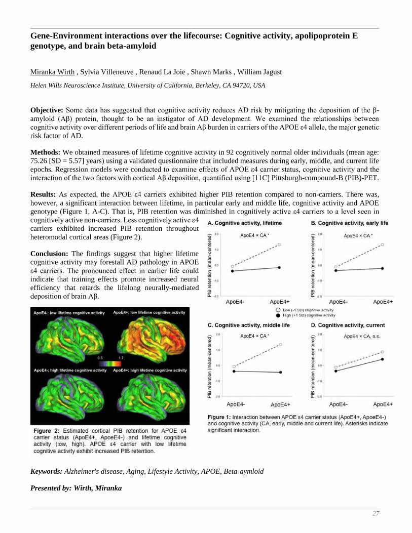
27
Gene-Environment interactions over the lifecourse: Cognitive activity, apolipoprotein E
genotype, and brain beta-amyloid
Miranka Wirth , Sylvia Villeneuve , Renaud La Joie , Shawn Marks , William Jagust
Helen Wills Neuroscience Institute, University of California, Berkeley, CA 94720, USA
Objective: Some data has suggested that cognitive activity reduces AD risk by mitigating the deposition of the β-
amyloid (Aβ) protein, thought to be an instigator of AD development. We examined the relationships between
cognitive activity over different periods of life and brain Aβ burden in carriers of the APOE ε4 allele, the major genetic
risk factor of AD.
Methods: We obtained measures of lifetime cognitive activity in 92 cognitively normal older individuals (mean age:
75.26 [SD = 5.57] years) using a validated questionnaire that included measures during early, middle, and current life
epochs. Regression models were conducted to examine effects of APOE ε4 carrier status, cognitive activity and the
interaction of the two factors with cortical Aβ deposition, quantified using [11C] Pittsburgh-compound-B (PIB)-PET.
Results: As expected, the APOE ε4 carriers exhibited higher PIB retention compared to non-carriers. There was,
however, a significant interaction between lifetime, in particular early and middle life, cognitive activity and APOE
genotype (Figure 1, A-C). That is, PIB retention was diminished in cognitively active ε4 carriers to a level seen in
cognitively active non-carriers. Less cognitively active ε4
carriers exhibited increased PIB retention throughout
heteromodal cortical areas (Figure 2).
Conclusion: The findings suggest that higher lifetime
cognitive activity may forestall AD pathology in APOE
ε4 carriers. The pronounced effect in earlier life could
indicate that training effects promote increased neural
efficiency that retards the lifelong neurally-mediated
deposition of brain Aβ.
Keywords: Alzheimer's disease, Aging, Lifestyle Activity, APOE, Beta-aymloid
Presented by: Wirth, Miranka
28 8th Human Amyloid Imaging – Miami, 2014
Preclinical effects of Aβ deposition on episodic memory and disease progression
Victor L Villemagne 1, Samantha Burnham 2, Pierrick Bourgeat 3, Belinda Brown 4, Kathryn Ellis 5, Olivier Salvado 3,
Ralph Martins 4, Lance Macaulay 6, David Ames 7, Colin L Masters 8, Christopher C Rowe 1
1 Department of Nuclear Medicine & Centre for PET, Austin Health, Melbourne, VIC, Australia 2 CSIRO Preventative Health Flagship: Mathematics, Informatics and Statistics, Floreat, WA, Australia 3 CSIRO Preventative Health Flagship: The Australian e-Health Research Centre, Brisbane, QLD, Australia 4 Edith Cowan Univerity, Perth, WA, Australia 5 Dept of Psychiatry, The University of Melbourne, VIC, Australia 6 CSIRO Preventative Health Flagship: Materials Science and Engineering, Melbourne, VIC, Australia 7 National Ageing Research Institute, Melbourne, VIC, Australia 8 The Florey Institute of Neuroscience and Mental Health, The University of Melbourne, VIC, Australia
Background: Predicting the rate of evolution of preclinical changes and the onset of clinical phase of AD might prove
essential for the design and timing of disease-modifying therapeutic interventions.
Methods: 207 participants (152 HC; 36 MCI; 19 AD) underwent periodic neuropsychological examination, MRI and
PiB-PET scans for a mean follow up of 4.2 (CI 3.4-4.8) years. A SUVRcb of 1.5 was used to discriminate high (Aβ+)
from low (Aβ-) Aβ burdens. Analyses were adjusted for age, gender, years of education, and ApoE status.
Results: 170 (82%) of 207 participants (79% HC, 89% MCI, 100% AD), were deemed Aβ accumulators. In HC and
MCI, Aβ deposition and memory decline were significantly faster in Aβ+ than in Aβ-. Aβ deposition was significantly
faster in participants who transitioned to either MCI or AD, although baseline Aβ burden was a better predictor of
progression. Significant associations between rates of Aβ deposition and rates of episodic memory (EM) decline were
found in HC (R2=0.13; p=0.004). There were no associations between Aβ deposition and EM decline in MCI or AD.
When Aβ+HC and Aβ-HC were considered separately, a significant association between rates of Aβ deposition and
EM decline was found only in Aβ+HC (R2=0.22; p=0.022), association that persisted after adjusting for baseline Aβ
burden. There was no association between rates of Aβ deposition and EM decline in Aβ-HC, not even when those with
positive rates of Aβ accumulation were examined separately.
Conclusions: Aβ seems to have a threshold/trigger effect on EM at the early stages of Aβ deposition. While this
support the theory that Aβ plays a fundamental role in the development of AD, it suggests that other factors have a
more direct effect on symptom progression at later stages of the disease. These results also suggest that in order to
prevent memory decline, anti-Aβ therapy should target these early stages of Aβ deposition.
Keywords: Aβ deposition, memory decline, disease progression
Presented by: Villemagne, Victor L
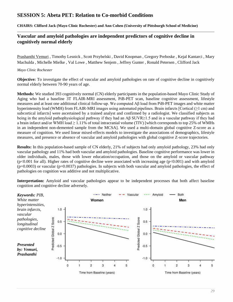
29
SESSION 5: Abeta PET: Relation to Co-morbid Conditions
CHAIRS: Clifford Jack (Mayo Clinic Rochester) and Ann Cohen (University of Pittsburgh School of Medicine)
Vascular and amyloid pathologies are independent predictors of cognitive decline in
cognitively normal elderly
Prashanthi Vemuri , Timothy Lesnick , Scott Przybelski , David Knopman , Gregory Preboske , Kejal Kantarci , Mary
Machulda , Michelle Mielke , Val Lowe , Matthew Senjem , Jeffrey Gunter , Ronald Petersen , Clifford Jack
Mayo Clinic Rochester
Objective: To investigate the effect of vascular and amyloid pathologies on rate of cognitive decline in cognitively
normal elderly between 70-90 years of age.
Methods: We studied 393 cognitively normal (CN) elderly participants in the population-based Mayo Clinic Study of
Aging who had a baseline 3T FLAIR-MRI assessment, PiB-PET scan, baseline cognitive assessment, lifestyle
measures and at least one additional clinical follow-up. We computed Aβ load from PiB-PET images and white matter
hyperintensity load (WMH) from FLAIR-MRI images using automated pipelines. Brain infarcts [Cortical (≥1 cm) and
subcortical infarcts] were ascertained by a trained analyst and confirmed by a radiologist. We classified subjects as
being in the amyloid pathophysiological pathway if they had an Aβ SUVR≥1.5 and in a vascular pathway if they had
a brain infarct and/or WMH load ≥ 1.11% of total intracranial volume (TIV) [which corresponds to top 25% of WMHs
in an independent non-demented sample from the MCSA]. We used a multi-domain global cognitive Z-score as a
measure of cognition. We used linear mixed-effects models to investigate the associations of demographics, lifestyle
measures, and presence or absence of vascular and amyloid pathologies with global cognitive Z-score trajectories.
Results: In this population-based sample of CN elderly, 21% of subjects had only amyloid pathology, 23% had only
vascular pathology and 11% had both vascular and amyloid pathologies. Baseline cognitive performance was lower in
older individuals, males, those with lower education/occupation, and those on the amyloid or vascular pathway
(p<0.001 for all). Higher rates of cognitive decline were associated with increasing age (p<0.001) and with amyloid
(p=0.0003) or vascular (p=0.0037) pathologies. In subjects with both vascular and amyloid pathologies, the effect of
pathologies on cognition was additive and not multiplicative.
Interpretation: Amyloid and vascular pathologies appear to be independent processes that both affect baseline
cognition and cognitive decline adversely.
Keywords: PIB,
White matter
hyperintensities,
brain infarcts,
vascular
pathologies,
longitudinal
cognitive decline
Presented
by: Vemuri,
Prashanthi

30 8th Human Amyloid Imaging – Miami, 2014
Multiple brain markers contribute to age-related variation in cognition
Trey Hedden 1, Aaron Schultz 1, Anna Rieckmann 1, Elizabeth C. Mormino 2, Keith A. Johnson 3, Reisa A. Sperling 4,
Randy L. Buckner 5
1 Athinoula A. Martinos Center for Biomedical Imaging, Department of Radiology, Massachusetts General Hospital, Harvard
Medical School 2 Department of Neurology, Massachusetts General Hospital, Harvard Medical School 3 Athinoula A. Martinos Center for Biomedical Imaging, Departments of Radiology and Neurology, Massachusetts General
Hospital, Harvard Medical School; 4 Athinoula A. Martinos Center for Biomedical Imaging, Departments of Radiology and Neurology, Massachusetts General
Hospital, Harvard Medical School; Center for Alzheimer Research and Treatment, Department of Neurology, Brigham and
Women's Hospital 5 Athinoula A. Martinos Center for Biomedical Imaging, Department of Radiology, Massachusetts General Hospital, Harvard
Medical School; Department of Psychology and Center for Brain Science, Harvard University
Rationale: Although aging is accompanied by evident changes in brain morphometry, function, and disease-related
biomarkers, direct links between neural markers and cognition have been elusive. Most studies have compared a small
number of brain markers or exhibit relatively small effect sizes (Hedden et al., 2013; Salthouse, 2011). Here, we
examined shared and selective contributions of multiple brain markers to age-related variation in cognition.
Methods: 144 cognitively normal older adults aged 65-90 from the Harvard Aging Brain Study were characterized on
MRI markers of cortical gray matter thickness and volume, white matter lesions (WML) and fractional anisotropy
(FA), resting state functional connectivity, functional activity during task-switching, and PET markers of glucose
metabolism and amyloid burden. Episodic memory, executive function, and processing speed were separately assessed.
Brain markers were primarily global in nature and were selected a priori without regard to relationships to age or
cognition in the current dataset. Hierarchical regression analyses examined the age-related variance in cognition shared
with each brain marker individually, and the unique age-related variance contributed by each marker when controlling
for all other markers.
Results: The largest individual and unique relationships to all cognitive factors involved FA and WML, with an
additional contribution of hippocampal volume to episodic memory. Individually, amyloid burden contributed ~9% of
age-related variance in memory, but only ~1% was unique to amyloid. Approximately 60% of the age-related variance
in cognition could be accounted for when all brain markers were combined (but only ~20% of total variance), and most
of this age-related variance was shared among two or more brain markers.
Conclusion: These results suggest that the majority of age-related variation in cognition is shared among multiple
brain markers, but point to limited specificity of associations between brain markers and cognitive domains, motivating
additional study of markers of neural health in advanced aging.
Keywords: biomarkers, FDG, PiB, fMRI, fcMRI
Presented by: Hedden, Trey

31
Cerebral amyloid related alterations in neuronal metabolism and the contribution of
multimodal measures of vascular function
Eric McDade 1, Albert Kim 2, Tim Hughes 3, Beth Snitz 1, Anne Cohen 1, Julie Price 1, Chester Mathis 1, James
Becker 1, William Klunk 1, Oscar Lopez 1
1 University of Pittsburgh 2 Medical College of Georgia 3 Wake Forest University
Objective: We explored how markers of vascular disease and cerebral blood flow (CBF) might impact the relationship
between cerebral plaques and neuronal function.
Background: Given the epidemiologic and pathologic evidence linking cardio-cerebrovascular disease and
Alzheimer’s dementia, it is critical to identify mechanistic links between these two disorders. Recently we have
identified a direct association between arterial stiffness and amyloid deposition implying vascular disease could
contribute to some of the amyloid related impairments of neuronal and cognitive function.
Design/Methods: 71 cognitively normal or MCI elderly subjects (mean 85y/o) underwent 18F-FDG and amyloid PET
scans, ultrasound measurement of central and peripheral arterial stiffness (PWV) and pulsed arterial spin labeled
(pASL)-MRI measures of CBF. Anatomical SUVR values from the PET scans were used for the regression models.
Spearman’s correlations identified areas of association between regional amyloid load and regional FDG values. Areas
of significant correlation were then added into separate linear regression models with FDG as the dependent variable,
and regional amyloid, PWV, CBF, blood pressure, age and education as potential predictors together in a stepwise
multivariate regression with a probability-of-F-to-enter less than .05 and Probability-of-F-to-remove greater than .100
for each model to determine the best fit.
Results: In the regression model greater amyloid deposition was a significant predictor of lower glucose utilization in
frontal and posterior cortical regions, however reduced CBF and greater vascular stiffness and diastolic blood pressure
were additionally found to improve the models predicting FDG metabolism mostly in areas of the frontal lobes
vulnerable to AD whereas in the more posterior regions PWV and blood pressure were less important.
Conclusions: Given the recent strong association we have identified between peripheral vascular stiffness and cerebral
amyloidosis our current work might indicate that part of the relationship between vascular disease and frontal lobe
dysfunction could result from a priming of these regions by vascular disease to the pathological cascade associated
with Alzheimer disease rather than direct vascular damage.
Keywords: Arterial stiffness, Cerebrovascular, FDG-PET, PiB-PET.
Presented by: McDade, Eric

32 8th Human Amyloid Imaging – Miami, 2014
Binding of Pittsburgh Compound B to both normal and abnormal white matter in elderly
cognitively normal controls.
Anna Goodheart 1, Erica Tamburo 1, Davneet Minhas 2, Howard Aizenstein 1, Eric McDade 3, Lisa Weissfeld 4, Beth
Snitz 3, Julie Price 2, Chester Mathis 2, Oscar Lopez 3, William Klunk 1, Ann Cohen 1
1 University of Pittsburgh School of Medicine, Psychiatry 2 University of Pittsburgh School of Medicine, Radiology 3 University of Pittsburgh School of Medicine, Neurology 4 University of Pittsburgh, Biostatistics
The amyloid imaging agent, Pittsburgh Compound B (PiB), binds with high affinity to β-amyloid (Aβ) in the brain and
it is well established that PiB also shows non-specific retention in white matter(WM). However, little is know about
retention of PiB in areas of white matter hyperintensities (WMH), abnormalities commonly seen in older adults.
Further, WMH are hypothesized to be related to both cognitive dysfunction and Aβ deposition. The goal of the present
study was to explore PiB retention in both normal-appearing WM (NAWM) and WMH in a group of elderly,
cognitively normal individuals.
Cognitively normal elderly (n=64; 86.5±2.6 years) who had a FLAIR-MRI, PiB-PET(SUVR 50-70, cerebellum
reference) and visual WMH score were included in this analysis. Two ROI analyses were applied: (1) ROIs of
standardized dimensions were placed over “typical” areas of periventricular WMH caps on all subjects, regardless of
WMH burden or size. (2) Subject-specific maps of NAWM and WMH were created using C++ and ITK, these maps
were then co-registered with the PiB-PET and mean SUVR values were calculated in these NAWM and WMH ROI.
PiB retention was significantly reduced in the “typical-WMH” ROIs of subjects with high WMH compared to subjects
with low WMH. Additionally, in subjects with high WMH, there was significantly lower PiB retention in subject-
specific ROIs of WMH compared to NAWM, this was not observed in subjects with low WMH, likely because of the
absence of WMH in this group.
These data suggest that white matter in areas of WMH may have different binding characteristics to PiB than does
normal WM. Further exploration of this phenomenon may lead to insights about the molecular basis of this non-specific
interaction.
Keywords: Pittsburgh Compound B, White matter hyperintensities
Presented by: Cohen, Ann

33
SESSION 6: INVITED LECTURE
The Centiloid method for quantifying Amyloid PET studies: Great illuminator or master or
illusion
Robert A. Koeppe
University of Michigan, Ann Arbor, MI
This talk will focus on two technical aspects of human amyloid PET imaging. First will be a review of different
metrics for defining amyloid load. The vast majority of amyloid research reported in the literature used SUVr, which
is nothing more than simple ratios of radiotracer concentration in some target tissue to some reference tissue. While
this has proved very useful in practice, it is important to keep in mind the potential limitations of SUVr measures
compared to more quantitative measures of distribution volumes or distribution volume ratios obtained from
compartmental modeling or reference tissue modeling, respectively. While the relationship between the measures has
been looked at extensively for [11C]PiB, there is considerably less information available concerning the correspondence
between DVr and SUVr measures for the newer [18F]labeled amyloid tracers. It will be important to examine this
relationship for any new amyloid tracers being developed, as well as for the new tau tracers that are coming on-line.
Second will be a brief review of the methods of the centiloid project. This will be followed with a discussion of
whether the centiloid approach will improve our ability to compare different radiotracers, different acquisition and
analysis methods, and different methods of extraction of amyloid data from the PET scans or whether the approach
will actually tend to cloud the differences by putting all radiotracers, and analysis approaches on to the same scale.
Presented by: Koeppe, Robert

34 8th Human Amyloid Imaging – Miami, 2014
SESSION 7: Technical Emphasis
CHAIRS: Chester Mathis (University of Pittsburgh) and John Seibyl (Institute for Neurodegenerative Disorders)
Modeling the influence of white matter contamination on detectability of brain amyloid
changes in longitudinal studies of Alzheimer’s progression: Segmentation analyses using the
PET β-Amyloid tracer 18F NAV4694
John Seibyl 1, Osama Sabri 2, Henryk Barthel 2, Olivier Barret 1, Kenneth Marek 1, Cornelia Reininger 3
1 Institute for Neurodegenerative Disorders 2 University of Leipzig 3 Navidea Biopharmaceuticals
Objectives: Current generation 18F β-amyloid PET radiotracers demonstrate significant non-specific white matter
uptake which influences quantitative assessment of cortical β-amyloid burden, e.g. for monitoring small within-
subjects drug effects. 18F NAV4694 is a novel brain β-amyloid PET agent with purportedly less white matter binding.
The purpose of the present investigation was to study the influence of the white matter uptake in NAV4694 PET images
on the ability of the tracer to diagnose Alzheimer’s disease (AD) and to monitor changes in brain β-amyloid load over
time.
Methods: 20 subjects (10 AD, 10 age-matched HV) had 18F NAV4694 PET and MRI scans. Individual MRI-based
gray matter segmentation of the PET data and a standardized VOI analysis were carried out determining SUVrs
(reference region: cerebellar cortex). Further, a theoretical model was constructed for demonstrating the influence of
the varying degrees of white matter contamination on resulting SUVr changes over time and effects on power to detect
subtle changes in brain amyloid in longitudinal treatment trials.
Results: Composite SUVr for AD subjects were 2.60±0.6 and 2.53±0.6 for the segmentation analysis and the no-
segmentation analysis, respectively (n.s.). HV had segmented composite SUVr of 1.56±0.5 and non-segmented SUVr
of 1.55±0.5 (n.s.). In the theoretical model, increasing white matter signal in cortical VOIs reduces the measured
percent change over time in cortical SUVrs in a direct fashion.
Conclusions: The theoretical model suggests an important white matter effect resulting in Type 2 errors in assessing
longitudinal data using PET biomarkers of β-amyloid. NAV4694 may be advantageous evidenced by the fact that MRI
segmentation to generate gray matter PET volumes does not impact cortical SUVrs. This is most likely due to lower
white matter uptake of NAV4694 compared with other 18F amyloid radiotracers, hence white matter contribution to
the SUVr is less, predicting better sensitivity and statistical power for detecting within subject changes.
Keywords: PET, NAV4694, amyloid, biomarker
Presented by: Seibyl, John

35
Amyloid PET screening results by APOE ε4 status from a Phase 1b Clinical Study
(221AD103) in patients with prodromal to mild Alzheimer’s disease
Ping Chiao 1, Suhy Joyce 2, Barakos Jerome 3, Burke Meredith 2, Klein Gregory 2, Verma Ajay 1, Sevigny Jeff 1
1 Biogen Idec, Cambridge, MA, USA 2 Synarc Inc, Newark, CA, USA 3 Synarc Inc, Newark, CA, USA, California Pacific Medical Center, San Francisco, CA, USA
Introduction: Requirement of a positive amyloid PET scan is being used as an enrichment technique in study
221AD103. Retrospective analyses of both bapineuzumab and solanezumab data have shown substantially higher
percentages of negative amyloid PET findings in APOE ε4 non-carriers than in APOE ε4 carriers in patients with mild
or moderate Alzheimer’s disease (AD). We report our amyloid PET screening results by APOE ε4 status in patients
with prodromal or mild AD.
Methods: During screening, patients fulfilling criteria for prodromal or mild AD underwent florbetapir PET scanning
and APOE genotyping. Florbetapir PET scans were visually evaluated for amyloid plaque burden.
Results: Data from the first 250 patients (Tables 1 and 2) are shown. Previously reported data from bapineuzumab and
solanezumab Phase 3 studies are also shown (Table 2).
Conclusions: A substantially higher percentage
of APOE ε4 non-carriers than APOE ε4 carriers
had negative amyloid PET findings, similar to the
bapineuzumab and solanezumab data. However,
the overall incidence of negative amyloid scans
observed in study 221AD103 is substantially
higher than reported in the two Phase 3 studies,
likely attributable to the earlier stage of AD
patients being recruited in this study
(prodromal/mild, mean MMSE ~ 25 versus
mild/moderate, mean MMSE ~21 in the Phase 3
studies). This finding is also consistent with the
solanezumab data which showed that the
percentage of negative amyloid PET findings was
higher in mild (27%) than in moderate (13%) AD
groups.
These results suggest that (1) enrichment using
amyloid PET imaging is effective and feasible, (2)
enrichment by assessing amyloid plaque burden is
critically important to clinical studies in early stages of AD because of a higher incidence of negative amyloid findings,
and (3) APOE genotyping may potentially improve the economy of enrichment by amyloid PET by lowering the
likelihood of negative amyloid findings.
Keywords: Florbetapir, Alzheimer’s disease, amyloid PET imaging, APOE4, clinical trial
Presented by: Chiao, Ping
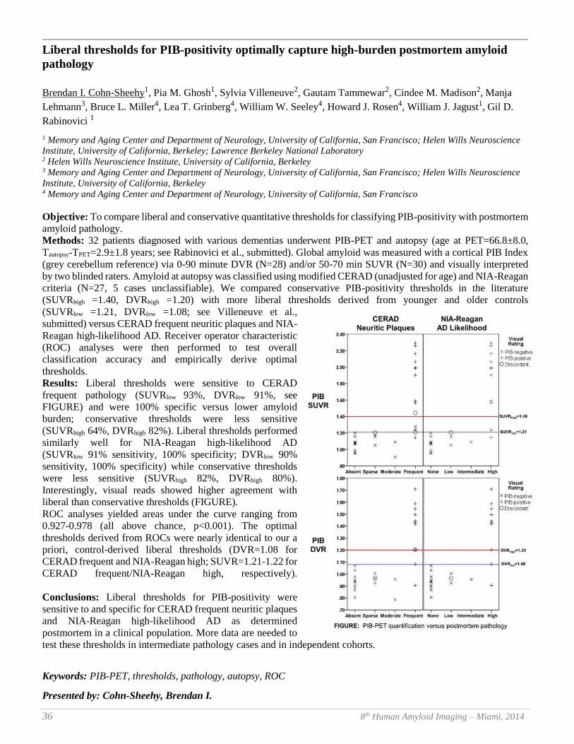
36 8th Human Amyloid Imaging – Miami, 2014
Liberal thresholds for PIB-positivity optimally capture high-burden postmortem amyloid
pathology
Brendan I. Cohn-Sheehy1, Pia M. Ghosh1, Sylvia Villeneuve2, Gautam Tammewar2, Cindee M. Madison2, Manja
Lehmann3, Bruce L. Miller4, Lea T. Grinberg4, William W. Seeley4, Howard J. Rosen4, William J. Jagust1, Gil D.
Rabinovici 1
1 Memory and Aging Center and Department of Neurology, University of California, San Francisco; Helen Wills Neuroscience
Institute, University of California, Berkeley; Lawrence Berkeley National Laboratory 2 Helen Wills Neuroscience Institute, University of California, Berkeley 3 Memory and Aging Center and Department of Neurology, University of California, San Francisco; Helen Wills Neuroscience
Institute, University of California, Berkeley 4 Memory and Aging Center and Department of Neurology, University of California, San Francisco
Objective: To compare liberal and conservative quantitative thresholds for classifying PIB-positivity with postmortem
amyloid pathology.
Methods: 32 patients diagnosed with various dementias underwent PIB-PET and autopsy (age at PET=66.8±8.0,
Tautopsy-TPET=2.9±1.8 years; see Rabinovici et al., submitted). Global amyloid was measured with a cortical PIB Index
(grey cerebellum reference) via 0-90 minute DVR (N=28) and/or 50-70 min SUVR (N=30) and visually interpreted
by two blinded raters. Amyloid at autopsy was classified using modified CERAD (unadjusted for age) and NIA-Reagan
criteria (N=27, 5 cases unclassifiable). We compared conservative PIB-positivity thresholds in the literature
(SUVRhigh =1.40, DVRhigh =1.20) with more liberal thresholds derived from younger and older controls
(SUVRlow =1.21, DVRlow =1.08; see Villeneuve et al.,
submitted) versus CERAD frequent neuritic plaques and NIA-
Reagan high-likelihood AD. Receiver operator characteristic
(ROC) analyses were then performed to test overall
classification accuracy and empirically derive optimal
thresholds.
Results: Liberal thresholds were sensitive to CERAD
frequent pathology (SUVRlow 93%, DVRlow 91%, see
FIGURE) and were 100% specific versus lower amyloid
burden; conservative thresholds were less sensitive
(SUVRhigh 64%, DVRhigh 82%). Liberal thresholds performed
similarly well for NIA-Reagan high-likelihood AD
(SUVRlow 91% sensitivity, 100% specificity; DVRlow 90%
sensitivity, 100% specificity) while conservative thresholds
were less sensitive (SUVRhigh 82%, DVRhigh 80%).
Interestingly, visual reads showed higher agreement with
liberal than conservative thresholds (FIGURE).
ROC analyses yielded areas under the curve ranging from
0.927-0.978 (all above chance, p<0.001). The optimal
thresholds derived from ROCs were nearly identical to our a
priori, control-derived liberal thresholds (DVR=1.08 for
CERAD frequent and NIA-Reagan high; SUVR=1.21-1.22 for
CERAD frequent/NIA-Reagan high, respectively).
Conclusions: Liberal thresholds for PIB-positivity were
sensitive to and specific for CERAD frequent neuritic plaques
and NIA-Reagan high-likelihood AD as determined
postmortem in a clinical population. More data are needed to
test these thresholds in intermediate pathology cases and in independent cohorts.
Keywords: PIB-PET, thresholds, pathology, autopsy, ROC
Presented by: Cohn-Sheehy, Brendan I.

37
Existing thresholds for PIB positivity are too high
Sylvia Villeneuve 1, Cindee Madison 1, Nagehan Ayakta 1, Renaud La Joie 1, Brendan I. Cohn-Sheehy 2, Jacob Vogel 1,
Shawn Marks 1, Samia K. Arthur-Bentil 1, Bruce Reed 3, Charles DeCarli 3, Gil Rabinovici 2, William Jagust 1
1 University of California, Berkeley 2 University of California, San Francisco 3 University of California, Davis
Background: There is no consensus about thresholds that define amyloid positivity, and there is little known about
the earliest phases of amyloid accumulation in older adults.
Objective: To examine the pattern of amyloid accumulation and derive a cutoff that captures early accumulation using
both DVR and SUVR data from the same subjects.
Methods: Amyloid accumulation was investigated in 152 cognitively normal older adults using: (1) a reference group
of young adults, (2) Gaussian mixture modeling (GMM), (3) cluster analyses and (4) voxel-wise analyses. All analyses
used DVR and SUVR data with a cerebellar gray reference ROI. For voxel-wise analyses, subjects were ranked based
on their global DVR status. To track when and where amyloid starts accumulating we compared a group of 22 subjects
with a mean DVR of 1 (control group) to the next 22 subjects (group of interest) and iteratively increased the mean
DVR of the group of interest by dropping the subject with the lowest value and adding the subject with the next higher
value. This procedure was repeated until the subject with the highest DVR was included in the group of interest.
Results: The threshold 2 SD above the
young subjects was a DVR of 1.07
(SUVR = 1.19). Both the GMM and the
cluster-derived thresholds were 1.09
(SUVR 1.22). The Figure shows that
amyloid starts accumulating in the
medial frontal cortex (mean DVR =
1.07, SUVR = 1.19), then spreads to the
precuneus, the lateral frontal and parietal
lobes, and finally the temporal lobe.
Conclusions: Amyloid starts to
accumulate long before people reached
the widely used SUVR cutoffs of 1.4 and
1.5. These results support an SUVR
cutoff of 1.21 (DVR = 1.08) to capture
early amyloid accumulation. This cutoff
was confirmed by an autopsy study of 32
dementia cases (Cohn-Sheehy et al.,
submitted to HAI).
Keywords: amyloid, pattern, threshold,
SUVR, DVR
Presented by: Villeneuve, Sylvia
38 8th Human Amyloid Imaging – Miami, 2014
SESSION 8: Neuropathologic Correlations
CHAIRS: William Klunk (University of Pittsburgh) and Gil Rabinovici (University of California San Francisco)
PiB PET and FDG PET quantitative analysis methods with autopsy correlation
Val Lowe 1, Melissa Murray 2, Emily Lundt 1, Stephen Weigand 1, Scott Przybelski 1, Lenon Jordan 1, Dennis
Dickson 2, Joseph Parisi 1, David Knopman 1, Kejal Kantarci 1, Clifford Jack 1, Ronald Petersen 1
1 Mayo Clinic, Rochester, MN 2 Mayo Clinic, Jacksonville, Fl
Background: Accurate biomarker evaluation methods are essential to discriminate changes in serial subjects and
accurately characterize probable Alzheimer’s disease. We evaluated different quantitative evaluation methods for PiB
and FDG PET in subjects who had come to autopsy.
Methods: Autopsies from 40 subjects were available. Subjects with primary Alzheimer’s disease pathology (n=14)
and all without neurodegenerative disease (n=11) were selected for this comparison. Quantitiative analysis of PIB data
was performed using grey matter segmentation vs grey+white matter, with partial volume correction (PVC), on or off,
and cerebellar normalization using whole cerebellum (WCn) vs. peripheral cerebellum (crus) (CCn) normalization.
FDG data was evaluated using an AD specific region method (Landau et al, 2010 ) vs. angular gyrus+posterior
cingulate (AG+PC) regions with the above noted segmentation variations and PVC. Sensitivity, specificity, ROC and
AUROC analysis were performed. Biomarker optimal cut-off values were determined and the influence of the
quantitative methods on dynamic range was assessed.
Results: For PIB without PVC, all AD pathology subjects had more abnormal values than all control subjects. This
complete separation was not observed after PVC. However, no AUROC difference was significant with any PiB
method. Increased dynamic PiB range was observed when CCn was used. Optimal performance was seen with a PiB
cutoff of 1.59-1.61 with non-PVC WCn and 1.70-1.72 with non-PVC CCn. For FDG, AG+PC AUROC ranged from
0.73-0.83 and Landau AUROC from 0.68-0.80 but differences were not significant. No different FDG AUROC values
were seen with or without PVC. AUROC performance was better for any PiB method vs any FDG method (p<0.05).
Conclusions: There was no significant difference in disease detection accuracy between different PiB or FDG analysis
methods tested. We observed suggestions of improved PiB accuracy when not using PVC and improved dynamic PiB
range when using CCn. PiB disease characterization was superior to FDG.
Keywords: Amyloid PET FDG PET Quantitative analysis Autopsy correlations
Presented by: Lowe, Val
39
Evaluation of cotton wool plaques using amyloid binding compounds and Aβ
immunohistochemistry: implications for PiB PET imaging
Milos Ikonomovic , Eric Abrahamson , Julie Price , Chester Mathis , William Klunk
University of Pittsburgh
Background: [C-11]PiB PET retention is believed to reflect amounts of cored/neuritic plaques consisting of fibrillar
amyloid-β (Aβ) in Alzheimer’s disease (AD). However, in familial AD (FAD) with presenilin-1 (PS-1) mutations, PiB
retention is most prominent in the striatum which contains diffuse Aβ plaques, and moderate in neocortical regions
containing both cored and diffuse Aβ plaques. The relationship between PiB binding and diffuse plaques is unclear.
Methods: We explored PiB labeling of striatal and neocortical plaques in FAD cases with PS-1 mutation (A426P)
compared to sporadic AD. Sections of caudate, frontal and temporal cortices, and cerebellum were processed using
highly fluorescent derivatives of PiB (6-CN-PiB) and Congo red (X-34), H&E, and Aβ immunohistochemistry (mab
4G8).
Results: In the neocortex of all cases, 6-CN-PiB and X-34 signals were strong in cored and weak in diffuse Aβ plaques.
The only exception was cotton wool plaques (CWP) in FAD; in these Aβ-immunoreactive and H&E delineated
deposits, positive X-34 labeling demonstrated their beta-sheet structure but 6-CN-PiB fluorescence was at background
levels. Diffuse Aβ-immunoreactive, X-34-positive and 6-CN-PiB-negative plaques were also observed in the
cerebellum of all cases. Striatal diffuse Aβ plaques were labeled moderately with 6-CN-PiB and X-34, similar in FAD
and sporadic AD except for greater striatal plaque load in FAD.
Conclusions: This histological study provides a high resolution examination of the interaction between PiB and diffuse
Aβ plaques, particularly of the CWP type. Besides cerebellar plaques, CWP are the second plaque type not detected
using 6-CN-PiB histology. The possibility that the difference in CWP labeling between X-34 and 6-CN-PiB is due to
differences in fluorescence intensities is unlikely as they showed comparable labeling in striatal and neocortical non-
CWP plaques. The apparent beta-sheet structure of CWPs, demonstrated using X-34, suggests that a unique tertiary
structure or modified Aβ forms may prevent their detection using PiB.
Keywords: amyloid, cotton wool plaques, diffuse plaques, PET, PiB
Presented by: Ikonomovic, Milos
40 8th Human Amyloid Imaging – Miami, 2014
[18F]Flutemetamol amyloid PET detection of Aβ plaque phases 4 and 5 and significant diffuse
plaque burden
Thomas Beach 1, Dietmar Thal 2, Michelle Zanette 3, Kirsten Heurling 4, Chris Buckley 5, Adrian Smith 5
1 Civin Laboratory for Neuropathology, Banner Sun Health Research Institute, Sun City, AZ, USA. 2 Institute of Pathology – Laboratory of Neuropathology, Center for biomedical Research, University of Ulm, Ulm, Germany. 3 GE Healthcare, 101 Carnegie Center, Princeton ,NJ, USA 4 GE Healthcare, Uppsala, Sweden. 5 GE Healthcare The Grove Centre, White Lion Rd, Amersham, HP7 9LL U.K.
Objectives: 1. To determine how amyloid PET imaging with [18F]flutemetamol correlates with the hierarchical pattern
of Aβ plaque deposition and which phases of Aβ pathology are detectable with PET amyloid imaging. 2. To ascertain
the extent to which different types of amyloid plaques, namely neuritic and diffuse, contribute to amyloid PET images.
Methods: Subjects: 68 autopsy cases included in the efficacy analysis of the end of life study GE067-007.
Post-Hoc Amyloid phase analysis: Subsequent to the GE067-007 study, the phase of Aβ plaque pathology was
determined by screening the Aβ-IHC stained sections for plaque distribution for each case. This was preformed blinded
to clinical, imaging and other histopathological assessments.
Image assessment: The blinded assessment of [18F]Flutemetamol PET images as normal or abnormal as recorded in
the GE067-007 study was used for this post hoc analysis.
Results: Abnormal image probability increases with amyloid phase: The probability of a positive (abnormal)
[18F]flutemetamol image interpretation increases with increasing amyloid phase going from less than 0.0001 in Ab
plaque phases 0 - 2, 0.111 in phase 3, 0.895 in phase 4 to 0.999 in phase 5.
[18F]Flutemetamol binding to diffuse plaques: The majority of deposits in the striatum were found in the form of
diffuse plaques. In 8 cases, frequent diffuse plaques in the absence of any neuritic plaques were considered positive by
PET visual assessment of the striatum (GE067-007 study), demonstrating that [18F]flutemetamol can detect diffuse
plaques when they are frequent.
Conclusion: [18F]flutemetamol PET imaging is well suited to detect amyloid pathology in vivo. We found a high
concordance of PET image abnormality with amyloid phase 4 to 5. This study also showed that [18F]flutemetamol
images could be positive in the striatal region in the absence of neuritic plaques if frequent diffuse plaques were present.
Keywords: [18F]flutemetamol PET imaging, Aβ plaque pathology phasing, neuritic and diffuse amyloid plaques
Presented by: Buckley, Chris
41
Diagnostic accuracy of amyloid and FDG PET in pathologically-confirmed dementia
Gil D. Rabinovici 1, Manja Lehmann 2, Howard J. Rosen 3, Pia M Ghosh 2, Brendan I Cohn-Sheehy 2, John Q.
Trojanowski 4, Mario F. Mendez 5, Harry V. Vinters 5, Dennis W. Dickson 6, Marilu Gorno-Tempini 3, Adam L.
Boxer 3, Bruce L. Miller 3, Lea T. Grinberg 3, William W. Seeley 3, William J. Jagust 7
1 Memory & Aging Center and Department of Neurology, University of California San Francisco Helen Wills Neuroscience
Institute, University of California Berkeley Lawrence Berkeley National Laboratory 2 Memory & Aging Center and Department of Neurology, University of California San Francisco Helen Wills Neuroscience
Institute, University of California Berkeley 3 Memory & Aging Center and Department of Neurology, University of California San Francisco 4 Center for Neurodegenerative Research, University of Pennsylvania, Philadelphia 5 David Geffen School of Medicine, University of California Los Angeles 6 Dept. of Neuroscience, Mayo Clinic Jacksonville 7 Helen Wills Neuroscience Institute, University of California Berkeley Lawrence Berkeley National Laboratory
Objective: Pathological validation studies of amyloid imaging have been conducted largely in end-of-life subjects that
may not represent clinical practice. We assessed the diagnostic accuracy of PIB-PET versus pathology in a consecutive
series of patients from an academic dementia clinic, and compared with the performance of FDG-PET.
Methods: 37 patients (mean age 66.5±8.8, MMSE 21.1±6.8) underwent PIB (all) and FDG (34/37) PET. Clinical
diagnoses included Alzheimer’s disease (AD, N=10), frontotemporal dementia (FTD, N=24), mild cognitive
impairment (N=2) and prion disease (N=1). PET scans were visually interpreted by two raters blinded to clinical
diagnosis (PIB: positive or negative; FDG: AD or FTD). Pathologic diagnoses (2.9±1.8 years after PET; 36 autopsies
and one biopsy) were based on standard research criteria.
Results: Pathologic diagnoses included: high-likelihood AD (N=9), FTD (N=24), mixed high-likelihood AD/FTD
(N=3) and prion disease (N=1). PIB reads had a sensitivity of 100% (both raters) and specificity of 88%-92% for post-
mortem high-likelihood AD. A single patient with clinical AD who was rated PIB-negative by consensus had FTD at
autopsy, while consensus PIB+ cases with clinical FTD were found to have either primary AD (N=2) or mixed
AD/FTD pathology (N=3). Cases with discordant visual reads had either CERAD frequent (2) or CERAD sparse but
frequent diffuse plaques (1): none met high-likelihood AD criteria.
FDG reads had a sensitivity of 83%-92% and specificity of 77% for AD. Mixed AD/FTD cases were usually read as
AD (Rater 1 2/3, Rater 2 3/3). Positive predictive values for high-likelihood AD: PIB 80%-86%, FDG 67%-69%;
negative predictive values: PIB 100%, FDG 89%-94%. Overall accuracy (excluding mixed high-likelihood AD/FTD):
PIB 91%-94%, FDG 81% (p>0.35).
Conclusions: Amyloid PET had high sensitivity and negative predictive value for the pathologic diagnosis of AD.
Positive test interpretation was complicated by mixed pathology. Amyloid imaging surpassed FDG in predicting
underlying neuropathology.
Keywords: Pathology, PIB, FDG, Frontotemporal dementia, Alzheimer's disease
Presented by: Rabinovici, Gil D.
42 8th Human Amyloid Imaging – Miami, 2014
SESSION 9: KEYNOTE LECTURE
Senile plaques: Classification, distribution, clinical correlation and Amyloid imaging
Thomas G. Beach
Banner Sun Health Research Institute, Sun City, AZ
Senile plaques have been intensively studied in postmortem brains for more than 100 years and the resultant
knowledge has not only helped us understand the etiology and pathogenesis of Alzheimer’s disease (AD), it has
pointed to possible modes of prevention and treatment that are being actively tested. Within the last 10 years, it has
become possible to image plaques in living subjects. This is arguably the single greatest advance in AD research
since the identification of the Ab peptide as the major plaque constituent. The limitations and potentialities of
amyloid imaging are still not completely clear but are perhaps best glimpsed through the perspective gained from the
accumulated postmortem histological studies, including some very recent clinical trials in which almost 200 subjects
have had both amyloid imaging during life as well as extensive histological examination after death. The original
morphological classification of plaques into classical, neuritic, cored and diffuse has been supplemented by ongoing
detailed biochemical analyses and increasingly detailed mapping of plaque brain distribution. All of this information
continues to be tested by clinicopathological correlation and it is through this means that we will best be able to
employ the powerful tool of amyloid imaging.
Presented by: Beach, Thomas G..
43
SESSION 10: Memory Complaints/JADNI
CHAIRS: Reisa Sperling (Massachusetts General Hospital/Harvard Medical School) and Christopher Rowe (Austin
Health)
Subjective memory complaints are related to default network disruption in clinically normal
older adults with high amyloid burden
Patrizia Vannini 1, Rebecca Amariglio 1, Andrew Ward 2, Sarah Wigman 3, Willem Huijbers 3, Koene Van Dijk 4,
Aaron Schultz 2, Tamy-Fee Meneide 2, Trey Hedden 5, Dorene Rentz 1, Keith Johnson 2, Reisa Sperling 1
1 Center for Alzheimer Research and Treatment, Department of Neurology, Brigham and Women’s Hospital, Harvard Medical
School, Boston, MA 02115 2 Athinoula A. Martinos Center for Biomedical Imaging, Department of Neurology, Massachusetts General Hospital, Harvard
Medical School, Charlestown, MA 02129,USA 3 Center for Alzheimer Research and Treatment, Department of Neurology, Brigham and Women’s Hospital, Harvard Medical
School, Boston, MA 02115. 4 Athinoula A. Martinos Center for Biomedical Imaging, Department of Psychiatry, Massachusetts General Hospital, Harvard
Medical School, Charlestown, MA 02129,USA 5 Athinoula A. Martinos Center for Biomedical Imaging, Department of Radiology Massachusetts General Hospital, Harvard
Medical School, Charlestown, MA 02129,USA
Memory is one of the earliest cognitive domains affected in Alzheimer’s disease (AD). Early in the course of AD, an
individual may be aware of these subtle memory deficits before the people around them start to notice. Recent fMRI
studies show that a specific set of brain regions, collectively called the default mode network (DMN), is important for
both episodic memory and self-awareness. The DMN may also be especially vulnerable to early AD pathology. One
striking possibility is that altered self-awareness in older adults represents an early indicator of progressive decline
towards AD due to disconnection of specific DMN regions. Here, we used resting-state fMRI to investigate whether
altered self-awareness of memory ability is related to DMN dysfunction and biomarker evidence of amyloid (Aβ)
pathology in preclinical AD.
Methods: Clinically normal (CN) older adults (n=170, mean age = 74) were administered two subjective memory
questionnaires (MFQ and E-Cog), which were combined to estimate a composite score of subjective memory appraisal.
The integrity of the DMN was estimated by analyzing functional connectivity between a priori defined regions
including the medial prefrontal (MPFC), posterior cingulate (PCC), lateral parietal (LPC) and the entorhinal cortex
(EC) as estimated by resting-statefunctional MRI. Subjects were classified into low and high amyloid burden groups
using a Gaussian mixture model approach.
Results: We found that more subjective memory complaints were associated with decreased functional connectivity
in the DMN (r=-0.16, p=0.018). More importantly, we found a significant interaction (F(1,169)=5.52, p=0.02) between
amyloid and functional connectivity between the EC and PCC, such that in the high amyloid group, more subjective
memory complaints were associated with lower functional connectivity.
Interpretation: Our results suggest that in CN individuals with high amyloid burden, self-reported memory changes
are related to functional disconnection of DMN regions. This suggests that subjectivememory complaints may offer
insight into early behavioral and pathological changes in AD.
Keywords: subjective memory impairment, default network, resting state fMRI, amyloid
Presented by: Vannini, Patrizia
44 8th Human Amyloid Imaging – Miami, 2014
Amyloid burden and neurodegeneration independently contribute to greater subjective
cognitive concerns in clinically normal older individuals
Rebecca Amariglio 1, Elizabeth Mormino 2, Patrizia Vannini 1, Gad Marshall 1, Keith Johnson 2, Reisa Sperling 1,
Dorene Rentz 1
1 Brigham and Women's Hospital 2 Massachusetts General Hospital
Emerging evidence suggests that subjective cognitive concerns (SCC) may herald initial cognitive decline at the
preclinical stage of Alzheimer’s disease (AD). Nonetheless, it remains unclear if amyloid (Ab) burden and
neurodegeneration (ND) are independently associated with SCC in clinically normal individuals.
Here, we sought to investigate the unique contribution of high Ab burden (preclinical stage 1) plus ND (preclinical
stage 2) in predicting SCC in clinically normal (CN) older individuals. Findings were contrasted against individuals
without AD biomarker positivity (stage 0) and individuals with evidence of ND, but low Ab burden (SNAP).
We studied 181 CN older individuals from the Harvard Aging Brain Study. Ab status was determined with PIB-PET
imaging and ND was measured using two a priori imaging markers: hippocampus volume and glucose metabolism
extracted from AD-vulnerable regions. A subjective cognitive concerns composite was calculated using three validated
questionnaires. Linear regression models with Ab and ND status were used to predict SCC, controlling for age,
education, and gender. SCC were also examined across preclinical groups based on joint Ab and ND status.
SCC was not related to age, gender or education. Both Ab (p=0.001) and ND (p=0.029) were independently associated
with greater SCC. Examination across preclinical groups revealed that SCC was greater in stage 2 compared to stage
0 (p<0.001), stage 1 (p=0.12) and SNAP (p=0.06). Furthermore, Stage 0 had lower SCC than both SNAP (p = 0.04)
and stage 1 (p=0.09). There was no difference between Stage 1 and SNAP (p=0.87).
We demonstrate independent and additive contributions of Ab and ND status in predicting SCC, such that individuals
that are positive on both biomarkers show the greatest SCC while individuals positive for a single biomarker show
intermediate levels of SCC. This pattern is consistent with heightened risk of subsequent progression in preclinical
stage 2.
Keywords: preclinical Alzheimer's disease, subjective cognitive concerns
Presented by: Amariglio, Rebecca
45
Longitudinal amyloid deposition with 11C-PiB in Japanese ADNI study
Kenji Ishii 1, Muneyuki Sakata 1, Keiichi Oda 1, Jun Toyohara 1, Kiichi Ishiwata 1, Michio Senda 2, Kengo Ito 3, Ryozo
Kuwano 4, Takeshi Iwatsubo 5, Study Group for Japanese Alzheimer's Disease Neuroimaging Initiative 6
1 Team for Neuroimaging Research, Tokyo Metropolitan Institute of Gerontology 2 Molecular Imaging Research Group, Foundation for Biomedical Research and Innovation 3 Department of Brain Science and Molecular Imaging, National Institute for Longevity Sciences 4 Center for Bioresources, Brain Research Institute, Niigata University 5 Department of Neuropathology and Neuroscience, Tokyo University Graduate School of Medicine 6 JADNI
Detailed longitudinal change of amyloid ß deposition in terms of each stage of Alzheimer’s disease (AD) and with
respect to the status of ApoE genotype have not been understood well. We examined the longitudinal change of 11C-
Pittsburgh compound B (PiB) PET imaging from the follow up data of Japanese ADNI Subjects.
Among 545 elderly subjects who were enrolled in the Japanese ADNI study, 170 subjects participated in 11C-PiB PET
study. We analyze the data from 106 subjects who completed the follow up visits (3 years for NL/MCI, 2 years for
AD) including 46 cognitively normal (NL), 41 mild cognitive impairment (MCI), and 19 possible AD.
The 11C-PiB PET images acquired 50-70 min post injection were co-registered to individual MRI data of same visit,
and automated VOI analysis were performed using DARTEL template, standard set of volumes of interest, and
cerebrospinal fluid volume correction. The regional (precuneus, frontal, temporal and parietal cortices) uptake was
evaluated in reference to that of the cerebellar cortex. The mean cortical uptake (mcSUVR) was calculated as the
average of composite VOIs regarded as the representative value of individual global cortical amyloid deposition. The
longitudinal mcSUVR were evaluated based on the clinical categories of the baseline visit and the possession of ApoE4
gene.
The number of subjects of ApoE4 +/- was 15/21 in NL, 24/17 in MCI and 8/11 in AD. The mcSUVR was significantly
larger in ApoE4 carrier subgroups of NL (p < 0.005), and MCI (p < 0.001) than that in ApoE4 non-carrier subgroups,
but not those of AD. The averaged annual change of mcSUVR in 11C-PiB positive subjects was 2.3%. In each subgroup
except for AD without ApoE4, steady increase of mcSUVR was observed, however, only AD without ApoE4 showed
decline by follow up, presumably reflecting the dominant effect of atrophy.
Keywords: longitudinal, amyloid deposition, ApoE, PiB
Presented by: Ishii, Kenji
46 8th Human Amyloid Imaging – Miami, 2014
Higher Aβ burden in subjective memory complainers: A flutemetamol sub-study in AIBL
Christopher C Rowe 1, Vincent Doré 2, Pierrick Bourgeat 2, Rachel Buckley 3, Robyn Veljanoski 1, Olivier Salvado 2,
Robert Williams 1, Kevin Ong 1, Alan Rembach 3, Lance Macaulay 4, David Ames 5, Colin L Masters 3, Victor L
Villemagne 1
1 Department of Nuclear Medicine & Centre for PET, Austin Health, Melbourne, VIC, Australia 2 CSIRO Preventative Health Flagship: The Australian e-Health Research Centre, Brisbane, QLD, Australia 3 The Florey Institute of Neuroscience and Mental Health, The University of Melbourne, VIC, Australia 4 CSIRO Preventative Health Flagship: Materials Science and Engineering, Melbourne, VIC, Australia 5 National Ageing Research Institute, Melbourne, VIC, Australia
Background: The underlying pathological process, diagnostic utility and prognostic value of subjective memory
complaints (SMC) in relation to Alzheimer's disease (AD) remains unclear. The relationship between SMC and Aβ
burden as assessed by 18F-flutemetamol was explored in healthy elderly controls (HC) with and without SMC and
compared to participants with mild cognitive impairment (MCI) and AD patients.
Methods: 187 AIBL participants who had not been previously imaged were evaluated: 134 HC (age 74.4±5.6), 42
MCI (age 73.9±6.2) and 11 mild AD patients (age 74.8±8.6). HC were further classified according to the presence
(HC-SMC, n=80) or absence of SMC (non-memory complainers HC-NMC, n=54). All participants underwent a
comprehensive neuropsychological examination, and a 3D T1 MP-RAGE MRI. 18F-flutemetamol-PET images were
acquired from 90-110 mins post-injection of 18F-flutemetamol and regional and global cortical SUVR were calculated
using the pons as reference region. A SUVR cut-off of 0.62 was used to define scans as low (Aβ-) or high (Aβ+) Aβ
burden.
Results: About 91% of AD, 55% of MCI, and 22% of HC were deemed Aβ+. Despite normal neuropsychological
scores, HC-SMC had significantly higher 18F-flutemetamol retention (0.54±0.14 vs. 0.49±0.10, respectively, p=0.026)
and significantly higher prevalence of Aβ+ cases (33% vs. 7%, respectively, p=0.0006) than HC-NMC.
Conclusions: Subjective memory complaint indicated increased risk of preclinical AD in this study population.
Longitudinal follow-up of this cohort continues.
Keywords: Alzheimer's disease, subjective memory complaints
Presented by: Rowe, Christopher C

47
Poster Presentations (Alphabetized by presenting author)*
POSTER SESSION
Early functional changes in the language network in response to increased
amyloid beta deposition in healthy older adults
Katarzyna Adamczuk 1, An-Sofie De Weer 1, Natalie Nelissen 1, Patrick Dupont 1, Koen Van Laere 2, Rik
Vandenberghe 3
1 Laboratory for Cognitive Neurology, KU Leuven, Belgium 2 Nuclear Medicine and Molecular Imaging, KU Leuven and UZ Leuven, Belgium 3 Laboratory for Cognitive Neurology, KU Leuven, Belgium; Neurology Department, UZ Leuven, Belgium
Background: Word finding symptoms are frequent early in the course of Alzheimer’s disease. The posterior superior
temporal sulcus (STS) is among the earliest language areas affected in amnestic MCI and early-stage probable AD
(Vandenbulcke et al., 2007; Nelissen et al., 2007).
Objectives: Is amyloid load in cognitively intact older adults associated with functional brain changes in the language
network (and in particular STS)?
Methods: Fifty-seven community-recruited right-handed adults (mean age=65, SD=5.5, range 52-74) underwent 18F-
flutemetamol PET, volumetric and functional MRI, and neurolinguistic evaluation. The fMRI design contained two
factors: task (associative-semantic versus visuoperceptual judgment) and input-modality (written words versus
pictures). Standardized uptake value ratios (SUVR) were calculated with cerebellar reference region. As primary
outcome analysis, we conducted a linear regression analysis between SUVR in a composite volume of interest
(SUVRcomp) and activity levels obtained from the contrast of the associative-semantic minus the visuoperceptual
control condition for words and pictures (cluster-level P<0.05 corrected for the whole brain search volume).
Results: Amyloid SUVRcomp was positively correlated with fMRI response in left posterior STS (cluster level
Pcorrected=0.007, 62 voxels, -57, -45, 9, Spearman r=0.33). This correlation was also significant when only the word
conditions were contrasted (cluster level Pcorrected=0.031, 44 voxels, -57, -45, 9, Spearman r=0.38) but not for pictures
(cluster level Pcorrected>0.7). Reaction times during an offline lexical decision task negatively correlated with the mean
fMRI responses in this region (Spearman r=-0.39, P=0.0032).
Conclusion: In cognitively intact older adults, amyloid load is associated with functional changes in left posterior
STS, an area involved in lexical-semantic processing. Increased activity with higher amyloid load in cognitively intact
individuals together with decreased activity in MCI and early AD is reminiscent of the activity pattern seen in medial
temporal cortex during episodic memory tasks (Sperling, 2007; Reiman et al., 2012) in the different stages of AD.
Keywords: amyloid, PET, fMRI, normal aging, cognition
Presented by: Adamczuk, Katarzyna
*To locate a poster by number, view Program at page 10.
P41
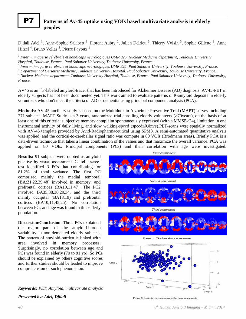
48 8th Human Amyloid Imaging – Miami, 2014
Patterns of Av-45 uptake using VOIs based multivariate analysis in elderly
peoples
Djilali Adel 1, Anne-Sophie Salabert 1, Florent Aubry 2, Julien Delrieu 3, Thierry Voisin 3, Sophie Gillette 3, Anne
Hitzel 4, Bruno Vellas 3, Pierre Payoux 1
1 Inserm, imagerie cérébrale et handicaps neurologiques UMR 825. Nuclear Medicine department, Toulouse University
Hospital, Toulouse, France. Paul Sabatier University, Toulouse University, France. 2 Inserm, imagerie cérébrale et handicaps neurologiques UMR 825. Paul Sabatier University, Toulouse University, France. 3 Department of Geriatric Medicine, Toulouse University Hospital. Paul Sabatier University, Toulouse University, France. 4 Nuclear Medicine department, Toulouse University Hospital, Toulouse, France. Paul Sabatier University, Toulouse University,
France.
AV45 is an 18F-labeled amyloid-tracer that has been introduced for Alzheimer Disease (AD) diagnosis. AV45-PET in
elderly subjects has not been documented yet. This work aimed to evaluate patterns of ß-amyloid deposits in elderly
volunteers who don't meet the criteria of AD or dementia using principal component analysis (PCA).
Methods: AV-45 ancillary study is based on the Multidomain Alzheimer Preventive Trial (MAPT) survey including
271 subjects. MAPT Study is a 3-years, randomized trial enrolling elderly volunteers (>70years), on the basis of at
least one of this criteria: subjective memory complaint spontaneously expressed (with a MMSE>24), limitation in one
instrumental activity of daily living, and slow walking-speed (speed≤0.8m/s).PET-scans were spatially normalized
with AV-45 template provided by Avid-Radiopharmaceutical using SPM8. A semi-automated quantitative analysis
was applied, and the cortical-to-cerebellar signal ratio was compute in 80 VOIs (Brodmann areas). Briefly PCA is a
data-driven technique that takes a linear combination of the values and that maximize the overall variance. PCA was
applied on 80 VOIs. Principal components (PCs) and their correlation with age were investigated.
Results: 91 subjects were quoted as amyloid
positive by visual assessment. Cattel’s scree-
test identified 3 PCs that contributing for
81.2% of total variance. The first PC
comprised mainly the medial temporal
(BA:21,22,39,48) involved in memory, and
prefrontal cortices (BA10,11,47). The PC2
involved BA35,38,30,29,34, and the third
mainly occipital (BA18,19) and prefrontal
cortices (BA10,11,45,25). No correlation
between PCs and age was found in this elderly
population.
Discussion/Conclusion: Three PCs explained
the major part of the amyloid-burden
variability in non-demented elderly subjects.
The pattern of amyloid-burden is linked with
area involved in memory processes.
Surprisingly, no correlation between age and
PCs was found in elderly (70 to 91 yo). So PCs
should be explained by others cognitive scores
and further studies should be leaded to improve
comprehension of such phenomenon.
Keywords: PET, Amyloid, multivariate analysis
Presented by: Adel, Djilali
P7
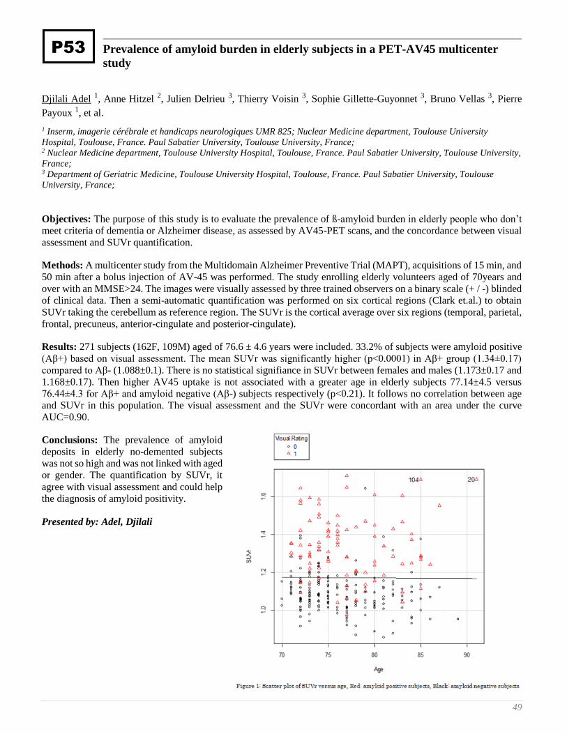
49
Prevalence of amyloid burden in elderly subjects in a PET-AV45 multicenter
study
Djilali Adel 1, Anne Hitzel 2, Julien Delrieu 3, Thierry Voisin 3, Sophie Gillette-Guyonnet 3, Bruno Vellas 3, Pierre
Payoux 1, et al.
1 Inserm, imagerie cérébrale et handicaps neurologiques UMR 825; Nuclear Medicine department, Toulouse University
Hospital, Toulouse, France. Paul Sabatier University, Toulouse University, France; 2 Nuclear Medicine department, Toulouse University Hospital, Toulouse, France. Paul Sabatier University, Toulouse University,
France; 3 Department of Geriatric Medicine, Toulouse University Hospital, Toulouse, France. Paul Sabatier University, Toulouse
University, France;
Objectives: The purpose of this study is to evaluate the prevalence of ß-amyloid burden in elderly people who don’t
meet criteria of dementia or Alzheimer disease, as assessed by AV45-PET scans, and the concordance between visual
assessment and SUVr quantification.
Methods: A multicenter study from the Multidomain Alzheimer Preventive Trial (MAPT), acquisitions of 15 min, and
50 min after a bolus injection of AV-45 was performed. The study enrolling elderly volunteers aged of 70years and
over with an MMSE>24. The images were visually assessed by three trained observers on a binary scale (+ / -) blinded
of clinical data. Then a semi-automatic quantification was performed on six cortical regions (Clark et.al.) to obtain
SUVr taking the cerebellum as reference region. The SUVr is the cortical average over six regions (temporal, parietal,
frontal, precuneus, anterior-cingulate and posterior-cingulate).
Results: 271 subjects (162F, 109M) aged of 76.6 ± 4.6 years were included. 33.2% of subjects were amyloid positive
(Aβ+) based on visual assessment. The mean SUVr was significantly higher (p<0.0001) in Aβ+ group (1.34±0.17)
compared to Aβ- (1.088±0.1). There is no statistical signifiance in SUVr between females and males (1.173±0.17 and
1.168±0.17). Then higher AV45 uptake is not associated with a greater age in elderly subjects 77.14±4.5 versus
76.44±4.3 for Aβ+ and amyloid negative (Aβ-) subjects respectively (p<0.21). It follows no correlation between age
and SUVr in this population. The visual assessment and the SUVr were concordant with an area under the curve
AUC=0.90.
Conclusions: The prevalence of amyloid
deposits in elderly no-demented subjects
was not so high and was not linked with aged
or gender. The quantification by SUVr, it
agree with visual assessment and could help
the diagnosis of amyloid positivity.
Presented by: Adel, Djilali
P53
50 8th Human Amyloid Imaging – Miami, 2014
Imaging amyloid in adults with Down’s syndrome using PiB-PET
Tiina Annus 1, Liam R. Wilson 1, Shahid Zaman 1, Young T. Hong 2, Tim Fryer 2, Franklin Aigbirhio 3, Rob Smith 2,
Anthony J. Holland 4
1 Cambridge Intellectual and Developmental Disabilities Research Group Department of Psychiatry University of Cambridge
Douglas House 18b Trumpington Road Cambridge CB2 8AH United Kingdom 2 Wolfson Brain Imaging Centre Department of Clinical Neurosciences University of Cambridge School of Clinical Medicine
Box 65 Cambridge Biomedical Campus Cambridge, CB2 0QQ 3 Department of Clinical Neurosciences Wolfson Brain Imaging Centre Department of Clinical Neurosciences University of
Cambridge School of Clinical Medicine Box 65 Cambridge Biomedical Campus Cambridge, CB2 0QQ 4 Cambridge Intellectual and Developmental Disabilities Research Group Department of Psychiatry University of Cambridge
Douglas House 18b Trumpington Road Cambridge CB2 8AH United Kingdom
Introduction: Down’s syndrome (DS) is associated with the trisomy of chromosome 21, which is believed to
significantly increase their risk of developing Alzheimer’s disease (AD). It is hypothesised that the presence of an extra
copy of the amyloid precursor protein (APP) gene leads to increased formation of β-amyloid (Aβ) plaques, neuronal
death and clinical AD. The present study aims to extend the findings of a pilot study by Landt et al. (2011) by
investigating the relationship between distribution and load of Aβ with age and dementia in DS.
Methods: Forty-two adults with DS aged 25-65 underwent an MRI scan and 90-minute dynamic PET scan using
Pittsburgh Compound-B (PiB). The BPND maps were created using simplified reference tissue model (Gunn et al,
NeuroImage 1997) with superior grey matter of cerebellum as reference tissue. T-1-template was created using
DARTEL toolbox in SPM8. Regions of interest (ROIs) were manually drawn on the template and applied to the warped
BPND map to obtain regional BPND values. Dementia was diagnosed using CAMDEX informant interview.
Results: Adults with DS without dementia (DS-AD, n=30), with early signs of dementia (DS+E, n=5) and with
dementia (DS+AD, n=6) demonstrated highest PIB binding in striatum and lowest in hippocampus. A significant
positive correlation of BPND values with age was seen for all ROIs. Significantly higher PiB retention was observed in
all ROIs in DS+AD, when compared to DS-AD, and in none of the ROIs, when compared to DS+E. Individuals with
DS+E demonstrated significantly higher BPND values in all ROIs apart from occipital lobe and hippocampus, when
compared to DS-AD.
Conclusion: A significant positive relationship between PiB-binding and age was observed in all groups. PiB binding
in ROIs apart from occipital lobe and hippocampus in DS+E suggests the course of amyloid spread, culminating in
positive binding in all ROIs in DS+AD.
Keywords: Down's syndrome, dementia, Alzheimer's disease, PiB,
Presented by: Annus, Tiina
P42
51
Exploring the best methods to detect longitudinal change in amyloid imaging
Suzanne Baker 1, Shawn Marks 2, Susan Landau 2, William Jagust 3
1 Lawrence Berkeley National Lab 2 Helen Wills Neuroscience Institute, UC Berkeley 3 Helen Wills Neuroscience Institute, UC Berkeley & Lawrence Berkeley National Lab
This project explored alternative methods of analyzing longitudinal PIB data. We looked at DVR versus SUVR (50-
70min) and the effect of reference region: cerebellar gray (cereg), whole cerebellum (cere), whole cerebellum+pons
(cere+p), whole cerebellum+eroded hemispheric white (cere+w), and whole cerebellum+pons+eroded hemispheric
white (cere+p+w). 28 older healthy control subjects had two 90 minute PIB scans within 3±0.9 years. Cingulate,
frontal, parietal and temporal cortex were the target regions used for comparison between methods. Despite the high
correlation between DVR and SUVR values from both visits within reference region, the annual percent change (a%c)
measured by DVR versus a%c measured by SUVR within reference region is less correlated (Figure1), although
highest correlations were found using cere or cere+p. Reference regions including white matter should minimize white
matter effects on cortical signal and thereby reduce the correlation between white matter a%c and target a%c. Figure
2 shows r2 between white matter a%c versus SUVR target ROIs a%c using various reference regions. Adding cerebellar
white to the reference region decreases the a%c correlation with white matter a%c; adding eroded hemispheric white
to the reference region further decreases this correlation. This decrease in correlation implies a decrease in white matter
in the target signal. However, the higher correlations with hemispheric white a%c (in comparison to cerebellar white
a%c) shows target signal in the hemispheric white matter still exists despite the erosion of the ROI, suggesting that
including hemispheric white matter in the reference region should be done with caution. The ideal reference region
would have sufficient white matter to divide out white matter signal in the cortex, but minimize white matter
contaminated with cortical signal. Whole cerebellum or cerebellum+pons may be the best option as a reference region
due to the DVR/SUVR correlation, removing white matter signal and not cortical signal.
Keywords: longitudinal, reference region, DVR, SUVR
Presented by: Baker, Suzanne
P37
52 8th Human Amyloid Imaging – Miami, 2014
Comparison of reference regions for [18F]Florbetapir PET SUVR computation
Arnaud Charil 1, Felix Carbonell 1, Alex Zijdenbos 1, Alan Evans 2, Robert Koeppe 3, Jeff Sevigny 4, Ping Chiao 4,Barry
Bedell 2
1 Biospective Inc., Montreal, QC, Canada 2 Biospective Inc. & Montreal Neurological Institute, McGill University, Montreal, QC, Canada 3 University of Michigan, Ann Arbor, MI, USA 4 Biogen Idec, Cambridge, MA, USA
Background: Amyloid PET is increasingly utilized in research studies and clinical practice. Quantitative assessment
of beta-amyloid burden is typically based on the standardized uptake value ratio (SUVR), which is derived from the
ratio of radiotracer uptake in target and reference brain regions. The cerebellar gray matter is commonly used as a
reference region for amyloid PET. However, this particular reference region may not be optimal for all tracers. As
such, we have performed a systematic study to determine the optimal reference region(s) for [18F]florbetapir scans.
Methods: [18F]florbetapir PET and 3D T1-weighted MR images were obtained from the ADNI database (n=454
subjects), as well as from subjects participating in an ongoing Phase IB clinical Alzheimer’s disease study sponsored
by Biogen Idec. Reference regions included cerebellar gray matter (cbGM), cerebellar white matter (cbWM), whole
cerebellum (cbFull), pons, cbFull+pons, deep white matter (dWM), and dWM+cbFull+pons. Target regions-of-interest
(ROIs) included frontal, parietal, lateral temporal, sensorimotor, anterior and posterior cingulate cortices, as well as a
composite ROI including all of these cortical regions. The target ROI SUVR measures were generated using
Biospective’s fully-automated PIANOTM image processing software. Probability density functions, correlation
analysis, and discriminant analysis were employed to compare SUVR measures using the different reference regions.
Results: Our results demonstrated that reference regions which did not include the cbGM were advantageous for
[18F]florbetapir studies. This observation was consistent across the datasets and study groups.
Conclusions: Several amyloid PET tracers currently exist and it is vital to determine the optimal reference region for
SUVR-based measures for each particular tracer. We employed multiple, complementary metrics, and utilized both
ADNI and clinical trial data for this study. Our results suggest that cbGM may not be the optimal reference region for
assessment of beta-amyloid plaque burden based on [18F]florbetapir PET scans.
Keywords: SUVR, Florbetapir, Reference Region, ADNI, Clinical Trial
Presented by: Bedell, Barry
P17
53
Statistically-driven, automated subject classification based on amyloid PET scans
Felix Carbonell 1, Arnaud Charil 1, Alex Zijdenbos 1, Alan Evans 2, Jeff Sevigny 3, Ping Chiao 3, Barry Bedell 2
1 Biospective Inc., Montreal, QC, Canada 2 Biospective Inc. & Montreal Neurological Institute, McGill University, Montreal, QC, Canada 3 Biogen Idec, Cambridge, MA, USA
Background: Classification of subjects as Amyloid-Positive (Aβ+) or Amyloid-Negative (Aβ-) based on amyloid PET
scans is increasingly utilized in research studies and clinical practice. While qualitative, visual assessment is currently
the gold-standard approach, automated classification techniques are inherently unbiased, reproducible, and efficient.
The objective of this work was to evaluate the performance of a recently developed statistical approach for the
automated classification of subjects with different levels of cognitive impairment using amyloid PET scans.
Methods: [18F]florbetapir PET and 3D T1-weighted MR images and were obtained from three cohorts, specifically
ADNI healthy control (HC) subjects (Cohort 1), ADNI MCI subjects (Cohort 2), and prodromal/mild Alzheimer’s
disease (AD) subjects from an ongoing Phase IB clinical study sponsored by Biogen Idec (Cohort 3). SUVR maps
were generated using Biospective’s fully-automated PIANOTMimage processing software. Subjects were classified into
Aβ+ and Aβ- groups based on visual evaluation of [18F]florbetapir PET scans. These classifications were used to
initialize an iterative, voxelwise, regularized discriminant analysis to optimize the selection of a set of regions-of-
interest (ROIs) and a threshold for automated classification of Aβ+ and Aβ- subjects in each cohort. The resulting
ROIs and thresholds were cross-validated between cohorts.
Results: The automated classifier demonstrated high accuracy, specificity, and sensitivity. The optimal ROIs included
areas of the default mode network (DMN), such as the precuneus and medial frontal cortex.
Conclusions: We have validated an automated approach for classification of subjects as Aβ+ or Aβ- using three
different subject cohorts. This strategy represents an improvement over current methods that rely on either subjective,
visual evaluation of amyloid PET images or quantitative measures based on pre-defined anatomical ROIs. Ultimately,
fully-automated subject classification may complement or supplant visual reads in clinical practice and for eligibility
assessment in AD clinical trials.
Keywords: Subject Classification, Florbetapir, Discriminant Analysis, ADNI, Clinical Trial
Presented by: Bedell, Barry
P27
54 8th Human Amyloid Imaging – Miami, 2014
Body mass index is associated with verbal episodic memory in cognitively normal
older individuals with low fibrillar amyloid-beta measured by [11C]-PiB
Murat Bilgel 1, Yi-Fang Chuang 2, Yang An 2, Jerry Prince 3, Dean Wong 3, Madhav Thambisetty 2, Susan Resnick 2
1 National Institute on Aging, NIH, Baltimore, MD, 21224, USA; Johns Hopkins University School of Medicine, Baltimore, MD,
21205, USA 2 National Institute on Aging, NIH, Baltimore, MD, 21224, USA 3 Johns Hopkins University School of Medicine, Baltimore, MD, 21205, USA
Background: Previous studies have implicated body mass index (BMI) as a predictor of verbal memory. In order to
determine whether this association is present independent of brain amyloid levels, we performed linear regression
analyses restricted to high and low amyloid individuals.
Method: Associations between verbal episodic memory measured by the California Verbal Learning Test (CVLT)
immediate recall trials on list A, concurrent BMI, and Pittsburgh Compound B (PiB) retention were studied in a cross-
sectional sample consisting of cognitively normal subjects (N=68), ages 61 to 92 (mean age= 77±7) from the Baltimore
Longitudinal Study of Aging (BLSA). Subjects were split into low PiB and high PiB groups based on mean cortical
distribution volume ratio (DVR) (using a threshold of DVR=1.09, the two groups had N=48 and 20, respectively).
Results: Linear regression models controlling for age, sex, race, education, APOE e4 status, and cardiovascular
conditions (including myocardial infarction and congestive heart failure) showed that CVLT immediate recall score is
inversely related to concurrent BMI (β=-0.8909, p=0.025) in cognitively normal persons with low PiB retention. No
other covariate was associated with CVLT except for age (β=-0.6474, p=0.021) in the low PiB group. These findings
remained significant after adjustment for mean cortical DVR. On the other hand, BMI was not associated with CVLT
immediate recall score in the high PiB subjects neither in the unadjusted nor adjusted models.
Conclusions: Higher BMI is associated with lower verbal immediate recall performance in individuals with no or
minimal levels of brain amyloid-beta. The lack of this association in individuals with high amyloid-beta suggests that
brain amyloid levels may alter the effects of factors reported to be associated with cognitive function.
Keywords: Verbal memory, body mass index
Presented by: Bilgel, Murat
P56
55
[18F]Flutemetamol blinded image interpretation: assessment of the electronic
training program for Japanese readers
Chris Buckley 1, Brian J McParland 1, Jan Wolber 1, Paul Sherwin 2, Michelle Zanette 2, Gill Farrar 1
1 GE Healthcare; Amersham, UK 2 GE Healthcare; Princeton, NJ
Objective: Previously (Sherwin, HAI 2013) reported the validation of an Electronic Training Program (ETP) for
interpreting [18F]flutemetamol (VIZAMYL™) PET images, using US readers. The successful use of the ETP with
Japanese-board trained readers in the Phase II study in Japan is reported.
Methods: Japanese subjects, AD (n = 20), aMCI (n=20) and healthy controls (n=25) were dosed with 185 MBq of
Flutemetamol F 18 Injection and underwent brain PET. Images (n=65) were interpreted blind by 10 readers (5
Japanese-board trained, 5 non-Japanese board trained), each trained using the ETP, who classified each image as
positive or negative for brain amyloid. Agreement across all five readers in the two groups was determined and reported
as Fleiss’ kappa. Results were also compared to the European Phase II study (Vandenberghe et al., Ann Neurol
2010;68:319–329) and the previous ETP study (Sherwin et al., HAI 2013). Intra-reader reproducibility (IRR) was also
determined by re-reading images from 7 subjects (10%) selected randomly from the 65 subjects.
Results: Among the 5 Japanese readers, there was complete agreement for 61 (94%) of 65/70 images read, (kappa =
0.94). Among the 5 non-Japanese readers, there was complete agreement for 62 (95%) of 65 images read, (kappa =
0.96). The performance of each group of readers is consistent with the EU Phase II data (kappa 0.96; Vandenberghe
2010) which used an in-person training method. These results are slightly higher than the Phase 3 read study (n=276,
kappa = 0.83; Sherwin et al., HAI 2013) which included a wider spectrum of subjects, including end-of-life subjects.
Intra-reader agreement ranged from 86% to 100% (median 100%) and was similar to those of the European Phase II
study (97.5%) and the Phase 3 study (93% to 100%).
Conclusion: The ETP is effective in training Japanese readers. This further validates the use of electronic media for
the training of readers in the routine interpretation of [18F]flutemetamol images.
Keywords: [18F]flutemetamol, VIZAMYL™, blinded image evaluation, electronic training program, kappa score.
Presented by: Buckley, Chris
P51
56 8th Human Amyloid Imaging – Miami, 2014
Disclosure of amyloid imaging results in cognitively normal individuals
David Johnson 1, Vidoni Eric 2, Magdalena Leszko 1, Rebecca Bothwell 2, Jeffrey Burns 2
1 University of Kansas 2 University of Kansas Medical Center
Introduction: Disclosing amyloid imaging results to cognitively normal (CN) older adults has begun in the setting of
prevention trials. Disclosing amyloid status to CN individuals remains debatable, in particular in the clinical setting,
given an imprecise understanding of its clinical significance, lack of effective therapeutic options, and unknown
psychological risk.
Methods: The KU Alzheimer’s Prevention Program Exercise (APEX) trial is among the coming wave of AD
prevention trials. We disclose amyloid status using a genetics counseling approach with pre- and post-scan counseling
conducted by clinicians. We assess the effect of disclosure on anxiety, depression, perception of risk, and health
behaviors at baseline and at 6-weeks and 6-months post-disclosure, similar to the REVEAL study. Measures include
the Beck Anxiety Inventory (BAI), Center for Epidemiologic Studies Depression Scale (CES-D), Impact of Genetic
Testing for AD (IGT-AD) and surveys of health behaviors.
Results: APEX will screen a total of 400 CN older adults (CDR 0) with amyloid PET to identify 100 participants with
cerebral amyloid for a one-year exercise trial. As of November 2013, a total of 24 participants have been screened with
4 individuals (17%) having elevated cerebral amyloid, as determined by visual reads informed by quantitative analyses.
Our preliminary analyses suggest disclosing a positive amyloid status to CN individuals does not negatively influence
anxiety (mean BAI 5.3 (4.4) at baseline vs 5.3 (2.6) at 6 weeks) or depression (mean CES-D 3.0(1.4) at baseline vs.
2.3(1.2) at 6-weeks). There was no apparent adverse psychological impact (mean IGT-AD 16.3(4.9), similar values to
the REVEAL study). Amyloid positive participants reported greater intent to change their daily diet and exercise.
Conclusion: Our preliminary experience suggests that disclosing amyloid imaging results to CN adults with pre- and
post-disclosure counseling has a low risk of psychological harm and may motivate an individual to adopt healthier
lifestyle behaviors.
Keywords: disclosure, psychology, ethics, amyloid status
Presented by: Burns, Jeffrey
P45

57
Differential temporal patterns of Amyloid-β and functional imaging markers
across mutation types in autosomal dominant Alzheimer’s disease: Findings from
the DIAN Study
Jasmeer Chhatwal 1, Aaron Schultz 1, Keith Johnson 2, Tammie Benzinger 3, Clifford Jack 4, Beau Ances 5, Caroline
Sullivan 1, Stephen Salloway 6, John Ringman 7, Robert Koeppe 8, Daniel Marcus 3, Paul Thompson 9, Andrew
Saykin10, John Morris 5, Reisa Sperling 11, et al.
1 Department of Neurology, Massachusetts General Hospital, Harvard Medical School, Boston MA 2 Departments of Neurology and Radiology, Brigham and Women's Hospital, Massachusetts General Hospital, Harvard Medical
School, Boston MA 3 Department of Radiology, Section of Neuroradiology, Washington University School of Medicine, St. Louis, MO 4 Department of Radiology, Mayo Clinic, Rochester MN 5 Department of Neurology, Washington University School of Medicine, St. Louis MO 6 Department of Neurology, Butler Hospital, Brown University School of Medicine, Providence, RI 7 Department of Neurology, Easton Center for Alzheimer’s Disease Research, University of California - Los Angeles, Los
Angeles, CA 8 Department of Radiology, University of Michigan School of Medicine, Ann Arbor, MI 9 Lab of Neuro Imaging, UCLA School of Medicine, Los Angeles, CA 10 Department of Radiology, IU Center for Neuroimaging, Indiana University School of Medicine, Indianapolis, IN 11 Brigham and Women's Hospital, Massachusetts General Hospital, Department of Neurology, Center for Alzheimer's Research
and Treatment, Harvard Medical School, Boston MA
Rationale: Imaging biomarkers will play a key role in upcoming Alzheimer’s disease (AD) prevention clinical trials,
including trials in autosomal-dominant AD (ADAD). Accordingly, we examine whether subjects harboring mutations
in Presenilin-1 (PSEN1), Presenilin-2 (PSEN2), or Amyloid Precursor Protein (APP) show differential temporal or
spatial patterns of imaging biomarker change in ADAD.
Methods: Focusing on ADAD subjects who were at or younger than their estimated age of symptom onset at their
baseline visit, we examined both main effects of mutation type and interactions between mutation type and estimated
years to symptom onset (EYO) using regional imaging biomarker values as the outcome. These relationships were
examined in cross-sectional PiB and FDG-PET, functional connectivity MRI (fcMRI), and MRI-derived structural
measures. Preselected Freesurfer ROIs were used for FDG, PiB and structural data. Template-based rotation was used
to assess fcMRI networks.
Results: Significant mutation type effects were observed in PiB
binding in several midline structures (posterior cingulate,
precuneus, anterior cingulate). With the exception of significant,
differential FDG hypometabolism in PSEN1 carriers in the
precuneus, no significant effects of mutation type were apparent
in other FDG and structural measures within pre-selected
cortical ROIs. Similarly, no significant mutation type or
interaction effects were observed in the Default Mode, Dorsal
Attention, Motor, or Visual Networks.
Conclusion: Mutation type may be an important consideration
when using regional amyloid and FDG-PET measures as
secondary outcomes in ADAD clinical trials. Though the
patterns observed here in cross-sectional data will need to be
verified in longitudinal data, plotting PiB binding against EYO
suggests that PSEN2 carriers may show a greater rate of midline
fibrillar amyloid deposition as compared to PSEN1 carriers, whereas APP carriers may show a lower rate. These
differences hint at underlying genotypic variation in the kinetics of amyloid production, processing, and clearance that
may our pathophysiologic understanding of ADAD.
Keywords: functional connectivity, amyloid, autosomal dominant AD
Presented by: Chhatwal, Jasmeer
P34
58 8th Human Amyloid Imaging – Miami, 2014
Comparison of PIB-PET data to Florbetapir-PET data acquired from different
patient cohorts and the role of age in the discriminative power of amyloid
imaging
Konstantinos Chiotis 1, Stephen F. Carter 2, Agneta Nordberg 3
1 Alzheimer Neurobiology Center, Karolinska Institutet; Sweden 2 Alzheimer Neurobiology Center, Karolinska Institutet; Sweden, Wolfson Molecular Imaging Center, University of Manchester,
Manchester; UK 3 Alzheimer Neurobiology Center, Karolinska Institutet; Sweden, Department of Geriatric Medicine, Karolinska University
Hospital Huddinge; Sweden
Introduction: Several 11C- and 18F-PET radiotracers, which bind with high affinity to fibrillar amyloid, have been
developed and are applied clinically into different patient cohorts.
Objectives: To investigate the comparability of two amyloid PET tracers (11C-PIB/18F-Florbetapir), when applied in
two different patient cohorts and to explore the effect of age in the discriminative properties of amyloid imaging.
Methods: 11C-PIB-PET data from 51 healthy controls (HC), 72 mild cognitive impairment (MCI) and 90 Alzheimer’s
disease (AD) patients deriving from the DIMI EU project (Nordberg et al.2013) were analysed using a PIB-PET
population template and atlas (mean age 68.5±7.8 years). 18F-Florbetapir-PET data were analysed by applying the
same methodology in a matched for age (mean age 69.4±6.9 years), gender and MMSE-score population (51 HC,72
MCI and 84 AD), deriving from the Alzheimer’s Disease Neuroimaging Initiative (ADNI). An additional ADNI
population (246 HC,344 MCI and 138 AD), with a Florbetapir scan, was split in two age groups matched for MMSE-
score and gender distribution, with mean ages of 70.9±3.5 and 80.9±3.9 years.
Results: Regional values of PIB and Florbetapir retention were strongly correlated in the young matched groups across
the different brain regions investigated (r=0.829,p=0.00). However, a narrower range and lower cut off was observed
for the Florbetapir data (0.99-2.06,1.34) compared to PIB (1.09-2.67,1.42). The lower discriminative power that
Florbetapir produced, with Sensitivity(SE)/Specificity(SP)=86/82 in comparison with 91/90 for PIB, was investigated
between the two age groups deriving from the ADNI population. The older subjects displayed much lower SE/SP in
comparison to the younger group (72.5/78.9 and 89.9/85.5 respectively).
Conclusions: This study demonstrates the comparability of amyloid imaging in two unrelated, but matched, relatively
young populations using two different fibrillar amyloid PET tracers. Moreover, the low discriminative ability of
amyloid imaging in older patients may suggest a limited clinical usefulness in an older patient population.
Keywords: Florbetapir, PIB, ADNI, comparison, age
Presented by: Chiotis, Konstantinos
P15
59
The value of early F18-Florbetapir scan information as an estimate of regional
cerebral blood flow and comparison to F18-FDG measures of cerebral
metabolism
Michael Devous , Abhinay Joshi , Michael Navitsky , Michael Pontecorvo , Daniel Skovronsky , Mark Mintun
Avid Radiopharmaceuticals Philadelphia, PA
Background: Neuroimaging assessments of amyloid plaque and of non-specific neurodegeneration, such as that
provided by FDG, may be complementary in the evaluation of neurodegenerative disorders. We explored whether
scanning in the first 5 min after florbetapir injection could be used as a surrogate marker of regional cerebral blood
flow (rCBF) and assessed the degree to which these images concurred with FDG images of cerebral glucose
metabolism (rCGM).
Methods: Florbetapir and FDG PET images from ADNI2 were analyzed for 70 subjects diagnosed as Alzheimer’s
Disease (AD, N=6), Mild Cognitive Impairment (MCI, N=15), subjective memory complaints (N=31) and cognitively
normal (N=18). Subjects had a dynamic PET acquisition from 0-20 min after injection of 370 (±10%) MBq florbetapir.
On a separate day all subjects underwent a dynamic (6x5 min) FDG scan (185 MBq ±10%) starting 30 min after
injection. Integrated images of the first 5 min of scanning were created for Florbetapir using a trapezoidal method and
for all 30 min of the FDG scan. After spatial normalization to MNI space, whole-brain normalized values were
calculated for both Florbetapir area under the curve (AUC) and FDG images. Correlation analysis was performed
between FDG and florbetapir data from ROIs and on a voxel-wise basis.
Results: For ROI analyses, the average correlation r value was 0.91 with an average slope of 0.81. For voxel-wise
correlations, the average r value was 0.84 and the average slope was 0.78. Visual inspection of the “rCBF”/rCGM
image pairs revealed substantial concordance among areas of both normal and reduced neuronal metabolism.
Conclusions: ROI and voxel-wise analyses between 0-5 min florbetapir data and 30 min FDG images showed high
mean correlations. Further work appears warranted to evaluate the utility of early florbetapir as a surrogate for the
functional information seen in an FDG scan.
Keywords: florbetapir, early scan, FDG, rCBF, rCGM
Presented by: Devous, Michael
P59
60 8th Human Amyloid Imaging – Miami, 2014
Amyloid imaging changes diagnosis and treatment in patients with progressive
cognitive impairment: Multicenter evaluation of 3-month post-scan outcomes
P. Murali Doraiswamy 1, Andrew Siderowf 2, Michael Pontecorvo 2, Stephen P. Salloway 3, Adam S. Fleisher 4, Carl
H. Sadowsky 5, Anil K. Nair 6, Ming Lu 2, Anupa K. Arora 2, Daniel M. Skovronsky 2, Mark A. Mintun 2, Michael
Grundman 7, AV45-A17 Study Group 8
1 Duke University 2 Avid Radiopharmaceuticals, Inc. 3 Butler Hospital 4 Department of Neurosciences, University of California, San Diego; Banner Alzheimer’s Institute 5 Nova Southeastern University 6 Department of Neurology, Alzheimers Disease Center, Quincy Medical Center 7 Global R&D Partners, LLC; Department of Neurosciences, University of California, San Diego 8 AV45-A17 Study Group
Background: We previously reported that clinicians may change their diagnosis and intended management of patients
with cognitive decline based on the results of amyloid imaging.
Objective: To determine whether amyloid imaging could influence actual diagnosis and management.
Methods: In our initial study, 229 patients with progressive cognitive decline and an uncertain diagnosis were recruited
to 19 clinical sites. The site physician provided a provisional diagnosis, an estimate of his/her diagnostic confidence,
and a management plan both before and after receipt of the florbetapir F18 PET results. In this follow-up study, the
clinical charts of participants for the three months following the initial study were reviewed to determine whether the
diagnosis reported before amyloid imaging changed and whether study physicians carried out their intended
management plan.
Results: Records for 172/229 (75.1%) of participants in the initial study had at least 3 months of clinical follow-up
and were available for review. The diagnosis recorded pre-scan was changed in the clinical charts in 46/81 (57.0%) of
cases when the florbetapir scan was negative, including24/59 (96.0%) of cases where the pre-scan diagnosis was AD.
When the scan was positive, the actual diagnosis changed in 45/91 (49.0%) of cases. Compared to the pre-scan
management plan, amyloid imaging results led to a decrease in cholinesterase or memantine use in 20/43 (46.5%)
subjects with a negative scan, and the addition of a cholinesterase inhibitor or memantine in 7/57 (12.3%) of cases with
a positive scan.
Conclusions: In this multi-center study, we found that evaluation with florbetapir F18 PET altered actual diagnosis
and management in a manner consistent with changes in intended management observed in our prior study, possibly
avoiding unnecessary treatment. While limited by retrospective methods, the results indicate that amyloid imaging
may meaningfully impact how physicians diagnose and treat cognitively impaired patients.
Keywords: Clinical management,Diagnosis,Amyloid imaging,Clinical trial
Presented by: Doraiswamy, P. Murali
P4
61
Practice effects relate to flutemetamol uptake, but not FDG or hippocampal
volume: Moving cognition earlier in Jack’s curves
Kevin Duff , Norman Foster , Richard King , John Hoffman
University of Utah
Background: Jack et al. (2013) have suggested a temporal ordering of biomarker positivity for Alzheimer’s disease,
with amyloid deposition occurring before neuronal degeneration and cognitive impairment. However, additional
support for this model is needed.
Methods: The current study examined short-term practice effects across one week as a new measure of cognitive
functioning and its relationship to amyloid burden (via 18F-flutemetamol PET imaging) and neuronal degeneration (via
metabolism on FDG-PET and hippocampal volumes on MRI) in 25 non-demented older adults.
Results: Short-term practice effects significantly correlated with 18F-flutemetamol uptake (r=-0.45, p=0.02), but was
more modestly related to FDG-PET metabolism (r=0.33, p=0.11). There was no relationship between practice effects
and hippocampal volume on MRI (r=0.04, p=0.85).
Conclusions: In this very mildly impaired sample, short-term practice effects were more strongly related to amyloid
deposition (18F-flutemetamol) than with either marker of neuronal degeneration (FDG-PET or MRI). If the temporal
ordering of biomarkers in Alzheimer’s disease proposed by Jack et al. (2013) is correct, then short-term practice effects
may be a more sensitive indicator of cognitive functioning in prodromal Alzheimer’s disease.
Keywords: cognition, amyloid, imaging
Presented by: Duff, Kevin
P35
62 8th Human Amyloid Imaging – Miami, 2014
Alpha-synuclein imaging: Identifying small molecules that bind to aggregated
Alpha-synuclein
Dale Mitchell 1, Kevin Nash 1, David Hardick 1, David Cronk 1, Paul Kotzbauer 2, Zhude Tu 2, Jinbin Xu 2, Robert
Mach 3, Jamie Eberling 4, N. Scott Mason 5, William Klunk 5, Chester Mathis 5
1 BioFocus 2 Washington University 3 University of Pennsylvania 4 Michael J. Fox Foundation 5 University of Pittsburgh
The ability to image alpha-synuclein deposition in the brain would be a game-changing achievement for the
Parkinson’s disease (PD) field. The accumulation of aggregated alpha-synuclein is a pathological hallmark of PD and
a priority target for drug development given its hypothesized contribution to neurodegeneration. Alpha-synuclein
pathology also contributes to other conditions, such as Lewy body dementia. In vivo imaging of alpha-synuclein
pathology could be useful as a biomarker of the presence of disease and disease progression, and as a pharmacodynamic
tool for drug development.
A consortium was initiated in 2011 with the goal of developing an alpha-synuclein PET tracer. Two high throughput
screens were utilized to identify small molecules that bind to aggregated alpha-synuclein using either the fluorescent
ligand Thioflavin T or the radioligand [3H]-Chrysamine G. 100,000 compounds selected from the BioFocus screening
libraries based on chemical diversity and lead-like properties were screened for their ability to bind to alpha-synuclein
fibrils in both assay formats and identified hit compounds targeting different binding sites.. Initial hit compounds were
subjected to hit confirmation, potency studies and for selectivity over beta-amyloid and tau. Compounds for further
investigation were prioritized based on selectivity and calculated properties for good CNS penetration. Competitive
radioligand binding assays were conducted using the inhibition of [3H]-PiB with 300 nM and 30 nM (cold)
concentrations of the compounds to further evaluate their binding to synthetic alpha-synuclein and beta-amyloid fibrils.
Compounds that were confirmed for selectivity and affinity in these assays were subsequently radiolabeled and
assessed for binding in PD and AD tissue. Two lead series were identified for further optimization and in vivo testing.
Keywords: alpha-synuclein, Parkinson's, Lewy body
Presented by: Eberling, Jamie
P54

63
Evaluation of F-18 radiolabeled cromolyn as a potential Aβ polymerization
inhibitor and PET tracer
David R. Elmaleh 1, Timothy M. Shoup 1, Alan J. Fischman 2, Kazue Takahashi 3, Mykol Larvie 1, Erik Vogan 4
1 Massachusetts General Hospital 2 Shriners Hospitals for Children 3 Pediatrics Services, Massachusetts General Hospital 4 Harvard Institutes of Medicine Ctr Neurologic Diseases
Objective: Multiple lines of evidence suggest that the accumulation of neurotoxic oligomeric aggregates of amyloid-
beta is a central event in the pathogenesis of Alzheimer’s disease. We have explored cromolyn and its fluorinated
analog for potential use as both an amyloid imaging agent and as therapeutic agents. In this study we report the Ab-
peptide polymerization inhibition results for these compounds and the synthesis and biodistribution of F-18 labeled
cromolyn in mice. Additionally, we have used mathematical modeling to evaluate cromolyn’s structural mode of
interaction with abeta oligomers.
Method: The Ab-peptide inhibition assays were performed according to the method of Findeis. The progress of Ab
polymerization was monitored by measuring turbidity as the apparent UV absorbance in a Bio-Rad microplate reader
equipped with a 405-nm filter. Inhibition values are the relative polymerization slow down and are expressed as the
relative increased time ratio of the polymerization to reach 50% of maximum signal. Biodistribution of F-18 labeled
cromolyn was performed in normal mice at 5, 30, and 60 min. Cromolyn binding to model protofibrils formed from
abeta residues 16 to 22 was examined using all-atom explicit-solvent molecular dynamics conducted at 320 K over 20
ns simulation.
Results: Polymerization inhibition assays reveal that
cromolyn and its fluorinated derivative caused 6 to 8 fold
inhibition at 5 and 50 nM concentration. Biodistribution of
the fluorinated analog demonstrates brain accumulation
which stayed constant for 5, 30 and 60 min at 0.95, 0.94 and
0.94 % DPG. Preliminary analysis of the binding model
indicates that cromolyn binds across the surface of the beta
strand in a manner similar to that of Thioflavin-T.
Conclusion: Cromolyn and its fluorinated derivative are
potential PET imaging agent for early detection and treatment
of Alzheimer’s disease.
Keywords: Cromolyn, F-18, Ab-peptide inhibition
Presented by: Elmaleh, David R.
P1
64 8th Human Amyloid Imaging – Miami, 2014
Effects of age and amyloid on the encoding of visual scene detail
Jeremy Elman 1, Hwamee Oh 2, Cindee Madison 2, Suzanne Baker 1, Jacob Vogel 2, Sam Crowley 2, William Jagust 2
1 Life Sciences Division, Lawrence Berkeley National Laboratory, Berkeley, CA 94720, USA 2 Helen Willis Neuroscience Institute, University of California, Berkeley, CA 94720, USA
Successful memory encoding is associated with increased and decreased activity across the brain. Both age and disease
may be associated with behavioral impairments. However, cognitively normal elderly adults harboring increased levels
of beta-amyloid (Aβ) demonstrate changes in brain activity without impaired memory function. It remains unclear
whether the quality of memory in these subjects is spared and to what degree changes in activity represent deleterious
versus compensatory processes.
Twenty-two young (mean age=23.6) and 49 older subjects (mean age=76.4) completed a visual scene encoding task
during an fMRI scan, followed by two tasks to assess gist recall and detail memory. Gist memory was assessed with
an old/new and high/low confidence response based on written descriptions of scenes. Subjects made true/false
judgments about 6 written details for each scene to asses detail memory. Activity at encoding was back-sorted based
on subsequent memory performance. Task-positive and negative networks were determined by contrasting high
confidence hits with misses and activity was extracted to assess parametric changes associated with the number of
details retrieved. Amyloid burden in the older subjects was examined using PIB-PET imaging and dichotomized into
PIB+ and PIB- groups (N= 16 PIB+/33 PIB-).
Memory performance was similar among all groups. Age effects were apparent in multiple regions including the right
hippocampus where young subjects showed greater overall activation and a steeper parametric increase of activity with
number of encoded details. Interestingly, PIB+ subjects displayed sharper decreases in right inferior parietal (a task-
negative region) activity associated with number of details encoded compared to PIB- subjects. In contrast, there was
a sharper increase of activity across detail levels among PIB+ subjects in the right inferior LOC, a task positive region.
While DMN activity was disrupted in PIB+ individuals, higher activation appeared to support memory formation and
may reflect compensatory mechanisms underlying normal functioning in PIB+ subjects.
Keywords: amyloid, memory, encoding, ageing
Presented by: Elman, Jeremy
P62

65
Clinical presentation and imaging findings in complex ALS with AD-like
cognitive impairment
Karim FARID 1, Stephen CARTER 1, Elena RODRIGUEZ-VIEITEZ 1, Ove ALMKVIST 1, Pia ANDERSEN 2,
Anders WALL 3, Peter ANDERSEN 4, Agneta NORDBERG 5
1 Alzheimer Neurobiology Center, Department of Neurobiology, Care Sciences and Society, Karolinska Institutet, Stockholm,
Sweden 2 Department of Geriatric Medicine, Karolinska University Hospital Huddinge, Stockholm, Sweden 3 Uppsala PET Center, Department of Radiology, Oncology and Radiation Sciences, Uppsala University, Uppsala, Sweden 4 Department of Neurology, Umeå University, Umeå, Sweden 5 Alzheimer Neurobiology Center, Department of Neurobiology, Care Sciences and Society, Karolinska Institutet, Stockholm,
Sweden. Department of Geriatric Medicine, Karolinska University Hospital Huddinge, Stockholm, Sweden
Introduction: Deposition of fibrillar amyloid in brain is a well-known pathological hallmark of Alzheimer disease
(AD). However, its presence in other forms of neurodegenerative diseases is unclear.
Aims: We here report clinical and imaging findings in a patient initially diagnosed as MCI due to AD and included in
a multi-tracer PET study who later developed rapid progression of ALS-like impairments in the clinical follow-up.
Methods: A 60-year-old well-educated male was assessed for cognitive impairments. He underwent 11C-PIB, 18F-FDG
and 11C-deuterium-L-deprenyl (11C-DED) PET imaging in addition to CSF analysis and neuropsychological
assessments.
Results: Neuropsychological examination revealed episodic memory impairments. CSF p-Tau, total-Tau were
increased but Aß42 was within normal range. Individual patient’s images as well as the comparison with age matched
healthy controls showed a brain 18F-FDG-PET hypometabolism in the left hippocampus and in the prefrontal cortices
consistent with neurodegeneration and a 11C-PiB-PET prefrontal, posterior cingulate and parietal cortices uptake
consistent with amyloidal deposition. Furthermore, 11C-DED-PET showed high brain binding consistent with increased
astrocytosis. Two consecutive MRI scans showed a progressive right frontal/temporal cortical atrophy. One year later,
the patient progressed in memory impairments, and was diagnosed as AD and shortly developed motor impairments
and was diagnosed with ALS. After rapid progression, the patient died with pronounced motoric and bulbar symptoms
18 months later. Relatives denied autopsy why post-mortem analysis was not available and the exact pathological
diagnosis remains unclear.
Conclusions: The case illustrates a complex pathology and clinical features suggestion that ALS may overlap with
other neurodegenerative diseases. Further prospective multi-tracer studies should be conducted to better identify
underlying disease mechanisms.
Keywords: Amyotrophic lateral sclerosis; Fronto temporal dementia; Alzheimer disease; PET imaging
Presented by: FARID, Karim
P32
66 8th Human Amyloid Imaging – Miami, 2014
Striatal 11C-PiB retention in AD, MCI and HC: the pathotololgical significance of
high PIB retention in the putamen
Karim FARID 1, Ove ALMKVIST 1, Katharina BRUEGGEN 1, Stephen CARTER 2, Anders WALL 3, Karl
HERHOLZ4, Agneta NORDBERG 5
1 1- Alzheimer Neurobiology Center, Department of Neurobiology, Care Sciences and Society, Karolinska Institutet, Stockholm,
Sweden 2 1- Alzheimer Neurobiology Center, Department of Neurobiology, Care Sciences and Society, Karolinska Institutet, Stockholm,
Sweden. 2- Wolfson Molecular Imaging Centre, University of Manchester, 27 Palatine Road, Manchester M20 3LJ, UK 3 1- Uppsala PET Center, Department of Radiology, Oncology and Radiation Sciences, Uppsala University, Uppsala, Sweden 4 1- Wolfson Molecular Imaging Centre, University of Manchester, 27 Palatine Road, Manchester M20 3LJ, UK 5 1- Alzheimer Neurobiology Center, Department of Neurobiology, Care Sciences and Society, Karolinska Institutet, Stockholm,
Sweden 2- Department of Geriatric Medicine, Karolinska University Hospital Huddinge, Stockholm, Sweden
High fibrillar Aβ deposition in the cerebral cortex has been well established by 11C-PiB-PET in sporadic Alzheimer´s
disease (AD) patients. A high 11C-PiB-PET retention was demonstrated in the striatum of PSN1 mutations carriers but
is less studied in sporadic AD/MCI.
To better understand the relationship between the striatum and AD pathophysiology, we compared the caudate,
putamen and neocortical 11C-PiB-PET retention in 97 AD, 72 mild cognitive impairment (MCI) patients, 51 healthy
controls (HC). The putamen and whole-cortex 18F-FDG-PET uptake was also studied in 15 AD, 17 MCI and 6 HC
from the same patients.
The putamen 11C-PiB retention exceeded neocortical and caudate 11C-PiB uptake in all subjects (p<0.05; HC and
MCI 11C-PiB-negative), (p<0.001; AD and MCI 11C-PiB-positive).
The putamen 11C-PiB retention was HC<AD (p=0.0001), HC<MCI 11C-PiB-positive (p<0.0001), AD>MCI 11C-PiB-
negative (p<0.0001), AD>MCI 11C-PiB-positive (p=NS).
The converter-MCI, all 11C-PiB-positive (n=29/72), had higher baseline 11C-PiB retention in the
putamen>caudate>neocortex (p<0.0001).
The putamen 18F-FDG uptake exceeded the whole-cortex uptake in all subjects (p<0.001) and was HC<AD (p=0.025),
HC<MCI (p<0.001), AD<MCI (p=NS).
Striatum is highly connected to the cortex via several pathways. One possible explanation for the findings might be
the functional connectivity that could be behind correlated Aβ plaque levels in functionally-related regions and is
supported by research on “hub” vulnerability that has shown highly connected regions to be especially vulnerable to
Aβ pathology. Furthermore, the putaminal 18F-FDG increases could reflect a compensation mechanism of this highly
connected structure and/or brain inflammation that has been previously described to precede neurodegeneration.
Interestingly, we found that even in HC, the 11C-PiB and the 18F-FDG uptake in putamen>neocortex.
Fibrillar amyloid might be present in regions that are not directly related to pathological degeneration and progression
of the disease. Moreover, these results emphasize the further understanding of the potential role of the putamen in the
AD pathophysiology.
Keywords: 11C-PiB-PET, 18F-FDG-PET, Striatum, Putamen, Alzheimer Disease
Presented by: FARID, Karim
P36

67
Advanced education mediates the impact of amyloid burden on reasoning in
healthy older adult
Michelle E. Farrell , Gérard N. Bischof , Karen M. Rodrigue , Kristen M. Kennedy , Denise C. Park
University of Texas at Dallas
Objective: To determine whether education, as a proxy of cognitive reserve, differentially mitigates the impact of
amyloid burden on four major cognitive constructs: processing speed, working memory, episodic memory and
reasoning.
Methods: Subjects were selected from a group of 187 healthy older adults (>60 years of age) from the Dallas Lifespan
Brain Study that underwent a comprehensive cognitive battery and a florbetapir amyloid PET scan. Participants were
divided into three education groups: Higher (>Bachelor’s, n=52), Intermediate (Bachelor’s degree, n = 45) and Lower
Results: A 3 x 2 (Amyloid level x Education level) ANOVA revealed main effects of Amyloid (p=0.05) and Education
(p<0.001) and an Amyloid x Education interaction (p=0.013) for reasoning. Lower-educated individuals exhibited
decreasing reasoning scores with increasing amyloid burden (R2=0.083, β= -1.471, p=0.006) whereas higher-educated
individuals were unaffected by amyloid burden (p=0.662). The analyses of working memory, processing speed, and
episodic memory yielded neither main effects nor interactions associated with amyloid burden.
Discussion: These findings provide evidence that when amyloid is detrimental to task performance, as in the case of
reasoning, a high level of education confers resilience to the negative effects of amyloid. These results also add to
growing evidence that amyloid deposition may have its strongest effects on higher level cognition.
Keywords: amyloid, cognitive reserve, education, normal aging, healthy aging, reasoning
Presented by: Farrell, Michelle E.
P29
68 8th Human Amyloid Imaging – Miami, 2014
Early-frame PiB PET perfusion and FDG PET glucose metabolism comparison in
cognitively normal persons at three levels of genetic risk for Alzheimer’s disease
Hillary Protas 1, Kewei Chen 1, Ji Luo 1, John Thompson Rausch 1, Robert Bauer III 1, Sandra Goodwin 1, Nicole
Richter 1, Daniel Bandy 1, Richard Caselli 2, Adam Fleisher 1, Eric Reiman 1
1 Banner Alzheimer's Institute;Arizona Alzheimer’s Consortium 2 Mayo Clinic;Arizona Alzheimer’s Consortium
Background: While Pittsburgh Compound B (PiB) positron emission tomography (PET) is commonly used for
assessing fibrillar beta-amyloid deposition, its early frames can also be used for evaluating cerebral perfusion (pPiB).
Recent reports showed a significant association between pPiB measured cerebral blood flow and fluorodeoxyglucose
(FDG) measured cerebral metabolic rate for glucose in patients with Alzheimer’s dementia (AD). This current study
characterizes the relationship between regional pPiB and FDG PET measurements in cognitively normal
apolipoprotein-E (APOE) ε4 homozygotes(HM), heterozygotes(HT) and noncarriers(NC).
Methods: Dynamic PiB-PET and FDG-PET scans were acquired with a between-scan interval of less than 6 months
in 10 APOE-ε4 HM, 18 HT, and 21 NC, 64±6 years of age. pPiB images were computed from frames acquired 1.0-6.5
min, and FDG-PET images from frames acquired 30-60 min after injection, each normalized for the count variation
using a cerebellar gray matter reference region. SPM8 was used to characterize and compare differences in regional
PET measurements among the three groups. Voxel-wise linear regression was used to characterize the relationship
between regional pPiB and FDG-PET measurements.
Results: In comparison with the HT and NC groups, the APOE-ε4 HMs had significantly lower pPiB and FDG PET
measurements in the vicinity of the precuneus, posterior cingulate, parieto-temporal cortex, and prefrontal regions
(P<0.005, uncorrected). There was a close correlation between voxel-based pPiB PET and FDG PET measurements
for all subjects (R2=0.94±0.02, max=0.97, min=0.84) and a close correlation between voxel-based pPiB-PET and FDG-
PET differences among the HM, HT and NC groups (R=0.71, p<1.1e-16).
Conclusions: PiB-PET and FDG-PET measurements are roughly comparable in their ability to characterize regional
differences between cognitively normal persons at differential risk for AD. Further studies are indicated to assess the
power of pPiB in the tracking of preclinical AD and in the evaluation of preclinical AD treatments.
Keywords: Perfusion, PiB, Glucose Metabolism
Presented by: Fleisher, Adam
P25
69
Florbetapir-PET has greater clinical impact than FDG-PET in the differential
diagnosis of AD and FTD
Pia M. Ghosh 1, Cindee Madison 2, Miguel Santos 3, Kristin Norton 4, Suzanne Baker 4, Adam L. Boxer 3, Howard J.
Rosen 3, Zachary A. Miller 3, Marilu Gorno-Tempini 3, Bruce L. Miller 3, William J. Jagust 5, Gil D. Rabinovici 1
1 University of California San Francisco Dept. of Neurology, Memory & Aging Center, Helen Wills Institute, University of
California Berkeley, Lawrence Berkeley National Laboratory 2 Helen Wills Institute, University of California Berkeley 3 University of California San Francisco Dept. of Neurology, Memory & Aging Center 4 Lawrence Berkeley National Laboratory 5 Helen Wills Institute, University of California Berkeley, Lawrence Berkeley National Laboratory
Objective: To prospectively compare the clinical impact of amyloid and FDG PET in discriminating Alzheimer’s
disease (AD) and frontotemporal dementia (FTD).
Methods: Patients with suspected AD (N=24) or FTD (N=13) were assessed by behavioral neurology fellows and
attending physicians at an academic dementia center (10 by fellows, 2 by attendings, 25 by both). Mean age was
63.6±7.6 and MMSE 21.3±6.6. All patients underwent florbetapir-PET, and 35/37 underwent FDG. Scans were
visually read blinded to clinical information. Written reports of scan reads were released to clinicians sequentially
using a balanced design (50% florbetapir disclosed first, balanced between fellows and attendings). Clinicians
independently rated their top clinical diagnosis, diagnostic confidence and management plan (e.g. drug treatment,
laboratory evaluations) prior to PET and following disclosure of each scan result.
Results: Florbetapir PET was positive in 63% of patients with suspected AD and negative in 85% of FTD patients.
FDG reads agreed with the clinical diagnosis in 73% of AD and 69% of FTD patients. Florbetapir and FDG agreed in
classifying 86% of patients (κ=0.72). There was no relationship between the order in which scans were disclosed and
concordance with pre-PET diagnosis (p>0.54). Fellows changed their primary clinical diagnosis in 15% of cases after
florbetapir disclosure and 0% after FDG disclosure (p<0.001). Attending physician diagnoses changed in 11% of
patients following florbetapir and 4% after FDG results (p=0.08). Clinicians reported high diagnostic confidence in
37% of patients pre-PET, 45% post-FDG and 71% post-florbetapir (p=0.004 vs. FDG). Changes in management were
more frequent after florbetapir (32%) than FDG disclosure (12%) but this was not significant (p=0.67). Clinicians
reported that amyloid results were more helpful than FDG in 76% of cases.
Conclusions: Amyloid PET had greater clinical impact than FDG, and should be considered the PET scan of choice
for the discrimination of AD and FTD.
Keywords: Florbetapir, FDG, clinical impact, FTD, AD
Presented by: Ghosh, Pia M.
P49
70 8th Human Amyloid Imaging – Miami, 2014
Source of cognitive complaint and amyloid binding in mild cognitive impairment
Katherine Gifford , Stephen Damon , Angela Jefferson
Vanderbilt University
Background A cognitive complaint is necessary for a mild cognitive impairment (MCI) diagnosis; however, it is
poorly understood how complaint relates to biomarkers of Alzheimer’s disease (AD) pathology in MCI. We assessed
the presence and source of complaint (i.e., self or informant) in relation to regional amyloid binding in MCI.
Methods: MCI participants were analyzed from the Alzheimer’s Disease Neuroimaging Initiative (n=66, 73±7 years,
45% female). Self- and informant-cognitive complaint was measured by the Everyday Cognition (ECog) questionnaire.
Amyloid binding was captured with positron emission tomography (18)F-AV-45 (florbetapir) and quantitatively
measured via the standard uptake value ratio (SUVr) across multiple cortical regions. Correlations related self- and
informant-cognitive complaint scores to regional SUVRs.
Results: Informant-based total ECog score of global cognitive complaint was related to increased global (r=0.35,
p=0.003), frontal (r=0.34, p=0.006), temporal (r=0.34, p=0.005), parietal (r=0.33, p=0.007), and precuneus regional
SUVRs (r=0.39, p=0.001). Self-based total ECog score of global cognitive complaint was not related to differences in
SUVR in any cortical region (all p-values>0.30). When limiting complaint to memory only (i.e., ECog memory
subscale), informant-complaints were related to increased global (r=0.39, p=0.001), frontal (r=0.36, p=0.003),
temporal (r=0.37, p=0.002), parietal (r=0.31, p=0.01), and precuneus regional SUVRs (r=0.44, p<0.001). Self-
complaint from the ECog memory subscale was related to increased parietal SUVRs (r=0.29, p=0.01) and trends were
noted for global (r=0.24, p=0.06), temporal (r=0.23, p=0.06), and precuneus regional SUVRs (r=0.22, p=0.07).
Conclusion: Findings highlight that in MCI, informant-complaint is modestly correlated with diffuse amyloid binding.
This association is present with both a global cognitive complaint and a memory-specific cognitive complaint.
However, only a memory-specific self-complaint is related to increased amyloid binding in selective regions. Results
suggest informant-complaint may more closely capture the presence of AD pathology than self-complaint in MCI.
Presented by: Gifford, Katherine
P44

71
Striatal and extrastriatal dopamine transporter levels relate to cognition in Lewy
body diseases
Marta Marquie , Joseph Locascio , Dorene Rentz , Alex Becker , Trey Hedden , Keith Johnson , John Growdon ,
Stephen Gomperts
Massachusetts General Hospital
Objective: The biological basis of cognitive impairment is believed to be multifactorial. We investigated the
contribution of dopamine deficiency to cognition in Lewy body disorders (Parkinson Disease [PD] and dementia with
Lewy bodies [DLB]) with dopamine transporter (DAT) imaging.
Methods: We acquired 11C-altropane PET and MRI in 19 nondemented subjects with PD, 10 DLB, and 17 healthy
control subjects (HCS). We analyzed DAT concentration in putamen, caudate, anterior cingulate (AC), orbitofrontal
and prefrontal regions, using the Standardized Uptake Volume Ratio with partial volume correction, and we related
DAT concentration and global cortical thickness to neuropsychological performance.
Results: DAT concentration in putamen and in caudate were similar in PD and DLB groups and lower than in HCS.
Adjusting for putamen DAT concentration, as a measure of severity of motor disease, caudate DAT concentration was
lower in DLB than in PD. Reduced caudate DAT concentration in DLB was associated with worse Clinical Dementia
Rating Scale - sum of boxes (CDR-SB) scores and visuospatial skills. Higher AC DAT concentration was associated
with lower putamen DAT concentration in DLB and with higher putamen DAT concentration in PD. Higher AC DAT
concentration in DLB correlated with greater impairment in semantic memory and language.
Conclusions: Caudate and extrastriatal dopamine dysfunction contribute in opposing directions to cognitive
impairment in dementia with Lewy bodies.
Keywords: Altropane dopamine transporter Parkinson
Presented by: Gomperts, Stephen
P11
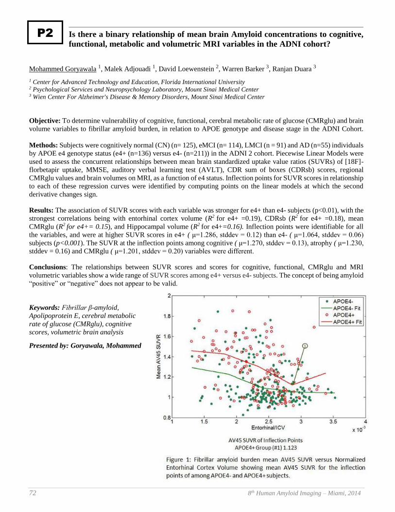
72 8th Human Amyloid Imaging – Miami, 2014
Is there a binary relationship of mean brain Amyloid concentrations to cognitive,
functional, metabolic and volumetric MRI variables in the ADNI cohort?
Mohammed Goryawala 1, Malek Adjouadi 1, David Loewenstein 2, Warren Barker 3, Ranjan Duara 3
1 Center for Advanced Technology and Education, Florida International University 2 Psychological Services and Neuropsychology Laboratory, Mount Sinai Medical Center 3 Wien Center For Alzheimer's Disease & Memory Disorders, Mount Sinai Medical Center
Objective: To determine vulnerability of cognitive, functional, cerebral metabolic rate of glucose (CMRglu) and brain
volume variables to fibrillar amyloid burden, in relation to APOE genotype and disease stage in the ADNI Cohort.
Methods: Subjects were cognitively normal (CN) (n= 125), eMCI (n= 114), LMCI (n = 91) and AD (n=55) individuals
by APOE e4 genotype status (e4+ (n=136) versus e4- (n=211)) in the ADNI 2 cohort. Piecewise Linear Models were
used to assess the concurrent relationships between mean brain standardized uptake value ratios (SUVRs) of [18F]-
florbetapir uptake, MMSE, auditory verbal learning test (AVLT), CDR sum of boxes (CDRsb) scores, regional
CMRglu values and brain volumes on MRI, as a function of e4 status. Inflection points for SUVR scores in relationship
to each of these regression curves were identified by computing points on the linear models at which the second
derivative changes sign.
Results: The association of SUVR scores with each variable was stronger for e4+ than e4- subjects (p<0.01), with the
strongest correlations being with entorhinal cortex volume (R2 for e4+ =0.19), CDRsb (R2 for e4+ =0.18), mean
CMRglu (R2 for e4+= 0.15), and Hippocampal volume (R2 for e4+=0.16). Inflection points were identifiable for all
the variables, and were at higher SUVR scores in e4+ ( μ=1.286, stddev = 0.12) than e4- ( μ=1.064, stddev = 0.06)
subjects (p<0.001). The SUVR at the inflection points among cognitive ( μ=1.270, stddev = 0.13), atrophy ( μ=1.230,
stddev = 0.16) and CMRglu ( μ=1.201, stddev = 0.20) variables were different.
Conclusions: The relationships between SUVR scores and scores for cognitive, functional, CMRglu and MRI
volumetric variables show a wide range of SUVR scores among e4+ versus e4- subjects. The concept of being amyloid
“positive” or “negative” does not appear to be valid.
Keywords: Fibrillar β-amyloid,
Apolipoprotein E, cerebral metabolic
rate of glucose (CMRglu), cognitive
scores, volumetric brain analysis
Presented by: Goryawala, Mohammed
P2
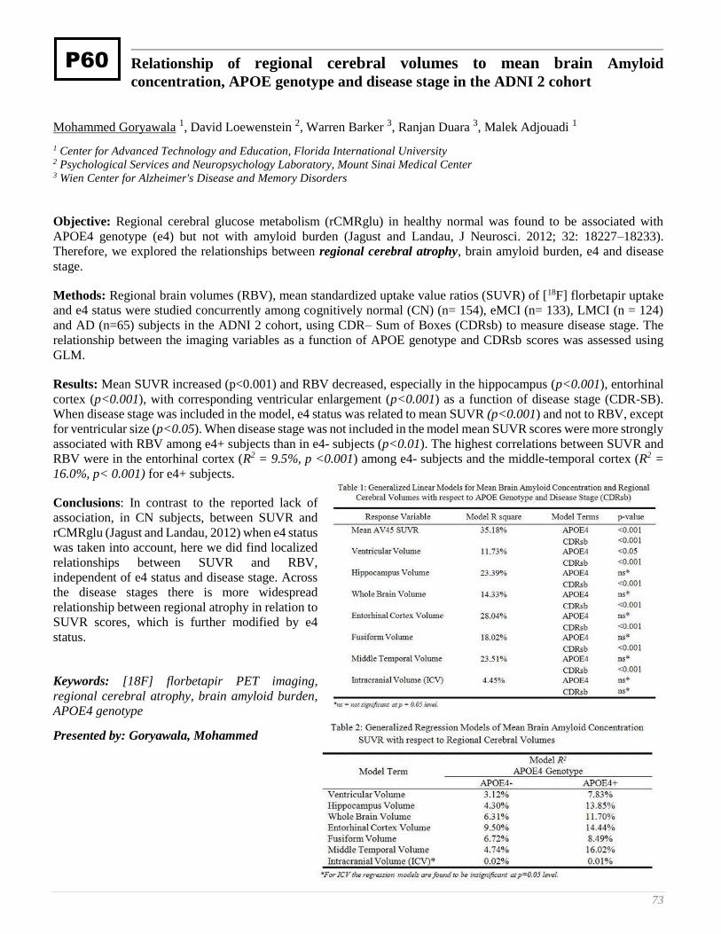
73
Relationship of regional cerebral volumes to mean brain Amyloid
concentration, APOE genotype and disease stage in the ADNI 2 cohort
Mohammed Goryawala 1, David Loewenstein 2, Warren Barker 3, Ranjan Duara 3, Malek Adjouadi 1
1 Center for Advanced Technology and Education, Florida International University 2 Psychological Services and Neuropsychology Laboratory, Mount Sinai Medical Center 3 Wien Center for Alzheimer's Disease and Memory Disorders
Objective: Regional cerebral glucose metabolism (rCMRglu) in healthy normal was found to be associated with
APOE4 genotype (e4) but not with amyloid burden (Jagust and Landau, J Neurosci. 2012; 32: 18227–18233).
Therefore, we explored the relationships between regional cerebral atrophy, brain amyloid burden, e4 and disease
stage.
Methods: Regional brain volumes (RBV), mean standardized uptake value ratios (SUVR) of [18F] florbetapir uptake
and e4 status were studied concurrently among cognitively normal (CN) (n= 154), eMCI (n= 133), LMCI (n = 124)
and AD (n=65) subjects in the ADNI 2 cohort, using CDR– Sum of Boxes (CDRsb) to measure disease stage. The
relationship between the imaging variables as a function of APOE genotype and CDRsb scores was assessed using
GLM.
Results: Mean SUVR increased (p<0.001) and RBV decreased, especially in the hippocampus (p<0.001), entorhinal
cortex (p<0.001), with corresponding ventricular enlargement (p<0.001) as a function of disease stage (CDR-SB).
When disease stage was included in the model, e4 status was related to mean SUVR (p<0.001) and not to RBV, except
for ventricular size (p<0.05). When disease stage was not included in the model mean SUVR scores were more strongly
associated with RBV among e4+ subjects than in e4- subjects (p<0.01). The highest correlations between SUVR and
RBV were in the entorhinal cortex (R2 = 9.5%, p <0.001) among e4- subjects and the middle-temporal cortex (R2 =
16.0%, p< 0.001) for e4+ subjects.
Conclusions: In contrast to the reported lack of
association, in CN subjects, between SUVR and
rCMRglu (Jagust and Landau, 2012) when e4 status
was taken into account, here we did find localized
relationships between SUVR and RBV,
independent of e4 status and disease stage. Across
the disease stages there is more widespread
relationship between regional atrophy in relation to
SUVR scores, which is further modified by e4
status.
Keywords: [18F] florbetapir PET imaging,
regional cerebral atrophy, brain amyloid burden,
APOE4 genotype
Presented by: Goryawala, Mohammed
P60
74 8th Human Amyloid Imaging – Miami, 2014
Genetic resilience to Tau- and Amyloid-related neurodegeneration
Timothy Hohman , Mary Ellen Koran , Tricia Thornton-Wells
Vanderbilt University Medical Center
A subset of individuals present at autopsy with the pathological features of Alzheimer’s disease (AD) having never
manifest the clinical symptoms. Biomarker advances provide the opportunity to investigate such neuroresilience by
leveraging measures of amyloid and tau pathology. We sought to identify genetic variants that modify the relation
between AD biomarkers and neurodegeneration using data from 690 subjects enrolled in the AD Neuroimaging
Initiative (ADNI). We first performed mixed-model regression to fully characterize the relation between the presence
of pathology (none, amyloid only, tau only, both) and longitudinal change in lateral inferior ventricle (LILV) volume.
Next, we used a genome-wide association study interaction analysis to identify genes that modify this relationship
between pathology and atrophy correcting for multiple comparisons. In posthoc analyses we stratified across the
ADNI-1 and ADNI-2/GO cohorts to validate identified effects.
Both the presence of amyloid-pathology alone and the combined presence of amyloid and tau was related to ventricular
dilation. In genetic interaction analyses we identified two SNP-amyloid (rs7849530, rs4866650) interactions that
modified the association between amyloid-positivity (in the absence of tau positivity) and neurodegeneration (Figure
1). These effects were validated across both ADNI cohorts. In each case, the minor allele was associated with increased
ventricular dilation in the presence of amyloid pathology. Interestingly, both of these intergenic SNPs are within 50
KB of SNPs previously implicated in AD. Next, we identified one SNP (rs12261764) that modified the relationship
between tau pathology (irrespective of amyloid positivity) and neurodegeneration (Figure 2). The minor allele of this
SNP was associated with less ventricular dilation (less atrophy) in those with pathology, and more dilation in those
without pathology. However, only the protective effect within tau-positive individuals was validated (Figure 3). These
findings highlight the potential utility of gene-environment interactions in identifying novel genetic factors relevant to
disease risk and resilience.
Keywords: Resilience, MRI, Genetics, Genetic Interaction, CSF
Presented by: Hohman, Timothy
P23

75
Specificity of [3H]T808, [3H]THK-5105 and [3H]AV-45 (florbetapir) binding to
aggregated tau and amyloid plaques in human AD tissue in vitro
Michael Honer , Henner Knust , Luca Gobbi , Dieter Muri , Edilio Borroni
F. Hoffmann-La Roche Ltd., Basel, Switzerland
Objectives: Amyloid-beta (Aβ) plaques and neurofibrillary tangles (NFT) are key histopathological features in
Alzheimer’s disease (AD). Their shared beta sheet secondary structure together with their co-existence in AD brains
poses major challenges for the development of Aβ- or tau-specific radiotracers. Several radioligands for specific
imaging of Aβ plaques and NFTs have recently been published. In this study, the in vitro specificity of [3H]T808,
[3H]THK-5105 and [3H]AV-45 (florbetapir) was tested by refined autoradiographic techniques along with
immunohistofluorescence analysis.
Methods: Radioligand binding to brain tissue sections of early and late stage AD patients as well as tau-overexpressing
Tg4510 mice and Aβ-expressing B6.152H mice was analyzed by macro- and microautoradiographies and co-staining
of Aβ plaques and NFTs on the same tissue section using Aβ- and tau-specific antibodies.
Results: [3H]AV-45 (florbetapir) revealed specific binding to Aβ plaques as present in human AD and Aβ-expressing
transgenic mouse brain tissue. On the contrary, binding of [3H]T808 to AD brain sections was found to be tau-specific,
clearly lacking binding to Aβ plaques in AD tissue but also missing binding to NFTs as present in Tg4510 mice. Under
the experimental conditions used [3H]THK-5105 showed binding to both Aβ plaques and NFTs in human AD brain
and was only positive on Aβ-expressing B6.152H mouse brain sections.
Conclusions: Co-localization studies of radioligand binding and antibody staining on the same tissue section provide
a powerful technique to investigate the specificity of tracer binding to human AD tissue on the macroscopic and
microscopic level while the use of transgenic mouse models for specificity assessment of novel radiotracers has to be
challenged.
Keywords: PET, autoradiography, T808, AV-45, THK-5105
Presented by: Honer, Michael
P21

76 8th Human Amyloid Imaging – Miami, 2014
Arterial stiffness is associated with amyloid accumulation over two years in very
elderly adults
Timothy Hughes 1, Lewis Kuller 2, Emma Barinas-Mitchell 2, Eric McDade 3, William Klunk 4, Ann Cohen 4, Chester
Mathis 5, Steven DeKosky 6, Julie Price 5, Oscar Lopez 3
1 Department of Internal Medicine, Wake Forest School of Medicine, Wake Forest University. 2 Department of Epidemiology, Graduate School of Public Health, University of Pittsburgh 3 Department of Neurology, School of Medicine, University of Pittsburgh 4 Department of Psychiatry, School of Medicine, University of Pittsburgh 5 Department of Radiology, School of Medicine, University of Pittsburgh 6 Office of the Dean and Department of Neurology, School of Medicine, University of Virginia
Importance: Recent studies show that cerebral amyloid deposition is associated with blood pressure and measures of
arterial stiffness in non-demented subjects.
Objective: To examine the association of measures of arterial stiffness and change in amyloid deposition over time.
Methods: In this longitudinal study of brain aging, 91 non-demented subjects age 83+ participating of Program Project
Grant at the University of Pittsburgh were examined baseline and 81 returned for follow-up imaging. Changes in β-
amyloid (Aβ) deposition were determined by positron emission tomography (PET) using the Pittsburgh compound B
twice two years apart. Aβ-positivity was assessed using the sparse K-means approach and a standardized uptake value
ratio of 1.64 over 6 regions. Arterial stiffness was assessed by pulse wave velocity (PWV) using a noninvasive and
automated waveform analyzer at the time closest to the second PET scan. All measures were performed under
standardized conditions. PWV was measured in the central (carotid-femoral (cfPWV) and heart femoral (hfPWV)),
peripheral (femoral-ankle (faPWV)), and mixed (brachial-ankle (baPWV)) vascular beds.
Results: The proportion of Aβ-positive subjects increased from 45% at baseline to 75% at follow-up. Systemic arterial
stiffness as measured by baPWV was significantly higher among Aβ-positive participants at baseline and follow-up.
Each standard deviation increase in central stiffness (cfPWV (p=0.004) and hfPWV (p=0.018)) was associated with
increases in Aβ deposition over time.
Conclusions: This study showed that amyloid deposition increases with age in non-demented individuals, and that
arterial stiffness is strongly associated with the progressive deposition of Aβ amyloid in the brain, especially in this
age group. The observed changes in Aβ deposition over time were associated with generalized arterial stiffness
indicating that there is an active relationship between these two processes.
Keywords: arterial stiffness, amyloid deposition, aging, hypertension
Presented by: Hughes, Timothy
P9
77
Estimation of the number of physicians specializing in treating adult dementia
patients in the US: Applying appropriate use criteria to medicare claims data
Noam Y. Kirson 1, Urvi Desai 1, Alice Kate G. Cummings 1, Howard G. Birnbaum 1, Craig A. Hunter 2
1 Analysis Group, Inc., Boston, MA 2 Eli Lilly and Company, Indianapolis, IN
Background: Experts have developed rigorous appropriate use criteria (AUC) for the use of beta amyloid PET
imaging. In part, these AUC emphasize that beta amyloid imaging should only be utilized by “dementia experts” who
have extensive and documented expertise in diagnosis and management of cognitively-impaired patients. The number
of physicians meeting the AUC is unknown.
Methods: Using de-identified administrative claims data for Medicare beneficiaries (5% random sample), physicians
were defined as dementia experts if they met the following criteria: 1) physician specialty of psychiatry, neurology or
geriatric medicine; and 2) ≥25% of their medical claims in 2011 were associated with patients diagnosed with dementia
(ICD-9 CM: 290.x, 294.1, 331.x). Three alternative definitions were used to estimate the share of claims associated
with dementia patients, ranging from inclusion of only claims directly associated with a dementia diagnosis code to all
claims associated with patients ever diagnosed with dementia. The number of dementia specialists in the US was
extrapolated from the sample using national estimates of practicing physicians. Board certification information was
unavailable.
Results: Less than 19,000 physicians in the Medicare database met AUC (~50% of all identified
psychiatrists/neurologists/geriatricians), with that number reaching as low as 2,740 using the most restrictive definition
(~8%). The corresponding extrapolation to the national level ranges from ~28,700 to ~4,400. Using the broader
definitions, psychiatrists (~45%) and neurologists (~47%) accounted for most specialists identified, with geriatricians
(~8%) representing a much smaller proportion.
Conclusion: This analysis suggests there is approximately one dementia expert for every 1,700–18,000 Medicare
beneficiaries. Nearly 5.2 million Medicare beneficiaries are already diagnosed with Alzheimer’s disease (AD), a
number projected to increase to 7.7 million by 2030. Given this, further agreement on how to operationalize the AUC
within Medicare could be valuable to patient management and future research.
Keywords: AUC, Medicare, dementia specialist
Presented by: Hunter, Craig A.
P61
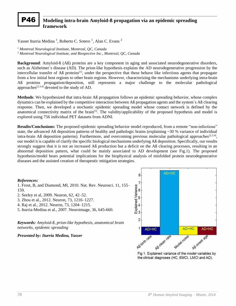
78 8th Human Amyloid Imaging – Miami, 2014
Modeling intra-brain Amyloid-ß propagation via an epidemic spreading
framework
Yasser Iturria Medina 1, Roberto C. Sotero 1, Alan C. Evans 2
1 Montreal Neurological Institute, Montreal, QC, Canada 2 Montreal Neurological Institute, and Biospective Inc., Montreal, QC, Canada
Background: Amyloid-ß (Aß) proteins are a key component in aging and associated neurodegenerative disorders,
such as Alzheimer´s disease (AD). The prion-like hypothesis explains the AD neurodegenerative progression by the
intercellular transfer of Aß proteins[1], under the perspective that these behave like infectious agents that propagate
from a few initial host regions to other brain regions. However, characterizing the mechanisms underlying intra-brain
Aß proteins propagation/deposition, still represents a major challenge to the molecular pathological
approaches[2,3,4] devoted to the study of AD.
Methods: We hypothesized that intra-brain Aß propagation follows an epidemic spreading behavior, whose complex
dynamics can be explained by the competitive interaction between Aß propagation agents and the system´s Aß clearing
response. Then, we developed a stochastic epidemic spreading model whose contact network is defined by the
anatomical connectivity matrix of the brain[5]. The validity/applicability of the proposed hypothesis and model is
explored using 756 individual PET datasets from ADNI.
Results/Conclusions: The proposed epidemic spreading behavior model reproduced, from a remote “non-infectious”
state, the advanced Aß deposition patterns of healthy and pathologic brains (explaining ~30 % variance of individual
intra-brain Aß deposition patterns). Furthermore, and overcoming previous molecular pathological approaches[2,3,4],
our model it is capable of clarify the specific biological mechanisms underlying Aß deposition. Specifically, our results
strongly suggest that it is not an increased Aß production but a deficit on the Aß clearing processes, resulting in an
abnormal deposition pattern, what could be mainly associated to AD development (see Fig.1). The proposed
hypothesis/model bears potential implications for the biophysical analysis of misfolded protein neurodegenerative
diseases and the assisted creation of therapeutic mitigation strategies.
References: 1. Frost, B, and Diamond, MI, 2010. Nat. Rev. Neurosci. 11, 155–
159.
2. Seeley et al, 2009. Neuron, 62, 42–52.
3. Zhou et al., 2012. Neuron, 73, 1216–1227.
4. Raj et al., 2012. Neuron, 73, 1204–1215.
5. Iturria-Medina et al., 2007. Neuroimage, 36, 645-660.
Keywords: Amyloid-ß, prion-like hypothesis, anatomical brain
networks, epidemic spreading
Presented by: Iturria Medina, Yasser
P46

79
The potential of florbetapir F 18 and an early scan PET protocol (0-20 minutes
after injection) to evaluate for the presence of amyloid
Abhinay D. Joshi 1, Michael D. Devous Sr. 1, Michael J. Pontecorvo 1, Michael Navitsky 1, Ian Kennedy 1, Mark A.
Mintun 1, Daniel M. Skovronsky 1, and Alzheimer's Disease NeuroImaging Intiative 2
1 Avid Radiopharmaceuticals Inc. a wholly owned subsidiary of Eli Lilly and Company 2 ADNI
Background: Florbetapir quantification typically involves calculation of cortical SUVr values from images acquired
50-60 min after injection. We investigated an empirical method to evaluate amyloid binding of florbetapir using an
acquisition only 0-20 min after injection.
Methods: Using florbetapir PET data from early phase development, an empirical approach was developed that used
the dynamic 0-20 min to calculate a florbetapir amyloid binding estimate (FABE). We then evaluated the performance
of this method with data from the ADNI2 database for 70 subjects diagnosed with Alzheimers Disease (n=6), early or
late mild cognitive impairment (n=15), subjective memory complaints (n=31) and cognitively normal (n=18). Subjects
had a dynamic PET acquisition from 0-20min (4x15sec, 4x30sec, 3x60sec, 3x120sec, 2x240sec) after injection of 370
MBq of florbetapir. A 10 min acquisition at 50 min after injection was also available for evaluation and was used to
calculate standard SUVr values as ratio of cortex to entire cerebellum. The 0-20 min dynamic data was processed to
create the FABE using Equation 1. Performance of FABE in classifying cases relative to SUVr was evaluated using
ROC analysis and Kappa statistics. A correlation analysis was done as well.
Results: The range of FABE values was -3733 to 11697. Correlation between SUVr and FABE was r=0.79 (p<0.001).
A pre-specified threshold (SUVr>1.10) was used to define florbetapir positivity. The optimal threshold for FABE using
the ROC curve and Youden index was determined as FABE=210. With this threshold, 96% (67/70) of cases were
classified into the same category (positive or negative) as with the standard SUVR method and the kappa between the
two measures was 0.91 (95% CI: 0.80 to 1.00).
Conclusion: An empirical method of processing florbetapir PET data from 0-20 min after injection appears to correlate
with conventional calculations of SUVr and agree with SUVr threshold-determined positive and negative
classifications.
Keywords: Florbetapir, AV45, PET, AD, early scan
Presented by: Joshi, Abhinay D.
P63
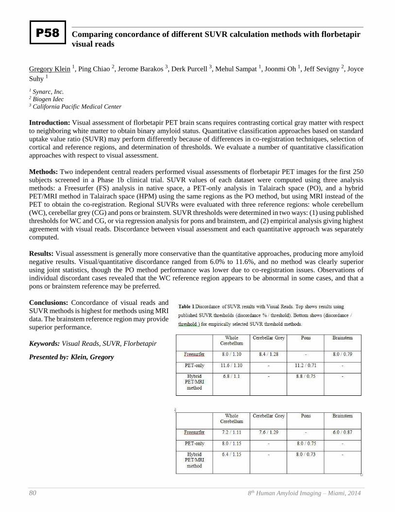
80 8th Human Amyloid Imaging – Miami, 2014
Comparing concordance of different SUVR calculation methods with florbetapir
visual reads
Gregory Klein 1, Ping Chiao 2, Jerome Barakos 3, Derk Purcell 3, Mehul Sampat 1, Joonmi Oh 1, Jeff Sevigny 2, Joyce
Suhy 1
1 Synarc, Inc. 2 Biogen Idec 3 California Pacific Medical Center
Introduction: Visual assessment of florbetapir PET brain scans requires contrasting cortical gray matter with respect
to neighboring white matter to obtain binary amyloid status. Quantitative classification approaches based on standard
uptake value ratio (SUVR) may perform differently because of differences in co-registration techniques, selection of
cortical and reference regions, and determination of thresholds. We evaluate a number of quantitative classification
approaches with respect to visual assessment.
Methods: Two independent central readers performed visual assessments of florbetapir PET images for the first 250
subjects screened in a Phase 1b clinical trial. SUVR values of each dataset were computed using three analysis
methods: a Freesurfer (FS) analysis in native space, a PET-only analysis in Talairach space (PO), and a hybrid
PET/MRI method in Talairach space (HPM) using the same regions as the PO method, but using MRI instead of the
PET to obtain the co-registration. Regional SUVRs were evaluated with three reference regions: whole cerebellum
(WC), cerebellar grey (CG) and pons or brainstem. SUVR thresholds were determined in two ways: (1) using published
thresholds for WC and CG, or via regression analysis for pons and brainstem, and (2) empirical analysis giving highest
agreement with visual reads. Discordance between visual assessment and each quantitative approach was separately
computed.
Results: Visual assessment is generally more conservative than the quantitative approaches, producing more amyloid
negative results. Visual/quantitative discordance ranged from 6.0% to 11.6%, and no method was clearly superior
using joint statistics, though the PO method performance was lower due to co-registration issues. Observations of
individual discordant cases revealed that the WC reference region appears to be abnormal in some cases, and that a
pons or brainstem reference may be preferred.
Conclusions: Concordance of visual reads and
SUVR methods is highest for methods using MRI
data. The brainstem reference region may provide
superior performance.
Keywords: Visual Reads, SUVR, Florbetapir
Presented by: Klein, Gregory
P58
81
Early age of onset is associated with greater neuroinflammation in Alzheimer’s
disease
William Charles Kreisl 1, Chul Hyoung Lyoo 1, Meghan McGwier 1, Joseph Snow 2, Kimberly Jenko 1, Winston
Corona 3, Cheryl Morse 1, Sami Zoghbi 1, Victor Pike 1, Francis McMahon 3, R. Scott Turner 4, Robert Innis 1
1 Molecular Imaging Branch, National Institute of Mental Health 2 Office of the Clinical Director, National Institute of Mental Health 3 Human Genetics Branch, National Institute of Mental Health 4 Memory Disorders Program, Georgetown University
Background: Individuals with early-onset Alzheimer’s disease (EOAD) demonstrate greater neurodegeneration than
late-onset AD (LOAD) patients of equal clinical severity. PET imaging studies show no difference in Pittsburgh
Compound B (PIB) binding between EOAD and LOAD patients, suggesting that factors other than fibrillar amyloid
contribute to the more aggressive course seen with EOAD. Our recent PET study found that EOAD patients have
greater binding with [11C]PBR28 - a radioligand for the inflammatory biomarker translocator protein (TSPO).
However, in this study patients were not matched for clinical severity.
Methods: PIB-positive EOAD (n = 20, age of onset = 56 ± 5 years) and LOAD (n = 8, age of onset = 72 ± 6 years)
patients were matched by clinical severity. [11C]PBR28 binding was compared using univariate ANOVA with TSPO
genotype as a fixed factor. Regional brain volumes were determined using Freesurfer.
Results: EOAD patients had greater [11C]PBR28 binding than LOAD patients in prefrontal cortex, superior and
inferior parietal lobule, precuneus, occipital cortex, and temporal cortex (p < 0.05). [11C]PBR28 binding inversely
correlated with age-of-onset in superior and inferior parietal lobule and precuneus (r > - 0.39, p < 0.05). EOAD patients
had greater atrophy than LOAD patients in the superior and inferior parietal lobule (p < 0.05). Groups did not differ in
Mini Mental State Exam score or Clinical Dementia Rating Scale score. However, EOAD patients had worse
performance on Block Design than LOAD patients (raw score 11.1 ± 11 vs. 25.8 ± 14, p = 0.02).
Conclusion: Among AD patients, earlier age-of-onset is associated with greater focal neuroinflammation. This
relationship is strongest in the parietal cortex where EOAD patients also reveal greater atrophy and worse performance
on functionally-related cognitive tasks. A more robust neuroinflammatory response to upstream pathological events
may explain the more aggressive clinical course of EOAD.
Keywords: Early onset Alzheimer's disease, inflammation
Presented by: Kreisl, William Charles
P67
82 8th Human Amyloid Imaging – Miami, 2014
The association between glucose metabolism in Alzheimer’s disease-vulnerable
regions and cognitive reserve is modified by amyloid status within clinically
normal individuals
Molly LaPoint 1, Elizabeth Mormino 1, Rebecca Amariglio 2, Aaron Schultz 1, Trey Hedden 3, J. Alex Becker 4, Keith
Johnson 5, Reisa Sperling 6, Dorene Rentz 2
1 Athinoula A. Martinos Center for Biomedical Imaging, Department of Radiology, and Department of Neurology,
Massachusetts General Hospital, Harvard Medical School, Boston, MA 2 Center for Alzheimer Research and Treatment, Department of Neurology, Brigham and Women’s Hospital, and Department of
Neurology, Massachusetts General Hospital, Harvard Medical School, Boston, MA 3 Athinoula A. Martinos Center for Biomedical Imaging, Department of Radiology, Massachusetts General Hospital, Harvard
Medical School, Boston, MA 4 Department of Radiology, Massachusetts General Hospital, Harvard Medical School, Boston, MA 5 Departments of Radiology and Neurology, Massachusetts General Hospital, Harvard Medical School, Boston, MA 6 Center for Alzheimer Research and Treatment, Department of Neurology, Brigham and Women’s Hospital, Athinoula A.
Martinos Center for Biomedical Imaging, and Department of Neurology, Massachusetts General Hospital, Harvard Medical
School, Boston, MA
Objective: To investigate the association between cognitive reserve (CR) and glucose metabolism in clinically normal
(CN) individuals with high and low beta-amyloid (Aβ) deposition.
Methods: We studied 162 CN subjects from the Harvard Aging Brain study. All completed PIB-PET and FDG-PET.
A CR factor score was created using a confirmatory factor analysis that included years of education, occupation score
(adapted from the Hollingshead Two-Factor Index of Social Position), average lifetime cognitive activity (Cognitive
Activities Scale), and premorbid verbal IQ (AMNART). We tested the interaction between the CR factor score and Aβ
status on patterns of glucose metabolism in temporoparietal regions previously shown to be vulnerable in Alzheimer’s
disease (AD). Age, gender, and total gray matter were used as covariates.
Results: There was no difference in CR between the Aβ+ and Aβ- subjects. Overall, Aβ+ subjects showed lower FDG
metabolism than Aβ- subjects. The interaction between Aβ status and the CR factor score in predicting FDG-PET was
significant (p=0.006). Further examination revealed that within Aβ- individuals, higher CR was associated
with higher FDG, whereas within Aβ+ individuals, higher CR was associated with lower FDG.
Conclusion: This pattern suggests that high CR may modify the relationship between Aβ and glucose metabolism in
AD-vulnerable regions, such that CN individuals who are Aβ+ yet have high CR exhibit lower FDG-PET, possibly
indicating an ability to maintain clinical function despite greater AD pathology. It is also possible that Aβ+ individuals
with low CR and low glucose metabolism were excluded from the study because they are more likely to progress to
clinical impairment sooner than their high-CR counterparts.
Keywords: FDG, PIB, cognitive reserve
Presented by: LaPoint, Molly
P31

83
Quantification and accuracy of clinical [11C]-PiB PET/MRI: The effect of MR-
based attenuation correction
Ian Law 1, Flemming L. Andersen 1, Adam E. Hansen 1, Steen G. Hasselbalch 2, Claes Ladefoged 1, Sune H Keller 1,
Søren Holm 1, Lotte Højgaard 1
1 The Department of Clinical Physiology, Nuclear Medicine and PET, Rigshospitalet, Denmark. 2 Memory Disorders Research Group,Rigshospitalet, Denmark.
Aims: The Dixon-Water-Fat segmentation (DWFS) method is a standard attenuation correction (AC) method in
PET/MRI and has demonstrated a systematic and spatially variable quantitative bias in the brain compared to PET/CT.
The aim of this study was to measure the regional and absolute bias of DWFS-AC in combined PET/MR images of
the brain using [11C]-PiB and evaluate the impact on the clinical reading of these images.
Methods: Twenty-eight healthy volunteers, 6
MCI, 4 AD, and 6 clinical dementia cases (26 M,
18 F, median age: 68 y (51-78 y)) underwent a
simultaneous PET/MRI (Siemens mMR)
acquisition 40 min pi of (170-709) MBq [11C]-
PiB. A single 30 min frame was reconstructed
twice (OSEM-3D, 21it4sub, 5 mm Gauss) with the
only difference being the AC calculation. AC was
performed using either DWFS or a head CT scan
acquired independently on the same day. Activity
concentration was sampled on AC-PET from
symmetrically delineated ROI’s in cortical areas,
deep white matter, and the caudate nuclei. Ratios
of region-to-cerebellar grey matter (SUVr) were
calculated. Bland-Altman analysis was used to
compare AC methods.
Results: Using CT-AC as the gold standard compared to DWFS-AC the average activity concentration (Bq/mL) in all
ROI’s was biased by -18 % (-7 to -34 %), and the SUVr's were biased by
+0.11 in the caudate nuclei and +0.04 in lateral cortical ROI’s. Although
the visual categorization of both, amyloid-positive (n=11) and -negative
(n=33) scans was unaffected by DWFS-AC, 2 healthy subjects were
quantitatively reclassified as amyloid positive.
Conclusion: A visual and quantitative evaluation of MR-AC using DWFS
in the brain with [11C]-PiB show a noticeable radially variable bias.
Although robust on visual reading, the quantitative diagnostic criteria
(SUVr) using this biomarker with DWFS-AC may need to be modified.
Keywords: PiB, Attenuation correction, PET/MRI,
Presented by: Law, Ian
P48

84 8th Human Amyloid Imaging – Miami, 2014
Relationship between Amyloid burden and clinical course across AD
pathophysiologic stages
Richard Margolin 1, Jianing Di 2, Randolph Andrews 3, Steven Salloway 4, Reisa Sperling 5, Leslie Shaw 6, HR
Brashear 2, Enchi Liu 2, Mark Schmidt 2, Dawn Matthews 3
1 CereSpir, Inc. 2 Janssen Pharmaceuticals R&D 3 ADM Diagnostics LLC 4 Butler Hospital, The Warren Alpert Medical School of Brown University 5 Departments of Neurology and Radiology, Massachusetts General Hospital, Harvard Medical School; Center for Alzheimer
Research and Treatment, Department of Neurology, Brigham and Women’s Hospital, Harvard Medical School 6 Department of Pathology and Laboratory Medicine, Institute on Aging, Center for Neurodegenerative Disease Research,
University of Pennsylvania Medical School
Background. Amyloid PET is used to assess fibrillar Aβ burden across AD stages and employed in therapeutic trials.
Using SUVRs, individuals are commonly divided into amyloid-positive (amy+) and negative (amy-) groups. In two
Phase 3 trials of experimental treatments in AD dementia, differences in clinical characteristics between amy+/- groups
were identified in placebo-treated subjects: amy- subjects generally scored better at baseline and declined less than
amy+ subjects. The same pattern was observed for demented and MCI subjects in an observational study (ADNI) and
was more robust in MCI. Longitudinal divergence between amy+/- groups was suggested on some instruments. The
present analysis extends previous work to assess the pattern in subtypes of MCI (EMCI/LMCI) and earlier stage
disease.
Methods. We examined initial and longitudinal performance in ADNI subjects with >1 amyloid PET scan, using
available ADAScog/11, MMSE, RAVLT, CDR-SB and FAQ data. Subjects in the dementia cohort, the MCI cohort,
and MCI sub-cohorts were categorized as amy+/- based on tracer-specific cutpoints applied to their initial PET scan.
Diagnosis was as recorded at that scan. The effect of APOE4 carrier status was evaluated. Data analysis used an
MMRM including age and baseline score.
Results. Of 153 dementia subjects, 12% were amy-. Of 537 MCI subjects, 44% were amy- (EMCI: 52%, LMCI 34%).
In both demented and MCI cohorts, amy- scored better at baseline and progressed less. Demented amy- scored
significantly better on only ADAScog; MCI amy- scored better on all measures. Similarly, demented amy- declined
less on only ADAScog; MCI amy- scored better on almost all measures.
Conclusions. Amy- subjects perform better at baseline and decline less on cognitive and functional measures at all
clinical stages; the difference seems most robust in MCI. Inclusion of slowly progressive amy- subjects in therapeutic
trials may degrade statistical power and reduce potential study success, especially in early stage disease.
Keywords: amyloid, burden, positive, negative
Presented by: Margolin, Richard
P13
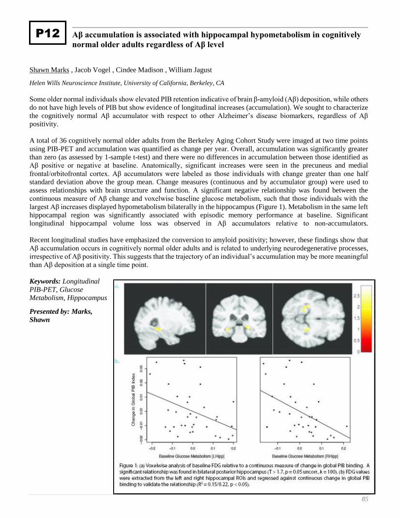
85
Aβ accumulation is associated with hippocampal hypometabolism in cognitively
normal older adults regardless of Aβ level
Shawn Marks , Jacob Vogel , Cindee Madison , William Jagust
Helen Wills Neuroscience Institute, University of California, Berkeley, CA
Some older normal individuals show elevated PIB retention indicative of brain β-amyloid (Aβ) deposition, while others
do not have high levels of PIB but show evidence of longitudinal increases (accumulation). We sought to characterize
the cognitively normal Aβ accumulator with respect to other Alzheimer’s disease biomarkers, regardless of Aβ
positivity.
A total of 36 cognitively normal older adults from the Berkeley Aging Cohort Study were imaged at two time points
using PIB-PET and accumulation was quantified as change per year. Overall, accumulation was significantly greater
than zero (as assessed by 1-sample t-test) and there were no differences in accumulation between those identified as
Aβ positive or negative at baseline. Anatomically, significant increases were seen in the precuneus and medial
frontal/orbitofrontal cortex. Aβ accumulators were labeled as those individuals with change greater than one half
standard deviation above the group mean. Change measures (continuous and by accumulator group) were used to
assess relationships with brain structure and function. A significant negative relationship was found between the
continuous measure of Aβ change and voxelwise baseline glucose metabolism, such that those individuals with the
largest Aβ increases displayed hypometabolism bilaterally in the hippocampus (Figure 1). Metabolism in the same left
hippocampal region was significantly associated with episodic memory performance at baseline. Significant
longitudinal hippocampal volume loss was observed in Aβ accumulators relative to non-accumulators.
Recent longitudinal studies have emphasized the conversion to amyloid positivity; however, these findings show that
Aβ accumulation occurs in cognitively normal older adults and is related to underlying neurodegenerative processes,
irrespective of Aβ positivity. This suggests that the trajectory of an individual’s accumulation may be more meaningful
than Aβ deposition at a single time point.
Keywords: Longitudinal
PIB-PET, Glucose
Metabolism, Hippocampus
Presented by: Marks,
Shawn
P12
86 8th Human Amyloid Imaging – Miami, 2014
Is there an asymmetric distribution of in vivo Amyloid in primary progressive
aphasia?
Adam Martersteck 1, Christopher Murphy 2, Christina Wieneke 1, Kewei Chen 3, Ji Luo 3, Pradeep Thiyyagura 3, M.-
Marsel Mesulam 4, Emily Rogalski 1
1 Cognitive Neurology and Alzheimer’s Disease Center, Northwestern University (NU) Feinberg School of Medicine, Chicago,
IL, USA 2 The University of Arizona College of Medicine, Phoenix, AZ, USA 3 Banner Alzheimer's Institute, Phoenix, AZ, USA 4 Cognitive Neurology and Alzheimer’s Disease Center, Northwestern University (NU) Feinberg School of Medicine, Chicago,
IL, USA NU Feinberg School of Medicine, Department of Neurology, Chicago, IL, USA
Background: Primary progressive aphasia (PPA) is a clinical dementia syndrome caused by neurodegenerative disease
often characterized by asymmetric atrophy in the language-dominant (usually left) hemisphere. The most common
neuropathologies found in PPA are Alzheimer’s disease (AD; ~40%) or a form of frontotemporal lobar degeneration
(~60%). PPA patients with postmortem AD show a leftward predominance of neurofibrillary tangles, but not beta-
amyloid plaques in neocortical areas compared to the symmetric pathology of patients with dementia of the
Alzheimer’s type. This study will determine whether in vivo amyloid imaging shows a clinically concordant
asymmetric pattern in a well-characterized cohort of amyloid positive PPA patients.
Methods: Twenty-one PPA subjects with florbetapir imaging were screened for amyloid positivity by calculating
cerebral-to-cerebellar standard uptake value ratios (SUVRs) using a gray-matter mask consisting of six bilateral
volumes of interest (VOIs) in the frontal, temporal, parietal, anterior cingulate, posterior cingulate, and precuneus.
Amyloid positivity was defined as SUVR ≥ 1.10. Laterality scores were examined for each VOI pair to determine if
there was a leftward asymmetry among the amyloid positive subjects.
Results: Thirteen subjects were amyloid positive. Their mean age was 68.7 (± 5.7) years and they had an average
WAB-AQ of 82.0 (± 10.7). Overall florbetapir retention only showed significant leftward asymmetry for the parietal
and anterior cingulate VOIs (p<0.007 after Bonferroni correction).
Conclusions: These results show that amyloid imaging can show clinically concordant asymmetric distributions in
PPA. The marked asymmetry of amyloid in the parietal lobe is consistent with cortical atrophy patterns seen in the
early stages of PPA. Previous studies of PPA using PiB have rarely reported lateralized PET amyloid uptake. No
quantitative florbetapir imaging studies have been reported in PPA. It will be important to determine if disease stage,
atrophy, amyloid agent, and other factors influence the estimated distribution of amyloid in vivo.
Keywords: dementia, Primary Progressive Aphasia, laterality, florbetapir
Presented by: Martersteck, Adam
P18
87
CogState computerized testing and neurodegenerative and amyloid imaging:
Implications for secondary preventive trials
Michelle Mielke , Heather Wiste , Stephen Weigand , Prashanthi Vemuri , Val Lowe , Mary Machulda , Rosebud
Roberts , Kejal Kantarci , David Knopman , Bradly Boeve , Clifford Jack , Petersen Ronald
Mayo Clinic, Rochester, MN
Background: Inexpensive, non-invasive screening tools of brain amyloidosis and neurodegeneration are needed for
secondary preventive Alzheimer’s disease (AD) trials enrolling cognitively normal (CN) individuals. We examined
the association between performance on the CogState computerized cognitive battery and AD neuroimaging measures
and compared it to the association between standard neuropsychological tests and neuroimaging measures.
Methods: We included CN Mayo Clinic Study of Aging (MCSA) participants, aged 51-70, who had undergone MRI
(n=324), FDG PET (n=259), and/or PiB PET (n=261), the CogState computerized assessment, and standard cognitive
battery within 6 months. CogState tests included: Detection (DET) – simple reaction time; Identification (IDN) –
choice reaction time; One Card Learning (OCL) – visual learning; One Back test (ONB) – working memory/attention;
and the Groton Maze Learning Test (GMLT) – spatial working memory. Linear regression assessed the relationship
between each test and continuous measures of PiB PET SUVR, hippocampal atrophy, and FDG PET metabolism in
an “AD signature region” (both partial volume corrected and non-corrected), controlling for age, sex, and education.
We also examined the relationship between the neuroimaging measures and domain-specific z-scores (memory,
language, executive function, visuospatial) using the MCSA standard neuropsychological battery.
Results: A slower reaction time for CogState IDN (p=0.04) and ONB (p=0.006), and lower memory (p=0.001) and
attention (p=0.03) z-scores, were associated with FDG hypometabolism without partial volume correction. Fewer
moves per second on the GMLT (p=0.001) and lower OCL accuracy (p=0.02) were associated with smaller
hippocampal volumes. No CogState tests or cognitive z-scores were associated with PIB at p<0.05.
Conclusion: Results suggest this computerized assessment may be a useful indicator of neurodegenerative pathology
among CN individuals. Notably, associations between standard cognitive testing and neuroimaging were not much
different than that between CogState and neuroimaging. The brief CogState tests might serve an equivalent function
as traditional cognitive testing for clinical trials in CN subjects.
Presented by: Mielke, Michelle
P52
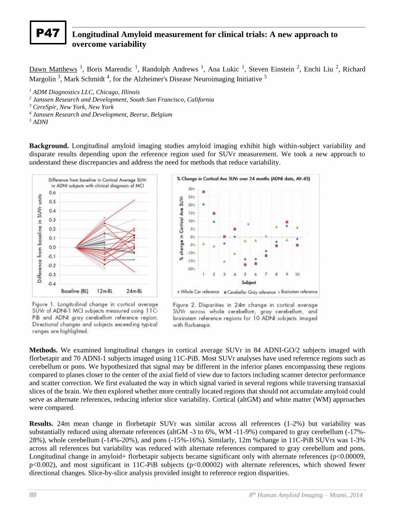
88 8th Human Amyloid Imaging – Miami, 2014
Longitudinal Amyloid measurement for clinical trials: A new approach to
overcome variability
Dawn Matthews 1, Boris Marendic 1, Randolph Andrews 1, Ana Lukic 1, Steven Einstein 2, Enchi Liu 2, Richard
Margolin 3, Mark Schmidt 4, for the Alzheimer's Disease Neuroimaging Initiative 5
1 ADM Diagnostics LLC, Chicago, Illinois 2 Janssen Research and Development, South San Francisco, California 3 CereSpir, New York, New York 4 Janssen Research and Development, Beerse, Belgium 5 ADNI
Background. Longitudinal amyloid imaging studies amyloid imaging exhibit high within-subject variability and
disparate results depending upon the reference region used for SUVr measurement. We took a new approach to
understand these discrepancies and address the need for methods that reduce variability.
Methods. We examined longitudinal changes in cortical average SUVr in 84 ADNI-GO/2 subjects imaged with
florbetapir and 70 ADNI-1 subjects imaged using 11C-PiB. Most SUVr analyses have used reference regions such as
cerebellum or pons. We hypothesized that signal may be different in the inferior planes encompassing these regions
compared to planes closer to the center of the axial field of view due to factors including scanner detector performance
and scatter correction. We first evaluated the way in which signal varied in several regions while traversing transaxial
slices of the brain. We then explored whether more centrally located regions that should not accumulate amyloid could
serve as alternate references, reducing inferior slice variability. Cortical (altGM) and white matter (WM) approaches
were compared.
Results. 24m mean change in florbetapir SUVr was similar across all references (1-2%) but variability was
substantially reduced using alternate references (altGM -3 to 6%, WM -11-9%) compared to gray cerebellum (-17%-
28%), whole cerebellum (-14%-20%), and pons (-15%-16%). Similarly, 12m %change in 11C-PiB SUVrs was 1-3%
across all references but variability was reduced with alternate references compared to gray cerebellum and pons.
Longitudinal change in amyloid+ florbetapir subjects became significant only with alternate references (p<0.00009,
p<0.002), and most significant in 11C-PiB subjects (p<0.00002) with alternate references, which showed fewer
directional changes. Slice-by-slice analysis provided insight to reference region disparities.
P47

89
Conclusions. Our analyses suggest transaxial fluctuation in longitudinal PET signal changes arising from factors likely
related to the scanner rather than amyloid. Our alternate reference region approach helps to address acquisition-related
variability, benefitting measurement accuracy and statistical power.
Keywords: imaging, florbetapir, variability, longitudinal, trials
Presented by: Matthews, Dawn
90 8th Human Amyloid Imaging – Miami, 2014
Prediction of Amyloid-beta hepatic clearance using sandwich cultured primary
rat hepatocytes
Loqman Mohamed , Amal Kaddoumi
Department of Basic Pharmaceutical Science, College of Pharmacy, University of Louisiana at Monroe, Monroe, Louisiana
Failure in amyloid beta (Aβ) systemic clearance across the liver has been suggested to play a role in Aβ brain
accumulation and thus contributes largely to the pathology of Alzheimer’s disease (AD). The purpose of this study
was to in vitro characterize the transport mechanisms of Aβ40 across the liver using sandwich cultured primary rat
hepatocytes (SCHs) and determine its biliary clearance (Clbile) and biliary excretion index (BEI%). 125I-Aβ40 BEI% was
time dependent and reached steady state at 30min with an average value of 29.8% and Clbile of 1.47ml/min/kg of body
weight. The role of low density lipoprotein-receptor related protein-1 (LRP1) in mediating the basolateral uptake
of 125I-Aβ40 in SCHs was assessed using receptor-associated protein (RAP, 2µM). Significant reduction in 125I-
Aβ40 BEI% and Clbile with RAP was observed, demonstrating a major contribution of LRP1 in mediating hepatic uptake
of intact 125I-Aβ40 via transcytosis. Furthermore, activity studies suggested lower role of receptor for advanced
glycation end products (RAGE) in 125I-Aβ40 hepatic uptake. Verapamil (50µM) and valspodar (20µM) significantly
reduced 125I-Aβ40 BEI% indicating a role for P-glycoprotein (P-gp) in the biliary excretion of 125I-Aβ40 in SCHs. LRP1
and P-gp mediated 125I-Aβ40 biliary excretion was inducible and increased BEI% by 26% following rifampicin
pretreatment. In conclusion, our findings demonstrated that beside LRP1, P-gp and to a lesser extent RAGE are
involved in 125I-Aβ40 hepatobiliary disposition and support the use of enhancement of 125I-Aβ40 hepatic clearance via
LRP1 and P-gp induction as a novel therapeutic approach for the prevention and treatment of AD.
Keywords: Amyloid-beta, P-glycoprotein, LRP1, hepatic sandwich culture, biliary clearance.
Presented by: Mohamed, Loqman
P16
91
Identifying cost-effective predictive rules of Amyloid-β level by integrating
neuropsychological tests and plasma-based marker
Shuai Huang 1, Amanda Smith 2, Mona Haghighi 3, Brent Small 4, Dave Morgan 2
1 Industrial and Management Systems Engineering & Byrd Alzheimer’s Institute, University of South Florida 2 Byrd Alzheimer’s Institute, University of South Florida 3 Industrial and Management Systems Engineering, University of South Florida 4 School of Aging Studies, University of South Florida
Background: Detecting participants who are positive for Aβ pathology is germane in designing prevention trials by
enriching for those cases that are more likely to be amyloid positive. Existing brain amyloid measurement techniques,
such as the Pib-PET and CSF, are not reasonable first-line approaches limited by either feasibility or cost.
Method: We compared different methods for integrating the Neuropsychological measurements and blood-based
markers to predict the amyloid-β level.
Results: Using the ADNI cohort, we successfully extracted predictive rules of amyloid-β level.
Conclusion: Our study demonstrated that the integration of the Neuropsychological measurements and blood-based
markers significantly improved the prediction accuracy. The prediction model has led to several simple rules, which
have a great potential of being naturally translated into clinical settings such as enrichment screening for Alzheimer’s
prevention trials of anti-amyloid treatments.
Keywords: amyloid prediction, Neuropsychological, plasma
Presented by: Morgan, Dave
P64
92 8th Human Amyloid Imaging – Miami, 2014
Associations between beta-amyloid, markers of neurodegeneration, and cognition
in clinically normal individuals from the Harvard Aging Brain Study
Elizabeth Mormino 1, Rebecca Betensky 2, Trey Hedden 3, Aaron Schultz 1, Rebecca Amariglio 1, Dorene Rentz 1,
Keith Johnson 4, Reisa Sperling 4
1 Department of Neurology, Massachusetts General Hospital, Massachusetts General Hospital, Harvard Medical School,
Boston, Massachusetts 02114 2 Department of Biostatistics, Harvard School of Public Health, Boston, Massachusetts 02115 3 Department of Radiology, Massachusetts General Hospital, Harvard Medical School, Boston, Massachusetts 02114 4 Department of Neurology, Massachusetts General Hospital, Massachusetts General Hospital, Harvard Medical School,
Boston, Massachusetts 02114; Department of Radiology, Massachusetts General Hospital, Harvard Medical School, Boston,
Massachusetts 02114; Cen
Objective: To determine the strength of the association between beta-amyloid (Ab) and neurodegeneration (ND), as
well as the relationship between these factors and cognitive decline in clinically normal subjects (CNs).
Methods: Data was examined from 191 CNs (age=74.5±6.0). Ab status was determined with PIB-PET, and ND was
measured using hippocampus volume and glucose metabolism from Alzheimer’s disease (AD) vulnerable regions. The
associations between Ab and each marker of ND was assessed. Cut-offs for each ND marker were derived based on
an AD sample, and CNs were considered ND+ if below the cut off for either ND marker. CNs were categorized into
preclinical AD stages based on Ab and ND, and longitudinal decline was contrasted across groups (episodic memory
and executive function composite scores, as well as Logical Memory delayed recall; follow-up=1.86±0.70).
Findings: Ab+ CNs showed significantly increased ND compared to Ab- CNs (however, these associations were
small: r-values<0.20, <4% of the variance). Categorizing CNs revealed 47% in preclinical stage 0 (Ab-/ND-), 11%
stage 1 (Ab+/ND-), 15% stage 2 (Ab+/ND+) and 24% SNAP (“Suspected Non-AD pathology”; Ab-/ND+). Stage 2
showed greater decline compared to other groups across multiple measures, whereas no other pair-wise contrast was
significant.
Interpretation: Small associations between Ab and ND suggest that additional factors may contribute to the variance
in markers of ND. It is also possible that the association between Ab and ND is weaken by a survival bias, such that
Ab+/ND+ CNs are more likely to progress to mild cognitive impairment and thus less likely to be included in the CN
sample. Consistent with this speculation, we found that stage 2 was most at risk for decline. Regardless of whether Ab
and ND pathways are linked in the initial phase of AD, the presence of both factors is critical in assessing risk of
decline among CNs.
Keywords: aging, preclinical AD, neurodegeneration, beta-amyloid
Presented by: Mormino, Elizabeth
P14
93
Beta-amyloid has a greater impact on memory in females than males across the
clinical spectrum
Elizabeth Mormino 1, Reisa Sperling 2
1 Department of Neurology, Massachusetts General Hospital, Massachusetts General Hospital, Harvard Medical School,
Boston, Massachusetts 02114 2 Department of Radiology, Massachusetts General Hospital, Harvard Medical School, Boston, Massachusetts 02114; Center
for Alzheimer Research and Treatment, Department of Neurology, Brigham and Women’s Hospital, Harvard Medical School,
Boston, Massachusetts
Objective: Although Alzheimer’s disease (AD) is more prevalent in females, recent biomarker studies of AD
development have not addressed mechanisms that may underlie this risk. We sought to determine whether female’s
heightened risk may be related to 1) a greater propensity to accumulate Aβ and/or 2) greater cognitive impairment for
a given level of Aβ.
Methods: We analyzed data for 639 clinically normal (CN), 539 mild cognitive impairment (MCI), and 175 AD
patients, collapsed across HABS, ADNI and AIBL. Subjects were included if they had amyloid imaging
(HABS/AIBL=PIB; ADNI=AV45) and neuropsychological (Logical Memory delayed recall) data available. Logistic
regression was used to assess whether gender was associated with Aβ positivity. Multiple regression models were
conducted to determine the association between Aβ status and memory, and how this association may vary by gender.
All analyses were conducted separately within each diagnosis, controlling for cohort, age,APOE4 and education.
Results: We did not find evidence for a greater proportion of Aβ+ female subjects compared to males. Examination
of memory scores revealed worse performance in Aβ+ female CNs than Aβ- female CNs (p=0.06), whereas there was
no association between Aβ status and performance in CN males. In MCI, there was an effect of Aβ in both females
and males (p-values<0.001), with high Aβ MCI females additionally showing worse memory than high Aβ MCI males
(p=0.02). Finally, Aβ+ AD females showed lower scores than Aβ+ AD males (p=0.05, see Figure).
Conclusion: Although females were not more likely to be Aβ+, females tended to show worse memory for a given
level of Aβ than their male counterparts. It is possible that additional gender-specific factors interact with Aβ to
influence this heightened vulnerability, such as hormonal changes and/or higher levels of inflammation.
Keywords: gender, memory, beta-amyloid
Presented by: Mormino, Elizabeth
P55
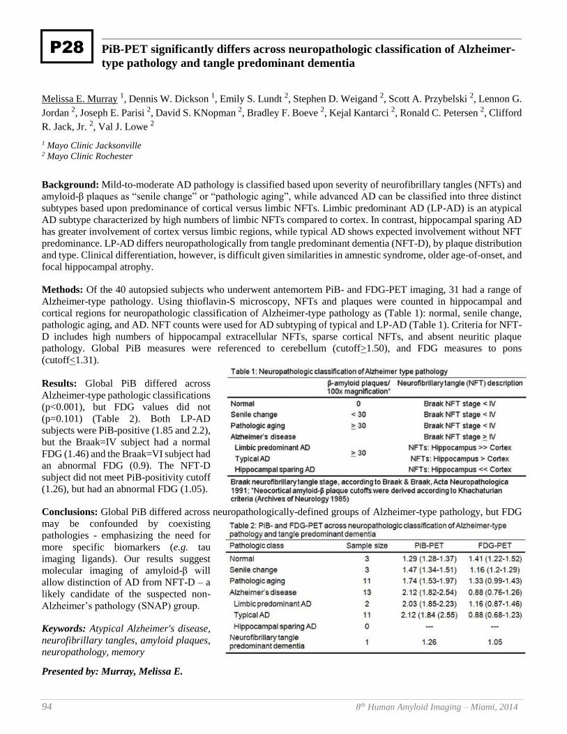
94 8th Human Amyloid Imaging – Miami, 2014
PiB-PET significantly differs across neuropathologic classification of Alzheimer-
type pathology and tangle predominant dementia
Melissa E. Murray 1, Dennis W. Dickson 1, Emily S. Lundt 2, Stephen D. Weigand 2, Scott A. Przybelski 2, Lennon G.
Jordan 2, Joseph E. Parisi 2, David S. KNopman 2, Bradley F. Boeve 2, Kejal Kantarci 2, Ronald C. Petersen 2, Clifford
R. Jack, Jr. 2, Val J. Lowe 2
1 Mayo Clinic Jacksonville 2 Mayo Clinic Rochester
Background: Mild-to-moderate AD pathology is classified based upon severity of neurofibrillary tangles (NFTs) and
amyloid-β plaques as “senile change” or “pathologic aging”, while advanced AD can be classified into three distinct
subtypes based upon predominance of cortical versus limbic NFTs. Limbic predominant AD (LP-AD) is an atypical
AD subtype characterized by high numbers of limbic NFTs compared to cortex. In contrast, hippocampal sparing AD
has greater involvement of cortex versus limbic regions, while typical AD shows expected involvement without NFT
predominance. LP-AD differs neuropathologically from tangle predominant dementia (NFT-D), by plaque distribution
and type. Clinical differentiation, however, is difficult given similarities in amnestic syndrome, older age-of-onset, and
focal hippocampal atrophy.
Methods: Of the 40 autopsied subjects who underwent antemortem PiB- and FDG-PET imaging, 31 had a range of
Alzheimer-type pathology. Using thioflavin-S microscopy, NFTs and plaques were counted in hippocampal and
cortical regions for neuropathologic classification of Alzheimer-type pathology as (Table 1): normal, senile change,
pathologic aging, and AD. NFT counts were used for AD subtyping of typical and LP-AD (Table 1). Criteria for NFT-
D includes high numbers of hippocampal extracellular NFTs, sparse cortical NFTs, and absent neuritic plaque
pathology. Global PiB measures were referenced to cerebellum (cutoff>1.50), and FDG measures to pons
(cutoff<1.31).
Results: Global PiB differed across
Alzheimer-type pathologic classifications
(p<0.001), but FDG values did not
(p=0.101) (Table 2). Both LP-AD
subjects were PiB-positive (1.85 and 2.2),
but the Braak=IV subject had a normal
FDG (1.46) and the Braak=VI subject had
an abnormal FDG (0.9). The NFT-D
subject did not meet PiB-positivity cutoff
(1.26), but had an abnormal FDG (1.05).
Conclusions: Global PiB differed across neuropathologically-defined groups of Alzheimer-type pathology, but FDG
may be confounded by coexisting
pathologies - emphasizing the need for
more specific biomarkers (e.g. tau
imaging ligands). Our results suggest
molecular imaging of amyloid-β will
allow distinction of AD from NFT-D – a
likely candidate of the suspected non-
Alzheimer’s pathology (SNAP) group.
Keywords: Atypical Alzheimer's disease,
neurofibrillary tangles, amyloid plaques,
neuropathology, memory
Presented by: Murray, Melissa E.
P28
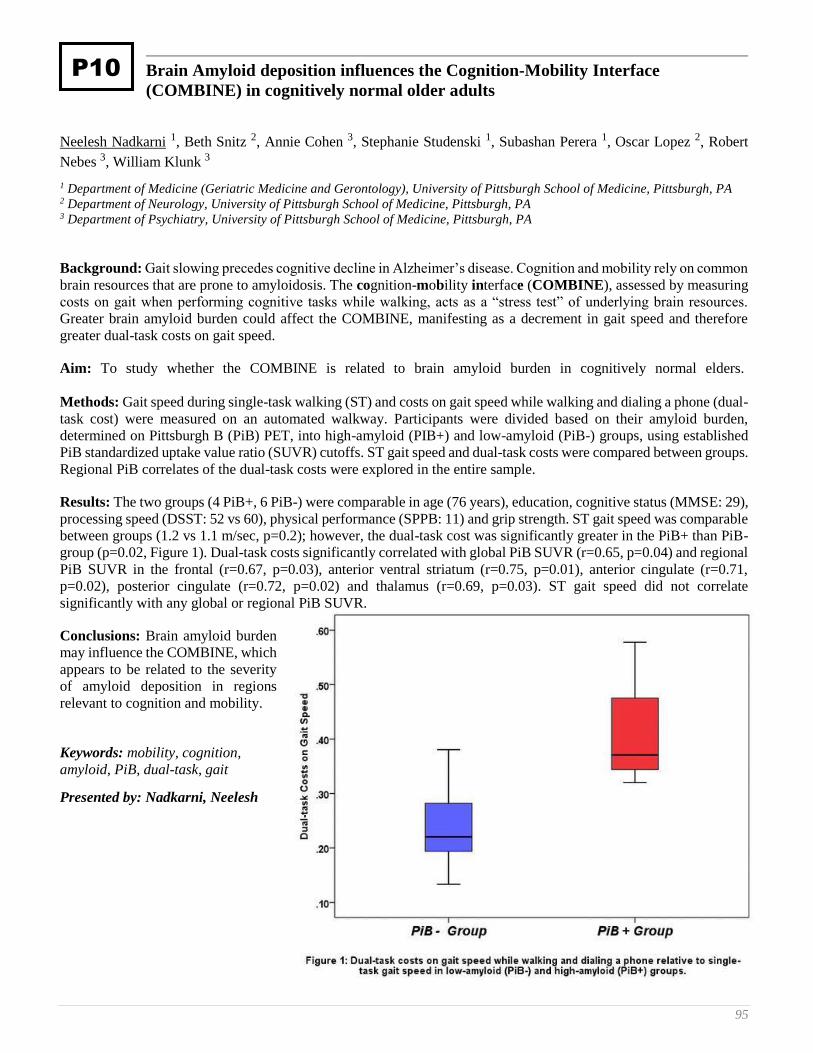
95
Brain Amyloid deposition influences the Cognition-Mobility Interface
(COMBINE) in cognitively normal older adults
Neelesh Nadkarni 1, Beth Snitz 2, Annie Cohen 3, Stephanie Studenski 1, Subashan Perera 1, Oscar Lopez 2, Robert
Nebes 3, William Klunk 3
1 Department of Medicine (Geriatric Medicine and Gerontology), University of Pittsburgh School of Medicine, Pittsburgh, PA 2 Department of Neurology, University of Pittsburgh School of Medicine, Pittsburgh, PA 3 Department of Psychiatry, University of Pittsburgh School of Medicine, Pittsburgh, PA
Background: Gait slowing precedes cognitive decline in Alzheimer’s disease. Cognition and mobility rely on common
brain resources that are prone to amyloidosis. The cognition-mobility interface (COMBINE), assessed by measuring
costs on gait when performing cognitive tasks while walking, acts as a “stress test” of underlying brain resources.
Greater brain amyloid burden could affect the COMBINE, manifesting as a decrement in gait speed and therefore
greater dual-task costs on gait speed.
Aim: To study whether the COMBINE is related to brain amyloid burden in cognitively normal elders.
Methods: Gait speed during single-task walking (ST) and costs on gait speed while walking and dialing a phone (dual-
task cost) were measured on an automated walkway. Participants were divided based on their amyloid burden,
determined on Pittsburgh B (PiB) PET, into high-amyloid (PIB+) and low-amyloid (PiB-) groups, using established
PiB standardized uptake value ratio (SUVR) cutoffs. ST gait speed and dual-task costs were compared between groups.
Regional PiB correlates of the dual-task costs were explored in the entire sample.
Results: The two groups (4 PiB+, 6 PiB-) were comparable in age (76 years), education, cognitive status (MMSE: 29),
processing speed (DSST: 52 vs 60), physical performance (SPPB: 11) and grip strength. ST gait speed was comparable
between groups (1.2 vs 1.1 m/sec, p=0.2); however, the dual-task cost was significantly greater in the PiB+ than PiB-
group (p=0.02, Figure 1). Dual-task costs significantly correlated with global PiB SUVR (r=0.65, p=0.04) and regional
PiB SUVR in the frontal (r=0.67, p=0.03), anterior ventral striatum (r=0.75, p=0.01), anterior cingulate (r=0.71,
p=0.02), posterior cingulate (r=0.72, p=0.02) and thalamus (r=0.69, p=0.03). ST gait speed did not correlate
significantly with any global or regional PiB SUVR.
Conclusions: Brain amyloid burden
may influence the COMBINE, which
appears to be related to the severity
of amyloid deposition in regions
relevant to cognition and mobility.
Keywords: mobility, cognition,
amyloid, PiB, dual-task, gait
Presented by: Nadkarni, Neelesh
P10
96 8th Human Amyloid Imaging – Miami, 2014
Astrocytosis, amyloid deposition and gray matter density in parahippocampus of
MCI patients
Il Han Choo 1, Stephen F Carter 2, Michael Schöll 3, Agneta Nordberg 4
1 Karolinska Institutet, Alzheimer Neurobiology Center, Stockholm.Sweden Department of Neuropsychiatry, School of Medicine,
Chrosun University, Gwangju, Republic of Korea 2 Karolinska Institutet,Alzheimer Neurobiology Center, Stockholm, Sweden Wolfson Imaging Center, Manchester University,
Manchester, United Kingdom 3 Karolinska Institutet, Alzheimer Neurobiology Center, Stockholm,Sweden 4 Karolinska Institutet, Alzheimer Neurobiology Center, Stockholm, Sweden Department of Geriatric Medicine,Karolinska
University Hospital Huddinge, Stockholm.Sweden
Introduction: The Alzheimer disease (AD) pathology is characterized by deposition of fibrillar amyloid, neurofibrillar
tangles as well as activation of astrocytes and microglia, atrophy, dysfunctional synapse and cognitive impairments.
Recent autoradiography studies have shown a different laminar distribution of astroglia and fibrillar Aß in cortex and
hippocampus autopsy AD brains (1). In addition PET studies have shown an increased astrocytosis measured by 11C-
d-deprenyl (DED) in patients with mild cognitive impairment (MCI) in comparison to AD patients and controls (2).
Aims: to test the hypothesis that neuronal inflammation is an early phenomena in AD pathology related to Aß plaque
deposition.
Methods: 20 patients with AD or mild cognitive impairment (MCI) underwent 11C-PIB, 18F-FDG and 11C-deuterium-
L-deprenyl (11C-DED) PET imaging in addition to MRI scans, CSF biomarker analysis and neuropsychological
assessments.
Results: A positive correlation was observed between PIB PET retention and DED binding in the parahippocampus
of AD patients (p<0.014) while a negative correlation between gray matter density and DED binding was observed in
PIB+ MCI patients. A negative correlation was observed between CSF Aß42 levels and PIB retention in
parahippocampus of MCI+AD patients (p<0.003) while a positive correlation between total Tau and PIB retention in
AD. CSF total tau also negatively correlated with decrease in gray matter density in MCI patients (p<0.001). No
significant correlation was observed with FDG PET and any of the other PET, MRI or CSF biomarkers.
Conclusions: High astrocytosis in the parahippocampus of PIB+ MCI patients suggest an early preclinical influence
on neuronal loss. The lack of correlation between astrocytosis and CSF tau levels but positive correlation between
astrocytosis and fibrillar amyloid deposition indicate that the parahippocampal astrocytosis is related to amyloid
pathology.
1. Marutle et al. J Neuroinflammation 2013 ; 10 : 90
2. Carter et al. J Nucl Med 2012 ; 53 :37-46.
Keywords: Fibrillar amyloid , astrocytosis, gray matter density, parahippocampus, MCI,AD, PET, 11C-d-deprenyl,
11C-PIB, MRI, CSF biomarkers
Presented by: Nordberg, Agneta
P22
97
Regional brain activity and functional connectivity during memory encoding are
differentially affected by age and β-amyloid deposition in cognitively normal
older adults
Hwamee Oh 1, Jeremy Elman 1, Suzanne Baker 2, Cindee Madison 1, Jacob Vogel 1, Sam Crowley 1, William Jagust 3
1 Helen Wills Neuroscience Institute, University of California- Berkeley, CA 94720, USA 2 Life Sciences Division, Lawrence Berkeley National Laboratory, Berkeley, CA 94720, USA 3 Helen Wills Neuroscience Institute, University of California- Berkeley, CA 94720, USA and Life Sciences Division, Lawrence
Berkeley National Laboratory, Berkeley, CA 94720, USA
While β-amyloid (Aβ) is present in a large proportion of cognitively intact older adults, its effect on brain activity
during normal aging is not yet well understood. We examined how brain activity and connectivity underlying
successful episodic encoding change as a function of age and Aβ deposition using fMRI and [11C] PIB PET.
Twenty-two young (mean age=23.6, 12 males) and 49 cognitively normal older adults (mean age=76.5, 20 males)
performed an episodic encoding task of visual scenes during fMRI scans, followed by a postscan recognition task
where subjects made an old/new judgment with confidence ratings based on written descriptions of the gist of visual
scenes and a true/false judgment on 6 written visual details of the scenes. 150 encoding trials were sorted based on gist
accuracy and the number of details correctly recognized. Older adults were further classified as PIB negative (PIB−)
Old and PIB positive (PIB+) Old.
Compared to PIB- older subjects, young subjects showed increased activation in visual association areas (VA), inferior
frontal cortex and hippocampus bilaterally for successful gist memory. PIB- older adults, however, showed an
increased activation in the lateral and medial parietal cortex and lateral and medial frontal cortex. In addition, PIB-
older adults showed stronger connectivity between the right parahippocampal gyrus (rPHG), VA, and lateral frontal
cortex for gist memory but reduced connectivity between rPHG, VA, and hippocampus for visual details. Compared
to PIB- older adults, PIB+ older adults showed greater activation in left hippocampus, VA and medial frontal cortex
but reduced connectivity between rPHG and hippocampus for gist memory.
These results suggest that neural correlates of gist memory and memory for specific episodic details are differentially
affected by age. In addition, the presence of Aβ is associated with hippocampal dysfunction seen as increased regional
activity but reduced functional connectivity during memory process.
Keywords: Normal Aging, β-amyloid, Memory system
Presented by: Oh, Hwamee
P65

98 8th Human Amyloid Imaging – Miami, 2014
Prevalence of Amyloid in cognitively normal, MCI and demented subjects –
A meta-analysis of Amyloid PET studies
Rik Ossenkoppele 1, Willemijn Jansen 2, Bart van Berckel 1, Pieter Jelle Visser 3
1 VU University Medical Center, Amsterdam, the Netherlands 2 Maastricht University, Maastricht, the Netherlands 3 VU University Medical Center, Amsterdam, the Netherlands & Maastricht University, Maastricht, the Netherlands
Introduction: The advent of PET ligands that bind to amyloid-β have caused a major paradigm shift in diagnostic
thinking. With almost a decade of experience, this is the time for a thorough meta-analysis of amyloid imaging studies
to date.
Objectives: 1) to estimate the prevalence of amyloid in cognitively normal, MCI and demented subjects;
2) to relate this prevalence to age and APOE genotype.
Methods: First, MEDLINE databases were searched for “PET”, “Amyloid”, “Amyloid-beta”, “PIB”, “Flutemetamol”,
“Florbetapir” or “Florbetaben”. Abstracts were screened and if the study appeared appropriate, corresponding authors
were approached for individual subject data (i.e. amyloid PET status, APOE genotype, age, sex, education, MMSE
and CDR). We applied random effects models to estimate the prevalence of amyloid.
Results: Clinical and demographic characteristics are presented in table 1. The prevalence of amyloid was 25.1% in
cognitively normal subjects (n=1348), 53.0% in MCI (n=1055), 87.6% in AD dementia (n=1146) and 22% in non-AD
dementia (n=510) (Figure 1). In cognitively normal subjects and MCI patients, older age and APOE ε4 carriership
were associated with higher prevalence of amyloid (p<0.05; Figure 2A+B). In AD dementia patients, amyloid PET
scans were more often negative in older and APOE ε4 negative patients (p<0.05, Figure 2C). Patients with a clinical
diagnosis of VaD appeared more often amyloid PET positive with increasing age (p<0.05, Figure 2D), while this was
not the case for DlB, FTD or dementia-other patients (e.g. CBD or PDD).
Conclusions: The major risk factors for the onset of AD, ageing and APOE ε4 genotype, were associated with higher
prevalence of amyloid pathology in cognitively normals and MCI patients. In non-AD dementias, this ageing effect
was observed only in VaD. APOE ε4 negative and older AD patients were more often amyloid-negative on PET.
Keywords: Amyloid-PET, meta-analysis, prevalence, APOE, ageing.
Presented by: Ossenkoppele, Rik
P66
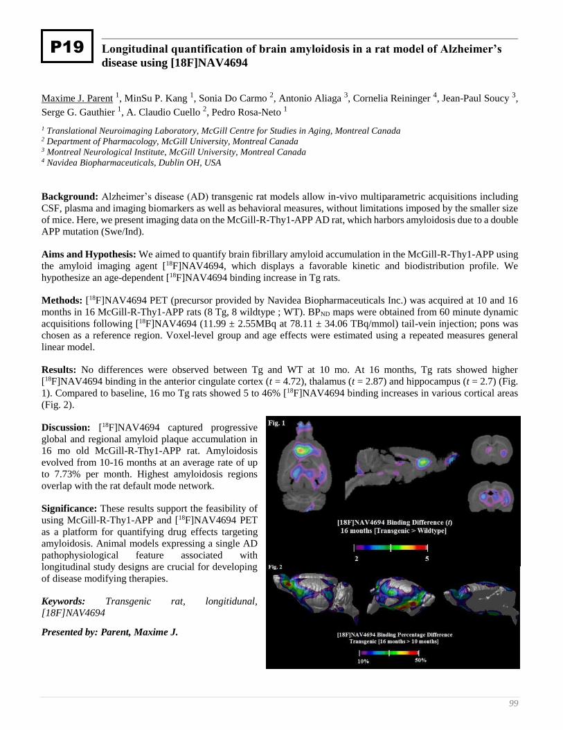
99
Longitudinal quantification of brain amyloidosis in a rat model of Alzheimer’s
disease using [18F]NAV4694
Maxime J. Parent 1, MinSu P. Kang 1, Sonia Do Carmo 2, Antonio Aliaga 3, Cornelia Reininger 4, Jean-Paul Soucy 3,
Serge G. Gauthier 1, A. Claudio Cuello 2, Pedro Rosa-Neto 1
1 Translational Neuroimaging Laboratory, McGill Centre for Studies in Aging, Montreal Canada 2 Department of Pharmacology, McGill University, Montreal Canada 3 Montreal Neurological Institute, McGill University, Montreal Canada 4 Navidea Biopharmaceuticals, Dublin OH, USA
Background: Alzheimer’s disease (AD) transgenic rat models allow in-vivo multiparametric acquisitions including
CSF, plasma and imaging biomarkers as well as behavioral measures, without limitations imposed by the smaller size
of mice. Here, we present imaging data on the McGill-R-Thy1-APP AD rat, which harbors amyloidosis due to a double
APP mutation (Swe/Ind).
Aims and Hypothesis: We aimed to quantify brain fibrillary amyloid accumulation in the McGill-R-Thy1-APP using
the amyloid imaging agent [18F]NAV4694, which displays a favorable kinetic and biodistribution profile. We
hypothesize an age-dependent [18F]NAV4694 binding increase in Tg rats.
Methods: [18F]NAV4694 PET (precursor provided by Navidea Biopharmaceuticals Inc.) was acquired at 10 and 16
months in 16 McGill-R-Thy1-APP rats (8 Tg, 8 wildtype ; WT). BPND maps were obtained from 60 minute dynamic
acquisitions following [18F]NAV4694 (11.99 ± 2.55MBq at 78.11 ± 34.06 TBq/mmol) tail-vein injection; pons was
chosen as a reference region. Voxel-level group and age effects were estimated using a repeated measures general
linear model.
Results: No differences were observed between Tg and WT at 10 mo. At 16 months, Tg rats showed higher
[18F]NAV4694 binding in the anterior cingulate cortex (t = 4.72), thalamus (t = 2.87) and hippocampus (t = 2.7) (Fig.
1). Compared to baseline, 16 mo Tg rats showed 5 to 46% [18F]NAV4694 binding increases in various cortical areas
(Fig. 2).
Discussion: [18F]NAV4694 captured progressive
global and regional amyloid plaque accumulation in
16 mo old McGill-R-Thy1-APP rat. Amyloidosis
evolved from 10-16 months at an average rate of up
to 7.73% per month. Highest amyloidosis regions
overlap with the rat default mode network.
Significance: These results support the feasibility of
using McGill-R-Thy1-APP and [18F]NAV4694 PET
as a platform for quantifying drug effects targeting
amyloidosis. Animal models expressing a single AD
pathophysiological feature associated with
longitudinal study designs are crucial for developing
of disease modifying therapies.
Keywords: Transgenic rat, longitidunal,
[18F]NAV4694
Presented by: Parent, Maxime J.
P19

100 8th Human Amyloid Imaging – Miami, 2014
Potential value of an interpretation method that incorporates quantitative
estimate of cortical to cerebellar SUVr as an adjunct to visual interpretation of
florbetapir PET scans
Michael Pontecorvo , Michael Devous Sr. , Anupa K. Arora , Marybeth Devine , Ming Lu , Andrew Siderowf , Stephen
P. Truocchio , Catherine Devadanam , Abhinay D. Joshi , Christopher Breault , Stephen L. Heun , Daniel M.
Skovronsky , Mark A. Mintun
Avid Radiopharmaceuticals, Inc.
Background: Commercial software is available for quantitation of amyloid PET images. We evaluated whether
employing quantitation by FDA-cleared software as an adjunct to visual interpretation (VisQ method) improves
accuracy of florbetapir scan interpretation in lower accuracy readers (those with <90% accuracy using visual
techniques alone).
Methods: Twenty-two physicians were trained to use quantitation of florbetapir images as an adjunct to visual
interpretation. Each reader then visually interpreted, without access to quantitation information, 46 florbetapir PET
images from subjects autopsied within one year of PET scan (Clark et al, 2012) and 50 images selected from patients
seeking diagnosis for cognitive decline (Grundman et al, 2012). Next, the readers reviewed all 96 images again, using
MIMneuro 6.0.6 software to calculate a quantitative estimate (SUVr) of florbetapir activity and applied the VisQ
method. For each case, the reader compared the SUVr to normal ranges. For scans in which the initial visual read and
quantitative result were discordant, readers reassessed their original interpretation in light of the quantitation,
reinspected images and verified ROI placement (a potential source of error in SUVr calculation) to resolve any
discordance. They then provided a final VisQ interpretation.
Results: With visual interpretation, the mean accuracy (compared to pathology truth standard) on the 46 autopsy
verified cases was 89.5%. 7 of the 22 readers were identified as lower accuracy visual readers. In these 7 readers,
accuracy improved significantly with VisQ from a mean of 81.7% to 88.8% (p=0.0029). Across all 22 readers, accuracy
improved from 89.5% to 93.8% (p<0.0001). Interreader agreement across all 96 cases increased (p < 0.05) from a
Fleiss kappa of 0.72 to 0.79.
Conclusions: Accuracy and interreader reliability increased significantly after applying the VisQ method that
incorporates quantitative estimates of florbetapir PET amyloid binding (SUVr).
Keywords: Amyloid,florbetapir,quantitation,interpretation
Presented by: Pontecorvo, Michael
P30
101
The Alzheimer’s structural connectome: Patterns of cortical reorganization with
increasing neuritic amyloid plaque burden
Jeff Prescott 1, Arnaud Guidon 2, P. Murali Doraiswamy 3, Kingshuk Choudhury 1, Chunlei Liu 2, Jeffrey Petrella 1
1 Department of Radiology, Duke University Medical Center 2 Brain Image Analysis Center, Duke University 3 Departments of Psychiatry, Medicine and the Duke Institute for Brain Sciences, Duke University Medical Center
Purpose: Evaluate differences in the structural connectome among patients with normal cognition, mild cognitive
impairment (MCI), and Alzheimer's disease (AD), and determine the effect of increasing cortical amyloid deposition.
Materials and Methods: There were 102 subjects in the Alzheimer's Disease Neuroimaging Initiative 2 who met
criteria for analysis. Subjects' T1 scans were automatically parcellated into regions of interest. Standardized uptake
value ratios (SUVr) were calculated from florbetapir PET scans for composite cortical regions (frontal, cingulate,
parietal, and temporal). Structural connectome graphs were created from DTI scans, and connectome organization was
analyzed in each region using graph theoretic metrics. ANOVA of structural connectome metrics and florbetapir SUVr
across diagnostic group was performed. Linear mixed effects models were fit to analyze the effect of florbetapir SUVr
on the structural connectome metrics.
Results: Diagnostic group was associated with changes in weighted structural connectome metrics, with decreases
from NC to MCI to AD demonstrated for strength in the bilateral frontal, right parietal, and bilateral temporal regions
(p < 0.05); weighted local efficiency in the left temporal region (p < 0.05); and weighted clustering coefficient in the
bilateral frontal and left temporal regions (p < 0.05). Increasing cortical florbetapir SUVr was associated with decreases
in weighted structural connectome metrics; namely, strength (p = 0.00001), weighted local efficiency (p = 0.00001),
and weighted clustering coefficient (p = 0.0006), independent of brain region.
Conclusions: Increasing amyloid burden is related to reorganization of the large-scale structural network architecture
of the brain even in the preclinical stages of AD.
Keywords: Structural connectome, Network theory, Florbetapir
Presented by: Prescott, Jeff
P57
102 8th Human Amyloid Imaging – Miami, 2014
Mathematical modeling of amyloid-β disposition by brain endothelial cells
Hisham Qosa 1, Bilal Abuasal 1, Jeffrey Keller 2, Amal Kaddoumi 1
1 College of Pharmacy University of Louisiana at Monroe 2 Pennington Biomedical Research Center Louisiana State University.
Alzheimer’s disease has a characteristic hallmark of amyloid-β (Aβ) accumulation in the brain. This accumulation of
Aβ has been related to its faulty cerebral clearance. Evaluation of kinetic parameters of Aβ uptake, efflux and
degradation by endothelial cells of the blood-brain barrier (BBB) is essential to understand key steps involved in Aβ
clearance across the BBB and its brain accumulation. Here, bEnd3 and hCMEC/D3 cells, as in vitro mouse and human
BBB models, respectively, were used to establish a mathematical model to describe Aβ disposition by endothelial
cells. Aβ disposition in brain endothelial cells involves several processes that simultaneously take place; therefore, the
utilization of a mechanistic model to estimate Aβ40 kinetic parameters in endothelial cells should provide a better
approach than the conventional Michaelis-Menten. The mechanistic model depends on measurement of Aβ uptake and
degradation at multiple time points with multiple Aβ concentrations at each time point. Based on our results from Aβ
uptake and degradation studies in bEnd3 and hCMEC/D3 cells, these processes were best described by a mechanistic
two-compartment model that could successfully describe Aβ disposition. The model structure consisted of saturable
uptake and efflux processes in addition to linear intracellular degradation. The established model showed that: a) Aβ
uptake process determines subsequent degradation and efflux processes, b) brain endothelial cells mediate Aβ
degradation however the degradation rate is dependent on uptake rate and is higher in bEnd3 cells, and c) both cells
mediate efficient efflux of intact Aβ. In conclusion, the established model offered a mathematical description of Aβ
clearance across the BBB which could be improved to predict Aβ accumulation, aggregation and Aβ binding to
different antibodies and imaging tracers in the brain.
Keywords: amyloid, blood-brain barrier, disposition, mechanistic modeling
Presented by: Qosa, Hisham
P39
103
Amyloid burden influences the relationship between hippocampal volume and
default mode network connectivity in cognitively normal elderly
Aaron Schultz 1, Elizabeth Mormino 1, Willem Huijbers 2, Jasmeer Chhatwal 1, Andrew Ward 3, Sarah Wigman 4,
Molly LaPoint 4, Trey Hedden 1, Keith Johnson 1, Reisa Sperling 4
1 Athinoula A. Martinos Center for Biomedical Imaging, Department of Radiology, Massachusetts General Hospital, Harvard
Medical School, Boston, MA 2 Department of Psychiatry, Massachusetts General Hospital/Harvard Medical School and the Athinoula A. Martinos Center for
Biomedical Imaging 3 Helen Wills Neuroscience Institute, University of California, Berkeley, CA Athinoula A. Martinos Center for Biomedical
Imaging, Department of Radiology, Massachusetts General Hospital, Harvard Medical School, Boston, MA 4 Center for Alzheimer Research and Treatment, Department of Neurology, Brigham and Women’s Hospital, Harvard Medical
School, Boston MA and the Athinoula A. Martinos Center for Biomedical Imaging
Rationale: Currently, preclinical Alzheimer’s disease (AD) is often defined by evidence of fibrillar amyloid-β
deposition. We hypothesized that within a cognitively normal group of elderly we would see differential relationships
between markers of synaptic dysfunction and neuronal loss in individuals harboring a high amyloid burden (Aβ+)
versus those who do not (Aβ-). In particular we investigated the cross-sectional relationship between default mode
network functional connectivity (DMN-FC) and hippocampal volume (HV).
Methods: We examined cross-sectional relationships in neuroimaging data from 195 cognitively normal subjects (55
Aβ+ and 140 Aβ-) from the Harvard Aging Brain Study. DMN-FC measurements were made using Template Based
Rotation, and HV measurements were made with Freesurfer v5.1. For Aβ we used a standard measure of cortical
deposition of PiB, computed as DVRs with a cerebellar grey matter reference region.
Results: We found an interaction between amyloid status and HV when predicting DMN-FC (F(1,190)=6.73; p=0.01),
such that there is a significant relationship between DMN-FC and HV only within the Aβ+ group (Aβ+: r=0.459,
p<0.001; Aβ-: r=0.031, p=0.717). Modeling HV as a dichotomous variable we confirmed the interaction between Aβ
and HV (F(1,90)=8.75, p=0.003), with the pattern of means showing lower DMN-FC in the Aβ+/small HV group as
compared to the other three groups.
Conclusion: The presence of a relationship between DMN-FC and HV in only the Aβ+ group suggests that AD-type
pathological changes drive alterations in both connectivity and structure, and that these changes are present prior to
clinically defined impairment. We suggest that MR biomarkers can serve an important role in determining the
approximate preclinical stage of AD in cognitively normal individuals with amyloid plaques.
Keywords: Default Mode Network, functional connectivity, amyloid, preclinical Alzheimer’s disease, hippocampal
volume
Presented by: Schultz, Aaron
P43
104 8th Human Amyloid Imaging – Miami, 2014
Update on the multicenter phase 3 histopathology study for ß-amyloid brain PET
imaging
Osama Sabri 1, Andrew Stephens 2, Ana Catafau 2, Henryk Barthel 3, John Seibyl 4
1 Department of Nuclear Medicine University of Leipzig, Leipzig, Germany For the Florbetaben Study Group 2 Piramal Imaging, Berlin, Germany 3 Department of Nuclear Medicine University of Leipzig, Leipzig, Germany 4 MNI, New Haven, CT, USA
Background: The pivotal histopathology phase 3 study of florbetaben was designed for an exact tissue-matched
regional analysis of post-mortem brain tissue and imaging during life to detect and locate a pathological state. This is
the first study demonstrating a direct correlation of amyloid deposition and tracer uptake in the identical anatomic
region. In addition to assessment of specific regions, final neuropathological diagnosis was established by the onsite
pathologist including the assessment of neuritic ß-amyloid plaque density according to CERAD criteria. A method for
the visual assessment of florbetaben PET scans in clinical practice was validated. The results from the first 31 brains
have been presented previously. After the initial analysis, brains of enrolled subjects that reached post-mortem
continued to be collected and analyzed. We now present new results from the subject-level analyses with additional
brain specimens.
Methods: 216 subjects were enrolled and underwent MRI and florbetaben PET scanning. All baseline PET scans were
quantitatively assessed and read by three in-person trained readers using the method proposed for clinical practice.
PET results were compared with presence/absence of neuritic β-amyloid plaque density according to CERAD criteria
at the patient’s autopsy and sensitivity & specificity was calculated. Subject-level quantitative analysis was performed
and the composite SUVR was determined for each subject. A ROC curve analysis was performed to determine the
optimal threshold for sensitivity / specificity calculation.
Results: In total 74 brain specimens became available and were assessed by onsite pathology. Sensitivity and
specificity of the majority read of the subject-level visual assessment vs. histopathology was 97.9% and 88.9%,
respectively. ROC curve analysis determined the optimal cut-off for sensitivity and specificity calculations to be 1.478.
Using this quantitative analysis, a high sensitivity (89 %) and specificity (92 %) was obtained.
Conclusions: Previous results from 31 subjects were confirmed in a larger cohort. Visual and quantitative assessment
is in good agreement.
Keywords: florbetaben, histopathology, amyloid, PET
Presented by: Seibyl, John
P40
105
TAU Tracer F18-T807 inversely relates with brain functional hubs of elderly
normals
Jorge Sepulcre 1, Sperling Sperling 2, Keith Johnson 3
1 Division of Nuclear Medicine and Molecular Imaging, Department of Radiology, Massachusetts General Hospital and
Harvard Medical School, Boston, MA, USA; Athinioula A. Martinos Center for Biomedical Imaging, Charlestown, MA, USA 2 Athinioula A. Martinos Center for Biomedical Imaging, Charlestown, MA, USA; Centre for Alzheimer Research and
Treatment, Department of Neurology, Brigham and Women's Hospital and Harvard Medical School, Boston, MA, USA,
Department of Neurology, Massachuse 3 Division of Nuclear Medicine and Molecular Imaging, Department of Radiology, Massachusetts General Hospital and
Harvard Medical School, Boston, MA, USA; Centre for Alzheimer Research and Treatment, Department of Neurology, Brigham
and Women's Hospital and
Background: Alzheimer’s disease neuropathologic processes are thought to affect brain function before cognitive
manifestations appear. Molecular imaging facilitates the early detection of Ab and the study of its effects on brain
dysfunction and cognitive decline. Recent development of tau tracers has similarly enabled studies of tau-related
pathology. In this study, we evaluated the spatial distribution of a novel tau PET tracer 18F-T807 and identify its
relationship with Ab deposition and functional network connectivity in normal elderly subjects.
Methods: Twenty-four normal elderly subjects (mean age (SD): 74 (6.6), M/F: 11/13) underwent two PET imaging
acquisitions (11C-PIB and 18F-T807) and MRI session. Conventional preprocessing and normalization procedures were
performed on the PET images. A functional connectivity map (Buckner et al, 2009; Sepulcre et al, 2010) was obtained
in each subject by quantifying the FDR-corrected degree of functional connectivity MRI. We used voxel-by-voxel
regressions to evaluate spatial correspondences between maps of 18F-T807, 11C-PIB and functional hubs.
Results: First, we found that 18F-T807 retention in elderly controls predominates in medial temporal regions, matching
well with previous descriptions of early tau deposits in Braak stages (Figure 1-A). Second, 18F-T807 and 11C-PIB
images had positive spatial correlations with r coefficients ranging 0.2 to 0.4. Third, while 11C-PIB distributions did
not show consistent spatial association with functional hubs, 18F-T807 deposits displayed significant negative
correlations from -0.2 to -0.3 (representative examples in Figure 1-B; and mean in Figure 1-C). Analyses comparing
the regression slopes of 11C-PIB with hubs versus those of 18F-T807 with hubs, show that they differ significantly in
explaining functional hub organization of elderly individuals (Figure 1-B).
Conclusions: Our results suggest that 18F-T807 deposits are inversely related to brain functional hubs in elderly
individuals. Further studies are needed to explicitly reveal whether this association relates to brain functional
breakdown.
Keywords: TAU tracer, PIB, PET, Functional Hubs, Elderly Normals
Presented by: Sepulcre, Jorge
P24
106 8th Human Amyloid Imaging – Miami, 2014
Amyloid PET screening for enrollment into Alzheimer’s disease clinical trials:
Initial experience in a Phase 1b clinical trial
Sevigny Jeff 1, Suhy Joyce 2, Chiao Ping 1, Klein Gregory 2, Oh Joonmi 2, Purcell Derk 3, Verma Ajay 1, Sampat
Mehul 2, Barakos Jerome 4
1 Biogen Idec, Cambridge, MA, USA 2 Synarc Inc, Newark, CA, USA 3 Synarc Inc, Newark, CA, USA, California Pacific Medical Center, San Francisco, CA, USA, University of California San
Francisco, San Francisco, CA, USA 4 Synarc Inc, Newark, CA, USA, California Pacific Medical Center, San Francisco, CA, USA
Introduction: Amyloid PET imaging is a method of Alzheimer’s disease (AD) clinical trial enrichment. We report
our ongoing experience with amyloid PET imaging as a screening tool in patients with prodromal or mild AD.
Methods: Patients fulfilling criteria for either prodromal or mild AD in a multicenter, Phase 1b clinical trial underwent
florbetapir PET scanning at screening. PET, MRI, and co-registered PET/MRI scans were reviewed by two
independent readers and binary visual readings tabulated. Ten duplicate cases were presented to the readers to allow
for calculation of intra-reader agreement statistics. Results were compared with semi-quantitative standard uptake
value (SUV) ratios using a threshold of 1.10 to classify amyloid-negative from amyloid-positive patients.
Results: Of the first 250 PET/MRI scans reviewed, 20 discordant cases were identified when comparing visual
readings to quantitative results: 17 with negative visual readings but positive quantitative results (composite SUV ratios
1.100–1.371) and 3 with positive visual readings but negative quantitative results (composite SUV ratios 1.040, 1.052
and 1.089). Inter- and intra-reader agreements from visual readings were 98.4% and 100%, respectively. Of those
patients who met all study clinical and laboratory inclusion and exclusion criteria, 36.8% were ultimately excluded
due to an amyloid-negative PET scan based on visual readings.
Conclusions: High inter- and intra-reader reliability was achieved. Factors potentially contributing to the high
reliability were: a) availability of co-registered PET/MRI scans; b) harmonized spatial resolution and image orientation
regardless of scanner model or patient position; c) 20-minute rather than 10-minute acquisition providing better
statistics/less noise; and d) dynamic acquisition allowing motion correction. Agreement between visual and
quantitative readings was good (92%). These data demonstrate that amyloid PET imaging can be an effective and
feasible tool to enrich AD clinical trials in amyloid-positive patients, in particular in patients with earlier stages of the
disease.
Keywords: Alzheimer’s disease, amyloid PET imaging, clinical trial, screening, prodromal
Presented by: Sevigny, Jeff
P3
107
Antidepressant decreases CSF Aβ production in healthy individuals and in
transgenic mice
Yvette Sheline 1, Tim West 2, Kevin Yarasheski 3, Robert Swarm 4, Mateusz Jasielec 5, Jonathan Fisher 6, Ping Yan 6,
Chengjie Xiong 7, Christine Frederiksen 8, Robert Chott 3, Randall Bateman 9, John Morris 9, Mark Mintun 10, Jin-
Moo Lee 11, John Cirrito 11
1 Departments of Psychiatry, Radiology, and Neurology University of Pennsylvania Perelman School of Medicine, Philadelphia
PA 2 C2N Diagnostics LLC, St. Louis MO 3 Department of Medicine, Washington University Medical Center, St. Louis MO 4 Department of Anesthesiology, Washington University Medical Center, St. Louis MO 5 Departments of Biostatistics and Surgery, Washington University Medical Center, St. Louis MO 6 Department of Neurology, Washington University Medical Center, St. Louis MO 7 Department of Biostatistics and The Knight Alzheimer’s Disease Research Center, Washington University Medical Center, St.
Louis MO 8 Department of Surgery, Washington University Medical Center, St. Louis MO 9 Department of Neurology and The Knight Alzheimer’s Disease Research Center, Washington University Medical Center, St.
Louis MO 10 Avid Radiopharmaceuticals and Eli Lilly, Inc, Philadelphia PA 11 Department of Neurology, The Knight Alzheimer’s Disease Research Center, and Hope Center for Neurological Disorders,
Washington University Medical Center, St. Louis MO
Background: Aggregation of the amyloid-β (Aβ) peptide into plaques appears to be concentration-dependent, and
methods for reducing Aβ production may have therapeutic potential. In this study we examined the dose-response
effects of the SSRI citalopram, on brain Aβ levels in mice, its effect on individual plaque growth in the same mouse
model, and the effects of an approximately equivalent dose of citalopram on human Aβ levels and production.
Methods: In vivo microdialysis assessed brain interstitial fluid (ISF) Aβ over time in the hippocampus of aged, freely
moving, plaque-bearing APP/PS1+/- mice. Separately, individual brain plaques of APP/PS1 mice were serially imaged
using intravital multiphoton microscopy before and after 28-day treatment with citalopram (10 mg/kg) vs. vehicle. In
healthy humans, a randomized double-blind placebo controlled study determined the effect of 60 mg of citalopram. A
lumbar catheter was placed at the L3-4 interspace, and hourly sampling of blood and CSF was conducted during and
after administration of a stable-isotope labeled amino acid (13C6-leucine). Incorporation of 13C6 leucine into Aβ was
measured using the stable isotope labeling kinetics (SILK-Aβ®) assay and the concentration of labeled and unlabeled
Ab was measured by stable isotope spike absolute quantitation (SISAQ™).
Results: In aged AD mouse models, citalopram produced a dose-dependent decrease in brain ISF Aβ levels. Individual
plaques in mice chronically administered citalopram showed 78% reduction in new plaque appearance and 29%
decrease in pre-existing plaque growth, but no regression of existing plaques. In humans, the production rate of Aβ
was significantly suppressed by 38% in the citalopram group compared to placebo, resulting in 40% decrease in total
CSF Aβ levels over 37 hours.
Discussion: The ability to safely decrease Aβ concentrations is potentially important as a preventive strategy for AD.
This study demonstrates key target engagement for future AD prevention trials.
Presented by: Sheline, Yvette
P20

108 8th Human Amyloid Imaging – Miami, 2014
Impact of regional flow/perfusion heterogeneity and metabolism on the
quantification of amyloid tracers: a preclinical exploration
Ann-Marie Waldron 1, Leonie Wyffels 1, Stefanie Dedeurwaerdere 2, Jill Richardson 3, Mark Schmidt 4, Xavier
Langlois 4, Sigrid Stroobants 1, Steven Staelens 1
1 Molecular Imaging Center Antwerp, Antwerp University, BELGIUM 2 Translational Neurosciences, University of Antwerp, Belgium 3 R&D China U.K. Group, GlaxoSmithKline, Stevenage, United Kingdom 4 Neuroscience, Janssen Pharmaceutica NV, Beerse, Belgium
Introduction: With a view to improving the accuracy of translational amyloid imaging the present study aimed to
explore flow/perfusion heterogeneity and tracer metabolism on the quantification of [18F]-AV45 by dynamic
μPET studies.
Methods: Healthy (n=12) C57BL6J mice were intravenously injected with 37MBq of [18F]-AV45. At 5, 10, 30 and
60min post injection mice (n=3/time point) were sacrificed and the brain and plasma were analyzed for
radiometabolites. Further, for μPET imaging we studied a double transgenic mouse model of amyloidosis (TASTPM,
n=5) and its wild-type counterpart (WT, n= 8) at 12 months of age. Mice were injected with 17MBq of [18F]-AV45 via
a tail vein catheterization and a dynamic 90min PET acquisition was started simultaneously. Tracer uptake was
quantified by normalization for the injected dose.
Results: [18F]-AV45 undergoes rapid metabolism with intact tracer accounting only for 28% of total plasma
radioactivity already at 5min and decreasing to 2% at 30min p.i in WT mice. From μPET imaging, there was a notable
difference in initial tracer uptake between WT and TASTPM, with the transgenic mice demonstrating lower peak
uptake values (insets of Fig.1)
This difference was most prominent in
the thalamus (on average relatively 17%
lower in TASTPMs) while uptake in the
cerebellum was comparable to that of
WT. Thereafter a greater retention of
[18F]-AV45 in TASTPM mice is
apparent at 10min and remains for all
subsequent time points (Fig. 2).
Conclusion: The initial differences in
tracer uptake are indicative of regional
flow/perfusion issues in TASTPM mice and
contend the use of cerebellum as a reference
region for normalization. These perfusion issues
taken in combination with the rapid metabolism
of [18F]-AV45 greatly complicate the accurate
estimation of amyloid burden.
Keywords: perfusion, flow, region heterogeneity, SUVR, quantification
Presented by: Staelens, Steven
P50

109
In Vivo assessment of two isomers of [18F]-THK5105 and [18F]-THK511
Olivier Barret 1, David Alagille 1, Danna Jennings 1, Nobuyuki Okamura 2, Shozo Furumoto 2, Yukitsuka Kudo 2,
Kenneth Marek 1, John Seibyl 1, Gilles Tamagnan 1
1 Molecular Neuroimaging 2 Tohoku University, School of Medicine
Objective: To assess four quinoline radiopharmeuticals as potential PET biomarkers for tau pathology in Alzheimer’s
disease (AD) subjects and similarly aged healthy controls (HC): [18F]MNI-718, [18F]MNI-720, [18F]MNI-721 and
[18F]MNI-723 ((R)-THK5117, (S)-THK5117, (R)-THK5105 and (S)-THK5105 isomer, respectively),
Methods: 10 AD and 7 HC (Age 72.1 AD and 55.1 HC,, MMSE range in AD subjects 13-26) received (185 ± 3.7
MBq) of one of the four tracers and were, imaged for up to 5 hours. AD subject also underwent amyloid imaging.
Venous blood samples collected during imaging session to characterize metabolism of compounds. PET images were
co-registered with subject's MRI and the uptake extracted in a variety of grey matter regions. Uptake was normalized
to grey matter cerebellar cortex (SUVr) and values between 75-90 min post injection were compared between the two
groups.
Results: All compounds displayed very good brain penetrance with a maximum SUV of 4-6 within 10 min of injection.
[18F]MNI-718 and [18F]MNI-720 had a similar brain uptake kinetics that were faster than [18F]MNI-721 and [18F]MNI-
723. SUVr for [18F]MNI-718, [18F]MNI-720, and [18F]MNI-723 were increased in AD compared to HC in regions
expected to demonstrate tau pathology.
Conclusions: These preliminary data suggest that both [18F]MNI-718 and [18F]MNI-720 appear to detect tau pathology
in AD subjects. The kinetic profile and tracer performance of [18F]MNI-718 and [18F]MNI-720 appears to be superior
to [18F]MNI-723 and [18F]MNI-721 to image the tau protein in human brain. Studies are currently ongoing to further
characterize the binding of these tracers in AD and related neurodegenerative disorders.
Keywords: Tau, imaging, Alzheimer, PET
Presented by: Tamagnan, Gilles
P6
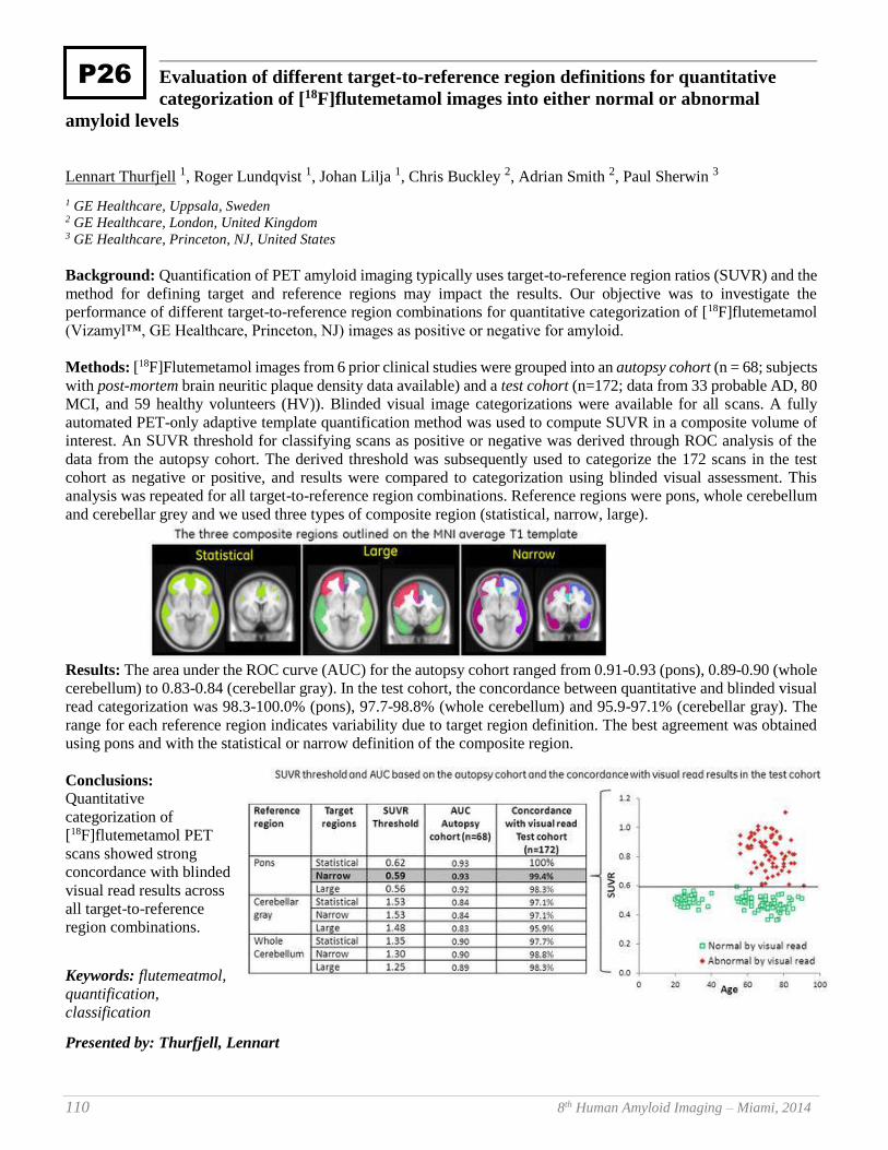
110 8th Human Amyloid Imaging – Miami, 2014
Evaluation of different target-to-reference region definitions for quantitative
categorization of [18F]flutemetamol images into either normal or abnormal
amyloid levels
Lennart Thurfjell 1, Roger Lundqvist 1, Johan Lilja 1, Chris Buckley 2, Adrian Smith 2, Paul Sherwin 3
1 GE Healthcare, Uppsala, Sweden 2 GE Healthcare, London, United Kingdom 3 GE Healthcare, Princeton, NJ, United States
Background: Quantification of PET amyloid imaging typically uses target-to-reference region ratios (SUVR) and the
method for defining target and reference regions may impact the results. Our objective was to investigate the
performance of different target-to-reference region combinations for quantitative categorization of [18F]flutemetamol
(Vizamyl™, GE Healthcare, Princeton, NJ) images as positive or negative for amyloid.
Methods: [18F]Flutemetamol images from 6 prior clinical studies were grouped into an autopsy cohort (n = 68; subjects
with post-mortem brain neuritic plaque density data available) and a test cohort (n=172; data from 33 probable AD, 80
MCI, and 59 healthy volunteers (HV)). Blinded visual image categorizations were available for all scans. A fully
automated PET-only adaptive template quantification method was used to compute SUVR in a composite volume of
interest. An SUVR threshold for classifying scans as positive or negative was derived through ROC analysis of the
data from the autopsy cohort. The derived threshold was subsequently used to categorize the 172 scans in the test
cohort as negative or positive, and results were compared to categorization using blinded visual assessment. This
analysis was repeated for all target-to-reference region combinations. Reference regions were pons, whole cerebellum
and cerebellar grey and we used three types of composite region (statistical, narrow, large).
Results: The area under the ROC curve (AUC) for the autopsy cohort ranged from 0.91-0.93 (pons), 0.89-0.90 (whole
cerebellum) to 0.83-0.84 (cerebellar gray). In the test cohort, the concordance between quantitative and blinded visual
read categorization was 98.3-100.0% (pons), 97.7-98.8% (whole cerebellum) and 95.9-97.1% (cerebellar gray). The
range for each reference region indicates variability due to target region definition. The best agreement was obtained
using pons and with the statistical or narrow definition of the composite region.
Conclusions: Quantitative
categorization of
[18F]flutemetamol PET
scans showed strong
concordance with blinded
visual read results across
all target-to-reference
region combinations.
Keywords: flutemeatmol,
quantification,
classification
Presented by: Thurfjell, Lennart
P26
111
En attendant Centiloid
Victor L Villemagne 1, Paul Yates 1, Kevin Ong 1, Belinda Brown 2, Svetlana Pejoska 1, Robert Williams 1, Robyn
Veljanoski 1, Stephanie Rainey-Smith 2, Kevin Taddei 2, Ralph Martins 2, Colin L Masters 3, Christopher C Rowe 1
1 Department of Nuclear Medicine & Centre for PET, Austin Health, Melbourne, VIC, Australia 2 Edith Cowan Univerity, Perth, WA, Australia 3 The Florey Institute of Neuroscience and Mental Health, The University of Melbourne, VIC, Australia
Background: Researchers often validate their assessments against the amount of Aβ in the brain. Given that the range
of available Aβ tracers have different kinetics and recommended quantification procedures as well as different autopsy-
validated SUVR thresholds to determine high and low Aβ burden, there is a need to standardize all these results and
display them under a single universal scale. There is an international effort to establish this single scale that, under the
name of Centiloid, (Mintun, 2012) will perform head-to-head comparison with the different tracers and PiB in both
young and elderly as well as Alzheimer’s disease patients, in order to cover the whole spectrum of Aβ deposition. In
the meantime, large cohort studies like AIBL require validation of results now, so we adopted a linear regression
approach to transform the results of each tracer generated with their own validated quantification method into PiB-like
units.
Methods: Aβ imaging in 534 participants was performed with three different radiotracers: flutemetamol (n=245),
florbetapir (n=172) and florbetaben (n=117). The results generated with the tracer-specific methods were classified as
High or Low based on their respective neuropathologically-validated thresholds. Linear regression transformations
based on reported head-to-head comparisons of each tracer with PiB were applied to each tracer result. Each tracer
native classification was compared with the classification derived from the transformed data into PiB-like SUVR using
1.50 as a cut-off.
Results: Misclassification after transformation to PiB-like SUVR compared to native classification was extremely
low, with only 3/245 (1.2%) of flutemetamol, and 0% of florbetapir and florbetaben cases assigned into the wrong
category. When misclassification occurred it was restricted to an extremely narrow margin around the 1.50 SUVR
threshold.
Conclusions: While a definitive conversion into centesimal units is worked out, application of linear regression
transformations provide an interim, albeit robust, way of converting results from different Aβ imaging tracers into
familiar PiB SUVR units.
Keywords: Aβ imaging, Aβ radiotracers, centiloid
Presented by: Villemagne, Victor L
P38
112 8th Human Amyloid Imaging – Miami, 2014
Investigating the involvement of the striatum in Down's syndrome and
Alzheimer's disease with 11C-Pittsburgh Compound B-positron emission
tomography (PiB-PET)
Liam Reese Wilson 1, Tiina Annus 1, Shahid Zaman 1, Young Hong 2, Tim Fryer 2, Robert Smith 2, Franklin Aigbirhio2,
Anthony Holland 1
1 Cambridge Intellectual and Developmental Disabilities Research Group (CIDDRG), Department of Psychiatry, University of
Cambridge, UK. 2 Wolfson Brain Imaging Centre, Department of Clinical Neurosciences, University of Cambridge, UK.
Introduction: Down’s syndrome (DS) brings an elevated risk for early onset Alzheimer’s disease (AD). In a subset
of people with DS the early symptoms of AD appear to be changes in behaviour and personality (Holland et al., 2000),
yet post mortem studies indicate AD pathology in DS is indistinguishable from sporadic AD. However, a recent PiB-
PET study of non-demented adults with DS found binding in the striatum for several subjects (Handen et al., 2012),
an area usually thought to be involved later in the course of AD.
Aims: PiB-PET and MRI data from the present study were analysed to investigate PiB retention in the striatum of DS
adults with dementia, in the early stages of dementia, and without dementia.
Methodology: Forty-two participants with DS underwent a MRI scan and a ninety-minute dynamic PiB-PET scan.
BPND maps were created using the simplified reference tissue model (Gunn et al., 1997), with the superior grey matter
of the cerebellum as the reference tissue. A T-1 template was created using the DARTEL toolbox in SPM8, on which
regions of interest (ROIs) were drawn manually. Regional BPND values were then obtained for 5 cortical and 2
subcortical areas.
Results: Binding was most frequently seen in the striatum, which had the highest average BPND value of any region.
Participants who were positive for PiB binding in more than one ROI were always positive in the striatum. A further
3 non-demented participants retained PiB solely in the striatum.
Conclusion: Findings suggest that the striatum may be involved in the early stages of AD in DS; this is in contrast to
published data on sporadic AD but is comparable to data from studies of familial autosomal dominant mutations
causing AD (e.g. Klunk et al., 2007). The implications for people with DS and AD require further investigation.
Keywords: Alzheimer's, Down's syndrome, PiB, Striatum.
Presented by: Wilson, Liam Reese
P33

113
Utility of regional [18F]-Florbetapir PET imaging SUVRs in discriminating
Alzheimer’s disease and its prodromal stages
Qi Zhou 1, Mohammed Goryawala 1, David Loewenstein 2, Warren Barker 3, Ranjan Duara 3, Malek Adjouadi 1
1 Center for Advanced Technology and Education, Florida International University 2 Psychological Services and Neuropsychology Laboratory, Mount Sinai Medical Center 3 Wien Center For Alzheimer's Disease & Memory Disorders, Mount Sinai Medical Center
Purpose: Amyloid-β PET imaging is reported to be effective in diagnosing Alzheimer’s disease (AD) and mild
cognitive impairment (MCI). The goal of this study is to explore the efficacy of AV45 PET scans for delineating
different stages of AD by performing exhaustive two-class classifications of AD, cognitive normal (CN), EMCI and
LMCI subjects.
Methods: 476 dynamic AV45 baseline PET scans from ADNI2 (AD (n=65), EMCI (n=133), LMCI (n=124) and CN
(n=154)) were included in this study. Mean SUVRs for six regions (cerebellum, brainstem, frontal, cingulate, parietal,
and temporal) and weighted whole brain SUVR were used as the 7 classification features. A statistical feature selection
method was used for classification between any two groups using a Support Vector Machine (SVM) classifier. The
results were based on 50 independent and randomized 2-fold cross validation.
Results: Among all the SUVRs, mean frontal SUVR showed the most discriminative power for the classification of
AD, LMCI and EMCI from CN; mean temporal SUVR for both EMCI versus AD and LMCI versus EMCI; and mean
cingulate SUVR for AD versus LMCI. An error analysis was used to select the top-ranked features that yielded the
highest accuracy [see Table 1]. A maximum accuracy of 84.4% (sensitivity: 89.0%) for AD versus CN, 72.5%
(sensitivity: 61.2%) for LMCI versus CN, 62.5% (sensitivity: 40.8%) for EMCI versus CN, 60.1% for LMCI versus
EMCI, 60.4% for AD versus LMCI and 73.3% for AD versus EMCI was obtained. Generally, mean SUVRs of
brainstem and cerebellum were found to be insignificant features for most classification.
Conclusion: Regional amyloid burden can be utilized as a potential biomarker for classification and staging of the
disease, however, its utility is best observed for discriminating AD from CN individuals. Also, mean frontal and
temporal SUVRs were deemed most significant towards classification applications.
Keywords: [18F]-Florbetapir PET Imaging, Alzheimer's disease, mild cognitive impairment, classification
Presented by: Zhou, Qi
P8

114 8th Human Amyloid Imaging – Miami, 2014
Diagnostic value of Amyloid imaging in early onset dementia
Marissa Zwan 1, Femke Bouwman 2, Wiesje van der Flier 3, Adriaan Lammertsma 4, Bart van Berckel 4, Philip
Scheltens 2
1 Alzheimer Center & Department of Neurology, Neuroscience Campus Amsterdam, VU University Medical Center, Amsterdam,
the Netherlands Department of Radiology & Nuclear Medicine, Neuroscience Campus Amsterdam, VU University Medical
Center, Amsterdam, the Ne 2 Alzheimer Center & Department of Neurology, Neuroscience Campus Amsterdam, VU University Medical Center, Amsterdam,
the Netherlands 3 Alzheimer Center & Department of Neurology, Neuroscience Campus Amsterdam, VU University Medical Center, Amsterdam,
the Netherlands Department of Epidemiology & Biostatistics, Neuroscience Campus Amsterdam, VU University Medical Center,
Amsterdam, the Ne 4 Department of Radiology & Nuclear Medicine, Neuroscience Campus Amsterdam, VU University Medical Center, Amsterdam,
the Netherlands
Introduction: In early onset dementia approximately one out of three patients has an atypical clinical presentation,
which substantially complicates correct etiological diagnosis. [18F]Flutemetamol PET, an investigational imaging
agent, is being studied for detection of amyloid deposition, a pathological hallmark of Alzheimer’s Disease (AD).
Methods: We included 80 patients with early onset dementia (age <70 years) and physician diagnostic confidence less
than 90% after a full routine diagnostic work-up for dementia. All patients underwent a [18F]Flutemetamol PET scan
which were visually assessed as amyloid positive or negative. Before and after disclosing PET results, confidence in
clinical diagnosis was determined. Also, impact on patient healthcare management was assessed after disclosing of the
PET imaging results.
Results: [18F]Flutemetamol PET scans were positive in 48 out of 63 (76%) patients diagnosed (pre-PET imaging)
with AD, 1 out of 9 (11%) patients diagnosed with frontotemporal dementia (FTD), and 3 out of 8 (38%) patients
diagnosed with other dementias. Overall, confidence in etiological diagnosis increased from 76±12% to 90±16% after
disclosing PET results (p<0.001). Access to [18F]Flutemetamol PET results led to a change in diagnosis in 16 (20%)
patients. In 11 out of 13 patients, a negative [18F]Flutemetamol PET caused a change of the initial AD diagnosis to
another dementia. In 3 out of 4 other dementia patients, the initial diagnosis was changed to AD after receiving a
positive PET scan. In 27 (34%) patients, PET results led to a change in patient healthcare management. For 13 patients
additional ancillary investigations were planned after access to the PET results.
Conclusion: [18F]Flutemetamol PET resulted in changes in the diagnostic process work-up of early onset dementia
patients and the diagnostic confidence of the managing physicians. Especially in patients diagnosed with AD pre-PET
imaging, a negative PET scan resulted in more changes in clinical diagnosis and patient management.
Keywords: Amyoid Imaging; [18F]Flutemetamol PET; Diagnostic Value; Early onset dementia
Presented by: Zwan, Marissa
P5

115
NOTES

GOLD
SILVER
PLATINUM
Associate
w w w . w o r l d e v e n t s f o r u m . c o m / h a i
The 8h Human Amyloid Imaging Conferenceis supported through educational grants from:
This activity is supported by an educational grant from Lilly USA, LLC.
Funding for this conference was made possible, in part by 1 R13 AG042201-01 from The National Institute on Aging and the NationalInstitute of Biomedical Imaging and Bioengineering. The views expressed in written conference materials or publications and by speakers
and moderators do not necessarily reflect the official policies of the Department of Health and Human Services; nor does mention bytrade names, commercial practices, or organizations imply endorsement by the U.S. Government.
National Instituteon Aging n F H X