Embed Size (px)
Citation preview

Basal lignified star-shaped cavity in oil palm (Elaeis
guineensis Jacq.): early development in nursery and
consequences for BSR control1
J.Ch. Jacquemard2, Edyana Suryana
3, F. Breton
4,
Zulkifli Lubis3, and H. de Franqueville
4.
ABSTRACT
The existence of a high lignified star-shaped cavity at the base of the bole, or more precisely
at the root – bole interface, has been clearly identified for the first time. This cavity appears
to be a perfect culture chamber for Ganoderma development. Random observations of the
initial stage of Basal Stem Rot infection in young palms showed the development of infectious
stroma-like structures inside the cavity prior to colonisation of the bole. The existence of a
lignified scar in 8-month-old seedlings was revealed by initial observation. The scar then
developed further inside the star-shaped cavity (Breton et al, 2009a; Breton et al, 2009b).
Specific observation during early growth stages in the nursery revealed the appearance of a
very tiny scar after 5 months on a few seedlings. The number of affected seedlings increased
rapidly to reach nearly 100% after 9 months. The scar grew quickly in all directions and the
resulting cavity could reach 50% of bole diameter, 13 mm wide and 8 mm deep at this stage.
The star-shaped cavities have 2 to 5 branches.
After 6 to 7 months in the nursery, once the star-shaped cavity was extended enough, the
development of mycelium (not identified) and / or presence of tiny arthropods such as aphids,
Chilopodae larva and small ants ware frequently observed. These results demonstrate the
role of this star-shaped cavity as a perfect culture chamber for fungus and a possible refuge
for pests, predators or disease vectors at very early stages of oil palm development.
This star-shaped cavity definitely appears to be an interesting target for preventive action
against Basal Stem Rot, i.e. Biocontrol by antagonistic, long-lasting fungicide, etc.
Key words: Elaeis guineensis, palm oil, lignified star-shaped cavity, nursery, basal stem rot
1 Paper presented at IOPC 2010 “Transforming Oil Palm Industry”. 1 – 3 June 2010, Jogja Expo Center,
Yogyakarta, Indonesia 2PT Socfindo Medan / UR Oil Palm Breeding – CIRAD Bios, PO Box 1254 – Medan 20001 – Sumatera Utara
– Indonesia 3 PT Socfindo Medan – PO Box 1254 – Medan 20001 – Sumatera Utara – Indonesia
4 CIRAD Oil Palm Breeding, TA A 28/01, Avenue Agropolis, Montpellier F34398 Cedex 5, France
INTRODUCTION
Ganoderma boninense, which causes a Basal Stem Rot (BSR) on Elaeis guineensis Jacq. is
the most dangerous soil-borne fungus. It is wide spread across oil palm estates in Southeast –
Asia. The disease appears to be increasingly economically important with the accumulation
of oil palm growing in the same place. Fields planted with susceptible material could be
rapidly devastated and the remaining density could be reduced to eighty trees per ha after 18
to 20 years of production.
The presence of high melanised mycelia or stroma-like structure (SLS) is necessary for
infection. SLS require specific conditions to develop: hard substrate, dark environment,
humidity and low competition (Breton et al, 2005a; Breton et al, 2005b).
Extensive observations on oil palms at all stages and on all continents have revealed the
existence of a lignified star- shaped cavity at the root bole interface. SLS have been detected
on the outer section of this star-shaped cavity (Breton et al, 2009a). Random observation of
the initial stage of infection on young palms showed that SLS development occurs before
penetration and colonisation of the palm bole (Breton et al, 2009b).
The star-shaped cavity displays all the prerequisite qualities to be a perfect culture chamber
for Ganoderma boninense SLS: hard substrate, dark environment, high humidity and low
competition. A scar, a future star-shaped cavity has been observed on the root – bole interface
of eight and eighteen-month-old seedlings (Breton et al, 2009a; Breton et al, 2009b).
In order to have a clearer understanding of cavity development, an experiment was carried
out in the nursery at the Aek Loba Estate. The aim was to detect the possible appearance of
the scar as early as possible and, if possible, understand how the scar was developing inside
the cavity.
EXPERIMENT
MATERIALS
400 five-month-old seedlings originating from one D Dabou Deli x T la Mé cross transferred
in the nursery in early March 2009.
Nursery bag: Socfindo standard
Nursery spacing: Socfindo standard
Water supply: Socfindo standard
Fertilisation: Socfindo standard
METHODS
20 seedlings were sacrificed each month. They were taken following their rank in the nursery
without choice. The nursery bags were opened with a knife and the soil carefully removed in
order to keep all the roots in their original state. The seedlings were transferred to the nursery
office.
At the office, all the primary roots were cut up to two centimetres from the bole. They were,
then, examined and divided into four categories: new, normal, injured and rotten.

New roots: freshly emitted roots, white, turgescent and without any secondary root
emission
Normal roots: roots with normal shape
Injured roots: roots bearing within their first 30 centimetres one or more re-growing
sections or rotten section not joining the bole
Rotten root: root that is rotten up to the bole
Then, with a very sharp large knife, the remaining root bases were cut off until the bole – root
interface was reached. This operation was very tricky, because the thickness of this interface
was a very tiny target at the beginning.
After cleaning, the bole – root interface was examined and all the possible observations were
recorded such as the existence or not of any early signs of a scar or cavity, mycelium, fungus,
or insects.
After initial observations, we added characterisation of scar morphology, then cavity
measurements (extension, width and depth) and, at least for the last observation, bole
diameter. All measurements were taken with a calliper square.
Eight dissections were carried out from 20/04/2009 to 27/01/2010 (Table 1). Pictures were
taken to characterise all significant steps.
RESULTS
The first series of data concerned root emissions (Table 2). After 1 month in the nursery,
around 60% of the roots were injured by the transfer process. After that, the number of
injured roots decreased regularly, probably through the natural process of decay and
replacement. On average, the number of new roots rose from 2 at 1 month to 4 – 5 between 8
and 11 months old. In parallel, the number of rotten root bases detected on the bole increased
slowly with a peak in the 7th
month. The total number of active primary roots was multiplied
by 4 over the 10 months of observation.
The first suspicion of a scar appeared during the second series of observations on 05/05/2009,
i.e. after 1.7 months in the main nursery. Table 3 summarises scar and cavity presence for
each dissection series. Between second and third dissections, around 35 to 40% of dissected
seedlings showed suspicious markings at the root – bole interface. Cavities were clearly
identified after 5 months in the nursery on 55% of the seedlings. All the seedlings displayed a
cavity after 8 months in the main nursery.
Cavities differed in appearance: an analysis of their morphology can be founded in Table 4.
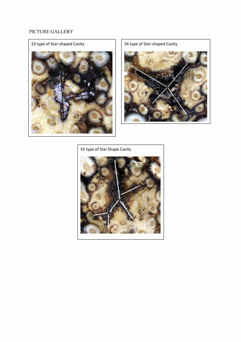
Basically, 4 morphological types were identified (see picture gallery):
S2: lengthened cavity with 2 extremities like irregular distaff
S3: star-shaped cavity with 3 irregular branches
S4: star-shaped cavity with 4 irregular branches
S5: star-shaped cavity with 5 irregular branches
Width and depth of cavities observed during the fourth dissection (5.4 months in the main
nursery) measured less than 2 mm. These cavities then grew significantly in all directions.
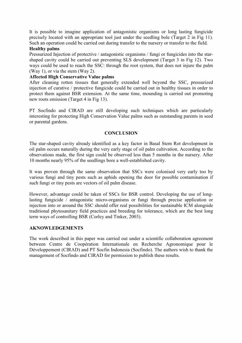
Table 5 summarizes development of the cavity at the root – bole interface. At 6.8 month-old,

the cavity reached already 15 mm in length and 3 mm in width on average. Three months
later, the cavity size had doubled and amounted to 40% of the bole diameter.

DISCUSSION
Very early occurrence of a scar or cavity was not that easy to identify because we did not
know what we were looking for. Figure 1 shows the appearance of normal root – bole
inferface. Successive root sections could be clearly identified. At some places, lignified spots
indicated the position of rotten roots (see arrow).
From time to time, rotten root bases appeared on the bole – root interface. This was very clear
on 1 to 3-month-old seedlings. Rotten tissues invaded the area around the root, but generally
not its central cylinder (Fig 2). This figure did not generate a scar or lignified cavity at this
stage.
Early development of the cavity
The very earliest cases we were able to recognise were small root bases, narrowing at the
middle of the bole – root interface, bearing one or two tiny cracks, 1 to 2 mm in length,
starting to divide its section into two parts. Such crack development appeared, initially,
external to the root (Fig 3) then grew through the root (Fig 4).
Very soon afterwards, a scar is developed like an irregular meandering line at the bole – root
interface (Fig 5). After a few weeks, the width and the depth of the scar increased allowing
development of the first cavity stages. Well developed cavities were always 4 to 6 times
longer than the width. The depth also appeared to be always slightly smaller than the cavity
width.
As mentioned in the results, this cavity could develop 2 to 5 irregular branches. The picture
gallery in the annex shows the morphology of such cavities.
At some bole – root interfaces (Fig 6), we saw appearance of rotten tissues at the narrowing
tip of a star branch. Rotten tissues were a pale brownish-yellow colour and really seemed to
be extending the cavity from a root base. A vertical section along the cavity length (Fig 7)
showed already lignified tissues limiting the cavity deeper in the bole and around the root
base, the early beginnings of scarring.
Observation carried out earlier by the authors indicated that SSC development also depended
on the genetic background, the origin of the planting material, or growth / stress conditions,
as shown by cavity extension at the bole – root interface of adult palms in Indonesia, Africa
or South America (Breton et al, 2009a). More studies need to be undertaken on that subject in
the future.
Cavity colonisation
At very early stages, the cavity was refilled with rotten and dry tissues, as shown in figure 7.
But very quickly, as can be seen from the picture gallery, the cavity developed free space at
the bole – root interface
Very early, this cavity was colonised by tiny animals such as Aphids (Fig 8) with their
cortege of companions such as ants (Monomorium sp) or predators such as Chilopodae larva.
Such colonisation was found in 3 out of 4 well-established cavities on 5-month-old seedlings.
Then, colonisation by an undetermined fungus was revealed one month after by the presence
of mycelium spots (yellow or white spots). As time went by, more and more cavities showed
such contamination by spots or large mycelium patches (Fig 9). This fungus development
was probably promoted by the presence of aphids with their sweet secretions too. After 10
months in the nursery, more than 70% of well established cavities were contaminated.
This proved that from a very early stage of seedling development, the cavity displayed all the
prerequisite qualities mentioned by Breton et al (2005a, 2005b) to be a perfect culture
chamber for Ganoderma boninense (SLS): hard substrate, dark environment, humidity and
low competition.
This repeated degradation of the tissues around the root bases as a possible explanation of the
cavity expansion could be the weakness point of the bole protection if the stroma-like
structure is well developed on a significant part of the cavity wall.
Under other conditions and on other continents, such contamination by tiny animals, bacteria
or whatever else could be a vector for oil palm disease.
Making the cavity beneficial for the plant
Attempts to control BSR through fungicide application or stem injection have been largely
documented in the literature. These methods are summarized by Turner & Gillbanks (2003)
or Corley & Tinker (2003).
More recently, use of antagonistic micro-organisms such as Trichoderma harzianum, T.
viride, Pseudomonas fluorescens or arbuscular Mycorrhizal fungi was promoted as an
efficient tool to control BSR development (Karthikeyan et al, 2006; Jayanthi Nagappan et al,
2009; Mhod Ahdly Arbain and Tey Chin Chong, 2009; Nor Sarashimatun and Tey, 2009;
Shamala Sundram et al, 2008; Susanto et al, 2005).
Our observations confirm that SSCs, which are always in central position, are a good target
for mitigation of Basal Stem Rot (BSR) development through the application of antagonistic
micro-organisms / fungi or long-lasting fungicides (Breton et al, 2009 a, Breton et al, 2009
b).
Combining efficient fungicides and / or antagonistic micro-organisms / fungi as mentioned
above and the characteristics of star-shaped cavity development, it could be possible to
promote more sustainable BSR control by precise application in or around SSCs.
Thus, several targets could be investigated: germinated seeds, young seedlings during transfer
phases to the nursery and / or to the field, young palms or still healthy palms, palms already
affected by BSR.
Germinated seeds
Antagonistic organisms could be inoculated on the germ just before transfer to the prenursery
(Target 1 in Fig 10).
Young seedlings
It is possible to imagine application of antagonistic organisms or long lasting fungicide
precisely located with an appropriate tool just under the seedling bole (Target 2 in Fig 11).
Such an operation could be carried out during transfer to the nursery or transfer to the field.
Healthy palms
Pressurized Injection of protective / antagonistic organisms / fungi or fungicides into the star-
shaped cavity could be carried out preventing SLS development (Target 3 in Fig 12). Two
ways could be used to reach the SSC: through the root system, that does not injure the palm
(Way 1), or via the stem (Way 2).
Affected High Conservative Value palms
After cleaning rotten tissues that generally extended well beyond the SSC, pressurized
injection of curative / protective fungicide could be carried out in healthy tissues in order to
protect them against BSR extension. At the same time, mounding is carried out promoting
new roots emission (Target 4 in Fig 13).
PT Socfindo and CIRAD are still developing such techniques which are particularly
interesting for protecting High Conservation Value palms such as outstanding parents in seed
or parental gardens.
CONCLUSION
The star-shaped cavity already identified as a key factor in Basal Stem Rot development in
oil palm occurs naturally during the very early stage of oil palm cultivation. According to the
observations made, the first sign could be observed less than 5 months in the nursery. After
10 months nearly 95% of the seedlings bore a well-established cavity.
It was proven through the same observation that SSCs were colonised very early too by
various fungi and tiny pests such as aphids opening the door for possible contamination if
such fungi or tiny pests are vectors of oil palm disease.
However, advantage could be taken of SSCs for BSR control. Developing the use of long-
lasting fungicide / antagonistic micro-organisms or fungi through precise application or
injection into or around the SSC should offer real possibilities for sustainable ICM alongside
traditional phytosanitary field practices and breeding for tolerance, which are the best long
term ways of controlling BSR (Corley and Tinker, 2003).
AKNOWLEDGEMENTS
The work described in this paper was carried out under a scientific collaboration agreement
between Centre de Coopération Internationale en Recherche Agronomique pour le
Développement (CIRAD) and PT Socfin Indonesia (Socfindo). The authors wish to thank the
management of Socfindo and CIRAD for permission to publish these results.

REFERENCES
BRETON F, HASAN Y, HARIADI, LUBIS Z. & de FRANQUEVILLE H (2005a).
Characterization of parameters for the development of an early screening test for basal stem
rot tolerance in oil palm progenies. In Agriculture, biotechnology & sustainability
conference, PIPOC International Palm Oil Congress, 167-183 (Eds Malaysian Palm Oil
Board). Kuala Lumpur.
BRETON F, HASAN Y, HARIADI, LUBIS Z. & de FRANQUEVILLE H (2005b).
Rhizotron: a demonstrative tool for monitoring in vivo the infection process of oil palm
seedlings by Ganoderma boninense. In Agriculture, biotechnology & sustainability
conference, PIPOC International Palm Oil Congress, 971 (Eds Malaysian Palm Oil Board).
Kuala Lumpur.
BRETON F, MIRANTI R, LUBIS Z, HAYUN Z, UMI S, FLORI A, NELSON SCP,
DURAND – GASSELIN T, JACQUEMARD JC and de FRANQUEVILLE H (2009a).
Ganoderma disease of the oil palm: Hypothesis on natural infection and implementation of
an early artificial inoculation test to screen oil palm progenies for their level of resistance and
hypothesis on natural infection. Communication presented to XVI International Oil Palm
Conference and Expopalma “Challenges in Sustainable Oil Palm Development”. 22 – 25
September 2009, Cartagena de Indias, Colombia.
BRETON F, LUBIS Z, MIRANTI R, JACQUEMARD JC and de FRANQUEVILLE H
(2009b). A Lignified Star-Shape Cavity at Root-Bole Interface: An Appropriate Culture
Chamber for Ganoderma boninense and Stromatic-Like Structures Development. In
proceedings of PIPOC 2009 “Palm Oil – balancing Ecologics with Economics”, Kuala
Lumpur Convention Center, 9 – 12 November 2009, Kuala Lumpur, Malaysia (Merit Award),
540 - 548.
CORLEY R H V and TINKER P B (2003). The Oil Palm – Fourth Edition. Blackwell
Science Ed. Oxford. UK. 562 p.
JAYANTHI NAGAPPAN, FARIDAH ABDULLAH, SHAMALA SUNDRAM and
RAJINDER SINGH (2009). Assessment of Trichoderma species as Biocontrol of Ganoderma
boninense, Basal Stem Rot of oil palm. In proceedings of PIPOC 2009 “Palm Oil –
balancing Ecologics with Economics”, Kuala Lumpur Convention Center, 9 – 12 November
2009, Kuala Lumpur, Malaysia. 977 – 990.
KARTIKEYAN G, RAGUCHANDER T and RABINDRAN R (2006). Integrated
management of basal stem rot / Ganoderma disease of coconut in India. Crop Research, 32
(1): 121 – 123.
MODH AHDLY ARBAIN and TEY CHIN CHONG (2009). Field application of
Trichoderma and Arbuscular Mycorrhizal fingi for the control of Ganoderma Basal Stem Rot
of oil palm. In proceedings of PIPOC 2009 “Palm Oil – balancing Ecologics with
Economics”, Kuala Lumpur Convention Center, 9 – 12 November 2009, Kuala Lumpur,
Malaysia. 439 – 449.

NOR SARASHIMATUN S and TEY C C (2009). Application of Arbuscular Mycorrhyzal
fungi for controlling Ganoderma basal Stem rot of oil palm. In proceedings of PIPOC 2009
“Palm Oil – balancing Ecologics with Economics”, Kuala Lumpur Convention Center, 9 –
12 November 2009, Kuala Lumpur, Malaysia, 415 – 424.
SHAMALA SUNDRAM, FARIDAH ABDULLAH, ZAINAL ABIDIN MIOR AHMAD and
UMI KALSOM YUSUF (2008). Efficacy of single and mixed treatments of Trichoderma
harzianum as bio-control agents of Ganoderma basal stem rot in oil palm. Journal of Oil
Palm Research, 20 (June): 470 – 483.
SUSANTO A, SUDHARTO P S and PURBA R Y (2005). Enhancing biological control of
basal stem rot disease (Ganoderma boninense) in oil palm plantations. Mycopathologia, 159 :
153 – 157.
TURNER PD and GILLBANKS A (2003). Oil Palm Cultivation and Management – Second
Edition. The Incorporated Society of Planters Ed. Kuala Lumpur. Malaysia. 915 p.

Table 1: Date of dissection and age of seedlings (in months after transfer to the main nursery)
Dissection Date Age
First 20/04/2009 1.2
Second 05/05/2009 1.7
Third 13/06/2009 3.0
Fourth 24/08/2009 5.4
Fifth 06/10/2009 6.8
Sixth 29/10/2009 7.6
Seventh 25/11/2009 8.5
Eighth 27/01/2010 10.6

Table 2: Number of roots Date of observation 20/04/2009 05/05/2009 13/06/2009 24/08/2009 06/10/2009 29/10/2009 25/11/2009 27/01/2010
Total number of roots 10.1 ± 0.52 11.0 ± 0.73 13.9 ± 0.77 20.5 ± 1.62 23.9 ± 1.63 29.1 ± 1.57 33.0 ± 2.56 39.1 ± 3.68
New roots 2.0 ± 0.40 2.1 ± 0.58 3.4 ± 0.54 3.0 ± 0.73 3.8 ± 0.88 3.9 ± 0.60 5.0 ± 1.00 4.5 ± 1.31
Normal roots 1.7 ± 0.61 2.8 ± 0.63 5.5 ± 0.89 13.2 ± 1.35 17.7 ± 1.53 21.8 ± 1.94 25.7 ± 2.24 33.0 ± 2.31
Injured roots 6.2 ± 0.50 5.6 ± 0.41 4.1 ± 0.52 3.8 ± 0.94 1.4 ± 0.67 1.9 ± 0.70 1.6 ± 0.64 1.2 ± 0.54
Rotten roots 0.3 ± 0.24 0.6 ± 0.30 0.9 ± 0.47 0.5 ± 0.30 1.1 ± 0.52 1.6 ± 0.75 0.8 ± 0.69 0.5 ± 0.30
Table 3: Number of root-bole interfaces showing a scar or a cavity Date of observation 20/04/2009 05/05/2009 13/06/2009 24/08/2009 06/10/2009 29/10/2009 25/11/2009 27/01/2010
No scar / cavity 20 12 13 5 5 3 0 0
Suspicious scar 0 8 7 4 1 0 0 0
Cavity 0 0 0 11 14 17 20 20
Total 20 20 20 20 20 20 20 20
Table 4: Morphology of scar/cavity on root-bole interface Date of observation 20/04/2009 05/05/2009 13/06/2009 24/08/2009 06/10/2009 29/10/2009 25/11/2009 27/01/2010
Suspicious 0 8 7 0 0 0 0 0
Very early 0 0 0 16 13 2 2 1
S2 0 0 0 4 1 5 5 6
S3 0 0 0 0 0 9 9 8
S4 0 0 0 0 0 1 4 4
S5 0 0 0 0 0 0 0 1
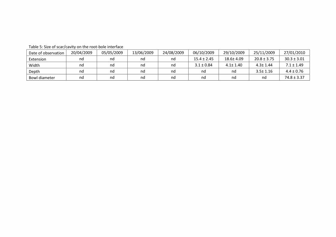
Table 5: Size of scar/cavity on the root-bole interface Date of observation 20/04/2009 05/05/2009 13/06/2009 24/08/2009 06/10/2009 29/10/2009 25/11/2009 27/01/2010
Extension nd nd nd nd 15.4 ± 2.45 18.6± 4.09 20.8 ± 3.75 30.3 ± 3.01
Width nd nd nd nd 3.1 ± 0.84 4.1± 1.40 4.3± 1.44 7.1 ± 1.49
Depth nd nd nd nd nd nd 3.5± 1.16 4.4 ± 0.76
Bowl diameter nd nd nd nd nd nd nd 74.8 ± 3.37
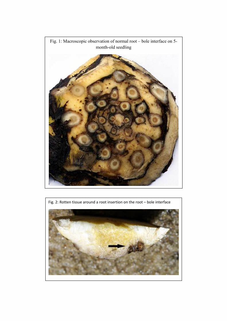
Fig. 2: Rotten tissue around a root insertion on the root – bole interface
Fig. 1: Macroscopic observation of normal root – bole interface on 5-
month-old seedling
Fig. 3: Very early scar foreshadowing future star-shaped cavity
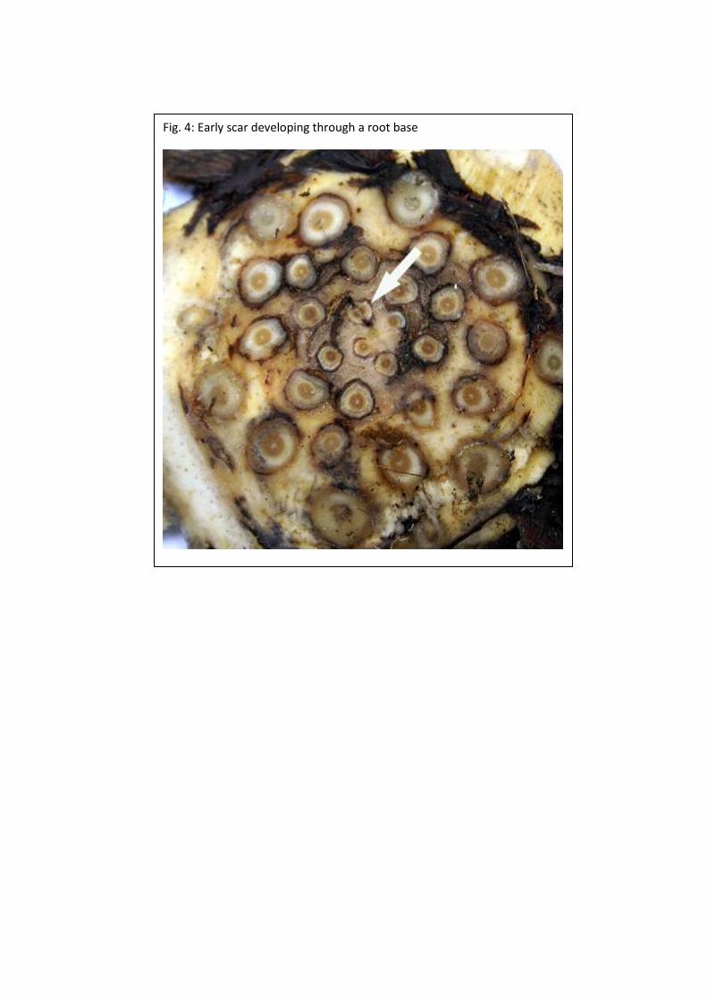
Fig. 4: Early scar developing through a root base

Fig. 5: Young SSC developing through the root – bole interface
Fig. 6: Rotten tissues extending SSC
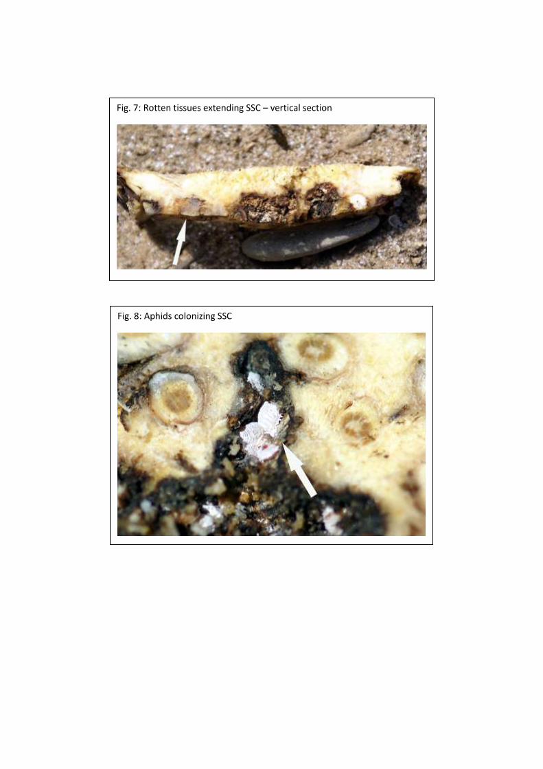
Fig. 7: Rotten tissues extending SSC – vertical section
Fig. 8: Aphids colonizing SSC
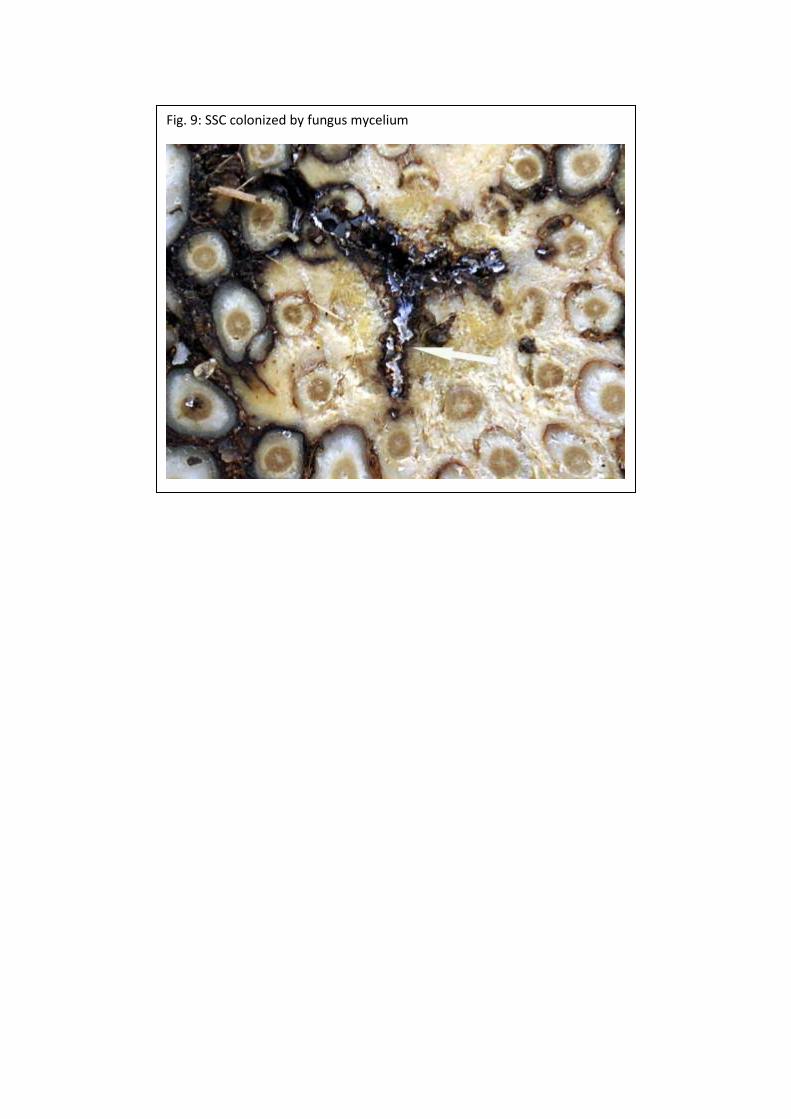
Fig. 9: SSC colonized by fungus mycelium

Fig. 10: Target of possible action on
germinated seeds
Fig. 11: Target of possible action on
seedlings or young palm
Fig. 12: Target of possible
prophylactic action on adult palm
Fig. 13: target of possible curative action on adult palm
PICTURE GALLERY
S3 type of Star-shaped Cavity
S4 type of Star-shaped Cavity
S5 type of Star Shape Cavity


![1019-1100 ASD · The studies and observations of the so-called ‘amarillamiento-secamiento’ [yellowing and drying] disorder in oil palm (Elaeis guineensis Jacq.) carried out over](https://img.pdfslide.us/doc/110x75/5bb3467909d3f2c5168b4b0a/1019-1100-the-studies-and-observations-of-the-so-called-amarillamiento-secamiento.jpg)