Embed Size (px)
Citation preview
International Journal of
Molecular Sciences
Article
Gran1: A Granulysin-Derived Peptide with Potent Activityagainst Intracellular Mycobacterium tuberculosis
Reiner Noschka 1, Fanny Wondany 2, Gönül Kizilsavas 3, Tanja Weil 3, Gilbert Weidinger 4, Paul Walther 5,Jens Michaelis 2 and Steffen Stenger 1,*
�����������������
Citation: Noschka, R.; Wondany, F.;
Kizilsavas, G.; Weil, T.; Weidinger, G.;
Walther, P.; Michaelis, J.; Stenger, S.
Gran1: A Granulysin-Derived
Peptide with Potent Activity against
Intracellular Mycobacterium
tuberculosis. Int. J. Mol. Sci. 2021, 22,
8392. https://doi.org/10.3390/
ijms22168392
Academic Editors: Maria
Rosalia Pasca, Vadim Makarov,
Giulia Degiacomi and
Laurent Chiarelli
Received: 14 July 2021
Accepted: 3 August 2021
Published: 4 August 2021
Publisher’s Note: MDPI stays neutral
with regard to jurisdictional claims in
published maps and institutional affil-
iations.
Copyright: © 2021 by the authors.
Licensee MDPI, Basel, Switzerland.
This article is an open access article
distributed under the terms and
conditions of the Creative Commons
Attribution (CC BY) license (https://
creativecommons.org/licenses/by/
4.0/).
1 Institute of Medical Microbiology and Hygiene, University Hospital Ulm, 89081 Ulm, Germany;[email protected]
2 Institute of Biophysics, Ulm University, 89081 Ulm, Germany; [email protected] (F.W.);[email protected] (J.M.)
3 Max Planck Institute for Polymer Research, 55128 Mainz, Germany; [email protected] (G.K.);[email protected] (T.W.)
4 Institute of Biochemistry and Molecular Biology, Ulm University, 89081 Ulm, Germany;[email protected]
5 Central Facility for Electron Microscopy, Ulm University, 89081 Ulm, Germany; [email protected]* Correspondence: [email protected]
Abstract: Granulysin is an antimicrobial peptide (AMP) expressed by human T-lymphocytes and nat-ural killer cells. Despite a remarkably broad antimicrobial spectrum, its implementation into clinicalpractice has been hampered by its large size and off-target effects. To circumvent these limitations,we synthesized a 29 amino acid fragment within the putative cytolytic site of Granulysin (termed“Gran1”). We evaluated the antimicrobial activity of Gran1 against the major human pathogenMycobacterium tuberculosis (Mtb) and a panel of clinically relevant non-tuberculous mycobacteriawhich are notoriously difficult to treat. Gran1 efficiently inhibited the mycobacterial proliferationin the low micro molar range. Super-resolution fluorescence microscopy and scanning electronmicroscopy indicated that Gran1 interacts with the surface of Mtb, causing lethal distortions of thecell wall. Importantly, Gran1 showed no off-target effects (cytokine release, chemotaxis, cell death)in primary human cells or zebrafish embryos (cytotoxicity, developmental toxicity, neurotoxicity,cardiotoxicity). Gran1 was selectively internalized by macrophages, the major host cell of Mtb, andrestricted the proliferation of the pathogen. Our results demonstrate that the hypothesis-drivendesign of AMPs is a powerful approach for the identification of small bioactive compounds withspecific antimicrobial activity. Gran1 is a promising component for the design of AMP-containingnanoparticles with selective activity and favorable pharmacokinetics to be pushed forward intoexperimental in vivo models of infectious diseases, most notably tuberculosis.
Keywords: tuberculosis; antimicrobial peptide; Granulysin; human; macrophages
1. Introduction
Granulysin is an antimicrobial peptide (AMP) which is stored in the granules ofhuman T-lymphocytes and natural killer cells [1]. The recombinant 9 kDa form of theprotein is active against fungi, parasites, and bacteria, including Mycobacterium tuberculosis(Mtb) [2]. Killing of the microbes is associated with the ability to disrupt cell membranesand to facilitate the granzyme-mediated perturbation of mitochondria [2,3]. Granulysinalso interacts with eukaryotic cell membranes, resulting in apoptosis of healthy and ma-lignant cells [4,5]. As with several other AMPs, Granulysin has immune modulatoryproperties, and induces chemotaxis, the maturation of macrophages, and the release ofpro-inflammatory cytokines [6,7]. While the combination of inflammation and apoptosisis beneficial for fighting malignant diseases, it is a double-edged sword in the treatmentof chronic diseases, where (hyper)-inflammation is the pathophysiological hallmark of
Int. J. Mol. Sci. 2021, 22, 8392. https://doi.org/10.3390/ijms22168392 https://www.mdpi.com/journal/ijms

Int. J. Mol. Sci. 2021, 22, 8392 2 of 20
tissue destruction and severe disease. Accordingly, Granulysin mediates tissue damagein graft-versus-host disease and inflammatory skin diseases such as folliculitis, psoriasis,acne, or Steven Johnsons Syndrome [8]. Besides these off-target effects, the large size ofGranulysin (9 kDa, 74 aa) accounts for rapid degradation in serum, the requirement of highmicromolar concentrations for activity, and high costs for manufacturing. Finally, Gran-ulysin alone is not active against intracellular pathogens, because it requires the supportof perforin and granzymes to enter the intracellular compartment [2,9]. One approachto overcome these limitations is the identification of small Granulysin-derived peptideswith specific bioactivities such as killing microbes, lysing cancer cells, or promoting inflam-mation [10–14]. Recent studies have demonstrated that helix 2 and helix 3 are essentialfor the antimicrobial activity of Granulysin [13,15]. By studying overlapping peptides,it was shown that the peptide “G8” (23–51 aa) lysis the extracellular, gram negative rodSalmonella typhimurium [13]. Based on these findings, we hypothesized that “G8” is activeagainst extra- and intracellular Mtb without causing an inflammatory response. We selectedmycobacteria as the target organisms because (i) the parental Granulysin is active againstextracellular Mtb [2], (ii) mycobacteria are major human pathogens, (iii) mycobacteria are in-trinsically resistant against most classes of antibiotics, (iv) the occurrence of infections withdrug resistance strains is increasing (multidrug-resistant and extensively drug-resistanttuberculosis), [16] and (v) the available treatment which lasts at least 6 months and consistsof a multidrug-regimen with potentially severe side effects. Therefore, novel therapeu-tic strategies against tuberculosis and related mycobacterial infections are desperatelyneeded. Here, we demonstrate that Gran1 (previously described as “G8”) interacts withthe mycobacterial cell wall and exerts antimicrobial activity against extra- and intracellularMtb without triggering inflammatory immune responses or cell death in human cells orzebrafish embryos.
2. Results2.1. Structure and Physicochemical Characteristics of Gran1
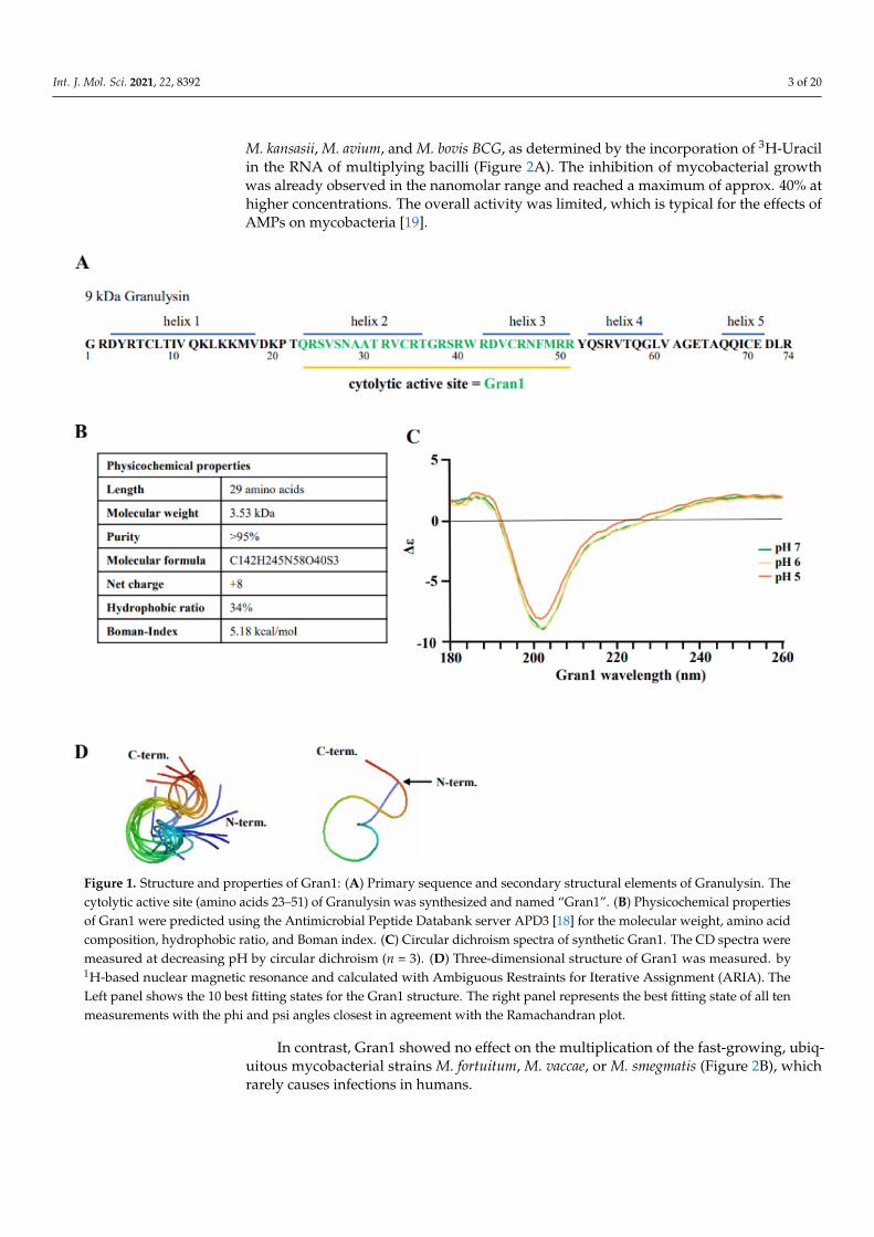
To investigate the interaction of Gran1 with extra- and intracellular mycobacteria, thefragment 23–51 aa within the cytolytic active site of the 9 kDa Granulysin was synthe-sized (Figure 1A). The peptide contains 29 amino acids, resulting in a molecular weightof 3.53 kDa. The ratio of hydrophobic residues within Gran1 is 34% and the net chargeis +8, which are common characteristics for antimicrobial peptides and are predictive foran efficient interaction with the microbial cell wall. The Boman index, when used as asurrogate marker for the potential of an AMP to bind to membranes, is 5.18 kcal/mol,indicating high binding potential (Figure 1B). Structural analysis by circular dichroism(CD) measurements revealed that Gran1 follows a random coil-like structure at a neutralpH, which was maintained at acidic pH 5 and 6 (Figure 1C). The 3-dimensional struc-ture of Gran1 in the solution (Figure 1D) was based on 1H Nuclear Resonance (NMR)spectroscopy data and calculations from Ambiguous Restraints for Iterative Assignment(ARIA) [17]. The 10 best fitting structure outputs of the calculations, based on NMR data(Figure 1D, left panel), and the state best fitting to the data that is in closest agreement withthe Ramachandran plot (right panel), suggested that Gran1 does not form characteristicα-helices. Both CD and NMR spectroscopy indicated that Gran1 appears in a flexible,random coil-like conformation, in contrast to the helical structure of this fragment in thefull-length protein. Overall, Gran1 fulfills the major physicochemical and structural criteriato interact with the mycobacterial cell wall.
2.2. Antibacterial Activity of Gran1 against Extracellular Mycobacteria
Based on these structural findings, we hypothesized that Gran1 inhibits the growthof bacterial pathogens. As model organisms, we initially evaluated the effect of Gran1 onnon-tuberculous mycobacteria (NTM), which are becoming an increasing clinical problemand have an intrinsic resistance against the majority of conventional antibiotics. Gran1showed dose-dependent activity against the three slow-growing mycobacterial species
Int. J. Mol. Sci. 2021, 22, 8392 3 of 20
M. kansasii, M. avium, and M. bovis BCG, as determined by the incorporation of 3H-Uracilin the RNA of multiplying bacilli (Figure 2A). The inhibition of mycobacterial growthwas already observed in the nanomolar range and reached a maximum of approx. 40% athigher concentrations. The overall activity was limited, which is typical for the effects ofAMPs on mycobacteria [19].
Figure 1. Structure and properties of Gran1: (A) Primary sequence and secondary structural elements of Granulysin. Thecytolytic active site (amino acids 23–51) of Granulysin was synthesized and named “Gran1”. (B) Physicochemical propertiesof Gran1 were predicted using the Antimicrobial Peptide Databank server APD3 [18] for the molecular weight, amino acidcomposition, hydrophobic ratio, and Boman index. (C) Circular dichroism spectra of synthetic Gran1. The CD spectra weremeasured at decreasing pH by circular dichroism (n = 3). (D) Three-dimensional structure of Gran1 was measured. by1H-based nuclear magnetic resonance and calculated with Ambiguous Restraints for Iterative Assignment (ARIA). TheLeft panel shows the 10 best fitting states for the Gran1 structure. The right panel represents the best fitting state of all tenmeasurements with the phi and psi angles closest in agreement with the Ramachandran plot.
In contrast, Gran1 showed no effect on the multiplication of the fast-growing, ubiq-uitous mycobacterial strains M. fortuitum, M. vaccae, or M. smegmatis (Figure 2B), whichrarely causes infections in humans.

Int. J. Mol. Sci. 2021, 22, 8392 4 of 20
Figure 2. Activity of Gran1 against extracellular mycobacteria: Extracellular mycobacteria (2 × 10⁶) were incubated withincreasing concentrations of Gran1 followed by incubation with 3H-Uracil for 18 h. The 3H-Uracil uptake was measuredby scintillation counting. Antibacterial activity was determined for (A) slow-growing strains M. kansasii (n = 4), M. avium(n = 5), M. bovis BCG (n = 5) and (B) fast-growing strains M. fortuitum (n = 4), M. vaccae (n = 5), and M. smegmatis (n = 5).Data points show the mean antibacterial activity (%) ± SEM calculated from triplicates of each independent experiment.Dotted horizontal lines indicate background activity of diluent control.
Next, we measured the antimicrobial activity of Gran1 on slow growing, highlypathogenic Mtb. Gran1 already limited the growth of M. tuberculosis by 15% at a con-centration of 10 nM (Figure 3A). This effect peaked at a concentration of 1 µM, wheregrowth was reduced by 38% when compared to un-treated control samples. Based on thedogma that AMP-mediated killing is mediated by the interaction of the positively chargedpeptide residues with the negatively charged bacterial cell walls [19], we investigatedthe interaction between Gran1 and extracellular Mtb with super-resolution fluorescencemicroscopy, using a STED microscope. Extracellular Mtb were labelled with a red flu-orescent dye and were exposed to Gran1 (1 µM) for 30 min (Figure 3B). In un-treatedsamples (left panel), STED microscopy revealed rod-shape bacilli with a smooth cell sur-face. Gran1-treated bacteria (middle panel) showed an irregularly labelled surface, and thecell wall appeared fragmented, indicating alterations of the bacterial surface. To visualizethe interaction of Mtb and Gran1, peptide-treated, labelled Mtb were stained with primary

Int. J. Mol. Sci. 2021, 22, 8392 5 of 20
anti-Gran1 antibodies and a green-conjugated secondary antibody. In addition to the cell al-terations, we observed a direct co-localization of the peptide and the mycobacterial surface(Figure 3B, right panel). To gain a detailed insight into the Gran1-mediated morphologicaleffects on the Mtb-surface, we analyzed Gran1-exposed Mtb using scanning electron mi-croscopy (Figure 3C). While untreated Mtb displayed a smooth surface with no alterations(Figure 3C, left panel), Gran-exposed Mtb showed surface lesions, including holes (middlepanel), blebbing with irregular clusters, and distortions (right panel). We concluded thatthe mechanism of the Gran1-mediated growth inhibition of extracellular Mtb is related tothe disruption of the integrity of the mycobacterial cell wall.
Figure 3. Colocalization of Gran1 with Mtb and cell wall distortion: (A) Extracellular Mtb (2 × 10⁶) were incubated for72 h with increasing concentrations of Gran1. Afterwards 3H-Uracil was added for 18 h, and uptake was measured byscintillation counting. Data points show the mean antibacterial activity (%) ± SEM calculated from triplicates of twentyindependent experiments. Dotted horizontal lines indicate background activity of diluent control. (B) Extracellular Mtbwere labelled with NHS-Atto647N and left un-treated (left panel) or were incubated with Gran1 for 30 min (middle andright panel). For signal overlay of mycobacteria and the peptide, Gran1 was labelled using an anti-Gran1 antibody and anAtto594-conjugated secondary antibody (right panel). Samples were acquired by STED microscopy. The middle panel showsGran-1 treated, NHS-Atto647N-labelled Mtb, and the right panel shows an overlay of NHS-Atto647N and Atto594-labelledGran1. Representative images of minimum ten examined Mtb per experiment are shown (n = 2). (C) Extracellular Mtbwere treated with Gran1 or left untreated for 3 d and processed for scanning electron microscopy. Representative imagesshow untreated Mtb and Gran1-treated Mtb. Images were acquired using a Hitachi S-5200 scanning electron microscope.Magnification 40.000× to 50.000×.
2.3. Off-Target Effects and Toxicity of Gran1
A prerequisite for the therapeutic application of AMPs is a favorable toxicity profile inthe host organism. This includes limited induction of inflammatory off-target effects, lackof toxicity for eukaryotic cells, and no systemic developmental-, neuro- or cardiotoxicity.As markers for the induction of inflammation, we determined whether Gran1 inducescytokine release and/or chemotaxis in human macrophages and PBMC. The incubationof Gran1 in concentrations that limited mycobacterial growth (1 µM) failed to induce thesecretion of IL-1β, IL-10, TNF-α, CCL-2, CCL-3, and CCL-5 in macrophages (Figure 4A)or PBMC (Figure 4B). Similarly, Gran1 did not promote the migration of freshly isolatedPBMC in a chemotaxis chamber, as opposed to the secondary lymphoid-tissue chemokineSLC (mean migration factor 3.4), which was included as a positive control (Figure 4C). Toevaluate the toxicity of Gran1 against primary human macrophages, the major host cellfor Mtb, and hence the cellular target for antimycobacterial peptides, cells were incubatedwith Gran1, and metabolic activity was determined as a correlation indicating viability.

Int. J. Mol. Sci. 2021, 22, 8392 6 of 20
Gran1 did not reduce the conversion of the substrate resazurin in macrophages obtainedfrom five independent donors (Figure 4D) or in PBMC (data not shown). To test for toxicityin vivo, we used zebrafish embryos. We exposed embryos for 24 h, starting at 24 h postfertilization (hpf), when most organ systems have already developed and are functional.Transparency of the embryos allows for the evaluation, not only of mortality, but alsoof sublethal cytotoxicity (necrosis, lysis), developmental toxicity (developmental delay,malformations), or toxicity affecting specific organ systems, in particular cardiotoxicity(heart edema, reduced circulation) and neurotoxicity (reduced touch escape response), asmentioned in prior studies [20,21]. Gran1 neither effected embryo viability nor causedsublethal toxicity (Figure 4E). Taken together, concentrations of Gran1, which limit thegrowth of extracellular mycobacteria, show no functional off-target effects on immune cellsand lack toxicity for primary human cells and live multicellular zebrafish embryos.
Figure 4. Cont.

Int. J. Mol. Sci. 2021, 22, 8392 7 of 20
Figure 4. Cytokine release, chemotaxis, and toxicity of Gran1: (A,B) 0.5 × 10⁶ macrophages or PBMCwere incubated overnight with Gran1 or positive controls (LPS, 100 ng/mL or PHA, 20 µg/mL, forCCL-5) and the release of the cytokines (left panels) or chemokines (right panels) was determinedafter overnight incubation with enzyme-linked immunosorbent assay (ELISA). Background of CCL-2and CCL-3 signals in unstimulated samples were deducted. The small numbers left from the blackbars indicate the sensitivity of each ELISA in ng/mL. Bars represent the mean cytokine release +SEM (n = 7). (C) PBMC were incubated for 3 h in a chemotaxis chamber in the presence of Gran1 orSLC (5 µg/mL). The chemotaxis index was calculated by comparison with the number of migratedcells in stimulate samples to the number of migrated cells in un-stimulated samples. Bars representthe mean chemotaxis index + SD of five independent donors. Samples were compared to mediumcontrol using a paired t-test (SLC: p = 0.0240). (D) 0.1 × 10⁶ macrophages were incubated with Gran1for 24 h. Cell viability was measured by PrestoBlue assay. Bars show the mean viability (%) + SEM offive independent experiments. (E) Zebrafish embryos were investigated for viability and phenotypesindicative of sublethal toxicity at 48 hpf after exposure for 24 h to Gran1 (1 µM), NRC-03 (positivecontrol causing cytotoxicity), and Aqua ad injectabilia (negative control). The graph shows theaffected zebrafish embryos (%) of two independent experiments with n = 30 embryos in each group.“Severe damage” indicates lysis or widespread necrosis, which precluded analysis of more specificsublethal phenotypes. “Altered phenotype” includes less severe lysis or necrosis (cytotoxicity),reduced or absent circulation or heart edema (cardiovascular toxicity), developmental delay ormalformation (developmental toxicity), and reduced or absent touch response (neurotoxicity).
2.4. Internalization of Gran1 by Macrophages
Since Mtb is an intracellular pathogen, we next investigated whether Gran1 is preferen-tially internalized into macrophages, the major host cell of the pathogen. We co-incubatedmacrophages with autologous PBMC and treated the cultures with Gran1. Confocal lasermicroscopy revealed that Gran1 was efficiently internalized into MHC class II-positivemacrophages (Figure 5A), whereas MHC class II negative cells (e.g., lymphocytes) didnot show a signal. To quantify these results, we performed flow cytometry analysis andcompared the intensity of Gran1-labelling in MHC class II-positive and -negative cells(Figure 5B, left and middle panel). The mean fluorescence intensity was five-fold higher inMHC class II-positive cells, showing that Gran1 is preferentially taken up by macrophagesin mixed cell cultures with PBMC (Figure 5C, right panel). Uptake of Gran1 was markedlyreduced when the cultures were incubated at 4 ◦C, indicating that this is an energy-dependent process, most likely phagocytosis (Figure 5C).
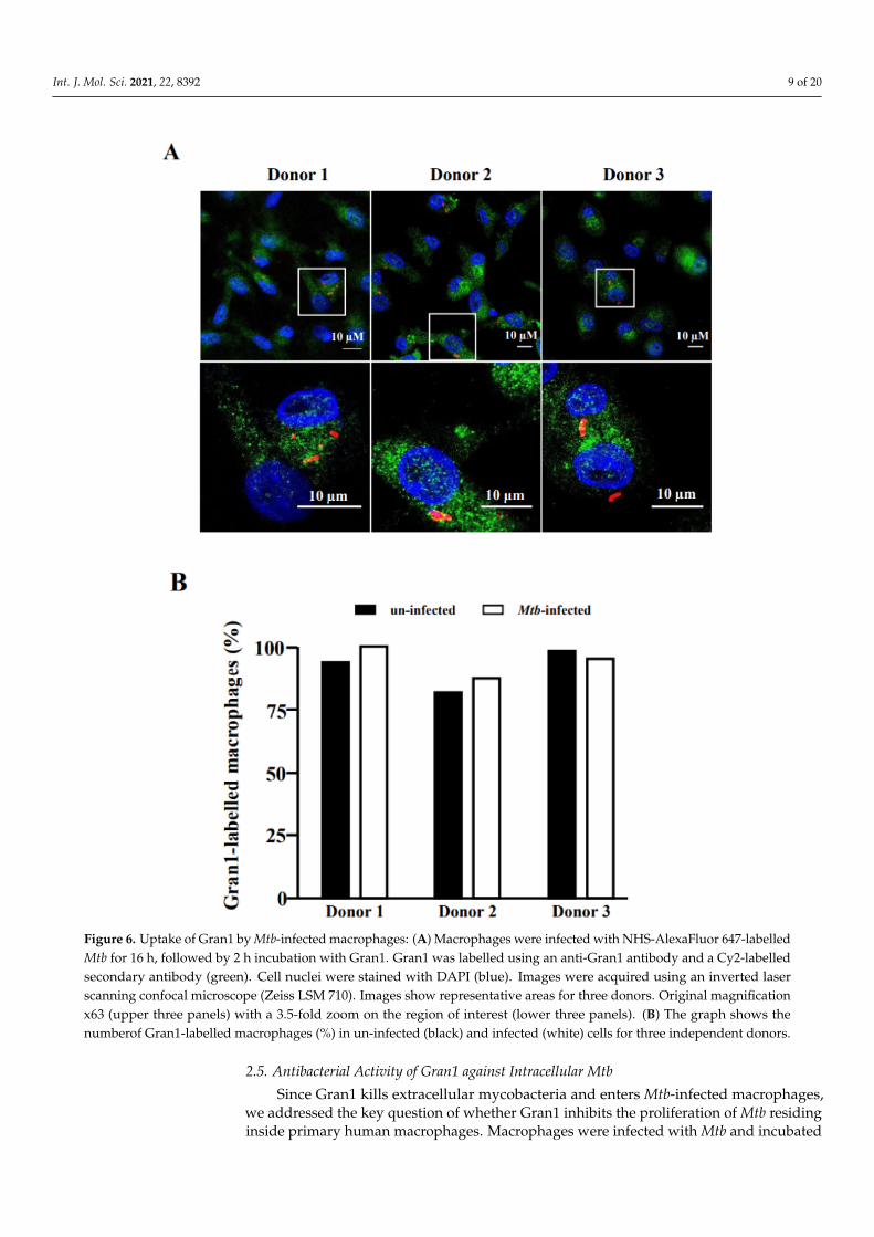
To exert its antimicrobial function, Gran1 needs to be taken up by Mtb-infectedmacrophages. Therefore, we investigated whether Mtb infection interferes with the up-take of Gran1 by exposing the Mtb-infected macrophages to Gran1 and analyzing cells byconfocal laser scanning microscopy. Labelled Gran1 was detected in the majority (>95%)of macrophages independently of Mtb-infection (Figure 6). Gran1 was homogenouslydistributed throughout the cytoplasm and no specific enrichment in the vicinity of thebacilli was detected.

Int. J. Mol. Sci. 2021, 22, 8392 8 of 20
Figure 5. Macrophage-specific uptake of Gran1 in mixed cell culture: (A) PBMC mixed with autologous macrophages (ratio1:1) were incubated with Gran1 for 2 h and labelled using an anti-Gran1 antibody (1:200) and a Cy2-labelled secondaryantibody. Cells were stained for MHC class II (red) and cell nuclei were stained with DAPI (blue). Original magnification×63 (upper four panels) with 3.3 zoomed region of interest (magnification ×210, lower four panels). Data shown arerepresentative for one out of three donors. (B) PBMC mixed with autologous macrophages were incubated with Gran1-Atto647N for 2 h, stained for MHC class II (MHCII-FITC), and analyzed by flow cytometry. The left panel shows arepresentative dot plot (n = 7). The histogram shows the percentage of Gran1-positive cells in MHC class II negative (red)and MHC class II positive (blue) population (representative donor, n = 7). The results of each individual donor are presentedin the right panel. Statistical analysis was performed using a non-parametric Wilcoxon rank test for paired samples(p = 0.0156). (C) Uptake of Gran1 in MHC class II positive cells after 2 h of incubation at 4 ◦C and 37 ◦C. Box plots show themedian (horizontal lines) with upper and lower quartile and whiskers indicating minimum and maximum values (n = 7).Statistical analysis was performed using a non-parametric Wilcoxon rank test for paired samples (p = 0.0156).
Int. J. Mol. Sci. 2021, 22, 8392 9 of 20
Figure 6. Uptake of Gran1 by Mtb-infected macrophages: (A) Macrophages were infected with NHS-AlexaFluor 647-labelledMtb for 16 h, followed by 2 h incubation with Gran1. Gran1 was labelled using an anti-Gran1 antibody and a Cy2-labelledsecondary antibody (green). Cell nuclei were stained with DAPI (blue). Images were acquired using an inverted laserscanning confocal microscope (Zeiss LSM 710). Images show representative areas for three donors. Original magnificationx63 (upper three panels) with a 3.5-fold zoom on the region of interest (lower three panels). (B) The graph shows thenumberof Gran1-labelled macrophages (%) in un-infected (black) and infected (white) cells for three independent donors.
2.5. Antibacterial Activity of Gran1 against Intracellular Mtb
Since Gran1 kills extracellular mycobacteria and enters Mtb-infected macrophages,we addressed the key question of whether Gran1 inhibits the proliferation of Mtb residinginside primary human macrophages. Macrophages were infected with Mtb and incubated

Int. J. Mol. Sci. 2021, 22, 8392 10 of 20
with Gran1. After 4 days, the number of live bacilli was determined by quantifying thenumber of CFU. The n-fold growth was normalized to the control cultures (broth) andrevealed that the diluent alone had no effect on Mtb multiplication (Figure 7). In contrast,the mean n-fold growth of Mtb in Gran1-treated cultures was 0.64, as compared to thebroth control, which demonstrated a statistically significant growth reduction of 36%.
Figure 7. Gran1 inhibits growth of intracellular Mtb. Macrophages were infected with Mtb followedby incubation with Gran1 for 4 d. Multiplication of Mtb was determined by plating cell lysateson 7H11 agar plates. After 21 d of incubation, the number of colony-forming units (CFU) wasdetermined. Lines indicate mean values and dots represent individual values of n-fold mycobacterialgrowth as compared to growth control (d4) for eleven individual donors. Statistical analysis wasperformed using multiple t-tests (Wilcoxon rank test, p = 0.05).
Taken together, Gran1 inhibits the growth of extra- and intracellular mycobacteria inconcentrations that are well tolerated in vitro (macrophages) and in vivo (zebrafish em-bryos), and do not trigger inflammatory off-target effects in macrophages or PBMC. Studiesthat are aimed at optimizing the pharmacokinetics of Gran1 to allow for efficacy studies inpreclinical and ultimately clinical studies in mycobacterial infection are currently ongoing.
3. Discussion
The clinical application of large AMPs such as Granulysin is limited by off-targeteffects, poor penetration into the cytoplasm of microbial host cells, rapid degradationin human serum, and high manufacturing costs. Therefore, we investigated whetherthe specific cytolytic region of Granulysin (23–51 aa; Gran1) exerts antimicrobial activityagainst extra- and intracellular mycobacteria without inducing inflammatory immuneresponses. Our results demonstrated that the 3.53 kDa Gran1 kills slow-growing non-tuberculous mycobacteria and virulent Mtb inside human macrophages without triggeringinflammatory immune responses such as cytokine release or chemotaxis. Thus, Gran1 is apromising candidate to be further evaluated for efficacy against microbial infection.
Prior studies showed that a conserved secondary structure of AMPs is crucial tomaintain its biological activity [22,23]. Hence, we investigated the secondary structure of

Int. J. Mol. Sci. 2021, 22, 8392 11 of 20
Gran1. We identified a random coil-like conformation without distinct alpha helices. Themultiple arginines present in the peptide sequence, the two cysteines both embedded in thesame tripeptide code VCR, and the predominance of amino acids with similar fingerprints(Ser-Cys, Asn-Asp-Phe-Trp) in the TOCSY spectrum, plus broad overlapping cross-peaksin the backbone HN-Hα coupling region, could interfere with the unequivocal signalassignment of the 29 amino acids. Additionally, heteronuclear measurements such as 1H-13C Heteronuclear Single Quantum Coherence (HSQC) and Heteronuclear Multiple BondCorrelation (HMBC) were unsuccessful due to a low concentration of the peptide, as relatedto the natural abundance of 13C. This problem will be addressed in future studies by usingrecombinant, rather than synthetic, Gran1 to allow for a precise analysis of the solutionstructure. We conclude, therefore, that even though the secondary structure of Gran1differs from Granulysin, the antimicrobial activity of Gran1 was retained, in agreementwith studies that have demonstrated the functional significance of helix2-loop2-helix3in antimicrobial activity [15]. Furthermore, the four basic arginine residues which arerequired for membrane binding are conserved in the sequence of Gran1 sequence [7]. Wehypothesized that the amino acid sequence is more critical for the antimicrobial functionthan the secondary structure, since Gran1 resembles the unmodified cytolytic active region(23–51 aa). Granulysin-derived peptides are active against extracellular Staphylococcusaureus, Streptococci [15], Propionibacterium acnes [10], Vibrio cholera [12], Escherichia coli [22],and Mtb [14,22]. Importantly two of these peptides had a protective effect against Vibriocholera in mice [12], and the local application of a peptide-containing formulation improvedthe clinical presentation of severe acne in humans [11]. Here, we demonstrated that aGranulysin-derived peptide is active against a panel of mycobacterial species, includingvirulent Mtb. We used mycobacteria as target organisms because they cause severe diseasein humans and are notoriously difficult to treat. Ongoing studies will demonstrate whetherGran1 is also active against extracellular, fast growing bacteria such as Staphylococcusaureus, Klebsiella pneumoniae, or Pseudomonas aeruginosa. Our results indicate that Gran1 hasa selective activity against the slow-growing species M. tuberculosis, M. kansasii, M. avium,and the vaccine strain M. bovis BCG, but not the rapid-growing strains M. fortuitum, M.vaccae, and M. smegmatis. One possible explanation for this is that the effect of the AMP onthe bacteria is slower than the growth rate of non-tuberculous mycobacteria (NTM) [24], soany harmful effect could be compensated by the rapid multiplication. Alternatively, thedifferential effect could be related to the selective activity of anti-tuberculosis drugs to fast-and slow-growing NTM [25,26]. For example, rifampin is less active against rapid-growingmycobacteria, based on a more efficient efflux activity, as compared to slow-growingmycobacteria [25]. By using super-resolution and electron microscopy studies, we foundthat Gran1 directly interacts with the mycobacterial cell wall, causing pore formationand distortions reminiscent of early studies performed with the parental Granulysin [2].We hypothesize that, via the positively charged arginine residues, Gran1 interacts withthe negatively charged mycobacterial cell wall, leading to deleterious disruption of cellwall integrity and consecutive disturbance of the ionic balance. Whether Gran1 interfereswith specific molecular targets in the mycobacterial cell wall remains to be determined.STED microscopy, recently established for studying peptide/mycobacteria interaction ([27],Figure 3) provides a powerful tool with which to address these mechanistic questions.
We have demonstrated for the first time that a Granulysin-derived peptide can enterhuman macrophages to kill an intracellular pathogen. Notably, concentrations of Gran1that induced growth inhibition did not induce apoptosis or necrosis in human macrophagesbut accumulated in the cytoplasm of un-infected and Mtb-infected macrophages (Figure 6).This distinguishes Gran1 from other antimicrobial peptides (e.g., Melittin), which preferablyincorporates into cell membranes [28], most likely due to a significantly lower Boman index(0.57 kcal/mol; APD3 [18]). Even though we found distinct areas of co-localization inGran1 and intracellular Mtb by confocal laser microscopy (Figure 6), the resolution of thistechnique does not allow for definitive conclusions, especially because of the homogenousdistribution of the peptide throughout the cytoplasm. Gran1 might interact with the

Int. J. Mol. Sci. 2021, 22, 8392 12 of 20
mycobacterial cell wall, as observed for extracellular Mtb (Figure 2). However, since Mtbresides within phagolysosomes and not in the cytoplasm, Gran1 may not gain access tothe mycobacteria. In this case, Gran1 could modulate macrophage function and indirectlycontribute to the growth inhibition of Mtb. Previously it was shown that Gran1/G8 activatessodium channels in human red blood cells, resulting in increased levels of intracellularsodium, chloride, and calcium ions and reduced levels of potassium [29]. This effect onthe ion-microenvironment could also be active in macrophages and modulate pathwaysinvolved in the antimicrobial function of macrophages [30], for example the ATP-inducedNLRP3 inflammasome activation [31]. Whether Gran1 directly affects antimycobacterialeffector pathways, such as the vitamin D-cathelicidin axis [32,33], the fusion of phagosomesand lysosomes [34] or autophagy [35] is currently under investigation. In addition, we arecurrently fine-tuning the use of super-resolution microscopy to allow for a more definitiveinsight into the intracellular (co)-localization of Gran1 and mycobacteria.
One caveat when using large peptides for the treatment of infections is the risk ofoff-target effects not related to the intended antimicrobial activity. Several AMPs com-bine antimicrobial and immune-modulatory activities, most notably cathelicidin [36] andGranulysin [7,8], both of which are active against mycobacteria [2,37,38]. Specifically, Gran-ulysin induces apoptosis in eukaryotic cells, is a chemoattractant, and triggers the releaseof immune modulatory cytokines (IL-10, IL-1β, IL-6 and IFN-α) [8]. While this immunemodulatory effect can be a helpful bystander effect to combat acute infections, it may beharmful in diseases where inflammation is a major component of tissue destruction. De-pending on the pathophysiology of the disease peptides, which avoid or suppress immunemodulatory functions, may be beneficial. For example, a Granulysin-derived peptide wasidentified (31–50 aa) which combines antimicrobial activity against the causative bacteriumPropionibacterium acnes with anti-inflammatory effects that contribute to the progressionof disease [10]. In tuberculosis, especially in the later stages of disease, when the majorityof the bacteria have been eliminated, inflammation contributes to tissue damage, mostnotably the destruction of lung parenchyma [39]. Under these circumstances, Gran1 mightbe particularly useful, because it maintains the antimicrobial activity of the parental peptidewithout inducing apoptosis, chemotaxis, or the release of immune modulatory cytokines.
Taken together, Gran1 is an AMP with antimicrobial activity against clinically rel-evant, slow-growing mycobacterial species, including virulent Mtb, without promotinginflammatory immune responses. In contrast to the parental protein Granulysin, it does notrequire the support of granzymes and perforin to inhibit the growth of intracellular Mtb.Our results pave the way for designing Gran1-containing nanoparticles, such as liposomesor mesoporous nanoparticles, which combine AMPs with conventional anti-tuberculosiscompounds. These nanoparticles will serve to optimize the stability, pharmacokinetics,and biological activity of Gran1, which can then be evaluated as a treatment againstmycobacterial infections in preclinical and ultimately clinical studies.
4. Materials and Methods4.1. Peptide Synthesis
Gran1 (QRSVSNAATRVCRTGRSTWRDVCRNFMRR) was synthesized by PSL Hei-delberg (PSL Heidelberg, Heidelberg, Germany) using F-moc chemistry. For visualizationof Gran1, the peptide was conjugated N-terminally to the fluorescent dye Atto647N (PSLHeidelberg). Peptides were purified to >95% homogeneity by reverse-phase HPLC. Com-position of each peptide was confirmed by amino acid analysis and mass spectrometry [40].Stock peptide solutions (10 mg/mL) were prepared in Ampuwa (Fresenius Kabi, BadHomburg, Germany)—aqua ad injectabilia. Physicochemical properties of Gran1 werepredicted using the Antimicrobial Peptide Databank server APD3 [18] for amino acidcomposition, molecular weight, hydrophobic ratio, and Boman index.
Int. J. Mol. Sci. 2021, 22, 8392 13 of 20
4.2. Structural Analysis of Gran14.2.1. Circular Dichroism Spectrometry
CD spectra were recorded on a JASCO J-1500 spectrometer (Jasco, Pfungstadt, Ger-many) in a 1 mm High Precision Cell by HellmaAnalytics (Müllheim, Germany). Thesamples with Gran1 were prepared at a concentration of 200 µM in 10 mM phosphate buffer,separately at pH 5, pH 6, and pH 7. Measurements were conducted using the followingparameters: path length: 0.1 mm, scan rate: 5 nm/min, scan range: 260 nm–180 nm, datapitch: 0.2 nm, and data integration time: 2 sec. Data were processed in Spectra Analysisby JASCO (Jasco, Pfungstadt, Germany) and Excel (Microsoft Cooperation, Redmond,WA, USA).
4.2.2. Nuclear Magnetic Resonance
For all 2D NMR experiments, 5 mg of Gran1 was dissolved in 450 mL aqua adinjectabilia (Ampuwa) and 50 mL D2O. All experiments were recorded on a BRUKERspectrometer (Billerica, MA, USA) operating at 850 MHz 1H frequency. Experiments werecarried out at 298 K. Nuclear Overhauser Effect Spectroscopy (NOESY) spectra acquiring2D homonuclear correlation via dipolar coupling with water suppression using watergateW5 pulse sequence with gradients [41] were recorded for a mixing time of 150, 300, and450 ms, using 2 × 16k × 256 data matrices, corresponding to acquisition times of ~480 and8 ms in the t1 and t2 dimensions, respectively. Next, 64 scans were acquired per t1 value.Through-bond connectivity was obtained from a Total Correlation Spectroscopy (TOCSY)spectrum, recorded with the MLEV-17 mixing scheme [42], with water suppression using3-9-19 pulse sequence, and with gradients [43,44] using a 13 µs 90◦ pulse and for an 80 msmixing period.
NMRFAM-Sparky was used for signal assignment and NOE signal volume determi-nation [45]. For NOE signal integration, a gaussian fit was used, allowing peak motionand the adjusting of linewidths and baseline fitting. For the 3D structure calculation ofGran1, the software package Ambiguous Restraints for Iterative Assignment (ARIA) wasused [17].
4.3. Source and Culture of Mycobacteria
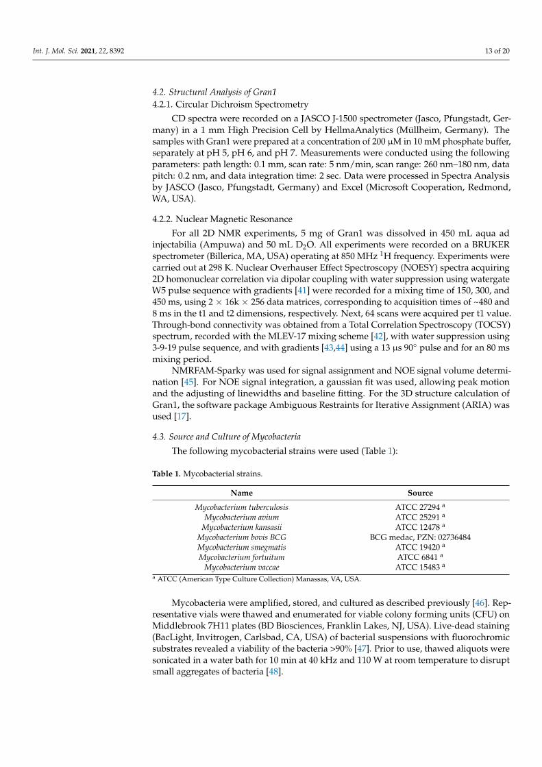
The following mycobacterial strains were used (Table 1):
Table 1. Mycobacterial strains.
Name Source
Mycobacterium tuberculosis ATCC 27294 a
Mycobacterium avium ATCC 25291 a
Mycobacterium kansasii ATCC 12478 a
Mycobacterium bovis BCG BCG medac, PZN: 02736484Mycobacterium smegmatis ATCC 19420 a
Mycobacterium fortuitum ATCC 6841 a
Mycobacterium vaccae ATCC 15483 a
a ATCC (American Type Culture Collection) Manassas, VA, USA.
Mycobacteria were amplified, stored, and cultured as described previously [46]. Rep-resentative vials were thawed and enumerated for viable colony forming units (CFU) onMiddlebrook 7H11 plates (BD Biosciences, Franklin Lakes, NJ, USA). Live-dead staining(BacLight, Invitrogen, Carlsbad, CA, USA) of bacterial suspensions with fluorochromicsubstrates revealed a viability of the bacteria >90% [47]. Prior to use, thawed aliquots weresonicated in a water bath for 10 min at 40 kHz and 110 W at room temperature to disruptsmall aggregates of bacteria [48].

Int. J. Mol. Sci. 2021, 22, 8392 14 of 20
4.4. Antibodies and Reagents
The anti-Gran1 antibody was generated in cooperation with ImmunoGlobe (Himmel-stadt, Germany). Rabbits were immunized by injecting intradermally synthetic Gran1 andthe adjuvant Montanide for three times over a timespan of 12 weeks. Afterwards, the IgGfraction was isolated from serum using an IgG column. Purified anti-Gran1 antibody wasstored at 4 ◦C in Tris-buffer with 0.02% NaN3. Cy2-conjugated donkey-anti-rabbit wasacquired from Dianova (Hamburg, Germany) and Cy5-conjugated goat anti-mouse fromJackson ImmunoResearch (Westgrove, PA, USA). 4′,6-diamidino-2-phenylindole (DAPI),Atto594-conjugated goat anti-rabbit, and succinimidylester Atto647N were all purchasedfrom Sigma–Aldrich (Steinheim, Germany). Succinimidylester AlexaFluor 647 and FITC-conjugated HLA-DR antibody were purchased from Invitrogen (Carlsbad, CA, USA), whileunconjugated HLA-DR antibody was purchased from Leinco Technologies (St. Louis,MO, USA). Macrophage serum-free media (M-SFM) and AIM V serum-free media werepurchased from Gibco, Thermo Fisher (Waltham, MA, USA). Middlebrook 7H9 broth waspurchased from Becton Dickinson (BD Biosciences, Franklin lakes, NJ, USA). Staining bufferwas prepared in PBS containing 0.5% Tween 80, 0.2 M sodium bicarbonate (both Roth,Karlsruhe, Germany) and pH was set to 8.8. FACS buffer contained 1% FCS (Biochrom,Berlin, Germany) and 0.1% sodium azide (VWR, Radnor, PA, USA) in PBS (Gibco, Carlsbad,CA, USA).
4.5. Chemotaxis Assay
Migration of PBMC was examined using a 96-well microchemotaxis chamber (Neu-roProbe, Gaithersburg, MD, USA). Cells were re-suspended in AIM V medium. Differ-ent concentrations of Gran1 were placed in the lower compartment of the chamber and0.1 × 106 cells were added to the upper compartment in a total volume of 50 µL. Cells wereallowed to migrate at 37 ◦C for 3 h. Filters were then washed with PBS and removed totransfer migrated cells from the lower compartment into FACS tubes. For quantification,5 µL of fluorescent-red latex beads were added to each sample. Cells were acquired usinga FACSCalibur flow cytometer (BD Biosciences, Franklin lakes, NJ, USA) and terminatedwhen 5 × 10⁴ events in the latex beads gate were detected. The chemotaxis factor wascalculated by dividing the number of migrated cells to Gran1 by the number of migratedcells to medium control.
4.6. Cytokine and Chemokine Release
For determination of cytokine and chemokine release 2 × 10⁶ human PBMC or0.5 × 106, macrophages were seeded in a 24-well plate and incubated overnight withGran1 at 37 ◦C. LPS (100 ng/mL) or PHA (20µg/mL, for CCL-5 release) served as positivecontrols. Supernatants were collected and analyzed for TNF-α, IL-1β (Endogen, Waltham,MA, USA), IL-10, CCL-2, CCL-3, and CCL-5 (all R&D systems, Minneapolis, MN, USA)by ELISA, as suggested by the manufacturer. Absorption was measured at 450 nm usingInfinite 200 Pro (Tecan, Männedorf, Switzerland) plate reader. Background signals werededucted for indicated ELISA specifically mentioned in the figure legend.
4.7. Growth of Extracellular Mycobacteria: 3H-Uracil Proliferation Assay
The activity of Gran1 against virulent Mtb, as well as non-tuberculous mycobacteria,was determined by measurement of RNA synthesis after the incorporation of radioactively-labelled 5.6-3H-Uracil (ART-0282, Biotrend, Cologne, Germany). Next, 2 × 10⁶ sonicatedmycobacteria (virulent H37Rv or non-tuberculous mycobacteria, respectively) were incu-bated with Gran1 in middlebrook 7H9 broth in a 96-well plate. All samples were set up intriplicate, using 2 µg/mL rifampicin or 2 µg/mL clarithromycin for rapid-growing strainsas a control, respectively. 3H-Uracil (0.3 µCi/well) was added after 72 h for fast-growingmycobacteria or 168 h for slow-growing mycobacteria, and cultures were incubated foradditional 18 h. Mtb were then inactivated by treatment with 4% paraformaldehyde (PFA,Sigma–Aldrich, Steinheim, Germany) for 30 min and transferred onto glass fiber filters
Int. J. Mol. Sci. 2021, 22, 8392 15 of 20
(Printed Filtermat A, PerkinElmer, Waltham, MA, USA) using a 96-well based FiltermatHarvester (Inotech, Nabburg, Germany). Fiber filters were dried in a microwave at 240 Wfor 5 min and sealed at 75 ◦C with a sheet of solid scintillant wax (MeltiLex, PerkinElmer,Waltham, MA, USA). Radioactivity was measured using a β-Counter (Sense Beta, Hidex,Turku, Finland). Antimicrobial activity (%) was calculated as counts per minute (cpm) ofthe treated sample/cpm of the un-treated sample × 100.
4.8. Stimulated Emission Depletion (STED) Microscopy
First, mycobacteria were fluorescently labelled as previously described [26]. Briefly,mycobacteria were washed with staining buffer, followed by incubation with succinimidylester Atto647N (1 µg/mL) for 1 h at 37 ◦C. Afterwards, bacteria were washed and re-suspended in M-SFM. For investigating the interaction between Gran1 and Mtb, stainedbacteria were seeded in a 12-well plate (Sarstedt, Nümbrecht, Germany) containing asterile precision glass coverslip (170 ± 5 µm thickness, Carl Roth, Karlsruhe, Germany)coated with poly-L-lysine (Sigma–Aldrich, Steinheim, Germany). Then, unconjugatedGran1 (1 µM) was added and incubated for 30 min. Excess peptide was removed by rinsingthe wells carefully. Coverslips were fixed with 4% PFA and washed with PBS. Bacteriawere blocked and permeabilized for 2 h in 3% BSA and 0.3% TritonX-100 in PBS. Thiswas followed by incubation overnight at 4 ◦C with primary anti-Gran1 antibody (1:1000).Afterwards, bacteria were incubated with secondary Atto594-conjugated goat anti-rabbitantibody (1:1000) for one hour at room temperature. Coverslips were mounted withMowiol (Sigma–Aldrich, Steinheim, Germany) onto a SuperFrost plus microscope slides (R.Langenbrinck GmbH, Emmendingen, Germany) for analysis by STED microscopy. Imageswere captured using a home-built dual-color STED microscope described elsewhere [49].Typically, an average power of ~0.8µW for each excitation beam (568 nm and 633 nm,respectively) and ~1.3 mW for each depletion beam (710 nm and 750 nm, respectively) wasused. STED images were captured at a pixel size of 10 nm and a dwell time of 300 µsec,with a typical peak photon number of ~100 counts. Images were analyzed in ImageJ 1.53c.For better visualization, a Gaussian blur of σ = 1 and an intensity threshold of >5 countswere applied in each channel.
4.9. Scanning Electron Microscopy
First, 5 × 10⁶ M. tuberculosis were seeded in a 24-well plate in middlebrook 7H9broth, followed by incubation with 1 µM Gran1 for 72 h. After infection, bacteria wereharvested, transferred to screw caps, and centrifuged at 10.000 rpm for 10 min. Supernatantwas discarded and the pellet of Mtb was resuspended and fixed in 100 µL 4% PFA for20 min. Afterwards, bacteria were chemically fixed with 2.5% glutaraldehyde (in PBSand 1% saccharose) for one hour. Then, Mtb were post-fixed with OsO4 (2% in PBS) forone hour at room temperature to dehydrate samples gradually in 30, 50, 70, 90 and 100%propanol (5 min at each step) [50]. Bacteria were then critical point dried using carbondioxide as translation medium (Critical Point Dryer CPD 030, Bal-Tec, Principality ofLiechtenstein). Samples were rotary coated in a BAF 300 freeze etching device (Bal-Tec,Principality of Liechtenstein) by electron beam evaporation with 3 nm of platinum–carbonfrom an angle of 45◦. Images were acquired using a Hitachi S-5200 in-lens field emissionSEM (Hitachi High-Tech, Tokyo, Japan) at an accelerating voltage of 10 kV by using thesecondary electron signal.
4.10. Confocal Laser Scanning Microscopy
For investigation of Gran1 cell-specific internalization, a mixed culture of primary hu-man macrophages and autologous PBMC (ratio 1:1, total cell counts 0.1 × 10⁶) were seededin 200 µL M-SFM in an 8-chamber slide (Thermo Fisher, Waltham, MA, USA). Followedby incubation with Gran1 for 2 h, cells were then fixed with 4% PFA and permeabilizedfor 10 min in 0.5% bovine serum albumin (BSA), 0.1% Triton X-100, and 0.05% Tween 20(all Sigma–Aldrich, Steinheim, Germany) in phosphate buffered saline (PBS). Samples

Int. J. Mol. Sci. 2021, 22, 8392 16 of 20
were blocked for one hour with blocking buffer (1% BSA, 0.1% Triton X-100 in PBS) andincubated at room temperature for one hour with primary anti-Gran1 (1:250) or MHC classII (1:300). Afterwards, cells were incubated with the secondary antibody Cy2-conjugateddonkey anti-rabbit (1:200) or Cy5-conjugated goat anti-mouse (1:250), respectively, for onehour at room temperature. Cell nuclei were stained with 1 µg/mL DAPI for 10 min andslides were mounted with Aquatex (Merck, Darmstadt, Germany). Images were acquiredusing the inverted laser scanning confocal microscope LSM 710 (Zeiss, Oberkochen, Ger-many) and analyzed in ImageJ 1.53c. For investigation of Gran1-localization in infectedmacrophages, Mtb were stained with the fluorescein succinimidyl ester AlexaFluor 647(1 mg/mL) as described [46]. Afterwards, macrophages were infected with stained Mtb ata theoretical multiplicity of infection of 50 for 16 h. Followed by incubation with Gran1 for2 h, cells were then fixed and stained as mentioned above.
4.11. Toxicity of Gran1 against Macrophages and Zebrafish
For in vitro studies, 1 × 10⁵ macrophages were incubated with Gran1 for 24 h ina 96-well plate, followed by an addition of 10% PrestoBlue (Thermo Fisher, Waltham,MA, USA) for 20 min. Reduction of the non-fluorescent resazurin-based PrestoBlue tofluorescent resorufin by mitochondrial enzymes of viable cells allowed for the calculationof cytotoxicity as described [48].
For in vivo studies, wild-type zebrafish embryos (Danio rerio) were dechorionatedat 24 h post-fertilization (hpf) using digestion with 1 mg/mL pronase (Sigma-Aldrich,Steinheim, Germany) in E3 medium (83 µM NaCl, 2.8 µM KCl, 5.5 µM 202 CaCl2, 5.5 µMMgSO4). In a 96-well plate, 3 embryos per well were exposed for 24 h to 100 µL of E3containing Gran1 (1 µM). Two independent assays were performed, each with 10 × 3embryos. The peptide solvent (Ampuwa, aqua ad injetibilia), diluted in E3, was usedas negative control at the same amount as introduced by the peptide stock. As posi-tive control for acute toxicity/cytotoxicity, the pleurocidin antimicrobial peptide NRC-03(GRRKRKWLRRIGKGVKIIGGAALDHL-NH2) was used at a concentration of 6 µM, asdescribed [20]. Abamectin at a concentration of 3.125 µM was used as positive control forneurotoxicity [51]. At 48 hpf (after 24 h of incubation), embryos were scored in a stereomi-croscope for signs of acute toxicity/cytotoxicity (lysis and/or necrosis), developmentaltoxicity (delay and/or malformations), or cardiotoxicity (heart edema and/or reduced orabsent circulation). Each embryo was also touched with a needle and the reduced or absenttouch response (escape movements) was evaluated for signs of neurotoxicity if, and onlyif, no signs of acute toxicity were present in the same embryo. Embryos were categorizedwithin each of these toxicity categories into several classes of severity. Chi-square test wasused to calculate whether the distribution of embryos into toxicity classes [21] differedsignificantly between the negative control and the test substances.
4.12. Intracellular Detection of Gran1 by Flow Cytometry
For investigation of Gran1 cell-specific uptake, macrophages were seeded in sterileFACS tubes alongside autologous PBMC in a 1:1 ratio for a total cell count of 0.5 × 10⁶ cellsin AIM V medium. Cells were incubated with Gran1-Atto647N for 2 h at 37 ◦C. Afterwards,cells were washed with FACS buffer and centrifuged for 10 min at 1300 rpm. Supernatantwas discarded and cells were stained against MHC class II by a FITC-conjugated HLA-DRantibody (1:200). Sample analysis was performed using a FACSCalibur flow cytometer(BD Biosciences, Franklin Lakes, NJ, USA). Data analysis was performed using FlowJoVersion 10.5.3 (BD Biosciences, Franklin Lakes, NJ, USA) and GraphPad Prism Version 8.2.1(GraphPad Software, La Jolla, CA, USA).
4.13. Quantification of Intracellular Mycobacterial Growth
Human peripheral blood mononuclear cells (PBMC) were isolated from buffy coats ofanonymous donors (Institute of Transfusion Medicine, Ulm University) by density gradientcentrifugation (Ficoll-Paque Plus, GE Healthcare, Buckinghamshire, UK). Monocytes were

Int. J. Mol. Sci. 2021, 22, 8392 17 of 20
selected from plastic adherence and thorough washing. For generation of monocyte-derived macrophages, cells were cultured in M-SFM with granulocyte–macrophage colony-stimulating factor (GM-CSF, 10 ng/mL, Miltenyi Biotec, Bergisch Gladbach, Germany) for6 d, as described [46]. Afterwards, macrophages were infected in 6-well plates with single-cell suspensions of Mtb at a multiplicity of infection of 5. After 2 h, cells were washed toremove extracellular bacteria and harvested using 1 mM EDTA (Sigma–Aldrich, Steinheim,Germany). Subsequently, 2 × 10⁵ infected macrophages were seeded in 24-well plates andincubated with Gran1 (1 µM) or diluent control for 4 d. To enumerate the number of viablebacilli, infected macrophages were lysed with 0.3% saponin (Sigma–Aldrich, Steinheim,Germany). Cell lysates were re-suspended vigorously, transferred into screw caps, andsonicated in a water bath for 10 min at room temperature. Afterwards, serial dilutions(1:10, 1:100, 1:1000) of sonicates were plated on 7H11 agar plates (BD Biosciences, Franklinlakes, NJ, USA) and incubated for 21 d before determining the number of colonies formingunits (CFU).
4.14. Statistical Analysis
All statistical analyses, as mentioned in the figure legends, were performed usingGraphPad Prism v8.2.1 (GraphPad Software, La Jolla, CA, USA). Significance was calcu-lated using non-parametric tests for paired samples (Wilcoxon rank test, paired t-test).Differences were considered significant when p-value < 0.05.
4.15. Ethical Statement
Zebrafish embryos were used at stages up to 2 days post-fertilization (dpf), whichis before they start to feed, at 6 dpf. Embryos that do not yet require feeding are notcovered by EU and German animal experiment and welfare legislation (§14 TierSchVersV).Embryos were euthanized at the end of the test by rapid freezing, which is consideredthe most humane method for euthanasia for fish embryos. Adult fish housing and carewas approved by the state of Baden-Württemberg and was monitored by Ulm Universityanimal welfare executives and veterinaries of the city of Ulm.
Author Contributions: Conceptualization R.N., S.S.; methodology, R.N., F.W., G.K.; validation, S.S.;formal analysis, R.N., F.W., G.K.; writing—original draft preparation, R.N., S.S.; writing—review andediting, R.N., S.S., G.W., J.M., F.W.; supervision, S.S., J.M., T.W., G.W., P.W.; funding acquisition, T.W.,G.W., P.W., J.M., S.S. All authors have read and agreed to the published version of the manuscript.
Funding: This research was funded by the German Research Foundation (CRC 1279 Exploitingthe Human Peptidome for Novel Antimicrobial and Anticancer Agents to S.S., T.W., P.W., J.M. andG.W.). R.N. is supported by the Graduate School of Molecular Medicine at the University Ulm.The Weidinger group was funded by the German Research Foundation (CRC 1149, project number251293561; CRC 1279, project number 316249678; WE 4223/6-1, project number 414077062; WE4223/8-1, project number 433187294).
Institutional Review Board Statement: Not applicable.
Informed Consent Statement: Not applicable.
Data Availability Statement: The raw data supporting the conclusions of this article will be madeavailable by the authors, without undue reservation, to any qualified researcher.
Acknowledgments: We appreciate the support by Ulm University Center for Translational ImagingMoMAN and Core Facility for Confocal and Multiphoton Microscopy and the excellent technicalsupport by Daniel Mayer, Mark Grieshober and Martina Raasholm.
Conflicts of Interest: The authors declare no conflict of interest.

Int. J. Mol. Sci. 2021, 22, 8392 18 of 20
Abbreviations
aa Amino acidsAPD3 Antimicrobial Peptide Database 3AMP antimicrobial peptideATCC American Type Culture CollectionBSA bovine serum albuminCCL Chemokine (C-C motif) ligandCFU colony forming unitsdpf days post fertilizationhpf hours post fertilizationFACS fluorescence activated cell sortingMHCII major histocompatibility complex class 2MOI multiplicity of infectionMtb Mycobacterium tuberculosisNTM Non-tuberculous mycobateriaPBMC peripheral blood mononuclear cellPBS phosphate buffered salinePHA PhytohaemagglutininPFA ParaformaldehydeSD Standard deviationSEM Standard error of the meanSFM Serum-free mediumSLC secondary lymphoid-tissue chemokineSTED Stimulated emission depletion
References1. Peña, S.V.; Hanson, D.A.; Carr, B.A.; Goralski, T.J.; Krensky, A.M. Processing, Subcellular Localization, and Function of 519
(Granulysin), a Human Late T Cell Activation Molecule with Homology to Small, Lytic, Granule Proteins. J. Immunol. 1997, 158,2680–2688.
2. Stenger, S.; Hanson, D.A.; Teitelbaum, R.; Dewan, P.; Niazi, K.R.; Froelich, C.J.; Ganz, T.; Thoma-Uszynski, S.; Melián, A.; Bogdan,C.; et al. An Antimicrobial Activity of Cytolytic T Cells Mediated by Granulysin. Science 1998, 282. [CrossRef]
3. Pardo, J.; Pérez-Galán, P.; Gamen, S.; Marzo, I.; Monleón, I.; Kaspar, A.A.; Susín, S.A.; Kroemer, G.; Krensky, A.M.; Naval, J.; et al.A Role of the Mitochondrial Apoptosis-Inducing Factor in Granulysin-Induced Apoptosis. J. Immunol. 2001, 167, 1222–1229.[CrossRef]
4. Kaspar, A.A.; Okada, S.; Kumar, J.; Poulain, F.R.; Drouvalakis, K.A.; Kelekar, A.; Hanson, D.A.; Kluck, R.M.; Hitoshi, Y.; Johnson,D.E.; et al. A Distinct Pathway of Cell-Mediated Apoptosis Initiated by Granulysin. J. Immunol. 2001, 167, 350–356. [CrossRef][PubMed]
5. Gamen, S.; Hanson, D.A.; Kaspar, A.; Naval, J.; Krensky, A.M.; Anel, A. Granulysin-Induced Apoptosis. I. Involvement of atLeast Two Distinct Pathways. J. Immunol. 1998, 161, 1758–1764.
6. Deng, A.; Chen, S.; Li, Q.; Lyu, S.; Clayberger, C.; Krensky, A.M. Granulysin, a Cytolytic Molecule, Is Also a Chemoattractant andProinflammatory Activator. J. Immunol. 2005, 174, 5243–5248. [CrossRef] [PubMed]
7. Dotiwala, F.; Lieberman, J. Granulysin: Killer Lymphocyte Safeguard against Microbes. Curr. Opin. Immunol. 2019, 19–29.[CrossRef] [PubMed]
8. Krensky, A.M.; Clayberger, C. Biology and Clinical Relevance of Granulysin: REVIEW ARTICLE. Tissue Antigens. 2009, 193–198.[CrossRef]
9. Dotiwala, F.; Mulik, S.; Polidoro, R.B.; Ansara, J.A.; Burleigh, B.A.; Walch, M.; Gazzinelli, R.T.; Lieberman, J. Killer LymphocytesUse Granulysin, Perforin and Granzymes to Kill Intracellular Parasites. Nat. Med. 2016, 22, 210–216. [CrossRef] [PubMed]
10. McInturff, J.E.; Wang, S.J.; Machleidt, T.; Lin, T.R.; Oren, A.; Hertz, C.J.; Krutzik, S.R.; Hart, S.; Zeh, K.; Anderson, D.H.; et al.Granulysin-Derived Peptides Demonstrate Antimicrobial and Anti-Inflammatory Effects against Propionibacterium Acnes. J.Investig. Dermatol. 2005, 125, 256–263. [CrossRef]
11. Lim, H.S.; Chun, S.M.; Soung, M.G.; Kim, J.; Kim, S.J. Antimicrobial Efficacy of Granulysin-Derived Synthetic Peptides in AcneVulgaris. Int. J. Dermatol. 2015, 54, 853–862. [CrossRef]
12. Da Silva, A.P.G.; Unks, D.; Lyu, S.C.; Ma, J.; Zbozien-Pacamaj, R.; Chen, X.; Krensky, A.M.; Clayberger, C. In Vitro and In VivoAntimicrobial Activity of Granulysin-Derived Peptides against Vibrio Cholerae. J. Antimicrob. Chemother. 2008, 61, 1103–1109.[CrossRef] [PubMed]
13. Wang, Z.; Choice, E.; Kaspar, A.; Hanson, D.; Okada, S.; Lyu, S.-C.; Krensky, A.M.; Clayberger, C. Bactericidal and TumoricidalActivities of Synthetic Peptides Derived from Granulysin. J. Immunol. 2000, 165, 1486–1490. [CrossRef]

Int. J. Mol. Sci. 2021, 22, 8392 19 of 20
14. Siano, A.; Tonarelli, G.; Imaz, M.; Perin, J.; Ruggeri, N.; Lopez, M.; Santi, M.; Zerbini, E. Bactericidal and Hemolytic Activities ofSynthetic Peptides Derived from Granulysin. Protein Pept. Lett. 2010, 17, 517–521. [CrossRef]
15. Andreu, D.; Carreño, C.; Linde, C.; Boman, H.G.; Andersson, M. Identification of an Anti-Mycobacterial Domain in NK-Lysinand Granulysin. Biochem. J. 1999, 344. [CrossRef]
16. Global Tuberculosis Report 2020; World Health Organization: Geneva, Switzerland, 2020; Licence: CC BY-NC-SA 3.0 IGO.17. Rieping, W.; Bardiaux, B.; Bernard, A.; Malliavin, T.E.; Nilges, M. ARIA2: Automated NOE Assignment and Data Integration in
NMR Structure Calculation. Bioinformatics 2007, 23, 381–382. [CrossRef] [PubMed]18. Wang, G.; Li, X.; Wang, Z. APD3: The Antimicrobial Peptide Database as a Tool for Research and Education. Nucleic Acids Res.
2016, 44, D1087–D1093. [CrossRef]19. Gutsmann, T. Interaction between Antimicrobial Peptides and Mycobacteria. Biochim. Biophys. Acta-Biomembr. 2016, 1858,
1034–1043. [CrossRef]20. Morash, M.G.; Douglas, S.E.; Robotham, A.; Ridley, C.M.; Gallant, J.W.; Soanes, K.H. The Zebrafish Embryo as a Tool for Screening
and Characterizing Pleurocidin Host-Defense Peptides as Anti-Cancer Agents. DMM Dis. Model. Mech. 2011, 4, 622–633.[CrossRef]
21. Beitzinger, B.; Gerbl, F.; Vomhof, T.; Schmid, R.; Noschka, R.; Rodriguez, A.; Wiese, S.; Weidinger, G.; Ständker, L.; Walther, P.;et al. Delivery by Dendritic Mesoporous Silica Nanoparticles Enhances the Antimicrobial Activity of a Napsin-Derived PeptideAgainst Intracellular Mycobacterium Tuberculosis. Adv. Healthc. Mater. 2021, 2100453. [CrossRef]
22. Ernst, W.A.; Thoma-Uszynski, S.; Teitelbaum, R.; Ko, C.; Hanson, D.A.; Clayberger, C.; Krensky, A.M.; Leippe, M.; Bloom, B.R.;Ganz, T.; et al. Granulysin, a T Cell Product, Kills Bacteria by Altering Membrane Permeability. J. Immunol. 2000, 165. [CrossRef]
23. Linde, C.M.A.; Grundström, S.; Nordling, E.; Refai, E.; Brennan, P.J.; Andersson, M. Conserved Structure and Function in theGranulysin and NK-Lysin Peptide Family. Infect. Immun. 2005, 73, 6332–6339. [CrossRef] [PubMed]
24. Boman, H.G. Antibacterial Peptides: Basic Facts and Emerging Concepts. J. Intern. Med. 2003, 254. [CrossRef]25. Zhang, Y.; Zhang, H.; Sun, Z. Susceptibility of Mycobacterium Tuberculosis to Weak Acids. J. Antimicrob. Chemother. 2003, 52,
56–60. [CrossRef] [PubMed]26. Zhang, Y.; Post-Martens, K.; Denkin, S. New Drug Candidates and Therapeutic Targets for Tuberculosis Therapy. Drug Discov.
Today 2006, 11, 21–27. [CrossRef]27. Deshpande, D.; Grieshober, M.; Wondany, F.; Gerbl, F.; Noschka, R.; Michaelis, J.; Stenger, S. Super-Resolution Microscopy Reveals
a Direct Interaction of Intracellular Mycobacterium Tuberculosis with the Antimicrobial Peptide LL-37. Int. J. Mol. Sci. 2020,21, 6741. [CrossRef] [PubMed]
28. Hong, J.; Lu, X.; Deng, Z.; Xiao, S.; Yuan, B.; Yang, K. How Melittin Inserts into Cell Membrane: And Disturbance on theMembrane. Molecules 2019, 24, 1775. [CrossRef] [PubMed]
29. Li, Q.; Dong, C.; Deng, A.; Katsumata, M.; Nakadai, A.; Kawada, T.; Okada, S.; Clayberger, C.; Krensky, A.M. Hemolysis ofErythrocytes by Granulysin-Derived Peptides but Not by Granulysin. Antimicrob. Agents Chemother. 2005, 49, 388–397. [CrossRef][PubMed]
30. Jobin, K.; Müller, D.N.; Jantsch, J.; Kurts, C. Sodium and Its Manifold Impact on Our Immune System. Trends Immunol. 2021, 42,469–479. [CrossRef] [PubMed]
31. Di, A.; Xiong, S.; Ye, Z.; Malireddi, R.K.S.; Kometani, S.; Zhong, M.; Mittal, M.; Hong, Z.; Kanneganti, T.-D.; Rehman, J.; et al. TheTWIK2 Potassium Efflux Channel in Macrophages Mediates NLRP3 Inflammasome-Induced Inflammation. Immunity 2018, 49.[CrossRef]
32. Chung, C.; Silwal, P.; Kim, I.; Modlin, R.L.; Jo, E.K. Vitamin D-Cathelicidin Axis: At the Crossroads between Protective Immunityand Pathological Inflammation during Infection. Immune Netw. 2020, 20, 1–26. [CrossRef]
33. Liu, P.T.; Stenger, S.; Li, H.; Wenzel, L.; Tan, B.H.; Krutzik, S.R.; Ochoa, M.T.; Schauber, J.; Wu, K.; Meinken, C.; et al. Toll-likeReceptor Triggering of a Vitamin D-Mediated Human Antimicrobial Response. Science 2006, 311, 1770–1773. [CrossRef]
34. Gutierrez, M.G.; Mishra, B.B.; Jordao, L.; Elliott, E.; Anes, E.; Griffiths, G. NF-KB Activation Controls Phagolysosome Fusion-Mediated Killing of Mycobacteria by Macrophages. J. Immunol. 2008, 181. [CrossRef] [PubMed]
35. Gutierrez, M.G.; Master, S.S.; Singh, S.B.; Taylor, G.A.; Colombo, M.I.; Deretic, V. Autophagy Is a Defense Mechanism InhibitingBCG and Mycobacterium Tuberculosis Survival in Infected Macrophages. Cell 2004, 119. [CrossRef] [PubMed]
36. Van Harten, R.M.; van Woudenbergh, E.; van Dijk, A.; Haagsman, H.P. Cathelicidins: Immunomodulatory Antimicrobials.Vaccines 2018, 6, 63. [CrossRef] [PubMed]
37. Silva, J.P.; Appelberg, R.; Gama, F.M. Antimicrobial Peptides as Novel Anti-Tuberculosis Therapeutics. Biotechnol. Adv. 2016, 34,924–940. [CrossRef]
38. Sonawane, A.; Santos, J.C.; Mishra, B.B.; Jena, P.; Progida, C.; Sorensen, O.E.; Gallo, R.; Appelberg, R.; Griffiths, G. Cathelicidin IsInvolved in the Intracellular Killing of Mycobacteria in Macrophages. Cell. Microbiol. 2011, 13, 1601–1617. [CrossRef]
39. Stek, C.; Allwood, B.; Walker, N.F.; Wilkinson, R.J.; Lynen, L.; Meintjes, G. The Immune Mechanisms of Lung ParenchymalDamage in Tuberculosis and the Role of Host-Directed Therapy. Front. Microbiol. 2018, 9. [CrossRef] [PubMed]
40. Ständker, L.; Wobst, P.; Mark, S.; Forssmann, W.G. Isolation and Characterization of Circulating 13-KDa C-Terminal Fragments ofHuman Insulin-like Growth Factor Binding Protein-5. FEBS Lett. 1998, 441, 281–286. [CrossRef]
41. Liu, M.; Mao, X.A.; Ye, C.; Huang, H.; Nicholson, J.K.; Lindon, J.C. Improved Watergate Pulse Sequences for Solvent Suppressionin NMR Spectroscopy. J. Magn. Reson. 1998, 132, 125–129. [CrossRef]
Int. J. Mol. Sci. 2021, 22, 8392 20 of 20
42. Bax, A.D.; Donald, G.D. MLEV-17-Based Two-Dimensional Homonuclear Magnetization Transfer Spectroscopy. J. Magn. Reson.1969, 65, 355–360. [CrossRef]
43. Piotto, M.; Saudek, V.; Sklenár, V. Gradient-Tailored Excitation for Single-Quantum NMR Spectroscopy of Aqueous Solutions. J.Biomol. NMR 1992, 2, 661–665. [CrossRef]
44. Sklenár, V.; Piotto, M.; Leppik, R.; Saudek, V. Gradient-Tailored Water Suppression for 1H-15N HSQC Experiments Optimized toRetain Full Sensitivity. J. Magn. Reson. Ser. A. 1993, 241–245. [CrossRef]
45. Lee, W.; Tonelli, M.; Markley, J.L. NMRFAM-SPARKY: Enhanced Software for Biomolecular NMR Spectroscopy. Bioinformatics2015, 31, 1325–1327. [CrossRef]
46. Bruns, H.; Stegelmann, F.; Fabri, M.; Döhner, K.; van Zandbergen, G.; Wagner, M.; Skinner, M.; Modlin, R.L.; Stenger, S. AbelsonTyrosine Kinase Controls Phagosomal Acidification Required for Killing of Mycobacterium Tuberculosis in Human Macrophages.J. Immunol. 2012, 189, 4069–4078. [CrossRef] [PubMed]
47. Stegelmann, F.; Bastian, M.; Swoboda, K.; Bhat, R.; Kiessler, V.; Krensky, A.M.; Roellinghoff, M.; Modlin, R.L.; Stenger, S.Coordinate Expression of CC Chemokine Ligand 5, Granulysin, and Perforin in CD8 + T Cells Provides a Host DefenseMechanism against Mycobacterium Tuberculosis. J. Immunol. 2005, 175, 7474–7483. [CrossRef] [PubMed]
48. Noschka, R.; Gerbl, F.; Löffler, F.; Kubis, J.; Rodríguez, A.A.; Mayer, D.; Grieshober, M.; Holch, A.; Raasholm, M.; Forssmann,W.G.; et al. Unbiased Identification of Angiogenin as an Endogenous Antimicrobial Protein With Activity Against VirulentMycobacterium Tuberculosis. Front. Microbiol. 2021, 11. [CrossRef]
49. Osseforth, C.; Moffitt, J.R.; Schermelleh, L.; Michaelis, J. Simultaneous Dual-Color 3D STED Microscopy. Opt. Express 2014,22, 7028. [CrossRef] [PubMed]
50. Walther, P.; Wang, L.; Ließem, S.; Frascaroli, G. Viral Infection of Cells in Culture. Approaches for Electron Microscopy; Academic Press:Cambridge, MA, USA, 2010; Volume 96. [CrossRef]
51. Raftery, T.D.; Isales, G.M.; Yozzo, K.L.; Volz, D.C. High-Content Screening Assay for Identification of Chemicals ImpactingSpontaneous Activity in Zebrafish Embryos. Environ. Sci. Technol. 2014, 48, 804–810. [CrossRef] [PubMed]