Embed Size (px)
Citation preview

The Dissertation Committee for Charles Fernando Capinos Scherer Certifies that
this is the approved version of the following dissertation:
Evaluation of granulysin and perforin as candidate biomarkers for
protection following vaccination with Mycobacterium bovis BCG or M.
bovis ∆RD1
Committee:
D. Mark Estes, PhD, Supervisor
Slobodan Paessler, DVM, PhD
Bernardo Villarreal-Ramos, PhD
Alfredo G. Torres, PhD
Gregg N. Milligan, PhD
Janice J. Endsley, PhD
__________________
Dean, Graduate School

Evaluation of granulysin and perforin as candidate biomarkers for
protection following vaccination with Mycobacterium bovis BCG or M.
bovis ∆RD1
By
Charles Fernando Capinos Scherer, DVM, MS
Dissertation
Presented to the Faculty of the Graduate School of
The University of Texas Medical Branch
in Partial Fulfillment
of the Requirements
for the Degree of
Doctor of Philosophy
The University of Texas Medical Branch
December, 2008

This dissertation is dedicated to my wonderful and always supporting wife Juliana and
my daughters Mariana and Carolina
iv
Acknowledgements
I would like to initially thank my family, mainly my wife Juliana and my
daughters Mariana and Carolina for their love, friendship during all these years of graduate school. Juliana, my lovely wife, I know you have done a lot of sacrifices for me. Thank you for giving me support during all these years. Thanks you for discussing and listening to my plans about science and our plans for the future. To Mariana and Carolina, thank you for giving me the necessary support and giving me your smiles and love.
A special thank you to my parents and to my in-laws, for giving me and my family support during these several years of dedication of the fulfillment of a dream. Thank you to “vó Leda” and “tia Thaís”, for your extensive support. Even though you were more than 6,000 miles away from of us, you are still very close.
Thank you to my mentor Dr. Estes, for being not only an advisor, but a friend during all this time. For being always prompt to discuss and find solution for my research problems. Thank you Dr. Golde and Dr. Rodriguez, for giving me the extraordinary opportunity to work with exotic animal diseases at Plum Island Animal Disease Center. Also Dr. Waters from NADC, Ames, IA, for the innumerable blood shipments you had to send me. Thanks to all for helping me on being and independent scientist.
To the Estes lab ex -post-doctoral fellows, Janice and Liz, for giving the opportunities and a good environment to be an independent researcher, and for always being prompt to discuss my research problems and giving me advice and support. To our current post-doctoral fellows Sandra and Arpaporn, thank you also for your constant support and sharing of good and bad moments.
To my committee members, Dr. Estes, Dr. Endsley, Dr. Milligan, Dr. Torres, Dr. Paessler and Dr. Villarreal-Ramos, thank you for the extraordinary guidance during my PhD and for the excellent comments and advice.
I would like also thank colleagues at the graduate school and in the lab, Mark Endsley, Barbara, Juliana and Blake. Thank you all for your support and for helping me during my experiments and for giving important advice for my progress in the lab. Thanks for proving the necessary environment to make me an independent researcher.
Thank you, all my other colleagues of the Microbiology department, especially Rohit and Santhanna, for sharing your experiences and opinions about the most diverse subjects.
Thank you Dr. Konig, our course director, and our course coordinator Martha Lewis, for the help ans support during my time at the Department of Microbiology and Immunology. A special thank you to Dr. Thomas K. Hughes, our former course director, for guidance and suggestions.
Thank you, all my Brazilian friends Sandra and Evandro, Adriana and Luis, Diva and Henrique, Sandro and Kathleen, for sharing very good moments together.

v
Thank you to USDA, for providing the resources for my research and to the University of Texas Medical Branch, for providing the space and the environment to make my work possible.
Thank you god for giving me life and giving me this wonderful family.

vi
Evaluation of granulysin and perforin as candidate biomarkers for
protection following vaccination with Mycobacterium bovis BCG or M.
bovis ∆RD1
Publication No._____________
Charles Fernando Capinos Scherer, PhD
The University of Texas Medical Branch, 2008
Supervisor: D. Mark Estes
The development of improved vaccines against tuberculosis (TB) is directly
linked to the investigation of new and better correlates of protection after vaccination against TB. Cloning and characterization of bovine homologues of the antimicrobial protein granulysin (Bo-lysin) and perforin by our group could be used as potential biomarkers for TB vaccination efficacy. In the present study we examined the kinetics of granulysin, perforin, IFNγ and Fas-L responses to Mycobacterium bovis purified protein derivative (PPD) stimulation by peripheral blood mononuclear cells from M. bovis ∆RD1-, BCG- and non-vaccinated cattle. Gene expression profiles following PPD stimulation showed significant increases in transcripts for granulysin and IFNγ in both CD4+ and CD8+ T cells in BCG-vaccinated as compared to non-vaccinated animals. Perforin and IFNγ examined by flow cytometry, showed a difference of 1-2% more PPD-specific cells in BCG-vaccinated than non-vaccinated animals. In the vaccine trial, granulysin and perforin were significantly increased in both vaccine groups as compared to control after vaccination and challenge. IFNγ expression was increased only after vaccination and secretion was higher in the control, as compared to both vaccine groups demonstrating no correlation with protection upon vaccination. In summary, results shown here provide evidence that granulysin and perforin are prospective candidates as biomarkers of protection after vaccination against TB.

vii
Table of Contents
List of Figures ..........................................................................................................x
List of Illustrations................................................................................................. xi
List of Abbreviations ............................................................................................ xii
CHAPTER 1- INTRODUCTION 1 Tuberculosis: still a major public health problem ..........................................2
Tuberculosis in bovine and humans: a zoonotic perspective .........................4
Mycobacterium tuberculosis and Mycobacterium bovis. ...............................5
Taxonomy, description and growth requirements ........................5
The Mycobacterium spp. genome and the RD1 (Region of Difference 1) .................................................................................7
Mycobacterium tuberculosis and Mycobacterium bovis pathogenesis. .........9
Granuloma: a hallmark of tuberculosis.........................................................12
The immune response against Mycobacterium spp. infection. ....................15
γδ T cells .....................................................................................16
NK cells ......................................................................................17
CD4+ and CD8+ T cells response against Mycobacterium spp ...20
Mechanism of intracellular Mycobacterium spp. killing .............................21
The P2X7 purinergic receptor .....................................................21
Nitric Oxide ................................................................................23
Granulysin...................................................................................25
Other mechanisms involved on the cell mediated immunity against mycobacterial infection ................................................................................27
Fas ligand (Fas-L) .......................................................................27
Granzymes and Perforin .............................................................28
Measurement of immune correlates of protection following vaccination ....31
BCG and other vaccines against tuberculosis ..............................................32
Animal models to study tuberculosis and to evaluate vaccines ...................36
The neonate immune response and the bovine neonate model ....................38

viii
Hypothesis, specific aims, rationale and significance of our studies ...........39
CHAPTER 2- MATERIALS AND METHODS 41 Animals, immunization and challenge .........................................................42
Isolation of Peripheral Blood Mononuclear cells (PBMC) and stimulation conditions .....................................................................................................42
Vaccine efficacy evaluation .........................................................................44
Flow cytometric analysis (FACS) ................................................................44
IFNγ ELISA .................................................................................................45
RNA extraction and Quantitative real-time RT-PCR ..................................46
Antibody production .....................................................................................47
Western blotting ...........................................................................................48
Tissue analysis .............................................................................................48
Statistics .......................................................................................................49
CHAPTER 3- KINETICS AND SPECIFIC ACTIVATION CONDITIONS OF BO-LYSIN EXPRESSION IN ACTIVATED BOVINE LYMPHOCYTE POPULATIONS IN PARALLEL WITH PERFORIN, IFNγ AND FAS-L 50
INTRODUCTION .......................................................................................51
RESULTS ....................................................................................................52
Granulysin is upregulated in CD4+ and CD8+ T cells after
vaccination with BCG.................................................................52
Granulysin, perforin and IFNγ proteins increase upon vaccination
with BCG ...................................................................................53
DISCUSSION ..............................................................................................56
CHAPTER 4- POTENTIAL USE OF BO-LYSIN AS A CORRELATE OF PROTECTION FOLLOWING VACCINATION WITH BCG AND ∆RD1 AND CHALLENGE WITH VIRULENT M. bovis 68
INTRODUCTION .......................................................................................69
RESULTS ....................................................................................................70
Granulysin expression is increased after vaccination against TB
and challenge with virulent M. bovis .........................................70

ix
IFNγ correlates with pathology 72
DISCUSSION ..............................................................................................77
CHAPTER 5- EXPRESSION OF GRANULYSIN IN PARALLEL WITH IFNγ, PERFORIN AND FAS-L AT THE SITES OF INFECTION OF M. Bovis 79
INTRODUCTION .......................................................................................80
RESULTS ....................................................................................................80
DISCUSSION ..............................................................................................82
CHAPTER 6 SUMMARY, FINAL DISCUSSION/ CONCLUSIONS AND FUTURE DIRECTIONS 84
APPENDIX 1 ........................................................................................................87
APPENDIX 2 ........................................................................................................98
APPENDIX 3 ......................................................................................................107
BIBLIOGRAPHY ..............................................................................................119
VITA ...................................................................................................................138

x
List of Figures
Figure 1: Isolation and purification of leukocyte populations..........................57
Figure 2A: Fold mRNA induction of granulysin, perforin, IFNγ and Fas-L
measured by semi-quantitative real-time PCR in purified CD4+ T
cells ..................................................................................................58
Figure 2B: Fold mRNA induction of granulysin, perforin, IFNγ and Fas-L
measured by semi-quantitative real-time PCR in purified CD8+ T
cells ...................................................................................................59
Figure 2C: Fold mRNA induction of granulysin, perforin, IFNγ and Fas-L
measured by semi-quantitative real-time PCR in purified γδ+ T
cells ...................................................................................................60
Figure 3: Flow cytometry detection of IFNγ and Perforin by intracellular
staining in CD4+, CD8+ and γδ+ T cells ............................................61
Figure 4: Granulysin detection by immunoblot................................................62
Figure 5: Comprehensive vaccine efficacy data...............................................74
Figure 6: Fold mRNA induction of granulysin, perforin, IFNγ and Fas-L in
a vaccine trial ....................................................................................75
Figure 7: Flow cytometry analysis of intracellular staining of IFNγ and
Perforin in CD4+, CD8+ and γδ+ T cells in a vaccine trial ...............75
Figure 8: IFNγ measured by ELISA.................................................................76
Figure 9: Relative gene expression of granulysin, perforin, IFNγ and Fas-L on
the mediastinal lymph node ..............................................................81

xi
List of Illustrations
Illustration 1: The P2X7 pathway ......................................................................24
Illustration 2: The Nitric oxide production pathway ........................................34
Illustration 3: The cytotoxic granule pathway ..................................................35

xii
List of Abbreviations
TB Tuberculosis
XDR-TB Extremely drug resistant tuberculosis
BCG Bacille of Calmette and Guérin
PPD Purified Protein Derivative
RD1 Region of Difference 1
DC Dendritic cell
CR3 Complement receptor 3
DC-SIGN Dendritic cell-specific intercellular adhesion molecule 3
grabbing non-integrin
MBL Mannose Binding Lectin
LAM Lipoarabinomannam
TLR Toll like receptor
MHC Major Histocompatibility Complex
Mtb Mycobacterium tuberculosis
CTL Cytotoxic T lymphocyte
WC1 Workshop Cluster 1
NK Natural Killer
NO Nitric Oxide
NOS Nitric Oxide Synthase
PBMC Peripheral Blood Mononuclear cells

1
CHAPTER 1
INTRODUCTION
2
Tuberculosis: still a major public health problem
Tuberculosis (TB) remains a major public health problem due to the increasing
numbers of reported cases throughout the US and worldwide. Several factors have been
involved in the increased rates of TB worldwide including (i) HIV co-infection, (ii)
emergence of drug resistant strains including extremely drug-resistance TB (XDR-TB),
(iii) immigration from high prevalence countries (related to US) and (iv) variable efficacy
of the BCG (Bacille of Calmette-Guérin) vaccine still extensively used worldwide.
According to CDC and WHO reports, it is estimated that more than 15 million people get
infected with Mycobacterium tuberculosis, and 10% of these will develop TB, worldwide
[1]. Each year it is estimated that more than 2 million individuals die due to infection;
there were 9.2 million new cases of TB, where 0.7 million are HIV –positive cases, with
an estimate of more than 50% of HIV-infected patients also TB positive [2]. The drug
resistance factor involves both primary resistance cases in people infected with a strain
that is already resistant, and individuals that acquire resistance during therapy. Treatment
of TB cases is based on the use of a cocktail of drugs including isoniazid, rifampin,
pyrazinamide, ethambutol, rifabutin and rifapentine. Besides these drugs several other
have been introduced in the cocktail such as streptomycin, cycloserine and levofloxacin.
The treatment lasts for at least 6 months but can be longer (up to 12 months) depending
on the strain and resistance profile of the strain infecting the patient [1, 2]. Multidrug
resistance represents 0.5 million cases in 2006, besides that, XDR-TB cases represent
16,000 cases from the period of 2007-2008 and treatment can be for up to 2 years [2].

3
Another factor contributing to the increased TB rates in US is immigration of
people from countries where TB has high incidence such as India, Africa and China. In
2006, data presented by CDC comparing TB rates by race/ethnicity shows that for every
100.000 people, from a total of 35 cases, 15 are from people of Asian origin and 20 are
from people from other origins (Hispanics, white, American Indians and African
Americans) [1, 2].
BCG, a Mycobacterium bovis attenuated strain, is the only currently licensed
vaccine against TB and has been used in more than 100 million people worldwide. BCG
was implemented initially on 1921 and since then it had its success, but also a reason for
a lot of controversy in terms of continuing its use or not. The efficacy of the vaccine goes
from 0-80% in several clinical trials. Factors involved in this extreme variability include,
but are not limited to, the use of different strains worldwide, conservation and
administration of the vaccine, pre-exposure to environmental mycobacteria and loss of
BCG genes over the year due to multiple passages (genetic drift). So far, several new
vaccine candidates are in phase I and II trials, but have not progressed to the point of
offering a better response than BCG.

4
Tuberculosis in bovine and humans: a zoonotic perspective
In addition to increasing rates of human TB, bovine TB can be considered an
important reemerging disease. The cattle pathogen, Mycobacterium bovis, presents
disease characteristics that parallel the human pathogen M. tuberculosis, including
disease pathogenesis and the host immune response. M. bovis is not an exclusive
pathogen of cattle but is able to infect and cause disease in all mammals. M. tuberculosis
was originally thought to be a pathogen of humans, but outbreaks in elephants in 2001
and 2003 in a Swedish zoo [3] added this species to the list of susceptible hosts. The
close interrelationships of humans with cattle in various settings world wide present a
potential risk for infection between the 2 species. Similarly to bovine, elephants in zoos
can be potential sources of tuberculosis to zoo keepers and to visitors. Bovine
tuberculosis in the US has been restricted to cases in California, Arizona, New Mexico,
Texas, Minnesota and Michigan. Cases in Minnesota and Michigan are mainly due to the
presence of whitetail deer, a natural reservoir of M. bovis [4, 5]. Sources of M. bovis in
the other states are also related to wildlife or to farmed game animals. Control of wildlife
species such as deer is done every year around farms with positive cases of bovine TB,
but the effort is contained by the number of animals that show up the year after. Also,
control of bovine TB in the mentioned states is done by quarantine and slaughtering of
the positive animals. Infections with M. bovis in humans were reported in New York,
where a total of 35 cases were due to consumption of contaminated unpasteurized cheese
from Mexico [6].
5
TB in cattle is a major livestock problem in UK, where the incidence of bovine
TB increases an average of 18% per year. As observed in the US, the increasing numbers
of bovine TB cases in UK are thought to be mainly due to a wildlife reservoir,
particularly the Eurasian badger. In New Zealand the brush tail possum is reported to be
the wildlife reservoir of M. bovis [7]. The policies involving quarantine and slaughtering,
and the hunting seasons do not control the rising cases of bovine TB. In addition to better
control measures in a variety of species, there is a major need for improved vaccines and
also better diagnostic test(s) to discriminate infected from vaccinated cattle. Bovine TB is
diagnosed based on comparing skin thickness after intradermal injection of both bovine
and avium PPD in different spots. Once increased thickness is observed in the bovine
PPD site, the animal is subjected to further testing.
Mycobacterium tuberculosis and Mycobacterium bovis
1-Taxonomy, description and growth requirements
Mycobacterium tuberculosis and M. bovis are in the family Mycobacteriaceae,
genus Mycobacterium. They are characterized as being aerobic, non-spore forming, non-
motile, rod shaped, and need a specific staining procedure to be visualized. Although they
are considered Gram positive (G+), an acid fast staining is required due to their high
content of lipids in the cell wall. Mycobacterium species present very complex growth
requirements, but can readily access ammonia, amino acids as sources of nitrogen and
carbon dioxide in the presence of salts. The growth is relatively slow, taking more than

6
20 hours for a next generation to form, and colonies are observed usually after several
days to 6 weeks of culture [8]. The most common media used to grow Mycobacterium
spp. is Middlebrook 7H11 that includes inorganic compounds with essential amino acids
and growth stimulating inorganic salts as well as vitamins. Glycerol or pyruvate is
provided as a source of carbon and energy, and sodium citrate is converted to citric acid.
Other compounds included on the 7H11 are selective agents such as Malachite green (BD
Pharmingen, CA).
Mycobacterium species can survive for weeks to several months in the
environment when protected from sunlight, but are easily killed by temperatures over
65°C/30 minutes or by UV light from the sun or by chemical compounds such as chloride
[9]. Some species like M. tuberculosis are able to survive for a long time in the
environment, and the presence of a living cell is important to allow pathogen replication.
Mycobacterium spp. is composed of several microorganisms that have different
requirements, being either obligate intracellular pathogens, opportunist or saprophytes
[8]. Example of saprophytic Mycobacterium spp. are the ones in the M.avium complex,
usually isolated from water, soil and plants [10] which represent a potential problem for
vaccination as will be discussed later.
According to Pfyffer et al. (2003) [8], close to 100 species of mycobacteria have
been described. Classification is based on groups, starting with the M. tuberculosis
complex, which includes M. tuberculosis, M. bovis, M. africanum, M. microti and M.
canettii. M. leprae, the causative agent of leprosy or Hansen’s diseases, is considered to
be in a separate group of mycobacterium due to its characteristic inability to grow in

7
vitro. Leprosy is characterized by a chronic, granulomatous and debilitating disease,
involving skin lesions and peripheral neuropathy [8]. The remaining mycobacteria of the
family Mycobacteriaceae include human and non- human pathogens such as M. avium
complex, M. marinum and M. smegmatis.
2-The Mycobacterium spp. genome and the RD1 (Region of Difference-1)
The Mycobacterium tuberculosis genome was published in 1998 based on the
H37Rv strain (a virulent lab strain). It was a very important step for the TB vaccine
development field. The genome comprises 4.4 Mb that encode around 4,000 genes, with
a high G+C content of over 65% [11]. The M. bovis genome was published in 2003
showing that it was 99.95% identical to M. tuberculosis. Comparisons between M. bovis
and M. tuberculosis showed 11 deletions in M. tuberculosis. The sequence of genes
missing in M. tuberculosis corresponds to a unique locus in M. bovis called TbD1 [12] .
Several other differences are described between the M . tuberculosis and M. bovis.
Among the genes present in M. tuberculosis are the genes that encode PE and PPE,
named after their conserved proline-glutamate (PE) or proline-proline-glutamate (PPE)
residues near the N terminus of the predicted proteins. PE and PPE maybe responsible for
host and tissue tropism difference between the 2 strains since PE was shown to bind
fibronectin [13]. PE and PPE were shown to be expanded in slow growing mycobacteria
and seem to be functionally linked to the ESAT-6 secretion system (ESX) [14].
Comparison of the BCG vaccine with virulent strains showed that BCG was lacking a

8
region called Region of Difference 1 (RD1). A comparison among all BCG strains with
the wild type M. bovis revealed that Regions of Difference encompass a total of 129 open
reading frames. However, only the RD1 region is absent in all BCG strains, being
composed of 9 genes which include 2 important secreted proteins, ESAT-6 and CFP-10
[11, 15]. These 2 antigens were shown to be potent T cell stimulators and may be linked
to spread of the organism. It was further shown that 6 different ESAT-6 proteins are
present in M. tuberculosis and missing in M. bovis, potentially impacting the antigen load
[12]. On the other hand the bovine bacillus presents 2 other serodominant proteins called
MPB70, a filtrate protein, and MPB 83, a glycosylated cell wall-associated protein [12].
Further characterization of the RD1 region and CFP-10: ESAT-6 protein-protein
interaction studies revealed that RD1 encodes a specialized secretion system in
mycobacteria, named ESX-1 (ESAT-6 secretion system). A close analysis of the genome
revealed that there are 5 clusters containing members of the CFP-10 and ESAT-6 family
of proteins, all very important T cell antigens, as well as other secreted proteins involved
on several metabolic pathways [14]. These 5 clusters also encode secretion systems
named from ESX-2 to ESX-5. The genes encoded in the RD1 region, specially the CFP-
10:ESAT-6 complex, are involved in a series of events as reviewed by Ganguly et al.,
(2008) [16]. These events include effects on pathogenesis and virulence such as cell lysis,
granuloma formation, cytokine suppression, phagosomal maturation arrest and interaction
with Toll-like receptors; and effects on immunomodulation such as down regulation of
macrophage cell signaling and effects in the DC-T cell interaction. An important
immunomodulatory effect of the CFP-10:ESAT-6 complex is the down regulation of

9
Reactive Oxidative Species (ROS). Modulation of the ROS response ultimately results on
inhibition of NF-kB activation [16].
The dual role of CFP-10:ESAT-6 complex on virulence and stimulation of the
immune response suggests an important source for vaccine development and new
diagnostic tests using ESAT-6. The secreted protein ESAT-6 was also evaluated as a
potential diagnostic to discriminate infected from vaccinated animals and may potentially
be used for humans [17-20].Taken together, alterations or deletions in the genome of
each strain, are important findings that will impact the understanding of evolution,
pathogenesis and immune response, development of better vaccines and diagnostic tests.
One of the strains used in our studies is based on a deletion of the RD1 region (∆RD1) on
a M. bovis strain.
Mycobacterium tuberculosis and Mycobacterium bovis pathogenesis
Both M. tuberculosis and M. bovis present similar pathology. M. tuberculosis was
recently described also as a pathogen of elephants [3, 21, 22], while M. bovis is a
pathogen for mammalian species [23]. Both species are carried in airborne particles
generated by infected persons or infected animals. Once these droplets containing the
pathogen are inhaled, they go to the alveoli and are engulfed/ phagocytized by resident
macrophages. It is now clear how many bacilli are required to lead to TB infection. In
cattle, it is suggested that less than 5 bacilli would be enough to result in infection but in
experimental models, aqueous suspensions of 10 4 cfu are used [24]. It is unknown how

10
many bacilli are required to establish infection in humans. As few as 10 bacilli maybe
enough to provide infection. Facts such as droplet size and breathing frequency of the
person being infected, have to be considered [25].
Besides resident macrophages, dendritic cells lying on the trachea may also be the
first ones to come in contact with the pathogen. Initial interaction of the pathogen with
these 2 cell types is performed through several receptors. Much of the immune response
will depend on the receptors used by Mycobacterium spp. Among the receptors used, the
mannose receptors are a potential safe route to facilitate intracellular survival.
Complement receptors (CR3) are also very advantageous for Mycobacterium since it
does not induce the release of oxygen intermediates [26]. Other receptors are DC-SIGN
(dendritic cell-specific intercellular adhesion molecule-3 grabbing non-integrin),
surfactant protein A (Sr-A), Class A scavenger receptor, mannose binding lectin (MBL)
and dectin -1 [27-29]. Internalization of the pathogen is followed by phago-lysosomal
fusion, destruction of bacteria, processing and presentation of antigens to T cells.
However, this process does not always occur. Mycobacterium spp. is able to adapt the
macrophage environment by inhibiting phago-lysosomal fusion, more specifically
inhibiting trafficking events that modulate the endosomal/phagosomal maturation. A
close analysis of the mycobacterial phagosome showed that it has an elevated pH, lack of
ATPases and late endosomal and lysosomal markers such as Rb7 or LAMP1 [30, 31] and
tryptophan-aspartate containing coat protein (TACO), normally removed from the
phagosome before fusion with the lysosome [32]. The mechanism involved in the
inhibition of the phagolysomal fusion starts with the prevention of the generation of PI3P

11
kinase (phosphatidylinositol 3 –phosphate) by a mycobacterial phosphatase called SapM
[33] and inhibition of recruitment of Ca++ influx by LAM (lipoarabinomannam). This
mechanism was observed when comparing killed and live mycobacteria internalized by
macrophages [34]. The potential influence on trafficking and phagolysosomal fusion
affects the presentation of antigen that would potentially activate cytotoxic T cells [35].
Infection of human macrophages by M. tuberculosis, results in the secretion of
TNFα, IL-1, IL-6, IL-8, RANTES and MCP-1, that would potentially induce the
recruitment of T cells to the site of infection and to the granuloma. IL-10 is also secreted
by infected macrophages, potentially suppressing the Th1 response needed to control the
infection [7, 36]. Another consequence of trafficking influence in the cell by
Mycobacteria, is the reduced expression of MHC-II potentially linked to stimulation via
TLR-2 [37, 38].
The interaction of mycobacteria with macrophages and dendritic cells is the
object of intense studies due to the consequences of infection in these 2 different cells.
Dendritic cells (DC) are the most potent antigen presenting cell and responsible for
initiating the immune response. DC are able to uptake mycobacteria via DC-SIGN, a
receptor used by the pathogen to evade the immune system [29, 39-41]. Following
uptake, DC of both humans and bovine mature, increase their expression of MHC-II and
co-stimulatory molecules CD80-CD86. Infected human DC secret several cytokines such
as IL-12, TNFα and IL-1, but bovine infected DC secrete mainly IL-12 [7]. Differences
exist between bovine DC and macrophages. It was demonstrated that bovine dendritic
cells are more permissive for M. bovis replication than macrophages, but as far as
12
cytokines, they release more IL-12 and induce a better immune T cell proliferation than
macrophages [36, 42]. In this work, DC and macrophages were treated with IFNγ to
evaluate a potential decrease of M. bovis viability. Only macrophages had their number
of acid fast bacilli decreased upon IFNγ treatment. Such observations result in a concern
that DC would be major reservoirs of M. bovis in vivo. By the same time, a low bacterial
turnover in DC would be reflecting in the constant availability of antigens to be presented
to T cells.
Granuloma: a hallmark of tuberculosis
After the inhalation of aerosol containing Mycobacterium tuberculosis / M. bovis,
an initial host-cell interaction of either macrophages or DC will provide the first line of
defense against infection by secreting cytokines and triggering of chemoattractant
mechanisms [43, 44].The interaction of pathogen with the different host cells will
determine the outcome of the disease by either controlling or clearing it, or resulting in
disease progression. Granuloma morphology is characterized by a central necrotic core
surrounded by concentric layers of macrophages, epithelioid cells, multinucleated giant
cells and lymphocytes [45-48].
Granuloma distribution presents variability depending on the species. In naturally
infected cattle, lesions are most frequently found in the dorso-caudal region, close to the
pleural surface. This lesion location seems to be the most distal end and the longest route
in the lungs, probably related to the size of the droplets containing the pathogen and the

13
topographical orientation that influences the air flow in lung [49]. In humans, it was
shown that lesions are more frequently found in the apical region of the lung [50].
Besides lungs, lesions are also frequently located in the lymph nodes responsible for
draining the region, such as the mediastinal lymph node. Several studies have also found
involvement of the retropharyngeal, submandibular and parotid lymph nodes as well as
tonsil and pharynx, suggesting that the pathogen may be excreted from the surface of
these tissues [49].
The center of the granuloma is characterized by an area of necrosis where
mycobacteria may still be intact and be a potential infection source for other
macrophages. The real battlefield in the granuloma happens in the surroundings where
pathogen and immune cells come together. The inner cell layer of the human granuloma
does not contain CD8+ T cells, but these cells are found on the granuloma periphery
surrounded by APC and B cells [51]. B cell aggregates were found in both humans and
mice, where B cells in different activation stages are observed [52]. Similar observations
in bovine showed that B cells were present but more restricted to outer fibrotic capsular
wall of advanced lesions [53, 54]. Granulomas in mice can be characterized by not being
organized and lacking the classical structure, observed in other species [55]. In calves,
mice, rats, guinea-pigs and hamsters, the initial lesion formation is performed by
migration of neutrophils, γδ T cells and NK cells that either kill macrophages or produce
IFNγ [56]. From these innate immunity players, γδ T cells are the ones that accumulate
in abundant numbers early in infection, in both cattle and mice [57, 58]. These events
culminate with further migration of CD4+ and CD8+ T cells. In a study where bovine

14
granulomas were analyzed for the cell composition, the authors demonstrated that cell
composition is dependent on the stage of the granuloma. Lesions that are in development
have more abundant γδ T cells than advanced lesions, but as happens in humans, lymph
node granulomas of unknown duration have around 80% of CD4+ and CD8+ T cells [59].
Among the cytokines that exert important functions in granuloma formation is
TNFα. This cytokine can promote beneficial or detrimental effects on the granuloma.
Increased blood flow and coagulation upon TNFα action will ultimately result in tissue
necrosis [49]. Secretion of TNFα seems to be detrimental for the Th1 response and
favors a Th2, characterized by the presence of IL-4. This last cytokine has been involved
in the formation of so called cavitation in tuberculosis, a phenomenon that occurs mostly
in humans [60]. Cavitation was also observed in guinea-pigs, rabbits and primates but not
in cattle. It is hypothesized that cattle may develop cavitation, but it is not observed due
to the detection and culling of the animals [49]. The cavitation process is a consequence
of liquefaction via a process facilitated by metalloproteinase enzymes that act on solid
caseous material [61, 62]. Bovine granulomas are classified in four stages according to
their cellular composition and degree of necrosis [7]. Stage 1 granulomas are
characterized by not having necrosis and consisting of primarily epithelioid cells, few
giant cells and some lymphocytes. Stage 4 granulomas are the ones where extensive
necrosis is observed, large number of epithelioid cells surrounded by WC1+ T cells and B
cells [57]. In experimental infections and vaccine trials using the bovine model, animals
receive a score based on their lung radiographic evaluation, where score 0 has no lesions

15
and score 5 has gross coalescent lesions [63]. More details of the immune response
against myobacterial infection are discussed in subsequent sections.
The immune response against Mycobacterium spp. infection
The initial response against TB infection is characterized by the engagement of
Mycobacterium spp.(Mtb) ligand with Toll like receptors (TLR). Mycobacterium spp. has
a broad range of ligands able to induce activation of different TLRs. Among the proteins
that are TLR-2 agonist are the secreted 19 KDa lipoprotein, lipoarabinomannan and
phosphatidyl-myo-inositol. Interaction between TLR-2 and the Mtb 19KDa lipoprotein
results in production of TNFα, IL-12 and nitric oxide production in macrophages [64].
The same 19 KDa lipoprotein either from M.tuberculosis or M. bovis BCG, was shown to
promote macrophage apoptosis. Macrophage apoptosis can be considered an evasion
mechanism as a way to promote disseminated mycobacterial infection [65]. Apoptosis of
infected macrophages can also induce CD8+ T cell stimulation via cross-priming. It was
shown that apoptotic vesicles from macrophages could be internalized by bystander DCs
and further activates CTL [66]. There are reports that apoptosis may be present in more
than 50% of macrophages in the granuloma [67]. TLR-4 deficient mice did not exhibit
any compromised responses against TB, unless a high dose of Mycobacterium was used
[68]. Besides TLR-2 and TLR-4, TLR-9 was also responsible for inducing IL-12
secretion by DCs, but the same was not observed in macrophages, where TLR-2 was the

16
main inducer [69]. The lack of activity by both TLR in mice seems to be linked to
tuberculosis progression [70] .
1-γδ T cells
These cells are early participants in the immune response against Mycobacterium
infection and are recruited promptly to the infected lung. In the presence of
phosphoantigens and IL-2, these cells proliferate and respond to mycobacteria infected
cells by secreting IFNγ and being cytotoxic [71, 72]. In humans a population designated
Vγ9Vδ2 appears to be the main population involved in mycobacteria response and is
present in circulating blood. Besides the secretion of IFNγ, these cells secrete TNFα and
induce IL-15 production by DCs [65]. IL-15 can further induce proliferation of CD8+ T
cells and activate NK cells. Mouse γδ T cells also proliferate 1 week after infection with
Mycobacterium spp.
Bovine γδ T cells differ from mouse and human γδ T cells, initially related to
numbers encountered in blood. They correspond to approximately 2-7% in both humans
and mice, 10-15% in cattle, but can be up to 40% in young calves from the total PBMC
population [73]. A role for the high number of these cells in young calves is related to a
possible early mechanism to produce Th1 cytokines [74]. Bovine γδ T cells use another
nomenclature, based on the Workshop Cluster 1 (WC1), and are then classified as WC1+
γδ T cells and WC1- γδ T cells. The first population is the predominant population on

17
bovine blood from young and adult ruminants, and WC1- γδ T cells are predominantly
located in spleen and mammary gland [75].
Upon infection of cattle with M. bovis by intranasal route, there is an initial
decrease in the size of the population of the WC1+ γδ T cells due to migration to the
infection sites, with a subsequent expansion in the circulation [76]. Analysis of the
bovine skin (after DTH) or granulomas identified WC1+ γδ T cells as one of the primary
cells that are present [57, 77]. The rapid migration and expansion upon infection may be
considered as a primary effort to control infection against M. bovis. Even though it has
been demonstrated that these cells can produce IFNγ and TNFα, the fast response
observed upon infection might be related to the production of chemokines and other
cytokines that promote recruitment of other T cells. Depletion of WC1+ γδ T cells had
minor effect on infection, mainly related to the anatomy of the granuloma [73], and
probably related to the lack of TNFα that was secreted by this cell type. Another study
showed that besides differences in the granuloma, there was an initial decrease of IFNγ
with a potential to compromise the Th1 bias upon infection with mycobacteria in favor of
a Th2 response [78].
2- NK cells
NK cells together with γδ T cells are important players of the innate immune
response. NK cells are large granular lymphocytes, bone marrow derived, with the ability

18
to lyse tumors cells, viral and bacterial infected cells [79-81]. NK cells are characterized
by expressing activating and inhibitory receptors, where action of a single or a
combination of receptors will determine the outcome of the interaction of the NK cells
with possible targets [82]. NK cells are able to distinguish normal healthy cells from
abnormal cells by using a mechanism that is dependent on the expression of MHC-I
molecules on cell surface. Activation or deactivation is dependent on expression of
several inhibitory receptors on the surface called KIR –killer cell Immunoglobulin like
receptors [83]. NK cells are not restricted to bone marrow, but are also encountered in
other organs. The origin of NK in cattle is unknown, but in mice and humans they
originate from an hematopoietic stem cell that colonizes liver, thymus, bone marrow and
spleen [84]. Different locations of the NK cells suggest an explanation why a diversity of
markers is needed in order to get a purified population from blood [84, 85].
Besides being important in the innate immune response, the interaction of NK
cells with antigen presenting cells such as DC, provides a link between innate and
adaptive immune response [86, 87]. Bovine NK cells are present in higher number in
neonates (≥ 10%) than in adults and the number decreases with age [88], similar to what
has been observed in humans [89]. NK cells are an important source of IFNγ in the
immune response against pathogens such as M. tuberculosis [90]. Upon infection of
mice with M. tuberculosis, there was a substantial increase of NK cells in the lung in the
initial 3 weeks [91]. Recruitment to lymph nodes was shown to be dependent on
chemokine receptor CXCR3 and CD62L-dependent mechanisms following activation by
DC or adjuvant [92].

19
Bovine NK cells have been the subject of many recent studies. A lot of progress
in the characterization and understanding of activator/inhibitory conditions has been
clarified in the last 5 years. Much of the progress on the phenotypic and functional
characterization of bovine NK cells was advanced by the characterization of a surrogate
marker called NKp46 [82, 93] where the use of an antibody to NKp46 provides more
than 98% pure NK cells [42]. NK cells are very important in the response of bovine
neonates against infection with M. bovis. The higher number of NK cells on animals of
young age seems to provide a mechanism by which the innate immune response is able
to compensate for the immature state of the adaptive system in these animals. It is
demonstrated that neonatal vaccination of cattle and humans enhances the protection of
the BCG vaccine as compared to adults [7]. Bovine NK cells were demonstrated to be
able to restrict the replication of M. bovis in infected bovine macrophages dependent on
activation by IL-12 and IL-15 [81]. They also enhance production of IL-12 and NO by
infected macrophages, when they were previously incubated with IL-2 [94]. The
production of IL-12 can serve to stimulate the production of more IFNγ and further
activate macrophages for the killing of intracellular pathogen.
Furthermore, it is not known if different populations of NK cells can induce
different responses according to the tissue they are homing. NK cells, as CD8+ T cells
are characterized by the production of granule proteins such as perforin and granulysin.
Production and release of granules in infected cells induce lyses or apoptosis and killing
of intracellular pathogens such as Mycobacterium sp. and viruses [79, 95-100]. It is well
described that there is a cross talk between dendritic cells (DCs) and NK cells, and

20
reciprocal activation via different cytokines such as type I interferon. The cross talk is
fundamental to link innate and cell immunity.
3-CD4+ and CD8+ T cells response against Mycobacterium spp.
Requirements for T cell mediated immunity against tuberculosis are well defined.
Both CD4+ and CD8+ T cells are essential to effectively control infection with M.
tuberculosis or M. bovis shown by the lack of CD4+ T cells in HIV positive patients and
the use of knock –out mice [7, 101]. CD4+ T cells are major producers of IFNγ, able to
activate macrophages and induce mycobacterial killing intracellularly [58, 102]. Besides
the production of IFNγ, both T cell populations have been shown to have important
cytotoxic function, by releasing granzymes, perforin, granulysin, and by expressing Fas-
L. The T cell involvement on the response against tuberculosis in cattle is characterized
by a sequential mechanism initiated by the migration of γδ T cells, followed by CD4+ T
cells and later on increased involvement of CD8+T cells [76, 103]. Both CD4+ and CD8+
T cells are activated by M. tuberculosis and M. bovis. CD4+ T cells respond to MHC-II
carrying mycobacterial antigens and CD8+ T cells respond upon engagement with MHC-
I. Several alternative pathway theories to explain peptide load to MHC-I have been
generated. A recent review by Vyas et al., (2008) [104], suggests that soluble antigens
may be loaded to MHC-I by a process involving leaking of phagosome vesicles.
Activation of CD8+ T cells has been described after treatment with mycobacterial soluble

21
antigens such as bovine PPD [76, 105] resulting in proliferation and IFNγ production by
CD8+ T cells. In summary, interplay involving γδ+, CD4+ and CD8+ T cells is essential
for the immune response against mycobacterial infection.
Mechanism of intracellular Mycobacterium killing
1-The P2X7 purinergic receptor
Mycobacteria are particularly resistant to intracellular killing mechanisms and
only succumb in vitro to the effects of granulysin or activation of the infected cell
through the P2X7 purinergic receptor [106, 107]. Binding of ATP to P2X7 purinergic
receptor induces macrophage apoptosis and mycobacteria killing. It seems that ATP
accumulates in the extracellular inflammatory fluid in a concentration sufficient to
stimulate P2X7 receptor . Several sources of ATP are reported including release from
necrotic cells, secretory granules from CTLs and export via plasma membrane ABC
transporters . Macrophages are also able to produce ATP that can be used in an autocrine
fashion by further activating themselves and induce a microbicidal activity [108]. The
ATP release from macrophages was also shown to be a consequence of M. tuberculosis
infection [109]. Even though no exact measure of ATP in the extracellular fluid has been
done, experimental evidence of ATP function is demonstrated by using 3mM of ATP.
The concentration used on in vitro studies has to be evaluated carefully. Studies using
human macrophages exposed to degranulating T cells or lysed bystander cells failed to

22
control mycobacterial growth and mice lacking P2X7 receptor (P2X7 -/-) that had the
same bacterial counts as wild type mice [110].
The signaling cascade of P2X7 purinergic receptor is not fully understood, but it
was shown that several events are induced upon activation. Upon ATP-P2X7
engagement, activation of Caspase-8 by epithelial membrane protein -2, induces
macrophage apoptosis independent of events that result in mycobacteria killing [111,
112]. This last pathway is shown to be induced by activation of Phospholipase D (PLD)
and phosphatidic acid. This results in phagosome-lysosome fusion (P-L) and /or
modulation of the phagosome pH within the cells and killing of the resident mycobacteria
[111, 113]. A more recent observation shows that autophagy together with apoptosis may
be directly linked to the ATP-mediated killing of mycobacteria [112].
Other effects were also observed after ATP treatment of macrophages. In
addition to apoptosis and mycobacteria killing, upon ATP binding, P2X7 turns into a
promiscuous channel that is involved on different functions: increase of intracellular Ca++
by allowing extracellular Ca++ influx in the cell; increased permeability to molecules with
size up to 900Da; and release of K+ and other molecules [107, 109, 114, 115]. It is not
clear if Ca++ influx is an event independent of P-L fusion and mycobacteria killing
[113], but it was shown that ATP-mediated killing of mycobacteria in M. tuberculosis
infected macrophages and BCG infected human macrophages was dependent of Ca++
influx event. The role of P2X7 was also demonstrated in bovine where alveolar
macrophages and peripheral blood-derived macrophages had increased P2X7 mRNA
expression upon infection with M. bovis and responded to ATP treatment by decreased

23
viability of mycobacteria. Such characteristic was not observed when inducing
complement mediated lysis of macrophages or cross-linking CD95 (Fas-L) [107].
In addition to the above described effects of P2X7 activation, T cells seem to
respond to ATP binding on the receptor by also forming a promiscuous pore that
ultimately can induce cell death [116]. Macrophages treated with concentrations lower
than 3mM were shown to release IL-1β or result in multinucleated cells [117-119].
When added, all ATP- P2X7 binding effects seem to converge on an effort to eliminate
cells infected with microorganisms that can potentially evade the immune system, and
may act concomitant to the action of IFNγ and nitric oxide. To better understand the
effects of P2X7 activation, please refer to Illustration 1 (page 24).
2-Nitric oxide
Nitric oxide (NO) is another very important molecule in the cell mediated
response against mycobacterial infection. Nitric oxide is a product of, but not restricted
to, macrophages activated either by cytokines or bacterial products such as LPS or both.
It is derived from L-arginine and enzymatic activity of the nitric oxide synthase (NOS).
NO has several other functions, not being restricted to the killing of pathogens. It can be
produced in response to tumors, necrosis and functions in other cells as a molecule that

Illustration 1: The P2X7 pathway. 1.ATP is released in the extracellular environment through T cell secreted granules, or necrotic cells, or exported by plasma membrane ABC transporters or from the own infected macrophage. 2. ATP binds to P2X7 receptor. 3. A signaling cascade starts by inducing and influx of extracellular Ca++ and increases the intracellular Ca++. 4. Macrophage apoptosis is induced via activation of caspase-8. 5. Non-selective pores are created on the cell membrane and Phospholypase D (PLD) pathway is activated. 6. PLD activation leads to phosphatidic acid increase and phagolysosome fusion (P-L). 7. Mycobacteria are killed in response to acidification and P-L fusion. Macrophage apoptosis and killing of intracellular mycobacteria can now be engulfed by Dendritic cells and prime T cells via cross presentation. Adapted from references [107, 111, 113, 120].
24
25
stimulates cytokine production such as IL-1, IL-6, IL-10 and IL-12 [121]. NO activation
conditions changes depending on the species. In mice and humans, IFNγ associated with
a response to LPS seems to be enough to elicit an antimicrobial effect [122]. In cattle,
IFNγ alone was not able to induce or induced low levels of NO in bovine monocyte-
derived macrophages and alveolar macrophages, but NO was observed when cells were
treated with IFNγ and exposed to LPS [123]. In humans, NO is not induced by either
IFNγ and/or LPS. On the other hand, it has been shown that IL-4 and anti-CD23 or
IFNα/β were more successful on inducing NO [124]. Please refer to illustration 2 (pg.
34) for better understanding of this mechanism.
3-Granulysin
Granulysin was initially identified in effector T cells by subtractive hybridization
[125]. Granulysin is a cationic protein with broad spectrum antimicrobial activity,
including gram negative and gram positive bacteria, protozoa, fungi, and parasites [95,
99, 106, 126, 127] One of the most important antimicrobial activities associated with
granulysin is the ability to directly kill mycobacteria in vitro [128]. Granulysin is
expressed by various human effector populations including CD4+, CD8+ WC1+γδ T cells
and NK cells [125, 129]. The molecular structure of granulysin indicates that it is a
member of the saposin-like protein family [130] and is characterized by having a five
helix bundle as demonstrated for other proteins of the same family [131]. Much of the

26
lytic activities of granulysin were demonstrated based on the use of peptides derived from
the granulysin sequence [126, 127, 132]. These peptides were demonstrated to have high
cytotoxic/broad spectrum antimicrobial activity against both gram positive and gram
negative bacteria [97, 106, 133]. Homologues of granulysin have been characterized for
several species, including pigs, horses and cattle [79, 95, 134]. The gene for granulysin
has not been identified in any rodent species to date, restricting the use of gene deficient
animals for in vivo studies. The bovine homologue of granulysin, based on nucleotide and
amino acid sequences, exhibits a relatively high degree of conservation of functional
domains with the other granulysin homologues, including a predicted 5 helix bundle and
demonstrated antimicrobial activity against several types of bacteria such as E. coli and
M. bovis BCG [79].
Importantly, cytotoxic activity by T lymphocytes against M. bovis BCG infected
macrophages correlates to increased expression of granulysin in human T cells isolated
from the peripheral blood of PPD reactive subjects [135]. Successful chemotherapy in
pediatric tuberculosis patients has also been correlated with increased expression of
granulysin by peripheral blood leukocytes [97, 136]. Mycobacterium leprae (M. leprae)
specific CD4+ T cell clones express greater amounts of granulysin following in vitro
antigen exposure while greater levels of perforin are expressed by M. leprae specific
CD8+ T cell clones [137]. Expression of granulysin by CD4+ T lymphocytes in contact
with M. leprae infected macrophages in that study was associated with containment of
the leprosy lesion. The human Vγ9δ2 gamma delta T cell subset proliferates dramatically
in response to mycobacterial antigens. The proliferative capacity of this gamma delta T

27
cell subset however, correlates to establishment of chronic tuberculosis infection while
expression of IFNγ and granulysin by the same cells correlate to recovery [136]. To
better understand the granulysin mechanism of release and action, please refer to
Illustration 3 (pg 35).
Other mechanisms involved in cell mediated immunity against mycobacterial
infection
1- Fas ligand ( Fas-L)
Fas-L is a type II transmembrane protein of 40 KDa that belongs to the tumor
necrosis factor superfamily (TNF). Fas-L is one of the effector mechanism involved in
target cell killing by T cells and NK cells, together with the release of cytotoxic granules
which include granulysin, perforin and granzymes [138]. Besides being encountered as a
transmembrane protein, Fas-L can also be found as a soluble factor. This soluble factor
exists as a trimer [139, 140] and can be either cleaved from a transmembrane Fas-L by
metalloproteinases or released as a soluble from T cell granules [140]. Coupling of Fas-L
–Fas triggers the so called death-inducing signaling complex (DISC) and recruitment of
the adapter molecule Fas associated protein with death domain (FADD). This will further
recruit procaspase 8 and induce apoptosis of the cell [138, 141-143]. Besides eliminating
potentially harmful cells, Fas-L is also involved on the control of other cells, such as B or
T cells. This process is important in limiting excess proliferation of these cells in the

28
periphery after elimination of an antigen [138]. It was shown also that Fas-L-Fas is
involved on the negative selection that occurs in the thymus, but this process also
involves other TNF-Receptors such as TNF-R1 and TNF-R2 [144, 145].
The activation of the surface expression of Fas-L in T cells is dependent on TCR
engagement with MHC and participation of co-stimulatory molecules CD80/CD86-
CD28. Expression of Fas-L upon engagement of these receptors-co-receptors is
dependent on the binding of the nuclear factor of activated T cells (NFAT) to the Fas-L
gene [146]. Several other proteins have been shown to be involved on Fas-L transcription
including NFkB and proteins from the interferon transcription family such as IRF-1 and
IRF-2 [147].
2- Granzymes and Perforin
The granzyme family is composed of a series of proteins with serine protease
characteristics. Granzymes are classified in various categories by their activities, and
their presence is variable depending on the species. Granzymes A, B, H, K and M were
found in humans and granzymes A, B, C, D, E, F, G, K, L, M and N were found in mice.
Granzymes A and B are the most abundant granzymes and are by consequence the most
studied [148]. The action of granzymes was shown to be dependent of perforin, where it
creates a pore on the plasma membrane of the target cells allowing granzymes and
granulysin to enter the cells, but it is also proposed that granzymes can enter the cell via
endosomes, a charge based process, where further action of perforin allows it to be

29
released into the cytoplasm [149, 150]. It is also proposed that granzyme B uses the
mannose-6 phosphate receptor as a way to enter the cell but there is extensive debate on
proving this concept [151].
Granzyme B is present in CTL and NK cell granules where it is responsible for
inducing apoptosis via proteolysis of several substrates. Granzyme B uses both caspase
activation and mitochondrial permeabilization to induce apoptosis of the cell. The
Caspase mediated pathway starts with the cleavage of pro-caspase 8 by Granzyme B after
aspartic acid residues [152]. The process that results in mitochondrial damage is
performed by granzyme B action on the Bid (BH3 Interacting Domain Death Agonist
Protein) protein with further oligomerization of Bax (Bcl-2 Associated X Protein) and
Bak (Bcl-2 Homologous Antagonist-Killer Protein) in the outer mitochondrial membrane.
This results on the release of cytochrome C and assembly of the apoptosome [153].
Granzyme B apoptosis induction seems to be preferentially done by cleaving caspases in
mice as in humans, but both caspase and Bid cleavage processes are used [154].
Granzyme A is also responsible for inducing apoptosis, but via a slower process
than Granzyme B. The process maybe related to different substrates used by granzyme A
in comparison to granzyme B. Depolarization of the mitochondrial membrane has also
been shown, which results in a rapid increase of reactive oxygen species and killing of
the cell [155]. In regard to the other granzymes of mice or humans not much is known,
but some findings have been very interesting showing target cell killing by a pathway
independent of granzyme A or B. Granzyme H has been shown to induce chromosome
condensation and nuclear fragmentation together with mitochondrial depolarization and
30
production of reactive oxygen species, all independent of using caspase or Bid as
substrates [156]. The divergent pathways used by granzymes in different species cannot
be considered as independent events, but maybe better understood as a complex
mechanism of action, where different granules can exert the same function at the same
time in a redundant way to control infection and protect other cells from being infected.
The last granule protein discussed here is perforin, an essential lymphocyte
effector molecule. The lack of this molecule in both mice and humans results in drastic
consequences with severe immune deficiencies or disorders in the lymphoid and myeloid
system [157]. As mentioned before, perforin facilitates the entry of granzymes and
granulysin into the target cells by forming pores in the cell membrane [158, 159].
Perforin is around 67 KDa, and its expression is regulated by lymphocyte activation
receptors and cytokines [160]. Biosynthesis and storage of perforin have interesting
characteristics. Once synthesized, it is carried by calreticulin to lysosomes, where an
acidic pH promotes cysteine protease cleavage, when it is then further released. The
whole process of transporting perforin and safe-guarding it promotes the safety of the
cells that produce it [161]. Once released from the T/NK cell, perforin will encounter a
neutral pH that will release it from its carrier, and promote aggregation and action in the
target cell membrane by a Ca++ dependent mechanism [162, 163]. After multimerization
and pore forming, granzymes and granulysin can use these pores as a passage tunnel to
the cell cytoplasm. The granzyme A and B, as well as perforin pathways are shown on
Illustration 3 (Page 35).

31
Measurement of immune correlates of protection following vaccination
The development of better vaccines against TB culminates with the development
of reliable prognostic indicators of a protective immunity following vaccination. A more
recent definition of correlates of protection is called biomarkers. Biomarkers are defined
as a set of characteristics that objectively measures and evaluates as an indicator of
normal biological processes, pathogenic processes or pharmacological responses to a
therapeutic intervention [164]. The biomarker term does not need to constitute a single
molecule but a combination of other indicators that will classify an individual of a group.
The current biomarkers used to evaluate protection upon vaccination against TB are IFNγ
secretion and leukocyte proliferation and increasing evidence demonstrates that they are
not adequate [136, 165-168]. Tests used to discriminate between BCG-vaccinated and
M. tuberculosis infected individuals also need to be developed and improved for rapid
identification of infected subjects with high specificity. The worldwide used tuberculin
skin test does not discriminate between subjects vaccinated with BCG and infected with
Mtb [169]. A new test based on the measurement of the response against 2
Mycobacterium spp. secreted proteins (ESAT-6 and CFP-10) present a potential solution
for this problem [170].
The frequent lack of association between protection from challenge and the
secretion of IFNγ or leukocyte proliferation following in vitro antigen exposure, indicates
the need to identify additional prognostic indicators. Other potential correlates under
32
evaluation are related to the analysis of cytokines secreted by T cell sub-populations after
antigen specific stimulation [164]. It is clear that protection after vaccination from TB
cannot be measured by a single marker. Instead several markers added to the current
IFNγ have to be used. In our approach, we are examining the potential use of granulysin
and perforin as potential biomarkers of protection in association with IFNγ.
BCG and other vaccines against tuberculosis
BCG has a long history as a vaccine given to neonates, due to several advantages
such as protecting against milliary tuberculosis. However, BCG presents variable
efficacy, it does not provide protection against adult lung tuberculosis, and cannot be
used in immunocompromised patients [171, 172]. Efforts are in progress to develop new
vaccines against TB, based either on pre-exposure to environmental mycobacteria or a
boosting effect later in life with previous BCG vaccination. The use of DNA vaccines has
shown promising results either in bovine or mice as potent adjuvants [166, 173]
(Appendix 3). DNA vaccines elicited both cell-mediated and humoral responses to
encoded antigens and are an option as a boosting strategy such as in
immunocompromised individuals [174, 175]. Vaccination in cattle with BCG and co-
administration of rFLT-3L and rGM-CSF enhanced CD4+ T cell response [176]. Some
problems exist related to DNA vaccines in large animals, such as high amounts of DNA
delivered in multiple doses [177]. Another potential strategy using BCG is based on
priming with BCG and boosting with a modified vaccinia virus that express

33
mycobcaterial antigen Ag85A [178-181]. This vaccine is currently in Phase II clinical
trials in humans in Africa. Work published by our group showed enhanced protection
when a DNA vaccine based on CD80 and CD86 or ESAT-6/CFP-10 was co-administered
with BCG [166, 182].
Several mycobacterial mutants have been shown to be an option for vaccination.
Auxotroph mutants of M. bovis or M. tuberculosis constructed based on deletion of
genes that abrogate growth of the pathogen, but maintains its effect as a potent vaccine.
Auxotroph mutants of M. tuberculosis showed protection in mice and guinea-pigs upon
challenge with virulent M. tuberculosis [183, 184] or M. bovis [185]. More recently, an
autoxtrophic vaccine candidate, characterized by lacking RD1 and the panthothenate
pathway (∆RD1∆panCD), and designated mc26030, failed to protect in our bovine model.
The same strain tested in monkeys also did not elicit protection [63]. In our studies we
are using another potential vaccine candidate called M. bovis ∆RD1 (Appendix2). The
strain has the same characteristics of the gold standard BCG, and it is expected to have
same vaccine efficacy as BCG. If successful, the strain will be used for further deletions
and tested on our bovine model. Overall, vaccines that elicit similar response in bovine
neonates and primates can be considered as a potential vaccine candidate for children.

Illustration 2: Nitric oxide production pathway. (1) Uptake of mycobacteria by macrophages and/or Dendritic cells will result in cytokine, chemokine gene expression, such as IL-12 and IL-18. (2) In response to IL-12 and/or IL-18, T cells and NK cells will produce IFNγ, a potent macrophage activator (3). (4) Binding of IFNγ to the receptor (IFNGR1/IFNGR2) results in the activation of several pathways including IFNγ and NADPH protein complex gene expression (5) and increased expression of MHC-I, MHC-II and co-stimulatory molecules (7). (6) Formation of the NADPH complex induces the production of Nitric Oxide radical by activation of the enzyme Nitric oxide synthase and further mycobacterial death (8-9). Adapted from references [42, 64, 94, 121, 186-193].
34

Illustration 3: Cytotoxic granule release pathway. (1) Upon the encounter of a T cell with the macrophage and formation of the immunological synapse, T cells (CD4+ or CD8+) recognize the antigen via their MHC-II or MHC-I (depicted on this cartoon). (2) The further activation of the T cells induces the release of cytotoxic granules on the immunological synapse clef. (3) Perforin, a pore forming granule promotes a passage for granzymes and granulysin to enter the cell. (4) Potential pathway involving entrance of granzymes and granulysin into the target cell cytoplasm (in this case, a macrophage) independent of perforin were shown for granzymes (5) and may possibly be involved on granulysin (6). The action of granzymes will ultimately result in apoptosis induction by cleavage o pro-caspases or damage of the mitochondrial membrane (8). Granulysin once inside the cell will promote killing of Mycobacterium tuberculosis /M. bovis (9). The combined action of all cytotoxic granules will result in the killing of the macrophage and of the pathogen. Cartoon adapted from information from the following references [81, 95, 98, 106, 126, 128, 153, 157, 160, 161, 194-203].
35

36
Animal models to study tuberculosis and to evaluate vaccines
Several animal models are used to study human tuberculosis, including mice,
guinea-pigs, rabbits, cattle and monkeys, but each model has its own limitation. Guinea-
pigs and rabbits are good models to study TB pathogenesis, mimicking several aspects of
the pathology present in humans such as cavitation [204]. The problem with these 2
models is that they are highly susceptible to fatal disease [55, 205]. Taking in
consideration the cost of the animal model, mice are the ones most affordable and easy to
handle. Other advantages of using mice are the number of reagents available and the
genetic manipulation that can be done on this species such as gene knockout [206, 207].
The disadvantages of using mice include their natural resistance to TB, differences in
granuloma formation which is characterized by lack of organization and classical
structure [55]; different repertoire of ligands for γδ T cells, lack of group 1 CD1
molecules and lack of the gene that encodes granulysin [207]. Due to the natural
resistance of mice to TB, latency studies are difficult to perform, unless combined with
chemotherapeutic treatment [208-210]. In experimentally infected mice, the pathogen
replicates for 2-3 weeks and then persists to a constant titer (between 104 and 106 cfu) for
up to 12 months [55, 211].
Following the use of mice as a model, guinea-pig and non-human primates are the
next ones in the progression to human trials. Guinea-pigs are less expensive as compared
to non-human primates, but the lack of reagents, and special requirements regarding

37
husbandry of these animals, are some of the limitations of their use. Guinea-pigs have
been extensively used to test vaccine potency and biological standardization of tuberculin
before human testing, as well as the testing of new drugs against TB [205, 210]; and are
excellent models to study pathogenesis and the TB pathology, since they have several
aspects that are similar to humans. Rabbits also follow the line of guinea-pigs, with
similar pathology to humans but they also lack immunological reagents, and require more
space and care as compared to mice and guinea-pigs [210].
Another potential model that could be used to study pathogenesis, vaccine
efficacy and reactivation, is the bovine model. As discussed earlier, cattle present several
similarities to humans in terms of the immune response, pathogenesis, and the variable
efficacy of BCG [49, 212, 213]. Other characteristics are the characterization of bovine
homologues of granulysin, present in humans and absent in mice, and perforin [79], as
well as vaccine trials of vaccine candidates that show similar results in bovine and in
non-human primates [63]. The use of the bovine model is further strengthened by results
presented in the current work, where potential new correlates of protection have been
evaluated. The primate model is the best model to be used, since it is the one that more
closely resembles what happens in human TB. The main problem with the use of non-
human primates is the cost for both acquisition and maintenance of the animals, as well
as ethical considerations.

38
The neonate immune response and the bovine neonate model
Neonates are the first target for BCG vaccination and a complete understanding of
the immune response in these animals needs to be carefully evaluated. The study of
vaccines in neonate humans becomes difficult, since there are no reliable correlates of
protection and studies that could evaluate natural exposure to mycobacteria would take
several years to be done and most important of all, is the ethical consideration. Neonatal
cattle offer several advantages such as exposure to environmental mycobacteria early in
life, immunocompetency at birth and the ability to challenge the animals [213]. Evidence
suggests that neonate cattle have higher number of NK cells and γδ T cells in the total T
cell population when compared to adult cattle [81]. Bovine TB is caused by M. bovis
which is characterized by similar pathology and immune responses when compared with
M. tuberculosis infection in humans. A study in dairy calves showed a strong cell-
mediated immune response upon vaccination with BCG, as generated in humans at
comparable stages of development. The same study in calves showed comparable cell-
mediated immune response with adults [214]. The experimental use of BCG vaccination
in cattle also showed a variable efficacy as observed in innumerable human studies
[165]. As occurs in humans, TB was demonstrated to be localized in the respiratory tract
with a very slow course of infection when cattle were aerosol-inoculated [215]. TB in
humans can be caused by both M. tuberculosis and by M. bovis (pathogen of cattle), but
as observed in humans, there is no cavitation present in bovine TB infections [212].

39
Therefore, there is also a concern about vaccination strategies in cattle that can prevent
human infections in places where both species are in more close contact. It is not fully
understood if the immune response in the lungs of cattle is different than the one in
humans, since there might be differences in the specific T cell populations such as γδ T
cells [212, 213].
Hypothesis, specific aims, rationale and significance of our studies
It is our hypothesis that Granulysin expression patterns in different T cell
populations is affected by vaccination against tuberculosis (TB) and after challenge
with virulent M. bovis, and that granulysin expression will positively correlate with
protection against TB. To provide evidence and give support to our hypothesis we
divided this study in three specific aims: (1) Characterize the kinetics and specific
activation conditions of Bo-lysin (bovine granulysin) expression in activated bovine
lymphocyte populations in parallel with perforin, IFNγ and Fas-L; (2) Evaluate the
potential use of granulysin as a correlate of protection following vaccination with
Mycobacterium bovis BCG and M. bovis ∆RD1, and challenge with virulent M. bovis;
(3) Examine the expression of granulysin in parallel with IFNγ, perforin and Fas-L
at the sites of infection of M. bovis.
The significance and rationale of our studies is initially based on the importance
of TB as a public health problem, the variable protection elicited by BCG and the lack of

40
vaccines that can surpass BCG’s efficacy. One important gap in the field of vaccine
development for TB is the lack of correlates of protection other than IFNγ and T cell
proliferation assays. Once better correlates of protection following vaccination for TB are
characterized, vaccine trials can be shortened, the challenge with virulent strains can be
avoided and multiple vaccines can be tested. As discussed in this manuscript, it is not
clear if cytotoxic granules such as granulysin and perforin have the potential for being
correlates of protection. Reports of granulysin gene in humans and in bovine, but the lack
of granulysin gene in rodent species, suggests the bovine model can be used to examine
this molecule as a correlate of protection and discards the possibility of granulysin gene
knock-out mice. There is limited information on granulysin kinetics and regulation in the
different T cell populations, especially in the bovine model. Activation requirements will
be important in determining its use as a correlate of protection after vaccination against
TB as compared to other important mediators of Th1 response and responsible for
mycobacteria infected cell death, such as IFNγ, perforin and Fas-L.
Nevertheless, studies looking at a vaccine response at different time points post-
vaccination/pre-challenge and post-challenge are difficult to be performed due to the
requirement of BSL-3 facilities and trained people to perform these studies. At the
National Animal Disease Center in Ames, IA, we have the proper facilities and trained
people to make these studies possible.

41
CHAPTER 2
MATERIALS AND METHODS

42
Animals, immunization and challenge procedures
All immunizations and challenge procedures were conducted at the National
Animal Disease Center, Ames, IA and were approved by the Institutional Animal Care
and Use Committee of the National Animal Disease Center. BCG- and non-vaccinated
animals were obtained as previously described [216]. The vaccine trial consisted of the
following groups: non-vaccinated (n=4), 1.8 X 10⁵ cfu BCG-Danish vaccinated (n=4) and
1.8 X 10⁵ cfu of M. bovis ∆RD1 vaccinated (Ravenel strain) (n=4). Vaccine groups were
vaccinated at 2 weeks of age and were challenged by aerosol inoculation as previously
described [63, 215] with 1.6 X10³ cfu of M. bovis originally isolated from a white-tailed
deer in Michigan [5]. All procedures were performed in BSL-3 facilities following strict
BSL-3 safety protocols.
Isolation of Peripheral Blood Mononuclear cells (PBMC) and stimulation
conditions.
Peripheral blood mononuclear cells (PBMC) were isolated as described
previously [216] from 4 healthy bovine donors and from 4 M. bovis BCG-vaccinated
animals. The different T cell populations- CD4, CD8 and γδ T cells- were enriched using
an AutoMACS (Miltenyi Biotech, Bergisch Gladbach, Germany) cell separation system
and antibodies to bovine anti-CD4, CD8 and γδ T cells (VMRD, Pullman, WA) and

43
magnetic bead-conjugated rat anti-mouse IgG1 and rat anti-mouse IgG2 as appropriate.
Monocytes were sorted by the same method using human anti-CD14 microbeads, and
were cultured for 12 h, with 1400U/ml of recombinant human GM-CSF prior to adding
the T cells. Each T cell population was analyzed for purity by flow cytometry as shown
in Figure 1. For the antigen specific stimulation, bovine macrophages were pulsed with
5µg/ml of M. bovis PPD for 8h, prior to addition of purified T cells (106 cells/ml). T cells
were exposed to PPD pulsed macrophages (1:5 ratio) for different time points (0, 4, 12,
24, 48 and 72 hours) and then were harvested for RNA extraction and cDNA synthesis.
Mitogen stimulation was performed by adding 10ng/ml of phorbol myristic acetate
(PMA; Sigma Chemical, St. Louis, MO) and 1 µg/ml of ionomycin (Sigma) to the T cells
[79]. Mitogen-stimulated T cells were harvested at the same time points as for the antigen
specific treatment. For the vaccine trial, blood samples were collected at 3, 2 and 1 week
before challenge and 2 weeks after challenge. PBMCs were purified from 50 ml of whole
blood from 12 animals (4 from each treatment group: BCG vaccinated n=4, ∆RD1 n=4
and non-vaccinated controls n=4) by Accuprep (Accurate Chemical, Westbury, NY)
density gradient. Purified PBMCs were plated at 106 cells/ml of complete RPMI 1640
supplemented with 10% FBS, 2 mM L glutamine, 1 mM sodium pyruvate, 0.1 mM
nonessential amino acids, and penicillin-streptomycin (Life Technologies, Grand Island,
NY). Cells were either non-stimulated or stimulated with 20µg/ml of bovine PPD, or
mitogen stimulated with 10ng/ml of PMA and 1 µg/ml of Ionomycin, for 5 days. Cells
for FACS analysis and for mRNA extraction were harvested on each of the time points.
44
Vaccine efficacy evaluation
Vaccine efficacy was measured by using different parameters: bacterial recovery,
mean disease score and lung radiographic analysis as previously described [63]. Briefly,
following necropsy at approximately 4.5 months after challenge, gross pathology was
evaluated in the lungs and lymph nodes. For quantitative assessment of mycobacterial
burden, tracheobronchial lymph nodes were removed, weighed, and homogenized in
phenol red nutrient broth using a blender (Oster, Shelton, CT). Logarithmic dilutions
(100-10-9) of homogenates in PBS were plated in 100 µl aliquots plated on Middlebrook
7H11 selective agar plates (Becton Dickinson, San Diego, CA) and incubated for 8 weeks
at 37°C. Data are presented as mean (± standard error) cfu per gram of tissue. To provide
additional information regarding the extent of the lung lesions, we individually
radiographed each lung as described [63, 166]. Affected areas were divided by total lung
area then multiplied by 100 to determine percent affected lung. Using combined data
from each lung lobe, results for individual animals are presented as the mean (± standard
error) percentage of affected lung.
Flow cytometric analysis (FACS)
Cells were treated with brefeldin A (BD Pharmingen, San Diego, CA) during the
last 5 h of culture prior to FACS analysis. Surface labeling was performed using the

45
following bovine specific antibodies: anti-CD3ε, CD4, CD8 or γδ TCR (MM1A,
CACT138A, CACT80C and GB21A, VMRD) and secondary PE –conjugated antibodies
rat anti-mouse IgG1 and rat anti-mouse IgG2a+b (BD Biosciences, , San Diego, CA).
Intracellular staining was performed by using Cytofix/cytoperm reagents (BD
Biosciences) according to manufacturers protocol and antibodies to bovine IFNγ-FITC
(Serotec, Raleigh, NC) or an antibody to human perforin, cross reactive to bovine
perforin (BD biosciences) [79]. Samples were fixed using 4% buffered paraformaldehyde
prior to collection with a FACScan flow cytometer (BD Biosciences). Analysis was
performed using CellQuest Software (BD biosciences). Flow cytometric analysis of
perforin and IFNγ was based on gating on T cell subset.
IFN-γ ELISA
ELISA for IFN-γ was measured as published before by Waters et al. (2007)[63].
Briefly, PBMC were seeded at 5 x 10⁵ cells in a total volume of 200 µl per well and were
further stimulated with 10 µg/ml M. bovis PPD or medium alone (no stimulation) for 48 h
at 38°C in 5% CO2. Culture supernatants were harvested and stored at -80°C until thawed
for analysis by an ELISA kit (Bovigam, Prionics AG). Concentrations of IFN-γ in test
samples were determined by comparing absorbances of test samples with absorbance’s of
standards within a linear curve fit. Mean IFN-γ concentrations (ng/ml) produced in 48-hr
46
cultures in response to antigen minus concentrations in non-stimulated cultures (i.e., ∆
IFN-γ) are presented.
RNA extraction and Real- time RT-PCR
RNA extraction was performed using the RNeasy extraction kit (Qiagen,
Valencia, CA) and cDNA synthesis was performed as previously described [216, 217].
Differences in gene expression were determined by using bovine specific primers and
probes for Granulysin [216], IFNγ forward 5'- CAG AAA GCG GAA GAG AAG TCA
GA -3' reverse 5'- CAG GCA GGA GGA CCA TTA CG -3' and probe TCT CTT TCG
AGG CCG GAG AGC ATC A, perforin, forward 5’-TTC GCC GCC CAG AAG AC -3’,
reverse 5’-CAC TCC ACT AAG TCC ATG CTG AA -3’, and probe 5’-ACC AGG ACA
ACT ACC G -3’labeled with FAM, Fas-L, forward 5’- CAA GGT CTT ACT CCA GGA
ACT TTA GG-3’, reverse 5’- TTC ATC ATC TTC CCC TCA TCA GT-3’, and probe
5’- ACC CCC AGG ACA TG-3’ (Applied Biosystems, Foster City, CA). Gene
expression was determined using an ABI PRISM 7000 sequence detector (Real-Time
PCR Core Facility, UTMB, Galveston, TX). Equivalent RNA template was based on the
amplification of ribosomal RNA.

47
Antibody production
To generate monoclonal antibodies (mAb) to bovine granulysin, four Balb/c mice
(Jackson Laboratories, Bar Harbor, ME) were immunized with granulysin peptide
conjugated to keyhole limpet hemocyanin (KLH). Non-conjugated peptide, peptide
conjugated to KLH, and peptide conjugated to bovine serum albumin were purchased
from Sigma-Genosys. The peptide sequence of bovine granulysin
(IQHLMDKLGDQPDENTC) used to generate the mAb is conserved among the bovine
granulysin 62 and 89 variants [79]. Mice were immunized i.m. with 3 doses of 25 µg of
peptide conjugated to KLH, mixed equally with Incomplete Freund’s Adjuvant (IFA) and
with 1 dose of peptide mixed equally with IFA. Sera from immunized mice was obtained
by using the submandibular bleeding method with a lancet [218], and screened for
specificity to granulysin peptides conjugated to BSA by ELISA. Splenocytes from mice
were fused with mouse myeloma CRL 1580 (ATCC, Manassas, VA) using polyethylene
glycol and fusions selected by growth in HAT media (Sigma, St. Louis, MO). Hybridoma
populations were screened for immunoglobulin production by ELISA. Purified
immunoglobulin was analyzed for granulysin specificity by ELISA and immunoblot
using lysates from PBMC.
48
Western blotting
PBMC or purified CD4+ and CD8+ T cells (2 x 10⁶ cells) isolated from PBMC
were stimulated with 20µg/ml of bovine PPD or left unstimulated and cell pellets were
harvested at 0, 3, 5 and 10 days after stimulation. Purity of both T cell populations was
examined by flow cytometry and is shown in Figure 1. Total cell pellets were frozen at -
80°C until used for Western blotting. Detection of granulysin was performed by using an
antibody to bovine granulysin (anti-granulysin) and an anti mouse- IgM-HRP conjugated
antibody. Total protein from each sample was measured by BCA protein assay kit
(Pierce, Rockford, IL) and β-actin was used as loading control by using a β-actin
antibody (Sigma-Aldrich).
Tissue analysis
Mediastinal lymph node from Control non-vaccinated (n=4), M bovis BCG-
vaccinated (n=4), and M bovis ∆RD1-vaccinated (n=4) animals were collected in RNAse
free conditions at necropsy, snap frozen in liquid nitrogen and stored at -80C. Collected
tissues were γ-irradiated at 3000 rads on dry ice for safety issues. Total RNA was isolated
from approximately 1 gram of tissue using Qiagen RNA extraction kit (Qiagen, Valencia,
CA). Resulting mRNA was used for cDNA synthesis and Gene expression was measured

49
for bo-lysin, perforin, IFNγ and Fas-L by Quantitative Real-Time PCR. Relative
expression on each tissue was calculated using the ribosomal RNA as the internal control.
Statistics
Statistical analysis was performed using GraphPad Prism 4 ® (La Jolla, CA).
Data were analyzed by 1 way ANOVA for significant effects and significant differences
among individual treatment groups were determined using a Student’s t test. A Spearman
Correlation was used to analyze cytokine gene expression with the different pathology
parameters in the different vaccine groups. Values for p < 0.05 were considered
significant.

50
CHAPTER 3
KINETICS AND SPECIFIC ACTIVATION CONDITIONS OF BO-
LYSIN EXPRESSION IN ACTIVATED BOVINE LYMPHOCYTE
POPULATIONS IN PARALLEL WITH PERFORIN, IFNγ AND FAS-
L

51
INTRODUCTION
The increasing rates of TB worldwide and the variable efficacy of the current
vaccine against TB demand the development of better vaccines against TB. The lack of a
vaccine that can be used in HIV infected patients also adds to the request for a better
vaccine that can be used in this increasing population. The lack of correlates of protection
better than IFNγ also demands the characterization and development of better tools to
evaluate vaccine candidates against TB [219-221]. In our first aim we are looking at the
kinetics of the cytotoxic granule proteins granulysin and perforin in parallel with the
surrogate marker IFNγ and also Fas-L. The characterization of granulysin in the bovine
species together with the important antimicrobial properties exhibited by granulysin
against M. tuberculosis and M. bovis, suggests its potential as a correlate of protection
following vaccination against these pathogens [79].
Further understanding of the kinetics of gene and protein expression of bovine
granulysin in the different T cell populations will yield important information to be
applied in the evaluation of vaccine trials in both bovine and humans.

52
RESULTS
Granulysin is upregulated in CD4+ and CD8+ T cells after vaccination with BCG
Bovine granulysin was previously demonstrated by our laboratory to be expressed
in bovine CD4+T cells, CD8+Tcells and WC1+γδ T cells, including BCG-specific CD4+T
cells cultured with PPD-pulsed macrophages from vaccinated animals [216] and in
granulomas from M. bovis infected cattle [79]. Based on these observations we sought to
determine expression kinetics and antigen specificity of this anti-mycobacterial molecule
relative to other effector molecules in different T cell populations from vaccinated and
non-vaccinated animals. Upon stimulation with bovine PPD-pulsed macrophages,
granulysin expression was observed as early as 24 h post stimulation in CD4+T cells and
as early as 12 h in CD8+T cells in BCG-vaccinates (Fig. 2A-2B). Expression of
granulysin mRNA upon an antigen specific stimulation was significantly increased (> 50
fold difference) in BCG-vaccinates as compared to non-vaccinates at 48h in CD4+T cells
(P< 0.05). Similar results were found when examining CD8+T cells, where granulysin
expression was significantly greater (> 50 fold at 12h to 90 fold difference at 48h) in
BCG-vaccinates as compared to non-vaccinates (P<0.05). In comparison to granulysin
expression, perforin was expressed later (72h) in all T cell populations upon antigen
stimulation. These findings corroborate our previous findings that perforin appears to be
a late activation molecule in cattle [79], being transcribed 3-5 days after antigen
stimulation in BCG-vaccinates (Fig. 2A and Fig. 3). Granulysin transcripts were not

53
significantly expressed in γδ T cells obtained from BCG-vaccinates (Fig. 2C). IFNγ was
significantly higher in BCG vaccinates as compared non-vaccinates in both CD4+ and
CD8+ T cells from 4 to 72 hours of antigen stimulation (P<0.05) (Fig. 2A-2B). Fas-L was
observed at or below baseline levels (< 1fold) in all T cell populations.
Polyclonal stimulation with PMA and Ionomycin significantly increased perforin,
IFNγ and Fas-L expression by PBMC from BCG- and non-vaccinates. As we have
previously observed in bovine CD4+ T cells [216] granulysin mRNA expression upon
polyclonal stimulation was lower than PPD-elicited responses in all tested T cell
populations evaluated (Fig. 2A-2C).
Granulysin, perforin and IFNγ proteins increase upon vaccination with BCG
To better correlate the results from our gene expression analysis with cell-
associated protein expression for select markers, we used flow cytometry to measure the
percentage of cells that were either perforin and/or IFNγ positive following PPD
stimulation. Cells that were positive for perforin and/or IFNγ increased over time post
activation (Fig. 3). The majority of the antigen-specific perforin-positive cells were
detected only at 5 days following stimulation but no significant differences were
observed between BCG vaccinated and non-vaccinated animals. In contrast, IFNγ-
positive cells were present relatively early after stimulation (1 day of stimulation),
comprising more than 4% of the total CD8+ T cell population at 5 days. Even though we

54
did not observe statistically significant differences between BCG vaccinated and non-
vaccinated animals, a careful comparison between animal groups showed that there was a
difference of 1-2% at 5 days of stimulation, which corresponds to a relatively high
number of antigen specific T cells (Fig. 3A). Such differences are depicted on
representative flow plots comparing intracellular perforin and IFNγ in purified CD8+T
cells after 5 days of stimulation (Fig. 3B).
To further correlate gene expression profile of granulysin with protein, we used
an immunoblot approach to detect granulysin in PBMC lysates previously stimulated
with bovine PPD and compared that to the gene expression profile of granulysin in non-
stimulated and antigen-specific stimulated PBMCs. To further examine the variability in
granulysin expression between BCG vaccinated and non-vaccinated animals we analyzed
protein expression in cell lysates from 3 BCG vaccinated and 3 untreated animals (Figure
4). Previous reports in humans evaluating granulysin expression in purified CD4+T cells
[195] or PBMCs [222] indicated that granulysin was expressed relatively late following
activation (no earlier than 5 days after IL-2 stimulation). We observed both granulysin
precursors and cleaved active proteins of 15 and 9 KDa respectively present at initial time
points in freshly harvested cells (zero hours post-stimulation) in 2 of 3 BCG vaccinated
animal. The 9 KDa molecule was detected with more intensity at 3-5 days of antigen-
specific stimulation in vaccinated animals. The 15 KDa noncleaved molecule was also
detected in untreated (non-vaccinated) animals between 3 to 5 days of antigen specific
stimulation. One animal produced granulysin protein at all time points including
conditions without exogenous stimulations, similar to BCG-vaccinated animals. In this

55
animal the 9KDa molecule was also produced at 3 days independent of stimulation and
disappeared thereafter with continued expression of the 15 KDa isoform. Granulysin
gene expression based on stimulated PBMC was similar among the 3 non-vaccinated
animals independent of stimulation, not exceeding 7 fold increases at 5 days. Fold
induction (mRNA) was significantly higher (P< 0.001) at 3 and 5 days of stimulation
with PPD when compared to non-stimulated controls on BCG-vaccinated animals. Even
though we observed differences in bovine granulysin gene expression, protein detected
by immunoblot in whole PBMC did not show any conclusive difference between BCG
vaccinated and non-vaccinated animals. As reported previously, NK cells constitutively
express granulysin independent of stimulation [79]. Moreover, the active 9KDa portion
was always present in BCG vaccinated animals, increasing after antigen-specific
stimulation, suggesting that the initial participation of NK cells contribute to baseline
expression of the active bo-Lysin molecule.
To elucidate a possible masking of our results by bovine NK cells, we used highly
purified T cells in our immunoblots. Before sorting pure T cells, we analyzed the number
of NK cells by a FACS surface staining with an antibody to NKp46, a surrogate marker
for NK cells [82, 93, 223]. We observed that NKp46 cells were approximately 6% (± 1)
of the total lymphocyte number (data not shown), which is in agreement with the
expected number for adult bovine reported by others [88]. As observed in Figure 4 the
15KDa isoform of the molecule was observed in both BCG vaccinated and non-
vaccinated animals but the 9 KDa portion was observed mostly in BCG vaccinated
animals upon stimulation with PPD upon 5 days of stimulation. One of the BCG

56
vaccinated animals had both isoforms present at all time points including in the non-
stimulated treatment, suggesting possible pre-activation or sensitization with an antigen,
even though band intensity was higher upon PPD stimulation in this particular animal.
Once both CD4+ and CD8+ T cells are analyzed on each animal, no major differences in
terms of expression were observed between both populations of cells demonstrating the
potential of CD4+ T cells for being cytotoxic. One interesting observation is that in most
of the animals, in both T cell populations, increasing amounts of the 9 KDa bo-Lysin
were observed once the pro-active portion of the molecule increased which is in
accordance with previous observations [129]. In summary, even though the active 9 KDa
bo-Lysin was observed in one of the non-vaccinated animals, these results suggest that
granulysin is a molecule predominantly produced in BCG vaccinated animals but not in
untreated animals upon recall response with the specific antigen PPD.
DISCUSSION
Investigation of new and better correlates of protection after vaccination
against animal and human forms of TB is key to the development of vaccines that are
more effective than BCG. The bovine model has been shown to be useful for vaccine
protection studies against TB having comparable findings to studies conducted in non-
human primates [63]. In this study a new candidate vaccine for TB (mc26030) evaluated
in both bovine neonates and non-human primates demonstrated that IFNγ levels post
challenge (60 days) did not correlate with a protective response.

FIGURE 1. Isolation and purification of leukocyte populations. Bovine PBMC were separated by accuprep gradient and monocytes and T cells were purified by AutoMACS ® using specific antibodies to bovine CD14, CD4, CD8 and γδ T cells. Figure demonstrates light scatter properties before and after sorting, and further purity analysis of mononuclear cell subsets.
57

FIGURE 2A. Fold mRNA induction of granulysin, Perforin, IFNγ, and Fas-L measured by semi-quantitative real time PCR in CD4+ T cells following 0, 4, 12, 24, 48 and 72 h of stimulation with 5µg/ml of boPPD or 10ng/ml of phorbol myristic acetate and 1 µg/ml of ionomycin, in both BCG-vaccinated (n=4) and non-vaccinated animals (n=4). Gene expression fold induction was calculated using the 2-∆∆Ct method and non-stimulated cells were used as calibrator. Data were tested in duplicates and are presented as the means ±S.E.M. *P< 0.05, level of statistical difference between vaccinates and non-vaccinates.
58
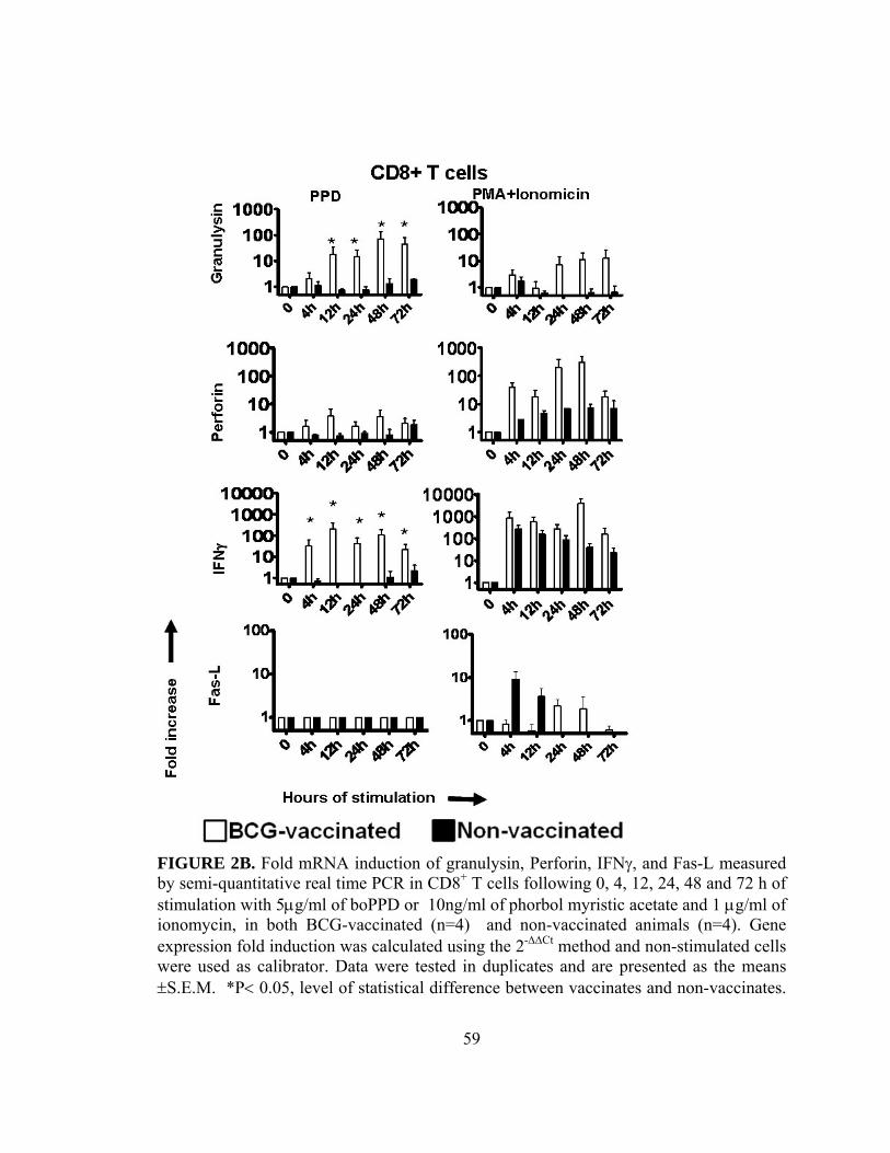
FIGURE 2B. Fold mRNA induction of granulysin, Perforin, IFNγ, and Fas-L measured by semi-quantitative real time PCR in CD8+ T cells following 0, 4, 12, 24, 48 and 72 h of stimulation with 5µg/ml of boPPD or 10ng/ml of phorbol myristic acetate and 1 µg/ml of ionomycin, in both BCG-vaccinated (n=4) and non-vaccinated animals (n=4). Gene expression fold induction was calculated using the 2-∆∆Ct method and non-stimulated cells were used as calibrator. Data were tested in duplicates and are presented as the means ±S.E.M. *P< 0.05, level of statistical difference between vaccinates and non-vaccinates.
59

FIGURE 2C. Fold mRNA induction of granulysin, Perforin, IFNγ, and Fas-L measured by semi-quantitative real time PCR in γδ+ T cells following 0, 4, 12, 24, 48 and 72 h of stimulation with 5µg/ml of boPPD or 10ng/ml of phorbol myristic acetate and 1 µg/ml of ionomycin, in both BCG-vaccinated (n=4) and non-vaccinated animals (n=4). Gene expression fold induction was calculated using the 2-∆∆Ct method and non-stimulated cells were used as calibrator. Data were tested in duplicates and are presented as the means ±S.E.M. **P< 0.001, level of statistical difference between vaccinates and non-vaccinates.
60
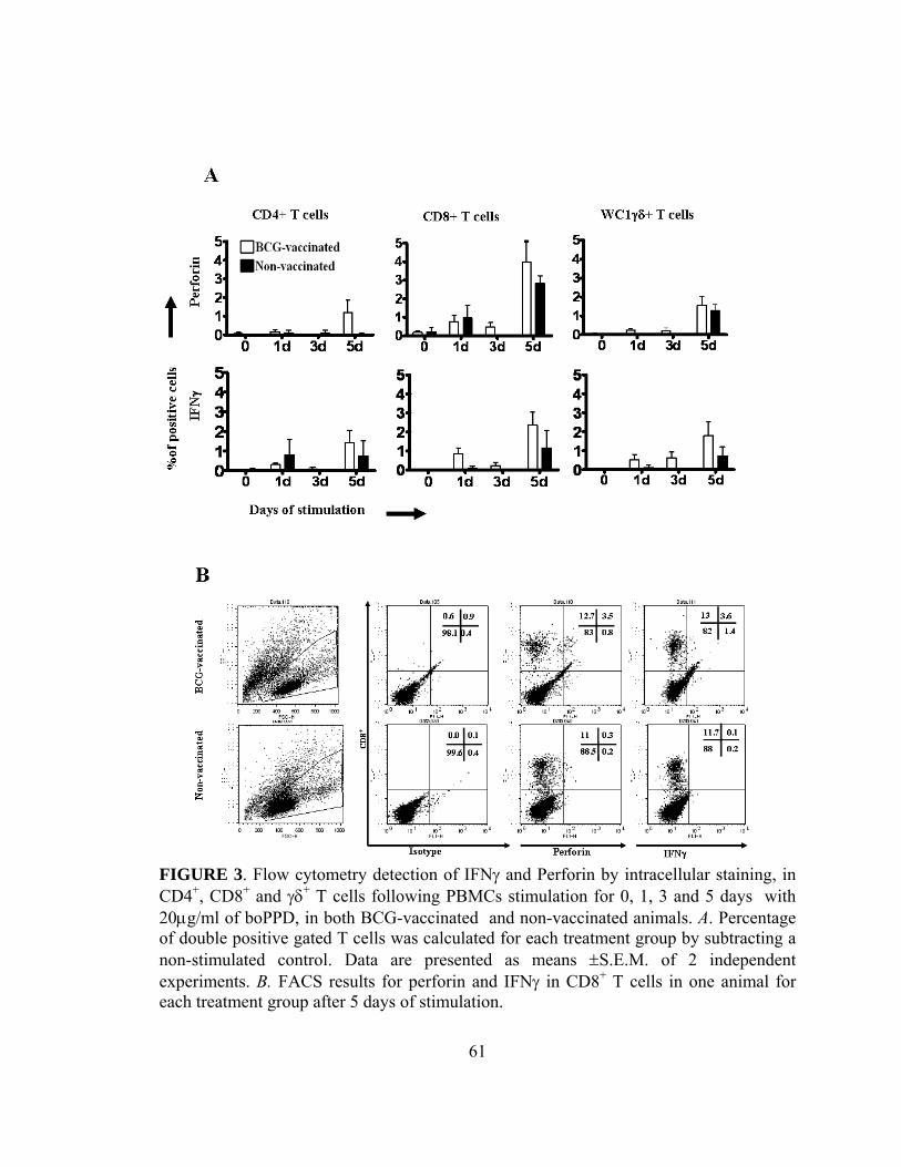
FIGURE 3. Flow cytometry detection of IFNγ and Perforin by intracellular staining, in CD4+, CD8+ and γδ+ T cells following PBMCs stimulation for 0, 1, 3 and 5 days with 20µg/ml of boPPD, in both BCG-vaccinated and non-vaccinated animals. A. Percentage of double positive gated T cells was calculated for each treatment group by subtracting a non-stimulated control. Data are presented as means ±S.E.M. of 2 independent experiments. B. FACS results for perforin and IFNγ in CD8+ T cells in one animal for each treatment group after 5 days of stimulation.
61

FIGURE 4. Granulysin detection by immunoblot. PBMCs (A) or purified CD4+ and CD8+ T cells (B) from both BCG- and non-vaccinates were stimulated with 20 µg/ml of boPPD or left unstimulated. Cells were harvested at 0, 3, 5 and 10 days either in RNA later for further RNA extraction and Quantitative Real-time PCR (PBMC only-right top panel) or were frozen at -80° C for immunoblot performed with specific bovine anti-granulysin antibody and an anti-mouse-IgM-HRP conjugated antibody. Fold induction mRNA was measured by real-time PCR calculated using the 2-∆∆Ct method and non-stimulated cells were used as calibrator. Gene expression fold induction are presented as the means ±S.E.M. of 2 repetitions where ** P< 0.001. Protein concentration was equalized before loading into the gel and β–actin was used as loading control in the western blots.
62

63
Several other studies have demonstrated increased levels of this cytokine
associated with disease persistence [63, 136, 220, 224]. The use of the bovine neonate as
a model presents other advantages for use as a screening tool for vaccine efficacy
including similarities in pathology, clinical presentation, transmission and host immune
responses and effector pathways including granulysin [220, 225]. It also has been shown
that a Th1 response is considered a good correlate of protection upon vaccination in both
humans and bovine. Studies in cattle have shown that the balance of the host Th1/Th2
pathways are more similar to humans than to mice [226]. Granulysin was initially
identified in effector T cells by subtractive hybridization [125]. The mechanism of action
of granulysin is under active investigation but is thought to involve membrane disruption
based on positively charged amino acids that interact with negative charges of the
membrane of the microorganism [98, 106, 131, 202]. Granulysin is expressed in various
effector populations including CD4+, CD8+, WC1+γδ T cells and NK cells [125, 129].
The molecular structure of granulysin indicates that it is a member of the saposin-like
protein family [130] and is characterized by having a five helix bundle as demonstrated
for other proteins of the same family [131]. Much of the lytic activities of granulysin
were demonstrated based on the use of peptides derived from the granulysin sequence
[126, 127, 132]. These peptides were demonstrated to have high cytotoxic/broad
spectrum antimicrobial activity against both gram positive and gram negative bacteria
[97, 106, 133]. Homologues of granulysin have been characterized for several species,
including pigs, horses and cattle [79, 95, 134]. The gene for granulysin has not been
identified in any rodent species to date, restricting the use of knock out mice for in vivo

64
studies. The bovine homologue of granulysin, based on nucleotide and amino acid
sequences, exhibits a relatively high degree of conservation of functional domains with
the other granulysin homologues, including the 5 helix bundle and antimicrobial activity
against several types of bacteria such as E.coli and M. bovis BCG [79].
The expression of granulysin by the different T cell populations and the capacity
to kill intracellular Mycobacterium, suggest that it may be a relevant biomarker for
tracking the immune response against TB both pre- and post-exposure and could be used
as a potential correlate of protection after vaccination against TB. Previous studies have
demonstrated that elevated plasma levels of granulysin and reduced IFNγ levels correlate
with curative responses against TB in human adults [227]. Similar results were observed
following chemotherapy against TB, where elevated granulysin and reduced IFNγ levels
correlate with curative responses [228].
Here we investigated the expression of granulysin in highly enriched effector T
cell populations (CD4+, CD8+ and WC1+γδ T cells) in animals that were vaccinated with
BCG or untreated. We demonstrated that granulysin is expressed early post exposure and
is upregulated upon stimulation with bovine PPD. In a previous study, granulysin
expression was not observed when pure CD4+ T cells were stimulated with PMA and
ionomycin but cytokines such as IFNγ and IL-21 and perforin levels were also elevated
[216] (Appendix 1). This result is in accordance with our observations, suggesting that
granulysin is upregulated in response to antigen-specific stimulation, and that its
expression is dependent on the presence of an antigen presenting cells or other soluble

65
factors such as IL-2. Treatment with IL-2 alone was sufficient to induce granulysin
protein at 5 days of stimulation in pure human CD4+ T cells, but granulysin was not
observed with the addition of α-CD3 to the culture [195] . This same study demonstrated
that addition of IL-2 alone was able to induce granulysin dependent killing of
Cryptococcus neoformans by CD4+ T cells. To our knowledge, so far only one study has
addressed signal transduction pathways involved in transcriptional regulation of the
granulysin gene [229]. It is clear from our studies that signaling mediated via PMA and
ionomycin does not activate transcription of granulysin as observed following antigen-
specific stimulation. It is established that there is extensive alternative splicing in the
granulysin gene where non-coding RNAs may function to stabilize or post-
transcriptionally regulate granulysin gene expression [222].
To our knowledge, this is the first comprehensive study demonstrating and
comparing granulysin gene expression and protein synthesis in T cell populations after
BCG vaccination. We showed that both granulysin isoforms (15 KDa and 9 KDa) were
present in T cell effector populations of BCG immunized animals and were increased
upon a recall response using PPD loaded macrophages. Differences in our finding versus
those reported in human studies are related to the different stimulation conditions used by
us and the potential differences on T cell populations, mainly NK cells, that can
constitutively express granulysin [81]. The presence of NK cells in our experiments was
ruled out by sorting purified CD4+ and CD8+ T cells. When human CD4+ and CD8+ T
cells were stimulated with Listeria sp. antigens, granulysin was observed after 6 to 12
days of stimulation, but LAK cells showed it as early as 4 days after stimulation [222].

66
Granulysin was detected by immunblot on purified human CD4+ T cells stimulated with
either IL-2 or anti-CD3 and IL-2 at 5 days of stimulation, increasing thereafter [195]. In
the same work, increase in granulysin by CD4+ T cells was accompanied by increased
killing of Cryptococcus neoformans that was independent of perforin. The same group
had showed years earlier that CD8+ T cell –mediated killing of the pathogen by
granulysin was independent of perforin, but dependent of IL-15 stimulation [230]. In
summary, granulysin expression seems to be variable according the cell type and
depending on the type of stimulation to which these cells are submitted.
As observed in our study there was a high variability among animals and between
the 2 treatment groups for granulysin protein isoforms, making it difficult to interpret
data and draw conclusions. The pre-exposure to M. avium constitutes a potential problem
upon vaccination with BCG or other TB vaccine candidates and also affects diagnosis
[231] and may be responsible for the antigen specific response observed in one non-
vaccinated animal. One interesting observation was the strong response observed by
purified CD8+ T cells upon stimulation with a soluble antigen (M. bovis PPD). As
suggested by Harding et al., (1994) [232], and reviewed by Vyas et al. (2008) [104],
exogenous antigens may escape phagosomes and be further presented via MHC-I. Our
observation is in accordance with additional reports where CD8+ T cells proliferated and
secreted IFNγ upon stimulation with the same soluble antigen [76, 105]. Bovine purified
CD8+ T cells were shown to proliferate upon stimulation with different antigen such as
live M. bovis and M. bovis PPD. Even though response was superior to live organisms,
considerable response was observed when using M. bovis PPD [105]. In summary, our

67
results suggest that granulysin and perforin are expressed upon an antigen specific
stimulation in BCG-vaccinated animals.

68
CHAPTER 4
POTENTIAL USE OF BO-LYSIN AS A CORRELATE OF
PROTECTION FOLLOWING VACCINATION WITH M. bovis BCG
AND M. bovis ∆RD1 AND CHALLENGE WITH VIRULENT M. bovis

69
INTRODUCTION
To further evaluate the potential use of granulysin and perforin, we evaluated
protection by vaccination by using a new candidate vaccine against TB called M. bovis
∆RD1, in parallel with M. bovis BCG. The sequencing of the M. tuberculosis genome
sequencing in 1998 was a very important step for vaccine development against TB [11,
233], making possible the development and testing of knock-out mutant strains of both
M. tuberculosis and M. bovis. Live knock-out mutants of M. tuberculosis and M. bovis
constitute important vaccine candidates since they still have limited replication capacity,
imunogenicity, but lack aspects of the pathology observed in the wild type. As discussed
earlier, comparison of M. tuberculosis and M. bovis genome with M. bovis BCG, showed
that this last one lacks a so called Region of Difference 1 (RD1), which encodes a
complex of 2 important proteins, ESAT-6 and CFP-10 [12]. These proteins are very
important inducers of IFNγ, and are also involved on several aspect of the pathogenesis
of Mycobacterium spp. In addition, RD1 genes that were engineered back to BCG
resulted in a strain capable of promoting disease similar to wild type M. bovis [15].
The use of auxotrophic strains lacking the RD1 region and other metabolic
pathway genes may be potential vaccine candidates and may provide important tool to
differentiate between vaccination and infection for both bovine and humans. Another
potential use for such vaccine strains is the application on wildlife reservoirs of M. bovis.
Initial testing of a ∆panCD mutant of M. tuberculosis, lacking the panthothenate

70
metabolic pathway was tested in normal and SCID mice showing better efficacy than
BCG and protection against challenge [184] (Appendix 2). To further evaluate this
vaccine candidate, a deletion of the RD1 region was performed and the strain used in
vaccine trials in non-human primates and bovines, but results showed lack of protection
for the mutant in both animal models [63]. In our current work we tested a second
candidate based on a single deletion of the RD1 region on a strain of M. bovis (M. bovis
Ravanel). We compared our vaccine candidate (M. bovis ∆RD1) with the gold standard
BCG and then measured our potential correlates of protection at different time points
after vaccination/before challenge and after challenge.
RESULTS
Granulysin expression is increased after vaccination against TB and challenge with
virulent M. bovis
Here we analyzed the potential for correlation of gene expression with vaccine
efficacy parameters (gross pathology score, radiographic assessment of lesions, and M.
bovis colonization) using samples obtained from three vaccine groups, non-, BCG-, and
∆RD1-vaccinates (Fig. 5, n = 4 / vaccine treatment) in a neonatal vaccine trial. All 3
parameters were significantly lower in BCG-vaccinates as compared to non-vaccinates

71
(P<0.05). There were no statistically significant differences between BCG- and ∆RD1-
vaccinates and between control non-vaccinated and ∆RD1-vaccinates (Fig. 5).
To examine the possible use of granulysin as a correlate of protection after
vaccination against TB, we sampled animals at different time points after
vaccination/before challenge (8, 9 and 10 weeks after vaccination) and one time point
after challenge (2 weeks after challenge) with virulent M. bovis. Based on relative fold
induction, granulysin was higher in both vaccinated groups as compared to non-
vaccinates at all time points (P<0.05) before and after challenge (Fig. 6). When
granulysin expression was averaged at all time points and compared among the three
groups, the expression was elevated in BCG vaccinates as compared to controls (P=0.04)
and in ∆RD1-vaccinates as compared to controls (P=0.03). There was no difference
between the two vaccinated groups (BCG and ∆RD1). No significant differences were
observed for Fas-L. Perforin transcript levels were elevated in BCG- and ∆RD1-
vaccinates as compared to controls after the challenge (P<0.05). Similar results were
observed when perforin was evaluated by intracellular staining in CD4+, CD8+ and γδ+ T
cells after the challenge (Fig. 6) (P<0.01 for BCG- and ∆RD1-vaccinates as compared to
controls in CD4+ T cells; P<0.01 for BCG-vaccinates as compared to controls, and
P<0.0001 for ∆RD1-vaccinates as compared to controls in CD8+T cells; P<0.01 for
∆RD1-vaccinates as compared to controls in γδ+ T cells).

72
IFNγ correlates with pathology
In parallel with granulysin and perforin, we also evaluated IFNγ, a claimed
surrogate marker of protection upon vaccination against TB. IFNγ gene expression was
greater in BCG- and ∆RD1-vaccinates as compared to controls before and after the
challenge (P<0.05) but intracellular staining showed that the percent of IFNγ+ cells in
vaccinates was equivalent or lower than the controls (Fig. 6). IFNγ measured by ELISA
showed that IFNγ responses to M. bovis PPD by ∆RD1-vaccinates was statistically higher
(P < 0.05) as compared to controls non-vaccinated prior to challenge (Fig. 8). After
challenge, IFNγ responses by the control non-vaccinated group exceeded the response of
both BCG- and ∆RD1-vaccinates at 30 days after challenge (P<0.001 for BCG- and
∆RD1-vaccinates, as compared to control) and at 60 days (P<0.05 for BCG- and ∆RD1-
vaccinates, as compared to control). In summary, both granulysin and perforin
expression were upregulated in BCG and ∆RD1 vaccine groups as compared to control.

FIGURE 5. Comprehensive vaccine efficacy data. Lung gross pathology scores, radiographic data (percent lung affected), and CFU/g of tissue from 3 animal groups: control non-vaccinated (n=4), BCG vaccinated (n=4) and ∆RD1 (n=4) evaluated after necropsy. Lung score, radiographic data and Lung CFUs were measured as described by [63]. Values are presented as the means ±S.E.M.
73

FIGURE 6. Fold mRNA induction of granulysin, Perforin, IFNγ, and Fas-L in 3 animal groups: control non-vaccinated (n=4), BCG vaccinated (n=4) and ∆RD1-vaccinated (n=4). PBMCs were stimulated for 5 days with 20µg/ml of boPPD or left unstimulated at 8, 9 and 10 weeks after vaccination and 2 weeks after challenge (14 weeks after vaccination). Cells were harvested and mRNA was measured by Quantitative Real-Time PCR. Gene expression fold induction was calculated using the 2-∆∆Ct method and non-stimulated cells were used as calibrator. Data are presented as the means ±S.E.M. of 2 repetitions. * P< 0.05, level of statistical difference between vaccinates and non-vaccinates.
74

FIGURE 7. Flow cytometric analysis of intracellular staining of IFNγ and Perforin in CD4+, CD8+ and γδ+ T cells following PBMCs stimulation for 5 days with 20µg/ml of boPPD at 8, 9 and 10 weeks after vaccination and 2 weeks after challenge (14 weeks after vaccination) Treatment groups were: control non-vaccinated (n=4), BCG-vaccinated (n=4) and ∆RD1 -vaccinated (n=4) animals. Percentage of double positive gated T cells was calculated for each treatment group by subtracting non-stimulated control value for each individual animal. Data are presented as means ±S.E.M. * P<0.05, **P<0.01, ***P< 0.0001, level of statistical difference between vaccinates and non-vaccinates.
75

FIGURE. 8. Interferon-γ measured by ELISA. Blood mononuclear cells were isolated from each treatment group (n = 4, non-vaccinates; n = 4, M. bovis BCG-vaccinates; n = 4, M. bovis ∆RD1-vaccinates) 30 days prior challenge, 30 and 60 days after challenge and were cultured with 10 µg/ml M. bovis PPD or medium alone (no stimulation) for 48 hrs, when supernatants were harvested and IFN-γ concentrations determined by ELISA (Bovigam, Prionics, Ag). Values represent mean (± standard error) responses to antigen minus the response to media alone. * P<0.05 as compared to control group non-vaccinated, ** P<0.001 as compared to control group non-vaccinated.
76
77
DISCUSSION
Our results demonstrate the potential of granulysin and perforin as correlates of
protective immunity to TB following vaccination and challenge of cattle. Even though
our protection data did not show significant results to support ∆RD1 vaccine efficacy,
comparable results were obtained with our gene expression analysis for both BCG and
∆RD1 vaccine groups. Both granulysin and perforin were upregulated at different time
points before and after the challenge. Granulysin gene expression was significantly
increased in both BCG and ∆RD1 vaccinates, and perforin was significantly increased in
the ∆RD1 group as compared to control and BCG vaccinates after challenge. These
results suggest that the ∆RD1 vaccine is able to induce an immune response comparable
to BCG, and can be potentially used as a strain for future vaccine development, but
further studies are needed in order to confirm our results. In regards to the bacterial
burden, previous work from our group testing another vaccine candidate called mc26030,
which showed protection only on the BCG vaccinated group as compared to control non-
vaccinated [63], culture results were similar between control non-vaccinated , BCG- and
mc26030 –vaccinated groups. This suggests that bacterial burden was not affected by
vaccination, but in this particular case, other vaccine efficacy parameters, such as lung
pathology, did show that BCG was protective as compared to the other 2 groups. Results
from the current study and from our previous work suggest that bacterial burden has to be
evaluated further in future vaccine trial and when developing new vaccine candidates. As

78
an overall conclusion we think that both granulysin and perforin do correlate with
protection against pathology based on lung score and radiographic data.
Other studies in human TB patients evaluating plasma granulysin levels show that
it correlate positively with TB recovery while high IFNγ levels corresponded to chronic
TB [227]. In another study, there was a decrease in serum granulysin after successful
chemotherapy treatment for TB in children [228]. The use of plasma granulysin as a way
to evaluate protection may be influenced by potential degradation in the serum and by the
time it takes from collection to testing of the plasma sample. Our results suggest that
perforin should also be investigated further as a biomarker of protective immunity to
human TB. Even though expression and intracellular detection of perforin were
predominant after the challenge, they were considerable better than results observed for
IFNγ in both vaccinated groups (BCG and ∆RD1) as compared to control group after the
challenge. In addition to that, levels of IFNγ measured by ELISA were relatively higher
in the Control non-vaccinated groups as compared to both BCG- and ∆RD1 vaccinates
after the challenge, suggesting a correlation with pathology.

79
CHAPTER 5
EXPRESSION OF GRANULYSIN IN PARALLEL WITH IFNγ,
PERFORIN AND FAS-L AT THE SITES OF INFECTION OF M.
bovis.
80
INTRODUCTION
In order to provide additional information that could strengthen our evidence that
both granulysin and Perforin could be used as potential correlates of protection, we
sought to determine the gene expression profile on the mediastinal lymph node. Both
lymphoid tissues and lung are foci of infection of M. bovis. Previously published data
from our group showed that granulysin was successfully amplified from a bovine TB
granuloma [79]. Analysis of tissue targets can provide strong insight on details of the
type of immune response present in the tissue and to the cells involved on the process.
RESULTS
In order to establish a relationship of our PBMC data with M. bovis infection, we
extracted RNA from portions of the mediastinal lymph node, adjacent to granulomas.
Gene expression measured by quantitative real time PCR and analyzed as relative
expression, showed that both granulysin and perforin had lower relative expression in
both BCG-and ∆RD1 vaccinated groups as compared to control. IFNγ relative expression
was lower in the Control and ∆RD1 vaccinated group as compared to BCG. Once sample
concentrations are standardized, we can conclude that a lower relative expression of these
molecules is correlated to higher gene expression fold induction (Fig. 9). No major
differences were observed for Fas-L when comparing the 3 vaccine groups.

FIGURE 9- Relative gene expression of granulysin, perforin, IFNγ and Fas-L on Mediastinal lymph node. A total of 1g (gram) of tissue section from the lymph node from the 3 vaccine groups (Control n=4; BCG=4; ∆RD1 n=4), were homogenized in a microcentrifuge tube with a pestle and RNA was extracted by using manufactures protocol (Qiagen, Valencia, CA). Results are shown as relative expression (∆Ct) based on Ribosomal RNA.
81

82
DISCUSSION
Tuberculosis is predominantly a pulmonary disease. Infection is established via
aerosol by a limited number of M. tuberculosis or M. bovis bacilli. The number of bacilli
sufficient to establish infection is still a matter of controversy, but in the bovine numbers
as low as 5 bacilli could be enough to result in infection [234]. Even though such a
number is not established for human infections, less than 10 bacilli maybe enough, but
could be influenced by factors such as droplet size [25]. Initial response starts with either
macrophages in the lungs or DC lying on the trachea, which will engulf the pathogen.
Further response is dependent on γδ +, CD4+ and CD8+ T cells, either migrating to the site
of infection without prior activation or following activation by DC and macrophages on
the local draining lymph node, such as the mediastinal lymph node (MLN). Effector
CD4+ T cells responses were detected in the MLN as early as 10 days post-infection in
mice [235]. In bovines that received intratonsillar inoculation of M. bovis, lesions at the
MLN were observed at 28 days post infection, but were detected as early as 15 days after
infection on the retropharyngeal lymph node [236].
In our studies we measured granulysin and perforin gene expression on MLN
collected 100 days post challenge. Even though our measures are in relative expression
and not calculated as fold induction, we observed that besides Fas-L and IFNγ, both
granulysin and perforin were lower on the vaccine groups as compared to control,
suggesting that MLN from both vaccinated groups had increased fold expression of
cytotoxic granules. This observation is in accordance to the data observed on PPD

83
stimulated PBMC after challenge. As observed in our previous data, in which both gene
expression and protein were measured, we can see that gene expression does not always
indicate protein expression. However, gene expression profile can give us information
related to the immune response against M. bovis infection, and as observed by Tacker et
al. (2007) [237], it may suggest the type of response, either Th1 or Th2, predominant at
the site of infection. In summary, results examining relative gene expression on tissues
support and strengthen our evidence that both granulysin and perforin may be used as
correlates of protection following vaccination against TB, together with IFNγ.

84
CHAPTER 6
SUMMARY, FINAL DISCUSSION/ CONCLUSIONS AND
FUTURE DIRECTIONS

85
In our studies we have shown that (1) granulysin, perforin and IFNγ gene
expression increase upon BCG vaccination; (2) both granulysin and perforin are
upregulated after vaccination and challenge with M. bovis in both BCG and ∆RD1
vaccinated, as compared to control non-vaccinated group, (3) IFNγ correlated with
pathology instead of protection, demonstrated by comparable positive cell numbers
among 3 vaccine groups throughout the experiment, and by having secretion levels
higher in the control group after challenge; (4) a comparable overall response was
observed in both BCG and ∆RD1 vaccine groups demonstrating the potential use of the
∆RD1 strain in future vaccine trials.
Our results suggest that granulysin and perforin are potential correlates of
protection upon vaccination, and can be used on future vaccine trials, together with IFNγ.
The development of better diagnostic tools for granulysin, such as antibodies suitable for
flow cytometric analysis and Confocal Microscopy, would help on providing more
evidence in terms of the use of granulysin and perforin as correlates of protection on
future vaccine trials. Other potential molecules that could be examined further would be
FoxP3, a T regulatory cell marker. This molecule was shown to have higher expression
after BCG vaccination and challenge, when compared to a control group non-vaccinated
and a treatment group that received a M. bovis ∆RD1∆PanCD vaccine candidate [63]
(appendix 2). This vaccine conferred no protection upon challenge with virulent M.
bovis. In addition to FoxP3 we have shown that IL-5 gene expression was relatively
higher on the control as compared to the other vaccine groups. IL-5 is a Th2 cytokine

86
produced by mast cells, eosinophils, γδ T cells and NK cells, with relevant functions on
the activation of eosinophils in asthma [238]. It is unclear what function IL-5 is playing,
as of lack of protection, in TB.
Results observed for the ∆RD1 vaccine candidate are promising and require
further evaluation as a potential vaccine candidate. Our results may have been masked by
the number of animals used during the experiments and we have to consider increasing
the number of animals in our next vaccine trials and evaluation of correlates of
protection. The use of ∆RD1 may represent a potential choice as a vaccine candidate for
both bovine and humans. As mentioned earlier, such vaccines can be combined with a
test that differentiates vaccinated from infected animals (i.e. DIVA), based on using the
protein complex ESAT-6 and CFP10.

APPENDIX 1
87
Captured from www.sciencedirect.com on 08-27-08: Endsley, J.J., Hogg, A., Shell, L.J., McAulay, M., Coffey,T., Howard, C., Capinos Scherer, C.F., Siddiqui, N., Waters, W.R., Nonnecke, B., Estes, D.M , Villarreal-Ramos, B. (2007). Mycobacterium bovis BCG vaccination induces memory CD4+ T cells characterized by effector biomarker expression and anti-mycobacterial activity. Vaccine Dec 5;25(50):8384-94. Elsevier Copyright License Number: 2022121003881

88

89

90

91

92

93
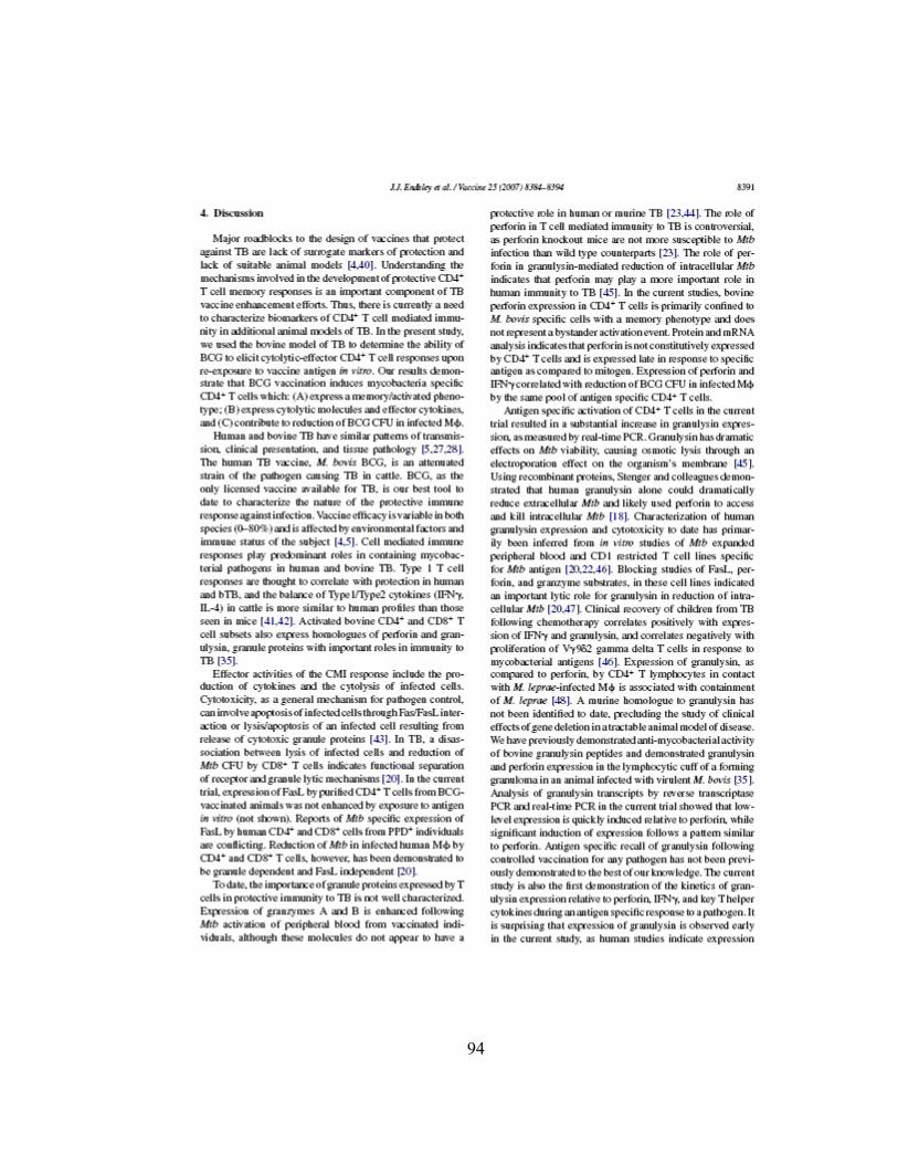
94

95

96
97

APPENDIX 2
Captured from www.sciencedirect.com on 08-27-08: Waters, W.R., Palmer, M.V., Nonnecke, B.J., Thacker, T.C., Scherer, C.F.C.,Estes, D.M., Jacobs, Jr., W., Glatman-Freedman, A., Larsen, M.L. (2007). Failure of a Mycobacterium tuberculosis RD1 x panCD double deletion mutant in a neonatal calf aerosol M. bovis challenge model: comparisons to responses elicited by M. bovis bacille Calmette Guerin. Vaccine Nov 7;25(45):7832-40. Elsevier Copyright License # 2022121003862
98

99

100

101

102
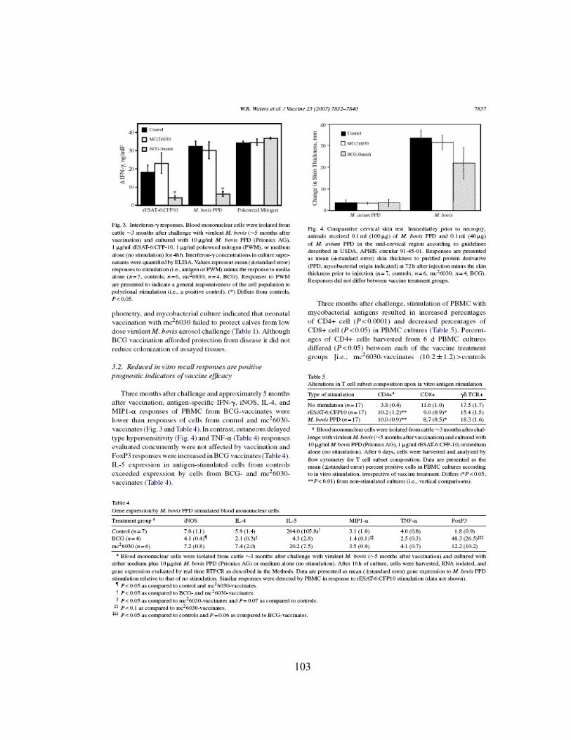
103

104

105

106

APPENDIX 3
Captured from www.sciencedirect.com on 08-27-08: Alexander C. Maue, A.C., Waters, W. R., Palmer, M.V., Nonnecke, B.J. ,Minion, F.C. , Wendy C. Brown, W.C., Norimine, J., Foote, M.R., Scherer, C.F.C., Estes, D.M. (2007). An ESAT-6:CFP10 DNA vaccine administered in conjunction with Mycobacterium bovis BCG confers protection to cattle challenged with virulent Mycobacterium bovis. Vaccine Jun 11;25(24):4735-46. Elsevier Copyright License Number: 2022130214488
107

108
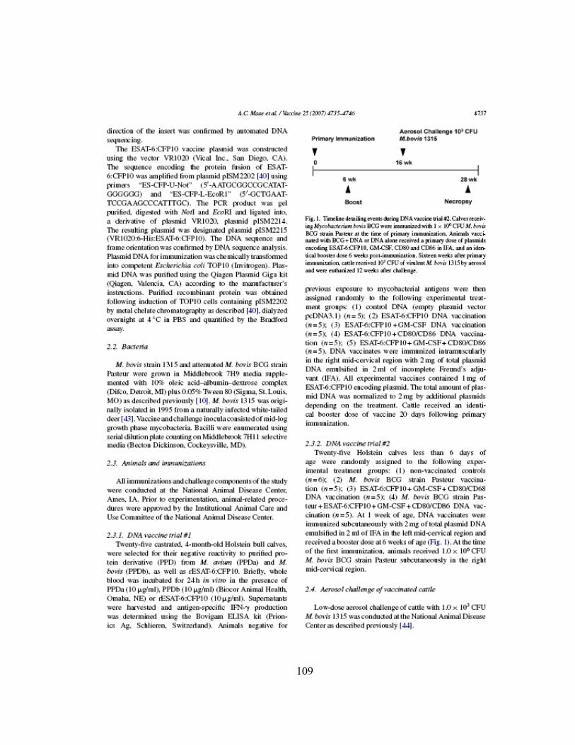
109

110

111

112

113

114

115

116

117

118

119
BIBLIOGRAPHY
1. CDC, Core Curriculum on tuberculosis. 2000, Division of Tuberculosis
Elimination. Center of Disease Control. 2. WHO, Global tuberculosis control : surveillance, planning, financing : WHO
report 2008, W.H.O. WHO Press, Editor. 2008, WHO: Geneva 27, Switzerland. 3. Lewerin, S.S., Olsson, S.L., Eld, K., Roken, B., Ghebremichael, S., Koivula, T.,
Kallenius, G., Bolske, G., 2005.Outbreak of Mycobacterium tuberculosis infection among captive Asian elephants in a Swedish zoo. Vet Rec, 156(6): p. 171-175.
4. Palmer, M.V., Waters, W.R., Whipple, D.L., 2004.Investigation of the transmission of Mycobacterium bovis from deer to cattle through indirect contact. Am J Vet Res, 65(11): p. 1483-1489.
5. Schmitt, S.M., Fitzgerald, S.D., Cooley, T.M., Bruning-Fann, C.S., Sullivan, L., Berry, D., Carlson, T., Minnis, R.B., Payeur, J.B., Sikarskie, J., 1997.Bovine tuberculosis in free-ranging white-tailed deer from Michigan. J Wildl Dis, 33(4): p. 749-758.
6. Harris, N.B., Payeur, J., Bravo, D., Osorio, R., Stuber, T., Farrell, D., Paulson, D., Treviso, S., Mikolon, A., Rodriguez-Lainz, A., Cernek-Hoskins, S., Rast, R., Ginsberg, M., Kinde, H., 2007.Recovery of Mycobacterium bovis from soft fresh cheese originating in Mexico. Appl Environ Microbiol, 73(3): p. 1025-1028.
7. Hope, J.C. ,Villarreal-Ramos, B., 2008.Bovine TB and the development of new vaccines. Comp Immunol Microbiol Infect Dis, 31(2-3): p. 77-100.
8. Pfyffer, G.E., Brown-Elliott, B.A., Wallace Jr, R.J., Mycobacterium: general characteristics, isolation, and staining procedures, in Manual of Clinical Microbiology, P.R. Murray, Baron, E.J., Jorgensen, J.H., Pfaller, M.A., Yolken, R.H., Editor. 2003, ASM Press: Washington, D.C. p. 1212.
9. Best, M., Sattar, S.A., Springthorpe, V.S., Kennedy, M.E., 1990.Efficacies of selected disinfectants against Mycobacterium tuberculosis. J Clin Microbiol, 28(10): p. 2234-2239.
10. Wallace, R.J., Jr., 1987.Nontuberculous mycobacteria and water: a love affair with increasing clinical importance. Infect Dis Clin North Am, 1(3): p. 677-686.
11. Cole, S.T. ,Barrell, B.G., 1998.Analysis of the genome of Mycobacterium tuberculosis H37Rv. Novartis Found Symp, 217: p. 160-172; discussion 172-167.
12. Garnier, T., Eiglmeier, K., Camus, J.C., Medina, N., Mansoor, H., Pryor, M., Duthoy, S., Grondin, S., Lacroix, C., Monsempe, C., Simon, S., Harris, B., Atkin, R., Doggett, J., Mayes, R., Keating, L., Wheeler, P.R., Parkhill, J., Barrell, B.G., Cole, S.T., Gordon, S.V., Hewinson, R.G., 2003.The complete genome sequence of Mycobacterium bovis. Proc Natl Acad Sci U S A, 100(13): p. 7877-7882.

120
13. Lewis, K.N., Liao, R., Guinn, K.M., Hickey, M.J., Smith, S., Behr, M.A., Sherman, D.R., 2003.Deletion of RD1 from Mycobacterium tuberculosis mimics bacille Calmette-Guerin attenuation. J Infect Dis, 187(1): p. 117-123.
14. Gey van Pittius, N.C., Warren, R.M., van Helden, P.D., 2002.ESAT-6 and CFP-10: what is the diagnosis? Infect Immun, 70(11): p. 6509-6510; author reply 6511.
15. Pym, A.S., Brodin, P., Brosch, R., Huerre, M., Cole, S.T., 2002.Loss of RD1 contributed to the attenuation of the live tuberculosis vaccines Mycobacterium bovis BCG and Mycobacterium microti. Mol Microbiol, 46(3): p. 709-717.
16. Ganguly, N., Giang, P.H., Gupta, C., Basu, S.K., Siddiqui, I., Salunke, D.M., Sharma, P., 2008.Mycobacterium tuberculosis secretory proteins CFP-10, ESAT-6 and the CFP10:ESAT6 complex inhibit lipopolysaccharide-induced NF-kappaB transactivation by downregulation of reactive oxidative species (ROS) production. Immunol Cell Biol, 86(1): p. 98-106.
17. Arend, S.M., de Haas, P., Leyten, E., Rosenkrands, I., Rigouts, L., Andersen, P., Mijs, W., van Dissel, J.T., van Soolingen, D., 2005.ESAT-6 and CFP-10 in clinical versus environmental isolates of Mycobacterium kansasii. J Infect Dis, 191(8): p. 1301-1310.
18. Arend, S.M., Ottenhoff, T.H., Andersen, P., van Dissel, J.T., 2001.Uncommon presentations of tuberculosis: the potential value of a novel diagnostic assay based on the Mycobacterium tuberculosis-specific antigens ESAT-6 and CFP-10. Int J Tuberc Lung Dis, 5(7): p. 680-686.
19. Munk, M.E., Arend, S.M., Brock, I., Ottenhoff, T.H., Andersen, P., 2001.Use of ESAT-6 and CFP-10 antigens for diagnosis of extrapulmonary tuberculosis. J Infect Dis, 183(1): p. 175-176.
20. Arend, S.M., Geluk, A., van Meijgaarden, K.E., van Dissel, J.T., Theisen, M., Andersen, P., Ottenhoff, T.H., 2000.Antigenic equivalence of human T-cell responses to Mycobacterium tuberculosis-specific RD1-encoded protein antigens ESAT-6 and culture filtrate protein 10 and to mixtures of synthetic peptides. Infect Immun, 68(6): p. 3314-3321.
21. Lyashchenko, K.P., Greenwald, R., Esfandiari, J., Olsen, J.H., Ball, R., Dumonceaux, G., Dunker, F., Buckley, C., Richard, M., Murray, S., Payeur, J.B., Andersen, P., Pollock, J.M., Mikota, S., Miller, M., Sofranko, D., Waters, W.R., 2006.Tuberculosis in elephants: antibody responses to defined antigens of Mycobacterium tuberculosis, potential for early diagnosis, and monitoring of treatment. Clin Vaccine Immunol, 13(7): p. 722-732.
22. Riley, L.W., 2006.Of mice, men, and elephants: Mycobacterium tuberculosis cell envelope lipids and pathogenesis. J Clin Invest, 116(6): p. 1475-1478.
23. Une, Y. ,Mori, T., 2007.Tuberculosis as a zoonosis from a veterinary perspective. Comp Immunol Microbiol Infect Dis, 30(5-6): p. 415-425.
24. Neill, S.D., Pollock, J.M., Bryson, D.B., Hanna, J., 1994.Pathogenesis of Mycobacterium bovis infection in cattle. Vet Microbiol, 40(1-2): p. 41-52.

121
25. Sundaramurthy, V. ,Pieters, J., 2007.Interactions of pathogenic mycobacteria with host macrophages. Microbes Infect, 9(14-15): p. 1671-1679.
26. Le Cabec, V., Cols, C., Maridonneau-Parini, I., 2000.Nonopsonic phagocytosis of zymosan and Mycobacterium kansasii by CR3 (CD11b/CD18) involves distinct molecular determinants and is or is not coupled with NADPH oxidase activation. Infect Immun, 68(8): p. 4736-4745.
27. Ernst, J.D., 1998.Macrophage receptors for Mycobacterium tuberculosis. Infect Immun, 66(4): p. 1277-1281.
28. Gomez, L.M., Anaya, J.M., Sierra-Filardi, E., Cadena, J., Corbi, A., Martin, J., 2006.Analysis of DC-SIGN (CD209) functional variants in patients with tuberculosis. Hum Immunol, 67(10): p. 808-811.
29. Neyrolles, O., Gicquel, B., Quintana-Murci, L., 2006.Towards a crucial role for DC-SIGN in tuberculosis and beyond. Trends Microbiol, 14(9): p. 383-387.
30. Sturgill-Koszycki, S., Schlesinger, P.H., Chakraborty, P., Haddix, P.L., Collins, H.L., Fok, A.K., Allen, R.D., Gluck, S.L., Heuser, J., Russell, D.G., 1994.Lack of acidification in Mycobacterium phagosomes produced by exclusion of the vesicular proton-ATPase. Science, 263(5147): p. 678-681.
31. Chua, J., Vergne, I., Master, S., Deretic, V., 2004.A tale of two lipids: Mycobacterium tuberculosis phagosome maturation arrest. Curr Opin Microbiol, 7(1): p. 71-77.
32. Pieters, J., 2001.Evasion of host cell defense mechanisms by pathogenic bacteria. Curr Opin Immunol, 13(1): p. 37-44.
33. Vergne, I., Chua, J., Lee, H.H., Lucas, M., Belisle, J., Deretic, V., 2005.Mechanism of phagolysosome biogenesis block by viable Mycobacterium tuberculosis. Proc Natl Acad Sci U S A, 102(11): p. 4033-4038.
34. Vergne, I., Chua, J., Deretic, V., 2003.Tuberculosis toxin blocking phagosome maturation inhibits a novel Ca2+/calmodulin-PI3K hVPS34 cascade. J Exp Med, 198(4): p. 653-659.
35. Bhatt, K. ,Salgame, P., 2007.Host innate immune response to Mycobacterium tuberculosis. J Clin Immunol, 27(4): p. 347-362.
36. Hope, J.C., Thom, M.L., McCormick, P.A., Howard, C.J., 2004.Interaction of antigen presenting cells with mycobacteria. Vet Immunol Immunopathol, 100(3-4): p. 187-195.
37. Noss, E.H., Pai, R.K., Sellati, T.J., Radolf, J.D., Belisle, J., Golenbock, D.T., Boom, W.H., Harding, C.V., 2001.Toll-like receptor 2-dependent inhibition of macrophage class II MHC expression and antigen processing by 19-kDa lipoprotein of Mycobacterium tuberculosis. J Immunol, 167(2): p. 910-918.
38. Noss, E.H., Harding, C.V., Boom, W.H., 2000.Mycobacterium tuberculosis inhibits MHC class II antigen processing in murine bone marrow macrophages. Cell Immunol, 201(1): p. 63-74.
39. Tailleux, L., Pham-Thi, N., Bergeron-Lafaurie, A., Herrmann, J.L., Charles, P., Schwartz, O., Scheinmann, P., Lagrange, P.H., de Blic, J., Tazi, A., Gicquel, B., Neyrolles, O., 2005.DC-SIGN induction in alveolar macrophages defines

122
privileged target host cells for mycobacteria in patients with tuberculosis. PLoS Med, 2(12): p. e381.
40. Tailleux, L., Gicquel, B., Neyrolles, O., 2003.[DC-SIGN, a key receptor of Mycobacterium tuberculosis?]. Med Sci (Paris), 19(6-7): p. 658-660.
41. van Kooyk, Y., Appelmelk, B., Geijtenbeek, T.B., 2003.A fatal attraction: Mycobacterium tuberculosis and HIV-1 target DC-SIGN to escape immune surveillance. Trends Mol Med, 9(4): p. 153-159.
42. Denis, M. ,Buddle, B.M., 2008.Bovine dendritic cells are more permissive for Mycobacterium bovis replication than macrophages, but release more IL-12 and induce better immune T-cell proliferation. Immunol Cell Biol, 86(2): p. 185-191.
43. Dannenberg, A.M., Jr., 1994.Roles of cytotoxic delayed-type hypersensitivity and macrophage-activating cell-mediated immunity in the pathogenesis of tuberculosis. Immunobiology, 191(4-5): p. 461-473.
44. Bhatt, K., Hickman, S.P., Salgame, P., 2004.Cutting edge: a new approach to modeling early lung immunity in murine tuberculosis. J Immunol, 172(5): p. 2748-2751.
45. Boros, D.L. ,Warren, K.S., 1973.The bentonite granuloma. Characterization of a model system for infectious and foreign body granulomatous inflammation using soluble mycobacterial, histoplasma and schistosoma antigens. Immunology, 24(3): p. 511-529.
46. Boros, D.L. ,Warren, K.S., 1970.Delayed hypersensitivity-type granuloma formation and dermal reaction induced and elicited by a soluble factor isolated from Schistosoma mansoni eggs. J Exp Med, 132(3): p. 488-507.
47. Chensue, S.W. ,Boros, D.L., 1979.Modulation of granulomatous hypersensitivity. I. Characterization of T lymphocytes involved in the adoptive suppression of granuloma formation in Schistosoma mansoni-infected mice. J Immunol, 123(3): p. 1409-1414.
48. Mariano, M., 1995.The experimental granuloma. A hypothesis to explain the persistence of the lesion. Rev Inst Med Trop Sao Paulo, 37(2): p. 161-176.
49. Cassidy, J.P., 2006.The pathogenesis and pathology of bovine tuberculosis with insights from studies of tuberculosis in humans and laboratory animal models. Vet Microbiol, 112(2-4): p. 151-161.
50. Balasubramanian, V., Wiegeshaus, E.H., Taylor, B.T., Smith, D.W., 1994.Pathogenesis of tuberculosis: pathway to apical localization. Tuber Lung Dis, 75(3): p. 168-178.
51. Ulrichs, T., Kosmiadi, G.A., Trusov, V., Jorg, S., Pradl, L., Titukhina, M., Mishenko, V., Gushina, N., Kaufmann, S.H., 2004.Human tuberculous granulomas induce peripheral lymphoid follicle-like structures to orchestrate local host defence in the lung. J Pathol, 204(2): p. 217-228.
52. Tsai, M.S., Hsieh, C.C., Chen, H.R., Chen, W.J., Huang, C.H., 2006.Tuberculosis mycobacterium sepsis as a rare cause of out-of-hospital cardiac arrest. Am J Emerg Med, 24(6): p. 755-756.

123
53. Liebana, E., Johnson, L., Gough, J., Durr, P., Jahans, K., Clifton-Hadley, R., Spencer, Y., Hewinson, R.G., Downs, S.H., 2008.Pathology of naturally occurring bovine tuberculosis in England and Wales. Vet J, 176(3): p. 354-360.
54. Johnson, L.K., Liebana, E., Nunez, A., Spencer, Y., Clifton-Hadley, R., Jahans, K., Ward, A., Barlow, A., Delahay, R., 2008.Histological observations of bovine tuberculosis in lung and lymph node tissues from British deer. Vet J, 175(3): p. 409-412.
55. Flynn, J.L., 2006.Lessons from experimental Mycobacterium tuberculosis infections. Microbes Infect, 8(4): p. 1179-1188.
56. Dieli, F., Singh, M., Spallek, R., Romano, A., Titone, L., Sireci, G., Friscia, G., Di Sano, C., Santini, D., Salerno, A., Ivanyi, J., 2000.Change of Th0 to Th1 cell-cytokine profile following tuberculosis chemotherapy. Scand J Immunol, 52(1): p. 96-102.
57. Cassidy, J.P., Bryson, D.G., Gutierrez Cancela, M.M., Forster, F., Pollock, J.M., Neill, S.D., 2001.Lymphocyte subtypes in experimentally induced early-stage bovine tuberculous lesions. J Comp Pathol, 124(1): p. 46-51.
58. Orme, I.M., Miller, E.S., Roberts, A.D., Furney, S.K., Griffin, J.P., Dobos, K.M., Chi, D., Rivoire, B., Brennan, P.J., 1992.T lymphocytes mediating protection and cellular cytolysis during the course of Mycobacterium tuberculosis infection. Evidence for different kinetics and recognition of a wide spectrum of protein antigens. J Immunol, 148(1): p. 189-196.
59. Palmer, M.V. ,Waters, W.R., 2006.Advances in bovine tuberculosis diagnosis and pathogenesis: what policy makers need to know. Vet Microbiol, 112(2-4): p. 181-190.
60. Helke, K.L., Mankowski, J.L., Manabe, Y.C., 2006.Animal models of cavitation in pulmonary tuberculosis. Tuberculosis (Edinb), 86(5): p. 337-348.
61. Chang, J.C., Wysocki, A., Tchou-Wong, K.M., Moskowitz, N., Zhang, Y., Rom, W.N., 1996.Effect of Mycobacterium tuberculosis and its components on macrophages and the release of matrix metalloproteinases. Thorax, 51(3): p. 306-311.
62. Price, N.M., Farrar, J., Tran, T.T., Nguyen, T.H., Tran, T.H., Friedland, J.S., 2001.Identification of a matrix-degrading phenotype in human tuberculosis in vitro and in vivo. J Immunol, 166(6): p. 4223-4230.
63. Waters, W.R., Palmer, M.V., Nonnecke, B.J., Thacker, T.C., Scherer, C.F., Estes, D.M., Jacobs, W.R., Jr., Glatman-Freedman, A., Larsen, M.H., 2007.Failure of a Mycobacterium tuberculosis DeltaRD1 DeltapanCD double deletion mutant in a neonatal calf aerosol M. bovis challenge model: comparisons to responses elicited by M. bovis bacille Calmette Guerin. Vaccine, 25(45): p. 7832-7840.
64. Brightbill, H.D., Libraty, D.H., Krutzik, S.R., Yang, R.B., Belisle, J.T., Bleharski, J.R., Maitland, M., Norgard, M.V., Plevy, S.E., Smale, S.T., Brennan, P.J., Bloom, B.R., Godowski, P.J., Modlin, R.L., 1999.Host defense mechanisms triggered by microbial lipoproteins through toll-like receptors. Science, 285(5428): p. 732-736.

124
65. Martino, A., 2008.Mycobacteria and innate cells: critical encounter for immunogenicity. J Biosci, 33(1): p. 137-144.
66. Winau, F., Weber, S., Sad, S., de Diego, J., Hoops, S.L., Breiden, B., Sandhoff, K., Brinkmann, V., Kaufmann, S.H., Schaible, U.E., 2006.Apoptotic vesicles crossprime CD8 T cells and protect against tuberculosis. Immunity, 24(1): p. 105-117.
67. Keane, J., Balcewicz-Sablinska, M.K., Remold, H.G., Chupp, G.L., Meek, B.B., Fenton, M.J., Kornfeld, H., 1997.Infection by Mycobacterium tuberculosis promotes human alveolar macrophage apoptosis. Infect Immun, 65(1): p. 298-304.
68. Abel, B., Thieblemont, N., Quesniaux, V.J., Brown, N., Mpagi, J., Miyake, K., Bihl, F., Ryffel, B., 2002.Toll-like receptor 4 expression is required to control chronic Mycobacterium tuberculosis infection in mice. J Immunol, 169(6): p. 3155-3162.
69. Pompei, L., Jang, S., Zamlynny, B., Ravikumar, S., McBride, A., Hickman, S.P., Salgame, P., 2007.Disparity in IL-12 release in dendritic cells and macrophages in response to Mycobacterium tuberculosis is due to use of distinct TLRs. J Immunol, 178(8): p. 5192-5199.
70. Bafica, A., Scanga, C.A., Feng, C.G., Leifer, C., Cheever, A., Sher, A., 2005.TLR9 regulates Th1 responses and cooperates with TLR2 in mediating optimal resistance to Mycobacterium tuberculosis. J Exp Med, 202(12): p. 1715-1724.
71. Tanaka, Y., Sano, S., Nieves, E., De Libero, G., Rosa, D., Modlin, R.L., Brenner, M.B., Bloom, B.R., Morita, C.T., 1994.Nonpeptide ligands for human gamma delta T cells. Proc Natl Acad Sci U S A, 91(17): p. 8175-8179.
72. Gao, Y., Yang, W., Pan, M., Scully, E., Girardi, M., Augenlicht, L.H., Craft, J., Yin, Z., 2003.Gamma delta T cells provide an early source of interferon gamma in tumor immunity. J Exp Med, 198(3): p. 433-442.
73. Pollock, J.M. ,Welsh, M.D., 2002.The WC1(+) gammadelta T-cell population in cattle: a possible role in resistance to intracellular infection. Vet Immunol Immunopathol, 89(3-4): p. 105-114.
74. Brown, W.C., Davis, W.C., Choi, S.H., Dobbelaere, D.A., Splitter, G.A., 1994.Functional and phenotypic characterization of WC1+ gamma/delta T cells isolated from Babesia bovis-stimulated T cell lines. Cell Immunol, 153(1): p. 9-27.
75. Park, Y.H., Fox, L.K., Hamilton, M.J., Davis, W.C., 1993.Suppression of proliferative response of BoCD4+ T lymphocytes by activated BoCD8+ T lymphocytes in the mammary gland of cows with Staphylococcus aureus mastitis. Vet Immunol Immunopathol, 36(2): p. 137-151.
76. Pollock, J.M., Pollock, D.A., Campbell, D.G., Girvin, R.M., Crockard, A.D., Neill, S.D., Mackie, D.P., 1996.Dynamic changes in circulating and antigen-responsive T-cell subpopulations post-Mycobacterium bovis infection in cattle. Immunology, 87(2): p. 236-241.

125
77. Doherty, M.L., Bassett, H.F., Quinn, P.J., Davis, W.C., Kelly, A.P., Monaghan, M.L., 1996.A sequential study of the bovine tuberculin reaction. Immunology, 87(1): p. 9-14.
78. Kennedy, H.E., Welsh, M.D., Bryson, D.G., Cassidy, J.P., Forster, F.I., Howard, C.J., Collins, R.A., Pollock, J.M., 2002.Modulation of immune responses to Mycobacterium bovis in cattle depleted of WC1(+) gamma delta T cells. Infect Immun, 70(3): p. 1488-1500.
79. Endsley, J.J., Furrer, J.L., Endsley, M.A., McIntosh, M.A., Maue, A.C., Waters, W.R., Lee, D.R., Estes, D.M., 2004.Characterization of bovine homologues of granulysin and NK-lysin. J Immunol, 173(4): p. 2607-2614.
80. Diefenbach, A. ,Raulet, D.H., 2001.Strategies for target cell recognition by natural killer cells. Immunol Rev, 181: p. 170-184.
81. Endsley, J.J., Endsley, M.A., Estes, D.M., 2006.Bovine natural killer cells acquire cytotoxic/effector activity following activation with IL-12/15 and reduce Mycobacterium bovis BCG in infected macrophages. J Leukoc Biol, 79(1): p. 71-79.
82. Storset, A.K., Kulberg, S., Berg, I., Boysen, P., Hope, J.C., Dissen, E., 2004.NKp46 defines a subset of bovine leukocytes with natural killer cell characteristics. Eur J Immunol, 34(3): p. 669-676.
83. Lanier, L.L., 2005.NK cell recognition. Annu Rev Immunol, 23: p. 225-274. 84. Colucci, F., Caligiuri, M.A., Di Santo, J.P., 2003.What does it take to make a
natural killer? Nat Rev Immunol, 3(5): p. 413-425. 85. Endsley, J.J., Endsley, M.A., Estes, D.M., 2005.Bovine natural killer cells acquire
cytotoxic/effector activity following activation with IL-12/15 and reduce Mycobacterium bovis BCG in infected macrophages. J Leukoc Biol.
86. Adam, C., King, S., Allgeier, T., Braumuller, H., Luking, C., Mysliwietz, J., Kriegeskorte, A., Busch, D.H., Rocken, M., Mocikat, R., 2005.DC-NK cell cross talk as a novel CD4+ T-cell-independent pathway for antitumor CTL induction. Blood, 106(1): p. 338-344.
87. Andrews, D.M., Andoniou, C.E., Scalzo, A.A., van Dommelen, S.L., Wallace, M.E., Smyth, M.J., Degli-Esposti, M.A., 2005.Cross-talk between dendritic cells and natural killer cells in viral infection. Mol Immunol, 42(4): p. 547-555.
88. Kulberg, S., Boysen, P., Storset, A.K., 2004.Reference values for relative numbers of natural killer cells in cattle blood. Dev Comp Immunol, 28(9): p. 941-948.
89. Erkeller-Yuksel, F.M., Deneys, V., Yuksel, B., Hannet, I., Hulstaert, F., Hamilton, C., Mackinnon, H., Stokes, L.T., Munhyeshuli, V., Vanlangendonck, F., et al., 1992.Age-related changes in human blood lymphocyte subpopulations. J Pediatr, 120(2 Pt 1): p. 216-222.
90. Raulet, D.H., 2004.Interplay of natural killer cells and their receptors with the adaptive immune response. Nat Immunol, 5(10): p. 996-1002.
91. Junqueira-Kipnis, A.P., Kipnis, A., Jamieson, A., Juarrero, M.G., Diefenbach, A., Raulet, D.H., Turner, J., Orme, I.M., 2003.NK cells respond to pulmonary
126
infection with Mycobacterium tuberculosis, but play a minimal role in protection. J Immunol, 171(11): p. 6039-6045.
92. Martin-Fontecha, A., Thomsen, L.L., Brett, S., Gerard, C., Lipp, M., Lanzavecchia, A., Sallusto, F., 2004.Induced recruitment of NK cells to lymph nodes provides IFN-gamma for T(H)1 priming. Nat Immunol, 5(12): p. 1260-1265.
93. Boysen, P., Olsen, I., Berg, I., Kulberg, S., Johansen, G.M., Storset, A.K., 2006.Bovine CD2-/NKp46+ cells are fully functional natural killer cells with a high activation status. BMC Immunol, 7: p. 10.
94. Denis, M., Keen, D.L., Parlane, N.A., Storset, A.K., Buddle, B.M., 2007.Bovine natural killer cells restrict the replication of Mycobacterium bovis in bovine macrophages and enhance IL-12 release by infected macrophages. Tuberculosis (Edinb), 87(1): p. 53-62.
95. Andreu, D., Carreno, C., Linde, C., Boman, H.G., Andersson, M., 1999.Identification of an anti-mycobacterial domain in NK-lysin and granulysin. Biochem J, 344 Pt 3: p. 845-849.
96. Clayberger, C. ,Krensky, A.M., 2003.Granulysin. Curr Opin Immunol, 15(5): p. 560-565.
97. Dieli, F., Troye-Blomberg, M., Ivanyi, J., Fournie, J.J., Krensky, A.M., Bonneville, M., Peyrat, M.A., Caccamo, N., Sireci, G., Salerno, A., 2001.Granulysin-dependent killing of intracellular and extracellular Mycobacterium tuberculosis by Vgamma9/Vdelta2 T lymphocytes. J Infect Dis, 184(8): p. 1082-1085.
98. Gamen, S., Hanson, D.A., Kaspar, A., Naval, J., Krensky, A.M., Anel, A., 1998.Granulysin-induced apoptosis. I. Involvement of at least two distinct pathways. J Immunol, 161(4): p. 1758-1764.
99. Gansert, J.L., Kiessler, V., Engele, M., Wittke, F., Rollinghoff, M., Krensky, A.M., Porcelli, S.A., Modlin, R.L., Stenger, S., 2003.Human NKT cells express granulysin and exhibit antimycobacterial activity. J Immunol, 170(6): p. 3154-3161.
100. Hata, A., Zerboni, L., Sommer, M., Kaspar, A.A., Clayberger, C., Krensky, A.M., Arvin, A.M., 2001.Granulysin blocks replication of varicella-zoster virus and triggers apoptosis of infected cells. Viral Immunol, 14(2): p. 125-133.
101. Kaufmann, S.H. ,Ladel, C.H., 1994.Role of T cell subsets in immunity against intracellular bacteria: experimental infections of knock-out mice with Listeria monocytogenes and Mycobacterium bovis BCG. Immunobiology, 191(4-5): p. 509-519.
102. Flynn, J.L. ,Ernst, J.D., 2000.Immune responses in tuberculosis. Curr Opin Immunol, 12(4): p. 432-436.
103. Pollock, J.M., Welsh, M.D., McNair, J., 2005.Immune responses in bovine tuberculosis: towards new strategies for the diagnosis and control of disease. Vet Immunol Immunopathol, 108(1-2): p. 37-43.

127
104. Vyas, J.M., Van der Veen, A.G., Ploegh, H.L., 2008.The known unknowns of antigen processing and presentation. Nat Rev Immunol, 8(8): p. 607-618.
105. Liebana, E., Girvin, R.M., Welsh, M., Neill, S.D., Pollock, J.M., 1999.Generation of CD8(+) T-cell responses to Mycobacterium bovis and mycobacterial antigen in experimental bovine tuberculosis. Infect Immun, 67(3): p. 1034-1044.
106. Stenger, S., Hanson, D.A., Teitelbaum, R., Dewan, P., Niazi, K.R., Froelich, C.J., Ganz, T., Thoma-Uszynski, S., Melian, A., Bogdan, C., Porcelli, S.A., Bloom, B.R., Krensky, A.M., Modlin, R.L., 1998.An antimicrobial activity of cytolytic T cells mediated by granulysin. Science, 282(5386): p. 121-125.
107. Smith, R.A., Alvarez, A.J., Estes, D.M., 2001.The P2X7 purinergic receptor on bovine macrophages mediates mycobacterial death. Vet Immunol Immunopathol, 78(3-4): p. 249-262.
108. Kusner, D.J. ,Adams, J., 2000.ATP-induced killing of virulent Mycobacterium tuberculosis within human macrophages requires phospholipase D. J Immunol, 164(1): p. 379-388.
109. Placido, R., Auricchio, G., Falzoni, S., Battistini, L., Colizzi, V., Brunetti, E., Di Virgilio, F., Mancino, G., 2006.P2X(7) purinergic receptors and extracellular ATP mediate apoptosis of human monocytes/macrophages infected with Mycobacterium tuberculosis reducing the intracellular bacterial viability. Cell Immunol, 244(1): p. 10-18.
110. Myers, A.J., Eilertson, B., Fulton, S.A., Flynn, J.L., Canaday, D.H., 2005.The purinergic P2X7 receptor is not required for control of pulmonary Mycobacterium tuberculosis infection. Infect Immun, 73(5): p. 3192-3195.
111. Fairbairn, I.P., Stober, C.B., Kumararatne, D.S., Lammas, D.A., 2001.ATP-mediated killing of intracellular mycobacteria by macrophages is a P2X(7)-dependent process inducing bacterial death by phagosome-lysosome fusion. J Immunol, 167(6): p. 3300-3307.
112. Biswas, D., Qureshi, O.S., Lee, W.Y., Croudace, J.E., Mura, M., Lammas, D.A., 2008.ATP-induced autophagy is associated with rapid killing of intracellular mycobacteria within human monocytes/macrophages. BMC Immunol, 9: p. 35.
113. Lammas, D.A., Stober, C., Harvey, C.J., Kendrick, N., Panchalingam, S., Kumararatne, D.S., 1997.ATP-induced killing of mycobacteria by human macrophages is mediated by purinergic P2Z(P2X7) receptors. Immunity, 7(3): p. 433-444.
114. Dubyak, G.R. ,el-Moatassim, C., 1993.Signal transduction via P2-purinergic receptors for extracellular ATP and other nucleotides. Am J Physiol, 265(3 Pt 1): p. C577-606.
115. Lemaire, I., Falzoni, S., Leduc, N., Zhang, B., Pellegatti, P., Adinolfi, E., Chiozzi, P., Di Virgilio, F., 2006.Involvement of the purinergic P2X7 receptor in the formation of multinucleated giant cells. J Immunol, 177(10): p. 7257-7265.
116. Chused, T.M., Apasov, S., Sitkovsky, M., 1996.Murine T lymphocytes modulate activity of an ATP-activated P2Z-type purinoceptor during differentiation. J Immunol, 157(4): p. 1371-1380.

128
117. Ferrari, G., Knight, A.M., Watts, C., Pieters, J., 1997.Distinct intracellular compartments involved in invariant chain degradation and antigenic peptide loading of major histocompatibility complex (MHC) class II molecules. J Cell Biol, 139(6): p. 1433-1446.
118. Chiozzi, P., Sanz, J.M., Ferrari, D., Falzoni, S., Aleotti, A., Buell, G.N., Collo, G., Di Virgilio, F., 1997.Spontaneous cell fusion in macrophage cultures expressing high levels of the P2Z/P2X7 receptor. J Cell Biol, 138(3): p. 697-706.
119. Sperlagh, B., Hasko, G., Nemeth, Z., Vizi, E.S., 1998.ATP released by LPS increases nitric oxide production in raw 264.7 macrophage cell line via P2Z/P2X7 receptors. Neurochem Int, 33(3): p. 209-215.
120. Fairbairn, I.P., 2004.Macrophage apoptosis in host immunity to mycobacterial infections. Biochem Soc Trans, 32(Pt3): p. 496-498.
121. Bogdan, C., Rollinghoff, M., Diefenbach, A., 2000.The role of nitric oxide in innate immunity. Immunol Rev, 173: p. 17-26.
122. Dalton, D.K., Haynes, L., Chu, C.Q., Swain, S.L., Wittmer, S., 2000.Interferon gamma eliminates responding CD4 T cells during mycobacterial infection by inducing apoptosis of activated CD4 T cells. J Exp Med, 192(1): p. 117-122.
123. Jungi, T.W., Adler, H., Adler, B., Thony, M., Krampe, M., Peterhans, E., 1996.Inducible nitric oxide synthase of macrophages. Present knowledge and evidence for species-specific regulation. Vet Immunol Immunopathol, 54(1-4): p. 323-330.
124. Weinberg, J.B., 1998.Nitric oxide production and nitric oxide synthase type 2 expression by human mononuclear phagocytes: a review. Mol Med, 4(9): p. 557-591.
125. Jongstra, J., Schall, T.J., Dyer, B.J., Clayberger, C., Jorgensen, J., Davis, M.M., Krensky, A.M., 1987.The isolation and sequence of a novel gene from a human functional T cell line. J Exp Med, 165(3): p. 601-614.
126. Ernst, W.A., Thoma-Uszynski, S., Teitelbaum, R., Ko, C., Hanson, D.A., Clayberger, C., Krensky, A.M., Leippe, M., Bloom, B.R., Ganz, T., Modlin, R.L., 2000.Granulysin, a T cell product, kills bacteria by altering membrane permeability. J Immunol, 165(12): p. 7102-7108.
127. Wang, Z., Choice, E., Kaspar, A., Hanson, D., Okada, S., Lyu, S.C., Krensky, A.M., Clayberger, C., 2000.Bactericidal and tumoricidal activities of synthetic peptides derived from granulysin. J Immunol, 165(3): p. 1486-1490.
128. Krensky, A.M., 2000.Granulysin: a novel antimicrobial peptide of cytolytic T lymphocytes and natural killer cells. Biochem Pharmacol, 59(4): p. 317-320.
129. Pena, S.V. ,Krensky, A.M., 1997.Granulysin, a new human cytolytic granule-associated protein with possible involvement in cell-mediated cytotoxicity. Semin Immunol, 9(2): p. 117-125.
130. Munford, R.S., Sheppard, P.O., O'Hara, P.J., 1995.Saposin-like proteins (SAPLIP) carry out diverse functions on a common backbone structure. J Lipid Res, 36(8): p. 1653-1663.

129
131. Anderson, D.H., Sawaya, M.R., Cascio, D., Ernst, W., Modlin, R., Krensky, A., Eisenberg, D., 2003.Granulysin crystal structure and a structure-derived lytic mechanism. J Mol Biol, 325(2): p. 355-365.
132. Hamamoto, K., Kida, Y., Zhang, Y., Shimizu, T., Kuwano, K., 2002.Antimicrobial activity and stability to proteolysis of small linear cationic peptides with D-amino acid substitutions. Microbiol Immunol, 46(11): p. 741-749.
133. Canaday, D.H., Wilkinson, R.J., Li, Q., Harding, C.V., Silver, R.F., Boom, W.H., 2001.CD4(+) and CD8(+) T cells kill intracellular Mycobacterium tuberculosis by a perforin and Fas/Fas ligand-independent mechanism. J Immunol, 167(5): p. 2734-2742.
134. Davis, E.G., Sang, Y., Rush, B., Zhang, G., Blecha, F., 2005.Molecular cloning and characterization of equine NK-lysin. Vet Immunol Immunopathol, 105(1-2): p. 163-169.
135. Worku, S. ,Hoft, D.F., 2003.Differential effects of control and antigen-specific T cells on intracellular mycobacterial growth. Infect Immun, 71(4): p. 1763-1773.
136. Dieli, F., Sireci, G., Caccamo, N., Di Sano, C., Titone, L., Romano, A., Di Carlo, P., Barera, A., Accardo-Palumbo, A., Krensky, A.M., Salerno, A., 2002.Selective depression of interferon-gamma and granulysin production with increase of proliferative response by Vgamma9/Vdelta2 T cells in children with tuberculosis. J Infect Dis, 186(12): p. 1835-1839.
137. Ochoa, M.T., Stenger, S., Sieling, P.A., Thoma-Uszynski, S., Sabet, S., Cho, S., Krensky, A.M., Rollinghoff, M., Nunes Sarno, E., Burdick, A.E., Rea, T.H., Modlin, R.L., 2001.T-cell release of granulysin contributes to host defense in leprosy. Nat Med, 7(2): p. 174-179.
138. Kavurma, M.M. ,Khachigian, L.M., 2003.Signaling and transcriptional control of Fas ligand gene expression. Cell Death Differ, 10(1): p. 36-44.
139. Tanaka, M., Suda, T., Takahashi, T., Nagata, S., 1995.Expression of the functional soluble form of human fas ligand in activated lymphocytes. Embo J, 14(6): p. 1129-1135.
140. Tanaka, M., Suda, T., Haze, K., Nakamura, N., Sato, K., Kimura, F., Motoyoshi, K., Mizuki, M., Tagawa, S., Ohga, S., Hatake, K., Drummond, A.H., Nagata, S., 1996.Fas ligand in human serum. Nat Med, 2(3): p. 317-322.
141. Hennino, A., Berard, M., Casamayor-Palleja, M., Krammer, P.H., Defrance, T., 2000.Regulation of the Fas death pathway by FLICE-inhibitory protein in primary human B cells. J Immunol, 165(6): p. 3023-3030.
142. Walczak, H. ,Krammer, P.H., 2000.The CD95 (APO-1/Fas) and the TRAIL (APO-2L) apoptosis systems. Exp Cell Res, 256(1): p. 58-66.
143. Krammer, P.H., 1998.The CD95(APO-1/Fas)/CD95L system. Toxicol Lett, 102-103: p. 131-137.
144. Sebzda, E., Choi, M., Fung-Leung, W.P., Mak, T.W., Ohashi, P.S., 1997.Peptide-induced positive selection of TCR transgenic thymocytes in a coreceptor-independent manner. Immunity, 6(5): p. 643-653.

130
145. Sebzda, E., Mariathasan, S., Ohteki, T., Jones, R., Bachmann, M.F., Ohashi, P.S., 1999.Selection of the T cell repertoire. Annu Rev Immunol, 17: p. 829-874.
146. Latinis, K.M., Carr, L.L., Peterson, E.J., Norian, L.A., Eliason, S.L., Koretzky, G.A., 1997.Regulation of CD95 (Fas) ligand expression by TCR-mediated signaling events. J Immunol, 158(10): p. 4602-4611.
147. Holmstrom, T.H., Schmitz, I., Soderstrom, T.S., Poukkula, M., Johnson, V.L., Chow, S.C., Krammer, P.H., Eriksson, J.E., 2000.MAPK/ERK signaling in activated T cells inhibits CD95/Fas-mediated apoptosis downstream of DISC assembly. Embo J, 19(20): p. 5418-5428.
148. Grossman, W.J., Revell, P.A., Lu, Z.H., Johnson, H., Bredemeyer, A.J., Ley, T.J., 2003.The orphan granzymes of humans and mice. Curr Opin Immunol, 15(5): p. 544-552.
149. Revell, P.A., Grossman, W.J., Thomas, D.A., Cao, X., Behl, R., Ratner, J.A., Lu, Z.H., Ley, T.J., 2005.Granzyme B and the downstream granzymes C and/or F are important for cytotoxic lymphocyte functions. J Immunol, 174(4): p. 2124-2131.
150. Buzza, M.S., Zamurs, L., Sun, J., Bird, C.H., Smith, A.I., Trapani, J.A., Froelich, C.J., Nice, E.C., Bird, P.I., 2005.Extracellular matrix remodeling by human granzyme B via cleavage of vitronectin, fibronectin, and laminin. J Biol Chem, 280(25): p. 23549-23558.
151. Chowdhury, D. ,Lieberman, J., 2008.Death by a thousand cuts: granzyme pathways of programmed cell death. Annu Rev Immunol, 26: p. 389-420.
152. Darmon, A.J., Nicholson, D.W., Bleackley, R.C., 1995.Activation of the apoptotic protease CPP32 by cytotoxic T-cell-derived granzyme B. Nature, 377(6548): p. 446-448.
153. Cullen, S.P. ,Martin, S.J., 2008.Mechanisms of granule-dependent killing. Cell Death Differ, 15(2): p. 251-262.
154. Cullen, S.P., Adrain, C., Luthi, A.U., Duriez, P.J., Martin, S.J., 2007.Human and murine granzyme B exhibit divergent substrate preferences. J Cell Biol, 176(4): p. 435-444.
155. Martinvalet, D., Zhu, P., Lieberman, J., 2005.Granzyme A induces caspase-independent mitochondrial damage, a required first step for apoptosis. Immunity, 22(3): p. 355-370.
156. Fellows, E., Gil-Parrado, S., Jenne, D.E., Kurschus, F.C., 2007.Natural killer cell-derived human granzyme H induces an alternative, caspase-independent cell-death program. Blood, 110(2): p. 544-552.
157. Trapani, J.A. ,Voskoboinik, I., 2007.The complex issue of regulating perforin expression. Trends Immunol, 28(6): p. 243-245.
158. Cullen, S.P., Luthi, A.U., Martin, S.J., 2008.Analysis of apoptosis in cell-free systems. Methods, 44(3): p. 273-279.
159. Stegelmann, F., Bastian, M., Swoboda, K., Bhat, R., Kiessler, V., Krensky, A.M., Roellinghoff, M., Modlin, R.L., Stenger, S., 2005.Coordinate expression of CC chemokine ligand 5, granulysin, and perforin in CD8+ T cells provides a host

131
defense mechanism against Mycobacterium tuberculosis. J Immunol, 175(11): p. 7474-7483.
160. Bolitho, P., Voskoboinik, I., Trapani, J.A., Smyth, M.J., 2007.Apoptosis induced by the lymphocyte effector molecule perforin. Curr Opin Immunol, 19(3): p. 339-347.
161. Pipkin, M.E. ,Lieberman, J., 2007.Delivering the kiss of death: progress on understanding how perforin works. Curr Opin Immunol, 19(3): p. 301-308.
162. Sauer, H., Pratsch, L., Peters, R., 1991.A microassay for the pore-forming activity of complement, perforin, and other cytolytic proteins based on confocal laser scanning microscopy. Anal Biochem, 194(2): p. 418-424.
163. Sauer, H., Pratsch, L., Tschopp, J., Bhakdi, S., Peters, R., 1991.Functional size of complement and perforin pores compared by confocal laser scanning microscopy and fluorescence microphotolysis. Biochim Biophys Acta, 1063(1): p. 137-146.
164. Jacobsen, M., Mattow, J., Repsilber, D., Kaufmann, S.H., 2008.Novel strategies to identify biomarkers in tuberculosis. Biol Chem.
165. Buddle, B.M., Pollock, J.M., Skinner, M.A., Wedlock, D.N., 2003.Development of vaccines to control bovine tuberculosis in cattle and relationship to vaccine development for other intracellular pathogens. Int J Parasitol, 33(5-6): p. 555-566.
166. Maue, A.C., Waters, W.R., Palmer, M.V., Whipple, D.L., Minion, F.C., Brown, W.C., Estes, D.M., 2004.CD80 and CD86, but not CD154, augment DNA vaccine-induced protection in experimental bovine tuberculosis. Vaccine, 23(6): p. 769-779.
167. Skinner, M.A., Ramsay, A.J., Buchan, G.S., Keen, D.L., Ranasinghe, C., Slobbe, L., Collins, D.M., de Lisle, G.W., Buddle, B.M., 2003.A DNA prime-live vaccine boost strategy in mice can augment IFN-gamma responses to mycobacterial antigens but does not increase the protective efficacy of two attenuated strains of Mycobacterium bovis against bovine tuberculosis. Immunology, 108(4): p. 548-555.
168. Cowley, S.C. ,Elkins, K.L., 2003.CD4+ T cells mediate IFN-gamma-independent control of Mycobacterium tuberculosis infection both in vitro and in vivo. J Immunol, 171(9): p. 4689-4699.
169. CDC, 2008.Core curriculum on tuberculosis. 170. Ravn, P., Munk, M.E., Andersen, A.B., Lundgren, B., Lundgren, J.D., Nielsen,
L.N., Kok-Jensen, A., Andersen, P., Weldingh, K., 2005.Prospective evaluation of a whole-blood test using Mycobacterium tuberculosis-specific antigens ESAT-6 and CFP-10 for diagnosis of active tuberculosis. Clin Diagn Lab Immunol, 12(4): p. 491-496.
171. Besnard, M., Sauvion, S., Offredo, C., Gaudelus, J., Gaillard, J.L., Veber, F., Blanche, S., 1993.Bacillus Calmette-Guerin infection after vaccination of human immunodeficiency virus-infected children. Pediatr Infect Dis J, 12(12): p. 993-997.

132
172. Kaufmann, S.H., 2005.Recent findings in immunology give tuberculosis vaccines a new boost. Trends Immunol, 26(12): p. 660-667.
173. Kamath, A.T., Hanke, T., Briscoe, H., Britton, W.J., 1999.Co-immunization with DNA vaccines expressing granulocyte-macrophage colony-stimulating factor and mycobacterial secreted proteins enhances T-cell immunity, but not protective efficacy against Mycobacterium tuberculosis. Immunology, 96(4): p. 511-516.
174. Cohen, A.D., Boyer, J.D., Weiner, D.B., 1998.Modulating the immune response to genetic immunization. Faseb J, 12(15): p. 1611-1626.
175. Gurunathan, S., Klinman, D.M., Seder, R.A., 2000.DNA vaccines: immunology, application, and optimization*. Annu Rev Immunol, 18: p. 927-974.
176. Mwangi, W., Brown, W.C., Lewin, H.A., Howard, C.J., Hope, J.C., Baszler, T.V., Caplazi, P., Abbott, J., Palmer, G.H., 2002.DNA-encoded fetal liver tyrosine kinase 3 ligand and granulocyte macrophage-colony-stimulating factor increase dendritic cell recruitment to the inoculation site and enhance antigen-specific CD4+ T cell responses induced by DNA vaccination of outbred animals. J Immunol, 169(7): p. 3837-3846.
177. Arulkanthan, A., Brown, W.C., McGuire, T.C., Knowles, D.P., 1999.Biased immunoglobulin G1 isotype responses induced in cattle with DNA expressing msp1a of Anaplasma marginale. Infect Immun, 67(7): p. 3481-3487.
178. Beveridge, N.E., Price, D.A., Casazza, J.P., Pathan, A.A., Sander, C.R., Asher, T.E., Ambrozak, D.R., Precopio, M.L., Scheinberg, P., Alder, N.C., Roederer, M., Koup, R.A., Douek, D.C., Hill, A.V., McShane, H., 2007.Immunisation with BCG and recombinant MVA85A induces long-lasting, polyfunctional Mycobacterium tuberculosis-specific CD4+ memory T lymphocyte populations. Eur J Immunol, 37(11): p. 3089-3100.
179. Ibanga, H.B., Brookes, R.H., Hill, P.C., Owiafe, P.K., Fletcher, H.A., Lienhardt, C., Hill, A.V., Adegbola, R.A., McShane, H., 2006.Early clinical trials with a new tuberculosis vaccine, MVA85A, in tuberculosis-endemic countries: issues in study design. Lancet Infect Dis, 6(8): p. 522-528.
180. McShane, H., Pathan, A.A., Sander, C.R., Goonetilleke, N.P., Fletcher, H.A., Hill, A.V., 2005.Boosting BCG with MVA85A: the first candidate subunit vaccine for tuberculosis in clinical trials. Tuberculosis (Edinb), 85(1-2): p. 47-52.
181. McShane, H., 2004.Developing an improved vaccine against tuberculosis. Expert Rev Vaccines, 3(3): p. 299-306.
182. Maue, A.C., Waters, W.R., Palmer, M.V., Nonnecke, B.J., Minion, F.C., Brown, W.C., Norimine, J., Foote, M.R., Scherer, C.F., Estes, D.M., 2007.An ESAT-6:CFP10 DNA vaccine administered in conjunction with Mycobacterium bovis BCG confers protection to cattle challenged with virulent M. bovis. Vaccine, 25(24): p. 4735-4746.
183. Sampson, S.L., Dascher, C.C., Sambandamurthy, V.K., Russell, R.G., Jacobs, W.R., Jr., Bloom, B.R., Hondalus, M.K., 2004.Protection elicited by a double leucine and pantothenate auxotroph of Mycobacterium tuberculosis in guinea pigs. Infect Immun, 72(5): p. 3031-3037.

133
184. Sambandamurthy, V.K., Derrick, S.C., Jalapathy, K.V., Chen, B., Russell, R.G., Morris, S.L., Jacobs, W.R., Jr., 2005.Long-term protection against tuberculosis following vaccination with a severely attenuated double lysine and pantothenate auxotroph of Mycobacterium tuberculosis. Infect Immun, 73(2): p. 1196-1203.
185. Chambers, M.A., Williams, A., Gavier-Widen, D., Whelan, A., Hall, G., Marsh, P.D., Bloom, B.R., Jacobs, W.R., Hewinson, R.G., 2000.Identification of a Mycobacterium bovis BCG auxotrophic mutant that protects guinea pigs against M. bovis and hematogenous spread of Mycobacterium tuberculosis without sensitization to tuberculin. Infect Immun, 68(12): p. 7094-7099.
186. Schroder, K., Hertzog, P.J., Ravasi, T., Hume, D.A., 2004.Interferon-gamma: an overview of signals, mechanisms and functions. J Leukoc Biol, 75(2): p. 163-189.
187. Davis, A.S., Vergne, I., Master, S.S., Kyei, G.B., Chua, J., Deretic, V., 2007.Mechanism of inducible nitric oxide synthase exclusion from mycobacterial phagosomes. PLoS Pathog, 3(12): p. e186.
188. Dalton, D.K. ,Wittmer, S., 2005.Nitric-oxide-dependent and independent mechanisms of protection from CNS inflammation during Th1-mediated autoimmunity: evidence from EAE in iNOS KO mice. J Neuroimmunol, 160(1-2): p. 110-121.
189. Werling, D., Hope, J.C., Howard, C.J., Jungi, T.W., 2004.Differential production of cytokines, reactive oxygen and nitrogen by bovine macrophages and dendritic cells stimulated with Toll-like receptor agonists. Immunology, 111(1): p. 41-52.
190. Fortune, S.M., Solache, A., Jaeger, A., Hill, P.J., Belisle, J.T., Bloom, B.R., Rubin, E.J., Ernst, J.D., 2004.Mycobacterium tuberculosis inhibits macrophage responses to IFN-gamma through myeloid differentiation factor 88-dependent and -independent mechanisms. J Immunol, 172(10): p. 6272-6280.
191. Cooper, A.M., Adams, L.B., Dalton, D.K., Appelberg, R., Ehlers, S., 2002.IFN-gamma and NO in mycobacterial disease: new jobs for old hands. Trends Microbiol, 10(5): p. 221-226.
192. Raupach, B. ,Kaufmann, S.H., 2001.Immune responses to intracellular bacteria. Curr Opin Immunol, 13(4): p. 417-428.
193. Bogdan, C., Rollinghoff, M., Diefenbach, A., 2000.Reactive oxygen and reactive nitrogen intermediates in innate and specific immunity. Curr Opin Immunol, 12(1): p. 64-76.
194. Besenicar, M.P., Metkar, S., Wang, B., Froelich, C.J., Anderluh, G., 2008.Granzyme B translocates across the lipid membrane only in the presence of lytic agents. Biochem Biophys Res Commun, 371(3): p. 391-394.
195. Zheng, C.F., Ma, L.L., Jones, G.J., Gill, M.J., Krensky, A.M., Kubes, P., Mody, C.H., 2007.Cytotoxic CD4+ T cells use granulysin to kill Cryptococcus neoformans, and activation of this pathway is defective in HIV patients. Blood, 109(5): p. 2049-2057.
196. Meslin, F., Thiery, J., Richon, C., Jalil, A., Chouaib, S., 2007.Granzyme B-induced cell death involves induction of p53 tumor suppressor gene and its activation in tumor target cells. J Biol Chem, 282(45): p. 32991-32999.

134
197. Packard, B.Z., Telford, W.G., Komoriya, A., Henkart, P.A., 2007.Granzyme B activity in target cells detects attack by cytotoxic lymphocytes. J Immunol, 179(6): p. 3812-3820.
198. Veugelers, K., Motyka, B., Goping, I.S., Shostak, I., Sawchuk, T., Bleackley, R.C., 2006.Granule-mediated killing by granzyme B and perforin requires a mannose 6-phosphate receptor and is augmented by cell surface heparan sulfate. Mol Biol Cell, 17(2): p. 623-633.
199. Kumar, J., Okada, S., Clayberger, C., Krensky, A.M., 2001.Granulysin: a novel antimicrobial. Expert Opin Investig Drugs, 10(2): p. 321-329.
200. Ochoa, M.T., Thoma-Uszynski, S., Sieling, P.A., Sabet, S., Cho, S., Krensky, A.M., Burdick, A., Rea, T.H., Modlin, R.L., 2000.Role of granulysin in immunity to leprosy. Lepr Rev, 71 Suppl: p. S115; discussion S115-116.
201. Stenger, S., Rosat, J.P., Bloom, B.R., Krensky, A.M., Modlin, R.L., 1999.Granulysin: a lethal weapon of cytolytic T cells. Immunol Today, 20(9): p. 390-394.
202. Hanson, D.A., Kaspar, A.A., Poulain, F.R., Krensky, A.M., 1999.Biosynthesis of granulysin, a novel cytolytic molecule. Mol Immunol, 36(7): p. 413-422.
203. Kaufmann, S.H., 1999.Cell-mediated immunity: dealing a direct blow to pathogens. Curr Biol, 9(3): p. R97-99.
204. Griffin, J.F., Chinn, D.N., Rodgers, C.R., Mackintosh, C.G., 2001.Optimal models to evaluate the protective efficacy of tuberculosis vaccines. Tuberculosis (Edinb), 81(1-2): p. 133-139.
205. Smith, D.W., Balasubramanian, V., Wiegeshaus, E., 1991.A guinea pig model of experimental airborne tuberculosis for evaluation of the response to chemotherapy: the effect on bacilli in the initial phase of treatment. Tubercle, 72(3): p. 223-231.
206. Orme, I., 1998.Cellular and genetic mechanisms underlying susceptibility of animal models to tuberculosis infection. Novartis Found Symp, 217: p. 112-117; discussion 117-119.
207. Kaufmann, S.H., 2003.Immune response to tuberculosis: experimental animal models. Tuberculosis (Edinb), 83(1-3): p. 107-111.
208. Orme, I.M., 2005.The use of animal models to guide rational vaccine design. Microbes Infect, 7(5-6): p. 905-910.
209. Orme, I.M., 2005.Mouse and guinea pig models for testing new tuberculosis vaccines. Tuberculosis (Edinb), 85(1-2): p. 13-17.
210. Gupta, U.D. ,Katoch, V.M., 2005.Animal models of tuberculosis. Tuberculosis (Edinb), 85(5-6): p. 277-293.
211. Glickman, M.S. ,Jacobs, W.R., Jr., 2001.Microbial pathogenesis of Mycobacterium tuberculosis: dawn of a discipline. Cell, 104(4): p. 477-485.
212. Buddle, B.M., Skinner, M.A., Wedlock, D.N., de Lisle, G.W., Vordermeier, H.M., Glyn Hewinson, R., 2005.Cattle as a model for development of vaccines against human tuberculosis. Tuberculosis (Edinb), 85(1-2): p. 19-24.

135
213. Hewinson, R.G., Vordermeier, H.M., Buddle, B.M., 2003.Use of the bovine model of tuberculosis for the development of improved vaccines and diagnostics. Tuberculosis (Edinb), 83(1-3): p. 119-130.
214. Nonnecke, B.J., Waters, W.R., Foote, M.R., Palmer, M.V., Miller, B.L., Johnson, T.E., Perry, H.B., Fowler, M.A., 2005.Development of an adult-like cell-mediated immune response in calves after early vaccination with Mycobacterium bovis bacillus Calmette-Guerin. J Dairy Sci, 88(1): p. 195-210.
215. Palmer, M.V., Waters, W.R., Whipple, D.L., 2002.Aerosol delivery of virulent Mycobacterium bovis to cattle. Tuberculosis (Edinb), 82(6): p. 275-282.
216. Endsley, J.J., Hogg, A., Shell, L.J., McAulay, M., Coffey, T., Howard, C., Capinos Scherer, C.F., Waters, W.R., Nonnecke, B., Estes, D.M., Villarreal-Ramos, B., 2007.Mycobacterium bovis BCG vaccination induces memory CD4+ T cells characterized by effector biomarker expression and anti-mycobacterial activity. Vaccine, 25(50): p. 8384-8394.
217. Scherer, C.F., O'Donnell, V., Golde, W.T., Gregg, D., Estes, D.M., Rodriguez, L.L., 2007.Vesicular stomatitis New Jersey virus (VSNJV) infects keratinocytes and is restricted to lesion sites and local lymph nodes in the bovine, a natural host. Vet Res, 38(3): p. 375-390.
218. Golde, W.T., Gollobin, P., Rodriguez, L.L., 2005.A rapid, simple, and humane method for submandibular bleeding of mice using a lancet. Lab Anim (NY), 34(9): p. 39-43.
219. Vordermeier, H.M., Chambers, M.A., Cockle, P.J., Whelan, A.O., Simmons, J., Hewinson, R.G., 2002.Correlation of ESAT-6-specific gamma interferon production with pathology in cattle following Mycobacterium bovis BCG vaccination against experimental bovine tuberculosis. Infect Immun, 70(6): p. 3026-3032.
220. Buddle, B.M., Wedlock, D.N., Denis, M., Skinner, M.A., 2005.Identification of immune response correlates for protection against bovine tuberculosis. Vet Immunol Immunopathol, 108(1-2): p. 45-51.
221. Villarreal-Ramos, B., McAulay, M., Chance, V., Martin, M., Morgan, J., Howard, C.J., 2003.Investigation of the role of CD8+ T cells in bovine tuberculosis in vivo. Infect Immun, 71(8): p. 4297-4303.
222. Latinovic-Golic, S., Walch, M., Sundstrom, H., Dumrese, C., Groscurth, P., Ziegler, U., 2007.Expression, processing and transcriptional regulation of granulysin in short-term activated human lymphocytes. BMC Immunol, 8(1): p. 9.
223. Boysen, P., Gunnes, G., Pende, D., Valheim, M., Storset, A.K., 2008.Natural killer cells in lymph nodes of healthy calves express CD16 and show both cytotoxic and cytokine-producing properties. Dev Comp Immunol, 32(7): p. 773-783.
224. Li, Q., Nakadai, A., Ishizaki, M., Morimoto, K., Ueda, A., Krensky, A.M., Kawada, T., 2005.Dimethyl 2,2-dichlorovinyl phosphate (DDVP) markedly decreases the expression of perforin, granzyme A and granulysin in human NK-92CI cell line. Toxicology, 213(1-2): p. 107-116.

136
225. Orme, I.M., 2001.Immunology and vaccinology of tuberculosis: can lessons from the mouse be applied to the cow? Tuberculosis (Edinb), 81(1-2): p. 109-113.
226. Brown, W.C., Rice-Ficht, A.C., Estes, D.M., 1998.Bovine type 1 and type 2 responses. Vet Immunol Immunopathol, 63(1-2): p. 45-55.
227. Sahiratmadja, E., Alisjahbana, B., Buccheri, S., Di Liberto, D., de Boer, T., Adnan, I., van Crevel, R., Klein, M.R., van Meijgaarden, K.E., Nelwan, R.H., van de Vosse, E., Dieli, F., Ottenhoff, T.H., 2007.Plasma granulysin levels and cellular interferon-gamma production correlate with curative host responses in tuberculosis, while plasma interferon-gamma levels correlate with tuberculosis disease activity in adults. Tuberculosis (Edinb).
228. Di Liberto, D., Buccheri, S., Caccamo, N., Meraviglia, S., Romano, A., Di Carlo, P., Titone, L., Dieli, F., Krensky, A.M., Salerno, A., 2007.Decreased serum granulysin levels in childhood tuberculosis which reverse after therapy. Tuberculosis (Edinb).
229. Zheng, C.F., Jones, G.J., Shi, M., Wiseman, J.C., Marr, K.J., Berenger, B.M., Huston, S.M., Gill, M.J., Krensky, A.M., Kubes, P., Mody, C.H., 2008.Late Expression of Granulysin by Microbicidal CD4+ T Cells Requires PI3K- and STAT5-Dependent Expression of IL-2R{beta} That Is Defective in HIV-Infected Patients. J Immunol, 180(11): p. 7221-7229.
230. Ma, L.L., Spurrell, J.C., Wang, J.F., Neely, G.G., Epelman, S., Krensky, A.M., Mody, C.H., 2002.CD8 T cell-mediated killing of Cryptococcus neoformans requires granulysin and is dependent on CD4 T cells and IL-15. J Immunol, 169(10): p. 5787-5795.
231. Hope, J.C., Thom, M.L., Villarreal-Ramos, B., Vordermeier, H.M., Hewinson, R.G., Howard, C.J., 2005.Exposure to Mycobacterium avium induces low-level protection from Mycobacterium bovis infection but compromises diagnosis of disease in cattle. Clin Exp Immunol, 141(3): p. 432-439.
232. Harding, C.V. ,Pfeifer, J.D., 1994.Antigen expressed by Salmonella typhimurium is processed for class I major histocompatibility complex presentation by macrophages but not infected epithelial cells. Immunology, 83(4): p. 670-674.
233. Cole, S.T., Brosch, R., Parkhill, J., Garnier, T., Churcher, C., Harris, D., Gordon, S.V., Eiglmeier, K., Gas, S., Barry, C.E., 3rd, Tekaia, F., Badcock, K., Basham, D., Brown, D., Chillingworth, T., Connor, R., Davies, R., Devlin, K., Feltwell, T., Gentles, S., Hamlin, N., Holroyd, S., Hornsby, T., Jagels, K., Krogh, A., McLean, J., Moule, S., Murphy, L., Oliver, K., Osborne, J., Quail, M.A., Rajandream, M.A., Rogers, J., Rutter, S., Seeger, K., Skelton, J., Squares, R., Squares, S., Sulston, J.E., Taylor, K., Whitehead, S., Barrell, B.G., 1998.Deciphering the biology of Mycobacterium tuberculosis from the complete genome sequence. Nature, 393(6685): p. 537-544.
234. Neill, S.D., Bryson, D.G., Pollock, J.M., 2001.Pathogenesis of tuberculosis in cattle. Tuberculosis (Edinb), 81(1-2): p. 79-86.
235. Wolf, A.J., Desvignes, L., Linas, B., Banaiee, N., Tamura, T., Takatsu, K., Ernst, J.D., 2008.Initiation of the adaptive immune response to Mycobacterium

137
tuberculosis depends on antigen production in the local lymph node, not the lungs. J Exp Med, 205(1): p. 105-115.
236. Palmer, M.V., Waters, W.R., Thacker, T.C., 2007.Lesion development and immunohistochemical changes in granulomas from cattle experimentally infected with Mycobacterium bovis. Vet Pathol, 44(6): p. 863-874.
237. Thacker, T.C., Palmer, M.V., Waters, W.R., 2007.Associations between cytokine gene expression and pathology in Mycobacterium bovis infected cattle. Vet Immunol Immunopathol, 119(3-4): p. 204-213.
238. Takatsu, K. ,Nakajima, H., 2008.IL-5 and eosinophilia. Curr Opin Immunol, 20(3): p. 288-294.

138
VITA
Charles Fernando Capinos Scherer was born on April 7, 1975 in Crissiumal, Rio
Grande do Sul state, Brazil. His parents are Edmundo Scherer and Geni Maria Capinos
Scherer. He attended Colégio Evangélico Augusto Pestana in Ijui, Brazil when he moved
to Santa Maria to attend Universidade Federal de Santa Maria in 1993. There he received
his Veterinary Medicine degree in 1998. Following his studies, Charles started Graduate
School at the college of Veterinary Medicine at the same university (UFSM) where he
got his Master’s degree in Veterinary Virology, on December 2000. On March of 2000
Charles started teaching Veterinary Microbiology at UFSM until May 2002. In the
meantime Charles continued doing research on the field of virology with Bovine Viral
Diarrhea Virus (BVDV) and Bovine Herpesvirus (BHV). Following his interest in
virology, on June 2002 Charles started as a Post-doctoral fellow at University of
Missouri, Columbia, MO with Dr. D. Mark Estes but to work at Plum Island Animal
Disease Center, Orient Point, NY, with Vesicular Stomatitis Virus pathogenesis and
immune response. Following his interest in immunology, Charles started his PhD at the
Graduate School of Biomedical Sciences, University of Texas Medical Branch (UTMB),
on 2004. Charles joined the Microbiology and Immunology program in 2005.
As a graduate student at UTMB and in the Department of Microbiology and
Immunology, Charles was co-chair of the International Student Organization from
September 2006 to August 2007. He presented his work on several meeting, including the
Conference of Research of Workers in Animal Diseases (CRWAD) in 2005 and at the
American Association of Immunologist (AAI) Annual meeting in 2006 and 2007 and
published 5 papers in peer –reviewed journals.
139
Charles is married to Juliana B. de Aguiar and has two lovely daughters Mariana
and Carolina. He can be contacted through his in-laws at Dr. Barcelos, 775, Porto Alegre,
RS, Brazil, 91910-251.
Education
DVM, August 1998, Universidade Federal de Santa Maria, Santa Maria, Brazil
MS, December 2000, Universidade Federal de Santa Maria, Santa Maria, Brazil
Publications
Capinos Scherer, C.F., Endsley, J.J., de Aguiar, J.B., Jacobs, W.R., Larsen, M., Palmer, M.V., Nonnecke, B.J., Waters, W.R., Estes, D.M.,(2008). Evaluation of granulysin and perforin as candidate biomarkers for protection following vaccination with Mycobacterium bovis BCG or M. bovis ∆RD1. Transboundary and Emerg Diseases Accepted August 2008.
Waters, W.R., Palmer, M.V., Nonnecke, B.J, Thacker, T.C., Capinos Scherer, C.F., Estes, D.M, Hewinson, R.G., Vordermeier, H.M., Larsen, M.H., Jacobs Jr, W.R. (2008). Efficacy and immunogenicity of Mycobacterium bovis ∆RD1 against aerosol M. bovis infection in neonatal calves . Vaccine in submission Endsley, J.J., Hogg, A., Shell, L.J., McAulay, M., Coffey,T., Howard, C., Capinos Scherer, C.F., Siddiqui, N., Waters, W.R., Nonnecke, B., Estes, D.M , Villarreal-Ramos, B. (2007). Mycobacterium bovis BCG vaccination induces memory CD4+ T cells characterized by effector biomarker expression and anti-mycobacterial activity. Vaccine Dec 5;25(50):8384-94. Waters, W.R., Palmer, M.V., Nonnecke, B.J., Thacker, T.C., Scherer, C.F.C.,Estes, D.M., Jacobs, Jr., W., Glatman-Freedman, A., Larsen, M.L. (2007). Failure of a Mycobacterium tuberculosis RD1 x panCD double deletion mutant in a neonatal calf aerosol M. bovis challenge model: comparisons to responses elicited by M. bovis bacille Calmette Guerin. Vaccine Nov 7;25(45):7832-40.

140
Alexander C. Maue, A.C., Waters, W. R., Palmer, M.V., Nonnecke, B.J. ,Minion, F.C. , Wendy C. Brown, W.C., Norimine, J., Foote, M.R., Scherer, C.F.C., Estes, D.M. (2007). An ESAT-6:CFP10 DNA vaccine administered in conjunction with Mycobacterium bovis BCG confers protection to cattle challenged with virulent Mycobacterium bovis. Vaccine Jun 11;25(24):4735-46 Scherer, C.F.C, O’Donell, V., Golde, W.T., Gregg, D., Estes, D.M., Rodriguez, L.L. (2007). Vesicular stomatitis New Jersey virus (VSNJV) infects keratinocytes and is restricted to lesion sites and local lymph nodes in bovine, a natural host. Veterinary Research May-Jun;38(3):375-90. Brum, L. P., Flores, E.F., Weiblen, R., Scherer, C. F. C. , Kreutz, L.C., Durr, J.V., Quadros, V.L., Lima, M., Mazzutti, K.C., Pan, K.A. (2004) . Deteccao de anticorpos contra o virus da diarreia viral bovina (BVDV) em amostras de tanques de leite de rebanhos do Rio Grande do Sul. Revista Brasileira de Ciencias Veterinarias. 11(1/2):84-87. Scherer, C.F.C., Flores, E.F., Weiblen, R., Kreutz, L.C. , Durr, J.W., Brum, L.P. Quadros, V.L, Lima, M. (2002). A rapid virus-neutralization test for detection of antibodies against bovine viral diarrhea virus (BVDV) in milk. Brazilian Veterinary Research. 22(2):45-50. Caron,L., Flores,E.F., Weiblen,R., Scherer,C.F.C., Irigoyen,L.F., Roehe,P.M., Odeon, A., Sur, J-H. (2002). Latent infection by bovine herpesvirus type-5 (BHV-5) in experimentally infected rabbits: spontaneous and dexamethasone-induced virus reactivation, shedding and recrudescence of neurological disease. Veterinary Microbiology .84(4):285. Brum, M.C.S, Scherer, C.F.C., Flores, E.F., Weiblen, R., Barros, C.S.L., Langohr, I.M. (2002). Gastroenteric and respiratory disease in calves inoculated with Brazilian isolates of bovine viral diarrhea virus type 2 (BVDV-2). Ciência Rural . v.32 , n. 5, p. 803-820. Scherer, C.F.C., Flores, E.F., Caron, L., Weiblen, R. Irigoyen, L.F., Neves, J.P., Maciel, M.N. (2001). Infection of pregnant sheep with Bovine Viral Diarrhea virus Type-2 (BVDV-2): effects on pregnancy and fetus. Veterinary Microbiology. 79: 285-299. Vogel, F.S.F., Scherer, C.F.C., Flores, E.F., Weiblen, R., Lima, M., Kunrath, C.F. (2001). Serological response and evaluation of fetal protection in pregnant ewes vaccinated against Bovine Viral Diarrhea Virus (BVDV). Ciência Rural. v .31 n. 5, p. 831-838.

141
Flores, E.F., Weiblen, R., Scherer, C.F.C., Gil, L.H.V.G., Pilati, C., Driemier, D., Moojen, V., Wendelstein, A .C. (2000). Identification of bovine viral diarrhea virus type-2 (BVDV-2) in southern Brazil. Brazilian Veterinary Research. v. 20, n. 2, 85-89.
Communication at meetings Mayer, S.V., Scherer, C.F.C, De Aguiar, J.B., Estes, D.M. (2008) . Activation of lymph node derived bovine NK cells by CD244 and CD16. The 95 American Association of Immunologists Annual Meeting, April 05-09, San Diego, CA. The FASEB Journal
th
22:864.10 (Abstract). Villarreal-Ramos, B., Mead, E., Siddiqui, N., McAulay, M., Coffey, T., Monaghan, P., Hawes, P., Scherer Capinos, C., Endsley, J., Estes, M. (2008). Stimulation of antigen specific CD4+ T-cells by B-cells loaded with mycobacterial antigens. The 95 American Association of Immunologists Annual Meeting, April 05-09, San Diego, CA. The FASEB Journal
th
22:864.1 (Abstract). Scherer, C.F.C, Endsley, J.J., de Aguiar, J.B., Waters, R., Estes, D.M. (2007) Bo-lysin: a potential candidate as a biomarker molecule for tuberculosis vaccination. The 94 American Association of Immunologists Annual Meeting, May 12-16, Miami Beach, FA.
th
J of Immunology 2007, 178: 43.49(Abstract) Endsley, J.J., Hogg, A., Shell, L, McAlauy, M., Scherer, C.F., Coffey, T., Howard, C., Nonnecke, B., Waters, W.R., Estes, D.M. (2007). Biomarkers of CD4+ CTL cell mediated immunity to tuberculosis. The 94 American Association of Immunologists Annual Meeting, May 12-16, Miami Beach, FA.
th
J of Immunology 2007, 178: 43.51. (Abstract) Scherer, C.F.C., Endsley, J.J., de Aguiar, J.B., Waters, W. R., Estes, D.M. (2007). Potential new biomarker candidates for tuberculosis vaccination. 2007 McLaughlin Colloquium on Infection and Immunity, University of Texas Medical Branch, Galveston TX March 22nd, 2007.(Abstract) Scherer, C.F.C., Goldblum, R., Cunningham-Rundles, C., Forbes, L., Indrikovs, A., Estes, D.M. (2006). IgA Inducing Protein (IGIP): A survey of circulating levels in normal subjects and IgA-deficiency patients. The American Association of Immunologists Annual Meeting, May 12-16, Boston, MA. J of Immunology. 176: S165. (Abstract) Scherer, C.F.C., Indrikovs, A., Goldblum, R., Estes, D.M. (2006). IgA Inducing Protein (IGIP): a role in IgA deficiency? 2006 McLaughlin Colloquium on Infection and Immunity, University of Texas Medical Branch, Galveston TX January 19th , 2006(Abstract)

142
Scherer, C.F.C., Golde, W.T., Gregg, D., Estes, D.M., Drolet, B., Rodriguez, L.L. (2004). Vesicular Stomatitis New Jersey Virus in cattle: pathogenesis, virus distribution and local immune response. In: CRWAD Conference of research workers in animal diseases, 2004, Chicago, IL. CRWAD Proceedings of the 85th annual meeting. Ames, IA : Blackwell Publishing. v. 1. p. 1-155. Scherer, C.F.C., Golde, W.T., Gregg, D., Estes, D.M., Rodriguez, L.L. (2003). Local VSV infection: pathogenesis , role of inflamatory cels and cytokines. ARS Workshop of Immunology. p. 38. Brum, M.C.S., Scherer, C.F.C., Flores, E.F., Weiblen, R., Barros, C.S.L. , Langohr, I.M., Winkelmann, E.R. (2001) Gastroenteric and respiratory disease in calves inoculated with Brazilian isolates of bovine viral diarrhea virus type 2 (BVDV-2) IN: XII National Meeting of Virology and VI Mercosul Meeting of Virology. Caldas Novas, GO, November 25 to 28, 2001. , Virus Reviews & Research. p. 144. Scherer, C.F.C., Brum, L.P., Flores, E.F., Weiblen, R., Quadros, V.L., Kunrath, C.F., Kreutz, L.C. (2001) A rapid virus neutralization test for detection of antibodies to bovine viral diarrhea virus (BVDV) in milk. IN: XII National Meeting of Virology and VI Mercosul Meeting of Virology. Caldas Novas, GO, November 25 to 28, 2001. , Virus Reviews & Research. p. 198. Caron, L., Flores, E.F., Weiblen, R., Scherer, C.F.C., J-Sur, J., Irigoyen, L.F. (2000). Latent infection by bovine herpesvirus type 5 (BHV-5) in experimentally infected rabbits: spontaneous and dexamethasone-induced viral reactivation and recrudescence of neurological disease. American Society for Virology-19th Annual Meeting, July 8-12, 2000, Colorado State University, Fort Collins, Colorado, EUA. Proceedings... p.170 Brum, M.C.S., Scherer, C.F.C., Flores, E.F., Weiblen, R., Langohr, I.M., Medeiros, M., Ilha M.R.S., Barros, C.S.L. (2000). Clinico-pathological findings in young calves inoculated with two Brazilian Bovine Viral Diarrhea virus type 2 (BVDV-2) isolates. IN: XI National Meeting of Virology, São Lourenço, MG, Brazil. November 25 to 29, 2000. , Virus Reviews & Research. p. 53 Vogel, F.S.F., Scherer,C.F.C., Flores, E.F., Weiblen, R., Lima, R., Quadros, V.L., Fagundes, D.G., Kunrath, C.F. (2000). Evaluation of fetal protection by bovine viral diarrhea virus (BVDV) vaccines in pregnant sheep. IN: XI National Meeting of Virology, São Lourenço, MG, Brazil. November 25 to 29, 2000. , Virus Reviews & Research. p.147 Scherer, C.F.C., Flores, E.F., Caron, L., Neves, J.P., Maciel, M.N., Weiblen, R., Irigoyen, L.F., Quadros, V.L., Oldoni, I., Garcez, D.C.P. (1999). Experimental infection of pregnant sheep with a Brazilian bovine viral diarrhea virus type 2 (BVDV-2) isolate. IN: Virológica
143
99-10° National Meeting of Virology, 2° Mercosul Meeting of Virology , November, 1999, Curitiba-PR., Brazil Virus Reviews & Research. p.65 Caron, L., Flores, E.F., Weiblen, R., Scherer, C.F.C., Irigoyen, L.F., Kunrath, C.F., Medeiros, M., Brum, M.C.S., Meyer, S.V. (1999). Latent infection by bovine herpesvirus type 5 (BHV-5) in experimentally infected rabbits: viral reactivation and recrudescence of neurological disease upon dexamethasone treatment. IN:, Virológica 99-10° National Meeting of Virology, 2° Mercosul Meeting of Virology , November, 1999, Curitiba-PR., Brazil. Virus Reviews & Research. p.65
144
Permanent address: Dr. Barcelos, 775, Porto Alegre, RS, Brazil, 91910-251.
This dissertation was typed by Charles Fernando Capinos Scherer.