Embed Size (px)
Citation preview

1
Gluconobacter as well as Asaia species, Newly Emerging Human Opportunistic 1
Pathogens among Acetic Acid Bacteria 2
3
Corentine Alauzet,1 Corinne Teyssier,
2 Estelle Jumas-Bilak,
2 Anne Gouby,
2 Raphael Chiron,
3 4
Christian Rabaud,4 François Counil,
3 Alain Lozniewski,
1 and Hélène Marchandin
2,5* 5
6
EA 4369, U.F.R. de Médecine, Nancy Université, Vandoeuvre-les-Nancy, France1
; EA 3755 7
UM1, U.F.R. des Sciences Pharmaceutiques et Biologiques, Université Montpellier 1, 8
Montpellier, France2
; Centre de Ressources et de Compétences pour la Mucoviscidose, 9
Centre Hospitalier Universitaire de Montpellier, Montpellier, France3
; Service des Maladies 10
Infectieuses et Tropicales, Centre Hospitalier Universitaire de Nancy, Vandoeuvre-les-11
Nancy, France4
; and Laboratoire de Bactériologie, Centre Hospitalier Universitaire de 12
Montpellier, Montpellier, France5 13
14
15
Running title: Gluconobacter and other acetic acid bacteria in man 16
17
18
* Corresponding author. Mailing address : EA3755 UM1, Laboratoire de Bactériologie-19
Virologie-Contrôle microbiologique, U.F.R. des Sciences Pharmaceutiques et Biologiques, 20
15 avenue Charles Flahault, BP 14491, 34093 Montpellier Cedex 5, France. Phone: 33 4 67 21
63 54 26. Fax: 33 4 67 63 45 11. E-mail: [email protected]. 22
23
24
Copyright © 2010, American Society for Microbiology and/or the Listed Authors/Institutions. All Rights Reserved.J. Clin. Microbiol. doi:10.1128/JCM.00767-10 JCM Accepts, published online ahead of print on 8 September 2010
on February 3, 2019 by guest
http://jcm.asm
.org/D
ownloaded from
on F
ebruary 3, 2019 by guesthttp://jcm
.asm.org/
Dow
nloaded from
on February 3, 2019 by guest
http://jcm.asm
.org/D
ownloaded from

2
This work was presented in part during the Meetings of the Three Divisions of the 25
International Union of Microbiological Societies, Istanbul, Turkey, August 2008.26
on February 3, 2019 by guest
http://jcm.asm
.org/D
ownloaded from

3
Abstract 27
28
Acetic Acid Bacteria (AAB) are broadly used in industrial food processing. Among them, 29
members of the genera Asaia, Acetobacter and Granulibacter were recently reported as 30
human opportunistic pathogens. We isolated AAB from clinical samples in three patients and 31
describe here the clinical and bacteriological features of these cases. We report for the first 32
time (i) the isolation of Gluconobacter sp. from human clinical samples, (ii) the successive 33
isolation of different AAB, i.e. Asaia sp. and two unrelated Gluconobacter sp., in a cystic 34
fibrosis patient, and (iii) a persistent colonization of the respiratory tract by Gluconobacter sp. 35
in this patient. We reviewed the main clinical features associated to AAB isolation in the ten 36
documented reports currently available in the literature. Albeit rare, infections as well as 37
colonization with AAB are increasingly reported in patients with underlying chronic diseases 38
and/or indwelling devices. Clinicians as well as medical microbiologists should be aware of 39
these unusual opportunistic pathogens, difficult to detect during standard medical 40
microbiological investigations and multiresistant to antimicrobial agents. Molecular methods 41
are required for identification of AAB genera but may remain inconclusive for identification 42
to the species level. 43
44
45
46
47
Key words: Acetic acid bacteria, Gluconobacter, Asaia, molecular identification, 48
bacteremia, cystic fibrosis. 49
on February 3, 2019 by guest
http://jcm.asm
.org/D
ownloaded from

4
Acetic acid bacteria (AAB) belong to the family Acetobacteraceae and oxidize alcohols or 50
sugars, leading to the production of acetic acid. AAB, such as members of the genera 51
Acetobacter, Asaia, Gluconobacter and Gluconacetobacter, are commonly found in soil or 52
associated with plants (35). They have been industrially used in food processing throughout 53
human history especially to convert wine to vinegar and produce tropical fermented products. 54
The first report of AAB in humans dates from 2004, when Snyder et al. reported a case of 55
peritonitis associated with Asaia bogorensis in a patient with a peritoneal dialysis catheter 56
(27). Greenberg et al. reported two years later that another AAB, Granulibacter bethesdensis, 57
was the cause of infection in patients originating from geographically distinct locations and 58
suffering from chronic granulomatous disease (CGD) (13, 14). Since then, AAB have been 59
increasingly reported as potentially infecting human organisms. 60
We isolated AAB from clinical samples in three patients consulting or hospitalized in two 61
French tertiary-care teaching hospitals. In this paper, we described the clinical and 62
bacteriological features of these cases and presented a summarized review of the ten 63
documented cases of AAB isolation previously reported in humans. 64
65
CASE REPORTS 66
67
Case 1. Gluconobacter sp. bacteremia in a non-immunocompromised patient with a 68
history of intravenous drug abuse. A 29 year-old HIV-negative man (patient 1), known as 69
intravenous-drug abuser, was hospitalized in October 2006 at the tertiary-care teaching 70
hospital of Nancy for a progressive decrease of visual acuity in his left eye. He was afebrile 71
upon admission. Fungal endophthalmitis was suspected and Candida albicans was isolated 72
from a vitreous sample. A central venous catheter was placed for amphotericin B 73
administration. Fever (39 °C) appeared after three days with an increase in C-reactive protein 74
on February 3, 2019 by guest
http://jcm.asm
.org/D
ownloaded from

5
level. No ultrasonographic signs of endocarditis were detected. Two peripheral blood samples 75
were drawn. Both aerobic culture vials (BACTEC Plus Aerobic/F medium; Becton 76
Dickinson, Le Pont de Claix, France) yielded the growth of a Gram-negative rod (strain LBN 77
175) after three days of incubation at 35 °C in a BACTEC 9000 system (Becton Dickinson). 78
The organism was subcultured on chromogenic CPS-ID2 (bioMérieux, Marcy l’Etoile, 79
France). Tiny colonies appeared after 48 h of incubation at 37 °C. Conventional methods and 80
commercialized systems did not allow the identification of this catalase-positive, oxidase-81
negative rod which was further identified as Gluconobacter sp. by molecular means. The 82
patient became afebrile after 24 h of antimicrobial treatment (ceftriaxone: 1 g/day for ten 83
days). The C-reactive protein level decreased progressively to normal values. To this day, no 84
recurrence has been documented. 85
Case 2. Successive isolation of Asaia sp. and of two unrelated Gluconobacter sp. in a 86
cystic fibrosis patient. A two year-old boy (patient 2) suffering from cystic fibrosis (CF) 87
consulted at the CF center of the tertiary-care teaching hospital of Montpellier for routine 88
evaluation in January 2008. CF was diagnosed at birth on the basis of positive sweat tests 89
(chloride concentration >60 mEq/liter) and F508del homozygous genotype. He had a 90
pancreatic insufficiency and presented several episodes of rhinitis but no recent antimicrobial 91
therapy was given. Nutritional status and respiratory conditions were normal. Sputum 92
analysis showed <25 polymorphonuclear leukocytes per microscopic field and yielded 8×10² 93
CFU/ml of a catalase-positive, oxidase-negative, Gram-negative rod (strain aP75), growing in 94
three days at 30 °C on the Burkholderia cepacia-selective medium CEPACIA agar (AES, 95
Combourg, France) together with Haemophilus influenzae (>107 CFU/ml), Moraxella 96
catarrhalis (8×10⁴ CFU/ml) and a polymorphic commensal microflora. Two and four months 97
later, two other isolates of atypical Gram-negative, oxidase-negative rods, strains aP78 and 98
aP81, were recovered during routine analysis of sputum samples on CEPACIA agar (3×10³ 99
on February 3, 2019 by guest
http://jcm.asm
.org/D
ownloaded from

6
and 103 CFU/ml, respectively). In the latter analysis, >25 polymorphonuclear leukocytes 100
were observed per microscopic field and the Gram-negative rod was the only notable isolate. 101
In December 2009, after several sputum analyses, which did not reveal any atypical Gram-102
negative rod, a strain (strain aP112) presenting similar phenotypic characteristics than strains 103
aP78 and aP81 was isolated from sputum at 6×10² CFU/ml. This strain was isolated together 104
with >106 CFU/ml of each Staphylococcus aureus, Streptococcus pneumoniae, Moraxella 105
catarrhalis and Haemophilus influenzae. The patient presented with increased cough, sputum 106
production with a loss of appetite and fatigue. On the basis of this respiratory exacerbation, a 107
combination of amoxicillin / clavulanic acid and cotrimoxazole for 15 days was started 108
leading to clinical condition improvement. No significant lesions were observed on thoracic 109
Computed Tomography (CT) scan. Phenotypic identification was unsuccessful for the four 110
isolates aP75, aP78, aP81 and aP112 and molecular tools affiliated the first strain to Asaia sp. 111
and the three others to Gluconobacter sp. 112
Case 3. Gluconobacter sp. in a second CF patient followed at the same CF center. A 113
three year-old girl (patient 3) with CF was hospitalized at the tertiary-care teaching hospital 114
of Montpellier for two successive fever episodes in August 2008. CF was diagnosed at birth 115
on the basis of positive sweat tests and F508del homozygous genotype. She had a pancreatic 116
insufficiency and her past medical history included central catheter infection by a methicillin-117
resistant coagulase negative Staphylococcus eight months ago. At first examination, no 118
inflammatory signs were apparent around the indwelling catheter. Blood cultures were 119
negative. Sputum analysis showed >25 polymorphonuclear leukocytes per microscopic field 120
and numerous Gram-negative rods. Cultures yielded 8×10³ CFU/ml of a catalase-positive, 121
oxidase-negative, Gram-negative rod (strain aP90), forming tiny, grey colonies in two days at 122
30 °C on CEPACIA agar together with Candida parapsilosis, Serratia marcescens (2×10² 123
CFU/ml) and polymicrobial oral microflora. Phenotypic methods failed to identify the isolate 124
on February 3, 2019 by guest
http://jcm.asm
.org/D
ownloaded from
7
while molecular-based methods identified this strain as Gluconobacter sp. Since the patient 125
became spontaneously afebrile, no antimicrobial therapy was administered. Her clinical 126
evolution was stable with a normal nutritional status and no significant thoracic lesions on 127
CT scan. 128
129
MATERIALS AND METHODS 130
131
Bacterial strains. Six strains, including one Asaia sp. isolate (aP75) and five 132
Gluconobacter sp. isolates (LBN 175, aP78, aP81, aP90 and aP112), recovered from patients 133
1, 2 and 3 were included in the study and analyzed as described below. 134
Molecular-based identification. 16S rRNA gene amplification was performed as 135
previously described (31). Housekeeping gene dnaK was amplified using primers dnaK-01-F 136
and dnaK-02-R as described by Cleenwerck et al. (4). 16S-23S rDNA internal transcribed 137
spacer regions (ITS) analysis was conducted as described by Yukphan et al. (36). PCR 138
products were sequenced on an Applied Biosystems automatic sequencer (Beckmann Coulter 139
Genomics) by using forward and/or reverse primer. Sequences were compared with 140
sequences deposited in databases using the standard nucleotide-nucleotide BLAST program 141
(http://www.ncbi.nlm.nih.gov/BLAST/) and, for 16S rDNA sequences, using the sequence 142
match system of the RDPII database (Ribosomal Database Project II at 143
http://rdp.cme.msu.edu). The sequences for similarity analysis were selected in the GenBank 144
database using BLAST program and taxonomy browser (http://www.ncbi.nlm.nih.gov). The 145
sequences were aligned using CLC Sequence Viewer, version 5.11 (Knudsen et al., 146
www.clcbio.com). Unaligned nucleotides at both ends of the sequences were removed using 147
Sequence Alignment Editor software (Se-Al Version 2.0a11, 148
http://tree.bio.ed.ac.uk/software/seal/). Levels of sequences identity were calculated from 149
on February 3, 2019 by guest
http://jcm.asm
.org/D
ownloaded from

8
similarity tables constructed with aligned sequences in the DNADIST program using 150
PHYLIP 3.68 package (9). 151
Molecular typing. The five Gluconobacter sp. strains isolated from patients 1, 2 and 3 152
were submitted to Pulsed-Field Gel Electrophoresis (PFGE) in order to assess 153
epidemiological link between strains. Genomic DNAs were prepared in agarose plugs and 154
submitted to SpeI (New England Biolabs, Hertfordshire, United Kingdom) restriction as 155
previously described (31). SpeI fragments were separated by PFGE using a contour-clamped 156
homogeneous electric field apparatus (CHEF-DRII; Bio-Rad, Hercules, Calif.) in a 1% 157
agarose gel in Tris-borate-EDTA buffer (TBE) 0.5X at 10 °C. Pulse ramps were 5 to 35 s for 158
28 h followed by 2 to 10 s for 8 h at 4.5 V/cm. The PFGE patterns were visually compared 159
and interpreted according to the criteria of Tenover et al. (30). 160
Phenotypic tests. Ability to growth was tested for all the isolates on the following agar 161
media: Burkholderia cepacia-selective medium CEPACIA agar (AES), CPS-ID2 agar, BCP 162
agar, Mac Conkey agar, Columbia blood sheep agar, chocolate-polyvitex agar and Mueller-163
Hinton (MH) agar (bioMérieux, Marcy l’Etoile, France) incubated up to 5 days at 37 °C or 30 164
°C. Conventional tests were used for the detection of catalase and oxidase production. The 165
following commercialized systems for identification of Gram-negative rods were used: API 166
20E strips, API 20NE strips and GN cards on Vitek2 system, version 03.01 (bioMérieux). All 167
tests were performed as recommended by the manufacturer. Due to enhanced growth at 30 °C 168
(26), the API 20E strips were incubated at both 37 °C and 30 °C for 24 h. API biochemical 169
profiles were converted to an identification by using the APILAB Plus software, version 170
3.3.3 (bioMérieux). Main metabolic characteristics of AAB, i.e. acid tolerance, acetic acid 171
production and use of ethanol as sole source of carbon, were tested using Acetobacter agar 172
medium, a culture medium containing ethanol and calcium carbonate (CaCO3) at pH 3.5 (2% 173
on February 3, 2019 by guest
http://jcm.asm
.org/D
ownloaded from

9
glucose, 0.5% ethanol, 0.5% peptone, 0.8% yeast extract, 0.7% CaCO3, 1.2% agar) and 174
allowing the visualization of calcium carbonate clearing (34). 175
Antimicrobial susceptibility testing. Antimicrobial susceptibility testing was performed 176
by using the AST-N052 card for aerobic Gram-negative bacilli (bioMérieux) on Vitek2 177
system according to the manufacturer’s recommendations and by the disk diffusion method 178
on MH agar according to the 2010 guidelines of the Comité de l’Antibiogramme de la 179
Société Française de Microbiologie (http://www.sfm.asso.fr/publi/general.php?pa=1), except 180
for incubation that was performed at 30 °C since Gluconobacter spp. and Asaia spp. grow 181
better at this temperature (1, 26). The following antibiotic disks were used: amoxicillin 182
(AMX, 25 µg), amoxicillin-clavulanic acid (AMC, 20 µg /10 µg), ticarcillin (TIC, 75 µg), 183
ticarcillin-clavulanic acid (TIM, 75 µg /10 µg), piperacillin (PIP, 75 µg), piperacillin-184
tazobactam (TZP, 75 µg /10 µg), imipenem (IMP, 10 µg), ertapenem (ETP, 10 µg), 185
meropenem (MEM, 10 µg), cephalotin (CEF, 30 µg), cefoxitin (FOX, 30 µg), 186
cefpodoxime (CPD, 30 µg), cefotaxime (CTX, 30 µg), ceftriaxone (CRO, 30 µg), ceftazidime 187
(CAZ, 30 µg), cefpirome (CPO, 30 µg), cefepime (FEP, 30 µg), moxalactam (MOX, 30 µg), 188
aztreonam (ATM, 30 µg), gentamicin (GEN, 10 UI), tobramycin (TOB, 10 µg), netilmicin 189
(NET, 30 µg), amikacin (AMK, 30 µg), isepamicin (ISP, 30 µg), chloramphenicol (CHL, 190
30 µg), tetracycline (TET, 30 µg), doxycycline (DOX, 30 µg), tigecycline (TIG, 15 µg), 191
colistin (CS, 50 µg), trimethoprim / sulfamethoxazole (SXT, 1.25 µg / 23.75 µg), nalidixic 192
acid (NAL, 30 µg), ofloxacin (OFX, 5 µg), ciprofloxacin (CIP, 5 µg), levofloxacin (LVX, 5 193
µg), and fosfomycin (FOF, 50 µg). The disks were purchased from Bio-Rad (Marnes-la-194
Coquette, France) except for ceftriaxone, doxycycline, tigecycline, ertapenem and 195
meropenem disks (Oxoid, Hampshire, England). MICs of amoxicillin / clavulanic acid, 196
imipenem, meropenem, ertapenem, doripenem, ceftazidime, ceftriaxone, tetracycline and 197
tigecycline were determined using Etest strips (AB Biodisk, Solna, Sweden) according to the 198
on February 3, 2019 by guest
http://jcm.asm
.org/D
ownloaded from
10
recommendations of the manufacturer except for incubation which was performed at 30 °C. 199
Considering the absence of interpretative breakpoints for susceptibility testing of AAB, the 200
results were comparatively analyzed. 201
Nucleotide sequence accession numbers. GenBank accession numbers for 16S rRNA 202
gene, 16S-23S rDNA ITS and dnaK gene sequences determined in this work for the six AAB 203
clinical isolates are indicated in the Table. 204
205
RESULTS 206
207
Molecular-based identification. Partial to nearly complete (880 to 1409 bp) 16S rDNA 208
sequences were obtained for the six isolates. Based on 16S rRNA gene sequences identities 209
>99% with closest relatives, strain aP75 belonged to the genus Asaia and the five other 210
isolates to the genus Gluconobacter. These five Gluconobacter sp. strains shared more than 211
99.6% of their nucleotide positions. However, the isolates could not be identified to the 212
species level due to a low 16S rDNA polymorphism in Asaia and Gluconobacter genera 213
(Table). With the aim to resolve the strains’ identification, we performed 16S-23S rDNA ITS 214
sequencing previously shown as a valuable tool for species identification in several AAB 215
genera (29). The Asaia sp. isolate was most closely related to Asaia krungthepensis and A. 216
bogorensis type strains but with no more than 98.2-98.3% ITS sequence similarity (Table). 217
Moreover, comparative analysis with Asaia spathodeae sequence could not be performed 218
because the latter was not available in the databases, thereby avoiding any identification to 219
the species level. The five Gluconobacter sp. isolates displayed 98 to 100% 16S-23S rDNA 220
ITS sequence similarity with each other and were most closely related to G. japonicus 221
(Table). However, they formed a heterogeneous group of strains. Strains aP78 and aP81 222
showed high level of sequence identity (99.7%) with the G. japonicus type strain and a 223
on February 3, 2019 by guest
http://jcm.asm
.org/D
ownloaded from

11
distance score of 1.8% to the next closest species, G. frateurii (Table). These data were in 224
agreement with both intraspecies and interspecies variability described by Malimas et al. (21) 225
for G. japonicus but further tests are still required to determine if strains aP78 and aP81 226
represent the first human isolates of G. japonicus. Indeed, a similarity table drawn with 18 G. 227
frateurii and nine G. japonicus 16S-23 rDNA ITS region sequences showed: (i) a G. 228
japonicus intraspecies variability ranging from 98.2 to 100%, (ii) a G. frateurii intraspecies 229
variability ranging from 97 to 100%, and (iii) an interspecies G. japonicus/G. frateurii 230
variability that may surpass intraspecies variability (data not shown). By contrast, the three 231
other Gluconobacter strains displayed no more than 98.2 to 98.9% sequence similarity with 232
the G. japonicus type strain and a lower discrimination was observed with G. frateurii. Thus, 233
these strains could not be unambiguously affiliated to a Gluconobacter species. It is 234
noteworthy that interpretation of the relationship of the sequenced strains was hindered due to 235
incorrect labeling of several G. japonicus ITS sequences in the database (G. japonicus strains 236
NBRC 3260, NBRC 3263, NBRC 3269, NRBC 3271T and NBRC 3272 appearing under G. 237
frateurii nomenclature - respective accession numbers AB163836, AB206585, AB206587, 238
AB162709 and AB163847; strains NBRC 3263 and NBRC 3269 being also deposited as G. 239
cerinus strains - respective Genbank accession numbers AB163838 and AB163844). Three 240
housekeeping genes (dnaK, groEL, and rpoB) were recently proposed for differentiation of 241
AAB species (4). Among them, we used dnaK gene sequences and showed that the 242
Gluconobacter isolates shared more than 97.7% of their sequences and that they displayed 90 243
to 95.9% identity with available sequences for Gluconobacter species, Gluconobacter 244
cerinus and Gluconobacter thailandicus being the two most closely related species. The 245
Asaia strain shared 98.1% of its dnaK sequence with that of the type strain of the closest 246
species, A. bogorensis (Table). These data did not contribute to the identification at the 247
species level of the Asaia and Gluconobacter strains due to low levels of sequence similarity 248
on February 3, 2019 by guest
http://jcm.asm
.org/D
ownloaded from

12
with known species and to the absence of deposited sequences for G. japonicus, Asaia 249
lannaensis and A. spathodeae. 250
Molecular fingerprinting. PFGE-based typing (Figure) showed that patient 2 was 251
colonized over a two months period with the same Gluconobacter sp. strain since strain aP78 252
was indistinguishable from strain aP81, and experienced a second episode of colonization 19 253
months later involving an unrelated strain (aP112). No clonal relatedness could be 254
demonstrated between Gluconobacter sp. strains isolated from sputum samples of patients 2 255
and 3, thereby excluding cross-contamination between the two patients consulting at the same 256
CF center. The Gluconobacter sp. strain isolated at the tertiary-care teaching hospital of 257
Nancy also displayed distinct PFGE pattern. These observations highlight the existence of 258
genomic diversity among the Gluconobacter clinical isolates. 259
Phenotypic features. The six AAB strains tested grew in 24 to 48 h at 30 °C on 260
CEPACIA agar, CPS ID2 agar, BCP agar, Columbia blood sheep agar, chocolate-polyvitex 261
agar, MH agar whereas culture on the same media at 37 °C yielded very tiny colonies. 262
Growth was not observed on Mac Conkey agar at 37 °C or 30 °C. Strains were oxidase-263
negative and catalase-positive. Colonies obtained from all isolates cleared Acetobacter agar 264
medium thereby confirming the acetic acid production by all the strains and their affiliation 265
to the AAB group. Other phenotypic characteristics are presented in the Table. Same 266
biochemical profiles were observed on API 20E incubated at 30 °C or at 37 °C for all the 267
clinical isolates. The genomically unrelated Gluconobacter sp. strains displayed identical 268
biochemical characteristics with each of the three commercialized systems tested. With these 269
methods, strains were either not identified or misidentified (Gluconobacter sp. strains 270
identified as either Acinetobacter sp. or Shigella sp. [low discrimination] by using the API 271
20E system; Gluconobacter sp. strains identified as Shigella sp. [good identification] by 272
using the Vitek2 GN system; Asaia sp. strain identified as Sphingomonas paucimobilis [good 273
on February 3, 2019 by guest
http://jcm.asm
.org/D
ownloaded from
13
identification] by using the API 20NE system). 274
Antimicrobial susceptibility patterns. No results were obtained using the Vitek2 Gram 275
negative panel due to insufficient growth. Using the disk diffusion method, no inhibition zone 276
or narrow inhibition zones (≤14 mm) were observed for almost all antibiotics (AMX, TIC, 277
PIP, ETP, CEF, FOX, CTX, CPD, CPO, FEP, MOX, ATM, CHL, CS, SXT, NAL, OFX, 278
CIP, LVX) for all the clinical isolates tested. Antimicrobial agents for which inhibition 279
diameters >14 mm were observed for Gluconobacter sp. or Asaia isolates are reported in the 280
Table. Based on these data, ceftazidime, imipenem, meropenem, aminoglycosides, 281
particularly gentamicin, tobramycin and netilmicin, tetracycline, doxycycline, tigecycline and 282
fosfomycin were the most active agents against Gluconobacter sp. Strains exhibited high 283
Etest MIC values for amoxicillin / clavulanic acid, ceftriaxone and ertapenem, while MICs 284
were lower for the other carbapenems tested as well as for ceftazidime, tetracycline and 285
tigecycline (Table). Against the Asaia clinical isolate, doxycycline, tetracycline, netilmicin 286
and gentamicin were the most active drugs. 287
Comparative clinical characteristics of patients. Clinical data of our patients together 288
with those of the ten documented cases previously reported in the literature are summarized 289
in Supplementary Table. The two cases of AAB isolation in CF patients reported herein are 290
the first cases of human colonization with this group of bacteria. The pathogenicity of the 291
Gluconobacter strain isolated in patient 1, although suspected, could not be definitely proven 292
as for most of the other cases reported in the literature. Of interest is that underlying chronic 293
diseases, a history of illegal drug abuse and/or indwelling devices, were reported for all 294
patients. 295
296
297
298
on February 3, 2019 by guest
http://jcm.asm
.org/D
ownloaded from

14
DISCUSSION 299
300
Until 2006, A. bogorensis was the only AAB reported as causing human disease (27). In 301
2006, Greenberg et al. reported a case of recurrent idiopathic lymphadenitis due to G. 302
bethesdensis in a patient with CGD and demonstrated that Koch’s postulates were fulfilled 303
(14). Since then, several reports described the isolation of A. bogorensis and G. bethesdensis 304
from humans and suggested that these bacteria emerged as human pathogenic bacteria as 305
reviewed in the Supplementary Table (10, 15, 20). Other isolates belonging to additional 306
AAB genera and species, like Acetobacter indonesiensis, Acetobacter cibinongensis and the 307
recently described A. lannaensis, have been recovered from human clinical samples during 308
bacteremia or in the course of CF thereby confirming this emergence (Supplementary table) 309
(1, 3, 12, 17). As illustrated by the three cases documented here, Gluconobacter sp. should be 310
considered as an additional AAB able to colonize or infect humans. In our report, AAB 311
including one Asaia sp. and four Gluconobacter sp. isolates have been recovered from the 312
respiratory tract of two CF patients that is in about 1 % of the patients attending the CF centre 313
of our institution. In these cases, the AAB were not incriminated in the evolution of the 314
disease because of favourable clinical evolution without any specific treatment. The only 315
previous case of AAB isolation during CF has been reported as a respiratory tract infection 316
caused by A. indonesiensis (3). Our report reinforced the hypothesis that AAB may 317
specifically colonize and potentially infect the respiratory tract of CF patients by their 318
propensity to grow in an acidic environment, which is a classic condition in the CF airway 319
liquid (22). AAB were also implied or suspected to be responsible for other types of 320
infections, including particularly bacteremia in patients for whom the probable source of 321
infection was an indwelling device (1, 12, 17, 27). The known use of acidic substances such 322
as vinegar or lemon juice to dilute heroin (2, 28) and the potential contamination of batches 323
on February 3, 2019 by guest
http://jcm.asm
.org/D
ownloaded from

15
of compounds for IV drug abuse by AAB (32, 33) make us speculate that patient 1 was 324
contaminated after intravenous-drug injection, probably during his hospitalization as he was 325
apyretic at the time of admission. An improvement of the clinical condition of this 326
immunocompetent patient was observed under ceftriaxone treatment whereas this molecule 327
was not active in vitro, without removing the central venous catheter placed for antifungal 328
drug administration. A similar observation has been reported during a bacteremia involving 329
A. bogorensis in an immunocompetent patient whose clinical condition improved before the 330
beginning of the treatment (32). AAB infections seemed to be more severe in 331
immunocompromised patients, requiring removal of the suspected indwelling device and/or 332
administration of an adapted antibiotic therapy for clinical condition improvement (1, 3, 17, 333
20, 27). 334
Asaia and Gluconobacter species are ubiquitous environmental AAB; flowers and fruits in 335
tropical and temperate countries represent their major recognized natural habitat (19, 34, 35). 336
They were also recovered from plant-derived natural products and from diverse sugary niches 337
(16, 25). Besides, the genus Gluconobacter is of great industrial interest because of its use in 338
vinegar or fermented food production and of its numerous biotechnological applications (6, 7, 339
8, 23). Little is known about the transmission of these emerging AAB pathogens to man but 340
food may represent a source for AAB as previously suggested for bears (11). Cases of human 341
contamination with strains thought to be exotic in patients living in temperate countries are 342
more and more frequently reported, with no history of travel in tropical countries or exotic 343
food consumption suggesting that these species may not be restricted to particular geographic 344
areas or natural niches (Supplementary Table) (3, 12, 28, 32). For our patients, the route of 345
contamination was not obvious except for patient 1 for whom a contamination occurring 346
during drug injection was suspected. Juretschko et al. recently reported two cases of A. 347
lannaensis bacteremia, which were suspected to be nosocomially related (17). In our study, 348
on February 3, 2019 by guest
http://jcm.asm
.org/D
ownloaded from

16
both patients colonized with Gluconobacter sp. received care in the same CF center but 349
cross-contamination was ruled out using the PFGE assay proposed in this study, which 350
revealed genomic diversity of the strains and supported the potential diversity of 351
contamination sources. 352
None of the current commercialized systems for bacterial identification are able to 353
recognize AAB and Gluconobacter sp. could even be misidentified as Shigella sp. by the 354
Vitek2 system, probably due to the test conditions, i.e. incubation at 37 °C for less than 12 355
hours. However, cultural characteristics of the strains, particularly the absence of growth on 356
Mac Conkey medium, avoided any confusion. Molecular methods for identification are 357
therefore required. Various microorganisms previously described as merely environmental, 358
plant or animal pathogens such as Caulobacter sp., Schineria sp. or some AAB were 359
identified from human clinical samples by 16S rDNA sequencing (18, 24, 27). However, 360
some AAB species may not be discriminated by 16S rRNA gene sequencing. In all previous 361
case reports involving Asaia spp., the final identification at the species level was achieved 362
using partial 16S rRNA gene sequencing. Among them, A. bogorensis was identified on the 363
basis of more than 99% 16S rDNA sequences identity with A. bogorensis strains in two cases 364
(27, 32) but this identification remains questionable because the type strains of the five 365
currently described Asaia species shared more than 99.5% of their 16S rDNA positions and 366
because the related species to A. bogorensis, i.e. A. lannaensis and A. spathodeae, were not 367
described at the time of the case reports. The discrimination between some closely related 368
Gluconobacter species may also remain unsuccessful. In these cases, 16S rRNA gene 369
sequencing should therefore be restricted to genus-level identification. Other molecular 370
techniques, such as restriction analysis of 16S-23S rDNA ITS regions (36) and more recently, 371
AFLP DNA fingerprinting (5) or sequencing of housekeeping genes, dnaK, rpoB or groEL 372
(4), have been used for species differentiation in several genera of the family 373
on February 3, 2019 by guest
http://jcm.asm
.org/D
ownloaded from
17
Acetobacteraceae. However, 16S-23S rDNA ITS regions and dnaK gene sequencing used in 374
did not allow accurate species-level identification of the Asaia and the Gluconobacter 375
isolates due to a lack of discriminatory power and/or incomplete databases. These molecular 376
tools would probably be helpful in precise recognition of some AAB species involved in 377
human infection or colonization when the databases will be completed with sequences from 378
all species, particularly in the genera Gluconobacter and Asaia, and when inaccurate species 379
names will be amended but new molecular tools allowing a more accurate discrimination 380
between some species might also be needed. The most important problem raised by the 381
isolation of AAB bacteria in human infections is the choice of an antimicrobial treatment, 382
especially since multiresistance is a common trait in most AAB isolated from human clinical 383
samples (1, 3, 17, 27, 33). We found that the Asaia isolate presented a susceptibility pattern 384
similar to those previously reported (1, 17, 33), aminoglycosides and cyclines being the only 385
drugs active in vitro. Gluconobacter isolates shared a similar susceptibility profile with 386
cyclines, aminoglycosides, carbapenems and fosfomycin being the molecules exhibiting the 387
highest inhibitory activity in vitro. Despite the lack of established interpretative criteria for 388
AAB antimicrobial susceptibility testing, we recommend the use of diffusion susceptibility 389
testing methods, particularly MICs determination using Etest strips when available, to 390
support clinicians in their choice of antibiotic treatment. 391
Infrequently cultured from human samples, AAB are increasingly recognized as emerging 392
human opportunistic pathogens. Their frequency and diversity may probably be 393
underestimated because of their growth characteristics, particularly their faint growth at 37 394
°C, a default temperature setting in routine medical microbiology, and because of the 395
difficulty to identify these microorganisms. We confirmed herein that underlying conditions 396
are constantly associated with AAB isolation, and particularly documented the second and 397
third cases of AAB isolation in CF patients. We report for the first time that multiresistant 398
on February 3, 2019 by guest
http://jcm.asm
.org/D
ownloaded from

18
strains belonging to the genus Gluconobacter may colonize or infect humans, highlighting 399
the fact that this microorganism should be considered as a new opportunistic human 400
pathogen. 401
402
ACKNOWLEDGEMENTS 403
404
The authors sincerely thank Linda Aleyrangues, Fabien Aujoulat, Marion Chazel and 405
Isabelle Scholtus for their excellent technical assistance as well as Jean-Christophe Lagier for 406
his help collecting the clinical data. 407
This work was partly supported by the convention 01045 (CHRU de Montpellier / 408
Université Montpellier 1) and the association ADEREMPHA, Sauzet, France. 409
410
on February 3, 2019 by guest
http://jcm.asm
.org/D
ownloaded from

19
REFERENCES 411
412
1. Abdel-Haq, N., S. Savaşan, M. Davis, B. I. Asmar, T. Painter, and H. Salimnia. 413
2009. Asaia lannaensis bloodstream infection in a child with cancer and bone marrow 414
transplantation. J. Med. Microbiol. 58:974-976. 415
416
2. Bisbe, J., Miro, J. M., Latorre, X., Moreno, A., Mallolas, J., Gatell, J. M., de la 417
Bellacasa, J. P., and E. Soriano. 1992. Disseminated candidiasis in addicts who use 418
brown heroin: report of 83 cases and review. Clin. Infect. Dis. 15:910-923. 419
420
3. Bittar, F., M. Reynaud-Gaubert, P. Thomas, S. Boniface, D. Raoult, and J. 421
Rolain. 2008. Acetobacter indonesiensis pneumonia after lung transplant. Emerg. 422
Infect. Dis. 14:997-998. 423
424
4. Cleenwerck, I., P. De Vos, and L. De Vuyst. 2009a. Phylogeny and differentiation 425
of species of the genus Gluconacetobacter and related taxa based on multilocus 426
sequence analyses of housekeeping genes and reclassification of Acetobacter xylinus 427
subsp. sucrofermentans as Gluconacetobacter sucrofermentans (Toyosaki et al. 1996) 428
comb. nov. Int. J. Syst. Evol. Microbiol. 2009 Nov 13. [Epub ahead of print]. 429
430
5. Cleenwerck, I., M. De Wachter, A. González, L. De Vuyst, and P. De Vos. 2009b. 431
Differentiation of species of the family Acetobacteraceae by AFLP DNA 432
fingerprinting: Gluconacetobacter kombuchae is a later heterotypic synonym of 433
Gluconacetobacter hansenii. Int. J. Syst. Evol. Microbiol. 59:1771-1786. 434
435
on February 3, 2019 by guest
http://jcm.asm
.org/D
ownloaded from
20
6. De Muynck, C., Pereira, C., Soetaert, W., and E. Vandamme. 2006. 436
Dehydrogenation of ribitol with Gluconobacter oxydans: production and stability of 437
L-ribulose. J. Biotechnol. 125:408-415. 438
439
7. De Wulf, P., Soetaert, W., and E. J. Vandamme. 2000. Optimized synthesis of L-440
sorbose by C(5)-dehydrogenation of D-sorbitol with Gluconobacter oxydans. 441
Biotechnol. Bioeng. 69:339-343. 442
443
8. Escalante, A., Rodríguez, M. E., Martínez, A., López-Munguía, A., Bolívar, F., 444
and G. Gosset. 2004. Characterization of bacterial diversity in pulque, a traditional 445
Mexican alcoholic fermented beverage, as determined by 16S rDNA analysis. FEMS 446
Microbiol. Lett. 235:273-279. 447
448
9. Felsenstein, J. 1984. Distance methods for inferring phylogenies: a justification. 449
Evolution. 38:16-24. 450
451
10. Fredricks, D., and L. Ramakrishnan. 2006. The Acetobacteraceae: extending the 452
spectrum of human pathogens. PLoS Pathog. 2:e36. 453
454
11. Goatcher, L. J., Barrett, M. W., Coleman, R. N., Hawley, A. W., and A. A. 455
Qureshi. 1987. A study of predominant aerobic microflora of black bears (Ursus 456
americanus) and grizzly bears (Ursus arctos) in northwestern Alberta. Can. J. 457
Microbiol. 33:949-954. 458
459
on February 3, 2019 by guest
http://jcm.asm
.org/D
ownloaded from

21
12. Gouby, A., C. Teyssier, F. Vecina, H. Marchandin, C. Granolleras, I. Zorgniotti, 460
and E. Jumas-Bilak. 2007. Acetobacter cibinongensis bacteremia in human. Emerg. 461
Infect. Dis. 13:784-785. 462
463
13. Greenberg, D. E., L. Ding, A. M. Zelazny, F. Stock, A. Wong, V. L. Anderson, G. 464
Miller, D. E. Kleiner, A. R. Tenorio, L. Brinster, D. W. Dorward, P. R. Murray, 465
and S. M. Holland. 2006a. A novel bacterium associated with lymphadenitis in a 466
patient with chronic granulomatous disease. PLoS Pathog. 2:e28. 467
468
14. Greenberg, D. E., S. F. Porcella, F. Stock, A. Wong, P. S. Conville, P. R. Murray, 469
S. M. Holland, and A. M. Zelazny. 2006b. Granulibacter bethesdensis gen. nov., sp. 470
nov., a distinctive pathogenic acetic acid bacterium in the family Acetobacteraceae. 471
Int. J. Syst. Evol. Microbiol. 56:2609-2616. 472
473
15. Greenberg, D. E., S. F. Porcella, A. M. Zelazny, K. Virtaneva, D. E. Sturdevant, 474
JJ 3rd
Kupko, K. D. Barbian, A. Babar, D. W. Dorward, and S. M. Holland. 475
2007. Genome sequence analysis of the emerging human pathogenic acetic acid 476
bacterium Granulibacter bethesdensis. J. Bacteriol. 189:8727-8736. 477
478
16. Gupta, A., Singh, V. K., Qazi, G. N., and A. Kumar. 2001. Gluconobacter oxydans: 479
its biotechnological applications. J. Mol. Microbiol. Biotechnol. 3:445-456. 480
481
17. Juretschko, S., Beavers-May, T. K., and S. H. Stovall. 2010. Nosocomial Infection 482
with Asaia lannaensis in two pediatric patients with idiopathic dilated cardiomyopathy. 483
J. Med. Microbiol. 59:848-852. 484
on February 3, 2019 by guest
http://jcm.asm
.org/D
ownloaded from
22
485
18. Justesen, U. S., H. M. Holt, H. C. Thiesson, J. Blom, X. C. Nielsen, R. Dargis, M. 486
Kemp, and J. J. Christensen. 2007. Report of the first human case of Caulobacter sp. 487
infection. J. Clin. Microbiol. 45:1366-1369. 488
489
19. Lisdiyanti, P., H. Kawasaki, T. Seki, Y. Yamada, T. Uchimura, and K. Komagata. 490
2001. Identification of Acetobacter strains isolated from Indonesian sources, and 491
proposals of Acetobacter syzygii sp. nov., Acetobacter cibinongensis sp. nov., and 492
Acetobacter orientalis sp. nov. J. Gen. Appl. Microbiol. 47:119-131. 493
494
20. López, F. C., F. F. de Luna, M. C. Delgado, I. I. de la Rosa, S. Valdezate, J. A. 495
Nieto, and M. Casal. 2008. Granulibacter bethesdensis isolated in a child patient with 496
chronic granulomatous disease. J. Infect. 57:275-277. 497
498
21. Malimas, T., P. Yukphan, M. Takahashi, Y. Muramatsu, M. Kaneyasu, W. 499
Potacharoen, S. Tanasupawat, Y. Nakagawa, M. Tanticharoen, and Y. Yamada. 500
2009. Gluconobacter japonicus sp. nov., an acetic acid bacterium in the 501
Alphaproteobacteria. Int. J. Syst. Evol. Microbiol. 59:466-471. 502
503
22. Poschet J., Perkett E., and V. Deretic. 2002. Hyperacidification in cystic fibrosis: 504
links with lung disease and new prospects for treatment. Trends Mol Med. 8:512-519. 505
506
23. Raspor, P., and D. Goranovic. 2008. Biotechnological applications of acetic acid 507
bacteria. Crit. Rev. Biotechnol. 28:101-124. 508
509
on February 3, 2019 by guest
http://jcm.asm
.org/D
ownloaded from

23
24. Roudiere, L., H. Jean-Pierre, C. Comte, I. Zorgniotti, H. Marchandin, and E. 510
Jumas-Bilak. 2007. Isolation of Schineria sp. from a man. Emerg. Infect. Dis. 13:659-511
661. 512
513
25. Ruiz, P., Seseña, S., Izquierdo, P. M., and M. L. Palop. 2010. Bacterial biodiversity 514
and dynamics during malolactic fermentation of Tempranillo wines as determined by a 515
culture-independent method (PCR-DGGE). Appl. Microbiol. Biotechnol. 86:1555-516
1562. 517
518
26. Sievers M., and J.Swings. 2005. Family II. Acetobacteraceae. In Garrity G., Brenner 519
D. J., Krieg N. R., Staley J. T. (eds): Bergey's Manual of Systematic Bacteriology. vol 520
2. Springer New York. pp 41-95. 521
522
27. Snyder, R. W., J. Ruhe, S. Kobrin, A. Wasserstein, C. Doline, I. Nachamkin, and 523
J. H. Lipschutz. 2004. Asaia bogorensis peritonitis identified by 16S ribosomal RNA 524
sequence analysis in a patient receiving peritoneal dialysis. Am. J. Kidney Dis. 44:e15-525
e17. 526
527
28. Strang, J., Keaney, F., Butterworth, G., Noble, A., and D. Best. 2001. Different 528
forms of heroin and their relationship to cook-up techniques: data on, and explanation 529
of, use of lemon juice and other acids. Subst. Use Misuse. 36:573-588. 530
531
29. Takahashi, M., P. Yukphan, Y. Yamada, K. Suzuki, T. Sakane, and Y. Nakagawa. 532
2006. Intrageneric structure of the genus Gluconobacter analyzed by the 16S rRNA 533
gene and 16S-23S rRNA gene internal transcribed spacer sequences. J. Gen. Appl. 534
on February 3, 2019 by guest
http://jcm.asm
.org/D
ownloaded from

24
Microbiol. 52:187-193. 535
536
30. Tenover, F. C., R. D. Arbeit, R. V. Goering, P. A. Mickelsen, B. E. Murray, D. H. 537
Persing, and B. Swaminathan. 1995. Interpreting chromosomal DNA restriction 538
patterns produced by pulsed-field gel electrophoresis: criteria for bacterial strain typing. 539
J. Clin. Microbiol. 33:2233-2239. 540
541
31. Teyssier, C., H. Marchandin, M. Siméon de Buochberg, M. Ramuz, and E. Jumas-542
Bilak. 2003. Atypical 16S rRNA gene copies in Ochrobactrum intermedium strains 543
reveal a large genomic rearrangement by recombination between rrn copies. J. 544
Bacteriol. 185:2901-2909. 545
546
32. Tuuminen, T., T. Heinäsmäki, and T. Kerttula. 2006. First report of bacteremia by 547
Asaia bogorensis, in a patient with a history of intravenous-drug abuse. J. Clin. 548
Microbiol. 44:3048-3050. 549
550
33. Tuuminen, T., A. Roggenkamp, and J. Vuopio-Varkila. 2007. Comparison of two 551
bacteremic Asaia bogorensis isolates from Europe. Eur. J. Clin. Microbiol. Infect. Dis. 552
26:523-524. 553
554
34. Yamada, Y., R. Hosono, P. Lisdyanti, Y. Widyastuti, S. Saono, T. Uchimura, and 555
K. Komagata. 1999. Identification of acetic acid bacteria isolated from Indonesian 556
sources, especially of isolates classified in the genus Gluconobacter. J. Gen. Appl. 557
Microbiol. 45:23-28. 558
559
on February 3, 2019 by guest
http://jcm.asm
.org/D
ownloaded from

25
35. Yamada, Y., and P. Yukphan. 2008. Genera and species in acetic acid bacteria. Int. J. 560
Food Microbiol. 125:15-24. 561
562
36. Yukphan, P., T. Malimas, M. Takahashi, W. Potacharoen, T. Busabun, S. 563
Tanasupawat, Y. Nakagawa, M. Tanticharoen, and Y. Yamada. 2004. Re-564
identification of Gluconobacter strains based on restriction analysis of 16S-23S rDNA 565
internal transcribed spacer regions. J. Gen. Appl. Microbiol. 50:189-195. 566
on February 3, 2019 by guest
http://jcm.asm
.org/D
ownloaded from
26
LEGEND TO FIGURE 567
568
Figure. PFGE patterns of SpeI-restricted DNAs for the five Gluconobacter sp. strains 569
reported in this study. Lanes 1, 2, 3, strains aP78, aP81, aP112 (case 2); lane 4, strain aP90 570
(case 3); lane 5, strain LBN 175 (case 1). Sc, Saccharomyces cerevisiae and Lc, concatemer 571
of phage lambda DNAs (Bio-Rad), used as molecular size markers. Sizes are indicated in 572
kilobases. 573
on February 3, 2019 by guest
http://jcm.asm
.org/D
ownloaded from
Table. Characteristics of the six AAB strains isolated from three patients
Patient 1 Patient 3
Strain LBN 175 Strain aP75 Strain aP78 Strain aP81 Strain aP112 Strain aP90
Isolation data
Sample Blood Sputum Sputum Sputum Sputum Sputum
Date October 2006 January 2008 March 2008 May 2008 December 2009 August 2008
Genotypic characteristics
16S rRNA gene
Sequence accession number HM051360 HM051372 HM051358 HM051359 HM051356 HM051357
Most closely related species
(type strains)G. frateurii
T (99.9%)
G. japonicusT (99.9%)
G. thailandicusT (99.6%)
G. cerinusT (99.4%)
G. kondoniiT (99.1%)
A. siamensisT(99.8%)
A. bogorensisT (99.7%)
A. spathodeaeT (99.7%)
A. krungthepensisT (99.4%)
A. lannaensisT (99.2%)
G. frateuriiT (100%)
G. japonicusT (100%)
G. thailandicusT (99.7%)
G. cerinusT (99.5%)
G. kondoniiT (99.2%)
G. frateuriiT (100%)
G. japonicusT (100%)
G. thailandicusT (99.7%)
G. cerinusT (99.5%)
G. kondoniiT (99.2%)
G. frateuriiT (99.7%)
G. japonicusT (99.7%)
G. thailandicusT (99.4%)
G. cerinusT (99.2%)
G. kondoniiT (98.9%)
G. frateuriiT (100%)
G. japonicusT (100%)
G. thailandicusT (99.7%)
G. cerinusT (99.5%)
G. kondoniiT (99.2%)
16S-23S rRNA ITS
Sequence accession number HM051361 HM051373 HM051362 HM051363 HM051364 HM051365
Most closely related species
(type strains)G. japonicus
T (98.9%)
G. frateuriiT (97.2%)
G. thailandicusT (96.6%)
G. cerinusT (95.4%)
A. krungthepensisT (98.3%)
A. bogorensisT (98.2%)
A. siamensisT (96.8%)
A. lannaensisT (96.5%)
G. japonicusT (99.7%)
G. frateuriiT (97.9%)
G. thailandicusT (97.2%)
G. cerinusT (95.8%)
G. japonicusT (99.7%)
G. frateuriiT (97.9%)
G. thailandicusT (97.2%)
G. cerinusT (95.8%)
G. japonicusT (98.7%)
G. frateuriiT (97.9%)
G. thailandicusT (97.6%)
G. cerinusT (96.4%)
G. japonicusT (98.2%)
G. frateuriiT (97.7%)
G. thailandicusT (97.2%)
G. cerinusT (96.4%)
dnaK gene
Sequence accession number HM051366 HM051371 HM051367 HM051368 HM051369 HM051370
Most closely related species
(type strains)G. thailandicus
T (95.7%)
G. cerinusT (95.4%)
G. frateuriiT (94.8%)
A. bogorensisT (97.6%)
A. krungthepensisT (94.3%)
A. siamensisT (92.9%)
G. thailandicusT (95.5%)
G. cerinusT (95.5%)
G. frateuriiT (94.6%)
G. thailandicusT (95.5%)
G. cerinusT (95.5%)
G. frateuriiT (94.6%)
G. cerinusT (95.9%)
G. thailandicusT (95.5%)
G. frateuriiT (94.6%)
G. thailandicusT (95.2%)
G. cerinusT (95.2%)
G. frateuriiT (94.2%)
Molecular fingerprintig
PFGE pattern A ND B B C D
Multiplex rep-PCR pattern 1 ND 2 2 3 4
Phenotypic characteristics a
API 20E strip (24 h - 37 °C)
Numerical profile 0004042 0000000 0004042 0004042
Positive reactions b GLU, MEL, ARA None GLU, MEL, ARA GLU, MEL, ARA Taxon Acinetobacter baumannii /
calcoaceticus (95%)
Shigella group (5%)
_ _Acinetobacter baumannii /
calcoaceticus (95%)
Shigella group (5%)
Acinetobacter baumannii /
calcoaceticus (95%)
Shigella group (5%)
Identification rating Low discrimation No identification Low discrimation Low discrimation
API 20NE strip (48 h - 30 °C)
Numerical profile 4044000 0465400 4044000 4044000
Positive reactions c GLU, GLUa, MANa
ESC, PNPG, GLUa, ARAa,
MANa, GNTa GLU, GLUa, MANa GLU, GLUa, MANa
Patient 2
0004042
4044000
Acinetobacter baumannii / calcoaceticus (95%)
Shigella group (5%)
Low discrimation
GLU, MEL, ARA
GLU, GLUa, MANa
on February 3, 2019 by guest
http://jcm.asm
.org/D
ownloaded from
Taxon
Aeromonas salmonicida
masoucida/achromogenes
Pasteurella spp.
Acinetobacter lwoffii
Sphingomonas paucimobilis
(97.6%)
Aeromonas salmonicida
masoucida/achromogenes
Pasteurella spp.
Acinetobacter lwoffii
Aeromonas salmonicida
masoucida/achromogenes
Pasteurella spp.
Acinetobacter lwoffii
Identification rating Unacceptable Good identification Unacceptable Unacceptable
Vitek 2 GN card
Numerical profile 2105410400001210 2005510520001210 2105410400001210 2105410400001210
Positive reactions d GlyA, O129R, ADO, dMAN,
lARL, dGLU, dMNE, CMT,
OFF, dSOR
BGLU, GlyA, O129R, ADO,
dTAG, dMAN, dGLU, dMNE,
TyrA, CMT, OFF, dSOR
GlyA, O129R, ADO, dMAN,
lARL, dGLU, dMNE, CMT,
OFF, dSOR
GlyA, O129R, ADO, dMAN,
lARL, dGLU, dMNE, CMT,
OFF, dSOR
Taxon Shigella group Unidentified organism Shigella group Shigella group
Identification rating Good _ _ Good Good
Antimicrobial suspectibility pattern a
Disk diffusion assay (diameter in mm)
AMC 12 6 18 12
TIM 16 6 17 16
TZP 15 6 16 14
IPM 38 13 28 32
MEM 34 6 33 26
CRO 14 14 21 12
CAZ 38 6 25 31
GEN 38 18 22 23
TOB 36 13 19 25
NET 36 22 22 22
AMK 28 11 16 21
ISP 30 11 16 21
TET 38 22 28 36
DOX 34 31 32 33
TIG 27 18 24 28
FOF 26 11 24 17
Etest MICs (mg/L)
Amoxicillin-clavulanic acid 48/2 ND 16/2 48/2
Imipenem 4 ND 3 3
Meropenem 2 ND 1.5 4
Doripenem 4 ND 2 4
Ertapenem >32 ND >32 >32
Ceftriaxone 48 ND 16 12
Ceftazidime 4 ND 1 1.5
Tetracycline 0.38 0.5 0.5 0.38
Tigecycline 1.5 2 1 1.5a Data for genotypically unrelated isolates, clonal strains aP78 and aP81 sharing identical phenotype
b Positive reactions on API 20E: ARA, arabinose; GLU, glucose; MEL, melibiose c Positive reactions on API 20NE: ARAa, arabinose assimilation; ESC, esculin hydrolysis; GLU, glucose fermentation; GLUa, glucose assimilation;
GNTa, gluconate assimilation; MANa, mannitol assimilation; PNPG,para-nitro-phenyl-(beta)D-galactopyranosided Positive reactions on VITEK 2 GN card: ADO, adonitol; BGLU, bêta-glucosidase; CMT, courmarate; dGLU, D-glucose; dMAN, D-mannitol; dMNE, D-mannose;
dSOR, D-sorbitol; dTAG, D-tagatose; GlyA, glycine arylamidase; lARL, L-arabitol; O129R, resistance to compound O129; OFF, glucose fermentation;
TyrA, tyrosine arylamidase
ND, not determined
26
48
0.75
31
34
25
27
22
27
18
18
32
16
16
14
30
17
36
3
>32
4
1.5
2105410400001210
Good
Shigella group
24/2
4
4
GlyA, O129R, ADO, dMAN, lARL, dGLU, dMNE, CMT,
OFF, dSOR
Unacceptable
Aeromonas salmonicida masoucida/achromogenes
Pasteurella spp. Acinetobacter lwoffii
on February 3, 2019 by guest
http://jcm.asm
.org/D
ownloaded from
JOURNAL OF CLINICAL MICROBIOLOGY, Jan. 2011, p. 480 Vol. 49, No. 10095-1137/11/$12.00 doi:10.1128/JCM.02280-10Copyright © 2011, American Society for Microbiology. All Rights Reserved.
ERRATUM
Gluconobacter as Well as Asaia Species, Newly Emerging Opportunistic HumanPathogens among Acetic Acid Bacteria
Corentine Alauzet, Corinne Teyssier, Estelle Jumas-Bilak, Anne Gouby, Raphael Chiron,Christian Rabaud, Francois Counil, Alain Lozniewski, and Helene Marchandin
EA 4369, U.F.R. de Medecine, Nancy Universite, Vandoeuvre-les-Nancy, France; EA 3755 UM1, U.F.R. des Sciences Pharmaceutiques etBiologiques, Universite Montpellier 1, Montpellier, France; Centre de Ressources et de Competences pour la Mucoviscidose,
Centre Hospitalier Universitaire de Montpellier, Montpellier, France; Service des Maladies Infectieuses et Tropicales,Centre Hospitalier Universitaire de Nancy, Vandoeuvre-les-Nancy, France; and Laboratoire de
Bacteriologie, Centre Hospitalier Universitaire de Montpellier, Montpellier, France
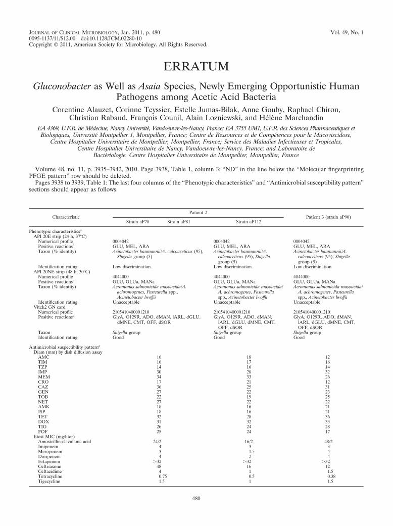
Volume 48, no. 11, p. 3935–3942, 2010. Page 3938, Table 1, column 3: “ND” in the line below the “Molecular fingerprintingPFGE pattern” row should be deleted.
Pages 3938 to 3939, Table 1: The last four columns of the “Phenotypic characteristics” and “Antimicrobial susceptibility pattern”sections should appear as follows.
CharacteristicPatient 2
Patient 3 (strain aP90)Strain aP78 Strain aP81 Strain aP112
Phenotypic characteristicsa
API 20E strip (24 h, 37°C)Numerical profile 0004042 0004042 0004042Positive reactionsb GLU, MEL, ARA GLU, MEL, ARA GLU, MEL, ARATaxon (% identity) Acinetobacter baumannii/A. calcoaceticus (95),
Shigella group (5)Acinetobacter baumannii/A.
calcoaceticus (95), Shigellagroup (5)
Acinetobacter baumannii/A.calcoaceticus (95), Shigellagroup (5)
Identification rating Low discrimination Low discrimination Low discriminationAPI 20NE strip (48 h, 30°C)
Numerical profile 4044000 4044000 4044000Positive reactionsc GLU, GLUa, MANa GLU, GLUa, MANa GLU, GLUa, MANaTaxon (% identity) Aeromonas salmonicida masoucida/A.
achromogenes, Pasteurella spp.,Acinetobacter lwoffii
Aeromonas salmonicida masoucida/A. achromogenes, Pasteurellaspp., Acinetobacter lwoffii
Aeromonas salmonicida masoucida/A. achromogenes, Pasteurellaspp., Acinetobacter lwoffii
Identification rating Unacceptable Unacceptable UnacceptableVitek2 GN card
Numerical profile 2105410400001210 2105410400001210 2105410400001210Positive reactionsd GlyA, O129R, ADO, dMAN, lARL, dGLU,
dMNE, CMT, OFF, dSORGlyA, O129R, ADO, dMAN,
lARL, dGLU, dMNE, CMT,OFF, dSOR
GlyA, O129R, ADO, dMAN,lARL, dGLU, dMNE, CMT,OFF, dSOR
Taxon Shigella group Shigella group Shigella groupIdentification rating Good Good Good
Antimicrobial suspectibility patterna
Diam (mm) by disk diffusion assayAMC 16 18 12TIM 16 17 16TZP 14 16 14IMP 30 28 32MEM 34 33 26CRO 17 21 12CAZ 36 25 31GEN 27 22 23TOB 22 19 25NET 27 22 22AMK 18 16 21ISP 18 16 21TET 32 28 36DOX 31 32 33TIG 26 24 28FOF 25 24 17
Etest MIC (mg/liter)Amoxicillin-clavulanic acid 24/2 16/2 48/2Imipenem 4 3 3Meropenem 3 1.5 4Doripenem 4 2 4Ertapenem �32 �32 �32Ceftriaxone 48 16 12Ceftazidime 4 1 1.5Tetracycline 0.75 0.5 0.38Tigecycline 1.5 1 1.5
480