Embed Size (px)
Citation preview
Glioma tumor suppressor candidate region gene 1 (GLTSCR1)and its paralog GLTSCR1-like form SWI/SNF chromatinremodeling subcomplexesReceived for publication, November 22, 2017, and in revised form, January 24, 2018 Published, Papers in Press, January 26, 2018, DOI 10.1074/jbc.RA117.001065
Aktan Alpsoy and Emily C. Dykhuizen1
From the Department of Medicinal Chemistry and Molecular Pharmacology, Purdue University, West Lafayette, Indiana 47907
Edited by Joel Gottesfeld
The mammalian SWI/SNF chromatin remodeling complexis a heterogeneous collection of related protein complexesrequired for gene regulation and genome integrity. It contains acentral ATPase (BRM or BRG1) and various combinations of10 –14 accessory subunits (BAFs for BRM/BRG1 AssociatedFactors). Two distinct complexes differing in size, BAF and theslightly larger polybromo-BAF (PBAF), share many of the samecore subunits but are differentiated primarily by having eitherAT-rich interaction domain 1A/B (ARID1A/B in BAF) orARID2 (in PBAF). Using density gradient centrifugation andimmunoprecipitation, we have identified and characterized athird and smaller SWI/SNF subcomplex. We termed this com-plex GBAF because it incorporates two mutually exclusive para-logs, GLTSCR1 (glioma tumor suppressor candidate regiongene 1) or GLTSCR1L (GLTSCR1-like), instead of an ARID pro-tein. In addition to GLTSCR1 or GLTSCR1L, the GBAF complexcontains BRD9 (bromodomain-containing 9) and the BAF sub-units BAF155, BAF60, SS18, BAF53a, and BRG1/BRM. Weobserved that GBAF does not contain the core BAF subunitsBAF45, BAF47, or BAF57. Even without these subunits, GBAFdisplayed in vitro ATPase activity and bulk chromatin affinitycomparable to those of BAF. GBAF associated with BRD4, but,unlike BRD4, the GBAF component GLTSCR1 was not requiredfor the viability of the LNCaP prostate cancer cell line. In con-trast, GLTSCR1 or GLTSCR1L knockouts in the metastatic pros-tate cancer cell line PC3 resulted in a loss in proliferation andcolony-forming ability. Taken together, our results provide evi-dence for a compositionally novel SWI/SNF subcomplex withcell type–specific functions.
The mammalian SWI/SNF (or BAF) complex is an ATP-de-pendent chromatin remodeler composed of 10 –14 subunits(1). The mammalian SWI/SNF chromatin remodeling complexis implicated in a variety of processes including mitosis, DNA
replication, DNA damage repair, genomic looping, and genesplicing, in addition to its well-established roles in the tran-scriptional regulation of genes involved in cellular differentia-tion, cellular maintenance, and adaptation to stimuli (2). Muta-tions in specific SWI/SNF complex members are common incancer (3, 4) and neural disorders (5), and the altered expressionof specific subunits is associated with tumorigenesis (6), viralinfection (7), viral latency (8), alcohol addiction (9, 10), heartdisease (11), and immune function (12). The ability of this com-plex to direct such numerous and diverse functions is facilitatedthrough the increase in SWI/SNF subunit number and diversityduring vertebrate evolution (13), which led to an exponentialincrease in the potential combinations of subunits (14, 15). AllSWI/SNF complexes contain the ATPase subunit BRG1 orBRM, along with the structural subunits BAF155/BAF170,which are required for full ATPase and nucleosome remodelingactivity in vitro (16). In addition, SWI/SNF complexes containBAF60 (A, B, or C), BAF47, BAF57, BAF53 (A or B), and actin.The larger and less abundant polybromo-BAF (PBAF)2 com-plex uniquely contains ARID2, PBRM1, BAF45D, and BRD7,whereas the more abundant BAF complex contains ARID1 (Aor B), BAF45 (B, C, or D), SS18, BCL7 (A, B, or C), and BCL11 (Aor B) (Fig. 1A). The altered expression of SWI/SNF paralogsduring cellular differentiation results in subunit switching,which is an important determinant of cell identity and cell-typetranscriptional programs (17). Additionally, paralogs are oftenexpressed simultaneously, leading to distinct subcomplexeswithin the same cell with both unique and redundant functions(18). For example, ARID1A is high in embryonic stem cells,whereas ARID1B is up-regulated upon differentiation (19).The different BAF complexes containing these two paralogsshare many of their genomic targets; however, they also bindunique genomic targets, and deletions are non-synonymous forgene regulation (20). ARID1A is the most commonly mutatedSWI/SNF subunit in cancer, because of transcriptional func-tions that are non-redundant with ARID1B (21, 22); however,cancers with deletions in ARID1A are dependent on ARID1Bfor viability (23) because of redundant, essential functions atenhancers (22). Additionally, homologous complexes can dis-play transcriptionally antagonistic roles, as has been observed
This work was supported with National Institutes of Health Grant CA207532, VFoundation for Cancer Research Grants V2014-004 and D2016-030 (toE. C. D.), a graduate endowment from the Lilly Foundation (to A. A.), andNational Institutes of Health Grant P30 CA023168 through the Purdue Uni-versity Center. The authors declare that they have no conflicts of interestwith the contents of this article. The content is solely the responsibility ofthe authors and does not necessarily represent the official views of theNational Institutes of Health.
This article contains Figs. S1–S3.1 To whom correspondence should be addressed: Dept. of Medicinal Chem-
istry and Molecular Pharmacology, HANS105A, 201 S. University St., WestLafayette IN 47907. Tel.: 765-494-4706; E-mail: [email protected].
2 The abbreviations used are: PBAF, polybromo-BAF; IP, immunoprecipita-tion; DMEM, Dulbecco’s modified Eagle’s medium; MEM, minimal essentialmedium; qPCR, quantitative PCR; DMSO, dimethyl sulfoxide; sgRNA, shortguide RNA; AML, acute myeloid leukemia; mESC, mouse embryonic stemcell.
croARTICLE
3892 J. Biol. Chem. (2018) 293(11) 3892–3903
© 2018 by The American Society for Biochemistry and Molecular Biology, Inc. Published in the U.S.A.
by guest on April 30, 2020
http://ww
w.jbc.org/
Dow
nloaded from
for ARID1A and ARID2-containing complexes at specific genetargets (8, 20, 24). Targeting specific SWI/SNF complexes hasbeen proposed both for alleviating subunit-specific pathogenicfunction and to target essential redundant functions in cancerswith mutations in the genes for specific subunits (25, 26). Bothof these strategies are dependent on a better understanding ofthe different biochemical and transcriptional functions of ho-mologous SWI/SNF complexes. We report here for the firsttime a novel, ubiquitously expressed SWI/SNF subcomplexdefined by mutually exclusive paralogs GLTSCR1 (or BICRAfor BRD4-interacting chromatin remodeling complex associ-ated) and GLTSCR1L (or BICRAL for BRD4-interacting chro-matin remodeling complex associated like), which also containsBRD9 and a subset of shared canonical SWI/SNF subunits.
Results
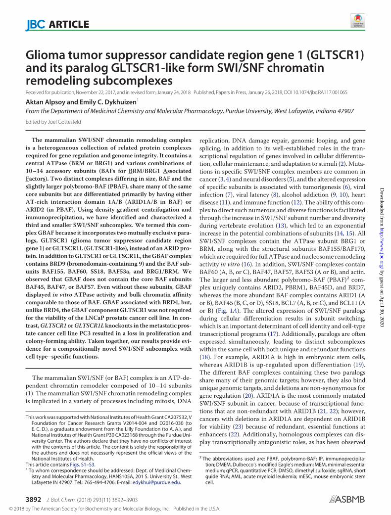
Proteomic analysis of BRG1 immunoprecipitations from tworenal clear cell carcinoma cell lines identified multiple uniquepeptides from the uncharacterized protein GLTSCR1 (Fig. 1B).GLTSCR1 has been identified in previous proteomic analyses ofthe SWI/SNF chromatin remodeling complex (18, 27–29) buthas never been validated or characterized as a BAF complexsubunit. After screening multiple commercially available anti-bodies against GLTSCR1, we identified an antibody thatstained a band in the predicted region of 180 kDa using immu-noblot analysis. Further, this band disappeared after CRISPR-mediated Gltscr1 knockout in mouse embryonic stem cell lines(Fig. 1C). Using this validated antibody, we confirmed the massspectrometry data using immunoblot analysis, detecting robustenrichment of GLTSCR1 in BRG1 immunoprecipitations (Fig.1D). To define whether GLTSCR1 is a true subunit of BAF andnot an associating factor, we performed urea denaturationfollowed by BRG1 immunoprecipitation and found thatGLTSCR1 stably associates with BRG1 at urea concentrationsup to 2.5 M, consistent with known BAF subunits ARID1A andBAF60A (Fig. 1D).
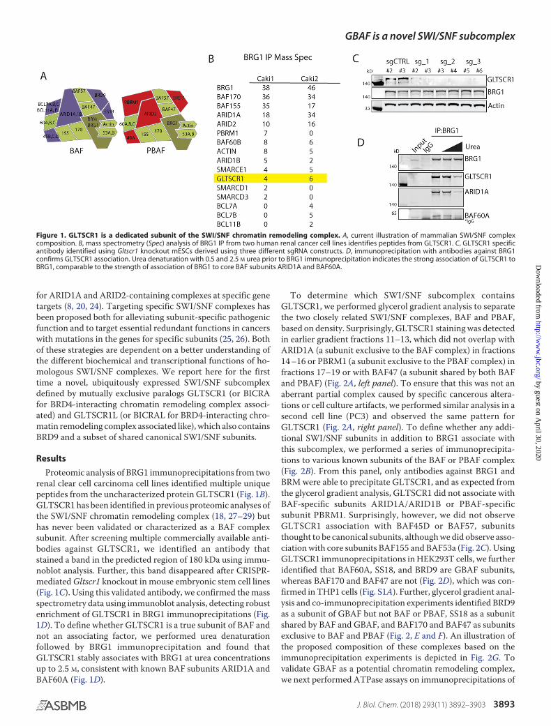
To determine which SWI/SNF subcomplex containsGLTSCR1, we performed glycerol gradient analysis to separatethe two closely related SWI/SNF complexes, BAF and PBAF,based on density. Surprisingly, GLTSCR1 staining was detectedin earlier gradient fractions 11–13, which did not overlap withARID1A (a subunit exclusive to the BAF complex) in fractions14 –16 or PBRM1 (a subunit exclusive to the PBAF complex) infractions 17–19 or with BAF47 (a subunit shared by both BAFand PBAF) (Fig. 2A, left panel). To ensure that this was not anaberrant partial complex caused by specific cancerous altera-tions or cell culture artifacts, we performed similar analysis in asecond cell line (PC3) and observed the same pattern forGLTSCR1 (Fig. 2A, right panel). To define whether any addi-tional SWI/SNF subunits in addition to BRG1 associate withthis subcomplex, we performed a series of immunoprecipita-tions to various known subunits of the BAF or PBAF complex(Fig. 2B). From this panel, only antibodies against BRG1 andBRM were able to precipitate GLTSCR1, and as expected fromthe glycerol gradient analysis, GLTSCR1 did not associate withBAF-specific subunits ARID1A/ARID1B or PBAF-specificsubunit PBRM1. Surprisingly, however, we did not observeGLTSCR1 association with BAF45D or BAF57, subunitsthought to be canonical subunits, although we did observe asso-ciation with core subunits BAF155 and BAF53a (Fig. 2C). UsingGLTSCR1 immunoprecipitations in HEK293T cells, we furtheridentified that BAF60A, SS18, and BRD9 are GBAF subunits,whereas BAF170 and BAF47 are not (Fig. 2D), which was con-firmed in THP1 cells (Fig. S1A). Further, glycerol gradient anal-ysis and co-immunoprecipitation experiments identified BRD9as a subunit of GBAF but not BAF or PBAF, SS18 as a subunitshared by BAF and GBAF, and BAF170 and BAF47 as subunitsexclusive to BAF and PBAF (Fig. 2, E and F). An illustration ofthe proposed composition of these complexes based on theimmunoprecipitation experiments is depicted in Fig. 2G. Tovalidate GBAF as a potential chromatin remodeling complex,we next performed ATPase assays on immunoprecipitations of
Figure 1. GLTSCR1 is a dedicated subunit of the SWI/SNF chromatin remodeling complex. A, current illustration of mammalian SWI/SNF complexcomposition. B, mass spectrometry (Spec) analysis of BRG1 IP from two human renal cancer cell lines identifies peptides from GLTSCR1. C, GLTSCR1 specificantibody identified using Gltscr1 knockout mESCs derived using three different sgRNA constructs. D, immunoprecipitation with antibodies against BRG1confirms GLTSCR1 association. Urea denaturation with 0.5 and 2.5 M urea prior to BRG1 immunoprecipitation indicates the strong association of GLTSCR1 toBRG1, comparable to the strength of association of BRG1 to core BAF subunits ARID1A and BAF60A.
GBAF is a novel SWI/SNF subcomplex
J. Biol. Chem. (2018) 293(11) 3892–3903 3893
by guest on April 30, 2020
http://ww
w.jbc.org/
Dow
nloaded from
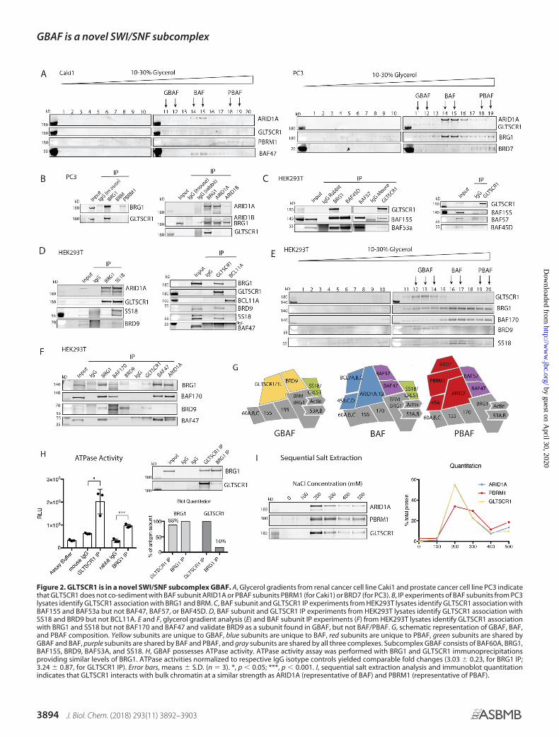
Figure 2. GLTSCR1 is in a novel SWI/SNF subcomplex GBAF. A, Glycerol gradients from renal cancer cell line Caki1 and prostate cancer cell line PC3 indicatethat GLTSCR1 does not co-sediment with BAF subunit ARID1A or PBAF subunits PBRM1 (for Caki1) or BRD7 (for PC3). B, IP experiments of BAF subunits from PC3lysates identify GLTSCR1 association with BRG1 and BRM. C, BAF subunit and GLTSCR1 IP experiments from HEK293T lysates identify GLTSCR1 association withBAF155 and BAF53a but not BAF47, BAF57, or BAF45D. D, BAF subunit and GLTSCR1 IP experiments from HEK293T lysates identify GLTSCR1 association withSS18 and BRD9 but not BCL11A. E and F, glycerol gradient analysis (E) and BAF subunit IP experiments (F) from HEK293T lysates identify GLTSCR1 associationwith BRG1 and SS18 but not BAF170 and BAF47 and validate BRD9 as a subunit found in GBAF, but not BAF/PBAF. G, schematic representation of GBAF, BAF,and PBAF composition. Yellow subunits are unique to GBAF, blue subunits are unique to BAF, red subunits are unique to PBAF, green subunits are shared byGBAF and BAF, purple subunits are shared by BAF and PBAF, and gray subunits are shared by all three complexes. Subcomplex GBAF consists of BAF60A, BRG1,BAF155, BRD9, BAF53A, and SS18. H, GBAF possesses ATPase activity. ATPase activity assay was performed with BRG1 and GLTSCR1 immunoprecipitationsproviding similar levels of BRG1. ATPase activities normalized to respective IgG isotype controls yielded comparable fold changes (3.03 � 0.23, for BRG1 IP;3.24 � 0.87, for GLTSCR1 IP). Error bars, means � S.D. (n � 3). *, p � 0.05; ***, p � 0.001. I, sequential salt extraction analysis and immunoblot quantitationindicates that GLTSCR1 interacts with bulk chromatin at a similar strength as ARID1A (representative of BAF) and PBRM1 (representative of PBAF).
GBAF is a novel SWI/SNF subcomplex
3894 J. Biol. Chem. (2018) 293(11) 3892–3903
by guest on April 30, 2020
http://ww
w.jbc.org/
Dow
nloaded from

GLTSCR1 and BRG1 from HEK293T cells. We used GLTSCR1immunoprecipitations containing comparable amounts ofBRG1 (�90%) (Fig. 2H and Fig. S1B) and found that GBAFcomplexes display robust DNA-stimulated ATPase activity(Fig. 2H). In fact, the ATPase activity of GLTSCR1 immunopre-cipitations was higher than BRG1 immunoprecipitations,although this is complicated by possible contributions fromBRM, which is lowly expressed in HEK293T cells (30). We nextused sequential salt extraction assays and determined that evenin the absence of association with DNA-binding subunitsBAF57 and ARID1/2, GLTSCR1 elutes from bulk chromatinwith similar salt concentrations as BAF-specific subunitARID1A, whereas PBAF-specific subunit PBRM1 requiresslightly higher salt concentrations to elute off bulk chromatin(31) (Fig. 2I).
Because we established GLTSCR1 as the unique subunit ofGBAF, we set to define whether GLTSCR1 is required forGBAF formation. Using GLTSCR1 knockout ES cells (Fig. 1C),we performed glycerol gradients with and without GLTSCR1(Fig. 3A). We observed a decrease in BRG1 and BAF60A stain-
ing in GBAF fractions 11–13 but not complete loss of staining.We hypothesized that this was due to the presence of the pre-dicted GLTSCR1 paralog, GLTSCR1L (now referred to asBICRAL for clarity), which has also been detected in BAF sub-unit IP mass spectrometry studies as KIAA0240 (28, 29).GLTSCR1 and BICRAL share 32% sequence homology (21%identity), and both contain a well-conserved “GLTSCR1”domain, which is also conserved between GLTSCR1 orthologspredicted in all multicellular organisms (Fig. 3B). We screenedcommercially available antibodies for BICRAL and identifiedone with weak staining at the predicted size of 140 kDa, alongwith many nonspecific bands. To confirm that the band is thecorrect protein, we developed a cell line with doxycycline-in-ducible FLAG-tagged BICRAL. Overexpression of BICRAL-FLAG in HEK293T cells resulted in a robust FLAG band at 140kDa and an increase in staining with the endogenous antibodyat the same molecular weight (Fig. 3C). To confirm thatBICRAL is mutually exclusive with GLTSCR1 in the GBAFcomplex, we performed co-immunoprecipitations of GLTSCR1and FLAG in our BICRAL-FLAG overexpression system and
Figure 3. GBAF contains GLTSCR1 or paralog GLTSCR1L (BICRAL). A, glycerol gradient analysis in mESCs showing that GBAF-associated BRG1 and BAF60Awere preserved in fractions 11–13 in the absence of GLTSCR1, suggesting that GBAF was not completely disrupted by Gltscr1 knockout. B, pairwise alignmentof amino acid sequences of GLTSCR1 and its paralog GLTSCR1L (BICRAL) show homology in the N-terminal region and strong homology at region identified ata conserved GLTSCR1 domain. C, verification of inducible expression of BICRAL-FLAG in HEK293T cells with both FLAG and endogenous BICRAL antibodies. D,immunoprecipitation analysis showed that similar to GLTSCR1, exogenous BICRAL interacts with BRD9, BAF53A, and BRG1. In addition, endogenous GLTSCR1and BICRAL do not immunoprecipitate one another. BICRAL overexpression results in reduced GLTSCR1 protein levels. E, endogenous BICRAL does notassociate with GLTSCR1, further validating that GLTSCR1 and BICRAL are mutually exclusive in GBAF context. BICRAL is detected in total BRG1 IP but not inGLTSCR1 IP, although both contain comparable levels of BRG1. Note that the same Western blotting is used in Fig. S1B to compare BRG1 levels for ATPase assay.
GBAF is a novel SWI/SNF subcomplex
J. Biol. Chem. (2018) 293(11) 3892–3903 3895
by guest on April 30, 2020
http://ww
w.jbc.org/
Dow
nloaded from
using endogenous proteins in HEK293T cells and found thatGLTSCR1 and BICRAL do not associate with each other (Fig. 3,D and E). In addition, both GLTSCR1 and BICRAL-FLAGenrich BRG1, BAF53A, and BRD9 but not ARID1A, indicatingincorporation into comparable SWI/SNF subcomplexes.Intriguingly, we also found that overexpression of BICRALdecreases GLTSCR1 expression, possibly indicating its abilityto compete with and replace GLTSCR1 in GBAF complexes.
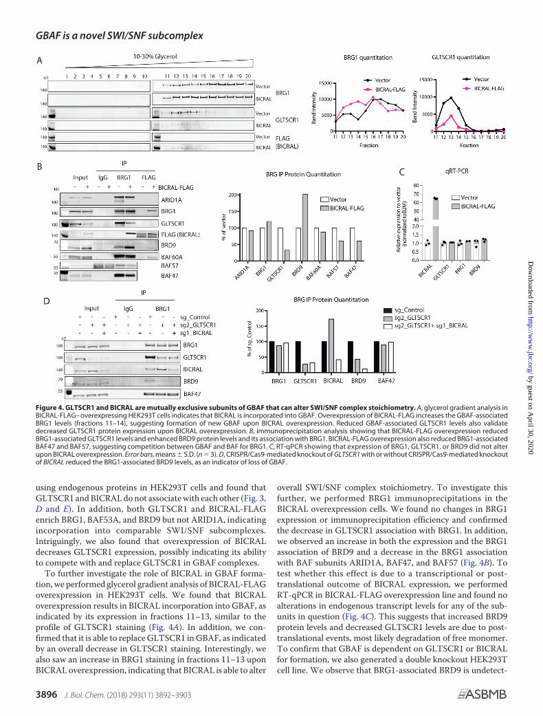
To further investigate the role of BICRAL in GBAF forma-tion, we performed glycerol gradient analysis of BICRAL-FLAGoverexpression in HEK293T cells. We found that BICRALoverexpression results in BICRAL incorporation into GBAF, asindicated by its expression in fractions 11–13, similar to theprofile of GLTSCR1 staining (Fig. 4A). In addition, we con-firmed that it is able to replace GLTSCR1 in GBAF, as indicatedby an overall decrease in GLTSCR1 staining. Interestingly, wealso saw an increase in BRG1 staining in fractions 11–13 uponBICRAL overexpression, indicating that BICRAL is able to alter
overall SWI/SNF complex stoichiometry. To investigate thisfurther, we performed BRG1 immunoprecipitations in theBICRAL overexpression cells. We found no changes in BRG1expression or immunoprecipitation efficiency and confirmedthe decrease in GLTSCR1 association with BRG1. In addition,we observed an increase in both the expression and the BRG1association of BRD9 and a decrease in the BRG1 associationwith BAF subunits ARID1A, BAF47, and BAF57 (Fig. 4B). Totest whether this effect is due to a transcriptional or post-translational outcome of BICRAL expression, we performedRT-qPCR in BICRAL-FLAG overexpression line and found noalterations in endogenous transcript levels for any of the sub-units in question (Fig. 4C). This suggests that increased BRD9protein levels and decreased GLTSCR1 levels are due to post-translational events, most likely degradation of free monomer.To confirm that GBAF is dependent on GLTSCR1 or BICRALfor formation, we also generated a double knockout HEK293Tcell line. We observe that BRG1-associated BRD9 is undetect-
Figure 4. GLTSCR1 and BICRAL are mutually exclusive subunits of GBAF that can alter SWI/SNF complex stoichiometry. A, glycerol gradient analysis inBICRAL-FLAG– overexpressing HEK293T cells indicates that BICRAL is incorporated into GBAF. Overexpression of BICRAL-FLAG increases the GBAF-associatedBRG1 levels (fractions 11–14), suggesting formation of new GBAF upon BICRAL overexpression. Reduced GBAF-associated GLTSCR1 levels also validatedecreased GLTSCR1 protein expression upon BICRAL overexpression. B, immunoprecipitation analysis showing that BICRAL-FLAG overexpression reducedBRG1-associated GLTSCR1 levels and enhanced BRD9 protein levels and its association with BRG1. BICRAL-FLAG overexpression also reduced BRG1-associatedBAF47 and BAF57, suggesting competition between GBAF and BAF for BRG1. C, RT-qPCR showing that expression of BRG1, GLTSCR1, or BRD9 did not alterupon BICRAL overexpression. Error bars, means � S.D. (n � 3). D, CRISPR/Cas9-mediated knockout of GLTSCR1 with or without CRISPR/Cas9-mediated knockoutof BICRAL reduced the BRG1-associated BRD9 levels, as an indicator of loss of GBAF.
GBAF is a novel SWI/SNF subcomplex
3896 J. Biol. Chem. (2018) 293(11) 3892–3903
by guest on April 30, 2020
http://ww
w.jbc.org/
Dow
nloaded from
able in GLTSCR1 knockout and double knockout cells, indicat-ing loss of GBAF formation (Fig. 4D). It is worth noting thatdifferences in knockout efficiencies and possibly the relativelevels of GLTSCR1 and BICRAL made it difficult to distinguishadditive or GLTSCR1-dominant effects of the paralogs onGBAF formation. Similar to decreased GLTSCR1 levels inBICRAL-overexpression lines, we consistently observed anincrease in BICRAL levels upon GLTSCR1 knockout (Fig. 4Dand Fig. S3B) via a similar increase of protein stability throughcomplex incorporation. This provides further evidence for acompensatory role of BICRAL for GBAF formation in theabsence of GLTSCR1. These results indicate that GLTSCR1/BICRAL are mutually exclusive subunits of GBAF that can, inpart, define SWI/SNF complex stoichiometry.
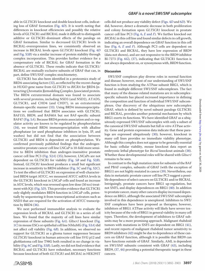
GLTSCR1 has also been identified in a proteomics study ofBRD4-associating factors (32), as reflected by the recent changein HUGO gene name from GLTSCR1 to BICRA for BRD4-In-teracting Chromatin Remodeling Complex Associated protein.The BRD4 extraterminal domain was found to associatewith several proteins, including NSD3 (and NSD2), ATAD5,GLTSCR1, and CHD4 (and CHD7), in an extraterminaldomain-specific manner (33). Using BRD4 immunoprecipita-tions we confirmed that BRD4 associates with GLTSCR1,BAF155, BRD9, and BAF60A but not BAF-specific subunitBAF47 (Fig. 5A). Because BRD4 protein association and co-reg-ulator activity are known to be regulated by phosphorylation(34), we treated BRD4 immunoprecipitation with alkalinephosphatase (or used phosphatase inhibitors in lysis, IP, andwashes) but did not find that the association betweenGLTSCR1 and BRD4 is dependent on phosphorylation. Weconfirmed previously published findings that the androgen-sensitive prostate cancer cell line LNCaP is 10-fold more sensi-tive to BRD4 inhibition than androgen-insensitive prostatecancer cell line PC3 (Fig. S2A) (35); however, LNCaPs are notdependent on GLTSCR1 for viability (Fig. 5B and Fig. S2B).Instead, GLTSCR1 knockout produces a small but significantincrease in sensitivity to BRD4 inhibitor (Fig. 5C and Fig. S2C).To test the effect of GLTSCR1 on expression of well-character-ized BRD4 target MYCC, we measured MYCC mRNA levels inthe GLTSCR1 knockout in LNCaP cells and found an increasein MYC levels, which was reversed upon low dose (50 nM) treat-ment with JQ1 (Fig. 5D). This provides evidence that GLTSCR1only slightly modulates BRD4 function in LNCaP cells, poten-tially by sequestering it from transcriptional activators such asNSD3 that are required for the activation of MYCC transcrip-tion by BRD4 (36).
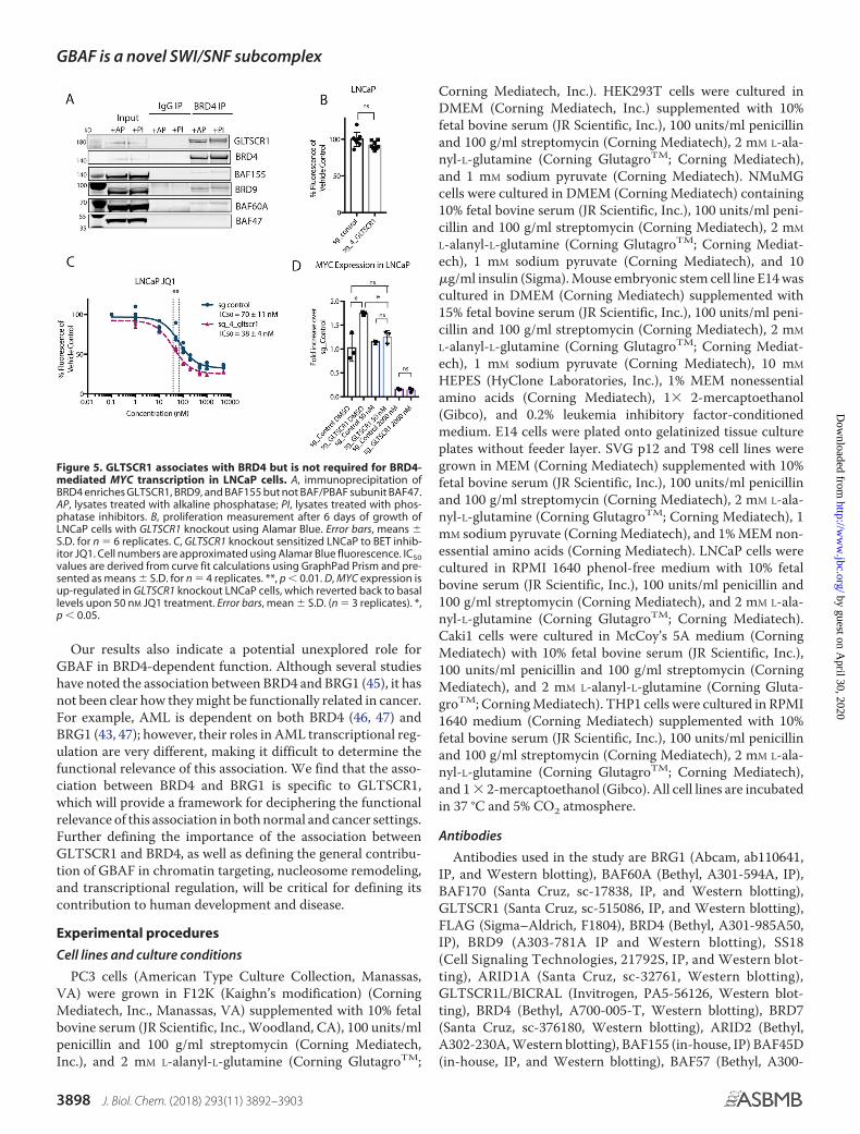
We next performed immunoblot analysis to evaluate theexpression levels of BICRAL and GLTSCR1 in a series of celllines. We found that the majority of cell lines have similarexpression of these subunits (Fig. 6A). Gltscr1 knockout (Fig.1C) in mouse ESCs or epithelial cell line NMuMG (Fig. S3A) didnot affect cell viability (Fig. 6B). In addition, we observed nosupport for GLTSCR1 as a glioma tumor suppressor becauseGLTSCR1 knockout in human astrocyte cell line SVG p12 andglioblastoma cell line T98G both resulted in no change in via-bility (Fig. 6C and Fig. S3B). Lastly, we did not find evidence thatBICRAL and GLTSCR1 have redundant, necessary functionsbecause knockout of both GLTSCR1 and BICRAL in HEK293T
cells did not produce any viability defect (Figs. 4D and 6D). Wedid, however, detect a dramatic decrease in both proliferationand colony formation upon GLTSCR1 knockout in prostatecancer cell line PC3 (Fig. 6, E and F). We further knocked outBICRAL in this cell line and found similar defects in cell growth,indicating an overall dependence on GBAF function in this cellline (Fig. 6, E and F). Although PC3 cells are dependent onGLTSCR1 and BICRAL, they have low expression of BRD9(data not shown), and are not responsive to the BRD9 inhibitorBI-7273 (Fig. 6G), (37), indicating that GLTSCR1 function isnot always dependent on, or synonymous with, BRD9 function.
Discussion
SWI/SNF complexes play diverse roles in normal functionand disease; however, most of our understanding of SWI/SNFfunction is from studying the ATPase subunit BRG1, which isfound in multiple different SWI/SNF subcomplexes. The factthat many of the disease-related mutations are in subcomplex-specific subunits has placed increased importance in definingthe composition and function of individual SWI/SNF subcom-plexes. Our discovery of the ubiquitous new subcomplexGBAF, which is defined by novel subunit paralogs GLTSCR1and BICRAL, provides another potential mechanism by whichBRG1 exerts its functions. We have identified GBAF as a ubiq-uitously expressed SWI/SNF subcomplex with only a subset ofthe canonical SWI/SNF subunits but full in vitro ATPase activ-ity. Gene and protein expression data indicate that these para-logs are expressed ubiquitously (30); however, knockout inmany cell lines provides no immediate viability phenotype.Although this complex does not appear to be generally essentialfor basic cellular viability, mouse knockout data report anembryonic lethal phenotype for Bicral knockout animals (38).Whether these developmental roles will be shared with Gltscr1remains to be seen.
In contrast to the high mutation rates for subunits of the BAFand PBAF complex, subunits of GBAF (with the exception ofBRG1) are not highly mutated in cancer (39). Nevertheless, ourdata in metastatic prostate cancer cell line PC3 suggest a possi-ble dependence of select cancers on GLTSCR1 and/or BICRAL.Intriguingly, prostate cancers have BRG1 up-regulation, butnot SNF5, and display dependences on BRG1 (40). In additionto prostate cancer, many other cancers display increased depen-dence on BRG1, although the associated SWI/SNF subcomplexinvolved in this dependence is unexplored. Inhibitors to SWI/SNF complexes have been proposed as therapies; however,inhibitors of BRG1 ATPase activity will likely have severe tox-icity because of the role of BRG1 in general viability in many celltypes. Therefore, the development of inhibitors to GBAF sub-units may be a more promising approach. Malignant rhabdoidtumors with mutations in SNF5 are dependent on BRG1 (41),and recent reports of malignant rhabdoid tumor sensitivity toBRD9 inhibitors (42) might be due to dependence of these can-cers on GBAF function, although it is possible that BRD9 canhave functions outside of GBAF. Similarly, AML is dependenton SWI/SNF subunits consistent with GBAF (43), includingBRD9, (37, 44) providing a potential therapeutic target in thesecancers.
GBAF is a novel SWI/SNF subcomplex
J. Biol. Chem. (2018) 293(11) 3892–3903 3897
by guest on April 30, 2020
http://ww
w.jbc.org/
Dow
nloaded from
Our results also indicate a potential unexplored role forGBAF in BRD4-dependent function. Although several studieshave noted the association between BRD4 and BRG1 (45), it hasnot been clear how they might be functionally related in cancer.For example, AML is dependent on both BRD4 (46, 47) andBRG1 (43, 47); however, their roles in AML transcriptional reg-ulation are very different, making it difficult to determine thefunctional relevance of this association. We find that the asso-ciation between BRD4 and BRG1 is specific to GLTSCR1,which will provide a framework for deciphering the functionalrelevance of this association in both normal and cancer settings.Further defining the importance of the association betweenGLTSCR1 and BRD4, as well as defining the general contribu-tion of GBAF in chromatin targeting, nucleosome remodeling,and transcriptional regulation, will be critical for defining itscontribution to human development and disease.
Experimental procedures
Cell lines and culture conditions
PC3 cells (American Type Culture Collection, Manassas,VA) were grown in F12K (Kaighn’s modification) (CorningMediatech, Inc., Manassas, VA) supplemented with 10% fetalbovine serum (JR Scientific, Inc., Woodland, CA), 100 units/mlpenicillin and 100 g/ml streptomycin (Corning Mediatech,Inc.), and 2 mM L-alanyl-L-glutamine (Corning GlutagroTM;
Corning Mediatech, Inc.). HEK293T cells were cultured inDMEM (Corning Mediatech, Inc.) supplemented with 10%fetal bovine serum (JR Scientific, Inc.), 100 units/ml penicillinand 100 g/ml streptomycin (Corning Mediatech), 2 mM L-ala-nyl-L-glutamine (Corning GlutagroTM; Corning Mediatech),and 1 mM sodium pyruvate (Corning Mediatech). NMuMGcells were cultured in DMEM (Corning Mediatech) containing10% fetal bovine serum (JR Scientific, Inc.), 100 units/ml peni-cillin and 100 g/ml streptomycin (Corning Mediatech), 2 mM
L-alanyl-L-glutamine (Corning GlutagroTM; Corning Mediat-ech), 1 mM sodium pyruvate (Corning Mediatech), and 10�g/ml insulin (Sigma). Mouse embryonic stem cell line E14 wascultured in DMEM (Corning Mediatech) supplemented with15% fetal bovine serum (JR Scientific, Inc.), 100 units/ml peni-cillin and 100 g/ml streptomycin (Corning Mediatech), 2 mM
L-alanyl-L-glutamine (Corning GlutagroTM; Corning Mediat-ech), 1 mM sodium pyruvate (Corning Mediatech), 10 mM
HEPES (HyClone Laboratories, Inc.), 1% MEM nonessentialamino acids (Corning Mediatech), 1� 2-mercaptoethanol(Gibco), and 0.2% leukemia inhibitory factor-conditionedmedium. E14 cells were plated onto gelatinized tissue cultureplates without feeder layer. SVG p12 and T98 cell lines weregrown in MEM (Corning Mediatech) supplemented with 10%fetal bovine serum (JR Scientific, Inc.), 100 units/ml penicillinand 100 g/ml streptomycin (Corning Mediatech), 2 mM L-ala-nyl-L-glutamine (Corning GlutagroTM; Corning Mediatech), 1mM sodium pyruvate (Corning Mediatech), and 1% MEM non-essential amino acids (Corning Mediatech). LNCaP cells werecultured in RPMI 1640 phenol-free medium with 10% fetalbovine serum (JR Scientific, Inc.), 100 units/ml penicillin and100 g/ml streptomycin (Corning Mediatech), and 2 mM L-ala-nyl-L-glutamine (Corning GlutagroTM; Corning Mediatech).Caki1 cells were cultured in McCoy’s 5A medium (CorningMediatech) with 10% fetal bovine serum (JR Scientific, Inc.),100 units/ml penicillin and 100 g/ml streptomycin (CorningMediatech), and 2 mM L-alanyl-L-glutamine (Corning Gluta-groTM; Corning Mediatech). THP1 cells were cultured in RPMI1640 medium (Corning Mediatech) supplemented with 10%fetal bovine serum (JR Scientific, Inc.), 100 units/ml penicillinand 100 g/ml streptomycin (Corning Mediatech), 2 mM L-ala-nyl-L-glutamine (Corning GlutagroTM; Corning Mediatech),and 1 � 2-mercaptoethanol (Gibco). All cell lines are incubatedin 37 °C and 5% CO2 atmosphere.
Antibodies
Antibodies used in the study are BRG1 (Abcam, ab110641,IP, and Western blotting), BAF60A (Bethyl, A301-594A, IP),BAF170 (Santa Cruz, sc-17838, IP, and Western blotting),GLTSCR1 (Santa Cruz, sc-515086, IP, and Western blotting),FLAG (Sigma–Aldrich, F1804), BRD4 (Bethyl, A301-985A50,IP), BRD9 (A303-781A IP and Western blotting), SS18(Cell Signaling Technologies, 21792S, IP, and Western blot-ting), ARID1A (Santa Cruz, sc-32761, Western blotting),GLTSCR1L/BICRAL (Invitrogen, PA5-56126, Western blot-ting), BRD4 (Bethyl, A700-005-T, Western blotting), BRD7(Santa Cruz, sc-376180, Western blotting), ARID2 (Bethyl,A302-230A, Western blotting), BAF155 (in-house, IP) BAF45D(in-house, IP, and Western blotting), BAF57 (Bethyl, A300-
Figure 5. GLTSCR1 associates with BRD4 but is not required for BRD4-mediated MYC transcription in LNCaP cells. A, immunoprecipitation ofBRD4 enriches GLTSCR1, BRD9, and BAF155 but not BAF/PBAF subunit BAF47.AP, lysates treated with alkaline phosphatase; PI, lysates treated with phos-phatase inhibitors. B, proliferation measurement after 6 days of growth ofLNCaP cells with GLTSCR1 knockout using Alamar Blue. Error bars, means �S.D. for n � 6 replicates. C, GLTSCR1 knockout sensitized LNCaP to BET inhib-itor JQ1. Cell numbers are approximated using Alamar Blue fluorescence. IC50values are derived from curve fit calculations using GraphPad Prism and pre-sented as means � S.D. for n � 4 replicates. **, p � 0.01. D, MYC expression isup-regulated in GLTSCR1 knockout LNCaP cells, which reverted back to basallevels upon 50 nM JQ1 treatment. Error bars, mean � S.D. (n � 3 replicates). *,p � 0.05.
GBAF is a novel SWI/SNF subcomplex
3898 J. Biol. Chem. (2018) 293(11) 3892–3903
by guest on April 30, 2020
http://ww
w.jbc.org/
Dow
nloaded from
810A, IP, and Western blotting), BAF155 (Santa Cruz,sc-32763, Western blotting), BAF53A (Abcam, ab131272,Western blotting), BAF60A (Santa Cruz, sc-514400, Westernblotting), PBRM1 (Bethyl, A301-590A, Western blotting),ARID1B (Bethyl, A301-047-T, Western blotting), BCL11A(Santa Cruz, sc-514842, IP and Western blotting), Actin (SantaCruz, sc-47778, Western blotting), GAPDH (Santa Cruz,sc-137179, Western blotting), and �-tubulin (Santa Cruz,sc-8035, Western blotting).
Immunoblot analysis
Proteins from whole cells, nuclear extracts, salt extractions,or glycerol gradient sedimentation analyses were mixed with4� lithium dodecyl sulfate sample buffer containing 10%2-merchaptoethanol. The proteins were denatured for 5 min at95 °C, separated on a 4 –12% SDS-polyacrylamide gel, andtransferred to a PVDF membrane (Immobilon FL, EMD Milli-pore, Billerica, MA). The membrane was blocked with 5%bovine serum albumin (VWR, Batavia, IL) in PBS containing0.1% Tween 20 for 30 min at room temperature and then incu-bated in primary antibodies overnight at 4 °C. The primaryantibodies were detected by incubating the membranes in goatanti-rabbit or goat anti-mouse secondary antibodies (LI-CORBiotechnology, Lincoln, NE) conjugated to IRDye 800CW orIRDye 680, respectively, for 1 h at room temperature, and thesignals were visualized using Odyssey Clx imager (LI-CORBiotechnology).
Immunoprecipitation
The cells were harvested by trypsinization and washed oncein ice-cold phosphate buffered saline (pH 7.2). The pellet was
resuspended in buffer A (20 mM HEPES, pH 7.9, 25 mM KCl,10% glycerol, 0.1% Nonidet P-40 with PMSF, aprotinin, leupep-tin, and pepstatin) at a concentration of 20 million cells/ml. Thecells were kept on ice for 5 min, and nuclei were isolated bycentrifugation at 600 � g (Eppendorf Centrifuge 5810 R, Ham-burg, Germany) for 10 min. Pelleted nuclei were washed once inbuffer A without Nonidet P-40 and pelleted again. The nucleipellet was resuspended in chromatin IP buffer (20 mM HEPES,pH 7.9, 150 mM NaCl, 1%Triton X-100, 7.5 mM MgCl2, 0.1 mM
CaCl2). 4 units/ml Turbo DNase (Ambion, Inc., Foster City,CA) was added to extracts and rotated at 4 °C for 30 min. Theextracts were cleared by centrifugation (Centrifuge 5424 R;Eppendorf, Hamburg, Germany) at 21,000 � g for 30 min. Thecleared extract was precleared with normal IgG-conjugated(Santa Cruz, Dallas, TX) protein A/G magnetic beads (Pierce).One microgram specific IgG was used per 0.2 mg lysate forimmunoprecipitation. After overnight incubation, immuno-complexes were captured using protein A/G magnetic beadsfollowing a 2-h incubation. The beads were washed twice inchromatin IP buffer and three times in high stringency washbuffer (20 mM HEPES, pH 7.9, 500 mM NaCl, 1% Triton X-100,0.5% sodium deoxycholate, 1 mM EDTA). The proteins wereeluted in 1� lithium dodecyl sulfate loading dye (Thermo Sci-entific) by boiling at 70 °C for 10 min. For urea denaturationfollowed by BRG1 IP, urea was added into nuclear lysates tofinal concentration of 0.5 or 2.5 M and incubated at 4 °C for 1 h.The lysates were then dialyzed against chromatin IP buffer for50 min, precleared, and incubated with normal IgG or BRG1antibodies. For on-bead alkaline phosphatase treatment duringBRD4 IP, proteins were extracted in buffers with or without 1�
Figure 6. GLTSCR1 and BICRAL are expressed in most cell lines but are uniquely required for the viability of prostate cancer cell line PC3. A, immuno-blot analysis of GLTSCR1 and BICRAL expression across a panel of cell lines. B, proliferation measurement after 6 days of growth of the non-transformed mousecell lines mESCs and NMuMG with Gltscr1 knockout using Alamar Blue. C, proliferation measurement after 6 days of growth of the transformed human astrocytecell line SVGp12 and glioblastoma cell line T98G with GLTSCR1 knockout using Alamar Blue. D, proliferation measurement after 6 days of growth of HEK293Tcells with GLTSCR1 and BICRAL knockout using Alamar Blue. E, validation of knockouts using multiple guide RNAs. F, left panel, Alamar Blue assay demonstratedthat loss of GLTSCR1 and BICRAL reduced the growth of PC3 cells 6 day after plating. Fluorescence values graphed (excitation, 560 nm; emission, 590 nm)represent the metric for cell number. Error bars, means � S.D. (n � 3 biological replicates). **, p � 0.01; ***, p � 0.001 compared with control cells. Right panel,loss of GLTSCR1 reduced the clonogenic growth of prostate cell line PC3. G, PC3 cells did not display sensitivity to BRD9 inhibitor BI-7273 (IC50 of 275 nM) up to10 �M treatment for 4 days. Cell number was approximated using Alamar Blue fluorescence (n � 3 biological replicates).
GBAF is a novel SWI/SNF subcomplex
J. Biol. Chem. (2018) 293(11) 3892–3903 3899
by guest on April 30, 2020
http://ww
w.jbc.org/
Dow
nloaded from
phosphatase inhibitor mixture 3 (Apexbio, Taiwan)/1 mM
sodium orthovanadate and immunoprecipitated as describedabove. Following two washes in chromatin IP buffer, thebeads were washed once in FastAP reaction buffer (ThermoScientific, Waltham, MA) and incubated at 37 °C for 1 h withor without 10 units alkaline phosphatase. Reaction mixtureswere removed, and the beads were washed in chromatin IPbuffer twice more. The beads were then boiled and run ongel.
Glycerol gradient sedimentation analysis
30 million cells were collected by trypsinization, lysed inbuffer A, and washed once with buffer A without Nonidet P-40.The nuclei were resuspended in buffer C (10 mM HEPES, pH7.6, 3 mM MgCl2, 100 mM KCl, 0.1 mM EDTA, 10% glycerol). 0.3M ammonium sulfate was added on nuclei suspension androtated at 4 °C for 30 min. Chromatin pellet was removed byultracentrifugation at 150,000 � g for 30 min. 0.3 g/mlammonium sulfate powder was added, and the supernatantwas incubated on ice for 20 min. Proteins were precipitatedby ultracentrifugation at 150,000 � g for 30 min. The proteinpellet was resuspended in 100 �l of HEMG1000 buffer (25mM HEPES, pH 7.6, 0.1 mM EDTA, 12.5 mM MgCl2, 100 mM
KCl) with protease inhibitors. 10 –30% glycerol gradient wasprepared using HEMG1000 buffer without glycerol andHEMG1000 buffer with 30% glycerol. Resuspended proteinwas layered over the top of 10 –30% glycerol gradient (10 ml)and was fractionated by centrifugation at 40,000 rpm (XL-100K; Beckman Coulter, Brea, CA) for 16 h using SW32Tirotor (Beckman Coulter). Twenty 500-�l fractions were col-lected sequentially from the top and used for immunoblotanalysis.
RT-qPCR
RNA was extracted using TRIzol (Ambion, Inc.). cDNA wassynthesized using Verso cDNA synthesis kit (Thermo Scien-tific) using random hexamers. Specific targets were amplifiedusing SYBR Green Master Mix (Roche) and the followingqPCR primers: BICRAL forward, 5�-GTTGCCACTCAGCTC-CTAAA-3�; BICRAL reverse, 5�-CCTCCTGGTTGAACATC-CTATC-3�; GLTSCR1 forward, 5�-GATGAGGATGGGAGA-TGCTTAC-3�; GLTSCR1 reverse, 5�-TCATAGAAGGCACT-TTGGGC-3�; BRG1 forward, 5�-TACAAGGACAGCAGCAG-TGG-3�; BRG1 reverse, 5�-TAGTACTCGGGCAGCTCCTT-3�; BRD9 forward, 5�-GCCACGACTCCAGTTACTATG-3�;BRD9 reverse, 5�-TCTCCTTCTCGGACTTCTTCT-3�; MYCCforward, 5�-AATGAAAAGGCCCCCAAGGTAGTTATCC-3�; and MYCC reverse, 5�-GTCGTTTCCGCAACAAGTC-CTCTTC-3�.
Serial salt extraction assay
Serial salt extraction assay was performed as published withsome modifications (48). Briefly, 5 million HEK293T cells wereharvested by trypsinization and washed once with ice-cold PBS.The cells were lysed in modified buffer A (60 mM Tris, 60 mM
KCl, 1 mM EDTA, 0.3 M sucrose, 0.5% Nonidet P-40, 1 mM DTT)with protease inhibitor, and nuclei were pelleted. The nucleiwere then incubated in 200 �l of extraction buffer 0 (50 mM
HEPES, pH 7.8, 0.3 M sucrose, 1 mM EGTA, 0.1% Triton X-100,1 mM DTT, protease inhibitors) for 10 min and centrifuged at7,000 � g for 5 min, and supernatant was collected as “0 mM
fraction.” The pellet was then resuspended in 200 �l ofextraction buffer 100 (50 mM HEPES, pH 7.8, 0.3 M sucrose, 1mM EGTA, 0.1% Triton X-100, 1 mM DTT, protease inhibi-tors, or 100 mM NaCl) and processed in the same manner toyield “100 mM fraction.” Serial extraction was implementedwith extraction buffers containing 200, 300, 400, and 500 mM
NaCl. 20-�l aliquots from each fraction were mixed with 4�lithium dodecyl sulfate loading buffer and run for Westernblotting.
Growth curve analysis and colony formation assay
For growth curve analysis, 500 or 1000 control or CRISPR-edited cells were plated in 96-well plates. After 6 days, culturemedium was refreshed with 1:10 Alamar Blue reagent (ThermoScientific) and incubated for 3 h. The fluorescence was mea-sured with excitation at 560 nm and emission at 590 nm usingBioTek plate reader. For colony formation assays, 100 –200cells were counted and plated on 6-well plates and allowed toform colonies for 10 –15 days. Culture medium was removedand washed twice in ice-cold PBS. Then cells were fixed in100% methanol for 10 min at �20 °C. Methanol was re-moved, and fixed cells were incubated in 0.5% crystal violet(prepared in 25% methanol) for 10 min at room temperature.Excess dye was removed by tap water washes until back-ground was cleared. The images were acquired using Chemi-Doc (Bio-Rad).
ATPase assay
ATPase assay was performed based on previously publishedprocedure (49) using ADP-GloMax Assay (Promega, Madison,WI) with minor modifications. 25 million (for BRG1 IP) or 100million (for GLTSCR1 IP) HEK293T cells were lysed in bufferA. Pelleted nuclei were extracted for 30 min at 4 °C using lysisbuffer (50 mM Tris, pH 8.0, 150 mM NaCl and 0.2% IPEGALCA-630, 1 mM DTT, 0.2 mM PMSF, and protease inhibitors) ata ratio of 50 million cells/400 �l of buffer. The extract wascleared at 21,000 � g for 1 h. One microliter of BRG1 antibody,10 �l of GLTSCR1 antibody or corresponding amount of nor-mal IgG antibodies were added per 400 �l of cleared extract forovernight immunoprecipitation at 4 °C in a rotating wheel. 10�l (for BRG1 and rabbit IgG) or 25 �l (for GLTSCR1 and mouseIgG) protein A/G magnetic beads were added to each of 400-�lIP samples and rotated for 2 h more. The beads were washedtwice in lysis buffer and then in wash buffer (10 mM Tris, pH 7.5,50 mM NaCl, 5 mM MgCl2, 1 mM DTT, and protease inhibitors).The number of beads were adjusted such that material from25 million (for BRG1 IP) or 100 million (for GLTSCR1 IP)HEK293T cells were included per ATPase reaction. Thebeads were resuspended in 25 �L of reaction buffer (10 mM
Tris, pH 7.5, 50 mM NaCl, 5 mM MgCl2, 20% glycerol, 1mg/ml BSA, 4 mM ATP, 0.5 �g/�l ssDNA, 1 mM DTT, andprotease inhibitors and incubated at 37 °C for 1 h on ashaker. The beads were separated, and the reactions weretransferred to 96-well opaque white plate. 25 �l of ADP-Gloreagent were added per well and gently shaken for 1 h at
GBAF is a novel SWI/SNF subcomplex
3900 J. Biol. Chem. (2018) 293(11) 3892–3903
by guest on April 30, 2020
http://ww
w.jbc.org/
Dow
nloaded from
room temperature. 50 �l of detection reagent were addedper well and further shaken for 1 h. Luminescence wasdetected at 1-s integration time.
Cytotoxicity analysis
10,000 (LNCaP) or 5,000 (PC3) cells were plated in 100 �l on96-well plate. The next day, JQ1, OTX015, BI-7372, or DMSOwas added, and the cells were further incubated for 4 days. Thecells were treated with Alamar Blue reagent for 3 h more, andabsorbance values were recorded at 570 and 600 nm. Percentviability was expressed relative to the DMSO-treated controlcells.
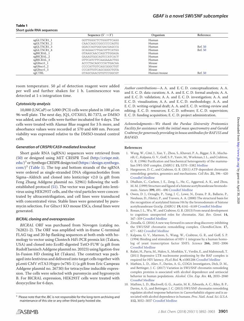
Generation of CRISPR/CAS9-mediated knockout
Short guide RNA (sgRNA) sequences were retrieved from(50) or designed using MIT CRISPR Tool (http://crispr.mit.edu/)3 or Synthego CRISPR design tool (https://design.synthego.com/)3 (Table 1). The top and bottom strands of the sgRNAwere ordered as single-stranded DNA oligonucleotides fromSigma–Aldrich and cloned into lenticrispr v2.0 (a gift fromFeng Zhang Addgene plasmid no. 52961) following the well-established protocol (51). The vector was packaged into lenti-virus using HEK293T cells, and the viral particles were concen-trated by ultracentrifugation, and cell lines were transducedwith concentrated virus. Stable lines were generated by puro-mycin selection. For Gltscr1 KO mouse ESCs, clonal lines weregenerated.
BICRAL cloning and overexpression
BICRAL ORF was purchased from Novogen (catalog no.762821-2). The ORF was amplified with in-frame C-terminalFLAG tag and 20-bp flanking sequences at both ends with ho-mology to vector using Clontech HiFi PCR premix kit (Takara,USA) and cloned into EcoRI-digested TetO-FUW (a gift fromRudolf Jaenisch Addgene plasmid no. 20323) using ligation-freeIn-Fusion HD cloning kit (Takara). The construct was pack-aged into lentivirus and delivered into target cells together withpLenti CMV rtTA3 Hygro (w785-1) (a gift from Eric CampeauAddgene plasmid no. 26730) for tetracycline inducible expres-sion. The cells were selected with puromycin and hygromycinB. For BICRAL expression, HEK293T cells were treated withdoxycycline for 6 days.
Author contributions—A. A. and E. C. D. conceptualization; A. A.and E. C. D. data curation; A. A. and E. C. D. formal analysis; A. A.and E. C. D. validation; A. A. and E. C. D. investigation; A. A. andE. C. D. visualization; A. A. and E. C. D. methodology; A. A. andE. C. D. writing-original draft; A. A. and E. C. D. writing-review andediting; E. C. D. resources; E. C. D. software; E. C. D. supervision;E. C. D. funding acquisition; E. C. D. project administration.
Acknowledgments—We thank the Purdue University ProteomicsFacility for assistance with the initial mass spectrometry and GeraldCrabtree for generously providing in house antibodies for BAF155 andBAF45D.
References1. Wang, W., Côté, J., Xue, Y., Zhou, S., Khavari, P. A., Biggar, S. R., Mucha-
rdt, C., Kalpana, G. V., Goff, S. P., Yaniv, M., Workman, J. L., and Crabtree,G. R. (1996) Purification and biochemical heterogeneity of the mamma-lian SWI-SNF complex. EMBO J. 15, 5370 –5382 Medline
2. Hargreaves, D. C., and Crabtree, G. R. (2011) ATP-dependent chromatinremodeling: genetics, genomics and mechanisms. Cell Res. 21, 396 – 420CrossRef Medline
3. Dhalluin, C., Carlson, J. E., Zeng, L., He, C., Aggarwal, A. K., and Zhou,M. M. (1999) Structure and ligand of a histone acetyltransferase bromodo-main. Nature 399, 491– 496 CrossRef Medline
4. Owen, D. J., Ornaghi, P., Yang, J. C., Lowe, N., Evans, P. R., Ballario, P.,Neuhaus, D., Filetici, P., and Travers, A. A. (2000) The structural basis forthe recognition of acetylated histone H4 by the bromodomain of histoneacetyltransferase Gcn5p. EMBO J. 19, 6141– 6149 CrossRef Medline
5. Ronan, J. L., Wu, W., and Crabtree, G. R. (2013) From neural developmentto cognition: unexpected roles for chromatin. Nat. Rev. Genet. 14,347–359 CrossRef Medline
6. Zinzalla, G. (2016) A new way forward in cancer drug discovery: inhibitingthe SWI/SNF chromatin remodelling complex. ChemBioChem. 17,677– 682 CrossRef Medline
7. Kalpana, G. V., Marmon, S., Wang, W., Crabtree, G. R., and Goff, S. P.(1994) Binding and stimulation of HIV-1 integrase by a human homo-log of yeast transcription factor SNF5. Science 266, 2002–2006CrossRef Medline
8. Rafati, H., Parra, M., Hakre, S., Moshkin, Y., Verdin, E., and Mahmoudi, T.(2011) Repressive LTR nucleosome positioning by the BAF complex isrequired for HIV latency. PLoS Biol. 9, e1001206 CrossRef Medline
9. Mathies, L. D., Aliev, F., Davies, A. G., COGA Investigators, Dick, D. M.,and Bettinger, J. C. (2017) Variation in SWI/SNF chromatin remodelingcomplex proteins is associated with alcohol dependence and antisocialbehavior in human populations. Alcohol. Clin. Exp. Res. 41, 2033–2040CrossRef Medline
10. Mathies, L. D., Blackwell, G. G., Austin, M. K., Edwards, A. C., Riley, B. P.,Davies, A. G., and Bettinger, J. C. (2015) SWI/SNF chromatin remodelingregulates alcohol response behaviors in Caenorhabditis elegans and is as-sociated with alcohol dependence in humans. Proc. Natl. Acad. Sci. U.S.A.112, 3032–3037 CrossRef Medline
3 Please note that the JBC is not responsible for the long-term archiving andmaintenance of this site or any other third party hosted site.
Table 1Short guide RNA sequences
Sequence (5�3 3�) Organism Reference
sgGLTSCR1_1 GGTTGGGCTCTGGGTTCAGG HumansgGLTSCR1_2 CAACCAGCCGGCCCCCAGTG HumansgGLTSCR1_3 GGACCAGTGGCGACGAGCCG Human Ref. 50sgGLTSCR1_4 GCAGAACCTGACGTTCATGG Human Ref. 50sgBICRAL_1 GTAAGCAACCAGCTTGGAGA HumansgBICRAL_2 GAAAGTGGCAGTCCATCACT HumansgBICRAL_3 GTTCATCTTCAAGAAACTGG HumansgGltscr1_1 ACCCTACAGCCCGCTGACAA MousesgGltscr1_2 CCCCATTGTCAGCGGGCTGT MousesgGltscr1_3 CCCATTGTCAGCGGGCTGTA MousesgCTRL GTAGCGAACGTGTCCGGCGT Human/mouse Ref. 50
GBAF is a novel SWI/SNF subcomplex
J. Biol. Chem. (2018) 293(11) 3892–3903 3901
by guest on April 30, 2020
http://ww
w.jbc.org/
Dow
nloaded from
11. Hang, C. T., Yang, J., Han, P., Cheng, H.-L., Shang, C., Ashley, E., Zhou, B.,and Chang, C.-P. (2010) Chromatin regulation by Brg1 underlies heartmuscle development and disease. Nature 466, 62– 67 CrossRef Medline
12. Wu, J. I. (2012) Diverse functions of ATP-dependent chromatin remod-eling complexes in development and cancer. Acta Biochim. Biophys. Sin.(Shanghai) 44, 54 – 69
13. Lopes Cardoso, D., and Sharpe, C. (2017) Relating protein functional di-versity to cell type number identifies genes that determine dynamic as-pects of chromatin organisation as potential contributors to organismalcomplexity. PLoS One 12, e0185409 CrossRef Medline
14. Mani, U., S, A. S., Goutham, R. N. A., and Mohan, S. S. (2017) SWI/SNFInfobase: an exclusive information portal for SWI/SNF remodeling com-plex subunits. PLoS One 12, e0184445 CrossRef Medline
15. Wu, J. I., Lessard, J., and Crabtree, G. R. (2009) Understanding the wordsof chromatin regulation. Cell 136, 200 –206 CrossRef Medline
16. Phelan, M. L.., Sif, S., Narlikar, G. J., and Kingston, R. E. (1999) Reconsti-tution of a core chromatin remodeling complex from SWI/SNF subunits.Mol. Cell 3, 247–253 CrossRef Medline
17. Ho, L., and Crabtree, G. R. (2010) Chromatin remodelling during devel-opment. Nature 463, 474 – 484 CrossRef Medline
18. Middeljans, E., Wan, X., Jansen, P. W., Sharma, V., Stunnenberg, H. G.,and Logie, C. (2012) SS18 together with animal-specific factors defineshuman BAF-type SWI/SNF complexes. PLoS One 7, e33834 CrossRefMedline
19. Staahl, B. T., Tang, J., Wu, W., Sun, A., Gitler, A. D., Yoo, A. S., andCrabtree, G. R. (2013) Kinetic analysis of npBAF to nBAF switching re-veals exchange of SS18 with CREST and integration with neural develop-mental pathways. J. Neurosci. 33, 10348 –10361 CrossRef Medline
20. Raab, J. R., Resnick, S., and Magnuson, T. (2015) Genome-wide transcrip-tional regulation mediated by biochemically distinct SWI/SNF complexes.PLoS Genet. 11, e1005748 CrossRef Medline
21. Mathur, R., Alver, B. H., San Roman, A. K., Wilson, B. G., Wang, X.,Agoston, A. T., Park, P. J., Shivdasani, R. A., and Roberts, C. W. (2017)ARID1A loss impairs enhancer-mediated gene regulation and drives co-lon cancer in mice. Nat. Genet. 49, 296 –302 CrossRef Medline
22. Kelso, T. W. R., Porter, D. K., Amaral, M. L., Shokhirev, M. N., Benner, C.,and Hargreaves, D. C. (2017) Chromatin accessibility underlies syntheticlethality of SWI/SNF subunits in ARID1A-mutant cancers. eLife 6, e30506Medline
23. Helming, K. C., Wang, X., Wilson, B. G., Vazquez, F., Haswell, J. R., Man-chester, H. E., Kim, Y., Kryukov, G. V., Ghandi, M., Aguirre, A. J., Jagani, Z.,Wang, Z., Garraway, L. A., Hahn, W. C., and Roberts, C. W. (2014)ARID1B is a specific vulnerability in ARID1A-mutant cancers. Nat. Med.20, 251–254 CrossRef Medline
24. Wurster, A. L., Precht, P., Becker, K. G., Wood, W. H., 3rd, Zhang, Y.,Wang, Z., and Pazin, M. J. (2012) IL-10 transcription is negatively regu-lated by BAF180, a component of the SWI/SNF chromatin remodelingenzyme. BMC Immunol. 13, 9 CrossRef Medline
25. Mayes, K., Qiu, Z., Alhazmi, A., and Landry, J. W. (2014) ATP-dependentchromatin remodeling complexes as novel targets for cancer therapy. Adv.Cancer Res. 121, 183–233 CrossRef Medline
26. Hohmann, A. F., and Vakoc, C. R. (2014) A rationale to target the SWI/SNF complex for cancer therapy. Trends Genet. 30, 356 –363 CrossRefMedline
27. Ho, L., Ronan, J. L., Wu, J., Staahl, B. T., Chen, L., Kuo, A., Lessard, J.,Nesvizhskii, A. I., Ranish, J., and Crabtree, G. R. (2009) An embryonic stemcell chromatin remodeling complex, esBAF, is essential for embryonicstem cell self-renewal and pluripotency. Proc. Natl. Acad. Sci. U.S.A. 106,5181–5186 CrossRef Medline
28. Hein, M. Y., Hubner, N. C., Poser, I., Cox, J., Nagaraj, N., Toyoda, Y., Gak,I. A., Weisswange, I., Mansfeld, J., Buchholz, F., Hyman, A. A., and Mann,M. (2015) A human interactome in three quantitative dimensions orga-nized by stoichiometries and abundances. Cell 163, 712–723 CrossRefMedline
29. Huttlin, E. L., Bruckner, R. J., Paulo, J. A., Cannon, J. R., Ting, L., Baltier, K.,Colby, G., Gebreab, F., Gygi, M. P., Parzen, H., Szpyt, J., Tam, S., Zarraga,G., Pontano-Vaites, L., Swarup, S., et al. (2017) Architecture of the human
interactome defines protein communities and disease networks. Nature545, 505–509 CrossRef Medline
30. Uhlén, M., Fagerberg, L., Hallström, B. M., Lindskog, C., Oksvold, P.,Mardinoglu, A., Sivertsson, Å., Kampf, C., Sjöstedt, E., Asplund, A., Ol-sson, I., Edlund, K., Lundberg, E., Navani, S., Szigyarto, C. A., et al. (2015)Tissue-based map of the human proteome. Science 347, 1260419CrossRef Medline
31. Porter, E. G., and Dykhuizen, E. C. (2017) Individual bromodomains ofpolybromo-1 contribute to chromatin association and tumor suppressionin clear cell renal carcinoma. J. Biol. Chem. 292, 2601–2610 CrossRefMedline
32. Rahman, S., Sowa, M. E., Ottinger, M., Smith, J. A., Shi, Y., Harper,J. W., and Howley, P. M. (2011) The Brd4 extraterminal domain con-fers transcription activation independent of pTEFb by recruiting mul-tiple proteins, including NSD3. Mol. Cell. Biol. 31, 2641–2652CrossRef Medline
33. Crowe, B. L., Larue, R. C., Yuan, C., Hess, S., Kvaratskhelia, M., and Foster,M. P. (2016) Structure of the Brd4 ET domain bound to a C-terminal motiffrom �-retroviral integrases reveals a conserved mechanism of interac-tion. Proc. Natl. Acad. Sci. U.S.A. 113, 2086 –2091 CrossRef Medline
34. Wu, S.-Y., Lee, A.-Y., Lai, H.-T., Zhang, H., and Chiang, C.-M. (2013)Phospho switch triggers Brd4 chromatin binding and activator recruit-ment for gene-specific targeting. Mol. Cell 49, 843– 857 CrossRefMedline
35. Asangani, I. A., Dommeti, V. L., Wang, X., Malik, R., Cieslik, M., Yang, R.,Escara-Wilke, J., Wilder-Romans, K., Dhanireddy, S., Engelke, C., Iyer,M. K., Jing, X., Wu, Y.-M., Cao, X., Qin, Z. S., et al. (2014) Therapeutictargeting of BET bromodomain proteins in castration-resistant prostatecancer. Nature 510, 278 –282 CrossRef Medline
36. Shen, C., Ipsaro, J. J., Shi, J., Milazzo, J. P., Wang, E., Roe, J.-S., Suzuki, Y.,Pappin, D. J., Joshua-Tor, L., and Vakoc, C. R. (2015) NSD3-Short is anadaptor protein that couples BRD4 to the CHD8 chromatin remodeler.Mol. Cell 60, 847– 859 CrossRef Medline
37. Hohmann, A. F., Martin, L. J., Minder, J. L., Roe, J.-S., Shi, J., Steurer, S.,Bader, G., McConnell, D., Pearson, M., Gerstberger, T., Gottschamel, T.,Thompson, D., Suzuki, Y., Koegl, M., and Vakoc, C. R. (2016) Sensitivityand engineered resistance of myeloid leukemia cells to BRD9 inhibition.Nat. Chem. Biol. 12, 672– 679 CrossRef Medline
38. Dickinson, M. E., Flenniken, A. M., Ji, X., Teboul, L., Wong, M. D., White,J. K., Meehan, T. F., Weninger, W. J., Westerberg, H., Adissu, H., Baker,C. N., Bower, L., Brown, J. M., Caddle, L. B., Chiani, F., et al. (2016) High-throughput discovery of novel developmental phenotypes. Nature 537,508 –514 CrossRef Medline
39. Forbes, S. A., Bindal, N., Bamford, S., Cole, C., Kok, C. Y., Beare, D., Jia, M.,Shepherd, R., Leung, K., Menzies, A., Teague, J. W., Campbell, P. J., Strat-ton, M. R., and Futreal, P. A. (2011) COSMIC: mining complete cancergenomes in the Catalogue of Somatic Mutations in Cancer. Nucleic AcidsRes. 39, D945–D950 CrossRef Medline
40. Sun, A., Tawfik, O., Gayed, B., Thrasher, J. B., Hoestje, S., Li, C., and Li, B.(2007) Aberrant expression of SWI/SNF catalytic subunits BRG1/BRM isassociated with tumor development and increased invasiveness in pros-tate cancers. Prostate 67, 203–213 CrossRef Medline
41. Wang, X., Sansam, C. G., Thom, C. S., Metzger, D., Evans, J. A., Nguyen,P. T., and Roberts, C. W. (2009) Oncogenesis caused by loss of the SNF5tumor suppressor is dependent on activity of BRG1, the ATPase of theSWI/SNF chromatin remodeling complex. Cancer Res. 69, 8094 – 8101CrossRef Medline
42. Krämer, K. F., Moreno, N., Frühwald, M. C., and Kerl, K. (2017) BRD9inhibition, alone or in combination with cytostatic compounds as atherapeutic approach in rhabdoid tumors. Int. J. Mol. Sci. 18, E1537Medline
43. Shi, J., Whyte, W. A., Zepeda-Mendoza, C. J., Milazzo, J. P., Shen, C., Roe,J.-S., Minder, J. L., Mercan, F., Wang, E., Eckersley-Maslin, M. A., Camp-bell, A. E., Kawaoka, S., Shareef, S., Zhu, Z., Kendall, J., et al. (2013) Role ofSWI/SNF in acute leukemia maintenance and enhancer-mediated Mycregulation. Gene Dev. 27, 2648 –2662 CrossRef Medline
44. Martin, L. J., Koegl, M., Bader, G., Cockcroft, X.-L., Fedorov, O., Fiegen,D., Gerstberger, T., Hofmann, M. H., Hohmann, A. F., Kessler, D., Knapp,
GBAF is a novel SWI/SNF subcomplex
3902 J. Biol. Chem. (2018) 293(11) 3892–3903
by guest on April 30, 2020
http://ww
w.jbc.org/
Dow
nloaded from
S., Knesl, P., Kornigg, S., Müller, S., Nar, H., et al. (2016) Structure-baseddesign of an in vivo active selective BRD9 inhibitor. J. Med. Chem. 59,4462– 4475 CrossRef Medline
45. Conrad, R. J., Fozouni, P., Thomas, S., Sy, H., Zhang, Q., Zhou, M.-M., andOtt, M. (2017) The short isoform of BRD4 promotes HIV-1 latency byengaging repressive SWI/SNF chromatin-remodeling complexes. Mol.Cell 67, 1001–1012 CrossRef Medline
46. Zuber, J., Shi, J., Wang, E., Rappaport, A. R., Herrmann, H., Sison, E. A.,Magoon, D., Qi, J., Blatt, K., Wunderlich, M., Taylor, M. J., Johns, C.,Chicas, A., Mulloy, J. C., Kogan, S. C., et al. (2011) RNAi screen identifiesBrd4 as a therapeutic target in acute myeloid leukaemia. Nature 478,524 –528 CrossRef Medline
47. Shi, J., Wang, E., Milazzo, J. P., Wang, Z., Kinney, J. B., and Vakoc,C. R. (2015) Discovery of cancer drug targets by CRISPR-Cas9 screen-
ing of protein domains. Nat. Biotechnol. 33, 661– 667 CrossRefMedline
48. Porter, E. G., Connelly, K. E., and Dykhuizen, E. C. (2017) Sequential saltextractions for the analysis of bulk chromatin binding properties of chromatinmodifying complexes. J. Vis. Exp. 128, 10.3791/55369 CrossRef Medline
49. Stanton, B. Z., Hodges, C., Crabtree, G. R., and Zhao, K. (2017) A GeneralNon-radioactive ATPase Assay for Chromatin Remodeling Complexes,John Wiley & Sons, Inc., Hoboken, NJ
50. Wang, T., Wei, J. J., Sabatini, D. M., and Lander, E. S. (2014) Geneticscreens in human cells using the CRISPR-Cas9 system. Science 343,80 – 84 CrossRef Medline
51. Ran, F. A., Hsu, P. D., Wright, J., Agarwala, V., Scott, D. A., and Zhang, F.(2013) Genome engineering using the CRISPR-Cas9 system. Nat. Protoc.8, 2281–2308 CrossRef Medline
GBAF is a novel SWI/SNF subcomplex
J. Biol. Chem. (2018) 293(11) 3892–3903 3903
by guest on April 30, 2020
http://ww
w.jbc.org/
Dow
nloaded from
Aktan Alpsoy and Emily C. DykhuizenGLTSCR1-like form SWI/SNF chromatin remodeling subcomplexes
Glioma tumor suppressor candidate region gene 1 (GLTSCR1) and its paralog
doi: 10.1074/jbc.RA117.001065 originally published online January 26, 20182018, 293:3892-3903.J. Biol. Chem.
10.1074/jbc.RA117.001065Access the most updated version of this article at doi:
Alerts:
When a correction for this article is posted•
When this article is cited•
to choose from all of JBC's e-mail alertsClick here
http://www.jbc.org/content/293/11/3892.full.html#ref-list-1
This article cites 50 references, 12 of which can be accessed free at
by guest on April 30, 2020
http://ww
w.jbc.org/
Dow
nloaded from