Embed Size (px)
Citation preview
University of Groningen
Genotypic diversity and transmission of livestock-associated MRSABosch, Thijs
IMPORTANT NOTE: You are advised to consult the publisher's version (publisher's PDF) if you wish to cite fromit. Please check the document version below.
Document VersionPublisher's PDF, also known as Version of record
Publication date:2016
Link to publication in University of Groningen/UMCG research database
Citation for published version (APA):Bosch, T. (2016). Genotypic diversity and transmission of livestock-associated MRSA. University ofGroningen.
CopyrightOther than for strictly personal use, it is not permitted to download or to forward/distribute the text or part of it without the consent of theauthor(s) and/or copyright holder(s), unless the work is under an open content license (like Creative Commons).
The publication may also be distributed here under the terms of Article 25fa of the Dutch Copyright Act, indicated by the “Taverne” license.More information can be found on the University of Groningen website: https://www.rug.nl/library/open-access/self-archiving-pure/taverne-amendment.
Take-down policyIf you believe that this document breaches copyright please contact us providing details, and we will remove access to the work immediatelyand investigate your claim.
Downloaded from the University of Groningen/UMCG research database (Pure): http://www.rug.nl/research/portal. For technical reasons thenumber of authors shown on this cover page is limited to 10 maximum.
Download date: 26-01-2022
Genotypic diversity and transmission of livestock-associated MRSA
Coverdesing: GVO drukkers & vormgevers B.V. | EdePrinted by: GVO drukkers & vormgevers B.V. | Ede
ISBN: 978-90-367-9136-6© T. Bosch, 2016No part of this publication may be reproduced, stored in a retrieval system, or transmitted in any form or by any means, without prior written permission of the holder of the copyright
Printing of this thesis was financially supported by the National Institute for Public Health and the Environment (RIVM), the University of Groningen (RUG) and:
www.debruijnadviseurs.nl
Genotypic diversity and transmission of livestock-associated
MRSA
PhD thesis
to obtain the degree of PhD at theUniversity of Groningenon the authority of the
Rector Magnificus Prof. E. Sterkenand in accordance with
the decision by the College of Deans.
This thesis will be defended in public on
monday 31 oktober at 11:00 am
by
Thijs Bosch
born on 24 July 1983in Hengelo
Supervisors
Prof. Dr. H.J. Grundmann
Prof. Dr. J.A.J.W. Kluytmans
Co-supervisor
Dr. L.M. Schouls
Assessment Committee
Prof. Dr. P.H.M. Savelkoul
Prof. Dr. M.C. Vos
R.L. Skov MD

Voor mijn ouders

Contents
1. Introduction and objective and outline of the thesis
2. Multiple-locus variable number tandem repeat analysis is superior to spa-typing and sufficient to characterize MRSA for surveillance purposes. Future microbiology. 2015;10(7):1155-1162. 3. PFGE diversity within the methicillin-resistant Staphylococcus aureus clonal lineage ST398. BMC microbiology. 2010;10(40).
4. Outbreak of methicillin-resistant Staphylococcus aureus ST398 in a Dutch nursing home. Infection control and hospital epidemiology. 2012;33(6):624-626. 5. High resolution typing by whol genome mapping enables discrimination of LA-MRSA (CC398) strains and identification of transmission events. PLoS One. 2013;8(6):e66493
6. Transmission and persistence of livestock-associated methicillin-resistanct Staphylo- coccus aureus among veterinarians and their household members. Applied and Environmental microbiology. 2015;81(1):124-129
7. Changing characteristics of LA-MRSA isolated from humans- emergence of a subclade transmitted without livestock contact, the Netherlands, 2003 to 2014. Eurosurveillance. 2016;21(21):pii=30236
8. Next generation sequencing confirms presumed nosocomial transmission of LA-MRSA in the Netherlands. Applied and Environmental microbiology. 2016;82(14):4081-4089
9. Livestock-associated MRSA carriage in patients without direct contact with livestock PLoS One. 2014;9(6):e100294
10 General discussion
Summary Samenvatting Dankwoord & About the author
9
22
32
42
48
60
70
82
98
108
118122128


9
1
Introd
uctio
n
1
Introduction and objective and outline of the thesis
10
Discovery of Staphylococcus aureus“My delight may be conceived when there were revealed to me beautiful tangles, tufts and chains of round organisms in great numbers, which stood out clear and distinct among the pus cells and debris...” is a quote by Alexander Ogston (1844-1929), a Scottish surgeon who discovered the bacteria Staphylococci in 1880 in the abscess of one of his patients [1]. Ogston, an early adapter of the antisepsis theory advocated by Joseph Lister (1827-1912), was convinced that the source of suppuration was not air or ‘miasma’, as postulated by Lister and others, but was caused by some special germ [2]. Ogston hypothesized that the micrococci, found in the abscesses of his patients, were the cause of infection. By injecting the pus from abscesses of his patients into mice and guinea pigs, he showed that new abscesses were formed, followed by signs of sepsis [3]. Examination of the blood of those septic animals once again revealed micrococci. However, pus and abscess formation was absent when Ogston threated the pus sample with heat or carbolic acid before injecting it [3].Ogston was not the first to describe micrococci and Theodor Billroth (1829-1894) already designated those in chains as Streptococci in 1874 [4]. In 1882, Ogston named the micrococci that clustered, Staphylococci, from the Greek staphyle, meaning bunch of grapes [5].Two years later in 1884, a German surgeon, Anton Rosenbach (1842-1924) was able to isolate two strains of staphylococci from solid medium and named them after the pigmented appearance of their colonies; Staphylococcus aureus, originating from aurum, the Latin word for gold and Staphylococcus albus (nowadays epidermidis), from albus, the Latin word for white [6]. Treatment of Staphylococcal infections In the pre-antibiotic era staphylococcal infections were often life-threatening [7]. The treatment of such infections mainly relied on the use of antiseptics such as carbolic acid [8]. During World War I, studies on antiseptics intensified because of the countless number of septic war wounds and treatment with hypochlorite became a common therapy [9]. After the Great War, it became clear that the use of antiseptics not only affected the growth of bacteria, but also harmed tissue cells and leukocytes resulting in a negative overall impact on healing [10]. In 1928, a British microbiologist named Alexander Fleming (1881-1955) noticed that staphylococcal growth was inhibited around a mold that had contaminated his plates. This historic observation eventually led to the discovery of penicillin [11]. The first clinical trial with this new drug was reported in 1941, where 10 cases showed a favorable outcome after treatment with penicillin [12]. Thereafter, penicillin was widely used in World War II (Figure 1) and its success led to the assumption that all staphylococcal infections could now be successfully treated.
Development of antibiotic resistance of S. aureus In 1944, shortly after the widespread introduction of penicillin, an increasing proportion of S. aureus cultured in hospitals developed resistance against the drug by producing an enzyme named penicillinase [13, 14]. In the 1950s, many of the hospital-acquired S. aureus infections were caused by these penicillin-resistant strains [15-17], but newly available antimicrobial agents, such as tetracycline and erythromycin, made staphylococcal infections still treatable. However, an assessment of the epidemiology of drug resistant S. aureus strains between 1957 and 1966 showed that replacement of penicillin and streptomycin by newer antibiotics resulted in mutually related S. aureus types, in which resistance to several antibiotics was acquired [18]. A revival in the battle against resistant S. aureus was seen in the 1960s when a new semi-synthetic penicillin was introduced [19, 20]. This new penicillin was designated celbenin, nowadays methicillin, and proved effective against penicillin-resistant staphylococci [21]. Besides methicillin, other semisynthetic penicillins, such as oxacillin and flucloxacillin, appeared on the market, which led to a major reduction of the morbidity and mortality caused by staphylococcal infections. However, this reduction was only temporary as new celbenin (methicillin) resistant S. aureus strains quickly emerged [22]. This resistance was caused by penicillin-binding-protein 2a, encoded by the mecA gene that is carried on a chromosomal genetic element designated staphylococcal chromosome cassette mec (SCCmec) [14, 23].

11
1
Introd
uctio
n
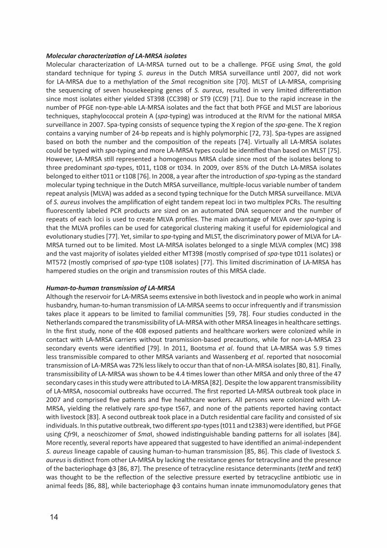
Figure 1: Penicillin use in World War II. In 1944, laboratories across the country were stepping up their production of penicillin, including Schenley in Indiana, whose advertisement stated, "When the thunderous battles of this war have subsided to pages of silent print in a history book, the greatest news event of World War II may well be the discovery and development of penicillin." Credit: Research and Development Division, Schenley Laboratories, Inc., Lawrenceburg, Indiana
12
Methicillin resistant S. aureus (MRSA) 1961-1990, a hospital-acquired pathogen After its first emergence in 1961, the number of staphylococcal infections caused by MRSA was limited although small outbreaks in Great Britain in the early 1960s have been described [24, 25]. After that, a period without major MRSA epidemics was observed until a new series of hospital outbreaks with MRSA occurred in the late 1960s [26]. This time, the MRSA outbreaks were not restricted to Great Britain, but also occurred in the United States and Australia [27, 28]. These early MRSA outbreaks could be controlled by new antimicrobial agents, mainly aminoglycosides such as gentamicin, but in 1976 a MRSA strain that acquired gentamicin resistance caused a hospital outbreak [29]. Subsequently, this MRSA variant and others spread globally, replacing methicillin-sensitive S. aureus (MSSA) isolates in many hospitals [30, 31]. In the 1980s, the magnitude of the MRSA problem became apparent when it was clear that MRSA was as virulent as MSSA and the prevalence of MRSA in many hospitals had rapidly risen [30, 32, 33]. The high prevalence of MRSA eventually led to the development of surveillance systems, infection control policies and restricted use of antibiotics. Among the first countries to implement these measurements was Denmark, rapidly leading to a decline of MRSA in Danish hospitals [34, 35]. In the Netherlands, following some MRSA outbreaks in the mid-1980s [36], a National surveillance of MRSA was introduced in 1989 [37]. Hospitals could send their MRSA isolates to the national institute for public health and the environment (RIVM) for further characterization to study the routes and rates of transmission of this pathogen. Initially, MRSA isolates were characterized using bacteriophage typing, but this was replaced by pulsed-field gel electrophoresis (PFGE) in 2002 [38, 39].
MRSA 1990-2003, emergence in the communityA Dutch MRSA surveillance study between 1989 and 1992 showed a MRSA prevalence of 0.54%, and nearly 70% of those MRSA isolates could be related to importation from foreign hospitals [37]. In those foreign hospitals, only limited differentiation was found among the circulation MRSA clones [40]. However, in the early 1980s reports appeared that described MRSA carriers without known risk factors and who were not previously hospitalized. Many of those patients turned out to be intravenous drug users [41, 42]. In 1995, Moreno et al. published a report showing that many patients already were MRSA positive upon hospital admission and had no identifiable risk factors [43].After that, other studies revealed the presence of this new variant of MRSA, now designated as community-acquired MRSA (CA-MRSA), in hospitals [44]. In the following years, it became clear that outbreaks and transmissions of CA-MRSA were not restricted to hospitals in the US, but also occurred in other countries and communities, such as prisons and football teams revealing additional reservoirs for this pathogen [45, 46].
Differences between hospital- and community-acquired MRSA; fading boundariesWith the appearance of the first reports on CA-MRSA, it became clear that CA-MRSA differed not only in epidemiology, but also in molecular characteristics. First, the SCCmec, a chromosomal mobile genetic element carrying the mecA gene, differs from HA-MRSA in both size and composition. The differences in SCCmec have been used for characterization [47]. HA-MRSA isolates typically carry SCCmec types I, II or III, whereas CA-MRSA isolates usually yield SCCmec types IV or V [48]. Second, CA-MRSA strains often carry a virulence factor designated Panton-Valentine leucocidin (PVL) that is capable of destroying leucocytes and other human immune response cells such as macrophages [48, 49]. However, shortly after the emergence of MRSA in the community, reports appeared describing the presence and transmission of CA-MRSA in healthcare settings [50]. David et al. showed that HA-MRSA infections were decreasing in an academic hospital, but this decrease was neutralized by an increase in the proportion of CA-MRSA infections [51]. In fact, USA300, one the main clades of CA-MRSA, was the most frequently found MRSA variant causing bacteremia in U.S. hospitals between 2009 and 2010 [52]. In addition, molecular characteristics, previously attributed to be specific for CA-MRSA, were also identified in MRSA strains with a clear hospital association, suggesting that the previously deposited definitions for CA-MRSA do no longer apply [53].
13
1
Introd
uctio
nMRSA 2003-present, introduction of MRSA from the animal reservoirMRSA in animals was first described in 1972 when MRSA was detected in the milk of mastitic cows [54]. After that, MRSA has been detected in many different animal species, but generally, a distinction is made between food production and companion animals, since MRSA acquisition in companion animals is considered to be as result of human-to-animal transmission [55, 56]. The first report of MRSA in pigs appeared in France in 2005, where indistinguishable MRSA isolates were found in pigs and pig farmers [57]. Within the same year, MRSA was found in pig farms in the Netherlands [58]. Surprisingly, the molecular characteristics of the Dutch MRSA isolates, based on multi-locus sequence typing (MLST), were identical to the French isolates and all belonged to clonal complex (CC) 398 [59]. Another study among Dutch slaughterhouses found a MRSA prevalence among pigs of 39% and all isolates belonged to MRSA CC398 [60]. After the initial publications, MRSA CC398 was also identified in other European countries [61, 62], North America [63, 64] and Asia [65], revealing a worldwide dissemination. Besides pigs, MRSA CC398 was also found in other livestock animals such as poultry, veal calves and horses leading to the designation livestock-associated MRSA (LA-MRSA) [66-68].
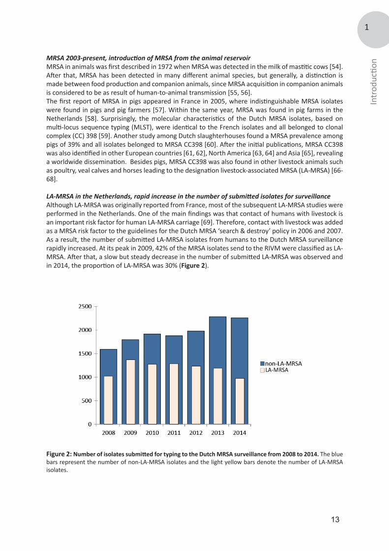
LA-MRSA in the Netherlands, rapid increase in the number of submitted isolates for surveillanceAlthough LA-MRSA was originally reported from France, most of the subsequent LA-MRSA studies were performed in the Netherlands. One of the main findings was that contact of humans with livestock is an important risk factor for human LA-MRSA carriage [69]. Therefore, contact with livestock was added as a MRSA risk factor to the guidelines for the Dutch MRSA ‘search & destroy’ policy in 2006 and 2007. As a result, the number of submitted LA-MRSA isolates from humans to the Dutch MRSA surveillance rapidly increased. At its peak in 2009, 42% of the MRSA isolates send to the RIVM were classified as LA-MRSA. After that, a slow but steady decrease in the number of submitted LA-MRSA was observed and in 2014, the proportion of LA-MRSA was 30% (Figure 2).
Figure 2: Number of isolates submitted for typing to the Dutch MRSA surveillance from 2008 to 2014. The blue bars represent the number of non-LA-MRSA isolates and the light yellow bars denote the number of LA-MRSA isolates.
14
Molecular characterization of LA-MRSA isolatesMolecular characterization of LA-MRSA turned out to be a challenge. PFGE using SmaI, the gold standard technique for typing S. aureus in the Dutch MRSA surveillance until 2007, did not work for LA-MRSA due to a methylation of the SmaI recognition site [70]. MLST of LA-MRSA, comprising the sequencing of seven housekeeping genes of S. aureus, resulted in very limited differentiation since most isolates either yielded ST398 (CC398) or ST9 (CC9) [71]. Due to the rapid increase in the number of PFGE non-type-able LA-MRSA isolates and the fact that both PFGE and MLST are laborious techniques, staphylococcal protein A (spa-typing) was introduced at the RIVM for the national MRSA surveillance in 2007. Spa-typing consists of sequence typing the X region of the spa-gene. The X region contains a varying number of 24-bp repeats and is highly polymorphic [72, 73]. Spa-types are assigned based on both the number and the composition of the repeats [74]. Virtually all LA-MRSA isolates could be typed with spa-typing and more LA-MRSA types could be identified than based on MLST [75]. However, LA-MRSA still represented a homogenous MRSA clade since most of the isolates belong to three predominant spa -types, t011, t108 or t034. In 2009, over 85% of the Dutch LA-MRSA isolates belonged to either t011 or t108 [76]. In 2008, a year after the introduction of spa-typing as the standard molecular typing technique in the Dutch MRSA surveillance, multiple-locus variable number of tandem repeat analysis (MLVA) was added as a second typing technique for the Dutch MRSA surveillance. MLVA of S. aureus involves the amplification of eight tandem repeat loci in two multiplex PCRs. The resulting fluorescently labeled PCR products are sized on an automated DNA sequencer and the number of repeats of each loci is used to create MLVA profiles. The main advantage of MLVA over spa-typing is that the MLVA profiles can be used for categorical clustering making it useful for epidemiological and evolutionary studies [77]. Yet, similar to spa-typing and MLST, the discriminatory power of MLVA for LA-MRSA turned out to be limited. Most LA-MRSA isolates belonged to a single MLVA complex (MC) 398 and the vast majority of isolates yielded either MT398 (mostly comprised of spa-type t011 isolates) or MT572 (mostly comprised of spa-type t108 isolates) [77]. This limited discrimination of LA-MRSA has hampered studies on the origin and transmission routes of this MRSA clade.
Human-to-human transmission of LA-MRSAAlthough the reservoir for LA-MRSA seems extensive in both livestock and in people who work in animal husbandry, human-to-human transmission of LA-MRSA seems to occur infrequently and if transmission takes place it appears to be limited to familial communities [59, 78]. Four studies conducted in the Netherlands compared the transmissibility of LA-MRSA with other MRSA lineages in healthcare settings. In the first study, none of the 408 exposed patients and healthcare workers were colonized while in contact with LA-MRSA carriers without transmission-based precautions, while for non-LA-MRSA 23 secondary events were identified [79]. In 2011, Bootsma et al. found that LA-MRSA was 5.9 times less transmissible compared to other MRSA variants and Wassenberg et al. reported that nosocomial transmission of LA-MRSA was 72% less likely to occur than that of non-LA-MRSA isolates [80, 81]. Finally, transmissibility of LA-MRSA was shown to be 4.4 times lower than other MRSA and only three of the 47 secondary cases in this study were attributed to LA-MRSA [82]. Despite the low apparent transmissibility of LA-MRSA, nosocomial outbreaks have occurred. The first reported LA-MRSA outbreak took place in 2007 and comprised five patients and five healthcare workers. All persons were colonized with LA-MRSA, yielding the relatively rare spa-type t567, and none of the patients reported having contact with livestock [83]. A second outbreak took place in a Dutch residential care facility and consisted of six individuals. In this putative outbreak, two different spa-types (t011 and t2383) were identified, but PFGE using Cfr9I, a neoschizomer of SmaI, showed indistinguishable banding patterns for all isolates [84]. More recently, several reports have appeared that suggested to have identified an animal-independent S. aureus lineage capable of causing human-to-human transmission [85, 86]. This clade of livestock S. aureus is distinct from other LA-MRSA by lacking the resistance genes for tetracycline and the presence of the bacteriophage φ3 [86, 87]. The presence of tetracycline resistance determinants (tetM and tetK) was thought to be the reflection of the selective pressure exerted by tetracycline antibiotic use in animal feeds [86, 88], while bacteriophage φ3 contains human innate immunomodulatory genes that
15
1
Introd
uctio
nplay a major role human niche adaptation [89]. For LA-MRSA it is suggested that after the jump from humans to livestock they acquired the resistance genes for tetracycline and the lost some human-niche specific genes, carried by φ3, while the animal-independent clade did not underwent these evolutionary alterations [87]. However, most studies that identified the animal-independent CC398 lineage were dominated by isolates obtained from animals and the human-associated isolates were mostly comprised of methicillin sensitive S. aureus (MSSA). In contrast, two recent reports suggest the spread of LA-MRSA into the general population. First, Wulf et al. found that 7% of the LA-MRSA in newly identified carriers from 2002 to 2008 in their laboratory was due to unexpected cases [90]. A broader study, including all Dutch MRSA surveillance data from 2008 and 2009, found that nearly a quarter of the MRSA isolates did not have a link with known defined risk groups and these MRSAs were designated as MRSA of unknown origin (MUO). Of the MUOs, 26% belonged to LA-MRSA suggesting spread through the community independent of contact with livestock [91].
Persistent carriage of LA-MRSA in humansDespite the high prevalence of LA-MRSA in the Netherlands, very little is known about the dynamics of LA-MRSA carriage in humans, especially in healthcare settings. Carriage studies performed in livestock settings revealed contradictory results. Van Cleef et al. suggested that LA-MRSA is not a good human colonizer, since 94% of the persons who acquired LA-MRSA during visits to livestock farms was tested negative after 24 hours [92]. Furthermore, LA-MRSA prevalence among veal calf farmers rapidly declined during absence of contact with animals. During holiday periods the prevalence was 11% compared to 26% in exposed periods, a decrease of 58% [93]. On the other hand, nasal colonization of LA-MRSA was found in 45% of the investigated livestock veterinarians [78]. In addition, 59% of the pig farmers in a German study were still carrying LA-MRSA after the holidays, suggesting persistent LA-MRSA carriage [94]. Recently, two longitudinal studies about the dynamics of LA-MRSA carriage among pig farmers and livestock veterinarians found a high prevalence of persistent LA-MRSA carriage [95, 96]. During the one-year sample period among pig farmers, 38% of the persons carried LA-MRSA on each of the six sampling moments [95]. Within the livestock veterinarians, 23% was LA-MRSA positive on all five sampling moments in this two-year prospective cohort study and 56% of those veterinarians carried LA-MRSA isolates with identical MLVA-types during the study period [96]. However, based on the limited differentiation with the typing techniques used these studies, it is hard to adjudicate whether these persons could be considered as truly persistent LA-MRSA carriers.
Infections in humans caused by LA-MRSA Similar to persistence and human-to-human transmission, infections caused by LA-MRSA seem to be scarce. In an European survey among 26 countries of invasive S. aureus infections cultured from blood, only 0.4% were CC398 and all isolates were MSSA [97]. A surveillance among 17 European countries found that the proportion of LA-MRSA cultured from blood was significantly lower compared to other MRSA variants, suggesting that LA-MRSA is less invasive [98]. Furthermore, several studies have shown that the presence of toxin genes in LA-MRSA is very limited [99, 100]. The virulence factor PVL seems also nearly absent in LA-MRSA. For instance, of the 793 LA-MRSA isolates, submitted to the Dutch MRSA surveillance in 2007, only a single LA-MRSA isolate harbored the PVL-genes [75]. In contrast, a recent review on human infections of S. aureus CC398 identified 74 publications were human infections of any type were described [101]. Most of these reports originated from Europe [98], but infections have also been found in other parts of the world such as USA [102] and Hong Kong [103]. The most common type of infections caused by LA-MRSA varies from mild or localized infections such as skin and soft tissue infections and wounds [79, 104]. However, serious invasive infections, such as endocarditis have also been described [105]. In addition, Kock et al. found that 8% of the isolates originating from blood cultures collected between 2008 and 2012 were classified as LA-MRSA [106].Although LA-MRSA is considered as a MRSA variant that only sporadically causes infections, a number of deaths have been attributed to LA-MRSA. In Spain, a fatal infection with LA-MRSA occurred in a 79-year-old patient. The patient lived on a pig farm and samples from the patient, pigs and a family
16
member were indistinguishable, based on spa-typing and PFGE, suggesting transmission from the pig or by the household member [107]. Recently, the Danish Statens Serum Institute reported four case histories of individuals that died from sepsis caused by LA-MRSA (SSI, Epi-News No. 24a -2014). Remarkably, none of these patients had any contact to pig farming.
Objective and outline of the thesisThe emergence of livestock-associated methicillin resistant Staphylococcus aureus (LA-MRSA) has had a major impact in the Netherlands. One of the main concerns of LA-MRSA is its clinical importance in regards to its ability to transmit, persist and cause disease in humans. The study presented in this thesis was aimed at studying these aspects utilizing isolates and data obtained during the Dutch national MRSA surveillance. This surveillance system can be used to study the routes of transmission, identify trends and shifts in the MRSA population in the Netherlands and supports infection control in healthcare settings. This requires molecular typing techniques with sufficient discriminatory power and for most MRSA variants the currently used techniques are very successful. However, molecular characterization of LA-MRSA turned out to be a challenge, since the typing techniques utilized by the RIVM provided very limited discrimination for LA-MRSA. This limited differentiation has hampered studies on the origin and the transmission routes of LA-MRSA and adjudications on the potential of this pathogen to cause public health problems are difficult to make. The objective of the research this thesis was to develop and use molecular typing tools to assess the capability of LA-MRSA to transmit, persist and cause disease in humans and to study the possible temporal changes in the characteristics of the most predominant MRSA clade in the Netherlands. For surveillance purposes, molecular tools with sufficient discriminatory power and a high throughput are needed. Starting in 2008, the Dutch MRSA surveillance is based on two typing techniques, Staphylococcal protein A (spa)-typing and multiple locus variable number of tandem repeat analysis (MLVA), for typing. In chapter 2 we assessed whether both spa-typing and MLVA are required for the MRSA surveillance. For this, we compared and evaluated the typing results of spa-typing and MLVA of all LA-MRSA and non-LA-MRSA isolates we received for our national MRSA surveillance during 2008–2013.
Pulsed-field gel electrophoreses (PFGE) of S. aureus, the gold-standard typing technique at the time of emergence of LA-MRSA, provides a banding pattern, which can be used for comparison with the profiles of other S. aureus isolates. PFGE is highly discriminative and is widely used in many laboratories making it a good tool to study possible transmission and outbreaks of MRSA isolates. However, PFGE of LA-MRSA did not result in banding patterns due to a methylation in the recognition site of the restriction enzyme SmaI, used in conventional PFGE approaches. In chapter 3, we optimized PFGE with restriction enzyme Cfr9I, a neoschizomer of SmaI, and evaluated its use to characterize a limited number of LA-MRSA isolates. In chapter 4, we applied PFGE using Cfr9I during a presumed outbreak of LA-MRSA in a Dutch nursing home. PFGE using Cfr9I was able to confirm the outbreak, but found that the isolates of a single healthcare worker did not belong to the outbreak despite the fact that other techniques suggested otherwise.
Although PFGE with Cfr9I yields a relatively high discriminatory power for LA-MRSA isolates, PFGE remains a time-consuming, laborious and non-portable method. In chapter 5, we assessed and validated the capability of whole genome mapping (WGM) to differentiate LA-MRSA isolates. A whole genome map is a high-resolution, ordered, whole genome restriction map and for S. aureus isolates these maps consists of 200–300 restriction fragments. In contrast, in PFGE of S. aureus only 10–15 non-ordered restriction fragments are used for the analysis. We used WGM in chapter 6 to identify possible persistence in and transmission of LA-MRSA between humans. For this, we used LA-MRSA isolates originating from a 2-year prospective longitudinal cohort study in which livestock veterinarians and their household members were repeatedly sampled for the presence of S. aureus.
17
1
Introd
uctio
nNext generation sequencing (NGS) is the ultimate typing tool for studying the origin and transmission routes of all virtually all microorganisms. In contrast to PFGE and WGM, NGS for LA-MRSA also allows for the study of genes involved in pathogenesis, antibiotic resistance and virulence. Chapter 7 describes the molecular characterization, including NGS of a subset of isolates, and epidemiological data of more than 9,000 LA-MRSA isolates submitted for the national MRSA surveillance from 2003-2014 to assess the characteristics of LA-MRSA in the Netherlands. Next generation sequencing was further applied in chapter 8, where we describe human-to-human transmission of LA-MRSA in Dutch healthcare settings based on the NGS data of more than 200 LA-MRSA isolates.
To further explore the capability of LA-MRSA to transmit between humans in a healthcare setting we performed a study, described in chapter 9, to assess the proportion of LA-MRSA in MRSA positive individuals without contact with pigs/veal calves or other known risk factors (MRSA of unknown origin; MUO) in 17 different hospitals. Finally, in chapter 10 the major findings of the thesis are summarized and discussed and future perspectives are presented. References 1. Elek SD. Staphylococcus pyogenus and its relation to disease. Edinburgh: E. & S. Livingstone; 1959. 776 p.2. Ogston A. Micrococcus Poisoning. Journal of anatomy and physiology. 1882;16(Pt 4):526-67.3. Ogston A. Report upon Micro-Organisms in Surgical Diseases. British medical journal. 1881;1(1054):369 b2-75.4. Billroth T. On the mutual action of living vegetable and animal cells. A biological study. Clinical lectures on subjects connected with medicine and surgery. London: The New Sydenham Society; 1894.5. Ogston A. Micrococcus Poisoning. Journal of anatomy and physiology. 1882;17(Pt 1):24-58.6. Rosenbach AJ. Mikro-organismen bei den wund-infections-krankheiten des menschen. Wiesbaden: J.F. Bergmann; 1884. 150 p.7. Smith IM, Vickers AB. Natural history of 338 treated and untreated patients with staphylococcal septicaemia (1936-1955). Lancet. 1960;1(7138):1318-22.8. Lister J. On the Antiseptic Principle in the Practice of Surgery. British medical journal. 1867;2(351):246-8.9. Haller JS, Jr. Treatment of infected wounds during the Great War, 1914 to 1918. Southern medical journal. 1992;85(3):303-15.10. Fleming A. The action of chemical and physiological antseptics in a septic wound. British Journal of Surgery. 1919(7):99-129.11. Fleming A. Classics in infectious diseases: on the antibacterial action of cultures of a penicillium, with special reference to their use in the isolation of B. influenzae by Alexander Fleming, Reprinted from the British Journal of Experimental Pathology 10:226-236, 1929. Rev Infect Dis. 1980;2(1):129-39.12. Abraham EP, Chain E, Fletcher CM, Florey HW, Gardner AD, Heatley NG, et al. Further observations on penicillin. 1941. Eur J Clin Pharmacol. 1992;42(1):3-9.13. Barber M, Rozwadowska-Dowzenko M. Infection by penicillin-resistant staphylococci. Lancet. 1948;2(6530):641-4.14. Kirby WM. Extraction of a Highly Potent Penicillin Inactivator from Penicillin Resistant Staphylococci. Science. 1944;99(2579):452-3.15. Hassall JE, Rountree PM. Staphylococcal septicaemia. Lancet. 1959;1(7066):213-7.16. Hausmann W, Karlish AJ. Staphylococcal pneumonia in adults. British medical journal. 1956;2(4997):845-7.17. Wilson R, Hamburger M. Fifteen years' experience with staphylococcus septicemia in a large city hospital; analysis of fifty-five cases in the Cincinnati General Hospital 1940 to 1954. The American journal of medicine. 1957;22(3):437-57.18. Jessen O, Rosendal K, Bulow P, Faber V, Eriksen KR. Changing staphylococci and staphylococcal infections. A ten-year study of bacteria and cases of bacteremia. The New England journal of medicine. 1969;281(12):627-35.19. Batchelor FR, Doyle FP, Nayler JH, Rolinson GN. Synthesis of penicillin: 6-aminopenicillanic acid in penicillin fermentations. Nature. 1959;183(4656):257-8.20. Rolinson GN, Stevens S, Batchelor FR, Wood JC, Chain EB. Bacteriological studies on a new penicillin-BRL. 1241. Lancet. 1960;2(7150):564-7.21. Knox R. A new penicillin (BRL 1241) active against penicillin-resistant staphylococci. British medical journal. 1960;2(5200):690-3.22. Jevons MP. "Celbenin" -resistant staphylococci. British medical journal. 1961;1::124-5.23. Hartman BJ, Tomasz A. Low-affinity penicillin-binding protein associated with beta-lactam resistance in Staphylococcus aureus. Journal of bacteriology. 1984;158(2):513-6.24. Colley EW, McNicol MW, Bracken PM. Methicillin-Resistant Staphylococci in a General Hospital. Lancet.
18
1965;1(7385):595-7.25. Harding JW. Infections due to methicillin-resistant strains of Staphylococcus pyogenes. Journal of clinical pathology. 1963;16:268-70.26. Benner EJ, Kayser FH. Growing clinical significance of methcillin-resistant Staphylococcus aureus. Lancet. 1968;2(7571):741-4.27. Barrett FF, McGehee RF, Jr., Finland M. Methicillin-resistant Staphylococcus aureus at Boston City Hospital. Bacteriologic and epidemiologic observations. The New England journal of medicine. 1968;279(9):441-8.28. Rountree PM, Vickery AM. Further observations on methicillin-resistant staphylococci. The Medical journal of Australia. 1973;1(21):1030-4.29. Shanson DC, Kensit JC, Duke R. Outbreak of hospital infection with a strain of Staphylococcus aureus resistant to gentamicin and methicillin. Lancet. 1976;2(7999):1347-8.30. Panlilio AL, Culver DH, Gaynes RP, Banerjee S, Henderson TS, Tolson JS, et al. Methicillin-resistant Staphylococcus aureus in U.S. hospitals, 1975-1991. Infection control and hospital epidemiology. 1992;13(10):582-6.31. Townsend DE, Ashdown N, Bolton S, Bradley J, Duckworth G, Moorhouse EC, et al. The international spread of methicillin-resistant Staphylococcus aureus. The Journal of hospital infection. 1987;9(1):60-71.32. Wenzel RP. The emergence of methicillin-resistant Staphylococcus aureus. Annals of internal medicine. 1982;97(3):440-2.33. Wenzel RP, Nettleman MD, Jones RN, Pfaller MA. Methicillin-resistant Staphylococcus aureus: implications for the 1990s and effective control measures. The American journal of medicine. 1991;91(3B):221S-7S.34. Rosendal K, Jessen O, Bentzon MW, Bulow P. Antibiotic policy and spread of Staphylococcus aureus strains in Danish hospitals, 1969-1974. Acta pathologica et microbiologica Scandinavica Section B, Microbiology. 1977;85(2):143-52.35. Westh H, Jarlov JO, Kjersem H, Rosdahl VT. The disappearance of multiresistant Staphylococcus aureus in Denmark: changes in strains of the 83A complex between 1969 and 1989. Clinical infectious diseases : an official publication of the Infectious Diseases Society of America. 1992;14(6):1186-94.36. Vandenbroucke-Grauls CM, Frenay HM, van Klingeren B, Savelkoul TF, Verhoef J. Control of epidemic methicillin-resistant Staphylococcus aureus in a Dutch university hospital. European journal of clinical microbiology & infectious diseases : official publication of the European Society of Clinical Microbiology. 1991;10(1):6-11.37. Frenay HM, Schot CS, Van Leeuwen WJ, Rost JA, De Neeling AJ, Van Klingeren B. Methicillin resistant staphylococcal infection. Infection spreads between hospitals. Bmj. 1994;308(6920):58.38. Murchan S, Kaufmann ME, Deplano A, de Ryck R, Struelens M, Zinn CE, et al. Harmonization of pulsed-field gel electrophoresis protocols for epidemiological typing of strains of methicillin-resistant Staphylococcus aureus: a single approach developed by consensus in 10 European laboratories and its application for tracing the spread of related strains. Journal of clinical microbiology. 2003;41(4):1574-85.39. Parker MT. The significance of phage-typing patterns in Staphylococcus aureus. Easmon C.S.F. AC, editor. New York: Academic Press; 1983.40. Oliveira D, Santos-Sanches I, Mato R, Tamayo M, Ribeiro G, Costa D, et al. Virtually all methicillin-resistant Staphylococcus aureus (MRSA) infections in the largest Portuguese teaching hospital are caused by two internationally spread multiresistant strains: the 'Iberian' and the 'Brazilian' clones of MRSA. Clinical microbiology and infection : the official publication of the European Society of Clinical Microbiology and Infectious Diseases. 1998;4(7):373-84.41. Saravolatz LD, Markowitz N, Arking L, Pohlod D, Fisher E. Methicillin-resistant Staphylococcus aureus. Epidemiologic observations during a community-acquired outbreak. Annals of internal medicine. 1982;96(1):11-6.42. Saravolatz LD, Pohlod DJ, Arking LM. Community-acquired methicillin-resistant Staphylococcus aureus infections: a new source for nosocomial outbreaks. Annals of internal medicine. 1982;97(3):325-9.43. Moreno F, Crisp C, Jorgensen JH, Patterson JE. Methicillin-resistant Staphylococcus aureus as a community organism. Clinical infectious diseases : an official publication of the Infectious Diseases Society of America. 1995;21(5):1308-12.44. Herold BC, Immergluck LC, Maranan MC, Lauderdale DS, Gaskin RE, Boyle-Vavra S, et al. Community-acquired methicillin-resistant Staphylococcus aureus in children with no identified predisposing risk. Jama. 1998;279(8):593-8.45. Centers for Disease C, Prevention. Methicillin-resistant Staphylococcus aureus infections among competitive sports participants--Colorado, Indiana, Pennsylvania, and Los Angeles County, 2000-2003. MMWR Morbidity and mortality weekly report. 2003;52(33):793-5.46. Pan ES, Diep BA, Carleton HA, Charlebois ED, Sensabaugh GF, Haller BL, et al. Increasing prevalence of methicillin-resistant Staphylococcus aureus infection in California jails. Clinical infectious diseases : an official publication of the Infectious Diseases Society of America. 2003;37(10):1384-8.47. Boye K, Bartels MD, Andersen IS, Moller JA, Westh H. A new multiplex PCR for easy screening of methicillin-resistant Staphylococcus aureus SCCmec types I-V. Clinical microbiology and infection : the official publication of the European Society of Clinical Microbiology and Infectious Diseases. 2007;13(7):725-7.48. Millar BC, Loughrey A, Elborn JS, Moore JE. Proposed definitions of community-associated meticillin-resistant Staphylococcus aureus (CA-MRSA). The Journal of hospital infection. 2007;67(2):109-13.49. Woodin AM. Staphylococcal leukocidin. T. C. Montje SK, and S. J. Ajl, editor. New York: Academic Press Inc.; 1970.50. Seybold U, Kourbatova EV, Johnson JG, Halvosa SJ, Wang YF, King MD, et al. Emergence of community-associated methicillin-resistant Staphylococcus aureus USA300 genotype as a major cause of health care-associated blood stream infections. Clinical infectious diseases : an official publication of the Infectious Diseases Society of America. 2006;42(5):647-56.
19
1
Introd
uctio
n
51. David MZ, Cadilla A, Boyle-Vavra S, Daum RS. Replacement of HA-MRSA by CA-MRSA infections at an academic medical center in the midwestern United States, 2004-5 to 2008. PloS one. 2014;9(4):e92760.52. Tenover FC, Tickler IA, Goering RV, Kreiswirth BN, Mediavilla JR, Persing DH, et al. Characterization of nasal and blood culture isolates of methicillin-resistant Staphylococcus aureus from patients in United States Hospitals. Antimicrobial agents and chemotherapy. 2012;56(3):1324-30.53. Mimica MJ, Berezin EN, Carvalho RB. Healthcare associated PVL negative methicillin-resistant Staphylococcus aureus with SCCmec type IV. The Pediatric infectious disease journal. 2009;28(10):934.54. Devriese LA, Van Damme LR, Fameree L. Methicillin (cloxacillin)-resistant Staphylococcus aureus strains isolated from bovine mastitis cases. Zentralblatt fur Veterinarmedizin Reihe B Journal of veterinary medicine Series B. 1972;19(7):598-605.55. Loeffler A, Boag AK, Sung J, Lindsay JA, Guardabassi L, Dalsgaard A, et al. Prevalence of methicillin-resistant Staphylococcus aureus among staff and pets in a small animal referral hospital in the UK. The Journal of antimicrobial chemotherapy. 2005;56(4):692-7.56. van Duijkeren E, Wolfhagen MJ, Box AT, Heck ME, Wannet WJ, Fluit AC. Human-to-dog transmission of methicillin-resistant Staphylococcus aureus. Emerging infectious diseases. 2004;10(12):2235-7.57. Armand-Lefevre L, Ruimy R, Andremont A. Clonal comparison of Staphylococcus aureus isolates from healthy pig farmers, human controls, and pigs. Emerging infectious diseases. 2005;11(5):711-4.58. Voss A, Loeffen F, Bakker J, Klaassen C, Wulf M. Methicillin-resistant Staphylococcus aureus in pig farming. Emerging infectious diseases. 2005;11(12):1965-6.59. Huijsdens XW, van Dijke BJ, Spalburg E, van Santen-Verheuvel MG, Heck ME, Pluister GN, et al. Community-acquired MRSA and pig-farming. Annals of clinical microbiology and antimicrobials. 2006;5:26.60. de Neeling AJ, van den Broek MJ, Spalburg EC, van Santen-Verheuvel MG, Dam-Deisz WD, Boshuizen HC, et al. High prevalence of methicillin resistant Staphylococcus aureus in pigs. Veterinary microbiology. 2007;122(3-4):366-72.61. Guardabassi L, Stegger M, Skov R. Retrospective detection of methicillin resistant and susceptible Staphylococcus aureus ST398 in Danish slaughter pigs. Veterinary microbiology. 2007;122(3-4):384-6.62. Witte W, Strommenger B, Stanek C, Cuny C. Methicillin-resistant Staphylococcus aureus ST398 in humans and animals, Central Europe. Emerging infectious diseases. 2007;13(2):255-8.63. Bhat M, Dumortier C, Taylor BS, Miller M, Vasquez G, Yunen J, et al. Staphylococcus aureus ST398, New York City and Dominican Republic. Emerging infectious diseases. 2009;15(2):285-7.64. Khanna T, Friendship R, Dewey C, Weese JS. Methicillin resistant Staphylococcus aureus colonization in pigs and pig farmers. Veterinary microbiology. 2008;128(3-4):298-303.65. Sergio DM, Koh TH, Hsu LY, Ogden BE, Goh AL, Chow PK. Investigation of meticillin-resistant Staphylococcus aureus in pigs used for research. Journal of medical microbiology. 2007;56(Pt 8):1107-9.66. Van den Eede A, Martens A, Lipinska U, Struelens M, Deplano A, Denis O, et al. High occurrence of methicillin-resistant Staphylococcus aureus ST398 in equine nasal samples. Veterinary microbiology. 2009;133(1-2):138-44.67. Mulders MN, Haenen AP, Geenen PL, Vesseur PC, Poldervaart ES, Bosch T, et al. Prevalence of livestock-associated MRSA in broiler flocks and risk factors for slaughterhouse personnel in The Netherlands. Epidemiology and infection. 2010;138(5):743-55.68. Graveland H, Wagenaar JA, Heesterbeek H, Mevius D, van Duijkeren E, Heederik D. Methicillin resistant Staphylococcus aureus ST398 in veal calf farming: human MRSA carriage related with animal antimicrobial usage and farm hygiene. PloS one. 2010;5(6):e10990.69. Van den Broeck IV, van Cleef BA, Haenen A, Broens EM, van der Wolf PJ, van den Broek MJ, et al. Methicillin-resistant Staphylococcus aureus in people living and working in pig farms. Epidemiology and infection. 2009;137(5):700-8.70. Bens CC, Voss A, Klaassen CH. Presence of a novel DNA methylation enzyme in methicillin-resistant Staphylococcus aureus isolates associated with pig farming leads to uninterpretable results in standard pulsed-field gel electrophoresis analysis. Journal of clinical microbiology. 2006;44(5):1875-6.71. van Loo I, Huijsdens X, Tiemersma E, de Neeling A, van de Sande-Bruinsma N, Beaujean D, et al. Emergence of methicillin-resistant Staphylococcus aureus of animal origin in humans. Emerging infectious diseases. 2007;13(12):1834-9.72. Frenay HM, Theelen JP, Schouls LM, Vandenbroucke-Grauls CM, Verhoef J, van Leeuwen WJ, et al. Discrimination of epidemic and nonepidemic methicillin-resistant Staphylococcus aureus strains on the basis of protein A gene polymorphism. Journal of clinical microbiology. 1994;32(3):846-7.73. Uhlen M, Guss B, Nilsson B, Gatenbeck S, Philipson L, Lindberg M. Complete sequence of the staphylococcal gene encoding protein A. A gene evolved through multiple duplications. The Journal of biological chemistry. 1984;259(3):1695-702.74. Harmsen D, Claus H, Witte W, Rothganger J, Claus H, Turnwald D, et al. Typing of methicillin-resistant Staphylococcus aureus in a university hospital setting by using novel software for spa repeat determination and database management. Journal of clinical microbiology. 2003;41(12):5442-8.75. Huijsdens XW, Bosch T, van Santen-Verheuvel MG, Spalburg E, Pluister GN, van Luit M, et al. Molecular characterisation of PFGE non-typable methicillin-resistant Staphylococcus aureus in The Netherlands, 2007. Euro surveillance : bulletin Europeen sur les maladies transmissibles = European communicable disease bulletin. 2009;14(38).76. Haenen APJ, Huijsdens XW, Pluister GN, van Luit M, Bosch T, van Santen-Verheuvel MG, Spalburg E, Heck MEOC, de Neeling AJ, Mulders MN. Surveillance of MRSA in the Netherlands in 2009, the number of livestock-associated MRSA stabilizes. Infectieziekten Bulletin. 2010;10(December 2010):373-9.77. Schouls LM, Spalburg EC, van Luit M, Huijsdens XW, Pluister GN, van Santen-Verheuvel MG, et al. Multiple-locus
20
variable number tandem repeat analysis of Staphylococcus aureus: comparison with pulsed-field gel electrophoresis and spa-typing. PloS one. 2009;4(4):e5082.78. Cuny C, Nathaus R, Layer F, Strommenger B, Altmann D, Witte W. Nasal colonization of humans with methicillin-resistant Staphylococcus aureus (MRSA) CC398 with and without exposure to pigs. PloS one. 2009;4(8):e6800.79. van Rijen MM, Van Keulen PH, Kluytmans JA. Increase in a Dutch hospital of methicillin-resistant Staphylococcus aureus related to animal farming. Clinical infectious diseases : an official publication of the Infectious Diseases Society of America. 2008;46(2):261-3.80. Bootsma MC, Wassenberg MW, Trapman P, Bonten MJ. The nosocomial transmission rate of animal-associated ST398 meticillin-resistant Staphylococcus aureus. Journal of the Royal Society, Interface / the Royal Society. 2011;8(57):578-84.81. Wassenberg MW, Bootsma MC, Troelstra A, Kluytmans JA, Bonten MJ. Transmissibility of livestock-associated methicillin-resistant Staphylococcus aureus (ST398) in Dutch hospitals. Clinical microbiology and infection : the official publication of the European Society of Clinical Microbiology and Infectious Diseases. 2011;17(2):316-9.82. Hetem DJ, Bootsma MC, Troelstra A, Bonten MJ. Transmissibility of livestock-associated methicillin-resistant Staphylococcus aureus. Emerging infectious diseases. 2013;19(11):1797-802.83. Wulf MW, Markestein A, van der Linden FT, Voss A, Klaassen C, Verduin CM. First outbreak of methicillin-resistant Staphylococcus aureus ST398 in a Dutch hospital, June 2007. Euro surveillance : bulletin Europeen sur les maladies transmissibles = European communicable disease bulletin. 2008;13(9).84. Fanoy E, Helmhout LC, van der Vaart WL, Weijdema K, van Santen-Verheuvel MG, Thijsen SF, et al. An outbreak of non-typeable MRSA within a residential care facility. Euro surveillance : bulletin Europeen sur les maladies transmissibles = European communicable disease bulletin. 2009;14(1).85. McCarthy AJ, van Wamel W, Vandendriessche S, Larsen J, Denis O, Garcia-Graells C, et al. Staphylococcus aureus CC398 clade associated with human-to-human transmission. Applied and environmental microbiology. 2012;78(24):8845-8.86. Uhlemann AC, Porcella SF, Trivedi S, Sullivan SB, Hafer C, Kennedy AD, et al. Identification of a highly transmissible animal-independent Staphylococcus aureus ST398 clone with distinct genomic and cell adhesion properties. mBio. 2012;3(2).87. Price LB, Stegger M, Hasman H, Aziz M, Larsen J, Andersen PS, et al. Staphylococcus aureus CC398: host adaptation and emergence of methicillin resistance in livestock. mBio. 2012;3(1).88. Phillips I, Casewell M, Cox T, De Groot B, Friis C, Jones R, et al. Does the use of antibiotics in food animals pose a risk to human health? A critical review of published data. The Journal of antimicrobial chemotherapy. 2004;53(1):28-52.89. Foster TJ. Immune evasion by staphylococci. Nature reviews Microbiology. 2005;3(12):948-58.90. Wulf MW, Verduin CM, van Nes A, Huijsdens X, Voss A. Infection and colonization with methicillin resistant Staphylococcus aureus ST398 versus other MRSA in an area with a high density of pig farms. European journal of clinical microbiology & infectious diseases : official publication of the European Society of Clinical Microbiology. 2012;31(1):61-5.91. Lekkerkerk WS, van de Sande-Bruinsma N, van der Sande MA, Tjon ATA, Groenheide A, Haenen A, et al. Emergence of MRSA of unknown origin in the Netherlands. Clinical microbiology and infection : the official publication of the European Society of Clinical Microbiology and Infectious Diseases. 2012;18(7):656-61.92. van Cleef BA, Graveland H, Haenen AP, van de Giessen AW, Heederik D, Wagenaar JA, et al. Persistence of livestock-associated methicillin-resistant Staphylococcus aureus in field workers after short-term occupational exposure to pigs and veal calves. Journal of clinical microbiology. 2011;49(3):1030-3.93. Graveland H, Wagenaar JA, Bergs K, Heesterbeek H, Heederik D. Persistence of livestock associated MRSA CC398 in humans is dependent on intensity of animal contact. PloS one. 2011;6(2):e16830.94. Kock R, Loth B, Koksal M, Schulte-Wulwer J, Harlizius J, Friedrich AW. Persistence of nasal colonization with livestock-associated methicillin-resistant Staphylococcus aureus in pig farmers after holidays from pig exposure. Applied and environmental microbiology. 2012;78(11):4046-7.95. van Cleef BA, van Benthem BH, Verkade EJ, van Rijen M, Kluytmans-van den Bergh MF, Schouls LM, et al. Dynamics of methicillin-resistant Staphylococcus aureus and methicillin-susceptible Staphylococcus aureus carriage in pig farmers: a prospective cohort study. Clinical microbiology and infection : the official publication of the European Society of Clinical Microbiology and Infectious Diseases. 2014.96. Verkade E, van Benthem B, den Bergh MK, van Cleef B, van Rijen M, Bosch T, et al. Dynamics and determinants of Staphylococcus aureus carriage in livestock veterinarians: a prospective cohort study. Clinical infectious diseases : an official publication of the Infectious Diseases Society of America. 2013;57(2):e11-7.97. Grundmann H, Aanensen DM, van den Wijngaard CC, Spratt BG, Harmsen D, Friedrich AW, et al. Geographic distribution of Staphylococcus aureus causing invasive infections in Europe: a molecular-epidemiological analysis. PLoS medicine. 2010;7(1):e1000215.98. van Cleef BA, Monnet DL, Voss A, Krziwanek K, Allerberger F, Struelens M, et al. Livestock-associated methicillin-resistant Staphylococcus aureus in humans, Europe. Emerging infectious diseases. 2011;17(3):502-5.99. Monecke S, Kuhnert P, Hotzel H, Slickers P, Ehricht R. Microarray based study on virulence-associated genes and resistance determinants of Staphylococcus aureus isolates from cattle. Veterinary microbiology. 2007;125(1-2):128-40.100. Stegger M, Lindsay JA, Sorum M, Gould KA, Skov R. Genetic diversity in CC398 methicillin-resistant Staphylococcus aureus isolates of different geographical origin. Clinical microbiology and infection : the official publication of the European Society of Clinical Microbiology and Infectious Diseases. 2010;16(7):1017-9.101. Smith TC, Wardyn SE. Human Infections with Staphylococcus aureus CC398. Current environmental health reports.
21
1
Introd
uctio
n
2015;2(1):41-51.102. Uhlemann AC, Hafer C, Miko BA, Sowash MG, Sullivan SB, Shu Q, et al. Emergence of sequence type 398 as a community- and healthcare-associated methicillin-susceptible Staphylococcus aureus in northern Manhattan. Clinical infectious diseases : an official publication of the Infectious Diseases Society of America. 2013;57(5):700-3.103. Ho PL, Chuang SK, Choi YF, Lee RA, Lit AC, Ng TK, et al. Community-associated methicillin-resistant and methicillin-sensitive Staphylococcus aureus: skin and soft tissue infections in Hong Kong. Diagnostic microbiology and infectious disease. 2008;61(3):245-50.104. Krziwanek K, Metz-Gercek S, Mittermayer H. Methicillin-Resistant Staphylococcus aureus ST398 from human patients, upper Austria. Emerging infectious diseases. 2009;15(5):766-9.105. Schijffelen MJ, Boel CH, van Strijp JA, Fluit AC. Whole genome analysis of a livestock-associated methicillin-resistant Staphylococcus aureus ST398 isolate from a case of human endocarditis. BMC genomics. 2010;11:376.106. Kock R, Schaumburg F, Mellmann A, Koksal M, Jurke A, Becker K, et al. Livestock-associated methicillin-resistant Staphylococcus aureus (MRSA) as causes of human infection and colonization in Germany. PloS one. 2013;8(2):e55040.107. Lozano C, Aspiroz C, Ezpeleta AI, Gomez-Sanz E, Zarazaga M, Torres C. Empyema caused by MRSA ST398 with atypical resistance profile, Spain. Emerging infectious diseases. 2011;17(1):138-40.
22
2
Multiple-locus variable number tandem repeat
analysis is superior to spa-typing and sufficient to
characterize MRSA for surveillance purposes
Thijs Bosch
Martijn van Luit
Leo Schouls
Kim van der Zwaluw
Max Heck
Gerlinde Pluister
Fabian Landman
Future microbiology
2015
10(7): 1155-62
Marga van Santen-Verheuvel
Corrie Schot
Sandra Witteveen
23
2
MLV
A is
suffi
cien
t to
char
acte
rize
MRS
A fo
r sur
veill
ance
pur
pose
s
AbstractAim: Assess the best approach to type methicillin resistant Staphylococcus aureus (MRSA), Staphylococcal protein A (spa)-typing, multiple-locus variable number tandem repeat analysis (MLVA) or both.Material & Methods: Discriminatory power of spa-typing and MLVA was determined using 20,771 MRSA isolates.Results: There were twice as many MLVA-types (MTs) as spa-types present in the collection. Among the top 70% of the isolates, 37 spa-types and 139 MTs were found. MLVA diversity among the top-10 spa-types was high (diversity index 0.96), while spa-diversity among the top-10 MTs was much lower (diversity index 0.83). The probability that two MRSA isolates with the same spa-type also had the same MT was low (Wallace’s coefficient 0.27). In contrast, most MRSA isolates yielding the same MT also had the same spa-type (Wallace’s coefficient 0.90).
24
IntroductionMethicillin resistant Staphylococcus aureus (MRSA) is a bacterial pathogen that is associated with serious hospital- and community-acquired infections [1, 2]. Adequate molecular characterization of MRSA strains and surveillance of MRSA has shown to be an effective control measure to prevent the spread of MRSA in healthcare settings [3]. In the Netherlands, a low MRSA prevalence country [4], MRSA surveillance started in 1989 by the National Institute for Public Health and the Environment (RIVM). Typing of the MRSA isolates was originally performed using phage typing, but replaced in 2002 by pulsed-field gel electrophoresis, the traditional gold standard for molecular typing of MRSA [5, 6]. However, due to its laborious character and difficulties in exchanging data between laboratories, this method was replaced by Staphylococcal protein A (spa)-typing in 2007 [7]. This method, based on the sequence analysis of the variable repeat X region of the spa-gene, has become a useful typing tool because of its ease of performance and standardized nomenclature [8-10]. Although spa-typing has been successfully used for approximately 30,000 MRSA isolates in the Dutch national MRSA surveillance since 2007, the utility of this typing technique has recently become a subject of debate. The main reason is its inability to provide solid supporting information on epidemiological questions, such as transmission events [11-13]. To overcome this problem additional typing techniques are required. For the Dutch MRSA surveillance, multiple-locus variable number of tandem repeat analysis (MLVA) was introduced for S. aureus in 2008 and since then all isolates have been characterized by both methods.To assess whether both spa-typing and MLVA are required for the MRSA surveillance, we compared and evaluated the typing results of spa-typing and MLVA of all MRSA isolates we received for our national MRSA surveillance during 2008-2013.
Material and methodsStrain selectionIn the Netherlands, the National Institute of Public Health and the Environment (RIVM) serves as the reference center for national surveillance of MRSA. All medical microbiology laboratories in the Netherlands are requested to send the first MRSA isolate of a patient or healthcare worker to the RIVM. The MRSA isolates originate from humans who were MRSA-positive during MRSA-screening or routine clinical diagnostics. As a result, virtually all MRSA isolated from humans admitted to health care centers are sent to the RIVM for molecular typing. From the resulting database, all surveillance MRSA isolates received from 2008 until 2013 with complete spa- and MLVA-profiles (n = 20,771) were selected for comparative analyses. Isolates were grouped as MRSA and LA-MRSA, where LA-MRSA is defined as isolates belonging to MLVA complex 398 (MC398).
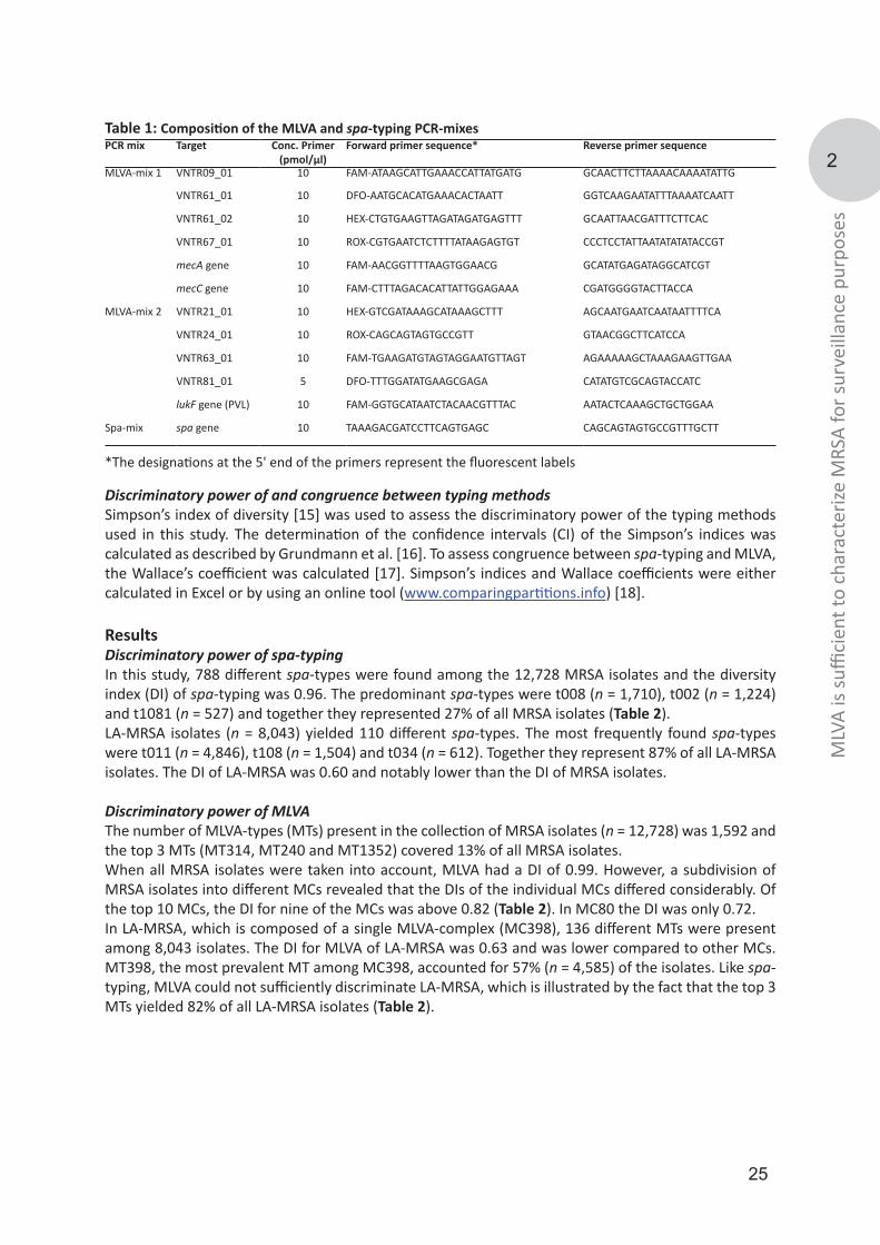
Molecular typingSpa-typing and MLVA were performed as described previously [7, 14], but in 2013 the separate components for the PCR mixes of spa and MLVA were replaced by ready-made PCR mixes (Eurogentec, Seraing, Belgium, art.no. CS-ALIQ-PROD-SPA and CS-ALIQ-PROD-MLVA) (Table 1). For spa-typing, 1 µl of lysate was added to 24 µl of the ready-made mix and for MLVA 2 µl of lysate was added to 23 µl of the ready-made mix. Lysates were made by resuspending a 1 µl inoculation loop of MRSA culture from blood agar plates, into 30 µl of lysis mix (100 µg/ml Lysostaphine (Sigma, catnr: L 7386) in TE (10 mM Tris.HCl, 1 mM EDTA, pH 8). After 30 min incubation at 37°C and 5 min at 95°C, 120 µl of TE was added to the lysates. The MLVA-mixes also contain specific primers for the detection of the mecA gene, the mecA gene variant LGA251 (mecC) and Panton Valentine leukocidin (PVL) (lukF). The primers for mecA and mecC were added to MLVA mix1 and the primers for the detection of PVL were included in MLVA mix 2. The presence of the mecA gene or mecC resulted either in a peak with a fixed size of 147 bp (mecA) or 165 bp (mecC), while a peak with fixed size of 165 bp appeared when an isolate carried the lukF gene (PVL).Isolates were considered as non-typeable (NT) for spa-typing if the spa-gene could not be amplified by PCR. Isolates lacking more than two variable number of tandem repeats (VNTRs) in MLVA were regarded as NT by MLVA.
25
2
MLV
A is
suffi
cien
t to
char
acte
rize
MRS
A fo
r sur
veill
ance
pur
pose
s
Discriminatory power of and congruence between typing methodsSimpson’s index of diversity [15] was used to assess the discriminatory power of the typing methods used in this study. The determination of the confidence intervals (CI) of the Simpson’s indices was calculated as described by Grundmann et al. [16]. To assess congruence between spa-typing and MLVA, the Wallace’s coefficient was calculated [17]. Simpson’s indices and Wallace coefficients were either calculated in Excel or by using an online tool (www.comparingpartitions.info) [18]. ResultsDiscriminatory power of spa-typingIn this study, 788 different spa-types were found among the 12,728 MRSA isolates and the diversity index (DI) of spa-typing was 0.96. The predominant spa-types were t008 (n = 1,710), t002 (n = 1,224) and t1081 (n = 527) and together they represented 27% of all MRSA isolates (Table 2). LA-MRSA isolates (n = 8,043) yielded 110 different spa-types. The most frequently found spa-types were t011 (n = 4,846), t108 (n = 1,504) and t034 (n = 612). Together they represent 87% of all LA-MRSA isolates. The DI of LA-MRSA was 0.60 and notably lower than the DI of MRSA isolates.
Discriminatory power of MLVAThe number of MLVA-types (MTs) present in the collection of MRSA isolates (n = 12,728) was 1,592 and the top 3 MTs (MT314, MT240 and MT1352) covered 13% of all MRSA isolates.When all MRSA isolates were taken into account, MLVA had a DI of 0.99. However, a subdivision of MRSA isolates into different MCs revealed that the DIs of the individual MCs differed considerably. Of the top 10 MCs, the DI for nine of the MCs was above 0.82 (Table 2). In MC80 the DI was only 0.72.In LA-MRSA, which is composed of a single MLVA-complex (MC398), 136 different MTs were present among 8,043 isolates. The DI for MLVA of LA-MRSA was 0.63 and was lower compared to other MCs. MT398, the most prevalent MT among MC398, accounted for 57% (n = 4,585) of the isolates. Like spa-typing, MLVA could not sufficiently discriminate LA-MRSA, which is illustrated by the fact that the top 3 MTs yielded 82% of all LA-MRSA isolates (Table 2).
Table 1: Composition of the MLVA and spa-typing PCR-mixesPCR mix Target Conc. Primer
(pmol/µl)Forward primer sequence* Reverse primer sequence
MLVA-mix 1 VNTR09_01 10 FAM-ATAAGCATTGAAACCATTATGATG GCAACTTCTTAAAACAAAATATTG
VNTR61_01 10 DFO-AATGCACATGAAACACTAATT GGTCAAGAATATTTAAAATCAATT
VNTR61_02 10 HEX-CTGTGAAGTTAGATAGATGAGTTT GCAATTAACGATTTCTTCAC
VNTR67_01 10 ROX-CGTGAATCTCTTTTATAAGAGTGT CCCTCCTATTAATATATATACCGT
mecA gene 10 FAM-AACGGTTTTAAGTGGAACG GCATATGAGATAGGCATCGT
mecC gene 10 FAM-CTTTAGACACATTATTGGAGAAA CGATGGGGTACTTACCA
MLVA-mix 2 VNTR21_01 10 HEX-GTCGATAAAGCATAAAGCTTT AGCAATGAATCAATAATTTTCA
VNTR24_01 10 ROX-CAGCAGTAGTGCCGTT GTAACGGCTTCATCCA
VNTR63_01 10 FAM-TGAAGATGTAGTAGGAATGTTAGT AGAAAAAGCTAAAGAAGTTGAA
VNTR81_01 5 DFO-TTTGGATATGAAGCGAGA CATATGTCGCAGTACCATC
lukF gene (PVL) 10 FAM-GGTGCATAATCTACAACGTTTAC AATACTCAAAGCTGCTGGAA
Spa-mix spa gene 10 TAAAGACGATCCTTCAGTGAGC CAGCAGTAGTGCCGTTTGCTT
*The designations at the 5' end of the primers represent the fluorescent labels
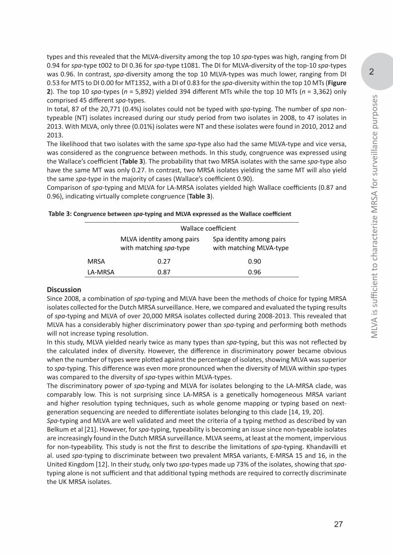
26
Comparing spa-typing and MLVA The number of MTs in the collection was nearly twice as high as the number of spa-types, yet little difference was observed between the DIs of spa-typing and MLVA. However, a difference between spa-typing and MLVA was seen when the number of types, sorted in frequency, was plotted against the cumulative proportion of the MRSA isolates. This revealed that the top 37 spa-types represent 70% of the MRSA isolates, while nearly four times as many MTs (139 MTs) represent a similar fraction of the isolates (Figure 1).No significant difference was seen among LA-MRSA types, where 70% of the isolates only represented two MTs and a single spa-type. All non-LA-MRSA isolates (n = 12,728) were ordered according to frequency of spa-types and subsequent subdivided into MTs and vice versa. Simpson’s diversity index was calculated for each of the top 10
*Percentage of either the MRSA or the LA-MRSA group
Figure 1: Discriminatory power of spa-typing and MLVA for MRSA isolates. The number of types, sorted in frequency, was plotted against the cumulative proportion of the MRSA isolates in the study. To prevent too much compression of the bars in the Y-axis direction only the results of 0-90% of the isolates are shown.
Table 2: Discriminatory power of spa-typing and MLVA
Group Typing method n No. of types Top 3 types % isolates in Top 3 types* DI (95% CI)
MRSA Spa-typing 12728 788 t008, t002, t1081 27 0.96 (0.96-0.96) LA-MRSA Spa-typing 8043 110 t011, t108, t034 87 0.60 (0.58-0.61) MRSA MLVA all MCs 12728 1592 MT314, MT240, MT1352 13 0.99 (0.99-0.99)
MLVA-MC1 343 78 MT149, MT152, MT222 48 0.90 (0.87-0.92)
MLVA-MC5 3146 346 MT5, MT130, MT67 19 0.97 (0.97-0.98)
MLVA-MC8 2980 183 MT314, MT240, MT195 50 0.90 (0.89-0.91)
MLVA-MC22 1282 131 MT22, MT491, MT489 46 0.91 (0.90-0.92)
MLVA-MC30 848 106 MT212, MT396, MT402 52 0.82 (0.80-0.85)
MLVA-MC45 1495 127 MT1352, MT527, MT528 47 0.89 (0.88-0.91)
MLVA-MC80 401 46 MT80, MT166, MT158 70 0.72 (0.67-0.76)
MLVA-MC482 229 37 MT482, MT631, MT1205 59 0.83 (0.78-0.87)
MLVA-MC621 475 75 MT621, MT710, MT625 48 0.85 (0.82-0.88)
MLVA-MCnone 624 260 MT3095, MT1574, MT840 17 0.98 (0.98-0.99)
LA-MRSA MLVA 8043 136 MT398, MT572, MT569 82 0.63 (0.62-0.64)
0
100
200
300
400
500
600
700
0 10 20 30 40 50 60 70 80 90
No. o
f typ
es
% of isolates (n= 12,728)
spa-typing
MLVA
27
2
MLV
A is
suffi
cien
t to
char
acte
rize
MRS
A fo
r sur
veill
ance
pur
pose
s
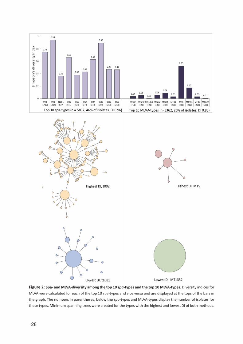
types and this revealed that the MLVA-diversity among the top 10 spa-types was high, ranging from DI 0.94 for spa-type t002 to DI 0.36 for spa-type t1081. The DI for MLVA-diversity of the top-10 spa-types was 0.96. In contrast, spa-diversity among the top 10 MLVA-types was much lower, ranging from DI 0.53 for MT5 to DI 0.00 for MT1352, with a DI of 0.83 for the spa-diversity within the top 10 MTs (Figure 2). The top 10 spa-types (n = 5,892) yielded 394 different MTs while the top 10 MTs (n = 3,362) only comprised 45 different spa-types. In total, 87 of the 20,771 (0.4%) isolates could not be typed with spa-typing. The number of spa non-typeable (NT) isolates increased during our study period from two isolates in 2008, to 47 isolates in 2013. With MLVA, only three (0.01%) isolates were NT and these isolates were found in 2010, 2012 and 2013.The likelihood that two isolates with the same spa-type also had the same MLVA-type and vice versa, was considered as the congruence between methods. In this study, congruence was expressed using the Wallace’s coefficient (Table 3). The probability that two MRSA isolates with the same spa-type also have the same MT was only 0.27. In contrast, two MRSA isolates yielding the same MT will also yield the same spa-type in the majority of cases (Wallace’s coefficient 0.90). Comparison of spa-typing and MLVA for LA-MRSA isolates yielded high Wallace coefficients (0.87 and 0.96), indicating virtually complete congruence (Table 3).
Table 3: Congruence between spa-typing and MLVA expressed as the Wallace coefficient
DiscussionSince 2008, a combination of spa-typing and MLVA have been the methods of choice for typing MRSA isolates collected for the Dutch MRSA surveillance. Here, we compared and evaluated the typing results of spa-typing and MLVA of over 20,000 MRSA isolates collected during 2008-2013. This revealed that MLVA has a considerably higher discriminatory power than spa-typing and performing both methods will not increase typing resolution. In this study, MLVA yielded nearly twice as many types than spa-typing, but this was not reflected by the calculated index of diversity. However, the difference in discriminatory power became obvious when the number of types were plotted against the percentage of isolates, showing MLVA was superior to spa-typing. This difference was even more pronounced when the diversity of MLVA within spa-types was compared to the diversity of spa-types within MLVA-types. The discriminatory power of spa-typing and MLVA for isolates belonging to the LA-MRSA clade, was comparably low. This is not surprising since LA-MRSA is a genetically homogeneous MRSA variant and higher resolution typing techniques, such as whole genome mapping or typing based on next-generation sequencing are needed to differentiate isolates belonging to this clade [14, 19, 20]. Spa-typing and MLVA are well validated and meet the criteria of a typing method as described by van Belkum et al [21]. However, for spa-typing, typeability is becoming an issue since non-typeable isolates are increasingly found in the Dutch MRSA surveillance. MLVA seems, at least at the moment, impervious for non-typeability. This study is not the first to describe the limitations of spa-typing. Khandavilli et al. used spa-typing to discriminate between two prevalent MRSA variants, E-MRSA 15 and 16, in the United Kingdom [12]. In their study, only two spa-types made up 73% of the isolates, showing that spa-typing alone is not sufficient and that additional typing methods are required to correctly discriminate the UK MRSA isolates.
Wallace coefficient
MLVA identity among pairs with matching spa-type
Spa identity among pairs with matching MLVA-type
MRSA 0.27 0.90LA-MRSA 0.87 0.96
28
G.
0.74
0.94
0.36
0.66
0.380.43
0.63
0.90
0.47 0.47
0.04 0.050.00
0.060.09
0.03
0.53
0.17
0.03 0.010
0.2
0.4
0.6
0.8
1
t008(1710)
t002(1224)
t1081(527)
t032(441)
t019(424)
t064(378)
t044(343)
t127(309)
t223(268)
t003(268)
MT314(711)
MT240(493)
MT1352(421)
MT212(339)
MT195(297)
MT22(253)
MT5(235)
MT491(212)
MT80(205)
MT130(196)
Top 10 spa-types (n = 5892, 46% of isolates, DI 0.96) Top 10 MLVA-types (n=3362, 26% of isolates, DI 0.83)
Sim
pson
's d
iver
sity
inde
x
Highest DI, t002
Lowest DI, t1081
Highest DI, MT5
Lowest DI, MT1352
Figure 2: Spa- and MLVA-diversity among the top 10 spa-types and the top 10 MLVA-types. Diversity indices for MLVA were calculated for each of the top 10 spa-types and vice versa and are displayed at the tops of the bars in the graph. The numbers in parentheses, below the spa-types and MLVA-types display the number of isolates for these types. Minimum spanning trees were created for the types with the highest and lowest DI of both methods.
29
2
MLV
A is
suffi
cien
t to
char
acte
rize
MRS
A fo
r sur
veill
ance
pur
pose
s
The introduction of spa-typing has been successful and the technique is widely used. The method is cost-effective, the results are unambiguous and portable and an Internet accessible database with type information is available. However, there are a number of drawbacks. First, it is very difficult to interpret the meaning of differences in a single chromosomal locus such as the spa-gene in an epidemiological context. So, if two isolates obtained from two patients carry different spa-genes, does this then mean they represent different MRSA strains? Is a single nucleotide as important as the loss or gain of complete repeats? In contrast, MLVA utilizes the composition of eight different loci in the MRSA chromosome, providing a more solid ground for strain identification. If two MRSA isolates differ in a single MLVA locus, we consider these likely to represent the same strain. If isolates differ in two MLVA loci, they may still represent the same strain, but isolates differing in more than two MLVA loci are considered to represent different strains. We have performed a separate study, using next generation sequencing (NGS) and whole genome mapping, in which we provide support for this rule of thumb (manuscript in preparation). Second, clustering of spa-types is performed using the ‘based upon repeat pattern’ (BURP) method, which is very complex and can lead to misinterpretation [10, 22]. BURP needs to take into account both the number of repeats and the DNA sequence variation of the repeat units. It also excludes spa-types with spa-genes carrying less than five repeats. In our collection, 608 of the isolates (2.9%) carry spa-genes with less than five repeats. MLVA profiles are categorical data that can easily be used in clustering based on a categorical coefficient without restrictions on the minimum or maximum number of repeats. The distance between types is expressed as the difference in the number of loci and relatedness can be displayed in trees such as a minimum spanning tree [14]. MLVA is as cost-effective as spa-typing, but has the advantage that primers for additional markers can be added to the MLVA PCR mixes. In our MLVA, we added primers for the detection of mecA, mecC and the lukF (PVL) genes, saving time and costs otherwise needed to perform additional PCRs. One of the drawbacks of MLVA is that the method is not as widely used as spa-typing. This complicates comparison of MLVA typing data with data from other studies that used spa-typing. However, because we have typed more than 20,000 Dutch S. aureus isolates by both spa-typing and MLVA, we were able to create tables to translate a spa-type into a MLVA-type and vice versa. Although MLVA is a fragment-based method, the number of repeats is deduced from the size of the PCR products assessed on a DNA sequencer and therefore very precise. However, small deletions and insertions in the regions flanking the repeat units may lead to misinterpretations, making the MLVA results slightly more ambiguous than sequence based results. To reduce this type of error, the DNA sequence of each new allele is determined to confirm the deduced number of repeats. Based on the limited discriminatory power of spa-typing, the fact that performing both methods does not increase resolution and the clear advantages of MLVA over spa-typing we conclude that MLVA alone will suffice to characterize MRSA isolates for surveillance purposes. References 1. Emori TG, Gaynes RP. An overview of nosocomial infections, including the role of the microbiology laboratory. Clin Microbiol Rev. 1993;6(4):428-42.2. Steinberg JP, Clark CC, Hackman BO. Nosocomial and community-acquired Staphylococcus aureus bacteremias from 1980 to 1993: impact of intravascular devices and methicillin resistance. Clin Infect Dis. 1996;23(2):255-9.3. Siegel JD, Rhinehart E, Jackson M, Chiarello L, Healthcare Infection Control Practices Advisory C. Management of multidrug-resistant organisms in health care settings, 2006. American journal of infection control. 2007;35(10 Suppl 2):S165-93.4. Wertheim HF, Vos MC, Boelens HA, Voss A, Vandenbroucke-Grauls CM, Meester MH, et al. Low prevalence of methicillin-resistant Staphylococcus aureus (MRSA) at hospital admission in the Netherlands: the value of search and destroy and restrictive antibiotic use. The Journal of hospital infection. 2004;56(4):321-5.5. Bannerman TL, Hancock GA, Tenover FC, Miller JM. Pulsed-field gel electrophoresis as a replacement for bacteriophage typing of Staphylococcus aureus. Journal of clinical microbiology. 1995;33(3):551-5.6. Murchan S, Kaufmann ME, Deplano A, de Ryck R, Struelens M, Zinn CE, et al. Harmonization of pulsed-field gel electrophoresis protocols for epidemiological typing of strains of methicillin-resistant Staphylococcus aureus: a single approach developed by consensus in 10 European laboratories and its application for tracing the spread of related strains. Journal of clinical microbiology. 2003;41(4):1574-85.7. Harmsen D, Claus H, Witte W, Rothganger J, Claus H, Turnwald D, et al. Typing of methicillin-resistant Staphylococcus
30
aureus in a university hospital setting by using novel software for spa repeat determination and database management. Journal of clinical microbiology. 2003;41(12):5442-8.8. Frenay HM, Bunschoten AE, Schouls LM, van Leeuwen WJ, Vandenbroucke-Grauls CM, Verhoef J, et al. Molecular typing of methicillin-resistant Staphylococcus aureus on the basis of protein A gene polymorphism. European journal of clinical microbiology & infectious diseases : official publication of the European Society of Clinical Microbiology. 1996;15(1):60-4.9. Koreen L, Ramaswamy SV, Graviss EA, Naidich S, Musser JM, Kreiswirth BN. spa typing method for discriminating among Staphylococcus aureus isolates: implications for use of a single marker to detect genetic micro- and macrovariation. Journal of clinical microbiology. 2004;42(2):792-9.10. Strommenger B, Braulke C, Heuck D, Schmidt C, Pasemann B, Nubel U, et al. spa Typing of Staphylococcus aureus as a frontline tool in epidemiological typing. Journal of clinical microbiology. 2008;46(2):574-81.11. Fossum Moen AE, Holberg-Petersen M, Andresen LL, Blomfeldt A. spa typing alone is not sufficient to demonstrate endemic establishment of meticillin-resistant Staphylococcus aureus in a low-prevalence country. The Journal of hospital infection. 2014;88(2):72-7.12. Khandavilli S, Wilson P, Cookson B, Cepeda J, Bellingan G, Brown J. Utility of spa typing for investigating the local epidemiology of MRSA on a UK intensive care ward. The Journal of hospital infection. 2009;71(1):29-35.13. Schmid D, Simons E, Ruppitsch W, Hrivniakova L, Stoeger A, Wechsler-Fordos A, et al. Limited value of routine spa typing: a cross-sectional study of methicillin-resistant Staphylococcus aureus-positive patients in an Austrian hospital. American journal of infection control. 2013;41(7):617-24.14. Schouls LM, Spalburg EC, van Luit M, Huijsdens XW, Pluister GN, van Santen-Verheuvel MG, et al. Multiple-locus variable number tandem repeat analysis of Staphylococcus aureus: comparison with pulsed-field gel electrophoresis and spa-typing. PloS one. 2009;4(4):e5082.15. Simpson EH. Measurement of species diversity. Nature. 1949;163:688.16. Grundmann H, Hori S, Tanner G. Determining confidence intervals when measuring genetic diversity and the discriminatory abilities of typing methods for microorganisms. Journal of clinical microbiology. 2001;39(11):4190-2.17. Wallace DL. A method for comapring two hierarchical clusterings: comment. J Am Stat Assoc. 1983;78:569-76.18. Carrico JA, Silva-Costa C, Melo-Cristino J, Pinto FR, de Lencastre H, Almeida JS, et al. Illustration of a common framework for relating multiple typing methods by application to macrolide-resistant Streptococcus pyogenes. Journal of clinical microbiology. 2006;44(7):2524-32.19. Bosch T, Verkade E, van Luit M, Pot B, Vauterin P, Burggrave R, et al. High Resolution Typing by Whole Genome Mapping Enables Discrimination of LA-MRSA (CC398) Strains and Identification of Transmission Events. PloS one. 2013;8(6):e66493.20. Price LB, Stegger M, Hasman H, Aziz M, Larsen J, Andersen PS, et al. Staphylococcus aureus CC398: host adaptation and emergence of methicillin resistance in livestock. mBio. 2012;3(1).21. van Belkum A, Tassios PT, Dijkshoorn L, Haeggman S, Cookson B, Fry NK, et al. Guidelines for the validation and application of typing methods for use in bacterial epidemiology. Clinical microbiology and infection : the official publication of the European Society of Clinical Microbiology and Infectious Diseases. 2007;13 Suppl 3:1-46.22. Mellmann A, Weniger T, Berssenbrugge C, Rothganger J, Sammeth M, Stoye J, et al. Based Upon Repeat Pattern (BURP): an algorithm to characterize the long-term evolution of Staphylococcus aureus populations based on spa polymorphisms. BMC microbiology. 2007;7:98.
31
2
MLV
A is
suffi
cien
t to
char
acte
rize
MRS
A fo
r sur
veill
ance
pur
pose
s
32
3
PFGE diversity within the methicillin-resistant Staphylococcus aureus clonal lineage ST398
Thijs Bosch
Albert de Neeling
Leo Schouls
Kim van der Zwaluw
Jan Kluytmans
Hajo Grundmann
Xander Huijsdens
BMC microbiology
2010
10:40
33
3
PFGE
div
ersit
y w
ithin
MRS
A ST
398
AbstractLivestock has recently been identified as a new reservoir of methicillin-resistant Staphylococcus aureus (MRSA). Most isolates belong to ST398 and are non-typeable with PFGE using SmaI, making it difficult to study transmission and outbreaks. Therefore, a new PFGE using Cfr9I, a neoschizomer of SmaI was optimized and evaluated to investigate ST398 isolates.After optimizing and evaluating the Cfr9I PFGE, clear and reproducible banding patterns were obtained from all previously non-typeable MRSA (NTSmaI -MRSA) isolates. The PFGE patterns of ST398 isolates showed more diversity than with spa -typing and/or MLST. The PFGE results showed diversity within and between the two most prevalent spa-types of NTSmaI –MRSA (t011 and t108). No match was found, when comparing banding patterns of the NTSmaI -MRSA with 700 different PFGE types, obtained with SmaI digestion, in our database of more than 4000 strains. Furthermore, possible transmission among veterinarians and their family members was investigated and an outbreak of ST398 MRSA in a residential care facility was confirmed with the Cfr9I PFGE.The adjusted PFGE can be used as a method for selecting important and distinct ST398 isolates for further research. The adjustments in the PFGE protocol using Cfr9I are easy to implement to study the ST398 clonal lineage in laboratories which already have a PFGE facility.

34
IntroductionMethicillin-resistant Staphylococcus aureus (MRSA) is a major cause of nosocomial and community-associated infections worldwide. Most cases of community-associated MRSA (CA-MRSA) have been associated with skin and soft-tissue infections in previously healthy individuals [1, 2]. Since 2003, pigs [3-7] and other animals such as horses [8, 9], poultry [10] and calves [11] have been identified as a new reservoir for CA-MRSA. Most of the livestock related MRSA strains share the same multi locus sequence typing (MLST) type, namely ST398. Throughout Europe [9, 12-14], Canada [6] and in the United States [15] ST398 has been found in association with animal husbandry, indicating a worldwide clonal lineage. Although the clinical importance of ST398 is still controversial, there are reports indicating transmission and infections among humans [16-18]. Pulsed Field Gel Electrophoresis (PFGE) using SmaI is considered to be the gold standard for typing MRSA isolates [19]. When PFGE was performed on ST398 isolates, no banding patterns could be generated, due to methylation of the SmaI site [20]. Therefore, ST398 isolates are referred to as PFGE non-typeable (NTSmaI)-MRSA. Some years ago staphylococcal protein A (spa) typing was introduced as a highly discriminatory typing method to characterize S. aureus isolates [21, 22]. However, spa-typing of the ST398 isolates revealed very limited variation within this group and 80% of our ST398 isolates had either spa-type t011, t108 or t034 [23]. Recently, a multiple-locus variable number of tandem repeat analysis (MLVA) has been presented [24]. Although MLVA is significantly more discriminatory than spa-typing, it was unable to yield a better discrimination of the isolates of the ST398 lineage. The lack of a typing method that can discriminate ST398 strains has hampered studies on the origin and transmission routes of this MRSA clade.In the Netherlands, all first MRSA isolates obtained from patients with staphylococcal disease and from patients that carry the pathogen are sent to the National MRSA reference centre for typing. In 2007, 30% of all forwarded MRSA isolates were NTSmaI -MRSA [23].Recently, a neoschizomer of SmaI, designated as Cfr9I, was shown to be insensitive for the DNA-methylation leading to NTSmaI -MRSA isolates. In two studies, this restriction enzyme was used for generating PFGE profiles of NTSmaI -MRSA isolates [18, 25]. In the study presented here, we optimized PFGE with restriction enzyme Cfr9I and evaluated its use to characterize NTSmaI -MRSA isolates.The data will yield important information about the genetic diversity of the ST398 clonal lineage in the Netherlands and demonstrates that Cfr9I PFGE is a powerful tool to study possible transmission and outbreaks of MRSA isolates, previously not typeable by conventional PFGE approaches.
Material and MethodsBacterial isolatesThe National Institute for Public Health and the Environment (RIVM) serves as the Dutch National MRSA reference center. All first MRSA isolates, one per patient, are sent to the RIVM for further typing. PFGE was carried out using restriction enzyme SmaI according to the Harmony protocol [26]. From this large MRSA collection a number of NTSmaI -MRSA was selected to optimize and validate the Cfr9I PFGE. To study the genetic diversity of the two most prevalent spa-types among NTSmaI -MRSA in the Netherlands, 60 NTSmaI -MRSA isolates (t011 (n = 30) and t108 (n = 30)) in 2008 from patients living in geographical dispersed regions in the Netherlands were used. In addition, 16 strains (8 pairs) from veterinarians and one of their family members, the latter whom did not have contact with animals and 40 pig and pig farmer isolates and 6 strains from an NTSmaI -MRSA outbreak in a residential care facility [18] were included in this study to assess the potential of the Cfr9I PFGE to identify transmissions. To validate the Cfr9I PFGE method, 10 typeable MRSA (T-MRSA) isolates and the reference strain NCTC 8325 were tested. Five non-typeable isolates were repeated 3 times with Cfr9I PFGE to ensure the reproducibility of the method.
Molecular typing All isolates were characterized with spa-typing [22]. Spa-types were assigned using Bionumerics software version 5.1 (Applied Maths, Sint-Martens-Latem, Belgium). SCCmec typing of the isolates was performed using the multiplex PCR described by Boye et al.[27]
35
3
PFGE
div
ersit
y w
ithin
MRS
A ST
398
In order to obtain clear and reproducible PFGE banding patterns using Cfr9I as restriction enzyme, the Harmony PFGE protocol had to be adjusted. This resulted in the following protocol: From each isolate, 100 µl bacterial suspension of an overnight Trypton Soy Broth (TSB) culture, was embedded in a plug mold (Biorad) with 1.2% low-melting-point agarose (Seakem gold®, Biorad). Then, 500 µl lysostaphine (100 µg/ml, Sigma) was added and incubated for 6 h at 37°C. Subsequently, the plugs were incubated overnight at 55°C with 500 µl Proteinase K (50 µg/ml, Merck). The plugs were then washed, 6 to 10 times in a shaking incubator for 30 min. in 1 x Tris-EDTA buffer (Fluka, pH 7) at 50°C in order to remove cell debris. Finally, the plugs were equilibrated in 1 x Cfr9I buffer (Fermentas, Ontario, Canada) for 15 min. at room temperature prior to digestion and then submerged in 200 µl of 1 x Cfr9I reaction buffer containing 40 U of Cfr9I restriction enzyme (Fermentas, Ontario, Canada). The reaction tubes were incubated overnight at 37°C in a shaking incubator. Further steps were carried out according to the Harmony protocol [26]. Briefly, a 1% agarose gel was poured into a gel tray and positioned in a contour-clamped homogeneous electric field (CHEF) (Biorad) tank and submerged in 1,700 ml of 0.5 x Tris-Borate-EDTA (TBE). The total run time was 22 h at 14°C with an initial pulse time of 5 s, a final pulse time of 50 s and a voltage of 6 V/cm or 200 V. Gels were stained in ethidium bromide (1 µg/ml, Invitrogen) and viewed and photographed with UV transillumination. Digital images were analyzed using Bionumerics software, version 5.1. If a difference in PFGE pattern was observed, a new pulsed field type was assigned. The definition of a PFGE cluster was based on a similarity cutoff of 80% [28] (Dice coefficient, represented by UPGMA, 0.5% optimization and 1.0% tolerance). Different PFGE clusters were given in alphabetical order. Every band difference within a PFGE cluster resulted in adding a numerical order to the pulsed field cluster.
Results Optimization and validation of the Cfr9I PFGE method.In the initial experiments the SmaI restriction enzyme was replaced by Cfr9I and exactly the same conditions were used as in the original PFGE protocol. This led to uninformative PFGE patterns consisting mainly of smears and faint bands obtained through partial digestion of the genomic DNA. A higher lysostaphine concentration (100 µg/ml), longer incubation steps for lysis (6 h), proteinase K and digestion overnight and hot washes at 50°C - instead of washes at room temperature - produced clear and reproducible banding profiles. After optimizing the PFGE method with Cfr9I, high quality banding patterns from all selected (n=124) previously non-typeable ST398 MRSA isolates were obtained. For validation, both PFGE protocols (SmaI and Cfr9I) were performed on 10 typeable MRSA isolates and the reference strain NCTC 8325. Side-by-side comparison of SmaI and Cfr9I PFGE profiles yielded identical banding patterns consistent with unequivocal comparability of both restriction patterns. Reproducibility of the method was confirmed with 5 NTSmaI -MRSA isolates which were re-analyzed 3 times and yielded identical banding patterns.
Genetic diversity of NTSmaI -MRSA All PFGE patterns of the NTSmaI -MRSA were compared with a database consisting of more than 4000 isolates containing over 700 different PFGE types obtained with SmaI digestion. Surprisingly, newly-obtained banding patterns of NTSmaI -MRSA isolates did not match with any known PFGE cluster in the national database of MRSA isolates collected since 2002. Thirty t011 isolates revealed 16 different PFGE patterns (Figure 1). The largest PFGE cluster consisted of 5 isolates, and 5 patterns were found more than once (n = 19). No correlation was found between PFGE cluster and geographic location. The minimal similarity (Dice coefficient, represented by UPGMA, 0.5% optimization and 1.0% tolerance) between the different patterns was 64 % (data not shown). Thirty t108 isolates revealed 14 different PFGE patterns (Figure 1).
36
Figure 1: Dendrogram of the Cfr9I PFGE results of NTSmaI-MRSA isolates with the 2 most prevalent spa-types in the Netherlands.
Samplenr Spa-type PFGE SCC mec State
Dice (Opt:0.50%) (Tol 1.0%-1.0%) (H>0.0% S>0.0%) [2.7%-90.0%]PFGE
100
908070605040 PFGE111080085111108008721110800195111080015111108005481110800575111080133211108063051110800966111080062111108019101110804282111080758211108076651110801230111080285611108004481110800503111080062611108008991110801122111080212511108024791110803759111080425611108043651110804500111080516411108067081110805513111080005511108007911110801223111080256811108051681110802246111080463911108056121110806174111080677711108075701110801952111080476711108006401110801331111080598211108073291110806164111080050611108056111110803440111080049011108011661110804637111080216511108072251110802550111080641811108000541110806179
.
.
.
.
.
.
.
.
.
.
.
.
.
.
.
.
.
.
.
.
.
.
.
.
.
.
.
.
.
.
.
.
.
.
.
.
.
.
.
.
.
.
.
.
.
.
.
.
.
.
.
.
.
.
.
.
.
.
.
.
.
.
.
.
.
.
.
.
.
.
.
.
.
.
.
.
.
.
.
.
.
.
.
.
.
.
.
.
.
.
.
.
.
.
.
.
.
.
.
.
.
.
.
.
.
.
.
.
.
.
.
.
.
.
.
.
.
.
.
.
t011t011t011t011t011t011t011t011t011t011t011t011t011t011t011t011t108t108t108t108t108t108t108t108t108t108t108t108t011t108t108t011t108t108t011t011t108t011t011t011t011t011t108t011t011t108t108t108t108t108t108t108t108t108t011t108t108t011t011t108
.
.
.
.
.
.
.
.
.
.
.
.
.
.
.
.
.
.
.
.
.
.
.
.
.
.
.
.
.
.
.
.
.
.
.
.
.
.
.
.
.
.
.
.
.
.
.
.
.
.
.
.
.
.
.
.
.
.
.
.
.
.
.
.
.
.
.
.
.
.
.
.
.
.
.
.
.
.
.
.
.
.
.
.
.
.
.
.
.
.
.
.
.
.
.
.
.
.
.
.
.
.
.
.
.
.
.
.
.
.
.
.
.
.
.
.
.
.
.
.
.
.
.
.
.
.
.
.
.
.
.
.
.
.
.
.
.
.
.
.
.
.
.
.
.
.
.
.
.
.
.
.
.
.
.
.
.
.
.
.
.
.
.
.
.
.
.
.
.
.
.
.
.
.
.
.
.
.
.
.
A1A2A3B1B1B1B1B1B2B3B3B3B3B3B4CD1D1D1D1D1D1D1D1D1D1D1D1D1D2D3D3D3D3D3D4D4D4D4D4D4D5D6D7D7D8D8D9D10D11D12D13D13D13D14D15E1FGH
.
.
.
.
.
.
.
.
.
.
.
.
.
.
.
.
.
.
.
.
.
.
.
.
.
.
.
.
.
.
.
.
.
.
.
.
.
.
.
.
.
.
.
.
.
.
.
.
.
.
.
.
.
.
.
.
.
.
.
VVVIVIVIVIVIVIVIVIVIVIVIVIVIVVVVVVVVVVVVVVVVVVVVVVVVVVVVVVVVVV *V *V *VVVVVVVIVV
LimburgOverijsselBrabantBrabantUtrechtUtrechtBrabantZuid-HollandOverijsselZuid-HollandGelderlandFlevolandGelderlandGelderlandZuid-HollandGroningenBrabantGelderlandZeelandBrabantBrabantGelderlandBrabantBrabantGelderlandBrabantBrabantLimburgBrabantLimburgBrabantGelderlandOverijsselGelderlandLimburgGelderlandGelderlandNoord-HollandZeelandOverijsselOverijsselBrabantLimburgBrabantGelderlandBrabantBrabantBrabantOverijsselUtrechtOverijsselGelderlandBrabantGelderlandFrieslandBrabantBrabantGroningenLimburgZuid-Holland
37
3
PFGE
div
ersit
y w
ithin
MRS
A ST
398
The largest cluster contained 12 isolates and 4 patterns were found more than once (n=20). The clusters showed no geographical correlation. The minimal similarity of the t108 isolates was 50% (data not shown). One t108 isolate yielded a very distinct PFGE pattern (Figure 1, pattern H). Without this isolate the minimal similarity of the t108 isolates would be 80%. The minimal similarity of the 60 NTSmaI -MRSA isolates was 35%, but most isolates share 80% or more similarity. SCCmec typing of the 60 NTSmaI -MRSA isolates showed SCCmec type IV (n =1 4) and SCCmec type V (n = 43). Three isolates yielded a variant of SCCmec type V (indicated in figure 1 with V*) and no SCCmec types I, II or III were found.
Figure 2: PFGE patterns of ST398 isolates digested with Cfr9I restriction enzyme using NCTC 8325 as the reference standard. Lanes 6, 12, 18, and 24, NCTC 8325; Lanes 1-5, isolates from an outbreak in a residential care facility, all PFGE pattern J; Lanes 7-8, and 14-15, two pairs of a veterinarian and a close family member with distinct PFGE patterns; Lanes 9-11, and 13, two pairs of a veterinarian and a close family member with identical banding patterns; Lanes 16-17, and 19-22, isolates of pig farm 6 with four different PFGE patterns; Lanes 23, and 25-28, isolates from pig farm 9 with identical banding patterns.
Transmission of ST398 isolatesThe results of Cfr9I PFGE of 8 pairs of veterinarians and one of their close family members showed that 5 pairs gave indistinguishable banding patterns suggesting possible transmission of ST398 (Figure 2 shows 2 pairs of indistinguishable banding patterns). Two pairs that did not match also had different spa-types. One pair which had the same spa-type differed in a single PFGE band (data not shown). Six isolates belonging to an outbreak in a residential care facility with spa-types t2383 and t011 all shared the same banding pattern. Furthermore, the transmission between pigs, pig farmers and their family on 9 different pig farms (Table 1, Figure 2) was studied. Farms 1 to 5 shared the same spa-type whereas on farms 6 to 9, two or more different spa-types were present. The number of different PFGE patterns (B1-K) differed between farms, ranging from indistinguishable patterns (farm 4) to 5 different PFGE patterns (farm 8). PFGE patterns B1, D1, D3, D4 and E1 were found on several farms. The minimal similarity within the farms varied from 52% (farm 5) to 100% (farm 4) and the minimal similarity between the farms was 61% (data not shown). Figure 2 shows the PFGE results of farm 6 with 4 different PFGE patterns and from farm 9 which all had indistinguishable PFGE patterns.
J J J J J A2 D16 B3 B3 B1 B1 D17 E1 D20 E1 D1 D4 D1 D1 D1 D1 D1 D1 D1
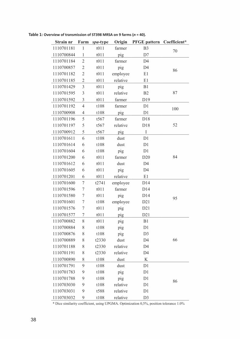
38
Table 1: Overview of transmission of ST398 MRSA on 9 farms (n = 40).
Strain nr Farm spa-type Origin PFGE pattern Coefficient* 1110701181 1 t011 farmer B3 701110700844 1 t011 pig D71110701184 2 t011 farmer D4
861110700857 2 t011 pig D41110701182 2 t011 employee E11110701185 2 t011 relative E11110701429 3 t011 pig B1
871110701595 3 t011 relative B21110701592 3 t011 farmer D191110701192 4 t108 farmer D1 1001110700908 4 t108 pig D11110701196 5 t567 farmer D18
521110701197 5 t567 relative D181110700912 5 t567 pig I1110701611 6 t108 dust D1
84
1110701614 6 t108 dust D11110701604 6 t108 pig D11110701200 6 t011 farmer D201110701612 6 t011 dust D41110701605 6 t011 pig D41110701201 6 t011 relative E11110701600 7 t2741 employee D14
95
1110701596 7 t011 farmer D141110701580 7 t011 pig D141110701601 7 t108 employee D211110701576 7 t011 pig D211110701577 7 t011 pig D211110700882 8 t011 pig B1
66
1110700884 8 t108 pig D11110700876 8 t108 pig D31110700889 8 t2330 dust D41110701188 8 t2330 relative D41110701191 8 t2330 relative D41110700890 8 t108 dust K1110701791 9 t108 dust D1
86
1110701783 9 t108 pig D11110701788 9 t108 pig D11110703030 9 t108 relative D11110703031 9 t588 relative D11110703032 9 t108 relative D3* Dice similarity coefficient, using UPGMA. Optimization 0,5%, position tolerance 1.0%
39
3
PFGE
div
ersit
y w
ithin
MRS
A ST
398
DiscussionMRSA isolates belonging to the ST398 clonal lineage are hard to discriminate based on spa-typing and/or MLST, hampering the assessment of transmission and outbreaks. Therefore, other techniques such as a modified PFGE could provide a new opportunity to differentiate ST398 isolates. The restriction enzyme SmaI does not cut the DNA of NTSmaI -MRSA isolates, due to methylation of the SmaI site. However, Cfr9I, a neoschizomer of SmaI, can be used for generating PFGE profiles of the NTSmaI -MRSA isolates. When the standard SmaI protocol was used for Cfr9I, banding patterns with smears and partial digests appeared. Other recently published articles seemed to have encountered similar problems with their Cfr9I PFGE [18, 25]. The results indicated that lysis of ST398 isolates and digestion with restriction enzyme Cfr9I is more cumbersome than lysis of typeable MRSA and digestion with SmaI [29]. After modifying the protocol, banding patterns of similar quality as those of typeable MRSA isolates digested with SmaI were obtained. All previously non-typeable MRSA isolates can be typed with the optimized PFGE method providing a new opportunity to differentiate the ST398 clonal lineage. From April 2002 until January 2008, all MRSA isolates sent to the RIVM have been typed with PFGE using SmaI as restriction enzyme creating a database with more than 4000 isolates with over 700 different PFGE types. Since Cfr9I recognizes the same restriction site as SmaI, Cfr9I enables analysis and comparison of the patterns with other profiles in our database. No comparison was found when comparing banding patterns of NTSmaI -MRSA with known PFGE patterns, suggesting that SmaI restriction modification is confined to a defined clonal lineage. Recently, ST398 isolates were typed using amplified fragment length polymorphism (AFLP). These data also suggested that ST398 is a distinct cluster recently introduced into the Dutch patient population [30].The PFGE patterns of the two most prevalent spa-types (t011 and t108) within the NTSmaI -MRSA isolates showed more variation than spa-typing or MLST. The genetic diversity within the ST398 clonal lineage of MRSA sharing the same spa-type creates an opportunity for improved investigation of outbreak and potential transmission events. Spa-typing, which is currently used as a MRSA typing standard, cannot differentiate these isolates further. Using Cfr9I PFGE, spa-type t011 seemed to be more diverse than t108. Although the minimal similarity of the t108 isolates was 50%, this was mainly caused by a single isolate with a very distinct PFGE pattern (pattern H). Without this isolate the minimal similarity of the t108 isolates was 80%. The t011 isolates showed a minimal similarity of 64% (data not shown). SCCmec typing showed an almost equal distribution between SCCmec type IV (n = 14) and V (n = 16) for t011 isolates, whereas all t108 isolates carried SCCmec type V or a SCCmec type V variant. Huijsdens and colleagues performed SCCmec typing on 300 NTSmaI –MRSA isolates and they showed similar results [23]. This variation in SCCmec types may also indicates a higher diversity among t011 MRSA isolates compared to t108 isolates. The minimal similarity of the Cfr9I PFGE patterns among ST398 isolates was 35% and showed variation within spa -types, but the diversity within this lineage is still limited. Furthermore, one isolate with spa-type t108 yielded a very distinct PFGE pattern which causes the similarity to be 35% (Figure 1). When excluding this isolate from the dendrogram the minimal similarity was 62%. Comparing the PFGE results using the criteria by Tenover et al. and when a similarity cut-off of 80% was applied, most NTSmaI -MRSA isolates should be classified as one PFGE cluster [31, 32]. However, the Cfr9I PFGE is still better in discriminating possible differences between NTSmaI -MRSA isolates. No geographical relation could be found in either spa-type. However, most NTSmaI -MRSA isolates are found in areas with the highest pig density. This could be explained by the frequent movement of pigs between farms in the Netherlands. This facilitates the dissemination of ST398 MRSA on a national scale. A similar situation took place during the foot- and -mouth epidemic in England of 2001 [33]. To provide additional resolution on the molecular evolution and dissemination of MRSA lineages, several typing techniques such as PFGE, SCCmec- and spa-typing have been developed. Since PFGE with SmaI does not digest the DNA of ST398 isolates, spa-typing has been the method of choice for characterizing NTSmaI -MRSA isolates. However, given the low diversity in spa-types it is hard to ascertain health care-associated transmission if two or more different spa-types are present in the
40
same institution. Fanoy et al. described an outbreak in a residential care facility where two spa-types (t2383 and t011) were prevalent [18]. After re-examination of the same isolates the PFGE profiles using Cfr9I were indistinguishable, indicating isogenicity. Moreover, the discriminatory ability of spa-typing of NTSmaI -MRSA is compromised by the fact that more than 80% of the NTSmaI -MRSA in the Netherlands belong either to spa-type t011 or t108 [23]. With the modified Cfr9I PFGE a better tool for epidemiological investigation has become available. The results obtained by Cfr9I PFGE of isolates from veterinarians and their close family members showed possible transmission of ST398. Five out of eight pairs had identical profiles. The family members had themselves no contact with animals and were presumably infected by the occupationally exposed veterinarian. Two pairs of PFGE patterns among family members were not identical. Their isolates also had different spa-types. Family members may have been colonized by one MRSA through the veterinarian and subsequently the veterinarian may have been re-colonized by another MRSA after occupational exposure. One pair differed only in a single PFGE band probably as a consequence of micro-evolution. A study on nine different farms revealed that the PFGE patterns of isolates from seven farms were related, but PFGE patterns varied within and between the farms. For example, farm 7, yielded only 2 very closely related PFGE patterns (D14, D21; similarity 95%), while other farms, like farm 8, showed 5 different PFGE patterns (B1, D1, D3, D4 and K) and had a similarity of only 66%. Different batches of animals entering the farm, carrying different NTSmaI -MRSA, could have caused variation within farms. Further study is needed to confirm that farms with a fast turnover of pigs indeed show a higher diversity of PFGE patterns of NTSmaI -MRSA.
ConclusionIn conclusion, the modified PFGE protocol for Cfr9I provided highly informative banding patterns and showed good reproducibility. The PFGE results showed diversity within and between the two most prevalent spa-types among NTSmaI -MRSA. PFGE confirmed transmission of the ST398 clonal lineage within families and in a residential care facility. The modified PFGE approach can be used as a method for selecting important and distinct ST398 isolates for further research. The adjustments in the PFGE protocol using Cfr9I are easy to implement in laboratories which already have a PFGE facility, creating a powerful tool to study the ST398 clonal lineage.
References1. Vandenesch F, Naimi T, Enright MC, Lina G, Nimmo GR, Heffernan H, et al. Community-acquired methicillin-resistant Staphylococcus aureus carrying Panton-Valentine leukocidin genes: worldwide emergence. Emerging infectious diseases. 2003;9(8):978-84.2. Zetola N, Francis JS, Nuermberger EL, Bishai WR. Community-acquired meticillin-resistant Staphylococcus aureus: an emerging threat. Lancet Infect Dis. 2005;5(5):275-86.3. de Neeling AJ, van den Broek MJ, Spalburg EC, van Santen-Verheuvel MG, Dam-Deisz WD, Boshuizen HC, et al. High prevalence of methicillin resistant Staphylococcus aureus in pigs. Veterinary microbiology. 2007;122(3-4):366-72.4. Huijsdens XW, van Dijke BJ, Spalburg E, van Santen-Verheuvel MG, Heck ME, Pluister GN, et al. Community-acquired MRSA and pig-farming. Annals of clinical microbiology and antimicrobials. 2006;5:26.5. van den Broeck IV, van Cleef BA, Haenen A, Broens EM, van der Wolf PJ, van den Broek MJ, et al. Methicillin-resistant Staphylococcus aureus in people living and working in pig farms. Epidemiol Infect. 2008:1-9.6. Khanna T, Friendship R, Dewey C, Weese JS. Methicillin resistant Staphylococcus aureus colonization in pigs and pig farmers. Veterinary microbiology. 2008;128(3-4):298-303.7. Voss A, Loeffen F, Bakker J, Klaassen C, Wulf M. Methicillin-resistant Staphylococcus aureus in pig farming. Emerging infectious diseases. 2005;11(12):1965-6.8. Cuny C, Strommenger B, Witte W, Stanek C. Clusters of infections in horses with MRSA ST1, ST254, and ST398 in a veterinary hospital. Microb Drug Resist. 2008;14(4):307-10.9. Witte W, Strommenger B, Stanek C, Cuny C. Methicillin-resistant Staphylococcus aureus ST398 in humans and animals, Central Europe. Emerging infectious diseases. 2007;13(2):255-8.10. Persoons D, Van Hoorebeke S, Hermans K, Butaye P, de Kruif A, Haesebrouck F, et al. Methicillin-resistant Staphylococcus aureus in poultry. Emerg Infect Dis. 2009;15(3):452-3.11. Mooij T TA, Jenkins J, Thijssen S. MRSA in calves. Infectieziekten Bulletin. 2007;18:234-6.12. Armand-Lefevre L, Ruimy R, Andremont A. Clonal comparison of Staphylococcus aureus isolates from healthy pig

41
3
PFGE
div
ersit
y w
ithin
MRS
A ST
398
farmers, human controls, and pigs. Emerging infectious diseases. 2005;11(5):711-4.13. Guardabassi L, Stegger M, Skov R. Retrospective detection of methicillin resistant and susceptible Staphylococcus aureus ST398 in Danish slaughter pigs. Veterinary microbiology. 2007;122(3-4):384-6.14. Meemken D, Cuny C, Witte W, Eichler U, Staudt R, Blaha T. [Occurrence of MRSA in pigs and in humans involved in pig production--preliminary results of a study in the northwest of Germany]. Dtsch Tierarztl Wochenschr. 2008;115(4):132-9.15. Smith TC, Male MJ, Harper AL, Kroeger JS, Tinkler GP, Moritz ED, et al. Methicillin-resistant Staphylococcus aureus (MRSA) strain ST398 is present in midwestern U.S. swine and swine workers. PLoS ONE. 2008;4(1):e4258.16. Ekkelenkamp MB, Sekkat M, Carpaij N, Troelstra A, Bonten MJ. [Endocarditis due to meticillin-resistant Staphylococcus aureus originating from pigs]. Ned Tijdschr Geneeskd. 2006;150(44):2442-7.17. Yu F, Chen Z, Liu C, Zhang X, Lin X, Chi S, et al. Prevalence of Staphylococcus aureus carrying Panton-Valentine leukocidin genes among isolates from hospitalised patients in China. Clinical microbiology and infection : the official publication of the European Society of Clinical Microbiology and Infectious Diseases. 2008;14(4):381-4.18. Fanoy E, Helmhout LC, van der Vaart WL, Weijdema K, van Santen-Verheuvel MG, Thijsen SF, et al. An outbreak of non-typeable MRSA within a residential care facility. Euro surveillance : bulletin Europeen sur les maladies transmissibles = European communicable disease bulletin. 2009;14(1).19. Kaufmann ME. Pulsed-field gel electrophoresis. Totowa N.J.: Humana press; 1998. 33-50 p.20. Bens CC, Voss A, Klaassen CH. Presence of a novel DNA methylation enzyme in methicillin-resistant Staphylococcus aureus isolates associated with pig farming leads to uninterpretable results in standard pulsed-field gel electrophoresis analysis. Journal of clinical microbiology. 2006;44(5):1875-6.21. Frenay HM, Bunschoten AE, Schouls LM, van Leeuwen WJ, Vandenbroucke-Grauls CM, Verhoef J, et al. Molecular typing of methicillin-resistant Staphylococcus aureus on the basis of protein A gene polymorphism. European journal of clinical microbiology & infectious diseases : official publication of the European Society of Clinical Microbiology. 1996;15(1):60-4.22. Harmsen D, Claus H, Witte W, Rothganger J, Claus H, Turnwald D, et al. Typing of methicillin-resistant Staphylococcus aureus in a university hospital setting by using novel software for spa repeat determination and database management. Journal of clinical microbiology. 2003;41(12):5442-8.23. Xander W. Huijsdens TB, Marga G. van Santen-Verheuvel, Emile Spalburg, Gerlinde N. Pluister, Martijn van Luit, Max E.O.C. Heck, Anja Haenen, Albert J. de Neeling. Molecular characterization of PFGE non-typeable methicillin-resistant Staphylococcus aureus in the Netherlands. Eurosurveillance. 2009;submitted for press.24. Schouls LM, Spalburg EC, van Luit M, Huijsdens XW, Pluister GN, van Santen-Verheuvel MG, et al. Multiple-locus variable number tandem repeat analysis of Staphylococcus aureus: comparison with pulsed-field gel electrophoresis and spa-typing. PloS one. 2009;4(4):e5082.25. Bhat M, Dumortier C, Taylor BS, Miller M, Vasquez G, Yunen J, et al. Staphylococcus aureus ST398, New York City and Dominican Republic. Emerging infectious diseases. 2009;15(2):285-7.26. Murchan S, Kaufmann ME, Deplano A, de Ryck R, Struelens M, Zinn CE, et al. Harmonization of pulsed-field gel electrophoresis protocols for epidemiological typing of strains of methicillin-resistant Staphylococcus aureus: a single approach developed by consensus in 10 European laboratories and its application for tracing the spread of related strains. Journal of clinical microbiology. 2003;41(4):1574-85.27. Boye K, Bartels MD, Andersen IS, Moller JA, Westh H. A new multiplex PCR for easy screening of methicillin-resistant Staphylococcus aureus SCCmec types I-V. Clinical microbiology and infection : the official publication of the European Society of Clinical Microbiology and Infectious Diseases. 2007;13(7):725-7.28. McDougal LK, Steward CD, Killgore GE, Chaitram JM, McAllister SK, Tenover FC. Pulsed-field gel electrophoresis typing of oxacillin-resistant Staphylococcus aureus isolates from the United States: establishing a national database. J Clin Microbiol. 2003;41(11):5113-20.29. Siksnys V, Pleckaityte M. Catalytic and binding properties of restriction endonuclease Cfr9I. Eur J Biochem. 1993;217(1):411-9.30. van Belkum A, Melles DC, Peeters JK, van Leeuwen WB, van Duijkeren E, Huijsdens XW, et al. Methicillin-resistant and -susceptible Staphylococcus aureus sequence type 398 in pigs and humans. Emerg Infect Dis. 2008;14(3):479-83.31. Struelens MJ, Deplano A, Godard C, Maes N, Serruys E. Epidemiologic typing and delineation of genetic relatedness of methicillin-resistant Staphylococcus aureus by macrorestriction analysis of genomic DNA by using pulsed-field gel electrophoresis. J Clin Microbiol. 1992;30(10):2599-605.32. Tenover FC, Arbeit RD, Goering RV, Mickelsen PA, Murray BE, Persing DH, et al. Interpreting chromosomal DNA restriction patterns produced by pulsed-field gel electrophoresis: criteria for bacterial strain typing. J Clin Microbiol. 1995;33(9):2233-9.33. Alexandersen S, Zhang Z, Donaldson AI, Garland AJ. The pathogenesis and diagnosis of foot-and-mouth disease. J Comp Pathol. 2003;129(1):1-36.
42
Outbreak of methicillin-resistant Staphylococcus aureus ST398 in a Dutch nursing home
4Thijs Bosch
Yvonne Hendriks
Jan Kluytmans
Erwin Verkade
Infection control and hospital epidemiology
2012
33(6): 624-626
43
4
Out
brea
k of
MRS
A ST
398
in a
nur
sing
hom
e
AbstractWe describe an outbreak of methicillin-resistant Staphylococcus aureus (MRSA) ST398 in a nursing home in the Netherlands. Seven residents and 4 healthcare workers were identified with MRSA ST398, but 2 of the healthcare workers carried other strains. This study demonstrates that MRSA ST398 can spread in nursing homes.
44
IntroductionTraditionally, methicillin-resistant Staphylococcus aureus (MRSA) has been considered a hospital-associated pathogen. Recently, MRSA has expanded its territory to the community, causing severe infections in previously healthy persons all over the world [1]. In 2003, a new clone of MRSA was identified that was related to an extensive reservoir found in pigs and veal calves [2, 3]. People who are in direct contact with pigs and veal calves have a high carriage rate of this MRSA (23% and 29%, respectively) [2, 4]. Using multilocus sequence typing (MLST), the vast majority of these strains belong to sequence type 398 (ST398). Transmission within families, as well as single cases of colonized healthcare workers, have been described [2, 5]. However, up to now there have been few reports of transmission of MRSA ST398 in healthcare set- tings. In the hospital setting, MRSA ST398 is reported to be less transmissible than other MRSA types [6]. We describe an outbreak of MRSA ST398 in a nursing home.
Material and methodsSettingThis is a prospective epidemiologic analysis of an outbreak of MRSA ST398 that occurred in a nursing home in the Netherlands from October 2010 to February 2011. The nursing home is located in the southeast of the Netherlands in a region with a high density of pigs (∼3,000 pigs per square kilometer). The nursing home consists of 3 separate wards, with a total of 51 residents living in individual units. Incident cases were defined as residents and healthcare workers with MRSA obtained from clinical cultures (ie, wound) or surveillance cultures (ie, anterior nares, throat, and perineum).
Outbreak InvestigationIn October 2010, MRSA was cultured from a wound on the leg of a resident. Subsequently, more extensive screening cultures of this resident were obtained in November 2010, which showed that he was also colonized in the throat, nose, and perineum. At the same time, another resident of the same ward had a wound culture with MRSA-positive test results. Subsequent screening in December 2010 of contacts among residents and healthcare workers of this ward revealed additional residents and healthcare workers with MRSA. Be- cause of the high prevalence of MRSA in this ward, a screening of the other 2 wards was performed in January 2011.
Infection Control MeasuresAccording to the current national guidelines for the control of MRSA in nursing homes, transmission-based precautions were taken when there was physical contact with residents who carried MRSA. This means that gowns and gloves were worn when contact with the residents or their equipment was anticipated.7 Also, instructions on hand hygiene were given. The healthcare workers who carried MRSA were temporarily suspended from work, and decolonization of all colonized subjects was initiated with mupirocin nasal ointment, chlorhexidine wash, and systemic treatment with clarithromycine and rifampicin.
Microbiologic MethodsNose, throat, and perineum swab samples were obtained from residents and healthcare workers. Samples were directly inoculated onto chromID MRSA (bioMerieux). In addition, broth enrichment containing Mueller-Hinton broth supplemented with 6.5% NaCl was inoculated using the same swabs. Direct-inoculated as well as overnight enriched–inoculated plates were read after 18–24 hours of incubation at 35°C–37°C. From the 11 individuals who were found to harbor MRSA, 16 MRSA isolates were genotyped by staphylococcal protein A (spa)-typing. In addition, all isolates were genotyped by pulsed-field gel electrophoresis (PFGE) using the restrictionenzyme Cfr9I according to previously described methods [8].
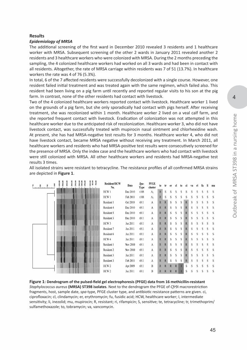
45
4
Out
brea
k of
MRS
A ST
398
in a
nur
sing
hom
e
Results Epidemiology of MRSAThe additional screening of the first ward in December 2010 revealed 3 residents and 1 healthcare worker with MRSA. Subsequent screening of the other 2 wards in January 2011 revealed another 2 residents and 3 healthcare workers who were colonized with MRSA. During the 2 months preceding the sampling, the 4 colonized healthcare workers had worked on all 3 wards and had been in contact with all residents. Altogether, the rate of MRSA carriage within residents was 7 of 51 (13.7%). In healthcare workers the rate was 4 of 76 (5.3%).In total, 6 of the 7 affected residents were successfully decolonized with a single course. However, one resident failed initial treatment and was treated again with the same regimen, which failed also. This resident had been living on a pig farm until recently and reported regular visits to his son at the pig farm. In contrast, none of the other residents had contact with livestock.Two of the 4 colonized healthcare workers reported contact with livestock. Healthcare worker 1 lived on the grounds of a pig farm, but she only sporadically had contact with pigs herself. After receiving treatment, she was recolonized within 1 month. Healthcare worker 2 lived on a veal calf farm, and she reported frequent contact with livestock. Eradication of colonization was not attempted in this healthcare worker due to the anticipated risk of recolonization. Healthcare worker 3, who did not have livestock contact, was successfully treated with mupirocin nasal ointment and chlorhexidine wash. At present, she has had MRSA-negative test results for 3 months. Healthcare worker 4, who did not have livestock contact, became MRSA negative without receiving any treatment. In March 2011, all healthcare workers and residents who had MRSA-positive test results were consecutively screened for the presence of MRSA. Only the index case and the healthcare workers who had contact with livestock were still colonized with MRSA. All other healthcare workers and residents had MRSA-negative test results 3 times.All isolated strains were resistant to tetracycline. The resistance profiles of all confirmed MRSA strains are depicted in Figure 1.
Figure 1: Dendrogram of the pulsed-field gel electrophoresis (PFGE) data from 16 methicillin-resistant Staphylococcus aureus (MRSA) ST398 isolates. Next to the dendrogram the PFGE of Cfr9I macrorestriction fragments, host, sample date, spa-type, PFGE cluster type, and antibiotic resistance patterns are given. ci, ciprofloxacin; cl, clindamycin; er, erythromycin; fu, fusidic acid; HCW, healthcare worker; I, intermediate sensitivity; li, inezolid; mu, mupirocin; R, resistant; ri, rifampicin; S, sensitive; te, tetracycline; tr, trimethoprim/sulfamethoxazole; to, tobramycin; va, vancomycin.
46
Molecular TypingRelatedness of the MRSA strains was confirmed by PFGE with Cfr9I restriction digestion in 12 of the 16 isolates [8]. Only the MRSA isolates originating from the 2 healthcare workers who reported livestock contact carried MRSA that had a different PFGE cluster type (Figure 1). Strains can also be subdivided into 3 different resistance profiles. Each PFGE cluster corresponds to a unique resistance profile.Moreover, spa-typing showed that 14 of the 16 strains were spa-type t011. Only the isolates originating from healthcare worker 1 were spa-type t108. Both spa-types are very frequently found within MRSA ST398.
Discussion To date, only one outbreak of MRSA ST398 in a Dutch hospital has been reported [9.] We report the first outbreak, to our knowledge, of MRSA ST398 in a nursing home that comprised 7 residents and 2 healthcare workers. The MRSA strain responsible for this outbreak was spa-type t011, which belongs to MLST type ST398. The most likely source for this outbreak was the 98-year-old male resident number 3. The index case had been living on a pig farm until recently, before he moved to the nursing home. He reported regular visits to his son at the pig farm. We assume that healthcare workers transmitted the outbreak strain to other residents because the index case did not have direct contact with the other MRSA-positive residents. Moreover, there was repeated intense physical contact between colonized healthcare workers and the index case due to his obesity and immobility. Furthermore, none of the other colonized residents had contact with pigs or veal calves. Although we did not assess the compliance to hand hygiene of healthcare workers, this is generally low in nursing homes and may have contributed to the spread of MRSA. When the outbreak was detected, the importance of hand hygiene was communicated to all healthcare workers. Hand sanitizer dispensers were placed at the entrances of all patients’ rooms. By doing this, the compliance to proper hand hygiene was probably increased.Two additional healthcare workers had MRSA-positive test results during the outbreak period, but they carried other strains. These healthcare workers reported contact with livestock and had worked for a long time in the nursing home. One of the healthcare workers who had contact with livestock had a similar spa-type of the outbreak-related strain, but the PFGE pattern was clearly different and the resistance profile also showed major differences. We concluded that they were not involved in this outbreak on the basis of these differences. The MRSA ST398 strains isolated from these healthcare workers were not found in any other residents, who all had been screened. This suggests that healthcare workers who are colonized with MRSA ST398 and comply with proper hygiene precautions are not a significant risk for transmission. It is unclear whether host adaptation of this animal-derived strain plays a role in its transmissibility.In conclusion, several studies have demonstrated that transmissibility of MRSA ST398 is probably lower than hospital- associated MRSA strains [5, 6]. However, this outbreak of MRSA ST398 in a community setting shows that substantial human- to-human transmission can occur. Further adaptation to humans may occur, and if MRSA ST398 can successfully spread from human to human, it may pose a significant public health problem in the future. Therefore, careful monitoring of the evolution and epidemiology of MRSA ST398 is important.
References1. Kluytmans-Vandenbergh MF, Kluytmans JA. Community-acquired methicillin-resistant Staphylococcus aureus: current perspectives. Clinical microbiology and infection : the official publication of the European Society of Clinical Microbiology and Infectious Diseases. 2006;12 Suppl 1:9-15.2. Voss A, Loeffen F, Bakker J, Klaassen C, Wulf M. Methicillin-resistant Staphylococcus aureus in pig farming. Emerging infectious diseases. 2005;11(12):1965-6.3. van Loo I, Huijsdens X, Tiemersma E, de Neeling A, van de Sande-Bruinsma N, Beaujean D, et al. Emergence of methicillin-resistant Staphylococcus aureus of animal origin in humans. Emerging infectious diseases. 2007;13(12):1834-9.4. van den Broeck IV, van Cleef BA, Haenen A, Broens EM, van der Wolf PJ, van den Broek MJ, et al. Methicillin-resistant Staphylococcus aureus in people living and working in pig farms. Epidemiology and infection. 2009;137(5):700-8.5. Cuny C, Nathaus R, Layer F, Strommenger B, Altmann D, Witte W. Nasal colonization of humans with methicillin-resistant Staphylococcus aureus (MRSA) CC398 with and without exposure to pigs. PloS one. 2009;4(8):e6800.
47
4
Out
brea
k of
MRS
A ST
398
in a
nur
sing
hom
e
6. Wassenberg MW, Bootsma MC, Troelstra A, Kluytmans JA, Bonten MJ. Transmissibility of livestock-associated methicillin-resistant Staphylococcus aureus (ST398) in Dutch hospitals. Clinical microbiology and infection : the official publication of the European Society of Clinical Microbiology and Infectious Diseases. 2011;17(2):316-9.7. WIP guidelines, MRSA in nursing homes and homecare. 2007.8. Bosch T, de Neeling AJ, Schouls LM, van der Zwaluw KW, Kluytmans JA, Grundmann H, et al. PFGE diversity within the methicillin-resistant Staphylococcus aureus clonal lineage ST398. BMC microbiology. 2010;10:40.9. Wulf MW, Markestein A, van der Linden FT, Voss A, Klaassen C, Verduin CM. First outbreak of methicillin-resistant Staphylococcus aureus ST398 in a Dutch hospital, June 2007. Euro surveillance : bulletin Europeen sur les maladies transmissibles = European communicable disease bulletin. 2008;13(9).
48
High resolution typing by whole genome mapping enables discrimination of LA-MRSA (CC398) strains
and identification of transmission events
5
Thijs Bosch
Martijn van Luit
Leo Schouls
Jan Kluytmans
Erwin Verkade
Bruno Pot
Plos One
2013
8(6): e66493
Paul Vauterin
Ronald Burggrave
Paul Savelkoul
49
5
WGM
ena
bles
disc
rimin
ation
of
LA-M
RSA
AbstractAfter its emergence in 2003, a livestock-associated (LA-)MRSA clade (CC398) has caused an impressive increase in the number of isolates submitted for the Dutch national MRSA surveillance and now comprises 40% of all isolates. The currently used molecular typing techniques have limited discriminatory power for this MRSA clade, which hampers studies on the origin and transmission routes. Recently, a new molecular analysis technique named whole genome mapping was introduced. This method creates high-resolution, ordered whole genome restriction maps that may have potential for strain typing. In this study, we assessed and validated the capability of whole genome mapping to differentiate LA-MRSA isolates.Multiple validation experiments showed that whole genome mapping produced highly reproducible results. Assessment of the technique on two well-documented MRSA outbreaks showed that whole genome mapping was able to confirm one outbreak, but revealed major differences between the maps of a second, indicating that not all isolates belonged to this outbreak. Whole genome mapping of LA-MRSA isolates that were epidemiologically unlinked provided a much higher discriminatory power than spa-typing or MLVA. In contrast, maps created from LA-MRSA isolates obtained during a proven LA-MRSA outbreak were nearly indistinguishable showing that transmission of LA-MRSA can be detected by whole genome mapping. Finally, whole genome maps of LA-MRSA isolates originating from two unrelated veterinarians and their household members showed that veterinarians may carry and transmit different LA-MRSA strains at the same time. No such conclusions could be drawn based spa-typing and MLVA.Although PFGE seems to be suitable for molecular typing of LA-MRSA, WGM provides a much higher discriminatory power. Furthermore, whole genome mapping can provide a comparison with other maps within 2 days after the bacterial culture is received, making it suitable to investigate transmission events and outbreaks caused by LA-MRSA.
50
IntroductionStaphylococcus aureus and in particular methicillin resistant S. aureus (MRSA) is a bacterial pathogen that is associated with serious hospital- and community-acquired infections [1,2]. In the Netherlands, the incidence of MRSA infections is still low due to the restricted use of antibiotics and the successful implementation of the ‘search and destroy’ policy. However, the number of MRSA isolates sent to the national institute for public health and the environment (RIVM) in the context of the national MRSA surveillance, has been gradually increasing in the last years [3]. This increase is mainly caused by the emergence of a new single MRSA clade multi-locus sequence type ST398 originating from livestock, mainly pigs. ST398 was first described by Voss et al. in 2005 and since then ST398 has been found in numerous countries worldwide [4-8]. ST398 has been isolated from different types of domesticized animals and therefore is ST398 also known as livestock-associated MRSA (LA-MRSA) [9,10]. After the first reports in 2005, LA-MRSA (ST398) has spread very rapidly in the Netherlands and has become the predominant MRSA clade since 2007. In 2010, 38% of all isolates sent to the RIVM were LA-MRSA [3].Typing of LA-MRSA however, has turned out to be a challenge. One of its characteristics is that LA-MRSA are non-typeable with regular pulsed-field gel electrophoresis (PFGE) due to methylation of the SmaI recognition site [11]. In recent years, a number of reports have shown that PFGE with restriction enzyme Cfr9I, a neoschizomer of SmaI, can be used to overcome the problems with DNA-methylation [12,13]. Although PFGE with Cfr9I yields a relatively high discriminatory power for LA-MRSA isolates, PFGE remains a time-consuming, laborious and non-portable method. Other typing techniques, such as staphylococcal protein A (spa)-typing and multiple-locus variable number of tandem repeat analysis (MLVA), can be used to characterize LA-MRSA, but yield very limited discrimination within this clade. From 2008-2012, 17,869 MRSA isolates were characterized by spa-typing and MLVA within our national MRSA surveillance. The predominant MLVA complex was MC398, representing LA-MRSA and comprising of 7,066 isolates. Although 96 different spa-types and 78 different MLVA-types (MT) were found within this clade, the 2 dominant types, spa-type t011, MT398 (n=4093) and spa-type t108, MT572 (n=1282), accounted for 76% of all LA-MRSA isolates. In contrast, MC5 (MRSA, n=2520), the most isolated clade after MC398, yielded 113 different spa-types and 205 different MLVA-types with spa-type t003, MT130 (n=182) and spa-type t002, MT5 (n=126) as the predominant types, but accounting only for 12% of all MC5 isolates. Strains within MC398 show limited variability and the absence of a highly discriminating typing method to characterize MC398 (LA-MRSA) isolates has hampered studies on the origin and transmission routes of this MRSA clade. In 2007, a molecular analysis technique was introduced initially called optical mapping and now designated as whole genome mapping (WGM), although whole chromosome mapping would be more appropriate. A whole genome map is a high-resolution, ordered, whole genome restriction map and for S. aureus isolates these maps consists of 200–300 restriction fragments. In contrast, in PFGE of S. aureus only 10–15 non-ordered restriction fragments are used for the analysis [14,15]. Although Shukla et al. have previously successfully used WGM to characterize MRSA belonging to the USA300 clade [16], the number of reports in which WGM was used for molecular typing of bacterial pathogens is very limited [16-18]. In this study, we assessed and validated the capability of whole genome mapping to differentiate LA-MRSA isolates. For this purpose, we used epidemiologically related and non-related MRSA and LA-MRSA isolates.
Material and methodsStrain selectionFor this study a total of 18 MRSA and 45 LA-MRSA isolates were selected to create 84 different whole genome maps (WGMs). Two MRSA strains (NCTC8325, N315) and one LA-MRSA strain (S0385), which are often used as reference strains and for which published genomes are available [25-27], were used for reproducibility experiments and comparison of whole genome maps created in our laboratory with in silico maps. In addition, two LA-MRSA (MC398) isolated from Dutch veterinarians (VET78 (t=0m) and VET35 (t=0m)) were used for reproducibility experiments. The capability to identify transmission
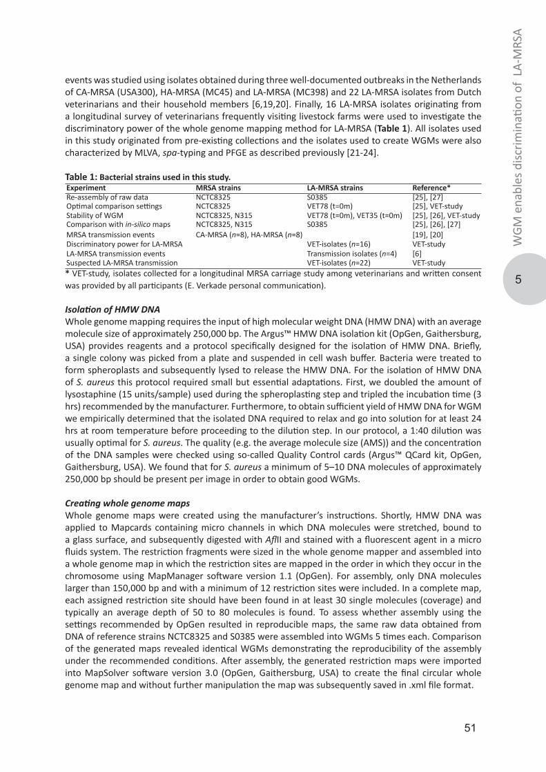
51
5
WGM
ena
bles
disc
rimin
ation
of
LA-M
RSA
events was studied using isolates obtained during three well-documented outbreaks in the Netherlands of CA-MRSA (USA300), HA-MRSA (MC45) and LA-MRSA (MC398) and 22 LA-MRSA isolates from Dutch veterinarians and their household members [6,19,20]. Finally, 16 LA-MRSA isolates originating from a longitudinal survey of veterinarians frequently visiting livestock farms were used to investigate the discriminatory power of the whole genome mapping method for LA-MRSA (Table 1). All isolates used in this study originated from pre-existing collections and the isolates used to create WGMs were also characterized by MLVA, spa-typing and PFGE as described previously [21-24].
Table 1: Bacterial strains used in this study.Experiment MRSA strains LA-MRSA strains Reference*Re-assembly of raw data NCTC8325 S0385 [25], [27]Optimal comparison settings NCTC8325 VET78 (t=0m) [25], VET-studyStability of WGM NCTC8325, N315 VET78 (t=0m), VET35 (t=0m) [25], [26], VET-studyComparison with in-silico maps NCTC8325, N315 S0385 [25], [26], [27]MRSA transmission events CA-MRSA (n=8), HA-MRSA (n=8) [19], [20]Discriminatory power for LA-MRSA VET-isolates (n=16) VET-studyLA-MRSA transmission events Transmission isolates (n=4) [6]Suspected LA-MRSA transmission VET-isolates (n=22) VET-study* VET-study, isolates collected for a longitudinal MRSA carriage study among veterinarians and written consent was provided by all participants (E. Verkade personal communication).
Isolation of HMW DNA Whole genome mapping requires the input of high molecular weight DNA (HMW DNA) with an average molecule size of approximately 250,000 bp. The Argus™ HMW DNA isolation kit (OpGen, Gaithersburg, USA) provides reagents and a protocol specifically designed for the isolation of HMW DNA. Briefly, a single colony was picked from a plate and suspended in cell wash buffer. Bacteria were treated to form spheroplasts and subsequently lysed to release the HMW DNA. For the isolation of HMW DNA of S. aureus this protocol required small but essential adaptations. First, we doubled the amount of lysostaphine (15 units/sample) used during the spheroplasting step and tripled the incubation time (3 hrs) recommended by the manufacturer. Furthermore, to obtain sufficient yield of HMW DNA for WGM we empirically determined that the isolated DNA required to relax and go into solution for at least 24 hrs at room temperature before proceeding to the dilution step. In our protocol, a 1:40 dilution was usually optimal for S. aureus. The quality (e.g. the average molecule size (AMS)) and the concentration of the DNA samples were checked using so-called Quality Control cards (Argus™ QCard kit, OpGen, Gaithersburg, USA). We found that for S. aureus a minimum of 5–10 DNA molecules of approximately 250,000 bp should be present per image in order to obtain good WGMs.
Creating whole genome mapsWhole genome maps were created using the manufacturer’s instructions. Shortly, HMW DNA was applied to Mapcards containing micro channels in which DNA molecules were stretched, bound to a glass surface, and subsequently digested with AflII and stained with a fluorescent agent in a micro fluids system. The restriction fragments were sized in the whole genome mapper and assembled into a whole genome map in which the restriction sites are mapped in the order in which they occur in the chromosome using MapManager software version 1.1 (OpGen). For assembly, only DNA molecules larger than 150,000 bp and with a minimum of 12 restriction sites were included. In a complete map, each assigned restriction site should have been found in at least 30 single molecules (coverage) and typically an average depth of 50 to 80 molecules is found. To assess whether assembly using the settings recommended by OpGen resulted in reproducible maps, the same raw data obtained from DNA of reference strains NCTC8325 and S0385 were assembled into WGMs 5 times each. Comparison of the generated maps revealed identical WGMs demonstrating the reproducibility of the assembly under the recommended conditions. After assembly, the generated restriction maps were imported into MapSolver software version 3.0 (OpGen, Gaithersburg, USA) to create the final circular whole genome map and without further manipulation the map was subsequently saved in .xml file format.
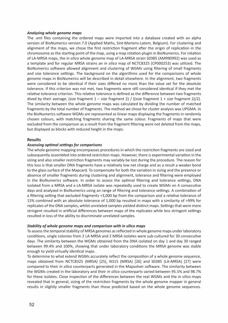
52
Analyzing whole genome mapsThe .xml files containing the ordered maps were imported into a database created with an alpha version of BioNumerics version 7.0 (Applied Maths, Sint-Martens-Latem, Belgium). For clustering and alignment of the maps, we chose the first restriction fragment after the origin of replication in the chromosome as the starting point of the map, using a map rotation plugin in BioNumerics. For rotation of LA-MRSA maps, the in silico whole genome map of LA-MRSA strain S0385 (AM990992) was used as a template and for regular MRSA strains an in silico map of NCTC8325 (CP000253) was utilized. The BioNumerics software allowed alignment and clustering of WGMs using filtering of small fragments and size tolerance settings. The background on the algorithms used for the comparisons of whole genome maps in BioNumerics will be described in detail elsewhere. In the alignment, two fragments were considered to be identical if their sizes differed no more than the value set for the absolute tolerance. If this criterion was not met, two fragments were still considered identical if they met the relative tolerance criterion. This relative tolerance is defined as the difference between two fragments dived by their average; (size fragment 1 – size fragment 2) / ((size fragment 1 + size fragment 2)/2). The similarity between the whole genome maps was calculated by dividing the number of matched fragments by the total number of fragments. The method we chose for cluster analysis was UPGMA. In the BioNumerics software WGMs are represented as linear maps displaying the fragments in randomly chosen colours, with matching fragments sharing the same colour. Fragments of maps that were excluded from the comparison as a result from the fragment filtering were not deleted from the maps, but displayed as blocks with reduced height in the maps.
ResultsAssessing optimal settings for comparisons The whole genome mapping encompasses procedures in which the restriction fragments are sized and subsequently assembled into ordered restriction maps. However, there is experimental variation in the sizing and also smaller restriction fragments may variably be lost during the procedure. The reason for this loss is that smaller DNA fragments have a relatively low net charge and as a result a weaker bond to the glass surface of the Mapcard. To compensate for both the variation in sizing and the presence or absence of smaller fragments during clustering and alignment, tolerance and filtering were employed in the BioNumerics software. In order to assess the optimal filtering and tolerance settings, DNA isolated from a MRSA and a LA-MRSA isolate was repeatedly used to create WGMs on 4 consecutive days and analyzed in BioNumerics using an range of filtering and tolerance settings. A combination of a filtering setting that excluded fragments <3,000 bp from the comparison and a relative tolerance of 15% combined with an absolute tolerance of 1,000 bp resulted in maps with a similarity of >99% for replicates of the DNA samples, whilst unrelated samples yielded distinct maps. Settings that were more stringent resulted in artificial differences between maps of the replicates while less stringent settings resulted in loss of the ability to discriminate unrelated samples.
Stability of whole genome maps and comparison with in silico mapsTo assess the temporal stability of MRSA genomes as reflected in whole genome maps under laboratory conditions, single colonies from 2 LA-MRSA and 2 MRSA isolates were sub-cultured for 30 consecutive days. The similarity between the WGMs obtained from the DNA isolated on day 1 and day 30 ranged between 99.4% and 100%, showing that under laboratory conditions the MRSA genome was stable enough to yield virtually identical maps. To determine to what extend WGMs accurately reflect the composition of a whole genome sequence, maps obtained from NCTC8325 (MRSA) [25], N315 (MRSA) [26] and S0385 (LA-MRSA) [27] were compared to their in silico counterparts generated in the Mapsolver software. The similarity between the WGMs created in the laboratory and their in silico counterparts varied between 95.5% and 98.7% for these isolates. Close inspection of the differences between the real WGMs and the in silico maps revealed that in general, sizing of the restriction fragments by the whole genome mapper in general results in slightly smaller fragments than those predicted based on the whole genome sequences.
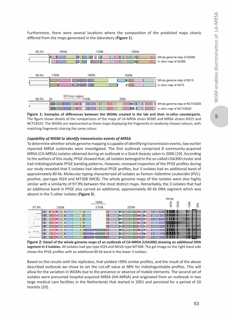
53
5
WGM
ena
bles
disc
rimin
ation
of
LA-M
RSA
Furthermore, there were several locations where the composition of the predicted maps clearly differed from the maps generated in the laboratory (Figure 1).
Figure 1: Examples of differences between the WGMs created in the lab and their in-silico counterparts. The figure shows details of the comparisons of the maps of LA-MRSA strain S0385 and MRSA strains N315 and NCTC8325. The WGMs are represented as linear maps displaying the fragments in randomly chosen colours, with matching fragments sharing the same colour.
Capability of WGM to identify transmission events of MRSATo determine whether whole genome mapping is capable of identifying transmission events, two earlier reported MRSA outbreaks were investigated. The first outbreak comprised 8 community-acquired MRSA (CA-MRSA) isolates obtained during an outbreak in a Dutch beauty salon in 2006 [19]. According to the authors of this study, PFGE showed that, all isolates belonged to the so-called USA300 cluster and had indistinguishable PFGE banding patterns. However, renewed inspection of the PFGE profiles during our study revealed that 5 isolates had identical PFGE profiles, but 3 isolates had an additional band of approximately 80 kb. Molecular typing characterized all isolates as Panton–Valentine Leukocidin (PVL)-positive, spa-type t024 and MT308 (MC8). The whole genome maps of the isolates were also highly similar with a similarity of 97.9% between the most distinct maps. Remarkably, the 3 isolates that had an additional band in PFGE also carried an additional, approximately 40 kb DNA segment which was absent in the 5 other isolates (Figure 2).
Figure 2: Detail of the whole genome maps of an outbreak of CA-MRSA (USA300) showing an additional DNA segment in 3 isolates. All isolates had spa-type t024 and MLVA-type MT308. The gel image on the right hand side shows the PFGE profiles with an additional 80 kb band in the lower 3 isolates.
Based on the results with the replicates, that yielded >99% similar profiles, and the result of the above described outbreak we chose to set the cut-off value at 98% for indistinguishable profiles. This will allow for the variation in WGMs due to the presence or absence of mobile elements. The second set of isolates were presumed hospital-acquired MRSA (HA-MRSA) and originated from an outbreak in two large medical care facilities in the Netherlands that started in 2001 and persisted for a period of 20 months [20].
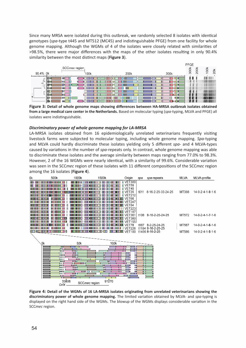
54
Since many MRSA were isolated during this outbreak, we randomly selected 8 isolates with identical genotypes (spa-type t445 and MT512 (MC45) and indistinguishable PFGE) from one facility for whole genome mapping. Although the WGMs of 4 of the isolates were closely related with similarities of >98.5%, there were major differences with the maps of the other isolates resulting in only 90.4% similarity between the most distinct maps (Figure 3).
Figure 3: Detail of whole genome maps showing differences between HA-MRSA outbreak isolates obtained from a large medical care center in the Netherlands. Based on molecular typing (spa-typing, MLVA and PFGE) all isolates were indistinguishable.
Discriminatory power of whole genome mapping for LA-MRSALA-MRSA isolates obtained from 16 epidemiologically unrelated veterinarians frequently visiting livestock farms were subjected to molecular typing, including whole genome mapping. Spa-typing and MLVA could hardly discriminate these isolates yielding only 5 different spa- and 4 MLVA-types caused by variations in the number of spa-repeats only. In contrast, whole genome mapping was able to discriminate these isolates and the average similarity between maps ranging from 77.0% to 98.3%. However, 2 of the 16 WGMs were nearly identical, with a similarity of 99.6%. Considerable variation was seen in the SCCmec region of these isolates with 11 different compositions of the SCCmec region among the 16 isolates (Figure 4).
Figure 4: Detail of the WGMs of 16 LA-MRSA isolates originating from unrelated veterinarians showing the discriminatory power of whole genome mapping. The limited variation obtained by MLVA- and spa-typing is displayed on the right hand side of the WGMs. The blowup of the WGMs displays considerable variation in the SCCmec region.
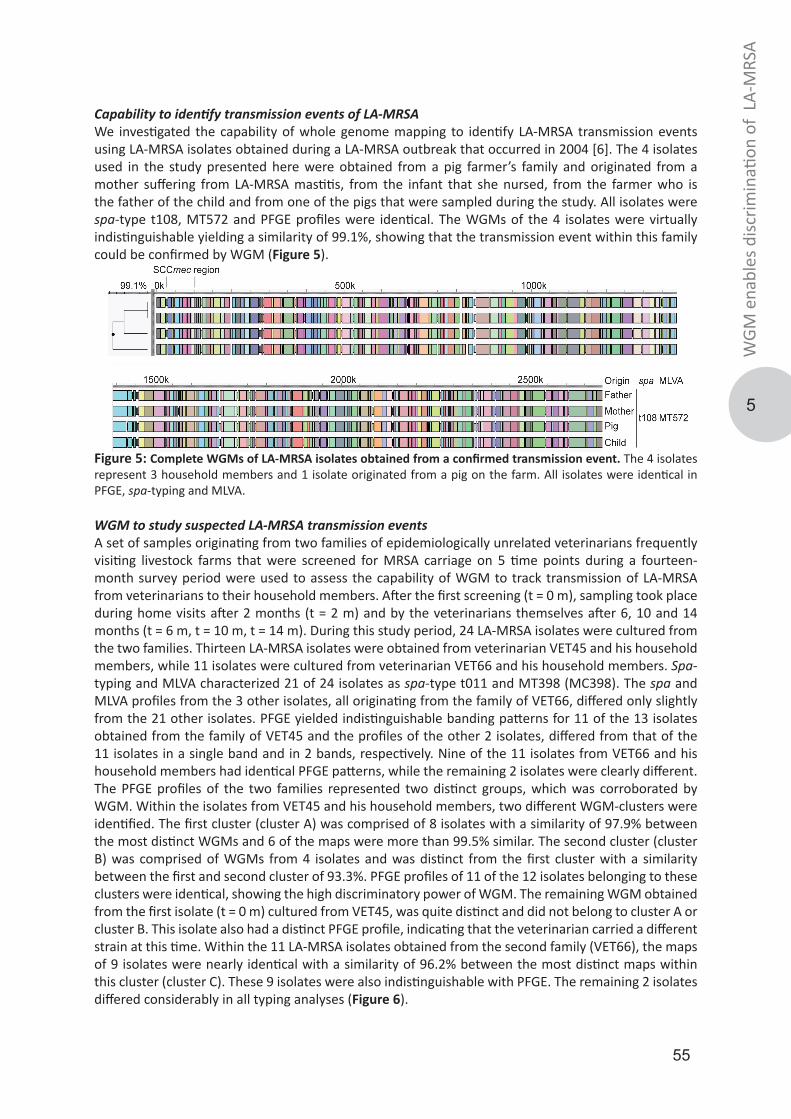
55
5
WGM
ena
bles
disc
rimin
ation
of
LA-M
RSA
Capability to identify transmission events of LA-MRSAWe investigated the capability of whole genome mapping to identify LA-MRSA transmission events using LA-MRSA isolates obtained during a LA-MRSA outbreak that occurred in 2004 [6]. The 4 isolates used in the study presented here were obtained from a pig farmer’s family and originated from a mother suffering from LA-MRSA mastitis, from the infant that she nursed, from the farmer who is the father of the child and from one of the pigs that were sampled during the study. All isolates were spa-type t108, MT572 and PFGE profiles were identical. The WGMs of the 4 isolates were virtually indistinguishable yielding a similarity of 99.1%, showing that the transmission event within this family could be confirmed by WGM (Figure 5).
Figure 5: Complete WGMs of LA-MRSA isolates obtained from a confirmed transmission event. The 4 isolates represent 3 household members and 1 isolate originated from a pig on the farm. All isolates were identical in PFGE, spa-typing and MLVA.
WGM to study suspected LA-MRSA transmission eventsA set of samples originating from two families of epidemiologically unrelated veterinarians frequently visiting livestock farms that were screened for MRSA carriage on 5 time points during a fourteen-month survey period were used to assess the capability of WGM to track transmission of LA-MRSA from veterinarians to their household members. After the first screening (t = 0 m), sampling took place during home visits after 2 months (t = 2 m) and by the veterinarians themselves after 6, 10 and 14 months (t = 6 m, t = 10 m, t = 14 m). During this study period, 24 LA-MRSA isolates were cultured from the two families. Thirteen LA-MRSA isolates were obtained from veterinarian VET45 and his household members, while 11 isolates were cultured from veterinarian VET66 and his household members. Spa-typing and MLVA characterized 21 of 24 isolates as spa-type t011 and MT398 (MC398). The spa and MLVA profiles from the 3 other isolates, all originating from the family of VET66, differed only slightly from the 21 other isolates. PFGE yielded indistinguishable banding patterns for 11 of the 13 isolates obtained from the family of VET45 and the profiles of the other 2 isolates, differed from that of the 11 isolates in a single band and in 2 bands, respectively. Nine of the 11 isolates from VET66 and his household members had identical PFGE patterns, while the remaining 2 isolates were clearly different. The PFGE profiles of the two families represented two distinct groups, which was corroborated by WGM. Within the isolates from VET45 and his household members, two different WGM-clusters were identified. The first cluster (cluster A) was comprised of 8 isolates with a similarity of 97.9% between the most distinct WGMs and 6 of the maps were more than 99.5% similar. The second cluster (cluster B) was comprised of WGMs from 4 isolates and was distinct from the first cluster with a similarity between the first and second cluster of 93.3%. PFGE profiles of 11 of the 12 isolates belonging to these clusters were identical, showing the high discriminatory power of WGM. The remaining WGM obtained from the first isolate (t = 0 m) cultured from VET45, was quite distinct and did not belong to cluster A or cluster B. This isolate also had a distinct PFGE profile, indicating that the veterinarian carried a different strain at this time. Within the 11 LA-MRSA isolates obtained from the second family (VET66), the maps of 9 isolates were nearly identical with a similarity of 96.2% between the most distinct maps within this cluster (cluster C). These 9 isolates were also indistinguishable with PFGE. The remaining 2 isolates differed considerably in all typing analyses (Figure 6).
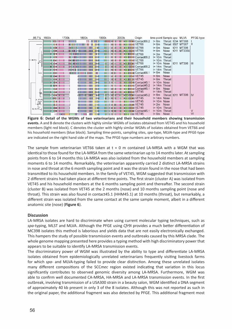
56
Figure 6: Detail of the WGMs of two veterinarians and their household members showing transmission events. A and B denote the clusters with highly similar WGMs of isolates obtained from VET45 and his household members (light red block). C denotes the cluster with highly similar WGMs of isolates obtained from VET66 and his household members (blue block). Sampling time-points, sampling sites, spa-type, MLVA-type and PFGE-type are indicated on the right hand side of the maps. The PFGE-type numbers are arbitrary numbers.
The sample from veterinarian VET66 taken at t = 0 m contained LA-MRSA with a WGM that was identical to those found for the LA-MRSA from the same veterinarian up to 14 months later. At sampling points from 6 to 14 months this LA-MRSA was also isolated from the household members at sampling moments 6 to 14 months. Remarkably, the veterinarian apparently carried 2 distinct LA-MRSA strains in nose and throat at the 6 month sampling point and it was the strain found in the nose that had been transmitted to its household members. In the family of VET45, WGM suggested that transmission with 2 different strains had taken place at different time points. The first strain (cluster A) was isolated from VET45 and his household members at the 6 months sampling point and thereafter. The second strain (cluster B) was isolated from VET45 at the 2 months (nose) and 10 months sampling point (nose and throat). This strain was also found in contact45.1 (HHM45.1) at 10 months (throat), but remarkably, a different strain was isolated from the same contact at the same sample moment, albeit in a different anatomic site (nose) (Figure 6).
DiscussionLA-MRSA isolates are hard to discriminate when using current molecular typing techniques, such as spa-typing, MLST and MLVA. Although the PFGE using Cfr9I provides a much better differentiation of MC398 isolates this method is laborious and yields data that are not easily electronically exchanged. This hampers the study of possible transmission events and outbreaks caused by this MRSA clade. The whole genome mapping presented here provides a typing method with high discriminatory power that appears to be suitable to identify LA-MRSA transmission events. The discriminatory power of WGM was illustrated by the ability to type and differentiate LA-MRSA isolates obtained from epidemiologically unrelated veterinarians frequently visiting livestock farms for which spa- and MLVA-typing failed to provide clear distinction. Among these unrelated isolates many different compositions of the SCCmec region existed indicating that variation in this locus significantly contributes to observed genomic diversity among LA-MRSA. Furthermore, WGM was able to confirm well documented CA-MRSA, HA-MRSA and LA-MRSA transmission events. In the first outbreak, involving transmission of a USA300 strain in a beauty salon, WGM identified a DNA segment of approximately 40 kb present in only 3 of the 8 isolates. Although this was not reported as such in the original paper, the additional fragment was also detected by PFGE. This additional fragment most
57
5
WGM
ena
bles
disc
rimin
ation
of
LA-M
RSA
likely represents a bacteriophage which usually has a genome size of approximately 40 kb and has the ability to jump in and out of bacterial genomes [28]. This shows that variation may occur within the same strain by gain or loss of mobile elements. Such events will lead to very localized changes whereas differences between distinct strains are the result of various genetic events and therefore in general occur scattered throughout the chromosome. This is an important criterion to decide whether two isolates may represent the same strain and thus might indicate the occurrence of a transmission event. WGM of isolates presumably belonging to a HA-MRSA outbreak in the Netherlands revealed that several isolates did not belong to the outbreak. Although, this multi-center outbreak expanded over a long period of time and involved many different patients, the isolates we selected for this study originated from one center and were received within 2 weeks of each other. We believe that we were unable to assign all isolates as part of the outbreak because multiple strains yielding the same molecular characteristics (spa-typing, MLVA and PFGE) were circulating at the time of the outbreak. Indeed in 2002, 19% (n=265) of all MRSA isolates sent to the RIVM had the PFGE-type that was identified as the outbreak-type. Of these isolates one-third originated from health care centers other than the two that were identified as the outbreak centers. This shows that the higher discriminatory power of WGM makes it possible to better assess whether isolates belong to an outbreak or not. We employed WGM to assess whether the technique is suitable to identify transmission events of LA-MRSA in a community setting (i.e. transmission from veterinarians to their household members). Indeed, we obtained virtually identical WGMs of the isolates obtained from the veterinarians and their household members. However, not all isolates were identical and two different clusters were identified among the isolates of one veterinarian and his household members. These results suggest that veterinarians may pick up different LA-MRSA strains during their visits to animal farms and become colonized for a longer time period. Apparently, veterinarians may carry different LA-MRSA strains in their nose and throat at the same time and both may be transmitted to their direct contacts. We conclude that WGM now enables us to identify transmission events of LA-MRSA which would be impossible using spa-typing or MLVA and with much more uncertainty when using PFGE. We are currently conducting WGM of isolates obtained from a larger number of veterinarians and their household members to study LA-MRSA transmission among this group in further detail.Based on the comparisons made of replicates of both MRSA and LA-MRSA isolates in this report and allowing for the presence of occasional mobile elements we now consider MRSA isolates with WGMs that have similarities of 98% or higher as indistinguishable. Isolates with WGMs with similarities between 95–98% may represent the same strain or should be regarded as highly related strains and those with maps that have similarities below 95% are deemed different strains. These cut-off values are supported by a recent report of Shukla et al. in which WGM of S. aureus was described and where a map distance of 5% was chosen as a cut-off point to define a WGM cluster [16]. This cut-off value was used for WGM of clonally related USA300 MRSA isolates using XbaI as restriction enzyme, but seems to be valid for LA-MRSA and other restriction enzymes as well. We do acknowledge that based on our cut-off criteria the in silico maps and the maps created in our lab would not be designated as identical as would be expected. Although showing good concordance, we found inconsistencies in the size, number and order of fragments between the WGMs created in the lab and the in silico maps. The fragments present in the in silico maps were generally larger, but that should have been compensated for by the tolerance settings. The subcultures of the reference strains from which DNA was isolated to generate the whole genome sequences which were used to create the in silico maps and the subcultures used in our lab to create the real WGMs were not identical. Since the isolates used for comparison of in silico and real maps may have been subcultured for many years in various laboratories, changes in the genome may have occurred over time and this might explain for some of the observed differences. Another possible explanation could be that either the whole genome sequences of the isolates used for generating in silico maps contain sequence errors or that the assembly of the WGMs is inaccurate. However, the reproducibility of the WGM procedure assessed by repeated mapping of the same DNA sample on 4 consecutive days turned out to be excellent. To exclude that subculture of the reference isolates caused the observed differences we are currently
58
assessing the complete genome sequences of 3 LA-MRSA isolates and we will compare the in silico maps based on these sequences with those obtained in the lab. Molecular typing is used to characterize pathogens like MRSA in order to provide evidence that will support epidemiological studies on transmission and outbreaks. Furthermore, it is used to study changes in bacterial population structures e.g. to assess the effects of human intervention such as widespread antibiotic treatment and vaccination. WGM seems to be suitable to type LA-MRSA and identify transmission events where existing typing methods usually fail. An alternative that was not included in the analysis, but is rapidly gaining interest as typing tool, is whole genome sequencing (WGS). WGS has many advantages over WGM e.g. it will be very difficult to infer phylogenetic relationships and assess population structures using WGM, whereas WGS is very well suited for these purposes. Furthermore, sequencing can potentially reveal all details on gene composition, the presence of virulence factors, etc. Although we believe that WGS is the ultimate typing method, there may be a number of drawbacks for outbreak investigations leaving a niche for methods like WGM. WGS has been suggested as the best tool for typing during outbreak investigations and a number of papers have been published supporting this claim [29-32]. However, these studies are all retrospective investigations and they do not yet show the utility of real time WGS. Due to the relatively simple data analysis, WGM can provide a comparison with other maps within 2 days after the bacterial culture has been received, making it suitable to investigate real time transmission events and outbreaks involving pathogens such as LA-MRSA. Furthermore, the generated WGM data can be stored as a table containing the ordered restriction fragments e.g. in .xml-format and therefore are easy accessible, allowing users to quickly exchange data and compare isolates.
References1. Emori TG, Gaynes RP. An overview of nosocomial infections, including the role of the microbiology laboratory. Clinical microbiology reviews. 1993;6(4):428-42.2. Steinberg JP, Clark CC, Hackman BO. Nosocomial and community-acquired Staphylococcus aureus bacteremias from 1980 to 1993: impact of intravascular devices and methicillin resistance. Clinical infectious diseases : an official publication of the Infectious Diseases Society of America. 1996;23(2):255-9.3. Haenen APJ, Huijsdens XW, Pluister GN, van Luit M, Bosch T, van Santen-Verheuvel MG et al. Surveillance van meticilline-resistente Staphylococcus aureus in Nederland in 2010. Infectieziekten bulletin. 2011;22(8):250-5.4. de Neeling AJ, van den Broek MJ, Spalburg EC, van Santen-Verheuvel MG, Dam-Deisz WD, Boshuizen HC, et al. High prevalence of methicillin resistant Staphylococcus aureus in pigs. Veterinary microbiology. 2007;122(3-4):366-72.5. Guardabassi L, Stegger M, Skov R. Retrospective detection of methicillin resistant and susceptible Staphylococcus aureus ST398 in Danish slaughter pigs. Veterinary microbiology. 2007;122(3-4):384-6.6. Huijsdens XW, van Dijke BJ, Spalburg E, van Santen-Verheuvel MG, Heck ME, Pluister GN, et al. Community-acquired MRSA and pig-farming. Annals of clinical microbiology and antimicrobials. 2006;5:26.7. Smith TC, Male MJ, Harper AL, Kroeger JS, Tinkler GP, Moritz ED, et al. Methicillin-resistant Staphylococcus aureus (MRSA) strain ST398 is present in midwestern U.S. swine and swine workers. PLoS One. 2009;4(1):e4258.8. Voss A, Loeffen F, Bakker J, Klaassen C, Wulf M. Methicillin-resistant Staphylococcus aureus in pig farming. Emerging infectious diseases. 2005;11(12):1965-6.9. Graveland H, Wagenaar JA, Heesterbeek H, Mevius D, van Duijkeren E, Heederik D. Methicillin resistant Staphylococcus aureus ST398 in veal calf farming: human MRSA carriage related with animal antimicrobial usage and farm hygiene. PloS one. 2010;5(6):e10990.10. Nemati M, Hermans K, Lipinska U, Denis O, Deplano A, Struelens M, et al. Antimicrobial resistance of old and recent Staphylococcus aureus isolates from poultry: first detection of livestock-associated methicillin-resistant strain ST398. Antimicrobial agents and chemotherapy. 2008;52(10):3817-9.11. Bens CC, Voss A, Klaassen CH. Presence of a novel DNA methylation enzyme in methicillin-resistant Staphylococcus aureus isolates associated with pig farming leads to uninterpretable results in standard pulsed-field gel electrophoresis analysis. Journal of clinical microbiology. 2006;44(5):1875-6.12. Argudin MA, Rodicio MR, Guerra B. The emerging methicillin-resistant Staphylococcus aureus ST398 clone can easily
59
5
WGM
ena
bles
disc
rimin
ation
of
LA-M
RSA
be typed using the Cfr9I SmaI-neoschizomer. Lett Appl Microbiol. 2010;50(1):127-30.13. van Wamel WJ, Hansenova Manaskova S, Fluit AC, Verbrugh H, de Neeling AJ, van Duijkeren E, et al. Short term micro-evolution and PCR-detection of methicillin-resistant and -susceptible Staphylococcus aureus sequence type 398. Eur J Clin Microbiol Infect Dis. 2010;29(1):119-22.14. Neely RK, Deen J, Hofkens J. Optical mapping of DNA: single-molecule-based methods for mapping genomes. Biopolymers. 2011;95(5):298-311.15. Riley MC, Kirkup BC, Jr., Johnson JD, Lesho EP, Ockenhouse CF. Rapid whole genome optical mapping of Plasmodium falciparum. Malar J. 2011;10:252.16. Shukla SK, Pantrangi M, Stahl B, Briska AM, Stemper ME, Wagner TK, et al. Comparative whole-genome mapping to determine Staphylococcus aureus genome size, virulence motifs, and clonality. Journal of clinical microbiology. 2012;50(11):3526-33.17. Shukla SK, Kislow J, Briska A, Henkhaus J, Dykes C. Optical mapping reveals a large genetic inversion between two methicillin-resistant Staphylococcus aureus strains. J Bacteriol. 2009;191(18):5717-23.18. Kotewicz ML, Jackson SA, LeClerc JE, Cebula TA. Optical maps distinguish individual strains of Escherichia coli O157 : H7. Microbiology. 2007;153(Pt 6):1720-33.19. Huijsdens XW, Janssen M, Renders NH, Leenders A, van Wijk P, van Santen Verheuvel MG, et al. Methicillin-resistant Staphylococcus aureus in a beauty salon, the Netherlands. Emerging infectious diseases. 2008;14(11):1797-9.20. Melles DC, Hendriks WD, Ott A, Verbrugh HA. [Outbreak of methicillin-resistant Staphylococcus aureus (MRSA) in the Rijnmond region: the largest outbreak in the Netherlands]. Ned Tijdschr Geneeskd. 2004;148(21):1038-43.21. Harmsen D, Claus H, Witte W, Rothganger J, Turnwald D, Vogel U. Typing of methicillin-resistant Staphylococcus aureus in a university hospital setting by using novel software for spa repeat determination and database management. Journal of clinical microbiology. 2003;41(12):5442-8.22. Schouls LM, Spalburg EC, van Luit M, Huijsdens XW, Pluister GN, van Santen-Verheuvel MG, et al. Multiple-locus variable number tandem repeat analysis of Staphylococcus aureus: comparison with pulsed-field gel electrophoresis and spa-typing. PloS one. 2009;4(4):e5082.23. Bosch T, de Neeling AJ, Schouls LM, van der Zwaluw KW, Kluytmans JA, Grundmann H, et al. PFGE diversity within the methicillin-resistant Staphylococcus aureus clonal lineage ST398. BMC microbiology. 2010;10:40.24. Murchan S, Kaufmann ME, Deplano A, de Ryck R, Struelens M, Zinn CE, et al. Harmonization of pulsed-field gel electrophoresis protocols for epidemiological typing of strains of methicillin-resistant Staphylococcus aureus: a single approach developed by consensus in 10 European laboratories and its application for tracing the spread of related strains. Journal of clinical microbiology. 2003;41(4):1574-85.25. Gillaspy AF, Worrel V, Orvis J, A. RB, Dyer DW, Iandolo JJ. Staphylococcus aureus NCTC8325 genome. Gram-positive pathogens ASM press. 2006:p381-412.26. Kuroda M, Ohta T, Uchiyama I, Baba T, Yuzawa H, Kobayashi I, et al. Whole genome sequencing of meticillin-resistant Staphylococcus aureus. Lancet. 2001;357(9264):1225-40.27. Schijffelen MJ, Boel CH, van Strijp JA, Fluit AC. Whole genome analysis of a livestock-associated methicillin-resistant Staphylococcus aureus ST398 isolate from a case of human endocarditis. BMC genomics. 2010;11:376.28. Novick RP, Christie GE, Penades JR. The phage-related chromosomal islands of Gram-positive bacteria. Nat Rev Microbiol. 2010;8(8):541-51.29. Diep BA. Use of whole-genome sequencing for outbreak investigations. Lancet Infect Dis. 2013;13(2):99-101.30. Harris SR, Cartwright EJ, Torok ME, Holden MT, Brown NM, Ogilvy-Stuart AL, et al. Whole-genome sequencing for analysis of an outbreak of meticillin-resistant Staphylococcus aureus: a descriptive study. The Lancet Infectious diseases. 2013;13(2):130-6.31. Reuter S, Harrison TG, Koser CU, Ellington MJ, Smith GP, Parkhill J, et al. A pilot study of rapid whole-genome sequencing for the investigation of a Legionella outbreak. BMJ Open. 2013;3(1).32. Walker TM, Ip CL, Harrell RH, Evans JT, Kapatai G, Dedicoat MJ, et al. Whole-genome sequencing to delineate Mycobacterium tuberculosis outbreaks: a retrospective observational study. Lancet Infect Dis. 2013;13(2):137-46.

60
6
Thijs Bosch
Martijn van Luit
Leo SchoulsJan Kluytmans
Erwin Verkade
Fabian Landman
Applied environmental microbiology
2015
81(1): 124-129
Transmission and persistence of livestock-associated MRSA among veterinarians and their household
members
61
6
Tran
smiss
ion
and
pers
istan
ce o
f LA-
MRS
A
AbstractAfter the first isolation of livestock-associated MRSA (LA-MRSA) in 2003, this MRSA variant quickly became the predominant MRSA obtained from humans in the Dutch national MRSA surveillance. Previous studies have suggested that human-to-human transmission of LA-MRSA rarely occurs compared to other MRSA lineages. However, these reports describe the transmission of LA-MRSA based on epidemiology and limited molecular characterization of isolates, making it difficult to assess whether transmission actually occurred. In this study, we used whole genome mapping (WGM) to identify possible transmission of LA-MRSA between humans. For this, we used LA-MRSA isolates originating from a 2-year prospective longitudinal cohort study in which livestock veterinarians and their household members were repeatedly sampled for the presence of S. aureus. Considerable degree of genotypic variation amongst LA-MRSA strains was observed. However, there was very limited variability between the maps of the isolates originating from the same veterinarian, indicating that each of the veterinarians persistently carried or had re-acquired the same LA-MRSA. Comparison of WGMs revealed that LA-MRSA transmission had likely occurred within virtually every veterinarian household. Yet, only a single LA-MRSA strain per household appeared to be involved in transmission. The results corroborate our previous finding that LA-MRSA is genetically diverse. Furthermore, this study shows that transmission of LA-MRSA between humans occurs and that carriage of LA-MRSA can be persistent, thus posing a potential risk for spread in the community of this highly resistant pathogen.
62
IntroductionShortly after the introduction of methicillin in 1959, methicillin resistant Staphylococcus aureus (MRSA) emerged as an important human pathogen [1]. Currently, MRSA is held responsible for numerous hospital-acquired infections worldwide, such as skin infections and toxic shock syndrome [2]. MRSA also emerged as a community-acquired pathogen and in recent years has increasingly been isolated from livestock [3, 4]. Livestock-associated MRSA (LA-MRSA) can be separated from other MRSA strains as all strains belong to one MLST clonal complex. Since the first detection of LA-MRSA in 2003, it has been found in many countries worldwide [5-7]. In the Netherlands, LA-MRSA has become quite prominent making up for approximately 40% of all MRSA isolated from humans that were sent to the National Institute for Public Health and the Environment (RIVM) for molecular typing for the Dutch national MRSA surveillance. From 2002 until 2007 all Dutch MRSA isolates were typed with PFGE using SmaI, but because of its labor intensive character and the subjectivity involved in interpretation this method was replaced by spa-typing in 2007 [8, 9]. In addition, multiple-locus variable number of tandem repeat analysis (MLVA) was introduced for S. aureus in 2008 [10-12] and since then all isolates have been characterized by MLVA and spa-typing.Although spa- and MLVA-typing are very well suited for characterizing most MRSA isolates, they provide very low discriminatory power for isolates belonging to MLVA complex 398 (MC398) [10, 13]. The limited differentiation of MC398 isolates, representing LA-MRSA, has impeded investigations on transmission events and possible outbreaks caused by LA-MRSA.Transmission of LA-MRSA has been described in multiple reports, suggesting that human-to-human transmission of LA-MRSA is less likely to occur compared to other MRSA lineages [14, 15]. However, these reports describe the transmission of the MC398 (or CC398) clade based on epidemiology and limited molecular characterization of isolates, making it difficult to interpret if actual transmission-events with LA-MRSA did occur. Recently, a new high-resolution typing technique for LA-MRSA was introduced named whole genome mapping (WGM) [13]. Using this method, epidemiologically unrelated LA-MRSA isolates that were previously indistinguishable by spa- and MLVA-typing, can now be differentiated. Furthermore, the method is able to identify transmission events between livestock veterinarians and their household showing its potential as typing tool for LA-MRSA. In the study presented here, we further investigated the potential of WGM to identify possible transmission of LA-MRSA between humans. To assess this, we used LA-MRSA isolates originating from a 2-year prospective longitudinal cohort study in which livestock veterinarians and their household members were repeatedly sampled for the presence of S. aureus [16, 17].
Material and methodsBacterial isolates and study design In this study, MRSA belonging to MC398 is defined and referred to as LA-MRSA. We used LA-MRSA isolates collected during a 2-year prospective longitudinal cohort study among 135 Dutch livestock veterinarians and their household members [16, 17]. Samples were taken at baseline (0 months, t = 0m) and at 2-3 (t = 2m), 6 (t = 6m), 10 (t = 10m) and 14 (t = 14m) months after inclusion. From this study, we selected 161 LA-MRSA isolates from 16 epidemiologically unrelated veterinarians with presumed LA-MRSA transmission to their household members based on MLVA and spa-typing. Of the 161 LA-MRSA isolates, 110 originated from the veterinarians and 51 LA-MRSA isolates were obtained from their household members (Table 1). All isolates were subjected to molecular characterization using the previously described spa-typing and MLVA method [10, 18].
63
6
Tran
smiss
ion
and
pers
istan
ce o
f LA-
MRS
A
veterinarianidentifier
No. ofisolates
Period(s) ofpersistence (mo.)
WGM cluster(s)
No. ofHHM
HHM identifier(s)(no. of isolates)
Period(s) of persistence(mo.)
WGM cluster(s)
VET35 5 2–6 cl-05 4 H1 (1) None NoneVET45 8 6–14 cl-10 3 H1 (3), H2 (2) H1: 6–10 cl-10VET59 6 2–14 cl-01 3 H1 (1) None cl-01VET66 7 0–14 cl-07 2 H1 (1), H2 (3) H2: 10–14 cl-07VET78 6 0–10 cl-13 3 H1 (5) H1: 6–14 cl-13VET84 7 2–14 cl-02 4 H1 (1), H2 (2) None cl-02VET100 8 0–14 cl-06 3 H1 (1) None cl-06VET155 8 0–14 cl-09 3 H1 (3) H1: 6–14 cl-09VET212 9 0–14 cl-04 3 H1 (1) None cl-04VET223 8 0–14 cl-07 1 H1 (3) H1: 6–10 cl-07VET226 6 0–2, 6–10 cl-07, cl-08 4 H1 (6), H2 (2) H1: 2–6 and 6–14; H2: 6–10 cl-07, cl-08VET247 7 0–14 cl-04 3 H1 (1), H2 (1) None cl-04VET300 7 0–14 cl-02, cl-07 4 H1 (1), H2 (5), H3 (2) H2: 2–10 cl-07VET312 6 0–10 cl-03 3 H1 (2) None cl-03VET360 8 2–10 cl-11 1 H1 (2) H1: 2–6 cl-11VET361 4 2–6 cl-07, cl-12 3 H1 (1), H2 (1) None cl-12
Veterinarians Household members Table 1: Bacterial isolates used in this study
Household by
Whole genome mapping of S. aureus isolatesWhole genome maps (WGMs) of the 161 S. aureus isolates were created as described before [13]. Briefly, whole genome maps were created using DNA that was digested with restriction enzyme AflII in a micro-fluids system. The resulting restriction fragments were sized in the whole genome mapper and assembled into a whole genome map and subsequently imported into a database of Bionumerics version 7.0 (Applied Maths, Sint-Martens-Latem, Belgium) for further analysis [13]. WGMs with similarities of ≥98% were considered indistinguishable, LA-MRSA strains with WGMs with similarities between 95% and 98% were classified as highly related strains and LA-MRSA isolates with maps with a similarity <95% were regarded as different strains. Minimum spanning trees were created using a similarity matrix, whereas nodes with identical colors represent isolates from the same veterinarian (similarity bin size = 0). The halos represent complexes based on a similarity cut-off value for indistinguishable WGMs of ≥ 98%.
Ethics Statement This study was approved by the medical ethics committee of the St. Elisabeth Hospital in Tilburg, the Netherlands (protocol number 0749). All adult subjects had provided written informed consent. In case of any child participant, a parent or guardian provided written informed consent on their behalf. The reviewing medical ethics committee approved this consent procedure.
ResultsGenetic diversity of LA-MRSAMLVA-based molecular typing revealed that the 161 LA-MRSA isolates comprised 13 different MLVA-types (MTs) of which 159 isolates belonged to MLVA-complex 398 (MC398). Although two isolates did not fulfil the MC398 complex-assignment criteria, they yielded MLVA-types that were strongly related to the MC398 complex MLVA-types and therefore were considered to be LA-MRSA.When a cut-off value for indistinguishable whole genome maps of 98% was applied on all LA-MRSA isolates originating from 16 veterinarians (n = 110), 13 different clusters and 8 singletons were identified (Figure 1, A). Clustering of the WGMs in a minimum spanning tree revealed that 10 of the 13 clusters contained LA-MRSA isolates obtained from a single veterinarian, whereas three clusters (cl-02, cl-04 and cl-07) yielded isolates originating from multiple veterinarians. Of these three clusters, cl-02 contained all isolates from VET84 and five isolates from VET300. One of the other clusters (cl-04) yielded isolates belonging to two veterinarians (VET212 and VET247). Although isolates from these veterinarians were separated from each other in the dendrogram, the overall similarity between the most distinct maps of

64
the VETs in this cluster was 98.2%. The third cluster (cl-07) comprised all isolates of VET223 and VET66 (98.5%), three isolates from VET226 (99.2%), two isolates originating from VET300 (98.9%) and a single isolate from VET361 (98.3%). The isolates that did not partition into a cluster belonged to six different veterinarians. Half of these singletons (4/8) were grouped with isolates originating from the same veterinarian (2 isolates of VET45 (cl-10), 1 isolate of VET226 (cl-08) and 1 isolate of VET35 (cl-05)). The remaining four singletons clustered within a group of isolates from a different veterinarian. The genetic diversity of LA-MRSA was not observed with MLVA and spa-typing as eight of the 16 veterinarians carried LA-MRSA with identical MLVA- and spa-types (Figure 1, C).
VET59VET300VET84VET312VET35VET212VET247VET100VET66VET223VET226VET361VET155VET45VET360VET78
cl-01
cl-02
cl-03cl-04
cl-05
cl-06
cl-07
cl-08
cl-09cl-10
cl-11
cl-12
cl-13
spa-type t011spa-type t108spa-type t1456spa-type t567spa-type t1184spa-type t571
A. B.
C.
60 km
VET155
VET84
VET35
VET59 VET212 VET247
VET300VET226
VET361
VET78
VET66VET45 VET312
VET360/100
VET223
Figure 1: Minimum spanning tree depicting the genotypic diversity among LA-MRSA isolates originating from veterinarians (n = 110). Each node in this minimum spanning tree represents the WGM of a single LA-MRSA isolate. Nodes with identical colors represent isolates from the same veterinarian. The halos represent groups based on a similarity cut-off value for indistinguishable WGMs of ≥ 98% (A). The demographic location of each veterinarian is represented in the map of the Netherlands (B). In the smaller version of the minimum spanning tree the colors of the nodes represent the spa-types (C).
Persistent carriage or re-acquisition of LA-MRSAAlthough the genetic diversity of LA-MRSA found among the veterinarians in this study was high enough to distinguish different strains, there was very limited variability between the maps of the isolates originating from the same veterinarian. Based on the criterion of indistinguishable WGMs (≥98% similarity), each of the 16 veterinarians persistently carried or had re-acquired the same LA-MRSA strain during at least 2 sampling moments with carriage periods ranging between 4 and 14 months. In eight veterinarians the LA-MRSA isolates obtained at all sampling moments were indistinguishable, yet each veterinarian carried his own distinct strain. In addition, a single veterinarian likely carried the same LA-MRSA strain at all sampling moments, but the similarities of the most distinct maps among the
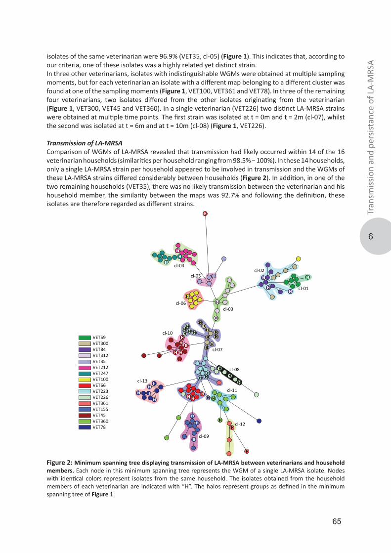
65
6
Tran
smiss
ion
and
pers
istan
ce o
f LA-
MRS
A
isolates of the same veterinarian were 96.9% (VET35, cl-05) (Figure 1). This indicates that, according to our criteria, one of these isolates was a highly related yet distinct strain.In three other veterinarians, isolates with indistinguishable WGMs were obtained at multiple sampling moments, but for each veterinarian an isolate with a different map belonging to a different cluster was found at one of the sampling moments (Figure 1, VET100, VET361 and VET78). In three of the remaining four veterinarians, two isolates differed from the other isolates originating from the veterinarian (Figure 1, VET300, VET45 and VET360). In a single veterinarian (VET226) two distinct LA-MRSA strains were obtained at multiple time points. The first strain was isolated at t = 0m and t = 2m (cl-07), whilst the second was isolated at t = 6m and at t = 10m (cl-08) (Figure 1, VET226).
Transmission of LA-MRSAComparison of WGMs of LA-MRSA revealed that transmission had likely occurred within 14 of the 16 veterinarian households (similarities per household ranging from 98.5% − 100%). In these 14 households, only a single LA-MRSA strain per household appeared to be involved in transmission and the WGMs of these LA-MRSA strains differed considerably between households (Figure 2). In addition, in one of the two remaining households (VET35), there was no likely transmission between the veterinarian and his household member, the similarity between the maps was 92.7% and following the definition, these isolates are therefore regarded as different strains.
VET59VET300VET84VET312VET35VET212VET247VET100VET66VET223VET226VET361VET155VET45VET360VET78
cl-06
cl-01
cl-02
cl-03
cl-04
cl-05
cl-07
cl-08
cl-09
cl-10
cl-11
cl-12
cl-13
H
H
H
HH
HH
H
HH
HHH
HH H
H
H
H
H HH
H
H
H
H
H
HH
H H
H
H
H
H
H H
HHH
H
H H
HH
HH
H
H
H
H
Figure 2: Minimum spanning tree displaying transmission of LA-MRSA between veterinarians and household members. Each node in this minimum spanning tree represents the WGM of a single LA-MRSA isolate. Nodes with identical colors represent isolates from the same household. The isolates obtained from the household members of each veterinarian are indicated with “H”. The halos represent groups as defined in the minimum spanning tree of Figure 1.
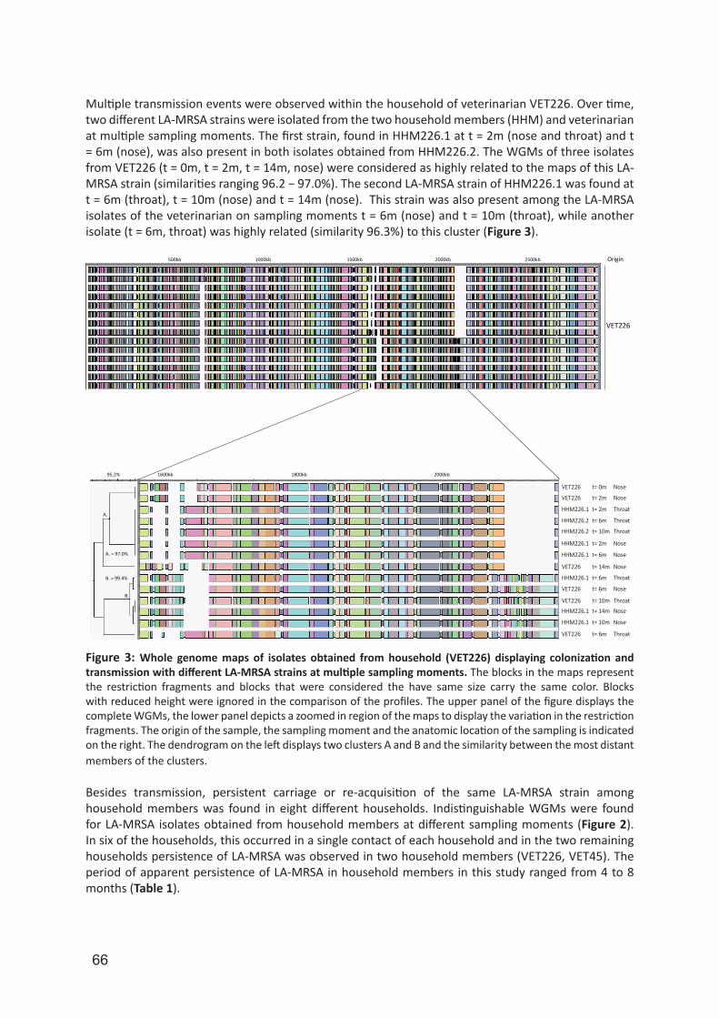
66
Multiple transmission events were observed within the household of veterinarian VET226. Over time, two different LA-MRSA strains were isolated from the two household members (HHM) and veterinarian at multiple sampling moments. The first strain, found in HHM226.1 at t = 2m (nose and throat) and t = 6m (nose), was also present in both isolates obtained from HHM226.2. The WGMs of three isolates from VET226 (t = 0m, t = 2m, t = 14m, nose) were considered as highly related to the maps of this LA-MRSA strain (similarities ranging 96.2 − 97.0%). The second LA-MRSA strain of HHM226.1 was found at t = 6m (throat), t = 10m (nose) and t = 14m (nose). This strain was also present among the LA-MRSA isolates of the veterinarian on sampling moments t = 6m (nose) and t = 10m (throat), while another isolate (t = 6m, throat) was highly related (similarity 96.3%) to this cluster (Figure 3).
VET226
Origin
t= 2m
t= 0m
t= 6m
t= 6m
t= 10mt= 10mt= 14m
t= 6m
t= 14m
t= 2m
t= 6m
t= 10m
t= 10m
t= 6m
t= 10mt= 14m
t= 6m
t= 2m
Throat
Nose
Nose
NoseNose
Nose
Nose
Nose
Nose
Nose
Nose
Nose
Throat
Throat
Throat
Throat
Throat
Throat
1600kb
1500kb1000kb500kb
2000kb1800kb
2000kb 2500kb
95.2%
A. = 97.0%
A.
B. = 99.4%
B.
VET226
VET226
HHM226.2
HHM226.1
HHM226.1
HHM226.1
HHM226.1
HHM226.1
HHM226.1
HHM226.2
VET226
VET226
VET226
VET226
Figure 3: Whole genome maps of isolates obtained from household (VET226) displaying colonization and transmission with different LA-MRSA strains at multiple sampling moments. The blocks in the maps represent the restriction fragments and blocks that were considered the have same size carry the same color. Blocks with reduced height were ignored in the comparison of the profiles. The upper panel of the figure displays the complete WGMs, the lower panel depicts a zoomed in region of the maps to display the variation in the restriction fragments. The origin of the sample, the sampling moment and the anatomic location of the sampling is indicated on the right. The dendrogram on the left displays two clusters A and B and the similarity between the most distant members of the clusters.
Besides transmission, persistent carriage or re-acquisition of the same LA-MRSA strain among household members was found in eight different households. Indistinguishable WGMs were found for LA-MRSA isolates obtained from household members at different sampling moments (Figure 2). In six of the households, this occurred in a single contact of each household and in the two remaining households persistence of LA-MRSA was observed in two household members (VET226, VET45). The period of apparent persistence of LA-MRSA in household members in this study ranged from 4 to 8 months (Table 1).
67
6
Tran
smiss
ion
and
pers
istan
ce o
f LA-
MRS
A
DiscussionIn this study, we used whole genome mapping, which revealed a considerable degree of genetic variation of LA-MRSA isolated from veterinarians and their household members from different geographic sources. Furthermore, we showed that there was frequent transmission of LA-MRSA between veterinarians and their household members. In addition, we have shown that both veterinarians and their household members carried LA-MRSA strains for prolonged periods of time with carriage lasting up to 14 months. This provides arguments that LA-MRSA is a successful human colonizer. Price et al. already showed that there is considerable genetic diversity among LA-MRSA CC398 isolates using whole genome sequence based SNP analysis. However, in their study, a broad selection of LA-MRSA isolates originating from different countries, various sources and isolated between 1993 and 2010 was used. In addition, only 30% (n = 26) of the LA-MRSA isolates in this study originated from humans. Genetic diversity is to be expected in such a broad selection of LA-MRSA strains. In our study, all LA-MRSA isolates originated from the Netherlands, were cultured from humans (specifically veterinarians and their household members) and were isolated within a period of 14 months. Even within this narrow selection, we still observed a considerable degree of genetic diversity. However, even with a high-resolution typing method as whole genome mapping LA-MRSA remains a clade that is genetically more homogenous compared to other MRSA variants. This was illustrated by the fact that WGMs of the most distinct LA-MRSA isolates used in this study still had an 83.9% similarity, whereas WGMs of other MRSA variants yielded similarities of approximately 60 − 70%. For example, within a selection of seven pairs of MRSA isolates that all yielded MC8 and spa-type t008, the most distinct maps showed a similarity of 74% (data not shown). In a minority of cases, we were unable to make a clear distinction between isolates obtained from two different veterinarians. This indicates acquisition of the same LA-MRSA strain in both veterinarians. A possible explanation may be that these veterinarians were colonized with LA-MRSA while visiting pig farms. Considering that pigs are distributed from one source to several farms, it is likely that identical strains are present in different farms [19, 20]. However, this remains speculation as we are unaware of what type of livestock farms the veterinarians visited and what the frequency of these visits were. Veterinarians and their household members were sampled longitudinally. Analysis of the WGMs of the LA-MRSA isolated during this period showed they carried the same strain up to 14 months. Whether this reflects persistent carriage or re-acquisition of the same LA-MRSA strain remains uncertain, but the veterinarians in this study generally visited two to three different farms each working day and up to ten different farms each week. Moreover, the household members had no direct contact with livestock animals. Therefore, it is likely that this reflects persistence of LA-MRSA within one individual and not re-acquisition. Also, Köck et al. showed that 59% of the subjected farmers enrolled in their study did not clear MRSA colonization during leave, corroborating our hypothesis that farmers are more likely to be persistently colonized with LA-MRSA than transiently contaminated [21].Since its emergence in 2003, the ability of LA-MRSA to cause transmission from humans to humans has been a subject of debate. Many of the reports describe the limited transmissibility of the LA-MRSA (CC398) clade in general and rely on sub-optimal typing methods such as spa-typing, making it difficult to determine whether these events were actual transmissions [14, 15, 22, 23]. In our study, we focused on presumed LA-MRSA transmission events in a well-defined longitudinal cohort study using a high-resolution typing technique, and this revealed that the presumed transmissions were indeed likely transmissions in nearly every household. However, we do acknowledge that the previously described transmissibility studies were performed in hospital settings and that the outcome might differ considerable under these conditions. Contact between veterinarians and their household members will be more frequent and prolonged than contact between healthcare workers and patients and between patients. Therefore, transmission will be more likely to occur in households of veterinarians than in healthcare facilities. To assess whether the presumed nosocomial transmission of LA-MRSA is really occurring or whether this is a misinterpretation due to the use of low-resolution typing methods, we are currently analyzing isolates obtained from persons involved in presumed nosocomial transmission of LA-MRSA.
68
In conclusion, our results show that LA-MRSA is genetically diverse and that this genetic variation can be used to characterize LA-MRSA strains. Also, we showed that carriage of LA-MRSA in veterinarians and their household members can be persistent, lasting up to 14 months. Furthermore, this study demonstrates that transmission of LA-MRSA between veterinarians and their household members occurs, posing a potential risk for spread in the community of this highly resistant pathogen.
References1. Ayliffe GA. The progressive intercontinental spread of methicillin-resistant Staphylococcus aureus. Clin Infect Dis. 1997;24 Suppl 1:S74-9.2. Lowy FD. Staphylococcus aureus infections. N Engl J Med. 1998;339(8):520-32.3. Kluytmans-Vandenbergh MF, Kluytmans JA. Community-acquired methicillin-resistant Staphylococcus aureus: current perspectives. Clinical microbiology and infection : the official publication of the European Society of Clinical Microbiology and Infectious Diseases. 2006;12 Suppl 1:9-15.4. Voss A, Loeffen F, Bakker J, Klaassen C, Wulf M. Methicillin-resistant Staphylococcus aureus in pig farming. Emerging infectious diseases. 2005;11(12):1965-6.5. Bhat M, Dumortier C, Taylor BS, Miller M, Vasquez G, Yunen J, et al. Staphylococcus aureus ST398, New York City and Dominican Republic. Emerging infectious diseases. 2009;15(2):285-7.6. Guardabassi L, Stegger M, Skov R. Retrospective detection of methicillin resistant and susceptible Staphylococcus aureus ST398 in Danish slaughter pigs. Veterinary microbiology. 2007;122(3-4):384-6.7. Kanerva M, Salmenlinna S, Vuopio-Varkila J, Lehtinen P, Mottonen T, Virtanen MJ, et al. Community-associated methicillin-resistant Staphylococcus aureus isolated in Finland in 2004 to 2006. J Clin Microbiol. 2009;47(8):2655-7.8. Murchan S, Kaufmann ME, Deplano A, de Ryck R, Struelens M, Zinn CE, et al. Harmonization of pulsed-field gel electrophoresis protocols for epidemiological typing of strains of methicillin-resistant Staphylococcus aureus: a single approach developed by consensus in 10 European laboratories and its application for tracing the spread of related strains. Journal of clinical microbiology. 2003;41(4):1574-85.9. Tenover FC, Arbeit R, Archer G, Biddle J, Byrne S, Goering R, et al. Comparison of traditional and molecular methods of typing isolates of Staphylococcus aureus. J Clin Microbiol. 1994;32(2):407-15.10. Schouls LM, Spalburg EC, van Luit M, Huijsdens XW, Pluister GN, van Santen-Verheuvel MG, et al. Multiple-locus variable number tandem repeat analysis of Staphylococcus aureus: comparison with pulsed-field gel electrophoresis and spa-typing. PloS one. 2009;4(4):e5082.11. Malachowa N, Sabat A, Gniadkowski M, Krzyszton-Russjan J, Empel J, Miedzobrodzki J, et al. Comparison of multiple-locus variable-number tandem-repeat analysis with pulsed-field gel electrophoresis, spa typing, and multilocus sequence typing for clonal characterization of Staphylococcus aureus isolates. J Clin Microbiol. 2005;43(7):3095-100.12. Tenover FC, Vaughn RR, McDougal LK, Fosheim GE, McGowan JE, Jr. Multiple-locus variable-number tandem-repeat assay analysis of methicillin-resistant Staphylococcus aureus strains. J Clin Microbiol. 2007;45(7):2215-9.13. Bosch T, Verkade E, van Luit M, Pot B, Vauterin P, Burggrave R, et al. High Resolution Typing by Whole Genome Mapping Enables Discrimination of LA-MRSA (CC398) Strains and Identification of Transmission Events. PloS one. 2013;8(6):e66493.14. Bootsma MC, Wassenberg MW, Trapman P, Bonten MJ. The nosocomial transmission rate of animal-associated ST398 meticillin-resistant Staphylococcus aureus. Journal of the Royal Society, Interface / the Royal Society. 2011;8(57):578-84.15. Wassenberg MW, Bootsma MC, Troelstra A, Kluytmans JA, Bonten MJ. Transmissibility of livestock-associated methicillin-resistant Staphylococcus aureus (ST398) in Dutch hospitals. Clinical microbiology and infection : the official publication of the European Society of Clinical Microbiology and Infectious Diseases. 2011;17(2):316-9.16. Verkade E, Kluytmans-van den Bergh M, van Benthem B, van Cleef B, van Rijen M, Bosch T, et al. Transmission of Methicillin-Resistant Staphylococcus aureus CC398 from Livestock Veterinarians to Their Household Members. PloS one. 2014;9(7):e100823.17. Verkade E, van Benthem B, den Bergh MK, van Cleef B, van Rijen M, Bosch T, et al. Dynamics and determinants of Staphylococcus aureus carriage in livestock veterinarians: a prospective cohort study. Clinical infectious diseases : an official publication of the Infectious Diseases Society of America. 2013;57(2):e11-7.18. Harmsen D, Claus H, Witte W, Rothganger J, Claus H, Turnwald D, et al. Typing of methicillin-resistant Staphylococcus aureus in a university hospital setting by using novel software for spa repeat determination and database management. Journal of clinical microbiology. 2003;41(12):5442-8.19. Broens EM, Graat EA, van der Wolf PJ, van de Giessen AW, van Duijkeren E, Wagenaar JA, et al. MRSA CC398 in the pig production chain. Preventive veterinary medicine. 2011;98(2-3):182-9.20. van Duijkeren E, Ikawaty R, Broekhuizen-Stins MJ, Jansen MD, Spalburg EC, de Neeling AJ, et al. Transmission of methicillin-resistant Staphylococcus aureus strains between different kinds of pig farms. Veterinary microbiology. 2008;126(4):383-9.21. Kock R, Loth B, Koksal M, Schulte-Wulwer J, Harlizius J, Friedrich AW. Persistence of nasal colonization with livestock-associated methicillin-resistant Staphylococcus aureus in pig farmers after holidays from pig exposure. Applied and environmental microbiology. 2012;78(11):4046-7.22. Graveland H, Wagenaar JA, Verstappen KM, Oosting-van Schothorst I, Heederik DJ, Bos ME. Dynamics of MRSA
69
6
Tran
smiss
ion
and
pers
istan
ce o
f LA-
MRS
A
carriage in veal calves: a longitudinal field study. Preventive veterinary medicine. 2012;107(3-4):180-6.23. Hetem DJ, Bootsma MC, Troelstra A, Bonten MJ. Transmissibility of livestock-associated methicillin-resistant Staphylococcus aureus. Emerging infectious diseases. 2013;19(11):1797-802.
70
7
Changing characteristics of livestock-associated meticillin-resistant Staphylococcus aureus isolated
from humans. Emergence of a subclade transmitted without livestock exposure, the Netherlands, 2003 to
2014
Thijs Bosch
Leo Schouls
Gerlinde Pluister
Dineke Frentz
Han van der Heide
Martijn van Luit
Eurosurveillance
2016
21(21):pii=30236
Naomi van Marm-WattimenaSandra Witteveen
Fabian Landman
Anja Haenen
71
7
Chan
ging
cha
ract
eristi
cs o
f LA-
MRS
A in
the
Net
herla
nds
AbstractSince 2007, livestock-associated meticillin-resistant Staphylococcus aureus (LA-MRSA) has become the predominant MRSA clade isolated from humans in the Netherlands. To assess possible temporal changes, we molecularly characterised over 9,000 LA-MRSA isolates submitted from 2003 to 2014 to the Dutch MRSA surveillance. After an initial rapid increase with a peak in 2009 (n = 1,368), the total number of submitted LA-MRSA isolates has been slowly decreasing to 968 in 2014 and over 80% of LA-MRSA belonged to one of three predominant MLVA/spa-types. Next generation sequencing (n=118) showed that MT569/t034 isolates were genetically more diverse than MT398/t011 and MT572/t108. Concurrent with the decrease in LA-MRSA, fewer people reported having contact with livestock and this was most prominent for people carrying MT569/t034 LA-MRSA. The proportion of LA-MRSA isolated from infection-related materials increased from 6% in 2009, to 13% in 2014 and most of these isolates originated from patients older than 50 years of age. Remarkably, 83% of these patients reported not having contact with livestock. The results reveal an ongoing change in the genotypic and epidemiological characteristics of Dutch LA-MRSA isolated from humans with the emergence of a LA-MRSA subclade independent of livestock exposure, suggesting LA-MRSA starts to resemble non-LA-MRSA in terms of transmissibility and pathogenicity.
72
IntroductionMeticillin-resistant Staphylococcus aureus (MRSA) is an important cause of hospital-acquired and community-acquired infections [1]. In 2003, a clonal lineage of MRSA cultured from pig farmers and designated as multilocus sequence typing clonal complex 398 (CC398), emerged in the Netherlands and France [2, 3]. A large number of countries reported CC398 cultured from animals, revealing a worldwide prevalence [4, 5]. CC398 has been found in pigs and other livestock, such as calves, and poultry [6, 7], and therefore designated as livestock-associated MRSA (LA-MRSA). LA-MRSA is prevalent in many European countries and LA-MRSA CC398 isolated from humans has become the predominant MRSA clade among isolates submitted for typing in the Dutch MRSA surveillance programme since 2007 [5, 8].Despite its widespread occurrence, the number of reported infections with LA-MRSA among humans remains low. In addition, nosocomial transmission of LA-MRSA in Dutch hospitals was reported to be 72% less likely to occur compared with non-LA-MRSA, although outbreaks of LA-MRSA have been described [9, 10]. The reason for this limited transmissibility remains unclear, but several studies suggest that the human innate immunomodulatory genes on bacteriophage φ3 are important genetic markers for the adaptation of LA-MRSA towards humans and human-to-human transmission of CC398 [11-13].LA-MRSA represents a homogenous clade with limited differentiation using typing techniques such as multiple-locus variable number of tandem repeat analysis (MLVA), and staphylococcal protein A (spa-) typing [14]. In contrast, whole genome mapping, and next generation sequencing (NGS), revealed more genotypic diversity and suggested a distinction between livestock- and human-associated CC398 clades, although most studies are dominated by isolates obtained from animals and the human-associated isolates were mostly comprised of meticillin-sensitive S. aureus [12,14-16].In this study, molecular characterisation, including NGS, and epidemiological data of more than 9,000 LA-MRSA isolates submitted to the national MRSA surveillance from 2003 to 2014 were used to assess the characteristics of the most predominant MRSA clade in the Netherlands.
Material and methodsBacterial isolatesMRSA isolates, obtained from humans admitted to healthcare centers, were submitted for molecular typing to the National Institute of Public Health and the Environment (RIVM) to the Dutch national MRSA surveillance. All S. aureus isolates were subjected to spa-typing, and MLVA. The MLVA also includes the detection of the genes for mecA, mecC and the lukF gene, indicative for Panton-Valentine leucocidin (PVL) [15, 16]. Isolates belonging to MLVA complex 398 (MC398) were classified as LA-MRSA. All isolates not belonging to MC398 were designated as non-LA-MRSA. The discriminatory power of MLVA was assessed using Simpson’s index of diversity, while the determination of the confidence intervals (CI) of the Simpson’s indices was calculated as described by Grundmann et al. [17, 18]. Only the first isolate per person per year was included. Medical microbiologists or infection control practitioners filled out questionnaires regarding epidemiological risk factors for MRSA colonisation or infection, including contact with livestock.
Classification of materialsThe MRSA isolates were sampled from various materials and sites. Materials were subdivided in four different classes. Swabs from nose and/or throat, and/or perineum were regarded as material indicative for ‘carriage’. Blood, cerebrospinal fluid (CSF), pus, sputum, urine, and wounds were considered as ‘infection-related’ materials. Other human materials were grouped as ‘other’ and if submitted without information regarding material, they were grouped as ‘unknown’.
Next generation sequencingThe first 10 (if available) isolates of the three predominant LA-MRSA MLVA/spa-types from 2003 to 2005 and the first five isolates from 2006 to 2012 were used for analysis by NGS. NGS on these 118
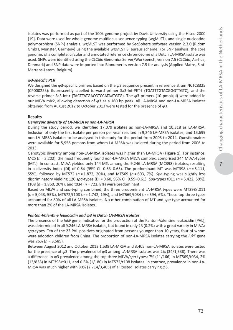
73
7
Chan
ging
cha
ract
eristi
cs o
f LA-
MRS
A in
the
Net
herla
nds
isolates was performed as part of the 100k genome project by Davis University using the Hiseq 2000 [19]. Data were used for whole genome multilocus sequence typing (wgMLST), and single nucleotide polymorphism (SNP-) analysis. wgMLST was performed by SeqSphere software version 2.3.0 (Ridom GmbH, Münster, Germany) using the available wgMLST S. aureus scheme. For SNP analysis, the core genome, of a complete, circular and annotated reference chromosome of a Dutch LA-MRSA isolate was used. SNPs were identified using the CLCbio Genomics Server/Workbench, version 7.5 (CLCbio, Aarhus, Denmark) and SNP data were imported into Bionumerics version 7.5 for analysis (Applied Maths, Sint-Martens-Latem, Belgium).
φ3-specific PCRWe designed the φ3-specific primers based on the φ3 sequence present in reference strain NCTC8325 (CP000253): fluorescently labelled forward primer Sa3-Int-PET-f (TGATTTGTACGGGTTGTC), and the reverse primer Sa3-Int-r (TACTTATGACGTCCATAATGTG). The φ3 primers (10 pmol/µl) were added in our MLVA mix2, allowing detection of φ3 as a 160 bp peak. All LA-MRSA and non-LA-MRSA isolates obtained from August 2012 to October 2013 were tested for the presence of φ3.
ResultsGenotypic diversity of LA-MRSA vs non-LA-MRSADuring the study period, we identified 17,079 isolates as non-LA-MRSA and 10,318 as LA-MRSA. Inclusion of only the first isolate per person per year resulted in 9,246 LA-MRSA isolates, and 13,699 non-LA-MRSA isolates to be analysed in this study for the period from 2003 to 2014. Questionnaires were available for 5,958 persons from whom LA-MRSA was isolated during the period from 2006 to 2013.Genotypic diversity among non-LA-MRSA isolates was higher than LA-MRSA (Figure 1). For instance, MC5 (n = 3,202), the most frequently found non-LA-MRSA MLVA complex, comprised 244 MLVA-types (MTs). In contrast, MLVA yielded only 144 MTs among the 9,246 LA-MRSA (MC398) isolates, resulting in a diversity index (DI) of 0.64 (95% CI: 0.63–0.65). The predominant MT was MT398 (n = 5,111, 55%), followed by MT572 (n = 1,872, 20%), and MT569 (n = 603, 7%). Spa-typing was slightly less discriminatory yielding 120 spa-types (DI = 0.60, 95% CI: 0.59–0.61). Spa-types t011 (n = 5,422, 59%), t108 (n = 1,860, 20%), and t034 (n = 723, 8%) were predominant.Based on MLVA and spa-typing combined, the three predominant LA-MRSA types were MT398/t011 (n = 5,043, 55%), MT572/t108 (n = 1,742, 19%), and MT569/t034 (n = 594, 6%). These top three types accounted for 80% of all LA-MRSA isolates. No other combination of MT and spa-type accounted for more than 2% of the LA-MRSA isolates.
Panton-Valentine leukocidin and φ3 in Dutch LA-MRSA isolatesThe presence of the lukF gene, indicative for the production of the Panton-Valentine leukocidin (PVL), was determined in all 9,246 LA-MRSA isolates, but found in only 23 (0.2%) with a great variety in MLVA/spa-types. Ten of the 23 PVL positives originated from persons younger than 10 years, four of whom were adoption children from China. The proportion of non-LA-MRSA isolates carrying the lukF gene was 26% (n = 3,585).Between August 2012 and October 2013 1,538 LA-MRSA and 3,405 non-LA-MRSA isolates were tested for the presence of φ3. The prevalence of φ3 among LA-MRSA isolates was 2% (34/1,538). There was a difference in φ3 prevalence among the top three MLVA/spa-types; 7% (11/166) in MT569/t034, 2% (13/838) in MT398/t011, and 0.6% (1/180) in MT572/t108 isolates. In contrast, prevalence in non-LA-MRSA was much higher with 80% (2,714/3,405) of all tested isolates carrying φ3.
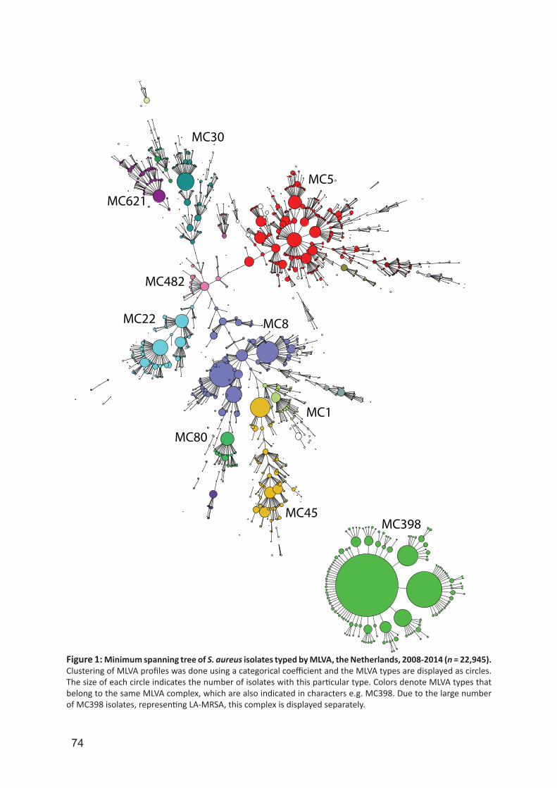
74
MC5
MC30
MC621
MC482
MC22 MC8
MC80
MC45
MC1
MC398
Figure 1: Minimum spanning tree of S. aureus isolates typed by MLVA, the Netherlands, 2008-2014 (n = 22,945).Clustering of MLVA profiles was done using a categorical coefficient and the MLVA types are displayed as circles. The size of each circle indicates the number of isolates with this particular type. Colors denote MLVA types that belong to the same MLVA complex, which are also indicated in characters e.g. MC398. Due to the large number of MC398 isolates, representing LA-MRSA, this complex is displayed separately.

75
7
Chan
ging
cha
ract
eristi
cs o
f LA-
MRS
A in
the
Net
herla
nds
Whole genome multilocus sequence typing and single nucleotide polymorphism analysisBoth wgMLST and SNP analysis of 118 isolates of the three predominant LA-MRSA types showed that they clustered in three different groups (Figure 2). Of the 1,864 genes of the S. aureus wgMLST scheme, 1,831 were present in all 118 isolates and used for comparison and tree construction. LA-MRSA isolates belonging to MT398/t011 (n = 45) clustered closely together as did MT572/t108 (n = 44) isolates. The average distance between MT398/t011 isolates was allelic variation in 25 genes with a maximum of 60 genes and for MT572/t108, the average distance was 24 genes with a maximum of 52 genes. The closest related isolates of the two groups differed in 145 genes. Compared with MT398/t011 and MT572/t108, the genetic diversity among the MT569/t034 (n = 29) isolates was higher. The MT569/t034 isolates differed on average 34 genes with a maximum of 148 genes. The distances between MT569/t034 and MT398/t011, and MT572/t108 were 102 and 136 genes, respectively.We identified 7,944 SNP positions in the core genome and used these for comparison. The minimum spanning tree was comparable to the wgMLST tree with groups of closely related MT398/t011, and MT572/t108 isolates and a genetic diverse MT569/t034. The average number of SNPs that differed among members of the MT398/t011 group was 43 with a maximum of 117 SNPs, while the MT572/t108 isolates differed on average in 39 SNPs with a maximum of 80 SNPs between the most distant members. The distance between the closest related isolates of MT398/t011, and MT572/t108 was 274 SNPs.
wgMLST SNP analysis
60 SNPs
272
229
MT398/t011
MT572/t108
MT569/t034
MT398/t011
MT572/t108
MT569/t034
25 genes
102
136
Figure 2: Minimum spanning trees based on next generation sequencing of LA-MRSA isolates, the Netherlands, 2003-2012 (n = 118). The left panel displays the minimum spanning tree based on 1,831 genes of the S. aureus wgMLST scheme, while the right panel shows the tree based on 7,944 SNPs. Colors represent the top 3 representatives of LA-MRSA, MT398/t011 (n = 45), MT572/t108 (n = 44) and MT569/t034 (n = 29). The lengths of the lines between isolates represent either the number of different genes (wgMLST) or the number of SNPs.
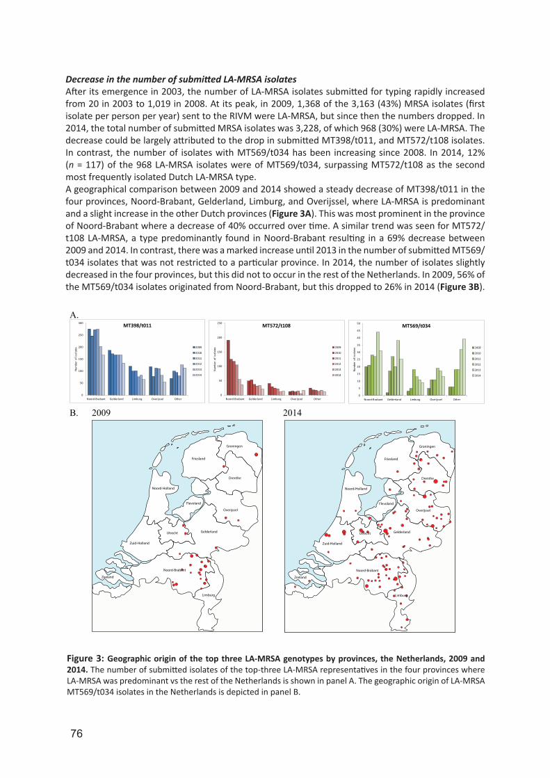
76
Decrease in the number of submitted LA-MRSA isolatesAfter its emergence in 2003, the number of LA-MRSA isolates submitted for typing rapidly increased from 20 in 2003 to 1,019 in 2008. At its peak, in 2009, 1,368 of the 3,163 (43%) MRSA isolates (first isolate per person per year) sent to the RIVM were LA-MRSA, but since then the numbers dropped. In 2014, the total number of submitted MRSA isolates was 3,228, of which 968 (30%) were LA-MRSA. The decrease could be largely attributed to the drop in submitted MT398/t011, and MT572/t108 isolates. In contrast, the number of isolates with MT569/t034 has been increasing since 2008. In 2014, 12% (n = 117) of the 968 LA-MRSA isolates were of MT569/t034, surpassing MT572/t108 as the second most frequently isolated Dutch LA-MRSA type.A geographical comparison between 2009 and 2014 showed a steady decrease of MT398/t011 in the four provinces, Noord-Brabant, Gelderland, Limburg, and Overijssel, where LA-MRSA is predominant and a slight increase in the other Dutch provinces (Figure 3A). This was most prominent in the province of Noord-Brabant where a decrease of 40% occurred over time. A similar trend was seen for MT572/t108 LA-MRSA, a type predominantly found in Noord-Brabant resulting in a 69% decrease between 2009 and 2014. In contrast, there was a marked increase until 2013 in the number of submitted MT569/t034 isolates that was not restricted to a particular province. In 2014, the number of isolates slightly decreased in the four provinces, but this did not to occur in the rest of the Netherlands. In 2009, 56% of the MT569/t034 isolates originated from Noord-Brabant, but this dropped to 26% in 2014 (Figure 3B).
A.
B. 2009
0
50
100
150
200
250
300
Noord-Brabant Gelderland Limburg Overijssel Other
Num
ber
of is
olat
es
MT398/t011
2009
2010
2011
2012
2013
2014
0
50
100
150
200
250
Noord-Brabant Gelderland Limburg Overijssel Other
Num
ber
of is
olat
es
MT572/t108
2009
2010
2011
2012
2013
2014
0
5
10
15
20
25
30
35
40
45
50
Noord-Brabant Gelderland Limburg Overijssel Other
Num
ber
of is
olat
es
MT569/t034
2009
2010
2011
2012
2013
2014
2014
Friesland
Groningen
Drenthe
Overijssel
GelderlandUtrecht
Noord-Holland
Zuid-Holland
Zeeland
Noord-Brabant
Limburg
Flevoland
Friesland
Groningen
Drenthe
Overijssel
GelderlandUtrecht
Noord-Holland
Zuid-Holland
Zeeland
Noord-Brabant
Limburg
Flevoland
Figure 3: Geographic origin of the top three LA-MRSA genotypes by provinces, the Netherlands, 2009 and 2014. The number of submitted isolates of the top-three LA-MRSA representatives in the four provinces where LA-MRSA was predominant vs the rest of the Netherlands is shown in panel A. The geographic origin of LA-MRSA MT569/t034 isolates in the Netherlands is depicted in panel B.
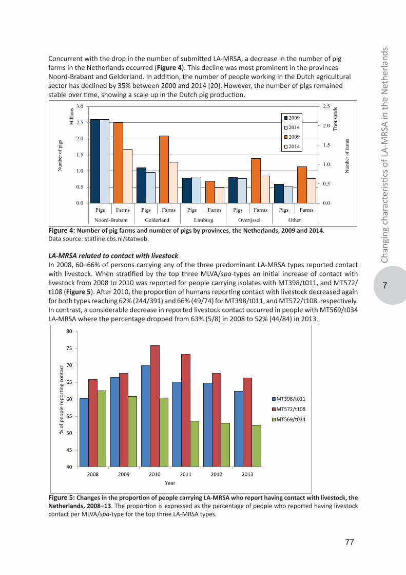
77
7
Chan
ging
cha
ract
eristi
cs o
f LA-
MRS
A in
the
Net
herla
nds
Concurrent with the drop in the number of submitted LA-MRSA, a decrease in the number of pig farms in the Netherlands occurred (Figure 4). This decline was most prominent in the provinces Noord-Brabant and Gelderland. In addition, the number of people working in the Dutch agricultural sector has declined by 35% between 2000 and 2014 [20]. However, the number of pigs remained stable over time, showing a scale up in the Dutch pig production.
0.0
0.5
1.0
1.5
2.0
2.5
0.0
0.5
1.0
1.5
2.0
2.5
3.0
Pigs Farms Pigs Farms Pigs Farms Pigs Farms Pigs Farms
Noord-Brabant Gelderland Limburg Overijssel Other
Num
ber o
f far
ms
Thou
sand
s
Num
ber o
f pig
s
Mill
ions
2009
2014
2009
2014
Figure 4: Number of pig farms and number of pigs by provinces, the Netherlands, 2009 and 2014.Data source: statline.cbs.nl/statweb.
LA-MRSA related to contact with livestockIn 2008, 60–66% of persons carrying any of the three predominant LA-MRSA types reported contact with livestock. When stratified by the top three MLVA/spa-types an initial increase of contact with livestock from 2008 to 2010 was reported for people carrying isolates with MT398/t011, and MT572/t108 (Figure 5). After 2010, the proportion of humans reporting contact with livestock decreased again for both types reaching 62% (244/391) and 66% (49/74) for MT398/t011, and MT572/t108, respectively. In contrast, a considerable decrease in reported livestock contact occurred in people with MT569/t034 LA-MRSA where the percentage dropped from 63% (5/8) in 2008 to 52% (44/84) in 2013.
40
45
50
55
60
65
70
75
80
2008 2009 2010 2011 2012 2013
% o
f peo
ple
repo
rting
con
tact
Year
MT398/t011
MT572/t108
MT569/t034
Figure 5: Changes in the proportion of people carrying LA-MRSA who report having contact with livestock, the Netherlands, 2008‒13. The proportion is expressed as the percentage of people who reported having livestock contact per MLVA/spa-type for the top three LA-MRSA types.
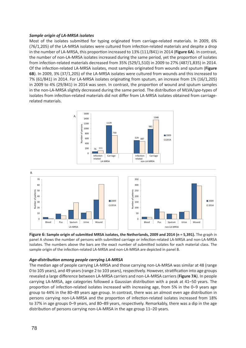
78
Sample origin of LA-MRSA isolatesMost of the isolates submitted for typing originated from carriage-related materials. In 2009, 6% (76/1,205) of the LA-MRSA isolates were cultured from infection-related materials and despite a drop in the number of LA-MRSA, this proportion increased to 13% (111/841) in 2014 (Figure 6A). In contrast, the number of non-LA-MRSA isolates increased during the same period, yet the proportion of isolates from infection-related materials decreased from 35% (529/1,510) in 2009 to 27% (487/1,835) in 2014.Of the infection-related LA-MRSA isolates, most samples originated from wounds and sputum (Figure 6B). In 2009, 3% (37/1,205) of the LA-MRSA isolates were cultured from wounds and this increased to 7% (61/841) in 2014. For LA-MRSA isolates originating from sputum, an increase from 1% (16/1,205) in 2009 to 4% (29/841) in 2014 was seen. In contrast, the proportion of wound and sputum samples in the non-LA-MRSA slightly decreased during the same period. The distribution of MLVA/spa-types of isolates from infection-related materials did not differ from LA-MRSA isolates obtained from carriage-related materials.
A.
B.
0
10
20
30
40
50
60
70
Blood Pus Sputum Urine Wound
LA-MRSA
Num
ber o
f pati
ents
2009
2014
0
50
100
150
200
250
300
350
Blood Pus Sputum Urine Wound
non-LA-MRSA
Num
ber o
f pati
ents
2009
2014
76
1129
529
981
111
730
487
1348
0
200
400
600
800
1000
1200
1400
1600
Infection-related
Carriage Carriage
LA-MRSA non-LA-MRSA
Num
ber o
f per
sons
2009
2014
Infection-related
Figure 6: Sample origin of submitted MRSA isolates, the Netherlands, 2009 and 2014 (n = 5,391). The graph in panel A shows the number of persons with submitted carriage or infection-related LA-MRSA and non-LA-MRSA isolates. The numbers above the bars are the exact number of submitted isolates for each material class. The sample origin of the infection-related LA-MRSA and non-LA-MRSA are depicted in panel B.
Age-distribution among people carrying LA-MRSAThe median age of people carrying LA-MRSA and those carrying non-LA-MRSA was similar at 48 (range 0 to 105 years), and 49 years (range 2 to 103 years), respectively. However, stratification into age groups revealed a large difference between LA-MRSA carriers and non-LA-MRSA carriers (Figure 7A). In people carrying LA-MRSA, age categories followed a Gaussian distribution with a peak at 41–50 years. The proportion of infection-related isolates increased with increasing age, from 5% in the 0–9 years age group to 44% in the 80–89 years age group. In contrast, there was an almost even age distribution in persons carrying non-LA-MRSA and the proportion of infection-related isolates increased from 18% to 37% in age groups 0–9 years, and 80–89 years, respectively. Remarkably, there was a dip in the age distribution of persons carrying non-LA-MRSA in the age group 11–20 years.
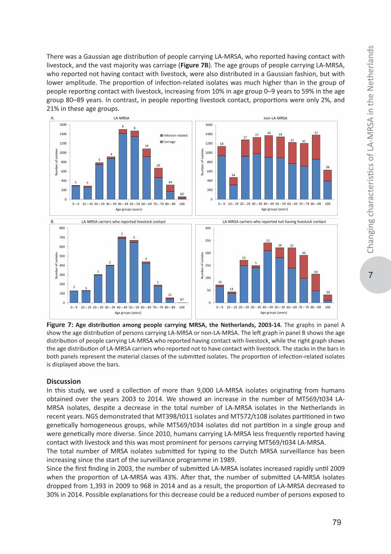
79
7
Chan
ging
cha
ract
eristi
cs o
f LA-
MRS
A in
the
Net
herla
nds
There was a Gaussian age distribution of people carrying LA-MRSA, who reported having contact with livestock, and the vast majority was carriage (Figure 7B). The age groups of people carrying LA-MRSA, who reported not having contact with livestock, were also distributed in a Gaussian fashion, but with lower amplitude. The proportion of infection-related isolates was much higher than in the group of people reporting contact with livestock, increasing from 10% in age group 0–9 years to 59% in the age group 80–89 years. In contrast, in people reporting livestock contact, proportions were only 2%, and 21% in these age groups.
10
14
135
1318 37
49
59
59
0
50
100
150
200
250
300
0 ̶ 9 10 ̶ 19 20 ̶ 29 30 ̶ 39 40 ̶ 49 50 ̶ 59 60 ̶ 69 70 ̶ 79 80 ̶ 89 ≥90
Num
ber o
f iso
late
s
Age groups (years)
2 3
1
2
23
4
5
2167
0
100
200
300
400
500
600
700
800
0 ̶ 9 10 ̶ 19 20 ̶ 29 30 ̶ 39 40 ̶ 49 50 ̶ 59 60 ̶ 69 70 ̶ 79 80 ̶ 89 ≥90
Num
ber o
f iso
late
s
Age groups (years)
18
34
2727 34 34
37 41
37
38
0
200
400
600
800
1000
1200
1400
1600
0 ̶ 9 10 ̶ 19 20 ̶ 29 30 ̶ 39 40 ̶ 49 50 ̶ 59 60 ̶ 69 70 ̶ 79 80 ̶ 89 ≥90N
umbe
r of i
sola
tes
Age groups (years)
5 5
5
4
6 8
16
29
44
600
200
400
600
800
1000
1200
1400
1600
0 ̶ 9 10 ̶ 19 20 ̶ 29 30 ̶ 39 40 ̶ 49 50 ̶ 59 60 ̶ 69 70 ̶ 79 80 ̶ 89 ≥90
Num
ber o
f iso
late
s
Age groups (years)
Infection-related
Carriage
LA-MRSA non-LA-MRSAA.
B. LA-MRSA carriers who reported livestock contact LA-MRSA carriers who reported not having livestock contact
Figure 7: Age distribution among people carrying MRSA, the Netherlands, 2003-14. The graphs in panel A show the age distribution of persons carrying LA-MRSA or non-LA-MRSA. The left graph in panel B shows the age distribution of people carrying LA-MRSA who reported having contact with livestock, while the right graph shows the age distribution of LA-MRSA carriers who reported not to have contact with livestock. The stacks in the bars in both panels represent the material classes of the submitted isolates. The proportion of infection-related isolates is displayed above the bars.
DiscussionIn this study, we used a collection of more than 9,000 LA-MRSA isolates originating from humans obtained over the years 2003 to 2014. We showed an increase in the number of MT569/t034 LA-MRSA isolates, despite a decrease in the total number of LA-MRSA isolates in the Netherlands in recent years. NGS demonstrated that MT398/t011 isolates and MT572/t108 isolates partitioned in two genetically homogeneous groups, while MT569/t034 isolates did not partition in a single group and were genetically more diverse. Since 2010, humans carrying LA-MRSA less frequently reported having contact with livestock and this was most prominent for persons carrying MT569/t034 LA-MRSA.The total number of MRSA isolates submitted for typing to the Dutch MRSA surveillance has been increasing since the start of the surveillance programme in 1989.Since the first finding in 2003, the number of submitted LA-MRSA isolates increased rapidly until 2009 when the proportion of LA-MRSA was 43%. After that, the number of submitted LA-MRSA isolates dropped from 1,393 in 2009 to 968 in 2014 and as a result, the proportion of LA-MRSA decreased to 30% in 2014. Possible explanations for this decrease could be a reduced number of persons exposed to
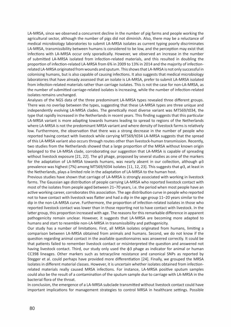
80
LA-MRSA, since we observed a concurrent decline in the number of pig farms and people working the agricultural sector, although the number of pigs did not diminish. Also, there may be a reluctance of medical microbiology laboratories to submit LA-MRSA isolates as current typing poorly discriminates LA-MRSA, transmissibility between humans is considered to be low, and the perception may exist that infections with LA-MRSA occur only sporadically. However, we observed an increase in the number of submitted LA-MRSA isolated from infection-related materials, and this resulted in doubling the proportion of infection-related LA-MRSA from 6% in 2009 to 13% in 2014 and the majority of infection-related LA-MRSA originated from wounds and sputum. This shows that LA-MRSA is not only successful in colonising humans, but is also capable of causing infections. It also suggests that medical microbiology laboratories that have already assessed that an isolate is LA-MRSA, prefer to submit LA-MRSA isolated from infection-related materials rather than carriage isolates. This is not the case for non-LA-MRSA, as the number of submitted carriage-related isolates is increasing, while the number of infection-related isolates remains unchanged.Analyses of the NGS data of the three predominant LA-MRSA types revealed three different groups. There was no overlap between the types, suggesting that these LA-MRSA types are three unique and independently evolving LA-MRSA clades. The genetically most diverse variant was MT569/t034, the type that rapidly increased in the Netherlands in recent years. This finding suggests that this particular LA-MRSA variant is more adapting towards humans leading to spread to regions of the Netherlands where LA-MRSA is not the predominant MRSA variant and where density of livestock farms is relatively low. Furthermore, the observation that there was a strong decrease in the number of people who reported having contact with livestock while carrying MT569/t034 LA-MRSA suggests that the spread of this LA-MRSA variant also occurs through routes other than livestock-human transmission. Recently, two studies from the Netherlands showed that a large proportion of the MRSA without known origin belonged to the LA-MRSA clade, corroborating our suggestion that LA-MRSA is capable of spreading without livestock exposure [21, 22]. The φ3 phage, proposed by several studies as one of the markers for the adaptation of LA-MRSA towards humans, was nearly absent in our collection, although φ3 prevalence was highest (7%) among MT569/t034 isolates [11, 12, 23]. This suggests that φ3, at least in the Netherlands, plays a limited role in the adaptation of LA-MRSA to the human host.Previous studies have shown that carriage of LA-MRSA is strongly associated with working in livestock farms. The Gaussian age distribution of people carrying LA-MRSA who reported livestock contact with most of the isolates from people aged between 21–70 years, i.e. the period when most people have an active working career, corroborates this association. The age distribution curve in people who reported not to have contact with livestock was flatter and had a dip in the age group 11–20 years similar to the dip in the non-LA-MRSA curve. Furthermore, the proportion of infection-related isolates in those who reported livestock contact was lower than in those reporting not to have contact with livestock. In the latter group, this proportion increased with age. The reasons for this remarkable difference in apparent pathogenicity remain unclear. However, it suggests that LA-MRSA are becoming more adapted to humans and start to resemble non-LA-MRSA in transmissibility and pathogenicity.Our study has a number of limitations. First, all MRSA isolates originated from humans, limiting a comparison between LA-MRSA obtained from animals and humans. Second, we do not know if the question regarding animal contact in the available questionnaires was answered correctly. It could be that patients failed to remember livestock contact or misinterpreted the question and answered not having livestock contact. Third, our study only used the φ3 phage as indicator for animal or human CC398 lineages. Other markers such as tetracycline resistance and canonical SNPs as reported by Stegger et al. could perhaps have provided more differentiation [24]. Finally, we grouped the MRSA isolates in different material classes. However, it is uncertain whether isolates obtained from infection-related materials really caused MRSA infections. For instance, LA-MRSA positive sputum samples could also be the result of a contamination of the sputum sample due to carriage with LA-MRSA in the bacterial flora of the throat.In conclusion, the emergence of a LA-MRSA subclade transmitted without livestock contact could have important implications for management strategies to control MRSA in healthcare settings. Possible
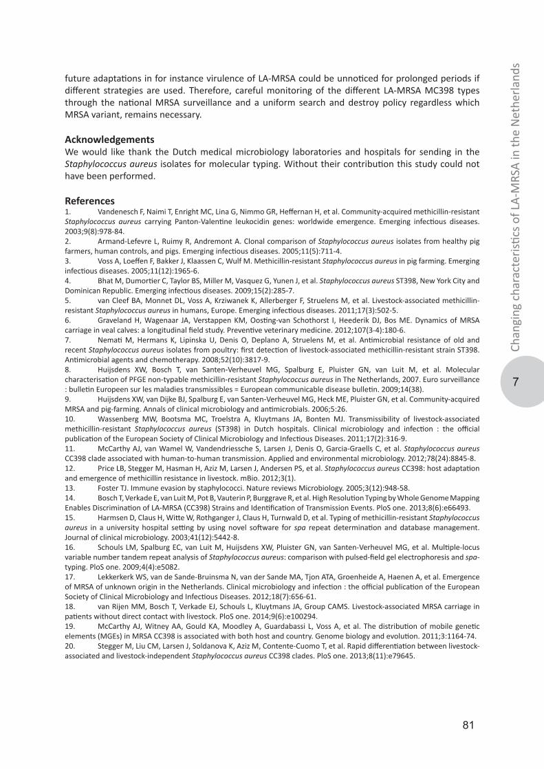
81
7
Chan
ging
cha
ract
eristi
cs o
f LA-
MRS
A in
the
Net
herla
nds
future adaptations in for instance virulence of LA-MRSA could be unnoticed for prolonged periods if different strategies are used. Therefore, careful monitoring of the different LA-MRSA MC398 types through the national MRSA surveillance and a uniform search and destroy policy regardless which MRSA variant, remains necessary.
AcknowledgementsWe would like thank the Dutch medical microbiology laboratories and hospitals for sending in the Staphylococcus aureus isolates for molecular typing. Without their contribution this study could not have been performed.
References1. Vandenesch F, Naimi T, Enright MC, Lina G, Nimmo GR, Heffernan H, et al. Community-acquired methicillin-resistant Staphylococcus aureus carrying Panton-Valentine leukocidin genes: worldwide emergence. Emerging infectious diseases. 2003;9(8):978-84.2. Armand-Lefevre L, Ruimy R, Andremont A. Clonal comparison of Staphylococcus aureus isolates from healthy pig farmers, human controls, and pigs. Emerging infectious diseases. 2005;11(5):711-4.3. Voss A, Loeffen F, Bakker J, Klaassen C, Wulf M. Methicillin-resistant Staphylococcus aureus in pig farming. Emerging infectious diseases. 2005;11(12):1965-6.4. Bhat M, Dumortier C, Taylor BS, Miller M, Vasquez G, Yunen J, et al. Staphylococcus aureus ST398, New York City and Dominican Republic. Emerging infectious diseases. 2009;15(2):285-7.5. van Cleef BA, Monnet DL, Voss A, Krziwanek K, Allerberger F, Struelens M, et al. Livestock-associated methicillin-resistant Staphylococcus aureus in humans, Europe. Emerging infectious diseases. 2011;17(3):502-5.6. Graveland H, Wagenaar JA, Verstappen KM, Oosting-van Schothorst I, Heederik DJ, Bos ME. Dynamics of MRSA carriage in veal calves: a longitudinal field study. Preventive veterinary medicine. 2012;107(3-4):180-6.7. Nemati M, Hermans K, Lipinska U, Denis O, Deplano A, Struelens M, et al. Antimicrobial resistance of old and recent Staphylococcus aureus isolates from poultry: first detection of livestock-associated methicillin-resistant strain ST398. Antimicrobial agents and chemotherapy. 2008;52(10):3817-9.8. Huijsdens XW, Bosch T, van Santen-Verheuvel MG, Spalburg E, Pluister GN, van Luit M, et al. Molecular characterisation of PFGE non-typable methicillin-resistant Staphylococcus aureus in The Netherlands, 2007. Euro surveillance : bulletin Europeen sur les maladies transmissibles = European communicable disease bulletin. 2009;14(38).9. Huijsdens XW, van Dijke BJ, Spalburg E, van Santen-Verheuvel MG, Heck ME, Pluister GN, et al. Community-acquired MRSA and pig-farming. Annals of clinical microbiology and antimicrobials. 2006;5:26.10. Wassenberg MW, Bootsma MC, Troelstra A, Kluytmans JA, Bonten MJ. Transmissibility of livestock-associated methicillin-resistant Staphylococcus aureus (ST398) in Dutch hospitals. Clinical microbiology and infection : the official publication of the European Society of Clinical Microbiology and Infectious Diseases. 2011;17(2):316-9.11. McCarthy AJ, van Wamel W, Vandendriessche S, Larsen J, Denis O, Garcia-Graells C, et al. Staphylococcus aureus CC398 clade associated with human-to-human transmission. Applied and environmental microbiology. 2012;78(24):8845-8.12. Price LB, Stegger M, Hasman H, Aziz M, Larsen J, Andersen PS, et al. Staphylococcus aureus CC398: host adaptation and emergence of methicillin resistance in livestock. mBio. 2012;3(1).13. Foster TJ. Immune evasion by staphylococci. Nature reviews Microbiology. 2005;3(12):948-58.14. Bosch T, Verkade E, van Luit M, Pot B, Vauterin P, Burggrave R, et al. High Resolution Typing by Whole Genome Mapping Enables Discrimination of LA-MRSA (CC398) Strains and Identification of Transmission Events. PloS one. 2013;8(6):e66493.15. Harmsen D, Claus H, Witte W, Rothganger J, Claus H, Turnwald D, et al. Typing of methicillin-resistant Staphylococcus aureus in a university hospital setting by using novel software for spa repeat determination and database management. Journal of clinical microbiology. 2003;41(12):5442-8.16. Schouls LM, Spalburg EC, van Luit M, Huijsdens XW, Pluister GN, van Santen-Verheuvel MG, et al. Multiple-locus variable number tandem repeat analysis of Staphylococcus aureus: comparison with pulsed-field gel electrophoresis and spa-typing. PloS one. 2009;4(4):e5082.17. Lekkerkerk WS, van de Sande-Bruinsma N, van der Sande MA, Tjon ATA, Groenheide A, Haenen A, et al. Emergence of MRSA of unknown origin in the Netherlands. Clinical microbiology and infection : the official publication of the European Society of Clinical Microbiology and Infectious Diseases. 2012;18(7):656-61.18. van Rijen MM, Bosch T, Verkade EJ, Schouls L, Kluytmans JA, Group CAMS. Livestock-associated MRSA carriage in patients without direct contact with livestock. PloS one. 2014;9(6):e100294.19. McCarthy AJ, Witney AA, Gould KA, Moodley A, Guardabassi L, Voss A, et al. The distribution of mobile genetic elements (MGEs) in MRSA CC398 is associated with both host and country. Genome biology and evolution. 2011;3:1164-74.20. Stegger M, Liu CM, Larsen J, Soldanova K, Aziz M, Contente-Cuomo T, et al. Rapid differentiation between livestock-associated and livestock-independent Staphylococcus aureus CC398 clades. PloS one. 2013;8(11):e79645.
82
Next generation sequencing confirms presumed nosocomial transmission of LA-MRSA in the
Netherlands.
8
Thijs Bosch
Leo Schouls
Applied and environmental microbiology
2016
82(14):4081-4089
Sandra Witteveen
Fabian Landman
Anja Haenen
83
8
NGS
con
firm
s nos
ocom
ial t
rans
miss
ion
of L
A-M
RSA
AbstractLivestock-associated MRSA was detected in 2003 and rapidly became the predominant MRSA clade in the Netherlands. Studies have shown that transmissions are difficult to identify since this MRSA variant represents a genetically homogenous clade when current typing techniques are used. Here, next generation sequencing was performed on 206 LA-MRSA isolates to assess the capability of LA-MRSA to transmit between humans. The usefulness of single nucleotide variants (SNVs), the composition of the SCCmec region and the presence of plasmids to identify transmission of LA-MRSA was assessed. In total, 30 presumed putative nosocomial transmission events and two LA-MRSA outbreaks were studied and in most cases, SNV analysis revealed that the isolates of the index patient and the contact(s) clustered closely together. In three presumed events, the isolates did not cluster together indicating that transmission was unlikely. The composition of the SCCmec region corroborated these findings. However, plasmid identification did not support our SNV analysis since different plasmids were present in several cases where SNV and SCCmec analysis suggested that transmission was likely.Next-generation sequencing shows that transmission of LA-MRSA does occur in Dutch health care settings. Transmission was identified based on SNV analysis combined with epidemiological data and in the context of epidemiologically related and unrelated isolates. Analysis of the SCCmec region provided limited, albeit useful information to corroborate conclusions on transmissions, but plasmid identification did not.
ImportanceIn 2003, a variant of methicillin-resistant Staphylococcus aureus (MRSA) isolated from pigs was also found in pig farmers in France and the Netherlands. Soon thereafter, this livestock-associated MRSA (LA-MRSA) was identified in many other countries. Transmission of LA-MRSA between humans, particularly in the health care setting, is regarded to occur sporadically. Moreover, studies that do describe LA-MRSA transmission used molecular characterization of isolates with limited discriminatory power, making the validity of the conclusion that transmission occurred questionable. In our study, we sequenced the complete genomes of 206 LA-MRSA isolates, obtained from more than 30 presumed LA-MRSA transmission events. Analysis of the data showed that transmission of LA-MRSA between humans had indeed occurred in more than 90% of these events. We conclude that transmission of LA-MRSA between humans does occur in Dutch health care settings and therefore a decision to discontinue the search and destroy policy for LA-MRSA should be taken with caution.
84
IntroductionMethicillin-resistant Staphylococcus aureus (MRSA) is an important cause of hospital-acquired and community-acquired infections [1]. In 2003, a MRSA variant cultured from pigs and pig farmers emerged in the Netherlands and France [2, 3]. This clonal lineage was designated as MLST clonal complex 398 (CC398) and a large number of countries reported CC398 cultured from animals, revealing a worldwide prevalence [4, 5]. Besides pigs, CC398 has been found in other livestock animals, such as veal calves, and poultry [6, 7], and was therefore designated as livestock-associated MRSA (LA-MRSA). In the Netherlands, LA-MRSA CC398 isolated from humans has become the predominant MRSA clade among isolates submitted for typing in the Dutch MRSA surveillance program since 2007 [5, 8]. Despite its high prevalence, the capability of LA-MRSA to transmit between humans has been a subject of debate. Transmission of LA-MRSA between animals and from animals to humans has been described in detail and transmission occurs frequently [9]. For human-to-human transmission of LA-MRSA, this is less clear and studies on this subject focused on two topics, namely transmission between humans in a livestock setting or within a healthcare setting. Within the livestock setting, transmission of LA-MRSA between humans has been described in livestock farmers where transmission among broilers, humans and their environment was found [10]. A recent study showed that transmission of LA-MRSA from the livestock veterinarian to household members who reported not having livestock-contact occurred in 15 of the 16 investigated households [11]. Surveys on the transmission of LA-MRSA within the healthcare setting showed that LA-MRSA was 4 to 6 times less transmissible than other MRSA lineages [12, 13]. In addition, nosocomial transmission of LA-MRSA in Dutch hospitals was reported to be 72% less likely to occur compared to non-LA-MRSA [14]. On the other hand, outbreaks of LA-MRSA have been described and two recent reports showed that between 21% and 26% of the MRSA isolates cultured from persons without known MRSA risk factors, the so-called MRSA of unknown origin (MUO), were LA-MRSA [15, 16]. These MUOs suggest spread of LA-MRSA through other routes than by livestock exposure. One of the difficulties regarding investigations on transmission routes of LA-MRSA is that this MRSA variant represents a genetically homogenous clade resulting in limited differentiation using frequently used typing techniques such as MLST, MLVA, and spa-typing [17]. Recently, studies using next generation sequencing (NGS) and whole genome mapping revealed more genotypic diversity among LA-MRSA and suggested a distinction between livestock- and human-associated CC398 clades [11, 18-20]. However, the use of NGS for LA-MRSA in these studies focused mainly on the population structure of LA-MRSA using isolates from different geographical sources and origin. Studies where NGS was applied for transmission studies have thus far been limited to human associated non-LA-MRSA clades, such as E-MRSA 15 [21, 22].In this study, we performed NGS on more than 200 LA-MRSA isolates obtained from humans submitted for the Dutch national MRSA surveillance. Together with epidemiological data, we assessed the capability of LA-MRSA to transmit between humans in Dutch healthcare settings.
Materials and methods Bacterial isolatesVirtually all MRSA isolates (one isolate per person per year), obtained from humans admitted to health care centers, are submitted for molecular typing to the National Institute of Public Health and the Environment (RIVM) for the Dutch national MRSA surveillance. In addition, medical microbiologists or infection control practitioners fill out questionnaires regarding epidemiologic risk factors for MRSA colonization or infection, including contact with livestock, for the persons from whom the MRSA were cultured. In this study, LA-MRSA was defined as isolates with MLVA-types belonging to MLVA complex 398 (MC398).To assess the capability of LA-MRSA to cause nosocomial transmission we included 12 LA-MRSA isolates from a presumed outbreak in a Dutch healthcare facility and 12 isolates from an outbreak in a Dutch nursing home [23]. Furthermore, inspection of the 9,698 questionnaires obtained from 2008-2012, showed that in 1,291 cases MRSA was isolated because nosocomial transmission was suspected. In 673 of these presumed transmission events, isolates were sent to the RIVM for typing. Of those, 41
85
8
NGS
con
firm
s nos
ocom
ial t
rans
miss
ion
of L
A-M
RSA
involved LA-MRSA, but in 15 events, the isolates of the index and secondary cases did not have the same MLVA/spa-types. As a result, 26 presumed LA-MRSA nosocomial transmission events comprising 60 isolates were included in this study. Besides these 26 events, four presumed transmission events, comprising eight isolates, in Dutch healthcare settings as described by van Rijen et al. [16] were also included resulting in a total of 30 investigated events. Of the 30 presumptive transmission events, 25 were single transmissions and the other five involved multiple transmissions.In addition to these presumed LA-MRSA transmission isolates, 114 LA-MRSA isolates comprising the three predominant LA-MRSA MLVA/spa-types; MT398/t011, MT572/t108 and MT569/t034, in the Netherlands obtained between 2003 and 2012 were included for analysis to provide epidemiological context. All available MT398/t011, MT572/t108 and MT569/t034 isolates from 2003 (n = 12), 2004 (n = 12) and 2005 (n = 10) and the first five isolates (if available) of those three types from 2006 until 2012 were used.All 206 isolates were previously characterized using staphylococcal protein A (spa-) typing, multiple-locus variable number of tandem repeat analysis (MLVA) and whole genome mapping [17, 24, 25].
Next generation sequencingAll 206 LA-MRSA isolates included in this study were subjected to NGS. In total, 148 LA-MRSA isolates, originating from the three predominant LA-MRSA MLVA/spa-types, the previously described outbreak [23], the presumed LA-MRSA outbreak and the five presumed nosocomial transmission events [16] were sequenced as part of the 100k genome project by Davis University on a Hiseq 2000 (www.100kgenome.vetmed.ucdavis.edu). The other 58 isolates were commercially sequenced on a Hiseq2500 sequencer (BaseClear, Leiden, Netherlands).
Core-genome single nucleotide variants (SNV-) analysis The complete, annotated genome of LA-MRSA strain RIVM1295 with MT572/t108 (accession number CP013616), isolated from a Dutch patient was determined by NGS. Furthermore, complete, annotated chromosomes from the two other LA-MRSA obtained from Dutch patients representing the two other dominant MLVA/spa-types, MT398/t011 (RIVM1607, CP013619) and MT569/t034 (RIVM3897, CP013621) were used for comparison with the reference genome RIVM1295 to exclude non-core genome regions in the SNV analysis (Supplemental Figure 1). In total, 30 regions, mostly comprising of genes encoding transposases (n = 10), a single bacteriophage, small regions flanking the insertions sites of bacteriophages present in strains RIVM1607 and RIVM3897 (n = 8) and the six rRNA gene regions, were excluded. The regions that were excluded from the core genome are indicated as annotations in the complete genome sequence (CP130616). The CLCbio Genomics Server/Workbench, version 7.5 (CLCbio, Aarhus, Denmark) was used for the identification of SNVs and SNV data were imported into Bionumerics version 7.5 for comparative analyses (Applied Maths, Sint-Martens-Latem, Belgium).
Staphylococcal cassette chromosome mec (SCCmec) and plasmid identification in LA-MRSA isolatesWe assessed the SCCmec region and plasmid composition of LA-MRSA isolates to collect additional information from the accessory genome. The SCCmec region of all 206 LA-MRSA isolates was mapped against all known SCCmec references (www.sccmec.org) available in the NCBI database in October 2015 and SCCmec types were assigned if a complete match was found. Divergent variants were named after the most similar SCCmec type followed by an extension to indicate their distinctive characteristics. To study the diversity of plasmids among Dutch LA-MRSA isolates we used the sequences of three plasmids [accession numbers AM990993, AM990994 and AM990995] found in ST398 reference strain S0385 [accession number AM990992] and sequences obtained from the NCBI database of six plasmids isolated from ST398 strains and sequenced by other researchers (Table 1, [26-32]). Furthermore, sequences from three plasmids, pRIVM1295-1 (CP013617), pRIVM1295-2 (CP013618) and pRIVM1607 (CP013620), identified in our in house LA-MRSA reference strains were also used. During screening of the 206 LA-MRSA isolates against the plasmids, NGS reads of 53 LA-MRSA isolates mapped against only parts of the plasmids pS0385-2, pS0385-3 and pKKS627. De novo assembly and a blast of the rep genes
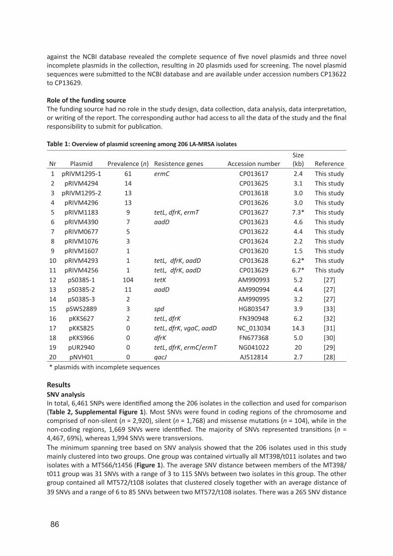
86
against the NCBI database revealed the complete sequence of five novel plasmids and three novel incomplete plasmids in the collection, resulting in 20 plasmids used for screening. The novel plasmid sequences were submitted to the NCBI database and are available under accession numbers CP13622 to CP13629. Role of the funding sourceThe funding source had no role in the study design, data collection, data analysis, data interpretation, or writing of the report. The corresponding author had access to all the data of the study and the final responsibility to submit for publication.
Table 1: Overview of plasmid screening among 206 LA-MRSA isolates
ResultsSNV analysis In total, 6,461 SNPs were identified among the 206 isolates in the collection and used for comparison (Table 2, Supplemental Figure 1). Most SNVs were found in coding regions of the chromosome and comprised of non-silent (n = 2,920), silent (n = 1,768) and missense mutations (n = 104), while in the non-coding regions, 1,669 SNVs were identified. The majority of SNVs represented transitions (n = 4,467, 69%), whereas 1,994 SNVs were transversions.The minimum spanning tree based on SNV analysis showed that the 206 isolates used in this study mainly clustered into two groups. One group was contained virtually all MT398/t011 isolates and two isolates with a MT566/t1456 (Figure 1). The average SNV distance between members of the MT398/t011 group was 31 SNVs with a range of 3 to 115 SNVs between two isolates in this group. The other group contained all MT572/t108 isolates that clustered closely together with an average distance of 39 SNVs and a range of 6 to 85 SNVs between two MT572/t108 isolates. There was a 265 SNV distance
Nr Plasmid Prevalence (n) Resistence genes Accession number Size (kb) Reference
1 pRIVM1295-1 61 ermC CP013617 2.4 This study 2 pRIVM4294 14
CP013625 3.1 This study
3 pRIVM1295-2 13
CP013618 3.0 This study 4 pRIVM4296 13
CP013626 3.0 This study
5 pRIVM1183 9 tetL, dfrK, ermT CP013627 7.3* This study 6 pRIVM4390 7 aadD CP013623 4.6 This study 7 pRIVM0677 5
CP013622 4.4 This study
8 pRIVM1076 3
CP013624 2.2 This study 9 pRIVM1607 1
CP013620 1.5 This study
10 pRIVM4293 1 tetL, dfrK, aadD CP013628 6.2* This study 11 pRIVM4256 1 tetL, dfrK, aadD CP013629 6.7* This study 12 pS0385-1 104 tetK AM990993 5.2 [27] 13 pS0385-2 11 aadD AM990994 4.4 [27] 14 pS0385-3 2
AM990995 3.2 [27]
15 pSWS2889 3 spd HG803547 3.9 [33] 16 pKKS627 2 tetL, dfrK FN390948 6.2 [32] 17 pKKS825 0 tetL, dfrK, vgaC, aadD NC_013034 14.3 [31] 18 pKKS966 0 dfrK FN677368 5.0 [30] 19 pUR2940 0 tetL, dfrK, ermC/ermT NG041022 20 [29] 20 pNVH01 0 qacJ AJ512814 2.7 [28] * plasmids with incomplete sequences
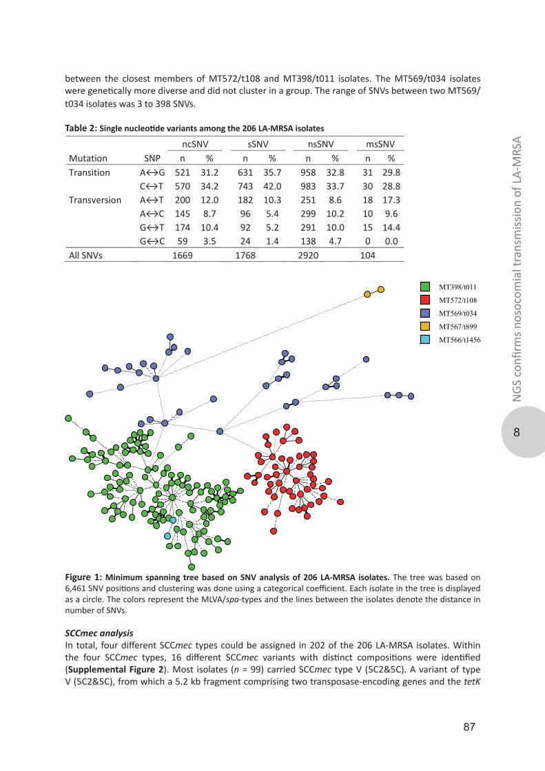
87
8
NGS
con
firm
s nos
ocom
ial t
rans
miss
ion
of L
A-M
RSA
between the closest members of MT572/t108 and MT398/t011 isolates. The MT569/t034 isolates were genetically more diverse and did not cluster in a group. The range of SNVs between two MT569/t034 isolates was 3 to 398 SNVs.
Table 2: Single nucleotide variants among the 206 LA-MRSA isolates
MT398/t011
MT572/t108
MT569/t034
MT567/t899
MT566/t1456
Figure 1: Minimum spanning tree based on SNV analysis of 206 LA-MRSA isolates. The tree was based on 6,461 SNV positions and clustering was done using a categorical coefficient. Each isolate in the tree is displayed as a circle. The colors represent the MLVA/spa-types and the lines between the isolates denote the distance in number of SNVs.
SCCmec analysisIn total, four different SCCmec types could be assigned in 202 of the 206 LA-MRSA isolates. Within the four SCCmec types, 16 different SCCmec variants with distinct compositions were identified (Supplemental Figure 2). Most isolates (n = 99) carried SCCmec type V (5C2&5C). A variant of type V (5C2&5C), from which a 5.2 kb fragment comprising two transposase-encoding genes and the tetK
ncSNV sSNV nsSNV msSNV Mutation SNP n %
n %
n %
n %
Transition A↔G 521 31.2
631 35.7
958 32.8
31 29.8
C↔T 570 34.2
743 42.0
983 33.7
30 28.8
Transversion A↔T 200 12.0
182 10.3
251 8.6
18 17.3
A↔C 145 8.7
96 5.4
299 10.2
10 9.6
G↔T 174 10.4
92 5.2
291 10.0
15 14.4
G↔C 59 3.5
24 1.4
138 4.7
0 0.0
All SNVs 1669
1768
2920
104
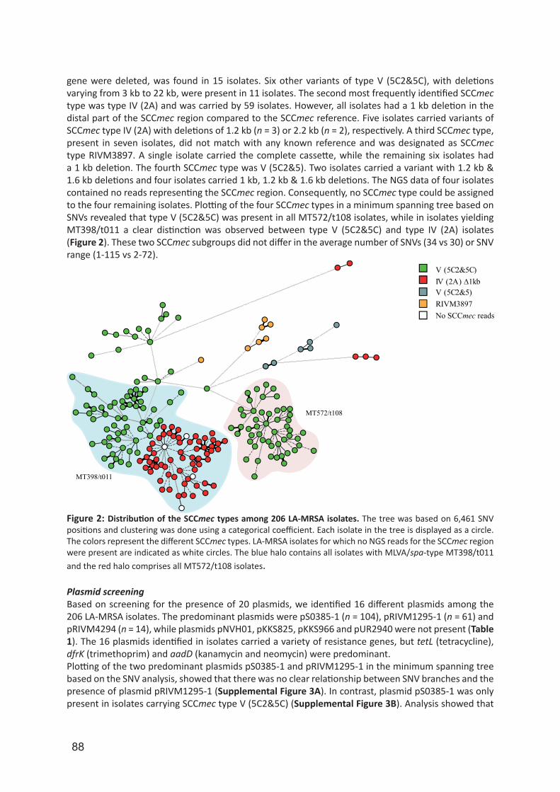
88
gene were deleted, was found in 15 isolates. Six other variants of type V (5C2&5C), with deletions varying from 3 kb to 22 kb, were present in 11 isolates. The second most frequently identified SCCmec type was type IV (2A) and was carried by 59 isolates. However, all isolates had a 1 kb deletion in the distal part of the SCCmec region compared to the SCCmec reference. Five isolates carried variants of SCCmec type IV (2A) with deletions of 1.2 kb (n = 3) or 2.2 kb (n = 2), respectively. A third SCCmec type, present in seven isolates, did not match with any known reference and was designated as SCCmec type RIVM3897. A single isolate carried the complete cassette, while the remaining six isolates had a 1 kb deletion. The fourth SCCmec type was V (5C2&5). Two isolates carried a variant with 1.2 kb & 1.6 kb deletions and four isolates carried 1 kb, 1.2 kb & 1.6 kb deletions. The NGS data of four isolates contained no reads representing the SCCmec region. Consequently, no SCCmec type could be assigned to the four remaining isolates. Plotting of the four SCCmec types in a minimum spanning tree based on SNVs revealed that type V (5C2&5C) was present in all MT572/t108 isolates, while in isolates yielding MT398/t011 a clear distinction was observed between type V (5C2&5C) and type IV (2A) isolates (Figure 2). These two SCCmec subgroups did not differ in the average number of SNVs (34 vs 30) or SNV range (1-115 vs 2-72).
V (5C2&5C)IV (2A) ∆1kb
No SCCmec reads
V (5C2&5) RIVM3897
MT572/t108
MT398/t011
Figure 2: Distribution of the SCCmec types among 206 LA-MRSA isolates. The tree was based on 6,461 SNV positions and clustering was done using a categorical coefficient. Each isolate in the tree is displayed as a circle. The colors represent the different SCCmec types. LA-MRSA isolates for which no NGS reads for the SCCmec region were present are indicated as white circles. The blue halo contains all isolates with MLVA/spa-type MT398/t011 and the red halo comprises all MT572/t108 isolates.
Plasmid screening Based on screening for the presence of 20 plasmids, we identified 16 different plasmids among the 206 LA-MRSA isolates. The predominant plasmids were pS0385-1 (n = 104), pRIVM1295-1 (n = 61) and pRIVM4294 (n = 14), while plasmids pNVH01, pKKS825, pKKS966 and pUR2940 were not present (Table 1). The 16 plasmids identified in isolates carried a variety of resistance genes, but tetL (tetracycline), dfrK (trimethoprim) and aadD (kanamycin and neomycin) were predominant.Plotting of the two predominant plasmids pS0385-1 and pRIVM1295-1 in the minimum spanning tree based on the SNV analysis, showed that there was no clear relationship between SNV branches and the presence of plasmid pRIVM1295-1 (Supplemental Figure 3A). In contrast, plasmid pS0385-1 was only present in isolates carrying SCCmec type V (5C2&5C) (Supplemental Figure 3B). Analysis showed that
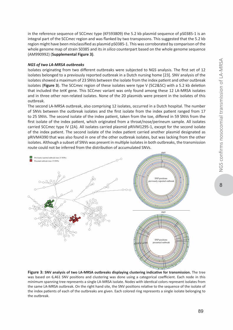
89
8
NGS
con
firm
s nos
ocom
ial t
rans
miss
ion
of L
A-M
RSA
in the reference sequence of SCCmec type (KF593809) the 5.2 kb plasmid sequence of pS0385-1 is an integral part of the SCCmec region and was flanked by two transposons. This suggested that the 5.2 kb region might have been misclassified as plasmid pS0385-1. This was corroborated by comparison of the whole genome map of strain S0385 and its in silico counterpart based on the whole genome sequence (AM990992) (Supplemental Figure 3).
NGS of two LA-MRSA outbreaks Isolates originating from two different outbreaks were subjected to NGS analysis. The first set of 12 isolates belonged to a previously reported outbreak in a Dutch nursing home [23]. SNV analysis of the isolates showed a maximum of 23 SNVs between the isolate from the index patient and other outbreak isolates (Figure 3). The SCCmec region of these isolates were type V (5C2&5C) with a 5.2 kb deletion that included the tetK gene. This SCCmec variant was only found among these 12 LA-MRSA isolates and in three other non-related isolates. None of the 20 plasmids were present in the isolates of this outbreak. The second LA-MRSA outbreak, also comprising 12 isolates, occurred in a Dutch hospital. The number of SNVs between the outbreak isolates and the first isolate from the index patient ranged from 17 to 25 SNVs. The second isolate of the index patient, taken from the toe, differed in 59 SNVs from the first isolate of the index patient, which originated from a throat/nose/perineum sample. All isolates carried SCCmec type IV (2A). All isolates carried plasmid pRIVM1295-1, except for the second isolate of the index patient. The second isolate of the index patient carried another plasmid designated as pRIVM4390 that was also found in one of the other outbreak isolates, but was lacking from the other isolates. Although a subset of SNVs was present in multiple isolates in both outbreaks, the transmission route could not be inferred from the distribution of accumulated SNVs.
Previously reported outbreak (max 23 SNPs)
Presumed outbreak (max 25 SNPs)
SNP positions presumed outbreak
SNP positions previously reported outbreak
start
start
Figure 3: SNV analysis of two LA-MRSA outbreaks displaying clustering indicative for transmission. The tree was based on 6,461 SNV positions and clustering was done using a categorical coefficient. Each node in this minimum spanning tree represents a single LA-MRSA isolate. Nodes with identical colors represent isolates from the same LA-MRSA outbreak. On the right hand site, the SNV positions relative to the sequence of the isolate of the index patients of each of the outbreaks are given. Each colored ring represents a single isolate belonging to the outbreak.
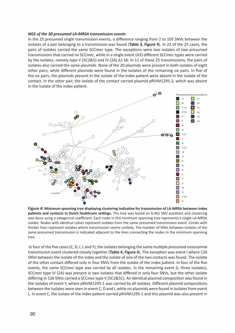
90
NGS of the 30 presumed LA-MRSA transmission events In the 25 presumed single transmission events, a difference ranging from 2 to 105 SNVs between the isolates of a pair belonging to a transmission was found (Table 3, Figure 4). In 22 of the 25 cases, the pairs of isolates carried the same SCCmec type. The exceptions were two isolates of two presumed transmissions that carried no SCCmec, while in a single event (A3) different SCCmec types were carried by the isolates, namely type V (5C2&5) and IV (2A) ∆1 kb. In 11 of these 25 transmissions, the pairs of isolates also carried the same plasmids. None of the 20 plasmids were present in both isolates of eight other pairs, while different plasmids were found in the isolates of the remaining six pairs. In five of the six pairs, the plasmids present in the isolate of the index patient were absent in the isolate of the contact. In the other pair, the isolate of the contact carried plasmid pRIVM1295-2, which was absent in the isolate of the index patient.
A A1
A2B
C (3)
D (3)
E
F
G
H
I (3)
J
K
L (3)
M
N
P
Q
R
S
T
U
W
X
Y (6)
Z
1172
40
99
4 3116
813
12
max 8 SNPs
45
32
25 26
17
2
17
12
4
8
20
3
37
22 23
Presumed transmission events (n)
9
A3
A4
6
8
A5
A6
2
Figure 4: Minimum spanning tree displaying clustering indicative for transmission of LA-MRSA between index patients and contacts in Dutch healthcare settings. The tree was based on 6,461 SNV positions and clustering was done using a categorical coefficient. Each node in this minimum spanning tree represents a single LA-MRSA isolate. Nodes with identical colors represent isolates from the same presumed transmission event. Circles with thicker lines represent isolates where transmission seems unlikely. The number of SNVs between isolates of the same presumed transmission is indicated adjacent to the lines connecting the nodes in the minimum spanning tree.
In four of the five cases (C, D, I, L and Y), the isolates belonging the same multiple presumed nosocomial transmission event clustered closely together (Table 4, Figure 4). The exception was event I where 126 SNVs between the isolate of the index and the isolate of one of the two contacts was found. The isolate of the other contact differed only in four SNVs from the isolate of the index patient. In four of the five events, the same SCCmec type was carried by all isolates. In the remaining event (I, three isolates), SCCmec type IV (2A) was present in two isolates that differed in only four SNVs, but the other isolate differing in 126 SNVs carried a SCCmec type V (5C2&5C). An identical plasmid composition was found in the isolates of event Y, where pRIVM1295-1 was carried by all isolates. Different plasmid compositions between the isolates were seen in event C, D and I, while no plasmids were found in isolates from event L. In event C, the isolate of the index patient carried pRIVM1295-1 and this plasmid was also present in
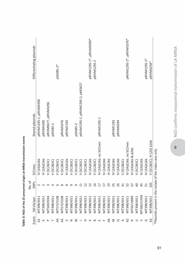
91
8
NGS
con
firm
s nos
ocom
ial t
rans
miss
ion
of L
A-M
RSA
Even
t M
LVA/
spa
No.
of
SNPs
SC
Cmec
Sh
ared
pla
smid
s Di
ffere
ntia
ting
plas
mid
s A1
M
T398
/t01
1 2
IV (2
A)∆1
kb
pRIV
M12
95-1
, pRI
VM43
90
K
MT3
98/t
011
2 IV
(2A)
∆1kb
pR
IVM
4390
P M
T569
/t03
4 3
V (5
C2&
5C)
pRIV
M06
77, p
RIVM
4296
T M
T398
/t01
1 4
V (5
C2&
5C)
pS03
85-2
A4
MT5
72/t
108
6 V
(5C2
&5C
)
pS03
85-2
* A5
M
T572
/t10
8 8
V (5
C2&
5C)
pRIV
M10
76
Z
MT3
98/t
011
8 IV
(2A)
∆1kb
pR
IVM
1183
N
MT3
98/t
011
9 V
(5C2
&5C
)
W
M
T398
/t01
1 9
V (5
C2&
5C)
pS03
85-2
J M
T398
/t01
1 11
V
(5C2
&5C
) pR
IVM
1295
-1, p
RIVM
1295
-2, p
KKS6
27
H
MT3
98/t
011
12
V (5
C2&
5C)
X M
T398
/t01
1 12
IV
(2A)
∆1kb
pRIV
M12
95-1
*, p
RIVM
4390
*
B M
T398
/t01
1 16
V
(5C2
&5C
)
pRIV
M12
95-2
E
MT3
98/t
011
17
IV (2
A)∆1
kb, n
o SC
Cmec
pR
IVM
1295
-1
F
MT3
98/t
011
17
IV (2
A)∆1
kb
A6
MT3
98/t
011
19
IV (2
A)∆1
kb
M
MT3
98/t
011
25
IV (2
A)∆1
kb
pRIV
M11
83
Q
M
T398
/t01
1 26
IV
(2A)
∆1kb
pR
IVM
4294
A M
T398
/t01
1 31
V
(5C2
&5C
)
A2
M
T398
/t01
1 32
IV
(2A)
∆1kb
, no
SCCm
ec
pR
IVM
1295
-1*,
pRI
VM10
76*
R M
T567
/t89
9 37
IV
(2A)
∆1kb
& ∆
2kb
U
MT3
98/t
011
40
V (5
C2&
5C)
S M
T566
/t14
56
45
IV (2
A)∆1
kb
G M
T398
/t01
1 51
IV
(2A)
∆1kb
pRIV
M12
95-1
* A3
M
T398
/t01
1 10
5 V
(5C2
&5C
), IV
(2A)
∆1k
b
pRIV
M42
56*
*Pla
smid
s pre
sent
in th
e iso
late
of t
he in
dex
case
onl
y
Tabl
e 3:
NG
S of
the
25 p
resu
med
sing
le L
A-M
RSA
tran
smis
sion
eve
nts
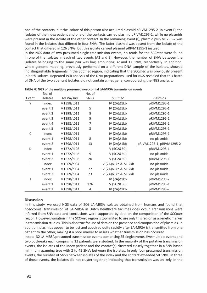
92
one of the contacts, but the isolate of this person also acquired plasmid pRIVM1295-2. In event D, the isolates of the index patient and one of the contacts carried plasmid pRIVM1295-1, while no plasmids were present in the isolate of the other contact. In the remaining event (I), plasmid pRIVM1295-2 was found in the isolates that differed in four SNVs. The latter plasmid was absent from the isolate of the contact that differed in 126 SNVs, but this isolate carried plasmid pRIVM1295-1 instead.In the NGS data of two presumed single transmission events, no reads for the SCCmec were found in one of the isolates in each of two events (A2 and E). However, the number of SNVs between the isolates belonging to the same pair was low, amounting 32 and 17 SNVs, respectively. In addition, whole genome mapping, previously performed on a different DNA sample of the isolates, showed indistinguishable fragments in the SCCmec region, indicating that the SCCmec was previously present in both isolates. Repeated PCR analysis of the DNA preparations used for NGS revealed that this batch of DNA of the two aberrant isolates did not contain a mec gene, corroborating the NGS analysis.
Table 4: NGS of the multiple presumed nosocomial LA-MRSA transmission events
DiscussionIn this study, we used NGS data of 206 LA-MRSA isolates obtained from humans and found that nosocomial transmission of LA-MRSA in Dutch healthcare facilities does occur. Transmissions were inferred from SNV data and conclusions were supported by data on the composition of the SCCmec region. However, variation in the SCCmec region is too limited to use only this region as a genetic marker in transmission studies. This is also true for use of data on the presence and composition of plasmids. In addition, plasmids appear to be lost and acquired quite rapidly after LA-MRSA is transmitted from one patient to the other, making it a poor marker to assess whether transmission has occurred.In total 32 LA-MRSA presumed transmission events comprising 25 single events, five multiple events and two outbreaks each comprising 12 patients were studied. In the majority of the putative transmission events, the isolates of the index patient and the contact(s) clustered closely together in a SNV based minimum spanning tree with 2 to 45 SNVs between the isolates. In only four presumed transmission events, the number of SNVs between isolates of the index and the contact exceeded 50 SNVs. In three of those events, the isolates did not cluster together, indicating that transmission was unlikely. In the
Event No. of
isolates MLVA/spa No. of SNPs SCCmec Plasmids
Y index MT398/t011
IV (2A)∆1kb pRIVM1295-1
event 1 MT398/t011 5 IV (2A)∆1kb pRIVM1295-1
event 2 MT398/t011 8 IV (2A)∆1kb pRIVM1295-1
event 3 MT398/t011 5 IV (2A)∆1kb pRIVM1295-1
event 4 MT398/t011 7 IV (2A)∆1kb pRIVM1295-1
event 5 MT398/t011 3 IV (2A)∆1kb pRIVM1295-1
C Index MT398/t011
IV (2A)∆1kb pRIVM1295-1
event 1 MT398/t011 8 IV (2A)∆1kb no plasmids
event 2 MT398/t011 13 IV (2A)∆1kb pRIVM1295-1, pRIVM1295-2
D Index MT572/t108
V (5C2&5C) pRIVM1295-1
event 1 MT572/t108 9 V (5C2&5C) no plasmids
event 2 MT572/t108 20 V (5C2&5C) pRIVM1295-1
L index MT569/t034
IV (2A)∆1kb & ∆1.2kb no plasmids
event 1 MT569/t034 27 IV (2A)∆1kb & ∆1.2kb no plasmids
event 2 MT569/t034 23 IV (2A)∆1kb & ∆1.2kb no plasmids
I index MT398/t011
IV (2A)∆1kb pRIVM1295-2
event 1 MT398/t011 126 V (5C2&5C) pRIVM1295-1
event 2 MT398/t011 4 IV (2A)∆1kb pRIVM1295-2
93
8
NGS
con
firm
s nos
ocom
ial t
rans
miss
ion
of L
A-M
RSA
remaining event, one of two isolates, obtained from different anatomically locations of the presumed index of an outbreak, differed 59 SNVs from its closest related outbreak isolate. However, it obviously belonged to the outbreak suggesting that a LA-MRSA strain colonizing or infecting different sites within the same person may evolve independently over time. Recent reports that applied whole genome sequencing on multiple colonies from the same person also identified a cloud of diversity among the isolates [21, 33, 34]. These studies were carried out on ST22 and ST239 MRSA isolates, but similar findings might be expected from LA-MRSA. This study showed that the number of SNVs alone is not sufficient to assess whether transmission has occurred. Combining NGS data and epidemiological data is essential to determine transmission and the use of SNV data of epidemiologically unrelated isolates as context proved extremely helpful. Furthermore, analysis of NGS data other than SNVs may be used to provide additional information to assess transmission events. Although variation of the SCCmec region turned out to be limited in LA-MRSA, it provided supportive evidence for several transmission events. For example, all isolates from a previously reported LA-MRSA outbreak had a distinctive 5.2 kb deletion in the SCCmec region that supported conclusion that transmission was indeed likely. Conversely, differences in SCCmec types provided further proof that transmission did not occur in other cases. Unexpectedly, two isolates belonging to two different transmission events and previously identified as LA-MRSA carried no SCCmec cassette. The most likely explanation for this observation is that the isolates consisted of heterogenetic populations comprising both SCCmec positive and negative variants and that the SCCmec positive variants were lost during sub-culturing to prepare DNA for NGS. The plasmid composition was identical in isolates of some of the transmissions. However, no plasmids were found in other events and in several cases differences in plasmid composition were observed although SNV and SCCmec analysis indicated that transmission was likely. Stanczak-Mrozek et al. recently showed MRSA variants that have acquired or lost mobile genetic elements were common in nasal colonizing populations [35]. Furthermore, a study using CC398 isolates showed that horizontal gene transfer including plasmids occurred in a very high frequency in vivo [36]. Since only a single colony was used for the initial culture from the clinical material and for various subcultures, different variants of the same strain, with or without plasmids, may have been sequenced, resulting in the observed differences. Our study has a number of limitations. First, all sequenced isolates belonged to the LA-MRSA (MC398) clade. This limits a comparison of NGS data between LA-MRSA and other MRSA variants and hampers studies on the transmission rates of the different MRSA clades in Dutch healthcare facilities. Second, the presumed nosocomial transmission events were selected based on the epidemiological data provided by the medical microbiology laboratories and affiliated infection prevention practitioners. We do not know whether all transmission events were correctly identified and this could lead to either an under- or over-representation of the number of presumed nosocomial transmissions. Finally, our NGS data were only screened for plasmids that were associated with livestock. Other plasmids were not taken into account and this could have provided more information. Furthermore, the use of data from other mobile genetic elements, such as bacteriophages were not included in this study [35]. In conclusion, our study strongly suggests that transmission of LA-MRSA in Dutch healthcare settings does occur. NGS could confirm previously reported transmission events and indicated that transmission was unlikely in three presumed transmissions. We conclude that investigations regarding transmissions of LA-MRSA should be supported by epidemiological data and be investigated using context of epidemiologically related and unrelated isolates. Analysis of SCCmec region proved to provide useful information to support SNV analysis, but plasmid identification did not.
Acknowledgements The authors would like to thank Rebecca te Riet from Infection Control, Ziekenhuisgroep Twente, Almelo, The Netherlands for supplying the isolates of the presumed hospital outbreak. Furthermore, the authors would like to thank all medical microbiology laboratories for sending their MRSA isolates to the RIVM. Finally, we thank the infection prevention practitioners for filling in and supplying the
94
questionnaires.
References1. Vandenesch F, Naimi T, Enright MC, Lina G, Nimmo GR, Heffernan H, et al. Community-acquired methicillin-resistant Staphylococcus aureus carrying Panton-Valentine leukocidin genes: worldwide emergence. Emerging infectious diseases. 2003;9(8):978-84.2. Armand-Lefevre L, Ruimy R, Andremont A. Clonal comparison of Staphylococcus aureus isolates from healthy pig farmers, human controls, and pigs. Emerging infectious diseases. 2005;11(5):711-4.3. Voss A, Loeffen F, Bakker J, Klaassen C, Wulf M. Methicillin-resistant Staphylococcus aureus in pig farming. Emerging infectious diseases. 2005;11(12):1965-6.4. Bhat M, Dumortier C, Taylor BS, Miller M, Vasquez G, Yunen J, et al. Staphylococcus aureus ST398, New York City and Dominican Republic. Emerging infectious diseases. 2009;15(2):285-7.5. van Cleef BA, Monnet DL, Voss A, Krziwanek K, Allerberger F, Struelens M, et al. Livestock-associated methicillin-resistant Staphylococcus aureus in humans, Europe. Emerging infectious diseases. 2011;17(3):502-5.6. Graveland H, Wagenaar JA, Verstappen KM, Oosting-van Schothorst I, Heederik DJ, Bos ME. Dynamics of MRSA carriage in veal calves: a longitudinal field study. Preventive veterinary medicine. 2012;107(3-4):180-6.7. Nemati M, Hermans K, Lipinska U, Denis O, Deplano A, Struelens M, et al. Antimicrobial resistance of old and recent Staphylococcus aureus isolates from poultry: first detection of livestock-associated methicillin-resistant strain ST398. Antimicrobial agents and chemotherapy. 2008;52(10):3817-9.8. Huijsdens XW, Bosch T, van Santen-Verheuvel MG, Spalburg E, Pluister GN, van Luit M, et al. Molecular characterisation of PFGE non-typable methicillin-resistant Staphylococcus aureus in The Netherlands, 2007. Euro surveillance : bulletin Europeen sur les maladies transmissibles = European communicable disease bulletin. 2009;14(38).9. Crombe F, Vanderhaeghen W, Dewulf J, Hermans K, Haesebrouck F, Butaye P. Colonization and transmission of methicillin-resistant Staphylococcus aureus ST398 in nursery piglets. Applied and environmental microbiology. 2012;78(5):1631-4.10. Wendlandt S, Kadlec K, Fessler AT, Mevius D, van Essen-Zandbergen A, Hengeveld PD, et al. Transmission of methicillin-resistant Staphylococcus aureus isolates on broiler farms. Veterinary microbiology. 2013;167(3-4):632-7.11. Bosch T, Verkade E, van Luit M, Landman F, Kluytmans J, Schouls LM. Transmission and persistence of livestock-associated MRSA among veterinarians and their household members. Applied and environmental microbiology. 2014.12. Bootsma MC, Wassenberg MW, Trapman P, Bonten MJ. The nosocomial transmission rate of animal-associated ST398 meticillin-resistant Staphylococcus aureus. Journal of the Royal Society, Interface / the Royal Society. 2011;8(57):578-84.13. Hetem DJ, Bootsma MC, Troelstra A, Bonten MJ. Transmissibility of livestock-associated methicillin-resistant Staphylococcus aureus. Emerging infectious diseases. 2013;19(11):1797-802.14. Wassenberg MW, Bootsma MC, Troelstra A, Kluytmans JA, Bonten MJ. Transmissibility of livestock-associated methicillin-resistant Staphylococcus aureus (ST398) in Dutch hospitals. Clinical microbiology and infection : the official publication of the European Society of Clinical Microbiology and Infectious Diseases. 2011;17(2):316-9.15. Lekkerkerk WS, van de Sande-Bruinsma N, van der Sande MA, Tjon ATA, Groenheide A, Haenen A, et al. Emergence of MRSA of unknown origin in the Netherlands. Clinical microbiology and infection : the official publication of the European Society of Clinical Microbiology and Infectious Diseases. 2012;18(7):656-61.16. van Rijen MM, Bosch T, Verkade EJ, Schouls L, Kluytmans JA, Group CAMS. Livestock-Associated MRSA Carriage in Patients without Direct Contact with Livestock. PloS one. 2014;9(6):e100294.17. Bosch T, Verkade E, van Luit M, Pot B, Vauterin P, Burggrave R, et al. High Resolution Typing by Whole Genome Mapping Enables Discrimination of LA-MRSA (CC398) Strains and Identification of Transmission Events. PloS one. 2013;8(6):e66493.18. Price LB, Stegger M, Hasman H, Aziz M, Larsen J, Andersen PS, et al. Staphylococcus aureus CC398: host adaptation and emergence of methicillin resistance in livestock. mBio. 2012;3(1).19. Stegger M, Liu CM, Larsen J, Soldanova K, Aziz M, Contente-Cuomo T, et al. Rapid differentiation between livestock-associated and livestock-independent Staphylococcus aureus CC398 clades. PloS one. 2013;8(11):e79645.20. Uhlemann AC, Porcella SF, Trivedi S, Sullivan SB, Hafer C, Kennedy AD, et al. Identification of a highly transmissible animal-independent Staphylococcus aureus ST398 clone with distinct genomic and cell adhesion properties. mBio. 2012;3(2).21. Harris SR, Cartwright EJ, Torok ME, Holden MT, Brown NM, Ogilvy-Stuart AL, et al. Whole-genome sequencing for analysis of an outbreak of meticillin-resistant Staphylococcus aureus: a descriptive study. The Lancet Infectious diseases. 2013;13(2):130-6.22. Koser CU, Holden MT, Ellington MJ, Cartwright EJ, Brown NM, Ogilvy-Stuart AL, et al. Rapid whole-genome sequencing for investigation of a neonatal MRSA outbreak. The New England journal of medicine. 2012;366(24):2267-75.23. Verkade E, Bosch T, Hendriks Y, Kluytmans J. Outbreak of methicillin-resistant Staphylococcus aureus ST398 in a Dutch nursing home. Infection control and hospital epidemiology. 2012;33(6):624-6.24. Harmsen D, Claus H, Witte W, Rothganger J, Claus H, Turnwald D, et al. Typing of methicillin-resistant Staphylococcus aureus in a university hospital setting by using novel software for spa repeat determination and database management. Journal of clinical microbiology. 2003;41(12):5442-8.25. Schouls LM, Spalburg EC, van Luit M, Huijsdens XW, Pluister GN, van Santen-Verheuvel MG, et al. Multiple-locus variable number tandem repeat analysis of Staphylococcus aureus: comparison with pulsed-field gel electrophoresis and spa-
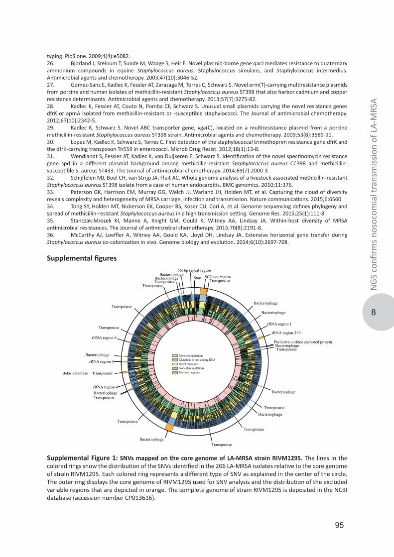
95
8
NGS
con
firm
s nos
ocom
ial t
rans
miss
ion
of L
A-M
RSA
typing. PloS one. 2009;4(4):e5082.26. Bjorland J, Steinum T, Sunde M, Waage S, Heir E. Novel plasmid-borne gene qacJ mediates resistance to quaternary ammonium compounds in equine Staphylococcus aureus, Staphylococcus simulans, and Staphylococcus intermedius. Antimicrobial agents and chemotherapy. 2003;47(10):3046-52.27. Gomez-Sanz E, Kadlec K, Fessler AT, Zarazaga M, Torres C, Schwarz S. Novel erm(T)-carrying multiresistance plasmids from porcine and human isolates of methicillin-resistant Staphylococcus aureus ST398 that also harbor cadmium and copper resistance determinants. Antimicrobial agents and chemotherapy. 2013;57(7):3275-82.28. Kadlec K, Fessler AT, Couto N, Pomba CF, Schwarz S. Unusual small plasmids carrying the novel resistance genes dfrK or apmA isolated from methicillin-resistant or -susceptible staphylococci. The Journal of antimicrobial chemotherapy. 2012;67(10):2342-5.29. Kadlec K, Schwarz S. Novel ABC transporter gene, vga(C), located on a multiresistance plasmid from a porcine methicillin-resistant Staphylococcus aureus ST398 strain. Antimicrobial agents and chemotherapy. 2009;53(8):3589-91.30. Lopez M, Kadlec K, Schwarz S, Torres C. First detection of the staphylococcal trimethoprim resistance gene dfrK and the dfrK-carrying transposon Tn559 in enterococci. Microb Drug Resist. 2012;18(1):13-8.31. Wendlandt S, Fessler AT, Kadlec K, van Duijkeren E, Schwarz S. Identification of the novel spectinomycin resistance gene spd in a different plasmid background among methicillin-resistant Staphylococcus aureus CC398 and methicillin-susceptible S. aureus ST433. The Journal of antimicrobial chemotherapy. 2014;69(7):2000-3.32. Schijffelen MJ, Boel CH, van Strijp JA, Fluit AC. Whole genome analysis of a livestock-associated methicillin-resistant Staphylococcus aureus ST398 isolate from a case of human endocarditis. BMC genomics. 2010;11:376.33. Paterson GK, Harrison EM, Murray GG, Welch JJ, Warland JH, Holden MT, et al. Capturing the cloud of diversity reveals complexity and heterogeneity of MRSA carriage, infection and transmission. Nature communications. 2015;6:6560.34. Tong SY, Holden MT, Nickerson EK, Cooper BS, Koser CU, Cori A, et al. Genome sequencing defines phylogeny and spread of methicillin-resistant Staphylococcus aureus in a high transmission setting. Genome Res. 2015;25(1):111-8.35. Stanczak-Mrozek KI, Manne A, Knight GM, Gould K, Witney AA, Lindsay JA. Within-host diversity of MRSA antimicrobial resistances. The Journal of antimicrobial chemotherapy. 2015;70(8):2191-8.36. McCarthy AJ, Loeffler A, Witney AA, Gould KA, Lloyd DH, Lindsay JA. Extensive horizontal gene transfer during Staphylococcus aureus co-colonization in vivo. Genome biology and evolution. 2014;6(10):2697-708.
Supplemental figures
Transposase
Transposase
Bacteriophage
Bacteriophage
BacteriophagePutitative surface anchored protein
rRNA region 2+3
rRNA region 1
Bacteriophage
Bacteriophage
SCCmec region
Bacteriophage
Transposase
TransposaseBacteriophagerRNA region 4
Beta-lactamase + Transposase
rRNA region 5Bacteriophage
rRNA region 6
Transposase
TransposaseTransposase
Transposase
BacteriophageBacteriophage
561bp repeat region
Start Transposase
Transposase
Transposase
Nonsense mutationsMutations in non-coding DNASilent mutationsNon-silent mutationsExcluded regions
Supplemental Figure 1: SNVs mapped on the core genome of LA-MRSA strain RIVM1295. The lines in the colored rings show the distribution of the SNVs identified in the 206 LA-MRSA isolates relative to the core genome of strain RIVM1295. Each colored ring represents a different type of SNV as explained in the center of the circle. The outer ring displays the core genome of RIVM1295 used for SNV analysis and the distribution of the excluded variable regions that are depicted in orange. The complete genome of strain RIVM1295 is deposited in the NCBI database (accession number CP013616).
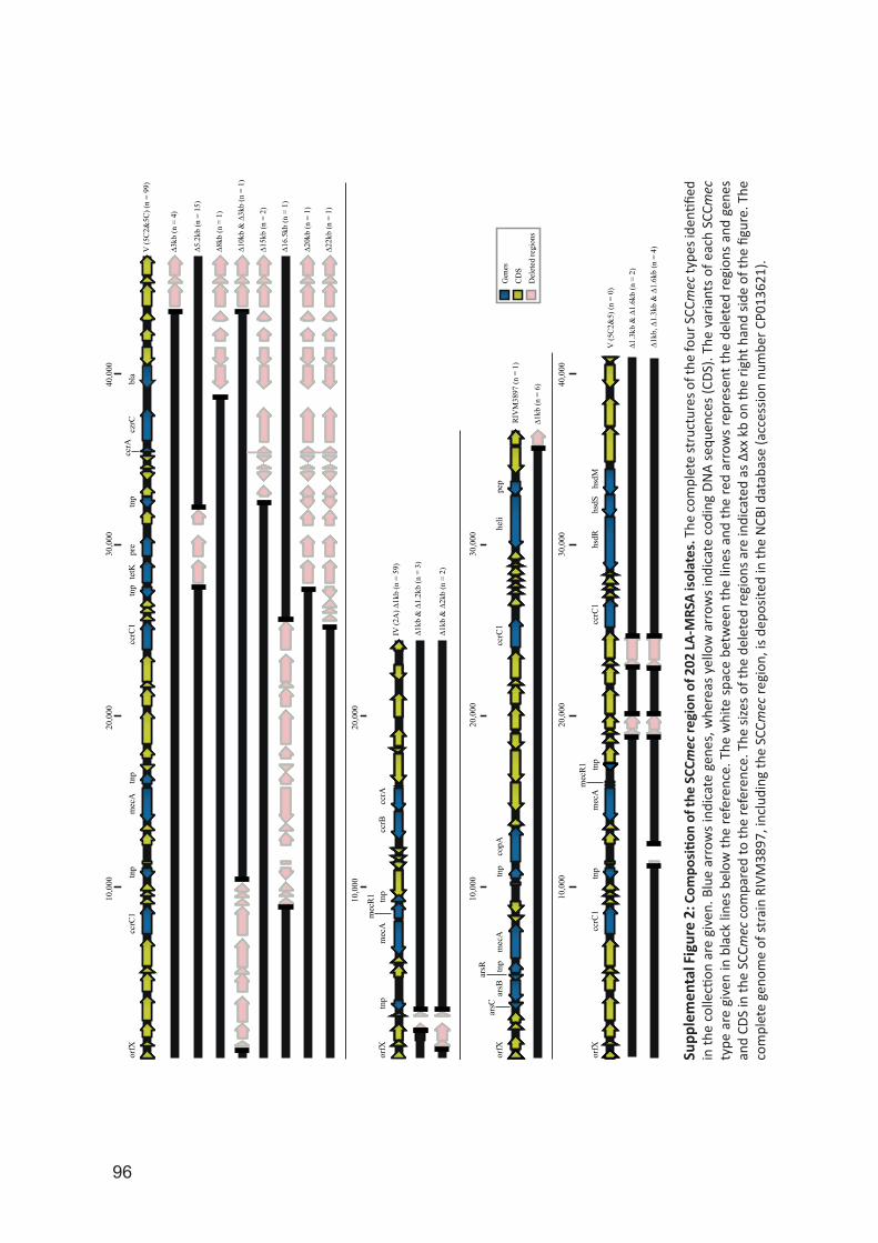
96
Gen
es
CD
S
Del
eted
regi
ons
10,0
0040
,000
30,0
0020
,000
orfX
tnp
mec
Atn
pcc
rC1
pre
tetK
ccrC
1tn
ptn
p
mec
Atn
ptn
por
fX
bla
czrC
ccrA
V (5
C2&
5C) (
n =
99)
∆3kb
(n =
4)
∆5.2
kb (n
= 1
5)
∆8kb
(n =
1)
∆10k
b &
∆3k
b (n
= 1
)
∆20k
b (n
= 1
)
∆15k
b (n
= 2
)
∆22k
b (n
= 1
)
∆ 16.
5kb
(n =
1)
10,0
0020
,000
mec
R1
ccrB
ccrA
IV (2
A) ∆
1kb
(n =
59)
∆1kb
& ∆
1.2k
b (n
= 3
)
∆1kb
& ∆
2kb
(n =
2)
10,0
0020
,000
30,0
00
orfX
tnp
tnp
ccrC
1m
ecA
copA
arsC
arsR
arsB
heli
pep
RIV
M38
97 (n
= 1
)
∆1kb
(n =
6)
20,0
0030
,000
10,0
0040
,000
ccrC
1tn
pm
ecA
mec
R1 tnp
ccrC
1hs
dRhs
dMhs
dS
V (5
C2&
5) (n
= 0
)
∆1.3
kb &
∆1.
6kb
(n =
2)
∆1kb
, ∆1.
3kb
& ∆
1.6k
b (n
= 4
)
orfX Supp
lem
enta
l Fig
ure
2: C
ompo
sitio
n of
the
SCCm
ec re
gion
of 2
02 LA
-MRS
A is
olat
es. T
he co
mpl
ete
stru
ctur
es o
f the
four
SCC
mec
type
s ide
ntifie
d in
the
colle
ction
are
giv
en. B
lue
arro
ws
indi
cate
gen
es, w
here
as y
ello
w a
rrow
s in
dica
te c
odin
g DN
A se
quen
ces
(CDS
). Th
e va
riant
s of
eac
h SC
Cmec
ty
pe a
re g
iven
in b
lack
line
s be
low
the
refe
renc
e. T
he w
hite
spa
ce b
etw
een
the
lines
and
the
red
arro
ws
repr
esen
t the
del
eted
regi
ons
and
gene
s an
d CD
S in
the
SCCm
ec c
ompa
red
to th
e re
fere
nce.
The
size
s of t
he d
elet
ed re
gion
s are
indi
cate
d as
∆xx
kb
on th
e rig
ht h
and
side
of th
e fig
ure.
The
co
mpl
ete
geno
me
of st
rain
RIV
M38
97, i
nclu
ding
the
SCCm
ec re
gion
, is d
epos
ited
in th
e N
CBI d
atab
ase
(acc
essio
n nu
mbe
r CP0
1362
1).
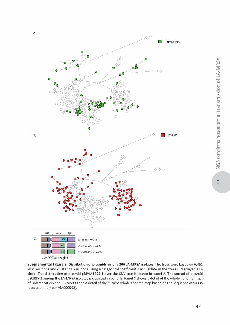
97
8
NGS
con
firm
s nos
ocom
ial t
rans
miss
ion
of L
A-M
RSA
80kb40kb 60kb
C.
B.
A.
S0385 real WGM
S0385 in silico WGM
pRIVM1295-1
pS0385-1
13kb
8kb
8kb RIVM5890 real WGM
SCCmec regionorfX
Supplemental Figure 3: Distribution of plasmids among 206 LA-MRSA isolates. The trees were based on 6,461 SNV positions and clustering was done using a categorical coefficient. Each isolate in the trees is displayed as a circle. The distribution of plasmid pRIVM1295-1 over the SNV tree is shown in panel A. The spread of plasmid pS0385-1 among the LA-MRSA isolates is depicted in panel B. Panel C shows a detail of the whole genome maps of isolates S0385 and RIVM5890 and a detail of the in silico whole genome map based on the sequence of S0385 (accession number AM990992).

98
9
Thijs Bosch
Leo Schouls
Erwin Verkade
Jan Kluytmans
Miranda van Rijen
Plos One
2014
9(6): e100294
the CAM study group
Livestock-associated MRSA carriage in patients without direct contact with livestock
99
LA-M
RSA
in p
atien
ts w
ithou
t liv
esto
ck c
onta
ct
9
AbstractBackground: Livestock-associated MRSA (MC398) has emerged and is related to an extensive reservoir in pigs and veal calves. Individuals with direct contact with these animals and their family members are known to have high MC398 carriage rates. Until now, it was assumed that MC398 does not spread to individuals in the community without pig or veal calf exposure. To test this, we identified the proportion of MC398 in MRSA positive individuals without contact with pigs/veal calves or other known risk factors (MRSA of unknown origin; MUO).Methods: In 17 participating hospitals, we determined during two years the occurrence of MC398 in individuals without direct contact with livestock and no other known risk factor (n = 271) and tested in a post analysis the hypothesis whether hospitals in pig-dense areas have higher proportions of MC398 of all MUO.Results: Fifty-six individuals (20.7%) without animal contact carried MC398. In hospitals with high pig-densities in the adherence area, the proportion of MC398 of all MUO was higher than this proportion in hospitals without pigs in the surroundings.Conclusions: One fifth of the individuals carrying MUO carried MC398. So, MC398 is found in individuals without contact to pigs or veal calves. The way of transmission from the animal reservoir to these individuals is unclear, probably by human-to- human transmission or by exposure to the surroundings of the stables. Further research is needed to investigate the way of transmission.
100
IntroductionSince 2003, the so-called livestock-associated MRSA (LA-MRSA) has emerged in animals and humans in areas with intensive animal farming in Europe, North America, and Asia [1]. Human carriage of LA-MRSA is strongly related to direct contact with pigs, veal calves and broilers [2, 3]. The majority of these LA- MRSA strains belong to multilocus sequence type clonal complex 398 (CC398) [4]. After its emergence, the risk factor ‘direct contact with living pigs, veal calves and broilers’ was added to the Dutch national MRSA guideline and an active screening program in hospitals was implemented [5]. By the end of 2011, 39% of all newly identified MRSA strains in humans in the Netherlands belonged to this variant in the Netherlands [6].Recent surveys showed that MRSA CC398 was 4 to 6-fold less transmissible than other MRSA strains in a hospital-setting [7–8]. At present, the human-to-human transmissibility of MRSACC398 in a community setting is still unclear. Considering the extensive reservoir in animals and people who work with livestock, the occurrence of MRSA CC398 in people who are not directly involved in farming is strikingly low. So far, there are no indications that MRSA CC398 has spread extensively into the general population [9]. A cross-sectional survey in a livestock- dense region found that only 0.2% of adult individuals without livestock contact were positive for MRSA CC398 [10]. On the other hand, there are observations that proximity of farms is a potential risk factor, even in absence of direct contact between humans and animals [11–13]. In addition, in a recent exploratory study an association was found between consumption of poultry and MRSA carriage [14]. A spectrum of infections with MRSA CC398 have been documented, ranging from relatively minor or localized infections including abscesses [15–17] and various skin and soft tissue infections (SSTI) [18–20], urinary tract infections [16], wound infections [16], mastitis [4], and conjunctivitis [21], as well as more serious or invasive infections, including bacteremia [21–24], endocarditis [24, 25], pneumonia (including necrotizing pneumonia, osteomyelitis, pyomyositis, and postoperative infections [26]. Despite the diverse array of infection types reported, it has been suggested that MRSA CC398 is less virulent than other human MRSA strains [27].Apart from LA-MRSA and hospital-associated (HA)MRSA, MRSA rates also are rapidly increasing in community dwelling individuals without known healthcare- or livestock-associated risk factors. This third entity has been referred to as community- acquired (CA) MRSA [28] or MUO [29]. In this study, the proportion of CC398 in MUO isolates was determined. We hypothesized that people living in an area in which CC398 is common have more risk of MRSA CC398 carriage than persons living in an area in which CC398 is rare.
MethodsEthics StatementEthical approval for the study was obtained by the medical ethics committee of the St. Elisabeth Hospital in Tilburg (NL 19489.008.07, protocol 0749, March 9th, 2009). Patient information was anonymized and de-identified prior to analysis.
MRSA source identificationTo identify MRSA sources in the Netherlands, Infection Control Practitioners (ICP) from seventeen hospitals (three academic, seven teaching and seven general hospitals) throughoutthe Netherlands were asked to complete a questionnaire on a website for all consecutive patients that were found to be MRSA positive (both infection and carriage) for the first time in the microbiological laboratory of the hospital from January 2009 until December 2010. Samples were taken during a visit to the outpatient’s clinic or during a stay on a ward in the hospital. Patients who had already been found MRSA-positive in the past were not included. The questionnaire on the website contained data about patient type (in- or outpatient), demographics, positive body sites, molecular typing results and probable source of MRSA. The MRSA source was identified based on the patient’s history combined with molecular typing results and then classified in risk groups described in the national infection prevention guidelines [5]. When neither of these risk groups was applicable, the MRSA was classified
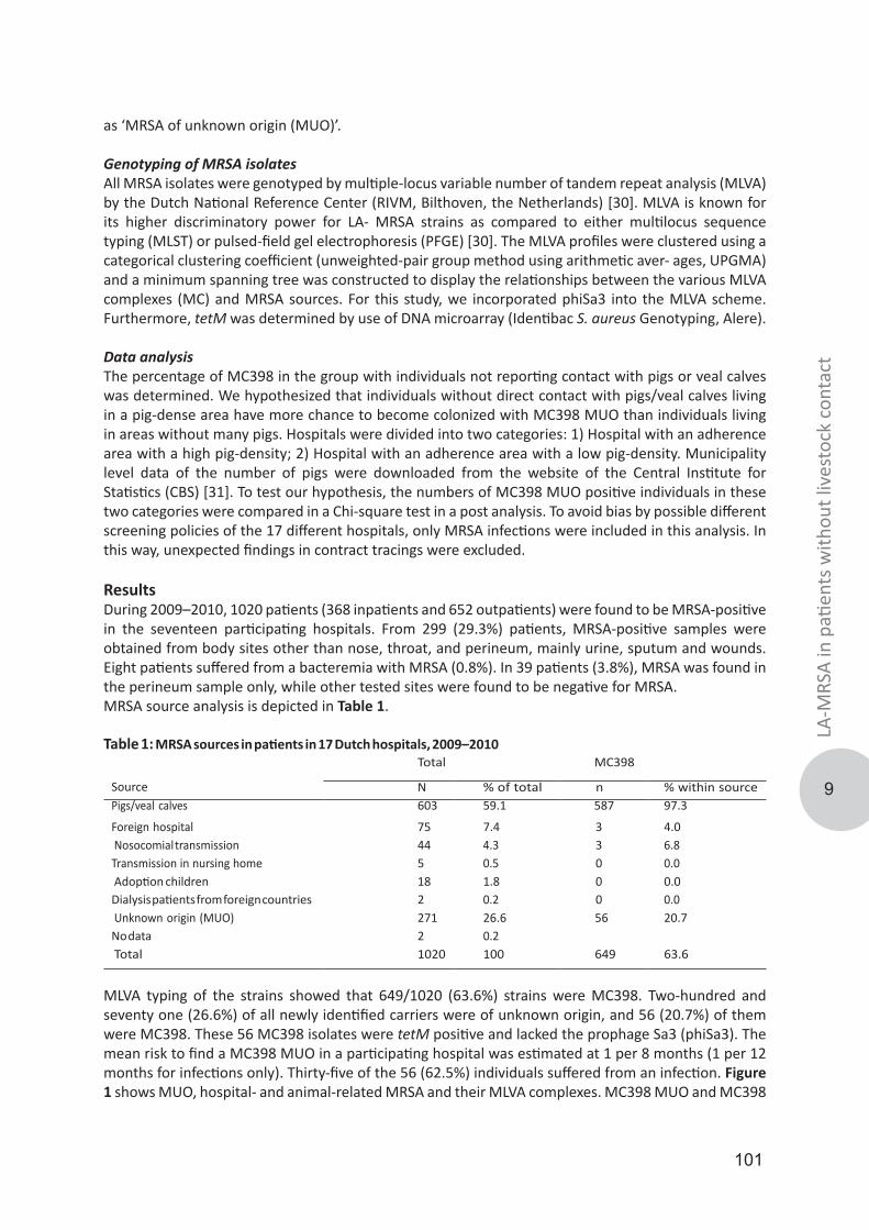
101
LA-M
RSA
in p
atien
ts w
ithou
t liv
esto
ck c
onta
ct
9
as ‘MRSA of unknown origin (MUO)’.
Genotyping of MRSA isolatesAll MRSA isolates were genotyped by multiple-locus variable number of tandem repeat analysis (MLVA) by the Dutch National Reference Center (RIVM, Bilthoven, the Netherlands) [30]. MLVA is known for its higher discriminatory power for LA- MRSA strains as compared to either multilocus sequence typing (MLST) or pulsed-field gel electrophoresis (PFGE) [30]. The MLVA profiles were clustered using a categorical clustering coefficient (unweighted-pair group method using arithmetic aver- ages, UPGMA) and a minimum spanning tree was constructed to display the relationships between the various MLVA complexes (MC) and MRSA sources. For this study, we incorporated phiSa3 into the MLVA scheme. Furthermore, tetM was determined by use of DNA microarray (Identibac S. aureus Genotyping, Alere).
Data analysisThe percentage of MC398 in the group with individuals not reporting contact with pigs or veal calves was determined. We hypothesized that individuals without direct contact with pigs/veal calves living in a pig-dense area have more chance to become colonized with MC398 MUO than individuals living in areas without many pigs. Hospitals were divided into two categories: 1) Hospital with an adherence area with a high pig-density; 2) Hospital with an adherence area with a low pig-density. Municipality level data of the number of pigs were downloaded from the website of the Central Institute for Statistics (CBS) [31]. To test our hypothesis, the numbers of MC398 MUO positive individuals in these two categories were compared in a Chi-square test in a post analysis. To avoid bias by possible different screening policies of the 17 different hospitals, only MRSA infections were included in this analysis. In this way, unexpected findings in contract tracings were excluded.
ResultsDuring 2009–2010, 1020 patients (368 inpatients and 652 outpatients) were found to be MRSA-positive in the seventeen participating hospitals. From 299 (29.3%) patients, MRSA-positive samples were obtained from body sites other than nose, throat, and perineum, mainly urine, sputum and wounds. Eight patients suffered from a bacteremia with MRSA (0.8%). In 39 patients (3.8%), MRSA was found in the perineum sample only, while other tested sites were found to be negative for MRSA.MRSA source analysis is depicted in Table 1.
MLVA typing of the strains showed that 649/1020 (63.6%) strains were MC398. Two-hundred and seventy one (26.6%) of all newly identified carriers were of unknown origin, and 56 (20.7%) of them were MC398. These 56 MC398 isolates were tetM positive and lacked the prophage Sa3 (phiSa3). The mean risk to find a MC398 MUO in a participating hospital was estimated at 1 per 8 months (1 per 12 months for infections only). Thirty-five of the 56 (62.5%) individuals suffered from an infection. Figure 1 shows MUO, hospital- and animal-related MRSA and their MLVA complexes. MC398 MUO and MC398
Total MC398
Source N % of total n % within sourcePigs/veal calves 603 59.1 587 97.3
Foreign hospital 75 7.4 3 4.0Nosocomial transmission 44 4.3 3 6.8Transmission in nursing home 5 0.5 0 0.0Adoption children 18 1.8 0 0.0Dialysis patients from foreign countries 2 0.2 0 0.0Unknown origin (MUO) 271 26.6 56 20.7No data 2 0.2Total 1020 100 649 63.6
Table 1: MRSA sources in patients in 17 Dutch hospitals, 2009–2010
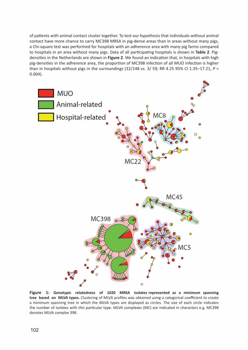
102
of patients with animal contact cluster together. To test our hypothesis that individuals without animal contact have more chance to carry MC398 MRSA in pig-dense areas than in areas without many pigs, a Chi-square test was performed for hospitals with an adherence area with many pig farms compared to hospitals in an area without many pigs. Data of all participating hospitals is shown in Table 2. Pig-densities in the Netherlands are shown in Figure 2. We found an indication that, in hospitals with high pig-densities in the adherence area, the proportion of MC398 infection of all MUO infection is higher than in hospitals without pigs in the surroundings (32/148 vs. 3/ 59; RR 4.25 95% CI 1.35–17.21, P = 0.004).
Figure 1: Genotypic relatedness of 1020 MRSA isolates represented as a minimum spanning tree based on MLVA types. Clustering of MLVA profiles was obtained using a categorical coefficient to create a minimum spanning tree in which the MLVA types are displayed as circles. The size of each circle indicates the number of isolates with this particular type. MLVA complexes (MC) are indicated in characters e.g. MC398 denotes MLVA complex 398.
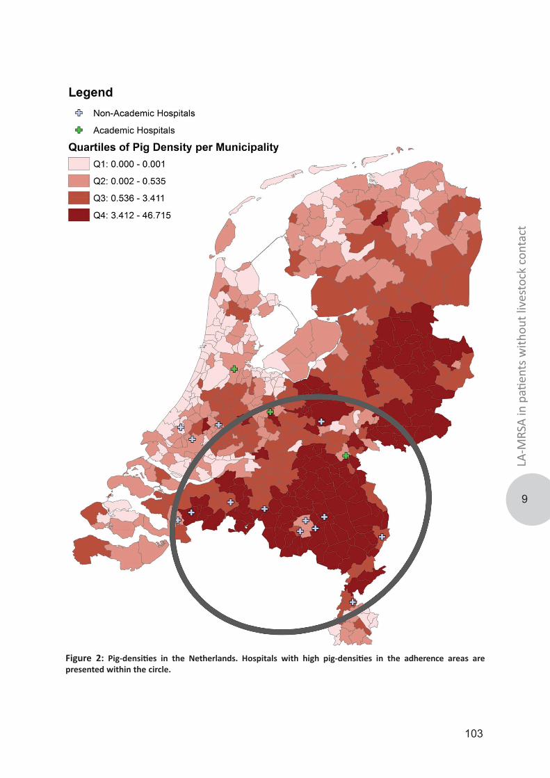
103
LA-M
RSA
in p
atien
ts w
ithou
t liv
esto
ck c
onta
ct
9
Figure 2: Pig-densities in the Netherlands. Hospitals with high pig-densities in the adherence areas are presented within the circle.
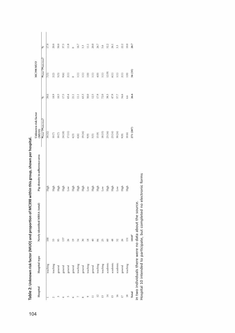
104
Hos
pita
lH
ospi
tal t
ype
New
ly id
enti
fied
MR
SA (t
otal
)Pi
g-de
nsit
y in
adh
eren
ce a
rea
Unk
now
n ri
sk fa
ctor
(M
UO
)M
C39
8 M
UO
nto
tal (n
infe
ctio
n)
%n
tota
l (n
infe
ctio
n)
%
1te
achi
ng10
0H
igh
39 (3
2)39
.07 (
5)17
.9
2te
achi
ng53
Hig
h10
(7)
18.9
2 (2)
20.0
3ge
nera
l95
Hig
h10
(7)
10.5
5 (5)
50.0
4ge
nera
l13
7H
igh
24 (1
8)17
.59 (
6)37
.5
5ge
nera
l26
Low
17 (1
1)65
.42 (
1)11
.8
6ge
nera
l19
Hig
h4 (
3)21
.10
0
7te
achi
ng54
Hig
h6 (
6)11
.11 (
1)16
.7
8ge
nera
l30
Hig
h19
(16)
63.3
1 (1)
5.3
9te
achi
ng18
Low
9 (9)
50.0
1 (0)
11.1
11ge
nera
l40
Hig
h5 (
5)12
.51 (
1)20
.0
12te
achi
ng84
Hig
h15
(9)
17.9
4 (0)
26.7
13te
achi
ng25
Low
18 (1
5)72
.01 (
1)5.
6
14ac
adem
ic60
Hig
h23
(16)
38.3
12 (9
)52
.2
15ac
adem
ic48
Hig
h23
(14)
47.9
6 (1)
26.1
16ac
adem
ic52
Low
30 (2
4)57
.71 (
1)3.
3
17ge
nera
l26
Hig
h9 (
9)34
.62 (
1)22
.2
18te
achi
ng15
1H
igh
10 (6
)6.
61 (
0)10
.0
Tota
l10
18a
271
(207
)26
.656
(35)
20.7
Tabl
e 2:
Unk
now
n ris
k fa
ctor
(MU
O) a
nd p
ropo
rtion
of M
C398
with
in th
is g
roup
, sho
wn
per h
ospi
tal.
in t
wo
indi
vidu
als
ther
e w
ere
no d
ata
abou
t th
e so
urce
.H
ospi
tal 1
0 in
tend
ed t
o pa
rtici
pate
, but
com
plet
ed n
o el
ectr
onic
for
ms
105
LA-M
RSA
in p
atien
ts w
ithou
t liv
esto
ck c
onta
ct
9
DiscussionThe majority (n = 603, 59.1%) of newly identified MRSA- positive patients in 17 hospitals in 2009 and 2010 was related to exposure to livestock. A substantial proportion could not be classified to an established risk group (n = 271, 26.6%) and are therefore assumed to have acquired their MRSA in the community. One fifth (20.7%) of these MRSA strains belonged to MC398. The presence of the tetM resistance gene and the absence of the phiSa3 suggest that these isolates were animal- associated [22, 32]. We found an indication that, in hospitals with high pig-density in the surroundings, the proportion of MC398 infection of all MUO infection was higher than in hospitals with a low pig-density in the surroundings. This indicates that LA-MRSA may be spreading through other sources than direct exposure to livestock. Until now it was assumed that LA-MRSA is able to spread to the pig/veal calf farmers and others who are in close contact with the animals, but is less able to spread from the farmer to household members who do not enter the stables, and is almost unable to spread to persons in the community without pig or veal calf exposure. Thus, it is assumed that constant pressure of LA- MRSA from animals with MRSA must be present to maintain the LA-MRSA colonization in humans. However, several recent studies have shown that persistent colonization with MC398 is possible [33–35]. Moreover, pig-, dairy cow, and veal calf densities per municipality were also found to be independent risk factors for carriage of MRSA MC398 in two recently published case-control studies [11, 14]. Although it cannot be excluded that human-to- human transmission occurs in areas with a high MRSA MC398 pressure, environmental contamination with MRSA MC398 may play a role as well. MRSA MC398 has been shown to be present in air and soil samples collected downwind of pig and swine barns [13]. Other transmission routes can play a role as well. For example, regular consumption of poultry was recently found to be associated with CA-MRSA transmission in an exploratory hospital-based case-control study [14]. De Boer et al. demonstrated that a substantial part of the meat products obtained from retail stores in the Netherlands were colonized with MRSA, including both MC398 and non-MC398 strain types [36]. However, meat consumption cannot explain the increased prevalence in people who live in pig-dense areas. We expect the risk, associated with meat consumption, to be the same for all areas over the country. Unless, locals consume more meat from their own area.
LimitationsWe performed a post hoc analysis to study whether the proportion of MC398 MUO infection is higher in hospitals in pig- dense areas than in areas with a low pig-density. Our study was originally not designed for this purpose. Therefore, we have to be careful with the conclusions. An analysis in which pig-density was determined based on postal code of the individuals would have been more reliable. These data were not available because of privacy issues. Also, the stratification of hospitals in ‘pig-dense’ and ‘pig-arm’ areas is arbitrary. Based on the CBS data, we classified the hospitals that are known to be situated in the most urbanized parts of the country as ‘pig-arm’. This resulted in four hospitals in pig-arm areas and 13 hospitals in pig-dense areas. So, more hospitals in pig-dense areas were included in the analysis. Furthermore, there may be detection bias due to differences in screening policies between hospitals. It is possible that physicians in some hospitals take more clinical samples than physicians in other hospitals. This may lead to an underestimation in the number of MUO findings. Also, classification bias may occur depending on the reliability of the history of risk factors. However, all participating hospitals screened the MRSA risk groups described in the national MRSA guideline [5]. After coincidental MRSA findings, patients were asked for these risk factors also.In conclusion, this study shows that the majority of newly identified MRSA patients in these 17 hospitals were acquired by direct contact with pigs/veal calves. The second largest group is the group of unknown origin. One fifth of these MUO are MC398. We found a significant association between individuals living in pig-dense areas and the likelihood of MC398 MUO carriage. MC398 MUO infections were rarely detected, i.e. 1 per 12 months for every participating hospital, so, currently, this MC398 MUO seems not to cause many problems. Because of the absence of known risk factors and probable risk for transmission in the healthcare settings, it is worthwhile to monitor the number of MUO in general, and of MC398 separately, in the coming years.
106
AcknowledgmentsCAM Study Group:Amphia Hospital, Breda: Brigitte van Cleef, Yvonne Hendriks, Danie¨lle op den Kamp, Jan Kluytmans, Marjolein Kluytmans – van den Bergh, Rene´e Ladestein, Rudolf Punselie, Miranda van Rijen, Erwin VerkadeCatharina Hospital, Eindhoven: Marieke Ernest, Mireille Wulf Duke University, Durham, NC, USA: Beth FeingoldElkerliek Hospital, Helmond: Marie¨lla Brekelmans, Patricia Willemse, Mireille WulfFranciscus Hospital, Roosendaal: Nelleke ten Feld, Rob Wintermans Hospital Gelderse Vallei, Ede: Bent Postma, Marja Terwee, Thuy-Nga Groene Hart Hospital, Gouda: Truus de Ruiter, Eric van der Vorm,Antoinette WijkerLievensberg Hospital, Bergen op Zoom: Henk Hamers, Rob Winter- mansMaxima Medical Centre, Eindhoven/Veldhoven: Philo Das, Ellen Sanders, Kees VerduinOrbis Medical Centre, Sittard: Dick van Dam, Jan Diederen Regional Health Authority, Leiden: Peter ten HamReinier de Graaf Hospital, Delft: Loes Nolles, Eric van der Vorm St. Anna Hospital, Geldrop: Anouk Smeulders, Kees VerduinSt. Elisabeth Hospital, Tilburg: Anton Buiting, Helma Ho¨rmann, Ellen NieuwkoopSt. Franciscus Gasthuis, Rotterdam: Yvonne MullerUniversity Medical Centre St Radboud, Nijmegen: Diana Haverkate, Paul VerweijUniversity Medical Centre Utrecht, Utrecht: Marc Bonten, Marischka van der Jagt-Zwetsloot, Annet TroelstraVieCuri Medical Centre, Venlo: Hanneke Berkhout, Liduine van den HoutVU Medical Centre, Amsterdam: Annie Kaiser, Christina Vanden- broucke-GraulsWe want to thank Beth Feingold for creating figure 2.
References1. Smith TC, Pearson N. The emergence of Staphylococcus aureus ST398. Vector borne and zoonotic diseases. 2011;11(4):327-39.2. van Loo I, Huijsdens X, Tiemersma E, de Neeling A, van de Sande-Bruinsma N, Beaujean D, et al. Emergence of methicillin-resistant Staphylococcus aureus of animal origin in humans. Emerging infectious diseases. 2007;13(12):1834-9.3. Mulders MN, Haenen AP, Geenen PL, Vesseur PC, Poldervaart ES, Bosch T, et al. Prevalence of livestock-associated MRSA in broiler flocks and risk factors for slaughterhouse personnel in The Netherlands. Epidemiology and infection. 2010;138(5):743-55.4. Huijsdens XW, van Dijke BJ, Spalburg E, van Santen-Verheuvel MG, Heck ME, Pluister GN, et al. Community-acquired MRSA and pig-farming. Annals of clinical microbiology and antimicrobials. 2006;5:26.5. Dutch working party on infection prevention. MRSA hospital. 20076. Surveillance of MRSA in the Netherlands 2011. Infectieziekten Bulletin. 2012.7. Wassenberg MW, Bootsma MC, Troelstra A, Kluytmans JA, Bonten MJ. Transmissibility of livestock-associated methicillin-resistant Staphylococcus aureus (ST398) in Dutch hospitals. Clinical microbiology and infection : the official publication of the European Society of Clinical Microbiology and Infectious Diseases. 2011;17(2):316-9.8. Bootsma MC, Wassenberg MW, Trapman P, Bonten MJ. The nosocomial transmission rate of animal-associated ST398 meticillin-resistant Staphylococcus aureus. Journal of the Royal Society, Interface / the Royal Society. 2011;8(57):578-84.9. Cuny C, Nathaus R, Layer F, Strommenger B, Altmann D, Witte W. Nasal colonization of humans with methicillin-resistant Staphylococcus aureus (MRSA) CC398 with and without exposure to pigs. PloS one. 2009;4(8):e6800.10. van Cleef BA, Verkade EJ, Wulf MW, Buiting AG, Voss A, Huijsdens XW, et al. Prevalence of livestock-associated MRSA in communities with high pig-densities in The Netherlands. PloS one. 2010;5(2):e9385.11. Feingold BJ, Silbergeld EK, Curriero FC, van Cleef BA, Heck ME, Kluytmans JA. Livestock density as risk factor for livestock-associated methicillin-resistant Staphylococcus aureus, the Netherlands. Emerging infectious diseases. 2012;18(11):1841-9.12. van Cleef BA, Monnet DL, Voss A, Krziwanek K, Allerberger F, Struelens M, et al. Livestock-associated methicillin-resistant Staphylococcus aureus in humans, Europe. Emerging infectious diseases. 2011;17(3):502-5.13. Schulz J, Friese A, Klees S, Tenhagen BA, Fetsch A, Rosler U, et al. Longitudinal study of the contamination of air and of soil surfaces in the vicinity of pig barns by livestock-associated methicillin-resistant Staphylococcus aureus. Applied and environmental microbiology. 2012;78(16):5666-71.14. van Rijen MM, Kluytmans-van den Bergh MF, Verkade EJ, Ten Ham PB, Feingold BJ, Kluytmans JA, et al. Lifestyle-
107
LA-M
RSA
in p
atien
ts w
ithou
t liv
esto
ck c
onta
ct
9
Associated Risk Factors for Community-Acquired Methicillin-Resistant Staphylococcus aureus Carriage in the Netherlands: An Exploratory Hospital-Based Case-Control Study. PloS one. 2013;8(6):e65594.15. Fanoy E, Helmhout LC, van der Vaart WL, Weijdema K, van Santen-Verheuvel MG, Thijsen SF, et al. An outbreak of non-typeable MRSA within a residential care facility. Euro surveillance : bulletin Europeen sur les maladies transmissibles = European communicable disease bulletin. 2009;14(1).16. van Belkum A, Melles DC, Peeters JK, van Leeuwen WB, van Duijkeren E, Huijsdens XW, et al. Methicillin-resistant and -susceptible Staphylococcus aureus sequence type 398 in pigs and humans. Emerging infectious diseases. 2008;14(3):479-83.17. Pan A, Battisti A, Zoncada A, Bernieri F, Boldini M, Franco A, et al. Community-acquired methicillin-resistant Staphylococcus aureus ST398 infection, Italy. Emerging infectious diseases. 2009;15(5):845-7.18. Declercq P, Petre D, Gordts B, Voss A. Complicated community-acquired soft tissue infection by MRSA from porcine origin. Infection. 2008;36(6):590-2.19. van Rijen MM, Van Keulen PH, Kluytmans JA. Increase in a Dutch hospital of methicillin-resistant Staphylococcus aureus related to animal farming. Clinical infectious diseases : an official publication of the Infectious Diseases Society of America. 2008;46(2):261-3.20. Krziwanek K, Metz-Gercek S, Mittermayer H. Methicillin-Resistant Staphylococcus aureus ST398 from human patients, upper Austria. Emerging infectious diseases. 2009;15(5):766-9.21. Grisold AJ, Zarfel G, Hoenigl M, Krziwanek K, Feierl G, Masoud L, et al. Occurrence and genotyping using automated repetitive-sequence-based PCR of methicillin-resistant Staphylococcus aureus ST398 in Southeast Austria. Diagnostic microbiology and infectious disease. 2010;66(2):217-21.22. Valentin-Domelier AS, Girard M, Bertrand X, Violette J, Francois P, Donnio PY, et al. Methicillin-susceptible ST398 Staphylococcus aureus responsible for bloodstream infections: an emerging human-adapted subclone? PloS one. 2011;6(12):e28369.23. Verkade E, Bergmans AM, Budding AE, van Belkum A, Savelkoul P, Buiting AG, et al. Recent emergence of Staphylococcus aureus clonal complex 398 in human blood cultures. PloS one. 2012;7(10):e41855.24. Ekkelenkamp MB, Sekkat M, Carpaij N, Troelstra A, Bonten MJ. [Endocarditis due to meticillin-resistant Staphylococcus aureus originating from pigs]. Ned Tijdschr Geneeskd. 2006;150(44):2442-7.25. Tristan A, Rasigade JP, Ruizendaal E, Laurent F, Bes M, Meugnier H, et al. Rise of CC398 lineage of Staphylococcus aureus among Infective endocarditis isolates revealed by two consecutive population-based studies in France. PloS one. 2012;7(12):e51172.26. Witte W, Strommenger B, Stanek C, Cuny C. Methicillin-resistant Staphylococcus aureus ST398 in humans and animals, Central Europe. Emerging infectious diseases. 2007;13(2):255-8.27. Grundmann H, Aanensen DM, van den Wijngaard CC, Spratt BG, Harmsen D, Friedrich AW, et al. Geographic distribution of Staphylococcus aureus causing invasive infections in Europe: a molecular-epidemiological analysis. PLoS medicine. 2010;7(1):e1000215.28. David MZ, Daum RS. Community-associated methicillin-resistant Staphylococcus aureus: epidemiology and clinical consequences of an emerging epidemic. Clin Microbiol Rev. 2010;23(3):616-87.29. Lekkerkerk WS, van de Sande-Bruinsma N, van der Sande MA, Tjon ATA, Groenheide A, Haenen A, et al. Emergence of MRSA of unknown origin in the Netherlands. Clinical microbiology and infection : the official publication of the European Society of Clinical Microbiology and Infectious Diseases. 2012;18(7):656-61.30. Schouls LM, Spalburg EC, van Luit M, Huijsdens XW, Pluister GN, van Santen-Verheuvel MG, et al. Multiple-locus variable number tandem repeat analysis of Staphylococcus aureus: comparison with pulsed-field gel electrophoresis and spa-typing. PloS one. 2009;4(4):e5082.31. CBS Central bureau of statistics. 200932. Price LB, Stegger M, Hasman H, Aziz M, Larsen J, Andersen PS, et al. Staphylococcus aureus CC398: host adaptation and emergence of methicillin resistance in livestock. mBio. 2012;3(1).33. Kock R, Loth B, Koksal M, Schulte-Wulwer J, Harlizius J, Friedrich AW. Persistence of nasal colonization with livestock-associated methicillin-resistant Staphylococcus aureus in pig farmers after holidays from pig exposure. Applied and environmental microbiology. 2012;78(11):4046-7.34. Slingerland BC, Tavakol M, McCarthy AJ, Lindsay JA, Snijders SV, Wagenaar JA, et al. Survival of Staphylococcus aureus ST398 in the human nose after artificial inoculation. PloS one. 2012;7(11):e48896.35. Verkade E, van Benthem B, den Bergh MK, van Cleef B, van Rijen M, Bosch T, et al. Dynamics and determinants of Staphylococcus aureus carriage in livestock veterinarians: a prospective cohort study. Clinical infectious diseases : an official publication of the Infectious Diseases Society of America. 2013;57(2):e11-7.36. de Boer E, Zwartkruis-Nahuis JT, Wit B, Huijsdens XW, de Neeling AJ, Bosch T, et al. Prevalence of methicillin-resistant Staphylococcus aureus in meat. Int J Food Microbiol. 2009;134(1-2):52-6.

108
10
General discussion
109
Gene
ral d
iscus
sion
10
General discussion After the first emergence in 1961, methicillin resistant Staphylococcus aureus (MRSA) has been considered a human pathogen for several decades. First, MRSA was deemed a problem restricted to healthcare settings, but since the 1990s was also recognized as a community-acquired pathogen [1, 2]. In 2005, a new reservoir was revealed when MRSA was isolated from pigs and pig farmers in France and the Netherlands [3, 4]. Further research in the Netherlands showed that the prevalence of this MRSA variant among pigs was high and that this type of MRSA was also present in other animals, leading to the term livestock-associated MRSA (LA-MRSA) [5, 6]. The rapid increase in the prevalence of LA-MRSA in livestock and the fact that contact of humans with livestock was often associated with human LA-MRSA carriage led to an adaption of the Dutch search & destroy guidelines whereby contact with livestock was regarded a MRSA risk factor [7]. The emergence of LA-MRSA might pose a serious public health threat and it is essential to have insight in the clinical importance of LA-MRSA in regards to its ability to transmit and cause disease in humans. In this thesis, we developed and used molecular typing tools to assess the capability of LA-MRSA to transmit between humans. Furthermore, we looked at the ability of LA-MRSA to cause disease and studied the possible temporal changes in the characteristics of the most predominant MRSA clade in the Netherlands. In this chapter, we discuss the experimental data, present the main conclusions and depict the future perspectives.
High-resolution typing reveals that LA-MRSA are genotypically diverseStudies on the origin and transmission routes of a microorganism require typing techniques with sufficient discriminatory power. For several decades, pulsed field gel electrophoresis (PFGE) has been considered to be the gold-standard typing tool for MRSA [8]. However, with the introduction of sequence-based techniques PFGE was replaced by multilocus sequence typing (MLST) and/or Staphylococcal protein A (spa-)typing in most laboratories [9, 10]. For most MRSA isolates sufficient discrimination could be achieved by any of the three methods, but for LA-MRSA the methods resulted in only limited differentiation. For instance, no banding patterns could be obtained when PFGE was applied on LA-MRSA isolates, due to a methylation of the SmaI recognition site [11]. MLST of LA-MRSA was applied in many of the first reports about this MRSA clade, but virtually all isolates belonged to sequence-type (ST)398 within clonal complex (CC) 398 [3, 12]. Although spa-typing could identify more LA-MRSA types than MLST, over 85% of all submitted Dutch LA-MRSA from humans belonged to either spa-type t011, t108 and t034 [13, 14]. In addition to spa-typing, the national institute for public health and the environment (RIVM) introduced a second typing technique named multiple-locus variable number of tandem repeat analysis (MLVA) in 2008 [15]. MLVA proved to be more discriminatory than spa-typing and MLST for most MRSA variants, but for LA-MRSA most isolates had either MLVA-type (MT)398, MT572 or MT569 and these MTs were mostly comprised of the three major spa-types t011 (MT398), t108 (MT572) and t034 (MT569) (Chapter 5). A calculation of Simpsons’ index of diversity revealed equally poor results for both spa-typing and MLVA, while a comparison by Wallace coefficients of both methods for LA-MRSA yielded high Wallace coefficients indicating virtually complete congruence (Chapter 2). These results indicate that higher resolution typing techniques are required to differentiate isolates belonging to this MRSA clade and that the lack of a typing method with sufficient discriminatory power for LA-MRSA hampers statements on the genetic diversity of this MRSA clade. Therefore, a PFGE using Cfr9I was optimized and used for characterizing LA-MRSA (Chapter 3). Since Cfr9I is a neo-schizomer of SmaI, it enables comparison with MRSA whose banding patterns were previously obtained with the standard PFGE protocol. The PFGE profiles of LA-MRSA were distinct from those of the other MRSA variants indicating that LA-MRSA isolates belong to a MRSA clade not previously seen in the Netherlands. These results were corroborated by a study using amplified fragment length polymorphism (AFLP) that also regarded LA-MRSA as a distinctive cluster recently introduced into the Dutch population [16]. In addition, Argudin et al. found that the MRSA controls in their study yielded low PFGE similarity compared to their LA-MRSA isolates [17].Comparative analysis showed that the profiles obtained by Cfr9I PFGE had more variation than spa-typing and MLST. Analysis of the two predominant spa-types in the Netherlands revealed that PFGE
110
patterns of spa-type t011 were more diverse than those of spa-type t108. However, when the cut-off values for PFGE were applied, most of the LA-MRSA isolates would still be considered as either indistinguishable or highly related [18]. Another technique that uses restriction enzymes is whole genome mapping (WGM), but instead 10-15 non-ordered bands obtained by PFGE, a whole genome map of S. aureus consists of 200-300 ordered restriction fragments [19]. The discriminatory power of WGM among epidemiologically unrelated LA-MRSA isolates, for which spa-typing and MLVA failed to provide clear distinction, was high (Chapter 4). Many different compositions of the SCCmec region were found among the LA-MRSA isolates and this locus contributed the most to the observed variation in WGMs. Price et al. also found a high variation in the SCCmec region using next generation sequencing (NGS) on a geographically diverse set of LA-MRSA isolates [20]. However, the variation in SCCmec was not observed in other studies where LA-MRSA isolates were subjected to PCR-based SCCmec typing, since most LA-MRSA isolates yielded type IV or V [14]. This indicates that variation present among LA-MRSA isolates was missed when conventional typing methods were applied. Furthermore, NGS of more than 200 LA-MRSA isolates from Dutch health care facilities revealed that most isolates carried SCCmec type IV (2A) and V (5C2&5C) and variants of those types (Chapter 8), suggesting that SCCmec variation among LA-MRSA in Dutch hospitals is limited. The genotypic diversity of LA-MRSA was also seen when 110 LA-MRSA isolates obtained from 16 livestock veterinarians from different geographical sources were subjected to WGM (Chapter 6). The resulting minimum spanning tree revealed 13 clusters and 8 singletons, based on a similarity cut-off value of 98% for indistinguishable WGMs, whereas only six types were found with spa-typing. In the majority of clusters, only isolates belonging to a single veterinarian were present making WGM suitable for high-resolution typing of LA-MRSA. However, even with a high-resolution typing method such as whole-genome mapping, LA-MRSA remains a clade that is genetically more homogenous than other MRSA variants. The most distinct LA-MRSA isolates used for whole genome mapping still had an 84% similarity, whereas WGMs of other MRSA variants yielded similarities of approximately 60 to 70%. The ultimate discriminatory typing method, not only for LA-MRSA, but also for virtually all microorganisms is next generation sequencing (NGS). Indeed, Price et al already showed a large genetic diversity among LA-MRSA based on singly nucleotide polymorphisms (SNP) and SCCmec analysis [20]. In addition, several other studies using NGS have suggested a distinction between livestock- and human-associated CC398 clades [21, 22]. SNP-analysis and core genome MLST based on the NGS data of the three predominant LA-MRSA types submitted to the Dutch MRSA surveillance revealed three different groups (Chapter 7). There was no overlap between the types, suggesting that these LA-MRSA types are three unique and independently evolving LA-MRSA clades. MT398/t011 isolates and MT572/t108 isolates partitioned in two genetically homogeneous groups, while MT569/t034 isolates, did not partition in a single group and were genetically more diverse. MT569/t034 rapidly increased in the Netherlands in recent years and persons carrying this LA-MRSA variant more often reported not having contact with livestock, indicating an emergence of a LA-MRSA subclade transmittable independent of livestock exposure as also suggested by others. One of the limitations regarding the genetic diversity of LA-MRSA is that the research has only focused on isolates belonging to the MLVA complex (MC)398. MC398, representing clonal complex (CC)398 [15], has been the predominant MRSA clade in the Netherlands since 2007, but other CCs, such as CC9 and CC97, have also been designated as livestock associated [23, 24]. In Asia, CC9 is more frequently found than CC398 isolates and has been associated with spa-types t1430, t899 and t337 [25, 26]. More recently, a novel mecA variant, designated as mecC, was identified among bovine MRSA isolates from UK and Denmark [27]. These isolates belong to CC130 and have also emerged in France [28]. Isolates belonging to these other LA-MRSA lineages have been found among humans in the Dutch MRSA surveillance, but in low numbers. Yet, monitoring of these MRSA clades might be important since these variants might also spread in the general population.
Confirmation of human-to-human transmission of LA-MRSA One of the main subjects of debate regarding LA-MRSA is the ability to cause human-to-human
111
Gene
ral d
iscus
sion
10
transmission. The reservoir of LA-MRSA is extensive in livestock and carrier rates in farms and farmers are high, but LA-MRSA human-to-human transmission seems to occur sporadically and limited to families [5, 7, 12, 29]. The rates of LA-MRSA transmission may even be overrated since most studies used typing techniques with limited discriminatory power. To study human-to-human transmission, livestock veterinarians and their household members who had no livestock contact were longitudinally followed and the obtained LA-MRSA isolates were subjected to high-resolution techniques, Cfr9I PFGE and WGM (Chapters 3, 5 and 6). With both techniques, it was clear that transmission with LA-MRSA between the veterinarian and the household member (HHM) occurred in 14 of the 16 studied households. In most households, a unique LA-MRSA strain was involved in transmission. Likely, transmission took place with two different strains in one of the remaining households, while no transmission occurred in the other household. These results clearly showed that human-to-human transmission of LA-MRSA occurred in individuals that might represent the general population.
Nosocomial transmission of LA-MRSA occurs, but in lower rates than other MRSA variantsThere have been several studies suggesting that nosocomial transmission rate of LA-MRSA in the Netherlands is limited [30-33]. However, outbreaks in hospitals and nursing homes have been described [34-36]. In chapter 4, PFGE using Cfr9I was applied on isolates of a presumed LA-MRSA outbreak in a Dutch nursing home [36]. The PFGE profiles showed that transmission likely occurred between isolates of seven residents and two healthcare workers (HCWs). Moreover, banding patters of isolates of two HCWs, one of which yielded the same spa-type as the outbreak isolates, were distinctive indicating that transmission had not likely occurred. NGS of the outbreak isolates confirmed the transmission. SNP analysis showed a maximum of 17 SNPs between the outbreak isolates and SCCmec analysis revealed a distinctive variant of type V (5C2&5C), that had lost a 5.2kb fragment, including the tetK gene (Chapter 8). Nosocomial transmission of LA-MRSA was also investigated using MRSA isolates submitted for typing to the RIVM that were selected on the basis of available questionnaires filled in by infection prevention specialists (Chapter 8). This revealed that nosocomial transmission of LA-MRSA occurred in more than 90% of the investigated cases. Furthermore, NGS showed that transmission had not occurred in two of the 30 presumed transmission events, corroborating previous reports that LA-MRSA transmission occurs only limited in Dutch healthcare settings. However, changes in the characteristics of LA-MRSA were observed in recent years suggesting that the paths of LA-MRSA human-to-human transmission are still evolving (Chapter 7). First, a decrease was seen in the proportion of people carrying LA-MRSA who reported having contact with livestock. This was especially prominent for isolates belonging MT569/t034, a LA-MRSA variant rapidly increasing in the Netherlands in recent years. Second, a study on community-acquired MRSA conducted among 17 Dutch hospitals, found that a fifth of all MRSA found among persons who had no known MRSA risk factors and who had no livestock contact, belonged to LA-MRSA (Chapter 9). Similar findings were reported in another study, where 26% of the MRSA of unknown origin was attributed to LA-MRSA [37]. Together these findings indicate that although nosocomial transmission of LA-MRSA is still limited, LA-MRSA might still be adapting towards the human niche and that transmissions taking place independently of livestock exposure may further increase in the future.A limitation on the studies performed on the transmissibility of LA-MRSA is that the research has focused on isolates of human origin. LA-MRSA isolates obtained from livestock were not included and have to the best of our knowledge not been compared to human isolates using high-resolution typing techniques. The genetic diversity of LA-MRSA among livestock might be different and the factors that may play a role in the transmission between animals and humans could reveal relevant information for LA-MRSA transmission in the general population. Recent literature of studies that used NGS shows that, unlike previous assumptions, extended spectrum beta lactamases (ESBL) isolates found in chickens differ from those found among humans despite carrying the same MLST characteristics, shedding a different light on transmission routes and mechanisms [38]. Whether this is the case for LA-MRSA needs to be investigated.
112
Persistence of LA-MRSAA second controversy regarding LA-MRSA is its capability to persist in humans. There have been several studies indicating that LA-MRSA is a poor persistent colonizer, but reports suggesting the opposite have also appeared [39, 40]. However, similar to the studies reporting on LA-MRSA transmission, typing was performed with low-resolution techniques making it difficult to assess whether persistence of LA-MRSA actually occurred. Longitudinal sampling and analysis using high-resolution WGM showed that persistence of LA-MRSA in veterinarians could last up to 14 months (Chapter 5). Possibly, people who are in contact with livestock are transiently contaminated with LA-MRSA, as was also suggested in another publication [41], but the veterinarians in our study usually visited around 10 different farms a week, making reacquisition less likely. In addition, household members who did not have contact with livestock were found to be persistent LA-MRSA carriers for periods up to 8 months in the same study. A limitation of our study on the persistence of LA-MRSA is that isolates were not subjected to NGS and that isolates originating from Dutch health care facilities are lacking. Similar to our studies on the transmissibility of LA-MRSA, NGS could provide more detailed information on the presumed persistence of LA-MRSA. Furthermore, recent research has shown that a cloud of diversity can be found among MRSA isolates in both SNPs and other mechanisms, such mobile genetic elements [42, 43]. To study the impact of these findings, comparison of NGS data of LA-MRSA isolates obtained from persons with and without livestock contact and other MRSA variants is required.
Increase of LA-MRSA isolated from infection-related materialsA variety of infections have been attributed to LA-MRSA, ranging from skin and soft tissue infections to bacteremia [16]. Like human-to-human transmission, it is suggested that infections caused by LA-MRSA are limited [44] and that this could be attributed to the fact that LA-MRSA isolates rarely carry any staphylococcal toxins [45]. Genes coding for the Panton-Valentine leucocidin (PVL) are also rarely reported among LA-MRSA isolates [46, 47]. In the Netherlands, only 23 PVL positive LA-MRSA isolates were present in a collection of more than 9,000 LA-MRSA isolates submitted for typing (Chapter 7). Of these 23, four were cultured from Chinese adoption children. ST398 PVL has previously been found in Chinese hospitals and micro-array analysis has shown that the isolates were epidemiologically distinct, indicating that PVL positive LA-MRSA originated from a different source [47, 48].Though most infections with LA-MRSA have occurred in people reporting livestock contact, several reports indicated infections with LA-MRSA without contact with the animal reservoir [49, 50]. In chapter 7, an increase was found in the proportion of LA-MRSA isolates cultured from infection-related materials between 2009 and 2014. When stratified into age-groups it became clear that most LA-MRSA obtained from infection-related materials originated from elder people (>60 years). More importantly, when subdivided between having contact with livestock or not, 83% infection-related isolates were found in the group who reported not having contact with livestock. There could have been a sampling bias causing the prominent difference. Since LA-MRSA is a subject of debate in order of pathogenicity and transmissibility in the Netherlands, medical microbiological laboratories might predominantly send their infection-related LA-MRSA isolates. However, the findings occurred concurrent with the emergence of an apparently human-adapted subclade (Chapter 9) suggesting that LA-MRSA starts to resemble non-LA-MRSA in terms of pathogenicity. One of the limitations regarding the isolates researched in chapter 7 is that they were all MRSA, since the Dutch surveillance work focusses on MRSA strains. MSSA belonging to the MC398 clade were therefore not included, but could potently reveal additional information on the origin of LA-MRSA. Price et al. included LA-MSSA in their NGS study and suggested that CC398 might have jumped from humans to livestock as MSSA, obtained the mecA gene and re-entered the human host as MRSA [20]. Furthermore, LA-MSSA seems more associated with severe infections. A French report showed that 17 of the 18 bloodstream infections were LA-MSSA and all isolates in this study came from patients lacking contact with livestock [51]. LA-MSSA was also described in a Dutch study, where LA-MSSA was identified in two bloodstream infections [52]. Research on the origin and spread of LA-MSSA might therefore be of vital importance for better understanding the LA-MRSA clade in general.
113
Gene
ral d
iscus
sion
10
ConclusionsThe research performed during this thesis provided more insight in the genetic diversity, transmissibility, persistence and pathogenicity of LA-MRSA in the Netherlands. The main conclusions are listed below:- MLVA and spa-typing both yield low diversity indices for LA-MRSA and performing both methods does not improve discriminatory power.- PFGE, WGM and NGS show that LA-MRSA are more genetically diverse than previously assumed based on current typing techniques, such as MLVA and spa-typing. However, even with high-resolution typing LA-MRSA remains more homogeneous than other MRSA variants.- NGS showed there was no overlap between the three main LA-MRSA representatives in the Netherlands indicating three unique and independently evolving LA-MRSA subclades.- PFGE, WGM and NGS analysis all showed that human-to-human transmission of LA-MRSA between veterinarians and household members occurred in nearly every household .- Nosocomial transmission of LA-MRSA was confirmed by NGS, but the transmission rate of LA-MRSA is lower compared to other LA-MRSA variants.- The number and proportion of LA-MRSA isolates in the Netherlands is decreasing since 2009. However, the presence of a high prevalence of LA-MRSA among MRSA without known origin and the increase of MT569/t034 isolates originating from people who report not having livestock contact suggest the emergence of a LA-MRSA subclade transmitted without livestock exposure.- The proportion of LA-MRSA originating from infection-related materials is increasing and most isolates were obtained from people who were above 60 years of age and reported not having livestock contact.
Future perspectiveThe use of high-resolution typing techniques has provided valuable insight in the origin, transmission and persistence of LA-MRSA among humans. Particularly next generation sequencing (NGS) has proved to be a very useful, because it can provide additional information on antibiotic resistance and virulence factors, such as plasmids and bacteriophages, where other tools cannot. I believe that within five years typing of MRSA using methods such as spa-typing and MLVA will be completely replaced by NGS in most laboratories that perform molecular typing of MRSA. Novel applications, such as whole genome MLST and single nucleotide polymorphism typing, can be used on the NGS data (Chapter 7, 8). This results in very accurate typing data that is unambiguous, portable and has extremely high discriminatory power. However, I think that performing NGS for surveillance purposes is still a couple of years away. First, if typing is performed to support infection prevention to identify transmission in healthcare centers, data should be accessible and analyzed within one or two days and currently, this cannot be achieved by NGS. Second, characterizing large numbers of MRSA isolates by NGS will be costly and one can argue whether NGS of all isolates is required. Many laboratories, especially in developing countries, will need to rely on current molecular typing techniques for years to come. For those laboratories, we have shown that a single molecular typing technique will suffice to characterize MRSA (Chapter 2) and that an optimized PFGE using Cfr9I can be used to differentiate between LA-MRSA isolates (Chapter 3). Whole genome mapping also proved to be very useful for characterizing LA-MRSA (Chapter 5, 6), but I believe this method has come to the end of its lifespan as a typing technique. Despite the high discriminatory power of WGM for LA-MRSA, the method has a maximum daily capacity of 12 isolates, which might not be sufficient in for example outbreak situations. In addition, the costs for generating a single whole genome map exceeds the current costs of creating a whole genome sequence of an isolate and the method is not widely used making it difficult to compare data with other laboratories. However, there might still be a niche for WGM as it can help finish the assembly whole genome sequences by comparing in silico maps of the sequence with the real whole genome map obtained from the isolate. This may prove particularly useful for large repetitive DNA regions. We observed a decrease in the number of submitted LA-MRSA in the Netherlands since 2009 (Chapter 7). It is difficult to predict whether this will be a temporary decline or that the drop in the proportion of LA-MRSA will continue. However, I think that the observed emergence of the subclade MT569/t034 indicates that LA-MRSA is still adapting and this process might in the future not be restricted
114
to this variant alone. In the national MRSA surveillance data, an increase in the number of other LA-MRSA types concurrent with the increase of MT569/t034 is becoming apparent, but the numbers and proportion of the types involved are still very low. I believe this continuing process could have important implications for management strategies used in the control of MRSA in healthcare settings in the future. Nowadays, the interpretation of the Dutch search & destroy policy in some healthcare facilities depends on which MRSA variant is present among persons, rather than regarding every MRSA clade as equally pathogenic. Possibly, future adaptations in for instance virulence of LA-MRSA could therefore be unnoticed for prolonged periods if different strategies are maintained. Therefore, I would plea for a careful monitoring of the different LA-MRSA MC398 types through the national MRSA surveillance and that the implementation of the search & destroy policy should remain uniform, regardless the MRSA variant involved. I believe that, until proven otherwise, LA-MRSA should be considered as pathogenic as most non-LA-MRSA and should be treated as such.
References 1. Moreno F, Crisp C, Jorgensen JH, Patterson JE. Methicillin-resistant Staphylococcus aureus as a community organism. Clinical infectious diseases : an official publication of the Infectious Diseases Society of America. 1995;21(5):1308-12.2. Panlilio AL, Culver DH, Gaynes RP, Banerjee S, Henderson TS, Tolson JS, et al. Methicillin-resistant Staphylococcus aureus in U.S. hospitals, 1975-1991. Infection control and hospital epidemiology. 1992;13(10):582-6.3. Armand-Lefevre L, Ruimy R, Andremont A. Clonal comparison of Staphylococcus aureus isolates from healthy pig farmers, human controls, and pigs. Emerging infectious diseases. 2005;11(5):711-4.4. Voss A, Loeffen F, Bakker J, Klaassen C, Wulf M. Methicillin-resistant Staphylococcus aureus in pig farming. Emerging infectious diseases. 2005;11(12):1965-6.5. de Neeling AJ, van den Broek MJ, Spalburg EC, van Santen-Verheuvel MG, Dam-Deisz WD, Boshuizen HC, et al. High prevalence of methicillin resistant Staphylococcus aureus in pigs. Veterinary microbiology. 2007;122(3-4):366-72.6. Graveland H, Wagenaar JA, Verstappen KM, Oosting-van Schothorst I, Heederik DJ, Bos ME. Dynamics of MRSA carriage in veal calves: a longitudinal field study. Preventive veterinary medicine. 2012;107(3-4):180-6.7. van den Broeck IV, van Cleef BA, Haenen A, Broens EM, van der Wolf PJ, vanden Broek MJ, et al. Methicillin-resistant Staphylococcus aureus in people living and working in pig farms. Epidemiology and infection. 2009;137(5):700-8.8. Kaufmann ME. Pulsed-field gel electrophoresis. N.J. T, editor1998.9. Enright MC, Day NP, Davies CE, Peacock SJ, Spratt BG. Multilocus sequence typing for characterization of methicillin-resistant and methicillin-susceptible clones of Staphylococcus aureus. Journal of clinical microbiology. 2000;38(3):1008-15.10. Harmsen D, Claus H, Witte W, Rothganger J, Claus H, Turnwald D, et al. Typing of methicillin-resistant Staphylococcus aureus in a university hospital setting by using novel software for spa repeat determination and database management. Journal of clinical microbiology. 2003;41(12):5442-8.11. Bens CC, Voss A, Klaassen CH. Presence of a novel DNA methylation enzyme in methicillin-resistant Staphylococcus aureus isolates associated with pig farming leads to uninterpretable results in standard pulsed-field gel electrophoresis analysis. Journal of clinical microbiology. 2006;44(5):1875-6.12. Huijsdens XW, van Dijke BJ, Spalburg E, van Santen-Verheuvel MG, Heck ME, Pluister GN, et al. Community-acquired MRSA and pig-farming. Annals of clinical microbiology and antimicrobials. 2006;5:26.13. A.P.J. Haenen XWH, G.N. Pluister, M. van Luit, T. Bosch, M.G. van Santen-Verheuvel, E. Spalburg, M.E.O.C. Heck, A.J. de Neeling, M.N. Mulders. Surveillance of MRSA in the Netherlands in 2009, the number of livestock-associated MRSA stabilizes. Infectieziekten Bulletin. 2010;10(December 2010):373-9.14. Huijsdens XW, Bosch T, van Santen-Verheuvel MG, Spalburg E, Pluister GN, van Luit M, et al. Molecular characterisation of PFGE non-typable methicillin-resistant Staphylococcus aureus in The Netherlands, 2007. Euro surveillance : bulletin Europeen sur les maladies transmissibles = European communicable disease bulletin. 2009;14(38).15. Schouls LM, Spalburg EC, van Luit M, Huijsdens XW, Pluister GN, van Santen-Verheuvel MG, et al. Multiple-locus variable number tandem repeat analysis of Staphylococcus aureus: comparison with pulsed-field gel electrophoresis and spa-typing. PloS one. 2009;4(4):e5082.16. van Belkum A, Melles DC, Peeters JK, van Leeuwen WB, van Duijkeren E, Huijsdens XW, et al. Methicillin-resistant and -susceptible Staphylococcus aureus sequence type 398 in pigs and humans. Emerging infectious diseases. 2008;14(3):479-83.17. Argudin MA, Fetsch A, Tenhagen BA, Hammerl JA, Hertwig S, Kowall J, et al. High heterogeneity within methicillin-resistant Staphylococcus aureus ST398 isolates, defined by Cfr9I macrorestriction-pulsed-field gel electrophoresis profiles and spa and SCCmec types. Applied and environmental microbiology. 2010;76(3):652-8.18. Tenover FC, Tickler IA, Goering RV, Kreiswirth BN, Mediavilla JR, Persing DH, et al. Characterization of nasal and blood culture isolates of methicillin-resistant Staphylococcus aureus from patients in United States Hospitals. Antimicrobial agents and chemotherapy. 2012;56(3):1324-30.19. Neely RK, Deen J, Hofkens J. Optical mapping of DNA: single-molecule-based methods for mapping genomes. Biopolymers. 2011;95(5):298-311.
115
Gene
ral d
iscus
sion
10
20. Price LB, Stegger M, Hasman H, Aziz M, Larsen J, Andersen PS, et al. Staphylococcus aureus CC398: host adaptation and emergence of methicillin resistance in livestock. mBio. 2012;3(1).21. Stegger M, Liu CM, Larsen J, Soldanova K, Aziz M, Contente-Cuomo T, et al. Rapid differentiation between livestock-associated and livestock-independent Staphylococcus aureus CC398 clades. PloS one. 2013;8(11):e79645.22. Ward MJ, Gibbons CL, McAdam PR, van Bunnik BA, Girvan EK, Edwards GF, et al. Time-scaled evolutionary analysis of the transmission and antibiotic resistance dynamics of Staphylococcus aureus CC398. Applied and environmental microbiology. 2014.23. Wagenaar JA, Yue H, Pritchard J, Broekhuizen-Stins M, Huijsdens X, Mevius DJ, et al. Unexpected sequence types in livestock associated methicillin-resistant Staphylococcus aureus (MRSA): MRSA ST9 and a single locus variant of ST9 in pig farming in China. Veterinary microbiology. 2009;139(3-4):405-9.24. Lozano C, Gomez-Sanz E, Benito D, Aspiroz C, Zarazaga M, Torres C. Staphylococcus aureus nasal carriage, virulence traits, antibiotic resistance mechanisms, and genetic lineages in healthy humans in Spain, with detection of CC398 and CC97 strains. International journal of medical microbiology : IJMM. 2011;301(6):500-5.25. Cui S, Li J, Hu C, Jin S, Li F, Guo Y, et al. Isolation and characterization of methicillin-resistant Staphylococcus aureus from swine and workers in China. The Journal of antimicrobial chemotherapy. 2009;64(4):680-3.26. Neela V, Mohd Zafrul A, Mariana NS, van Belkum A, Liew YK, Rad EG. Prevalence of ST9 methicillin-resistant Staphylococcus aureus among pigs and pig handlers in Malaysia. Journal of clinical microbiology. 2009;47(12):4138-40.27. Garcia-Alvarez L, Holden MT, Lindsay H, Webb CR, Brown DF, Curran MD, et al. Meticillin-resistant Staphylococcus aureus with a novel mecA homologue in human and bovine populations in the UK and Denmark: a descriptive study. The Lancet Infectious diseases. 2011;11(8):595-603.28. Laurent F, Chardon H, Haenni M, Bes M, Reverdy ME, Madec JY, et al. MRSA harboring mecA variant gene mecC, France. Emerging infectious diseases. 2012;18(9):1465-7.29. Cuny C, Nathaus R, Layer F, Strommenger B, Altmann D, Witte W. Nasal colonization of humans with methicillin-resistant Staphylococcus aureus (MRSA) CC398 with and without exposure to pigs. PloS one. 2009;4(8):e6800.30. Bootsma MC, Wassenberg MW, Trapman P, Bonten MJ. The nosocomial transmission rate of animal-associated ST398 meticillin-resistant Staphylococcus aureus. Journal of the Royal Society, Interface / the Royal Society. 2011;8(57):578-84.31. Hetem DJ, Bootsma MC, Troelstra A, Bonten MJ. Transmissibility of livestock-associated methicillin-resistant Staphylococcus aureus. Emerging infectious diseases. 2013;19(11):1797-802.32. van Rijen MM, Van Keulen PH, Kluytmans JA. Increase in a Dutch hospital of methicillin-resistant Staphylococcus aureus related to animal farming. Clinical infectious diseases : an official publication of the Infectious Diseases Society of America. 2008;46(2):261-3.33. Wassenberg MW, Bootsma MC, Troelstra A, Kluytmans JA, Bonten MJ. Transmissibility of livestock-associated methicillin-resistant Staphylococcus aureus (ST398) in Dutch hospitals. Clinical microbiology and infection : the official publication of the European Society of Clinical Microbiology and Infectious Diseases. 2011;17(2):316-9.34. Wulf MW, Markestein A, van der Linden FT, Voss A, Klaassen C, Verduin CM. First outbreak of methicillin-resistant Staphylococcus aureus ST398 in a Dutch hospital, June 2007. Euro surveillance : bulletin Europeen sur les maladies transmissibles = European communicable disease bulletin. 2008;13(9).35. Fanoy E, Helmhout LC, van der Vaart WL, Weijdema K, van Santen-Verheuvel MG, Thijsen SF, et al. An outbreak of non-typeable MRSA within a residential care facility. Euro surveillance : bulletin Europeen sur les maladies transmissibles = European communicable disease bulletin. 2009;14(1).36. Verkade E, Bosch T, Hendriks Y, Kluytmans J. Outbreak of methicillin-resistant Staphylococcus aureus ST398 in a Dutch nursing home. Infection control and hospital epidemiology. 2012;33(6):624-6.37. Lekkerkerk WS, van de Sande-Bruinsma N, van der Sande MA, Tjon ATA, Groenheide A, Haenen A, et al. Emergence of MRSA of unknown origin in the Netherlands. Clinical microbiology and infection : the official publication of the European Society of Clinical Microbiology and Infectious Diseases. 2012;18(7):656-61.38. de Been M, Lanza VF, de Toro M, Scharringa J, Dohmen W, Du Y, et al. Dissemination of cephalosporin resistance genes between Escherichia coli strains from farm animals and humans by specific plasmid lineages. PLoS Genet. 2014;10(12):e1004776.39. Graveland H, Wagenaar JA, Bergs K, Heesterbeek H, Heederik D. Persistence of livestock associated MRSA CC398 in humans is dependent on intensity of animal contact. PloS one. 2011;6(2):e16830.40. Kock R, Loth B, Koksal M, Schulte-Wulwer J, Harlizius J, Friedrich AW. Persistence of nasal colonization with livestock-associated methicillin-resistant Staphylococcus aureus in pig farmers after holidays from pig exposure. Applied and environmental microbiology. 2012;78(11):4046-7.41. van Cleef BA, Graveland H, Haenen AP, van de Giessen AW, Heederik D, Wagenaar JA, et al. Persistence of livestock-associated methicillin-resistant Staphylococcus aureus in field workers after short-term occupational exposure to pigs and veal calves. Journal of clinical microbiology. 2011;49(3):1030-3.42. Paterson GK, Harrison EM, Murray GG, Welch JJ, Warland JH, Holden MT, et al. Capturing the cloud of diversity reveals complexity and heterogeneity of MRSA carriage, infection and transmission. Nature communications. 2015;6:6560.43. Stanczak-Mrozek KI, Manne A, Knight GM, Gould K, Witney AA, Lindsay JA. Within-host diversity of MRSA antimicrobial resistances. The Journal of antimicrobial chemotherapy. 2015;70(8):2191-8.44. Grundmann H, Aanensen DM, van den Wijngaard CC, Spratt BG, Harmsen D, Friedrich AW, et al. Geographic distribution of Staphylococcus aureus causing invasive infections in Europe: a molecular-epidemiological analysis. PLoS
116
medicine. 2010;7(1):e1000215.45. Monecke S, Kuhnert P, Hotzel H, Slickers P, Ehricht R. Microarray based study on virulence-associated genes and resistance determinants of Staphylococcus aureus isolates from cattle. Veterinary microbiology. 2007;125(1-2):128-40.46. Lewis HC, Molbak K, Reese C, Aarestrup FM, Selchau M, Sorum M, et al. Pigs as source of methicillin-resistant Staphylococcus aureus CC398 infections in humans, Denmark. Emerging infectious diseases. 2008;14(9):1383-9.47. Stegger M, Lindsay JA, Sorum M, Gould KA, Skov R. Genetic diversity in CC398 methicillin-resistant Staphylococcus aureus isolates of different geographical origin. Clinical microbiology and infection : the official publication of the European Society of Clinical Microbiology and Infectious Diseases. 2010;16(7):1017-9.48. Yu F, Chen Z, Liu C, Zhang X, Lin X, Chi S, et al. Prevalence of Staphylococcus aureus carrying Panton-Valentine leukocidin genes among isolates from hospitalised patients in China. Clinical microbiology and infection : the official publication of the European Society of Clinical Microbiology and Infectious Diseases. 2008;14(4):381-4.49. Davies PR, Wagstrom EA, Bender JB. Lethal necrotizing pneumonia caused by an ST398 Staphylococcus aureus strain. Emerging infectious diseases. 2011;17(6):1152-3; author reply 3.50. Wulf MW, Verduin CM, van Nes A, Huijsdens X, Voss A. Infection and colonization with methicillin resistant Staphylococcus aureus ST398 versus other MRSA in an area with a high density of pig farms. European journal of clinical microbiology & infectious diseases : official publication of the European Society of Clinical Microbiology. 2012;31(1):61-5.51. Valentin-Domelier AS, Girard M, Bertrand X, Violette J, Francois P, Donnio PY, et al. Methicillin-susceptible ST398 Staphylococcus aureus responsible for bloodstream infections: an emerging human-adapted subclone? PloS one. 2011;6(12):e28369.52. Verkade E, Bergmans AM, Budding AE, van Belkum A, Savelkoul P, Buiting AG, et al. Recent emergence of Staphylococcus aureus clonal complex 398 in human blood cultures. PloS one. 2012;7(10):e41855.
117
Gene
ral d
iscus
sion
10

118
Summary
119
Sum
mar
y
SummaryStaphylococcus aureus has been regarded as a human pathogen since its first detection at the end of the 19th century. With the discovery of antibiotics in the 1920s, infections with S. aureus and other pathogens seemed to belong to the past. However, S. aureus quickly adapted and became resistant to a variety of these drugs. The first appearance of methicillin resistant S. aureus (MRSA) was described in 1961 and this variant was resistant to all antibiotics in the beta-lactam class. In the first 20 years after its emergence, the number of MRSA infections remained relatively low although several outbreaks were reported worldwide. In the 1980s, the magnitude of the MRSA problem became apparent with the rapid rise in the prevalence of MRSA in many hospitals, replacing methicillin-sensitive S. aureus.
To contain the spread of MRSA, surveillance systems and restrictions on the use of antibiotics were implemented. However, the MRSA control strategies were quickly challenged when MRSA was found in people without known risk factors and who had not been previously hospitalized. This MRSA variant, designated as community-acquired MRSA (CA-MRSA), first appeared in the USA, but was reported globally within a few years. Shortly after its emergence, the presence and transmission of CA-MRSA was described in hospitals. In addition, molecular characteristics previously attributed to CA-MRSA, such as a smaller staphylococcal chromosomal cassette mec (SCCmec), were reported in MRSA isolates that had a clear hospital association, suggesting that the term CA-MRSA does no longer apply.
Even though MRSA infections in cows had been described in 1972, MRSA acquisition in animals was generally considered the result of human-to-animal transmission. In 2005, the presence of MRSA in pigs and pig farmers was reported in France and the Netherlands. In these studies, molecular characterization based on multi-locus sequence typing showed that all isolates were designated as clonal complex (CC)398. A follow up study in the Netherlands reported a MRSA CC398 prevalence of 39% in slaughterhouses. Since these initial reports, MRSA CC398 has been found in other European countries, as well as in North America and Asia. In addition, carriage was not restricted to pigs because this MRSA clade has also been found in other livestock animals, such as poultry and veal calves, leading to the designation livestock-associated MRSA (LA-MRSA).Concurrent with the high prevalence among livestock, the number of LA-MRSA isolated from humans and submitted for typing to the Dutch MRSA surveillance rapidly increased. In 2009, 42% of all isolates were LA-MRSA.
Molecular typing of LA-MRSA has been a challenge. The gold standard technique when LA-MRSA emerged was pulsed field gel electrophorese (PFGE). However, PFGE did not work for LA-MRSA because of a methylation of the recognition site of the restriction enzyme SmaI. Furthermore, two other typing techniques, staphylococcal protein A (spa-) typing and multiple locus variable number of tandem repeat analysis (MLVA), did not differentiate LA-MRSA and hampered studies on transmission routes and origin of this MRSA clade.
In the studies reported in this thesis, molecular typing tools were developed and used to assess the capability of LA-MRSA to transmit, persist and cause disease in humans. In addition, possible temporal changes in the characteristics of the most predominant MRSA clade in the Netherlands were studied.
Since 2008, two techniques MLVA and spa-typing have been used for typing isolates in the Dutch MRSA surveillance. Whether both techniques are required for the MRSA surveillance was assessed in chapter 2. The typing results of MLVA and spa-typing were evaluated and compared for >20,000 LA-MRSA and non-LA-MRSA isolates received for the national MRSA surveillance in the period 2008–2013. The number of MLVA-types (MTs) was twice as high as the number of spa-types for non-LA-MRSA isolates. Furthermore, the discriminatory power of MLVA was higher than spa-diversity. Among the top 70% of the isolates, 37 spa-types and 139 MTs were found. However, for LA-MRSA isolates, the discriminatory power for both methods was comparably low and the top three MTs and spa-types for LA-MRSA
120
comprised more than 80% of all LA-MRSA isolates. This study shows that a single molecular typing technique can suffice for characterizing non-LA-MRSA isolates, but that for LA-MRSA high-resolution typing tools are required to differentiate isolates belonging to this MRSA clade.
A modified and optimized PFGE protocol using Cfr9I, a neoschizomer of SmaI, showed an excellent reproducibility and provided highly informative banding patterns of previously non-typeable LA-MRSA isolates (Chapter 3). PFGE results showed more diversity than did spa-typing and could differentiate within and between LA-MRSA of the two dominant spa -types, t011 and t108. The modified PFGE confirmed LA-MRSA transmission in families and in a residential care facility in the Netherlands. The modified PFGE was applied in a presumed LA-MRSA outbreak in a Dutch nursing home (see Chapter 4). In total, seven residents and four healthcare workers (HCWs) carried LA-MRSA isolates with indistinguishable PFGE profiles, showing that human-to-human transmission of LA-MRSA had occurred in a community setting. Moreover, PFGE revealed that two additional HCWs carried other LA-MRSA strains, not associated with the outbreak. Although PFGE using Cfr9I proved to be discriminatory for LA-MRSA, the method is laborious and the data obtained are not exchangeable. In 2007, the molecular technique whole genome mapping (WGM) was developed. The capability of WGM to differentiate LA-MRSA isolates has been assessed and validated (Chapter 5). WGM has shown to produce highly reproducible results and to have a higher discriminatory power for epidemiologically unrelated LA-MRSA isolates than other techniques, such as spa-typing, MLVA and PFGE using Cfr9I. WGM was also used to confirm a previously reported LA-MRSA outbreak and to show that multiple LA-MRSA strains were transmitted between veterinarians and their household members. In contrast to PFGE, WGM provides a comparison with other whole genome maps within 2 days of receipt of bacterial cultures, making it more suitable for investigation of LA-MRSA transmission events and outbreaks than PFGE. The potential of WGM in transmission and persistence of LA-MRSA in humans is discussed in chapter 6. WGM was performed on LA-MRSA isolates from a 2-year prospective longitudinal cohort of livestock veterinarians and their household members, who were regularly sampled for the presence of S. aureus. The isolates showed a considerable degree of genotypic variation, which corroborates our previous findings that LA-MRSA is genetically more diverse than previously assumed and this genetic variation can be used to characterize LA-MRSA strains. Maps of multiple isolates from the same veterinarian obtained over time showed little variation between the maps, indicating LA-MRSA persistence or re-acquisition of the same LA-MRSA strain. The observed persistence was shown to last up to 14 months in some veterinarians, whereas indistinguishable whole genome maps were found in household members over an 8-month period. LA-MRSA transmission between a livestock veterinarian and a household member was found in 15 of the 16 households investigated. In most households, a single LA-MRSA strain was transmitted, but in one household transmission likely occurred with two LA-MRSA strains.
The ultimate typing technique to study LA-MRSA and many other micro-organisms, is next-generation sequencing (NGS). This technique results in unambiguous typing data with an extremely high discriminatory power. In addition, NGS enables the study of genes involved in virulence, pathogenesis and antibiotic resistance. NGS was applied to a subset of 118 LA-MRSA isolates as a part of a larger study on assessing possible temporal changes among LA-MRSA submitted in the period 2003 to 2014 (see Chapter 7). The study showed that after the initially rapid increase, the proportion of submitted LA-MRSA isolates peaked in 2009 with a prevalence of 42%. Since 2009, the proportion of LA-MRSA has been decreasing and was 30% in 2014. Of the LA-MRSA isolates, more than 80% were MT398/t011, MT572/t108 or MT569/t034. NGS of the three predominant LA-MRSA types showed that MT569/t034 isolates were genetically more diverse than MT398/t011 and MT572/t108 isolates. Concurrent with the decrease in LA-MRSA, the number of people who reported livestock-contact decreased and this was most prominent for people carrying MT569/t034 LA-MRSA. However, the proportion of LA-MRSA isolated from infection-related materials doubled from 6.3% in 2009 to 13% in
121
Sum
mar
y
2014. Most of these isolates were obtained from patients older than 50 years of age who reported no contact with livestock. These results revealed an emergence of a LA-MRSA subclade independent of livestock exposure and showed an ongoing change in characteristics of Dutch LA-MRSA.
NGS was also performed on more than 200 LA-MRSA isolates obtained from humans submitted to the Dutch national MRSA surveillance to assess the capability of LA-MRSA to transmit between humans in Dutch healthcare settings (Chapter 8). In total, 32 presumed putative nosocomial transmission events were studied. SNP analysis showed that the isolates of the index patient and the contact(s) clustered closely together in most cases. In three presumed events, the isolates did not cluster together indicating that transmission was unlikely. In this study, NGS data were also used to assess of the SCCmec-type and the plasmid composition of the isolates. The composition of the SCCmec region corroborated the findings based on SNP analysis. However, plasmid identification did not support our SNP analysis since different plasmids were present in several cases where SNP and SCCmec analysis suggested that transmission was likely. This study showed that nosocomial transmission occurs in Dutch healthcare settings where transmission was identified based on SNP analysis combined with epidemiological data and in the context of epidemiologically related and unrelated isolates. Analysis of the SCCmec region provided limited, albeit useful information to corroborate conclusions on transmissions, but plasmid identification did not. To further investigate the changing characteristics and the spread of LA-MRSA the proportion of LA-MRSA in MRSA positive individuals without contact with pigs/veal calves or other known risk factors, MRSA of unknown origin (MUO) was identified (Chapter 9). The occurrence of LA-MRSA was determined in 17 hospitals over a two-year period. In total, 56 of the 271 individuals investigated were found to carry MC0398, representing LA-MRSA. In hospitals in areas with high pig-densities, the proportion of LA-MRSA among the MUOs was higher than in areas with low pig densities. Transmission is probably human-to-human or exposure to livestock without knowledge by the participants. The results showed that LA-MRSA was present in 21% of individuals carrying MUO.
Main conclusions and findings of the research in this thesis are:• MLVA alone can suffice for typing non-LA-MRSA isolates, but to differentiate LA-MRSA,
typing techniques with a high discriminatory power are required. • LA-MRSA are genotypically more diverse than previously assumed if isolates are
characterized with high-resolution typing techniques, such as PFGE, whole genome mapping and next generation sequencing.
• The three predominant LA-MRSA MLVA/spa-types form three LA-MRSA subclades, where MT569/t034 isolates are genetically more diverse than MT398/t011 and MT572/t108 isolates.
• Human-to-human transmission of LA-MRSA occurs in Dutch healthcare facilities and between livestock veterinarians and their household members.
• Persistence of LA-MRSA can last up to 14 months in livestock veterinarians and up to 8 months in their household members.
• Even though the number of LA-MRSA isolates in the Netherlands is decreasing in recent years, the number of MT569/t034 isolates is increasing and most of these isolates originate from people who report not having livestock contact.
• The number of infection related LA-MRSA isolates doubled from 2009 to 2014. Most of these isolates were obtained from patients older than 50 years of age who reported no contact with livestock.

122
Samenvatting
123
Samen
vatting
SamenvattingSinds de eerste ontdekking aan het einde van de 19e eeuw wordt Staphylococcus aureus beschouwd als een micro-organisme dat pathogeen is voor de mens. Met de ontdekking van de antibiotica in de jaren 20 van de 20e eeuw leken infecties met S. aureus en andere pathogenen tot het verleden te behoren. Echter, S. aureus was in staat zich snel aan te passen en werd resistent tegen een groot aantal van deze middelen. Het voorkomen van een methicilline resistente S. aureus (MRSA) werd voor het eerst beschreven in 1961 en deze variant was ongevoelig voor alle antibiotica uit de beta-lactam klasse. In de eerste twintig jaar na de opkomst bleef het aantal infecties veroorzaakt door MRSA relatief laag, al werden meerdere uitbraken gerapporteerd van over de gehele wereld. In de jaren 80 van de vorige eeuw werd de volledige omvang van het MRSA probleem duidelijk toen een snelle stijging van de MRSA prevalentie in vele ziekenhuizen plaatsvond.
Om de verspreiding van MRSA tegen te gaan, werden surveillancesystemen en beperkingen aangaande antibioticagebruik geïmplementeerd. Echter, de strategieën om verspreiding van MRSA onder controle te houden kwamen spoedig onder druk te staan toen MRSA werd gevonden in mensen zonder bekende risicofactoren en in mensen die niet eerder in een ziekenhuis waren opgenomen. Deze MRSA variant, bestempeld als community-acquired MRSA (CA-MRSA), verscheen voor het eerst in de Verenigde Staten van Amerika, maar werd binnen enkele jaren wereldwijd gerapporteerd. Spoedig na de opkomst werd de aanwezigheid en transmissie van CA-MRSA ook in ziekenhuizen gerapporteerd. Daarnaast werden moleculaire karakteristieken die eerder werden toegeschreven aan CA-MRSA, zoals een kleiner staphylococcal chromosomal cassette mec (SCCmec), gerapporteerd in MRSA isolaten met een duidelijke ziekenhuisassociatie. Dit suggereert dat de term CA-MRSA niet langer van toepassing is.
Ondanks dat MRSA-infecties in koeien reeds in 1972 waren beschreven, werd de acquisitie van MRSA in dieren over het algemeen toegeschreven aan mens-tot-dier transmissie. In 2005 werd MRSA gevonden bij varkens en varkensboeren in Frankrijk en Nederland. Moleculaire karakterisering, gebaseerd op multi-locus sequence typing (MLST), toonde aan dat alle isolaten uit deze studies tot het clonal complex (CC)398 behoorden. Een vervolgstudie in Nederland rapporteerde een MRSA CC398 prevalentie van 39% in varkens in slachthuizen. Sinds de eerste publicaties is MRSA CC398 ook aangetroffen in andere Europese landen, evenals in Noord-Amerika en Azië. Verder bleek dragerschap niet beperkt tot varkens, maar werd deze MRSA-clade ook gevonden bij andere vee-gerelateerde dieren, zoals pluimvee en kalveren. Dit leidde tot de term livestock-associated MRSA (LA-MRSA). Gelijktijdig met de hoge prevalentie in dieren, steeg het aantal LA-MRSA isolaten afkomstig uit mensen en ingestuurd naar de Nederlandse MRSA surveillance voor typering razendsnel en in 2009 behoorde 42% van alle isolaten tot de LA-MRSA.
Moleculair typeren van LA-MRSA is een uitdaging gebleken. Toen LA-MRSA verscheen was pulsed-field gel elektroforese (PFGE) de gouden standaard voor typeren. Echter, PFGE werkte niet voor LA-MRSA door een methylering van het herkenningspunt van het restrictie-enzym SmaI. Twee andere typeertechnieken, staphylococcal protein A (spa-) typering en multiple-locus variable number of tandem repeat analyse (MLVA), konden LA-MRSA niet verder onderscheiden. Daardoor werd het onderzoek naar de transmissieroutes en het ontstaan van deze MRSA-clade belemmerd.
Voor de studies beschreven in dit proefschrift zijn moleculaire typeertechnieken ontwikkeld en gebruikt om te bepalen wat het vermogen van LA-MRSA is om zich te verspreiden, te handhaven in de gastheer en ziekte te veroorzaken bij mensen. Daarnaast zijn mogelijke veranderingen in de karakteristieken van het meest dominante MRSA-clade sinds de eerste ontdekking in Nederland onderzocht.
Sinds 2008 zijn twee technieken, MLVA en spa-typering, gebruikt voor het typeren van isolaten voor de Nederlandse MRSA surveillance. Dat het gebruik van beide technieken niet noodzakelijk is voor de MRSA surveillance is bepaald in hoofdstuk 2. De typeerresultaten van de MLVA en spa-typering van meer dan
124
20.000 LA-MRSA en non-LA-MRSA isolaten, ontvangen voor de nationale MRSA surveillance tijdens de periode 2008 tot 2013, zijn geëvalueerd en vergeleken. Het aantal MLVA-types (MTs) bleek twee keer zo hoog als het aantal spa-types voor niet-LA-MRSA isolaten. Daarnaast was het onderscheidend vermogen van de MLVA hoger als die van de spa-typering. Binnen de top 70% van de isolaten werden 37 spa-types en 139 MTs gevonden. Echter, het onderscheidende vermogen voor LA-MRSA isolaten was voor beide methoden vergelijkbaar laag en de top drie MTs en spa-types bevatte meer dan 80% van alle LA-MRSA isolaten. Deze studie laat zien dat een enkele moleculaire typeermethode volstaat om non-LA-MRSA isolaten te typeren, maar dat voor LA-MRSA hoog-resolutie typeertechnieken noodzakelijk zijn om isolaten behorend tot deze MRSA-clade van elkaar te kunnen onderscheiden.
Een aangepast en geoptimaliseerd PFGE protocol dat gebruikt maakt van Cfr9I, een neoschizomer van SmaI, vertoonde een uitstekende reproduceerbaarheid en leverde zeer informatieve banden patronen op van voorheen niet-typeerbare LA-MRSA isolaten (Hoofdstuk 3). De PFGE resultaten lieten meer diversiteit zien dan de spa-typering en kon onderscheid maken binnen en tussen de LA-MRSA behorend tot de twee dominante spa-types t011 en t108. De aangepaste PFGE bevestigde transmissie van LA-MRSA binnen families en binnen een verpleegtehuis in Nederland. De gemodificeerde PFGE werd ook toegepast in een veronderstelde LA-MRSA uitbraak in een Nederlands verpleegtehuis (zie Hoofdstuk 4). In totaal droegen zeven bewoners en vier medewerkers LA-MRSA isolaten met niet van elkaar te onderscheiden PFGE profielen, waarmee werd aangetoond dat mens-tot-mens transmissie van LA-MRSA had plaatsgevonden in de omgeving. Bovendien toonde PFGE aan dat twee overige medewerkers andere LA-MRSA isolaten droegen, die niet gerelateerd aan de uitbraak waren.
Ondanks dat de PFGE met Cfr9I discriminerend bleek te zijn voor LA-MRSA, blijft de methode tijdrovend en kan de verkregen data niet worden uitgewisseld. In 2007 werd een moleculaire techniek ontwikkeld genaamd whole genome mapping (WGM). Het vermogen van WGM om onderscheid te kunnen maken tussen LA-MRSA isolaten is vervolgens bepaald en gevalideerd (Hoofdstuk 5). WGM liet zien dat het zeer reproduceerbare resultaten opleverde en toonde aan dat het een hoger discriminerend vermogen had voor epidemiologisch niet-gerelateerde LA-MRSA isolaten dan andere technieken, zoals spa-typering, MLVA en PFGE met Cfr9I. WGM is ook toegepast om een voorheen gerapporteerde LA-MRSA uitbraak te bevestigen en om te laten zien dat meerdere LA-MRSA stammen werden overgedragen tussen veeartsen en hun huisgenoten. In tegenstelling tot PFGE kan met WGM een vergelijking met andere whole genome maps plaatsvinden binnen twee dagen na ontvangst van de bacterieculturen. Dit maakt WGM meer geschikt voor het onderzoeken van LA-MRSA transmissies en uitbraken dan PFGE.
De kracht van WGM om transmissies en persistentie van LA-MRSA bij mensen aan te tonen, wordt besproken in hoofdstuk 6. WGM werd hier toegepast op LA-MRSA isolaten afkomstig van een twee jarig prospectief longitudinaal cohort van veeartsen en hun huisgenoten, die regelmatig getest werden op de aanwezigheid van S. aureus. De isolaten vertoonden een aanzienlijke genotypische variatie, daarmee onze eerdere bevindingen bevestigend dat LA-MRSA genetische meer divers is dan eerder werd aangenomen en dat deze genetische diversiteit gebruikt kan worden om LA-MRSA isolaten te karakteriseren. Whole genome maps van meerdere isolaten afkomstig van dezelfde veearts verkregen gedurende een langere periode vertoonde weinig variatie tussen de maps, wat kan duiden op persistentie of her-acquisitie van dezelfde LA-MRSA stam. De waargenomen persistentie duurde tot 14 maanden bij sommige veeartsen, terwijl niet van elkaar te onderscheiden whole genome maps gevonden bij isolaten afkomstig van de huisgenoten over een periode van acht maanden. Transmissie van LA-MRSA tussen een veearts en een huisgenoot vond plaats bij 15 van de 16 onderzochte huishoudens van de veeartsen. In de meeste huishoudens werd een enkele LA-MRSA stam overgedragen, maar in een huishouden vond transmissie waarschijnlijk plaats met twee LA-MRSA stammen.
De ultieme methode om LA-MRSA, en vele andere micro-organismen, te onderzoeken is next-
125
Samen
vatting
generation sequencing (NGS). Deze techniek resulteert in ondubbelzinnige typeerdata en heeft een extreem hoog discriminerend vermogen. Daarbij maakt NGS het mogelijk om de genen betrokken bij virulentie, pathogenese en antibiotica-resistentie te bestuderen. NGS werd toegepast op een subset van 118 LA-MRSA isolaten als onderdeel van een grotere studie naar het bepalen van mogelijke tijdelijke veranderingen van LA-MRSA ingestuurd van de periode 2003 tot en met 2014 (zie Hoofdstuk 7). De studie toonde aan dat na een initiële sterke stijging, de proportie van LA-MRSA van ingestuurde LA-MRSA isolaten piekte in 2009 met een prevalentie van 42%. Sinds 2009 daalt de proportie LA-MRSA en deze was 30% in 2014. Van de ingestuurde LA-MRSA isolaten behoorde ruim 80% tot MT398/t011, MT572/t108 of MT569/t034. NGS van deze drie dominante LA-MRSA types toonde aan dat MT569/t034 isolaten genetisch meer divers waren dan MT398/t011 en MT572/t108 isolaten.Gelijktijdig met de daling van de LA-MRSA, daalde ook het aantal mensen dat rapporteerde dat zij contact hadden met vee en dit was het meest prominent voor mensen die drager waren van MT569/t034 LA-MRSA. Echter, de proportie van LA-MRSA geïsoleerd uit infectie-gerelateerde materialen verdubbelde van 6.3% in 2009 tot 13% in 2014. De meeste van deze isolaten waren afkomstig van patiënten ouder dan 50 jaar die aangaven geen contact te hebben met vee. Deze resultaten onthulden een opkomst van een LA-MRSA subclade onafhankelijk van blootstelling aan vee en liet een voortdurende verandering in de karakteristieken zien van LA-MRSA afkomstig uit Nederland.
NGS werd ook uitgevoerd op meer dan 200 LA-MRSA isolaten afkomstig van mensen en ingestuurd in het kader van de Nederlandse MRSA surveillance, om te bepalen of LA-MRSA in staat is om te verspreiden tussen mensen in Nederlandse gezondheidsinstellingen (Hoofdstuk 8). In totaal werden 32 veronderstelde nosocomiale transmissies onderzocht. Single nucleotide polymorphism (SNP) analyse toonde aan dat de isolaten van de index patiënt en die van diens contact(en) in de meeste gevallen sterk aan elkaar verwant waren. In drie veronderstelde transmissies clusterden de isolaten niet bij elkaar, wat impliceert dat transmissie waarschijnlijk niet heeft plaatsgevonden. In deze studie werd de NGS data ook gebruikt om het SCCmec type en de plasmide samenstelling van de isolaten te bepalen. De samenstelling van de SCCmec regio bevestigde de bevindingen op basis van de SNP analyse. Echter, plasmide identificatie ondersteunde de SNP analyses niet, aangezien verschillende plasmiden werden gevonden bij meerdere gevallen waar transmissie op basis van SNP en SCCmec analyse waarschijnlijk was. Deze studie toonde aan dat nosocomiale transmissie van LA-MRSA plaatsvindt in Nederlandse gezondheidsinstellingen waarbij transmissie werd geïdentificeerd op basis van SNP analyses in combinatie met epidemiologische data en in de context van epidemiologisch gerelateerde en niet-gerelateerde isolaten. Analyse van de SCCmec regio leverde gelimiteerde, maar desondanks bruikbare informatie op om de conclusies aangaande transmissies te bevestigen, maar plasmide identificatie deed dit niet.
Om de veranderende karakteristieken en verspreiding van LA-MRSA verder te onderzoeken werd de proportie LA-MRSA in MRSA positieve individuen zonder contact met varkens of kalveren en zonder andere bekende risicofactoren, de MRSA of unknown origin (MUO) geïdentificeerd (Hoofdstuk 9). Het voorkomen van La-MRSA was bepaald in 17 ziekenhuizen gedurende twee jaar. In totaal bleken 56 van de 271 onderzochte individuen drager te zijn van MC0398, het complex dat LA-MRSA vertegenwoordigt. In ziekenhuizen die in gebieden liggen met een hoge varkensdichtheid bleek de proportie van LA-MRSA hoger te zijn dan in gebieden met een lage varkensdichtheid. Transmission is hier waarschijnlijk veroorzaakt door mens-tot-mens transmissie of door contact met vee zonder dat de deelnemer dit wist. De resultaten lieten zien dat LA-MRSA werd gevonden in 21% van de individuen die drager waren van een MUO.
De belangrijkste conclusies en bevindingen van het onderzoek in deze thesis zijn:• Het uitvoeren van alleen de MLVA volstaat voor het typeren van niet-LA-MRSA isolaten, maar
om LA-MRSA isolaten van elkaar te kunnen onderscheiden zijn typeertechnieken met een hoger discriminerend vermogen noodzakelijk.
126
• Wanneer LA-MRSA isolaten worden gekarakteriseerd met hoog-resolutie typeertechnieken, zoals PFGE, whole genome mapping en next-generation sequencing blijken LA-MRSA genotypisch meer divers dat eerder werd aangenomen.
• De drie dominante LA-MRSA MLVA/spa-types vormen drie LA-MRSA subclades, waarbij MT569/t034 isolaten genetisch meer divers zijn dan MT398/t011 en MT572/t108 isolaten.
• Mens-tot-mens transmissie van LA-MRSA vindt plaats in Nederlandse gezondheidsinstellingen en tussen veeartsen en hun huisgenoten.
• Persistentie van LA-MRSA kan tot 14 maanden duren in veeartsen en tot 8 maanden in hun huisgenoten.
• Ondanks dat het aantal ingestuurde LA-MRSA isolaten in de laatste jaren daalt in Nederland, stijgt het aantal MT569/t034 isolaten en de meeste van deze isolaten zijn afkomstig van personen die aangeven geen contact te hebben met vee.
• Het aantal infectie-gerelateerde LA-MRSA isolaten verdubbelde van 2009 tot 2014. De meeste van deze isolaten werden verkregen van patiënten ouder dan 50 jaar die rapporteerden geen contact te hebben met vee.

127
Samen
vatting

128
Dankwoord & about the author
129
Dank
woo
rd &
abo
ut th
e au
thor
DankwoordToen ik een kleine 7 jaar geleden begon aan mijn promotieonderzoek kon ik niet inschatten en overzien hoeveel werk, tijd en energie mij dit zou kosten. Nu mijn proefschrift afgerond is, kijk ik met trots terug op deze periode. Naast de wetenschappelijke kennis heeft mijn promotieonderzoek mij ook in staat gesteld om met vele mensen te mogen samenwerken. Hieronder wil ik graag de vele mensen die steun en toeverlaat zijn geweest in deze periode bedanken.
Op de eerste plaats staat natuurlijk mijn co-promotor Leo Schouls. Beste Leo, toen ik na mijn eerste artikel bij jou terecht kwam, nadat door omstandigheden de andere ‘co-promotoren’ moesten afhaken in de begeleiding, moest ik (of wij) een beetje aan elkaar wennen ;-). Jouw manier van werken was totaal anders dan ik gewend was, maar het heeft mij absoluut geen windeieren gelegd. Jouw toewijding, kennis en inzicht hebben van elk hoofdstuk in dit boekje een volwaardig artikel gemaakt. Daarnaast waren je tips, begeleiding en uitspraken ook goud waard. Ik denk hierbij ‘hou de focus’, ‘alles wat tussen haakjes staat kan je net zo goed weglaten’ en natuurlijk ‘het is altijd beter om te schrappen in een manuscript dan er iets bij te moeten verzinnen’, die ik ook tot stelling heb gepromoveerd. Kortom Leo, ik meen het oprecht dat dit boekje zonder jou niet tot stand was gekomen.
Ten tweede wil mijn 1e promotor Hajo Grundmann bedanken. Hajo, toen ik eind 2009 een andere baan had gevonden omdat ik een andere uitdaging zocht kwam jij als toemalig hoofd bacteriologie bij het RIVM bij me met de vraag ‘what can I do to make you change your mind?’. Hierop antwoordde ik dat ik graag een promotietraject zou willen starten. Binnen een week had jij dit via je positie op de universiteit Groningen geregeld en ben ik begonnen aan dit avontuur. We hebben elkaar in de promotiejaren soms een tijdje niet gesproken, maar het contact is altijd hartelijk gebleven en ik ben je eeuwig dankbaar voor de kans die je mij hebt gegeven.
Als derde wil ik mijn 2e promotor Jan Kluytmans bedanken. Beste Jan, gedurende mijn hele promotietraject ben je nauw betrokken geweest bij vrijwel alle onderzoeken in dit proefschrift. Daarnaast heb jij mij en het RIVM ook betrokken bij vele grote projecten, zoals de VET-, POM- en CAM-studie waarvan een aantal ook hier opgenomen zijn. Je toewijding bij mijn promotie zal ik nooit vergeten en ik ben bijzonder trots dat ik je mocht ‘toevoegen’ als 2e promotor. Ik hoop dat we in de toekomst nog veelvuldig samen mogen werken.
Ik wil ook graag de leden van mijn lees- en promotiecommissie bedanken voor het beoordelen van mijn proefschrift en het zitting nemen in de oppositie.
Veel van de artikelen in mijn proefschrift had ik nooit kunnen schrijven zonder samenwerkingen met mensen van buiten het RIVM. Hier wil ik er graag enkele bedanken. Ten eerste Erwin Verkade. Beste Erwin, onze samenwerking begon met een tripje naar Washington voor een cursus bij OpGen voor de whole genome mapping technologie. Daarna is de VET-studie een verbindende schakel geweest tussen ons beider promotietrajecten. Dank voor je enthousiasme, hulp en het meedoen met het RIVM tourspel ;-).
130
Ten tweede wil ik Miranda van Rijen bedanken. Miranda, gedurende de jaren hebben we veelvuldig contact gehad over het onderwerp van ons beider promotieonderzoek, de Staphylococcen. Dit heeft geresulteerd in enkele mooie publicaties, waarvan jouw onderzoek naar de LA-MRSA bij patienten zonder diercontact ook in dit boekje is opgenomen. Dank voor de fijne samenwerking!Als derde wil ik Ronald Burggrave bedanken. Ron, we maakten met elkaar kennis toen jij als penvoerder optrad bij het LA-Screen project, waar een gedeelte van mijn proefschrift op is gebaseerd. Daarna hebben we veelvuldig contact gehad in je rol als medewerker bij OpGen. Je enthousiasme zorgde ervoor dat het project ondanks sommige moeilijkheden toch door bleef gaan. Ook naast het werk hebben we veel contact gehad, wat ik als zeer prettig heb ervaren. Ik hoop dat we dit in toekomst blijven doen.
Dan hebben we natuurlijk de collega’s binnen het RIVM die van mijn promotieonderzoek een succes hebben gemaakt. Graag wil ik beginnen met het bedanken van Max Heck. Beste Max, als afdelingshoofd van BSR ben je als manager nauw betrokken geweest bij mijn traject en heb je mij alle ruimte gegeven om mijn onderzoek tot een goed einde te brengen. Daarnaast hebben we vele jaren samen op een kamer gezeten. Wat een fantastische periode was dat. Ik herinner mij vooral nog onze voorliefde voor de niet-alledaagse muziek. Ik denk dan met een grote glimlach terug aan de Deutsche donnerstag (Ich bin wie du ahahaa), de Hollandse 100 (wie kent Joop de Knegt met ‘Ik sta op wacht’ nu niet!) en natuurlijk de Freitagmittag, Tanzmittag (Brother Louie van Modern Talking!). Verder hebben we ook veel dezelfde interesses zoals voetbal (met name West Ham united en Schalke’ 04), wielrennen en eigenlijk alle andere sporten :-). Tenslotte heb ik erg genoten van onze uitstapjes tijdens de verschillende buitenlandse congressen over de jaren. Hoogtepunten waren toch wel de wedstrijd van West Ham United op de Bobby Moore upper inclusief scheldende Engelsman zonder tanden en het bezoek aan de Olympia Gelande in Berlin, waar ik zonodig de toren moest beklimmen en daardoor 4 km om moest lopen. Kortom Max, bedankt voor alles!
Mijn onderzoek was nergens geweest zonder de inzet van de Staphylococcen Boys! Gerlinde, als vrouwelijk member van de Boys heb je letterlijk vele duizenden isolaten weggewerkt voor mijn onderzoek. Het opzetten en regelen van de pipeteerfabrieken zoals we het noemden waren echt aan jou besteed. Dat waren echt mooie en bovenal gezellig dagen. Daarnaast ben je een fantastisch lief mens, die zeer positief in het leven staat. Het verlies wat je hebt meegemaakt was een verschrikkelijke periode voor je, maar ook daar ben je sterker uitgekomen met als recentelijk hoogtepunt de geboorte van je zoon. Ondertussen werk je aan de pneumococcen, maar ik hoop dat we elkaar veel blijven zien op de gang! Martijn, als mannelijk member van de Boys geldt het bovenstaande ook voor jou. Wat heb ook jij keihard gewerkt om de isolaten voor de verschillende studies weg te werken. Jij maakte deze dagen nog leuker door het inzetten van de D-I-S-C-O. Man, wat hebben we wat uurtjes Klubbheads voorbij horen komen en wat gingen die dagen lekker snel. Op dit moment heb je jezelf helemaal omgeturnd naar bioinformaticus en zorg je voor het reilen en zeilen van de databases. Een fantastische prestatie en ik hoop nog lang van je kennis en kunde gebruik te mogen maken.
Vervolgens zou ik graag Fabian willen danken. Fabs, als student bij mij begonnen om de whole genome mapping methode te optimaliseren en ondertussen werk je alweer een paar jaar bij ons in het AMR
131
Dank
woo
rd &
abo
ut th
e au
thor
team. Je inzet en hulp bij de vele tussendoor en opdrachten hebben geleid dat vele open eindjes van het onderzoek konden worden opgelost en daar ben ik je zeer dankbaar voor. Het was zeer fijn om aan iemand iets te kunnen vragen en dan snel antwoord te krijgen. Dan richt ik graag nog een woord van dank aan Angela, Han, Marga en Sandra. Als leden van het AMR team hebben jullie de afgelopen jaren bergen werk verzet. Niet alleen voor mijn promotie, maar ook voor de nationale surveillance van MRSA en CPE. Sandra, Marga, ik ben jullie erg dankbaar voor jullie tomeloze inzet omtrent alle NGS analyses. Ik heb jullie letterlijk bestookt met vragen en jullie gaven mij gelukkig altijd bruikbare antwoorden. Angela, als keiharde werker kan het AMR team niet zonder je. Dank voor alles en ik hoop dat je een vast contract krijgt ;-). Han, als bioinformaticus heb jij ontzettend veel werk gestopt in het ontwikkelen van de Type-Ned en BN databases. Zonder deze gegevens had ik mijn onderzoek nooit kunnen uitvoeren.Dan hebben we natuurlijk nog de rest van de afdeling BSR. Beste collega’s, jullie zijn een fantastische club mensen. Ik heb mij altijd gewaardeerd en op mijn gemak gevoeld binnen de afdeling en ik hoop dat dit nog lang zo mag blijven.
Binnen het RIVM wil ook graag Anja Haenen, Dineke Frentz en de overige collega’s van de afdeling EPI bedanken voor het aanleveren van de onmisbare epidemiologische gegevens bij mijn onderzoeken.
Een bijzonder woord van dank gaat uit naar mijn twee paranimfen Kim en Maaike. Kim, in 2003 was ik je student bij toen nog de afdeling BBT. Na mijn stage bij het RIVM kwam ik gelukkig niet op je beruchte zwarte lijst, maar liet je geen moment onbenut om te benadrukken dat je graag zou willen dat ik bij het RIVM kwam werken. In 2007 was het eindelijk zover en sindsdien hebben we zowel zakelijk als prive een hechte band. Je parate kennis is ongekend, net als je humor die soms heerlijk op of over het randje is. Daarnaast ben je altijd te porren voor een uitje (kerstmarkt!) of een spelletje (Amstellovich cup!). Ik waardeer je zeer als collega en als mens en het was een absolute no-brainer wie mijn paranimf moest worden. Maaike, ik denk dat een aantal mensen verbaasd waren toen ik zei dat jij mijn paranimf werd. Voor mij was het net als bij Kim een no-brainer. Je bent een fantastische collega en in de afgelopen jaren heb ik regelmatig wat promotie-ergenissen over je heen kunnen storten en was je een goed luisterend oor. Daarnaast ben je qua denkvermogen een super sparringspartner waarbij ik vaak ideeen en of tips oppikte voor onderzoek of omgang met bovengenoemde ergenissen. Verder hebben we ook prive goed contact, ook al breekt je zoontje ons waterornament af door 50x in 10 seconden op het aan/uit knopje te rammen ;-).
Naast collega’s zijn er natuurlijk ook heel veel mensen die wetenschappelijk niet hebben bijgedragen aan dit onderzoek, maar die voor mij prive ongekend belangrijk zijn. Ik wil graag beginnen met Maarten & Shiela, Mike & Linda, Kjeld & Femke, Remco & Annerieke en Martin & Debby die al jaren als hecht blok om mij heen staan. Maarten, steun en toeverlaat, dank voor alle biertjes op de woensdagavonden, onze trips naar de USA en alle andere fantastische hoogtepunten. Met jou heb ik never a dull moment en je bent ook een fantastisch luiterend oor. Mike, dank voor alle avonden Fc Twente en de biertjes daarna bij jullie thuis. Je bent er altijd en bent altijd volledig jezelf.
132
Dat waardeer ik enorm. Kjeld, ik ken je letterlijk mijn hele leven en onze vriendschap is ontzettend sterk. Een half woord is vaak genoeg en je hebt altijd een verfrissende kijk op dingen en hebt mij ook geleerd minder van andere mensen aan te trekken. Daarnaast ben je al 3 jaar ‘stagiaire’ bij het RIVM zodat je lekker mee kunt zaalvoetballen met ons waardoor we elkaar ook regelmatig blijven zien. Ik hoop dat we dit, ondanks gebrek aan conditie, nog lang vol blijven houden. Remco, man van weinig woorden, maar geweldig droge humor. Onze hamburgeravonden bij de Burgerbar zijn altijd super! Ik hoop dat er nog vele volgen! Martin, de laatste tijd wat minder contact, maar daarom niet minder belangrijk. Ik kijk weer uit naar ons volgende stapavondje!Mark, we leerden elkaar kennen tijdens ons jaar BK5. Sindsdien zijn we goede vrienden en ik hoop dat we in de toekomst nog wat papadagen met elkaar op pad gaan.
Bjorn, Jasper, Jord en de overige mannen van Sv Houten 7. Toen ik in 2009 bij jullie kwam ballen, hebben jullie mij snel opgenomen in het elftal. Het voelt na al die jaren nog steeds als een warm bad, al zijn we voetbaltechnisch een tikkeltje achteruit gegaan. Een speciaal dankwoord aan Arno, die naast onverschrokken linksback ook trots sponsor is van mijn boekje.
Tot slot komen we nog bij de meest speciale groep mensen, namelijk mijn familie.Sebastiaan, beste neef, de afgelopen jaren hebben we regelmatig onder het genot van een Griekje (met 2x de Griekse dorps-salade) de voortgang van mijn onderzoek besproken. Vaak gaf je bruikbare tips hoe om te gaan met zaken, waarvan ik er eentje heb gepromoveerd tot stelling in mijn proefschrift. Hard work always pays off! Je bent voor mij een voorbeeld en ik geniet van alle tripjes en vinkjes die we de afgelopen jaren hebben gezet. Dank voor alles!
Kees, Sjanie als schoonouders is ook jullie aandeel in dit boekje niet te onderschatten. Jullie staan altijd klaar om ons gezin te helpen met allerlei zaken. Verder passen jullie twee dagen in de week op onze kids waardoor Karin en ik alle ruimte kregen om ons te focussen op het werk. Daarnaast zijn jullie fantastisch lieve mensen! Dank voor alles wat jullie voor ons gezin doen.Joyce, Bruno en Lauren als schoonfamilie vormen jullie een belangrijk onderdeel in mijn leven, zeker aangezien mijn familie verder weg woont. Samen met Kees en Sjanie zorgen jullie ervoor dat ik mij helemaal thuis voel in het Houtense.
Lieve Lianne & Dennis en Lenn. Als zus en zwager zijn jullie onmisbaar voor mij. Jullie vormen een heerlijke uitvalsbasis als ik in Hengelo ben. Onze gedeelde liefde voor Amerika zorgt ervoor dat we nooit zonder gesprekstof zitten. Daarnaast is het BBQ-en in de zomer door Dennis verheven tot absolute haute cuisine en daar mag ik gelukkig vaak van meegenieten.
Lieve pa en ma. De studie is klaar! Jullie waren de basis hiervoor. Altijd hard gewerkt om te zorgen dat Lianne en ik niks tekort kwamen. Ons altijd vrijgelaten om zelf onze keuzes te maken in het leven. Altijd staan jullie voor ons klaar. Met liefde draag ik mijn boekje aan jullie op. Jullie zijn een absoluut voorbeeld voor mij.

133
Dank
woo
rd &
abo
ut th
e au
thor
Lieve Karin, wie had gedacht dat we zouden staan waar we nu staan nadat je mij had gedropt om 8:00 uur in de morgen op station Houten na onze eerste date? Nu ruim 8 jaar later hebben we een fantastisch leventje opgebouwd met twee prachtige kids. Je geeft mij alle ruimte om mijn eigen ding te doen en bij elke nieuwe toevoeging aan mijn 100+ hobbies weet je nog enthousiast te zijn. We weten voor de volle honderd procent wat we aan elkaar hebben en dat maakt ons huwelijk zo sterk. Je bent de liefde van mijn leven en ik hou ontzettend veel van je!
Dit dankwoord eindig ik met de twee belangrijkste persoontjes in mijn leven. Lieve Twan & Frederique jullie zijn mijn alles. Elk cliche over ouderschap is gewoon waar. Het is een fantastische beleving! Jullie te zien opgroeien is een genot en soms heerlijk chaotisch. Ik hou van jullie!
The writing an article discographyEen artikel publiceren is een langdurig proces. Hieronder een lijst met nummers die mij altijd hebben geholpen bij het voltooien ervan:
Ultimate songs for in the laboratory while pipeting numerous isolates for your manuscript- Power of American natives - Dance 2 trance- Heaven - U96- Flying high - Captain hollywood projectUltimate songs while preparing your manuscript- Sundown - Gordon Lightfoot- Wish you were here - Lee Fields & the Expressions- L.A. Women - The Doors- Highwayman - The Highwaymen- Solitary man - Johnny Cash- How long - Charles Bradley- Sunday morning comin’ down - Kris KristoffersonUltimate song when you have submitted your manuscript- Runnin’ down a dream - Tom PettyUltimate songs when you manuscript is rejected after ‘careful consideration’- Break stuff - Limp Bizkit- The sun ain’t gonna shine anymore - The Walker Brothers Ultimate songs while revising your reviewed manuscript- For what it’s worth - Buffalo Springfield- Don’t stop believin’ - Journey- All or nothing - Small FacesUltimate songs when your manuscript is (finally) accepted- Highway to Hell - AC/DC- Who’s the king - Dog Eat Dog- Mr. Brightside - The Killers- Licht uit - coone remix - The Opposites

134
About the author
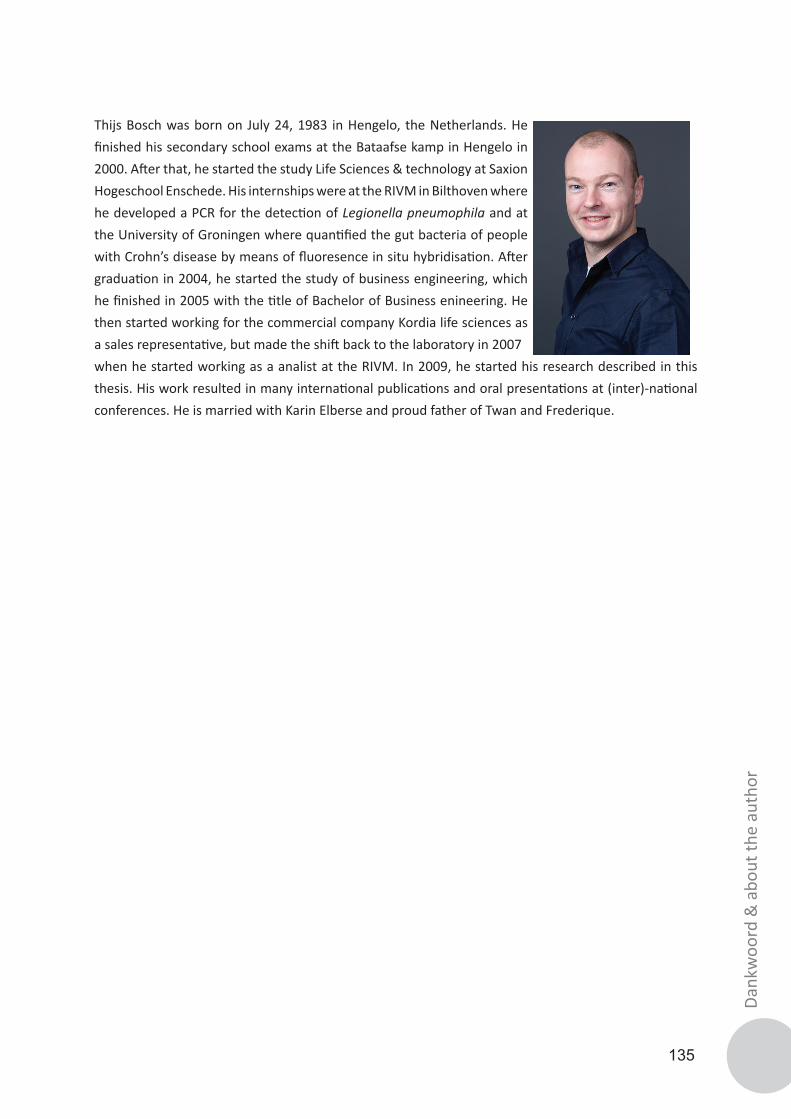
135
Dank
woo
rd &
abo
ut th
e au
thor
Thijs Bosch was born on July 24, 1983 in Hengelo, the Netherlands. He finished his secondary school exams at the Bataafse kamp in Hengelo in 2000. After that, he started the study Life Sciences & technology at Saxion Hogeschool Enschede. His internships were at the RIVM in Bilthoven where he developed a PCR for the detection of Legionella pneumophila and at the University of Groningen where quantified the gut bacteria of people with Crohn’s disease by means of fluoresence in situ hybridisation. After graduation in 2004, he started the study of business engineering, which he finished in 2005 with the title of Bachelor of Business enineering. He then started working for the commercial company Kordia life sciences as a sales representative, but made the shift back to the laboratory in 2007when he started working as a analist at the RIVM. In 2009, he started his research described in this thesis. His work resulted in many international publications and oral presentations at (inter)-national conferences. He is married with Karin Elberse and proud father of Twan and Frederique.

136
List of publications1. de Boer E, Zwartkruis-Nahuis JT, Wit B, Huijsdens XW, de Neeling AJ, Bosch T, et al. Prevalence of methicillin-resistant Staphylococcus aureus in meat. Int J Food Microbiol. 2009;134(1-2):52-6.2. Huijsdens XW, Bosch T, van Santen-Verheuvel MG, Spalburg E, Pluister GN, van Luit M, et al. Molecular characterisation of PFGE non-typable methicillin-resistant Staphylococcus aureus in The Netherlands, 2007. Euro surveillance : bulletin Europeen sur les maladies transmissibles = European communicable disease bulletin. 2009;14(38).3. van de Giessen AW, van Santen-Verheuvel MG, Hengeveld PD, Bosch T, Broens EM, Reusken CB. Occurrence of methicillin-resistant Staphylococcus aureus in rats living on pig farms. Preventive veterinary medicine. 2009;91(2-4):270-3.4. van Rijen MM, Bosch T, Heck ME, Kluytmans JA. Meticillin-resistant Staphylococcus aureus epidemiology and transmission in a Dutch hospital. The Journal of hospital infection. 2009;72(4):299-306.5. Wagenaar JA, Yue H, Pritchard J, Broekhuizen-Stins M, Huijsdens X, Mevius DJ, et al. Unexpected sequence types in livestock associated methicillin-resistant Staphylococcus aureus (MRSA): MRSA ST9 and a single locus variant of ST9 in pig farming in China. Veterinary microbiology. 2009;139(3-4):405-9.6. Bosch T, de Neeling AJ, Schouls LM, van der Zwaluw KW, Kluytmans JA, Grundmann H, et al. PFGE diversity within the methicillin-resistant Staphylococcus aureus clonal lineage ST398. BMC microbiology. 2010;10:40.7. Mulders MN, Haenen AP, Geenen PL, Vesseur PC, Poldervaart ES, Bosch T, et al. Prevalence of livestock-associated MRSA in broiler flocks and risk factors for slaughterhouse personnel in The Netherlands. Epidemiology and infection. 2010;138(5):743-55.8. Verkade E, Bosch T, Hendriks Y, Kluytmans J. Outbreak of methicillin-resistant Staphylococcus aureus ST398 in a Dutch nursing home. Infection control and hospital epidemiology. 2012;33(6):624-6.9. Bosch T, Verkade E, van Luit M, Pot B, Vauterin P, Burggrave R, et al. High Resolution Typing by Whole Genome Mapping Enables Discrimination of LA-MRSA (CC398) Strains and Identification of Transmission Events. PloS one. 2013;8(6):e66493.10. Chlebowicz MA, Bosch T, Sabat AJ, Arends JP, Grundmann H, van Dijl JM, et al. Distinction of Staphylococcal Cassette Chromosome mec type V elements from Staphylococcus aureus ST398. International journal of medical microbiology : IJMM. 2013;303(8):422-32.11. van Cleef BA, van Benthem BH, Haenen AP, Bosch T, Monen J, Kluytmans JA. Low incidence of livestock-associated methicillin-resistant Staphylococcus aureus bacteraemia in The Netherlands in 2009. PloS one. 2013;8(8):e73096.12. Verkade E, van Benthem B, den Bergh MK, van Cleef B, van Rijen M, Bosch T, et al. Dynamics and determinants of Staphylococcus aureus carriage in livestock veterinarians: a prospective cohort study. Clinical infectious diseases : an official publication of the Infectious Diseases Society of America. 2013;57(2):e11-7.13. Wendlandt S, Kadlec K, Fessler AT, Mevius D, van Essen-Zandbergen A, Hengeveld PD, et al. Transmission of methicillin-resistant Staphylococcus aureus isolates on broiler farms. Veterinary microbiology. 2013;167(3-4):632-7.14. van Rijen MM, Bosch T, Verkade EJ, Schouls L, Kluytmans JA, Group CAMS. Livestock-associated MRSA carriage in patients without direct contact with livestock. PloS one. 2014;9(6):e100294.15. Verkade E, Kluytmans-van den Bergh M, van Benthem B, van Cleef B, van Rijen M, Bosch T, et al. Transmission of methicillin-resistant Staphylococcus aureus CC398 from livestock veterinarians to their household members. PloS one. 2014;9(7):e100823.16. Bosch T, Euser SM, Landman F, Bruin JP, EP IJ, den Boer JW, et al. Whole-Genome Mapping as a Novel High-Resolution Typing Tool for Legionella pneumophila. Journal of clinical microbiology.
137
Dank
woo
rd &
abo
ut th
e au
thor
2015;53(10):3234-8.17. Bosch T, Pluister GN, van Luit M, Landman F, van Santen-Verheuvel M, Schot C, et al. Multiple-locus variable number tandem repeat analysis is superior to spa typing and sufficient to characterize MRSA for surveillance purposes. Future Microbiol. 2015;10(7):1155-62.18. Bosch T, Schouls LM. Livestock-associated MRSA: innocent or serious health threat? Future Microbiol. 2015;10(4):445-7.19. Bosch T, Verkade E, van Luit M, Landman F, Kluytmans J, Schouls LM. Transmission and persistence of livestock-associated methicillin-resistant Staphylococcus aureus among veterinarians and their household members. Applied and environmental microbiology. 2015;81(1):124-9.20. van Cleef BA, van Benthem BH, Verkade EJ, van Rijen MM, Kluytmans-van den Bergh MF, Graveland H, et al. Livestock-associated MRSA in household members of pig farmers: transmission and dynamics of carriage, a prospective cohort study. PloS one. 2015;10(5):e0127190.21. van Cleef BA, van Benthem BH, Verkade EJ, van Rijen MM, Kluytmans van den Broeck MF, Graveland H, et al. Health and health-related quality of life in pig farmers carrying livestock-associated methicillin-resistant Staphylococcus aureus. Epidemiology and infection. 2016;144(8):1774-83.22. Bosch T, van Luit M, Pluister GN, Frentz D, Haenen A, Landman F, et al. Changing characteristics of livestock-associated meticillin-resistant Staphylococcus aureus isolated from humans - emergence of a subclade transmitted without livestock exposure, the Netherlands, 2003 to 2014. Euro surveillance : bulletin Europeen sur les maladies transmissibles = European communicable disease bulletin. 2016;21(21).23. Bosch T, Witteveen S, Haenen A, Landman F, Schouls LM. Next-Generation Sequencing Confirms Presumed Nosocomial Transmission of Livestock-Associated Methicillin-Resistant Staphylococcus aureus in the Netherlands. Applied and environmental microbiology. 2016;82(14):4081-9.24. Dierikx CM, Hengeveld PD, Veldman KT, de Haan A, van der Voorde S, Dop PY, et al. Ten years later: still a high prevalence of MRSA in slaughter pigs despite a significant reduction in antimicrobial usage in pigs the Netherlands. The Journal of antimicrobial chemotherapy. 2016;71(9):2414-8.25. Donker T, Bosch T, Ypma RJ, Haenen AP, van Ballegooijen WM, Heck ME, et al. Monitoring the spread of meticillin-resistant Staphylococcus aureus in The Netherlands from a reference laboratory perspective. The Journal of hospital infection. 2016;93(4):366-74.26. van Duijkeren E, Hengeveld P, Zomer TP, Landman F, Bosch T, Haenen A, et al. Transmission of MRSA between humans and animals on duck and turkey farms. The Journal of antimicrobial chemotherapy. 2016;71(1):58-62.

138