Embed Size (px)
Citation preview
REVIEWpublished: 10 February 2017
doi: 10.3389/fnagi.2017.00020
Frontiers in Aging Neuroscience | www.frontiersin.org 1 February 2017 | Volume 9 | Article 20
Edited by:
Daniela Tropea,
Trinity College, Ireland
Reviewed by:
Robert Petersen,
Case Western Reserve University,
USA
Anelyssa D’Abreu,
Brown University, USA
*Correspondence:
Vita Dolžan
Received: 26 October 2016
Accepted: 25 January 2017
Published: 10 February 2017
Citation:
Redenšek S, Trošt M and Dolžan V
(2017) Genetic Determinants of
Parkinson’s Disease: Can They Help
to Stratify the Patients Based on the
Underlying Molecular Defect?
Front. Aging Neurosci. 9:20.
doi: 10.3389/fnagi.2017.00020
Genetic Determinants of Parkinson’sDisease: Can They Help to Stratifythe Patients Based on the UnderlyingMolecular Defect?Sara Redenšek 1, Maja Trošt 2 and Vita Dolžan 1*
1 Pharmacogenetics Laboratory, Institute of Biochemistry, Faculty of Medicine, University of Ljubljana, Ljubljana, Slovenia,2Department of Neurology, University Medical Centre Ljubljana, Ljubljana, Slovenia
Parkinson’s disease (PD) is a sporadic progressive neurodegenerative brain disorder with
a relatively strong genetic background. We have reviewed the current literature about the
genetic factors that could be indicative of pathophysiological pathways of PD and their
applications in everyday clinical practice. Information on novel risk genes is coming from
several genome-wide association studies (GWASs) and their meta-analyses. GWASs
that have been performed so far enabled the identification of 24 loci as PD risk factors.
These loci take part in numerous cellular processes that may contribute to PD pathology:
protein aggregation, protein, and membrane trafficking, lysosomal autophagy, immune
response, synaptic function, endocytosis, inflammation, and metabolic pathways are
among the most important ones. The identified single nucleotide polymorphisms
are usually located in the non-coding regions and their functionality remains to be
determined, although they presumably influence gene expression. It is important to be
aware of a very low contribution of a single genetic risk factor to PD development;
therefore, novel prognostic indices need to account for the cumulative nature of genetic
risk factors. A better understanding of PD pathophysiology and its genetic background
will help to elucidate the underlying pathological processes. Such knowledge may help
physicians to recognize subjects with the highest risk for the development of PD, and
provide an opportunity for the identification of novel potential targets for neuroprotective
treatment. Moreover, it may enable stratification of the PD patients according to their
genetic fingerprint to properly personalize their treatment as well as supportive measures.
Keywords: Parkinson’s disease, genetic risk factors, genome-wide association studies, single nucleotide
polymorphisms, personalized medicine
INTRODUCTION
Parkinson’s disease (PD) is the second most common neurodegenerative brain disease afterAlzheimer’s disease. It is believed that both genetic and environmental factors contribute to thedevelopment of PD. It is a chronic, progressive and incurable disease (Pringsheim et al., 2014).Two pathological hallmarks characterize PD: the formation of cytoplasmic inclusions termed Lewybodies and Lewy neurites and the loss of dopaminergic neurons in the substantia nigra parscompacta (Gallegos et al., 2015). The disease manifests as bradykinesia, muscular rigidity, rest
Redenšek et al. Genetic Determinants of Parkinson’s Disease
tremor and postural and gait impairment (Postuma et al., 2015),which can be accompanied by several other nonmotor symptoms(Gallegos et al., 2015). At the present time, there is unfortunatelyno cure for PD except for the symptomatic treatment andsupportive measures (Connolly and Lang, 2014).
Two different forms of PD have been identified, i.e., sporadicand familial. The familial form starts at an earlier stage of life(<50 years), is usually more severe and progresses more quickly(Klein and Westenberger, 2012). At early stages of researchon genetics of PD, only linkage studies were performed, whichmeans that families with PD history were included in the study.These studies identified mutations in six genes that conclusivelycause monogenic PD—SNCA, LRRK2, Parkin, PINK1, DJ-1,ATP13A2 (Klein and Westenberger, 2012).
In this review, we focus mainly on the sporadic form of thedisease. However, there is some overlap between genes associatedwith familial and sporadic disease; in particular, SNCA andLRRK2 are involved in both forms (Verstraeten et al., 2015; vander Brug et al., 2015). Recently, genome-wide association studies(GWASs) that compared single nucleotide polymorphisms’(SNPs) frequencies between sporadic PD patients and healthyindividuals have identified several loci as Parkinson’s diseasesusceptibility genes (Klein and Westenberger, 2012; Nalls et al.,2014). A few of these loci, 24 in particular, harboring SNPsthat were found to be associated with PD were validated inthe replication phase of the latest and largest meta-analysis ofGWASs performed by Nalls et al. in 2014. In the discoveryphase, they performed a meta-analysis of GWASs including13,708 cases and 95,282 controls chosen from the populationsof European descent (the USA, France, Germany, Iceland,the Netherlands, the UK; 23 and Me), while the replicationphase of the study included 5,353 cases and 5,551 controls(Nalls et al., 2014). The identified loci segregate with numerouscellular pathways that may contribute to PD pathology: proteinaggregation, protein, and membrane trafficking, lysosomalautophagy, immune response, synaptic function, endocytosis,inflammation, and metabolic pathways being among the mostimportant ones (Kumaran and Cookson, 2015). Each locusidentified as a risk factor has a rather low contribution toPD development; therefore, a combination of molecular defectsrather than a single event probably plays a role in PD risk, hencethe idea of a cumulative nature of genetic risk factors (Pihlstromet al., 2016).
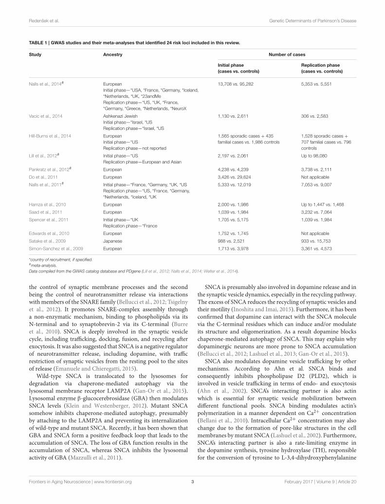
This review summarizes the latest knowledge on genetics andgenomics of PD susceptibility, obtained by GWASs and theirmeta-analyses. We focus on the largest meta-analysis of GWASson PD risk so far (Nalls et al., 2014), but we also searched thePubMed database and GWAS catalog (Welter et al., 2014) forstudies that pointed out the same loci as the above-mentionedmeta-analysis. We identified 13 GWASs and their meta-analyses(summarized in Table 1), which we included in this review. Wecompiled the available data on the 24 loci that were found tobe associated with the risk of sporadic form of PD. To obtainthe information about gene functions, we searched the PubMeddatabase and collected the available data on genes’ functionswith the help of the following words: “Parkinson’s diseaseand gene name” or “Parkinson’s disease and polymorphisms
and gene name.” We divided the susceptibility genes intoseven groups according to their physiological functions: proteinaggregation; protein, and membrane trafficking; lysosomalautophagy; immune system; neurodevelopment, neuron celldifferentiation, and survival; mitochondrial homeostasis; andgenes involved in other processes. This review highlights themain functions of these genes’ products and their role in the PDpathogenesis.
GENETIC DETERMINANTS OF RISK FORSPORADIC PD AND THEIR IMPLICATEDPATHWAYS
Protein AggregationThe products of the genes listed below, i.e., SNCA and tau, areboth constituents of the protein aggregates typical of PD calledLewy bodies. PD is often thought to be a prion-like diseasebecause of the presence of these bodies (Hasegawa et al., 2016).
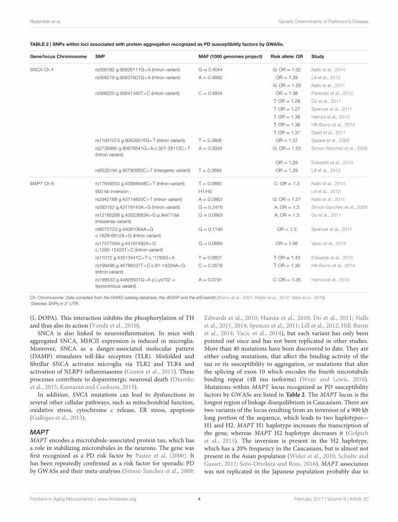
SNCASNCA codes for α-synuclein (SNCA), which is a small, acidicprotein of 14.5 kDA and 140 amino acids (Gallegos et al., 2015).It is the main component of aggregates called Lewy bodies, ahallmark of PD pathology. Lewy bodies are formed because amutated protein usually adopts the β-sheet structure, which isharder to degrade than α-helixes, the main conformation ofnative proteins (Gallegos et al., 2015; Inoshita and Imai, 2015).Different variants of the gene have been connected to bothfamilial and sporadic forms of PD. Several studies on familieswith positive PD history have reported the association of SNCAwith PD, the first one being published in 1997 by Polymeropouloset al. (1997). The association with sporadic PD was first describedby Kruger et al. who compared the allelic frequences of REP1polymorphism in the promoter region of SNCA between casesand controls (Kruger et al., 1999). This association was laterconfirmed on a larger set of data (Maraganore et al., 2006).The majority of GWASs performed also confirmed SNCA asa susceptibility gene (Satake et al., 2009; Simon-Sanchez et al.,2009; Edwards et al., 2010; Hamza et al., 2010; Do et al., 2011;Nalls et al., 2011, 2014; Saad et al., 2011; Spencer et al., 2011;Lill et al., 2012; Pankratz et al., 2012; Hill-Burns et al., 2014).To date, over 800 SNPs within SNCA have been reported, withnearly half of them showing a positive association with PD(Kumaran and Cookson, 2015). SNPs within SNCA recognizedas PD susceptibility factors by GWASs are listed in Table 2. Themost replicated SNP rs256220 was confirmed as a risk factorin six studies, whereas several others were found in only oneor two studies. Some SNCA mutations can cause both sporadicand familial forms of PD. These are usually more penetrant(Singleton et al., 2013). Besides point mutations—Ala53Thr,Ala30Pro, and Glu46Lys in the amino-terminal sequence, wholelocus multiplications (duplications and triplications) were alsofound in both forms (Bisaglia et al., 2009).
SNCA is widely expressed in the central nervous system,especially in the presynaptic terminals of neurons (Inoshita andImai, 2015). It has two main physiological roles, the first being
Frontiers in Aging Neuroscience | www.frontiersin.org 2 February 2017 | Volume 9 | Article 20
Redenšek et al. Genetic Determinants of Parkinson’s Disease
TABLE 1 | GWAS studies and their meta-analyses that identified 24 risk loci included in this review.
Study Ancestry Number of cases
Initial phase
(cases vs. controls)
Replication phase
(cases vs. controls)
Nalls et al., 2014# European
Initial phase—*USA, *France, *Germany, *Iceland,
*Netherlands, *UK, *23andMe
Replication phase—*US, *UK, *France,
*Germany, *Greece, *Netherlands, *NeuroX
13,708 vs. 95,282 5,353 vs. 5,551
Vacic et al., 2014 Ashkenazi Jewish
Initial phase—*Israel, *US
Replication phase—*Israel, *US
1,130 vs. 2,611 306 vs. 2,583
Hill-Burns et al., 2014 European
Initial phase—*US
Replication phase—not reported
1,565 sporadic cases + 435
familial cases vs. 1,986 controls
1,528 sporadic cases +
707 familial cases vs. 796
controls
Lill et al., 2012# Initial phase—*US
Replication phase—European and Asian
2,197 vs. 2,061 Up to 98,080
Pankratz et al., 2012# European 4,238 vs. 4,239 3,738 vs. 2,111
Do et al., 2011 European 3,426 vs. 29,624 Not applicable
Nalls et al., 2011# Initial phase—*France, *Germany, *UK, *US
Replication phase—*US, *France, *Germany,
*Netherlands, *Iceland, *UK
5,333 vs. 12,019 7,053 vs. 9,007
Hamza et al., 2010 European 2,000 vs. 1,986 Up to 1,447 vs. 1,468
Saad et al., 2011 European 1,039 vs. 1,984 3,232 vs. 7,064
Spencer et al., 2011 Initial phase—*UK
Replication phase—*France
1,705 vs. 5,175 1,039 vs. 1,984
Edwards et al., 2010 European 1,752 vs. 1,745 Not applicable
Satake et al., 2009 Japanese 988 vs. 2,521 933 vs. 15,753
Simon-Sanchez et al., 2009 European 1,713 vs. 3,978 3,361 vs. 4,573
*country of recruitment, if specified.#meta-analysis.
Data compiled from the GWAS catalog database and PDgene (Lill et al., 2012; Nalls et al., 2014; Welter et al., 2014).
the control of synaptic membrane processes and the secondbeing the control of neurotransmitter release via interactionswithmembers of the SNARE family (Bellucci et al., 2012; Tsigelnyet al., 2012). It promotes SNARE-complex assembly througha non-enzymatic mechanism, binding to phospholipids via itsN-terminal and to synaptobrevin-2 via its C-terminal (Burreet al., 2010). SNCA is deeply involved in the synaptic vesiclecycle, including trafficking, docking, fusion, and recycling afterexocytosis. It was also suggested that SNCA is a negative regulatorof neurotransmitter release, including dopamine, with trafficrestriction of synaptic vesicles from the resting pool to the sitesof release (Emanuele and Chieregatti, 2015).
Wild-type SNCA is translocated to the lysosomes fordegradation via chaperone-mediated autophagy via thelysosomal membrane receptor LAMP2A (Gan-Or et al., 2015).Lysosomal enzyme β-glucocerebrosidase (GBA) then modulatesSNCA levels (Klein and Westenberger, 2012). Mutant SNCAsomehow inhibits chaperone-mediated autophagy, presumablyby attaching to the LAMP2A and preventing its internalizationof wild-type and mutant SNCA. Recently, it has been shown thatGBA and SNCA form a positive feedback loop that leads to theaccumulation of SNCA. The loss of GBA function results in theaccumulation of SNCA, whereas SNCA inhibits the lysosomalactivity of GBA (Mazzulli et al., 2011).
SNCA is presumably also involved in dopamine release and inthe synaptic vesicle dynamics, especially in the recycling pathway.The excess of SNCA reduces the recycling of synaptic vesicles andtheir motility (Inoshita and Imai, 2015). Furthermore, it has beenconfirmed that dopamine can interact with the SNCA moleculevia the C-terminal residues which can induce and/or modulateits structure and oligomerization. As a result dopamine blockschaperone-mediated autophagy of SNCA. This may explain whydopaminergic neurons are more prone to SNCA accumulation(Bellucci et al., 2012; Lashuel et al., 2013; Gan-Or et al., 2015).
SNCA also modulates dopamine vesicle trafficking by othermechanisms. According to Ahn et al. SNCA binds andconsequently inhibits phospholipase D2 (PLD2), which isinvolved in vesicle trafficking in terms of endo- and exocytosis(Ahn et al., 2002). SNCA’s interacting partner is also actinwhich is essential for synaptic vesicle mobilization betweendifferent functional pools. SNCA binding modulates actin’spolymerization in a manner dependent on Ca2+ concentration(Bellani et al., 2010). Intracellular Ca2+ concentration may alsochange due to the formation of pore-like structures in the cellmembranes bymutant SNCA (Lashuel et al., 2002). Furthermore,SNCA’s interacting partner is also a rate-limiting enzyme inthe dopamine synthesis, tyrosine hydroxylase (TH), responsiblefor the conversion of tyrosine to L-3,4-dihydroxyphenylalanine
Frontiers in Aging Neuroscience | www.frontiersin.org 3 February 2017 | Volume 9 | Article 20
Redenšek et al. Genetic Determinants of Parkinson’s Disease
TABLE 2 | SNPs within loci associated with protein aggregation recognized as PD susceptibility factors by GWASs.
Gene/locus Chromosome SNP MAF (1000 genomes project) Risk allele: OR Study
SNCA Ch 4 rs356182 g.90626111G>A (intron variant) G = 0.4044 G: OR = 1.32 Nalls et al., 2014
rs356219 g.90637601G>A (intron variant) A = 0.4892 OR = 1.29 Lill et al., 2012
G: OR = 1.29 Nalls et al., 2011
rs356220 g.90641340T>C (intron variant) C = 0.4834 OR = 1.38 Pankratz et al., 2012
T: OR = 1.29 Do et al., 2011
T: OR = 1.27 Spencer et al., 2011
T: OR = 1.38 Hamza et al., 2010
T: OR = 1.38 Hill-Burns et al., 2014
T: OR = 1.37 Saad et al., 2011
rs11931074 g.90639515G>T (intron variant) T = 0.3806 OR = 1.37 Satake et al., 2009
rs2736990 g.90678541G>A c.307-28113C>T
(intron variant)
A = 0.3934 G: OR = 1.23 Simon-Sanchez et al., 2009
OR = 1.29 Edwards et al., 2010
rs6532194 g.90780902C>T (intergenic variant) T = 0.3694 OR = 1.29 Lill et al., 2012
MAPT Ch 8 rs17649553 g.43994648C>T (intron variant) T = 0.0865 C: OR = 1.3 Nalls et al., 2014
900 kb inversion H1/H2 Lill et al., 2012
rs2942168 g.43714850C>T (intron variant) A = 0.0863 G: OR = 1.27 Nalls et al., 2011
rs393152 g.43719143A>G (intron variant) G = 0.2476 A: OR = 1.3 Simon-Sanchez et al., 2009
rs12185268 g.43923683A>G p.Ile471Val
(missense variant)
G = 0.0863 A: OR = 1.3 Do et al., 2011
rs8070723 g.44081064A>G
c.1828-6612A>G (intron variant)
G = 0.1190 OR = 1.3 Spencer et al., 2011
rs17577094 g.44187492A>G
c.1290-15425T>C (intron variant)
G = 0.0865 OR = 1.56 Vacic et al., 2014
rs11012 g.43513441C>T c.*1783G>A T = 0.0807 T: OR = 1.43 Edwards et al., 2010
rs199498 g.46788237T>C c.81-14328A>G
(intron variant)
C = 0.2578 T: OR = 1.35 Hill-Burns et al., 2014
rs199533 g.44828931G>A p.Lys702 =
(synonimous variant)
A = 0.0791 C: OR = 1.35 Hamza et al., 2010
Ch, Chromosome. Data compiled from the GWAS catalog database, the dbSNP and the e!Ensembl (Sherry et al., 2001; Welter et al., 2014; Yates et al., 2016).
*Denotes SNPs in 3’-UTR.
(L-DOPA). This interaction inhibits the phosphorylation of THand thus also its action (Venda et al., 2010).
SNCA is also linked to neuroinflammation. In mice withaggregated SNCA, MHCII expression is induced in microglia.Moreover, SNCA as a danger-associated molecular pattern(DAMP) stimulates toll-like receptors (TLR). Misfolded andfibrillar SNCA activates microglia via TLR2 and TLR4 andactivation of NLRP3 inflammasome (Gustot et al., 2015). Theseprocesses contribute to dopaminergic neuronal death (Dzamkoet al., 2015; Kumaran and Cookson, 2015).
In addition, SNCA mutations can lead to dysfunctions inseveral other cellular pathways, such as mitochondrial function,oxidative stress, cytochrome c release, ER stress, apoptosis(Gallegos et al., 2015).
MAPTMAPT encodes a microtubule-associated protein tau, which hasa role in stabilizing microtubules in the neurons. The gene wasfirst recognized as a PD risk factor by Pastor et al. (2000). Ithas been repeatedly confirmed as a risk factor for sporadic PDby GWASs and their meta-analyses (Simon-Sanchez et al., 2009;
Edwards et al., 2010; Hamza et al., 2010; Do et al., 2011; Nallset al., 2011, 2014; Spencer et al., 2011; Lill et al., 2012; Hill-Burnset al., 2014; Vacic et al., 2014), but each variant has only beenpointed out once and has not been replicated in other studies.More than 40 mutations have been discovered to date. They areeither coding mutations, that affect the binding activity of thetau or its susceptibility to aggregation, or mutations that alterthe splicing of exon 10 which encodes the fourth microtubulebinding repeat (4R tau isoforms) (Wray and Lewis, 2010).Mutations within MAPT locus recognized as PD susceptibilityfactors by GWASs are listed in Table 2. The MAPT locus is thelongest region of linkage disequilibrium in Caucasians. There aretwo variants of the locus resulting from an inversion of a 900 kblong portion of the sequence, which leads to two haplotypes—H1 and H2. MAPT H1 haplotype increases the transcription ofthe gene, whereas MAPT H2 haplotype decreases it (Golpichet al., 2015). The inversion is present in the H2 haplotype,which has a 20% frequency in the Caucasians, but is almost notpresent in the Asian population (Wider et al., 2010; Schulte andGasser, 2011; Soto-Ortolaza and Ross, 2016). MAPT associationwas not replicated in the Japanese population probably due to
Frontiers in Aging Neuroscience | www.frontiersin.org 4 February 2017 | Volume 9 | Article 20
Redenšek et al. Genetic Determinants of Parkinson’s Disease
inter-population heterogeneity at this locus (Satake et al., 2009;Labbé and Ross, 2014). H1 haplotype is more dynamic and canbe subdivided into subhaplotypes. Homozygous MAPT H1/H1genotype is a biomarker of dementia in PD (Lin and Wu,2015). As H1 haplotype is associated with a greater risk of PDand other neurodegenerative diseases, it may be assumed thatH2 haplotype is under positive selection (Schulte and Gasser,2011). Polymorphisms in the MAPT gene are also involvedin the pathologies of Alzheimer’s disease (AD), progressivesupranuclear palsy (PSP), and corticobasal degeneration (CBD)(Vandrovcova et al., 2009).
MAPT encoded protein tau is a potential componentof Lewy bodies. In PD patients with a more pronouncedcognitive decline, neurofibrillary tangles, more typical of AD,can also be found besides Lewy bodies. These tangles consistof hyperphosphorylated tau (Horvath et al., 2013; Lin andWu, 2015). Physiologically, phosphorylation of the tau proteinregulates its propensity to interact with microtubules (Wray andLewis, 2010). According to some studies, SNCA also promotestau fibrillization and influences tau phosphorylation (Giassonet al., 2003). The interplay between the two genes was alsoconfirmed by the study of Goris et al., reporting nearly a doublerisk of PD in carriers of the combined MAPT H1/H1 andSNCA rs356219 G/G genotype (Goris et al., 2007). Most of thepathological changes in the MAPT sequence disrupt the abilityof tau to interact with microtubules (Wray and Lewis, 2010). Tauis also fundamental for the maintenance of cytoskeletal networkvia the regulation of axonal transport by interactions with kinesinand dynein (Kumaran and Cookson, 2015).
Mutated MAPT may also affect the function of lysosomesand its role in autophagy, since it is degraded with this process(Gan-Or et al., 2015).
MAPT gene lies in a block of nearly complete linkagedisequilibrium that extends over nearly 2 Mb, therefore it ispossible that other genes from this region are also associated withPD risk (Charlesworth et al., 2012). For example, a variant inthe CRHR1 gene coding for the corticotropin-releasing hormonereceptor has recently been recognized as a factor that decreasesthe PD susceptibility (Davis et al., 2016).
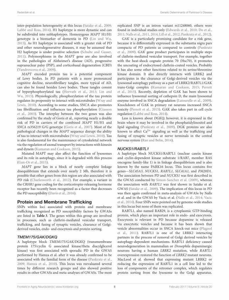
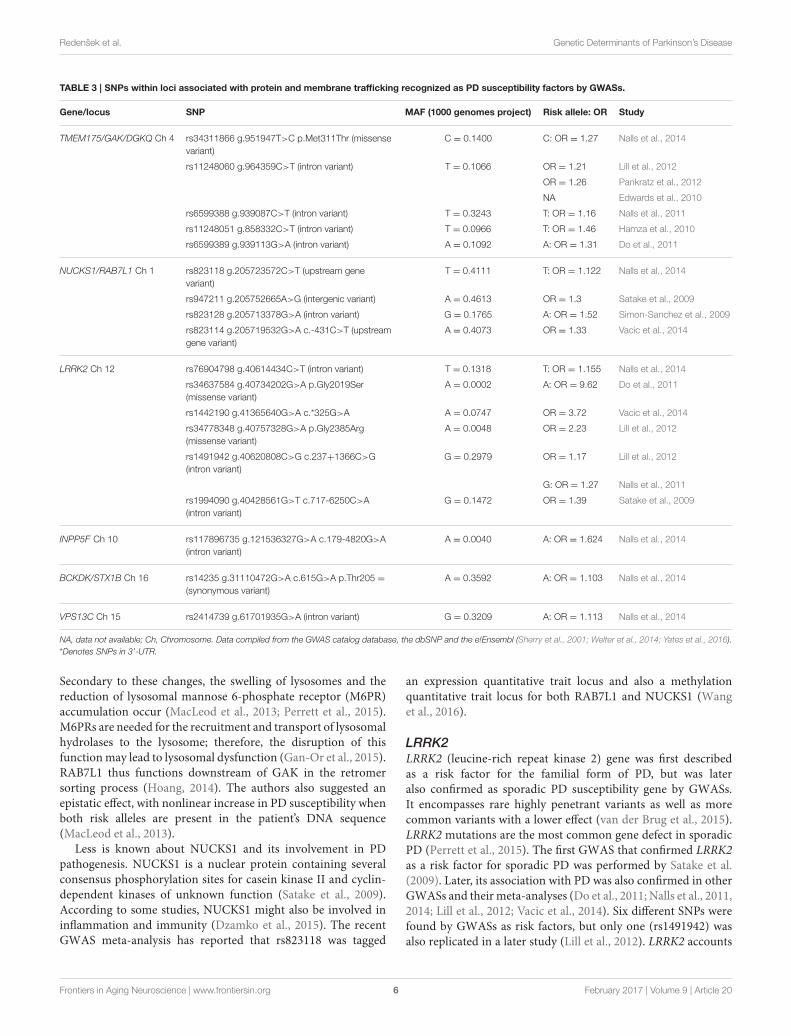
Protein and Membrane TraffickingSNPs within loci associated with protein and membranetrafficking recognized as PD susceptibility factors by GWASsare listed in Table 3. The genes within this group are involvedin processes, such as clathrin-mediated vesicular transport,trafficking, and fusing of synaptic vesicles, clearance of Golgi-derived vesicles, endo- and exocytosis and protein sorting.
TMEM175/GAK/DGKQA haplotype block TMEM175/GAK/DGKQ (transmembraneprotein 175/cyclin G associated kinase/theta diacylglicerolkinase) was first associated with sporadic PD in the GWASperformed by Hamza et al. after it was already confirmed to beassociated with the familial form of the disease (Pankratz et al.,2009; Hamza et al., 2010). This locus was reevaluated severaltimes by different research groups and also showed positiveresults in other GWASs and meta-analyses of GWASs. The most
replicated SNP is an intron variant rs11248060, others werefound in individual studies only (Edwards et al., 2010; Do et al.,2011; Nalls et al., 2011, 2014; Lill et al., 2012; Pankratz et al., 2012).
GAK is a particularly promising candidate for a risk gene,because it is differentially expressed in the substantia nigra parscompacta of PD patients as compared to controls (Pankratzet al., 2009). GAK gene product participates in multiple stepsof clathrin-mediated vesicular transport. For example, togetherwith the heat-shock cognate protein 70 (Hsc70), it promotesthe uncoating of endocytosed clathrin-coated vesicles. Probablyit has also some other functions related to its serine/threoninekinase domain. It also directly interacts with LRRK2 andparticipates in the clearance of Golgi-derived vesicles via thelysosomal autophagy pathway as a part of LRRK2/RAB7L1/GAKtrans-Golgi complex (Kumaran and Cookson, 2015; Perrettet al., 2015). Recently, depletion of GAK has been shown toinfluence lysosomal sorting of cathepsin D, the main lysosomalenzyme involved in SNCA degradation (Latourelle et al., 2009).Knockdown of GAK in primary rat neurons increased SNCAtoxicity (Perrett et al., 2015). GAK also takes part in cell cycleregulation (Labbé and Ross, 2014).
Less is known about DGKQ; however, it is expressed in thebrain where it may be involved in the phosphatidylinositol andlipid signaling (Pankratz et al., 2009). DGK proteins are alsoknown to affect Ca2+ signaling as well as the trafficking andfusing of synaptic vesicles at nerve terminals in the centralnervous system (Ran and Belin, 2014).
NUCKS1/RAB7L1A haplotype block NUCKS1/RAB7L1 (nuclear casein kinaseand cyclin-dependent kinase substrate 1/RAB7, member RASoncogene family-like 1) is in linkage disequilibrium and is alsoknown by the name PARK16 locus. This locus contains fivegenes—SLC45A3, NUCKS1, RAB7L1, SLC41A1, and PM20D1.The association between PD and NUCKS1 was first described inthe GWAS conducted by Simon-Sanchez et al. (2009), whereasthe association with RAB7L1 was first shown in Satake et al.GWAS (Satake et al., 2009). The implication of this locus in PDwas then again confirmed in meta-analysis of GWASs by Nallset al. and in the GWAS by Vacic et al. (Nalls et al., 2014; Vacicet al., 2014). Four SNPs were pointed out by genome-wide studiesin this locus but none of them was replicated.
RAB7L1, also named RAB29, is a cytoplasmic GTP-bindingprotein, which plays an important role in endo- and exocytosis.Exocytosis is relevant in PD because dopamine is releasedvia exocytotic vesicles and because it has been shown thatvesicle abnormalities occur in SNCA knock-out mice (Plagnolet al., 2011). RAB7L1 is one of the LRRK2 interactingpartners in the process of removal of Golgi derived vesicles byautophagy-dependent mechanisms. RAB7L1 deficiency causedneurodegeneration in mammalian or Drosophila dopaminergicneurons having a human LRRK2 mutation, while RAB7L1overexpression restored the function of LRRK2 mutant neurons.MacLeod et al. showed that expressing mutant LRRK2 orreducing the expression of RAB7L1 in a cell line led to theloss of components of the retromer complex, which regulatesprotein sorting from the lysosome to the Golgi apparatus.
Frontiers in Aging Neuroscience | www.frontiersin.org 5 February 2017 | Volume 9 | Article 20
Redenšek et al. Genetic Determinants of Parkinson’s Disease
TABLE 3 | SNPs within loci associated with protein and membrane trafficking recognized as PD susceptibility factors by GWASs.
Gene/locus SNP MAF (1000 genomes project) Risk allele: OR Study
TMEM175/GAK/DGKQ Ch 4 rs34311866 g.951947T>C p.Met311Thr (missense
variant)
C = 0.1400 C: OR = 1.27 Nalls et al., 2014
rs11248060 g.964359C>T (intron variant) T = 0.1066 OR = 1.21 Lill et al., 2012
OR = 1.26 Pankratz et al., 2012
NA Edwards et al., 2010
rs6599388 g.939087C>T (intron variant) T = 0.3243 T: OR = 1.16 Nalls et al., 2011
rs11248051 g.858332C>T (intron variant) T = 0.0966 T: OR = 1.46 Hamza et al., 2010
rs6599389 g.939113G>A (intron variant) A = 0.1092 A: OR = 1.31 Do et al., 2011
NUCKS1/RAB7L1 Ch 1 rs823118 g.205723572C>T (upstream gene
variant)
T = 0.4111 T: OR = 1.122 Nalls et al., 2014
rs947211 g.205752665A>G (intergenic variant) A = 0.4613 OR = 1.3 Satake et al., 2009
rs823128 g.205713378G>A (intron variant) G = 0.1765 A: OR = 1.52 Simon-Sanchez et al., 2009
rs823114 g.205719532G>A c.-431C>T (upstream
gene variant)
A = 0.4073 OR = 1.33 Vacic et al., 2014
LRRK2 Ch 12 rs76904798 g.40614434C>T (intron variant) T = 0.1318 T: OR = 1.155 Nalls et al., 2014
rs34637584 g.40734202G>A p.Gly2019Ser
(missense variant)
A = 0.0002 A: OR = 9.62 Do et al., 2011
rs1442190 g.41365640G>A c.*325G>A A = 0.0747 OR = 3.72 Vacic et al., 2014
rs34778348 g.40757328G>A p.Gly2385Arg
(missense variant)
A = 0.0048 OR = 2.23 Lill et al., 2012
rs1491942 g.40620808C>G c.237+1366C>G
(intron variant)
G = 0.2979 OR = 1.17 Lill et al., 2012
G: OR = 1.27 Nalls et al., 2011
rs1994090 g.40428561G>T c.717-6250C>A
(intron variant)
G = 0.1472 OR = 1.39 Satake et al., 2009
INPP5F Ch 10 rs117896735 g.121536327G>A c.179-4820G>A
(intron variant)
A = 0.0040 A: OR = 1.624 Nalls et al., 2014
BCKDK/STX1B Ch 16 rs14235 g.31110472G>A c.615G>A p.Thr205 =
(synonymous variant)
A = 0.3592 A: OR = 1.103 Nalls et al., 2014
VPS13C Ch 15 rs2414739 g.61701935G>A (intron variant) G = 0.3209 A: OR = 1.113 Nalls et al., 2014
NA, data not available; Ch, Chromosome. Data compiled from the GWAS catalog database, the dbSNP and the e!Ensembl (Sherry et al., 2001; Welter et al., 2014; Yates et al., 2016).
*Denotes SNPs in 3’-UTR.
Secondary to these changes, the swelling of lysosomes and thereduction of lysosomal mannose 6-phosphate receptor (M6PR)accumulation occur (MacLeod et al., 2013; Perrett et al., 2015).M6PRs are needed for the recruitment and transport of lysosomalhydrolases to the lysosome; therefore, the disruption of thisfunctionmay lead to lysosomal dysfunction (Gan-Or et al., 2015).RAB7L1 thus functions downstream of GAK in the retromersorting process (Hoang, 2014). The authors also suggested anepistatic effect, with nonlinear increase in PD susceptibility whenboth risk alleles are present in the patient’s DNA sequence(MacLeod et al., 2013).
Less is known about NUCKS1 and its involvement in PDpathogenesis. NUCKS1 is a nuclear protein containing severalconsensus phosphorylation sites for casein kinase II and cyclin-dependent kinases of unknown function (Satake et al., 2009).According to some studies, NUCKS1 might also be involved ininflammation and immunity (Dzamko et al., 2015). The recentGWAS meta-analysis has reported that rs823118 was tagged
an expression quantitative trait locus and also a methylationquantitative trait locus for both RAB7L1 and NUCKS1 (Wanget al., 2016).
LRRK2LRRK2 (leucine-rich repeat kinase 2) gene was first describedas a risk factor for the familial form of PD, but was lateralso confirmed as sporadic PD susceptibility gene by GWASs.It encompasses rare highly penetrant variants as well as morecommon variants with a lower effect (van der Brug et al., 2015).LRRK2 mutations are the most common gene defect in sporadicPD (Perrett et al., 2015). The first GWAS that confirmed LRRK2as a risk factor for sporadic PD was performed by Satake et al.(2009). Later, its association with PD was also confirmed in otherGWASs and their meta-analyses (Do et al., 2011; Nalls et al., 2011,2014; Lill et al., 2012; Vacic et al., 2014). Six different SNPs werefound by GWASs as risk factors, but only one (rs1491942) wasalso replicated in a later study (Lill et al., 2012). LRRK2 accounts
Frontiers in Aging Neuroscience | www.frontiersin.org 6 February 2017 | Volume 9 | Article 20
Redenšek et al. Genetic Determinants of Parkinson’s Disease
for a large part of risk for sporadic PD in some populations—Ashkenazi Jews, Arabs, East Asians. Coding variability at theLRRK2 locus explains 10% of PD risk in these populations. In theoutbred Europeans, LRRK2 and GBA account for around 8% ofrisk. Carriers of LRRK2 mutations usually have Lewy bodies, theminority has also tangles, but some of them have neither (Hardy,2010).
LRRK2 is a multidomain protein that has a GTPase and kinasefunction as well as many protein/protein interaction motifs.It is expressed in axons and dendrites in the striatum andthe cortex, but expression is low in nigral cell bodies. LRRK2seems to regulate actin complexes, vesicle trafficking, endosomematuration, cytoskeletal dynamics, and protein translation(Volta et al., 2015). It was detected as a binding partner of thelate-endosomal marker Rab7 and also as a binding partner ofthe lysosomal marker LAMP2A, which generated the idea thatit has an important function in the endo-lysosomal pathway.Mutant LRRK2-RAB7 colocalization can lead to a reducedRAB7 function and impaired late endosomal trafficking events.LRRK2 also interacts with RAB7L1 to modify protein sorting.It also binds the PD susceptibility factor GAK in a complexthat promotes removal of Golgi derived vesicles by autophagy-dependent mechanisms (Perrett et al., 2015). It is tightly linkedto endocytotic and exocytotic processes required for rapidsynaptic vesicles and receptor recycling via interaction withclathrin (Volta et al., 2015). It regulates endophilin associationto clathrin-coated vesicles through phosphorylation (Drouetand Lesage, 2014). Furthermore, it influences the action ofEndoA via phosphorylation, a protein important for endocyticsynaptic vesicles recycling (Inoshita and Imai, 2015). Moreover,LRRK2 is involved in mitochondrial membrane maintenancevia fusion/fission processes and in lysosomal autophagy andrecycling of V-ATPase required for lysosomal acidification(Ryan et al., 2015; Volta et al., 2015). The dysregulation ofmitochondrial function can also be caused by the inhibition ofthe endogenous peroxidase phosphorylation by mutant LRRK2(Saiki et al., 2012). LRRK2 also has a role in neuroinflammationby increasing the cytokine release from activated primarymicroglial cells, which results in neurotoxicitiy (Russo et al., 2014;Blesa et al., 2015). Moreover, it interferes with the translationmachinery by phosphorylation of several proteins, for exampletranslation repressor protein 4E-BP (Taymans et al., 2015).
Among PD patients having the LRRK2 mutation, the mostcommon one is p.Gly2019Ser (rs34637584). The penetrance ishigh—50% at the age of 50 and 74% at the age of 79. Thismutation is located in the kinase domain resulting in an increasein the activity (Wallings et al., 2015). 1.6% of sporadic PD patientspossess this mutation (Satake et al., 2009). Mutated LRRK2(p.Gly2019Ser) actually binds to outer mitochondrial membrane,which leads to a decrease in mitochondrial membrane potentialand to a decrease in the intracellular ATP level. On the contrary,mitochondrial elongation and interconnectivity were elevated(Subramaniam and Chesselet, 2013).
INPP5FInositol polyphosphate-5-phosphatase F (INPP5F), one of thepolyphosphoinositide phosphatases, was first described as a PD
risk factor in a meta-analysis carried out by Nalls et al. in 2014(Nalls et al., 2014), which means that only one SNP was found tobe associated with PD susceptibility so far. The gene’s product issupposed to be involved in the endocytic pathways. It containsa Sac domain, which is involved in the endocytosis of synapticvesicles (Inoshita and Imai, 2015; Nakatsu et al., 2015). Theknowledge about the gene’s function is limited and on the basisof only one study pointing out the association, we cannot reachany conclusions as to the main role of the gene in PD pathologyand the actual association.
BCKDK/STX1BBCKDK/STX1B (branched chain ketoacid dehydrogenasekinase/syntaxin 1B) locus was established as a PD risk factor inGWAS meta-analysis by Nalls et al. Only one SNP (rs14235)is known to be associated with PD, thus replication studies areneeded to confirm the actual association (Nalls et al., 2014).
BCKDK is located in the mitochondrial matrix and playsa major role in valine, leucine and isoleucine catabolism. Itsfunction is phosphorylation and thus inactivation of the BCKD.BCKD concentration is the same in all tissues, whereas BCKDKconcentration varies. BCKD’s level of function is thus regulatedby BCKDK (García-Cazorla et al., 2014).
STX1B codes for syntaxin 1B, which functions as a synapticreceptor for vesicle transport. It was previously shown to bedirectly implicated in the process of calcium-dependent synaptictransmission in rat brain, having been suggested to play a role inthe excitatory pathway of synaptic transmission (Plagnol et al.,2011). It plays a role in dopamine neurotransmission (Kalia andLang, 2015).
VPS13CVacuolar protein sorting 13 homolog C (VPS13C) was shownto be associated with PD as a risk factor only in the meta-analysis by Nalls et al. (2014). First, it was considered only as arisk factor for sporadic PD, but it has recently been discoveredthat it could also cause the familial autosomal-recessive early-onset form of PD with a very severe course of the disease(Lesage et al., 2016). The gene has two variants, variant 2 beingspecific to brain tissue (Velayos-Baeza et al., 2004). It functionsin the vesicular transport, more specifically endosomal transportpathway (Inoshita and Imai, 2015). Functional studies of theprotein are lacking, but a very recent one also suggests a proteinquality control function (Yang et al., 2016). More association andfunctional studies will have to be conducted to be completelyconfident of the gene’s role in PD susceptibility and pathology.
Lysosomal AutophagySNPs within loci associated with lysosomal autophagy recognizedas PD susceptibility factors by GWASs are listed in Table 4.These genes are playing their part in general lysosomal functions,especially lysosomal autophagy.
GBA/SYT11GBA (beta acid glucosidase) is associated with PD susceptibilityaccording to several GWASs and their meta-analyses, but thefirst association was found in 2011 by Do et al. (Do et al., 2011;
Frontiers in Aging Neuroscience | www.frontiersin.org 7 February 2017 | Volume 9 | Article 20

Redenšek et al. Genetic Determinants of Parkinson’s Disease
TABLE 4 | SNPs within loci associated with lysosomal autophagy recognized as PD susceptibility factors by GWASs.
Gene/locus SNP MAF (1000 genomes project) Risk allele: OR Study
GBA/SYT11 Ch 1 rs35749011 g.155135036G>A (intergenic variant) A = 0.0054 A: OR = 1.824 Nalls et al., 2014
i4000416 g.155205634T>C c.1226A>C
p.Asn409Ser (missense variant)
C = 0.0006 C: OR = 4.05 Do et al., 2011
rs1630500 g.154855055G>A (intergenic variant) A = 0.0559 OR = 1.75 Vacic et al., 2014
rs12726330 g.155108167G>A c.-262G>A (splice
region variant)
A = 0.0052 OR = 1.71 Pankratz et al., 2012
rs34372695 g.156030037C>T c.-1155C>T
(upstream gene variant)
T = 0.0088 T: OR = 1.47 Nalls et al., 2011
SCARB2/FAM47E Ch 4 rs6812193 g.77198986C>T c.871-236C>T (intron
variant)
T = 0.3133 C: OR = 1.1 Nalls et al., 2014
C: OR = 1.19 Do et al., 2011
C: OR = 1.12 Simon-Sanchez et al., 2009
Ch, Chromosome. Data compiled from the GWAS catalog database, the dbSNP and the e!Ensembl (Sherry et al., 2001; Welter et al., 2014; Yates et al., 2016).
Pankratz et al., 2012; Nalls et al., 2014; Vacic et al., 2014). On theother hand, SYT11 (synaptotagmin XI) was confirmed as a PDrisk gene by Nalls et al. in 2011 (Nalls et al., 2011, 2014). No SNPhas been replicated in a later study.
GBA is located within the inner lysosomalmembrane, where itcleaves membrane glucocerebrosides into ceramide and glucose.Functional studies have shown that any kind of reduction inGBA enzymatic activity leads to the accumulation of SNCAwhich, in turn, inhibits the function of normal GBA, causingthe additional aggregation of SNCA. The authors hypothesizethat the reduction of GBA function, either caused by GBAmutations or impaired GBA trafficking from ER and Golgi tolysosomes, increases the glucocerebrosides concentration in lipidrafts. This change in membrane composition may lead to thereduced formation of LAMP2A (chaperone-mediated autophagyreceptor) protein-complexes, which could result in diminishedautophagy of SNCA and its accumulation. GBA dysfunctionmay also lead to general disruption in lysosomal function andautophagy. Furthermore, it can also be the reason for defectivemitophagy and mitochondrial damage. It was also shown thatPD patients with no mutation in the GBA may have a lowerenzyme activity. This implies that other factors may also affectthe GBA function, for example environmental factors (Gan-Oret al., 2015).
SYT11 encodes a calcium-sensing protein involved inmembrane trafficking in synaptic transmision and is also asubstrate of Parkin, which is a risk factor for the familial formof PD. Due to its ability of calcium-dependent phospholipidbinding, it is important in the regulation of vesicle fusion andendocytosis at synaptic terminals (Ran and Belin, 2014). Morestudies will have to be performed in order to properly determinethe function of the gene.
SCARB2/FAM47ESCARB2/FAM47E (scavenger receptor class B member 2) locuswas first found to be associated with PD in 2009 by Simon-Sanchez et al.; this was later confirmed by two other GWASs—Doet al. and Nalls et al. The same SNP (rs6812193) was pointed out
in all three studies (Simon-Sanchez et al., 2009; Do et al., 2011;Nalls et al., 2014).
SCARB2 encodes the lysosomal membrane protein type 2(LIMP-2), which is a GCase receptor. It directs GBA to lysosomes.The reduced function of SCARB2 can lead to a reduced GBAactivity and decreased SNCA degradation (Gan-Or et al., 2015).The receptor, being an enterovirus 71 receptor, also plays a role inthe neuroinflammation process (Dzamko et al., 2015). FAM47Elies in close proximity to SCARB2 and the most frequentlyassociated SNP (rs6812193) is situated between the two genes.However, among the two, SCARB2 seems to be a more promisingcandidate for a PD risk factor (Do et al., 2011; Ran and Belin,2014).
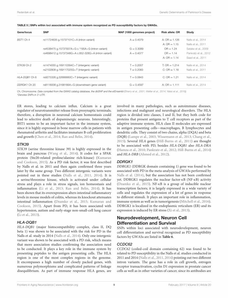
Immune SystemSNPs within loci associated with the immune system recognizedas PD susceptibility factors by GWASs are listed in Table 5. Thegenes’ function in PD susceptibility and pathology should betested in a more targeted way with the help of cell and animalmodels. There are some indications that genes mentioned belowplay a role in the immune system, but we do not know to whatextent and if this is their main purpose, except for HLA-DQB1whose function is quite well determined. There is also a need formore association studies especially for STK39, HLA-DQB1 andDDRGK1.
BST1BST1 (bone marrow stromal cell antigen-1) was recognized asa PD risk factor in the Satake et al. GWAS in 2009 and afterthat in several other GWASs. Three intron variants were foundto be associated with PD risk. Two of them (rs11724635 andrs4698412) were replicated in a later study (Satake et al., 2009;Nalls et al., 2011, 2014; Saad et al., 2011; Pankratz et al., 2012).BST1, a member of the CD38 gene family, is a cell surface proteinbound to the membrane by glycosylphosphatidylinositol linkage;it possesses both ADP ribosyl cyclase and cyclic ADP ribosehydrolase enzymatic activities. It produces cyclic ADP-ribose,which as a second messenger releases calcium from intracellular
Frontiers in Aging Neuroscience | www.frontiersin.org 8 February 2017 | Volume 9 | Article 20
Redenšek et al. Genetic Determinants of Parkinson’s Disease
TABLE 5 | SNPs within loci associated with immune system recognized as PD susceptibility factors by GWASs.
Gene/locus SNP MAF (1000 genomes project) Risk allele: OR Study
BST1 Ch 4 rs11724635 g.15737101C>A (intron variant) A = 0.4079 A: OR = 1.126 Nalls et al., 2014
A: OR = 1.15 Nalls et al., 2011
rs4538475 g.15737937A>G c.*195A>G (intron variant) G = 0.3089 OR = 1.24 Satake et al., 2009
rs4698412 g.15737348G>A c.852-328G>A (intron variant) A = 0.4077 OR = 1.14 Pankratz et al., 2012
A: OR = 1.14 Saad et al., 2011
STK39 Ch 2 rs1474055 g.169110394C>T (intergenic variant) T = 0.2007 T: OR = 1.214 Nalls et al., 2014
rs2102808 g.169117025G>T (intergenic variant) T = 0.2083 C: OR = 1.18 Nalls et al., 2011
HLA-DQB1 Ch 6 rs9275326 g.32666660C>T (intergenic variant) T = 0.0843 C: OR = 1.21 Nalls et al., 2014
DDRGK1 Ch 20 rs8118008 g.3168166A>G (downstream gene variant) G = 0.4597 A: OR = 1.111 Nalls et al., 2014
Ch, Chromosome. Data compiled from the GWAS catalog database, the dbSNP and the e!Ensembl (Sherry et al., 2001; Welter et al., 2014; Yates et al., 2016).
*Denotes SNPs in 3’-UTR.
ER stores, leading to calcium influx. Calcium is a greatregulator of neurotransmitter release from presynaptic terminals;therefore, a disruption in neuronal calcium homeostasis couldlead to selective death of dopaminergic neurons. Interestingly,BST1 seems to be an important factor in the immune system,since it is highly expressed in bone marrow cells in patients withrheumatoid arthritis and facilitates immature B-cell proliferationand growth (Chen et al., 2014; Ran and Belin, 2014).
STK39STK39 (serine threonine kinase 39) is highly expressed in thebrain and pancreas (Wang et al., 2014). It codes for a SPAKprotein (Ste20-related proline/alanine rich-kinase) (Kumaranand Cookson, 2015). As a PD risk factor, it was first describedby Nalls et al. in 2011 and then again confirmed three yearslater by the same group. Two different intergenic variants werepointed out in these studies (Nalls et al., 2011, 2014). It isa serine/threonine kinase, which is activated under cellularstress and plays a role in stress signals, ion homeostasis andinflammation (Li et al., 2013; Ran and Belin, 2014). It hasbeen shown that its overexpression alters intestinal inflammatorylevels in mouse models of colitis, whereas its knockout attenuatesintestinal inflammation (Dzamko et al., 2015; Kumaran andCookson, 2015). Apart from PD, it has been associated withhypertension, autism and early-stage non-small-cell lung cancer(Li et al., 2013).
HLA-DQB1HLA-DQB1 (major histocompatibility complex, class II, DQbeta 1) was shown to be associated with the risk for PD in theNalls et al. study in 2014 (Nalls et al., 2014). Only one intergenicvariant was shown to be associated with a PD risk, which meansthat more association studies confirming the association needto be conducted. It plays a key role in the immune system bypresenting peptides to the antigen presenting cells. The HLAregion is one of the most complex regions in the genome.It encompasses a high number of closely packed genes, withnumerous polymorphisms and complicated patterns of linkagedisequilibrium. As part of immune response HLA genes, are
involved in many pathologies, such as autoimmune diseases,infections and malignant and neurological disorders. The HLAregion is divided into classes, I and II, but they both code forproteins that present antigens to T cell receptors as part of theadaptive immune system. HLA class II molecules are expressedin antigen presenting cells—macrophages, B lymphocytes anddendritic cells. They consist of two chains, alpha (DQA) and beta(DQB) (Lampe et al., 2003; Wissemann et al., 2013; Chang et al.,2015). Several HLA genes (Hill-Burns et al., 2011) are thoughtto be associated with PD, besides HLA-DQB1 also HLA-DRA(Hamza et al., 2010; Pankratz et al., 2012; Hill-Burns et al., 2014)and HLA-DRB (Ahmed et al., 2012).
DDRGK1DDRGK1 (DDRGK domain containing 1) gene was found to beassociated with PD in the meta-analysis of GWASs performed byNalls et al. (2014), but the association has not been confirmedyet. DDRGK1 regulates the nuclear factor-κB (NF-κB) activity(Dzamko et al., 2015). NF-κB is a group of inducible nucleartranscription factors; it is largely expressed in a wide variety ofcells and regulates the expression of a lot of genes in responseto different stimuli. It plays an important role in inflammation,immune system as well as in tumorigenesis (Mitchell et al., 2016).DDRGK1 is localized in the endoplasmic reticulum (ER) and itsexpression is induced by ER stress (Xi et al., 2013).
Neurodevelopment, Neuron CellDifferentiation and SurvivalSNPs within loci associated with neurodevelopment, neuroncell differentiation and survival recognized as PD susceptibilityfactors by GWASs are listed in Table 6.
CCDC62CCDC62 (coiled-coil domain containing 62) was found to berelated to PD susceptibility in the Nalls et al. studies conducted in2011 and 2014 (Nalls et al., 2011, 2014) pointing out two differentintron variants. The gene has a role in cell growth, estrogenreceptor transactivation, cyclin D1 expression in prostate cancercells as well as in other varieties of cancer, since its antibodies are
Frontiers in Aging Neuroscience | www.frontiersin.org 9 February 2017 | Volume 9 | Article 20

Redenšek et al. Genetic Determinants of Parkinson’s Disease
TABLE 6 | SNPs within loci associated with neurodevelopment, neuron cell differentiation and survival recognized as PD susceptibility factors by GWASs.
Gene/locus SNP MAF (1000 genomes project) Risk allele: OR Study
CCDC62 Ch 12 rs11060180 g.123303586A>G c.2002-4327A>G (intron variant) G = 0.2516 A: OR = 1.105 Nalls et al., 2014
rs12817488 g.123296294G>A c.1852-1523G>A (intron variant) A = 0.4159 T: OR = 1.14 Nalls et al., 2011
RIT2 Ch 18 rs12456492 g.43093415A>G c.103+22002T>C (intron variant) G = 0.3297 G: OR = 1.11 Nalls et al., 2014
OR = 1.19 Pankratz et al., 2012
rs4130047 g.43098270T>C c.103+17147A>G (intron variant) C = 0.3237 C: OR = 1.16 Do et al., 2011
FGF20 Ch 8 rs591323 g.16697091G>A (intron variant) A = 0.3652 G: OR = 1.09 Nalls et al., 2014
GCH1 Ch 14 rs11158026 g.55348869C>T c.344-16715G>A (intron variant) C = 0.4898 C: OR = 1.11 Nalls et al., 2014
GPNMB Ch 2 rs199347 g.23293746A>G c.224-42A>G (intron variant) A = 0.4832 A: OR = 1.11 Nalls et al., 2014
Ch, Chromosome. Data compiled from the GWAS catalog database, the dbSNP and the e!Ensembl (Sherry et al., 2001; Welter et al., 2014; Yates et al., 2016).
often produced and thus detected (Li et al., 2013; Lu et al., 2016).Until recently, it was mainly reported as related to cancer (Yuet al., 2015). In order to define the gene’s function more precisely,more functional studies are required.
RIT2RIT2 (ras-like without CAAX 2) was first described as a PDsusceptibility factor in the GWAS performed by Do et al. in2011 and after that confirmed in two meta-analyses (Pankratzet al., 2012; Nalls et al., 2014). Two variants within the genewere found to be associated with the PD risk. One of them(rs12456492) was replicated, while the other (rs4130047) wassignificant in only one study (Do et al., 2011; Pankratz et al., 2012;Nalls et al., 2014). RIT2 is a small GTPase of the Ras family. Itis neuron-specific and preferentially expressed in dopaminergicneurons in substantia nigra (Ran and Belin, 2014). Accumulatingreports suggest its important role in neuronal differentiationand function. It promotes neurite outgrowth through Rac/Cdc42activation and calmodulin association (Zhang et al., 2013). Itbinds calmodulin 1, a phosphorylase kinase, in a calcium-dependent manner and regulates certain signaling pathways andcellular processes (Do et al., 2011). It interacts with SNCA andtau via calmodulin (Lu et al., 2015). RIT2 is also a specificinteracting partner of the dopamine transporter (DAT). DAT isa transmembrane protein which can be internalized by proteinkinase C-mediated endocytosis and thus downregulated. It wasproposed that extracellular dopamine concentrations and half-life are regulated in this manner. This process presumablydepends on RIT2 GTPase activity (Zhang et al., 2013; Ran andBelin, 2014). A reduced production of RIT2 was detected inpostmortem samples of PD patients when compared to controls(Bossers et al., 2009). In another postmortem study, a possibleregulation of INF-γ signaling by RIT2 was discovered (Liscovitchand French, 2014).
FGF20FGF20 (fibroblast growth factor 20) was proved to be associatedwith PD in the Nalls et al. meta-analysis (Nalls et al., 2014),in which one intron variant within the gene with a positiveassociation was found. More studies will have to be done
to confirm this association. FGF20 is a neurotrophic factorpreferentially expressed in substantia nigra pars compacta. Itacts in an autocrine/paracrine manner. FGF20 regulates centralnervous development and function (Plagnol et al., 2011). It playsamajor role in dopaminergic neurons differentiation and survival(Itoh and Ohta, 2013). According to some studies, FGF20 alsoincreases SNCA levels in dopaminergic neurons, but there is ahuge discrepancy among studies (Wang et al., 2008; Wider et al.,2009; Sekiyama et al., 2014; Tarazi et al., 2014).
GCH1GCH1 (GTP cyclohydrolase 1) was found to be associated withPD pathology only in the Nalls et al. meta-analysis of GWASs(Nalls et al., 2014), which means that more studies whose resultswill show a positive association are required to claim that theassociation is in fact true. GCH1 is an essential enzyme indopamine synthesis in the nigrostriatal nervous cells. Mutationsin this gene can result in the degeneration of nigral neurons.Loss of function leads to severe depletion of dopamine levelsand is the most frequent cause of DOPA-responsive dystonia(DRD), a rare disease that classically occurs during the childhoodand is manifested as generalized dystonia and an excellentsustained response to low doses of levodopa, usually withoutmotor fluctuations. DRD is often associated with PD. Mutationsin the gene might cause striatal cell death and thus evolve intoPD. The enzyme controls the first and rate-limiting step in thebiosynthesis of tetrahydrobiopterin (BH4), which is a cofactorof tyrosine hydroxylase, which converts tyrosine to levodopa(Mencacci et al., 2014; Rengmark et al., 2016). It may also havesome role in inflammation (Dzamko et al., 2015). The debate onwhether this gene is related to the familial or sporadic form is stillongoing. Some say that PD-like symptoms in adulthood couldalso be a different phenotype of DRD.
GPNMBGPNMB (glycoprotein nonmetastatic melanoma protein B) wasrecognized as associated with a PD risk in the Nalls et al.meta-analysis in 2014. As the association was found in onestudy only, we cannot conclude that the association is definite(Nalls et al., 2014). It presumably plays an important role in
Frontiers in Aging Neuroscience | www.frontiersin.org 10 February 2017 | Volume 9 | Article 20
Redenšek et al. Genetic Determinants of Parkinson’s Disease
TABLE 7 | SNPs within loci associated with mitochondrial homeostasis recognized as PD susceptibility factors by GWASs.
Gene/locus SNP MAF (1000 genomes project) Risk allele: OR Study
SREBF1/RAI1 Ch 17 rs11868035 g.17715101G>A c.*835C>T (splice region variant) G = 0.4876 G: OR = 1.18 Do et al., 2011
MCCC1 Ch 3 rs12637471 g.182762437G>A c.732+764C>T (intron variant) A = 0.3375 G: OR = 1.1876 Nalls et al., 2014
rs11711441 g.182821275G>A (intron variant) A = 0.1406 G: OR = 1.19 Nalls et al., 2011
rs10513789 g.182760073T>G c.733-535A>C (intron variant) G = 0.3115 T: OR = 1.25 Do et al., 2011
Ch, Chromosome. Data compiled from the GWAS catalog database, the dbSNP and the e!Ensembl (Sherry et al., 2001; Welter et al., 2014; Yates et al., 2016).
*Denotes SNPs in 3’-UTR.
neuronal survival and neuroprotection (Xu et al., 2016). Itis also known as osteoactivin, dendritic cell-heparin integrinligand or haematopoietic growth factor inducible neurokinin-1 type. It is important for the differentiation and functioningof osteoclasts and osteoblasts, the impairment T-cell activation,the invasion, and metastasis of many cancers and the regulationof degeneration/regeneration of extracellular matrix in skeletalmuscles. The gene was also associated with amyotrophic lateralsclerosis as another type of a neurodegenerative disease (Tanakaet al., 2012). The protein mostly localizes in lysosomes. It isalso involved in phagocytosis and helps to recruit an autophagyprotein LC3-II to the phagosome. It is essential for the fusionof phagosome and lysosome to degrade the phagosome content(Gan-Or et al., 2015). GPNMB is somehow involved in innateand adaptive immunity along with many PD susceptibilityfactors. It may play a role in regulating microglial inflammationdownstream of LPS activation (Dzamko et al., 2015; Herreroet al., 2015).
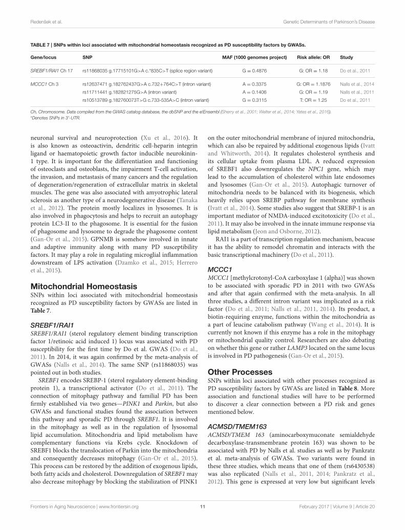
Mitochondrial HomeostasisSNPs within loci associated with mitochondrial homeostasisrecognized as PD susceptibility factors by GWASs are listed inTable 7.
SREBF1/RAI1SREBF1/RAI1 (sterol regulatory element binding transcriptionfactor 1/retinoic acid induced 1) locus was associated with PDsusceptibility for the first time by Do et al. GWAS (Do et al.,2011). In 2014, it was again confirmed by the meta-analysis ofGWASs (Nalls et al., 2014). The same SNP (rs11868035) waspointed out in both studies.
SREBF1 encodes SREBP-1 (sterol regulatory element-bindingprotein 1), a transcriptional activator (Do et al., 2011). Theconnection of mitophagy pathway and familial PD has beenfirmly established via two genes—PINK1 and Parkin, but alsoGWASs and functional studies found the association betweenthis pathway and sporadic PD through SREBF1. It is involvedin the mitophagy as well as in the regulation of lysosomallipid accumulation. Mitochondria and lipid metabolism havecomplementary functions via Krebs cycle. Knockdown ofSREBF1 blocks the translocation of Parkin into the mitochondriaand consequently decreases mitophagy (Gan-Or et al., 2015).This process can be restored by the addition of exogenous lipids,both fatty acids and cholesterol. Downregulation of SREBF1mayalso decrease mitophagy by blocking the stabilization of PINK1
on the outer mitochondrial membrane of injured mitochondria,which can also be repaired by additional exogenous lipids (Ivattand Whitworth, 2014). It regulates cholesterol synthesis andits cellular uptake from plasma LDL. A reduced expressionof SREBF1 also downregulates the NPC1 gene, which maylead to the accumulation of cholesterol within late endosomesand lysosomes (Gan-Or et al., 2015). Autophagic turnover ofmitochondria needs to be balanced with its biogenesis, whichheavily relies upon SREBP pathway for membrane synthesis(Ivatt et al., 2014). Some studies also suggest that SREBP-1 is animportant mediator of NMDA-induced excitotoxicity (Do et al.,2011). It may also be involved in the innate immune response vialipid metabolism (Jeon and Osborne, 2012).
RAI1 is a part of transcription regulation mechanism, beacuseit has the ability to remodel chromatin and interacts with thebasic transcriptional machinery (Do et al., 2011).
MCCC1MCCC1 [methylcrotonyl-CoA carboxylase 1 (alpha)] was shownto be associated with sporadic PD in 2011 with two GWASsand after that again confirmed with the meta-analysis. In allthree studies, a different intron variant was implicated as a riskfactor (Do et al., 2011; Nalls et al., 2011, 2014). Its product, abiotin-requiring enzyme, functions within the mitochondria asa part of leucine catabolism pathway (Wang et al., 2014). It iscurrently not known if this enzyme has a role in the mitophagyor mitochondrial quality control. Researchers are also debatingon whether this gene or rather LAMP3 located on the same locusis involved in PD pathogenesis (Gan-Or et al., 2015).
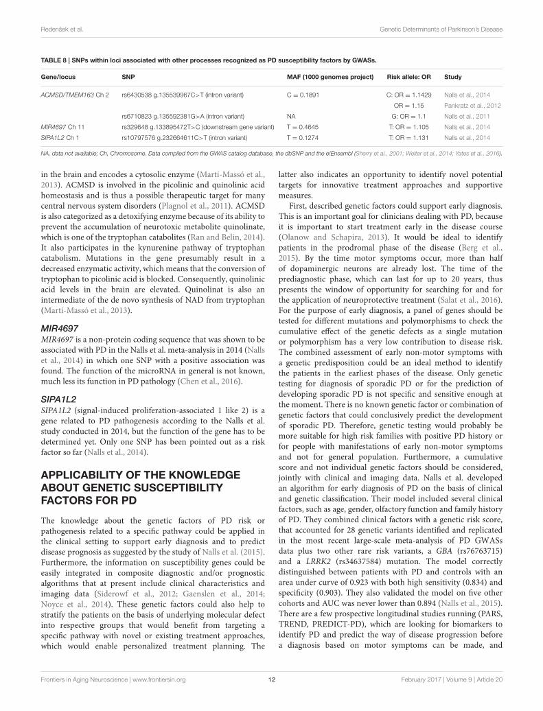
Other ProcessesSNPs within loci associated with other processes recognized asPD susceptibility factors by GWASs are listed in Table 8. Moreassociation and functional studies will have to be performedto discover a clear connection between a PD risk and genesmentioned below.
ACMSD/TMEM163ACMSD/TMEM 163 (aminocarboxymuconate semialdehydedecarboxylase-transmembrane protein 163) was shown to beassociated with PD by Nalls et al. studies as well as by Pankratzet al. meta-analysis of GWASs. Two variants were found inthese three studies, which means that one of them (rs6430538)was also replicated (Nalls et al., 2011, 2014; Pankratz et al.,2012). This gene is expressed at very low but significant levels
Frontiers in Aging Neuroscience | www.frontiersin.org 11 February 2017 | Volume 9 | Article 20
Redenšek et al. Genetic Determinants of Parkinson’s Disease
TABLE 8 | SNPs within loci associated with other processes recognized as PD susceptibility factors by GWASs.
Gene/locus SNP MAF (1000 genomes project) Risk allele: OR Study
ACMSD/TMEM163 Ch 2 rs6430538 g.135539967C>T (intron variant) C = 0.1891 C: OR = 1.1429 Nalls et al., 2014
OR = 1.15 Pankratz et al., 2012
rs6710823 g.135592381G>A (intron variant) NA G: OR = 1.1 Nalls et al., 2011
MIR4697 Ch 11 rs329648 g.133895472T>C (downstream gene variant) T = 0.4645 T: OR = 1.105 Nalls et al., 2014
SIPA1L2 Ch 1 rs10797576 g.232664611C>T (intron variant) T = 0.1274 T: OR = 1.131 Nalls et al., 2014
NA, data not available; Ch, Chromosome. Data compiled from the GWAS catalog database, the dbSNP and the e!Ensembl (Sherry et al., 2001; Welter et al., 2014; Yates et al., 2016).
in the brain and encodes a cytosolic enzyme (Martí-Massó et al.,2013). ACMSD is involved in the picolinic and quinolinic acidhomeostasis and is thus a possible therapeutic target for manycentral nervous system disorders (Plagnol et al., 2011). ACMSDis also categorized as a detoxifying enzyme because of its ability toprevent the accumulation of neurotoxic metabolite quinolinate,which is one of the tryptophan catabolites (Ran and Belin, 2014).It also participates in the kynurenine pathway of tryptophancatabolism. Mutations in the gene presumably result in adecreased enzymatic activity, which means that the conversion oftryptophan to picolinic acid is blocked. Consequently, quinolinicacid levels in the brain are elevated. Quinolinat is also anintermediate of the de novo synthesis of NAD from tryptophan(Martí-Massó et al., 2013).
MIR4697MIR4697 is a non-protein coding sequence that was shown to beassociated with PD in the Nalls et al. meta-analysis in 2014 (Nallset al., 2014) in which one SNP with a positive association wasfound. The function of the microRNA in general is not known,much less its function in PD pathology (Chen et al., 2016).
SIPA1L2SIPA1L2 (signal-induced proliferation-associated 1 like 2) is agene related to PD pathogenesis according to the Nalls et al.study conducted in 2014, but the function of the gene has to bedetermined yet. Only one SNP has been pointed out as a riskfactor so far (Nalls et al., 2014).
APPLICABILITY OF THE KNOWLEDGEABOUT GENETIC SUSCEPTIBILITYFACTORS FOR PD
The knowledge about the genetic factors of PD risk orpathogenesis related to a specific pathway could be applied inthe clinical setting to support early diagnosis and to predictdisease prognosis as suggested by the study of Nalls et al. (2015).Furthermore, the information on susceptibility genes could beeasily integrated in composite diagnostic and/or prognosticalgorithms that at present include clinical characteristics andimaging data (Siderowf et al., 2012; Gaenslen et al., 2014;Noyce et al., 2014). These genetic factors could also help tostratify the patients on the basis of underlying molecular defectinto respective groups that would benefit from targeting aspecific pathway with novel or existing treatment approaches,which would enable personalized treatment planning. The
latter also indicates an opportunity to identify novel potentialtargets for innovative treatment approaches and supportivemeasures.
First, described genetic factors could support early diagnosis.This is an important goal for clinicians dealing with PD, becauseit is important to start treatment early in the disease course(Olanow and Schapira, 2013). It would be ideal to identifypatients in the prodromal phase of the disease (Berg et al.,2015). By the time motor symptoms occur, more than halfof dopaminergic neurons are already lost. The time of theprediagnostic phase, which can last for up to 20 years, thuspresents the window of opportunity for searching for and forthe application of neuroprotective treatment (Salat et al., 2016).For the purpose of early diagnosis, a panel of genes should betested for different mutations and polymorphisms to check thecumulative effect of the genetic defects as a single mutationor polymorphism has a very low contribution to disease risk.The combined assessment of early non-motor symptoms witha genetic predisposition could be an ideal method to identifythe patients in the earliest phases of the disease. Only genetictesting for diagnosis of sporadic PD or for the prediction ofdeveloping sporadic PD is not specific and sensitive enough atthe moment. There is no known genetic factor or combination ofgenetic factors that could conclusively predict the developmentof sporadic PD. Therefore, genetic testing would probably bemore suitable for high risk families with positive PD history orfor people with manifestations of early non-motor symptomsand not for general population. Furthermore, a cumulativescore and not individual genetic factors should be considered,jointly with clinical and imaging data. Nalls et al. developedan algorithm for early diagnosis of PD on the basis of clinicaland genetic classification. Their model included several clinicalfactors, such as age, gender, olfactory function and family historyof PD. They combined clinical factors with a genetic risk score,that accounted for 28 genetic variants identified and replicatedin the most recent large-scale meta-analysis of PD GWASsdata plus two other rare risk variants, a GBA (rs76763715)and a LRRK2 (rs34637584) mutation. The model correctlydistinguished between patients with PD and controls with anarea under curve of 0.923 with both high sensitivity (0.834) andspecificity (0.903). They also validated the model on five othercohorts and AUC was never lower than 0.894 (Nalls et al., 2015).There are a few prospective longitudinal studies running (PARS,TREND, PREDICT-PD), which are looking for biomarkers toidentify PD and predict the way of disease progression beforea diagnosis based on motor symptoms can be made, and
Frontiers in Aging Neuroscience | www.frontiersin.org 12 February 2017 | Volume 9 | Article 20
Redenšek et al. Genetic Determinants of Parkinson’s Disease
assessing combinations of risk factors and early features of PD.Among these studies, PARS included a cohort of elderly patientswith olfactory dysfunction, TREND included people older than50 years with one of prodromal symptoms of PD (olfactorydysfunction, REM sleep behavior disorder or depression), whilePREDICT-PD included general population from 60 to 80 yearsof age irrespective of PD prodromal symptoms (Salat et al.,2016).
Furthermore, the impact of genetic susceptibility factorson the disease prognosis could be evaluated. It wouldbe interesting to explore the association between thecourse of the disease and the genetic defect in genesinvolved in certain pathways. For example, GBA mutationsor MAPT H1 allele status might be independent riskfactors for cognitive impairment in PD patients and theknowledge of these allele statuses in patients could have animpact on the treatment plan (Clarimón and Kulisevsky,2013).
Moreover, genetic susceptibility factors segregating indifferent pathways could help to stratify PD patients into distinctgroups according to the differences in the main molecularcause of the disease. So far, the stratification of patients hasonly been made based on different phenotypes (van Roodenet al., 2010, 2011; Fereshtehnejad et al., 2015), but our mainidea is to group them on the basis of the main molecular causeof the disease rather than on the basis of different ways ofmanifestation. The stratification should be based on cumulativeeffects of genetic susceptibility factors within a particularpathway as well as across pathways, so different combinationsof genetic defects should be tested to find a way to stratifythe PD patients. This approach was already addressed in areview written by Schapira et al. (Schapira, 2013), where theproblem of aetiologically heterogeneous cohorts of PD patientsevaluated in different studies was exposed. The latter makesit difficult to seek for different genetic biomarkers as theremay be different ones in distinct groups. The stratification ofPD patients based on underlying genetic defects could alsobe beneficial in a clinical setting as patients with differentgenetic defects may need different treatment plans. This kindof personalized treatment could become the treatment ofchoice in PD in the future, as we could stratify patients intogroups according to their compromised pathway and treatthem according to their underlying pathogenic processes. Thetreatment could be adjusted for each group to obtain the bestpossible response.
Last but not least, the disease genes or the products ofthe genes involved in the aethiopathogenesis could serve asnovel targets for neuroprotective or disease-modifying drugs todelay or prevent disease progression. However, the problem ofa very modest impact of the above-mentioned mutations andpolymorphisms on the PD pathology occurs. If the impact ofa single mutation is very low, there is probably no point indeveloping a new active pharmaceutical agent specific for thattarget, because the effect on the disease course would probablynot be satisfactory. A more feasible way of incorporating meansof personalized medicine and patient startification into PDmanagement is to target a corrupted pathway as a whole instead
of focusing only on the compromised gene and its product.The goal would be to strengthen or improve the function ofa compromised pathway, such as protein aggregation; proteinand membrane trafficking; lysosomal autophagy; immunesystem; neurodevelopment, neuron cell differentiation andsurvival; mitochondrial homeostasis; and genes involved in otherprocesses. Although this is a less specific approach, it focuseson the putative main cause of the disease occurrence in aparticular patient. In the light of this, there is a possibility ofthe repurposing of drugs already used for other indications,for example ursodeoxycholic acid, nicotine, caffeine, isradipine,exenatide, statins (Salat et al., 2016). In order to establishsuch a pathway-oriented approach, clinical trials must focuson PD patients with the same underlying molecular defect. Aslong as heterogenous cohorts are evaluated in these studies,no such treatment will be available (Korczyn and Hassin-Baer,2015).
FUTURE PERSPECTIVES
With the emergence of GWASs, researchers believed that thediscovery of the exact genetic background of PD is at theirfingertips, but more questions arose afterwards. GWASs area type of studies with no hypothesis at the time of theirperformance, so the results must be critically evaluated andsupported by functional studies of genes recognized to beinvolved in the pathogenesis of a certain disease. Apart fromfunctional studies of not yet known genes’ functions, animaldisease models should also be investigated to check and validatethe results of GWASs. We also have to be aware of thefact, that GWAS usually detect common SNPs that mostprobably have a modest impact only. Rare but highly penetrantSNPs are often overlooked. Hence, in terms of susceptibilityprediction or early diagnosis, physicians should check severalgenetic biomarkers (SNPs) and their cumulative impact on theprognosis along with clinical and imaging data. Additionally,SNPs associated with a certain disease are often not locatedwithin the disease gene, but are rather in linkage disequlibriumwith it.
The main goal of searching for genetic biomarkers ofPD susceptibility is early diagnosis or more optimistically,a chance to slow down or even prevent the developmentof the disease. Furthermore, with better characterization ofrisk genes’ functions, we could stratify the PD patients intogroups according to the main route of pathogenesis. Prospectivemonitoring is needed to compare the symptomatology aswell as the rate of progression between the groups inorder to get a chance of tailoring the therapy to eachgroup. To take this idea to the next level, one could alsofind new drug targets based on the susceptibility genesor their products if the impact of the susceptibility geneis high enough. A personalized approach would allow totreat each group of patients with the most effective andsafest drug and adopt optimal supportive measures, e.g.,antiinflammatory therapy for patients with an increasedinflammatory response.
Frontiers in Aging Neuroscience | www.frontiersin.org 13 February 2017 | Volume 9 | Article 20

Redenšek et al. Genetic Determinants of Parkinson’s Disease
CONCLUSIONS
In conclusion, the newest GWASs have identified top hitsusceptibility genes with emerging information on theirphysiological functions and involvement in PD pathology. Thesesusceptibility genes belong to specific pathways that are alreadyknown to be compromised in PD and could thus serve as agenetic tool for the stratification of patients. In our opinion,this would allow the treatment of patients according to theunderlying cause of their clinical signs to choose the mostbeneficial treatment with minimal side effects. This knowledgegives the opportunity to personalize the treatment of PD patients,but more studies need to be carried out on cell models, animalmodels and patients before new knowledge can be translatedinto the everyday clinical practice of PD treatment.
AUTHOR CONTRIBUTIONS
All authors have made a substantial intellectual contributionto this work and approved its final version for submission.VD and SR formed the review focus. SR conducted theliterature review and summarized and wrote the first draftof the manuscript under the supervision of VD. VD andMT evaluated the manuscript and contributed to the finalversion.
FUNDING
This work was partially funded by the Horizon 2020 Artemida664536 Grant and the Slovenian Research Agency (ARRS) GrantP1-0170.
REFERENCES
Ahmed, I., Tamouza, R., Delord, M., Krishnamoorthy, R., Tzourio, C., Mulot, C.,
et al. (2012). Association between Parkinson’s disease and theHLA-DRB1 locus.
Mov. Disord. 27, 1104–1110. doi: 10.1002/mds.25035
Ahn, B. H., Rhim, H., Kim, S. Y., Sung, Y. M., Lee, M. Y., Choi, J. Y., et al. (2002).
α-Synuclein interacts with phospholipase D isozymes and inhibits pervanadate-
induced phospholipase D activation in human embryonic kidney-293 cells. J.
Biol. Chem. 277, 12334–12342. doi: 10.1074/jbc.M110414200
Bellani, S., Sousa, V. L., Ronzitti, G., Valtorta, F., Meldolesi, J., and Chieregatti, E.
(2010). The regulation of synaptic function by α-synuclein. Commun. Integr.
Biol. 3, 106–109. doi: 10.4161/cib.3.2.10964
Bellucci, A., Zaltieri, M., Navarria, L., Grigoletto, J., Missale, C., and
Spano, P. (2012). From α-synuclein to synaptic dysfunctions: new insights
into the pathophysiology of Parkinson’s disease. Brain Res. 2, 183–202.
doi: 10.1016/j.brainres.2012.04.014
Berg, D., Postuma, R. B., Adler, C. H., Bloem, B. R., Chan, P., Dubois, B., et al.
(2015). MDS research criteria for prodromal Parkinson’s disease. Mov. Disord.
30, 1600–1611. doi: 10.1002/mds.26431
Bisaglia, M., Mammi, S., and Bubacco, L. (2009). Structural insights on
physiological functions and pathological effects of α-synuclein. FASEB J. 23,
329–340. doi: 10.1096/fj.08-119784
Blesa, J., Trigo-Damas, I., Quiroga-Varela, A., and Jackson-Lewis, V. R.
(2015). Oxidative stress and Parkinson’s disease. Front. Neuroanat. 9:91.
doi: 10.3389/fnana.2015.00091
Bossers, K., Meerhoff, G., Balesar, R., van Dongen, J. W., Kruse, C. G., Swaab, D.
F., et al. (2009). Analysis of gene expression in Parkinson’s disease: possible
involvement of neurotrophic support and axon guidance in dopaminergic cell
death. Brain Pathol. 19, 91–107. doi: 10.1111/j.1750-3639.2008.00171.x
Burre, J., Sharma, M., Tsetsenis, T., Buchman, V., Etherton, M. R., and Sudhof, T.
C. (2010). α-synuclein promotes SNARE-complex assembly in vivo and in vitro.
Science 329, 1663–1667. doi: 10.1126/science.1195227
Chang, K. H., Wu, Y. R., Chen, Y. C., Fung, H. C., Lee-Chen, G. J., and Chen, C.
M. (2015). STK39, But Not BST1, HLA-DQB1, and SPPL2B polymorphism, is
associated with han-chinese parkinson’s disease in taiwan. Medicine 94:e1690.
doi: 10.1097/MD.0000000000001690
Charlesworth, G., Gandhi, S., Bras, J. M., Barker, R. A., Burn, D. J., Chinnery, P.
F., et al. (2012). Tau acts as an independent genetic risk factor in pathologically
proven PD. Neurobiol. Aging 33, 4. doi: 10.1016/j.neurobiolaging.2011.11.001
Chen, C.-M., Chen, Y.-C., Chiang, M.-C., Fung, H.-C., Chang, K.-H., Lee-Chen,
G.-J., et al. (2016). Association of GCH1 and MIR4697, but not SIPA1L2 and
VPS13C polymorphisms, with Parkinson’s disease in Taiwan. Neurobiol. Aging
39, 221. e1–5. doi: 10.1016/j.neurobiolaging.2015.12.016
Chen, M.-L., Lin, C.-H., Lee, M.-J., and Wu, R.-M. (2014). BST1 rs11724635
interacts with environmental factors to increase the risk of Parkinson’s
disease in a Taiwanese population. Parkinsonism Relat. Disord. 20, 280–283.
doi: 10.1016/j.parkreldis.2013.11.009
Clarimón, J., and Kulisevsky, J. (2013). Parkinson’s disease: from
genetics to clinical practice. Curr. Genomics 14, 560–567.
doi: 10.2174/1389202914666131210212305
Connolly, B. S., and Lang, A. E. (2014). Pharmacological treatment of Parkinson
disease: a review. JAMA 311, 1670–1683. doi: 10.1001/jama.2014.3654
Davis, A. A., Andruska, K. M., Benitez, B. A., Racette, B. A., Perlmutter, J.
S., and Cruchaga, C. (2016). Variants in GBA, SNCA, and MAPT influence
Parkinson disease risk, age at onset, and progression. Neurobiol. Aging 37, 30.
doi: 10.1016/j.neurobiolaging.2015.09.014
Do, C. B., Tung, J. Y., Dorfman, E., Kiefer, A. K., Drabant, E. M., Francke, U.,
et al. (2011). Web-based genome-wide association study identifies two novel
loci and a substantial genetic component for Parkinson’s disease. PLoS Genet.
7, 23. doi: 10.1371/journal.pgen.1002141
Drouet, V., and Lesage, S. (2014). Synaptojanin 1 mutation in Parkinson’s disease
brings further insight into the neuropathological mechanisms. Biomed Res. Int.
2014:289728. doi: 10.1155/2014/289728
Dzamko, N., Geczy, C. L., and Halliday, G. M. (2015). Inflammation is
genetically implicated in Parkinson’s disease. Neuroscience 302, 89–102.
doi: 10.1016/j.neuroscience.2014.10.028
Edwards, T. L., Scott, W. K., Almonte, C., Burt, A., Powell, E. H., Beecham, G. W.,
et al. (2010). Genome-wide association study confirms SNPs in SNCA and the
MAPT region as common risk factors for Parkinson disease. Ann. Hum. Genet.
74, 97–109. doi: 10.1111/j.1469-1809.2009.00560.x
Emanuele, M., and Chieregatti, E. (2015). Mechanisms of α-synuclein action
on neurotransmission: cell-autonomous and non-cell autonomous role.
Biomolecules 5, 865–892. doi: 10.3390/biom5020865
Fereshtehnejad, S. M., Romenets, S. R., Anang, J. B., Latreille, V., Gagnon, J. F.,
and Postuma, R. B. (2015). New Clinical Subtypes of Parkinson Disease and
their longitudinal progression: a prospective cohort comparison with other
phenotypes. JAMA Neurol. 72, 863–873. doi: 10.1001/jamaneurol.2015.0703
Gaenslen, A., Wurster, I., Brockmann, K., Huber, H., Godau, J., Faust, B., et al.
(2014). Prodromal features for Parkinson’s disease–baseline data from the
TREND study. Eur. J. Neurol. 21, 766–772. doi: 10.1111/ene.12382
Gallegos, S., Pacheco, C., Peters, C., Opazo, C. M., and Aguayo, L. G. (2015).
Features of α-synuclein that could explain the progression and irreversibility
of Parkinson’s disease. Front. Neurosci. 9:59. doi: 10.3389/fnins.2015.00059
Gan-Or, Z., Dion, P. A., and Rouleau, G. A. (2015). Genetic perspective on the
role of the autophagy-lysosome pathway in Parkinson disease. Autophagy 11,
1443–1457. doi: 10.1080/15548627.2015.1067364
García-Cazorla, A., Oyarzabal, A., Fort, J., Robles, C., Castejon, E., Ruiz-Sala,
P., et al. (2014). Two novel mutations in the BCKDK (branched-chain
keto-acid dehydrogenase kinase) gene are responsible for a neurobehavioral
deficit in two pediatric unrelated patients. Hum. Mutat. 35, 470–477.
doi: 10.1002/humu.22513
Giasson, B. I., Forman, M. S., Higuchi, M., Golbe, L. I., Graves, C. L., Kotzbauer, P.
T., et al. (2003). Initiation and synergistic fibrillization of tau and α-synuclein.
Science 300, 636–640. doi: 10.1126/science.1082324
Frontiers in Aging Neuroscience | www.frontiersin.org 14 February 2017 | Volume 9 | Article 20
Redenšek et al. Genetic Determinants of Parkinson’s Disease
Golpich, M., Amini, E., Hemmati, F., Ibrahim, N. M., Rahmani, B., Mohamed, Z.,
et al. (2015). Glycogen synthase kinase-3 β (GSK-3β) signaling: implications for
Parkinson’s disease. Pharmacol. Res. 97, 16–26. doi: 10.1016/j.phrs.2015.03.010
Goris, A., Williams-Gray, C. H., Clark, G. R., Foltynie, T., Lewis, S. J., Brown,
J., et al. (2007). Tau and α-synuclein in susceptibility to, and dementia in,
Parkinson’s disease. Ann. Neurol. 62, 145–153. doi: 10.1002/ana.21192
Gustot, A., Gallea, J. I., Sarroukh, R., Celej, M. S., Ruysschaert, J. M., and
Raussens, V. (2015). Amyloid fibrils are the molecular trigger of inflammation
in Parkinson’s disease. Biochem. J. 471, 323–333. doi: 10.1042/BJ20150617
Hamza, T. H., Zabetian, C. P., Tenesa, A., Laederach, A., Montimurro, J.,
Yearout, D., et al. (2010). Common genetic variation in the HLA region is
associated with late-onset sporadic Parkinson’s disease.Nat. Genet. 42, 781–785.
doi: 10.1038/ng.642
Hardy, J. (2010). Genetic analysis of pathways to Parkinson disease. Neuron 68,
201–206. doi: 10.1016/j.neuron.2010.10.014
Hasegawa, M., Nonaka, T., and Masuda-Suzukake, M. (2016). Prion-like
mechanisms and potential therapeutic targets in neurodegenerative disorders.
Pharmacol. Ther. 1, 30236–30234. doi: 10.1016/j.pharmthera.2016.11.010
Herrero, M. T., Estrada, C., Maatouk, L., and Vyas, S. (2015). Inflammation
in Parkinson’s disease: role of glucocorticoids. Front. Neuroanat. 9:32.
doi: 10.3389/fnana.2015.00032
Hill-Burns, E. M., Factor, S. A., Zabetian, C. P., Thomson, G., and
Payami, H. (2011). Evidence for more than one Parkinson’s disease-
associated variant within the HLA region. PLoS ONE 6:e27109.
doi: 10.1371/journal.pone.0027109
Hill-Burns, E. M., Wissemann, W. T., Hamza, T. H., Factor, S. A., Zabetian,
C. P., and Payami, H. (2014). Identification of a novel Parkinson’s disease
locus via stratified genome-wide association study. BMC Genomics 15:118.
doi: 10.1186/1471-2164-15-118
Hoang, Q. Q. (2014). Pathway for Parkinson disease. Proc. Natl. Acad. Sci. U.S.A.
111, 2402–2403. doi: 10.1073/pnas.1324284111
Horvath, J., Herrmann, F. R., Burkhard, P. R., Bouras, C., and Kovari,
E. (2013). Neuropathology of dementia in a large cohort of patients
with Parkinson’s disease. Parkinsonism Relat. Disord. 19, 864–868.
doi: 10.1016/j.parkreldis.2013.05.010
Inoshita, T., and Imai, Y. (2015). Regulation of vesicular trafficking
by Parkinson’s disease-associated genes. AIMS Mol. Sci. 2, 461–475.
doi: 10.3934/molsci.2015.4.461
Itoh, N., and Ohta, H. (2013). Roles of FGF20 in dopaminergic neurons and
Parkinson’s disease. Front. Mol. Neurosci. 6:15. doi: 10.3389/fnmol.2013.00015
Ivatt, R. M., Sanchez-Martinez, A., Godena, V. K., Brown, S., Ziviani, E., and
Whitworth, A. J. (2014). Genome-wide RNAi screen identifies the Parkinson
disease GWAS risk locus SREBF1 as a regulator of mitophagy. Proc. Natl. Acad.
Sci. U.S.A. 111, 8494–8499. doi: 10.1073/pnas.1321207111
Ivatt, R. M., and Whitworth, A. J. (2014). SREBF1 links lipogenesis to
mitophagy and sporadic Parkinson disease. Autophagy 10, 1476–1477.
doi: 10.4161/auto.29642
Jeon, T. I., and Osborne, T. F. (2012). SREBPs: metabolic integrators
in physiology and metabolism. Trends Endocrinol. Metab. 23, 65–72.
doi: 10.1016/j.tem.2011.10.004
Kalia, L. V., and Lang, A. E. (2015). Parkinson’s disease. Lancet 386, 896–912.
doi: 10.1016/S0140-6736(14)61393-3
Klein, C., andWestenberger, A. (2012). Genetics of Parkinson’s disease.Cold Spring
Harb. Perspect. Med. 2:a008888. doi: 10.1101/cshperspect.a008888
Korczyn, A. D., and Hassin-Baer, S. (2015). Can the disease course in Parkinson’s
disease be slowed? BMCMed. 13, 295. doi: 10.1186/s12916-015-0534-x
Kruger, R., Vieira-Saecker, A. M., Kuhn, W., Berg, D., Muller, T., Kuhnl, N., et al.
(1999). Increased susceptibility to sporadic Parkinson’s disease by a certain
combined α-synuclein/apolipoprotein E genotype. Ann. Neurol. 45, 611–617.
Kumaran, R., and Cookson, M. R. (2015). Pathways to Parkinsonism Redux:
convergent pathobiological mechanisms in genetics of Parkinson’s disease.
Hum. Mol. Genet. 24, 22. doi: 10.1093/hmg/ddv236
Labbé, C., and Ross, O. A. (2014). Association studies of sporadic
Parkinson’s Disease in the genomic era. Curr. Genomics 15, 2–10.
doi: 10.2174/1389202914666131210212745
Lampe, J. B., Gossrau, G., Herting, B., Kempe, A., Sommer, U., Fussel, M.,
et al. (2003). HLA typing and Parkinson’s disease. Eur. Neurol. 50, 64–68.
doi: 10.1159/000072500
Lashuel, H. A., Overk, C. R., Oueslati, A., and Masliah, E. (2013). The many
faces of α-synuclein: from structure and toxicity to therapeutic target. Nat. Rev.
Neurosci. 14, 38–48. doi: 10.1038/nrn3406
Lashuel, H. A., Petre, B. M., Wall, J., Simon, M., Nowak, R. J., Walz, T., et al.
(2002). α-synuclein, especially the Parkinson’s disease-associated mutants,
forms pore-like annular and tubular protofibrils. J. Mol. Biol. 322, 1089–1102.
doi: 10.1016/S0022-2836(02)00735-0
Latourelle, J. C., Pankratz, N., Dumitriu, A., Wilk, J. B., Goldwurm, S., Pezzoli, G.,
et al. (2009). Genomewide association study for onset age in Parkinson disease.
BMCMed. Genet. 10:98. doi: 10.1186/1471-2350-10-98
Lesage, S., Drouet, V., Majounie, E., Deramecourt, V., Jacoupy, M., Nicolas, A.,
et al. (2016). Loss of VPS13C Function in autosomal-recessive parkinsonism
causes mitochondrial dysfunction and increases pink1/parkin-dependent
mitophagy. Am. J. Hum. Genet. 98, 500–513. doi: 10.1016/j.ajhg.2016.01.014
Li, N. N., Tan, E. K., Chang, X. L., Mao, X. Y., Zhang, J. H., Zhao, D. M., et al.
(2013). Genetic association study between STK39 and CCDC62/HIP1R and
Parkinson’s disease. PLoS ONE 8:e79211. doi: 10.1371/journal.pone.0079211
Lill, C. M., Roehr, J. T., McQueen, M. B., Kavvoura, F. K., Bagade, S., Schjeide,
B. M., et al. (2012). Comprehensive research synopsis and systematic meta-
analyses in Parkinson’s disease genetics: the PDGene database. PLoS Genet.
8:e1002548. doi: 10.1371/journal.pgen.1002548.
Lin, C. H., and Wu, R. M. (2015). Biomarkers of cognitive decline
in Parkinson’s disease. Parkinsonism Relat. Disord. 21, 431–443.
doi: 10.1016/j.parkreldis.2015.02.010
Liscovitch, N., and French, L. (2014). Differential co-expression between α-
Synuclein and IFN-gamma signaling genes across development and in
Parkinson’s disease. PLoS ONE 9:e115029.. doi: 10.1371/journal.pone.0115029
Lu, Y., Liu, W., Tan, K., Peng, J., Zhu, Y., andWang, X. (2015). Genetic association
of RIT2 rs12456492 polymorphism and Parkinson’s disease susceptibility in
Asian populations: a meta-analysis. Sci. Rep. 5:13805. doi: 10.1038/srep13805
Lu, Y., Tan, L., Shen, N., Peng, J., Wang, C., Zhu, Y., et al. (2016).
Possible association of CCDC62 rs12817488 polymorphism and Parkinson’s
disease risk in Chinese population: a meta-analysis. Sci. Rep. 6:23991.
doi: 10.1038/srep23991
MacLeod, D. A., Rhinn, H., Kuwahara, T., Zolin, A., Di Paolo, G., McCabe, B.
D., et al. (2013). RAB7L1 interacts with LRRK2 to modify intraneuronal
protein sorting and Parkinson’s disease risk. Neuron 77, 425–439.
doi: 10.1016/j.neuron.2012.11.033
Maraganore, D.M., de Andrade, M., Elbaz, A., Farrer, M. J., Ioannidis, J. P., Kruger,
R., et al. (2006). Collaborative analysis of α-synuclein gene promoter variability
and Parkinson disease. JAMA 296, 661–670. doi: 10.1001/jama.296.6.661
Martí-Massó, J. F., Bergareche, A., Makarov, V., Ruiz-Martinez, J., Gorostidi, A.,
de Munain, A. L., et al. (2013). The ACMSD gene, involved in tryptophan
metabolism, is mutated in a family with cortical myoclonus, epilepsy, and
parkinsonism. J. Mol. Med. 91, 1399–1406. doi: 10.1007/s00109-013-1075-4
Mazzulli, J. R., Xu, Y. H., Sun, Y., Knight, A. L., McLean, P. J., Caldwell,
G. A., et al. (2011). Gaucher disease glucocerebrosidase and α-synuclein
form a bidirectional pathogenic loop in synucleinopathies. Cell 146, 37–52.
doi: 10.1016/j.cell.2011.06.001
Mencacci, N. E., Isaias, I. U., Reich, M. M., Ganos, C., Plagnol, V., Polke, J. M.,
et al. (2014). Parkinson’s disease in GTP cyclohydrolase 1 mutation carriers.
Brain 137(Pt 9), 2480–2492. doi: 10.1093/brain/awu179
Mitchell, S., Vargas, J., and Hoffmann, A. (2016). Signaling via the
NFkappaB system. Wiley Interdiscip. Rev. Syst. Biol. Med. 8, 227–241.
doi: 10.1002/wsbm.1331
Nakatsu, F., Messa, M., Nandez, R., Czapla, H., Zou, Y. X., Strittmatter, S. M.,
et al. (2015). Sac2/INPP5F is an inositol 4-phosphatase that functions in the
endocytic pathway. J. Cell Biol. 209, 85–95. doi: 10.1083/jcb.201409064
Nalls, M. A., McLean, C. Y., Rick, J., Eberly, S., Hutten, S. J., Gwinn, K.,
et al. (2015). Diagnosis of Parkinson’s disease on the basis of clinical and
genetic classification: a population-based modelling study. Lancet Neurol. 14,
1002–1009. doi: 10.1016/S1474-4422(15)00178-7
Nalls, M. A., Pankratz, N., Lill, C. M., Do, C. B., Hernandez, D. G., Saad,
M., et al. (2014). Large-scale meta-analysis of genome-wide association data
identifies six new risk loci for Parkinson’s disease. Nat. Genet. 46, 989–993.
doi: 10.1038/ng.3043
Nalls, M. A., Plagnol, V., Hernandez, D. G., Sharma, M., Sheerin, U. M., Saad, M.,
et al. (2011). Imputation of sequence variants for identification of genetic risks
Frontiers in Aging Neuroscience | www.frontiersin.org 15 February 2017 | Volume 9 | Article 20
Redenšek et al. Genetic Determinants of Parkinson’s Disease
for Parkinson’s disease: a meta-analysis of genome-wide association studies.
Lancet 377, 641–649. doi: 10.1016/S0140-6736(10)62345-8
Noyce, A. J., Bestwick, J. P., Silveira-Moriyama, L., Hawkes, C. H., Knowles, C. H.,
Hardy, J., et al. (2014). PREDICT-PD: identifying risk of Parkinson’s disease in
the community: methods and baseline results. J. Neurol. Neurosurg. Psychiatr.
85, 31–37. doi: 10.1136/jnnp-2013-305420
Olanow, C. W., and Schapira, A. H. (2013). Therapeutic prospects for Parkinson
disease. Ann. Neurol. 74, 337–347. doi: 10.1002/ana.24011
Pankratz, N., Beecham, G. W., DeStefano, A. L., Dawson, T. M., Doheny, K. F.,
Factor, S. A., et al. (2012). Meta-analysis of Parkinson’s disease: identification of
a novel locus, RIT2. Ann. Neurol. 71, 370–384. doi: 10.1002/ana.22687
Pankratz, N., Wilk, J. B., Latourelle, J. C., DeStefano, A. L., Halter, C.,
Pugh, E. W., et al. (2009). Genomewide association study for susceptibility
genes contributing to familial Parkinson disease. Hum. Genet. 124, 593–605.
doi: 10.1007/s00439-008-0582-9
Pastor, P., Ezquerra, M., Munoz, E., Marti, M. J., Blesa, R., Tolosa,
E., et al. (2000). Significant association between the tau gene
A0/A0 genotype and Parkinson’s disease. Ann. Neurol. 47, 242–245.
doi: 10.1002/1531-8249(200002)47:2<242::AID-ANA16>3.0.CO;2-L
Perrett, R. M., Alexopoulou, Z., and Tofaris, G. K. (2015). The endosomal
pathway in Parkinson’s disease. Mol. Cell Neurosci. 66(Pt A), 21–28.
doi: 10.1016/j.mcn.2015.02.009
Pihlstrom, L., Morset, K. R., Grimstad, E., Vitelli, V., and Toft, M. (2016). A
cumulative genetic risk score predicts progression in Parkinson’s disease.Mov.
Disord. 8:26505. doi: 10.1002/mds.26505
Plagnol, V., Nalls, M. A., Bras, J. M., Hernandez, D. G., Sharma, M.,
Sheerin, U. M., et al. (2011). A two-stage meta-analysis identifies
several new loci for parkinson’s disease. PLoS Genet. 7:e1002142.
doi: 10.1371/journal.pgen.1002142
Polymeropoulos, M. H., Lavedan, C., Leroy, E., Ide, S. E., Dehejia,
A., Dutra, A., et al. (1997). Mutation in the α-synuclein gene
identified in families with Parkinson’s disease. Science 276, 2045–2047.
doi: 10.1126/science.276.5321.2045
Postuma, R. B., Berg, D., Stern, M., Poewe, W., Olanow, C. W., Oertel, W., et al.
(2015). MDS clinical diagnostic criteria for Parkinson’s disease. Mov. Disord.
30, 1591–1601. doi: 10.1002/mds.26424
Pringsheim, T., Jette, N., Frolkis, A., and Steeves, T. D. (2014). The prevalence of
Parkinson’s disease: a systematic review and meta-analysis. Mov. Disord. 29,
1583–1590. doi: 10.1002/mds.25945
Ran, C., and Belin, A. C. (2014). The genetics of Parkinson’s disease: review of
current and emerging candidates. J. Parkinsonism Restless Legs Syndrome 4,
63–75. doi: 10.2147/JPRLS.S38954
Rengmark, A., Pihlstrom, L., Linder, J., Forsgren, L., and Toft, M. (2016). Low
frequency of GCH1 and TH mutations in Parkinson’s disease. Parkinsonism
Relat. Disord. 29, 109–111. doi: 10.1016/j.parkreldis.2016.05.010
Russo, I., Bubacco, L., and Greggio, E. (2014). LRRK2 and neuroinflammation:
partners in crime in Parkinson’s disease? J. Neuroinflammation 11, 1742–2094.
doi: 10.1186/1742-2094-11-52
Ryan, B. J., Hoek, S., Fon, E. A., and Wade-Martins, R. (2015). Mitochondrial
dysfunction and mitophagy in Parkinson’s: from familial to sporadic disease.
Trends Biochem. Sci. 40, 200–210. doi: 10.1016/j.tibs.2015.02.003
Saad, M., Lesage, S., Saint-Pierre, A., Corvol, J. C., Zelenika, D., Lambert, J. C., et al.
(2011). Genome-wide association study confirms BST1 and suggests a locus on
12q24 as the risk loci for Parkinson’s disease in the european population. Hum.
Mol. Genet. 20, 615–627. doi: 10.1093/hmg/ddq497
Saiki, S., Sato, S., and Hattori, N. (2012). Molecular pathogenesis of
Parkinson’s disease: update. J. Neurol. Neurosurg. Psychiatr. 83, 430–436.
doi: 10.1136/jnnp-2011-301205
Salat, D., Noyce, A. J., Schrag, A., and Tolosa, E. (2016). Challenges of modifying
disease progression in prediagnostic Parkinson’s disease. Lancet Neurol. 15,
637–648. doi: 10.1016/S1474-4422(16)00060-0
Satake, W., Nakabayashi, Y., Mizuta, I., Hirota, Y., Ito, C., Kubo, M., et al.
(2009). Genome-wide association study identifies common variants at four
loci as genetic risk factors for Parkinson’s disease. Nat. Genet. 41, 1303–1307.
doi: 10.1038/ng.485
Schapira, A. H. (2013). Recent developments in biomarkers in Parkinson disease.
Curr. Opin. Neurol. 26, 395–400. doi: 10.1097/WCO.0b013e3283633741
Schulte, C., and Gasser, T. (2011). Genetic basis of Parkinson’s disease:
inheritance, penetrance, and expression. Appl. Clin. Genet. 4, 67–80.
doi: 10.2147/TACG.S11639
Sekiyama, K., Takamatsu, Y., Waragai, M., and Hashimoto, M. (2014). Role of
genomics in translational research for Parkinson’s disease. Biochem. Biophys.
Res. Commun. 452, 226–235. doi: 10.1016/j.bbrc.2014.06.028
Sherry, S. T.,Ward,M.-H., Kholodov, M., Baker, J., Phan, L., Smigielski, E. M., et al.
(2001). dbSNP: the NCBI database of genetic variation. Nucleic Acids Res. 29,
308–311. doi: 10.1093/nar/29.1.308
Siderowf, A., Jennings, D., Eberly, S., Oakes, D., Hawkins, K. A., Ascherio,
A., et al. (2012). Impaired olfaction and other prodromal features in the
Parkinson at-risk syndrome study.Mov. Disord. 27, 406–412. doi: 10.1002/mds.
24892
Simon-Sanchez, J., Schulte, C., Bras, J. M., Sharma, M., Gibbs, J. R., Berg, D.,
et al. (2009). Genome-wide association study reveals genetic risk underlying
Parkinson’s disease. Nat. Genet. 41, 1308–1312. doi: 10.1038/ng.487
Singleton, A. B., Farrer, M. J., and Bonifati, V. (2013). The genetics of Parkinson’s
disease: progress and therapeutic implications. Mov. Disord. 28, 14–23.
doi: 10.1002/mds.25249
Soto-Ortolaza, A. I., and Ross, O. A. (2016). Genetic susceptibility
variants in parkinsonism. Parkinsonism Relat. Disord. 22, 7.
doi: 10.1016/j.parkreldis.2015.09.011
Spencer, C. C., Plagnol, V., Strange, A., Gardner,M., Paisan-Ruiz, C., Band, G., et al.
(2011). Dissection of the genetics of Parkinson’s disease identifies an additional
association 5′ of SNCA andmultiple associated haplotypes at 17q21.Hum.Mol.
Genet. 20, 345–353. doi: 10.1093/hmg/ddq469
Subramaniam, S. R., and Chesselet, M. F. (2013). Mitochondrial dysfunction
and oxidative stress in Parkinson’s disease. Prog. Neurobiol. 107, 17–32.
doi: 10.1016/j.pneurobio.2013.04.004
Tanaka, H., Shimazawa, M., Kimura, M., Takata, M., Tsuruma, K., Yamada, M.,
et al. (2012). The potential of GPNMB as novel neuroprotective factor in
amyotrophic lateral sclerosis. Sci. Rep. 2:573. doi: 10.1038/srep00573
Tarazi, F. I., Sahli, Z. T., Wolny, M., and Mousa, S. A. (2014). Emerging therapies
for Parkinson’s disease: from bench to bedside. Pharmacol. Ther. 144, 123–133.
doi: 10.1016/j.pharmthera.2014.05.010
Taymans, J. M., Nkiliza, A., and Chartier-Harlin, M. C. (2015). Deregulation
of protein translation control, a potential game-changing hypothesis
for Parkinson’s disease pathogenesis. Trends Mol. Med. 21, 466–472.
doi: 10.1016/j.molmed.2015.05.004
Tsigelny, I. F., Sharikov, Y., Wrasidlo, W., Gonzalez, T., Desplats, P. A.,
Crews, L., et al. (2012). Role of α-synuclein penetration into the membrane
in the mechanisms of oligomer pore formation. FEBS J. 279, 1000–1013.
doi: 10.1111/j.1742-4658.2012.08489.x
Vacic, V., Ozelius, L. J., Clark, L. N., Bar-Shira, A., Gana-Weisz, M., Gurevich,
T., et al. (2014). Genome-wide mapping of IBD segments in an Ashkenazi
PD cohort identifies associated haplotypes. Hum. Mol. Genet. 23, 4693–4702.
doi: 10.1093/hmg/ddu158
van der Brug, M. P., Singleton, A., Gasser, T., and Lewis, P. A. (2015). Parkinson’s
disease: from human genetics to clinical trials. Sci. Transl. Med. 7, 305ps20.
doi: 10.1126/scitranslmed.aaa8280
Vandrovcova, J., Pittman, A. M., Malzer, E., Abou-Sleiman, P. M., Lees, A.
J., Wood, N. W., et al. (2009). Association of MAPT haplotype-tagging
SNPs with sporadic Parkinson’s disease. Neurobiol. Aging 30, 1477–1482.
doi: 10.1016/j.neurobiolaging.2007.11.019
van Rooden, S. M., Colas, F., Martinez-Martin, P., Visser, M., Verbaan, D.,
Marinus, J., et al. (2011). Clinical subtypes of Parkinson’s disease.Mov. Disord.
26, 51–58. doi: 10.1002/mds.23346
van Rooden, S. M., Heiser, W. J., Kok, J. N., Verbaan, D., van Hilten, J. J.,
and Marinus, J. (2010). The identification of Parkinson’s disease subtypes
using cluster analysis: a systematic review. Mov. Disord. 25, 969–978.
doi: 10.1002/mds.23116
Velayos-Baeza, A., Vettori, A., Copley, R. R., Dobson-Stone, C., and Monaco, A.
P. (2004). Analysis of the human VPS13 gene family. Genomics 84, 536–549.
doi: 10.1016/j.ygeno.2004.04.012
Venda, L. L., Cragg, S. J., Buchman, V. L., and Wade-Martins, R. (2010). α-
Synuclein and dopamine at the crossroads of Parkinson’s disease. Trends
Neurosci. 33, 559–568. doi: 10.1016/j.tins.2010.09.004
Frontiers in Aging Neuroscience | www.frontiersin.org 16 February 2017 | Volume 9 | Article 20
Redenšek et al. Genetic Determinants of Parkinson’s Disease
Verstraeten, A., Theuns, J., and Van Broeckhoven, C. (2015). Progress in
unraveling the genetic etiology of Parkinson disease in a genomic era. Trends
Genet. 31, 140–149. doi: 10.1016/j.tig.2015.01.004
Volta, M., Milnerwood, A. J., and Farrer, M. J. (2015). Insights from late-
onset familial parkinsonism on the pathogenesis of idiopathic Parkinson’s
disease. Lancet Neurol. 14, 1054–1064. doi: 10.1016/S1474-4422(15)
00186-6
Wallings, R., Manzoni, C., and Bandopadhyay, R. (2015). Cellular processes
associated with LRRK2 function and dysfunction. FEBS J. 282, 2806–2826.
doi: 10.1111/febs.13305
Wang, G., van der Walt, J. M., Mayhew, G., Li, Y.-J., Züchner, S., Scott, W. K.,
et al. (2008). Variation in the miRNA-433 binding site of fgf20 confers risk for
parkinson disease by overexpression of α-synuclein. Am. J. Hum. Genet. 82,
283–289. doi: 10.1016/j.ajhg.2007.09.021
Wang, L., Cheng, L., Lu, Z. J., Sun, X. Y., Li, J. Y., and Peng, R. (2016). Association
of three candidate genetic variants in RAB7L1/NUCKS1, MCCC1 and STK39
with sporadic Parkinson’s disease in Han Chinese. J. Neural Transm. 123,
425–430. doi: 10.1007/s00702-016-1526-5
Wang, Y. Q., Tang, B. S., Yu, R. L., Li, K., Liu, Z. H., Xu, Q., et al. (2014). Association
analysis of STK39, MCCC1/LAMP3 and sporadic PD in the Chinese Han
population. Neurosci. Lett. 566, 206–209. doi: 10.1016/j.neulet.2014.03.007
Welter, D., MacArthur, J., Morales, J., Burdett, T., Hall, P., Junkins, H., et al. (2014).
The NHGRI GWAS Catalog, a curated resource of SNP-trait associations.
Nucleic Acids Res. 42(Database issue):6. doi: 10.1093/nar/gkt1229
Wider, C., Dachsel, J. C., Soto, A. I., Heckman, M. G., Diehl, N. N., Yue,
M., et al. (2009). FGF20 and parkinson’s disease: no evidence of association
or pathogenicity via α-synuclein expression. Mov. Disord. 24, 455–459.
doi: 10.1002/mds.22442
Wider, C., Vilarino-Guell, C., Jasinska-Myga, B., Heckman, M. G., Soto-Ortolaza,
A. I., Cobb, S. A., et al. (2010). Association of the MAPT locus with Parkinson’s
disease. Eur. J. Neurol. 17, 483–486. doi: 10.1111/j.1468-1331.2009.02847.x
Wissemann, W. T., Hill-Burns, E. M., Zabetian, C. P., Factor, S. A., Patsopoulos,
N., Hoglund, B., et al. (2013). Association of Parkinson disease with structural
and regulatory variants in the HLA region. Am. J. Hum. Genet. 93, 984–993.
doi: 10.1016/j.ajhg.2013.10.009
Wray, S., and Lewis, P. A. (2010). A tangled web - tau and sporadic Parkinson’s
disease. Front. Psychiatry 1:150. doi: 10.3389/fpsyt.2010.00150
Xi, P., Ding, D., Zhou, J., Wang, M., and Cong, Y. S. (2013). DDRGK1
regulates NF-κB activity by modulating IκBα stability. PLoS ONE 8:e64231.
doi: 10.1371/journal.pone.0064231
Xu, Y., Chen, Y., Ou, R., Wei, Q.-Q., Cao, B., Chen, K., et al. (2016). No association
of GPNMB rs156429 polymorphism with Parkinson’s disease, amyotrophic
lateral sclerosis and multiple system atrophy in Chinese population. Neurosci.
Lett. 622, 113–117. doi: 10.1016/j.neulet.2016.04.060
Yang, R.-Y., Xue, H., Yu, L., Velayos-Baeza, A., Monaco, A. P., and Liu,
F.-T. (2016). Identification of VPS13C as a Galectin-12-Binding Protein
That Regulates Galectin-12 Protein Stability and Adipogenesis. PLoS ONE
11:e0153534. doi: 10.1371/journal.pone.0153534
Yates, A., Akanni, W., Amode, M. R., Barrell, D., Billis, K., Carvalho-
Silva, D., et al. (2016). Ensembl 2016. Nucleic Acids Res. 44, D710–D716.
doi: 10.1093/nar/gkv1157
Yu, R.-l., Guo, J.-f., Wang, Y.-q., Liu, Z.-h., Sun, Z.-f., Su, L., et al.
(2015). The single nucleotide polymorphism Rs12817488 is associated with
Parkinson’s disease in the Chinese population. J. Clin. Neurosci. 22, 1002–1004.
doi: 10.1016/j.jocn.2014.11.024
Zhang, L., Wahlin, K., Li, Y. Y., Masuda, T., Yang, Z. Y., Zack, D. J., et al. (2013).
RIT2, a neuron-specific small guanosine triphosphatase, is expressed in retinal
neuronal cells and its promoter is modulated by the POU4 transcription factors.
Mol. Vis. 19, 1371–1386.
Conflict of Interest Statement: The authors declare that the research was
conducted in the absence of any commercial or financial relationships that could
be construed as a potential conflict of interest.
Copyright © 2017 Redenšek, Trošt and Dolžan. This is an open-access article
distributed under the terms of the Creative Commons Attribution License (CC BY).
The use, distribution or reproduction in other forums is permitted, provided the
original author(s) or licensor are credited and that the original publication in this
journal is cited, in accordance with accepted academic practice. No use, distribution
or reproduction is permitted which does not comply with these terms.
Frontiers in Aging Neuroscience | www.frontiersin.org 17 February 2017 | Volume 9 | Article 20