Embed Size (px)
Citation preview
ORIGINAL ARTICLE
Genetic and neurodevelopmental spectrum ofSYNGAP1-associated intellectual disability andepilepsyCyril Mignot,1,2,3 Celina von Stülpnagel,4,5 Caroline Nava,1,6 Dorothée Ville,7
Damien Sanlaville,8,9,10 Gaetan Lesca,8,9,10 Agnès Rastetter,6 Benoit Gachet,6
Yannick Marie,6 G Christoph Korenke,11 Ingo Borggraefe,12
Dorota Hoffmann-Zacharska,13 Elżbieta Szczepanik,14 Mariola Rudzka-Dybała,14
Uluç Yiş,15 Hande Çağlayan,16 Arnaud Isapof,17 Isabelle Marey,1
Eleni Panagiotakaki,18 Christian Korff,19 Eva Rossier,20 Angelika Riess,21
Stefanie Beck-Woedl,21 Anita Rauch,22 Christiane Zweier,23 Juliane Hoyer,23
André Reis,23 Mikhail Mironov,24 Maria Bobylova,24 Konstantin Mukhin,24
Laura Hernandez-Hernandez,25 Bridget Maher,25 Sanjay Sisodiya,25 Marius Kuhn,26
Dieter Glaeser,26 Sarah Wechuysen,6,27 Candace T Myers,28 Heather C Mefford,28
Konstanze Hörtnagel,29 Saskia Biskup,29 EuroEPINOMICS-RES MAE working group,Johannes R Lemke,30 Delphine Héron,1,2,3,4 Gerhard Kluger,4,5 Christel Depienne1,6
▸ Additional material ispublished online only. To viewplease visit the journal online(http://dx.doi.org/10.1136/jmedgenet-2015-103451).
For numbered affiliations seeend of article.
Correspondence toDr Cyril Mignot, Départementde Génétique, AP-HP, GroupeHospitalier Pitié-Salpêtrière,Paris F-75013, France;[email protected] orDr Christel depienne,Service de CytogénétiqueConstitutionnelle et Prénatale,Hôpital de Hautepierre,1 avenue Moliére, 67098STRASBOURG Cedex, France
Received 12 August 2015Revised 15 February 2016Accepted 16 February 2016
To cite: Mignot C, vonStülpnagel C, Nava C, et al.J Med Genet PublishedOnline First: [please includeDay Month Year]doi:10.1136/jmedgenet-2015-103451
ABSTRACTObjective We aimed to delineate the neurodevelopmentalspectrum associated with SYNGAP1 mutations and toinvestigate genotype–phenotype correlations.Methods We sequenced the exome or screened the exonsof SYNGAP1 in a total of 251 patients withneurodevelopmental disorders. Molecular and clinical datafrom patients with SYNGAP1 mutations from other centreswere also collected, focusing on developmental aspects andthe associated epilepsy phenotype. A review of SYNGAP1mutations published in the literature was also performed.Results We describe 17 unrelated affected individualscarrying 13 different novel loss-of-function SYNGAP1mutations. Developmental delay was the first manifestationof SYNGAP1-related encephalopathy; intellectual disabilitybecame progressively obvious and was associated withautistic behaviours in eight patients. Hypotonia and unstablegait were frequent associated neurological features. With theexception of one patient who experienced a single seizure,all patients had epilepsy, characterised by falls or head dropsdue to atonic or myoclonic seizures, (myoclonic) absencesand/or eyelid myoclonia. Triggers of seizures were frequent(n=7). Seizures were pharmacoresistant in half of thepatients. The severity of the epilepsy did not correlate withthe presence of autistic features or with the severity ofcognitive impairment. Mutations were distributed throughoutthe gene, but spared spliced 30 and 50 exons. Seizures inpatients with mutations in exons 4–5 were morepharmacoresponsive than in patients with mutations inexons 8–15.Conclusions SYNGAP1 encephalopathy is characterisedby early neurodevelopmental delay typically preceding theonset of a relatively recognisable epilepsy comprisinggeneralised seizures (absences, myoclonic jerks) and frequenttriggers.
INTRODUCTIONThe human SYNGAP1 gene on chromosome6p21.3 encodes the synaptic RAS-GTPase-activatingprotein 1, a protein of the post-synaptic density(PSD) of glutamatergic neurons.1 2 SYNGAP1 inter-acts with PSD95 (DLG4) and SAP102 (DLG3), and isable to positively or negatively regulate the density ofN-Methyl-D-aspartic acid (NMDA) and α-amino-3-hydroxy-5-methyl-4-isoxazolepropionic acid (AMPA)receptors at the glutamatergic synapses and mediatesignalling downstream of glutamate receptor activa-tion.3 4 While complete Syngap1 deficiency in mice islethal at early postnatal stages, heterozygous syngap1+/- mice are viable but show behavioural and cogni-tive disturbances.5–8 Syngap1 haploinsufficiency dis-rupts the excitatory/inhibitory balance in thedeveloping hippocampus and cortex and results inaccelerated glutamatergic synapse maturation. Whenthis process occurs during critical developmentalwindows, it alters the synaptic plasticity necessary forthe refinement of connections that ultimately shapecognitive and behavioural modalities.4 9 DifferentSYNGAP1 protein isoforms exist and are generatedthrough alternative splicing and alternative promoterusage, in a process regulated by synaptic activity andpostnatal age in mice. Two of the main SYNGAP1mouse isoforms that differ in their N-terminal and C-terminal sequences have opposite effects on glutam-ate activation pathway.10 Although several isoformshave also been described in humans, their specificrole has not yet been established.Recently, several groups have independently
reported de novo SYNGAP1 mutations in patientswith intellectual disability (ID), epileptic encephal-opathy (EE) or autism spectrum disorders (ASD)identified by exome sequencing11–15 or direct
Mignot C, et al. J Med Genet 2016;0:1–12. doi:10.1136/jmedgenet-2015-103451 1
Cognitive and behavioural genetics JMG Online First, published on March 17, 2016 as 10.1136/jmedgenet-2015-103451
Copyright Article author (or their employer) 2016. Produced by BMJ Publishing Group Ltd under licence.
group.bmj.com on November 23, 2016 - Published by http://jmg.bmj.com/Downloaded from group.bmj.com on November 23, 2016 - Published by http://jmg.bmj.com/Downloaded from group.bmj.com on November 23, 2016 - Published by http://jmg.bmj.com/Downloaded from
sequencing of the SYNGAP1 gene through a candidate geneapproach.16–24 Recently, seven SYNGAP1 mutations were identi-fied by exome sequencing in a series of 1133 patients, 83% ofwhom had ID, indicating a frequency of SYNGAP1 mutation of∼0.74% in patients with ID.25 One patient with a chromosomaltranslocation interrupting SYNGAP126 and five patients with6p21.3 deletions encompassing SYNGAP123 27–30 have alsobeen reported. Thus, to date, SYNGAP1 appears one of themost relevant ID-causing genes, with mutations possiblyexplaining 0.7 to 1% of ID. Genotype–phenotype correlationshave not been clearly established. Moreover, because mostpatients with SYNGAP1 mutation were identified in large-scaleexome or panel studies, the clinical features and the naturalhistory of the SYNGAP1-associated ID and epilepsy remain tobe precisely described. Here, we have gathered the molecularand clinical data of 15 unreported and two previously reportedpatients to investigate in more detail the SYNGAP1 mutationaland neurodevelopmental spectra.
METHODSPatientsWe analysed 251 patients with variable neurodevelopmental phe-notypes including ID, EE and ASD (see online supplementarymethods for details) by exome sequencing (n=59) or directsequencing of genes encoding synaptic proteins (n=192). Oneadditional patient had an intragenic SYNGAP1 deletion identi-fied by microarray-based comparative genomic hybridisation(array-CGH). Clinical and molecular data of 13 additionalpatients with SYNGAP1 mutation, identified in 12 other centres,were collected: all patients with a mutation introducing a prema-ture termination codon or occurring de novo (ie, proven patho-genic), with the exception of patients with genomic deletionsencompassing other genes than SYNGAP1, were eligible for inclu-sion. Patients #2 and #10 have been previously reported.12 24
Each patient’s referring physician filled out a table with detaileddevelopmental, neurological, behavioural and epilepsy history,including EEG and imaging data if available. Most patients wereevaluated according to developmental scales routinely used inenrolled centres by clinicians trained in neurodevelopment orneuropsychologists (eg, Brunet-Lezine, HAWIK-IV or SON-R2scales). The sex ratio was eight males/nine females. Mean age atthe time of the study was 10.3 years (range 3–29 years).
Exome sequencingThe exome of index cases or parent–offspring trios wassequenced by IntegraGen (Evry, France) or by the Genotypicand sequencing facility of ICM.31 Exons were captured fromfragmented genomic DNA samples using the SureSelect HumanAll Exon 50 Mb exome kit (Agilent Technologies) or theSeqCap EZ Solution-Based Enrichment V.3.0 (Roche), andpaired-end 150-base massive parallel sequencing was carried outon an Illumina HiSeq2500 or a NextSeq500, according to man-ufacturers’ protocols. Bioinformatics analyses were respectivelydone using the in-house pipeline developed by Integragen SA,as previously described,31 or by the iCONICS ICM facility plat-form as follows: sequencing reads passing quality filtering werealigned to the human reference genome (hg19) with Burrows–Wheeler aligner (BWA);32 GATK33 was used to recalibrate basequality scores, realign around indels and mark duplicate reads.Variants were filtered based on their impact on the gene (mis-sense, nonsense, frameshift, splice site-altering variants) and aminor allele frequency <1% in databases (Exome VariantServer, 1000 Genomes, HapMap, Exome Aggregation
Consortium and in-house databases). Calling of de novo variantsin trios was done using the Eris interface (Integragen SA) orPolyweb (University Paris-Descartes).
SYNGAP1 screening and Sanger sequencingAll exons and intron–exon junctions of SYNGAP1(NM_006772.2) and 18 other synaptic genes were amplifiedusing the Fluidigm Access Array technology (IFC ControllerAX, FC1 Cycler, 48×48 Access Arrays) and sequenced on aMiSeq Illumina sequencer as paired-end 2×250 bp reads.Alignment of reads on the human reference was performed withBWA and GATK, and additional bioinformatics steps includingfiltering for novel coding variants were done using an in-housepipeline. Mutations identified by next-generation sequencing(exome or panel) were validated by Sanger sequencing. De novooccurrence was tested by analysing available parents. The pre-dicted effect of mutations was interpreted with Alamut 2.2(Interactive Biosoftware).
SYNGAP1 isoforms and genotype–phenotype correlationsHuman SYNGAP1 cDNA and protein sequences were retrievedfrom NCBI and Uniprot, aligned using Clustalw2 (http://www.ebi.ac.uk/Tools/msa/clustalw2/) and compared with mouse andrat isoforms.10 We first assessed genotype–phenotype correla-tions in the 17 affected individuals from our cohort.
Review of individuals with previously published SYNGAP1mutationsThe terms ‘SYNGAP1’ and ‘mutation’ were used to search forarticles reporting patients with SYNGAP1 mutation in PubMed.In addition, SYNGAP1 mutations and variants present in theHGMD professional (Biobase) and Exac databases wereretrieved, listed and visualised on the schematic representationof the SYNGAP1 gene. Statistical analysis was done using theFisher exact test.
RESULTSGenetic analyses and review of SYNGAP1 mutationsIn our cohort of 251 patients with neurodevelopmental disor-ders, we identified 3 patients (1.2%) with novel de novo patho-genic heterozygous mutations of SYNGAP1 using exome orpanel sequencing. One additional patient had a SYNGAP1 dele-tion of 16.6 kb encompassing exons 2–9, identified byarray-CGH. We collected additional phenotypic information for2 cases published previously12 24 and 11 additional patientswith SYNGAP1 mutations identified in other centres (see table 1and online supplementary table S2).
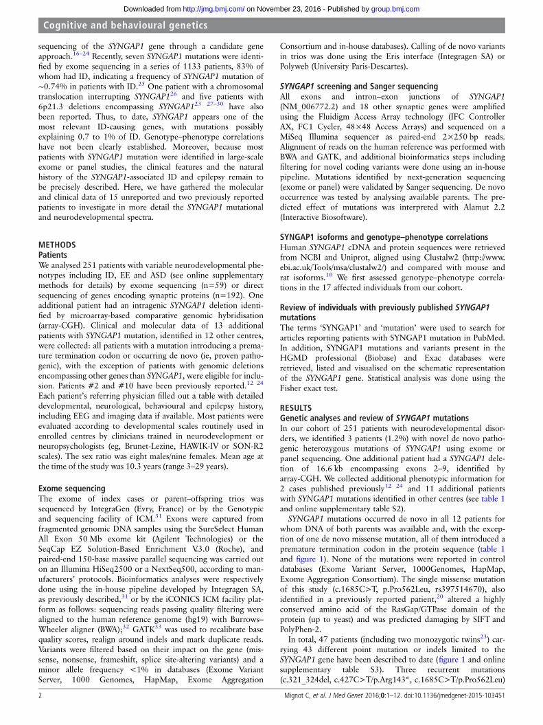
SYNGAP1 mutations occurred de novo in all 12 patients forwhom DNA of both parents was available and, with the excep-tion of one de novo missense mutation, all of them introduced apremature termination codon in the protein sequence (table 1and figure 1). None of the mutations were reported in controldatabases (Exome Variant Server, 1000Genomes, HapMap,Exome Aggregation Consortium). The single missense mutationof this study (c.1685C>T, p.Pro562Leu, rs397514670), alsoidentified in a previously reported patient,20 altered a highlyconserved amino acid of the RasGap/GTPase domain of theprotein (up to yeast) and was predicted damaging by SIFT andPolyPhen-2.
In total, 47 patients (including two monozygotic twins23) car-rying 43 different point mutation or indels limited to theSYNGAP1 gene have been described to date (figure 1 and onlinesupplementary table S3). Three recurrent mutations(c.321_324del, c.427C>T/p.Arg143*, c.1685C>T/p.Pro562Leu)
2 Mignot C, et al. J Med Genet 2016;0:1–12. doi:10.1136/jmedgenet-2015-103451
Cognitive and behavioural genetics
group.bmj.com on November 23, 2016 - Published by http://jmg.bmj.com/Downloaded from
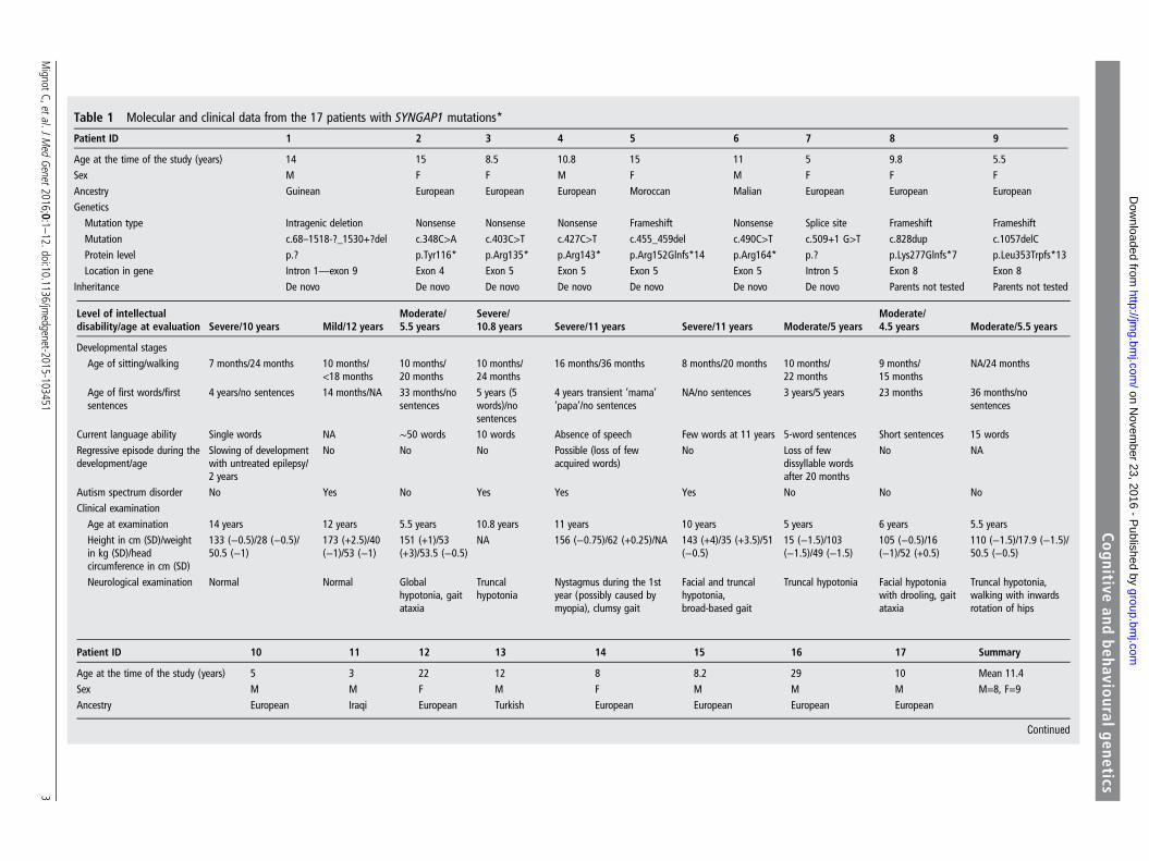
Table 1 Molecular and clinical data from the 17 patients with SYNGAP1 mutations*
Patient ID 1 2 3 4 5 6 7 8 9
Age at the time of the study (years) 14 15 8.5 10.8 15 11 5 9.8 5.5Sex M F F M F M F F FAncestry Guinean European European European Moroccan Malian European European EuropeanGenetics
Mutation type Intragenic deletion Nonsense Nonsense Nonsense Frameshift Nonsense Splice site Frameshift FrameshiftMutation c.68–1518-?_1530+?del c.348C>A c.403C>T c.427C>T c.455_459del c.490C>T c.509+1 G>T c.828dup c.1057delCProtein level p.? p.Tyr116* p.Arg135* p.Arg143* p.Arg152Glnfs*14 p.Arg164* p.? p.Lys277Glnfs*7 p.Leu353Trpfs*13Location in gene Intron 1—exon 9 Exon 4 Exon 5 Exon 5 Exon 5 Exon 5 Intron 5 Exon 8 Exon 8
Inheritance De novo De novo De novo De novo De novo De novo De novo Parents not tested Parents not tested
Level of intellectualdisability/age at evaluation Severe/10 years Mild/12 years
Moderate/5.5 years
Severe/10.8 years Severe/11 years Severe/11 years Moderate/5 years
Moderate/4.5 years Moderate/5.5 years
Developmental stages
Age of sitting/walking 7 months/24 months 10 months/<18 months
10 months/20 months
10 months/24 months
16 months/36 months 8 months/20 months 10 months/22 months
9 months/15 months
NA/24 months
Age of first words/firstsentences
4 years/no sentences 14 months/NA 33 months/nosentences
5 years (5words)/nosentences
4 years transient ‘mama’‘papa’/no sentences
NA/no sentences 3 years/5 years 23 months 36 months/nosentences
Current language ability Single words NA ∼50 words 10 words Absence of speech Few words at 11 years 5-word sentences Short sentences 15 wordsRegressive episode during thedevelopment/age
Slowing of developmentwith untreated epilepsy/2 years
No No No Possible (loss of fewacquired words)
No Loss of fewdissyllable wordsafter 20 months
No NA
Autism spectrum disorder No Yes No Yes Yes Yes No No NoClinical examinationAge at examination 14 years 12 years 5.5 years 10.8 years 11 years 10 years 5 years 6 years 5.5 yearsHeight in cm (SD)/weightin kg (SD)/headcircumference in cm (SD)
133 (−0.5)/28 (−0.5)/50.5 (−1)
173 (+2.5)/40(−1)/53 (−1)
151 (+1)/53(+3)/53.5 (−0.5)
NA 156 (−0.75)/62 (+0.25)/NA 143 (+4)/35 (+3.5)/51(−0.5)
15 (−1.5)/103(−1.5)/49 (−1.5)
105 (−0.5)/16(−1)/52 (+0.5)
110 (−1.5)/17.9 (−1.5)/50.5 (−0.5)
Neurological examination Normal Normal Globalhypotonia, gaitataxia
Truncalhypotonia
Nystagmus during the 1styear (possibly caused bymyopia), clumsy gait
Facial and truncalhypotonia,broad-based gait
Truncal hypotonia Facial hypotoniawith drooling, gaitataxia
Truncal hypotonia,walking with inwardsrotation of hips
Patient ID 10 11 12 13 14 15 16 17 Summary
Age at the time of the study (years) 5 3 22 12 8 8.2 29 10 Mean 11.4Sex M M F M F M M M M=8, F=9Ancestry European Iraqi European Turkish European European European European
Continued
MignotC,etal.J
Med
Genet2016;0:1
–12.doi:10.1136/jmedgenet-2015-103451
3
Cognitiveand
behaviouralgenetics
group.bmj.com
on Novem
ber 23, 2016 - Published by
http://jmg.bm
j.com/
Dow
nloaded from
Table 1 Continued
Patient ID 10 11 12 13 14 15 16 17 Summary
GeneticsMutation type Nonsense Nonsense Missense Nonsense Frameshift Frameshift Frameshift Splice site Nonsense 7;
frameshift 5; splice 2;missense 1; intragenicdeletion 1
Mutation c.1253_1254del c.1630C>T c.1685C>T c.1995T>A c.2214_2217del c.2933del c.3406dup c.3408+1G>AProtein level p.Lys418Argfs*54 p.Arg544* p.Pro562Leu p.Tyr665* p.Glu739Glyfs*20 p.Pro978Hisfs*99 p.Gln1136Profs*17 p.?Location in gene Exon 8 Exon 10 Exon 11 Exon 12 Exon 13 Exon 15 Exon 15 Intron 15Inheritance De novo De novo De novo Parents not tested De novo De novo Parents not tested De novo
Level of intellectual disability/ageat evaluation Severe/4 years Severe/3 years Severe/22 years
Severe/12 years Mild/8 years
Moderate/5 years Severe/8.5 years Severe/10 years
Mild n=2; moderate n=5;severe n=10/mean age atevaluation 8.7 years
Developmental stagesAge of sitting/walking 15–18 months/36 months 12 months/walks
only with aid12 months/38 months
NA/36 months 8 months/18 months
10 months/18months
16 months/30 months
25 months/4.5 years
Mean 12 months/27.7 months
Age of first words/first sentences ∼29 months transient‘mama’, ‘papa’/nosentences
3 years ’papa’only/no sentences
No words/nosentences
No words/nosentences
12 months/6 years 3 years/nosentences
17 months/nosentences
No words/nosentences
Mean age first words2.6 years
Current language ability Absence of speech Absence ofspeech
Absence ofspeech
Absence ofspeech
120 words, 3- to4-word sentences
5 words Absence of speech Absence ofspeech
Absence of speech 7;speaks words 5;associates words or simplesentences 3
Regressive episode during thedevelopment/age
Since age of 36 monthsloss of ‘mama’, ‘papa’
No 12 months—withfebrile seizures
No 14 months No Loss of words atage 18–30 months
Possible (loss of2-syllable words)
n=7
Autism spectrum disorder YesToo young tobe evaluated No No Yes No Yes Yes Yes 8; no 8
Clinical examinationAge at examination 5.2 years 3 years 22 years 12 years 8 years 7 years 8.5 years 6.6 years Mean 8.9 yearsHeight in cm (SD)/weight in kg(SD)/head circumference in cm (SD)
149 (+1.5)/48.6 (+2)/52(−1.5)
105 (−0.5)/20(+1.5)/49.3(−1)
93 (0)/13.8 (0)/48(−2)
146.5 (+1)/35(+0.5)/55 (+1)
NA/21 (−1)/54 (+1) 116 (+1)/21 (+1)/50(0)
124 cm(−1.5)/22 kg(−1.8)/50.8 cm(−1.7)
116 cm (+0.4)/22.3 kg (+0.7)/51.3 cm (+0.4)
Normal OFC 15/15
Neurological examination Truncal hypotonia,broad-based gait,hypotonic-atacticmovements
Truncalhypotonia,swallowingdifficulties
Mild gait ataxia,flexion deformity ofleft hip, hyperlordoticlumbar spine
Hyperactive deeptendon reflexes,unsteady gait
Motor slowness andmoderate akinesia, ataxicgait, truncal hypotonia,dystonic postures of handsand feet, plastic hypertonia
Truncal hypotonia,orthostatic truncaltremor, slightpyramidal tetraparesis,gait ataxia
Truncalhypotonia
Truncalhypotonia,orofacialhypotonia,wide-based gait
Clumsy/ataxic gait10, truncalhypotonia 10, facialhypotonia 4, normalexam 2
*Patients are ordered by mutation from the 50 end of the gene.NA, not available; OFC, occipitofrontal circumference.
4MignotC,etal.J
Med
Genet2016;0:1
–12.doi:10.1136/jmedgenet-2015-103451
Cognitiveand
behaviouralgenetics
group.bmj.com
on Novem
ber 23, 2016 - Published by
http://jmg.bm
j.com/
Dow
nloaded from
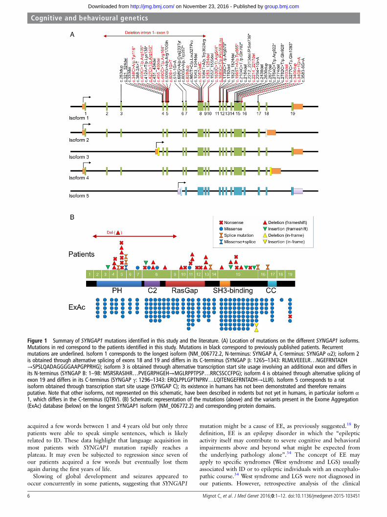
were found in two patients each. Pathogenic mutations inSYNGAP1 are distributed throughout the gene, especially inexons 5, 8 and 15, which are among the largest exons ofSYNGAP1. Interestingly, the two first and two last exons, whichare alternatively spliced and included in 3 out of 5 SYNGAP1 iso-forms, but also exons 9 and 16, present in all known isoformsseem to be spared (figure 1).
Clinical and neurodevelopmental features ofSYNGAP1-related encephalopathyAll patients with SYNGAP1 anomalies of our series had ID,which was evaluated as severe in 10 patients, moderate in 5 andmild in 2 (see table 1 and online supplementary table S1). Themean age of sitting unsupported was 12 months (median age10 months, n=15) and of walking 27.7 months (median age 24months, n=15). Also, 10/17 patients could walk by age 2 yearsand 14/17 by age 3 years. All patients had speech delay: 12 ofthem spoke first words at a mean age of 2.5 years and 5 patientsdid not speak at age 10 years or older. In most patients, bothreceptive and expressive languages were affected. Two patientshad mild ID, including one without motor delay. In those, mild,progressive language delay and behavioural anomalies were themost prominent features.
In total, 8 out of 16 patients (50%) older than 3 years oldwere diagnosed with ASD. Patients with ASD had remarkablypoor verbal and non-verbal communication abilities as well asimpaired social interactions (see online supplementary table S1).Half of the patients (n=4/8) with severe ID, 1/5 with moderateID and 2/2 with mild ID were diagnosed with ASD.Independent from a formal diagnosis of ASD, many of thepatients exhibited stereotypies (n=10), temper tantrums, aggres-siveness, self-injurious behaviour and/or restlessness (n=7).
Neurological examination, performed at a mean age of8.9 years, was considered normal in two patients. Gait wasclumsy or unsteady in five patients and ataxic in five others.Truncal hypotonia was reported in 10 patients and facial hypo-tonia in 4. Some patients had orthopaedic problems, such as pesplanus and rotation of the hips.
Brain MRI performed in all 17 patients (mean age 5.4 years)was either normal or revealed nonspecific features (arachnoidcysts in two patients, mild myelination delay in one and signalabnormities in another).
Epilepsy was diagnosed in 16/17 patients (table 2). The onlypatient without epilepsy, who was aged 5 at the time of thisstudy, had a single afebrile seizure at the age of 3.5 years.Excluding this patient, first seizures occurred at a mean age of3 years (range 1–8 years) and consisted of drop attacks, massivemyoclonic jerks, atonic seizures, myoclonic absences orabsences. A diagnosis of myoclonic astatic epilepsy (MAE, ie,Doose syndrome) and epilepsy with myoclonic absences (EMAs)was made in three and one patients, respectively. The otherswere diagnosed with unclassified genetic generalised epilepsy(GGE). None had a diagnosis of Lennox–Gastaut syndrome(LGS).
The epilepsy responded to a single antiepileptic drug (AED),mostly sodium valproate, in seven patients and was pharmacore-sistant in nine (list of AEDs is reported in table 2). During theactive phases of epilepsy, seizures occurred daily in five patients,10 times per day or more in two and 100 times daily or more intwo others. Seizures were of short duration, and the most fre-quent seizure types were typical or atypical absences (n=9),massive myoclonic jerks with or without falls (n=7), eyelidmyoclonia (n=3), clonic or tonic clonic seizures (n=3), myo-clonic absences (n=3) and atonic seizures (n=2). Head drops or
falls were relatively frequent (n=5) and reported as myoclonicastatic, atonic seizures or drop attacks. Eight patients had severalseizure types. No patient had status epilepticus, and exacerba-tion by fever was mentioned in four. We found no correlationsbetween the diagnosis of ASD and the age at epilepsy onset.The proportion of patients with ASD was identical among thosewith pharmacoresistant (n=5/10) and pharmacosensitive epi-lepsy (n=3/6).
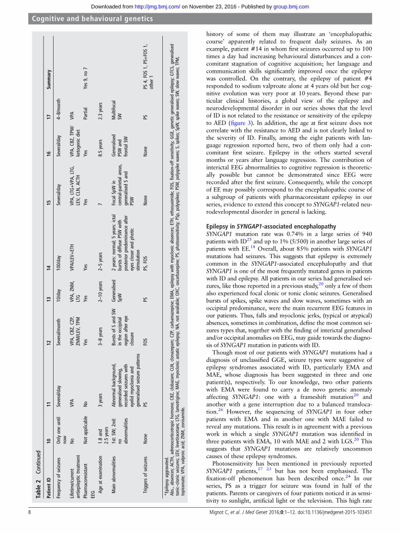
The most frequent anomalies reported on EEG traces (figure 2)from 16 patients were ictal or interictal bursts of spikes, spikewaves or slow waves that were either generalised (n=13), general-ised with a posterior predominance or posterior only (n=5).Paroxysmal anomalies were localised to central regions in sixinstances. Triggers of seizures were identified in seven patients,including photosensitivity (PS, n=5), fixation-off sensitivity (FOS,n=1), PS and FOS (n=1) and chewing (n=1).
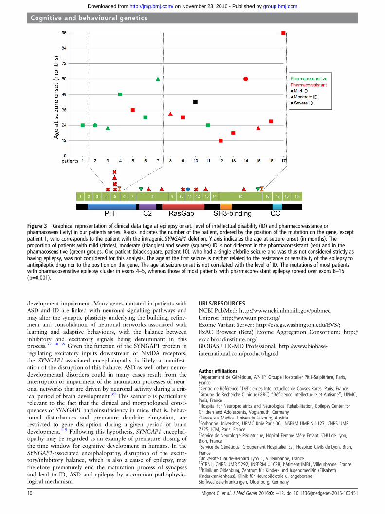
Genotype/phenotype correlationsWe observed no definite correlation between the location of themutation on the gene and the severity of ID or ASD diagnosis.However, schematic representation of the clinical features ofour 17 patients, ordered by the position of the mutation on thegene (figure 3), revealed that the epilepsy of patients with muta-tions in exons 4–5 was mainly pharmacosensitive (5/6 patients),whereas that of patients with mutations in exons 8–15 wasmainly pharmacoresistant (8/9, p=0.01).
DISCUSSIONIn this study, we collected the comprehensive molecular andclinical data of the largest series of patients with SYNGAP1mutation so far in order to describe more accurately the neuro-developmental and epilepsy phenotype and to address geno-type–phenotype correlations. Delineation of the phenotypefrom 36 patients with SYNGAP1 mutations showed that itincludes mild to severe ID in all, generalised epilepsy in mostand autistic behaviour in a half of them (see online supplemen-tary table S3). In the present study, we describe the phenotypeof 17 cases with SYNGAP1-associated encephalopathy, bringingthe total number of reported patients with SYNGAP1 mutationsto 47.
Neurological examination in SYNGAP1-associatedencephalopathyTruncal hypotonia, sometimes in association with facial hypo-tonia, was the main recurrent feature in our patients, in linewith previous series.20 23 Likewise, ataxia, with a broad-basedor clumsy gait, was frequent in our patients and recurrentlymentioned in others.20 23 Gait abnormalities are probably dueto a combination of hypotonia, lack of global coordination,poor motor control, inattentiveness and orthopaedic issues.
Occipitofrontal circumference was normal in 78% of patientsfrom the literature and in 100% of ours. Though microcephalyhas been mentioned in some cases,17 20 23 it seems to be not acommon aspect in patients with SYNGAP1 mutations.
As with previously reported patients, MRI in our patientsshowed either no or nonspecific features, implying that brainimaging is not helpful in the diagnosis of SYNGAP1-relateddisorders.
The neurodevelopmental phenotype in SYNGAP1-associatedencephalopathyIn our series as well as in the literature, early motor delay withsevere language impairment is the first manifestation ofSYNGAP1 encephalopathy. Fourteen patients of our series
Mignot C, et al. J Med Genet 2016;0:1–12. doi:10.1136/jmedgenet-2015-103451 5
Cognitive and behavioural genetics
group.bmj.com on November 23, 2016 - Published by http://jmg.bmj.com/Downloaded from
acquired a few words between 1 and 4 years old but only threepatients were able to speak simple sentences, which is likelyrelated to ID. These data highlight that language acquisition inmost patients with SYNGAP1 mutation rapidly reaches aplateau. It may even be subjected to regression since seven ofour patients acquired a few words but eventually lost themagain during the first years of life.
Slowing of global development and seizures appeared tooccur concurrently in some patients, suggesting that SYNGAP1
mutation might be a cause of EE, as previously suggested.18 Bydefinition, EE is an epilepsy disorder in which the “epilepticactivity itself may contribute to severe cognitive and behavioralimpairments above and beyond what might be expected fromthe underlying pathology alone”.34 The concept of EE mayapply to specific syndromes (West syndrome and LGS) usuallyassociated with ID or to epileptic individuals with an encephalo-pathic course.34 West syndrome and LGS were not diagnosed inour patients. However, retrospective analysis of the clinical
Figure 1 Summary of SYNGAP1 mutations identified in this study and the literature. (A) Location of mutations on the different SYNGAP1 isoforms.Mutations in red correspond to the patients identified in this study. Mutations in black correspond to previously published patients. Recurrentmutations are underlined. Isoform 1 corresponds to the longest isoform (NM_006772.2, N-terminus: SYNGAP A, C-terminus: SYNGAP α2); isoform 2is obtained through alternative splicing of exons 18 and 19 and differs in its C-terminus (SYNGAP β: 1265–1343: RLMLVEEELR…NGEFRNTADH→SPSLQADAGGGGAAPGPPRHG); isoform 3 is obtained through alternative transcription start site usage involving an additional exon and differs inits N-terminus (SYNGAP B: 1–98: MSRSRASIHR…PVEGRPHGEH→MGLRPPTPSP…RRCSSCCFPG); isoform 4 is obtained through alternative splicing ofexon 19 and differs in its C-terminus (SYNGAP γ: 1296–1343: ERQLPPLGPTNPRV…LQITENGEFRNTADH→LLIR). Isoform 5 corresponds to a ratisoform obtained through transcription start site usage (SYNGAP C); its existence in humans has not been demonstrated and therefore remainsputative. Note that other isoforms, not represented on this schematic, have been described in rodents but not yet in humans, in particular isoform α1, which differs in the C-terminus (QTRV). (B) Schematic representation of the mutations (above) and the variants present in the Exome Aggregation(ExAc) database (below) on the longest SYNGAP1 isoform (NM_006772.2) and corresponding protein domains.
6 Mignot C, et al. J Med Genet 2016;0:1–12. doi:10.1136/jmedgenet-2015-103451
Cognitive and behavioural genetics
group.bmj.com on November 23, 2016 - Published by http://jmg.bmj.com/Downloaded from
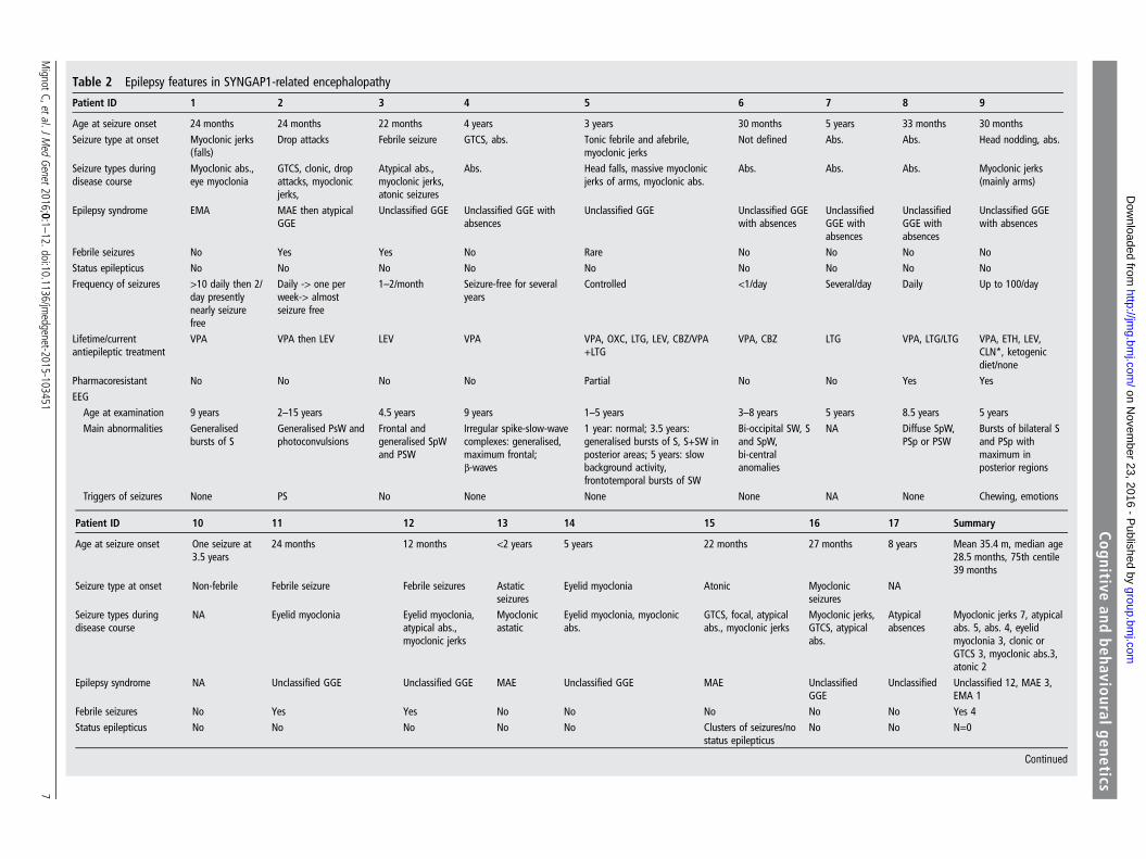
Table 2 Epilepsy features in SYNGAP1-related encephalopathy
Patient ID 1 2 3 4 5 6 7 8 9
Age at seizure onset 24 months 24 months 22 months 4 years 3 years 30 months 5 years 33 months 30 monthsSeizure type at onset Myoclonic jerks
(falls)Drop attacks Febrile seizure GTCS, abs. Tonic febrile and afebrile,
myoclonic jerksNot defined Abs. Abs. Head nodding, abs.
Seizure types duringdisease course
Myoclonic abs.,eye myoclonia
GTCS, clonic, dropattacks, myoclonicjerks,
Atypical abs.,myoclonic jerks,atonic seizures
Abs. Head falls, massive myoclonicjerks of arms, myoclonic abs.
Abs. Abs. Abs. Myoclonic jerks(mainly arms)
Epilepsy syndrome EMA MAE then atypicalGGE
Unclassified GGE Unclassified GGE withabsences
Unclassified GGE Unclassified GGEwith absences
UnclassifiedGGE withabsences
UnclassifiedGGE withabsences
Unclassified GGEwith absences
Febrile seizures No Yes Yes No Rare No No No NoStatus epilepticus No No No No No No No No NoFrequency of seizures >10 daily then 2/
day presentlynearly seizurefree
Daily -> one perweek-> almostseizure free
1–2/month Seizure-free for severalyears
Controlled <1/day Several/day Daily Up to 100/day
Lifetime/currentantiepileptic treatment
VPA VPA then LEV LEV VPA VPA, OXC, LTG, LEV, CBZ/VPA+LTG
VPA, CBZ LTG VPA, LTG/LTG VPA, ETH, LEV,CLN*, ketogenicdiet/none
Pharmacoresistant No No No No Partial No No Yes YesEEG
Age at examination 9 years 2–15 years 4.5 years 9 years 1–5 years 3–8 years 5 years 8.5 years 5 yearsMain abnormalities Generalised
bursts of SGeneralised PsW andphotoconvulsions
Frontal andgeneralised SpWand PSW
Irregular spike-slow-wavecomplexes: generalised,maximum frontal;β-waves
1 year: normal; 3.5 years:generalised bursts of S, S+SW inposterior areas; 5 years: slowbackground activity,frontotemporal bursts of SW
Bi-occipital SW, Sand SpW,bi-centralanomalies
NA Diffuse SpW,PSp or PSW
Bursts of bilateral Sand PSp withmaximum inposterior regions
Triggers of seizures None PS No None None None NA None Chewing, emotions
Patient ID 10 11 12 13 14 15 16 17 Summary
Age at seizure onset One seizure at3.5 years
24 months 12 months <2 years 5 years 22 months 27 months 8 years Mean 35.4 m, median age28.5 months, 75th centile39 months
Seizure type at onset Non-febrile Febrile seizure Febrile seizures Astaticseizures
Eyelid myoclonia Atonic Myoclonicseizures
NA
Seizure types duringdisease course
NA Eyelid myoclonia Eyelid myoclonia,atypical abs.,myoclonic jerks
Myoclonicastatic
Eyelid myoclonia, myoclonicabs.
GTCS, focal, atypicalabs., myoclonic jerks
Myoclonic jerks,GTCS, atypicalabs.
Atypicalabsences
Myoclonic jerks 7, atypicalabs. 5, abs. 4, eyelidmyoclonia 3, clonic orGTCS 3, myoclonic abs.3,atonic 2
Epilepsy syndrome NA Unclassified GGE Unclassified GGE MAE Unclassified GGE MAE UnclassifiedGGE
Unclassified Unclassified 12, MAE 3,EMA 1
Febrile seizures No Yes Yes No No No No No Yes 4Status epilepticus No No No No No Clusters of seizures/no
status epilepticusNo No N=0
Continued
MignotC,etal.J
Med
Genet2016;0:1
–12.doi:10.1136/jmedgenet-2015-103451
7
Cognitiveand
behaviouralgenetics
group.bmj.com
on Novem
ber 23, 2016 - Published by
http://jmg.bm
j.com/
Dow
nloaded from
history of some of them may illustrate an ‘encephalopathiccourse’ apparently related to frequent daily seizures. As anexample, patient #14 in whom first seizures occurred up to 100times a day had increasing behavioural disturbances and a con-comitant stagnation of cognitive acquisition; her language andcommunication skills significantly improved once the epilepsywas controlled. On the contrary, the epilepsy of patient #4responded to sodium valproate alone at 4 years old but her cog-nitive evolution was very poor at 10 years. Beyond these par-ticular clinical histories, a global view of the epilepsy andneurodevelopmental disorder in our series shows that the levelof ID is not related to the resistance or sensitivity of the epilepsyto AED (figure 3). In addition, the age at first seizure does notcorrelate with the resistance to AED and is not clearly linked tothe severity of ID. Finally, among the eight patients with lan-guage regression reported here, two of them only had a con-comitant first seizure. Epilepsy in the others started severalmonths or years after language regression. The contribution ofinterictal EEG abnormalities to cognitive regression is theoretic-ally possible but cannot be demonstrated since EEG wererecorded after the first seizure. Consequently, while the conceptof EE may possibly correspond to the encephalopathic course ofa subgroup of patients with pharmacoresistant epilepsy in ourseries, evidence to extend this concept to SYNGAP1-related neu-rodevelopmental disorder in general is lacking.
Epilepsy in SYNGAP1-associated encephalopathySYNGAP1 mutation rate was 0.74% in a large series of 940patients with ID25 and up to 1% (5/500) in another large series ofpatients with EE.18 Overall, about 85% patients with SYNGAP1mutations had seizures. This suggests that epilepsy is extremelycommon in the SYNGAP1-associated encephalopathy and thatSYNGAP1 is one of the most frequently mutated genes in patientswith ID and epilepsy. All patients in our series had generalised sei-zures, like those reported in a previous study,20 only a few of themalso experienced focal clonic or tonic clonic seizures. Generalisedbursts of spikes, spike waves and slow waves, sometimes with anoccipital predominance, were the main recurrent EEG features inour patients. Thus, falls and myoclonic jerks, (typical or atypical)absences, sometimes in combination, define the most common sei-zures types that, together with the finding of interictal generalisedand/or occipital anomalies on EEG, may guide towards the diagno-sis of SYNGAP1mutation in patients with ID.
Though most of our patients with SYNGAP1 mutations had adiagnosis of unclassified GGE, seizure types were suggestive ofepilepsy syndromes associated with ID, particularly EMA andMAE, whose diagnosis has been suggested in three and onepatient(s), respectively. To our knowledge, two other patientswith EMA were found to carry a de novo genetic anomalyaffecting SYNGAP1: one with a frameshift mutation20 andanother with a gene interruption due to a balanced transloca-tion.26 However, the sequencing of SYNGAP1 in four otherpatients with EMA and in another one with MAE failed toreveal any mutations. This result is in agreement with a previouswork in which a single SYNGAP1 mutation was identified inthree patients with EMA, 10 with MAE and 2 with LGS.20 Thissuggests that SYNGAP1 mutations are relatively uncommoncauses of these epilepsy syndromes.
Photosensitivity has been mentioned in previously reportedSYNGAP1 patients,17 23 but has not been emphasised. Thefixation-off phenomenon has been described once.24 In ourseries, PS as a trigger for seizure was found in half of thepatients. Parents or caregivers of four patients noticed it as sensi-tivity to sunlight, artificial light or the television. This high rate
Table2
Continued
Patie
ntID
1011
1213
1415
1617
Summary
Frequencyof
seizures
Onlyoneuntil
now
Several/day
Several/m
onth
10/day
100/day
Several/day
Several/day
4–8/month
Lifetim
e/curre
ntantiepileptictre
atment
No
VPA
VPA,
CBZ,CZP,
ZNM/LEV,TPM
VPA,
ZNM,
LTG
VPA/LEV+
ETH
VPA,
LTG+VPA,
LTG,
LEV,
CLN,A
CTH
VPA,
CBZ,TPM/
ketogenicdiet
VPA
Pharmacoresistant
Not
applicable
No
Yes
Yes
Yes
Yes
Yes
Partial
Yes9,
no7
EEG Ag
eat
exam
ination
1.8and
2.5years
3years
3–8years
2–10
years
2–5years
78.5years
2.3years
Mainabnorm
alities
1st:SW
;2nd:
no abnorm
alities
Abnorm
albackground,
generalised
slowing,
recorded
seizures
with
eyelid
myocloniaand
generalised
seizurepatte
rns
Burstsof
SandSW
intheoccipital
region
aftereye
closure
Generalise
dSpW
2years:norm
al;5
years:ictal
burstsof
diffu
sePSW
with
posteriorpredom
inance
after
eyes
closer
andphotic
stimulation
FocalSpW
incentral-parietalareas,
generalised
Sand
PSW
Generalise
dPSW
and
frontalSW
Multifocal
SW
Triggersof
seizures
None
PSFO
SPS
PS,FOS
None
None
PSPS
4,FOS1,
PS+FOS1,
other1
*Epilepsyaggravated.
Abs.,a
bsences;AC
TH,a
drenocorticotropichorm
one;CB
Z,clobazam
;CLN
,clonazepam;C
ZP,carbamazepine;EM
A,epilepsywith
myoclonicabsences;E
TH,ethosuximide;FO
S,fixation-offsensitivity;G
GE,
genetic
generalised
epilepsy;GTCS,generalised
tonic–clonicseizures;LEV,levetiracetam;LTG
,lam
otrigine;MAE
,myoclonicastatic
epilepsy;NA,
notavailable;OXC
,oxcarbazepine;P
S,photosensitivity;P
Sp,p
olyspikes;PSW,p
olyspike
waves;S,spikes;SpW,spike
waves;SW,slowwaves;TPM
,topiramate;VPA,
valproicacid;Z
NM,zonisa
mide.
8 Mignot C, et al. J Med Genet 2016;0:1–12. doi:10.1136/jmedgenet-2015-103451
Cognitive and behavioural genetics
group.bmj.com on November 23, 2016 - Published by http://jmg.bmj.com/Downloaded from
of PS is significant since clinical PS is found in only 10% ofpatients with epilepsy in the 7–19-year-old group.35 We assumethat PS may have not been detected in some of our patientsbecause it is an age-dependent phenomenon with a peak aroundpuberty; it could therefore still appear in some of them; orbecause of the poor cooperation of patients during the record-ing. These data suggest that PS, when present, might be a diag-nostic clue from the EEG of an underlying SYNGAP1 mutation.
Genotype/phenotype correlationsAlthough patients with SYNGAP1 mutations show a commoncore clinical picture, the phenotype is relatively variable, par-ticularly regarding the severity of ID, pharmacoresistance andthe presence of ASD. Since SYNGAP1 is a complex gene, givingrise to several protein isoforms with opposite effects on the glu-tamate activation pathway, via alternative splicing and transcrip-tion start sites,10 it was tempting to speculate that the locationof the mutation on the gene could correlate to the clinicaloutcome. However, we found little correlation between thelocation of the mutation and the severity of ID, epilepsy and/orASD. Yet, the epilepsy of patients with mutations in exons 4–5appeared more pharmacosensitive than that of patients withmutations in exons 8–15. Interestingly, exons 4 and 5 are notpresent in SYNGAP C, an isoform obtained through alternativepromoter usage, whose existence has been demonstrated in miceand rats. Although this isoform has not been shown to exist inhumans as well, our results suggest that it could also exist andhave a different function, as already proven for isoforms α1 andα2, which differ in their C-terminus. Further study is necessaryto confirm this finding and decrypt the precise function of each
human SYNGAP1 isoform and its relationship with the humanpathology characteristics.
Nevertheless, the comparison of the clinical features ofpatients with identical mutations revealed significant clinical dif-ferences (see online supplementary tables S2 and S3), confirm-ing that there is a real variability of the phenotype that dependson other factors than the mutation itself. On the contrary,monozygotic twins had strikingly similar phenotypes, suggestingthat these modifier factors could be of genetic origin.23
ASD in SYNGAP1-associated encephalopathy andhypothetical consequences of SYNGAP1 mutations on braindevelopmentAlthough all patients with validated pathogenic SYNGAP1 muta-tions reported to date had ID, only half of them had a diagnosisof ASD (including data from the literature and our series). Inour series, the presence of autistic traits was neither limited topatients with moderate or severe ID, nor to those with pharma-coresistant or early-onset epilepsy. Thus, ASD, like epilepsy,could be considered as an additional feature of theSYNGAP1-related phenotype in the context of ID, irrespectivelyof its severity, rather than an ‘isolated’ diagnosis.
This observation is in agreement with previous studiesshowing that many neurodevelopmental disorders are caused bymutations in genes encoding synaptic proteins, and more specif-ically constituents of the PSD.36 The fact that a subset ofpatients with SYNGAP1 mutations exhibit autistic behaviourssuggests that a single mutation in a synaptic gene is not suffi-cient to cause ASD and that the genetic or epigenetic back-ground of the patient probably plays an important role in theoccurrence of autistic features in a context of intellectual
Figure 2 EEG samples from patients exemplifying electroencephalographic findings in SYNGAP1-related encephalopathy. (A) Sampledemonstrating normalisation of paroxysmal activity by eye opening, that is, fixation-off sensitivity, in patient #2. (B) Sample showing paroxysmalactivity under photic stimulation, that is, photosensitivity, in patient #2. (C) Sample from patient #1: burst of generalised spikes concomitant of arapid eye deviation (fast rhythms are due to benzodiazepine therapy). (D) Sample from patient #12 showing the appearance of generalised spikewave complexes with a low degree of bilateral synchronisation after eye closure (fixation-off phenomenon).
Mignot C, et al. J Med Genet 2016;0:1–12. doi:10.1136/jmedgenet-2015-103451 9
Cognitive and behavioural genetics
group.bmj.com on November 23, 2016 - Published by http://jmg.bmj.com/Downloaded from
development impairment. Many genes mutated in patients withASD and ID are linked with neuronal signalling pathways andmay alter the synaptic plasticity underlying the building, refine-ment and consolidation of neuronal networks associated withlearning and adaptive behaviours, with the balance betweeninhibitory and excitatory signals being determinant in thisprocess.37 38 39 Given the function of the SYNGAP1 protein inregulating excitatory inputs downstream of NMDA receptors,the SYNGAP1-associated encephalopathy is likely a manifest-ation of the disruption of this balance. ASD as well other neuro-developmental disorders could in many cases result from theinterruption or impairment of the maturation processes of neur-onal networks that are driven by neuronal activity during a crit-ical period of brain development.39 This scenario is particularlyrelevant to the fact that the clinical and morphological conse-quences of SYNGAP1 haploinsufficiency in mice, that is, behav-ioural disturbances and premature dendrite elongation, arerestricted to gene disruption during a given period of braindevelopment.4 9 Following this hypothesis, SYNGAP1 encephal-opathy may be regarded as an example of premature closing ofthe time window for cognitive development in humans. In theSYNGAP1-associated encephalopathy, disruption of the excita-tory/inhibitory balance, which is also a cause of epilepsy, maytherefore prematurely end the maturation process of synapsesand lead to ID, ASD and epilepsy by a common pathophysio-logical mechanism.
URLS/RESOURCESNCBI PubMed: http://www.ncbi.nlm.nih.gov/pubmedUniprot: http://www.uniprot.org/Exome Variant Server: http://evs.gs.washington.edu/EVS/;ExAC Browser (Beta)|Exome Aggregation Consortium: http://exac.broadinstitute.org/BIOBASE HGMD Professional: http://www.biobase-international.com/product/hgmd
Author affiliations1Département de Génétique, AP-HP, Groupe Hospitalier Pitié-Salpêtrière, Paris,France2Centre de Référence “Déficiences Intellectuelles de Causes Rares, Paris, France3Groupe de Recherche Clinique (GRC) “Déficience Intellectuelle et Autisme”, UPMC,Paris, France4Hospital for Neuropediatrics and Neurological Rehabilitation, Epilepsy Center forChildren and Adolescents, Vogtareuth, Germany5Paracelsus Medical University Salzburg, Austria6Sorbonne Universités, UPMC Univ Paris 06, INSERM UMR S 1127, CNRS UMR7225, ICM, Paris, France7Service de Neurologie Pédiatrique, Hôpital Femme Mère Enfant, CHU de Lyon,Bron, France8Service de Génétique, Groupement Hospitalier Est, Hospices Civils de Lyon, Bron,France9Université Claude-Bernard Lyon 1, Villeurbanne, France10CRNL, CNRS UMR 5292, INSERM U1028, bâtiment IMBL, Villeurbanne, France11Klinikum Oldenburg, Zentrum für Kinder- und Jugendmedizin (ElisabethKinderkrankenhaus), Klinik für Neuropädiatrie u. angeboreneStoffwechselerkrankungen, Oldenburg, Germany
Figure 3 Graphical representation of clinical data (age at epilepsy onset, level of intellectual disability (ID) and pharmacoresistance orpharmacosensitivity) in our patients series. X-axis indicates the number of the patient, ordered by the position of the mutation on the gene, exceptpatient 1, who corresponds to the patient with the intragenic SYNGAP1 deletion. Y-axis indicates the age at seizure onset (in months). Theproportion of patients with mild (circles), moderate (triangles) and severe (squares) ID is not different in the pharmacoresistant (red) and in thepharmacosensitive (green) groups. One patient (black square, patient 10), who had a single afebrile seizure and was thus not considered strictly ashaving epilepsy, was not considered for this analysis. The age at the first seizure is neither related to the resistance or sensitivity of the epilepsy toantiepileptic drug nor to the position on the gene. The age at seizure onset is not correlated with the level of ID. The mutations of most patientswith pharmacosensitive epilepsy cluster in exons 4–5, whereas those of most patients with pharmacoresistant epilepsy spread over exons 8–15(p=0.001).
10 Mignot C, et al. J Med Genet 2016;0:1–12. doi:10.1136/jmedgenet-2015-103451
Cognitive and behavioural genetics
group.bmj.com on November 23, 2016 - Published by http://jmg.bmj.com/Downloaded from
12Department of Pediatric Neurology and Developmental Medicine and EpilepsyCenter, University of Munich, Munich, Germany13Department of Medical Genetics, Institute of Mother and Child, Warsaw, Poland14Clinic of Neurology of Children and Adolescents, Institute of Mother and Child,Warsaw, Poland15Division of Child Neurology, Department of Pediatrics, School of Medicine, DokuzEylül University, İzmir, Turkey16Department of Molecular Biology and Genetics Istanbul, Boğaziçi University,Istanbul, Turkey17AP-HP, Hôpital Trousseau, Service de Neuropédiatrie, Paris, France18Epilepsy, Sleep and Pediatric Neurophysiology Department (ESEFNP), UniversityHospitals of Lyon (HCL), France19Département de l’Enfant et de l’Adolescent, Neuropédiatrie—HôpitauxUniversitaires de Genève, Genève, Switzerland20Institute of Human Genetics, University of Tuebingen, Tuebingen, Germany21Institute of Medical Genetics and Applied Genomics, University of Tübingen,Tübingen, Germany22Institute of Medical Genetics, University of Zurich, Schwerzenbach, Switzerland23Institute of Human Genetics, Friedrich-Alexander-Universität Erlangen-Nürnberg,Erlangen, Germany24Svt. Luka’s Institute of Child Neurology and Epilepsy, Moscow, Russia25Department of Clinical and Experimental Epilepsy, Institute of Neurology, UniversityCollege London, London, UK26Genetikum, Neu-Ulm, Germany27Neurogenetics Group, Department of Molecular Genetics, VIB, Antwerp, Belgium28Division of Genetic Medicine, Department of Pediatrics, University of Washington,Seattle, USA29CeGaT GmbH, Tübingen, Germany30Institut für Humangenetik, Universitätsklinikum Leipzig, Leipzig, Germany
Acknowledgements The authors thank the families for their participation in thisstudy, the iCONICS facility, especially Ivan Moszer and Justine Guegan, forbioinformatic analysis of exome and panel sequencing data, Patrick Nitschké and theParis-Descartes Bioinformatics Platform for access to the Polyweb interface, and theDNA and cell bank of the U1127 for DNA extraction and collection.
Collaborators List of EuroEPINOMICS-RES MAE working group coinvestigators:Dana Craiu (Pediatric Neurology Clinic II, Department of Neurology, PediatricNeurology, Psychiatry, Neurosurgery, “Carol Davila” University of Medicine, Bucharest,Romania; Pediatric Neurology Clinic, “Professor Doctor Alexandru Obregia” ClinicalHospital, Bucharest, Romania), Peter De Jonghe (Neurogenetics Group, Department ofMolecular Genetics, VIB, Antwerp, Belgium), Ingo Helbig (Division of Neurology, TheChildren’s Hospital of Philadelphia, Philadelphia, Pennsylvania; Department ofNeuropediatrics, University Medical Center Schleswig-Holstein, Christian AlbrechtsUniversity, Kiel, Germany), Renzo Guerrini (Pediatric Neurology Unit and Laboratories,Children’s Hospital A. Meyer, University of Florence, Florence, Italy), Anna-ElinaLehesjoki (Folkhälsan Institute of Genetics, Helsinki, Finland; Research Programs Unit,Molecular Neurology and Neuroscience Center, University of Helsinki, Helsinki,Finland), Carla Marini (Pediatric Neurology Unit and Laboratories, Children’s HospitalA. Meyer, University of Florence, Florence, Italy), Hiltrud Muhle (Department ofNeuropediatrics, University Medical Center Schleswig-Holstein, Christian AlbrechtsUniversity, Kiel, Germany), Rikke S Møller (Danish Epilepsy Centre, Dianalund,Denmark), Bernd Neubauer (Department of Neuropediatrics, University MedicalFaculty Giessen and Marburg, Giessen, Germany), Deb Pal (Department of ClinicalNeuroscience, Institute of Psychiatry, King’s College London, London, UK), KajaSelmer (Department of Medical Genetics, Oslo University Hospital, Oslo, Norway),Ulrich Stephani (Department of Neuropediatrics, University Medical CenterSchleswig-Holstein, Christian Albrechts University, Kiel, Germany), Katalin Sterbova(Child Neurology Department, University Hospital Motol, Prague, Czech Republic),Pasquale Striano (Pediatric Neurology and Muscular Diseases Unit, Department ofNeurosciences, Rehabilitation, Ophthalmology, Genetics, Maternal and Child Health,‘G Gaslini Institute’, Genova, Italy), Tiina Talvik (Department of Pediatrics, Universityof Tartu, Tartu, Estonia; Department of Neurology and Neurorehabilitation, Children’sClinic, Tartu University Hospital, Tartu, Estonia), Sarah von Spiczak (Department ofNeuropediatrics, University Medical Center Schleswig-Holstein, Christian AlbrechtsUniversity, Kiel, Germany).
Contributors CM, CvS, and CN contributed equally. Design and coordination ofthe study: CM, CvS, CN, GK, CD. Contributing genetic and/or phenotypic data: CM,CvS, CN, DV, DS, GL, AR, BG, YM, CK, IB, DH-Z, ES, MR-D, UY, HÇ, AI, IM, EP,CK, ER, AR, SB-W, AR, CZ, JH, AR, MM, MB, KM, LH-H, BM, SS, SW, CTM, HCM,KH, SB, JL, DH, GK, CD. Writing of the manuscript: CM, CvS, CN, GK, CD. Revisionof the manuscript: CM, CvS, CN, GL, CZ, SS, SW, GK, CD.
Funding This study was financially supported by INSERM, Fondation de France (FdF—Engt n°15144 to D. Héron), Agence Nationale de la Recherche (ANR SAMENTASynDivAutism), Assistance Publique des Hôpitaux de Paris (APHP) and the“Investissements d’Avenir” programme ANR-10-IAIHU-06 (IHU-A-ICM). C D and CNare members of the Bio-Psy Labex.
Competing interests None declared.
Patient consent Obtained.
Ethics approval This study was approved by INSERM (RBM C12-06) and theethical CCPRB committee from La Pitié-Salpêtrière (Paris, France).
Provenance and peer review Not commissioned; externally peer reviewed.
REFERENCES1 Kim JH, Liao D, Lau LF, Huganir RL. SynGAP: a synaptic RasGAP that associates
with the PSD-95/SAP90 protein family. Neuron 1998;20:683–91.2 Chen HJ, Rojas-Soto M, Oguni A, Kennedy MB. A synaptic Ras-GTPase activating
protein (p135 SynGAP) inhibited by CaM kinase II. Neuron 1998;20:895–904.3 Krapivinsky G, Medina I, Krapivinsky L, Gapon S, Clapham DE. SynGAP-MUPP1-
CaMKII synaptic complexes regulate p38 MAP kinase activity and NMDAreceptor-dependent synaptic AMPA receptor potentiation. Neuron 2004;43:563–74.
4 Clement JP, Aceti M, Creson TK, Ozkan ED, Shi Y, Reish NJ, Almonte AG, MillerBH, Wiltgen BJ, Miller CA, Xu X, Rumbaugh G. Pathogenic SYNGAP1 mutationsimpair cognitive development by disrupting maturation of dendritic spine synapses.Cell 2012;151:709–23.
5 Komiyama NH, Watabe AM, Carlisle HJ, Porter K, Charlesworth P, Monti J,Strathdee DJ, O’Carroll CM, Martin SJ, Morris RG, O’Dell TJ, Grant SG. SynGAPregulates ERK/MAPK signaling, synaptic plasticity, and learning in the complex withpostsynaptic density 95 and NMDA receptor. J Neurosci 2002;22:9721–32.
6 Kim JH, Lee HK, Takamiya K, Huganir RL. The role of synaptic GTPase-activatingprotein in neuronal development and synaptic plasticity. J Neurosci2003;23:1119–24.
7 Guo X, Hamilton PJ, Reish NJ, Sweatt JD, Miller CA, Rumbaugh G. Reducedexpression of the NMDA receptor-interacting protein SynGAP causes behavioralabnormalities that model symptoms of Schizophrenia. Neuropsychopharmacology2009;34:1659–72.
8 Muhia M, Yee BK, Feldon J, Markopoulos F, Knuesel I. Disruption ofhippocampus-regulated behavioural and cognitive processes by heterozygousconstitutive deletion of SynGAP. Eur J Neurosci 2010;31:529–43.
9 Aceti M, Creson TK, Vaissiere T, Rojas C, Huang WC, Wang YX, Petralia RS, PageDT, Miller CA, Rumbaugh G. Syngap1 haploinsufficiency damages a postnatalcritical period of pyramidal cell structural maturation linked to cortical circuitassembly. Biol Psychiatry 2015;77:805–15.
10 McMahon AC, Barnett MW, O’Leary TS, Stoney PN, Collins MO, Papadia S,Choudhary JS, Komiyama NH, Grant SG, Hardingham GE, Wyllie DJ, Kind PC.SynGAP isoforms exert opposing effects on synaptic strength. Nat Commun2012;3:900.
11 Vissers LE, de Ligt J, Gilissen C, Janssen I, Steehouwer M, de Vries P, van Lier B,Arts P, Wieskamp N, del Rosario M, van Bon BW, Hoischen A, de Vries BB, BrunnerHG, Veltman JA. A de novo paradigm for mental retardation. Nat Genet2010;42:1109–12.
12 Rauch A, Wieczorek D, Graf E, Wieland T, Endele S, Schwarzmayr T, Albrecht B,Bartholdi D, Beygo J, Di Donato N, Dufke A, Cremer K, Hempel M, Horn D, HoyerJ, Joset P, Ropke A, Moog U, Riess A, Thiel CT, Tzschach A, Wiesener A, WohlleberE, Zweier C, Ekici AB, Zink AM, Rump A, Meisinger C, Grallert H, Sticht H, SchenckA, Engels H, Rappold G, Schrock E, Wieacker P, Riess O, Meitinger T, Reis A, StromTM. Range of genetic mutations associated with severe non-syndromic sporadicintellectual disability: an exome sequencing study. Lancet 2012;380:1674–82.
13 de Ligt J, Willemsen MH, van Bon BW, Kleefstra T, Yntema HG, Kroes T, Vulto-vanSilfhout AT, Koolen DA, de Vries P, Gilissen C, del Rosario M, Hoischen A, SchefferH, de Vries BB, Brunner HG, Veltman JA, Vissers LE. Diagnostic exome sequencingin persons with severe intellectual disability. N Engl J Med 2012;367:1921–9.
14 Allen AS, Berkovic SF, Cossette P, Delanty N, Dlugos D, Eichler EE, Epstein MP,Glauser T, Goldstein DB, Han Y, Heinzen EL, Hitomi Y, Howell KB, Johnson MR,Kuzniecky R, Lowenstein DH, Lu YF, Madou MR, Marson AG, Mefford HC, EsmaeeliNieh S, O’Brien TJ, Ottman R, Petrovski S, Poduri A, Ruzzo EK, Scheffer IE, SherrEH, Yuskaitis CJ, Abou-Khalil B, Alldredge BK, Bautista JF, Berkovic SF, Boro A,Cascino GD, Consalvo D, Crumrine P, Devinsky O, Dlugos D, Epstein MP, Fiol M,Fountain NB, French J, Friedman D, Geller EB, Glauser T, Glynn S, Haut SR,Hayward J, Helmers SL, Joshi S, Kanner A, Kirsch HE, Knowlton RC, Kossoff EH,Kuperman R, Kuzniecky R, Lowenstein DH, McGuire SM, Motika PV, Novotny EJ,Ottman R, Paolicchi JM, Parent JM, Park K, Poduri A, Scheffer IE, Shellhaas RA,Sherr EH, Shih JJ, Singh R, Sirven J, Smith MC, Sullivan J, Lin Thio L, Venkat A,Vining EP, Von Allmen GK, Weisenberg JL, Widdess-Walsh P, Winawer MR. De novomutations in epileptic encephalopathies. Nature 2013;501:217–21.
15 Purcell SM, Moran JL, Fromer M, Ruderfer D, Solovieff N, Roussos P, O’Dushlaine C,Chambert K, Bergen SE, Kahler A, Duncan L, Stahl E, Genovese G, Fernandez E,Collins MO, Komiyama NH, Choudhary JS, Magnusson PK, Banks E, Shakir K,Garimella K, Fennell T, DePristo M, Grant SG, Haggarty SJ, Gabriel S, Scolnick EM,Lander ES, Hultman CM, Sullivan PF, McCarroll SA, Sklar P. A polygenic burden ofrare disruptive mutations in schizophrenia. Nature 2014;506:185–90.
16 Hamdan FF, Gauthier J, Spiegelman D, Noreau A, Yang Y, Pellerin S, DobrzenieckaS, Cote M, Perreau-Linck E, Carmant L, D’Anjou G, Fombonne E, Addington AM,
Mignot C, et al. J Med Genet 2016;0:1–12. doi:10.1136/jmedgenet-2015-103451 11
Cognitive and behavioural genetics
group.bmj.com on November 23, 2016 - Published by http://jmg.bmj.com/Downloaded from
Rapoport JL, Delisi LE, Krebs MO, Mouaffak F, Joober R, Mottron L, Drapeau P,Marineau C, Lafreniere RG, Lacaille JC, Rouleau GA, Michaud JL. Mutations inSYNGAP1 in autosomal nonsyndromic mental retardation. N Engl J Med2009;360:599–605.
17 Hamdan FF, Gauthier J, Araki Y, Lin DT, Yoshizawa Y, Higashi K, Park AR,Spiegelman D, Dobrzeniecka S, Piton A, Tomitori H, Daoud H, Massicotte C,Henrion E, Diallo O, Shekarabi M, Marineau C, Shevell M, Maranda B, Mitchell G,Nadeau A, D’Anjou G, Vanasse M, Srour M, Lafreniere RG, Drapeau P, Lacaille JC,Kim E, Lee JR, Igarashi K, Huganir RL, Rouleau GA, Michaud JL. Excess of de novodeleterious mutations in genes associated with glutamatergic systems innonsyndromic intellectual disability. Am J Hum Genet 2011;88:306–16.
18 Carvill GL, Heavin SB, Yendle SC, McMahon JM, O’Roak BJ, Cook J, Khan A,Dorschner MO, Weaver M, Calvert S, Malone S, Wallace G, Stanley T, Bye AM,Bleasel A, Howell KB, Kivity S, Mackay MT, Rodriguez-Casero V, Webster R, KorczynA, Afawi Z, Zelnick N, Lerman-Sagie T, Lev D, Moller RS, Gill D, Andrade DM,Freeman JL, Sadleir LG, Shendure J, Berkovic SF, Scheffer IE, Mefford HC. Targetedresequencing in epileptic encephalopathies identifies de novo mutations in CHD2and SYNGAP1. Nat Genet 2013;45:825–30.
19 Redin C, Gérard B, Lauer J, Herenger Y, Muller J, Quartier A, Masurel-Paulet A,Willems M, Lesca G, El-Chehadeh S, Le Gras S, Vicaire S, Philipps M, Dumas M,Geoffroy V, Feger C, Haumesser N, Alembik Y, Barth M, Bonneau D, Colin E,Dollfus H, Doray B, Delrue MA, Drouin-Garraud V, Flori E, Fradin M, Francannet C,Goldenberg A, Lumbroso S, Mathieu-Dramard M, Martin-Coignard D, Lacombe D,Morin G, Polge A, Sukno S, Thauvin-Robinet C, Thevenon J, Doco-Fenzy M,Genevieve D, Sarda P, Edery P, Isidor B, Jost B, Olivier-Faivre L, Mandel JL, Piton A.Efficient strategy for the molecular diagnosis of intellectual disability using targetedhigh-throughput sequencing. J Med Genet 2014;51:724–36.
20 Berryer MH, Hamdan FF, Klitten LL, Moller RS, Carmant L, Schwartzentruber J, PatryL, Dobrzeniecka S, Rochefort D, Neugnot-Cerioli M, Lacaille JC, Niu Z, Eng CM, YangY, Palardy S, Belhumeur C, Rouleau GA, Tommerup N, Immken L, Beauchamp MH,Patel GS, Majewski J, Tarnopolsky MA, Scheffzek K, Hjalgrim H, Michaud JL, Di CristoG. Mutations in SYNGAP1 cause intellectual disability, autism, and a specific form ofepilepsy by inducing haploinsufficiency. Hum Mutat 2013;34:385–94.
21 O’Roak BJ, Stessman HA, Boyle EA, Witherspoon KT, Martin B, Lee C, Vives L,Baker C, Hiatt JB, Nickerson DA, Bernier R, Shendure J, Eichler EE. Recurrent denovo mutations implicate novel genes underlying simplex autism risk. Nat Commun2014;5:5595.
22 Hamdan FF, Daoud H, Piton A, Gauthier J, Dobrzeniecka S, Krebs MO, Joober R,Lacaille JC, Nadeau A, Milunsky JM, Wang Z, Carmant L, Mottron L, BeauchampMH, Rouleau GA, Michaud JL. De Novo SYNGAP1 Mutations in NonsyndromicIntellectual Disability and Autism. Biol Psychiatry 2011;69:898–901.
23 Parker MJ, Fryer AE, Shears DJ, Lachlan KL, McKee SA, Magee AC, Mohammed S,Vasudevan PC, Park SM, Benoit V, Lederer D, Maystadt I, FitzPatrick DR. De novo,heterozygous, loss-of-function mutations in SYNGAP1 cause a syndromic form ofintellectual disability. Am J Med Genet A 2015;167A:2231–7.
24 von Stülpnagel C, Funke C, Haberl C, Hortnagel K, Jungling J, Weber YG, Staudt M,Kluger G. SYNGAP1 mutation in focal and generalized epilepsy: a literature overviewand a case report with special aspects of the EEG. Neuropediatrics 2015;46:287–91.
25 Large-scale discovery of novel genetic causes of developmental disorders. Nature2015;519:223–8.
26 Klitten LL, Møller RS, Nikanorova M, Silahtaroglu A, Hjalgrim H, Tommerup N.A balanced translocation disrupts SYNGAP1 in a patient with intellectual disability,speech impairment, and epilepsy with myoclonic absences (EMA). Epilepsia2011;52:e190–3.
27 Krepischi AC, Rosenberg C, Costa SS, Crolla JA, Huang S, Vianna-Morgante AM.A novel de novo microdeletion spanning the SYNGAP1 gene on the short arm ofchromosome 6 associated with mental retardation. Am J Med Genet A2010;152A:2376–8.
28 Zollino M, Gurrieri F, Orteschi D, Marangi G, Leuzzi V, Neri G. Integrated analysis ofclinical signs and literature data for the diagnosis and therapy of a previouslyundescribed 6p21.3 deletion syndrome. Eur J Hum Genet 2011;19:239–42.
29 Writzl K, Knegt AC. 6p21.3 microdeletion involving the SYNGAP1 gene in a patientwith intellectual disability, seizures, and severe speech impairment. Am J MedGenet A 2013;161A:1682–5.
30 Pinto D, Pagnamenta AT, Klei L, Anney R, Merico D, Regan R, Conroy J, MagalhaesTR, Correia C, Abrahams BS, Almeida J, Bacchelli E, Bader GD, Bailey AJ, Baird G,Battaglia A, Berney T, Bolshakova N, Bolte S, Bolton PF, Bourgeron T, Brennan S,Brian J, Bryson SE, Carson AR, Casallo G, Casey J, Chung BH, Cochrane L, CorselloC, Crawford EL, Crossett A, Cytrynbaum C, Dawson G, de Jonge M, Delorme R,Drmic I, Duketis E, Duque F, Estes A, Farrar P, Fernandez BA, Folstein SE,Fombonne E, Freitag CM, Gilbert J, Gillberg C, Glessner JT, Goldberg J, Green A,Green J, Guter SJ, Hakonarson H, Heron EA, Hill M, Holt R, Howe JL, Hughes G,Hus V, Igliozzi R, Kim C, Klauck SM, Kolevzon A, Korvatska O, Kustanovich V,Lajonchere CM, Lamb JA, Laskawiec M, Leboyer M, Le Couteur A, Leventhal BL,Lionel AC, Liu XQ, Lord C, Lotspeich L, Lund SC, Maestrini E, Mahoney W,Mantoulan C, Marshall CR, McConachie H, McDougle CJ, McGrath J, McMahonWM, Merikangas A, Migita O, Minshew NJ, Mirza GK, Munson J, Nelson SF,Noakes C, Noor A, Nygren G, Oliveira G, Papanikolaou K, Parr JR, Parrini B, PatonT, Pickles A, Pilorge M, Piven J, Ponting CP, Posey DJ, Poustka A, Poustka F, PrasadA, Ragoussis J, Renshaw K, Rickaby J, Roberts W, Roeder K, Roge B, Rutter ML,Bierut LJ, Rice JP, Salt J, Sansom K, Sato D, Segurado R, Sequeira AF, Senman L,Shah N, Sheffield VC, Soorya L, Sousa I, Stein O, Sykes N, Stoppioni V, StrawbridgeC, Tancredi R, Tansey K, Thiruvahindrapduram B, Thompson AP, Thomson S, TryfonA, Tsiantis J, Van Engeland H, Vincent JB, Volkmar F, Wallace S, Wang K, Wang Z,Wassink TH, Webber C, Weksberg R, Wing K, Wittemeyer K, Wood S, Wu J, YaspanBL, Zurawiecki D, Zwaigenbaum L, Buxbaum JD, Cantor RM, Cook EH, Coon H,Cuccaro ML, Devlin B, Ennis S, Gallagher L, Geschwind DH, Gill M, Haines JL,Hallmayer J, Miller J, Monaco AP, Nurnberger JI Jr, Paterson AD, Pericak-Vance MA,Schellenberg GD, Szatmari P, Vicente AM, Vieland VJ, Wijsman EM, Scherer SW,Sutcliffe JS, Betancur C. Functional impact of global rare copy number variation inautism spectrum disorders. Nature 2010;466:368–72.
31 Nava C, Dalle C, Rastetter A, Striano P, de Kovel CG, Nabbout R, Cances C, VilleD, Brilstra EH, Gobbi G, Raffo E, Bouteiller D, Marie Y, Trouillard O, Robbiano A,Keren B, Agher D, Roze E, Lesage S, Nicolas A, Brice A, Baulac M, Vogt C, El HajjN, Schneider E, Suls A, Weckhuysen S, Gormley P, Lehesjoki AE, De Jonghe P,Helbig I, Baulac S, Zara F, Koeleman BP, Euro ERESC, Haaf T, LeGuern E, DepienneC. De novo mutations in HCN1 cause early infantile epileptic encephalopathy. NatGenet 2014;46:640–5.
32 Li H, Durbin R. Fast and accurate long-read alignment with Burrows-Wheelertransform. Bioinformatics 2010;26:589–95.
33 McKenna A, Hanna M, Banks E, Sivachenko A, Cibulskis K, Kernytsky A, GarimellaK, Altshuler D, Gabriel S, Daly M, DePristo MA. The Genome Analysis Toolkit: aMapReduce framework for analyzing next-generation DNA sequencing data.Genome Res 2010;20:1297–303.
34 Berg AT, Berkovic SF, Brodie MJ, Buchhalter J, Cross JH, van Emde Boas W, Engel J,French J, Glauser TA, Mathern GW, Moshe SL, Nordli D, Plouin P, Scheffer IE.Revised terminology and concepts for organization of seizures and epilepsies: reportof the ILAE Commission on Classification and Terminology, 2005–2009. Epilepsia2010;51:676–85.
35 Quirk JA, Fish DR, Smith SJ, Sander JW, Shorvon SD, Allen PJ. First seizuresassociated with playing electronic screen games: a community-based study in GreatBritain. Ann Neurol 1995;37:733–7.
36 Toro R, Konyukh M, Delorme R, Leblond C, Chaste P, Fauchereau F, Coleman M,Leboyer M, Gillberg C, Bourgeron T. Key role for gene dosage and synaptichomeostasis in autism spectrum disorders. Trends Genet 2010;26:363–72.
37 Ebert DH, Greenberg ME. Activity-dependent neuronal signalling and autismspectrum disorder. Nature 2013;493:327–37.
38 Berger JM, Rohn TT, Oxford JT. Autism as the early closure of a neuroplastic criticalperiod normally seen in adolescence. Biol Syst Open Access 2013;1:1–15.
39 Meredith RM. Sensitive and critical periods during neurotypical and aberrantneurodevelopment: a framework for neurodevelopmental disorders. NeurosciBiobehav Rev 2015;50:180–8.
12 Mignot C, et al. J Med Genet 2016;0:1–12. doi:10.1136/jmedgenet-2015-103451
Cognitive and behavioural genetics
group.bmj.com on November 23, 2016 - Published by http://jmg.bmj.com/Downloaded from
and epilepsy-associated intellectual disabilitySYNGAP1
Genetic and neurodevelopmental spectrum of
Christel Depienneworking group, Johannes R Lemke, Delphine Héron, Gerhard Kluger andKonstanze Hörtnagel, Saskia Biskup, EuroEPINOMICS-RES MAE
Mefford,Dieter Glaeser, Sarah Wechuysen, Candace T Myers, Heather C Hernandez-Hernandez, Bridget Maher, Sanjay Sisodiya, Marius Kuhn,Mikhail Mironov, Maria Bobylova, Konstantin Mukhin, Laura Beck-Woedl, Anita Rauch, Christiane Zweier, Juliane Hoyer, André Reis,Panagiotakaki, Christian Korff, Eva Rossier, Angelika Riess, Stefanie Yis, Hande Çaglayan, Arnaud Isapof, Isabelle Marey, EleniHoffmann-Zacharska, Elzbieta Szczepanik, Mariola Rudzka-Dybala, Uluç Yannick Marie, G Christoph Korenke, Ingo Borggraefe, DorotaDamien Sanlaville, Gaetan Lesca, Agnès Rastetter, Benoit Gachet, Cyril Mignot, Celina von Stülpnagel, Caroline Nava, Dorothée Ville,
published online March 17, 2016J Med Genet
http://jmg.bmj.com/content/early/2016/03/17/jmedgenet-2015-103451Updated information and services can be found at:
These include:
References
#BIBLhttp://jmg.bmj.com/content/early/2016/03/17/jmedgenet-2015-103451This article cites 39 articles, 5 of which you can access for free at:
serviceEmail alerting
box at the top right corner of the online article. Receive free email alerts when new articles cite this article. Sign up in the
Errata
http://jmg.bmj.com/content/53/10/720.full.pdf or: page
nextAn erratum has been published regarding this article. Please see
CollectionsTopic Articles on similar topics can be found in the following collections
(67)Memory disorders (psychiatry) (197)Epilepsy and seizures
Notes
http://group.bmj.com/group/rights-licensing/permissionsTo request permissions go to:
http://journals.bmj.com/cgi/reprintformTo order reprints go to:
http://group.bmj.com/subscribe/To subscribe to BMJ go to:
group.bmj.com on November 23, 2016 - Published by http://jmg.bmj.com/Downloaded from
Erratum: Genetic and neurodevelopmental spectrum ofSYNGAP1-associated intellectual disability and epilepsy
Mignot C, von Stülpnagel C, Nava C, Ville D, Sanlaville D, Lesca G, Rastetter A, Gachet B,Marie Y, Korenke GC, Borggraefe I, Hoffmann-Zacharska D, Szczepanik E, Rudzka-Dybala M, Yiş U, Çağlayan H, Isapof A, Marey I, Panagiotakaki E, Korff C, Rossier E, Riess A,Beck-Woedl S, Rauch A, Zweier C, Hoyer J, Reis A, Mironov M, Bobylova M, Mukhin K,Hernandez-Hernandez L, Maher B, Sisodiya S, Kuhn M, Glaeser D, Wechuysen S, Myers CT,Mefford HC, Hörtnagel K, Biskup S, EuroEPINOMICS-RES MAE working group,Lemke JR, Héron D, Kluger G, Depienne C. Genetic and neurodevelopmental spectrumof SYNGAP1-associated intellectual disability and epilepsy. Journal of Medical Genetics2016;53:511–22. Published Online First: 17 March 2016 doi:10.1136/jmedgenet-2015-103451. The author name Sarah Weckhuysen has been corrected.
J Med Genet 2016;53:720. doi:10.1136/jmedgenet-2015-103451corr1
720 J Med Genet October 2016 Vol 53 No 10
Correction