Embed Size (px)
Citation preview
For Research Use Only. Not for use in diagnostic procedures.
GeneChipTM
Expression Analysis Technical Manual With Specific Protocols for Using the GeneChip
TM
Hybridization, Wash, and Stain Kit

Information in this document is subject to change without notice.
DISCLAIMER
TO THE EXTENT ALLOWED BY LAW, THERMO FISHER SCIENTIFIC AND/OR ITS AFFILIATE(S) WILL NOT BE LIABLE FOR SPECIAL, INCIDENTAL, INDIRECT, PUNITIVE, MULTIPLE OR CONSEQUENTIAL DAMAGES IN CONNECTION WITH OR ARISING FROM THIS DOCUMENT, INCLUDING YOUR USE OF IT.
Important Licensing Information
These products may be covered by one or more Limited Use Label Licenses. By use of these products, you accept the terms and conditions of all applicable Limited Use Label Licenses.
Corporate entity
Life Technologies | Carlsbad, CA 92008 USA | Toll free in USA 1.800.955.6288
Trademarks
All trademarks are the property of Thermo Fisher Scientific and its subsidiaries unless otherwise specified. All other trademarks are the property of their respective owners.
© 2017 Thermo Fisher Scientific Inc. All rights reserved.
P/N 702232


2 GeneChipTM Expression Analysis Technical Manual
Contents
Chapter 1 Overview .......................................................... 7
Introduction and Objectives .................................................................................................... 7
Explanation of GeneChipTM Probe Arrays ............................................................................... 7
GeneChipTM Expression Analysis Overview ............................................................................ 8
STEP 1: TARGET PREPARATION .................................................................................................................. 8
STEP 2: TARGET HYBRIDIZATION ............................................................................................................... 8
STEP 3: FLUIDICS STATION SETUP .............................................................................................................. 8
STEP 4: PROBE ARRAY WASHING AND STAINING ................................................................................ 8
STEP 5: PROBE ARRAY SCAN ........................................................................................................................ 9
STEP 6: DATA ANALYSIS ................................................................................................................................ 9
Precautions ............................................................................................................................. 9
Terminology ........................................................................................................................... 9
Interfering Conditions ........................................................................................................... 10
Instruments ........................................................................................................................... 10
References ............................................................................................................................. 10
Limitations ............................................................................................................................ 10
Chapter 2 Eukaryotic Target Preparation ........................ 11
Introduction .......................................................................................................................... 11
Reagents and Materials Required .......................................................................................... 13
Total RNA and mRNA Isolation for One-Cycle Target Labeling Assay ................................. 15
ISOLATION OF RNA FROM YEAST ............................................................................................................ 15
ISOLATION OF RNA FROM ARABIDOPSIS .............................................................................................. 15
ISOLATION OF RNA FROM MAMMALIAN CELLS OR TISSUES ........................................................ 16
PRECIPITATION OF RNA ............................................................................................................................. 16
QUANTIFICATION OF RNA ........................................................................................................................ 17
Total RNA Isolation for Two-Cycle Target Labeling Assay ................................................... 17
One-Cycle cDNA Synthesis1 .................................................................................................. 18
STEP 1: PREPARATION OF POLY-A RNA CONTROLS FOR ONE-CYCLE cDNA SYNTHESIS
(SPIKE-IN CONTROLS) .................................................................................................................................. 18
STEP 2: FIRST-STRAND cDNA SYNTHESIS ............................................................................................... 20
STEP 3: SECOND-STRAND cDNA SYNTHESIS ......................................................................................... 22
GeneChipTM Expression Analysis Technical Manual 3
Two-Cycle cDNA Synthesis1 ................................................................................................ 23
STEP 1: PREPARATION OF POLY-A RNA CONTROLS FOR TWO-CYCLE cDNA SYNTHESIS
(SPIKE-IN CONTROLS) .................................................................................................................................. 23
STEP 2: FIRST-CYCLE, FIRST-STRAND cDNA SYNTHESIS .................................................................... 26
STEP 3: FIRST-CYCLE, SECOND-STRAND cDNA SYNTHESIS .............................................................. 28
STEP 4: FIRST-CYCLE, IVT AMPLIFICATION OF cRNA ......................................................................... 29
STEP 5: FIRST-CYCLE, CLEANUP OF cRNA .............................................................................................. 30
STEP 6: SECOND-CYCLE, FIRST-STRAND cDNA SYNTHESIS .............................................................. 32
STEP 7: SECOND-CYCLE, SECOND-STRAND cDNA SYNTHESIS ........................................................ 34
Cleanup of Double-Stranded cDNA for Both the One-Cycle and Two-Cycle Target Labeling
Assays ................................................................................................................................... 36
Synthesis of Biotin-Labeled cRNA for Both the One-Cycle and Two-Cycle Target Labeling
Assays ................................................................................................................................... 38
Cleanup and Quantification of Biotin-Labeled cRNA ............................................................ 40
STEP 1: CLEANUP OF BIOTIN-LABELED cRNA ...................................................................................... 40
STEP 2: QUANTIFICATION OF THE cRNA ............................................................................................... 41
STEP 3: CHECKING UNFRAGMENTED SAMPLES BY GEL ELECTROPHORESIS ............................ 42
Fragmenting the cRNA for Target Preparation ................................................................................................... 43
Chapter 3 Eukaryotic Target Hybridization ................... 45
Reagents and Materials Required ............................................................................................... 45
Eukaryotic Target Hybridization ........................................................................................... 46
Chapter 4 Prokaryotic Target Preparation ...................... 49
Reagents and Materials Required .......................................................................................... 50
Reagent Preparation ......................................................................................................................................... 51
Total RNA Isolation .............................................................................................................. 52
cDNA Synthesis .................................................................................................................... 53
STEP 1: PREPARATION OF POLY-A RNA CONTROLS .......................................................................... 53
STEP 2: cDNA SYNTHESIS............................................................................................................................. 55
STEP 3: REMOVAL OF RNA .......................................................................................................................... 56
STEP 4: PURIFICATION AND QUANTITATION OF cDNA ................................................................... 56
cDNA Fragmentation ............................................................................................................ 57
Terminal Labeling ................................................................................................................. 58
4 GeneChipTM Expression Analysis Technical Manual
Chapter 5 Prokaryotic Target Hybridization .................. 61
Reagents and Materials Required .......................................................................................... 61
Prokaryotic Target Hybridization .................................................................................................................. 61
Chapter 6 Prokaryotic Arrays: Washing, Staining and
Scanning .......................................................................... 64
Reagents and Materials Required .......................................................................................... 64
Experiment and Fluidics Station Setup .................................................................................. 65
STEP 1: DEFINING FILE LOCATIONS ........................................................................................................ 65
STEP 2: ENTERING EXPERIMENT INFORMATION ................................................................................ 65
STEP 3: PREPARING THE FLUIDICS STATION ........................................................................................ 66
Probe Array Wash and Stain ................................................................................................. 67
PREPARING THE STAIN REAGENTS ......................................................................................................... 68
USING THE FLUIDICS STATION 450/250 .................................................................................................. 69
USING THE FLUIDICS STATION 400 .......................................................................................................... 72
Probe Array Scan .................................................................................................................. 75
HANDLING THE GENECHIPTM PROBE ARRAY ................................................................. 76
SCANNING THE PROBE ARRAY ................................................................................................................ 77
Shutting Down the Fluidics Station ....................................................................................... 78
Customizing the Protocol ...................................................................................................... 79
Appendix A Alternative Protocols for One-Cycle cDNA
Synthesis ......................................................................... 80
Reagents and Materials Required .......................................................................................... 80
Alternative Protocol for One-Cycle cDNA Synthesis from Total RNA ................................... 81
STEP 1: FIRST-STRAND cDNA SYNTHESIS ............................................................................................... 81
STEP 2: SECOND-STRAND cDNA SYNTHESIS ......................................................................................... 83
Alternative Protocol for One-Cycle cDNA Synthesis from Purified Poly-A mRNA ............... 84
STEP 1: FIRST-STRAND cDNA SYNTHESIS ............................................................................................... 85
STEP 2: SECOND-STRAND cDNA SYNTHESIS ......................................................................................... 86
Appendix B Alternative Protocol for Eukaryotic Target
Hybridization .................................................................. 87
GeneChipTM Expression Analysis Technical Manual 5
Introduction and Objectives .................................................................................................. 87
Reagents and Materials Required .......................................................................................... 88
Reagent Preparation ......................................................................................................................................... 89
Eukaryotic Target Hybridization ........................................................................................... 90
Appendix C Alternative Protocol for Prokaryotic Target
Hybridization .................................................................. 92
Appendix Contents ............................................................................................................... 92
Introduction and Objectives .................................................................................................. 92
Reagents and Materials Required .......................................................................................... 92
Reagent Preparation .............................................................................................................. 93
Prokaryotic Target Hybridization ......................................................................................... 94
Appendix D Alternative Protocol for Prokaryotic
Arrays:Washing, Staining and Scanning ........................ 96
Introduction and Objectives .................................................................................................. 96
Reagents and Materials Required .......................................................................................... 97
Reagent Preparation .............................................................................................................. 98
Experiment and Fluidics Station Setup ................................................................................ 100
STEP 1: DEFINING FILE LOCATIONS ...................................................................................................... 100
STEP 2: ENTERING EXPERIMENT INFORMATION .............................................................................. 100
STEP 3: PREPARING THE FLUIDICS STATION ...................................................................................... 101
Probe Array Wash and Stain ............................................................................................... 102
USING THE FLUIDICS STATION 450/250 ................................................................................................ 107
USING THE FLUIDICS STATION 400 ........................................................................................................ 109
Probe Array Scan ................................................................................................................ 111
HANDLING THE GENECHIPTM PROBE ARRAY .................................................................................... 112
SCANNING THE PROBE ARRAY .............................................................................................................. 113
Shutting Down the Fluidics Station ..................................................................................... 114
Customizing the Protocol .................................................................................................... 115
Appendix E List of Controls on GeneChipTM Probe Arrays ....................................................................................... 116
6 GeneChipTM Expression Analysis Technical Manual
Control Genes on GeneChipTM Eukaryotic Probe Arrays ..................................................... 116
Control Genes on GeneChipTM Prokaryotic Probe Arrays .................................................... 117
Appendix G Contact Information ................................. 118
Supplier Contact Information .............................................................................................. 118
GeneChipTM Expression Analysis Technical Manual 7
Chapter 1 Overview
This Chapter Contains:
An overview of GeneChipTM Expression Analysis.
A summary of the procedures covered in the remainder of the manual.
Introduction and Objectives
Welcome to the GeneChipTM Expression Analysis Technical Manual. This manual is a technical guide for using
GeneChipTM expression analysis probe arrays. All protocols included in this manual have been used
successfully by our scientists, or have been recommended by our collaborators during the development of
particular products. The field of mRNA gene expression monitoring is rapidly evolving and periodic
technical updates to this manual will reflect the newest protocols and information for using GeneChipTM
probe arrays. This manual applies to all GeneChipTM 3’ eukaryotic arrays in cartridge format and
GeneChipTM prokaryotic arrays in cartridge format.
As an GeneChipTM user, your feedback is welcome. Contact our technical support team with any input on
how we can improve this resource.
Explanation of GeneChipTM
Probe Arrays
GeneChipTM probe arrays are manufactured using technology that combines photolithography and
combinatorial chemistry.1,2 Up to 1.3 million different oligonucleotide probes are synthesized on each array.
Each oligonucleotide is located in a specific area on the array called a probe cell. Each probe cell contains
hundreds of thousands to millions of copies of a given oligonucleotide.
Probe arrays are manufactured in a series of cycles. Initially, a glass substrate is coated with linkers
containing photolabile protecting groups. Then, a mask is applied that exposes selected portions of the
probe array to ultraviolet light. Illumination removes the photolabile protecting groups enabling selective
nucleoside phosphoramidite addition only at the previously exposed sites. Next, a different mask is applied
and the cycle of illumination and chemical coupling is performed again. By repeating this cycle, a specific
set of oligonucleotide probes is synthesized with each probe type in a known location. The completed
probe arrays are packaged into cartridges.
During the laboratory procedure described in this manual, biotin-labeled RNA or DNA fragments referred
to as the “target” are hybridized to the probe array. The hybridized probe array is stained with streptavidin
phycoerythrin conjugate and scanned by the GeneArrayTM Scanner or the GeneChipTM Scanner 3000. The
amount of light emitted at 570 nm is proportional to the bound target at each location on the probe array.
1 Sambrook, J., Fritsch, E.F., Maniatis, T. Molecular Cloning: A Laboratory Manual, v.1 Cold Spring Harbor Labo-ratory Press, Cold Spring Harbor, NY p 21-52 (1989).
2 Visit our website for current GeneChi TM
p technology references.
8 GeneChipTM Expression Analysis Technical Manual
GeneChipTM
Expression Analysis Overview
The following major steps outline GeneChipTM expression analysis:
1. Target Preparation
2. Target Hybridization
3. Fluidics Station Setup
4. Probe Array Washing and Staining
5. Probe Array Scan
6. Data Analysis
Due to the differences in the RNA species between eukaryotic and prokaryotic organisms, different target
labeling protocols have been optimized. Chapters 2 through 6 provide detailed protocols for target
preparation, hybridization, array washing, and staining for eukaryotic and prokaryotic arrays, respectively.
Refer to the sections in this manual for detailed protocols appropriate for your arrays.
STEP 1: TARGET PREPARATION
This manual describes procedures using GeneChipTM reagent kits for preparing biotinylated target from
purified eukaryotic and prokaryotic RNA samples suitable for hybridization to GeneChipTM expression
probe arrays. For more information on these procedures, contact Technical Support.
For eukaryotic samples, using protocols referenced in Chapter 2, double-stranded cDNA is synthesized
from total RNA or purified poly-A messenger RNA isolated from tissue or cells. An in vitro transcription
(IVT) reaction is then done to produce biotin-labeled cRNA from the cDNA. The cRNA is fragmented
before hybridization.
For prokaryotic samples, Chapter 4 describes a detailed protocol to isolate total RNA followed by reverse
transcription with random hexamers to produce cDNA. After fragmentation by DNase I, the cDNA is end-
labeled with biotin by terminal transferase.
STEP 2: TARGET HYBRIDIZATION
A hybridization cocktail is prepared, including the fragmented target, and probe array controls. It is then
hybridized to the probe array during a 16-hour incubation. The hybridization process is described in the
respective sections for the different probe array types. Refer to Chapter 3 for hybridization of eukaryotic
samples, and Chapter 5 for prokaryotic samples.
STEP 3: FLUIDICS STATION SETUP
Specific experimental information is defined using Microarray Suite or GeneChipTM Operating Software
(GCOS) on a PC-compatible workstation. The probe array type, sample description, and comments are
entered and saved with a unique experiment name. The fluidics station is then prepared for use by priming
with the appropriate buffers. Refer to the GeneChipTM Expression Wash, Stain and Scan User Manual, P/N
702731 for information on fluidics station setup for eukaryotic samples, and Chapter 6 for prokaryotic
samples. For more information on the fluidics station, refer to the GeneChipTM Fluidics Station User’s Guide.
STEP 4: PROBE ARRAY WASHING AND STAINING
Immediately following hybridization, the probe array undergoes an automated washing and staining
protocol on the fluidics station. The GeneChipTM Expression Wash, Stain and Scan User Manual, P/N 702731
provides information for eukaryotic samples, and Chapter 6 provides information for prokaryotic samples.
GeneChipTM Expression Analysis Technical Manual 9
STEP 5: PROBE ARRAY SCAN
Once the probe array has been hybridized, washed, and stained, it is scanned. Each workstation running
Microarray Suite or GCOS can control one scanner. The software defines the probe cells and computes an
intensity for each cell.
Each complete probe array image is stored in a separate data file identified by the experiment name and is
saved with a data image file (.dat) extension.
Review the scanner user’s manual for safety precautions and for more information on using the scanner.
STEP 6: DATA ANALYSIS
The .dat image is analyzed for probe intensities; results are reported in tabular and graphical formats.
Information on data analysis is provided in the enclosed GeneChipTM Expression Analysis: Data Analysis
Fundamentals booklet (P/N 701190).
Precautions
1. FOR RESEARCH USE ONLY; NOT FOR USE IN DIAGNOSTIC PROCEDURES.
2. Avoid microbial contamination, which may cause erroneous results.
All biological specimens and materials with which they come into contact should be handled as if capable of transmitting infection and disposed of with proper precautions in accordance with federal, state, and local regulations. This includes adherence to the OSHA Bloodborne Pathogens Standard (29 CFR 1910.1030) for blood-derived and other samples governed by this act. Never pipet by mouth. Avoid specimen contact with skin and mucous membranes.:
3. Exercise standard precautions when obtaining, handling, and disposing of potentially carcinogenic
reagents.
4. Exercise care to avoid cross contamination of samples during all steps of this procedure, as this may
lead to erroneous results.
5. Use powder-free gloves whenever possible to minimize introduction of powder particles into sample or
probe array cartridges.
Terminology
Probes The oligonucleotides on the surface of the probe arrays are called probes because they
probe, or interrogate, the sample.
Target The target is the labeled nucleic acid that is being interrogated. It is hybridized to the
probes on the array.
Probe Cell Specific areas on the probe array that contain oligonucleotides of a specific sequence.
10 GeneChipTM Expression Analysis Technical Manual
Interfering Conditions
Wear powder-free gloves throughout procedure. Take steps to minimize the introduction of exogenous nucleases. Water used in the protocols below is molecular biology grade (nuclease free).
Proper storage and handling of reagents and samples is essential for robust performance.
All laboratory equipment used to prepare the target during this procedure should be calibrated and
carefully maintained to ensure accuracy, as incorrect measurement of reagents may affect the outcome of
the procedure.
Instruments
The GeneChipTM Expression Analysis Technical Manual is designed for use in a system consisting of a
Fluidics Station, a Hybridization Oven 640, and a Scanner.
References
1. Sambrook, J., Fritsch, E.F., Maniatis, T. Molecular Cloning: A Laboratory Manual, v.1 Cold Spring Harbor
Laboratory Press, Cold Spring Harbor, NY p 21-52 (1989).
2. Visit our website for current GeneChipTM technology references.
Limitations
The results of the assay are dependent upon the quality of the input RNA, subsequent proper handling
of nucleic acids and other reagents.
The results should be evaluated by a qualified individual.
Do not store enzymes in a frost-free freezer.

GeneChipTM Expression Analysis Technical Manual 11
Chapter 2 Eukaryotic Target Preparation
This chapter contains:
Complete One-Cycle Target Labeling Assay with 1 to 15 μg of total RNA or 0.2 to 2 μg of poly-A
mRNA
Complete Two-Cycle Target Labeling Assay with 10 to 100 ng of total RNA
Introduction
This chapter describes the assay procedures recommended for eukaryotic target labeling in expression
analysis using GeneChipTM brand probe arrays. Following the protocols and using high-quality starting
materials, a sufficient amount of biotin-labeled cRNA target can be obtained for hybridization to at least
two arrays in parallel. The reagents and protocols have been developed and optimized specifically for use
with the GeneChipTM system.
Depending on the amount of starting material, two procedures are described in detail in this manual. Use
the following table to select the most appropriate labeling protocol for your samples:
Table 2.1
Total RNA as Starting Material
mRNA as Starting Material
Protocol
1 µg – 15 µg 0.2 µg – 2 µg One-Cycle Target Labeling
10 ng – 100 ng N/A Two-Cycle Target Labeling
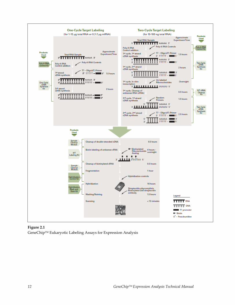
The One-Cycle Eukaryotic Target Labeling Assay experimental outline is represented in Figure 2.1. Total
RNA (1 μg to 15 μg) or mRNA (0.2 μg to 2 μg) is first reverse transcribed using a T7-Oligo(dT) Promoter
Primer in the first-strand cDNA synthesis reaction. Following RNase H-mediated second-strand cDNA
synthesis, the double-stranded cDNA is purified and serves as a template in the subsequent in vitro
transcription (IVT) reaction. The IVT reaction is carried out in the presence of T7 RNA Polymerase and a
biotinylated nucleotide analog/ribonucleotide mix for complementary RNA (cRNA) amplification and
biotin labeling. The biotinylated cRNA targets are then cleaned up, fragmented, and hybridized to
GeneChipTM expression arrays.
For smaller amounts of starting total RNA, in the range of 10 ng to 100 ng, an additional cycle of cDNA
synthesis and IVT amplification is required to obtain sufficient amounts of labeled cRNA target for analysis
with arrays. The Two-Cycle Eukaryotic Target Labeling Assay experimental outline is also represented in
Figure 2.1. After cDNA synthesis in the first cycle, an unlabeled ribonucleotide mix is used in the first cycle
of IVT amplification. The unlabeled cRNA is then reverse transcribed in the first-strand cDNA synthesis
step of the second cycle using random primers. Subsequently, the T7-Oligo(dT) Promoter Primer is used in
the second-strand cDNA synthesis to generate double-stranded cDNA template containing T7 promoter
sequences. The resulting double-stranded cDNA is then amplified and labeled using a biotinylated
nucleotide analog/ribonucleotide mix in the second IVT reaction. The labeled cRNA is then cleaned up,
fragmented, and hybridized to GeneChipTM expression arrays.
12 GeneChipTM Expression Analysis Technical Manual
Figure 2.1
GeneChipTM Eukaryotic Labeling Assays for Expression Analysis
GeneChipTM Expression Analysis Technical Manual 13
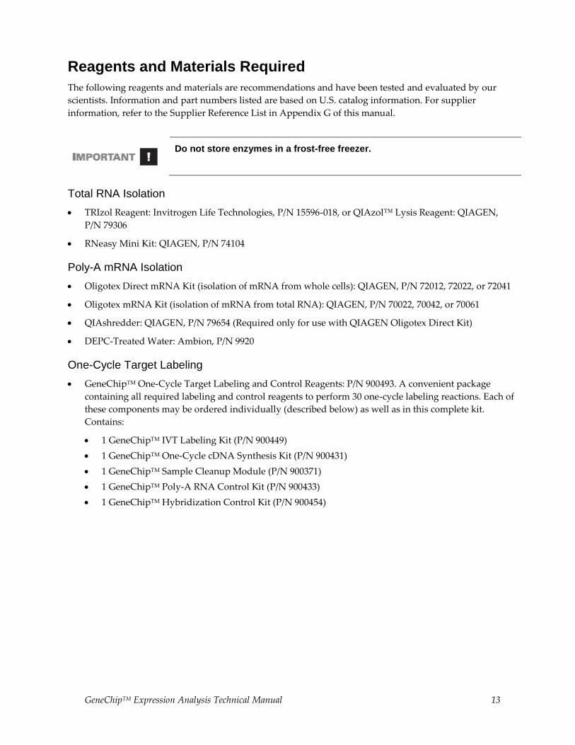
Reagents and Materials Required
The following reagents and materials are recommendations and have been tested and evaluated by our
scientists. Information and part numbers listed are based on U.S. catalog information. For supplier
information, refer to the Supplier Reference List in Appendix G of this manual.
Do not store enzymes in a frost-free freezer.
Total RNA Isolation
TRIzol Reagent: Invitrogen Life Technologies, P/N 15596-018, or QIAzol™ Lysis Reagent: QIAGEN,
P/N 79306
RNeasy Mini Kit: QIAGEN, P/N 74104
Poly-A mRNA Isolation
Oligotex Direct mRNA Kit (isolation of mRNA from whole cells): QIAGEN, P/N 72012, 72022, or 72041
Oligotex mRNA Kit (isolation of mRNA from total RNA): QIAGEN, P/N 70022, 70042, or 70061
QIAshredder: QIAGEN, P/N 79654 (Required only for use with QIAGEN Oligotex Direct Kit)
DEPC-Treated Water: Ambion, P/N 9920
One-Cycle Target Labeling
GeneChipTM One-Cycle Target Labeling and Control Reagents: P/N 900493. A convenient package
containing all required labeling and control reagents to perform 30 one-cycle labeling reactions. Each of
these components may be ordered individually (described below) as well as in this complete kit.
Contains:
1 GeneChipTM IVT Labeling Kit (P/N 900449)
1 GeneChipTM One-Cycle cDNA Synthesis Kit (P/N 900431)
1 GeneChipTM Sample Cleanup Module (P/N 900371)
1 GeneChipTM Poly-A RNA Control Kit (P/N 900433)
1 GeneChipTM Hybridization Control Kit (P/N 900454)
14 GeneChipTM Expression Analysis Technical Manual
Two-Cycle Target Labeling
GeneChipTM Two-Cycle Target Labeling and Control Reagents:, P/N 900494. A convenient package
containing required labeling and control reagents to perform 30 two-cycle labeling reactions. Each of
these components may be ordered individually (described below) as well as in this complete kit.
Contains:
1 GeneChipTM IVT Labeling Kit (P/N 900449)
1 GeneChipTM Two-Cycle cDNA Synthesis Kit (P/N 900432)
1 GeneChipTM Sample Cleanup Module (P/N 900371)
1 GeneChipTM IVT cRNA Cleanup Kit (900547)
1 GeneChipTM Poly-A RNA Control Kit (P/N 900433)
1 GeneChipTM Hybridization Control Kit (P/N 900454)
MEGAscriptTM High Yield Transcription Kit: Ambion Inc, P/N 1334 Purchased separately from Ambion,
Inc. (2 kits required to complete 30 reactions)
Miscellaneous Reagents
10X TBE: Cambrex, P/N 50843
Absolute ethanol (stored at –20°C for RNA precipitation; store ethanol at room temperature for use
with the GeneChipTM Sample Cleanup Module and IVT cRNA Kit)
80% ethanol (stored at –20°C for RNA precipitation; store ethanol at room temperature for use with the
GeneChipTM Sample Cleanup Module)
SYBR Green II: Cambrex, P/N 50523; or Molecular Probes, P/N S7586 (optional)
Pellet Paint: Novagen, P/N 69049-3 (optional)
Glycogen: Ambion, P/N 9510 (optional)
3M Sodium Acetate (NaOAc): Sigma-Aldrich, P/N S7899
Ethidium Bromide: Sigma-Aldrich, P/N E8751
1N NaOH
1N HCl
Miscellaneous Supplies
Sterile, RNase-free, microcentrifuge vials, 1.5 mL: USA Scientific, P/N 1415-2600 (or equivalent)
Micropipettors, (P-2, P-20, P-200, P-1000): Rainin Pipetman or equivalent
Sterile-barrier, RNase-free pipette tips (Tips must be pointed, not rounded, for efficient use with the
probe arrays) Beveled pipette tips may cause damage to the array septa and cause leakage.
Mini agarose gel electrophoresis unit with appropriate buffers
UV spectrophotometer
Bioanalyzer
Non-stick RNase-free microfuge tubes, 0.5 mL and 1.5 mL: Ambion, P/N12350 and P/N 12450,
respectively

GeneChipTM Expression Analysis Technical Manual 15
Total RNA and mRNA Isolation for One-Cycle Target Labeling Assay
Protocols are provided for preparing labeled cRNA from either total RNA or purified poly-A mRNA. It was
found that results obtained from samples prepared by both of these methods are similar, but not identical.
Therefore, to get the best results, it is suggested to only compare samples prepared using the same type of
RNA material.
Review precautions and interfering conditions in Chapter 1
The quality of the RNA is essential to the overall success of the analysis. Since the most appropriate protocol for the isolation of RNA can be source dependent, we recommend using a protocol that has been established for the tissues or cells being used. In the absence of an established protocol, using one of the commercially available kits designed for RNA isolation is suggested.
When using a commercial kit, follow the manufacturer’s instructions for RNA isolation.
ISOLATION OF RNA FROM YEAST
Total RNA
Good-quality total RNA has been isolated successfully from yeast cells using a hot phenol protocol
described by Schmitt, et al. Nucl Acids Res 18:3091-3092 (1990).
Poly-A mRNA
We recommend first purifying total RNA from yeast cells before isolating poly-A mRNA from total RNA.
Good-quality mRNA has been successfully isolated from total RNA using QIAGEN’s Oligotex mRNA Kit.
A single round of poly-A mRNA selection provides mRNA of sufficient purity and yield to use as a
template for cDNA synthesis. Two rounds of poly-A mRNA selection will result in significantly reduced
yield and are not generally recommended.
ISOLATION OF RNA FROM ARABIDOPSIS
Total RNA
TRIzol Reagent from Invitrogen Life Technologies has been used to isolate total RNA from Arabidopsis.
Follow the instructions provided by the supplier and, when necessary, use the steps outlined specifically
for samples with high starch and/or high lipid content. QIAzol Lysis Reagent from QIAGEN can also be
used.
Poly-A mRNA
Arabidopsis poly-A mRNA has been successfully isolated using QIAGEN’s Oligotex products. However,
other standard isolation products are likely to be adequate.
16 GeneChipTM Expression Analysis Technical Manual
ISOLATION OF RNA FROM MAMMALIAN CELLS OR TISSUES
Total RNA
High-quality total RNA has been successfully isolated from mammalian cells (such as cultured cells and
lymphocytes) using the RNeasy Mini Kit from QIAGEN.
If mammalian tissue is used as the source of RNA, it is recommended to isolate total RNA with a
commercial reagent, such as TRIzol or QIAzol reagents.
If going directly from TRIzol-isolated total RNA to cDNA synthesis, it may be beneficial to perform a second cleanup on the total RNA before starting. After the ethanol precipitation step in the TRIzol extraction procedure, perform a cleanup using the QIAGEN RNeasy Mini Kit. Much better yields of labeled cRNA are obtained from the in vitro transcription-labeling reaction when this second cleanup is performed.
Poly-A mRNA
Good-quality mRNA has been successfully isolated from mammalian cells (such as cultured cells and
lymphocytes) using QIAGEN’s Oligotex Direct mRNA kit and from total RNA using the Oligotex mRNA
kit. If mammalian tissue is used as the source of mRNA, total RNA should be first purified using a
commercial reagent, such as TRIzol, and then using a poly-A mRNA isolation procedure or a commercial
kit. QIAzol™ Lysis Reagent from QIAGEN can also be used.
PRECIPITATION OF RNA
Total RNA
It is not necessary to precipitate total RNA following isolation or cleanup with the RNeasy Mini Kit. Adjust
elution volumes from the RNeasy column to prepare for cDNA synthesis based upon expected RNA yields
from your experiment. Ethanol precipitation is required following TRIzol or QIAzol reagent isolation and
hot phenol extraction methods.
Poly-A mRNA
Most poly-A mRNA isolation procedures will result in dilution of RNA. It is necessary to concentrate
mRNA prior to the cDNA synthesis.
Precipitation Procedure
1. Add 1/10 volume 3M NaOAc, pH 5.2, and 2.5 volumes ethanol.*
2. Mix and incubate at –20°C for at least 1 hour.
3. Centrifuge at ≥ 12,000 x g in a microcentrifuge for 20 minutes at 4°C.
4. Wash pellet twice with 80% ethanol.
5. Air dry pellet. Check for dryness before proceeding.
6. Resuspend pellet in DEPC-treated H2O. The appropriate volume for resuspension depends on the
expected yield and the amount of RNA required for the cDNA synthesis. Read ahead to the cDNA
synthesis protocol in order to determine the appropriate resuspension volume at this step.
GeneChipTM Expression Analysis Technical Manual 17
*Addition of Carrier to Ethanol Precipitations
Adding carrier material has been shown to improve the RNA yield of precipitation reactions.
Pellet Paint
Addition of 0.5 μL of Pellet Paint per tube to nucleic acid precipitations makes the nucleic acid pellet
easier to visualize and helps reduce the chance of losing the pellet during washing steps. The pellet
paint does not appear to affect the outcome of subsequent steps in this protocol; however, it can
contribute to the absorbance at 260 nm when quantifying the mRNA.
Glycogen
Addition of 0.5 to 1 μL of glycogen (5 mg/mL) to nucleic acid precipitations aids in visualization of the
pellet and may increase recovery. The glycogen does not appear to affect the outcome of subsequent
steps in this protocol.
QUANTIFICATION OF RNA
Quantify RNA yield by spectrophotometric analysis using the convention that 1 absorbance unit at 260 nm
equals 40 μg/mL RNA.
The absorbance should be checked at 260 and 280 nm for determination of sample concentration and
purity.
The A260/A280 ratio should be close to 2.0 for pure RNA (ratios between 1.9 and 2.1 are acceptable).
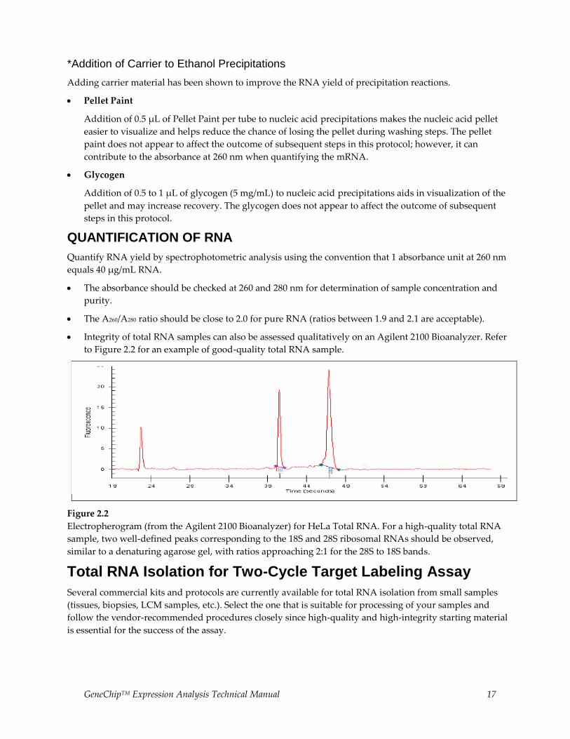
Integrity of total RNA samples can also be assessed qualitatively on an Agilent 2100 Bioanalyzer. Refer
to Figure 2.2 for an example of good-quality total RNA sample.
Figure 2.2
Electropherogram (from the Agilent 2100 Bioanalyzer) for HeLa Total RNA. For a high-quality total RNA
sample, two well-defined peaks corresponding to the 18S and 28S ribosomal RNAs should be observed,
similar to a denaturing agarose gel, with ratios approaching 2:1 for the 28S to 18S bands.
Total RNA Isolation for Two-Cycle Target Labeling Assay
Several commercial kits and protocols are currently available for total RNA isolation from small samples
(tissues, biopsies, LCM samples, etc.). Select the one that is suitable for processing of your samples and
follow the vendor-recommended procedures closely since high-quality and high-integrity starting material
is essential for the success of the assay.

18 GeneChipTM Expression Analysis Technical Manual
One-Cycle cDNA Synthesis1
STEP 1: PREPARATION OF POLY-A RNA CONTROLS FOR ONE-CYCLE cDNA SYNTHESIS (SPIKE-IN CONTROLS)
Eukaryotic Poly-A RNA Control Kit is used for this step.
Designed specifically to provide exogenous positive controls to monitor the entire eukaryotic target
labeling process, a set of poly-A RNA controls is supplied in the GeneChipTM Eukaryotic Poly-A RNA
Control Kit.
Each eukaryotic GeneChipTM probe array contains probe sets for several B. subtilis genes that are absent in
eukaryotic samples (lys, phe, thr, and dap). These poly-A RNA controls are in vitro synthesized, and the
polyadenylated transcripts for the B. subtilis genes are pre-mixed at staggered dilutions. The concentrated
Poly-A Control Stock can be diluted with the Poly-A Control Dil Buffer and spiked directly into RNA
samples to achieve the final dilutions (referred to as a ratio of copy number) summarized in Table 2.2.
Table 2.2 Final Dilutions of Poly-A RNA Controls in Samples
Poly-A RNA Spike Final Dilution (estimated ratio of copy number)
lys 1:100,000
phe 1:50,000
thr 1:25,000
dap 1:6,667
The controls are then amplified and labeled together with the samples. Examining the hybridization
intensities of these controls on GeneChipTM arrays helps to monitor the labeling process independently
from the quality of the starting RNA samples. Anticipated relative signal strength follows the order of lys <
phe < thr< dap.
The Poly-A RNA Control Stock and Poly-A Control Dil Buffer are provided with the kit to prepare the
appropriate serial dilutions based on Table 2.3. This is a guideline when 1, 5, or 10 μg of total RNA or 0.2
μg of mRNA is used as starting material. For starting sample amounts other than those listed here,
calculations are needed in order to perform the appropriate dilutions to arrive at the same proportionate
final dilution of the spike-in controls in the samples.
1 Users who do not purchase this Kit may be required to obtain a license under U.S. Patent Nos. 5,716,785, 5,891,636, 6,291,170, and 5,545,522 or to purchase another licensed kit.
Use non-stick RNase-free microfuge tubes to prepare all of the dilutions.

GeneChipTM Expression Analysis Technical Manual 19
Table 2.3 Serial Dilutions of Poly-A RNA Control Stock
Starting Amount Serial Dilutions Spike-in Volume
Total RNA mRNA First Second Third
1 µg 1:20 1:50 1:50 2 µL
5 µg 1:20 1:50 1:10 2 µL
10 µg 0.2 µg 1:20 1:50 1:5 2 µL
Avoid pipetting solutions less than 2 μL in volume to maintain precision and consistency when preparing the dilutions.
For example, to prepare the poly-A RNA dilutions for 5 μg of total RNA:
1. Add 2 μL of the Poly-A Control Stock to 38 μL of Poly-A Control Dil Buffer for the First Dilution
(1:20).
2. Mix thoroughly and spin down to collect the liquid at the bottom of the tube.
3. Add 2 μL of the First Dilution to 98 μL of Poly-A Control Dil Buffer to prepare the Second Dilution
(1:50).
4. Mix thoroughly and spin down to collect the liquid at the bottom of the tube.
5. Add 2 μL of the Second Dilution to 18 μL of Poly-A Control Dil Buffer to prepare the Third Dilution
(1:10).
6. Mix thoroughly and spin down to collect the liquid at the bottom of the tube.
7. Add 2 μL of this Third Dilution to 5 μg of sample total RNA.
The First Dilution of the poly-A RNA controls can be stored up to six weeks in a non-frost-free freezer at –20°C and frozen-thawed up to eight times.
20 GeneChipTM Expression Analysis Technical Manual
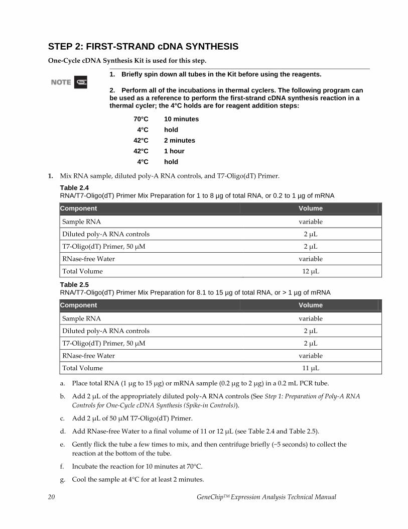
STEP 2: FIRST-STRAND cDNA SYNTHESIS
One-Cycle cDNA Synthesis Kit is used for this step.
1. Briefly spin down all tubes in the Kit before using the reagents.
2. Perform all of the incubations in thermal cyclers. The following program can be used as a reference to perform the first-strand cDNA synthesis reaction in a thermal cycler; the 4°C holds are for reagent addition steps:
70°C 10 minutes
4°C hold
42°C 2 minutes
42°C 1 hour
4°C hold
1. Mix RNA sample, diluted poly-A RNA controls, and T7-Oligo(dT) Primer.
Table 2.4 RNA/T7-Oligo(dT) Primer Mix Preparation for 1 to 8 µg of total RNA, or 0.2 to 1 µg of mRNA
Component Volume
Sample RNA variable
Diluted poly-A RNA controls 2 µL
T7-Oligo(dT) Primer, 50 µM 2 µL
RNase-free Water variable
Total Volume 12 µL
Table 2.5 RNA/T7-Oligo(dT) Primer Mix Preparation for 8.1 to 15 µg of total RNA, or > 1 µg of mRNA
Component Volume
Sample RNA variable
Diluted poly-A RNA controls 2 µL
T7-Oligo(dT) Primer, 50 µM 2 µL
RNase-free Water variable
Total Volume 11 µL
a. Place total RNA (1 μg to 15 μg) or mRNA sample (0.2 μg to 2 μg) in a 0.2 mL PCR tube.
b. Add 2 μL of the appropriately diluted poly-A RNA controls (See Step 1: Preparation of Poly-A RNA
Controls for One-Cycle cDNA Synthesis (Spike-in Controls)).
c. Add 2 μL of 50 μM T7-Oligo(dT) Primer.
d. Add RNase-free Water to a final volume of 11 or 12 μL (see Table 2.4 and Table 2.5).
e. Gently flick the tube a few times to mix, and then centrifuge briefly (~5 seconds) to collect the
reaction at the bottom of the tube.
f. Incubate the reaction for 10 minutes at 70°C.
g. Cool the sample at 4°C for at least 2 minutes.
GeneChipTM Expression Analysis Technical Manual 21
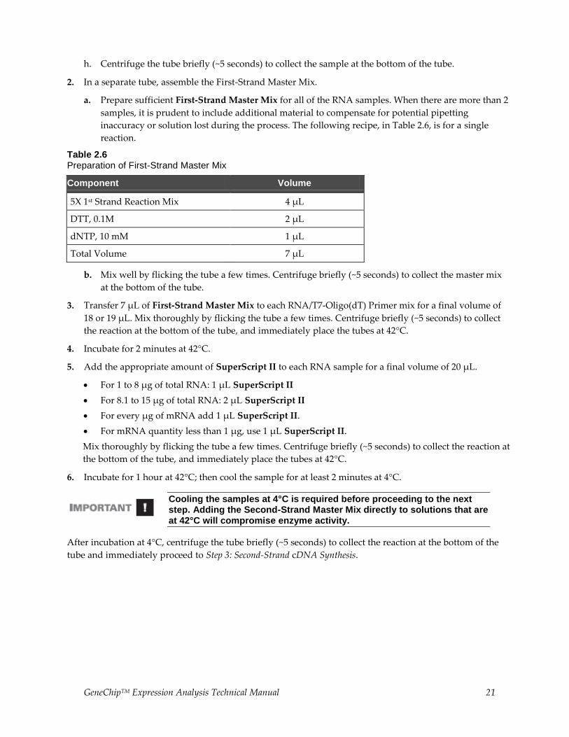
h. Centrifuge the tube briefly (~5 seconds) to collect the sample at the bottom of the tube.
2. In a separate tube, assemble the First-Strand Master Mix.
a. Prepare sufficient First-Strand Master Mix for all of the RNA samples. When there are more than 2
samples, it is prudent to include additional material to compensate for potential pipetting
inaccuracy or solution lost during the process. The following recipe, in Table 2.6, is for a single
reaction.
Table 2.6 Preparation of First-Strand Master Mix
Component Volume
5X 1st Strand Reaction Mix 4 µL
DTT, 0.1M 2 µL
dNTP, 10 mM 1 µL
Total Volume 7 µL
b. Mix well by flicking the tube a few times. Centrifuge briefly (~5 seconds) to collect the master mix
at the bottom of the tube.
3. Transfer 7 μL of First-Strand Master Mix to each RNA/T7-Oligo(dT) Primer mix for a final volume of
18 or 19 μL. Mix thoroughly by flicking the tube a few times. Centrifuge briefly (~5 seconds) to collect
the reaction at the bottom of the tube, and immediately place the tubes at 42°C.
4. Incubate for 2 minutes at 42°C.
5. Add the appropriate amount of SuperScript II to each RNA sample for a final volume of 20 μL.
For 1 to 8 μg of total RNA: 1 μL SuperScript II
For 8.1 to 15 μg of total RNA: 2 μL SuperScript II
For every μg of mRNA add 1 μL SuperScript II.
For mRNA quantity less than 1 μg, use 1 μL SuperScript II.
Mix thoroughly by flicking the tube a few times. Centrifuge briefly (~5 seconds) to collect the reaction at
the bottom of the tube, and immediately place the tubes at 42°C.
6. Incubate for 1 hour at 42°C; then cool the sample for at least 2 minutes at 4°C.
Cooling the samples at 4°C is required before proceeding to the next step. Adding the Second-Strand Master Mix directly to solutions that are at 42°C will compromise enzyme activity.
After incubation at 4°C, centrifuge the tube briefly (~5 seconds) to collect the reaction at the bottom of the
tube and immediately proceed to Step 3: Second-Strand cDNA Synthesis.
22 GeneChipTM Expression Analysis Technical Manual
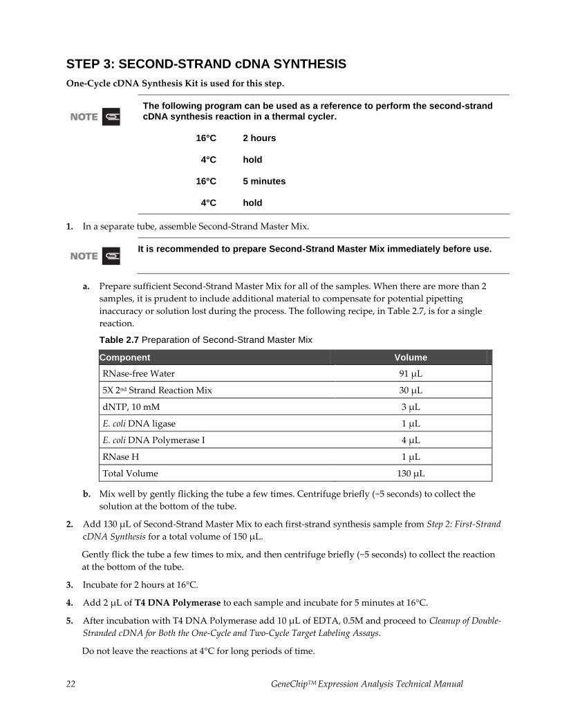
STEP 3: SECOND-STRAND cDNA SYNTHESIS
One-Cycle cDNA Synthesis Kit is used for this step.
The following program can be used as a reference to perform the second-strand cDNA synthesis reaction in a thermal cycler.
16°C 2 hours
4°C hold
16°C 5 minutes
4°C hold
1. In a separate tube, assemble Second-Strand Master Mix.
It is recommended to prepare Second-Strand Master Mix immediately before use.
a. Prepare sufficient Second-Strand Master Mix for all of the samples. When there are more than 2
samples, it is prudent to include additional material to compensate for potential pipetting
inaccuracy or solution lost during the process. The following recipe, in Table 2.7, is for a single
reaction.
Table 2.7 Preparation of Second-Strand Master Mix
Component Volume
RNase-free Water 91 µL
5X 2nd Strand Reaction Mix 30 µL
dNTP, 10 mM 3 µL
E. coli DNA ligase 1 µL
E. coli DNA Polymerase I 4 µL
RNase H 1 µL
Total Volume 130 µL
b. Mix well by gently flicking the tube a few times. Centrifuge briefly (~5 seconds) to collect the
solution at the bottom of the tube.
2. Add 130 μL of Second-Strand Master Mix to each first-strand synthesis sample from Step 2: First-Strand
cDNA Synthesis for a total volume of 150 μL.
Gently flick the tube a few times to mix, and then centrifuge briefly (~5 seconds) to collect the reaction
at the bottom of the tube.
3. Incubate for 2 hours at 16°C.
4. Add 2 μL of T4 DNA Polymerase to each sample and incubate for 5 minutes at 16°C.
5. After incubation with T4 DNA Polymerase add 10 μL of EDTA, 0.5M and proceed to Cleanup of Double-
Stranded cDNA for Both the One-Cycle and Two-Cycle Target Labeling Assays.
Do not leave the reactions at 4°C for long periods of time.
GeneChipTM Expression Analysis Technical Manual 23
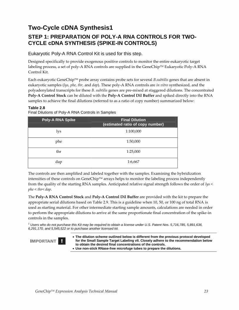
Two-Cycle cDNA Synthesis1
STEP 1: PREPARATION OF POLY-A RNA CONTROLS FOR TWO-CYCLE cDNA SYNTHESIS (SPIKE-IN CONTROLS)
Eukaryotic Poly-A RNA Control Kit is used for this step.
Designed specifically to provide exogenous positive controls to monitor the entire eukaryotic target
labeling process, a set of poly-A RNA controls are supplied in the GeneChipTM Eukaryotic Poly-A RNA
Control Kit.
Each eukaryotic GeneChipTM probe array contains probe sets for several B.subtilis genes that are absent in
eukaryotic samples (lys, phe, thr, and dap). These poly-A RNA controls are in vitro synthesized, and the
polyadenylated transcripts for these B. subtilis genes are pre-mixed at staggered dilutions. The concentrated
Poly-A Control Stock can be diluted with the Poly-A Control Dil Buffer and spiked directly into the RNA
samples to achieve the final dilutions (referred to as a ratio of copy number) summarized below:
Table 2.8 Final Dilutions of Poly-A RNA Controls in Samples
Poly-A RNA Spike Final Dilution (estimated ratio of copy number)
lys 1:100,000
phe 1:50,000
thr 1:25,000
dap 1:6,667
The controls are then amplified and labeled together with the samples. Examining the hybridization
intensities of these controls on GeneChipTM arrays helps to monitor the labeling process independently
from the quality of the starting RNA samples. Anticipated relative signal strength follows the order of lys <
phe < thr< dap.
The Poly-A RNA Control Stock and Poly-A Control Dil Buffer are provided with the kit to prepare the
appropriate serial dilutions based on Table 2.9. This is a guideline when 10, 50, or 100 ng of total RNA is
used as starting material. For other intermediate starting sample amounts, calculations are needed in order
to perform the appropriate dilutions to arrive at the same proportionate final concentration of the spike-in
controls in the samples.
1 Users who do not purchase this Kit may be required to obtain a license under U.S. Patent Nos. 5,716,785, 5,891,636, 6,291,170, and 5,545,522 or to purchase another licensed kit.
The dilution scheme outlined below is different from the previous protocol developed for the Small Sample Target Labeling vII. Closely adhere to the recommendation below to obtain the desired final concentrations of the controls.
Use non-stick RNase-free microfuge tubes to prepare the dilutions.
24 GeneChipTM Expression Analysis Technical Manual
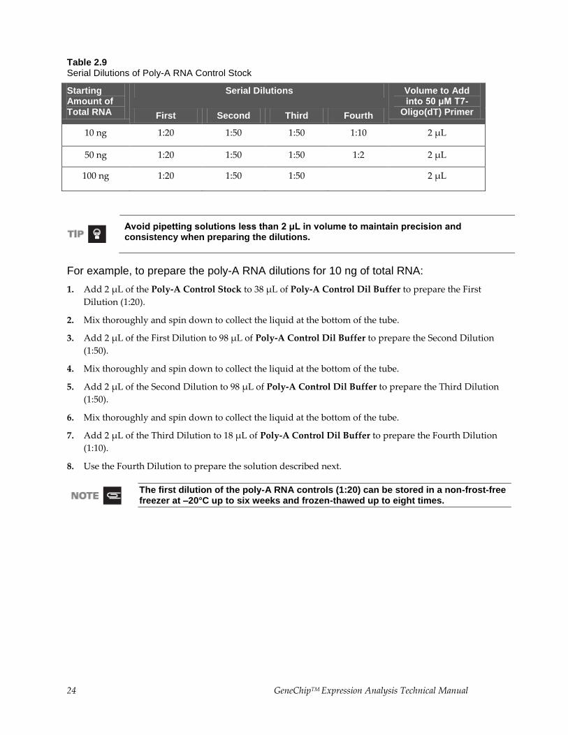
Table 2.9 Serial Dilutions of Poly-A RNA Control Stock
Starting Amount of Total RNA
Serial Dilutions Volume to Add into 50 μM T7-
Oligo(dT) Primer First Second Third Fourth
10 ng 1:20 1:50 1:50 1:10 2 µL
50 ng 1:20 1:50 1:50 1:2 2 µL
100 ng 1:20 1:50 1:50 2 µL
Avoid pipetting solutions less than 2 μL in volume to maintain precision and consistency when preparing the dilutions.
For example, to prepare the poly-A RNA dilutions for 10 ng of total RNA:
1. Add 2 μL of the Poly-A Control Stock to 38 μL of Poly-A Control Dil Buffer to prepare the First
Dilution (1:20).
2. Mix thoroughly and spin down to collect the liquid at the bottom of the tube.
3. Add 2 μL of the First Dilution to 98 μL of Poly-A Control Dil Buffer to prepare the Second Dilution
(1:50).
4. Mix thoroughly and spin down to collect the liquid at the bottom of the tube.
5. Add 2 μL of the Second Dilution to 98 μL of Poly-A Control Dil Buffer to prepare the Third Dilution
(1:50).
6. Mix thoroughly and spin down to collect the liquid at the bottom of the tube.
7. Add 2 μL of the Third Dilution to 18 μL of Poly-A Control Dil Buffer to prepare the Fourth Dilution
(1:10).
8. Use the Fourth Dilution to prepare the solution described next.
The first dilution of the poly-A RNA controls (1:20) can be stored in a non-frost-free freezer at –20°C up to six weeks and frozen-thawed up to eight times.
GeneChipTM Expression Analysis Technical Manual 25
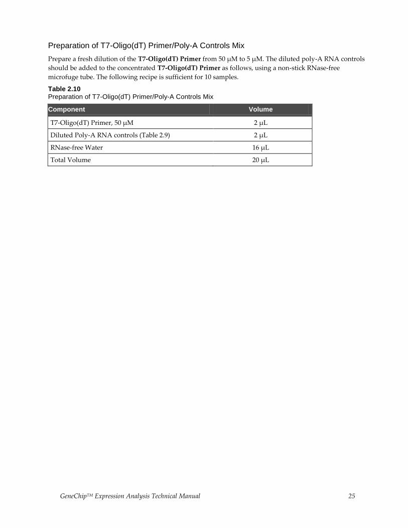
Preparation of T7-Oligo(dT) Primer/Poly-A Controls Mix
Prepare a fresh dilution of the T7-Oligo(dT) Primer from 50 μM to 5 μM. The diluted poly-A RNA controls
should be added to the concentrated T7-Oligo(dT) Primer as follows, using a non-stick RNase-free
microfuge tube. The following recipe is sufficient for 10 samples.
Table 2.10 Preparation of T7-Oligo(dT) Primer/Poly-A Controls Mix
Component Volume
T7-Oligo(dT) Primer, 50 µM 2 µL
Diluted Poly-A RNA controls (Table 2.9) 2 µL
RNase-free Water 16 µL
Total Volume 20 µL
26 GeneChipTM Expression Analysis Technical Manual
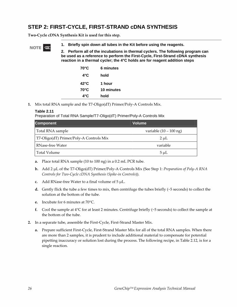
STEP 2: FIRST-CYCLE, FIRST-STRAND cDNA SYNTHESIS
Two-Cycle cDNA Synthesis Kit is used for this step.
1. Briefly spin down all tubes in the Kit before using the reagents.
2. Perform all of the incubations in thermal cyclers. The following program can be used as a reference to perform the First-Cycle, First-Strand cDNA synthesis reaction in a thermal cycler; the 4°C holds are for reagent addition steps
70°C 6 minutes
4°C hold
42°C 1 hour
70°C 10 minutes
4°C hold
1. Mix total RNA sample and the T7-Oligo(dT) Primer/Poly-A Controls Mix.
Table 2.11 Preparation of Total RNA Sample/T7-Oligo(dT) Primer/Poly-A Controls Mix
Component Volume
Total RNA sample variable (10 – 100 ng)
T7-Oligo(dT) Primer/Poly-A Controls Mix 2 µL
RNase-free Water variable
Total Volume 5 µL
a. Place total RNA sample (10 to 100 ng) in a 0.2 mL PCR tube.
b. Add 2 μL of the T7-Oligo(dT) Primer/Poly-A Controls Mix (See Step 1: Preparation of Poly-A RNA
Controls for Two-Cycle cDNA Synthesis (Spike-in Controls)).
c. Add RNase-free Water to a final volume of 5 μL.
d. Gently flick the tube a few times to mix, then centrifuge the tubes briefly (~5 seconds) to collect the
solution at the bottom of the tube.
e. Incubate for 6 minutes at 70°C.
f. Cool the sample at 4°C for at least 2 minutes. Centrifuge briefly (~5 seconds) to collect the sample at
the bottom of the tube.
2. In a separate tube, assemble the First-Cycle, First-Strand Master Mix.
a. Prepare sufficient First-Cycle, First-Strand Master Mix for all of the total RNA samples. When there
are more than 2 samples, it is prudent to include additional material to compensate for potential
pipetting inaccuracy or solution lost during the process. The following recipe, in Table 2.12, is for a
single reaction.
GeneChipTM Expression Analysis Technical Manual 27
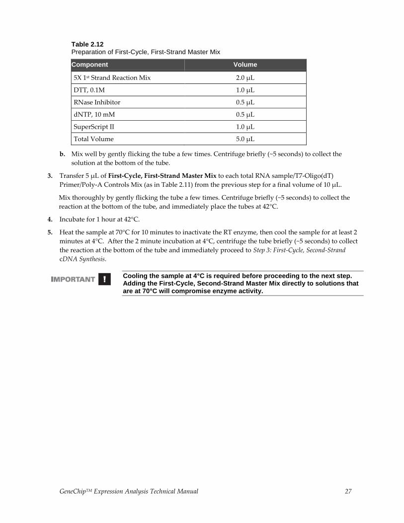
Table 2.12 Preparation of First-Cycle, First-Strand Master Mix
Component Volume
5X 1st Strand Reaction Mix 2.0 µL
DTT, 0.1M 1.0 µL
RNase Inhibitor 0.5 µL
dNTP, 10 mM 0.5 µL
SuperScript II 1.0 µL
Total Volume 5.0 µL
b. Mix well by gently flicking the tube a few times. Centrifuge briefly (~5 seconds) to collect the
solution at the bottom of the tube.
3. Transfer 5 μL of First-Cycle, First-Strand Master Mix to each total RNA sample/T7-Oligo(dT)
Primer/Poly-A Controls Mix (as in Table 2.11) from the previous step for a final volume of 10 μL.
Mix thoroughly by gently flicking the tube a few times. Centrifuge briefly (~5 seconds) to collect the
reaction at the bottom of the tube, and immediately place the tubes at 42°C.
4. Incubate for 1 hour at 42°C.
5. Heat the sample at 70°C for 10 minutes to inactivate the RT enzyme, then cool the sample for at least 2
minutes at 4°C. After the 2 minute incubation at 4°C, centrifuge the tube briefly (~5 seconds) to collect
the reaction at the bottom of the tube and immediately proceed to Step 3: First-Cycle, Second-Strand
cDNA Synthesis.
Cooling the sample at 4°C is required before proceeding to the next step. Adding the First-Cycle, Second-Strand Master Mix directly to solutions that are at 70°C will compromise enzyme activity.
28 GeneChipTM Expression Analysis Technical Manual
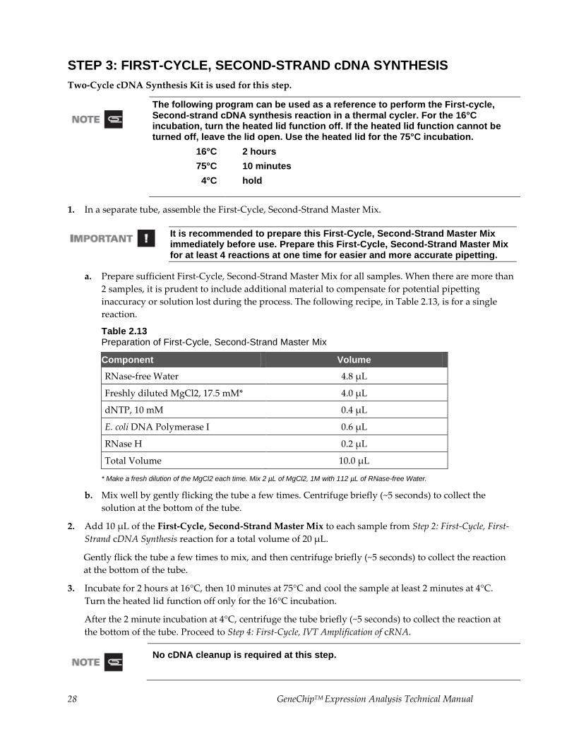
STEP 3: FIRST-CYCLE, SECOND-STRAND cDNA SYNTHESIS
Two-Cycle cDNA Synthesis Kit is used for this step.
The following program can be used as a reference to perform the First-cycle, Second-strand cDNA synthesis reaction in a thermal cycler. For the 16°C incubation, turn the heated lid function off. If the heated lid function cannot be turned off, leave the lid open. Use the heated lid for the 75°C incubation.
16°C 2 hours
75°C 10 minutes
4°C hold
1. In a separate tube, assemble the First-Cycle, Second-Strand Master Mix.
It is recommended to prepare this First-Cycle, Second-Strand Master Mix immediately before use. Prepare this First-Cycle, Second-Strand Master Mix for at least 4 reactions at one time for easier and more accurate pipetting.
a. Prepare sufficient First-Cycle, Second-Strand Master Mix for all samples. When there are more than
2 samples, it is prudent to include additional material to compensate for potential pipetting
inaccuracy or solution lost during the process. The following recipe, in Table 2.13, is for a single
reaction.
Table 2.13 Preparation of First-Cycle, Second-Strand Master Mix
Component Volume
RNase-free Water 4.8 µL
Freshly diluted MgCl2, 17.5 mM* 4.0 µL
dNTP, 10 mM 0.4 µL
E. coli DNA Polymerase I 0.6 µL
RNase H 0.2 µL
Total Volume 10.0 µL
* Make a fresh dilution of the MgCl2 each time. Mix 2 µL of MgCl2, 1M with 112 µL of RNase-free Water.
b. Mix well by gently flicking the tube a few times. Centrifuge briefly (~5 seconds) to collect the
solution at the bottom of the tube.
2. Add 10 μL of the First-Cycle, Second-Strand Master Mix to each sample from Step 2: First-Cycle, First-
Strand cDNA Synthesis reaction for a total volume of 20 μL.
Gently flick the tube a few times to mix, and then centrifuge briefly (~5 seconds) to collect the reaction
at the bottom of the tube.
3. Incubate for 2 hours at 16°C, then 10 minutes at 75°C and cool the sample at least 2 minutes at 4°C.
Turn the heated lid function off only for the 16°C incubation.
After the 2 minute incubation at 4°C, centrifuge the tube briefly (~5 seconds) to collect the reaction at
the bottom of the tube. Proceed to Step 4: First-Cycle, IVT Amplification of cRNA.
No cDNA cleanup is required at this step.
GeneChipTM Expression Analysis Technical Manual 29
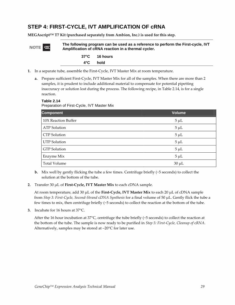
STEP 4: FIRST-CYCLE, IVT AMPLIFICATION OF cRNA
MEGAscriptTM T7 Kit (purchased separately from Ambion, Inc.) is used for this step.
The following program can be used as a reference to perform the First-cycle, IVT Amplification of cRNA reaction in a thermal cycler.
37°C 16 hours
4°C hold
1. In a separate tube, assemble the First-Cycle, IVT Master Mix at room temperature.
a. Prepare sufficient First-Cycle, IVT Master Mix for all of the samples. When there are more than 2
samples, it is prudent to include additional material to compensate for potential pipetting
inaccuracy or solution lost during the process. The following recipe, in Table 2.14, is for a single
reaction.
Table 2.14 Preparation of First-Cycle, IVT Master Mix
Component Volume
10X Reaction Buffer 5 µL
ATP Solution 5 µL
CTP Solution 5 µL
UTP Solution 5 µL
GTP Solution 5 µL
Enzyme Mix 5 µL
Total Volume 30 µL
b. Mix well by gently flicking the tube a few times. Centrifuge briefly (~5 seconds) to collect the
solution at the bottom of the tube.
2. Transfer 30 μL of First-Cycle, IVT Master Mix to each cDNA sample.
At room temperature, add 30 μL of the First-Cycle, IVT Master Mix to each 20 μL of cDNA sample
from Step 3: First-Cycle, Second-Strand cDNA Synthesis for a final volume of 50 μL. Gently flick the tube a
few times to mix, then centrifuge briefly (~5 seconds) to collect the reaction at the bottom of the tube.
3. Incubate for 16 hours at 37°C.
After the 16 hour incubation at 37°C, centrifuge the tube briefly (~5 seconds) to collect the reaction at
the bottom of the tube. The sample is now ready to be purified in Step 5: First-Cycle, Cleanup of cRNA.
Alternatively, samples may be stored at –20°C for later use.
30 GeneChipTM Expression Analysis Technical Manual
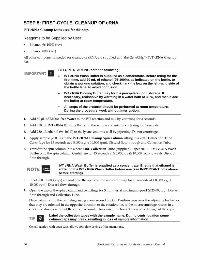
STEP 5: FIRST-CYCLE, CLEANUP OF cRNA
IVT cRNA Cleanup Kit is used for this step.
Reagents to be Supplied by User
Ethanol, 96-100% (v/v)
Ethanol, 80% (v/v)
All other components needed for cleanup of cRNA are supplied with the GeneChipTM IVT cRNA Cleanup
Kit.
BEFORE STARTING note the following:
IVT cRNA Wash Buffer is supplied as a concentrate. Before using for the first time, add 20 mL of ethanol (96-100%), as indicated on the bottle, to obtain a working solution, and checkmark the box on the left-hand side of the bottle label to avoid confusion.
IVT cRNA Binding Buffer may form a precipitate upon storage. If necessary, redissolve by warming in a water bath at 30°C, and then place the buffer at room temperature.
All steps of the protocol should be performed at room temperature. During the procedure, work without interruption.
1. Add 50 μL of RNase-free Water to the IVT reaction and mix by vortexing for 3 seconds.
2. Add 350 μL IVT cRNA Binding Buffer to the sample and mix by vortexing for 3 seconds.
3. Add 250 μL ethanol (96-100%) to the lysate, and mix well by pipetting. Do not centrifuge.
4. Apply sample (700 μL) to the IVT cRNA Cleanup Spin Column sitting in a 2 mL Collection Tube.
Centrifuge for 15 seconds at ≥ 8,000 x g (≥ 10,000 rpm). Discard flow-through and Collection Tube.
5. Transfer the spin column into a new 2 mL Collection Tube (supplied). Pipet 500 μL IVT cRNA Wash
Buffer onto the spin column. Centrifuge for 15 seconds at ≥ 8,000 x g (≥ 10,000 rpm) to wash. Discard
flow-through.
IVT cRNA Wash Buffer is supplied as a concentrate. Ensure that ethanol is added to the IVT cRNA Wash Buffer before use (see IMPORTANT note above before starting).
6. Pipet 500 μL 80% (v/v) ethanol onto the spin column and centrifuge for 15 seconds at ≥ 8,000 x g (≥
10,000 rpm). Discard flow-through.
7. Open the cap of the spin column and centrifuge for 5 minutes at maximum speed (≤ 25,000 x g). Discard
flow-through and Collection Tube.
Place columns into the centrifuge using every second bucket. Position caps over the adjoining bucket so
that they are oriented in the opposite direction to the rotation (i.e., if the microcentrifuge rotates in a
clockwise direction, orient the caps in a counterclockwise direction). This avoids damage of the caps.
Label the collection tubes with the sample name. During centrifugation some column caps may break, resulting in loss of sample information.
Centrifugation with open caps allows complete drying of the membrane.
GeneChipTM Expression Analysis Technical Manual 31
8. Transfer spin column into a new 1.5 mL Collection Tube (supplied), and pipet 13 μL of RNase-free
Water directly onto the spin column membrane. Ensure that the water is dispensed directly onto the
membrane. Centrifuge 1 minute at maximum speed (≤ 25,000 x g) to elute. The average volume of
eluate is 11 μL from 13 μL RNase-free Water.
9. To determine cRNA yield for samples starting with 50 ng or higher, remove 2 μL of the cRNA, and add
78 μL of water to measure the absorbance at 260 nm. Use 600 ng of cRNA in the following Step 6:
Second-Cycle, First-Strand cDNA Synthesis.
For starting material less than 50 ng, or if the yield is less than 600 ng, use the entire eluate for the Second-Cycle,
First-Strand cDNA Synthesis Reaction.
Samples can be stored at –20°C for later use, or proceed to Step 6: Second-Cycle, First-Strand cDNA Synthesis
described next.
32 GeneChipTM Expression Analysis Technical Manual
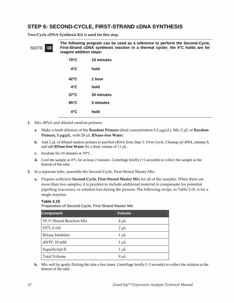
STEP 6: SECOND-CYCLE, FIRST-STRAND cDNA SYNTHESIS
Two-Cycle cDNA Synthesis Kit is used for this step.
The following program can be used as a reference to perform the Second-Cycle, First-Strand cDNA synthesis reaction in a thermal cycler; the 4°C holds are for reagent addition steps:
70°C 10 minutes
4°C hold
42°C 1 hour
4°C hold
37°C 20 minutes
95°C 5 minutes
4°C hold
1. Mix cRNA and diluted random primers.
a. Make a fresh dilution of the Random Primers (final concentration 0.2 μg/μL). Mix 2 μL of Random
Primers, 3 μg/μL, with 28 μL RNase-free Water.
b. Add 2 μL of diluted random primers to purified cRNA from Step 5: First-Cycle, Cleanup of cRNA, substep 9,
and add RNase-free Water for a final volume of 11 μL.
c. Incubate for 10 minutes at 70°C.
d. Cool the sample at 4°C for at least 2 minutes. Centrifuge briefly (~5 seconds) to collect the sample at the
bottom of the tube.
2. In a separate tube, assemble the Second-Cycle, First-Strand Master Mix.
a. Prepare sufficient Second-Cycle, First-Strand Master Mix for all of the samples. When there are
more than two samples, it is prudent to include additional material to compensate for potential
pipetting inaccuracy or solution lost during the process. The following recipe, in Table 2.15, is for a
single reaction.
Table 2.15 Preparation of Second-Cycle, First-Strand Master Mix
Component Volume
5X 1st Strand Reaction Mix 4 µL
DTT, 0.1M 2 µL
RNase Inhibitor 1 µL
dNTP, 10 mM 1 µL
SuperScript II 1 µL
Total Volume 9 µL
b. Mix well by gently flicking the tube a few times. Centrifuge briefly (~5 seconds) to collect the solution at the
bottom of the tube.
GeneChipTM Expression Analysis Technical Manual 33
3. Transfer 9 μL of Second-Cycle, First-Strand Master Mix to each cRNA/random primer sample from
Step 6: Second-Cycle, First-Strand cDNA Synthesis, substep 1, for a final volume of 20 μL.
Mix thoroughly by gently flicking the tube a few times. Centrifuge briefly (~5 seconds) to collect the
reaction at the bottom of the tube and place the tubes at 42°C immediately.
4. Incubate for 1 hour at 42°C, then cool the sample for at least 2 minutes at 4°C.
After the incubation at 4°C, centrifuge briefly (~5 seconds) to collect the reaction at the bottom of the tube.
5. Add 1 μL of RNase H to each sample for a final volume of 21 μL.
Mix thoroughly by gently flicking the tube a few times.
Centrifuge briefly (~5 seconds) to collect the reaction at the bottom of the tube and incubate for 20
minutes at 37°C.
6. Heat the sample at 95°C for 5 minutes. Cool the sample for at least 2 minutes at 4°C; then, proceed
directly to Step 7: Second-Cycle, Second-Strand cDNA Synthesis.
34 GeneChipTM Expression Analysis Technical Manual
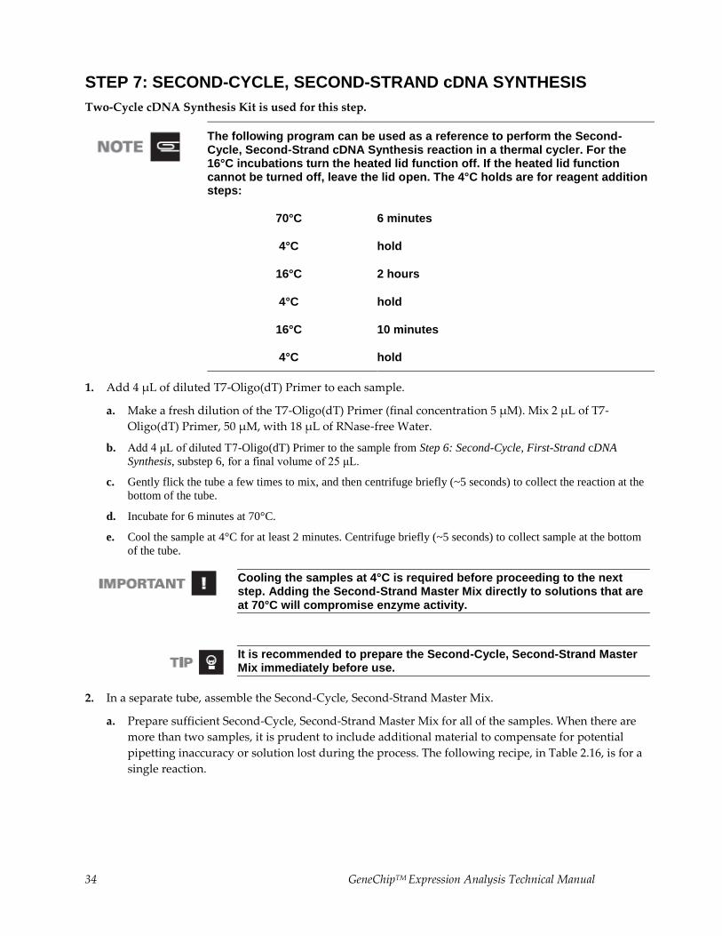
STEP 7: SECOND-CYCLE, SECOND-STRAND cDNA SYNTHESIS
Two-Cycle cDNA Synthesis Kit is used for this step.
The following program can be used as a reference to perform the Second-Cycle, Second-Strand cDNA Synthesis reaction in a thermal cycler. For the 16°C incubations turn the heated lid function off. If the heated lid function cannot be turned off, leave the lid open. The 4°C holds are for reagent addition steps:
70°C 6 minutes
4°C hold
16°C 2 hours
4°C hold
16°C 10 minutes
4°C hold
1. Add 4 μL of diluted T7-Oligo(dT) Primer to each sample.
a. Make a fresh dilution of the T7-Oligo(dT) Primer (final concentration 5 μM). Mix 2 μL of T7-
Oligo(dT) Primer, 50 μM, with 18 μL of RNase-free Water.
b. Add 4 μL of diluted T7-Oligo(dT) Primer to the sample from Step 6: Second-Cycle, First-Strand cDNA
Synthesis, substep 6, for a final volume of 25 μL.
c. Gently flick the tube a few times to mix, and then centrifuge briefly (~5 seconds) to collect the reaction at the
bottom of the tube.
d. Incubate for 6 minutes at 70°C.
e. Cool the sample at 4°C for at least 2 minutes. Centrifuge briefly (~5 seconds) to collect sample at the bottom
of the tube.
Cooling the samples at 4°C is required before proceeding to the next step. Adding the Second-Strand Master Mix directly to solutions that are at 70°C will compromise enzyme activity.
It is recommended to prepare the Second-Cycle, Second-Strand Master Mix immediately before use.
2. In a separate tube, assemble the Second-Cycle, Second-Strand Master Mix.
a. Prepare sufficient Second-Cycle, Second-Strand Master Mix for all of the samples. When there are
more than two samples, it is prudent to include additional material to compensate for potential
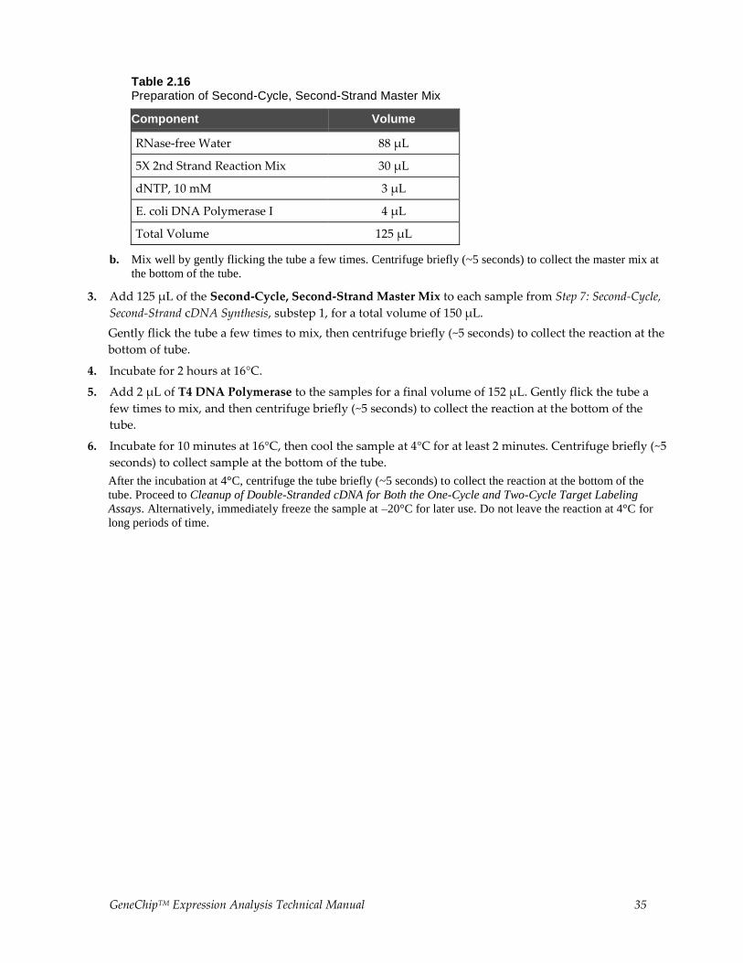
pipetting inaccuracy or solution lost during the process. The following recipe, in Table 2.16, is for a
single reaction.
GeneChipTM Expression Analysis Technical Manual 35
Table 2.16 Preparation of Second-Cycle, Second-Strand Master Mix
Component Volume
RNase-free Water 88 µL
5X 2nd Strand Reaction Mix 30 µL
dNTP, 10 mM 3 µL
E. coli DNA Polymerase I 4 µL
Total Volume 125 µL
b. Mix well by gently flicking the tube a few times. Centrifuge briefly (~5 seconds) to collect the master mix at
the bottom of the tube.
3. Add 125 μL of the Second-Cycle, Second-Strand Master Mix to each sample from Step 7: Second-Cycle,
Second-Strand cDNA Synthesis, substep 1, for a total volume of 150 μL.
Gently flick the tube a few times to mix, then centrifuge briefly (~5 seconds) to collect the reaction at the
bottom of tube.
4. Incubate for 2 hours at 16°C.
5. Add 2 μL of T4 DNA Polymerase to the samples for a final volume of 152 μL. Gently flick the tube a
few times to mix, and then centrifuge briefly (~5 seconds) to collect the reaction at the bottom of the
tube.
6. Incubate for 10 minutes at 16°C, then cool the sample at 4°C for at least 2 minutes. Centrifuge briefly (~5
seconds) to collect sample at the bottom of the tube.
After the incubation at 4°C, centrifuge the tube briefly (~5 seconds) to collect the reaction at the bottom of the
tube. Proceed to Cleanup of Double-Stranded cDNA for Both the One-Cycle and Two-Cycle Target Labeling
Assays. Alternatively, immediately freeze the sample at –20°C for later use. Do not leave the reaction at 4°C for
long periods of time.
36 GeneChipTM Expression Analysis Technical Manual
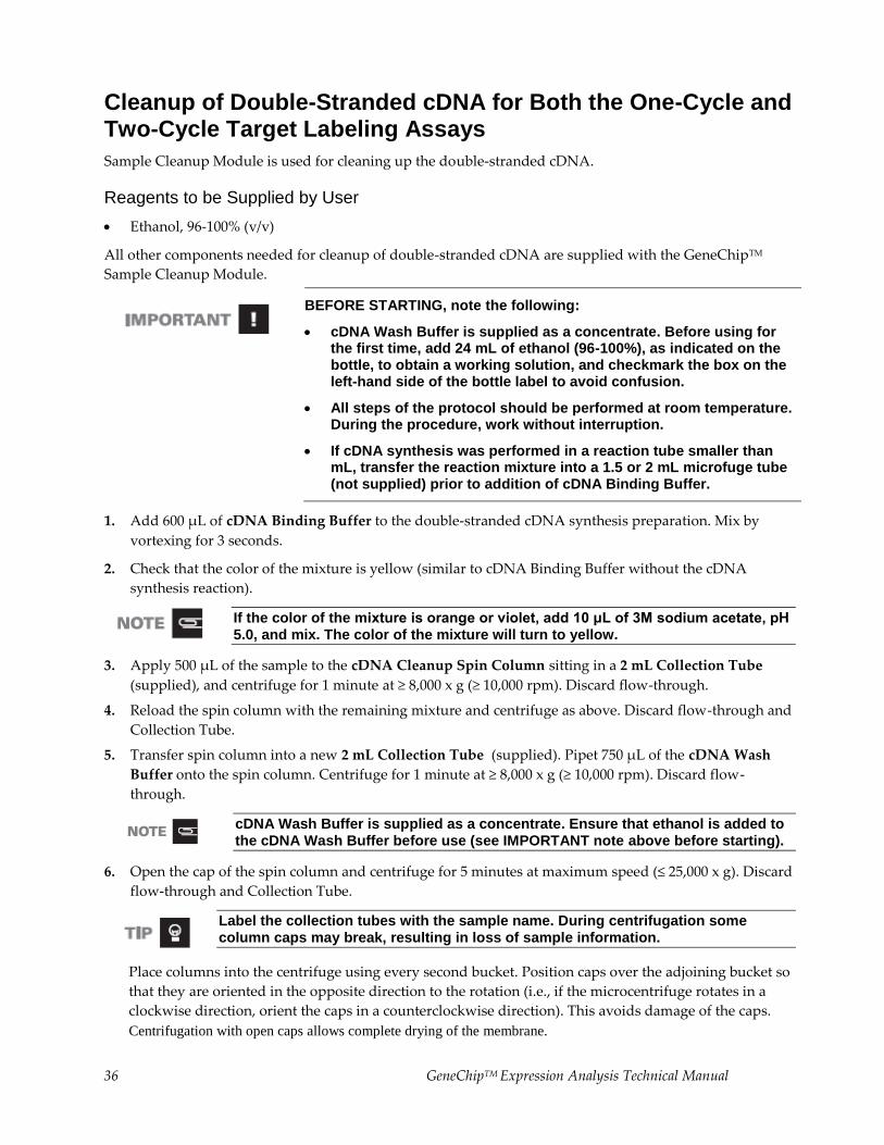
Cleanup of Double-Stranded cDNA for Both the One-Cycle and Two-Cycle Target Labeling Assays
Sample Cleanup Module is used for cleaning up the double-stranded cDNA.
Reagents to be Supplied by User
Ethanol, 96-100% (v/v)
All other components needed for cleanup of double-stranded cDNA are supplied with the GeneChipTM
Sample Cleanup Module.
BEFORE STARTING, note the following:
cDNA Wash Buffer is supplied as a concentrate. Before using for the first time, add 24 mL of ethanol (96-100%), as indicated on the bottle, to obtain a working solution, and checkmark the box on the left-hand side of the bottle label to avoid confusion.
All steps of the protocol should be performed at room temperature. During the procedure, work without interruption.
If cDNA synthesis was performed in a reaction tube smaller than mL, transfer the reaction mixture into a 1.5 or 2 mL microfuge tube (not supplied) prior to addition of cDNA Binding Buffer.
1. Add 600 μL of cDNA Binding Buffer to the double-stranded cDNA synthesis preparation. Mix by
vortexing for 3 seconds.
2. Check that the color of the mixture is yellow (similar to cDNA Binding Buffer without the cDNA
synthesis reaction).
If the color of the mixture is orange or violet, add 10 μL of 3M sodium acetate, pH 5.0, and mix. The color of the mixture will turn to yellow.
3. Apply 500 μL of the sample to the cDNA Cleanup Spin Column sitting in a 2 mL Collection Tube
(supplied), and centrifuge for 1 minute at ≥ 8,000 x g (≥ 10,000 rpm). Discard flow-through.
4. Reload the spin column with the remaining mixture and centrifuge as above. Discard flow-through and
Collection Tube.
5. Transfer spin column into a new 2 mL Collection Tube (supplied). Pipet 750 μL of the cDNA Wash
Buffer onto the spin column. Centrifuge for 1 minute at ≥ 8,000 x g (≥ 10,000 rpm). Discard flow-
through.
cDNA Wash Buffer is supplied as a concentrate. Ensure that ethanol is added to the cDNA Wash Buffer before use (see IMPORTANT note above before starting).
6. Open the cap of the spin column and centrifuge for 5 minutes at maximum speed (≤ 25,000 x g). Discard
flow-through and Collection Tube.
Label the collection tubes with the sample name. During centrifugation some column caps may break, resulting in loss of sample information.
Place columns into the centrifuge using every second bucket. Position caps over the adjoining bucket so
that they are oriented in the opposite direction to the rotation (i.e., if the microcentrifuge rotates in a
clockwise direction, orient the caps in a counterclockwise direction). This avoids damage of the caps.
Centrifugation with open caps allows complete drying of the membrane.
GeneChipTM Expression Analysis Technical Manual 37
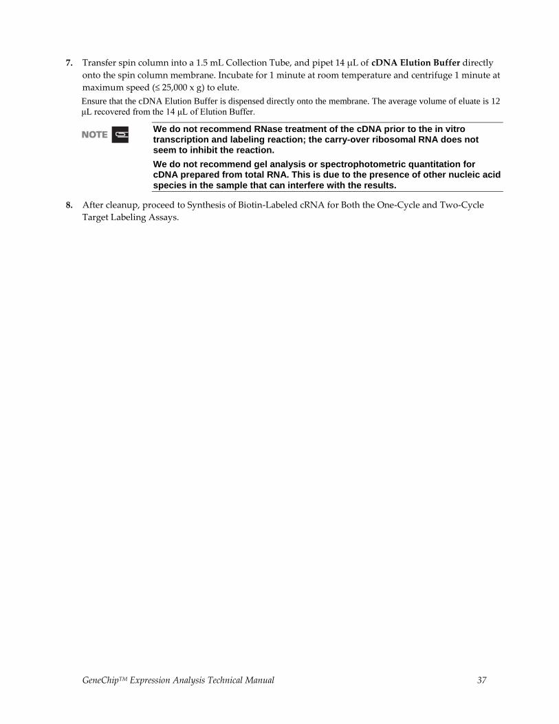
7. Transfer spin column into a 1.5 mL Collection Tube, and pipet 14 μL of cDNA Elution Buffer directly
onto the spin column membrane. Incubate for 1 minute at room temperature and centrifuge 1 minute at
maximum speed (≤ 25,000 x g) to elute.
Ensure that the cDNA Elution Buffer is dispensed directly onto the membrane. The average volume of eluate is 12
μL recovered from the 14 μL of Elution Buffer.
We do not recommend RNase treatment of the cDNA prior to the in vitro transcription and labeling reaction; the carry-over ribosomal RNA does not seem to inhibit the reaction.
We do not recommend gel analysis or spectrophotometric quantitation for cDNA prepared from total RNA. This is due to the presence of other nucleic acid species in the sample that can interfere with the results.
8. After cleanup, proceed to Synthesis of Biotin-Labeled cRNA for Both the One-Cycle and Two-Cycle
Target Labeling Assays.
38 GeneChipTM Expression Analysis Technical Manual
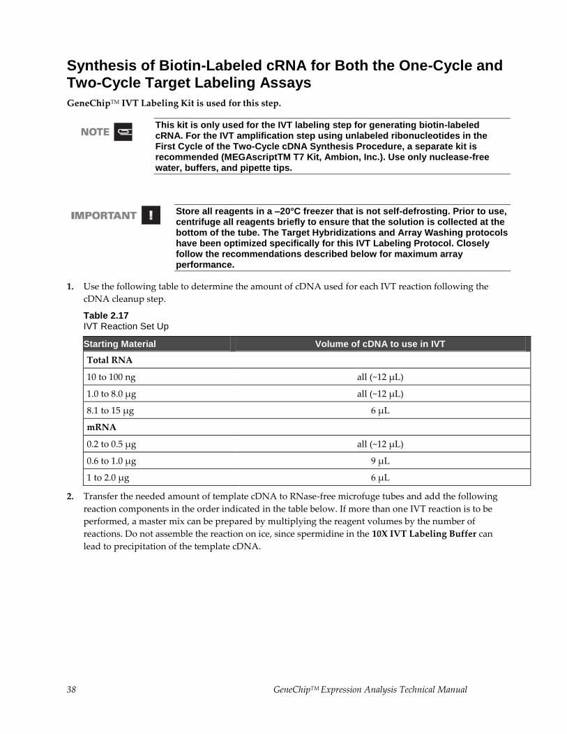
Synthesis of Biotin-Labeled cRNA for Both the One-Cycle and Two-Cycle Target Labeling Assays
GeneChipTM IVT Labeling Kit is used for this step.
This kit is only used for the IVT labeling step for generating biotin-labeled cRNA. For the IVT amplification step using unlabeled ribonucleotides in the First Cycle of the Two-Cycle cDNA Synthesis Procedure, a separate kit is recommended (MEGAscriptTM T7 Kit, Ambion, Inc.). Use only nuclease-free water, buffers, and pipette tips.
Store all reagents in a –20°C freezer that is not self-defrosting. Prior to use, centrifuge all reagents briefly to ensure that the solution is collected at the bottom of the tube. The Target Hybridizations and Array Washing protocols have been optimized specifically for this IVT Labeling Protocol. Closely follow the recommendations described below for maximum array performance.
1. Use the following table to determine the amount of cDNA used for each IVT reaction following the
cDNA cleanup step.
Table 2.17 IVT Reaction Set Up
Starting Material Volume of cDNA to use in IVT
Total RNA
10 to 100 ng all (~12 µL)
1.0 to 8.0 µg all (~12 µL)
8.1 to 15 µg 6 µL
mRNA
0.2 to 0.5 µg all (~12 µL)
0.6 to 1.0 µg 9 µL
1 to 2.0 µg 6 µL
2. Transfer the needed amount of template cDNA to RNase-free microfuge tubes and add the following
reaction components in the order indicated in the table below. If more than one IVT reaction is to be
performed, a master mix can be prepared by multiplying the reagent volumes by the number of
reactions. Do not assemble the reaction on ice, since spermidine in the 10X IVT Labeling Buffer can
lead to precipitation of the template cDNA.
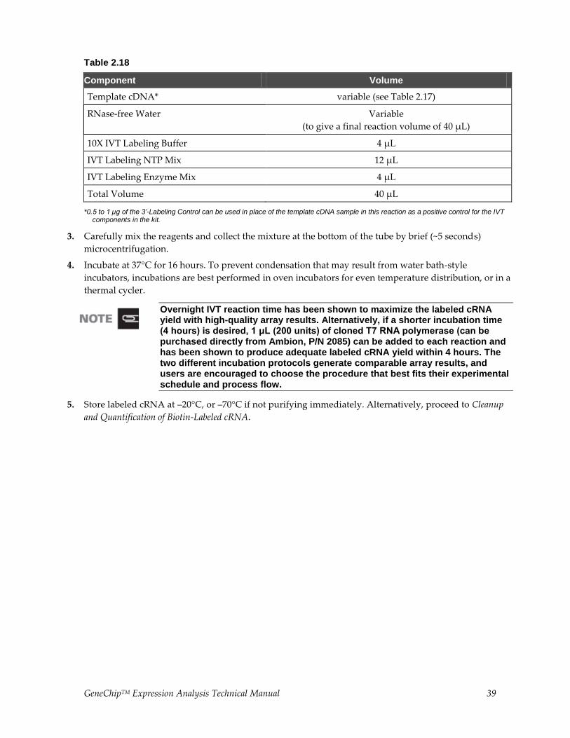
GeneChipTM Expression Analysis Technical Manual 39
Table 2.18
Component Volume
Template cDNA* variable (see Table 2.17)
RNase-free Water Variable
(to give a final reaction volume of 40 µL)
10X IVT Labeling Buffer 4 µL
IVT Labeling NTP Mix 12 µL
IVT Labeling Enzyme Mix 4 µL
Total Volume 40 µL
*0.5 to 1 µg of the 3’-Labeling Control can be used in place of the template cDNA sample in this reaction as a positive control for the IVT components in the kit.
3. Carefully mix the reagents and collect the mixture at the bottom of the tube by brief (~5 seconds)
microcentrifugation.
4. Incubate at 37°C for 16 hours. To prevent condensation that may result from water bath-style
incubators, incubations are best performed in oven incubators for even temperature distribution, or in a
thermal cycler.
Overnight IVT reaction time has been shown to maximize the labeled cRNA yield with high-quality array results. Alternatively, if a shorter incubation time (4 hours) is desired, 1 μL (200 units) of cloned T7 RNA polymerase (can be purchased directly from Ambion, P/N 2085) can be added to each reaction and has been shown to produce adequate labeled cRNA yield within 4 hours. The two different incubation protocols generate comparable array results, and users are encouraged to choose the procedure that best fits their experimental schedule and process flow.
5. Store labeled cRNA at –20°C, or –70°C if not purifying immediately. Alternatively, proceed to Cleanup
and Quantification of Biotin-Labeled cRNA.
40 GeneChipTM Expression Analysis Technical Manual
Cleanup and Quantification of Biotin-Labeled cRNA
Sample Cleanup Module is used for cleaning up the biotin-labeled cRNA.
Reagents to be Supplied by User
Ethanol, 96-100% (v/v)
Ethanol, 80% (v/v)
All other components needed for cleanup of biotin-labeled cRNA are supplied with the GeneChipTM
Sample Cleanup Module.
STEP 1: CLEANUP OF BIOTIN-LABELED cRNA
BEFORE STARTING, note the following:
It is essential to remove unincorporated NTPs, so that the concentration and purity of cRNA can be accurately determined by 260 nm absorbance.
DO NOT extract biotin-labeled RNA with phenol-chloroform. The biotin will cause some of the RNA to partition into the organic phase. This will result in low yields.
Save an aliquot of the unpurified IVT product for analysis by gel electrophoresis.
IVT cRNA Wash Buffer is supplied as a concentrate. Before using for the first time, add 20 mL of ethanol (96-100%), as indicated on the bottle, to obtain a working solution, and checkmark the box on the left-hand side of the bottle label to avoid confusion.
IVT cRNA Binding Buffer may form a precipitate upon storage. If necessary, redissolve by warming in a water bath at 30°C, and then place the buffer at room temperature.
All steps of the protocol should be performed at room temperature. During the procedure, work without interruption.
1. Add 60 μL of RNase-free Water to the IVT reaction and mix by vortexing for 3 seconds.
2. Add 350 μL IVT cRNA Binding Buffer to the sample and mix by vortexing for 3 seconds.
3. Add 250 μL ethanol (96-100%) to the mixture, and mix well by pipetting. Do not centrifuge.
4. Apply sample (700 μL) to the IVT cRNA Cleanup Spin Column sitting in a 2 mL Collection Tube.
Centrifuge for 15 seconds at ≥ 8,000 x g (≥ 10,000 rpm). Discard flow-through and Collection Tube.
5. Transfer the spin column into a new 2 mL Collection Tube (supplied). Pipet 500 μL IVT cRNA Wash
Buffer onto the spin column. Centrifuge for 15 seconds at ≥ 8,000 x g (≥ 10,000 rpm) to wash. Discard
flow-through.
IVT cRNA Wash Buffer is supplied as a concentrate. Ensure that ethanol is added to the IVT cRNA Wash Buffer before use (see IMPORTANT note above before starting).
6. Pipet 500 μL 80% (v/v) ethanol onto the spin column and centrifuge for 15 seconds at ≥ 8,000 x g (≥
10,000 rpm). Discard flow-through.
GeneChipTM Expression Analysis Technical Manual 41
7. Open the cap of the spin column and centrifuge for 5 minutes at maximum speed (≤ 25,000 x g). Discard
flow-through and Collection Tube.
Place columns into the centrifuge using every second bucket. Position caps over the adjoining bucket so
that they are oriented in the opposite direction to the rotation (i.e., if the microcentrifuge rotates in a
clockwise direction, orient the caps in a counterclockwise direction). This avoids damage of the caps.
Label the collection tubes with the sample name. During centrifugation some column caps may break, resulting in loss of sample information.
Centrifugation with open caps allows complete drying of the membrane.
8. Transfer spin column into a new 1.5 mL Collection Tube (supplied), and pipet 11 μL of RNase-free
Water directly onto the spin column membrane. Ensure that the water is dispensed directly onto the
membrane. Centrifuge 1 minute at maximum speed (≤ 25,000 x g) to elute.
9. Pipet 10 μL of RNase-free Water directly onto the spin column membrane. Ensure that the water is
dispensed directly onto the membrane. Centrifuge 1 minute at maximum speed (≤ 25,000 x g) to elute.
10. For subsequent photometric quantification of the purified cRNA, we recommend dilution of the eluate
between 1:100 fold and 1:200 fold.
11. Store cRNA at –20°C, or –70°C if not quantitated immediately. Alternatively, proceed to Step 2:
Quantification of the cRNA.
STEP 2: QUANTIFICATION OF THE cRNA
Use spectrophotometric analysis to determine the cRNA yield. Apply the convention that 1 absorbance unit
at 260 nm equals 40 μg/mL RNA.
Check the absorbance at 260 nm and 280 nm to determine sample concentration and purity.
Maintain the A260/A280 ratio close to 2.0 for pure RNA (ratios between 1.9 and 2.1 are acceptable).
For quantification of cRNA when using total RNA as starting material, an adjusted cRNA yield must be
calculated to reflect carryover of unlabeled total RNA. Using an estimate of 100% carryover, use the
formula below to determine adjusted cRNA yield:
adjusted cRNA yield = RNAm - (total RNAi) (y)
RNAm = amount of cRNA measured after IVT (μg)
total RNAi = starting amount of total RNA (μg)
y = fraction of cDNA reaction used in IVT
Example: Starting with 10 μg total RNA, 50% of the cDNA reaction is added to the IVT, giving a yield of 50
μg cRNA. Therefore, adjusted cRNA yield = 50 μg cRNA - (10 μg total RNA) (0.5 cDNA reaction) = 45.0 μg.
Use adjusted yield in Fragmenting the cRNA for Target Preparation.
Refer to Chapter 3 Eukaryotic Target Hybridization for the amount of cRNA required for one array hybridization experiment. The amount varies depending on the array format. Refer to the specific probe array package insert for information on the array format.
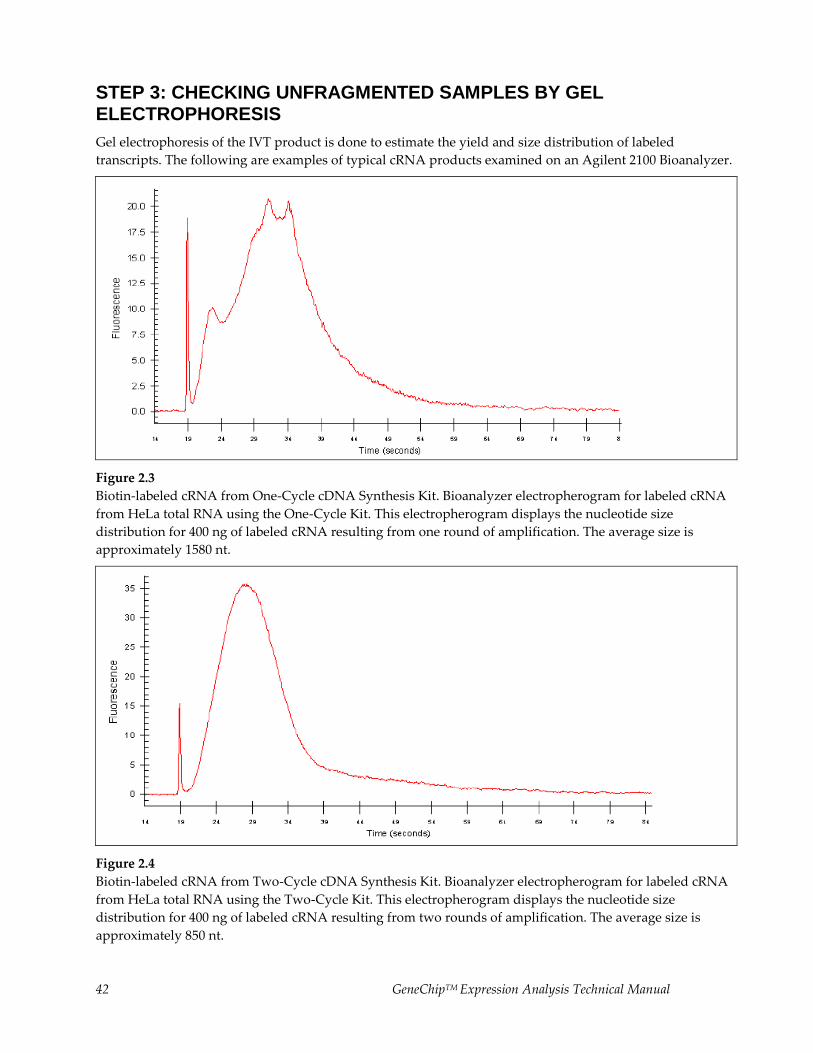
42 GeneChipTM Expression Analysis Technical Manual
STEP 3: CHECKING UNFRAGMENTED SAMPLES BY GEL ELECTROPHORESIS
Gel electrophoresis of the IVT product is done to estimate the yield and size distribution of labeled
transcripts. The following are examples of typical cRNA products examined on an Agilent 2100 Bioanalyzer.
Figure 2.3
Biotin-labeled cRNA from One-Cycle cDNA Synthesis Kit. Bioanalyzer electropherogram for labeled cRNA
from HeLa total RNA using the One-Cycle Kit. This electropherogram displays the nucleotide size
distribution for 400 ng of labeled cRNA resulting from one round of amplification. The average size is
approximately 1580 nt.
Figure 2.4
Biotin-labeled cRNA from Two-Cycle cDNA Synthesis Kit. Bioanalyzer electropherogram for labeled cRNA
from HeLa total RNA using the Two-Cycle Kit. This electropherogram displays the nucleotide size
distribution for 400 ng of labeled cRNA resulting from two rounds of amplification. The average size is
approximately 850 nt.
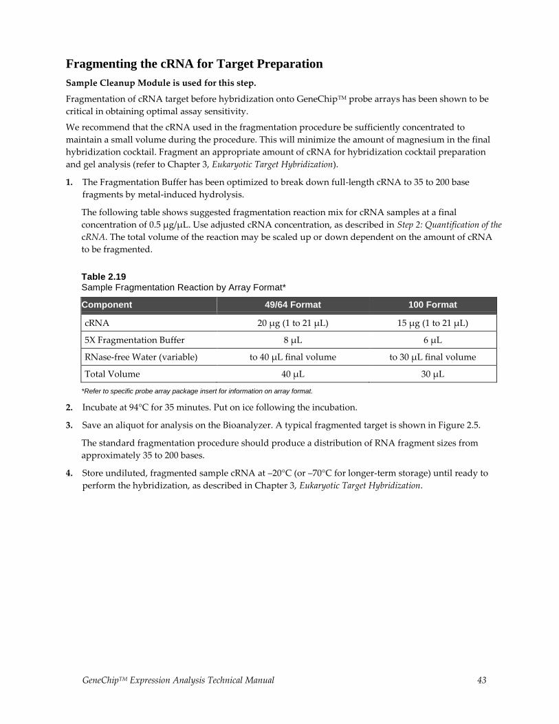
GeneChipTM Expression Analysis Technical Manual 43
Fragmenting the cRNA for Target Preparation
Sample Cleanup Module is used for this step.
Fragmentation of cRNA target before hybridization onto GeneChipTM probe arrays has been shown to be
critical in obtaining optimal assay sensitivity.
We recommend that the cRNA used in the fragmentation procedure be sufficiently concentrated to
maintain a small volume during the procedure. This will minimize the amount of magnesium in the final
hybridization cocktail. Fragment an appropriate amount of cRNA for hybridization cocktail preparation
and gel analysis (refer to Chapter 3, Eukaryotic Target Hybridization).
1. The Fragmentation Buffer has been optimized to break down full-length cRNA to 35 to 200 base
fragments by metal-induced hydrolysis.
The following table shows suggested fragmentation reaction mix for cRNA samples at a final
concentration of 0.5 μg/μL. Use adjusted cRNA concentration, as described in Step 2: Quantification of the
cRNA. The total volume of the reaction may be scaled up or down dependent on the amount of cRNA
to be fragmented.
Table 2.19 Sample Fragmentation Reaction by Array Format*
Component 49/64 Format 100 Format
cRNA 20 µg (1 to 21 µL) 15 µg (1 to 21 µL)
5X Fragmentation Buffer 8 µL 6 µL
RNase-free Water (variable) to 40 µL final volume to 30 µL final volume
Total Volume 40 µL 30 µL
*Refer to specific probe array package insert for information on array format.
2. Incubate at 94°C for 35 minutes. Put on ice following the incubation.
3. Save an aliquot for analysis on the Bioanalyzer. A typical fragmented target is shown in Figure 2.5.
The standard fragmentation procedure should produce a distribution of RNA fragment sizes from
approximately 35 to 200 bases.
4. Store undiluted, fragmented sample cRNA at –20°C (or –70°C for longer-term storage) until ready to
perform the hybridization, as described in Chapter 3, Eukaryotic Target Hybridization.

44 GeneChipTM Expression Analysis Technical Manual
Figure 2.5
Fragmented cRNA. Bioanalyzer electropherogram for fragmented labeled cRNA from HeLa total RNA.
This electropherogram displays the nucleotide size distribution for 150 ng of fragmented labeled cRNA
resulting from one round of amplification. The average size is approximately 100 nt.
GeneChipTM Expression Analysis Technical Manual 45
Chapter 3 Eukaryotic Target Hybridization
This Chapter Contains:
Detailed steps for preparing the eukaryotic hybridization mix containing labeled target and control
cRNA.
Instructions for hybridizing the target mix to a eukaryotic GeneChipTM probe array.
After completing the procedures described in this chapter, the hybridized probe array is ready for washing,
staining, and scanning, as detailed in the GeneChipTM Expression Wash, Stain and Scan User Manual, P/N
702731.
Reagents and Materials Required
The following reagents and materials have been tested and evaluated by our scientists. Information and
part numbers listed are based on U.S. catalog information. For supplier information, refer to the Supplier
Reference List in Appendix G of this manual.
GeneChipTM Hybridization, Wash and Stain Kit: P/N 900720 (30 reactions)
Hybridization Module from Box 1:
Pre-Hybridization Mix
2X Hybridization Mix
DMSO
Nuclease-free Water
GeneChipTM Eukaryotic Hybridization Control Kit: P/N 900454 (30 reactions) or P/N 900457 (150
reactions), both contain Control cRNA and Control Oligo B2
Control Oligo B2, 3 nM: P/N 900301
Miscellaneous Supplies
Hybridization Oven 640: P/N 800138 (110V) or 800139 (220V)
Sterile, RNase-free, microcentrifuge vials, 1.5 mL: USA Scientific, P/N 1415-2600 (or equivalent)
Micropipettors, (P-2, P-20, P-200, P-1000): Rainin Pipetman (or equivalent)
Sterile-barrier pipette tips and non-barrier pipette tips
Heatblock
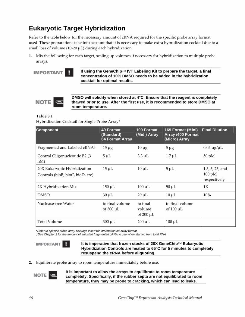
46 GeneChipTM Expression Analysis Technical Manual
Eukaryotic Target Hybridization
Refer to the table below for the necessary amount of cRNA required for the specific probe array format
used. These preparations take into account that it is necessary to make extra hybridization cocktail due to a
small loss of volume (10-20 μL) during each hybridization.
1. Mix the following for each target, scaling up volumes if necessary for hybridization to multiple probe
arrays.
If using the GeneChipTM IVT Labeling Kit to prepare the target, a final
concentration of 10% DMSO needs to be added in the hybridization cocktail for optimal results.
DMSO will solidify when stored at 4°C. Ensure that the reagent is completely thawed prior to use. After the first use, it is recommended to store DMSO at room temperature.
Table 3.1
Hybridization Cocktail for Single Probe Array*
Component 49 Format (Standard) 64 Format Array
100 Format (Midi) Array
169 Format (Mini) Array /400 Format (Micro) Array
Final Dilution
Fragmented and Labeled cRNA† 15 µg 10 µg 5 µg 0.05 µg/µL
Control Oligonucleotide B2 (3
nM)
5 µL 3.3 µL 1.7 µL 50 pM
20X Eukaryotic Hybridization
Controls (bioB, bioC, bioD, cre)
15 µL 10 µL 5 µL 1.5, 5, 25, and
100 pM
respectively
2X Hybridization Mix 150 µL 100 µL 50 µL 1X
DMSO 30 µL 20 µL 10 µL 10%
Nuclease-free Water to final volume of 300 µL
to final
volume of 200 µL
to final volume of 100 µL
Total Volume 300 µL 200 µL 100 µL
*Refer to specific probe array package insert for information on array format. †See Chapter 2 for the amount of adjusted fragmented cRNA to use when starting from total RNA.
It is imperative that frozen stocks of 20X GeneChipTM Eukaryotic
Hybridization Controls are heated to 65°C for 5 minutes to completely resuspend the cRNA before aliquoting.
2. Equilibrate probe array to room temperature immediately before use.
It is important to allow the arrays to equilibrate to room temperature completely. Specifically, if the rubber septa are not equilibrated to room temperature, they may be prone to cracking, which can lead to leaks.
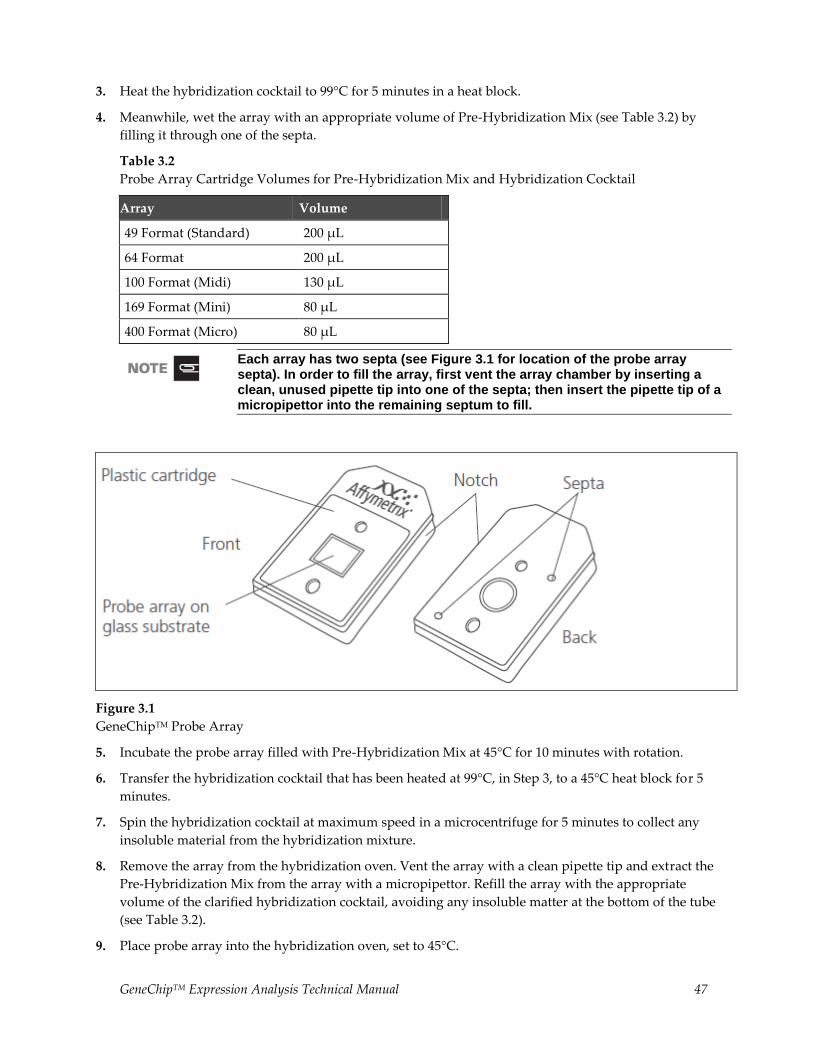
GeneChipTM Expression Analysis Technical Manual 47
3. Heat the hybridization cocktail to 99°C for 5 minutes in a heat block.
4. Meanwhile, wet the array with an appropriate volume of Pre-Hybridization Mix (see Table 3.2) by
filling it through one of the septa.
Table 3.2
Probe Array Cartridge Volumes for Pre-Hybridization Mix and Hybridization Cocktail
Array Volume
49 Format (Standard) 200 µL
64 Format 200 µL
100 Format (Midi) 130 µL
169 Format (Mini) 80 µL
400 Format (Micro) 80 µL
Each array has two septa (see Figure 3.1 for location of the probe array septa). In order to fill the array, first vent the array chamber by inserting a clean, unused pipette tip into one of the septa; then insert the pipette tip of a micropipettor into the remaining septum to fill.
Figure 3.1
GeneChipTM Probe Array
5. Incubate the probe array filled with Pre-Hybridization Mix at 45°C for 10 minutes with rotation.
6. Transfer the hybridization cocktail that has been heated at 99°C, in Step 3, to a 45°C heat block for 5
minutes.
7. Spin the hybridization cocktail at maximum speed in a microcentrifuge for 5 minutes to collect any
insoluble material from the hybridization mixture.
8. Remove the array from the hybridization oven. Vent the array with a clean pipette tip and extract the
Pre-Hybridization Mix from the array with a micropipettor. Refill the array with the appropriate
volume of the clarified hybridization cocktail, avoiding any insoluble matter at the bottom of the tube
(see Table 3.2).
9. Place probe array into the hybridization oven, set to 45°C.

48 GeneChipTM Expression Analysis Technical Manual
10. To avoid stress to the motor, load probe arrays in a balanced configuration around the axis. Rotate at 60
rpm.
11. Hybridize for 16 hours. During the latter part of the 16-hour hybridization, proceed to the GeneChipTM
Expression Wash, Stain and Scan User Manual, P/N 702731, to prepare reagents for the washing and
staining steps required immediately after completion of hybridization.
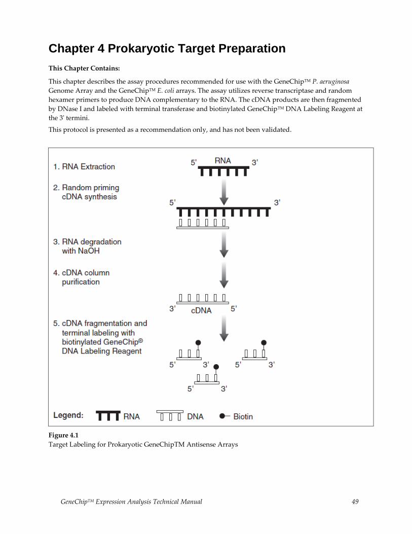
GeneChipTM Expression Analysis Technical Manual 49
Chapter 4 Prokaryotic Target Preparation
This Chapter Contains:
This chapter describes the assay procedures recommended for use with the GeneChipTM P. aeruginosa
Genome Array and the GeneChipTM E. coli arrays. The assay utilizes reverse transcriptase and random
hexamer primers to produce DNA complementary to the RNA. The cDNA products are then fragmented
by DNase I and labeled with terminal transferase and biotinylated GeneChipTM DNA Labeling Reagent at
the 3' termini.
This protocol is presented as a recommendation only, and has not been validated.
Figure 4.1
Target Labeling for Prokaryotic GeneChipTM Antisense Arrays
50 GeneChipTM Expression Analysis Technical Manual
Reagents and Materials Required
The following reagents and materials are recommendations and have been tested and evaluated by our
scientists. Information and part numbers listed are based on U.S. catalog information. For supplier
information, refer to the Supplier Reference List in Appendix G of this manual.
Labeling
dNTP: Invitrogen Life Technologies, P/N 18427-013
Random Primers, 3 μg/μL: Invitrogen Life Technologies, P/N 48190-011
GeneChipTM Eukaryotic Poly-A RNA Control Kit: P/N 900433
SuperScript II™ Reverse Transcriptase: Invitrogen Life Technologies, P/N 18064-071
SUPERase•In™: Ambion, P/N 2696
Nuclease-free Water: Ambion, P/N 9930
NaOH, 1N solution: VWR Scientific Products, P/N MK469360
HCl, 1N solution: VWR Scientific Products, P/N MK638860
MinElute PCR Purification Kit: QIAGEN, P/N 28004
10X DNase I Buffer: USB, P/N 78331
Deoxyribonuclease I (DNase I): Pierce, P/N 89835
GeneChipTM DNA Labeling Reagent: P/N 900542
Terminal Deoxynucleotidyl Transferase: Promega, P/N M1875
EDTA, 0.5M, pH 8.0: Invitrogen Life Technologies, P/N 15575-020
Non-stick RNase-free microfuge tubes, 0.5 mL and 1.5 mL: Ambion, P/N 12350 and P/N 12450,
respectively
Gel-Shift Assay
Novex XCell SureLock™ Mini-Cell: Invitrogen Life Technologies, P/N EI0001
4-20% TBE Gel, 1.0 mm, 12 well: Invitrogen Life Technologies, P/N EC62252
Sucrose Gel Loading Dye, 5X: Amresco, P/N E-274
10X TBE Running Buffer
SYBR Gold: Molecular Probes, P/N S-11494
10 bp and 100 bp DNA ladder: Invitrogen Life Technologies, P/N 10821-015 and 15628-019, respectively
ImmunoPure NeutrAvidin: Pierce Chemical, P/N 31000
1M Tris, pH 7.0: Ambion, P/N 9850G
PBS, pH 7.2: Invitrogen Life Technologies, P/N 20012-027
GeneChipTM Expression Analysis Technical Manual 51
Reagent Preparation
75 ng/μL Random Primers
For 1000 μL:
25 μL of 3 μg/μL Random Primers
975 μL of Nuclease-free H2O
Store at –20°C in a non-frost-free freezer.
2 mg/mL NeutrAvidin
Resuspend 10 mg NeutrAvidin in 5 mL PBS solution. Store at 4°C.
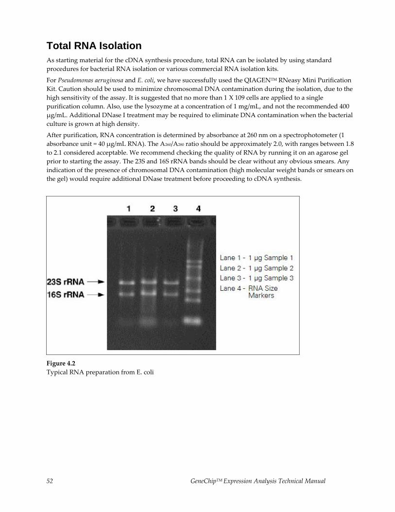
52 GeneChipTM Expression Analysis Technical Manual
Total RNA Isolation
As starting material for the cDNA synthesis procedure, total RNA can be isolated by using standard
procedures for bacterial RNA isolation or various commercial RNA isolation kits.
For Pseudomonas aeruginosa and E. coli, we have successfully used the QIAGENTM RNeasy Mini Purification
Kit. Caution should be used to minimize chromosomal DNA contamination during the isolation, due to the
high sensitivity of the assay. It is suggested that no more than 1 X 10⁹ cells are applied to a single
purification column. Also, use the lysozyme at a concentration of 1 mg/mL, and not the recommended 400
μg/mL. Additional DNase I treatment may be required to eliminate DNA contamination when the bacterial
culture is grown at high density.
After purification, RNA concentration is determined by absorbance at 260 nm on a spectrophotometer (1
absorbance unit = 40 μg/mL RNA). The A260/A280 ratio should be approximately 2.0, with ranges between 1.8
to 2.1 considered acceptable. We recommend checking the quality of RNA by running it on an agarose gel
prior to starting the assay. The 23S and 16S rRNA bands should be clear without any obvious smears. Any
indication of the presence of chromosomal DNA contamination (high molecular weight bands or smears on
the gel) would require additional DNase treatment before proceeding to cDNA synthesis.
Figure 4.2
Typical RNA preparation from E. coli
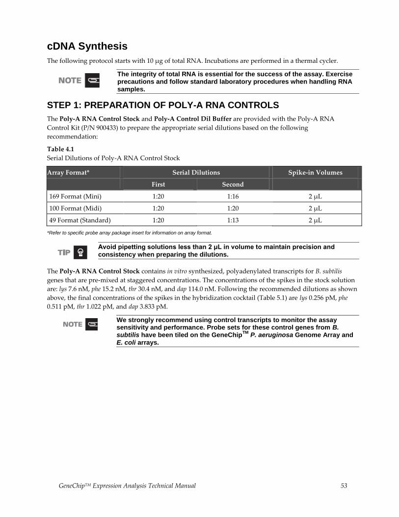
GeneChipTM Expression Analysis Technical Manual 53
cDNA Synthesis
The following protocol starts with 10 μg of total RNA. Incubations are performed in a thermal cycler.
The integrity of total RNA is essential for the success of the assay. Exercise precautions and follow standard laboratory procedures when handling RNA samples.
STEP 1: PREPARATION OF POLY-A RNA CONTROLS
The Poly-A RNA Control Stock and Poly-A Control Dil Buffer are provided with the Poly-A RNA
Control Kit (P/N 900433) to prepare the appropriate serial dilutions based on the following
recommendation:
Table 4.1
Serial Dilutions of Poly-A RNA Control Stock
Array Format* Serial Dilutions Spike-in Volumes
First Second
169 Format (Mini) 1:20 1:16 2 µL
100 Format (Midi) 1:20 1:20 2 µL
49 Format (Standard) 1:20 1:13 2 μL
*Refer to specific probe array package insert for information on array format.
Avoid pipetting solutions less than 2 μL in volume to maintain precision and consistency when preparing the dilutions.
The Poly-A RNA Control Stock contains in vitro synthesized, polyadenylated transcripts for B. subtilis
genes that are pre-mixed at staggered concentrations. The concentrations of the spikes in the stock solution
are: lys 7.6 nM, phe 15.2 nM, thr 30.4 nM, and dap 114.0 nM. Following the recommended dilutions as shown
above, the final concentrations of the spikes in the hybridization cocktail (Table 5.1) are lys 0.256 pM, phe
0.511 pM, thr 1.022 pM, and dap 3.833 pM.
We strongly recommend using control transcripts to monitor the assay sensitivity and performance. Probe sets for these control genes from B. subtilis have been tiled on the GeneChip
TM P. aeruginosa Genome Array and
E. coli arrays.
54 GeneChipTM Expression Analysis Technical Manual
For example, to prepare the poly-A RNA dilutions for a 100 format array:
Use non-stick RNase-free microfuge tubes to prepare all of the dilutions.
a. Add 2 μL of the Poly-A RNA Control Stock to 38 μL of Poly-A Control Dil Buffer for the First
Dilution (1:20).
b. Mix thoroughly and spin down to collect the liquid at the bottom of the tube.
c. Add 2 μL of the First Dilution to 38 μL of Poly-A Control Dil Buffer to prepare the Second
Dilution (1:20).
d. Mix thoroughly and spin down to collect the liquid at the bottom of the tube.
e. Add 2 μL of this Second Dilution to the total RNA as indicated in Table 4.2.
The First Dilution of the poly-A RNA controls can be stored up to six weeks in a non-frost-free freezer at –20°C and frozen-thawed up to eight times.
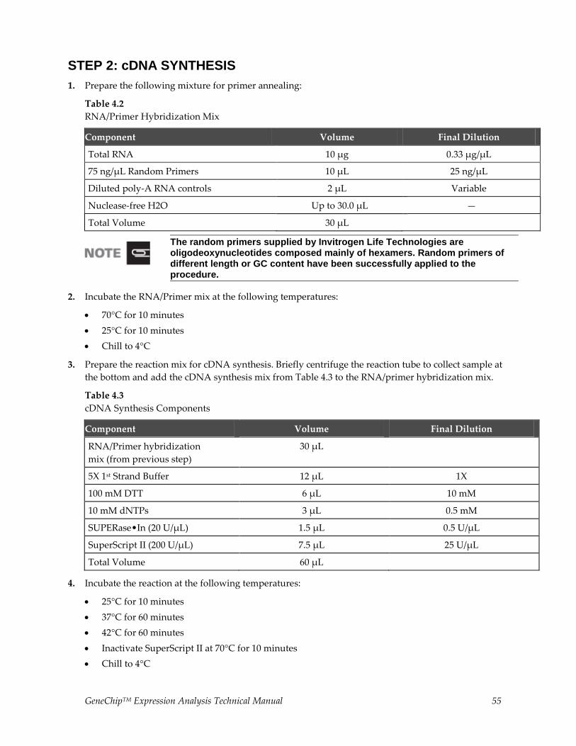
GeneChipTM Expression Analysis Technical Manual 55
STEP 2: cDNA SYNTHESIS
1. Prepare the following mixture for primer annealing:
Table 4.2
RNA/Primer Hybridization Mix
Component Volume Final Dilution
Total RNA 10 µg 0.33 µg/µL
75 ng/µL Random Primers 10 µL 25 ng/µL
Diluted poly-A RNA controls 2 µL Variable
Nuclease-free H2O Up to 30.0 µL —
Total Volume 30 µL
The random primers supplied by Invitrogen Life Technologies are oligodeoxynucleotides composed mainly of hexamers. Random primers of different length or GC content have been successfully applied to the procedure.
2. Incubate the RNA/Primer mix at the following temperatures:
70°C for 10 minutes
25°C for 10 minutes
Chill to 4°C
3. Prepare the reaction mix for cDNA synthesis. Briefly centrifuge the reaction tube to collect sample at
the bottom and add the cDNA synthesis mix from Table 4.3 to the RNA/primer hybridization mix.
Table 4.3
cDNA Synthesis Components
Component Volume Final Dilution
RNA/Primer hybridization
mix (from previous step)
30 µL
5X 1st Strand Buffer 12 µL 1X
100 mM DTT 6 µL 10 mM
10 mM dNTPs 3 µL 0.5 mM
SUPERase•In (20 U/μL) 1.5 µL 0.5 U/µL
SuperScript II (200 U/µL) 7.5 µL 25 U/µL
Total Volume 60 µL
4. Incubate the reaction at the following temperatures:
25°C for 10 minutes
37°C for 60 minutes
42°C for 60 minutes
Inactivate SuperScript II at 70°C for 10 minutes
Chill to 4°C
56 GeneChipTM Expression Analysis Technical Manual
STEP 3: REMOVAL OF RNA
1. Add 20 μL of 1N NaOH and incubate at 65°C for 30 minutes.
2. Add 20 μL of 1N HCl to neutralize.
STEP 4: PURIFICATION AND QUANTITATION OF cDNA
1. Use MinElute PCR Purification Columns to clean up the cDNA synthesis product (for detailed protocol,
see MinElute PCR Purification Kit Protocols provided by the supplier). Elute the product with 12 μL of
EB Buffer (supplied with the kit). The average volume of eluate is 11 μL from 12 μL of EB Buffer.
2. Quantify the purified cDNA product by 260 nm absorbance (1.0 A260 unit = 33 μg/mL of single-
stranded DNA).
Typical yields of cDNA are 3 to 7 μg. A minimum of 1.5 μg of cDNA is required for subsequent procedures to obtain sufficient material to hybridize onto the array and to perform necessary quality control experiments.
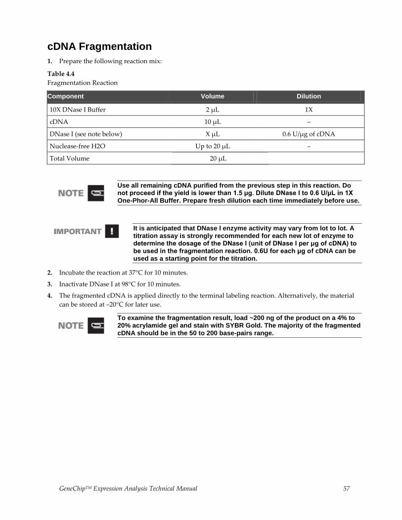
GeneChipTM Expression Analysis Technical Manual 57
cDNA Fragmentation
1. Prepare the following reaction mix:
Table 4.4
Fragmentation Reaction
Component Volume Dilution
10X DNase I Buffer 2 µL 1X
cDNA 10 µL –
DNase I (see note below) X µL 0.6 U/µg of cDNA
Nuclease-free H2O Up to 20 µL –
Total Volume 20 µL
Use all remaining cDNA purified from the previous step in this reaction. Do not proceed if the yield is lower than 1.5 μg. Dilute DNase I to 0.6 U/μL in 1X One-Phor-All Buffer. Prepare fresh dilution each time immediately before use.
It is anticipated that DNase I enzyme activity may vary from lot to lot. A titration assay is strongly recommended for each new lot of enzyme to determine the dosage of the DNase I (unit of DNase I per μg of cDNA) to be used in the fragmentation reaction. 0.6U for each μg of cDNA can be used as a starting point for the titration.
2. Incubate the reaction at 37°C for 10 minutes.
3. Inactivate DNase I at 98°C for 10 minutes.
4. The fragmented cDNA is applied directly to the terminal labeling reaction. Alternatively, the material
can be stored at –20°C for later use.
To examine the fragmentation result, load ~200 ng of the product on a 4% to 20% acrylamide gel and stain with SYBR Gold. The majority of the fragmented cDNA should be in the 50 to 200 base-pairs range.
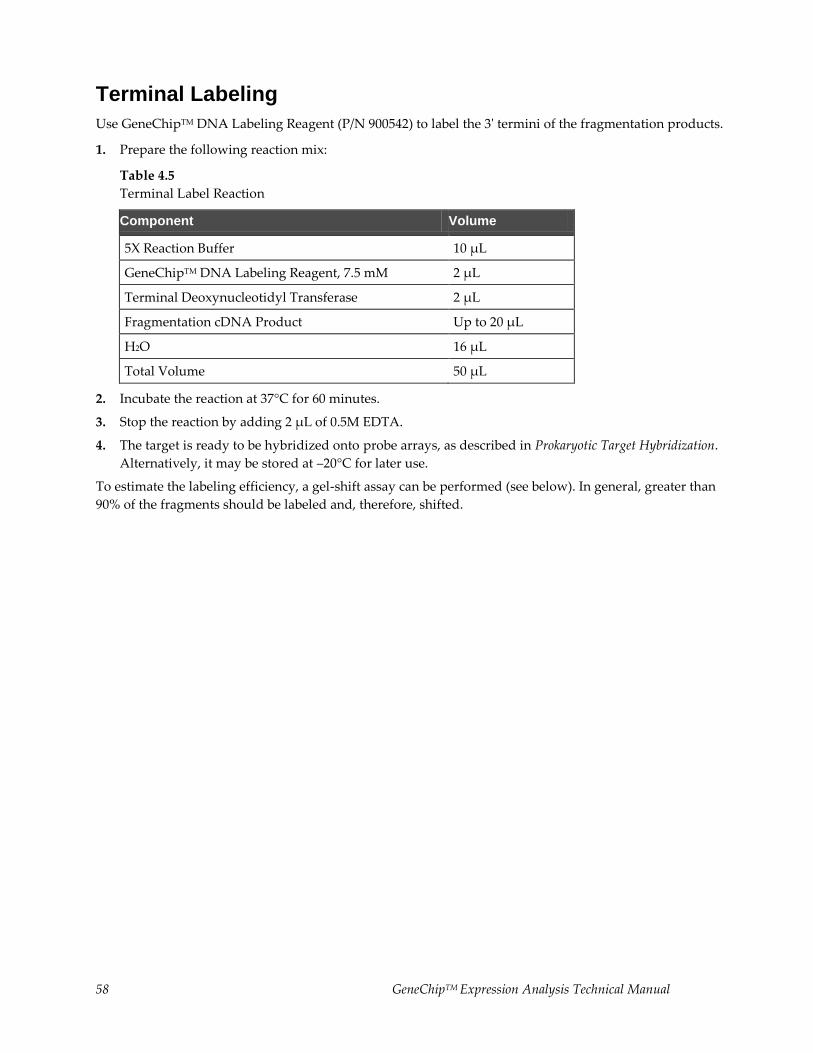
58 GeneChipTM Expression Analysis Technical Manual
Terminal Labeling
Use GeneChipTM DNA Labeling Reagent (P/N 900542) to label the 3' termini of the fragmentation products.
1. Prepare the following reaction mix:
Table 4.5
Terminal Label Reaction
Component Volume
5X Reaction Buffer 10 µL
GeneChipTM DNA Labeling Reagent, 7.5 mM 2 µL
Terminal Deoxynucleotidyl Transferase 2 µL
Fragmentation cDNA Product Up to 20 µL
H2O 16 µL
Total Volume 50 µL
2. Incubate the reaction at 37°C for 60 minutes.
3. Stop the reaction by adding 2 μL of 0.5M EDTA.
4. The target is ready to be hybridized onto probe arrays, as described in Prokaryotic Target Hybridization.
Alternatively, it may be stored at –20°C for later use.
To estimate the labeling efficiency, a gel-shift assay can be performed (see below). In general, greater than
90% of the fragments should be labeled and, therefore, shifted.
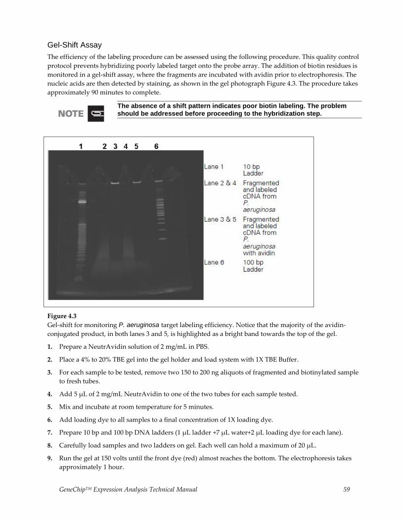
GeneChipTM Expression Analysis Technical Manual 59
Gel-Shift Assay
The efficiency of the labeling procedure can be assessed using the following procedure. This quality control
protocol prevents hybridizing poorly labeled target onto the probe array. The addition of biotin residues is
monitored in a gel-shift assay, where the fragments are incubated with avidin prior to electrophoresis. The
nucleic acids are then detected by staining, as shown in the gel photograph Figure 4.3. The procedure takes
approximately 90 minutes to complete.
The absence of a shift pattern indicates poor biotin labeling. The problem should be addressed before proceeding to the hybridization step.
Figure 4.3
Gel-shift for monitoring P. aeruginosa target labeling efficiency. Notice that the majority of the avidin-
conjugated product, in both lanes 3 and 5, is highlighted as a bright band towards the top of the gel.
1. Prepare a NeutrAvidin solution of 2 mg/mL in PBS.
2. Place a 4% to 20% TBE gel into the gel holder and load system with 1X TBE Buffer.
3. For each sample to be tested, remove two 150 to 200 ng aliquots of fragmented and biotinylated sample
to fresh tubes.
4. Add 5 μL of 2 mg/mL NeutrAvidin to one of the two tubes for each sample tested.
5. Mix and incubate at room temperature for 5 minutes.
6. Add loading dye to all samples to a final concentration of 1X loading dye.
7. Prepare 10 bp and 100 bp DNA ladders (1 μL ladder +7 μL water+2 μL loading dye for each lane).
8. Carefully load samples and two ladders on gel. Each well can hold a maximum of 20 μL.
9. Run the gel at 150 volts until the front dye (red) almost reaches the bottom. The electrophoresis takes
approximately 1 hour.

60 GeneChipTM Expression Analysis Technical Manual
10. While the gel is running, prepare at least 100 mL of a 1X solution of SYBR Gold for staining.
SYBR Gold is light sensitive. Therefore, use caution and shield the staining solution from light. Prepare a new batch of stain at least once a week.
11. After the gel is complete, break open cartridge and stain the gel in 1X SYBR Gold for 10 minutes.
12. Place the gel on the UV light box and produce an image following standard procedure. Be sure to use
the appropriate filter for SYBR Gold.

GeneChipTM Expression Analysis Technical Manual 61
Chapter 5 Prokaryotic Target Hybridization
This Chapter Contains:
This chapter contains detailed steps for preparing the hybridization mix, and instructions for hybridizing
the target mix to the GeneChipTM P. aeruginosa Genome Array and GeneChipTM E. coli arrays. The
hybridized probe array is then ready for washing, staining, and scanning as detailed in Chapter 6.
Reagents and Materials Required
The following reagents and materials have been tested and evaluated by our scientists. Information and
part numbers listed are based on U.S. catalog information. For supplier information, refer to the Supplier
Reference List in Appendix G of this manual.
GeneChipTM Hybridization, Wash and Stain Kit: P/N 900720 (30 reactions)
Hybridization Module, Box 1
2X Hybridization Mix
DMSO
Nuclease-free Water
Control Oligo B2, 3 nM: P/N 900301
Miscellaneous Supplies
Hybridization Oven 640: P/N 800138 (110V) or 800139 (220V)
Sterile, RNase-free, microcentrifuge vials, 1.5 mL: USA Scientific, P/N 1415-2600 (or equivalent)
Micropipettors, (P-2, P-20, P-200, P-1000): Rainin Pipetman (or equivalent)
Sterile-barrier pipette tips and non-barrier pipette tips
Prokaryotic Target Hybridization
After determining that the fragmented cDNA is labeled with biotin, prepare the hybridization cocktail. The
minimum amount of cDNA product required for target hybridization is 1 μg. The solution is stable for
approximately 6 to 8 hours at 4°C. The following protocol can be used for freshly prepared or frozen
hybridization cocktail. Re-use of prokaryotic sample has not been thoroughly tested and, therefore, is not
recommended.
1. Prepare the following hybridization cocktail.
DMSO will solidify when stored at 4°C. Ensure that the reagent is completely thawed prior to use. After the first use, it is recommended to store DMSO at room temperature.
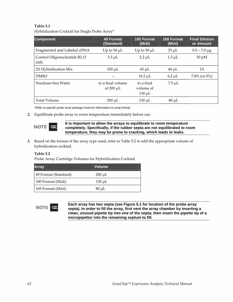
62 GeneChipTM Expression Analysis Technical Manual
Table 5.1
Hybridization Cocktail for Single Probe Array*
Component 49 Format (Standard)
100 Format (Midi)
169 Format (Mini)
Final Dilution or Amount
Fragmented and Labeled cDNA Up to 50 µL Up to 50 µL 25 µL 0.5 – 7.0 µg
Control Oligonucleotide B2 (3
nM)
3.3 µL 2.2 µL 1.3 µL 50 pM
2X Hybridization Mix 100 µL 65 µL 40 µL 1X
DMSO – 10.2 µL 6.2 µL 7.8% (or 0%)
Nuclease-free Water to a final volume
of 200 µL
to a final
volume of
130 µL
7.5 µL
Total Volume 200 µL 130 µL 80 µL
*Refer to specific probe array package insert for information on array format.
2. Equilibrate probe array to room temperature immediately before use.
It is important to allow the arrays to equilibrate to room temperature completely. Specifically, if the rubber septa are not equilibrated to room temperature, they may be prone to cracking, which leads to leaks.
3. Based on the format of the array type used, refer to Table 5.2 to add the appropriate volume of
hybridization cocktail.
Table 5.2
Probe Array Cartridge Volumes for Hybridization Cocktail
Array Volume
49 Format (Standard) 200 µL
100 Format (Midi) 130 µL
169 Format (Mini) 80 µL
Each array has two septa (see Figure 5.1 for location of the probe array septa). In order to fill the array, first vent the array chamber by inserting a clean, unused pipette tip into one of the septa; then insert the pipette tip of a micropipettor into the remaining septum to fill.
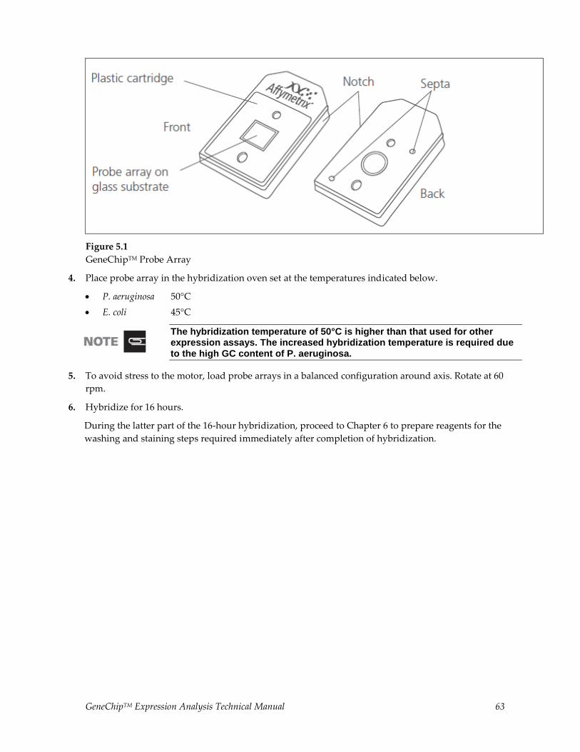
GeneChipTM Expression Analysis Technical Manual 63
Figure 5.1
GeneChipTM Probe Array
4. Place probe array in the hybridization oven set at the temperatures indicated below.
P. aeruginosa 50°C
E. coli 45°C
The hybridization temperature of 50°C is higher than that used for other expression assays. The increased hybridization temperature is required due to the high GC content of P. aeruginosa.
5. To avoid stress to the motor, load probe arrays in a balanced configuration around axis. Rotate at 60
rpm.
6. Hybridize for 16 hours.
During the latter part of the 16-hour hybridization, proceed to Chapter 6 to prepare reagents for the
washing and staining steps required immediately after completion of hybridization.
64 GeneChipTM Expression Analysis Technical Manual
Chapter 6 Prokaryotic Arrays: Washing, Staining and Scanning
This Chapter Contains:
Instructions for using the Fluidics Station 400 or 450/250 to automate the washing and staining of
GeneChipTM P. aeruginosa and GeneChipTM E. coli arrays.
Instructions for scanning probe arrays using the GeneArrayTM Scanner or the GeneChipTM Scanner 3000.
After completing the procedures described in this chapter, the scanned probe array image (.dat file) is
ready for analysis, as explained in the enclosed GeneChipTM Expression Analysis: Data Analysis Fundamentals
booklet (P/N 701190).
Reagents and Materials Required
The following reagents and materials have been tested and evaluated by our scientists. Information and
part numbers listed are based on U.S. catalog information. For supplier information, refer to the Supplier
Reference List in Appendix G of this manual.
GeneChipTM Hybridization, Wash and Stain Kit: P/N 900720 (30 reactions)
Stain Module, Box 1
Stain Cocktail 1
Stain Cocktail 2
Array Holding Buffer
Wash Buffers A and B, Box 2
Wash Buffer A (P/N 900721)
Wash Buffer B (P/N 900722)
RNase-free water
Miscellaneous Supplies
Fluidics Station 450: P/N 00-0079
GeneChipTM Scanner 3000: P/N 00-00212
Sterile, RNase-free, microcentrifuge vials, 1.5 mL: USA Scientific, P/N 1415-2600 (or equivalent)
Sterile, RNase-free, microcentrifuge vials, 1.5 mL, amber: USA Scientific, P/N 1615-5507 (or equivalent)
Micropipettors, (P-2, P-20, P-200, P-1000), Rainin Pipetman (or equivalent)
Sterile-barrier pipette tips and non-barrier pipette tips
Tygon Tubing, 0.04″ inner diameter: Cole-Parmer, P/N H-06418-04
Tough-Spots™, Label Dots: USA Scientific, P/N 9185-0000
Media Bottle, SQ, 1,000 mL: P/N 400119
Media Bottle, SQ, 500 mL (set of 3): P/N 400118
Bottle Cap, Pre-Drilled (set of 4): P/N 400137
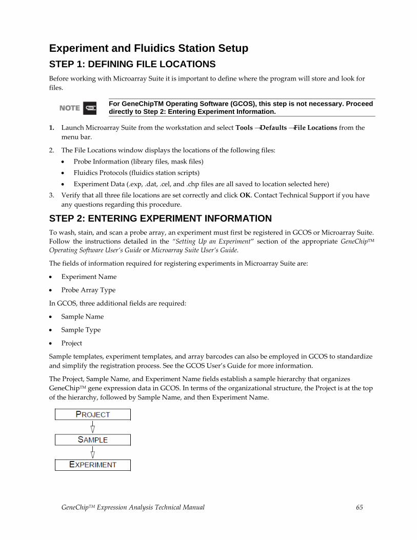
GeneChipTM Expression Analysis Technical Manual 65
Experiment and Fluidics Station Setup
STEP 1: DEFINING FILE LOCATIONS
Before working with Microarray Suite it is important to define where the program will store and look for
files.
For GeneChipTM Operating Software (GCOS), this step is not necessary. Proceed directly to Step 2: Entering Experiment Information.
1. Launch Microarray Suite from the workstation and select Tools → Defaults → File Locations from the
menu bar.
2. The File Locations window displays the locations of the following files:
Probe Information (library files, mask files)
Fluidics Protocols (fluidics station scripts)
Experiment Data (.exp, .dat, .cel, and .chp files are all saved to location selected here)
3. Verify that all three file locations are set correctly and click OK. Contact Technical Support if you have
any questions regarding this procedure.
STEP 2: ENTERING EXPERIMENT INFORMATION
To wash, stain, and scan a probe array, an experiment must first be registered in GCOS or Microarray Suite.
Follow the instructions detailed in the “Setting Up an Experiment” section of the appropriate GeneChipTM
Operating Software User’s Guide or Microarray Suite User’s Guide.
The fields of information required for registering experiments in Microarray Suite are:
Experiment Name
Probe Array Type
In GCOS, three additional fields are required:
Sample Name
Sample Type
Project
Sample templates, experiment templates, and array barcodes can also be employed in GCOS to standardize
and simplify the registration process. See the GCOS User’s Guide for more information.
The Project, Sample Name, and Experiment Name fields establish a sample hierarchy that organizes
GeneChipTM gene expression data in GCOS. In terms of the organizational structure, the Project is at the top
of the hierarchy, followed by Sample Name, and then Experiment Name.
66 GeneChipTM Expression Analysis Technical Manual
STEP 3: PREPARING THE FLUIDICS STATION
The Fluidics Station 400, or 450/250 is used to wash and stain the probe arrays. It is operated using
GCOS/Microarray Suite.
Setting Up the Fluidics Station
1. Turn on the Fluidics Station using the switch on the lower left side of the machine.
2. Select Run → Fluidics from the menu bar.
The Fluidics Station dialog box appears with a drop-down list for selecting the experiment name for
each of the fluidics station modules. A second list is accessed for choosing the Protocol for each of the
four fluidics station modules.
Refer to the appropriate GeneChipTM
Fluidics Station User’s Guide for instructions on connecting and addressing multiple fluidics stations.
Priming the Fluidics Station
Priming ensures that the lines of the fluidics station are filled with the appropriate buffers and the fluidics
station is ready for running fluidics station protocols.
Priming should be done:
When the fluidics station is first started
When wash solutions are changed
Before washing if a shutdown has been performed
If the LCD window instructs the user to prime
1. Select Protocol in the Fluidics Station dialog box.
2. Choose Prime or Prime_450 for the respective modules in the Protocol drop-down list.
3. Ensure that the designated Fluidics Station Wash A and Wash B media bottles are clean. Transfer Wash
Buffer A and Wash Buffer B from the kit to the clean, empty Fluidics Station bottles.
4. If using Microarray Suite, click Run for each module to begin priming. If using GCOS, select the All
Modules check box, then click Run.
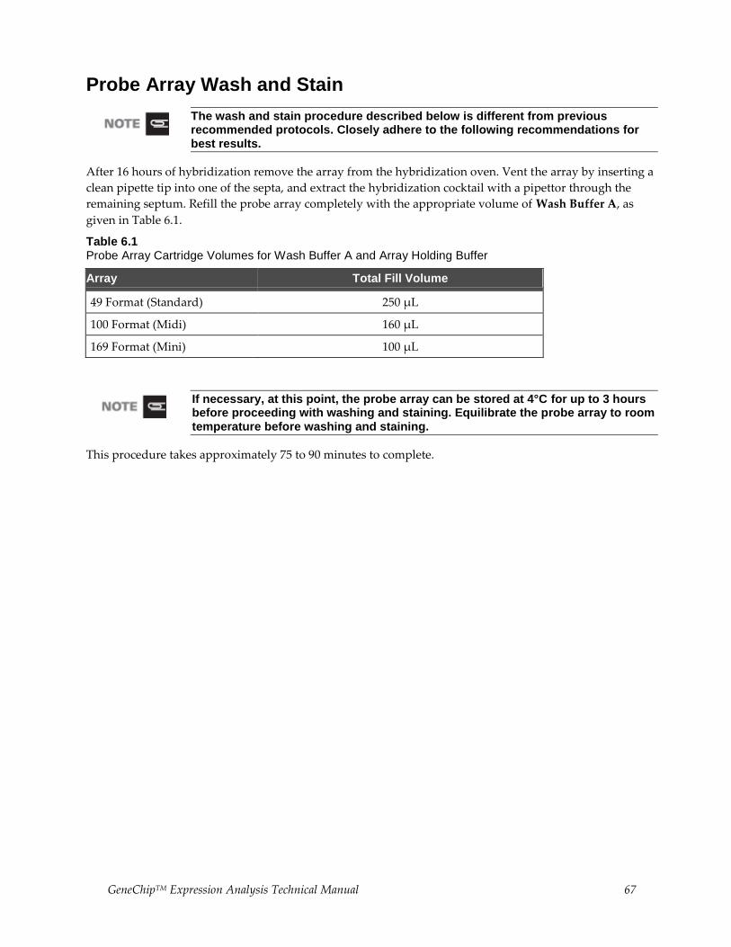
GeneChipTM Expression Analysis Technical Manual 67
Probe Array Wash and Stain
The wash and stain procedure described below is different from previous recommended protocols. Closely adhere to the following recommendations for best results.
After 16 hours of hybridization remove the array from the hybridization oven. Vent the array by inserting a
clean pipette tip into one of the septa, and extract the hybridization cocktail with a pipettor through the
remaining septum. Refill the probe array completely with the appropriate volume of Wash Buffer A, as
given in Table 6.1.
Table 6.1 Probe Array Cartridge Volumes for Wash Buffer A and Array Holding Buffer
Array Total Fill Volume
49 Format (Standard) 250 µL
100 Format (Midi) 160 µL
169 Format (Mini) 100 µL
If necessary, at this point, the probe array can be stored at 4°C for up to 3 hours before proceeding with washing and staining. Equilibrate the probe array to room temperature before washing and staining.
This procedure takes approximately 75 to 90 minutes to complete.
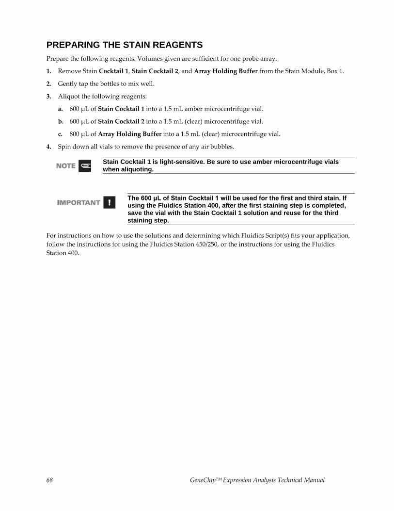
68 GeneChipTM Expression Analysis Technical Manual
PREPARING THE STAIN REAGENTS
Prepare the following reagents. Volumes given are sufficient for one probe array.
1. Remove Stain Cocktail 1, Stain Cocktail 2, and Array Holding Buffer from the Stain Module, Box 1.
2. Gently tap the bottles to mix well.
3. Aliquot the following reagents:
a. 600 μL of Stain Cocktail 1 into a 1.5 mL amber microcentrifuge vial.
b. 600 μL of Stain Cocktail 2 into a 1.5 mL (clear) microcentrifuge vial.
c. 800 μL of Array Holding Buffer into a 1.5 mL (clear) microcentrifuge vial.
4. Spin down all vials to remove the presence of any air bubbles.
Stain Cocktail 1 is light-sensitive. Be sure to use amber microcentrifuge vials when aliquoting.
The 600 μL of Stain Cocktail 1 will be used for the first and third stain. If using the Fluidics Station 400, after the first staining step is completed, save the vial with the Stain Cocktail 1 solution and reuse for the third staining step.
For instructions on how to use the solutions and determining which Fluidics Script(s) fits your application,
follow the instructions for using the Fluidics Station 450/250, or the instructions for using the Fluidics
Station 400.
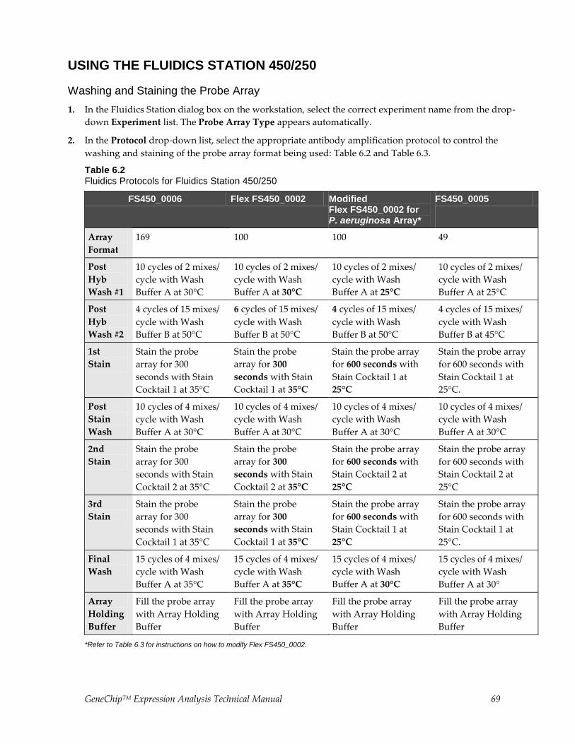
GeneChipTM Expression Analysis Technical Manual 69
USING THE FLUIDICS STATION 450/250
Washing and Staining the Probe Array
1. In the Fluidics Station dialog box on the workstation, select the correct experiment name from the drop-
down Experiment list. The Probe Array Type appears automatically.
2. In the Protocol drop-down list, select the appropriate antibody amplification protocol to control the
washing and staining of the probe array format being used: Table 6.2 and Table 6.3.
Table 6.2 Fluidics Protocols for Fluidics Station 450/250
FS450_0006 Flex FS450_0002 Modified Flex FS450_0002 for P. aeruginosa Array*
FS450_0005
Array
Format
169 100 100 49
Post
Hyb
Wash #1
10 cycles of 2 mixes/
cycle with Wash
Buffer A at 30°C
10 cycles of 2 mixes/
cycle with Wash
Buffer A at 30°C
10 cycles of 2 mixes/
cycle with Wash
Buffer A at 25°C
10 cycles of 2 mixes/
cycle with Wash
Buffer A at 25°C
Post
Hyb
Wash #2
4 cycles of 15 mixes/
cycle with Wash
Buffer B at 50°C
6 cycles of 15 mixes/
cycle with Wash
Buffer B at 50°C
4 cycles of 15 mixes/
cycle with Wash
Buffer B at 50°C
4 cycles of 15 mixes/
cycle with Wash
Buffer B at 45°C
1st
Stain
Stain the probe
array for 300
seconds with Stain
Cocktail 1 at 35°C
Stain the probe
array for 300
seconds with Stain
Cocktail 1 at 35°C
Stain the probe array
for 600 seconds with
Stain Cocktail 1 at
25°C
Stain the probe array
for 600 seconds with
Stain Cocktail 1 at
25°C.
Post
Stain
Wash
10 cycles of 4 mixes/
cycle with Wash
Buffer A at 30°C
10 cycles of 4 mixes/
cycle with Wash
Buffer A at 30°C
10 cycles of 4 mixes/
cycle with Wash
Buffer A at 30°C
10 cycles of 4 mixes/
cycle with Wash
Buffer A at 30°C
2nd
Stain
Stain the probe
array for 300
seconds with Stain
Cocktail 2 at 35°C
Stain the probe
array for 300
seconds with Stain
Cocktail 2 at 35°C
Stain the probe array
for 600 seconds with
Stain Cocktail 2 at
25°C
Stain the probe array
for 600 seconds with
Stain Cocktail 2 at
25°C
3rd
Stain
Stain the probe
array for 300
seconds with Stain
Cocktail 1 at 35°C
Stain the probe
array for 300
seconds with Stain
Cocktail 1 at 35°C
Stain the probe array
for 600 seconds with
Stain Cocktail 1 at
25°C
Stain the probe array
for 600 seconds with
Stain Cocktail 1 at
25°C.
Final
Wash
15 cycles of 4 mixes/
cycle with Wash
Buffer A at 35°C
15 cycles of 4 mixes/
cycle with Wash
Buffer A at 35°C
15 cycles of 4 mixes/
cycle with Wash
Buffer A at 30°C
15 cycles of 4 mixes/
cycle with Wash
Buffer A at 30°
Array
Holding
Buffer
Fill the probe array
with Array Holding
Buffer
Fill the probe array
with Array Holding
Buffer
Fill the probe array
with Array Holding
Buffer
Fill the probe array
with Array Holding
Buffer
*Refer to Table 6.3 for instructions on how to modify Flex FS450_0002.
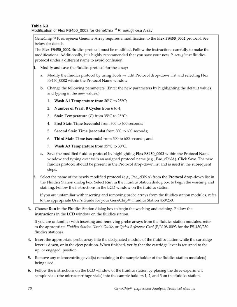
70 GeneChipTM Expression Analysis Technical Manual
Table 6.3 Modification of Flex FS450_0002 for GeneChip
TM P. aeruginosa Array
GeneChipTM P. aeruginosa Genome Array requires a modification to the Flex FS450_0002 protocol. See
below for details.
The Flex FS450_0002 fluidics protocol must be modified. Follow the instructions carefully to make the
modifications. Additionally, it is highly recommended that you save your new P. aeruginosa fluidics
protocol under a different name to avoid confusion.
1. Modify and save the fluidics protocol for the assay:
a. Modify the fluidics protocol by using Tools → Edit Protocol drop-down list and selecting Flex
FS450_0002 within the Protocol Name window.
b. Change the following parameters: (Enter the new parameters by highlighting the default values
and typing in the new values.)
1. Wash A1 Temperature from 30°C to 25°C;
2. Number of Wash B Cycles from 6 to 4;
3. Stain Temperature (C) from 35°C to 25°C;
4. First Stain Time (seconds) from 300 to 600 seconds;
5. Second Stain Time (seconds) from 300 to 600 seconds;
6. Third Stain Time (seconds) from 300 to 600 seconds; and
7. Wash A3 Temperature from 35°C to 30°C.
c. Save the modified fluidics protocol by highlighting Flex FS450_0002 within the Protocol Name
window and typing over with an assigned protocol name (e.g., Pae_cDNA). Click Save. The new
fluidics protocol should be present in the Protocol drop-down list and is used in the subsequent
steps.
2. Select the name of the newly modified protocol (e.g., Pae_cDNA) from the Protocol drop-down list in
the Fluidics Station dialog box. Select Run in the Fluidics Station dialog box to begin the washing and
staining. Follow the instructions in the LCD window on the fluidics station.
If you are unfamiliar with inserting and removing probe arrays from the fluidics station modules, refer
to the appropriate User’s Guide for your GeneChipTM Fluidics Station 450/250.
3. Choose Run in the Fluidics Station dialog box to begin the washing and staining. Follow the
instructions in the LCD window on the fluidics station.
If you are unfamiliar with inserting and removing probe arrays from the fluidics station modules, refer
to the appropriate Fluidics Station User’s Guide, or Quick Reference Card (P/N 08-0093 for the FS-450/250
fluidics stations).
4. Insert the appropriate probe array into the designated module of the fluidics station while the cartridge
lever is down, or in the eject position. When finished, verify that the cartridge lever is returned to the
up, or engaged, position.
5. Remove any microcentrifuge vial(s) remaining in the sample holder of the fluidics station module(s)
being used.
6. Follow the instructions on the LCD window of the fluidics station by placing the three experiment
sample vials (the microcentrifuge vials) into the sample holders 1, 2, and 3 on the fluidics station.
GeneChipTM Expression Analysis Technical Manual 71
a. Place one vial containing 600 μL Stain Cocktail 1 in sample holder 1.
b. Place one vial containing 600 μL Stain Cocktail 2 in sample holder 2.
c. Place one vial containing 800 μL of Array Holding Buffer in sample holder 3.
d. Press down on the needle lever to snap needles into position and to start the run.
The run begins. The Fluidics Station dialog box at the workstation terminal and the LCD window
display the status of the washing and staining as the protocol progresses.
7. When the protocol is complete, the LCD window displays the message EJECT & INSPECT
CARTRIDGE.
8. Remove the probe arrays from the fluidics station modules by first pressing down the cartridge lever to
the eject position.
9. Check the probe array window for large bubbles or air pockets.
If the probe array has no large bubbles, it is ready to scan on the GeneArrayTM Scanner, or the
GeneChipTM Scanner 3000. Pull up on the cartridge lever to close the washblock and proceed to
Probe Array Scan.
If bubbles are present, do the following:
Return the probe array to the probe array holder. Follow instructions on the LCD window. Engage
the washblock by gently pushing up on the cartridge lever to the engaged, or closed, position.
The fluidics station will drain the probe array and then fill it with a fresh volume of Array Holding
Buffer. When it is finished, the LCD window will display EJECT AND INSPECT CARTRIDGE.
Again, remove the probe array and inspect it for bubbles. If no bubbles are present, it is ready to
scan. Pull up on the cartridge lever to close the washblock and proceed to Probe Array Scan.
If attempt to fill the probe array without bubbles is unsuccessful, the array should be filled
manually with Array Holding Buffer using a micropipette. Excessive washing will result in a loss
of signal intensity.
10. Keep the probe arrays at 4°C and in the dark until ready for scanning.
11. If there are no more samples to hybridize, shut down the fluidics station following the procedure in
Shutting Down the Fluidics Station.
For proper cleaning and maintenance of the fluidics station, including the bleach protocol, refer to the appropriate GeneChip
TM Fluidics Station User’s Guide for
instructions.
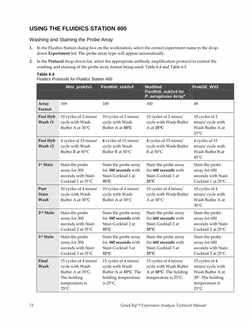
72 GeneChipTM Expression Analysis Technical Manual
USING THE FLUIDICS STATION 400
Washing and Staining the Probe Array
1. In the Fluidics Station dialog box on the workstation, select the correct experiment name in the drop-
down Experiment list. The probe array type will appear automatically.
2. In the Protocol drop-down list, select the appropriate antibody amplification protocol to control the
washing and staining of the probe array format being used: Table 6.4 and Table 6.5.
Table 6.4 Fluidics Protocols for Fluidics Station 400
Mini_prok2v1 FlexMidi_euk2v3 Modified FlexMidi_euk2v3 for P. aeruginosa Array*
ProkGE_WS2
Array
Format
169 100 100 49
Post Hyb
Wash #1
10 cycles of 2 mixes/
cycle with Wash
Buffer A at 30°C
10 cycles of 2 mixes/
cycle with Wash
Buffer A at 30°C
10 cycles of 2 mixes/
cycle with Wash Buffer
A at 25°C
10 cycles of 2
mixes/ cycle with
Wash Buffer A at
25°C
Post Hyb
Wash #2
4 cycles of 15 mixes/
cycle with Wash
Buffer B at 50°C
6 cycles of 15 mixes/
cycle with Wash
Buffer B at 50°C
4 cycles of 15 mixes/
cycle with Wash Buffer
B at 50°C
4 cycles of 15
mixes/ cycle with
Wash Buffer B at
45°C
1st Stain Stain the probe
array for 300
seconds with Stain
Cocktail 1 at 35°C
Stain the probe array
for 300 seconds with
Stain Cocktail 1 at
35°C
Stain the probe array
for 600 seconds with
Stain Cocktail 1 at
25°C
Stain the probe
array for 600
seconds with Stain
Cocktail 1 at 25°C.
Post
Stain
Wash
10 cycles of 4 mixes/
cycle with Wash
Buffer A at 30°C
10 cycles of 4 mixes/
cycle with Wash
Buffer A at 30°C
10 cycles of 4 mixes/
cycle with Wash Buffer
A at 30°C
10 cycles of 4
mixes/ cycle with
Wash Buffer A at
30°C
2nd Stain Stain the probe
array for 300
seconds with Stain
Cocktail 2 at 35°C
Stain the probe array
for 300 seconds with
Stain Cocktail 2 at
35°C
Stain the probe array
for 600 seconds with
Stain Cocktail 2 at
25°C
Stain the probe
array for 600
seconds with Stain
Cocktail 2 at 25°C
3rd Stain Stain the probe
array for 300
seconds with Stain
Cocktail 3 at 35°C
Stain the probe array
for 300 seconds with
Stain Cocktail 3 at
35°C
Stain the probe array
for 600 seconds with
Stain Cocktail 3 at
25°C
Stain the probe
array for 600
seconds with Stain
Cocktail 3 at 25°C
Final
Wash
15 cycles of 4 mixes/
cycle with Wash
Buffer A at 35°C.
The holding
temperature is
25°C.
15 cycles of 4 mixes/
cycle with Wash
Buffer A at 35°C. The
holding temperature
is 25°C.
15 cycles of 4 mixes/
cycle with Wash Buffer
A at 30°C. The holding
temperature is 25°C.
15 cycles of 4
mixes/ cycle with
Wash Buffer A at
30°. The holding
temperature is
25°C.
GeneChipTM Expression Analysis Technical Manual 73
Array
Holding
Buffer
N/A - manual
process
N/A - manual
process
N/A - manual process N/A - manual
process
*Refer to Table 6.5 for instructions on how to modify FlexMidi_euk2v3.
Table 6.5 Modification of FlexMidi_euk2v3 for GeneChip
TM P. aeruginosa Array
GeneChipTM P. aeruginosa Genome Array requires a modification to the FlexMidi_euk2v3 protocol. See
below for details.
The FlexMidi_euk2v3 fluidics protocol must be modified. Follow the instructions carefully to make the
modifications. Additionally, it is highly recommended that you save your new P. aeruginosa fluidics
protocol under a different name to avoid confusion.
1. Modify and save the fluidics protocol for the assay:
a. Modify the fluidics protocol by using Tools → Edit Protocol drop-down list and selecting
FlexMidi_euk2v3 within the Protocol Name window.
b. Change the following parameters: (Enter the new parameters by highlighting the default values
and typing in the new values.)
1. Wash A1 Temperature from 30°C to 25°C;
2. Number of Wash B Cycles from 6 to 4;
3. Stain Temperature (C) from 35°C to 25°C;
4. First Stain Time (seconds) from 300 to 600 seconds;
5. Second Stain Time (seconds) from 300 to 600 seconds;
6. Third Stain Time (seconds) from 300 to 600 seconds; and
7. Wash A3 Temperature from 35°C to 30°C.
c. Save the modified fluidics protocol by highlighting FlexMidi_euk2v3 within the Protocol Name
window and typing over with an assigned protocol name (e.g., Pae_cDNA). Click Save. The new
fluidics protocol should be present in the Protocol drop-down list and is used in the subsequent
steps.
2. Select the name of the newly modified protocol (e.g., Pae_cDNA) from the Protocol drop-down list in
the Fluidics Station dialog box. Select Run in the Fluidics Station dialog box to begin the washing and
staining. Follow the instructions in the LCD window on the fluidics station.
If you are unfamiliar with inserting and removing probe arrays from the fluidics station modules, refer
to the appropriate User’s Guide for your GeneChipTM Fluidics Station 400.
3. Choose Run in the Fluidics Station dialog box to begin the washing and staining. Follow the
instructions on the LCD window on the fluidics station.
If you are unfamiliar with inserting and removing probe arrays from the fluidics station modules, refer
to the Fluidics Station 400 User’s Guide, Fluidics Station 400 Video In-Service CD (P/N 900374), or Quick
Reference Card (P/N 08-0072).
4. Insert the appropriate probe array into the designated module of the fluidics station while the cartridge
lever is in the EJECT position. When finished, verify that the cartridge lever is returned to the ENGAGE
position.
74 GeneChipTM Expression Analysis Technical Manual
5. Remove any microcentrifuge tube remaining in the sample holder of the fluidics station module(s)
being used.
6. When the LCD window indicates LOAD 1ST STAIN, place a microcentrifuge tube containing 600 μL of
Stain Cocktail 1 into the sample holder, making sure that the metal sampling needle is in the tube with
its tip near the bottom.
The Fluidics Station dialog box and the LCD window display the status of the washing and staining as
they progress.
7. When the LCD window indicates LOAD 2ND STAIN, replace the microcentrifuge vial containing the
Stain Cocktail 1 with a microcentrifuge vial containing Stain Cocktail 2 into the sample holder,
making sure that the metal sampling needle is in the vial with its tip near the bottom.
Do not dispose the vial containing Stain Cocktail 1. Be sure to save the Stain Cocktail 1 vial for use in Step 8.
8. When the LCD window indicates LOAD 3RD STAIN, replace the microcentrifuge vial containing
Stain Cocktail 2 with the saved microcentrifuge vial containing 600 μL of Stain Cocktail 1 (retained
from the previous step) into the sample holder. Verify that the metal sampling needle is in the vial with
its tip near the bottom.
The Fluidics Station dialog box and the LCD window display the status of the washing and staining as
they progress. When the wash is complete, the LCD window displays the message EJECT
CARTRIDGE.
9. Remove microcentrifuge vial containing stain and replace with an empty microcentrifuge tube.
10. Remove the probe arrays from the fluidics station modules by first moving the cartridge lever to the
EJECT position.
11. Fill the probe array manually with Array Holding Buffer as follows:
a. Vent one of the septa with a clean pipette tip. Drain the Wash Buffer A from the remaining septum
of the probe array using a micropipettor. Refill the array completely with Array Holding Buffer.
Refer to Table 6.1 for the fill volume specific to the array type used.
b. Inspect the array and ensure that no bubbles are present. The probe array is ready to be scanned on
the GeneChipTM Scanner 3000 or the GeneArrayTM Scanner.
c. Pull up on the cartridge lever to engage wash board and proceed to Probe Array Scan.
If you do not scan the arrays right away, keep the probe arrays at 4°C and in the dark until ready for
scanning.
If there are no more samples to hybridize, shut down the fluidics station following the procedure
outlined in the section, Shutting Down the Fluidics Station.
For proper cleaning and maintenance of the fluidics station, including the bleach protocol, refer to the appropriate GeneChip
TM Fluidics Station User’s Guide for
instructions.
GeneChipTM Expression Analysis Technical Manual 75
Probe Array Scan
The scanner is also controlled by Microarray Suite or GCOS. The probe array is scanned after the wash
protocols are complete. Make sure the laser is warmed up prior to scanning by turning it on at least 15
minutes before use if you are using the Agilent GeneArrayTM Scanner, or 10 minutes if you are using the
GeneChipTM Scanner 3000. If the probe array was stored at 4°C, warm to room temperature before scanning.
Refer to the Microarray Suite or GCOS online help and the appropriate scanner user’s manual for more
information on scanning.
The scanner uses a laser and is equipped with a safety interlock system. Defeating the interlock system may result in exposure to hazardous laser light.
You must have read and be familiar with the operation of the scanner before attempting to scan a probe array. Refer to the Microarray Suite User’s Guide (P/N
08-0081), GeneChipTM Operating Software User’s Guide (P/N 701439), or to the
GeneChipTM
Scanner 3000 Quick Reference Card (P/N 08-0075).
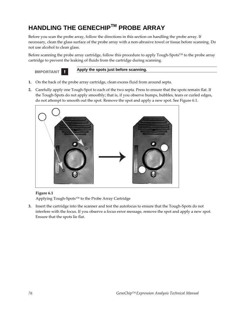
76 GeneChipTM Expression Analysis Technical Manual
HANDLING THE GENECHIPTM
PROBE ARRAY
Before you scan the probe array, follow the directions in this section on handling the probe array. If
necessary, clean the glass surface of the probe array with a non-abrasive towel or tissue before scanning. Do
not use alcohol to clean glass.
Before scanning the probe array cartridge, follow this procedure to apply Tough-Spots™ to the probe array
cartridge to prevent the leaking of fluids from the cartridge during scanning.
Apply the spots just before scanning.
1. On the back of the probe array cartridge, clean excess fluid from around septa.
2. Carefully apply one Tough-Spot to each of the two septa. Press to ensure that the spots remain flat. If
the Tough-Spots do not apply smoothly; that is, if you observe bumps, bubbles, tears or curled edges,
do not attempt to smooth out the spot. Remove the spot and apply a new spot. See Figure 6.1.
Figure 6.1
Applying Tough-Spots™ to the Probe Array Cartridge
3. Insert the cartridge into the scanner and test the autofocus to ensure that the Tough-Spots do not
interfere with the focus. If you observe a focus error message, remove the spot and apply a new spot.
Ensure that the spots lie flat.
GeneChipTM Expression Analysis Technical Manual 77
SCANNING THE PROBE ARRAY
1. Select Run → Scanner from the menu bar. Alternatively, click the Start Scan icon in the tool bar.
The Scanner dialog box appears with a drop-down list of experiments that have not been run.
2. Select the experiment name that corresponds to the probe array to be scanned.
A previously run experiment can also be selected by using the Include Scanned Experiments option
box. After selecting this option, previously scanned experiments appear in the drop-down list.
3. By default, for the GeneArrayTM Scanner only, after selecting the experiment the number [2] is
displayed in the Number of Scans box to perform the recommended 2X image scan. For the
GeneChipTM Scanner 3000, only one scan is required.
4. Once the experiment has been selected, click the Start button.
A dialog box prompts you to load a sample into the scanner.
5. If you are using the GeneArrayTM Scanner, click the Options button to check for the correct pixel value
and wavelength of the laser beam.
Pixel value = 3 μm
Wavelength = 570 nm
If you are using the GeneChipTM Scanner 3000, pixel resolution and wavelength are preset and cannot
be changed.
6. Open the sample door on the scanner and insert the probe array into the holder. Do not force the probe
array into the holder. Close the sample door of the scanner. If you are using the GeneChipTM Scanner
3000, do not attempt to close the door by hand. The door closes automatically through the User
Interface when start scan is selected or the scanner goes into stand-by mode.
7. Click OK in the Start Scanner dialog box.
The scanner begins scanning the probe array and acquiring data. When Scan in Progress is Selected
from the View menu, the probe array image appears on the screen as the scan progresses.
78 GeneChipTM Expression Analysis Technical Manual
Shutting Down the Fluidics Station
1. After removing a probe array from the probe array holder, the LCD window displays the message
ENGAGE WASHBLOCK.
2. If you are using the FS-400, latch the probe array holder by gently pushing up until a light click is heard.
Engage the washblock by firmly pushing up on the cartridge lever to the ENGAGE position.
If you are using the FS-450, gently lift up the cartridge lever to engage, or close, the washblock.
The fluidics station automatically performs a Cleanout procedure. The LCD window indicates the
progress of the Cleanout procedure.
3. When the fluidics station LCD window indicates REMOVE VIALS, the Cleanout procedure is
complete.
4. Remove the sample microcentrifuge vial(s) from the sample holder(s).
5. If no other hybridizations are to be performed, place wash lines into a bottle filled with deionized water.
6. Select Shutdown or Shutdown_450 for all modules from the drop-down Protocol list in the Fluidics
Station dialog box. Click the Run button for all modules.
The Shutdown protocol is critical to instrument reliability. Refer to the appropriate Fluidics Station
User’s Guide for more information.
7. After Shutdown protocol is complete, flip the ON/OFF switch of the fluidics station to the OFF position.
To maintain the cleanliness of the fluidics station and obtain the highest quality image and data possible, a weekly bleach protocol and a monthly decontamination protocol are highly recommended. Refer to the appropriate GeneChip
TM Fluidics Station User’s Guide for further detail.
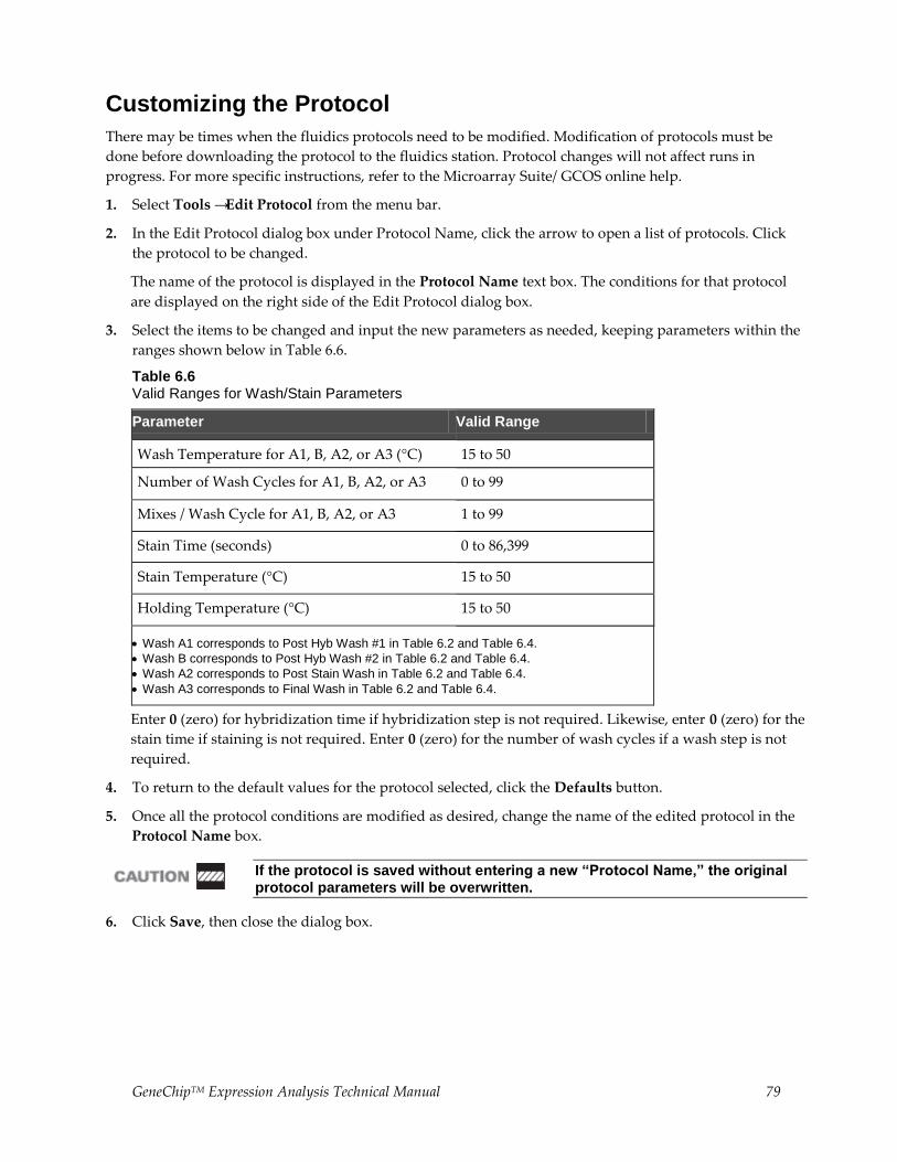
GeneChipTM Expression Analysis Technical Manual 79
Customizing the Protocol
There may be times when the fluidics protocols need to be modified. Modification of protocols must be
done before downloading the protocol to the fluidics station. Protocol changes will not affect runs in
progress. For more specific instructions, refer to the Microarray Suite/ GCOS online help.
1. Select Tools → Edit Protocol from the menu bar.
2. In the Edit Protocol dialog box under Protocol Name, click the arrow to open a list of protocols. Click
the protocol to be changed.
The name of the protocol is displayed in the Protocol Name text box. The conditions for that protocol
are displayed on the right side of the Edit Protocol dialog box.
3. Select the items to be changed and input the new parameters as needed, keeping parameters within the
ranges shown below in Table 6.6.
Table 6.6 Valid Ranges for Wash/Stain Parameters
Parameter Valid Range
Wash Temperature for A1, B, A2, or A3 (°C) 15 to 50
Number of Wash Cycles for A1, B, A2, or A3 0 to 99
Mixes / Wash Cycle for A1, B, A2, or A3 1 to 99
Stain Time (seconds) 0 to 86,399
Stain Temperature (°C) 15 to 50
Holding Temperature (°C) 15 to 50
Wash A1 corresponds to Post Hyb Wash #1 in Table 6.2 and Table 6.4.
Wash B corresponds to Post Hyb Wash #2 in Table 6.2 and Table 6.4.
Wash A2 corresponds to Post Stain Wash in Table 6.2 and Table 6.4.
Wash A3 corresponds to Final Wash in Table 6.2 and Table 6.4.
Enter 0 (zero) for hybridization time if hybridization step is not required. Likewise, enter 0 (zero) for the
stain time if staining is not required. Enter 0 (zero) for the number of wash cycles if a wash step is not
required.
4. To return to the default values for the protocol selected, click the Defaults button.
5. Once all the protocol conditions are modified as desired, change the name of the edited protocol in the
Protocol Name box.
If the protocol is saved without entering a new “Protocol Name,” the original protocol parameters will be overwritten.
6. Click Save, then close the dialog box.
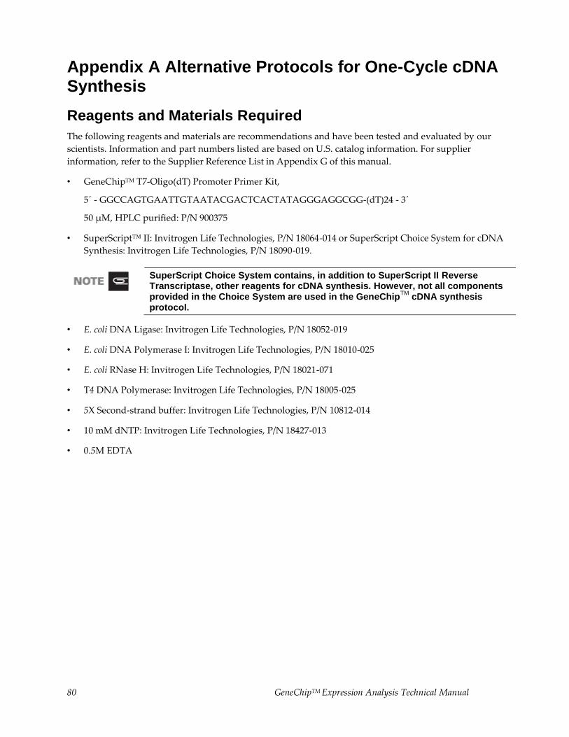
80 GeneChipTM Expression Analysis Technical Manual
Appendix A Alternative Protocols for One-Cycle cDNA Synthesis
Reagents and Materials Required
The following reagents and materials are recommendations and have been tested and evaluated by our
scientists. Information and part numbers listed are based on U.S. catalog information. For supplier
information, refer to the Supplier Reference List in Appendix G of this manual.
• GeneChipTM T7-Oligo(dT) Promoter Primer Kit,
5´ - GGCCAGTGAATTGTAATACGACTCACTATAGGGAGGCGG-(dT)24 - 3´
50 μM, HPLC purified: P/N 900375
• SuperScript™ II: Invitrogen Life Technologies, P/N 18064-014 or SuperScript Choice System for cDNA
Synthesis: Invitrogen Life Technologies, P/N 18090-019.
SuperScript Choice System contains, in addition to SuperScript II Reverse Transcriptase, other reagents for cDNA synthesis. However, not all components provided in the Choice System are used in the GeneChip
TM cDNA synthesis
protocol.
• E. coli DNA Ligase: Invitrogen Life Technologies, P/N 18052-019
• E. coli DNA Polymerase I: Invitrogen Life Technologies, P/N 18010-025
• E. coli RNase H: Invitrogen Life Technologies, P/N 18021-071
• T4 DNA Polymerase: Invitrogen Life Technologies, P/N 18005-025
• 5X Second-strand buffer: Invitrogen Life Technologies, P/N 10812-014
• 10 mM dNTP: Invitrogen Life Technologies, P/N 18427-013
• 0.5M EDTA
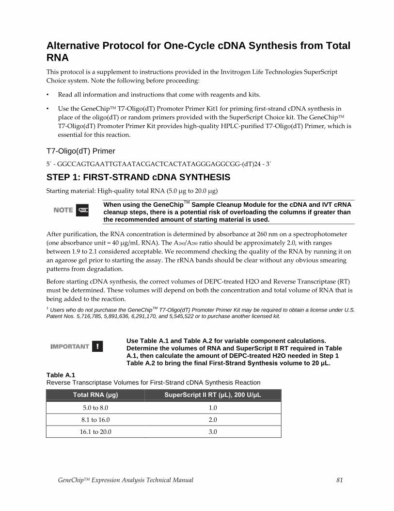
GeneChipTM Expression Analysis Technical Manual 81
Alternative Protocol for One-Cycle cDNA Synthesis from Total RNA
This protocol is a supplement to instructions provided in the Invitrogen Life Technologies SuperScript
Choice system. Note the following before proceeding:
• Read all information and instructions that come with reagents and kits.
• Use the GeneChipTM T7-Oligo(dT) Promoter Primer Kit1 for priming first-strand cDNA synthesis in
place of the oligo(dT) or random primers provided with the SuperScript Choice kit. The GeneChipTM
T7-Oligo(dT) Promoter Primer Kit provides high-quality HPLC-purified T7-Oligo(dT) Primer, which is
essential for this reaction.
T7-Oligo(dT) Primer
5´ - GGCCAGTGAATTGTAATACGACTCACTATAGGGAGGCGG-(dT)24 - 3´
STEP 1: FIRST-STRAND cDNA SYNTHESIS
Starting material: High-quality total RNA (5.0 μg to 20.0 μg)
When using the GeneChipTM
Sample Cleanup Module for the cDNA and IVT cRNA cleanup steps, there is a potential risk of overloading the columns if greater than the recommended amount of starting material is used.
After purification, the RNA concentration is determined by absorbance at 260 nm on a spectrophotometer
(one absorbance unit = 40 μg/mL RNA). The A260/A280 ratio should be approximately 2.0, with ranges
between 1.9 to 2.1 considered acceptable. We recommend checking the quality of the RNA by running it on
an agarose gel prior to starting the assay. The rRNA bands should be clear without any obvious smearing
patterns from degradation.
Before starting cDNA synthesis, the correct volumes of DEPC-treated H2O and Reverse Transcriptase (RT)
must be determined. These volumes will depend on both the concentration and total volume of RNA that is
being added to the reaction.
1 Users who do not purchase the GeneChipTM T7-Oligo(dT) Promoter Primer Kit may be required to obtain a license under U.S. Patent Nos. 5,716,785, 5,891,636, 6,291,170, and 5,545,522 or to purchase another licensed kit.
Use Table A.1 and Table A.2 for variable component calculations. Determine the volumes of RNA and SuperScript II RT required in Table A.1, then calculate the amount of DEPC-treated H2O needed in Step 1 Table A.2 to bring the final First-Strand Synthesis volume to 20 μL.
Table A.1 Reverse Transcriptase Volumes for First-Strand cDNA Synthesis Reaction
Total RNA (μg) SuperScript II RT (μL), 200 U/μL
5.0 to 8.0 1.0
8.1 to 16.0 2.0
16.1 to 20.0 3.0
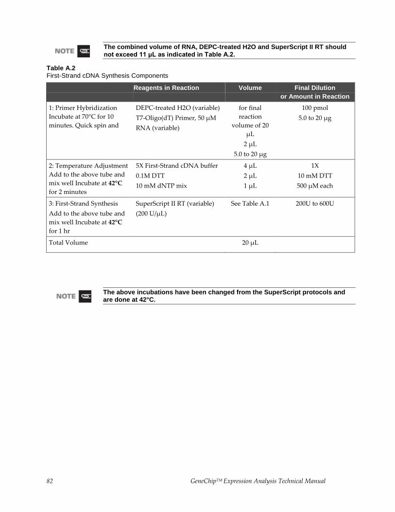
82 GeneChipTM Expression Analysis Technical Manual
The combined volume of RNA, DEPC-treated H2O and SuperScript II RT should not exceed 11 μL as indicated in Table A.2.
Table A.2 First-Strand cDNA Synthesis Components
Reagents in Reaction Volume Final Dilution
or Amount in Reaction
1: Primer Hybridization
Incubate at 70°C for 10
minutes. Quick spin and
DEPC-treated H2O (variable)
T7-Oligo(dT) Primer, 50 µM
RNA (variable)
for final
reaction
volume of 20
µL
2 µL
5.0 to 20 µg
100 pmol
5.0 to 20 µg
2: Temperature Adjustment
Add to the above tube and
mix well Incubate at 42°C
for 2 minutes
5X First-Strand cDNA buffer
0.1M DTT
10 mM dNTP mix
4 µL
2 µL
1 µL
1X
10 mM DTT
500 µM each
3: First-Strand Synthesis
Add to the above tube and
mix well Incubate at 42°C
for 1 hr
SuperScript II RT (variable)
(200 U/µL)
See Table A.1 200U to 600U
Total Volume 20 µL
The above incubations have been changed from the SuperScript protocols and are done at 42°C.
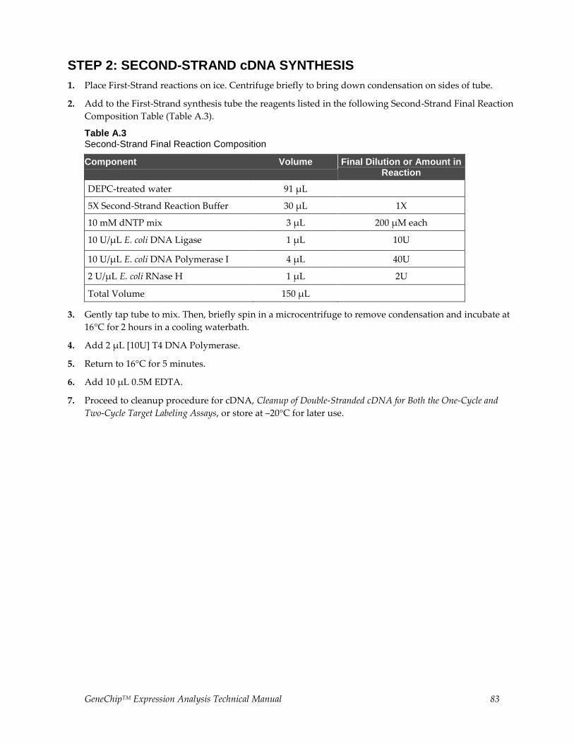
GeneChipTM Expression Analysis Technical Manual 83
STEP 2: SECOND-STRAND cDNA SYNTHESIS
1. Place First-Strand reactions on ice. Centrifuge briefly to bring down condensation on sides of tube.
2. Add to the First-Strand synthesis tube the reagents listed in the following Second-Strand Final Reaction
Composition Table (Table A.3).
Table A.3 Second-Strand Final Reaction Composition
Component Volume Final Dilution or Amount in Reaction
DEPC-treated water 91 µL
5X Second-Strand Reaction Buffer 30 µL 1X
10 mM dNTP mix 3 µL 200 µM each
10 U/µL E. coli DNA Ligase 1 µL 10U
10 U/µL E. coli DNA Polymerase I 4 µL 40U
2 U/µL E. coli RNase H 1 µL 2U
Total Volume 150 µL
3. Gently tap tube to mix. Then, briefly spin in a microcentrifuge to remove condensation and incubate at
16°C for 2 hours in a cooling waterbath.
4. Add 2 μL [10U] T4 DNA Polymerase.
5. Return to 16°C for 5 minutes.
6. Add 10 μL 0.5M EDTA.
7. Proceed to cleanup procedure for cDNA, Cleanup of Double-Stranded cDNA for Both the One-Cycle and
Two-Cycle Target Labeling Assays, or store at –20°C for later use.
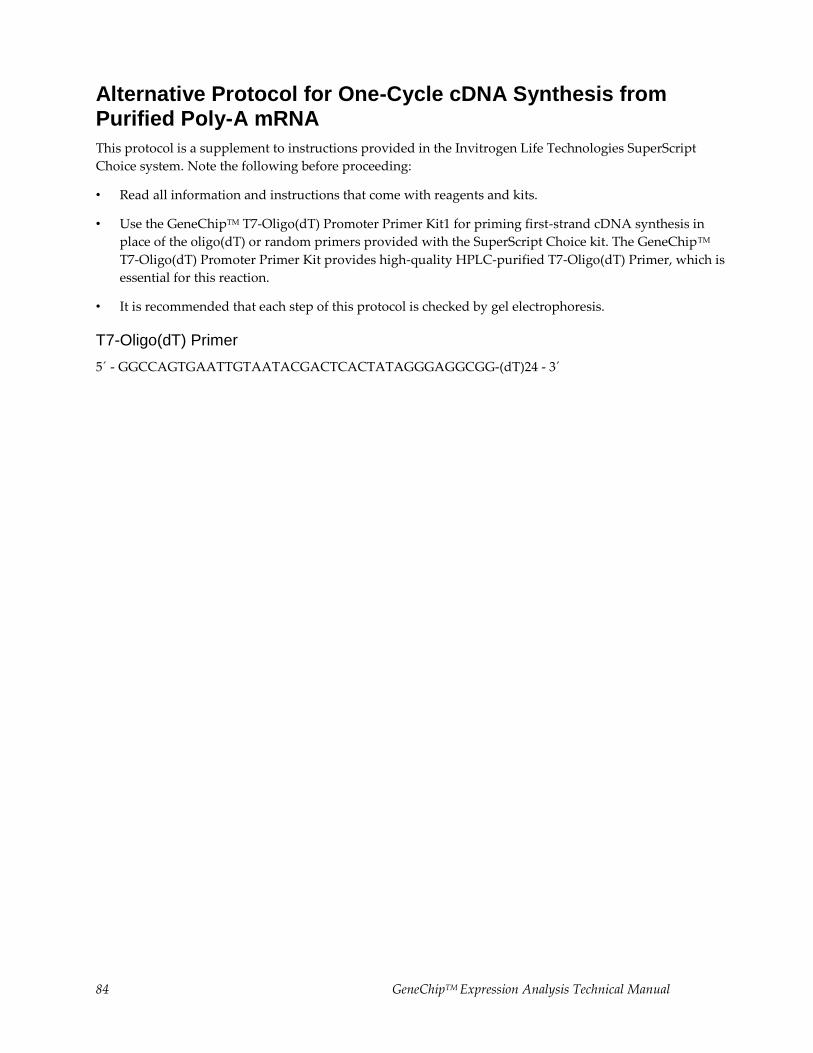
84 GeneChipTM Expression Analysis Technical Manual
Alternative Protocol for One-Cycle cDNA Synthesis from Purified Poly-A mRNA
This protocol is a supplement to instructions provided in the Invitrogen Life Technologies SuperScript
Choice system. Note the following before proceeding:
• Read all information and instructions that come with reagents and kits.
• Use the GeneChipTM T7-Oligo(dT) Promoter Primer Kit1 for priming first-strand cDNA synthesis in
place of the oligo(dT) or random primers provided with the SuperScript Choice kit. The GeneChipTM
T7-Oligo(dT) Promoter Primer Kit provides high-quality HPLC-purified T7-Oligo(dT) Primer, which is
essential for this reaction.
• It is recommended that each step of this protocol is checked by gel electrophoresis.
T7-Oligo(dT) Primer
5´ - GGCCAGTGAATTGTAATACGACTCACTATAGGGAGGCGG-(dT)24 - 3´
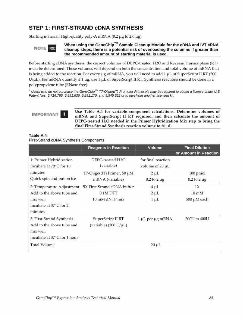
GeneChipTM Expression Analysis Technical Manual 85
STEP 1: FIRST-STRAND cDNA SYNTHESIS
Starting material: High-quality poly-A mRNA (0.2 μg to 2.0 μg).
When using the GeneChipTM
Sample Cleanup Module for the cDNA and IVT cRNA cleanup steps, there is a potential risk of overloading the columns if greater than the recommended amount of starting material is used.
Before starting cDNA synthesis, the correct volumes of DEPC-treated H2O and Reverse Transcriptase (RT)
must be determined. These volumes will depend on both the concentration and total volume of mRNA that
is being added to the reaction. For every μg of mRNA, you will need to add 1 μL of SuperScript II RT (200
U/μL). For mRNA quantity ≤ 1 μg, use 1 μL of SuperScript II RT. Synthesis reactions should be done in a
polypropylene tube (RNase-free).
1 Users who do not purchase the GeneChipTM T7-Oligo(dT) Promoter Primer Kit may be required to obtain a license under U.S. Patent Nos. 5,716,785, 5,891,636, 6,291,170, and 5,545,522 or to purchase another licensed kit.
Use Table A.4 for variable component calculations. Determine volumes of mRNA and SuperScript II RT required, and then calculate the amount of DEPC-treated H2O needed in the Primer Hybridization Mix step to bring the final First-Strand Synthesis reaction volume to 20 μL.
Table A.4 First-Strand cDNA Synthesis Components
Reagents in Reaction Volume Final Dilution
or Amount in Reaction
1: Primer Hybridization
Incubate at 70°C for 10
minutes
Quick spin and put on ice
DEPC-treated H2O
(variable)
for final reaction
volume of 20 µL
T7-Oligo(dT) Primer, 50 µM
mRNA (variable)
2 µL
0.2 to 2 µg
100 pmol
0.2 to 2 µg
2: Temperature Adjustment
Add to the above tube and
mix well
Incubate at 37°C for 2
minutes
5X First-Strand cDNA buffer
0.1M DTT
10 mM dNTP mix
4 µL
2 µL
1 µL
1X
10 mM
500 µM each
3: First-Strand Synthesis
Add to the above tube and
mix well
Incubate at 37°C for 1 hour
SuperScript II RT
(variable) (200 U/µL)
1 µL per µg mRNA 200U to 400U
Total Volume 20 µL
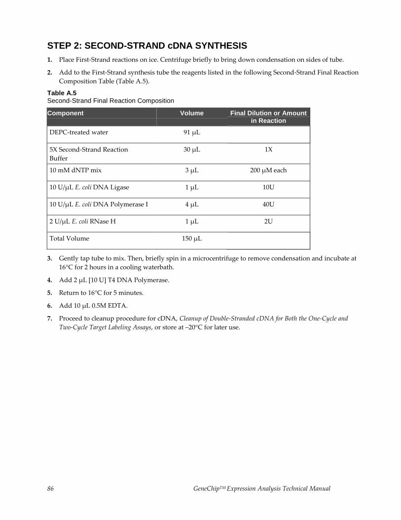
86 GeneChipTM Expression Analysis Technical Manual
STEP 2: SECOND-STRAND cDNA SYNTHESIS
1. Place First-Strand reactions on ice. Centrifuge briefly to bring down condensation on sides of tube.
2. Add to the First-Strand synthesis tube the reagents listed in the following Second-Strand Final Reaction
Composition Table (Table A.5).
Table A.5 Second-Strand Final Reaction Composition
Component Volume Final Dilution or Amount in Reaction
DEPC-treated water 91 µL
5X Second-Strand Reaction
Buffer
30 µL 1X
10 mM dNTP mix 3 µL 200 µM each
10 U/µL E. coli DNA Ligase 1 µL 10U
10 U/µL E. coli DNA Polymerase I 4 µL 40U
2 U/µL E. coli RNase H 1 µL 2U
Total Volume 150 µL
3. Gently tap tube to mix. Then, briefly spin in a microcentrifuge to remove condensation and incubate at
16°C for 2 hours in a cooling waterbath.
4. Add 2 μL [10 U] T4 DNA Polymerase.
5. Return to 16°C for 5 minutes.
6. Add 10 μL 0.5M EDTA.
7. Proceed to cleanup procedure for cDNA, Cleanup of Double-Stranded cDNA for Both the One-Cycle and
Two-Cycle Target Labeling Assays, or store at –20°C for later use.
GeneChipTM Expression Analysis Technical Manual 87
Appendix B Alternative Protocol for Eukaryotic Target Hybridization
This Appendix Contains:
• Introduction and objectives for information provided in this Appendix.
• Detailed steps for preparing the eukaryotic hybridization mix containing labeled target and control
cRNA.
• Instructions for hybridizing the target mix to a eukaryotic GeneChipTM probe array.
After completing the procedures described in this chapter, the hybridized probe array is ready for washing,
staining, and scanning, as detailed in the GeneChipTM Expression Wash, Stain and Scan User Manual, P/N
702731.
Introduction and Objectives
We offer a complete set of GeneChipTM reagents developed specifically for use with GeneChipTM 3’
eukaryotic arrays in cartridge format and GeneChipTM prokaryotic arrays in cartridge format. GeneChipTM
reagent kits include the One- and Two-Cycle cDNA Synthesis Kits, IVT Labeling Kits, Sample Cleanup
Modules, and Hybridization, Wash, and Stain Kits. For best results, it is highly recommended that all
GeneChipTM reagents are used for target preparation, hybridization, wash, and staining when using
GeneChipTM arrays.
The information and protocols described in this Appendix have been provided in previous revisions of the
GeneChipTM Expression Analysis Technical Manual describing hybridization, wash, and staining procedures
for GeneChipTM 3’ eukaryotic arrays in cartridge format. These protocols should be closely followed when
the GeneChipTM Hybridization, Wash, and Stain Kit is not used.
88 GeneChipTM Expression Analysis Technical Manual
Reagents and Materials Required
The following reagents and materials are recommendations and have been tested and evaluated by our
scientists. Information and part numbers listed are based on U.S. catalog information. For supplier
information, refer to the Supplier Reference List in Appendix G of this manual.
• Water, Molecular Biology Grade: Cambrex, P/N 51200
• Bovine Serum Albumin (BSA) solution (50 mg/mL): Invitrogen Life Technologies, P/N 15561-020
• Herring Sperm DNA: Promega Corporation, P/N D1811
• GeneChipTM Eukaryotic Hybridization Control Kit:
• P/N 900454 (30 reactions) or P/N 900457 (150 reactions), contains
• Control cRNA and Control Oligo B2
• Control Oligo B2, 3 nM: P/N 900301 (can be ordered separately)
• 5 M NaCl, RNase-free, DNase-free: Ambion, P/N 9760G
• MES hydrate SigmaUltra: Sigma-Aldrich, P/N M5287
• MES Sodium Salt: Sigma-Aldrich, P/N M5057
• EDTA Disodium Salt, 0.5 M solution (100 mL): Sigma-Aldrich, P/N E7889
• DMSO: Sigma-Aldrich, P/N D5879
• Surfact-Amps 20 (Tween-20), 10%: Pierce Chemical, P/N 28320
Miscellaneous Supplies
• Hybridization Oven 640: P/N 800138 (110V) or 800139 (220V)
• Sterile, RNase-free, microcentrifuge vials, 1.5 mL: USA Scientific, P/N 1415-2600 (or equivalent)
• Micropipettors, (P-2, P-20, P-200, P-1000): Rainin Pipetman (or equivalent)
• Sterile-barrier pipette tips and non-barrier pipette tips
• Heatblock
GeneChipTM Expression Analysis Technical Manual 89
Reagent Preparation
12X MES Stock Buffer
(1.22 M MES, 0.89M [Na+])
For 1,000 mL:
64.61 g of MES hydrate
193.3 g of MES Sodium Salt
800 mL of Molecular Biology Grade water Mix and adjust volume to 1,000 mL.
The pH should be between 6.5 and 6.7. Filter through a 0.2 μm filter.
Do not autoclave. Store at 2°C to 8°C, and shield from light. Discard solution if yellow.
2X Hybridization Buffer
(Final 1X concentration is 100 mM MES, 1 M [Na+], 20 mM EDTA, 0.01% Tween-20)
For 50 mL:
mL of 12X MES Stock Buffer
17.7 mL of 5 M NaCl
mL of 0.5 M EDTA
mL of 10% Tween-20
19.9 mL of water
Store at 2°C to 8°C, and shield from light
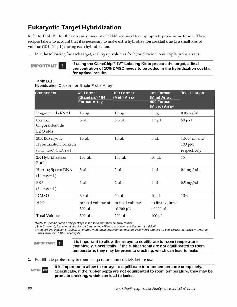
90 GeneChipTM Expression Analysis Technical Manual
Eukaryotic Target Hybridization
Refer to Table B.1 for the necessary amount of cRNA required for appropriate probe array format. These
recipes take into account that it is necessary to make extra hybridization cocktail due to a small loss of
volume (10 to 20 μL) during each hybridization.
1. Mix the following for each target, scaling up volumes for hybridization to multiple probe arrays.
If using the GeneChipTM IVT Labeling Kit to prepare the target, a final
concentration of 10% DMSO needs to be added in the hybridization cocktail for optimal results.
Table B.1 Hybridization Cocktail for Single Probe Array*
Component 49 Format (Standard) / 64 Format Array
100 Format (Midi) Array
169 Format (Mini) Array / 400 Format (Micro) Array
Final Dilution
Fragmented cRNA† 15 µg 10 µg 5 µg 0.05 µg/µL
Control
Oligonucleotide
B2 (3 nM)
5 µL 3.3 µL 1.7 µL 50 pM
20X Eukaryotic
Hybridization Controls
(bioB, bioC, bioD, cre)
15 µL 10 µL 5 µL 1.5, 5, 25, and
100 pM
respectively
2X Hybridization
Buffer
150 µL 100 µL 50 µL 1X
Herring Sperm DNA
(10 mg/mL)
3 µL 2 µL 1 µL 0.1 mg/mL
BSA
(50 mg/mL)
3 µL 2 µL 1 µL 0.5 mg/mL
DMSO‡ 30 µL 20 µL 10 µL 10%
H2O to final volume of
300 µL
to final volume
of 200 µL
to final volume
of 100 µL
Total Volume 300 µL 200 µL 100 µL
*Refer to specific probe array package insert for information on array format. †See Chapter 2, for amount of adjusted fragmented cRNA to use when starting from total RNA. ‡Note that the addition of DMSO is different from previous recommendations. Follow this protocol for best results on arrays when using
the GeneChipTM IVT Labeling Kit.
It is important to allow the arrays to equilibrate to room temperature completely. Specifically, if the rubber septa are not equilibrated to room temperature, they may be prone to cracking, which can lead to leaks.
2. Equilibrate probe array to room temperature immediately before use.
It is important to allow the arrays to equilibrate to room temperature completely. Specifically, if the rubber septa are not equilibrated to room temperature, they may be prone to cracking, which can lead to leaks.
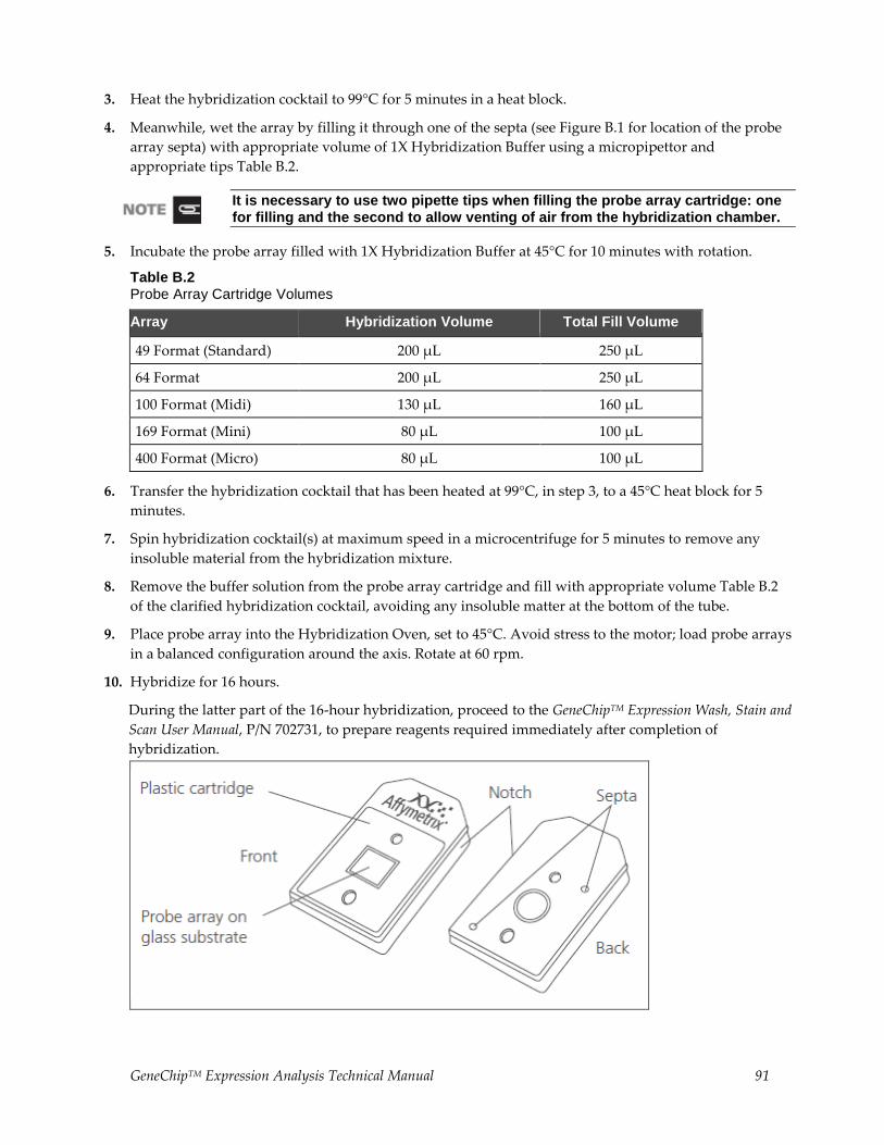
GeneChipTM Expression Analysis Technical Manual 91
3. Heat the hybridization cocktail to 99°C for 5 minutes in a heat block.
4. Meanwhile, wet the array by filling it through one of the septa (see Figure B.1 for location of the probe
array septa) with appropriate volume of 1X Hybridization Buffer using a micropipettor and
appropriate tips Table B.2.
It is necessary to use two pipette tips when filling the probe array cartridge: one for filling and the second to allow venting of air from the hybridization chamber.
5. Incubate the probe array filled with 1X Hybridization Buffer at 45°C for 10 minutes with rotation.
Table B.2 Probe Array Cartridge Volumes
Array Hybridization Volume Total Fill Volume
49 Format (Standard) 200 µL 250 µL
64 Format 200 µL 250 µL
100 Format (Midi) 130 µL 160 µL
169 Format (Mini) 80 µL 100 µL
400 Format (Micro) 80 µL 100 µL
6. Transfer the hybridization cocktail that has been heated at 99°C, in step 3, to a 45°C heat block for 5
minutes.
7. Spin hybridization cocktail(s) at maximum speed in a microcentrifuge for 5 minutes to remove any
insoluble material from the hybridization mixture.
8. Remove the buffer solution from the probe array cartridge and fill with appropriate volume Table B.2
of the clarified hybridization cocktail, avoiding any insoluble matter at the bottom of the tube.
9. Place probe array into the Hybridization Oven, set to 45°C. Avoid stress to the motor; load probe arrays
in a balanced configuration around the axis. Rotate at 60 rpm.
10. Hybridize for 16 hours.
During the latter part of the 16-hour hybridization, proceed to the GeneChipTM Expression Wash, Stain and
Scan User Manual, P/N 702731, to prepare reagents required immediately after completion of
hybridization.
92 GeneChipTM Expression Analysis Technical Manual
Figure B.1
GeneChipTM Probe Array
Appendix C Alternative Protocol for Prokaryotic Target Hybridization
Appendix Contents
This Appendix Contains:
• Introduction and objectives for information provided in this Appendix.
• Steps for preparing the hybridization mix, and instructions for hybridizing the target mix to the
GeneChipTM P. aeruginosa Genome Array and GeneChipTM E. coli arrays. The hybridized probe array is
then ready for washing, staining, and scanning as detailed in Appendix D.
Introduction and Objectives
We offer a complete set of GeneChipTM reagents developed specifically for use with GeneChipTM 3’
eukaryotic arrays in cartridge format and GeneChipTM prokaryotic arrays in cartridge format. GeneChipTM
reagent kits include the One- and Two-Cycle cDNA Synthesis Kits, IVT Labeling Kits, Sample Cleanup
Modules, and Hybridization, Wash, and Stain Kits. For best results, it is highly recommended that all
GeneChipTM reagents are used for target preparation, hybridization, wash, and staining when using
GeneChipTM arrays.
The information and protocols described in this Appendix have been provided in previous revisions of the
GeneChipTM Expression Analysis Technical Manual describing hybridization, wash, and staining procedures
for GeneChipTM prokaryotic arrays in cartridge format. These protocols should be closely followed when
the GeneChipTM Hybridization, Wash, and Stain Kit is not used.
Reagents and Materials Required
The following reagents and materials are recommendations and have been tested and evaluated by our
scientists. Information and part numbers listed are based on U.S. catalog information. For supplier
information, refer to the Supplier Reference List in Appendix G of this manual.
• Water, Molecular Biology Grade: Cambrex, P/N 51200
• Bovine Serum Albumin (BSA) solution, 50 mg/mL: Invitrogen Life Technologies, P/N 15561-020
• Herring Sperm DNA: Promega Corporation, P/N D1811
• Control Oligo B2, 3 nM: P/N 900301 (can be ordered separately)
• NaCl, 5M, RNase-free, DNase-free: Ambion TM, P/N 9760G
• MES hydrate SigmaUltra: Sigma-Aldrich, P/N M5287
• MES Sodium Salt: Sigma-Aldrich, P/N M5057
• EDTA Disodium Salt, 0.5M solution (100 mL): Sigma-Aldrich, P/N E7889
Miscellaneous Reagents
• 100% DMSO: Sigma-Aldrich, P/N D2650
GeneChipTM Expression Analysis Technical Manual 93
• Surfact-Amps 20 (Tween-20), 10%: Pierce Chemical, P/N 28320
Miscellaneous Supplies
• Hybridization Oven 640: P/N 800138 (110V) or 800139 (220V)
• Sterile, RNase-free, microcentrifuge vials, 1.5 mL: USA Scientific TM, P/N 1415-2600 (or equivalent)
• Micropipettors, (P-2, P-20, P-200, P-1000): Rainin TM Pipetman TM (or equivalent)
• Sterile-barrier pipette tips and non-barrier pipette tips
Reagent Preparation
12X MES Stock Buffer
(1.22M MES, 0.89 M [Na+])
For 1,000 mL:
64.61 g of MES hydrate
193.3 g of MES Sodium Salt
800 mL of Molecular Biology Grade water Mix and adjust volume to 1,000 mL.
The pH should be between 6.5 and 6.7. Filter through a 0.2 μm filter.
Do not autoclave, store at 2°C to 8°C, and shield from light. Discard solution if yellow.
2X Hybridization Buffer (50 mL)
(Final 1X concentration is 100mM MES, 1 M [Na+], 20 mM EDTA, 0.01% Tween-20)
For 50 mL:
8.3 mL of 12X MES Stock Buffer
17.7 mL of 5 M NaCl
4.0 mL of 0.5 M EDTA
0.1 mL of 10% Tween-20
19.9 mL of water
Store at 2°C to 8°C, and shield from light.
94 GeneChipTM Expression Analysis Technical Manual
Prokaryotic Target Hybridization
After determining that the fragmented cDNA is labeled with biotin, prepare the hybridization solution mix.
The minimum amount of cDNA product required for target hybridization is 1 μg. The solution is stable for
approximately 6 to 8 hours at 4°C. The following protocol can be used for freshly prepared or frozen
hybridization cocktail. Re-use of prokaryotic sample has not been thoroughly tested and, therefore, is not
recommended.
1. Prepare the following hybridization solution mix.
Table C.1 Hybridization Cocktail for Single Probe Array*
Component 49 Format (Standard)
100 Format (Midi) 169 Format (Mini)
Final Dilution or Amount
Fragmented and
Labeled cDNA
Up to 50 µL Up to 50 µL 25 µL 0.5 – 7.0 µg
Control Oligonucleotide
B2 (3 nM)
3.3 µL 2.2 µL 1.3 µL 50 pM
2X Hybridization Buffer 100 µL 65 µL 40 µL 1X
100% DMSO – 10.2 µL 6.2 µL 7.8% (or 0%)
10 mg/mL Herring
Sperm DNA
2.0 µL 1.3 µL 0.8 µL 0.1 mg/mL
50 mg/mL BSA 2.0 µL 1.3 µL 0.8 µL 0.5 mg/mL
Molecular Biology
Grade Water
42.7 µL – 5.9 µL
Total Volume 200 µL 130 µL 80 µL
*Refer to specific probe array package insert for information on array format.
2. Equilibrate probe array to room temperature immediately before use.
It is important to allow the arrays to normalize to room temperature completely. Specifically, if the rubber septa are not equilibrated to room temperature, they may be prone to cracking, which leads to leaks.
3. Add the indicated amount of hybridization solution mix to the probe array. Refer to specific probe
array package insert for information on array format.
It is necessary to use two pipette tips when filling the probe array cartridge: one for filling and the second to allow venting of air from the hybridization chamber.
4. Place the probe array in the hybridization oven set at the temperatures indicated below.
• P. aeruginosa array 50°C
• E. coli Antisense
arrays 45°C
GeneChipTM Expression Analysis Technical Manual 95
The hybridization temperature of 50°C is higher than that used for other expression assays. The increased hybridization temperature is required due to the high GC content of P. aeruginosa.
5. Avoid stress to the motor; load probe arrays in a balanced configuration around axis. Rotate at 60 rpm.
6. Hybridize for 16 hours.
During the latter part of the 16-hour hybridization, proceed to Appendix D, Alternative Protocol for
Prokaryotic Arrays: Washing, Staining and Scanning to prepare reagents required immediately after
completion of hybridization.
96 GeneChipTM Expression Analysis Technical Manual
Appendix D Alternative Protocol for Prokaryotic Arrays:Washing, Staining and Scanning
This Appendix Contains:
• Introduction and objectives for information provided in this Appendix.
• Instructions for using the Fluidics Station 400 or 450/250 to automate the washing and staining of
GeneChipTM P. aeruginosa and GeneChipTM E. coli arrays.
• Instructions for scanning probe arrays using the GeneArrayTM Scanner or the GeneChipTM Scanner 3000.
After completing the procedures described in this chapter, the scanned probe array image (.dat file) is
ready for analysis, as explained in the enclosed GeneChipTM Expression Analysis: Data Analysis Fundamentals
booklet (P/N 701190).
Introduction and Objectives
We offer a complete set of GeneChipTM reagents developed specifically for use with GeneChipTM 3’
eukaryotic arrays in cartridge format and GeneChipTM prokaryotic arrays in cartridge format. GeneChipTM
reagent kits include the One- and Two-Cycle cDNA Synthesis Kits, IVT Labeling Kits, Sample Cleanup
Modules, and Hybridization, Wash, and Stain Kits. For best results, it is highly recommended that all
GeneChipTM reagents are used for target preparation, hybridization, wash, and staining when using
GeneChipTM arrays.
The information and protocols described in this Appendix have been provided in previous revisions of the
GeneChipTM Expression Analysis Technical Manual describing hybridization, wash, and staining procedures
for GeneChipTM prokaryotic arrays in cartridge format. These protocols should be closely followed when
the GeneChipTM Hybridization, Wash, and Stain Kit is not used.
GeneChipTM Expression Analysis Technical Manual 97
Reagents and Materials Required
The following reagents and materials are recommendations and have been tested and evaluated by our
scientists. Information and part numbers listed are based on U.S. catalog information. For supplier
information, refer to the Supplier Reference List in Appendix G of this manual.
• Water, Molecular Biology Grade: Cambrex, P/N 51200
• Nuclease-free Water: Ambion, P/N 9930
• Bovine Serum Albumin (BSA) solution, 50 mg/mL: Invitrogen Life Technologies, P/N 15561-020
• R-Phycoerythrin Streptavidin: Molecular Probes, P/N S-866
• NaCl, 5M, RNase-free, DNase-free: Ambion, P/N 9760G
• PBS, pH 7.2: Invitrogen Life Technologies, P/N 20012-027
• 20X SSPE (3 M NaCl, 0.2M NaH2PO4, 0.02 M EDTA): Cambrex, P/N 51214
• Goat IgG, Reagent Grade: Sigma-Aldrich, P/N I 5256
• Anti-streptavidin antibody (goat), biotinylated: Vector Laboratories, P/N BA-0500
• Surfact-Amps 20 (Tween-20), 10%: Pierce Chemical, P/N 28320
• Bleach (5.25% Sodium Hypochlorite): VWR Scientific, P/N 37001-060 (or equivalent)
• ImmunoPure Streptavidin: Pierce Chemical, P/N 21125
Miscellaneous Supplies
• Sterile, RNase-free, microcentrifuge vials, 1.5 mL: USA Scientific, P/N 1415-2600 (or equivalent)
• Micropipettors, (P-2, P-20, P-200, P-1000): Rainin Pipetman (or equivalent)
• Sterile-barrier pipette tips and non-barrier pipette tips
• Tygon Tubing, 0.04″ inner diameter: Cole-Parmer, P/N H-06418- 04
• Tough-Spots, Label Dots: USA Scientific, P/N 9185-0000
98 GeneChipTM Expression Analysis Technical Manual
Reagent Preparation
Wash Buffer A: Non-Stringent Wash Buffer
(6X SSPE, 0.01% Tween-20)
For 1,000 mL:
300 mL of 20X SSPE
1.0 mL of 10% Tween-20
699 mL of water
Filter through a 0.2 μm filter.
Store at room temperature.
Wash Buffer B: Stringent Wash Buffer
(100 mM MES, 0.1 M [Na+], 0.01% Tween-20)
For 1,000 mL:
83.3 mL of 12 X MES Stock Buffer (see Appendix C for reagent preparation) mL of 5M NaCl
1.0 mL of 10% Tween-20
910.5 mL of water
Filter through a 0.2 μm filter
Store at 2°C to 8°C and shield from light.
2X Stain Buffer
(final 1X concentration: 100 mM MES, 1 M [Na+], 0.05% Tween-20)
For 250 mL:
41.7 mL of 12X MES Stock Buffer (see Appendix C)
92.5 mL of 5M NaCl
2.5 mL of 10% Tween-20
113.3 mL of water
Filter through a 0.2 μm filter.
Store at 2°C to 8°C and shield from light.
GeneChipTM Expression Analysis Technical Manual 99
10 mg/mL Goat IgG Stock
Resuspend 50 mg in 5 mL 150 mM NaCl.
Store at 4°C.
If a larger volume of the 10 mg/mL IgG stock is prepared, aliquot and store at –20°C until use. After the solution has been thawed it should be stored at 4°C. Avoid additional freezing and thawing.
1 mg/mL Streptavidin Stock
Resuspend 5 mg in 5 mL of PBS. Store at 4°C.
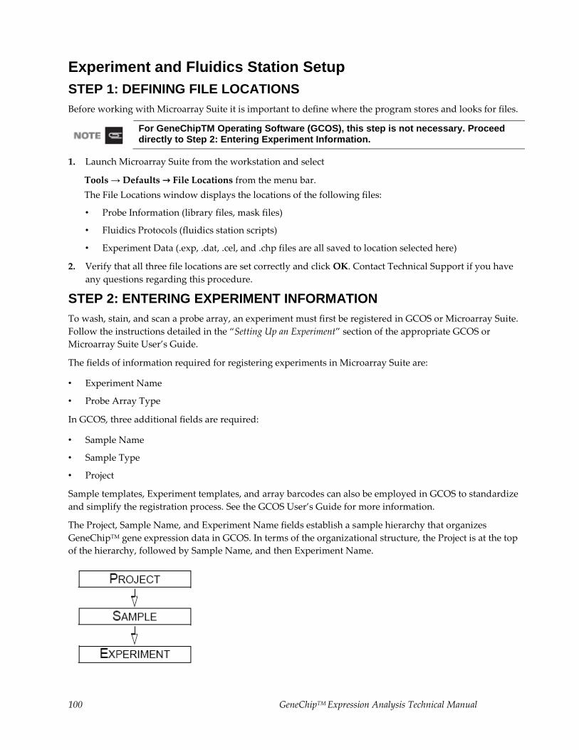
100 GeneChipTM Expression Analysis Technical Manual
Experiment and Fluidics Station Setup
STEP 1: DEFINING FILE LOCATIONS
Before working with Microarray Suite it is important to define where the program stores and looks for files.
For GeneChipTM Operating Software (GCOS), this step is not necessary. Proceed directly to Step 2: Entering Experiment Information.
1. Launch Microarray Suite from the workstation and select
Tools → Defaults → File Locations from the menu bar.
The File Locations window displays the locations of the following files:
• Probe Information (library files, mask files)
• Fluidics Protocols (fluidics station scripts)
• Experiment Data (.exp, .dat, .cel, and .chp files are all saved to location selected here)
2. Verify that all three file locations are set correctly and click OK. Contact Technical Support if you have
any questions regarding this procedure.
STEP 2: ENTERING EXPERIMENT INFORMATION
To wash, stain, and scan a probe array, an experiment must first be registered in GCOS or Microarray Suite.
Follow the instructions detailed in the “Setting Up an Experiment” section of the appropriate GCOS or
Microarray Suite User’s Guide.
The fields of information required for registering experiments in Microarray Suite are:
• Experiment Name
• Probe Array Type
In GCOS, three additional fields are required:
• Sample Name
• Sample Type
• Project
Sample templates, Experiment templates, and array barcodes can also be employed in GCOS to standardize
and simplify the registration process. See the GCOS User’s Guide for more information.
The Project, Sample Name, and Experiment Name fields establish a sample hierarchy that organizes
GeneChipTM gene expression data in GCOS. In terms of the organizational structure, the Project is at the top
of the hierarchy, followed by Sample Name, and then Experiment Name.
GeneChipTM Expression Analysis Technical Manual 101
STEP 3: PREPARING THE FLUIDICS STATION
The Fluidics Station 400, or 450/250 is used to wash and stain the probe arrays. It is operated using
GCOS/Microarray Suite.
Setting Up the Fluidics Station
1. Turn on the Fluidics Station using the switch on the lower left side of the machine.
2. Select Run → Fluidics from the menu bar.
The Fluidics Station dialog box appears with a drop-down list for selecting the experiment name for
each of the fluidics station modules. A second list is accessed for choosing the Protocol for each of the
four fluidics station modules.
Refer to the appropriate GeneChipTM
Fluidics Station User’s Guide for instructions on connecting and addressing multiple fluidics stations.
Priming the Fluidics Station
Priming ensures that the lines of the fluidics station are filled with the appropriate buffers and the fluidics
station is ready for running fluidics station protocols.
Priming should be done:
• When the fluidics station is first started
• When wash solutions are changed
• Before washing if a shutdown has been performed
• If the LCD window instructs the user to prime
1. Select Protocol in the Fluidics Station dialog box.
2. Choose Prime or Prime_450 for the respective modules in the Protocol drop-down list.
3. Change the intake buffer reservoir A to Non-stringent Wash Buffer and intake buffer reservoir B to
Stringent Wash Buffer.
4. For MAS, click Run for each module to begin priming. In GCOS, select the All Modules check box,
then click Run.
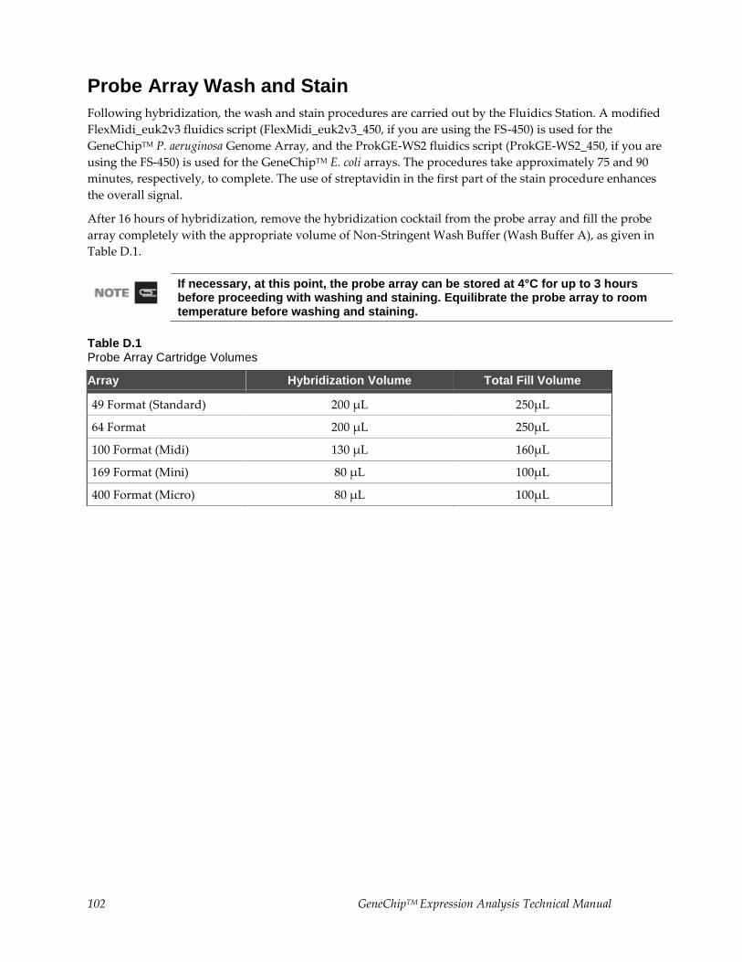
102 GeneChipTM Expression Analysis Technical Manual
Probe Array Wash and Stain
Following hybridization, the wash and stain procedures are carried out by the Fluidics Station. A modified
FlexMidi_euk2v3 fluidics script (FlexMidi_euk2v3_450, if you are using the FS-450) is used for the
GeneChipTM P. aeruginosa Genome Array, and the ProkGE-WS2 fluidics script (ProkGE-WS2_450, if you are
using the FS-450) is used for the GeneChipTM E. coli arrays. The procedures take approximately 75 and 90
minutes, respectively, to complete. The use of streptavidin in the first part of the stain procedure enhances
the overall signal.
After 16 hours of hybridization, remove the hybridization cocktail from the probe array and fill the probe
array completely with the appropriate volume of Non-Stringent Wash Buffer (Wash Buffer A), as given in
Table D.1.
If necessary, at this point, the probe array can be stored at 4°C for up to 3 hours before proceeding with washing and staining. Equilibrate the probe array to room temperature before washing and staining.
Table D.1 Probe Array Cartridge Volumes
Array Hybridization Volume Total Fill Volume
49 Format (Standard) 200 µL 250µL
64 Format 200 µL 250µL
100 Format (Midi) 130 µL 160µL
169 Format (Mini) 80 µL 100µL
400 Format (Micro) 80 µL 100µL
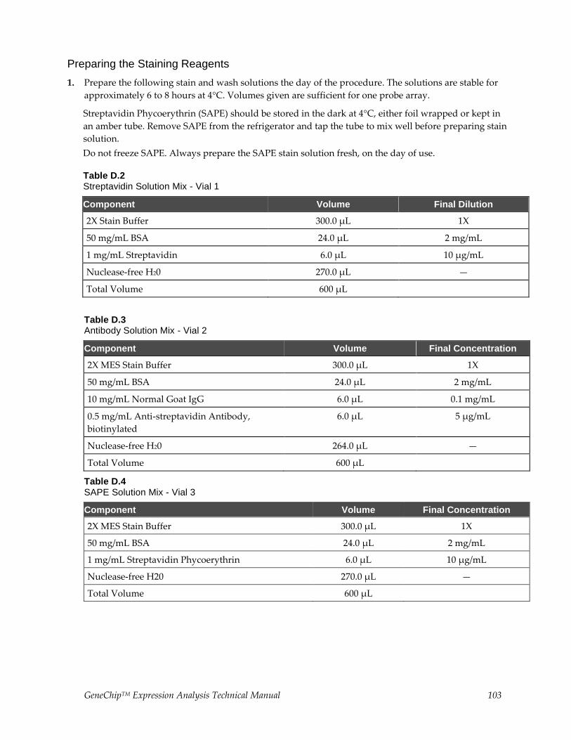
GeneChipTM Expression Analysis Technical Manual 103
Preparing the Staining Reagents
1. Prepare the following stain and wash solutions the day of the procedure. The solutions are stable for
approximately 6 to 8 hours at 4°C. Volumes given are sufficient for one probe array.
Streptavidin Phycoerythrin (SAPE) should be stored in the dark at 4°C, either foil wrapped or kept in
an amber tube. Remove SAPE from the refrigerator and tap the tube to mix well before preparing stain
solution.
Do not freeze SAPE. Always prepare the SAPE stain solution fresh, on the day of use.
Table D.2 Streptavidin Solution Mix - Vial 1
Component Volume Final Dilution
2X Stain Buffer 300.0 µL 1X
50 mg/mL BSA 24.0 µL 2 mg/mL
1 mg/mL Streptavidin 6.0 µL 10 µg/mL
Nuclease-free H20 270.0 µL —
Total Volume 600 µL
Table D.3 Antibody Solution Mix - Vial 2
Component Volume Final Concentration
2X MES Stain Buffer 300.0 µL 1X
50 mg/mL BSA 24.0 µL 2 mg/mL
10 mg/mL Normal Goat IgG 6.0 µL 0.1 mg/mL
0.5 mg/mL Anti-streptavidin Antibody,
biotinylated
6.0 µL 5 µg/mL
Nuclease-free H20 264.0 µL —
Total Volume 600 µL
Table D.4 SAPE Solution Mix - Vial 3
Component Volume Final Concentration
2X MES Stain Buffer 300.0 µL 1X
50 mg/mL BSA 24.0 µL 2 mg/mL
1 mg/mL Streptavidin Phycoerythrin 6.0 µL 10 µg/mL
Nuclease-free H20 270.0 µL —
Total Volume 600 µL
104 GeneChipTM Expression Analysis Technical Manual
2. In the Fluidics Station dialog box on the workstation, select the correct experiment name from the drop-
down Experiment list. The probe array type will appear automatically.
Arrays & Fluidics Protocols
• Array: GeneChipTM E. coli Genome 2.0 Array Protocol: Mini_prok2v1(if using FS-450,
Mini_prok2v1_450)
• Array: GeneChipTM E. coli Antisense Genome Array Protocol: ProkGE-WS2 (if using FS-450,
ProkGE-WS2_450)
• Array: GeneChipTM P. aeruginosa Genome Array Protocol: Modified FlexMidi_euk2v3*
(*See Table D.5. If using FS-450, FlexMidi_euk2v3_450)
Fluidics protocols are specific to array format and content. Follow procedures below for specific arrays.
3. Choose Run in the Fluidics Station dialog box to begin the washing and staining. Follow the
instructions in the LCD window on the fluidics station when using the Fluidics Station 400.
If you are unfamiliar with inserting and removing probe arrays from the fluidics station modules, refer
to the appropriate User’s Guide for your GeneChipTM Fluidics Station 400, or 450/250.
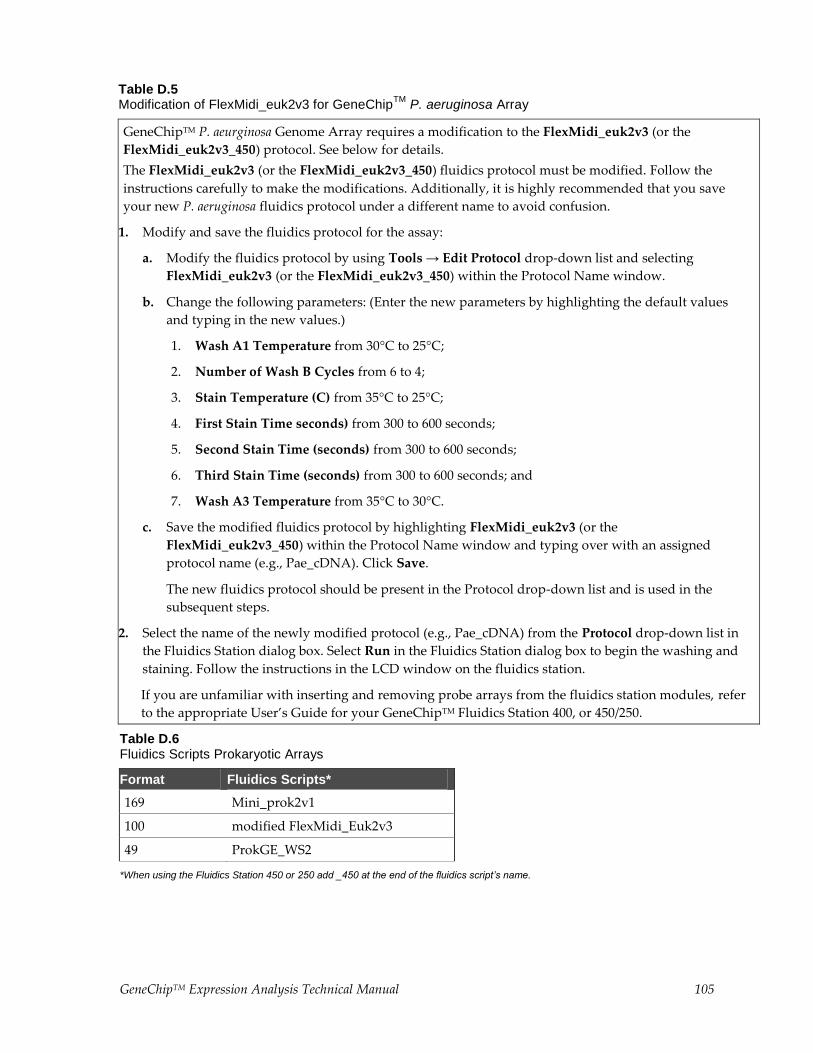
GeneChipTM Expression Analysis Technical Manual 105
Table D.5 Modification of FlexMidi_euk2v3 for GeneChip
TM P. aeruginosa Array
GeneChipTM P. aeurginosa Genome Array requires a modification to the FlexMidi_euk2v3 (or the
FlexMidi_euk2v3_450) protocol. See below for details.
The FlexMidi_euk2v3 (or the FlexMidi_euk2v3_450) fluidics protocol must be modified. Follow the
instructions carefully to make the modifications. Additionally, it is highly recommended that you save
your new P. aeruginosa fluidics protocol under a different name to avoid confusion.
1. Modify and save the fluidics protocol for the assay:
a. Modify the fluidics protocol by using Tools → Edit Protocol drop-down list and selecting
FlexMidi_euk2v3 (or the FlexMidi_euk2v3_450) within the Protocol Name window.
b. Change the following parameters: (Enter the new parameters by highlighting the default values
and typing in the new values.)
1. Wash A1 Temperature from 30°C to 25°C;
2. Number of Wash B Cycles from 6 to 4;
3. Stain Temperature (C) from 35°C to 25°C;
4. First Stain Time seconds) from 300 to 600 seconds;
5. Second Stain Time (seconds) from 300 to 600 seconds;
6. Third Stain Time (seconds) from 300 to 600 seconds; and
7. Wash A3 Temperature from 35°C to 30°C.
c. Save the modified fluidics protocol by highlighting FlexMidi_euk2v3 (or the
FlexMidi_euk2v3_450) within the Protocol Name window and typing over with an assigned
protocol name (e.g., Pae_cDNA). Click Save.
The new fluidics protocol should be present in the Protocol drop-down list and is used in the
subsequent steps.
2. Select the name of the newly modified protocol (e.g., Pae_cDNA) from the Protocol drop-down list in
the Fluidics Station dialog box. Select Run in the Fluidics Station dialog box to begin the washing and
staining. Follow the instructions in the LCD window on the fluidics station.
If you are unfamiliar with inserting and removing probe arrays from the fluidics station modules, refer
to the appropriate User’s Guide for your GeneChipTM Fluidics Station 400, or 450/250.
Table D.6 Fluidics Scripts Prokaryotic Arrays
Format Fluidics Scripts*
169 Mini_prok2v1
100 modified FlexMidi_Euk2v3
49 ProkGE_WS2
*When using the Fluidics Station 450 or 250 add _450 at the end of the fluidics script’s name.
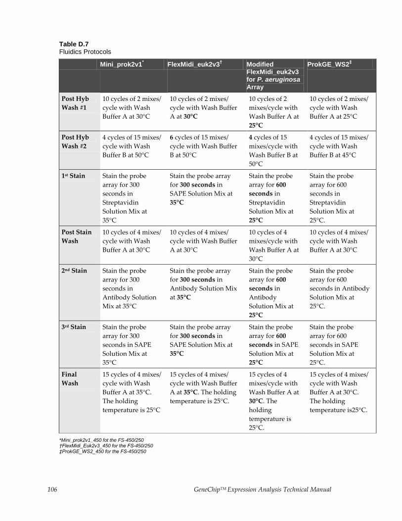
106 GeneChipTM Expression Analysis Technical Manual
Table D.7 Fluidics Protocols
Mini_prok2v1* FlexMidi_euk2v3
† Modified
FlexMidi_euk2v3 for P. aeruginosa Array
ProkGE_WS2‡
Post Hyb
Wash #1
10 cycles of 2 mixes/
cycle with Wash
Buffer A at 30°C
10 cycles of 2 mixes/
cycle with Wash Buffer
A at 30°C
10 cycles of 2
mixes/cycle with
Wash Buffer A at
25°C
10 cycles of 2 mixes/
cycle with Wash
Buffer A at 25°C
Post Hyb
Wash #2
4 cycles of 15 mixes/
cycle with Wash
Buffer B at 50°C
6 cycles of 15 mixes/
cycle with Wash Buffer
B at 50°C
4 cycles of 15
mixes/cycle with
Wash Buffer B at
50°C
4 cycles of 15 mixes/
cycle with Wash
Buffer B at 45°C
1st Stain Stain the probe
array for 300
seconds in
Streptavidin
Solution Mix at
35°C
Stain the probe array
for 300 seconds in
SAPE Solution Mix at
35°C
Stain the probe
array for 600
seconds in
Streptavidin
Solution Mix at
25°C
Stain the probe
array for 600
seconds in
Streptavidin
Solution Mix at
25°C.
Post Stain
Wash
10 cycles of 4 mixes/
cycle with Wash
Buffer A at 30°C
10 cycles of 4 mixes/
cycle with Wash Buffer
A at 30°C
10 cycles of 4
mixes/cycle with
Wash Buffer A at
30°C
10 cycles of 4 mixes/
cycle with Wash
Buffer A at 30°C
2nd Stain Stain the probe
array for 300
seconds in
Antibody Solution
Mix at 35°C
Stain the probe array
for 300 seconds in
Antibody Solution Mix
at 35°C
Stain the probe
array for 600
seconds in
Antibody
Solution Mix at
25°C
Stain the probe
array for 600
seconds in Antibody
Solution Mix at
25°C.
3rd Stain Stain the probe
array for 300
seconds in SAPE
Solution Mix at
35°C
Stain the probe array
for 300 seconds in
SAPE Solution Mix at
35°C
Stain the probe
array for 600
seconds in SAPE
Solution Mix at
25°C
Stain the probe
array for 600
seconds in SAPE
Solution Mix at
25°C.
Final
Wash
15 cycles of 4 mixes/
cycle with Wash
Buffer A at 35°C.
The holding
temperature is 25°C
15 cycles of 4 mixes/
cycle with Wash Buffer
A at 35°C. The holding
temperature is 25°C.
15 cycles of 4
mixes/cycle with
Wash Buffer A at
30°C. The
holding
temperature is
25°C.
15 cycles of 4 mixes/
cycle with Wash
Buffer A at 30°C.
The holding
temperature is25°C.
*Mini_prok2v1_450 fot the FS-450/250 †FlexMidi_Euk2v3_450 for the FS-450/250 ‡ProkGE_WS2_450 for the FS-450/250
GeneChipTM Expression Analysis Technical Manual 107
USING THE FLUIDICS STATION 450/250
Washing and Staining the Probe Array
1. In the Fluidics Station dialog box on the workstation, select the correct experiment name from the drop-
down Experiment list.
The Probe Array Type appears automatically.
2. In the Protocol drop-down list, select the appropriate antibody amplification protocol to control the
washing and staining of the probe array format being used: Table D.6.
3. Choose Run in the Fluidics Station dialog box to begin the washing and staining. Follow the
instructions in the LCD window on the fluidics station.
If you are unfamiliar with inserting and removing probe arrays from the fluidics station modules, refer
to the appropriate Fluidics Station User’s Guide, or Quick Reference Card (P/N 08-0093 for the FS-450/250
fluidics stations).
4. Insert the appropriate probe array into the designated module of the fluidics station while the cartridge
lever is down, or in the eject position. When finished, verify that the cartridge lever is returned to the
up, or engaged, position.
5. Remove any microcentrifuge vial(s) remaining in the sample holder of the fluidics station module(s)
being used.
6. If prompted to “Load Vials 1-2-3,” place the three experiment sample vials (the microcentrifuge vials)
into the sample holders 1, 2, and 3 on the fluidics station.
1. Place one vial containing streptavidin solution in sample holder 1.
2. Place one vial containing the anti-streptavidin biotinylated antibody solution in sample holder 2.
3. Place one vial containing the streptavidin phycoerythrin (SAPE) solution in sample holder 3.
4. Press down on the needle lever to snap needles into position and to start the run.
The run begins. The Fluidics Station dialog box at the workstation terminal and the LCD window
display the status of the washing and staining as the protocol progresses.
7. When the protocol is complete, the LCD window displays the message EJECT CARTRIDGE.
8. Remove the probe arrays from the fluidics station modules by first pressing down the cartridge lever to
the eject position.
9. Lift up on the needle lever to disengage the needles from the microcentrifuge vials. Remove the three
microcentrifuge vials from the needle holders.
10. Check the probe array window for large bubbles or air pockets.
• If bubbles are present, refer to Table D.8.
• If the probe array has no large bubbles, it is ready to scan on the GeneArrayTM Scanner, or the
GeneChipTM Scanner 3000. Pull up on the cartridge lever to close the washblock and proceed to
Probe Array Scan.
11. If there are no more samples to hybridize, shut down the fluidics station following the procedure in
Shutting Down the Fluidics Station.
12. Keep the probe arrays at 4°C and in the dark until ready for scanning.
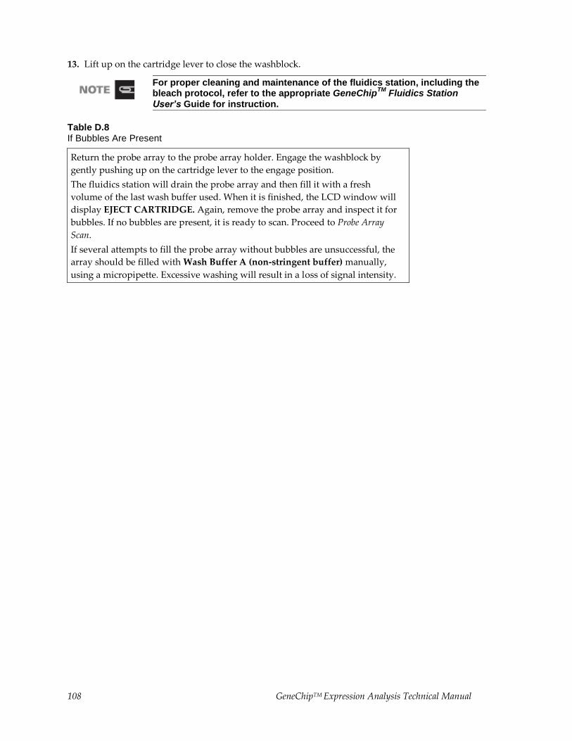
108 GeneChipTM Expression Analysis Technical Manual
13. Lift up on the cartridge lever to close the washblock.
For proper cleaning and maintenance of the fluidics station, including the bleach protocol, refer to the appropriate GeneChip
TM Fluidics Station
User’s Guide for instruction.
Table D.8 If Bubbles Are Present
Return the probe array to the probe array holder. Engage the washblock by
gently pushing up on the cartridge lever to the engage position.
The fluidics station will drain the probe array and then fill it with a fresh
volume of the last wash buffer used. When it is finished, the LCD window will
display EJECT CARTRIDGE. Again, remove the probe array and inspect it for
bubbles. If no bubbles are present, it is ready to scan. Proceed to Probe Array
Scan.
If several attempts to fill the probe array without bubbles are unsuccessful, the
array should be filled with Wash Buffer A (non-stringent buffer) manually,
using a micropipette. Excessive washing will result in a loss of signal intensity.

GeneChipTM Expression Analysis Technical Manual 109
USING THE FLUIDICS STATION 400
Washing and Staining the Probe Array
1. In the Fluidics Station dialog box on the workstation, select the correct experiment name in the drop-
down Experiment list. The probe array type will appear automatically.
2. In the Protocol drop-down list, select the appropriate antibody amplification protocol to control the
washing and staining of the probe array format being used: Table D.6.
3. Choose Run in the Fluidics Station dialog box to begin the washing and staining. Follow the
instructions on the LCD window on the fluidics station.
If you are unfamiliar with inserting and removing probe arrays from the fluidics station modules, refer
to the Fluidics Station 400 User’s Guide, Fluidics Station 400 Video In-Service CD (P/N 900374), or Quick
Reference Card (P/N 08-0072).
4. Insert the appropriate probe array into the designated module of the fluidics station while the cartridge
lever is in the EJECT position. When finished, verify that the cartridge lever is returned to the ENGAGE
position.
5. Remove any microcentrifuge tube remaining in the sample holder of the fluidics station module(s)
being used.
6. Place a microcentrifuge tube containing the streptavidin solution into the sample holder, making sure
that the metal sampling needle is in the tube with its tip near the bottom
The Fluidics Station dialog box and the LCD window display the status of the washing and staining as
they progress.
7. When the LCD window indicates, replace the microcentrifuge vial containing the streptavidin stain
with a microcentrifuge vial containing antibody stain solution into the sample holder, making sure that
the metal sampling needle is in the vial with its tip near the bottom.
8. When the LCD window indicates, replace the microcentrifuge vial containing antibody solution with
the microcentrifuge vial containing the streptavidin phycoerythrin (SAPE) solution.
9. When the protocol is complete, the LCD window displays the message EJECT CARTRIDGE.
10. Remove microcentrifuge vial containing stain and replace with an empty microcentrifuge tube.
11. Remove the probe arrays from the fluidics station modules by first moving the cartridge lever to the
EJECT position.
12. Check the probe array window for large bubbles or air pockets.
• If bubbles are present, refer to Table D.9.
• If the probe array has no large bubbles, it is ready to scan on the GeneChipTM Scanner 3000 or the
GeneArrayTM Scanner. ENGAGE washblock and proceed to Probe Array Scan.
If you do not scan the arrays right away, keep the probe arrays at 4°C and in the dark until ready for
scanning.
If there are no more samples to hybridize, shut down the fluidics station following the procedure
outlined in the section, Shutting Down the Fluidics Station.
For proper cleaning and maintenance of the fluidics station, including the bleach protocol, refer to the appropriate GeneChip
TM Fluidics Station
User’s Guide for instruction.
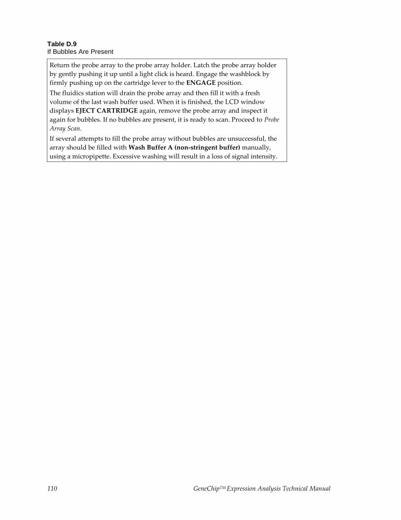
110 GeneChipTM Expression Analysis Technical Manual
Table D.9 If Bubbles Are Present
Return the probe array to the probe array holder. Latch the probe array holder
by gently pushing it up until a light click is heard. Engage the washblock by
firmly pushing up on the cartridge lever to the ENGAGE position.
The fluidics station will drain the probe array and then fill it with a fresh
volume of the last wash buffer used. When it is finished, the LCD window
displays EJECT CARTRIDGE again, remove the probe array and inspect it
again for bubbles. If no bubbles are present, it is ready to scan. Proceed to Probe
Array Scan.
If several attempts to fill the probe array without bubbles are unsuccessful, the
array should be filled with Wash Buffer A (non-stringent buffer) manually,
using a micropipette. Excessive washing will result in a loss of signal intensity.
GeneChipTM Expression Analysis Technical Manual 111
Probe Array Scan
The scanner is also controlled by Microarray Suite or GCOS. The probe array is scanned after the wash
protocols are complete. Make sure the laser is warmed up prior to scanning by turning it on at least 15
minutes before use if you are using the Agilent GeneArrayTM Scanner, or 10 minutes if you are using the
GeneChipTM Scanner 3000. If the probe array was stored at 4°C, warm to room temperature before scanning.
Refer to the Microarray Suite or GCOS online help and the appropriate scanner user’s manual for more
information on scanning.
The scanner uses a laser and is equipped with a safety interlock system. Defeating the interlock system may result in exposure to hazardous laser light.
You must have read and be familiar with the operation of the scanner before attempting to scan a probe array. Refer to the Microarray Suite User’s Guide (P/N 08-0081) or to the GeneChip
TM Scanner 3000 quick reference card (P/N 08-
0075).
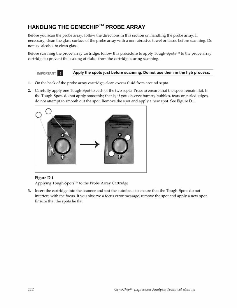
112 GeneChipTM Expression Analysis Technical Manual
HANDLING THE GENECHIPTM PROBE ARRAY
Before you scan the probe array, follow the directions in this section on handling the probe array. If
necessary, clean the glass surface of the probe array with a non-abrasive towel or tissue before scanning. Do
not use alcohol to clean glass.
Before scanning the probe array cartridge, follow this procedure to apply Tough-Spots™ to the probe array
cartridge to prevent the leaking of fluids from the cartridge during scanning.
Apply the spots just before scanning. Do not use them in the hyb process.
1. On the back of the probe array cartridge, clean excess fluid from around septa.
2. Carefully apply one Tough-Spot to each of the two septa. Press to ensure that the spots remain flat. If
the Tough-Spots do not apply smoothly; that is, if you observe bumps, bubbles, tears or curled edges,
do not attempt to smooth out the spot. Remove the spot and apply a new spot. See Figure D.1.
Figure D.1
Applying Tough-Spots™ to the Probe Array Cartridge
3. Insert the cartridge into the scanner and test the autofocus to ensure that the Tough-Spots do not
interfere with the focus. If you observe a focus error message, remove the spot and apply a new spot.
Ensure that the spots lie flat.
GeneChipTM Expression Analysis Technical Manual 113
SCANNING THE PROBE ARRAY
1. Select Run → Scanner from the menu bar. Alternatively, click the Start Scan icon in the tool bar.
The Scanner dialog box appears with a drop-down list of experiments that have not been run.
2. Select the experiment name that corresponds to the probe array to be scanned.
A previously run experiment can also be selected by using the Include Scanned Experiments option
box. After selecting this option, previously scanned experiments appear in the drop-down list.
3. By default, for the AgilentTM GeneArrayTM Scanner only, after selecting the experiment the number [2]
is displayed in the Number of Scans box to perform the recommended 2X image scan. For the
GeneChipTM Scanner 3000, only one scan is required.
4. Once the experiment has been selected, click the Start button.
A dialog box prompts you to load a sample into the scanner.
5. If you are using the GeneArrayTM Scanner, click the Options button to check for the correct pixel value
and wavelength of the laser beam.
• Pixel value = 3 μm
• Wavelength = 570 nm
If you are using the GeneChipTM Scanner 3000, pixel resolution and wavelength are preset and cannot
be changed.
6. Open the sample door on the scanner and insert the probe array into the holder. Do not force the probe
array into the holder. Close the sample door of the scanner. If you are using the GeneChipTM Scanner
3000, do not attempt to close the door by hand. The door closes automatically through the User
Interface when start scan is selected or the scanner goes into stand-by mode.
7. Click OK in the Start Scanner dialog box.
The scanner begins scanning the probe array and acquiring data. When Scan in Progress is Selected
from the View menu, the probe array image appears on the screen as the scan progresses.

114 GeneChipTM Expression Analysis Technical Manual
Shutting Down the Fluidics Station
1. After removing a probe array from the probe array holder, the LCD window displays the message
ENGAGE WASHBLOCK.
2. If you are using the FS-400, latch the probe array holder by gently pushing up until a light click is heard.
Engage the washblock by firmly pushing up on the cartridge lever to the ENGAGE position.
If you are using the FS-450, gently lift up the cartridge lever to engage, or close, the washblock.
The fluidics station automatically performs a Cleanout procedure. The LCD window indicates the
progress of the Cleanout procedure.
3. When the fluidics station LCD window indicates REMOVE
VIALS, the Cleanout procedure is complete.
4. Remove the sample microcentrifuge vial(s) from the sample holder(s).
5. If no other hybridizations are to be performed, place wash lines into a bottle filled with deionized water.
6. Select Shutdown or Shutdown_450 for all modules from the drop-down Protocol list in the Fluidics
Station dialog box. Click the Run button for all modules.
The Shutdown protocol is critical to instrument reliability. Refer to the appropriate Fluidics Station
User’s Guide for more information.
7. After Shutdown protocol is complete, flip the ON/OFF switch of the fluidics station to the OFF position.
To maintain the cleanliness of the fluidics station and obtain the highest quality image and data possible, a weekly bleach protocol and a monthly decontamination protocol are highly recommended. Refer to the appropriate GeneChipTM Fluidics Station User’s Guide for further detail.
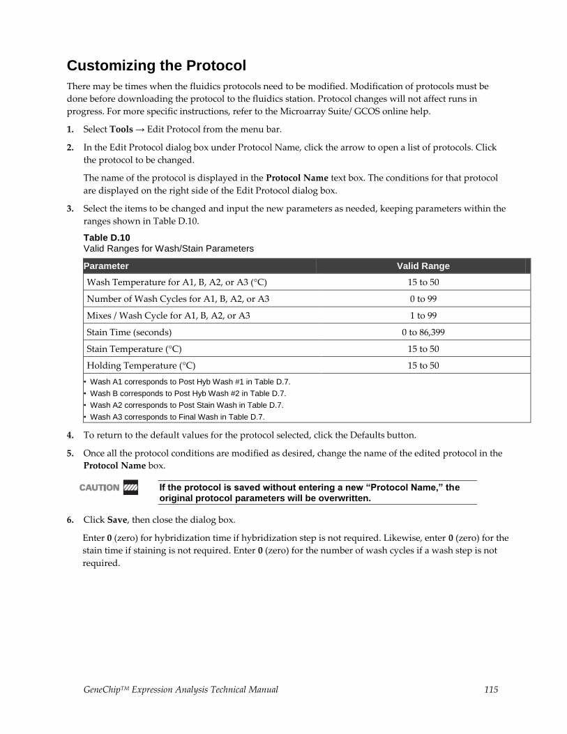
GeneChipTM Expression Analysis Technical Manual 115
Customizing the Protocol
There may be times when the fluidics protocols need to be modified. Modification of protocols must be
done before downloading the protocol to the fluidics station. Protocol changes will not affect runs in
progress. For more specific instructions, refer to the Microarray Suite/ GCOS online help.
1. Select Tools → Edit Protocol from the menu bar.
2. In the Edit Protocol dialog box under Protocol Name, click the arrow to open a list of protocols. Click
the protocol to be changed.
The name of the protocol is displayed in the Protocol Name text box. The conditions for that protocol
are displayed on the right side of the Edit Protocol dialog box.
3. Select the items to be changed and input the new parameters as needed, keeping parameters within the
ranges shown in Table D.10.
Table D.10 Valid Ranges for Wash/Stain Parameters
Parameter Valid Range
Wash Temperature for A1, B, A2, or A3 (°C) 15 to 50
Number of Wash Cycles for A1, B, A2, or A3 0 to 99
Mixes / Wash Cycle for A1, B, A2, or A3 1 to 99
Stain Time (seconds) 0 to 86,399
Stain Temperature (°C) 15 to 50
Holding Temperature (°C) 15 to 50
• Wash A1 corresponds to Post Hyb Wash #1 in Table D.7.
• Wash B corresponds to Post Hyb Wash #2 in Table D.7.
• Wash A2 corresponds to Post Stain Wash in Table D.7.
• Wash A3 corresponds to Final Wash in Table D.7.
4. To return to the default values for the protocol selected, click the Defaults button.
5. Once all the protocol conditions are modified as desired, change the name of the edited protocol in the
Protocol Name box.
If the protocol is saved without entering a new “Protocol Name,” the original protocol parameters will be overwritten.
6. Click Save, then close the dialog box.
Enter 0 (zero) for hybridization time if hybridization step is not required. Likewise, enter 0 (zero) for the
stain time if staining is not required. Enter 0 (zero) for the number of wash cycles if a wash step is not
required.
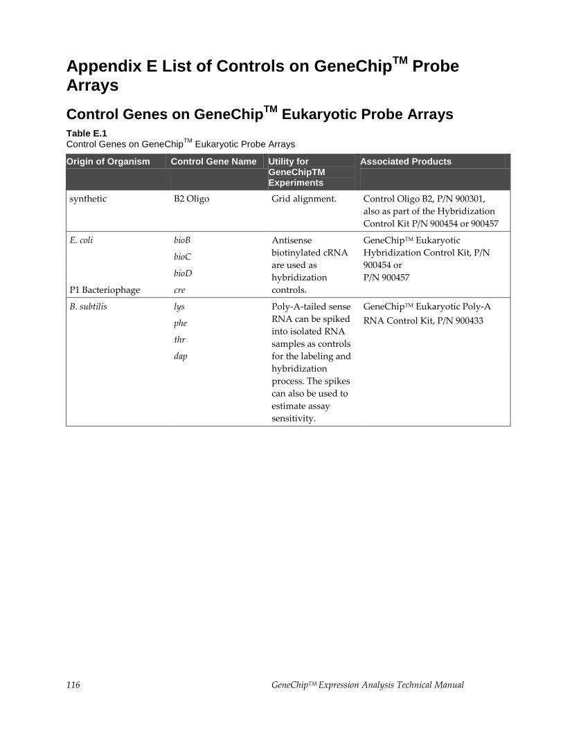
116 GeneChipTM Expression Analysis Technical Manual
Appendix E List of Controls on GeneChipTM
Probe Arrays
Control Genes on GeneChipTM
Eukaryotic Probe Arrays
Table E.1 Control Genes on GeneChip
TM Eukaryotic Probe Arrays
Origin of Organism Control Gene Name Utility for GeneChipTM Experiments
Associated Products
synthetic B2 Oligo Grid alignment. Control Oligo B2, P/N 900301,
also as part of the Hybridization
Control Kit P/N 900454 or 900457
E. coli bioB Antisense
biotinylated cRNA
are used as
hybridization
controls.
GeneChipTM Eukaryotic
Hybridization Control Kit, P/N
900454 or
P/N 900457
bioC
bioD
P1 Bacteriophage cre
B. subtilis lys Poly-A-tailed sense
RNA can be spiked
into isolated RNA
samples as controls
for the labeling and
hybridization
process. The spikes
can also be used to
estimate assay
sensitivity.
GeneChipTM Eukaryotic Poly-A
RNA Control Kit, P/N 900433 phe
thr
dap
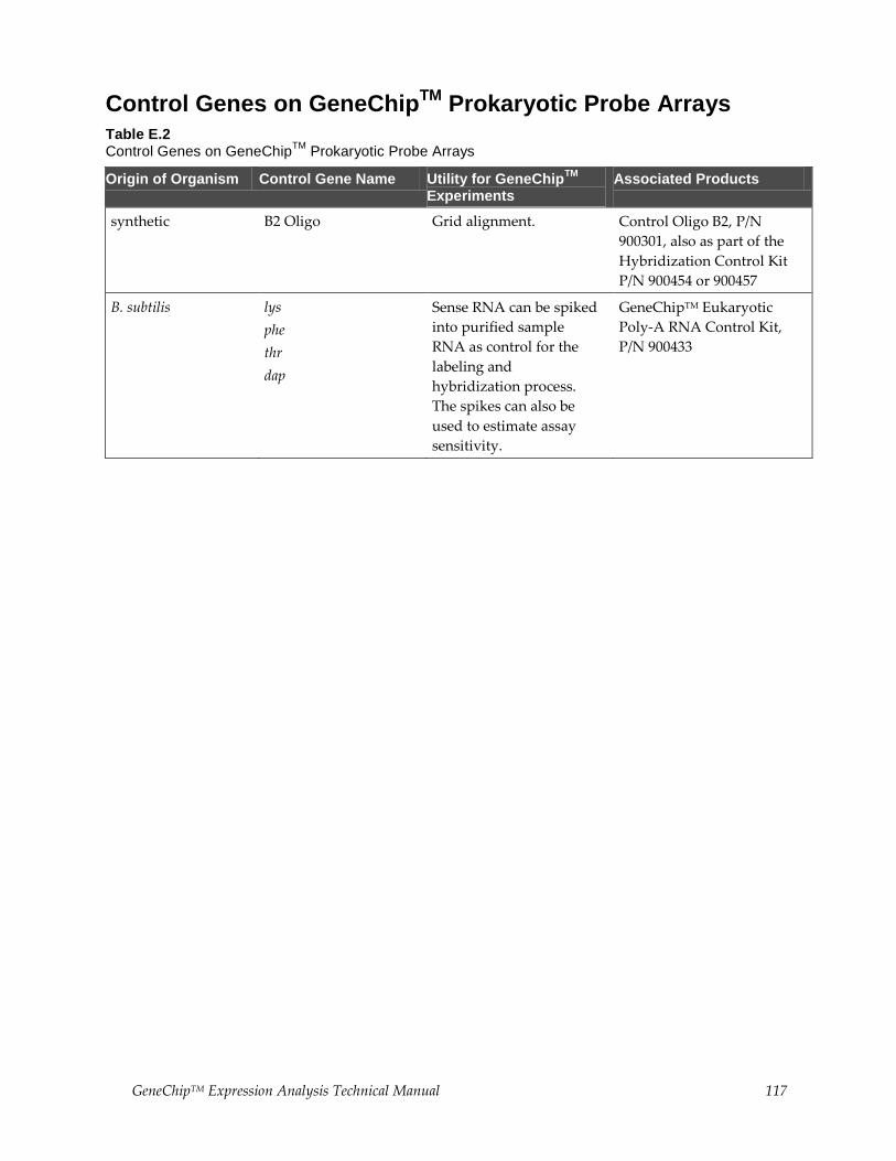
GeneChipTM Expression Analysis Technical Manual 117
Control Genes on GeneChipTM
Prokaryotic Probe Arrays
Table E.2 Control Genes on GeneChip
TM Prokaryotic Probe Arrays
Origin of Organism Control Gene Name Utility for GeneChipTM
Experiments
Associated Products
synthetic B2 Oligo Grid alignment. Control Oligo B2, P/N
900301, also as part of the
Hybridization Control Kit
P/N 900454 or 900457
B. subtilis lys
phe
thr
dap
Sense RNA can be spiked
into purified sample
RNA as control for the
labeling and
hybridization process.
The spikes can also be
used to estimate assay
sensitivity.
GeneChipTM Eukaryotic
Poly-A RNA Control Kit,
P/N 900433
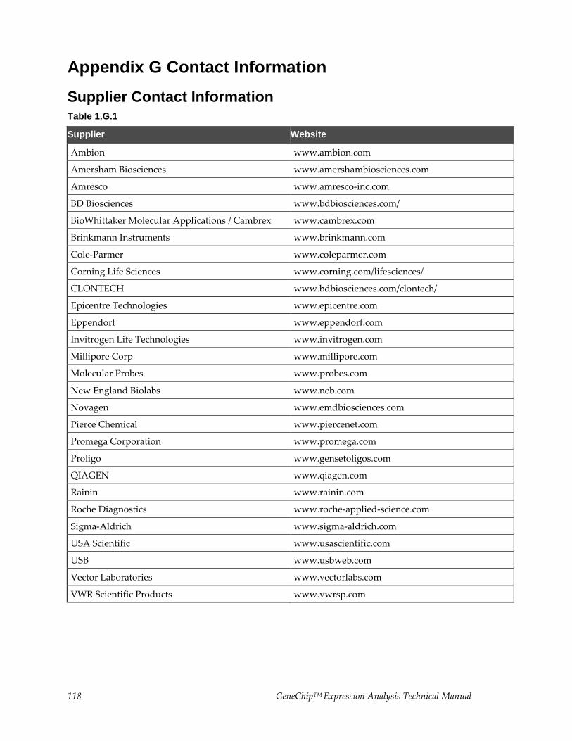
118 GeneChipTM Expression Analysis Technical Manual
Appendix G Contact Information
Supplier Contact Information
Table 1.G.1
Supplier Website
Ambion www.ambion.com
Amersham Biosciences www.amershambiosciences.com
Amresco www.amresco-inc.com
BD Biosciences www.bdbiosciences.com/
BioWhittaker Molecular Applications / Cambrex www.cambrex.com
Brinkmann Instruments www.brinkmann.com
Cole-Parmer www.coleparmer.com
Corning Life Sciences www.corning.com/lifesciences/
CLONTECH www.bdbiosciences.com/clontech/
Epicentre Technologies www.epicentre.com
Eppendorf www.eppendorf.com
Invitrogen Life Technologies www.invitrogen.com
Millipore Corp www.millipore.com
Molecular Probes www.probes.com
New England Biolabs www.neb.com
Novagen www.emdbiosciences.com
Pierce Chemical www.piercenet.com
Promega Corporation www.promega.com
Proligo www.gensetoligos.com
QIAGEN www.qiagen.com
Rainin www.rainin.com
Roche Diagnostics www.roche-applied-science.com
Sigma-Aldrich www.sigma-aldrich.com
USA Scientific www.usascientific.com
USB www.usbweb.com
Vector Laboratories www.vectorlabs.com
VWR Scientific Products www.vwrsp.com
GeneChipTM Expression Analysis Technical Manual 119
Documentation and support
Obtaining support
Technical support For the latest services and support information for all locations, visit
www.thermofisher.com.
At the website, you can:
Access worldwide telephone and fax numbers to contact Technical Support
and Sales facilities
Search through frequently asked questions (FAQs)
Submit a question directly to Technical Support (thermofisher.com/support)
Search for user documents, SDSs, vector maps and sequences, application
notes, formulations, handbooks, certificates of analysis, citations, and other
product support documents
Obtain information about customer training
Download software updates and patches
Safety Data Sheets (SDS)
Safety Data Sheets (SDSs) are available at thermofisher.com/support.
Limited product warranty
Life Technologies Corporation and/or its affiliate(s) warrant their products as set
forth in the Life Technologies’ General Terms and Conditions of Sale found on
Life Technologies’ website at www.thermofisher.com/us/en/home/global/terms-
and-conditions.html. If you have any questions, contact Life Technologies at
www.thermofisher.com/support.


For support visit thermofisher.com/support or email [email protected]
thermofisher.com
23 January 2017