Embed Size (px)
Citation preview

Proc. Natl. Acad. Sci. USAVol. 80, pp. 3958-3962, July 1983Biochemistry
Gene structure of a phenobarbital-inducible cytochrome P-450in rat liver
(amino acid sequence/monooxygenase/recombinant DNA/DNA sequence/mRNA structure)
YUZURU MIZUKAMI*, KAZUHIRO SOGAWA, YORIMASA SUWA, MASAMI MURAMATSU, ANDYOSHIAKI FUJII-KURIYAMAtDepartment of Biochemistry, Cancer Institute, Japanese Foundation for Cancer Research, 1-37-1 Kamiikebukuro, Toshima-ku, Tokyo, Japan
Communicated by Ronald W. Estabrook, April 1, 1983
ABSTRACT The gene structure of a phenobarbital-induciblerat liver cytochrome P-450 was elucidated by sequence analysis oftwo isolated genomic clones. The total length of the gene was ap-proximately 14 kilobases and was separated into nine exons by eightintervening sequences. Nucleotide sequences of all exon/intronboundaries follow the G-T/A-G rule. A putative transcription ini-tiation site was assigned to an A, 30 base pairs upstream from theinitiation codon by SI nuclease protection mapping. A possible"TATA" equivalent sequence, C-A-T-A-A-A, was found 27 basepairs further upstream from this initiation site. A poly(A) attach-ment site was determined to be 386 or 387 base pairs downstreamfrom the termination codon by comparison with the cDNA se-quence. Detailed comparison with two cDNA sequences deter-mined previously showed the coding nucleotide sequence of thegenomic clones to concur with that of the pcP-450pb2 cDNA clonecoding for cytochrome P-450e except for three neutral base sub-stitutions. Therefore, we conclude that the gene sequence deter-mined here is for the cytochrome P-450e gene or a similar gene.On the other hand, 40 base substitutions were found in about 1,900base pairs compared between the sequences of the genomic cloneand the other cDNA clones (pcP-450pbl and 4) coding for cyto-chrome P-450b, and 15 of them result in 14 amino acid replace-ments in the total 491 amino acid residues. These base substitu-tions occur in relatively limited regions of the sequences. Most ofthem are found in exons 6, 7, 8, and 9; most frequently in exon7.
Cytochrome P-450 is a component of the microsomal monooxy-genase system which metabolizes endogenous substrates suchas steroids and fatty acids as well as exogenous substrates suchas many kinds of drugs and other lipophilic xenobiotics. Thisunusual metabolic versatility of the monooxygenase system re-sults from the participation of multiple forms of cytochrome P-450, each of which exhibits a different but, in some cases, over-lapping substrate specificity (1-3). It is known, in addition, thatmost of these species of cytochrome P-450 are inducible en-zymes; administration of drugs and other xenobiotics to animalsinduces the synthesis of specific forms of cytochrome P-450 (2,3). The molecular multiplicity and mechanisms of induction ofcytochrome P-450 can be best understood by using recombi-nant DNA technology to perform studies at the gene or DNAlevel.We previously reported deduced primary amino acid se-
quences of phenobarbital-inducible cytochrome P-450 from se-quence analysis of the cDNA clones (4). By using these cDNAsas probes, we have isolated several genomic clones of the cv-tochrome P-450 which are independent of one another on thebasis of their restriction analyses (5).
Here we report the structural analysis of one of the isolatedgenes for phenobarbital-inducible rat liver cytochrome P-450and a comparison of the nucleotide sequences of the genomicand cDNA clones.
MATERIALS AND METHODS
.Restriction endonucleases were obtained from Takara Shuzo(Kyoto, Japan), Bethesda Research Laboratories, and New En-gland BioLabs. T4 DNA ligase was from Takara Shuzo. Esch-erichia coli DNA polymerase I and polynucleotide kinase werepurchased from Boehringer Mannheim. Bacterial alkalinephosphatase was from Worthington. [y-32P]ATP (3,000 Ci/mmol;1 Ci = 3.7 X 10'° Bq) and [a-32P]dCTP (2,000-3,000 Ci/mmol)were from the Radiochemical Centre. S1 nuclease was obtainedfrom P-L Biochemicals. The mRNA for cytochrome P-450 waspartially purified from livers of phenobarbital-treated rats(Sprague-Dawley) as described (6).DNA Preparations and Blot-Hybridization Experiments.
Recombinant plasmid (6) and phage DNAs (7) were purified asdescribed. The purified DNAs were cleaved with various re-striction endonucleases as recommended by the manufacturers.The DNA fragments were resolved on either 0.8-1.2% agarosegels or 5-8% polyacrylamide gels (6). Recovery of DNA frag-ments from gels (8) and restriction analysis (9) were conductedas described. Blot-hybridization analysis was performed ac-cording to the procedure of Southern (10).DNA Sequence Analysis. Sequence analysis of DNA was
performed as described by Maxam and Gilbert (11).Isolation of the Genomic Clones for Cytochrome P-450
Genes. The genomic clone pgP-450pb6 was previously isolated(5) from the rat (Sprague-Dawley) EcoRI gene library (a giftfrom T. D. Sargent, R. B. Wallace, and J. Bonner). This clonecontained approximately two-thirds of the cytochrome P-450gene sequence. Another clone, pgP-450pbl2, which covers themissing sequence of the cytochrome P-450 gene in pgP-450pb6,was isolated by screening the rat (Sprague-Dawley) Hae III genelibrary (a gift from L. L. Jagodzinsky and J. Bonner) with a nick-translated 2.5-kilobase (kb) BamHI/EcoRI fragment of pgP-450pb6 (see Fig. 2) as a probe as described (12).
To facilitate sequence analysis, some DNA fragments of thegenomic clones were subcloned into the plasmid pBR322 aftercleavage of the genomic DNAs with appropriate restriction en-zymes. The BamHI/BamHI 7.0-kb and BamHI/EcoRI 2.5-kbfragments were inserted at the BamHI site and between theEcoRI and the BamHI site of pBR322, respectively.
Abbreviations: kb, kilobase(s); bp, base pair(s).* Present address: Laboratory of Microbiology 1I, Central ResearchLaboratories, Meiji Seika, LTD, Kohoku-ku Yokohama, 222, Japan.
tTo whom reprint requests should be addressed.
3958
The publication costs of this article were defrayed in part by page chargepayment. This article must therefore be hereby marked "advertise-ment" in accordance with 18 U.S.C. § 1734 solely to indicate this fact.

Proc. Natl. Acad. Sci. USA 80 (1983) 3959
cDNA Clones. Four cDNA clones, pcP-450pbl, -2, -3, and-4, were isolated previously (4). The DNA sequence of the in-sert in pcP-450pb3 (C-4-7) which contained the cDNA se-quence corresponding to the 3' end region of the cytochromeP-450 mRNA was determined as described and included in Fig.4.
SI Nuclease Mapping. SI nuclease protection mapping wasperformed as described (13).
A23 4 5 6
,I I,, ,, , I,,
,3 ,1 //6 822 ,,
,1 ,'
RESULTS AND DISCUSSIONIsolation and Restriction Mapping of Cytochrome P-450
Gene. From the rat EcoRI gene library, we had isolated six dif-ferent genomic clones for phenobarbital-inducible cytochromeP-450 (5). One of them, pgP-450pb6, was chosen for the pres-ent study because this clone contained a DNA insert that hy-bridized most strongly with the cDNA probe and was one of themost extended toward the 5' direction of the gene. However,because this clone was found to lack the sequence of the 3' endregion of the cytochrome P-450 gene, we isolated another clone,pgP-450pbl2, whose insert covered the missing sequence inpgP-450pb6 from rat Hae III gene library. Restriction enzymedigestions and subsequent Southern blot analysis of this clonedDNA confirmed the presence of the 2.5-kb BamHI-EcoRIfragment in the insert (Fig. 1). Some faint hybridization bandswere occasionally observed at positions other than 14 kb (Fig.1A Left), 7 and 2.5 kb (Fig. 1A Right), and 2.5 kb (Fig. 1B);these bands could be due to either partial or aberrant digestionby the enzymes. Restriction cleavage sites in the DNA se-quence of pgP-450pb6 and -12 were mapped by using a com-bination of complete and partial enzyme digestions as de-
B
PP45SO 6
PgP450Sp 12 X E HibXXHiE X Xh HiL-E-X-6
c
B-7
3E-2
E X-6
., AA Bg
.~~~~~~~~~~~~~~~~~~~~~~~~~~~~~~~~~~~~~~~~~~~~~~~~~~~~~~~~~~~~~~~~~~~~~~~~~~~~~-IgS3 ~ 1~S3Sgg
No xIpv H
i4-
I I t",~~ i 4i' --Ii ~4KH, 0 P4 K c!k
~~~A A
A Ba
24-
9.5-6.7-
4-3-
2.3-2.0-
0.6-
Eco R
b a b
24
95--
667-
43-
2.3-
20-
EcoRi+ BamHl
a b
EcoRI+ Bamr Hl
FIG. 1. Blot-hybridization analyses of recombinant phage DNAscontaining rat cytochrome P-450 sequences. (A) DNA (1 Ug) from ge-nomic clone pgP-450pb6 isolated previously (5) was digested with EcoRIor EcoRI/BamHI, electrophoresed on a 0.8% agarose gel, and stainedwith ethidium bromide (lane a). The DNA was transferred from the gelto filter papers for hybridization (7) with the nick-translated cDNA pre-
pared from pcP-450pb2 (4). The filter paper was washed with 0.015 MNaCl/1.5 mM sodium citrate at pH 7.0 (0.1 x standard saline citrate)at 65°C for 1 hr and autoradiographed (lane b). (B) DNA (1 Ug) frompgP-450pbl2 was digested with EcoRI/BamHI and electrophoresed as
in A. The DNA was blot-hybridized with the nick-translated 2.5-kbEcoRI-BamHI DNA fragment from pgP-450pb6 as described. The lengths(in kb) of representative size markers are shown at the left of each panel.
FIG. 2. Organization of rat cytochrome P-450 gene and strategy forits sequence analysis. (A) The locations of exons 1-9 (solid boxes) andintrons a-h were first estimated by Southern blot analysis using thecDNA as a probe and then determined by sequence analysis as de-scribed in the text. The mRNA structure is depicted schematically onthe lower line; the hatched box represents the coding nucleotide se-quence and "A" and "poly(A)" indicate the transcription-initiation andpoly(A) addition sites of the mRNA, respectively. "ATG" and "TGA"refer to translation-initiation and termination codons of the mRNA,respectively. Scale = 1 kb. (B) Two genomic clones (pgP-450pb6 and-12) arranged to show their overlappingDNA fragments. The exon andintron sequences are shown as in A. The fine and heavy lines indicateCharon 4A vector sequences and inserted rat DNA sequences, respec-tively. The 7.0-kb BamHI fragment (B-7) and 2.5-kb BamHI-EcoRIfragment (B-E-2) of pgP-450pb6 were subcloned into plasmid pBR322for sequence analysis. The 6.5-kbEcoRI-Xho I fragment (E-X-6) ofpgP-450pb12 was used directly for sequence determination. The orientationof the gene is indicated by the arrow on the top of the figure. Scale =1 kb. (C) Restriction maps of fragments B-7, B-E-2, and E-X-6 in B areindicated on an expanded scale. Each arrow indicates the direction andlength of the DNA fragment analyzed from appropriate restriction sites.E, EcoRI; X, Xba I; Hi, HindIII; B, BamHI; P, Pst I; H,HihfI; Bs, BstNI;Sc, Sac I; Bg, Bgl II; Pv, Pvu II; A, Ava Il; Hp, Hpa II; S9, Sau96I; S3,Sau3A; K, Kpn I; Xh, Xho I; Ac, Acc I. Scale bars: B-7, 1 kb; B-E-2, 0.1kb; E-X-6, 1 kb.
scribed by Smith and Birnstiel (9). These two genomic clonesactually contained regions overlapping with each other in ad-dition to the BamHI-EcoRI fragment (Fig. 2).
Sequence Analysis of Cytochrome P-450 Gene. We deter-mined the nucleoside sequence of all the exons and their flank-ing regions in the introns as follows. Restriction fragments con-
taining exon sequences were first identified by Southern blotanalysis (10) using the labeled cDNA as a probe (data not shown),and then appropriate restriction sites were chosen for end la-beling by the method of Maxam and Gilbert (11). In most cases,
restriction sites that were commonly found in both the cDNA
I
f /I/ /!
/I'
r ///152 120 14-
h / ,-
I -py(
it^ 1862 )
I-IL
9L-
Biochemistry: Mizukami et al.
B-7 K-2-". J1 PIE x Hi Hi I i i
3960 Biochemistry: Mizukami et al
and the genomic sequences were used. Finally, the exact cod-ing nucleotide sequences in the exons and their flanking re-
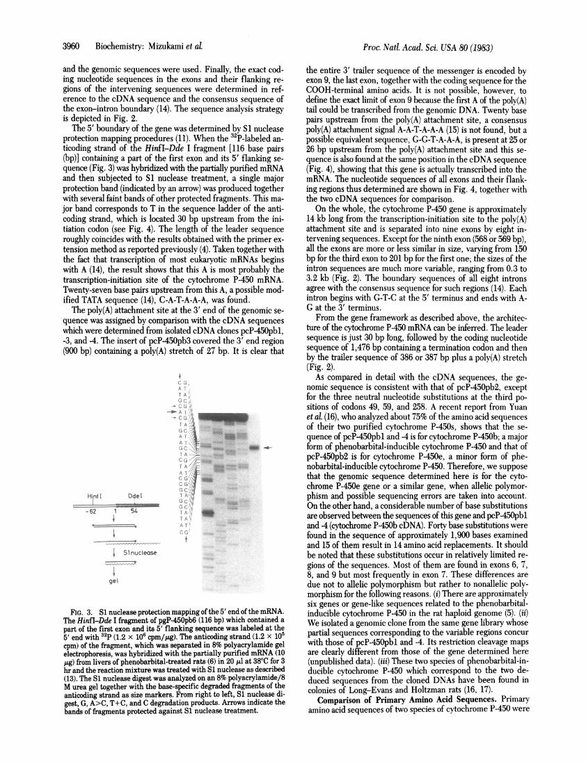
gions of the intervening sequences were determined in ref-erence to the cDNA sequence and the consensus sequence ofthe exon-intron boundary (14). The sequence analysis strategyis depicted in Fig. 2.The 5' boundary of the gene was determined by S1 nuclease
protection mapping procedures (11). When the 32P-labeled an-
ticoding strand of the HinfI-Dde I fragment [116 base pairs(bp)] containing a part of the first exon and its 5' flanking se-
quence (Fig. 3) was hybridized with the partially purified mRNAand then subjected to S1 nuclease treatment, a single majorprotection band (indicated by an arrow) was produced togetherwith several faint bands of other protected fragments. This ma-
jor band corresponds to T in the sequence ladder of the anti-coding strand, which is located 30 bp upstream from the ini-tiation codon (see Fig. 4). The length of the leader sequence
roughly coincides with the results obtained with the primer ex-
tension method as reported previously (4). Taken together withthe fact that transcription of most eukaryotic mRNAs beginswith A (14), the result shows that this A is most probably thetranscription-initiation site of the cytochrome P-450 mRNA.Twenty-seven base pairs upstream from this A, a possible mod-ified TATA sequence (14), C-A-T-A-A-A, was found.The poly(A) attachment site at the 3' end of the genomic se-
quence was assigned by comparison with the cDNA sequences
which were determined from isolated cDNA clones pcP450pbl,-3, and -4. The insert of pcP-450pb3 covered the 3' end region(900 bp) containing a poly(A) stretch of 27 bp. It is clear that
-rA.TOAT
TAT(i.c
Hinf I Ddel
- 62 1 54
S51nuclease
gel
*-
1
.t
3:i
I;C
T A
G( I A
TAve >-
fA
FIG. 3. S1 nuclease protection mapping of the5' end of the mRNA.
The HinfI-Dde I fragment of pgP-450pb6 (116 bp) which contained a
part of the first exon and its 5' flanking sequence was labeled at the
5' end with 32p (1.2 106 cpm//Lg). The anticoding strand (1.2 105
cpm) of the fragment, which was separated in 8% polyacrylamide gel
electrophoresis, was hybridized with the partially purified mRNA (10
gg) from livers of phenobarbital-treated rats (6) in 20ul at 380C for 3
hr and the reaction mixture was treated withS1 nuclease as described
(13). The Si nuclease digest was analyzed on an 8% polyacrylamide/8
M urea gel together with the base-specific degraded fragments of the
anticoding strand as size markers. From right to left, Si nuclease di-
gest, G, A>C, T+C, and C degradation products. Arrows indicate the
bands of fragments protected against Si nuclease treatment.
the entire 3' trailer sequence of the messenger is encoded byexon 9, the last exon, together with the coding sequence for theCOOH-terminal amino acids. It is not possible, however, todefine the exact limit of exon 9 because the first A of the polv(A)tail could be transcribed from the genomic DNA. Twenty basepairs upstream from the poly(A) attachment site, a consensuspoly(A) attachment signal A-A-T-A-A-A (15) is not found, but apossible equivalent sequence, G-G-T-A-A-A, is present at 25 or26 bp upstream from the poly(A) attachment site and this se-quence is also found at the same position in the cDNA sequence(Fig. 4), showing that this gene is actually transcribed into themRNA. The nucleotide sequences of all exons and their flank-ing regions thus determined are shown in Fig. 4, together withthe two cDNA sequences for comparison.On the whole, the cytochrome P-450 gene is approximately
14 kb long from the transcription-initiation site to the poly(A)attachment site and is separated into nine exons by eight in-tervening sequences. Except for the ninth exon (568 or 569 bp),all the exons are more or less similar in size, varying from 150bp for the third exon to 201 bp for the first one; the sizes of theintron sequences are much more variable, ranging from 0.3 to3.2 kb (Fig. 2). The boundary sequences of all eight intronsagree with the consensus sequence for such regions (14). Eachintron begins with G-T-C at the 5' terminus and ends with A-G at the 3' terminus.From the gene framework as described above, the architec-
ture of the cytochrome P450 mRNA can be inferred. The leadersequence is just 30 bp long, followed by the coding nucleotidesequence of 1,476 bp containing a termination codon and thenby the trailer sequence of 386 or 387 bp plus a poly(A) stretch(Fig. 2).
As compared in detail with the cDNA sequences, the ge-nomic sequence is consistent with that of pcP-450pb2, exceptfor the three neutral nucleotide substitutions at the third po-sitions of codons 49, 59, and 258. A recent report from Yuanet al. (16), who analyzed about 75% of the amino acid sequencesof their two purified cytochrome P-450s, shows that the se-quence of pcP-450pbl and -4 is for cytochrome P-450b; a majorform of phenobarbital-inducible cytochrome P-450 and that ofpcP-450pb2 is for cytochrome P-450e, a minor form of phe-nobarbital-inducible cytochrome P-450. Therefore, we supposethat the genomic sequence determined here is for the cyto-chrome P-450e gene or a similar gene, when allelic polymor-phism and possible sequencing errors are taken into account.On the other hand, a considerable number of base substitutionsare observed between the sequences of this gene and pcP450pbland4 (cytochrome P-450b cDNA). Forty base substitutions werefound in the sequence of approximately 1,900 bases examinedand 15 of them result in 14 amino acid replacements. It shouldbe noted that these substitutions occur in relatively limited re-gions of the sequences. Most of them are found in exons 6, 7,8, and 9 but most frequently in exon 7. These differences aredue not to allelic polymorphism but rather to nonallelic poly-morphism for the following reasons. (i) There are approximatelysix genes or gene-like sequences related to the phenobarbital-inducible cytochrome P-450 in the rat haploid genome (5). (ii)We isolated a genomic clone from the same gene library whosepartial sequences corresponding to the variable regions concurwith those of pcP-450pbl and4. Its restriction cleavage mapsare clearly different from those of the gene determined here(unpublished data). (iii) These two species of phenobarbital-in-ducible cytochrome P-450 which correspond to the two de-duced sequences from the cloned DNAs have been found incolonies of Long-Evans and Holtzman rats (16, 17).
Comparison of Primary Amino Acid Sequences. Primaryamino acid sequences of two species of cytochrome P-450 were
Proc. Natl. Acad. Sci. USA 80 (1983)

Biochemistry: Mizukami et aL Proc. Nati. Acad. Sci. USA 80 (1983) 3961
TiZAACATATGAAGTTGCATAACT6A6JTGTAGG66CAGATTCAUCATAAAAGATCCTGCTG6IGCATAGCACT~ GAAGTCTACCGTGGTTACACCAGGACC ATG GAG CCC AGT ATC
FTi CTC CTC CIT GCT CTC CTT GrG GGC TC TTG TTA CrC TTA GrC AGG GGA CAC CCA AAG TCC CGT GGC AAC TTC CCA CCA GGA ccr CGf
CCC CTT CCC CTC TTG GGG AAC CTC CTG CAG TTG GAC AGA GGA GGC CTC CTC AAT TCC TTC ATG CAG GTGAGATATTCACAGGGCCTGGTGT----------- -- - - -- - -- - -- --G - -- - -- - -- -- - --- - -- - -- - -------- - -- - -- --- - -- --G -- - - -- -- - - -- -- - -- - - -- ---
3.2 Kb -----CAGTCCTGTGCCTTTTAGTTTGCAG CTT CGC GAA AAA TAT GGA GAT GTG TTC ACA GTA CAC CTG GGA CCA AGG CCT GTG GTC A----A --- --- --- --- --- -- - --- --
TG CTA TGT GGG ACA GAC ACC ATA AAG GAG GCT CTG GTG GGC CAA GCT GAG GAT TTC TCT GGT CGG GGA ACA ATC GCT GTG ATT GAG CCA
ATC TTC AAG GAA TAT G GTAAGACTCTCAAAGGTTTGGGATG----- 0.3 Kb -----ATCCATGCTTCCCCTACTTTGTCAG GT GTG ATC TTT GCC AAT G
GG GAA CGC TGG AAG GCC CTT CGG CGA TTC TCT CTG GCT ACC ATG AGA GAC TTT GGG ATG GGA AAG AGG AGT GTG GAA GAA CGG ATT CAb
GAG' GAA GCC CAA TGT TTG GTG GAG 6AA CTG CGG AAA TCC CAG G GTGAATCGGAAAGG----- 2.3 Kb -----CTCTTATCCTGCCTCCTCATCCTCC
AG GA GCC CCA CTG GAT CCC ACC TTC CTC TTC CAG TGC ATC ACA GCC AAC ATC ATC TGC TCC ATT GTG TTT GGA GAG CGC TTT GAC TAC A
CA GAC CGC CAG TTC CTG CGC CTG TTG GAG CTG TTC TAC CGG ACC TTT TCC CHC CTA AGT TCA TTC TCC AGC CAG GTCCGTGGGTGGGAAGAGAA
GAGTA----- 0.8 Kb -----TGAGGTGGTGGTTCTTGCCTTACAG GTG TTT GAG TTC TTC TCT GGG TTC CTG AAA TAC TTT CCT GGT GCC CAC AG
A CAA ATC TCC AAA AhC CIC CAG GAA ATC CTC GAT TAC ATT GGC CAT ATT GTG GAG AAG CAC AGG GCC ACC TTA GAC CCC AGC GCT CCA C--- --- --- --- --- --- --- --- --- --- --- --- --A ---
--- --- --- --- --- --- --- --- --- --- --A --- --- -
bA GAC TTC ATC GAC ACT TAC CTT CTG CGC ATG GC. AA,, GTGAGTCCTGCATGGATGAGAGAGG----- 0.5 Kb -----CCATTCGCCCCCCA.GGTGCAA
CCAG GAG AAG TCG AAC CAC CAC ACA GAG TTC CAT GAT GAG AAC CTC ATG ATC TCC CTG CTC TCT CTC TTC TTT GCT GGC ACT GAG ACC GG------ --- --- --- --- --- --- --- --- --- --- --- --- --- --- A-
C AGC ACC ACA CTC CGC TAT GGT TTC CTG CTC ATG CTC AAG TAC CCC CAT 5TC ACA G GTATATCATGGGGGGTACCGTTGG----- 1.6 Kb --
- --- --- --- --- --- --- --- --- --- --G --- --- --- ---- --- --- --- G-- -
---CCCTTTCTCGCAG TG AAA GTC CAA AAG GAG ATT GAT CAG GTG ATT GGC TCT CAC AGG CCA CCA TCC CTT GAT GAT CGT ACC AAA ATG CCAA- --- --- --- --- --- --- --- --- --- --C --- --A ---C- - A-- --- --- C --C -GT --- ---
TAC ACT GAT GCA GTC ATC CAC GAG ATT CAG AGG TTT GCA GAT CTT GCC CCA ATT GGT TTA CCA CAC AGA GTC ACC AAA GAC ACC ATG TTC--- --- --- --- --T --- --- --- --- --- --- --- T-- --- --- -T- --T --- --A G-- --- --- --- --- --- --- --- --- --- ---
CGA GGG TAC CTG CIC CCC AAG GTGAGGCCAACCCGTGAATTCCGCGA----- 0.3 Kb -----AACATCTTATCTATAACTCTCCCAG AAC ACT GAG GTG I--- --- --- --- --T--- --- --- --- --A --- -
AT CCC ATC CTG AGIT TCA 6CT CTC CAT GAC CCA CAG TAC TTT GAC CAT CCA GAC ACC TTC AAT CCT GAG CAC TTC CTG GAT 3CC GAT 6GG-C --- --- --- --- --- --- --- --- --- --- --- --- --- -----AC--- --- -G- --A --- --- --- --- --- A-
ACA CTG AAA AAG AGT GAA GCT TTT ATG CCC TIC ICC ACA G GTGAGGCAGAATTGTGATTCCTTT----- 3.2 Kb -----AGCATGTCACTCTCTTGGT
CAC CC" AAG CGC ATT TGT CTT GGC GAA GGC ATT GCC CGA AAT GAA TTG TTC CTC TTC TTC ACC ACC ATC CHC CAG MC TIC TCT GTG TCA
AGC CAT TTG GCT CCC AAG GAC ATT GAC CTC ACG CCC ATG GAG AGT GGC ATT GCA AAA ATA CCT CCA ACG TAC CAG ATC TGC TTC TCA GCT- - - -- - -- - -- -A - --r-
CVIG TGA TCCE(!CTGAGGCAGCCATG TGCCCCAGT T CTTTGGCAAT GGCCTCATGT TTCTGCCTCTG5GGGGACCTGCTGAAAACCAGGCTCCAAGGCCACTGCTCCACAT CTTCCTA
TTGCA6TTCTCCAAAGTCCCAAGGC TTGTT CTTATT CCTGTGAATGGCACTGAAGAAGT CAATCGACTGT CTTAT TTTGACATGTGACAGAGATTTCATGAGTACACATCTCATGCTGA- -- -- -- -- -- -- -- -- -- -- -- - ---T- -- -- -- -- -- - ---- -- -- - -------- -- - --- ---G--- -- -- -- -- -- -- ----- -- -- - -------------C-- --------- ----G TCAC TTCCL T CT TCC TCCTAATAGCCCA(.GTCCCCAC TTATCAGCCCTCCATGGTC TGTGATCTGTGCTAATGGhCTCTGTATATGGTCTCAGTGCTATGTCTACAGACTTACATAGT
-------------------C------T-------G -----------------------------------------------------------------
ATU TATU{J T TCAG'ThAACA'-AATCACAGAGTCTGTGA GCT TCG(,TG TCTCTGCC T TTACT TCACATi;AT,^iTTATTCTAGCTTCCTGTGTTCTACAGGCCACAGTCACACACATTCAT-------------------------------------- AA.AAAAAA4APAAAAAAAAAAAAA
FIG. 4. Nucleotide sequence ofphenobarbital-inducible cytochrome P-450 gene and comparison with the cDNA sequences. Nucleotide sequencescorresponding to all nine exons and their flanking regions in the intervening sequences were determined as described in Fig. 2 and are shown onthe upper line. The exon-intron boundaries were determined in reference to the cDNA sequences and the G-T/A-G consensus sequence. The ap-proximate length of each intron is shown in kb. The 5' end of the gene (transcription-initiation site) was assigned as shown in Fig. 3 and the poly(A)attachment site was determined by comparison with the cDNA sequence (pcP-450pb3 or C-4-7); they are indicated by arrows. The cDNA sequencesdetermined from pcP-450pb2 and from pcP-450pbl, -3, and -4 are shown in the middle and lower lines, respectively. A possible equivalent "TATA"sequence, C-A-T-A-A-A, was found 27 bp upstream from the putative transcription-initiation site and is underlined. A possible modified poly(A)addition signal, G-G-T-A-A-A, is present 25 or 26 bp upstream from the poly(A) addition site and is shown by a wavy line. Dashes show absenceof nucleotide substitution from the upper genomic sequence.
deduced from the nucleotide analysis of the cloned DNAs (Fig. a strain difference (Sprague-Dawlev and Long-Evans). Four-5). The upper sequence is for cytochrome P-450e and the lower teen amino acid replacements between cvtochrome P-450b andone for cytochrome P-450b as described (4). Only one amino P-450e occur in relatively limited portions of the molecules andacid change (Met/Lys) at position 473 has been found so far are not found in any of the four conserved regions of cvto-between our deduced sequence and the partial amino acid se- chrome P-450 that were previously pointed out from a com-quence (75%) of cytochrome P-450e (16) and could be due to parison of the primary amino acid sequences of rat cytochrome

3962 Biochemistry: Mizukami et al.
10 20 30NEPT I LLLLALLVGFLLLLVRGHPKSRGNF
40 so 60FPP6RPLPLLGNLLOLDRGGLLNSFMQLRE
70 s0 90KYGDVFTVHL6PRPVVMLCGTDT I KEALVG
100 110 120QAEDFSGRGT IAVI EPIFKEYGVI FANGER
130 140 lSRWKALRRFSLATNRDFGMGKRSjEERIQEjFA
160 170 180CLVEELRKSQ6APLDPTFLFQC ITAN II C
190 200 210SI VF6ERFDYTDRY FLRLLELFYRTFSLLS
220 230 2j0SFSSQVFEFFS6FLKYF PGAHRQIrS7RVLA
c_-L _7 _1
I6 D 270TY [ IH KHA T LD P S APRD F IDT Y L L
__ _ '_ _j-_230 290 300
RNEKEKSNHHTEFHHENLMI SLLSLFFAGT
310 320 330ETGSTTLRYGFLLLKYPHVTVKVQKE I DQ--S-----------------AE--------
340 350 360VI GSHRPPSLDDRTKMPYTDAV I HE QRFA- - - - - -L-T- - - -S--- - - -- --- - -- - -S
~~~~~~~~~~~~~~~9400 410 420
I ISSALHDPQYFDHFDTFNFEHFIDADGTL---------S-----N-A-
&A 450KKSEAFMP IFSGKRICIGEGIARNELFIIFF
460 470 430TT I LQNFSVSSHLAPKD I DLTPMESG AK I
490PPTYQ I CF SAR
FIG. 5. Primary amino acid sequences of rat phenobarbital-induc-ible cytochrome P-450. The upper sequence is deduced from the nu-cleotide sequence of the genomic clones shown in Fig. 4. The lower se-quence was determined previously from sequence analysis ofthe cDNAclones (pcP-450pbl and -4) and is shown here for comparison. The se-quences enclosed by solid lines show the highly conserved regions aspointed out previously from comparisons of the amino acid sequencesof rat cytochrome P-450b andPseudomonas putida cytochrome P-450cam(18). Two other weakly conserved regions were found by the same com-parison as above and are enclosed by broken lines. The analogous pep-tide found by Ozols et al. (19) is underlined. The dashes show absenceof difference from the upper sequence.
P-450b and Pseudomonas cytochrome P-450cam (18) nor in theanalogous peptide as found by Ozols et al. (19) in rabbit livercytochromes P-450LM2 and LM3b. Because the catalytic ac-tivities of cytochrome P-450b and -e are clearly different fromeach other (20), at least a part of these variable amino acids maytake part in the interaction with substrates.
Most of the amino acid replacements between the two spe-cies of cytochrome P-450 are relatively conservative changes
Proc. Natl. Acad. Sci. USA 80 (1983)
such as Gly/Ser, Thr/Ala, Ser/Thr, Ala/Ser, Ala/Val, Leu/Val,and Ala/Gly. However, four substitutions-Pro/Leu, Val/Glu,Asn/Asp, and Met/Lys-are likely to contribute either tostructural changes or to charge differences between these twoforms of cytochrome P-450. It would be interesting to study indetail how these variable and conserved regions are related tothe function of the cytochrome P-450 molecule.
We thank Drs. T. D. Sargent, R. B. Wallace, and J. Bonner for therat EcoRI gene library and also thank Drs. L. L. Jagodzinsky and J.Bonner for the rat Hae III gene library. We also thank Dr. W. Levinfor providing us with amino acid sequence data before publication. Thiswork was supported in part by Grant-in-Aids from the Ministry of Ed-ucation, Science and Culture of Japan, funds obtained under the LifeScience Project from the Institute of Physical and Chemical Research,Japan, and funds from the Society for Promotion of Cancer Research,Japan.
1. Sato, R. & Omura, T., eds. (1978) Cytochrome P450 (Kodansha,Tokyo, and Academic, New York).
2. Guengerich, F. P. (1979) Pharmacol. Ther. 6, 99-121.3. Lu, A. Y. H. & West, S. B. (1980) Pharmacol. Rev. 31, 227-295.4. Fujii-Kuriyama, Y., Mizukami, Y., Kawajiri, K., Sogawa, K. &
Muramatsu, M. (1982) Proc. Natl. Acad. Sci. USA 79, 2793-2797.5. Mizukami, Y., Fujii-Kuriyama, Y. & Muramatsu, M. (1983) Bio-
chemistry 22, 1223-1229.6. Fujii-Kuriyama, Y., Taniguchi, T., Mizukami, Y., Sakai, M.,
Tashiro, Y. & Muramatsu, M. (1981)J. Biochem. 89, 1896-1879.7. Blattner, E. R., Williams, B. G., Blad, A. E., Denniston-Thomp-
son, K., Faber, H. E., Eurlong, L. A. & Smithies, 0. (1977) Sci-ence 196, 161-169. '
8. Taback, H. F. & Flavell, R. A. (1978) Nucleic Acids Res. 5, 2321-2332.
9. Smith, H. 0. & Birnstiel, 1M. H. (1976) Nucleic Acids Res. 3, 2387-2399.
10. Southern, E. (1975)J. Mol. Biol. 98, 503-517.11. Maxam, A. M. & Gilbert, W. (1977) Proc. Natl. Acad. Sci. USA 74,
560-564.12. Benton, W. D. & Davis, R. W. (1977) Science 196, 180-182.13. Berk, A. J. & Sharp, P. A. (1977) Cell 12, 721-732.14. Breathnach, R. & Chambon, P. (1981) Annu. Rev. Biochem. 50,
349-383.15. Proudfoot, N. J. & Brownlee, G. G. (1976) Nature (London) 263,
211-214.16. Yuan, P.-M., Ryan, D. E., Levin, W. & Shively, J. E. (1983) Proc.
Natl. Acad. Sci. USA 80, 1169-1173.17. Vlasuk, G. P., Ghrayeb, J., Ryan, D. E., Reik, L., Thomas, P. E.,
Levin, W. & Walz, F. G., Jr. (1982) Biochemistry 21, 789-798.18. Gotoh, O., Tagashira, Y., lizuka, T. & Fujii-Kuriyama, Y. (1983)
J. Biochem. 93, 807-817.19. Ozols, J., Heinemann, F. S. & Johnson, E. F. (1981)J. Biol. Chem.
256, 11405-11408.20. Ryan, D. E., Thomas, P. E. & Levin, W. (1982) Arch. Biochem.
Biophys. 216, 272-288.


![Postnatal phenobarbital for the prevention of ...perinatal.com.br/9simposiointneorj/pdf/Aula11 - Whitelaw prevencao... · [Intervention Review] Postnatal phenobarbital for the prevention](https://img.pdfslide.us/doc/110x75/5af131777f8b9ac2468f06a7/postnatal-phenobarbital-for-the-prevention-of-whitelaw-prevencaointervention.jpg)