Embed Size (px)
Citation preview
GAT-3, a High-Affinity GABA Plasma Membrane Transporter, IsLocalized to Astrocytic Processes, and It Is Not Confined to theVicinity of GABAergic Synapses in the Cerebral Cortex
Andrea Minelli,1 Silvia DeBiasi,2 Nicholas C. Brecha,3,4,5,6,7 Laura Vitellaro Zuccarello,2 and Fiorenzo Conti1
1Institute of Human Physiology, University of Ancona, I-60131 Ancona, Italy; 2Department of General Physiology andBiochemistry, Section of Histology and Human Anatomy, University of Milan, 20133 Milan, Italy; 3Department ofNeurobiology, 4Department of Medicine, 5Brain Research Institute, and 6CURE: Digestive Diseases Research Center,UCLA School of Medicine; and 7Veterans Administration Medical Center, Los Angeles, California 90073
The termination of GABA synaptic action by high-affinity, Na1-dependent, neuronal, and glial plasma membrane transportersplays an important role in regulating neuronal activity in phys-iological and pathological conditions. We have investigated thecellular localization and distribution in the cerebral cortex ofadult rats of one GABA transporter (GAT), GAT-3, by immuno-cytochemistry with affinity-purified polyclonal antibodies di-rected to its predicted C terminus that react monospecificallywith a protein of ;70 kDa.Light microscopic studies revealed specific GAT-3 immuno-
reactivity (ir) in small punctate structures, and it was neverobserved in fibers or cell bodies. No changes in immunostain-ing were observed in sections incubated with GAT-3 antibodiespreadsorbed with the related rat GAT-1 or mouse GAT-2/BGT-1 C-terminal peptides, whereas in sections incubated withGAT-3 antibodies preadsorbed with rat GAT-3 C-terminal pep-tide, ir was not present. The highest number of GAT-3-positivepuncta was in layer IV and in a narrow band corresponding to
layer Vb, followed by layers II and III. Many GAT-3-positivepuncta were in close association with pyramidal and nonpyra-midal neuron cell bodies. Ultrastructural studies showed thatGAT-3 ir was localized exclusively to astrocytic processes,which were found in the neuropil and adjacent to axon termi-nals having either symmetric or asymmetric specializations. Insections processed by both preembedding labeling for GAT-3and postembedding immunogold labeling for GABA, only someof the GAT-3-positive astrocytic processes were found close toGABAergic profiles.These findings on the localization of GAT-3 in the cerebral
cortex indicate that this transporter mediates GABA uptake intoglial cells, and suggest that glial GABA uptake may function tolimit the spread of GABA from the synapse, as well as toregulate overall GABA levels in the neuropil.Key words: synaptic transmission; GABA; GABA transport-
ers; neocortex; symmetric and asymmetric synapses;astrocytes
The magnitude and duration of GABA synaptic action are regu-lated by plasma membrane proteins, termed GABA transporters(GATs), which mediate a high-affinity, Na1/Cl2-dependent, up-take of GABA into presynaptic axon terminals and glial processes(Iversen and Neal, 1968; Iversen and Snyder, 1968; Neal andIversen, 1969; Iversen, 1971; Iversen and Kelly, 1975). GATs mayalso release GABA into the extracellular space in a Ca21-independent, nonvesicular manner (Schwartz, 1982; Pin and Boc-kaert, 1989; Attwell et al., 1993; Levi and Raitieri, 1993), and theyare targets for pharmacological intervention in neurological dis-eases characterized by GABAergic imbalance, such as epilepsy(Schousboe et al., 1983; Krogsgaard-Larsen et al., 1987; During etal., 1995). To date, four cDNAs encoding highly homologousGATs have been isolated from the rodent and human nervoussystems (Guastella et al., 1990; Borden et al., 1992, 1994b; Clark
et al., 1992; Liu et al., 1993). GATs have different pharmacolog-ical properties (Guastella et al., 1990; Borden et al., 1992, 1994b;Clark et al., 1992; Yamauchi et al., 1992; Liu et al., 1993) andtissue distributions (Ikegaki et al., 1994; Brecha et al., 1995;Durkin et al., 1995; Minelli et al., 1995; Ribak et al., 1996).In a previous study, we reported that numerous neurons and
some astrocytes express GAT-1 mRNA, and that GAT-1 immu-noreactivity (ir) is localized to axon terminals forming symmetricsynapses as well as to astrocytic processes (Minelli et al., 1995).This indicates that GAT-1, which exhibits a pharmacologicalprofile (Guastella et al., 1990) typical of a “neuronal” transporter(Beart et al., 1972; Iversen and Kelly, 1975; Bowery et al., 1976;Larsson et al., 1983; Mabjeesh et al., 1992), also mediates GABAuptake into glial cells. These findings suggest that the organizationof GABA uptake systems in the cerebral cortex is more complexthan previously believed on the basis of pharmacological studies,and emphasize the importance of defining further the cellularlocalization of GATs.Immunoblot, immunocytochemical, and in situ hybridization
studies published to date indicate that GAT-3, a predicted 627-amino-acid protein found only in the nervous system (Borden etal., 1992; Ikegaki et al., 1994), is either absent or very weaklyexpressed in the cerebral cortex (Clark et al., 1992; Ikegaki et al.,1994; Brecha et al., 1995; Durkin et al., 1995). Because GAT-2 isnot present in the cortex and it is expressed only by arachnoid and
Received December 1, 1995; revised June 27, 1996; accepted July 8, 1996.This work was supported by NATO (CRG 910273), Consiglio Nazionale delle
Ricerche (AI 90-01371, 91-00731, CT04), Ministero dell’Universita e della RicercaScientifica e Tecnologica, National Institutes of Health (EY04067), VA MedicalResearch Funds and Morphology Imaging CORE DK 41301. We thank Dr. CatiaSternini for helpful comments and discussions, Ty K. Chen and Katherine Wen forassistance in antibody production, purification, and characterization, and FrancescaNatalini for help with histology.Correspondence should be addressed to Fiorenzo Conti, Istituto di Fisiologia
Umana, Universita di Ancona, Via Ranieri, Monte d’Ago, I-60131 Ancona, Italy.Copyright q 1996 Society for Neuroscience 0270-6474/96/166255-10$05.00/0
The Journal of Neuroscience, October 1, 1996, 16(19):6255–6264
ependymal cells (Ikegaki et al., 1994; Durkin et al., 1995), thesefindings imply that glial GABA transport in the cerebral cortex ismediated mainly by GAT-1. However, it seems unlikely thatGAT-1 is the sole transporter to mediate glial GABA uptake inthe neocortex, because there is significant glial GABA uptake andthere is a limited expression of GAT-1 in astrocytes (Minelli et al.,1995). To better understand GABA uptake systems in the cere-bral cortex, we have used a new and specific affinity-purifiedantibody to evaluate the cellular localization and distribution ofGAT-3 in the cerebral cortex of adult rats.
MATERIALS AND METHODSAdult albino rats (Harlan Sprague Dawley, San Diego, CA, and CharlesRiver, Milan, Italy), weighing 180–250 gm, were used in the presentstudies. Care and handling of animals were approved by the AnimalResearch Committees of the VAMC-West Los Angeles and of theUniversity of Ancona.
Tissue preparationFor light microscopy, rats were deeply anesthetized with 30% chloralhydrate and perfused transcardially with 0.1 M PBS, pH 7.4, followed by4% paraformaldehyde (PFA) in 0.1 M phosphate buffer (PB; pH 7.4). Forelectron microscopy, rats were perfused with 4% PFA plus 1% glutaral-dehyde in PB. Brains were post-fixed for 1–2 hr at 48C in the same fixativeused for the perfusion, cut with a vibratome in either coronal or para-sagittal plane into 25- to 30-mm-thick sections, which were collectedserially in PBS and stored at 48C until processing. Data were collectedfrom a region of the parietal cortex characterized by the presence of aconspicuous layer IV, with intermingled dysgranular regions, denselypacked layers II and III, and a relatively cell-free layer Va. This regioncorresponds to the first somatic sensory cortex (SI), as defined by Zilles(1985) and Chapin and Lin (1990).
ImmunocytochemistryAntibody production. Affinity-purified rabbit polyclonal antibodies (369-Dand 374-E) directed to the predicted C terminus (Borden et al., 1992;Clark et al., 1992) of rat GAT-3 (rGAT-3607-627) were used for thesestudies. Rabbits were initially immunized with 100 nmol of theGAT-3607-627 conjugated to keyhole limpet hemocyanin (KLH) in com-plete Freund’s adjuvant, and subsequently immunized at 4–6 week in-tervals with 50 nmol of the GAT-3607-627 conjugated to KLH in incom-plete Freund’s adjuvant. Plasma was harvested at regular intervals aftereach immunization, and sera were tested for specific immunostaining.Selected sera were affinity-purified using an Epoxy–Sepharose columnprepared with the C-terminal sequence of GAT-3 following the manu-facturer’s instructions (Pharmica Biotech, Piscataway, NJ). Antibodieswere eluted with 3 M KSCN, collected and concentrated with aCentriprep-30 (Amicon, Beverly, MA), and stored in 1% BSA and 0.1 MNaN3 in 0.1 M PB at 2708C.Antibody characterization by immunoblotting and immunoblocking.
Sprague Dawley rats (150–250 gm) were perfused with cold 4 mMTris-HCl, pH 7.4, containing 0.32 M sucrose, 1 mM EDTA, 0.5 mMphenylmethylsulphonyl fluoride (PMSF), and 0.5 mM N-ethylmaleimide(NEM). After the meninges were removed, brains were homogenized byglass–Teflon homogenizer in 10 vol of ice-cold buffer (0.32 M sucrose; 4mM Tris-HCl, pH 7.4; 1 mM EDTA; and 0.25 mM dithiothreitol) (Ikegakiet al., 1994). The homogenate was centrifuged at 1000 3 g for 15 min at48C. The pellet was discarded, and aliquots of the supernatant (total brainextract) were either used immediately or stored at 2808C. A crudemembrane preparation of the cerebral cortex was also made by the sameprocedure. After removing the low-speed pellet, the remaining superna-tant was recentrifuged at 105,000 3 g for 1 hr at 48C and the resultingcrude membrane pellet (Thomas and McNamee, 1990) was resuspendedin homogenization buffer containing protease inhibitors (1 mM EDTA,0.5 mM PMSF, 0.5 mM NEM) and either used immediately or stored at2808C. Protein concentrations were measured according to Bradford(1976), with the Bio-Rad protein assay kit (Bio-Rad Laboratories, Her-cules, CA). Aliquots of total brain extract and of crude membranefraction were mixed with equal volumes of 23 electrophoresis samplebuffer with or without 4 M urea (final concentration). The samples weresubjected to 10% SDS-PAGE with a 3% stacking gel under reducingconditions, and the separated proteins were electrophoretically trans-
ferred to a nitrocellulose filter (0.45 mm) according to the method ofTowbin et al. (1979). After the transfer, the gels were stained withCoomassie blue and visually examined for transfer efficiency. The blotswere sequentially incubated with the GAT-3 antibody (369D, dilution1:500) and goat anti-rabbit IgG conjugated to horseradish peroxidase andthen reacted with BM chemoluminescence Western blotting kit (Boeh-ringer Mannheim, Mannheim, Germany) following the instructions pro-vided by the manufacturer. Labeled bands were visualized on X-OmatAR films (Eastman Kodak, Rochester, NY). As a control, blots weresubmitted to the same immunolabeling procedure except that the primaryantibody was either omitted or preadsorbed with 1025 M rat GAT-3607-627peptide.GAT-3 antibodies were characterized also by immunoblocking with
1025 M or 1026 M rat GAT-3607-627 peptide, with 1025 M rat GAT-1588-599,
or mouse GAT-2/BGT-1596-614, C-terminal peptides (Guastella et al.,1990, 1992; Borden et al., 1992; Yamauchi et al., 1992; Liu et al., 1993).In all blocking experiments, the antibody dilution was 1:1000.To directly compare GAT-3 and GAT-1 immunostaining, sections
adjacent to those processed with GAT-3 antisera were incubated withaffinity-purified polyclonal antibodies (341-F and 346-J) (Minelli et al.,1995) directed to the predicted C terminus of rat GAT-1 (rGAT-1588-599)(Guastella et al., 1990) peptide.Procedure. Free-floating sections were preincubated for 1 hr in normal
goat serum (NGS; 10% in PBS) with 0.5% Triton X-100 and thenincubated overnight at 48C in GAT-3 or GAT-1 primary antibodies(dilution 1:1000 for both antibodies). The next day, sections were rinsedin PBS and incubated for 15 min in 10% NGS and then in biotinylatedanti-rabbit IgG at a dilution of 1:100 in PBS (1 hr at room temperatureor overnight at 48C). Sections were rinsed in PBS, incubated in avidin–biotin–peroxidase complex (ABC) (Hsu et al., 1981) for 30 min, washedseveral times in PBS, and then incubated first in 50–75 mg DAB/100 mlTris (0.05 M) and then in fresh DAB with 0.02% H2O2. Sections werewashed in PBS, mounted on subbed slides, air-dried, dehydrated, andcoverslipped.For electron microscopy, vibratome sections were pretreated to in-
crease the penetration of immunoreagents by a mild ethanol treatment(10, 25, and 10%; 5 min each). GAT-3 antibodies were used at 1:1000dilution; Triton X-100 was not used. After completion of the ICC pro-cedure, sections were washed in PB, post-fixed for ;30 min in 2.5%glutaraldehyde in PB, washed in PB, and post-fixed for 1 hr in 1% OsO4.After dehydration, sections were cleared in propylene oxide, flat-embedded in Epon–Spurr between acetate foils (Aclar; Ted Pella, Red-ding, CA), and polymerized at 608C for 36 hr. When polymerization wascomplete, the embedded sections were examined under a dissectingmicroscope. Small strips of cortex were excised with razor blades andeither glued to cured resin blocks or reembedded in Epon–Spurr. Semi-thin (1 mm) sections were cut with a Reichert ultramicrotome andcollected on glass slides without counterstaining for light microscopicalinspection. Ultrathin sections were cut either from the surface or fromthe edge (i.e., perpendicular to the plane of section), counterstained withuranyl acetate and lead citrate or with lead citrate only, and examinedwith a Jeol T8 electron microscope.Some ultrathin sections from different animals were processed first for
the visualization of GAT-3 ir and subsequently for the demonstration ofGABA ir by a standard postembedding immunogold method (De Biasi etal., 1994) using a rabbit polyclonal GABA antiserum (Sigma, St. Louis,MO). Briefly, thin sections collected on nickel grids were immersed in 1%sodium borohydride (15 min) and, after several rinses in Tris-bufferedsaline (TBS), incubated at room temperature with 10% NGS (30 min)and with the primary antiserum (1:10,000, overnight). After extensiverinses, grids were incubated in a solution of goat anti-rabbit IgG coupledto 15 nm gold particles (Biocell, Cardiff, UK) diluted 1:30 (1 hr) and thencounterstained with uranyl acetate and lead citrate or with lead citrateonly. Specificity of the immunogold labeling was evaluated by blockingexperiments in which the anti-GABA serum was adsorbed with GABA, inboth free and conjugated forms. Method specificity was controlled by theapplication of rabbit nonimmune serum as well as by processing series ofsections omitting various stages of the regular staining sequence. Pread-sorption of the antiserum, diluted for tissue reaction, with GABA (free orconjugated) abolished labeling, as did substitution of the primary anti-serum with preimmune serum. The pattern of GABA immunogold label-ing obtained was the same as that described previously (Beaulieu et al.,1994). Terminals with asymmetric specialization were always devoid oflabeling. Nonspecific or background labeling was low in our preparations
6256 J. Neurosci., October 1, 1996, 16(19):6255–6264 Minelli et al. • GAT-3 in the Cerebral Cortex
and, therefore, it was not necessary to perform a statistical analysis of thedistribution of gold particles to determine whether a profile was labeled.The immunogold protocol used to visualize GABA ir allowed the
preservation of the DAB reaction product indicating GAT-3 ir. There-fore, in all of the thin sections examined, the following profiles could beidentified: (1) profiles containing only the granular electron-dense reac-tion product of DAB indicating the presence of GAT-3 ir; (2) profilescontaining only gold particles indicating the presence of GABA; and (3)profiles unlabeled by either antibodies.
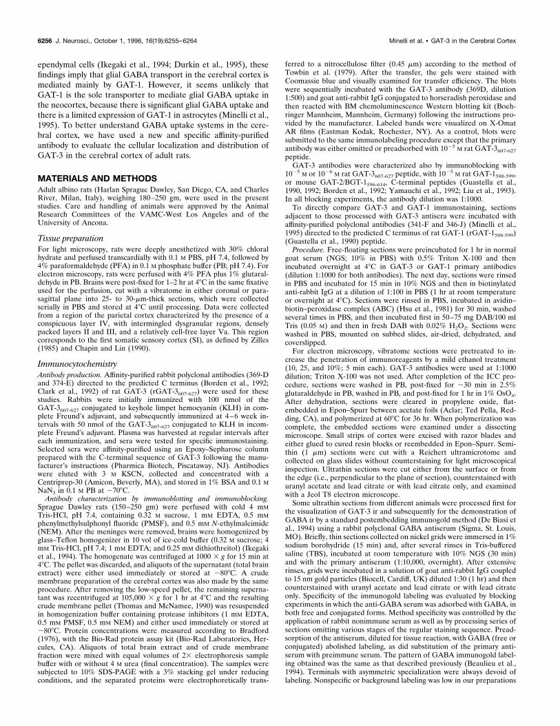
RESULTSThe specificity of the antibody was examined by immunoblot. Thepurified antibody against GAT-3 revealed a band of ;70 kDaboth in total brain extract and in the crude membrane fraction ofthe cerebral cortex (Fig. 1A,B). Omission of the primary antibodyor preadsorption with GAT-3607-627 peptide prevented labeling ofthe blots (Fig. 1C).
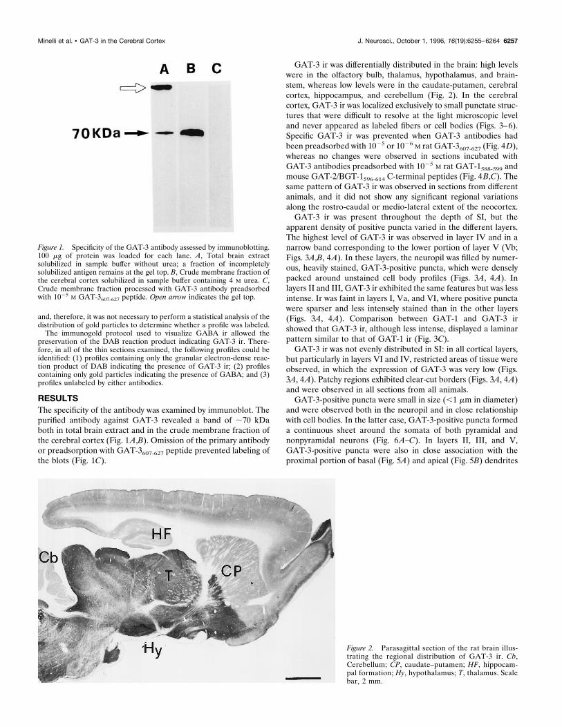
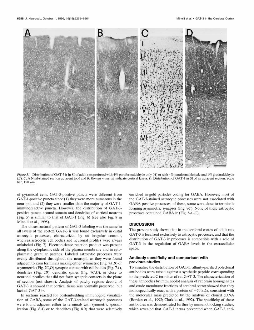
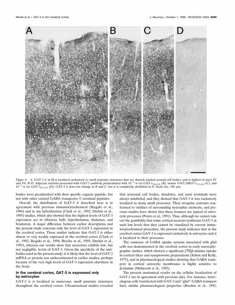
GAT-3 ir was differentially distributed in the brain: high levelswere in the olfactory bulb, thalamus, hypothalamus, and brain-stem, whereas low levels were in the caudate-putamen, cerebralcortex, hippocampus, and cerebellum (Fig. 2). In the cerebralcortex, GAT-3 ir was localized exclusively to small punctate struc-tures that were difficult to resolve at the light microscopic leveland never appeared as labeled fibers or cell bodies (Figs. 3–6).Specific GAT-3 ir was prevented when GAT-3 antibodies hadbeen preadsorbed with 1025 or 1026 M rat GAT-3607-627 (Fig. 4D),whereas no changes were observed in sections incubated withGAT-3 antibodies preadsorbed with 1025 M rat GAT-1588-599 andmouse GAT-2/BGT-1596-614 C-terminal peptides (Fig. 4B,C). Thesame pattern of GAT-3 ir was observed in sections from differentanimals, and it did not show any significant regional variationsalong the rostro-caudal or medio-lateral extent of the neocortex.GAT-3 ir was present throughout the depth of SI, but the
apparent density of positive puncta varied in the different layers.The highest level of GAT-3 ir was observed in layer IV and in anarrow band corresponding to the lower portion of layer V (Vb;Figs. 3A,B, 4A). In these layers, the neuropil was filled by numer-ous, heavily stained, GAT-3-positive puncta, which were denselypacked around unstained cell body profiles (Figs. 3A, 4A). Inlayers II and III, GAT-3 ir exhibited the same features but was lessintense. Ir was faint in layers I, Va, and VI, where positive punctawere sparser and less intensely stained than in the other layers(Figs. 3A, 4A). Comparison between GAT-1 and GAT-3 irshowed that GAT-3 ir, although less intense, displayed a laminarpattern similar to that of GAT-1 ir (Fig. 3C).GAT-3 ir was not evenly distributed in SI: in all cortical layers,
but particularly in layers VI and IV, restricted areas of tissue wereobserved, in which the expression of GAT-3 was very low (Figs.3A, 4A). Patchy regions exhibited clear-cut borders (Figs. 3A, 4A)and were observed in all sections from all animals.GAT-3-positive puncta were small in size (,1 mm in diameter)
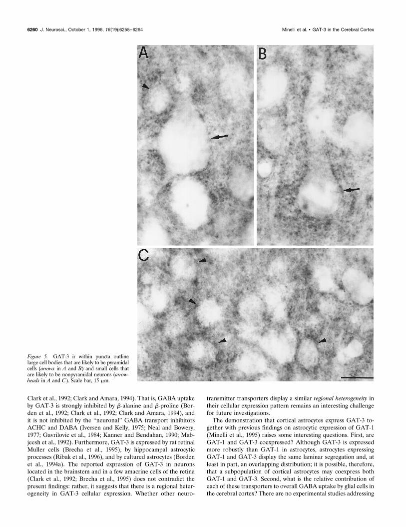
and were observed both in the neuropil and in close relationshipwith cell bodies. In the latter case, GAT-3-positive puncta formeda continuous sheet around the somata of both pyramidal andnonpyramidal neurons (Fig. 6A–C). In layers II, III, and V,GAT-3-positive puncta were also in close association with theproximal portion of basal (Fig. 5A) and apical (Fig. 5B) dendrites
Figure 2. Parasagittal section of the rat brain illus-trating the regional distribution of GAT-3 ir. Cb,Cerebellum; CP, caudate–putamen; HF, hippocam-pal formation; Hy, hypothalamus; T, thalamus. Scalebar, 2 mm.
Figure 1. Specificity of the GAT-3 antibody assessed by immunoblotting.100 mg of protein was loaded for each lane. A, Total brain extractsolubilized in sample buffer without urea; a fraction of incompletelysolubilized antigen remains at the gel top. B, Crude membrane fraction ofthe cerebral cortex solubilized in sample buffer containing 4 M urea. C,Crude membrane fraction processed with GAT-3 antibody preadsorbedwith 1025 M GAT-3607-627 peptide. Open arrow indicates the gel top.
Minelli et al. • GAT-3 in the Cerebral Cortex J. Neurosci., October 1, 1996, 16(19):6255–6264 6257
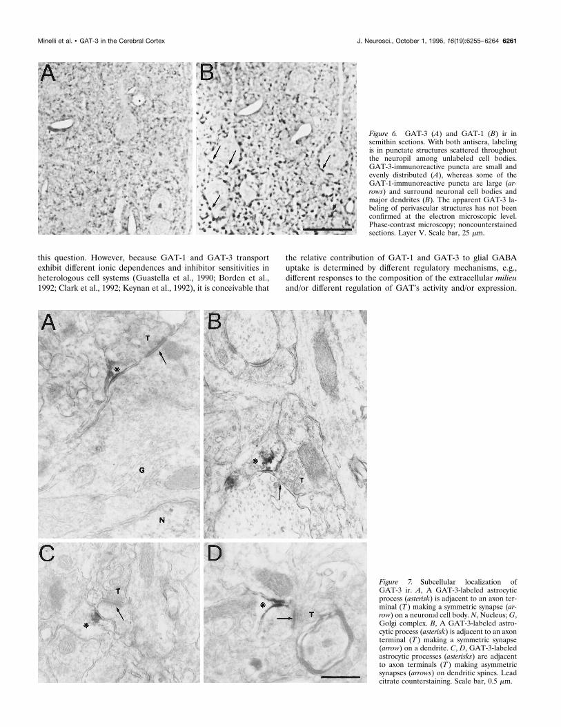
of pyramidal cells. GAT-3-positive puncta were different fromGAT-1-positive puncta since (1) they were more numerous in theneuropil, and (2) they were smaller than the majority of GAT-1-immunoreactive puncta. However, the distribution of GAT-3-positive puncta around somata and dendrites of cortical neurons(Fig. 5) is similar to that of GAT-1 (Fig. 6) (see also Fig. 8 inMinelli et al., 1995).The ultrastructural pattern of GAT-3 labeling was the same in
all layers of the cortex. GAT-3 ir was found exclusively in distalastrocytic processes, characterized by an irregular contour,whereas astrocytic cell bodies and neuronal profiles were alwaysunlabeled (Fig. 7). Electron-dense reaction product was presentalong the cytoplasmic side of the plasma membrane and in cyto-plasmatic granular patches. Labeled astrocytic processes wereevenly distributed throughout the neuropil, as they were foundadjacent to axon terminals making either symmetric (Fig. 7A,B) orasymmetric (Fig. 7C,D) synaptic contact with cell bodies (Fig. 7A),dendrites (Fig. 7B), dendritic spines (Fig. 7C,D), or close toneuronal profiles that did not form synaptic contacts in the planeof section (not shown). Analysis of patchy regions devoid ofGAT-3 ir showed that cortical tissue was normally preserved, butlacked GAT-3 ir.In sections reacted for postembedding immunogold visualiza-
tion of GABA, some of the GAT-3-stained astrocytic processeswere found adjacent either to terminals with symmetric special-ization (Fig. 8A) or to dendrites (Fig. 8B) that were selectively
enriched in gold particles coding for GABA. However, most ofthe GAT-3-stained astrocytic processes were not associated withGABA-positive processes: of these, some were close to terminalsforming asymmetric synapses (Fig. 8C). None of these astrocyticprocesses contained GABA ir (Fig. 8A–C).
DISCUSSIONThe present study shows that in the cerebral cortex of adult ratsGAT-3 is localized exclusively to astrocytic processes, and that thedistribution of GAT-3 ir processes is compatible with a role ofGAT-3 in the regulation of GABA levels in the extracellularspace.
Antibody specificity and comparison withprevious studiesTo visualize the distribution of GAT-3, affinity-purified polyclonalantibodies were raised against a synthetic peptide correspondingto the predicted C terminus of rat GAT-3. The characterization ofthese antibodies by immunoblot analysis of rat brain homogenatesand crude membrane fractions of cerebral cortex showed that theymonospecifically react with a protein of ;70 kDa, consistent withthe molecular mass predicted by the analysis of cloned cDNA(Borden et al., 1992; Clark et al., 1992). The specificity of theseantibodies was demonstrated further by immunoblocking studies,which revealed that GAT-3 ir was prevented when GAT-3 anti-
Figure 3. Distribution of GAT-3 ir in SI of adult rats perfused with 4% paraformaldehyde only (A) or with 4% paraformaldehyde and 1% glutaraldehyde(B). C, A Nissl-stained section adjacent to A and B. Roman numerals indicate cortical layers. D, Distribution of GAT-1 in SI of an adjacent section. Scalebar, 150 mm.
6258 J. Neurosci., October 1, 1996, 16(19):6255–6264 Minelli et al. • GAT-3 in the Cerebral Cortex
bodies were preadsorbed with their specific cognate peptide, butnot with other related GABA transporter C-terminal peptides.Overall, the distribution of GAT-3 ir described here is in
agreement with previous immunocytochemical (Ikegaki et al.,1994) and in situ hybridization (Clark et al., 1992; Durkin et al.,1995) studies, which also showed that the highest levels of GAT-3expression are in olfactory bulb, hypothalamus, thalamus, andbrainstem. A major difference between earlier descriptions andthe present study concerns only the level of GAT-3 expression inthe cerebral cortex. These studies indicate that GAT-3 is eitherabsent or very weakly expressed in the cerebral cortex (Clark etal., 1992; Ikegaki et al., 1994; Brecha et al., 1995; Durkin et al.,1995), whereas our results show that neocortex exhibits low, butnot negligible, levels of GAT-3. Given the specificity of the anti-bodies used in the present study, it is likely that the level of GAT-3mRNA or protein was underestimated in earlier studies, perhapsbecause of the very high levels of GAT-3 expression elsewhere inthe brain.
In the cerebral cortex, GAT-3 is expressed onlyby astrocytesGAT-3 ir is localized in numerous, small punctate structuresthroughout the cerebral cortex. Ultrastructural studies revealed
that neuronal cell bodies, dendrites, and axon terminals werealways unlabeled, and they showed that GAT-3 ir was exclusivelylocalized to many small processes. Their irregular contours con-formed to outlines of surrounding neuropilar elements, and pre-vious studies have shown that these features are typical of astro-cytic processes (Peters et al., 1991). Thus, although we cannot ruleout the possibility that some cortical neurons synthesize GAT-3 atsuch low levels that they cannot be visualized by current immu-nocytochemical procedure, the present study indicates that in thecerebral cortex GAT-3 is expressed exclusively in astrocytes and itis localized to their processes.The existence of GABA uptake systems associated with glial
cells was demonstrated in the cerebral cortex in early autoradio-graphic studies, which showed a significant [3H]b-alanine uptakein cortical slices and synaptosome preparations (Schon and Kelly,1975), and in pharmacological studies showing that GABA trans-port in cortical astrocytic membranes is highly sensitive tob-alanine (Mabjeesh et al., 1992).The present anatomical results on the cellular localization of
GAT-3 are in agreement with previous data. For instance, heter-ologous cells transfected with GAT-3 and “glial” GABA transporthave similar pharmacological properties (Borden et al., 1992;
Figure 4. A, GAT-3 ir in SI is localized exclusively to small punctate structures that are densely packed around cell bodies, and is highest in layer IVand Vb. B–D, Adjacent sections processed with GAT-3 antibody preincubated with 1025 M rat GAT-1588-599 (B), mouse GAT-2/BGT-1593-602 (C), and1025 M rat GAT-3607-627 (D). GAT-3 ir does not change in B and C, but it is completely abolished in D. Scale bar, 100 mm.
Minelli et al. • GAT-3 in the Cerebral Cortex J. Neurosci., October 1, 1996, 16(19):6255–6264 6259
Clark et al., 1992; Clark and Amara, 1994). That is, GABA uptakeby GAT-3 is strongly inhibited by b-alanine and b-proline (Bor-den et al., 1992; Clark et al., 1992; Clark and Amara, 1994), andit is not inhibited by the “neuronal” GABA transport inhibitorsACHC and DABA (Iversen and Kelly, 1975; Neal and Bowery,1977; Gavrilovic et al., 1984; Kanner and Bendahan, 1990; Mab-jeesh et al., 1992). Furthermore, GAT-3 is expressed by rat retinalMuller cells (Brecha et al., 1995), by hippocampal astrocyticprocesses (Ribak et al., 1996), and by cultured astrocytes (Bordenet al., 1994a). The reported expression of GAT-3 in neuronslocated in the brainstem and in a few amacrine cells of the retina(Clark et al., 1992; Brecha et al., 1995) does not contradict thepresent findings: rather, it suggests that there is a regional heter-ogeneity in GAT-3 cellular expression. Whether other neuro-
transmitter transporters display a similar regional heterogeneity intheir cellular expression pattern remains an interesting challengefor future investigations.The demonstration that cortical astrocytes express GAT-3 to-
gether with previous findings on astrocytic expression of GAT-1(Minelli et al., 1995) raises some interesting questions. First, areGAT-1 and GAT-3 coexpressed? Although GAT-3 is expressedmore robustly than GAT-1 in astrocytes, astrocytes expressingGAT-1 and GAT-3 display the same laminar segregation and, atleast in part, an overlapping distribution; it is possible, therefore,that a subpopulation of cortical astrocytes may coexpress bothGAT-1 and GAT-3. Second, what is the relative contribution ofeach of these transporters to overall GABA uptake by glial cells inthe cerebral cortex? There are no experimental studies addressing
Figure 5. GAT-3 ir within puncta outlinelarge cell bodies that are likely to be pyramidalcells (arrows in A and B) and small cells thatare likely to be nonpyramidal neurons (arrow-heads in A and C). Scale bar, 15 mm.
6260 J. Neurosci., October 1, 1996, 16(19):6255–6264 Minelli et al. • GAT-3 in the Cerebral Cortex
this question. However, because GAT-1 and GAT-3 transportexhibit different ionic dependences and inhibitor sensitivities inheterologous cell systems (Guastella et al., 1990; Borden et al.,1992; Clark et al., 1992; Keynan et al., 1992), it is conceivable that
the relative contribution of GAT-1 and GAT-3 to glial GABAuptake is determined by different regulatory mechanisms, e.g.,different responses to the composition of the extracellular milieuand/or different regulation of GAT’s activity and/or expression.
Figure 7. Subcellular localization ofGAT-3 ir. A, A GAT-3-labeled astrocyticprocess (asterisk) is adjacent to an axon ter-minal (T ) making a symmetric synapse (ar-row) on a neuronal cell body. N, Nucleus;G,Golgi complex. B, A GAT-3-labeled astro-cytic process (asterisk) is adjacent to an axonterminal (T ) making a symmetric synapse(arrow) on a dendrite. C, D, GAT-3-labeledastrocytic processes (asterisks) are adjacentto axon terminals (T ) making asymmetricsynapses (arrows) on dendritic spines. Leadcitrate counterstaining. Scale bar, 0.5 mm.
Figure 6. GAT-3 (A) and GAT-1 (B) ir insemithin sections. With both antisera, labelingis in punctate structures scattered throughoutthe neuropil among unlabeled cell bodies.GAT-3-immunoreactive puncta are small andevenly distributed (A), whereas some of theGAT-1-immunoreactive puncta are large (ar-rows) and surround neuronal cell bodies andmajor dendrites (B). The apparent GAT-3 la-beling of perivascular structures has not beenconfirmed at the electron microscopic level.Phase-contrast microscopy; noncounterstainedsections. Layer V. Scale bar, 25 mm.
Minelli et al. • GAT-3 in the Cerebral Cortex J. Neurosci., October 1, 1996, 16(19):6255–6264 6261
For instance, protein kinase C (PKC) activators, which increaseGABA uptake in cells transfected with GAT-1 (Corey et al.,1994), reduce glial GABA transport in primary astrocyte cultures(Gomeza et al., 1991). A differential regulation of astrocyticGAT-1 and GAT-3 would allow a great adaptability in the fineregulation of GABA extracellular levels under various physiolog-ical conditions.
The distribution of GAT-3 in the cerebral cortex iscompatible with a dual physiological roleThe major features of the distribution of GAT-3 ir in the cerebralcortex, as revealed for the first time by this study, are as follows:(1) GAT-3 ir astrocytic processes display a laminar segregationsimilar to that of GAT-1 ir puncta (Minelli et al.;, 1995) and ofGABAergic axon terminals (Ribak, 1978; Houser et al., 1984);and (2) GAT-3-positive astrocytic processes are not exclusivelyassociated with GABAergic profiles, as shown by double-labelingwith GABA.Axon terminals releasing GABA are densest in layer IV, fol-
lowed by layers II and III, and in layer Vb GABAergic terminalsoutline unstained pyramidal cell bodies (Ribak, 1978; Freund etal., 1983; Houser et al., 1984). These cortical layers or sublayersare also the richest in GAT-1-positive puncta (Minelli et al.,1995). Here we have shown that the distribution of GAT-3-positive puncta is similar to that of axon terminals releasingGABA and of GAT-1-positive puncta. This correlation impliesthat GAT-3 is highly expressed in the very layers in which GABA isprominently released and, therefore, highly concentrated. Thepresence of numerous “patches” devoid of GAT-3 ir in neocortexindicates some differences in GAT-3 expression and activity. Thefunctional meaning of this organizational feature of GAT-3 ir is atpresent unknown; it is worth noting, however, that an analogousdistribution has been reported for a glial glutamate (Glu) trans-porter, GLT-1 (Rothstein et al., 1994).GAT-3-positive puncta are in close relationship with neuronal cell
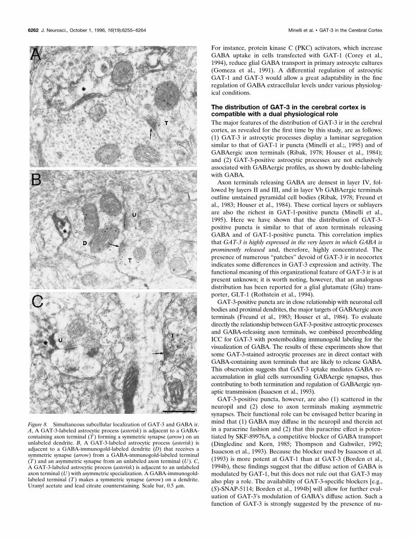
bodies and proximal dendrites, the major targets of GABAergic axonterminals (Freund et al., 1983; Houser et al., 1984). To evaluatedirectly the relationship betweenGAT-3-positive astrocytic processesand GABA-releasing axon terminals, we combined preembeddingICC for GAT-3 with postembedding immunogold labeling for thevisualization of GABA. The results of these experiments show thatsome GAT-3-stained astrocytic processes are in direct contact withGABA-containing axon terminals that are likely to release GABA.This observation suggests that GAT-3 uptake mediates GABA re-accumulation in glial cells surrounding GABAergic synapses, thuscontributing to both termination and regulation of GABAergic syn-aptic transmission (Isaacson et al., 1993).GAT-3-positive puncta, however, are also (1) scattered in the
neuropil and (2) close to axon terminals making asymmetricsynapses. Their functional role can be envisaged better bearing inmind that (1) GABA may diffuse in the neuropil and therein actin a paracrine fashion and (2) that this paracrine effect is poten-tiated by SKF-89976A, a competitive blocker of GABA transport(Dingledine and Korn, 1985; Thompson and Gahwiler, 1992;Isaacson et al., 1993). Because the blocker used by Isaacson et al.(1993) is more potent at GAT-1 than at GAT-3 (Borden et al.,1994b), these findings suggest that the diffuse action of GABA ismodulated by GAT-1, but this does not rule out that GAT-3 mayalso play a role. The availability of GAT-3-specific blockers [e.g.,(S)-SNAP-5114; Borden et al., 1994b] will allow for further eval-uation of GAT-3’s modulation of GABA’s diffuse action. Such afunction of GAT-3 is strongly suggested by the presence of nu-
Figure 8. Simultaneous subcellular localization of GAT-3 and GABA ir.A, A GAT-3-labeled astrocytic process (asterisk) is adjacent to a GABA-containing axon terminal (T ) forming a symmetric synapse (arrow) on anunlabeled dendrite. B, A GAT-3-labeled astrocytic process (asterisk) isadjacent to a GABA-immunogold-labeled dendrite (D) that receives asymmetric synapse (arrow) from a GABA-immunogold-labeled terminal(T ) and an asymmetric synapse from an unlabeled axon terminal (U ). C,A GAT-3-labeled astrocytic process (asterisk) is adjacent to an unlabeledaxon terminal (U ) with asymmetric specialization. A GABA-immunogold-labeled terminal (T ) makes a symmetric synapse (arrow) on a dendrite.Uranyl acetate and lead citrate counterstaining. Scale bar, 0.5 mm.
6262 J. Neurosci., October 1, 1996, 16(19):6255–6264 Minelli et al. • GAT-3 in the Cerebral Cortex
merous GAT-3-positive astrocytic processes in the neuropil. Theresults presented here show that some GAT-3-positive astrocyticprocesses are adjacent to axon terminals forming asymmetricsynapses, which contain and presumably release Glu (Conti andMinelli, 1996). Interestingly, Isaacson et al. (1993) showed thatinhibition of GABA uptake enhances the presynaptic action ofGABA on presumed GABAB receptors located on excitatorynerve terminals. GAT-3-positive astrocytic processes adjacent toaxon terminals forming asymmetric synapses could contribute,therefore, to the regulation of GABA action on excitatory synap-tic transmission.Finally, it has been shown that GATs may release GABA in a
nonvesicular, Ca21-independent manner (Schwartz, 1982; Attwellet al., 1983; Levi and Raiteri, 1993). These data, together with thepresent demonstration of a widespread distribution of GAT-3-positive astrocytic processes in the neuropil, suggest that GAT-3may mediate GABA release in the cerebral cortex.
REFERENCESAttwell D, Barbour B, Szatkowski M (1993) Nonvesicular release ofneurotransmitter. Neuron 11:401–407.
Beaulieu C, Campistron G, Crevier C (1994) Quantitative aspects of theGABA circuitry in the primary visual cortex of the adult rat. J CompNeurol 339:559–572.
Beart PM, Johnston GAR, Uhr ML (1972) Competitive inhibition ofGABA uptake in rat brain slices by some GABA analogues of restrictedconformation. J Neurochem 19:1855–1861.
Borden LA, Smith KE, Hartig PR, Brancheck TA, Weinshank RL (1992)Molecular heterogeneity of the g-aminobutyric acid (GABA) transportsystem. J Biol Chem 267:21098–21104.
Borden LA, Smith KE, Vaysse PJ-J, Weinshank RL, Branchek TA(1994a) GABA transporters in neuronal and glial cell cultures: corre-lation of pharmacological activity with mRNA localization. Soc Neuro-sci Abstr 20:919.
Borden LA, Dhar TGM, Smith KE, Branchek TA, Glucowski C, Weins-hank RL (1994b) Cloning of the human homologue of the GABAtransporter GAT-3 and identification of a novel inhibitor with selectivityfor this site. Receptors Channels 2:207–213.
Bowery NG, Jones GP, Neal MJ (1976) Selective inhibition of neuronalGABA uptake by cis-1,3-aminocyclohexane carboxylic acid (ACHC).Nature 264:281–284.
Bradford MM (1976) A rapid and sensitive method for the quantitationof microgram quantities of protein utilizing the principle of protein-dyebinding. Anal Biochem 72:248–254.
Brecha NC, Johnson J, Chen T, Conti F, Minelli A, DeBiasi S, Ribak C(1995) GABA transporter expression in the rat nervous system. Pre-synaptic mechanisms of neurotransmission. Soc Neurosci Abstr 124:1.
Chapin JK, Lin C-S (1990) The somatic sensory cortex of rat. In: Thecerebral cortex of the rat (Kolb B, Tees RG, eds), pp 341–380. Cam-bridge: MIT.
Clark JA, Amara SG (1994) Stable expression of a neuronalg-aminobutyric acid transporter, GAT-3, in mammalian cells demon-strates unique pharmacological properties and ion dependence. MolPharmacol 46:550–557.
Clark JA, Deutch AY, Gallipoli PZ, Amara SG (1992) Functional ex-pression and CNS distribution of a b-alanine-sensitive neuronal GABAtransporter. Neuron 9:337–348.
Conti F, Minelli A (1996) The anatomy of glutamatergic transmission inthe cerebral cortex. In: Excitatory amino acids & the cerebral cortex(Conti F, Hicks TP, eds), pp 81–98. Cambridge: MIT.
Corey JL, Davidson N, Lester HA, Brecha NC, Quick MW (1994) Pro-tein kinase C modulates the activity of a cloned g-aminobutyric acidtransporter expressed in Xenopus oocytes via regulated subcellularredistribution of the transporter. J Biol Chem 269:14759–14767.
De Biasi S, Vitellaro-Zuccarello L, Bernardi P, Valtschanoff JG, Wein-berg RJ (1994) Ultrastructural and immunocytochemical characteriza-tion of primary afferent terminals in the rat cuneate nucleus. J CompNeurol 347:275–287.
Dingledine R, Korn SJ (1985) g-Aminobutyric acid uptake and the ter-mination of inhibitory synaptic potentials in the rat hippocampal slices.J Physiol (Lond) 366:387–409.
During MJ, Ryder KM, Spencer DD (1995) Hippocampal GABA trans-porter function in temporal lobe epilepsy. Nature 376:174–177.
Durkin MM, Smith KE, Borden LA, Weinshank RL, Branchek TA,Gustafson EL (1995) Localization of messenger RNAs encoding threeGABA transporters in rat brain: an in situ hybridization study. MolBrain Res 33:7–21.
Freund TF, Martin KAC, Smith AD, Somogyi P (1983) Glutamatedecarboxylase-immunoreactive terminals of Golgi-impregnated axoax-onic cells and presumed basket cells in synaptic contacts with pyramidalneurons of the cat’s visual cortex. J Comp Neurol 221:263–278.
Gavrilovic J, Raff M, Cohen G (1984) GABA uptake by purified ratSwann cells in culture. Brain Res 303:183–185.
Gomeza J, Casado M, Gimenez C, Aragon C (1991) Inhibition of high-affinity g-aminobutyric acid uptake in primary astrocyte cultures byphorbol esters and phospholipase C. Biochem J 275:435–439.
Guastella J, Nelson N, Nelson H, Czyzyk L, Keynan S, Miedel MC,Davidson N, Lester HA, Kanner BI (1990) Cloning and expression ofa rat brain GABA transporter. Science 249:1303–1306.
Guastella J, Brecha NC, Weigman C, Lester HA, Davidson N (1992)Cloning, expression, and localization of a rat brain high-affinity glycinetransporter. Proc Natl Acad Sci USA 89:7189–7193.
Houser CR, Vaughn GE, Hendry SHC, Jones EG, Peters A (1984)GABA neurons in the cerebral cortex. In: Cerebral cortex, Vol 2,Functional properties of cortical cells (Jones EG, Peters A, eds), pp63–90. New York: Plenum.
Hsu SM, Raine L, Fanger H (1981) Use of avidin-biotin-peroxidasecomplex (ABC) in immunoperoxidase techniques: a comparison be-tween ABC and unlabelled antibody (PAP) procedures. J HistochemCytochem 29:557–580.
Ikegaki N, Saito N, Hashima M, Tanaka C (1994) Production of specificantibodies against GABA transporter subtypes (GAT-1, GAT-2,GAT-3) and their application to immunocytochemistry. Mol Brain Res26:47–54.
Isaacson JS, Solis JM, Nicoll RA (1993) Local and diffuse synaptic ac-tions of GABA in the hippocampus. Neuron 10:165–175.
Iversen LL (1971) Role of transmitter uptake mechanisms in synapticneurotransmission. Br J Pharmacol 41:571–591.
Iversen LL, Kelly JS (1975) Uptake and metabolism of g-aminobutyricacid by neurons and glial cells. Biochemical Pharmacol 24:933–938.
Iversen LL, Neal MJ (1968) The uptake of [3H]GABA by slices of ratcerebral cortex. J Neurochem 15:1141–1149.
Iversen LL, Snyder SH (1968) Synaptosomes: different population stor-ing catecholamines and gamma-aminobutyric acid in homogenate of ratbrain. Nature 220:796–798.
Kanner BI, Bendahan A (1990) Two pharmacologically distinct sodium-and chloride-coupled high-affinity g-aminobutyric acid transporters arepresent in plasma membrane vesicles and reconstituted preparationsfrom rat brain. Proc Natl Acad Sci USA 87:2550–2554.
Keynan S, Suh Y-J, Kanner BI, Rudnick G (1992) Expression of a clonedg-aminobutyric acid transporter in mammalian cells. Biochemistry31:1974–1979.
Krogsgaard-Larsen P, Falch E, Larsson OM, Schousboe A (1987) GABAuptake inhibitors: relevance to antiepileptic drug research. Epilepsy Res1:77–93.
Larsson OM, Johnston GAR, Schousboe A (1983) Differences in uptakekinetics of cis-3-aminocyclohexane carboxylic acid into neurons andastrocytes in primary cultures. Brain Res 260:279–285.
Levi G, Raitieri M (1993) Carrier-mediated release of neurotransmitters.Trends Neurosci 16:415–418.
Liu Q-R, Lopez-Corcuera B, Mandiyan S, Nelson H, Nelson N (1993)Molecular characterization of four pharmacologically distinctg-aminobutyric acid transporters in mouse brain. J Biol Chem268:2106–2112.
Mabjeesh NJ, Frese M, Rauen T, Jeserich G, Kanner BI (1992) Neu-ronal and glial g-aminobutyric acid transporters are distinct proteins.FEBS Lett 299:99–102.
Minelli A, Brecha NC, Karschin C, DeBiasi S, Conti F (1995) GAT-1, ahigh-affinity GABA plasma membrane transporter, is localized to neu-rons and astroglia in the cerebral cortex. J Neurosci 15:7734–7746.
Neal MJ, Bowery NG (1977) Cis-3-aminocyclohexane carboxylic acid: asubstrate for the neuronal GABA transport system. Brain Res138:169–174.
Neal MJ, Iversen LL (1969) Subcellular distribution of endogenous and[3H]g-aminobutyric acid in rat cerebral cortex. J Neurochem16:1245–1252.
Minelli et al. • GAT-3 in the Cerebral Cortex J. Neurosci., October 1, 1996, 16(19):6255–6264 6263
Peters A, Palay SL, Webster H deF (1991) The fine structure of thenervous system: neurons and their supporting cells. New York:Oxford UP.
Pin J-P, Bockaert J (1989) Two distinct mechanisms, differentially af-fected by excitatory amino acids, trigger GABA release from fetalmouse striatal neurons in primary cultures. J Neurosci 9:648–656.
Ribak CE (1978) Aspinous and sparsely-spinous stellate neurons in thevisual cortex of rat contain glutamic acid decarboxylase. J Neurocytol7:461–478.
Ribak CE, Tong W, Brecha NC (1996) The GABA plasma membranetransporters, GAT-1 and GAT-3, display different distributions in therat hippocampus. J Comp Neurol 367:595–606.
Rothstein JD, Martin L, Levey AI, Dykes-Hoberg M, Jin L, Wu D, NashM, Kuncl RW (1994) Localization of neuronal and glial glutamatetransporters. Neuron 13:713–725.
Schon F, Kelly JS (1975) Selective uptake of [3H]b-alanine by glia: asso-ciation with the glial uptake system for GABA. Brain Res 86:243–257.
Schousboe A, Larsson OM, Wood JD, Krogsgaard-Larsen P (1983)Transport and metabolism of g-aminobutyric acid in neurons and glia:implications for epilepsy. Epilepsia 24:531–538.
Schwartz EA (1982) Calcium-independent release of GABA from iso-lated horizontal cells of the toad retina. J Physiol (Lond) 323:211–227.
Thomas CT, McNamee MG (1990) Purification of membrane proteins.Methods Enzymol 182:499–520.
Thompson SM, Gahwiler BH (1992) Effects of the GABA uptake inhib-itor tiagabine on inhibitory synaptic potentials in rat hippocampal slicecultures. J Neurophysiol 67:1698–1670.
Towbin H, Staehelin T, Gordon J (1979) Electrophoretic transfer ofproteins from polyacrylamide gels to nitrocellulose sheets: procedureand some applications. Proc Natl Acad Sci USA 76:4350–4354.
Yamauchi A, Uchida S, Kwon HM, Preston AS, Robey RB, Garcia-PerezA, Burg MB, Handler JS (1992) Cloning of a Na1- and Cl2-dependentbetaine transporter that is regulated by hypertonicity. J Biol Chem267:649–652.
Zilles K (1985) The cortex of rat. Berlin: Springer.
6264 J. Neurosci., October 1, 1996, 16(19):6255–6264 Minelli et al. • GAT-3 in the Cerebral Cortex