Embed Size (px)
Citation preview
Galactose-Depleted Xyloglucan Is Dysfunctional andLeads to Dwarfism in Arabidopsis1
Yingzhen Kong, Maria J. Peña, Luciana Renna, Utku Avci, Sivakumar Pattathil, Sami T. Tuomivaara,Xuemei Li2, Wolf-Dieter Reiter, Federica Brandizzi, Michael G. Hahn, Alan G. Darvill,William S. York, and Malcolm A. O’Neill*
Complex Carbohydrate Research Center (Y.K., M.J.P., U.A., S.P., S.T.T., M.G.H., A.G.D., W.S.Y., M.A.O.),Department of Plant Biology (M.G.H.), and Department of Biochemistry and Molecular Biology (A.G.D.,W.S.Y.), University of Georgia, Athens, Georgia 30602; Tobacco Research Institute, Chinese Academy ofAgricultural Sciences, Qingdao, Shandong 266101, China (Y.K.); United States Department of Energy PlantResearch Laboratory (L.R., F.B.) and United States Department of Energy Great Lakes Bioenergy ResearchCenter (F.B.), Michigan State University, East Lansing, Michigan 48824; and Department of Molecular and CellBiology, University of Connecticut, Storrs, Connecticut 06269 (X.L., W.-D.R.)
Xyloglucan is a polysaccharide that has important roles in the formation and function of the walls that surround growing landplant cells. Many of these plants synthesize xyloglucan that contains galactose in two different side chains (L and F), which exist indistinct molecular environments. However, little is known about the contribution of these side chains to xyloglucan function. Here,we show that Arabidopsis (Arabidopsis thaliana) mutants devoid of the F side chain galactosyltransferase MURUS3 (MUR3) formxyloglucan that lacks F side chains and contains much less galactosylated xylose than its wild-type counterpart. The galactose-depleted xyloglucan is dysfunctional, as it leads to mutants that are dwarfed with curled rosette leaves, short petioles, and shortinflorescence stems. Moreover, cell wall matrix polysaccharides, including xyloglucan and pectin, are not properly secreted andinstead accumulate within intracellular aggregates. Near-normal growth is restored by generating mur3 mutants that produce nodetectable amounts of xyloglucan. Thus, cellular processes are affected more by the presence of the dysfunctional xyloglucan thanby eliminating xyloglucan altogether. To identify structural features responsible for xyloglucan dysfunction, xyloglucan structurewas modified in situ by generating mur3 mutants that lack specific xyloglucan xylosyltransferases (XXTs) or that overexpress theXYLOGLUCAN L-SIDE CHAIN GALACTOSYLTRANSFERASE2 (XLT2) gene. Normal growth was restored in the mur3-3 mutantoverexpressing XLT2 and inmur3-3 xxt double mutants when the dysfunctional xyloglucan was modified by doubling the amountsof galactosylated side chains. Our study assigns a role for galactosylation in normal xyloglucan function and demonstrates thataltering xyloglucan side chain structure disturbs diverse cellular and physiological processes.
There have been considerable advances in under-standing how changes in specific amino acids or nu-cleotides affect the ability of a protein or an RNA tofold and to function (Wan et al., 2011; Sauer, 2013). Bycontrast, much less is known about how polysaccharidestructure and function are affected by altering specificmonosaccharides within its glycosyl sequence. Here,
we report that xyloglucan, a plant cell wall polysac-charide, becomes dysfunctional rather than nonfunc-tional when its glycosyl sequence is altered. The termdysfunctional polysaccharide is used here to describe astructure that, in and of itself, has a deleterious effecton the growth and development of a plant.
XXXG-type xyloglucans (XyGs; Fig. 1A) are presentin the cell walls of many flowering plants (Hoffmanet al., 2005), gymnosperms (Hsieh and Harris, 2012),ferns and their allies, club and spike mosses, and theavascular hornworts (Peña et al., 2008). XyGs of thistype are composed of subunits (Fig. 1, A and B) thathave three consecutive b-D-glucopyranose (b-D-Glcp)backbone residues bearing a side chain followed by asingle unbranched b-D-Glcp residue (Tuomivaara et al.,2015). Little is known about the contribution of theseside chains to the role of XyG in wall structure andfunction, although the results of in vitro studies sug-gest that they modulate how an XyG interacts withcellulose (Whitney et al., 2006) and with itself (Shirakawaet al., 1998; de Freitas et al., 2011).
The Arabidopsis (Arabidopsis thaliana) genes that en-code the glycosyltransferases required for the formationof the XyG side chains have been identified (see Fig. 1A).
1 This work was supported by the Division of Chemical Sciences,Geosciences, and Biosciences, Office of Basic Energy Sciences, U.S.Department of Energy (grant nos. DE–FG02–96ER20220 to A.G.D.,M.J.P., M.G.H., M.A.O., and W.S.Y.; DE–FG02–93ER20097 to A.G.D.;DE–FG02–12ER16324 to W.S.Y., M.A.O., and M.J.P.; and DE–FG02–91ER20021 to F.B.); the U.S. Department of Energy Great Lakes Bio-energy Research Center (grant nos. BER DE–FC02–07ER64494 to F.B.and DE–FG02–08ER20203 to W.-D.R.); and the National ScienceFoundation (Plant Genome grant no. ISO–0923992 to M.G.H.).
2 Present address: Agrivida Inc., 200 Boston Avenue, Medford,MA 02155.
* Address correspondence to [email protected] author responsible for distribution of materials integral to the
findings presented in this article in accordance with the policy de-scribed in the Instructions for Authors (www.plantphysiol.org) is:Malcolm A. O’Neill ([email protected]).
www.plantphysiol.org/cgi/doi/10.1104/pp.114.255943
1296 Plant Physiology�, April 2015, Vol. 167, pp. 1296–1306, www.plantphysiol.org � 2014 American Society of Plant Biologists. All Rights Reserved. www.plantphysiol.orgon February 10, 2020 - Published by Downloaded from
Copyright © 2015 American Society of Plant Biologists. All rights reserved.
Five XYLOGLUCAN XYLOSYLTRANSFERASE (XXT1–XXT5) genes encode proteins that have been implicatedin the formation of the a-D-xylopyranose-(1→6)-b-D-Glcplinkage (Cavalier et al., 2008; Zabotina et al., 2008;Vuttipongchaikij et al., 2012; Zabotina et al., 2012). Twoside chain-specific galactosyltransferases have beencharacterized (Madson et al., 2003; Jensen et al., 2012).XYLOGLUCAN L-SIDE CHAIN GALACTOSYL-TRANSFERASE2 (XLT2) catalyzes the addition of Galto the middle Xyl (Jensen et al., 2012), whereas MURUS3(MUR3) adds Gal to the Xyl adjacent to the unbranchedGlc (Madson et al., 2003). The Gal added by MUR3 isitself the acceptor for FUCOSYLTRANSFERASE1(FUT1), which adds a fucosyl residue to form the F sidechain (Perrin et al., 1999). Up to 60% of the F side chainsof Arabidopsis leaf XyG contain O-acetylated Gal (Perrinet al., 2003).Arabidopsis fut1, xlt2, and xxt1 to xxt5 mutants that
form structurally abnormal XyG (Vanzin et al., 2002;Madson et al., 2003; Perrin et al., 2003; Peña et al., 2004;Jensen et al., 2012; Schultink et al., 2013) or that havereduced (Zabotina et al., 2008; Vuttipongchaikij et al.,2012; Zabotina et al., 2012) or no discernible amountsof XyG (Cavalier et al., 2008) have no severe devel-opmental or growth phenotypes. This has led plantscientists to question the biological role of XyG in wallassembly and architecture as well as its function inplant growth and development (Cavalier et al., 2008;Park and Cosgrove, 2012a, 2012b).Arabidopsis mur3-1 and mur3-2 mutants, which
carry different single-point mutations in MUR3, havebeen reported to form XyG that lacks F side chains(Madson et al., 2003; Peña et al., 2004), although they
do produce discernible amounts of the MUR3 protein(Tamura et al., 2005). Both of these mutants are visiblysimilar to wild-type plants (Madson et al., 2003; Peñaet al., 2004; Tamura et al., 2005). By contrast, mutantplants including katamari1 (kam1-1, kam1-2/mur3-7, andkam1-3/mur3-3; Tamura et al., 2005; Tedman-Joneset al., 2008), constitutive ICS1:luc expression1 (Tedman-Jones et al., 2008), and short root in salt medium (rsa3; Liet al., 2013), which are devoid of functional MUR3,have a dwarf cabbage-like growth phenotype withcurled rosette leaves, short petioles, and endomem-brane aggregates. Tamura et al. (2005) have proposedthat the KAM1/MUR3 protein itself, rather than itsgalactosyltransferase activity, is required for cell elon-gation and normal endomembrane organization. Thishypothesis is based in part on the assumption that themur3-1, mur3-2, and mur3-3/kam1-3 mutants all lackMUR3 galactosyltransferase activity, but only the mur3-3/kam1-3mutant lacks the MUR3 protein itself (Tamuraet al., 2005).
Here, we provide genetic and chemical evidencethat XyG deficient in extended side chains is dys-functional and is responsible for the growth defects ofmur3-3 and mur3-7 plants. The extent of XyG gal-actosylation is the major factor in determining theseeffects. The deleterious growth effects caused by thisdysfunctional XyG are suppressed by introducing ad-ditional mutations that eliminate XyG formation alto-gether, by mutations that modify XyG structure, or byaltering the plant’s growth conditions. These obser-vations provide new insight into XyG biosynthesis andthe structural requirements that allow XyG to functionnormally in plant growth and development.
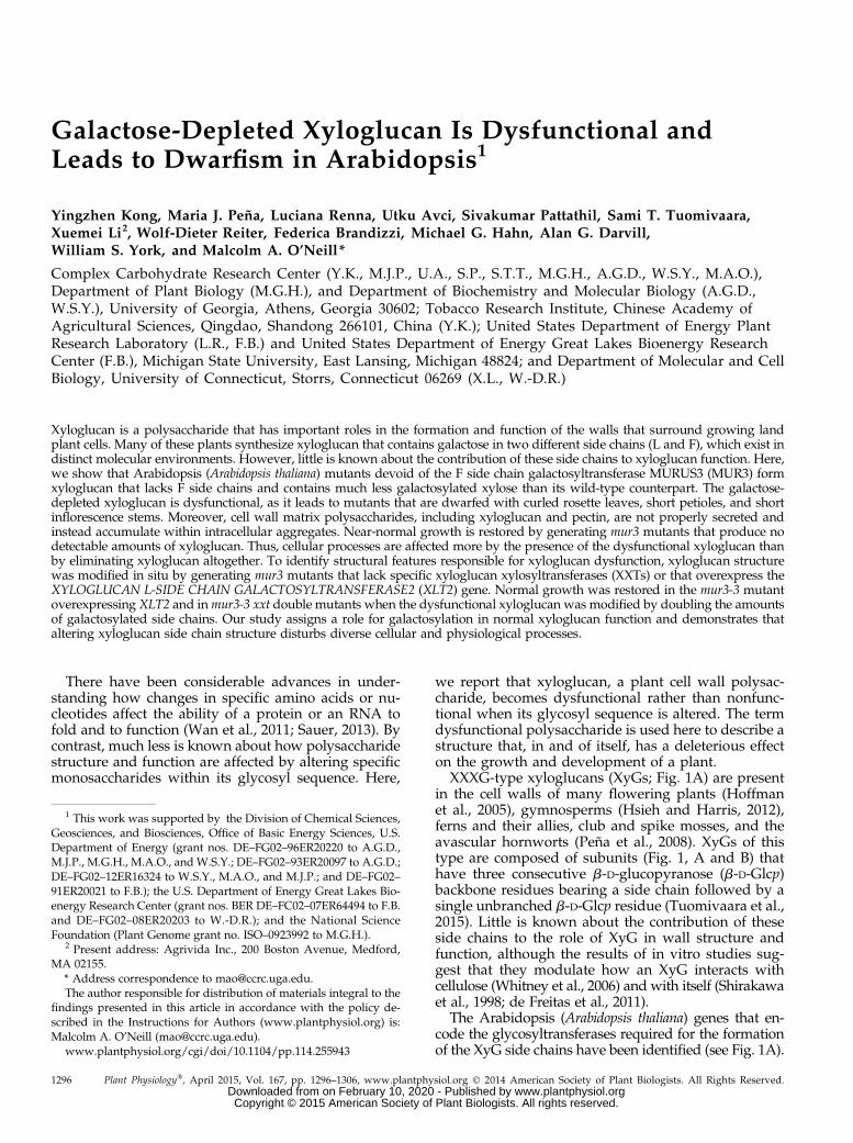
Figure 1. Eliminating xyloglucan suppresses the cabbage-like phenotype of mur3-3 plants. A, The major structural features ofArabidopsis XyG and the glycosyltransferases required for side chain formation. Side chains are represented by the letters X, L,and F. An unbranched Glc is designated by the letter G. B, The predominant subunits of Arabidopsis XyG. C, Eliminating XyGsuppresses the dwarf phenotype of the mur3-3 mutant. mur3-3 was crossed with xxt1 xxt2, and two independent xxt1 xxt2mur3-3 triple mutants (nos. 12 and 20) were identified by reverse transcription (RT)-PCR. Plants were grown with a 14-h-light(19˚C) and 10-h-dark (15˚C) cycle. D, The glycome profiles of the materials solubilized from the cell walls by enzymatic andKOH treatments. Each extract was probed with a series of monoclonal antibodies that recognize epitopes of XyG irrespective ofthe presence or absence of fucosylated side chains and monoclonal antibodies that bind only to XyG containing fucosylatedside chains. Yellow, Strong binding; black, no binding. No XyG was detected in the xxt1 xxt2 double mutant or the xxt1 xxt2mur3-3 triple mutants. E, Cross sections of wild-type and mutant leaves stained with toluidine blue (left column) or immunolabeledwith monoclonal antibodies that recognize epitopes of deesterified homogalacturonan (CCRC-M38), XyG (CCRC-M88), or fuco-sylated XyG (CCRC-M1). WT, Wild type; EPG, endopolygalacturonase.
Plant Physiol. Vol. 167, 2015 1297
Abnormal Xyloglucan Leads to Arabidopsis Dwarfism
www.plantphysiol.orgon February 10, 2020 - Published by Downloaded from Copyright © 2015 American Society of Plant Biologists. All rights reserved.
RESULTS AND DISCUSSION
Reducing XyG to below Detectable Levels Suppresses theDwarf Phenotype of mur3-3 Plants
MUR3 encodes an XyG-specific galactosyltransfer-ase responsible for most of the galactosylation ofArabidopsis XyG. To address the possibility that al-tered XyG structure leads to the phenotypes of Arabi-dopsis plants lacking functional MUR3, we took agenetic approach to reduce the amounts of XyG toundetectable levels in the mur3-3 mutant. Thus, wecrossed mur3-3 with the xxt1 xxt2 double mutant,which has no detectable amounts of XyG in its cellwall (Cavalier et al., 2008; Zabotina et al., 2012). Thexxt1 xxt2 mutant grows somewhat more slowlythan wild-type plants but does not have a cabbage-like phenotype (Cavalier et al., 2008) or abnormalendomembrane aggregates (Uehara et al., 2014),making it visibly distinct from the mur3-3 mutant(Fig. 1C).
The xxt1 xxt2 mur3-3 triple mutant, which pro-duces no discernible XXT1, XXT2, or MUR3 tran-scripts (Supplemental Fig. S1A), does not have acabbage-like phenotype and is visibly indistinguish-able from the xxt1 xxt2 mutant (Fig. 1C). Toluidineblue-stained cross sections of wild-type, xxt1 xxt2,and xxt1 xxt2 mur3-3 leaves are also visibly similar(Fig. 1E). Most of the mur3-3 cells are smaller thantheir wild-type counterparts, a result that is in agree-ment with the proposal that these cells have lost theability to expand normally (Tamura et al., 2005).Glycome profiling (Fig. 1D) and immunocytochem-istry (Fig. 1E) indicate that the XyG present in mur3-3walls is not fucosylated, whereas wild-type wallscontain fucosylated XyG. No discernible amounts ofXyG were detected in xxt1 xxt2 mur3-3 leaf cell walls(Fig. 1, D and E), nor were xyloglucan oligosaccha-rides (XyGOs) detected after XyG-specific endoglu-canase (XEG) treatment of the 1 and 4 N KOH-solublematerials from xxt1 xxt2 mur3-3 root, leaf, and stemcell walls. Thus, the presence of XyG is stronglycorrelated with the cabbage-like phenotype of themur3-3 mutant. Our data suggest that the absenceof the MUR3 galactosyltransferase leads to the for-mation of dysfunctional XyG, and that cellular pro-cesses are affected more by the presence of thisdysfunctional xyloglucan than by eliminating XyGaltogether.
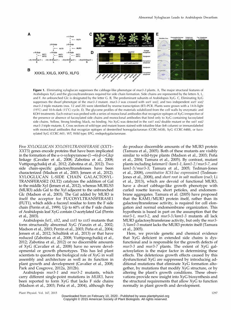
The mur3-1, mur3-2, mur3-3, and mur3-7 Phenotypes AreAssociated with Altered XyG Chemotypes
Since the presence of dysfunctional XyG is corre-lated with the cabbage-like phenotype of mur3-3plants, we next determined the structural features ofXyG that are associated with this phenotype. To thisend, the XyGs from mur3-1, mur3-2, mur3-3, and mur3-7,each of which carries a different mutation in MUR3(Supplemental Fig. S1B), were structurally characterized.The structures of these XyGs have not previously beencompared, since only the distinguishing chemical struc-tures of the mur3-1 XyG have been described (Madsonet al., 2003; Peña et al., 2004). Here, the XyGOs generatedby XEG fragmentation of leaf XyG obtained from eachmutant grown at 19°C (Fig. 2) were characterized using1H-NMR spectroscopy (Hoffman et al., 2005), matrix-assisted laser-desorption ionization time of flight(MALDI-TOF) mass spectrometry (MS), and high-performance anion-exchange chromatography (HPAEC)with pulsed-amperometric detection (PAD; Tuomivaaraet al., 2015).
Homozygous mur3-3 and mur3-7 plants, respectively,carry a transfer DNA insertion in the region encodingthe exostosin-like domain and the protein’s N terminus(Tedman-Jones et al., 2008; Supplemental Fig. S1B).Both mutants produce no detectable MUR3 transcripts(Supplemental Fig. S1A) and have a cabbage-like phe-notype when grown at 19°C in soil (Fig. 2). These mu-tants are visibly distinct from the mur3-1 and mur3-2mutants (Fig. 2), both of which produce detectableamounts of the MUR3 protein (Tamura et al., 2005).
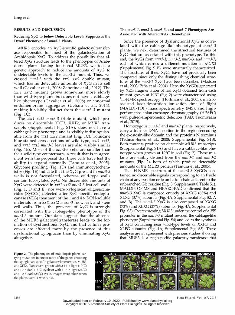
The 1H-NMR spectrum of the mur3-3 XyGOs con-tained no discernible signals corresponding to an F sidechain at any position or to an L side chain adjacent to theunbranched Glc residue (Fig. 3; Supplemental Table S1).MALDI-TOF MS and HPAEC-PAD confirmed that themur3-3 XyG is composed entirely of XXXG (63%) andXLXG (37%) subunits (Fig. 4A; Supplemental Fig. S2, Aand B). The mur3-7 XyG is also composed of XXXG(73%) and XLXG (27%) subunits (Fig. 4A; SupplementalFig. S3). OverexpressingMUR3 under the control of a 35Spromoter in the mur3-3 mutant rescued the cabbage-likephenotype (Supplemental Fig. S4) and led to the synthesisof XyG containing near wild-type levels of XXFG andXLFG subunits (Fig. 4A; Supplemental Fig. S3). Theseanalyses are in agreement with previous studies showingthat MUR3 is a regiospecific galactosyltransferase that
Figure 2. The phenotypes of Arabidopsis plants car-rying mutations in one or more of the genes encodingthe xyloglucan-specific galactosyltransferases MUR3and XLT2. Plants were grown with a 14-h-light (19˚C)and 10-h-dark (15˚C) cycle or with a 14-h-light (28˚C)and 10-h-dark (24˚C) cycle. Images were taken whenthe plants were 4 weeks old.
1298 Plant Physiol. Vol. 167, 2015
Kong et al.
www.plantphysiol.orgon February 10, 2020 - Published by Downloaded from Copyright © 2015 American Society of Plant Biologists. All rights reserved.
adds a Gal residue to the Xyl adjacent to the unbranchedGlc in the XyG backbone (Madson et al., 2003; Peña et al.,2004).In contrast to mur3-3, the 1H-NMR spectra of the
XyGOs generated from mur3-1 and mur3-2 XyG con-tained low-intensity signals diagnostic of F side chains(Fig. 3; Supplemental Table S1). Subunits containing Fside chains were also discernible in the MALDI-TOFMS and HPAEC-PAD profiles of the mur3-1 and mur3-2 XyGOs (Fig. 4A; Supplemental Figs. S2 and S4).Moreover, the MALDI-TOF MS of the XyGO subunitsgenerated by XEG treatment of mur3-1 and mur 3-2 cellwalls contained low-intensity ions corresponding to monoO-acetylated XXFG and XLFG subunits (SupplementalFig. S5). No O-acetylated XyG subunits were generatedby XEG treatment of mur3-3 cell walls (SupplementalFig. S5). Such results are consistent with previousstudies suggesting that the F side chain Gal accountsfor the bulk of the O-acetylated glycoses in Arabi-dopsis leaf XyG (Pauly et al., 2001; Perrin et al., 2003).Our NMR data indicate that mur3-1 and mur3-2 XyGscontain approximately 7% and 14%, respectively, of theF side chain present in wild-type plants (SupplementalTable S1) and are consistent with the results of recentstudies indicating that small amounts of the F side chainare present in the xyloglucan isolated from the mur3-1 mutant (Jensen et al., 2012; Schultink et al., 2013;Vinueza et al., 2013).Our data show that the S470L amino acid substi-
tution in mur3-1 has a more severe effect on xyloglucangalactosylation than the A290V substitution in mur3-2. Indeed, the mur3-1 and mur3-2 mutants are notidentical, since the etiolated hypocotyls of mur3-1 areapproximately 50% shorter than their mur3-2 coun-terparts (Supplemental Fig. S6). Li et al. (2013) havereported that the roots of the mur3-1 and mur3-2plants are somewhat less sensitive to salt stress thantheir rsa3-1 and kam1-3 counterparts and concludedthat RSA3/MUR3/KAM1 is important in the saltstress tolerance pathway. We extend this notion byproposing that the phenotype of each mur3 mutant isdetermined in large part by the extent of residualxyloglucan galactosylation at the MUR3-specificposition.
Xyloglucan Side Chain Extension Is Increased and theCabbage-Like Phenotype of mur3-3 and mur3-7 Plants IsSuppressed by Overexpression of XLT2
Our results indicated that reduced extension of XyGside chains is correlated with the cabbage-like pheno-type of mur3-3 and mur3-7 plants. To determine if thisis the case, we overexpressed XLT2 in the mur3-3mutant (see Supplemental Fig. S7). The 35Spro:XLT2:mur3-3 plants (36 independent transgenic lines identi-fied) are visibly indistinguishable from their wild-typecounterparts (Fig. 5) and produce XyG containing lessthan 15% XXXG (Fig. 4B; Supplemental Fig. S4). Com-parable results were obtained when XLT2 was overex-pressed in mur3-7 plants. The ability of overexpressedXLT2 to significantly decrease the XXXG content of theXyG while suppressing the cabbage-like phenotype isagain consistent with the notion that this phenotype iscorrelated with the presence of dysfunctional XyG. In-deed, the mild dwarf phenotype of the xlt2 mur3-1 mutant is also rescued by extending the XyG sidechains with arabinofuranosyl residues (Schultink et al.,2013). Such data provide further evidence that XyGdeficient in extended side chains is dysfunctional, andthat the synthesis of dysfunctional XyG leads to ab-normal growth.
The Phenotypes of mur3-3 and mur3-7 Plants AreSuppressed by Knocking out XXT2 or XXT5
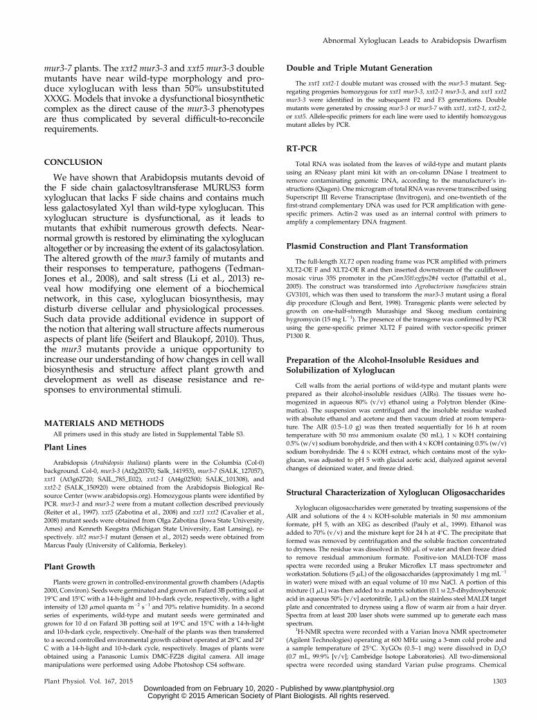
Plants that had a wild-type phenotype, yet weregenotyped as XXT1 xxt2 mur3-3, were obtained duringthe generation of the xxt1 xxt2 mur3-3 triple mutant,which led us to suspect that abolishing the expressionof specific XXT genes suppressed the cabbage-likephenotype. To obtain experimental support for thishypothesis, we generated a series of double mutantsby crossing mur3-3 and mur3-7 with the xxt1, xxt2, andxxt5 mutants (see Fig. 5), which themselves have wild-type phenotypes (Cavalier et al., 2008; Vuttipongchaikijet al., 2012; Zabotina et al., 2012).
The xxt2-1 mur3-3, xxt2-2 mur3-7, xxt5 mur3-3, andxxt5 mur3-7 mutants were phenotypically comparablewith wild-type plants (Fig. 4). The walls of these four
Figure 3. Partial 600-MHz 1H-NMR spectra of theXyG oligosaccharides generated by XEG treatment ofthe 4 N KOH-soluble XyG from wild-type (Col-0),mur3-1, mur3-2, and mur3-3 leaf cell walls. Plantswere grown with a 14-h-light (19˚C) and 10-h-dark(15˚C) cycle. The residues corresponding to eachgroup of proton resonances (e.g. 2-Xyl) are indicatedat the top of the wild-type spectrum. The location ofthese residues in a side chain (X, L, or F) is indicated.
Plant Physiol. Vol. 167, 2015 1299
Abnormal Xyloglucan Leads to Arabidopsis Dwarfism
www.plantphysiol.orgon February 10, 2020 - Published by Downloaded from Copyright © 2015 American Society of Plant Biologists. All rights reserved.
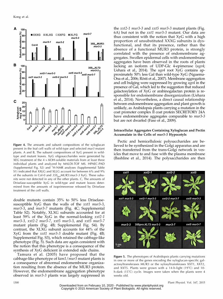
double mutants contain 35% to 50% less Driselase-susceptible XyG than the walls of the xxt1 mur3-3,mur3-3, and mur3-7 mutants (Fig. 4C; SupplementalTable S2). Notably, XLXG subunits accounted for atleast 59% of the XyG in the normal-looking xxt2-1mur3-3, xxt2-2 mur3-7, xxt5 mur3-3, and xxt5 mur3-7mutant plants (Fig. 4B; Supplemental Fig. S4). Bycontrast, the XLXG subunit accounts for 44% of theXyG from the xxt1 mur3-3 double mutant (Fig. 4B;Supplemental Fig. S3), which retained the cabbage-likephenotype (Fig. 5). Such data are again consistent withthe notion that this phenotype is a consequence of thesynthesis of XyG deficient in extended side chains.
Tamura et al. (2005) have proposed that thecabbage-like phenotype of kam1/mur3mutant plants isa consequence of abnormal endomembrane organiza-tion resulting from the absence of the MUR3 protein.However, the endomembrane aggregation phenotypeobserved in mur3-3 plants was largely suppressed in
the xxt2-1 mur3-3 and xxt5 mur3-3 mutant plants (Fig.6A) but not in the xxt1 mur3-3 mutant. Our data arethus consistent with the notion that XyG with a highproportion of unsubstituted XXXG subunits is dys-functional, and that its presence, rather than theabsence of a functional MUR3 protein, is stronglycorrelated with the presence of endomembrane ag-gregates. Swollen epidermal cells with endomembraneaggregates have been observed in the roots of plantslacking an isoform of UDP-Glc 4-epimerase (uge4;Uehara et al., 2014). The uge4 root XyG contains ap-proximately 50% less Gal than wild-type XyG (Nguema-Ona et al., 2006; Rösti et al., 2007). Membrane aggregationand cell bulging were suppressed by growing uge4 in thepresence of Gal, which led to the suggestion that reducedgalactosylation of XyG or arabinogalactan protein is re-sponsible for endomembrane disorganization (Ueharaet al., 2014). Nevertheless, a direct causal relationshipbetween endomembrane aggregation and plant growth isunlikely, as Arabidopsis plants carrying a mutation in thecoat promoter complex II coat protein SECRETORY 24Ahave endomembrane aggregates comparable to mur3-3but are not dwarfed (Faso et al., 2009).
Intracellular Aggregates Containing Xyloglucan and PectinAccumulate in the Cells of mur3-3 Hypocotyls
Pectic and hemicellulosic polysaccharides are be-lieved to be synthesized in the Golgi apparatus and arethen transferred from the trans-Golgi network in ves-icles that move to and fuse with the plasma membrane(Bashline et al., 2014). The polysaccharides are then
Figure 5. The phenotypes of Arabidopsis plants carrying mutationsin one or more of the genes encoding the xyloglucan-specific gal-actosyltransferases MUR3 or the xylosyltransferases XXT1, XXT2,and XXT5. Plants were grown with a 14-h-light (19˚C) and 10-h-dark (15˚C) cycle. Images were taken when the plants were 4weeks old.
Figure 4. The amounts and subunit compositions of the xyloglucanpresent in the leaf cell walls of wild-type and selected mur3 mutantplants. A and B, The subunit compositions of XyG present in wild-type and mutant leaves. XyG oligosaccharides were generated byXEG treatment of the 4 N KOH-soluble materials from at least threeindividual plants and analyzed by MALDI-TOF MS. HPAEC-PAD(Supplemental Fig. S2) and 1H-NMR analyses (Supplemental TableS1) indicated that XXLG and XLLG account for between 6% and 9%of the subunits in Col-0 and 35SproMUR3:mur3-3 XyG. These subu-nits were not detected in any of the other plants. C, The amounts ofDriselase-susceptible XyG in wild-type and mutant leaves deter-mined from the amounts of isoprimeverose released by Driselasetreatment of the cell walls.
1300 Plant Physiol. Vol. 167, 2015
Kong et al.
www.plantphysiol.orgon February 10, 2020 - Published by Downloaded from Copyright © 2015 American Society of Plant Biologists. All rights reserved.
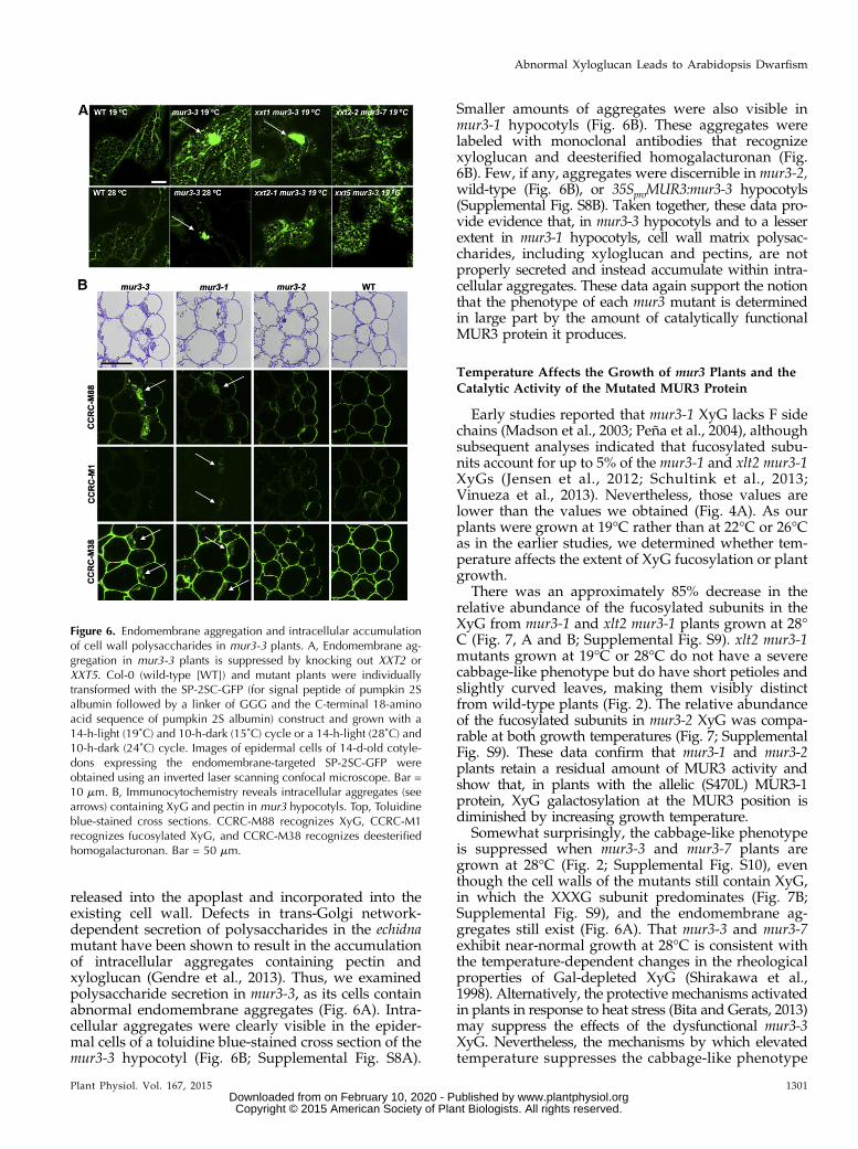
released into the apoplast and incorporated into theexisting cell wall. Defects in trans-Golgi network-dependent secretion of polysaccharides in the echidnamutant have been shown to result in the accumulationof intracellular aggregates containing pectin andxyloglucan (Gendre et al., 2013). Thus, we examinedpolysaccharide secretion in mur3-3, as its cells containabnormal endomembrane aggregates (Fig. 6A). Intra-cellular aggregates were clearly visible in the epider-mal cells of a toluidine blue-stained cross section of themur3-3 hypocotyl (Fig. 6B; Supplemental Fig. S8A).
Smaller amounts of aggregates were also visible inmur3-1 hypocotyls (Fig. 6B). These aggregates werelabeled with monoclonal antibodies that recognizexyloglucan and deesterified homogalacturonan (Fig.6B). Few, if any, aggregates were discernible in mur3-2,wild-type (Fig. 6B), or 35SproMUR3:mur3-3 hypocotyls(Supplemental Fig. S8B). Taken together, these data pro-vide evidence that, in mur3-3 hypocotyls and to a lesserextent in mur3-1 hypocotyls, cell wall matrix polysac-charides, including xyloglucan and pectins, are notproperly secreted and instead accumulate within intra-cellular aggregates. These data again support the notionthat the phenotype of each mur3 mutant is determinedin large part by the amount of catalytically functionalMUR3 protein it produces.
Temperature Affects the Growth of mur3 Plants and theCatalytic Activity of the Mutated MUR3 Protein
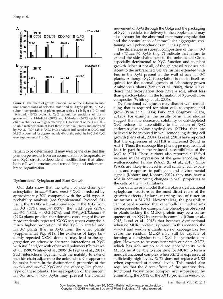
Early studies reported that mur3-1 XyG lacks F sidechains (Madson et al., 2003; Peña et al., 2004), althoughsubsequent analyses indicated that fucosylated subu-nits account for up to 5% of the mur3-1 and xlt2 mur3-1XyGs (Jensen et al., 2012; Schultink et al., 2013;Vinueza et al., 2013). Nevertheless, those values arelower than the values we obtained (Fig. 4A). As ourplants were grown at 19°C rather than at 22°C or 26°Cas in the earlier studies, we determined whether tem-perature affects the extent of XyG fucosylation or plantgrowth.
There was an approximately 85% decrease in therelative abundance of the fucosylated subunits in theXyG from mur3-1 and xlt2 mur3-1 plants grown at 28°C (Fig. 7, A and B; Supplemental Fig. S9). xlt2 mur3-1mutants grown at 19°C or 28°C do not have a severecabbage-like phenotype but do have short petioles andslightly curved leaves, making them visibly distinctfrom wild-type plants (Fig. 2). The relative abundanceof the fucosylated subunits in mur3-2 XyG was compa-rable at both growth temperatures (Fig. 7; SupplementalFig. S9). These data confirm that mur3-1 and mur3-2plants retain a residual amount of MUR3 activity andshow that, in plants with the allelic (S470L) MUR3-1protein, XyG galactosylation at the MUR3 position isdiminished by increasing growth temperature.
Somewhat surprisingly, the cabbage-like phenotypeis suppressed when mur3-3 and mur3-7 plants aregrown at 28°C (Fig. 2; Supplemental Fig. S10), eventhough the cell walls of the mutants still contain XyG,in which the XXXG subunit predominates (Fig. 7B;Supplemental Fig. S9), and the endomembrane ag-gregates still exist (Fig. 6A). That mur3-3 and mur3-7exhibit near-normal growth at 28°C is consistent withthe temperature-dependent changes in the rheologicalproperties of Gal-depleted XyG (Shirakawa et al.,1998). Alternatively, the protective mechanisms activatedin plants in response to heat stress (Bita and Gerats, 2013)may suppress the effects of the dysfunctional mur3-3XyG. Nevertheless, the mechanisms by which elevatedtemperature suppresses the cabbage-like phenotype
Figure 6. Endomembrane aggregation and intracellular accumulationof cell wall polysaccharides in mur3-3 plants. A, Endomembrane ag-gregation in mur3-3 plants is suppressed by knocking out XXT2 orXXT5. Col-0 (wild-type [WT]) and mutant plants were individuallytransformed with the SP-2SC-GFP (for signal peptide of pumpkin 2Salbumin followed by a linker of GGG and the C-terminal 18-aminoacid sequence of pumpkin 2S albumin) construct and grown with a14-h-light (19˚C) and 10-h-dark (15˚C) cycle or a 14-h-light (28˚C) and10-h-dark (24˚C) cycle. Images of epidermal cells of 14-d-old cotyle-dons expressing the endomembrane-targeted SP-2SC-GFP wereobtained using an inverted laser scanning confocal microscope. Bar =10 mm. B, Immunocytochemistry reveals intracellular aggregates (seearrows) containing XyG and pectin in mur3 hypocotyls. Top, Toluidineblue-stained cross sections. CCRC-M88 recognizes XyG, CCRC-M1recognizes fucosylated XyG, and CCRC-M38 recognizes deesterifiedhomogalacturonan. Bar = 50 mm.
Plant Physiol. Vol. 167, 2015 1301
Abnormal Xyloglucan Leads to Arabidopsis Dwarfism
www.plantphysiol.orgon February 10, 2020 - Published by Downloaded from Copyright © 2015 American Society of Plant Biologists. All rights reserved.
remain to be determined. It may well be the case that thisphenotype results from an accumulation of temperature-and XyG structure-dependent modifications that affectboth cell wall structure and remodeling and endomem-brane organization.
Dysfunctional Xyloglucan and Plant Growth
Our data show that the extent of side chain gal-actosylation in mur3-3 and mur3-7 XyG is reduced byapproximately 70% compared with the wild type. Aprobability analysis (see Supplemental Protocol S1)using the XXXG subunit abundance in the XyG frommur3-3 (63%), mur3-7 (73%), the wild type (25%),mur3-1 (48%), mur3-2 (47%), and 35SproMUR3:mur3-3(29%) plants predicts that domains consisting of five ormore tandemly repeated XXXG sequences comprise amuch higher proportion of the XyG in mur3-3 andmur3-7 plants than in XyG from the other plants(Supplemental Fig. S11). The existence of large tan-demly repeated XXXG domains may lead to the ag-gregation or otherwise aberrant interactions of XyGwith itself and/or with other wall polymers (Shirakawaet al., 1998; Whitney et al., 2006; de Freitas et al., 2011).Such interactions together with the inability to extendthe side chain adjacent to the unbranched Glc appear tobe major factors in the dysfunctional properties of themur3-3 and mur3-7 XyG and the cabbage-like pheno-type of these plants. The aggregation of the nascentmur3-3 and mur3-7 XyGs may prevent the normal
movement of XyG through the Golgi and the packagingof XyG in vesicles for delivery to the apoplast, and mayalso account for the abnormal membrane organizationand the accumulation of intracellular aggregates con-taining wall polysaccharides in mur3-3 plants.
The differences in subunit composition of the mur3-3and xlt2 mur3-1 XyGs (Fig. 7) indicate that failure toextend the side chains next to the unbranched Glc isespecially detrimental to XyG function and to plantgrowth. Most, if not all, of the galactosyl residues ad-jacent to the unbranched Glc are further extended withFuc in the XyG present in the wall of xlt2 mur3-1plants. Although XyG fucosylation is not in itself re-quired for the normal growth of laboratory-grownArabidopsis plants (Vanzin et al., 2002), there is evi-dence that fucosylation does have a role, albeit lessthan galactosylation, in the formation of XyG-cellulosecomposites (Whitney et al., 2006).
Dysfunctional xyloglucan may disrupt wall remod-eling that is required for plant cells to expand andgrow (Peña et al., 2004; Park and Cosgrove, 2012a,2012b). For example, the results of in vitro studiessuggest that the decreased solubility of Gal-depletedXyG reduces its accessibility to the action of XyGendotransglucosylases/hydrolases (XTHs) that arebelieved to be involved in wall remodeling during cellgrowth (Peña et al., 2004). Li et al. (2013) have reportedthat the expression of XTH18 is increased 2-fold inrsa3-1. Thus, the cabbage-like phenotype may result atleast in part from the reduced susceptibilities of theXyG to XTH. These authors also reported a 12-foldincrease in the expression of the gene encoding thewall-associated kinase WAK1 (Li et al., 2013). SinceWAKs are likely involved in wall sensing, cell expan-sion, and responses to pathogens and environmentalsignals (Kohorn and Kohorn, 2012), they may have arole in communicating changes in wall properties tothe cells of the mur3 mutants.
Our data favor a model that invokes a dysfunctionalxyloglucan structure as the most direct cause of thegrowth defects of plants carrying loss-of-functionmutations in MUR3. Nevertheless, the possibilitycannot be discounted that other cellular mechanismsare responsible. For example, the phenotypes observedin plants lacking the MUR3 protein may be a conse-quence of an XyG biosynthesis complex (Chou et al.,2015; Lund et al., 2015) that becomes dysfunctionalwhen no MUR3 protein is present. In this scenario, themur3-1 and mur3-2 mutants are not cabbage like be-cause the residual MUR3 may still be capable offorming a nondysfunctional XyG biosynthetic com-plex. However, to be consistent with our data, XLT2,which has 42% amino acid sequence identity withMUR3, must be able to substitute for MUR3 to form anondysfunctional complex when XLT2 is expressed atsufficiently high levels. XLT2 does not replace MUR3when expressed at normal levels. This model alsorequires that the direct effects of the putative dys-functional biosynthetic complex are suppressed byeliminating the XXT2 or the XXT5 protein in mur3-3 or
Figure 7. The effect of growth temperature on the xyloglucan sub-unit compositions of selected mur3 and wild-type plants. A, XyGsubunit compositions of plants grown with a 14-h-light (19˚C) and10-h-dark (15˚C) cycle. B, XyG subunit compositions of plantsgrown with a 14-h-light (28˚C) and 10-h-dark (24˚C) cycle. XyGoligosaccharides were generated by XEG treatment of the 4 N KOH-soluble materials from at least three individual plants and analyzedby MALDI-TOF MS. HPAEC-PAD analyses indicated that XXLG andXLLG accounted for approximately 6% of the subunits in Col-0 XyG(see Supplemental Fig. S9).
1302 Plant Physiol. Vol. 167, 2015
Kong et al.
www.plantphysiol.orgon February 10, 2020 - Published by Downloaded from Copyright © 2015 American Society of Plant Biologists. All rights reserved.
mur3-7 plants. The xxt2 mur3-3 and xxt5 mur3-3 doublemutants have near wild-type morphology and pro-duce xyloglucan with less than 50% unsubstitutedXXXG. Models that invoke a dysfunctional biosyntheticcomplex as the direct cause of the mur3-3 phenotypesare thus complicated by several difficult-to-reconcilerequirements.
CONCLUSION
We have shown that Arabidopsis mutants devoid ofthe F side chain galactosyltransferase MURUS3 formxyloglucan that lacks F side chains and contains muchless galactosylated Xyl than wild-type xyloglucan. Thisxyloglucan structure is dysfunctional, as it leads tomutants that exhibit numerous growth defects. Near-normal growth is restored by eliminating the xyloglucanaltogether or by increasing the extent of its galactosylation.The altered growth of the mur3 family of mutants andtheir responses to temperature, pathogens (Tedman-Jones et al., 2008), and salt stress (Li et al., 2013) re-veal how modifying one element of a biochemicalnetwork, in this case, xyloglucan biosynthesis, maydisturb diverse cellular and physiological processes.Such data provide additional evidence in support ofthe notion that altering wall structure affects numerousaspects of plant life (Seifert and Blaukopf, 2010). Thus,the mur3 mutants provide a unique opportunity toincrease our understanding of how changes in cell wallbiosynthesis and structure affect plant growth anddevelopment as well as disease resistance and re-sponses to environmental stimuli.
MATERIALS AND METHODSAll primers used in this study are listed in Supplemental Table S3.
Plant Lines
Arabidopsis (Arabidopsis thaliana) plants were in the Columbia (Col-0)background. Col-0, mur3-3 (At2g20370; Salk_141953), mur3-7 (SALK_127057),xxt1 (At3g62720; SAIL_785_E02), xxt2-1 (At4g02500; SALK_101308), andxxt2-2 (SALK_150920) were obtained from the Arabidopsis Biological Re-source Center (www.arabidopsis.org). Homozygous plants were identified byPCR. mur3-1 and mur3-2 were from a mutant collection described previously(Reiter et al., 1997). xxt5 (Zabotina et al., 2008) and xxt1 xxt2 (Cavalier et al.,2008) mutant seeds were obtained from Olga Zabotina (Iowa State University,Ames) and Kenneth Keegstra (Michigan State University, East Lansing), re-spectively. xlt2 mur3-1 mutant (Jensen et al., 2012) seeds were obtained fromMarcus Pauly (University of California, Berkeley).
Plant Growth
Plants were grown in controlled-environmental growth chambers (Adaptis2000, Conviron). Seeds were germinated and grown on Fafard 3B potting soil at19°C and 15°C with a 14-h-light and 10-h-dark cycle, respectively, with a lightintensity of 120 mmol quanta m22 s21 and 70% relative humidity. In a secondseries of experiments, wild-type and mutant seeds were germinated andgrown for 10 d on Fafard 3B potting soil at 19°C and 15°C with a 14-h-lightand 10-h-dark cycle, respectively. One-half of the plants was then transferredto a second controlled environmental growth cabinet operated at 28°C and 24°C with a 14-h-light and 10-h-dark cycle, respectively. Images of plants wereobtained using a Panasonic Lumix DMC-FZ28 digital camera. All imagemanipulations were performed using Adobe Photoshop CS4 software.
Double and Triple Mutant Generation
The xxt1 xxt2-1 double mutant was crossed with the mur3-3 mutant. Seg-regating progenies homozygous for xxt1 mur3-3, xxt2-1 mur3-3, and xxt1 xxt2mur3-3 were identified in the subsequent F2 and F3 generations. Doublemutants were generated by crossing mur3-3 or mur3-7 with xxt1, xxt2-1, xxt2-2,or xxt5. Allele-specific primers for each line were used to identify homozygousmutant alleles by PCR.
RT-PCR
Total RNA was isolated from the leaves of wild-type and mutant plantsusing an RNeasy plant mini kit with an on-column DNase I treatment toremove contaminating genomic DNA, according to the manufacturer’s in-structions (Qiagen). Onemicrogram of total RNAwas reverse transcribed usingSuperscript III Reverse Transcriptase (Invitrogen), and one-twentieth of thefirst-strand complementary DNA was used for PCR amplification with gene-specific primers. Actin-2 was used as an internal control with primers toamplify a complementary DNA fragment.
Plasmid Construction and Plant Transformation
The full-length XLT2 open reading frame was PCR amplified with primersXLT2-OE F and XLT2-OE R and then inserted downstream of the cauliflowermosaic virus 35S promoter in the pCam35tl:egfps2#4 vector (Pattathil et al.,2005). The construct was transformed into Agrobacterium tumefaciens strainGV3101, which was then used to transform the mur3-3 mutant using a floraldip procedure (Clough and Bent, 1998). Transgenic plants were selected bygrowth on one-half-strength Murashige and Skoog medium containinghygromycin (15 mg L21). The presence of the transgene was confirmed by PCRusing the gene-specific primer XLT2 F paired with vector-specific primerP1300 R.
Preparation of the Alcohol-Insoluble Residues andSolubilization of Xyloglucan
Cell walls from the aerial portions of wild-type and mutant plants wereprepared as their alcohol-insoluble residues (AIRs). The tissues were ho-mogenized in aqueous 80% (v/v) ethanol using a Polytron blender (Kine-matica). The suspension was centrifuged and the insoluble residue washedwith absolute ethanol and acetone and then vacuum dried at room tempera-ture. The AIR (0.5–1.0 g) was then treated sequentially for 16 h at roomtemperature with 50 mM ammonium oxalate (50 mL), 1 N KOH containing0.5% (w/v) sodium borohydride, and then with 4 N KOH containing 0.5% (w/v)sodium borohydride. The 4 N KOH extract, which contains most of the xylo-glucan, was adjusted to pH 5 with glacial acetic acid, dialyzed against severalchanges of deionized water, and freeze dried.
Structural Characterization of Xyloglucan Oligosaccharides
Xyloglucan oligosaccharides were generated by treating suspensions of theAIR and solutions of the 4 N KOH-soluble materials in 50 mM ammoniumformate, pH 5, with an XEG as described (Pauly et al., 1999). Ethanol wasadded to 70% (v/v) and the mixture kept for 24 h at 4°C. The precipitate thatformed was removed by centrifugation and the soluble fraction concentratedto dryness. The residue was dissolved in 500 mL of water and then freeze driedto remove residual ammonium formate. Positive-ion MALDI-TOF massspectra were recorded using a Bruker Microflex LT mass spectrometer andworkstation. Solutions (5 mL) of the oligosaccharides (approximately 1 mg mL21
in water) were mixed with an equal volume of 10 mM NaCl. A portion of thismixture (1 mL) was then added to a matrix solution (0.1 M 2,5-dihydroxybenzoicacid in aqueous 50% [v/v] acetonitrile, 1 mL) on the stainless steel MALDI targetplate and concentrated to dryness using a flow of warm air from a hair dryer.Spectra from at least 200 laser shots were summed up to generate each massspectrum.
1H-NMR spectra were recorded with a Varian Inova NMR spectrometer(Agilent Technologies) operating at 600 MHz using a 3-mm cold probe anda sample temperature of 25°C. XyGOs (0.5–1 mg) were dissolved in D2O(0.7 mL, 99.9% [v/v]; Cambridge Isotope Laboratories). All two-dimensionalspectra were recorded using standard Varian pulse programs. Chemical
Plant Physiol. Vol. 167, 2015 1303
Abnormal Xyloglucan Leads to Arabidopsis Dwarfism
www.plantphysiol.orgon February 10, 2020 - Published by Downloaded from Copyright © 2015 American Society of Plant Biologists. All rights reserved.
shifts were measured relative to internal acetone (d 2.225). Data wereprocessed using MestReNova software (Universidad de Santiago deCompostela).
Xyloglucan Quantification
The amounts of xyloglucan present in the AIR were determined by mea-suring the amounts of isoprimeverose released by Driselase treatment of theAIR (Fry, 1982). Suspensions of the AIR (approximately 1 g) from mutant andwild-type plants in aqueous 80% (v/v) ethanol (100 mL) were ball milled for16 h at 4°C and 90 rpm in ceramic milling jars (0.3 L) containing a mixture ofone-fourth- and one-half-inch zirconia milling beads (U.S. Stoneware). Themilled AIR was collected by centrifugation, washed with absolute ethanol andthen with acetone, and then vacuum dried at room temperature. Suspensionsof the milled AIR (250–500 mg) in 50 mM sodium acetate, pH 5.2 (30 mL), werethen treated for 24 h at 45°C with Spirizyme (30 mL, Novozymes A/S) andLiquizyme (150 mL, Novozymes A/S) to remove starch. The destarched AIRwas collected by centrifugation, washed thoroughly with deionized water,and freeze dried.
Triplicate suspensions of the AIR (6.25 mg) in 50 mM ammonium formate,pH 4.7 (500 mL), containing 0.5% (w/v) chlorobutanol were treated for 24 h at22°C with 0.25% (w/v) Driselase (100 mL, Sigma-Aldrich) prepared as de-scribed (Fry, 1982). A second portion of Driselase (50 mL) was added and thesuspension kept for a further 24 h at 22°C. Ethanol (1.5 mL) was added and themixture kept at 4°C overnight. The suspension was centrifuged and the solublefraction concentrated to dryness. The residue was dissolved in water (1 mL)and freeze dried to remove residual ammonium formate. The residue wasdissolved in water (1 mL), and a portion (250 mL) was filtered using a 0.45-mmcentrifugal microfilter. Portions (10 mL) of the soluble material were thenanalyzed using a Dionex ICS-3000 HPAEC system with pulsed amperometricdetection and a CarboPac PA1 column (250 3 4.6 mm, Thermo Fisher). Thecolumn was eluted at 1.0 mL min21 for 5 min with 0.1 N NaOH, then with agradient of sodium acetate (0–100 mM) in 0.1 N NaOH for 30 min. The columnwas washed for 15 min with 1 M sodium acetate in 0.1 N NaOH and thenreequilibrated for 15 min in 0.1 N NaOH. The chromatography system wascalibrated using known amounts of isoprimeverose (Megazyme).
Xyloglucan Subunit Composition Analysis Using HPAECwith Pulsed Amperometric Detection
Solutions (10 mL) of XyGOs in water (approximately 200 mg mL21) releasedby XEG treatment of AIR or the 4 N KOH-soluble materials were analyzedusing a Dionex ICS-3000 system and CarboPac PA1 column (250 3 4.6 mm;Thermo Fisher). The column was eluted at 1.0 mL min21 for 3 min with 0.1 N
NaOH, from 3 to 5 min with a gradient of sodium acetate (0–40 mM) in 0.1 N
NaOH and from 5 to 38 min with a gradient of sodium acetate (40–100 mM) in0.1 N NaOH. The column was washed with 1 M sodium acetate in 0.1 N NaOHfor 7 min and reequilibrated for 15 min in 0.1 N NaOH. The column wascalibrated using known amounts (0.5–2 mg injected) of homogeneous XXXG,XXLG, XLXG, XXFG, XLLG, and XLFG purified from XEG digests of tamarindand sycamore cell wall xyloglucan (Tuomivaara et al., 2015).
Glycome Profiling of Plant Cell Wall Extracts
AIR generated from wild-type and mutant plants (approximately 500 mg)was treated sequentially with a-amylase, endopolygalacturonase, 50 mM
sodium carbonate, 1 N KOH, 4 N KOH, and 5 N KOH containing 3% (w/v)boric acid to generate fractions enriched in pectic and hemicellulosic poly-saccharides. The soluble extracts were adjusted to pH 5 if required, dialyzed(3,500 Mr cutoff) against deionized water, and freeze dried. Solutions (50 mL)of each wall extract (60 mg total sugar mL21 water) were then used for enzyme-linked immunosorbent assays using monoclonal antibodies that recognize xylo-glucan epitopes (see Supplemental Table S4) as described (Pattathil et al., 2010).
Confocal Imaging of Endomembrane Organization
Col-0, mur3-3, xxt1 mur3-3, xxt2-1 mur3-3, and xxt5 mur3-3 plants wereindividually transformed with the SP-2SC-GFP construct (Mitsuhashi et al.,2000; Tamura et al., 2005). Transgenic plants were obtained by screening theseeds on kanamycin plates. Epidermal cells of 14-d-old cotyledons were an-alyzed using an inverted laser scanning confocal microscope (LSM510 META,Zeiss) as described (Faso et al., 2009).
Light Microscopy
Leaves from 4-week-old soil-grown Col-0, mur3-3, xxt1 xxt2, and xxt1 xxt2mur3-3 plants and hypocotyls from Col-0, mur3-1, mur3-2, mur3-3, and mur3-3:35SproMUR3 seedlings grown for 4 d under constant light at 22°C were fixedfor 3 h in 25 mM sodium phosphate, pH 7.1, containing 2.5% (v/v) glutaral-dehyde. The fixed leaves and hypocotyls were processed for light microscopyas described previously (Avci et al., 2012).
Immunocytochemistry
Hypocotyl cross sections approximately 3 to 4 mm away from the shootmeristem and leaf cross sections (250 nm thick) approximately 3 to 4 mmaway from the leaf tip were mounted on ColorFrost Plus glass slides (ThermoFisher Scientific) and blocked for 30 min with 10 mM potassium phosphate,pH 7.1, containing 0.5 M NaCl and 3% (w/v) nonfat dry milk. Hypocotyland leaf cross sections were processed for immunolabeling with monoclonalantibodies CCRC-M1, CCRC-M38, or CCRC-M88 as described (Avci et al.,2012).
Supplemental Data
The following supplemental materials are available.
Supplemental Figure S1. RT-PCR analysis of gene transcripts in the mu-tant lines.
Supplemental Figure S2. The MALDI-TOF mass spectra and HPAEC-PADprofiles of the XyGOs generated by XEG treatment of the 4 N KOH-soluble materials from Col-0, mur3-1, mur3-2, and mur3-3 mutant plants.
Supplemental Figure S3. The MALDI-TOF mass spectra and HPAEC-PADprofiles of the XyGOs generated by XEG treatment of the 4 N KOH-soluble materials from Col-0, mur3, xxt, and mur3 xxt mutant plants.
Supplemental Figure S4. The phenotypes and MALDI-TOF mass spectraof the XyGOs generated by XEG treatment of the 4 N KOH-soluble ma-terials from individual 35SproMUR3:mur3, mur3-1, and mur3-2 plants.
Supplemental Figure S5. The MALDI-TOF mass spectra of the XyGOsreleased by XEG treatment of Col-0, mur3-1, mur3-2, and mur3-3 AIR.
Supplemental Figure S6. The effect of the absence of light on the hypocotyllength of seedlings carrying single or combined mutations in genes en-coding xylosyltransferases XXT1, XXT2, and XXT5 and galactosyltrans-ferases MUR3 and XLT2.
Supplemental Figure S7. RT-PCR analyses of XLT2 transcripts in the ro-sette leaves of mur3-3:35Spro:XLT2 plants.
Supplemental Figure S8. Intracellular accumulation of cell wall polysac-charides in mur3 hypocotyls.
Supplemental Figure S9. The MALDI-TOF mass spectra and HPAEC-PADprofiles of the XyGOs generated by XEG treatment of the 4 N KOH-soluble materials from Col-0 and selected mutant plants grown at 19°C and 28°C.
Supplemental Figure S10. The effects of temperature on the growth anddevelopment of mur3-3 plants.
Supplemental Figure S11. Abundance of tandemly repeated XXXG do-mains in the xyloglucan from transgenic plants.
Supplemental Table S1. Side chain compositions of the xyloglucans iso-lated from the cell walls of Arabidopsis wild-type, mur3-1, mur3-2, andmur3-3 plants.
Supplemental Table S2. The amounts and subunit compositions of thexyloglucan present in the leaf cell walls of wild-type and selected xxtArabidopsis mutants.
Supplemental Table S3. Primers used in this study.
Supplemental Table S4. Monoclonal antibodies used for glycome profiling.
Supplemental Protocol S1. Calculating the fraction of a polysaccharidethat is composed of domains containing n or more tandemly repeatedcopies of a sequence.
1304 Plant Physiol. Vol. 167, 2015
Kong et al.
www.plantphysiol.orgon February 10, 2020 - Published by Downloaded from Copyright © 2015 American Society of Plant Biologists. All rights reserved.
ACKNOWLEDGMENTS
We thank Ikuko Hara-Nishimura (Kyoto University) for providing the 2sc-GFP construct and Maor Bar-Peled (Complex Carbohydrate Research Center)for comments on the manuscript.
Received December 18, 2014; accepted February 10, 2015; published February11, 2015.
LITERATURE CITED
Avci U, Pattathil S, Hahn MG (2012) Immunological approaches to plantcell wall and biomass characterization: immunolocalization of glycanepitopes. In ME Himmell, ed, Biomass Conversion: Methods and Protocols,Vol 908. Humana Press, New York, pp 73-82
Bashline L, Lei L, Li S, Gu Y (2014) Cell wall, cytoskeleton, and cell ex-pansion in higher plants. Mol Plant 7: 586–600
Bita CE, Gerats T (2013) Plant tolerance to high temperature in a changingenvironment: scientific fundamentals and production of heat stress-tolerant crops. Front Plant Sci 4: 273
Cavalier DM, Lerouxel O, Neumetzler L, Yamauchi K, Reinecke A, FreshourG, Zabotina OA, Hahn MG, Burgert I, Pauly M, et al (2008) Disrupting twoArabidopsis thaliana xylosyltransferase genes results in plants deficient inxyloglucan, a major primary cell wall component. Plant Cell 20: 1519–1537
Chou YH, Pogorelko G, Young ZT, Zabotina OA (2015) Protein–proteininteractions among xyloglucan-synthesizing enzymes and formation ofGolgi-localized multiprotein complexes. Plant Cell Physiol 56: 255–267
Clough SJ, Bent AF (1998) Floral dip: a simplified method for Agrobacterium-mediated transformation of Arabidopsis thaliana. Plant J 16: 735–743
de Freitas RA, Busato AP, Mitchell DA, Silveira JL (2011) Degalactosylation ofxyloglucan: effect on aggregation and conformation, as determined by timedependent static light scattering, HPSEC–MALLS and viscosimetry. Carbo-hydr Polym 83: 1636–1642
Faso C, Chen YN, Tamura K, Held M, Zemelis S, Marti L, Saravanan R,Hummel E, Kung L, Miller E, et al (2009) A missense mutation in theArabidopsis COPII coat protein Sec24A induces the formation of clusters ofthe endoplasmic reticulum and Golgi apparatus. Plant Cell 21: 3655–3671
Fry SC (1982) Phenolic components of the primary cell wall: feruloylateddisaccharides of D-galactose and L-arabinose from spinach polysac-charide. Biochem J 203: 493–504
Gendre D, McFarlane HE, Johnson E, Mouille G, Sjödin A, Oh J, Levesque-Tremblay G, Watanabe Y, Samuels L, Bhalerao RP (2013) Trans-Golginetwork localized ECHIDNA/Ypt interacting protein complex is required forthe secretion of cell wall polysaccharides in Arabidopsis. Plant Cell 25: 2633–2646
Hoffman M, Jia Z, Peña MJ, Cash M, Harper A, Blackburn II AR, DarvillA, York WS (2005) Structural analysis of xyloglucans in the primary cellwalls of plants in the subclass Asteridae. Carbohydr Res 340: 1826–1840
Hsieh YSY, Harris PJ (2012) Structures of xyloglucans in primary cell wallsof gymnosperms, monilophytes (ferns sensu lato) and lycophytes. Phyto-chemistry 79: 87–101
Jensen JK, Schultink A, Keegstra K, Wilkerson CG, Pauly M (2012) RNA-Seqanalysis of developing nasturtium seeds (Tropaeolum majus): identificationand characterization of an additional galactosyltransferase involved inxyloglucan biosynthesis. Mol Plant 5: 984–992
Kohorn BD, Kohorn SL (2012) The cell wall-associated kinases, WAKs, aspectin receptors. Front Plant Sci 3: 88
Li W, Guan Q, Wang ZY, Wang Y, Zhu J (2013) A bi-functional xyloglucangalactosyltransferase is an indispensable salt stress tolerance determi-nant in Arabidopsis. Mol Plant 6: 1344–1354
Lund CH, Bromley JR, Stenbæk A, Rasmussen RE, Scheller HV, SakuragiY (2015) A reversible Renilla luciferase protein complementation assayfor rapid identification of protein-protein interactions reveals the exis-tence of an interaction network involved in xyloglucan biosynthesis inthe plant Golgi apparatus. J Exp Bot 66: 85–97
Madson M, Dunand C, Li X, Verma R, Vanzin GF, Caplan J, Shoue DA,Carpita NC, Reiter WD (2003) The MUR3 gene of Arabidopsis encodes axyloglucan galactosyltransferase that is evolutionarily related to animalexostosins. Plant Cell 15: 1662–1670
Mitsuhashi N, Shimada T, Mano S, Nishimura M, Hara-Nishimura I(2000) Characterization of organelles in the vacuolar-sorting pathway byvisualization with GFP in tobacco BY-2 cells. Plant Cell Physiol 41: 993–1001
Nguema-Ona E, Andème-Onzighi C, Aboughe-Angone S, Bardor M, IshiiT, Lerouge P, Driouich A (2006) The reb1-1 mutation of Arabidopsis:effect on the structure and localization of galactose-containing cell wallpolysaccharides. Plant Physiol 140: 1406–1417
Park YB, Cosgrove DJ (2012a) Changes in cell wall biomechanical prop-erties in the xyloglucan-deficient xxt1/xxt2 mutant of Arabidopsis. PlantPhysiol 158: 465–475
Park YB, Cosgrove DJ (2012b) A revised architecture of primary cell wallsbased on biomechanical changes induced by substrate-specific endo-glucanases. Plant Physiol 158: 1933–1943
Pattathil S, Avci U, Baldwin D, Swennes AG, McGill JA, Popper Z,Bootten T, Albert A, Davis RH, Chennareddy C, et al (2010) A com-prehensive toolkit of plant cell wall glycan-directed monoclonal anti-bodies. Plant Physiol 153: 514–525
Pattathil S, Harper AD, Bar-Peled M (2005) Biosynthesis of UDP-xylose:characterization of membrane-bound AtUxs2. Planta 221: 538–548
Pauly M, Andersen LN, Kauppinen S, Kofod LV, York WS, AlbersheimP, Darvill A (1999) A xyloglucan-specific endo-b-1,4-glucanase fromAspergillus aculeatus: expression cloning in yeast, purification andcharacterization of the recombinant enzyme. Glycobiology 9: 93–100
Pauly M, Eberhard S, Albersheim P, Darvill A, York WS (2001) Effects ofthe mur1 mutation on xyloglucans produced by suspension-culturedArabidopsis thaliana cells. Planta 214: 67–74
Peña MJ, Darvill AG, Eberhard S, York WS, O’Neill MA (2008) Moss andliverwort xyloglucans contain galacturonic acid and are structurallydistinct from the xyloglucans synthesized by hornworts and vascularplants. Glycobiology 18: 891–904
Peña MJ, Ryden P, Madson M, Smith AC, Carpita NC (2004) The galactoseresidues of xyloglucan are essential to maintain mechanical strength ofthe primary cell walls in Arabidopsis during growth. Plant Physiol 134:443–451
Perrin RM, DeRocher AE, Bar-Peled M, Zeng W, Norambuena L,Orellana A, Raikhel NV, Keegstra K (1999) Xyloglucan fucosyl-transferase, an enzyme involved in plant cell wall biosynthesis. Science284: 1976–1979
Perrin RM, Jia Z, Wagner TA, O’Neill MA, Sarria R, York WS, RaikhelNV, Keegstra K (2003) Analysis of xyloglucan fucosylation in Arabidopsis.Plant Physiol 132: 768–778
Reiter WD, Chapple C, Somerville CR (1997) Mutants of Arabidopsisthaliana with altered cell wall polysaccharide composition. Plant J 12:335–345
Rösti J, Barton CJ, Albrecht S, Dupree P, Pauly M, Findlay K, Roberts K,Seifert GJ (2007) UDP-glucose 4-epimerase isoforms UGE2 and UGE4cooperate in providing UDP-galactose for cell wall biosynthesis andgrowth of Arabidopsis thaliana. Plant Cell 19: 1565–1579
Sauer RT (2013) Mutagenic dissection of the sequence determinants ofprotein folding, recognition, and machine function. Protein Sci 22: 1675–1687
Schultink A, Cheng K, Park YB, Cosgrove DJ, Pauly M (2013) The iden-tification of two arabinosyltransferases from tomato reveals functionalequivalency of xyloglucan side chain substituents. Plant Physiol 163: 86–94
Seifert GJ, Blaukopf C (2010) Irritable walls: the plant extracellular matrixand signaling. Plant Physiol 153: 467–478
Shirakawa M, Yamatoya K, Nishinari K (1998) Tailoring of xyloglucanproperties using an enzyme. Food Hydrocoll 12: 25–28
Tamura K, Shimada T, Kondo M, Nishimura M, Hara-Nishimura I (2005)KATAMARI1/MURUS3 Is a novel golgi membrane protein that is re-quired for endomembrane organization in Arabidopsis. Plant Cell 17:1764–1776
Tedman-Jones JD, Lei R, Jay F, Fabro G, Li X, Reiter WD, Brearley C,Jones JDG (2008) Characterization of Arabidopsis mur3 mutations thatresult in constitutive activation of defence in petioles, but not leaves.Plant J 56: 691–703
Tuomivaara ST, Yaoi K, O’Neill MA, York WS (2015) Generation andstructural validation of a library of diverse xyloglucan-derived oligosaccharides,including an update on xyloglucan nomenclature. Carbohydr Res 402: 56–66
Uehara M, Wang S, Kamiya T, Shigenobu S, Yamaguchi K, Fujiwara T,Naito S, Takano J (2014) Identification and characterization of an Arabi-dopsis mutant with altered localization of NIP5;1, a plasma membrane boricacid channel, reveals the requirement for D-galactose in endomembrane or-ganization. Plant Cell Physiol 55: 704–714
Vanzin GF, Madson M, Carpita NC, Raikhel NV, Keegstra K, Reiter WD(2002) The mur2 mutant of Arabidopsis thaliana lacks fucosylated
Plant Physiol. Vol. 167, 2015 1305
Abnormal Xyloglucan Leads to Arabidopsis Dwarfism
www.plantphysiol.orgon February 10, 2020 - Published by Downloaded from Copyright © 2015 American Society of Plant Biologists. All rights reserved.
xyloglucan because of a lesion in fucosyltransferase AtFUT1. Proc NatlAcad Sci USA 99: 3340–3345
Vinueza NR, Gallardo VA, Klimek JF, Carpita NC, Kenttämaa HI (2013)Analysis of xyloglucans by ambient chloride attachment ionizationtandem mass spectrometry. Carbohydr Polym 98: 1203–1213
Vuttipongchaikij S, Brocklehurst D, Steele-King C, Ashford DA, GomezLD, McQueen-Mason SJ (2012) Arabidopsis GT34 family contains fivexyloglucan a-1,6-xylosyltransferases. New Phytol 195: 585–595
Wan Y, Kertesz M, Spitale RC, Segal E, Chang HY (2011) Under-standing the transcriptome through RNA structure. Nat Rev Genet 12:641–655
Whitney SEC, Wilson E, Webster J, Bacic A, Reid JSG, Gidley MJ (2006)Effects of structural variation in xyloglucan polymers on interactionswith bacterial cellulose. Am J Bot 93: 1402–1414
Zabotina OA, Avci U, Cavalier D, Pattathil S, Chou YH, Eberhard S,Danhof L, Keegstra K, Hahn MG (2012) Mutations in multiple XXTgenes of Arabidopsis reveal the complexity of xyloglucan biosynthesis.Plant Physiol 159: 1367–1384
Zabotina OA, van de Ven WTG, Freshour G, Drakakaki G, Cavalier D,Mouille G, Hahn MG, Keegstra K, Raikhel NV (2008) ArabidopsisXXT5 gene encodes a putative a-1,6-xylosyltransferase that is involvedin xyloglucan biosynthesis. Plant J 56: 101–115
1306 Plant Physiol. Vol. 167, 2015
Kong et al.
www.plantphysiol.orgon February 10, 2020 - Published by Downloaded from Copyright © 2015 American Society of Plant Biologists. All rights reserved.