Embed Size (px)
Citation preview

ORIGINAL ARTICLE
Evaluation of Structure and Assembly of Xyloglucanfrom Tamarind Seed (Tamarindus indica L.) with AtomicForce Microscopy
Arkadiusz Kozioł1 & Justyna Cybulska1 &
Piotr M. Pieczywek1& Artur Zdunek1
Received: 23 December 2014 /Accepted: 30 March 2015 /Published online: 12 April 2015# The Author(s) 2015. This article is published with open access at Springerlink.com
Abstract The role of xyloglucan (XG) in the cell wall ofplants and its technological usability depends on several fac-tors, pertaining to molecular structure. Therefore, the goal ofthis study was to evaluate the nano-structure and self-assembly of XG by atomic force microscopy (AFM). As themodel, a non-modified xyloglucan from a tamarind seed(Tamarindus indica L.) was used. Samples were minimallyprocessed, i.e., treated with low-power ultrasound and studiedon the surface of mica in ambient butanol. AFM topographicimages revealed rod-like nanomolecules of xyloglucan with amean height of 2.3±0.5 nm and mean length of 640±360 nm.The AFM study also showed that XG chains possessed ahelical structure with a period of 115.8±29.2 nm. This studyshowed possible-bending of molecules with a mean angle of127.8±25.6°. The xyloglucan molecules were able to aggre-gate as cross-like and a parallel like assemblies, and possiblyas rope-like structures. The self-assembled bundles ofxyloglucan chains were often complexed at an angle of114.2±36.3°.
Keywords AFM .Xyloglucan . Nanostructure . Tamarindseeds
Introduction
Xyloglucan (XG) is the most abundant polysaccharide fromthe group of hemicelluloses in the primary cell wall of higherplants comprising of about 20 % of all constituents of dicoty-ledons [1]. According to the dominating concept of architec-ture of a cell wall in plants, cellulose and hemicellulose con-struct a major load-bearing structure where XG has the struc-tural function affecting orientation of cellulose microfibrils[2]. Together with pectic polysaccharides, xyloglucan is clas-sified as a matrix polysaccharide. In cotyledon or some dicot-yledon seeds it plays the role of a storage polysaccharide [3, 4]and may be present in the secondary cell wall. The nanostruc-ture of a xyloglucan polysaccharide determines the cross-linking mechanism to cellulose microfibrils, i.e., a lesssubstituted backbone of XG (fucosyl branching), the greatercapability for binding to cellulose [5, 6]. The arrangement ofXG chains contributes to a higher rigidity of the wall. Thisbiopolymer is a heteropolysaccharide possessing the back-bone composed of 1,4-β-D-glucose. In the backbone, threeout of four glucose residues are substituted with 1,6-α-D xy-lose, which are about 70 % of glucosyl residues [7]. Addition-al residues, in form 1,2-β-D galactose, may be attached toxylose, depending on the origin of xyloglucan. Fucose canattach to outer galactose residue. A xyloglucan chain has aribbon-like, twofold helical conformation [8, 9]. XG, as across-linking material, coats the surface of cellulose microfi-brils, although some of them may penetrate and disrupt themicrocrystalline parts of cellulose molecules [10].
Tamarind seeds are one of the most abundant sources ofxyloglucan and commonly used as a storage of commerciallyavailable polysaccharides. Tamarind XG (like many otherpolysaccharides) is water-soluble, but its individual macro-molecules tend not to fully hydrate, thus supramolecular ag-gregated chains remain present even in very diluted solutions
Highlights • Nanostructure of tamarind xyloglucan molecules and itsassemblies were studied• AFM was used for observation of the nanostructure evaluation ofxyloglucan• Diameter, length, bending, complexing angles, and folding propertieswere analysed• Parallel like and rope-like xyloglucan assemblies were observed
* Artur [email protected]
1 Institute of Agrophysics, Polish Academy of Sciences,Doswiadczalna 4, 20-290 Lublin, Poland
Food Biophysics (2015) 10:396–402DOI 10.1007/s11483-015-9395-2

[11]. The diversity of the possible side chains of a xyloglucanbackbone determines the functionality and physico-chemicalproperties, like water solubility or gelling capability, therefore;currently tamarind xyloglucan is used e.g., as a gelling agent,an emulsion stabilizer or as a food thickener [4]. XG, likemany other biopolymers, is potentially important in commer-cial and medical applications. Xyloglucan is also a source ofbiologically active oligosaccharides and was indicated as anantitumor factor [12, 13]. Lately, XG has been found as amaterial for the preparation of edible, non-toxic, biodegrad-able and transparent films for various applications especiallyin the controlled release of drugs and cosmetics [14–17].
Tamarind XG was examined by X-ray scattering and lightscattering [11] to evaluate its structure or by chromatographicmethods to estimate XG size and molecular weight [3, 18].Most of the studies on tamarind xyloglucan also focused onthe rheological or physico-chemical properties of chemicallymodified molecules [16, 19, 20]. Recently, AFM was used tostudy the structure of individual hemicelluloses extracted fromthe cell walls of fruit [21, 22]. So far, only a few experimentshave been devoted to nanostructure characterization of natu-ral, chemically non-treated, xyloglucan molecules or hemicel-lulose (understood as components of few hemicellulosic poly-saccharides) from fruit [21, 23].
Thus, structural characterization is still indeed importantsince it determines the function of XG in plants and function-ality for commercial use in food systems. In this paper, wewould present the structural characterization of xyloglucanmolecules with a particular emphasis on assemblies whichcould be formed by the XG molecules. For this purpose theXG molecules from tamarind seeds, as a model system, werestudied with an atomic force microscope, where it is possibleto obtain a nanometre resolution [24–29]. To study the struc-ture of individual polysaccharides with AFM, molecules mustbe adsorbed on the mica [30–37]. Before placement on micawe applied weak ultrasound treatment and low dilution of anXG solution to obtain individual non-highly-aggregated mol-ecules for an objective analysis. This method gave useful to-pographical information about the structure of XG rod-likemolecules and their assemblies. Our experimental results onXG structure are in line with previous simulations of moleculedimensions by molecular dynamics.
Materials and Methods
Sample Preparation
Tamarind xyloglucan from seeds (Tamarindus indica L.) pur-chased from Megazyme (Bray, Ireland) was composed of xy-lose, glucose, galactose and arabinose in the proportion of38:42:16:4. The 1 % (w/v) aqueous solution of the xyloglucanwas diluted 20 times. Then it was stirred for 12 h in room
temperature. Since XG has a tendency for self-association inaqueous solutions, the diluted sample (1.5 ml) was treatedwith low power ultrasounds (vibration amplitude 32 μmand power 130 W) for 30 s to reduce aggregates andmacroparticles. The ultrasonic processor Vibra-CellVCX130FSJ (Sonics & Materials, Inc., Newtown, USA)with microtip probe of 3 mm diameter was used. Thetreatment was optimized to possibly obtain a high yieldof molecule deployment during AFM scanning.
For structure characterization, 5 μl of the xyloglucan solu-tion was dropped onto the freshly cleaved mica and dried for1 h in ambient air. This causes adhesion of molecules to sub-strate which facilitates scanning by AFM tip of biopolymers[21, 25, 38].
Fig. 1 Method of determination of the height profile of chain-like mole-cules from an AFM height image. a The red line is a skeleton of XGmolecule drawn from segmented images at a threshold 700 pm b Thedashed red line depicts the height profile, which was determined along theskeleton. The height histogram was built from the each pixel along the line
Table 1 Characterization of tamarind xyloglucan molecules
Mean value Np N
Height (nm) 2.3 (0.5)a 6672 85
Length (nm) 640 (360) 85 85
Peak to peak distance (nm) 115.8 (29.2) 44 1 (the longestchain)
Angles
Single molecules (deg) 127.8 (25.6) 19 16
Complexed molecules (deg) 114.2 (36.3) 14 10
Np – number of data used for mean calculation
N – number of analyzed chainsa Standard deviation in parentheses
Food Biophysics (2015) 10:396–402 397
AFM Imaging
Topographic observations were performed using the atomicforce microscope Bioscope Catalyst II (Bruker, St. Barbara,U.S.A.) and semiautomatic PeakForce Tapping mode. TheAFM imaging was carried out in butanol (POCH, Gliwice,Poland) which reduces undesirable effects appearing duringimaging; decreases adhesion of the tip and reduces influenceof the water layer on the surface [30, 37]. The height imageswere taken with resolution 512×512 points and 0.5 Hz scanrate with use of a silicon probe (Bruker) of the nominal springconstant 0.4 Nm−1 and the resonance frequency of 70 kHz.Measured RMS roughness on the mica surface at the samescanning conditions was 0.3 nm.
XG chains in this experiment were characterized by theirlength and height. Molecule height was chosen instead of
width since for a typical AFM tip, the width of molecules isoften even several dozen times bigger than their height due toso-called Bprofile broadening^ or Btip convolution^ effect[39]. Additionally, scanning in Z axis is more accurate thanin X-Y due to scanner range size. In the case of chain length,this effect is not fundamentally important.
Data Analysis
The height images were subjected to the 3rd order flattening inSPIP 6.0.14 (Image Metrology, Hørsholm, Denmark) beforequantitative analysis. Images were analyzed by self-developedprotocol inMatlab (TheMathWorks, MA, USA). In the imageanalysis protocol molecules were segmented from back-ground. The minima threshold was set at 0.2 nm. Then askeleton of the segmented objects was determined [22]. The
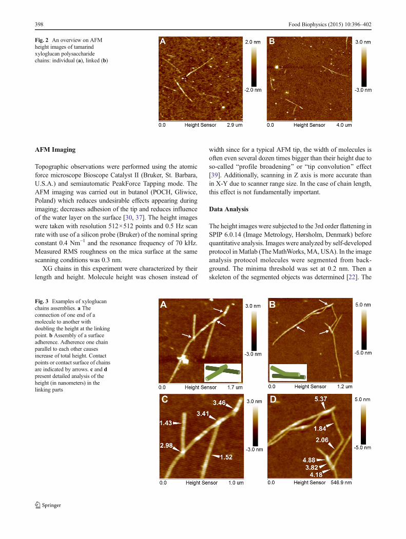
Fig. 2 An overview on AFMheight images of tamarindxyloglucan polysaccharidechains: individual (a), linked (b)
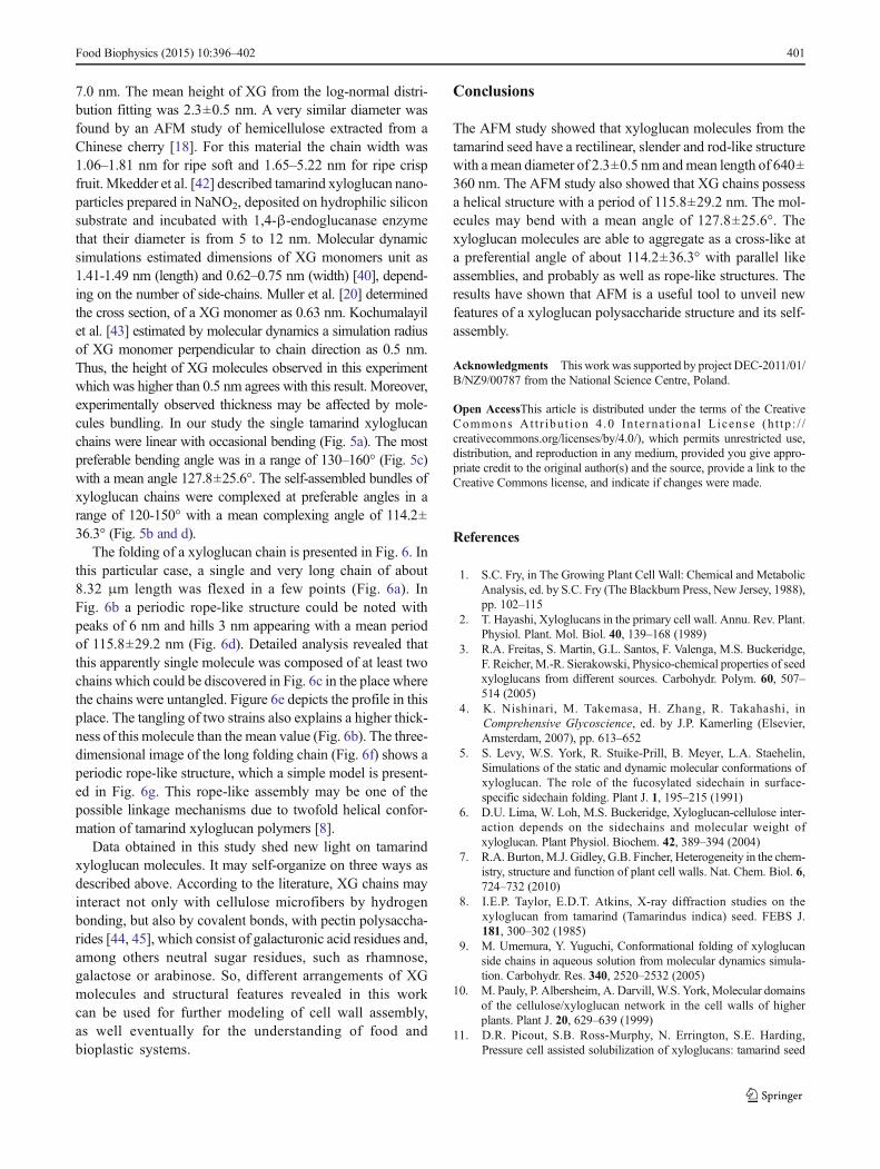
Fig. 3 Examples of xyloglucanchains assemblies. a Theconnection of one end of amolecule to another withdoubling the height at the linkingpoint. b Assembly of a surfaceadherence. Adherence one chainparallel to each other causesincrease of total height. Contactpoints or contact surface of chainsare indicated by arrows. c and dpresent detailed analysis of theheight (in nanometers) in thelinking parts
398 Food Biophysics (2015) 10:396–402

skeleton was used as the mask to the original image to markpixels where the heights along the molecules were collected(Fig. 1). The height histogram was built from each point de-fined by the skeleton. The skeletonized images were also usedto evaluate the length of molecules. Angles and peak to peakdistances were measured manually in computer image analy-sis SPIP 6.0.14 software. In the peak to peak analysis, theheights greater than 4 nm were considered to determine thedistance between two adjacent peaks along the chain.
Results and Discussion
Results of study are summarized in Table 1. AFM heightimages presented in Fig. 2 show that pure xyloglucan formedrectilinear, slender and rod-like chains. In the case of the pre-sented AFM scans; the fibrils are deposited on a film-likematrix which covers the mica. A root mean square averageof height deviations of mean data was about 0.6 nm, whereas;for clean mica it was about 0.3 nm. The film-like depositionon mica presumably consists of xyloglucan particles partially
solubilized in water after ultrasound treatment. Consequently,the height of objects was determined relatively to the meanheight of the background. The XG molecules were randomlyspread as individual, separated rods (Fig. 2a) or as bundles ofseveral chains (Figs. 2b and 3). Despite the ultrasound treat-ment priory to imaging XGmolecules still showed a tendencyfor assembly. Two ways of linking between chains in the bun-dles were observed. The first way consisted in the linking ofone end of the rod to another one in the middle of its chain(Fig. 3a). Profile analysis showed that this is rather an overlapof one molecule on another because the total height in thelinking point was doubled (Fig. 3a and c). In the second wayof assembly, chains were connected along some distance thatalso caused an increase of total height (Fig. 3b and d). Theparallel connection of neighboring chains is likely to link themechanism of cellulose by xyloglucan where a smooth XGregion adheres by hydrogen bonds to cellulose. This resultalso confirms simulations performed on tamarind xyloglucanchains which showed the ability for parallel-arrangement [40].The observation that xyloglucan molecules have a tendency tolink is in line with AFM images of hemicelluloses obtained
Fig. 4 The distribution of the length a and the height b of xyloglucanmolecules with fitting curves (Gaussian for the length, log-normal for theheight). NPmeans the number of data from histograms, non-branched XGchains taken for analyses (for length distribution it is a number of chains,
for height distribution – number of height points along skeleton lines fromall chains); R2 is coefficient of the determination of fitting. Polysaccharidechains longer than 2 μm have been omitted on graph a in order to showthe essential part of the histogram
Fig. 5 Characteristic angles of xyloglucan assemblies. a bending angle, b complexing angle, c distribution of bending angles, d distribution of thecomplexing angles. NP is a number of events taken to analysis
Food Biophysics (2015) 10:396–402 399

from Chinese cherries [21]. It suggests that xyloglucan in thecell wall of plants may form branched structures and in thisway affects cell wall properties.
Image analysis allowed to characterize the dimensions oftamarind xyloglucan chains (Fig. 4). These characteristicswere obtained for separated XGmolecules and did not includethe increase of height as the result of assembling. A length oftamarind xyloglucan chains was in a range from 178 nm up to2 μm and the mean value from the fitting of normal distribu-tion was 640±360 nm (Fig. 4a). In a studied population oftamarind XG chains substantial fraction of molecules of thelength ca. 300-1000 nm was noted. A previous study ofxyloglucan from tamarind seeds showed that for trimer thelength of spine was estimated as 3.54 nm (using gyration radiifrom SAXS experiment) [40]. Although the value was notexactly a multiplication of the monomer length it was
concluded that one monomer unit adds approximately 1 nmto the total length of the spine. Taylor and Atkins [8] haveanalyzed the structure of xyloglucan polysaccharide from thetamarind seed. They have prepared a XG film and by using X-ray diffraction in order to show the length of XG’s repeatingunit consisting of four D-glucose residues (XGmonomer) wasestimated as 2.06 nm. Small-angle neutron scattering showedthe XG monomer persistence length as 8 nm [18]. It meansthat the mean length of tamarind XG molecules obtained inthis experiment agrees with the theory that an XG backbonemay contain from 300 to 3000 glucose units, which corre-sponds to the length from 0.15 to 1.5 μm, if each glucose unitis 0.5 nm [41].
The height distribution is presented in Fig. 4b. The heightvalue of XG, which more accurately represents diameter dueto the lack of the tip convolution effect, was within 0.5–
Fig. 6 a Topographic image of a xyloglucan chain chosen for analysis offolding. b Periodicity on XG fiber. c The bending place with a split ofchain in two chains. d Distribution of peak-to-peak on XG chains (NP
means the number of data from histograms, R2 is coefficient in the
determination of fitting); e A profile of the case where a helical structurewas untangled (marked in c). f 3D visualization of XG rope-like assem-bly; gModel of XG rope-like assembly with mean peak-to-peak distance
400 Food Biophysics (2015) 10:396–402
7.0 nm. The mean height of XG from the log-normal distri-bution fitting was 2.3±0.5 nm. A very similar diameter wasfound by an AFM study of hemicellulose extracted from aChinese cherry [18]. For this material the chain width was1.06–1.81 nm for ripe soft and 1.65–5.22 nm for ripe crispfruit. Mkedder et al. [42] described tamarind xyloglucan nano-particles prepared in NaNO2, deposited on hydrophilic siliconsubstrate and incubated with 1,4-β-endoglucanase enzymethat their diameter is from 5 to 12 nm. Molecular dynamicsimulations estimated dimensions of XG monomers unit as1.41-1.49 nm (length) and 0.62–0.75 nm (width) [40], depend-ing on the number of side-chains. Muller et al. [20] determinedthe cross section, of a XG monomer as 0.63 nm. Kochumalayilet al. [43] estimated by molecular dynamics a simulation radiusof XG monomer perpendicular to chain direction as 0.5 nm.Thus, the height of XG molecules observed in this experimentwhich was higher than 0.5 nm agrees with this result. Moreover,experimentally observed thickness may be affected by mole-cules bundling. In our study the single tamarind xyloglucanchains were linear with occasional bending (Fig. 5a). The mostpreferable bending angle was in a range of 130–160° (Fig. 5c)with a mean angle 127.8±25.6°. The self-assembled bundles ofxyloglucan chains were complexed at preferable angles in arange of 120-150° with a mean complexing angle of 114.2±36.3° (Fig. 5b and d).
The folding of a xyloglucan chain is presented in Fig. 6. Inthis particular case, a single and very long chain of about8.32 μm length was flexed in a few points (Fig. 6a). InFig. 6b a periodic rope-like structure could be noted withpeaks of 6 nm and hills 3 nm appearing with a mean periodof 115.8±29.2 nm (Fig. 6d). Detailed analysis revealed thatthis apparently single molecule was composed of at least twochains which could be discovered in Fig. 6c in the place wherethe chains were untangled. Figure 6e depicts the profile in thisplace. The tangling of two strains also explains a higher thick-ness of this molecule than the mean value (Fig. 6b). The three-dimensional image of the long folding chain (Fig. 6f) shows aperiodic rope-like structure, which a simple model is present-ed in Fig. 6g. This rope-like assembly may be one of thepossible linkage mechanisms due to twofold helical confor-mation of tamarind xyloglucan polymers [8].
Data obtained in this study shed new light on tamarindxyloglucan molecules. It may self-organize on three ways asdescribed above. According to the literature, XG chains mayinteract not only with cellulose microfibers by hydrogenbonding, but also by covalent bonds, with pectin polysaccha-rides [44, 45], which consist of galacturonic acid residues and,among others neutral sugar residues, such as rhamnose,galactose or arabinose. So, different arrangements of XGmolecules and structural features revealed in this workcan be used for further modeling of cell wall assembly,as well eventually for the understanding of food andbioplastic systems.
Conclusions
The AFM study showed that xyloglucan molecules from thetamarind seed have a rectilinear, slender and rod-like structurewith a mean diameter of 2.3±0.5 nm andmean length of 640±360 nm. The AFM study also showed that XG chains possessa helical structure with a period of 115.8±29.2 nm. The mol-ecules may bend with a mean angle of 127.8±25.6°. Thexyloglucan molecules are able to aggregate as a cross-like ata preferential angle of about 114.2±36.3° with parallel likeassemblies, and probably as well as rope-like structures. Theresults have shown that AFM is a useful tool to unveil newfeatures of a xyloglucan polysaccharide structure and its self-assembly.
Acknowledgments This work was supported by project DEC-2011/01/B/NZ9/00787 from the National Science Centre, Poland.
Open AccessThis article is distributed under the terms of the CreativeCommons At t r ibut ion 4 .0 In te rna t ional License (h t tp : / /creativecommons.org/licenses/by/4.0/), which permits unrestricted use,distribution, and reproduction in any medium, provided you give appro-priate credit to the original author(s) and the source, provide a link to theCreative Commons license, and indicate if changes were made.
References
1. S.C. Fry, in The Growing Plant Cell Wall: Chemical and MetabolicAnalysis, ed. by S.C. Fry (The Blackburn Press, New Jersey, 1988),pp. 102–115
2. T. Hayashi, Xyloglucans in the primary cell wall. Annu. Rev. Plant.Physiol. Plant. Mol. Biol. 40, 139–168 (1989)
3. R.A. Freitas, S. Martin, G.L. Santos, F. Valenga, M.S. Buckeridge,F. Reicher, M.-R. Sierakowski, Physico-chemical properties of seedxyloglucans from different sources. Carbohydr. Polym. 60, 507–514 (2005)
4. K. Nishinari, M. Takemasa, H. Zhang, R. Takahashi, inComprehensive Glycoscience, ed. by J.P. Kamerling (Elsevier,Amsterdam, 2007), pp. 613–652
5. S. Levy, W.S. York, R. Stuike-Prill, B. Meyer, L.A. Staehelin,Simulations of the static and dynamic molecular conformations ofxyloglucan. The role of the fucosylated sidechain in surface-specific sidechain folding. Plant J. 1, 195–215 (1991)
6. D.U. Lima, W. Loh, M.S. Buckeridge, Xyloglucan-cellulose inter-action depends on the sidechains and molecular weight ofxyloglucan. Plant Physiol. Biochem. 42, 389–394 (2004)
7. R.A. Burton,M.J. Gidley, G.B. Fincher, Heterogeneity in the chem-istry, structure and function of plant cell walls. Nat. Chem. Biol. 6,724–732 (2010)
8. I.E.P. Taylor, E.D.T. Atkins, X-ray diffraction studies on thexyloglucan from tamarind (Tamarindus indica) seed. FEBS J.181, 300–302 (1985)
9. M. Umemura, Y. Yuguchi, Conformational folding of xyloglucanside chains in aqueous solution from molecular dynamics simula-tion. Carbohydr. Res. 340, 2520–2532 (2005)
10. M. Pauly, P. Albersheim, A. Darvill, W.S. York,Molecular domainsof the cellulose/xyloglucan network in the cell walls of higherplants. Plant J. 20, 629–639 (1999)
11. D.R. Picout, S.B. Ross-Murphy, N. Errington, S.E. Harding,Pressure cell assisted solubilization of xyloglucans: tamarind seed
Food Biophysics (2015) 10:396–402 401
polysaccharide and detarium gum. Biomacromolecules 4, 799–807(2003)
12. Y. Cao, I. Ikeda, Antioxidant activity and antitumor activity(in vitro) of xyloglucan selenious ester and surfated xyloglucan.Int. J. Biol. Macromol. 45, 231–235 (2009)
13. Y. Kato, J. Uchida, S. Ito, Y. Mitsuishi, Structural analysis of theoligosaccharide units of xyloglucan and their effects on growth ofCOLO 201 human tumor cells. Int. Congr. Ser. 1223, 161–164(2001)
14. C.V. Cerclier, A. Guyomard-Lack, F. Cousin, B. Jean, E. Bonnin, B.Cathala, C.Moreau, Xyloglucan-cellulose nanocrystal multilayeredfilms: effect of film architecture on enzymatic hydrolysis.Biomacromolecules 14, 3599–3609 (2013)
15. K.S. Chandroth, E.A. Tholath, Biodegradable biocompatiblexyloglucan films for various applications. Colloid Polym. Sci.288, 297–306 (2010)
16. C.K. Simi, T.E. Abraham, Physico-chemical properties of aminatedtamarind xyloglucan. Colloids Surf. B 81, 513–520 (2009)
17. A. Villares, C. Moreau, I. Capron, B. Cathala, Chitin nanocrystal-xyloglucan multilayer thin films. Biomacromolecules 15, 188–194(2014)
18. F. Muller, S. Manet, B. Jean, G. Chambat, F. Boué, L. Heux, F.Cousin, SANS measurement of semiflexible xyloglucan polysac-charide chains in water reveal their self avoiding statistics.Biomacromolecules 12, 3330–3336 (2011)
19. I.M. Sims, A.M. Gane, D. Dunstan, G.C. Allan, D.V. Boger, L.D.Melton, A. Bacic, Rheological properties of xyloglucans from dif-ferent plant species. Carbohydr. Polym. 37, 61–69 (1998)
20. R. Pongsawatmanit, T. Temsiriponga, S. Ikeda, K. Nishinari,Influence of tamarind seed xyloglucan on rheological propertiesand thermal stability of tapioca starch. J. Food Eng. 77, 41–50(2006)
21. F. Chen, L. Zhang, H. An, H. Yang, X. Sun, H. Liu, Y. Yao, L. Li,The nanostructure of hemicellulose of crisp and soft Chinese cherry(Prunus pseudocerasus L.) cultivars at different stages of ripeness.LWT-Food. Sci. Technol. 42, 125–130 (2009)
22. A. Zdunek, A. Kozioł, P.M. Pieczywek, J. Cybulska, Evaluation ofthe nanostructure of pectin. Hemicellulose and cellulose in the cellwalls of pears of different texture and firmness. Food BioprocessTechnol. 7(12), 3525–3535 (2014)
23. T.A. Jó, D.F.S. Petri, L.M. Beltramini, N. Lucyszyn, M.R.Sierakowski, Xyloglucan nano-aggregates: Physico-chemical char-acterisation in buffer solution and potential application as a carrierfor camptothecin, an anti-cancer drug. Carbohydr. Polym. 82, 355–362 (2010)
24. A.R. Kirby, Atomic force microscopy of plant cell walls. The plantcell wall. Methods Mol. Biol. 715, 169–178 (2011)
25. A.R. Kirby, A.P. Gunning, K.W. Waldron, V.J. Morris, A. Ng,Visualization of plant cell walls by atomic force microscopy.Biophys. J. 70, 1138–1143 (1996)
26. J. Cybulska, K. Konstankiewicz, A. Zdunek, K. Skrzypiec,Nanostructure of natural andmodel cellwallmaterials. Int.Agrophys. 24, 107–114 (2010)
27. J. Cybulska, E. Vanstreels, Q.T. Ho, C.M. Courtin, V. VanCraeyveld, B. Nicolaï, A. Zdunek, K. Konstankiewicz,Mechanical characteristics of artificial cell walls. J. Food Eng. 96,287–294 (2010)
28. J. Cybulska, A. Zdunek, K. Konstankiewicz, Calcium effect onmechanical properties of model cell walls and apple tissue. J.Food Eng. 102, 217–223 (2011)
29. J. Cybulska, A. Zdunek, A. Kozioł, The self-assembled networkand physiological degradation of pectins in carrot cell walls. FoodHydrocoll. 43, 41–50 (2015)
30. A.R. Kirby, A.P. Gunning, V.J. Morris, M.J. Ridout, Observation ofthe helical structure of the bacterial polysaccharide acetan by atomicforce microscopy. Biophys. J. 68, 359–362 (1995)
31. A.R. Kirby, A.P. Gunning, V.J. Morris, Imaging xanthan gum byatomic force microscopy. Carbohydr. Res. 267, 161–166 (1995)
32. A.R. Kirby, A.J. MacDougall, V.J. Morris, Atomic force microsco-py of tomato and sugar beet pectin molecules. Carbohydr. Polym.71, 640–647 (2008)
33. H. Liu, F. Chen, H. Yang, Y. Yao, X. Gong, Y. Xin, C. Ding, Effectof calcium treatment on nanostructure of chelate-soluble pectin andphysicochemical and textural properties of apricot fruits. Food Res.Int. 42, 1131–1140 (2009)
34. H. Yang, H. An, G. Feng, Y. Li, S. Lai, Atomic force microscopy ofthe water-soluble pectin of peaches during storage. Eur. Food Res.Technol. 220, 587–591 (2005)
35. L. Zhang, F. Chen, H. Yang, X. Sun, H. Liu, X. Gong, C. Jiang, C.Ding, Changes in firmness, pectin content and nanostructure of twocrisp peach cultivars after storage. LWT- Food Sci. Technol. 43, 26–32 (2010)
36. L. Zhang, F. Chen, H. Yang, X. Ye, X. Sun, D. Liu, B. Yang, H. An,Y. Deng, Effects of temperature and cultivar on nanostructuralchanges of water-soluble pectin and chelate-soluble pectin inpeaches. Carbohydr. Polym. 87, 816–821 (2012)
37. S. Pose, A.R. Kirby, J.A. Mercado, V.J. Morris, M.A. Quesada,Structural characterization of cell wall pectin fractions in ripe straw-berry fruits using AFM. Carbohydr. Polym. 88, 882–890 (2012)
38. M. Foschiatti, M. Hearshaw, P. Cescutti, N. Ravenscroft, R. Rizzo,Conformational studies of the capsular polysaccharide produced byNeisseria meningitides group A. Carbohydr. Res. 344, 940–943(2009)
39. F. Gołek, P. Mazur, Z. Ryszka, S. Zuber, AFM image artifacts.Appl. Surf. Sci. 304, 11–19 (2014)
40. H. Urakawa, M. Mimura, K. Kajiwara, Diversity and versatility ofplant seed xyloglucan. Trends Glycosci. Glycotechnol. 14, 355–376 (2002)
41. S.C. Fry, The structure and functions of xyloglucan. J. Exp. Bot. 40,1–11 (1989)
42. I. Mkedder, C. Travelet, A. Durand-Terrasson, S. Halila, F.Dubreuil, R. Borsali, Preparation and enzymatic hydrolysis ofnanoparticles made from single xyloglucan polysaccharide chain.Carbohydr. Polym. 94, 934–939 (2013)
43. J.J. Kochumalayil, H. Sehaqui, Q. Zhou, L.A. Berglund, Tamarindseed xyloglucan – a thermostable high-performance biopolymerfrom non-food feedstock. J. Mater. Chem. 20, 4321–4327 (2010)
44. W.D. Bauer, K.W. Talmadge, K. Keegstra, P. Albersheim, Thestructure of plant cell walls. II. The hemicelluloses of suspension-cultured sycamore cells. Plant Physiol. 51, 174–187 (1973)
45. B.J.H. Stevens, R.R. Selvendran, Structural features of cell wallpolymers of the apple. Carbohydr. Res. 135, 155–166 (1984)
402 Food Biophysics (2015) 10:396–402