Embed Size (px)
Citation preview

Med. J. Cairo Univ., Vol. 62, No. 3, September (Suppl.): 215 - 226, 1994.
Fungal Sinusitis: Diagnosis with C. T. and M. R. Imaging
RASHAD HAMDI, M.D.; WAHID TANTAWY, M.D. and
MOKHTAR M. EL ZAMZAMY, M.D.
The Radiology and Orthopcdics Departments,
Faculty of Medicine, Cairo University und Erfun Hospitul, Jcdduh.
Abstract
Fifteen patients with surgically proven diagnosis of fungal sinusitis were
examined by plain film, CT and MRI. Plain films revealed calcifications
within the sinuses in 8 cases while CT showed calcifications either in the form
of a cast or of focal calcifications in 11 cases. The remaining 4 cases were false
negative by CT. The attenuation of the calcifications was always above 120
HU. MR showed a characteristic signal pattern in the form of a central signal
void within the sinus representing the Eungal ball surrounded by a hyperin-
tense rim on the T2-weighted images representing the inflamed mucosa. Fol-
lowing contrast media injection there was a strong enhancement of the in-
named mucosa but not of the central void or mycetoma. The void caused by
the mycetoma ball may either be due to the calcifications or the presence of
trace amounts of manganese and magnesium, as well as by the decreased water
content of the mycetoma balls and the high concentration of its protein con-
tent, being above 40%. MR1 gave us important information about the con-
tent of the sinuses allowing the differentiation of lesions which are otherwise
indistinguishable by CT. Still MR seems to be not specific as the above-
mentioned characteristic signal pattern could be seen in patients with muco-
cele, acute intrasinus hcmorrhage, partially aerated and inflamed paranasal si-
nuses, dentigerous cysts of the maxillary sinuses and postoperative sinuses
with fibrosis. To differentiate between these difrerent entities, one has to re-
sort to CT.
216 Rashad Hamdi, et al. cr iis
Introduction
FUNGAL sinusitis affecting the parana-
sal sinuses appears early as a circumferen-
tial mucosal inflammation forming a cen-
tral ball and later extending into the
surrounding structures such as the nose,
orbit and intracranially [l]. Clinically,
fungal sinusitis can not be differentiated
from chronic polypoidal hypertrophic sin-
usitis. Both are manifested by a triad of
nasal discharge, pain and tenderness relat-
ed to the inflamed sinus and nasal ob-
struction [2]. Plain film may show a mu-
cosal thickening or opacification of the
paranasal sinuses, but can only suggest a
fungal sinusitis in the presence of dense
concretions which are due to the calcium
deposition within the necrotic material of
the fungal balls [3]. This is seen in ap-
proximately 50% of patients [4]. They
vary in size from 2-10 mm, but may reach
up to 20 mm. Most are solitary concre-
tions and they result in a gritty appearance
on the plain and.CT images. On CT the
densities may range from 120 till 800 HU.
Other radiological findings seen on plain
films and CT are concentric or polypoidal
thickening or complete opacification of
the sinuses or intracranial extension in the
form of destruction of the bony bounda-
ries. The most common sinuses involved
are the maxillary sinuses followed by the
ethmoid, frontal and sphenoid sinuses
[S]. Histochemical analysis such as kossa
stain and Dahl method for calcium analy-
sis proved that the concretions consisted
of tertiary calcium phosphate deposited in
necrotic areas of mycelium [6]. MR1 ap-
peared to show some characteristic find-
iqgs in fungal sinusitis [7].
We attempted to analyze the sign:!1
pattern of fungal sinusitis on MR and to
see if it has specific appearance and com-
pared the sensitivity of plain films and
CT to MR in diagnosing fungal sinusitis.
Patients and Methods
The study population included 15 pa-
tients. 9 were females and 6 were males.
Their age ranged from 22 till 58 years,
with the mean age of 42.
The most common complaints were
those of chronic sinusitis which included
headache, nasal obstruction, nasal dis-
charge, characteristic pain radiating to the
upper molar teeth and tenderness of the in-
volved sinuses. All patients were exam-
ined by plain films which included a si-
nus view, direct PA and lateral view.
All patients were examinecl also by CT
and by MRI. Some patients had CT before
MR1 while others had MR1 before CT.
The CT examination was performed on a
Philips scanner as well as on a Somatom
ART (Siemens, Iselin). The patients were
scanned first in the coronal view with a
slice thickness of 3.0 mm in the osteomea-
tal complex region and 4.0 mm in the rest
of the paranasal sinuses. We started anteri-
orly at the level of the fronal sinuses and
extended the examination till the sphenoid
Fungal Sinusitis 217
sinus. Other parameters included 5-sec.
scan time, 450 mAs and 125 Kvp. This
was followed hy an intravenous contrast
study and was final&d hy axial views of
the paranasal sinuses with the same par-
ameters mentioned above.
To evaluate the presence of calcifica-
tions or fungal concretions, all rxamina-
tions were performed initially without
administration of contrast media. The CT
scans were analyzed for the presence and
extent of soft tissue masses in the parana-
sal sinuses and extension into the nasal
cavities, into the orhit or intracranially,
areas of increased attenuation in the soft
tissue masses in the form of casts or con-
cretions, and extent of bone erosion. For
optimal evaluation of paranasal Soft tis-
sue masses and simultaneous demonstra-
tion of calcifications, a window width of
approximately 2000 and a level of -200
were used [3]. The attenuation of parana-
sal sinuses soft tissue masses is similar
to those of orbital rectae muscles. Fun-
gal concretions were suspected if areas of
increased attenuation in the sinus masses
appeared denser than the intraorhital mus-
culature. In those cases with calcifica-
tions the window seems to maximally en-
hance the contrast between the increased
attenuation, a suspected fungal concretion
and the surrounding inflammatory tissue
(window width of 300 and level of 30)
[Il.
All patients underwent MR1 study
and were studied hy a Philips 1.5 Tesla
superconductive magnet or hy a Siemens
1.0 Tesla superconductive magnet with a
head coil. The Tl weighted images were
obtained with a short repetition time (TR)
200-800 msec. and a short echo time (TE)
20-40 msec. The T-2 weighted images were
obtained with a long TR and a long TE
(2000-2500/60-80). Images were recon-
structed with a 256 x 256 data matrix.
The slice thickness was 5 mm. After the
acquisition of the Tl and T2-weighted im- . ages, intravenous gadolinium was injected
and TR weighted images were performed
again in the axial, sagittal and coronal
views. The signal intensities of the nasal
cavity and paranasal sinuses on Tl and
T2-weighted images were compared with
those of the normal turhinates mucosa.
The first 4 patients were originally di-
agnosed hy CT and MR1 as chronic poly-
poidal hyprrtrophic sinusitis, hut this di-
agnosis was revised after we received the
operative report. All other ten patients
were immediately diagnosed hy the radio-
logical methods as fungal sinusitis. The
patients were examined in the Radiology
Department of the Cairo University Hospi-
tal, Egypt, and in Dr. Erfan Hospital, Jed-
dah, Saudi Arabia. The patients were ex-
amined over a period of approximately
three years. The criteria used for diagnos-
ing a fungal disease on plain films and CT
was the presence of foci of increased atten-
uation within the sinuses associated with
mucosal thickening, complete opacification
of the paranasal sinuses, possible hone

218 Rashad Hamdi. et al.
destruction or hone sclerosis of the walls
of the paranasal sinuses and partial or
complete obstruction of one or both na-
sal cavities. The criteria used in MR1
were the presence of hypointense signal
within the sinus, most pronounced on
the TZweighted with the other ahovr-
mentioned findings. All patients unhr-
went durgrry in the form of evacuation of
the involved paranasal sinuses.
Histopathological examination of the
tissue removed at surgery included the
search for the presence of fungal distkse,
calcifications and hemosiderin depositlbn
by Prussian blur stain.
Results
Fungal sinusitis was diagnosed in 15
patients. The diagnosis was based on the
presence of fungal balls or mud during
surgery and after histopathological exam-
ination of the tissues removed during
surgery.
Plain film suggested the diagnosis of
fungal sinusitis in 8 patients. The maxil-
lary sinuses were involved in 14 cases, 8
were bilateral. The ethmoid sinuses were
involved in 12 cases, while the frontal
sinuses were involved as frequent as the
sphenoid sinuses in 7 cases. Unilateral
disease was seen only in 2 patients,
while all the other patients had bilateral
disease involving the various sinuses.
Clinically, there were no specific
symptoms that would suggest the diag-
nosis of fungal sinusitis.
The plain film and CT findings were:
Complete opacification of at least one of
the paranasal sinuses and it was this sinus
that showed dense concretions within.
These dense concretions which suggested
the diagnosis of fungal sinusitis wt!rt! sren
in ts patients on plain films and in 12 pa-
tients on CT’ (Fig. 1, 2 and 4). CT, king
more sensitive in detecting calcifications
especially in such a complex analomic area
where there is a large superimposition he-
tween thy various bony sjructures of the
pa&&al s’inuses and the skull hones. The
most persistent and characteristic finding
was the prs&nce of decksed signal inten-
sity on the MR images mainly on the T2-
weighted MR images (Fig. 4). Thus, plain
film was false-negative in 7 patients, while
CT was false-negative in 4 patients and
MR suggested the correct diagnosis in all
cases. The calcification bring an important
diagnostic criterion in the diagnosis of
fungal sinusitis was further analyzrd. The
lowest CT number was 120 HU, while the
highest was 800 HU, and the mean was
422 HU. These areas of focal hyperattenu-
ation varied in size. The smallest measured
4 mm in diametr;, while the largest nearly
formed a cast of the maxillary sinuses and
measured 22 mm at the greatest width
(Figs. 1 and 2).
As regards the intracranial and intraor-
bital extension, MR was as sensitive as
CT in drmon’strating intraorhital (4 cases)
(Fig. 4) and intracranial (3 cases) exten-
sion (Figs. 4 and 5).
Fungal Sinusitis 219
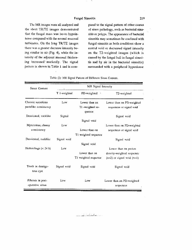
The MR images were all analyzrd and
the short TR/TE images demonstrated
that the fungal mass was iso-to hypoin-
tense compared with the normal mucosal
turhinates. On the long TR/TE images
there was a greater decrease intensity be-
ing similar to air (Fig. 4), while the in-
tensity of the adjacent mucosal thicken-
ing increased. markedly. The signal
pattern is shown in Table 1 and is com-
pared to the signal pattern of other causes
of sinus pathology, such as bacterial sinu-
sitis or polyps. The appearance of bacterial
sinusitis may sometimes be confused with
fungal sinusitis as both conditions show a
central void or decreased signal intensity
on the T2-weighted images (which is
caused by the fungai ball in fungal sinusi-
tis and by air in the bacterial sinusitis)
surrounded with a peripheral hyperdense
Table (I): MR Signal Pattrrn of Differen Sinus Content.
Sinus Conlent MR Signal Intensity
T I-weighted PD-weighted * R-weighted
Chronic secretions
pastelike consistency
Drssicatcd, rocklike
Mycetomas, cheesy
consislency
Dcssicated, rocklike
Hemorrhagt: (< 24 h)
Tooth in dentige-
rous cyst
Fibrosis in post-
oprralivc sinus
Low Lower than on
Tl -weighted se-
quence
Signal
Signal void
Low
Lower than on
Tl weighted sequence
Signal void
Signal void
Low
Lower than on
Tl weighted sequence
Signal void Signal void
LOW Low
Lower than on PD-weighted
scqucnces or signal void
Signal void
Lower than on PD-weighted
sequences or signal void
Signal void
Lower than on proton
density-weighted sequcncc
(n=2) or signal void (n=I)
Signal void
Lower than on PD-weighted
sequences

Rashad Hamdi, et al.
Fig. (1): Case 1: CT shows cast-like homoge- nous calcification best seen in the center of the right maxillary sinus representing the partially calcified fungal ball surrounded by a hypo- intense rim representing the in- flamed and edematous mucosa.
Fig. (2): Case 2: Marked calcification repre- senting a calcified fungal balls fill- ing both maxillary and ethmoid si- nus.
signal due to the mucosal thickening. Para-
nasal neoplasms have a lower signal inten-
sity than allergically or bacterially infected
mucosa and usually shows intermediate
signal intensity on the T2 weighted imag-
es. They should not be confused with fun-
gal sinusitis because of the different signal
patterns, but may be confused in cases
where there is intraorbital or intracranial
extension.
During the course of our work, we en-
countered 3 cases that showed decreased
signal intensity in the central part of the
sinus that were not proven to be fungal
sinusitis. Two cases proved to be muco-
cele of the sinus containing a central in-
spissated or past-like or rock-hard material,
Fig. (3): Case 3: Coronal, post-Gadolinium Tl weighted MR images showing central hypointensity in both max- illary sinuses representing the fun- gal ball surrounded by an enhanc- ing rim of inflamed mucosa.

Fungal Sinusitis 221
Fig. (4): Case 4: Coronal CT (A) showing fungal
disease involving the left ethmoid and
maxillary sinuses with foci of calcifica-
tion within. Evidence of intraorhilal
and intracranial extension. The Axial
TZ weighted MR images (B) show a sig-
nal void in the cthmoid sinus with in-
vasion of the apex of the left orbit.
Coronal Post-Gad6liiiitim MR (C)
showed a charkcteristic’centrum hypo-
intensity in the left ethmoid sinus sur-
rounded by peripheral enhancement 01
inflamed mucosa.

222 R‘lchad Hamdi. et al
Fig. (5 c 5: Sagittal TI wcightcd post- i: olinium MR showing fungus si-
nusttiis with large sub-frontal intra- cranial extension. The intracranial component shows a central hypo- density suggestive of the fungal ball surrounded by peripheral en- hancement represhniing- inflamed mucosa and dura.
while’the third case proved to he acute
hemorrhage into the sinuses as aheady
mentioned. All 3 cases are not included
in OUT study.
As regards the laboratory findings,
‘ihe use of Von Kossa stain and Dahl
method for calcium analysis revealed the
presence of calcium in each of the fungal
specimens.
Discussion
Fungal sinus disease may appear in
two forms. A slowly progressing extramu-
cosal fungus hall usually caused hy Aspttr-
gillus species or in immunologically com-
promised patients, as a fulminant infection
usually caused hy mucomycosis. The ex-
tramucosal fungal sinusitis is more com-
mon in dusty, damp, tropical countries
such as in Egypt and Saudi Arabia, and
usually develops as a saprophytic growth
in retained secretions in a sinus cavity.
The disease appears to he more frtqutM
fhan previously recognized [S]. This may
he related to increased recognition hecause
of increased availability of sophisticated
equipment in today’s time. Since fungal
sinusitis usuallv requires surgical interven-
tion, accurate preoprrative rarlic\logicnl di-
agnosis is important for the Climcian. The
typical course of the disease is that it
starts first as a chronic sinusitis that does
not resolve with antibiotic therapy or nor-
mal saline sinus irrigation [6]. Typically,
however, the true identity of disease is not
recognized until surgery, where usually a
brownish muddy substance is seen filling
the sinus. The treatment usually involves
removal of the fungal hall, the restoration
of the mucociliary drainage and. this
should ht: followed hy biopsy of the mu-
cosa of the sinus to evaluate mucosal inva-
sion [7].

Fungal Sinusitis 223
Plain film and tomography usually
shows in the early cases circumferential
mucosal thickening with characteristic
absence of air fluid level followed by
complete opacification of the sinus, wall
destruction and rarely wall sclerosis and
a somewhat characteristic increased atten-
uation wi!hin the fungal ball which is
usually seen in 50% of cases 181. These
focal hyperattenuations may appear as fo-
cal concretions or may form a diffuse in-
creased density of a ca!t of the sinus and
were extensively studied hy Stammberger
at al (91 and were found to represent cal-
cium phosphate and calcium sulfatr dr-
posits within necrotic areas of the myce-
lium. These characteristics, however, are
insufficiently nonspecific so that distinc-
tion between chronic sinusitis and neo-
plasms or o!hcr sinus pathology remains
difficult [lo]. On CT study, the ahove-
mentioned changes, as well as the calcifi-
cations were better seen because of the
multiplanner dirrcticln of CT and hecause
of its ahility to visualize lesions in thin
cuts without any overlap. Plain film or
CT could measure the density of the con-
cretions. Our results correlate well with
those of Stammhrrger et al [II] as re-
gards the incidence of calcifications with-
in the sinus which is the most specific
finding as regards the plain film and the
CT. The only differential diagnosis of a
hyperintense structure in the sinus is a
dentigerous cyst or the presence of a for-
eign body in the sinus (sinolith) [lt].
Unfortunately, plain films show calcifi-
cations (the only characteristic finding) in
only 50% of cases. CT proved to be
slightly more specific hut also not conclu-
sive as shown in our results, as only 11
patients were shown to have calcifications
within the sinus. Furthermore, ostroma,
osteol~lastoma, as well as osteogenic sarco-
ma may sometimes give a similar appear-
ance to an invasive fungal disease on CT
1121.
MR1 proved to he more specific than
CT a shown in our CBXS. The characteris-
tic finding is the presence of a decreased
signal intensity on the Tl and a signal
void on the T2-weighted images which’
arise from the center of the sinus and repre-
sents the fungal hall surrounded hy a char-
acteristic thin rim of hyperintensity which
represents the intlamrd surrounding muco-
sa. The cause of the decreased signal inten-
sity arising from the fungal hall was exten-
sively studied and was suggested to he
partly caused hy the calcium in the calcifi-
cation. (10). Still this would not explain
the signal void in patients who do not
show any calcification on the plain film or
on the CT and Stammhergrr et al [II] af-
ter studying samples of the fungal halls hy
absorption spectromrtry showed high con-
centration of magnesium and manganese
within the fungal ball with higher concen-
tration then seen in bacterially infected si-
nuses. The concentration of iron was also
studied by them, as it is known that iron’
and hemosiderin cause a decreased signal
224 Rashad Hamdi, et al.
intensity on the T2-weighted images and
this was found elevated only in patients
with chronic hemorrhage (lo]. Thus, af-
ter evaluating Tl, proton density and
T2weighted images, we showed a fairly
characteristic appearance and a definite
difference between fungal sinusitis and
its differential diagnosis which is bacteri-
al sinusitis or malignant neoplasms of
the sinuses. Our study showed that MR1
was correct in all patients. Towards the
end of our study, we examined 3 other
patients which showed an appearance
similar to that of fungal sinusitis which
is a signal void in the center surrounded
by a hyperdense rim but was proven by
surgery to be two cases of mucoceles
containing paste-like and rock-like chron-
ic secretions, and one patient with acute
hemorrhage in a leukemic child.
After reviewing the literature, we real-
ized that MR1 usually gives us a plethora
of information in the form of signal pat-
terns which gives us important informa-
tion about the content of the sinuses. The
appearance of a central signal void sur-
rounding a hyperdense rim which was
thought to be specific for fungal sinusitis
can be seen in five conditions [IO]. The
first condition is in patients with fungal
sinusitis infected with aspergillus fungus
and the cause of the decreased signal in-
tensity is the presence of calcium and
other minerals such as magnesium and
manganese and because of the lack of
hydration of tluid within these fungal balls
or mycetomaswhich becomes thick, cheesy
or may even have solid stony consistency.
The second cause for this characteristic sig-
nal pattern is mucoceles and where the sig-
nal void represents chronic inspissated se-
cretions and dried polyps in which the
mucous protein concentration is gteater
than 35-40%. At this concentration at all
the free water and some of the bound water
have been eliminated resulting in a signal
void on the T2-weighted images on MRl.
This has been extensively studied and was
shown that below this protein concentia-
tion, the secretions are liquid in nature
while above this within the concentration
the secretions .rapidly progress towards a
thick paste or dessicated solid rock-like
substance [lo].
The third cause of this signal pattern is
seen in acute intrasinus hrmorrhage where
at least two major factors account for the
low signal intensity. The first is the sus-
ceptibility effect of deoxyhrmoglobin
which causes a local field heterogrnrcity
and thus T2 shortening. The second factor
results from the formation of a fibrin clot
which effectively squeezes the serum from
the remaining protein complex, and thus
the clot represents a poorly hydrated, semi-
solid, macromolecular protein mixture that
causes a decreased signal intensity on the
T2-weighted images [12]. The fourth
cause is the presence of a dentigerous cyst
within the sinus while the fifth cause is

Fungal Sinusitis 225
the presence of air in a partially intlamed
sinus. Air not containing any water
molecules will again give a signal void.
The obvious problem realized by us
and others [12] is that although MR1
can give us a large amount of information
about the nature of the sinus secretion
and content, it may in specific cases not
give us the final diagnosis and it is CT
which may differentiate between the
above-mentioned entities. CT will easily
distinguish between the above five men-
tioned substances. CT will clearly show
the air within a partially inflamed sinus.
In patients with dentigerous cyst, the le-
sion will always be in the maxillary si-
nus and the sinus will be expanded and
the tooth will always be in an eccentric
location rather in a central location in pa-
tients with mucocele or fungal affection
and CT will clearly show that we are
dealing with enamel or with a tooth.
Chronic secretions, mycetomas and hemo-
rhage will appear as a soft tissue on CT
and can easily be differentiated from a
dentigerous cyst or air within an in-
flamed sinus. In conclusion, we believe
that MR1 can give us a plethora of infor-
mation about sinus disease and one
should analyze this large amount of in-
formation before reaching a diagnosis.
Although the characteristic appearance of
fungal sinusitis has been shown in MRI,
still this appearance may be seen in par-
tially inflamed sinuses, acute intrasinu-
sal hemorrhage, dentigerous cysts and rare-
ly in postoperative sinuses with fibrosis
and scar. Once this characteristic pattern of
central signal void surrounded by a hyper-
dense rim is seen on the T2-weighted im-
ages, one should resort to CT in coronal
view to differentiate those entities from
each other.
References
1. SOM PM, DILLON WP, SZE G, LIDOV M,
BILLER HF. LAWSON W: Benign and
malignant sinonasal lesions with intracra-
nial extension: differentiation with MR
imging. Radiology, 172: 763-766, 1989.
FULLERTON GD, POTTER JL, DORN-
BLUTH NC: NMR relaxation of protons
in tissues and other macromolecular water
solutions. Magn. Reson. Imaging. 1: 209-
228, 1982.
SOM PM, DILLON WP, FULLERTON
GD, ZIMMERMAN RA, RAJAGOPALAN
B, MAROMZ: Chronically obstructed
sinonasal secretions: observalons on Tl
and T2 shortening. Radiology, 172: 5 l5-
520, 1989.
SOM PM, SHAPIRO MD, BILLER HR.
SASAKI C, LAWSON W: Sinonasal tu-
mars and inflammalory tissues: differenti-
ation with MR. Radiology, 167: X03-
808, 1988.
5. ZINREITH SJ, KENNEDY DW, MALAT J,
et al.: Fungal sinusitis: diagnosis with CT
and MR imaging. Radiology, 169:439-
444, 1988.
226 Rashad Hamdi, et al.
6. BROWN DC, GEYER CA, CITRIN CM: GROSSMAN RI: Paranasal sinus hemor-
ATYPICAL MR1 appearance of chronic rhage: Evaluation with MR imaging. Ra-
expansile fungal sinusitis. Orlando, diology, 162:499-503, 1987.
Fla: American Society of Neuroradiolo- 10. VAN TASSEL P, LEE YY, JING B, DE
gy, 169, 1986.
7. AISEN AM, MARTEL W, BRAUNSTEIN
EM, et al.: MR1 and CT evaluation of
primary bone and soft Slissue tumors.
AJR, 146:749-756, 19X6.
li
DILLON WP, SOM PM, FULLERTON
GD: Hypointense MR signal in chroni-
cally inspissated sinonasal secretions.
Radiology, 174:73-7X, 1990. I?
ZIMMERMAN RA, BILANIUK LT,
HACKNEY DB. EOLDBERG HI,
PENA C: Mucoceles of the paranasal si-
nuscs: MR imaging with CT correlation.
AJNR, 10: 607-612, 19X9.
SlJNDARAM M, McGUlRE MH, SCHA-
JOWlCZ F: Soft-tissue masses: Hiskkgic
hasis for decreased signal (short T2) on
TZ-weighted MR Images. AJR, 148: 1247-
250, 1987.
HAYMAN LA, TABER KH, FORD JJ, et
al: Effect of clot formation and retraction
on spin-echo MR images of blood: an in
vilro studv. AJNR: IO:1 155-I 158. 19X9.


![Pathogenesis of eosinophilic chronic rhinosinusitis · 2017. 8. 25. · sinusitis [19], 3) nonallergic fungal ECRS [20], and 4) aspirin-exacerbated ECRS. Within each subcategory,](https://img.pdfslide.us/doc/110x75/61198275387af96a314aa9d0/pathogenesis-of-eosinophilic-chronic-rhinosinusitis-2017-8-25-sinusitis-19.jpg)