Embed Size (px)
Citation preview
Cardiovascular, Pulmonary and Renal Pathology
Functions of Type II Pneumocyte-Derived VascularEndothelial Growth Factor in Alveolar Structure,Acute Inflammation, and Vascular Permeability
Marco Mura,* Matthew Binnie,†‡ Bing Han,*Chengjin Li,† Cristiano F. Andrade,*Atsushi Shiozaki,* Yu Zhang,*Napoleone Ferrara,§ David Hwang,*¶
Thomas K. Waddell,*� Shaf Keshavjee,*�
and Mingyao Liu*‡�
From the Latner Thoracic Surgery Research Laboratories,*
Toronto General Research Institute, University Health Network,
and The Samuel Lunenfeld Research Institute,† Mount Sinai
Hospital, Toronto, Ontario, Canada; the Departments of
Medicine,‡ Laboratory Medicine and Pathobiology,¶ and
Surgery,� University of Toronto, Toronto, Ontario, Canada; and
the Department of Molecular Oncology,§ Genentech Inc., South
San Francisco, California
Vascular endothelial growth factor-A (VEGF) is a po-tent regulator of vascular permeability, inflammatoryresponse, and cell survival in the lung. To explore thefunctions of VEGF produced locally in type II pneu-mocytes, we generated mice with a conditional dele-tion of VEGF-A using Cre recombinase driven by thehuman surfactant protein C (SPC) promoter. In 7-to 10-week-old VEGF-knockout (SPC-VEGF-KO) mice,lung histology and physiology were essentially nor-mal, except for higher dynamic lung compliance andlower pulmonary vascular permeability. Emphysemawas seen in 28- to 32-week-old animals. To investigatethe role of type II pneumocyte-derived VEGF in acutelung injury, we challenged 7- to 10-week-old SPC-VEGF-KO mice and their wild-type littermates withintestinal ischemia-reperfusion. Bronchoalveolar la-vage fluid total cell count, pulmonary permeability,and lung injury score were significantly attenuated,and total lung VEGF levels were significantly lower inSPC-VEGF-KO mice compared with wild-type controls.In SPC-VEGF-KO mice, activated caspase 3-positivetype II epithelial cells were increased after intestinalischemia-reperfusion, even though there was no sig-nificant difference in the total number of cells posi-tive for terminal deoxynucleotidyl transferase dUTPnick-end labeling. We conclude that VEGF in type II
cells helps protect alveolar epithelial cells fromcaspase-dependent apoptosis. However, VEGF pro-duced from type II cells may contribute to increasedvascular permeability during acute lung injury. (Am JPathol 2010, 176:1725–1734; DOI: 10.2353/ajpath.2010.090209)
Acute lung injury (ALI) and its severe form, acute respi-ratory distress syndrome (ARDS), are characterized byincreased capillary permeability, leading to interstitialand alveolar edema, an influx of circulating inflammatorycells, and formation of hyaline membranes. However, themolecular mechanisms of ALI/ARDS are still largely un-known. Vascular endothelial growth factor-A (VEGF) is apotent regulator of vascular permeability, a key player inangiogenesis, and a survival factor for endothelial cells.1
The biological properties of VEGF are mainly mediatedby VEGF receptor 2, also called Flk-1 (Fetal liver kinase1), which is expressed on endothelial and epithelialcells.2,3 VEGF also induces monocyte activation and mi-gration, which has been found to be mediated throughVEGF receptor 1, also called Flt-1 (FMS-like tyrosine ki-nase 1).4 VEGF is considered to be both a vascularpermeability factor and a pro-inflammatory cytokine.VEGF increases endothelial permeability via specific sig-nal transduction pathways.5,6 Blocking VEGF has beenshown to reduce tissue inflammation and damage in vivo.For example, VEGF-induced brain edema has some fea-tures that resemble stroke,7 and VEGF antagonism re-duced edema formation and tissue damage in the brainafter ischemia-reperfusion in mice.8 However, the role ofVEGF in ALI/ARDS is controversial.9
Intrapulmonary overexpression of VEGF with an ad-enoviral vector resulted in high-permeability edema in
Supported by Canadian Institutes of Health Research grants MOP-13270and MOP-42546.
M.M. and M.B. contributed equally to this work.
Accepted for publication December 16, 2009.
Address reprint requests to Dr. Mingyao Liu, Professor of Surgery,University of Toronto, Toronto General Hospital, Room TMDT 2-814, 101College Street, M5G 1L7, Toronto, Ontario, Canada. E-mail: [email protected].
The American Journal of Pathology, Vol. 176, No. 4, April 2010
Copyright © American Society for Investigative Pathology
DOI: 10.2353/ajpath.2010.090209
1725
murine lungs.10 Using an inducible transgenic mousemodel, it was shown that VEGF overexpression in airwayepithelium also induces pulmonary edema.11 Transfec-tion of sFlt-1 gene attenuated bleomycin-induced pneu-mopathy in mice.12 Similarly, pretreatment of rats withadenovirus encoding soluble Flk-1 prevented ischemia-reperfusion-induced lung injury.6 These results all suggestthat VEGF signaling through its receptors may promotepulmonary permeability and inflammation. However, in-triguingly, ischemia-reperfusion does not affect totalVEGF protein levels in the lung.6 In addition, VEGF levelsin the alveolar compartment of patients with ARDS areindistinguishable from those in patients with hydrostaticpulmonary edema.13 Lung epithelial lining fluid fromARDS patients actually contained lower levels of VEGF thanthat from at-risk subjects, and increased levels at Day 4were associated with better recovery.14 Consistent with thisfinding, VEGF blockade has been shown to result in in-creased markers of oxidative stress, alveolar cell apoptosisand alveolar enlargement,15,16 suggesting a protective rolefor VEGF in the epithelial barrier. These interesting obser-vations also suggest that the expression and function ofVEGFmay be regulated locally at sites of lung injury, withoutcausing dramatic changes in overall lung VEGF levels.
VEGF is highly compartmentalized in the lung17 and isproduced by multiple sources, such as epithelial andmesenchymal cells, myofibroblasts, and macrophages.1,18
However, type II alveolar epithelial cells are considered tobe the main source of VEGF in the lung parenchyma,17
although the physiological role of VEGF from this sourceremains unclear. VEGF released by alveolar epithelial cellsmay act as an autocrine trophic factor for epithelial cells,19 andmodulate functions of the adjacent vascular endothelium in aparacrine fashion.20 To investigate the roles of locally pro-duced VEGF in the lung, we generated transgenic mice inwhich the expression of VEGF was selectively deleted indistal lung epithelial cells using a Cre recombinase systemdriven by the surfactant protein C (SPC)-promoter. Whileyoung transgenic mice showed a normal phenotype, olderanimals spontaneously developed typical features of initialemphysema. The severity of ALI, pulmonary permeabilityand inflammation, and the expression of cell death andstress markers were compared between transgenic youngmice and their wild-type littermates after intestinal isch-emia-reperfusion, a model of extrapulmonary ARDS.21
Materials and Methods
This study was approved by the Animal Use and CareCommittee of the University Health Network. All animalsreceived humane care in compliance with the Principles ofLaboratory Animal Care formulated by the National Societyfor Medical Research and theGuide for the Care andUse ofLaboratory Animals prepared by the Institute of LaboratoryAnimal Resources and published by the National Institutesof Health (NIH publication 0-309-05337-3, revised 1996),and the Guide for the Care and Use of Experimental Ani-mals formulated by the Canadian Council on Animal Care.
Cell-Specific Gene Targeting and Genotyping
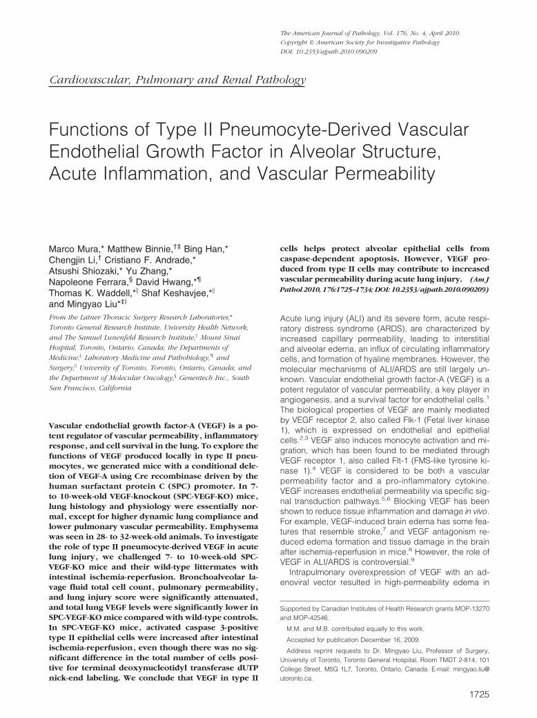
SPC-Cre transgenic mice were generated by cloning acDNA encoding Cre recombinase and a nuclear localiza-tion signal downstream of the 3.7-kb human SPC promotersequence, thus driving Cre expression in distal lung epithe-lium. This promoter confers gene expression throughout thelung epithelium during gestation and in a subset of type IIpneumocytes postnatally. Transgene positive founderswere identified by Southern blotting for Cre and tested forCre activity by mating them to Z/EG reporter mice (ICRbackground), in which Cre-mediated excision of a stopcodon leads to cell-specific expression of enhanced jelly-fish green fluorescent protein (GFP).22,23 To confirm thespecificity of gene targeting, dual transgenic SPC-CreZ/EG offspring from one founder line were tested forGFP/prosurfactant protein C colocalization with immuno-fluorescent staining. The same SPC-Cre recombinaseline (ICR background) was bred to floxed VEGF mice(ICR background) which have loxP sites inserted aroundthe third exon (Figure 1A). Site-specific recombinationbetween the two loxP sites of the VEGF gene results in anull VEGF allele. To generate homozygous floxed VEGF-
A Tissue specific expression
300 bp
140 bp100 bp
B Cre PCR
C VEGF PCR MW 1 2 3 4 + -
MW 1 2 3 4 5 6 7 8 9
SP-C Cre P 4
SP-C Cre
SP-C Cre VEGF PP 4
VEGF PP 4
excision
no excision
Figure 1. Cell-specific gene targeting and genotyping. A: Scheme to generate typeII cell-specific VEGF knockout mice. B: Genomic DNA was isolated from mouse tailsand used for genotypic analysis. The Cre recombinase transgene was identified as a300 bp PCR product. �, positive control (DNA from known Cre positive mice); �,negative control (DNA from known Cre negative mice); MW, molecular weightmarkers. C: The floxed VEGF allele measures 140 bp by PCR analysis, whereasthe wild-type allele measures 100 bp. Each lane represents one animal.
1726 Mura et alAJP April 2010, Vol. 176, No. 4
Cre recombinase mice, mice carrying both a SPC-Cretransgene and one floxed VEGF allele were bred to ho-mozygous floxed VEGF mice. The presence of the floxedVEGF gene was detected by PCR.24 Only homozygousknock-out animals (ie, Spc-Cre�/0 VEGFlox/lox) (SPC-VEGFKO) and wild-type littermates were used. Recombinationof the floxed allele deletes all VEGF isoforms (VEGF120,164, and 188).
Genotyping
Genomic DNA was isolated from mouse tails and used forgenotypic analysis. The presence of the floxed VEGF genewas detected by PCR using the oligonucleotide primersmuVEGF 419.F (5�-CCTGGCCCTCAAGTACACCTT-3�)and muVEGF 567.R (5�-TCCGTACGACGCATTTCTAG-3�)(Sigma-Genosys, The Woodlands, TX), which generates a140-bp fragment of the VEGF allele in the presence of theloxP-1 site and a 100-bp fragment in the wild-type allele.
GFP-proSurfactant Protein C DoubleImmunofluorescent Staining
To test for specific Cre recombinase expression in distallung epithelium, colocalization of Cre-dependent GFPand the type II pneumocyte marker, surfactant protein Cprecursor protein (proSPC), was confirmed by doubleimmunofluorescent staining. Lungs were chopped intofragments with a blade, fixed with 4% buffered formalin,processed, and embedded in paraffin; 5 �m sectionswere deparaffinized and rehydrated. Sections wereboiled in 0.01 mol/L NaCitrate [pH 6.0] for 10 minutes andthen cooled to room temperature. Sections were blockedwith 5% goat serum in PBS for 1 hour at room tempera-ture, incubated with primary antibody overnight at 4°C,washed in PBS, then stained with secondary antibodyfor 1 hour at room temperature, washed in PBS, andmounted in Vectashield medium with 4,6-diamidino-2-phenylindole (Vector labs, Burlingame CA). The antibod-ies used were rabbit anti-mouse proSPC (AB3786 1:500;Millipore, Billerica MA) and chicken anti-GFP (ab139701:1000; Abcam, Cambridge, MA) fluorescein isothiocya-nate-conjugated anti-chicken IgY (1:500) and Cy3-con-jugated anti-rabbit IgG (1:500; Jackson ImmunoresearchLaboratories, West Grove, PA) in the dark for 1 hour. Afterwashing, slides were mounted with GVA mounting solu-tion (Invitrogen, Carlsbad, CA). Thirty randomly chosenfields from two animals at �200 magnification were ana-lyzed. The average number of GFP-positive cells wasquantified as a percentage of total proSPC-positive cellscounted. To determine the specificity of staining, theprimary antibody was omitted from the staining protocolas a negative control. We also used lung tissue slide fromwild-type animals, to show the lack of anti-GFP staining.
Real-Time Reverse Transcription-PCR
Quantitative real-time reverse transcriptase-PCR analysis ofthe RNA expression of VEGF was performed on RNA iso-lated from frozen lung tissues as previously described.25
Total RNA extraction was performed with the RNeasy kit(Qiagen, Mississauga, Ontario, Canada). Results areexpressed as the ratio of VEGF and the housekeeping geneB2M expression levels. Primers were designed using thePrimer Express 1.5 software (Applied Biosystems) and pur-chased from ACGT (Toronto, Ontario, Canada).
Lung Morphometry
For morphometric studies, the mean linear intercept wasused as a measure of interalveolar wall distance, as de-scribed.26 The mean linear intercept was calculated foreach sample based on 20 random fields observed at amagnification of �400 using a cross-line. Fields containingairways and blood vessels were excluded. The total numberof alveolar intercepts encountered in each field wascounted and the mean value for each animal (20 fields) wascalculated. A lower mean linear intercept count representsmore pronounced airspace enlargement.
Physiological Analyses
Seven- to ten-week-old male mice (weight 24.2 � 4.9 g inwild-type group, 26.7 � 5.3 g in SPC-VEGF KO group)were anesthetized with 5% isoflurane, intubated with a can-nula (1.0 mm, Harvard Apparatus Canada, St. Laurent,Canada), and connected to a volume-controlled constantflow ventilator (Inspira Advanced Safety Ventilator, HarvardApparatus). After intubation, anesthesia was continuouslymaintained with isoflurane and body temperature wasmain-tained at 37°C by an immersion thermostat throughoutbaseline observation or IIR surgical procedure. The animalswere ventilated with a tidal volume of 6 ml/kg, inspiratoryoxygen fraction 1.0, inspiratory/expiratory ratio 1:2, and afrequency of 140 breaths/min. Dynamic lung compliancewas measured with an esophageal catheter. After 10 min-utes of observation, either at baseline level or after 30 min-utes of IIR, animals were sacrificed by exsanguination.Arterial blood gas analysis was performed in four 7- to 10-week-old wild-type animals, three 7- to 10-weeks-old SPC-VEGF KO animals, and four SPC-VEGF KO 28- to 32-week-old animals. HSE-USB acquisition system and Pulmodynsoftware (H. Sachs Elektronik, March-Hugstetten, Germany)were used for all pulmonary function measurements.
Intestinal Ischemia-Reperfusion Model of ALI
The IIR procedure was performed by occluding the su-perior mesenteric artery below the celiac trunk with anarterial microclamp for 30 minutes, as previously de-scribed27,28; the reperfusion period was extended to 24hours without mechanical ventilation or supplemental ox-ygen. Intestinal ischemia was confirmed by paleness ofthe jejunum and ileum. After 30 minutes the clamp wasremoved, 0.5 ml of sterile saline at 37°C was injected intothe peritoneal cavity and the skin was sutured. Anesthe-sia was terminated and animals were observed until aspontaneous breath appeared. Mechanical ventilationwas then stopped and animals were let wean until aregular pattern of breathing was achieved. After 24
VEGF in Acute Lung Injury 1727AJP April 2010, Vol. 176, No. 4
hours, animals were resubmitted to anesthesia and lap-arotomy and sacrificed by exsanguination. For sham-operated animals, the same procedures were performedbut the mesenteric artery was not clamped. Separatesubgroups of lungs were 1) fixed for histological evalua-tion and immunohistochemistry studies; 2) snap-frozen inliquid nitrogen for protein or RNA analysis; 3) used forbronchoalveolar lavage (BAL) or Evans blue dye assay.Blood samples were collected after 24 hours of reperfu-sion by puncture of the aorta and centrifuged (4000 � g,10 minutes). Plasma samples were stored at �80°C.
Assessment of Lung Injury
ALI was investigated by evaluating BAL cell counts, pulmo-nary vascular permeability, lung histology, and fibrin depo-sition with procedures previously described.27,28 Evansblue dye permeability assay was performed with themethod described by Kaner et al.10 To assess fibrin depo-sition, fibrinogen immunohistochemistry was performed.29
The degree of lung injury was determined using thegrading system developed by Ginsberg et al.30 The lunginjury score comprises the assessment of four parame-ters: 1) alveolar hemorrhage, 2) vascular congestion, 3)infiltration or aggregation of neutrophils in the airspace orvessel wall, and 4) fibrin deposition in alveoli. The severityof each parameter was scored as 0, absent; 1, mild; 2,moderate; and 3, severe. The combined score of all fourparameters was taken for each animal.30 Ten randomlychosen fields from each animal with approximately thesame number of alveoli were analyzed by a pulmonarypathologist (D.H.), who was blinded to the group allocation.
VEGF Expression
VEGF gene expression was assessed by in situ hybridiza-tion using a probe kindly provided by Dr. Andras Nagy(University of Toronto, Canada). Lung tissues were pre-pared as previously described.31 In situ hybridization wasconducted by a staff member of the core facility in ourinstitute. For quantitative analysis, 10 optical fields of alve-olar area from each animal (3 mice/group), not includingmajor airways or vessels, were randomly chosen at �1000magnification. The number of VEGF-positive cells as well asthe total cell number of cell nuclei in the chosen fields werecounted in a blinded fashion. The percentage of positive-stained cells for each optical field was quantified as apercentage of total cells counted. VEGF levels in lung ho-mogenates, BAL fluid and plasma were determined usingan ELISA kit (R&D Systems, Minneapolis, MN)28 that recog-nizes VEGF isoforms with either 120 or 164 amino acids.The lower detection limit was 31 pg/ml. ELISA values inlung homogenates were standardized to total proteinconcentration.
Immunofluorescent Staining
Terminal transferase dUTP nick end labeling (TUNEL)staining, and TUNEL/activated caspase 3 double stain-ing have been described.27 Activated caspase 3/Surfac-
tant Protein B (SPB) double immunofluorescent stainingwas also performed. SPB was selected as a marker ofType II cells because SPC is known to be down-regulated inanimal models of ALI.32 Clara cells are also SPB-positive,and therefore airways were excluded from the semiquanti-tative analysis. The staining was performed on 4 �m lungslides. After deparaffinization and dehydration, antigen re-trieval was performed with 10 �g/ml proteinase K in 10mmol/L Tris/HCl [pH 7.4 to 8] for 15 minutes. Slides wereincubated with a rabbit anti-SPB polyclonal antibody (1:50in PBS-1% bovine serum albumin[BSA]) for 6 hours,washed with PBS, and then incubated with a Texas Redmouse anti-rabbit IgG (1:100 in PBS-1% BSA; Jackson Im-munoresearch) for 1 hour. Slides were next incubated witha goat polyclonal antibody against the activated (cleaved)form of caspase 3 (1:50 in PBS-1% BSA) (Santa Cruz)overnight at 4°C, washed with PBS and then incubated witha fluorescein isothiocyanate-conjugated donkey anti-goatIgG (1:500 in PBS-1% BSA, Biotium, Hayward, CA) at roomtemperature for 1 hour. The nuclei were counterstained withHoechst 33258 (1:5000, 10 minutes). All slides were sub-jected to identical exposure times. Nonimmune serum in-stead of primary antibody for caspase 3 was used for neg-ative controls. Spleen sections (secondary follicles) wereused as positive control.33 The average number of activatedcaspase 3-positive cells was quantified as a percentage oftotal Hoescht-positive cells counted; the average number ofactivated caspase 3-positive epithelial cells was also quan-tified as a percentage of total SPB-positive cells counted.
Immunohistochemistry
The detailed protocols published previously were fol-lowed.28 The primary antibodies used were anti-VEGF(1:200) (Santa Cruz Biotechnology, Santa Cruz, CA) andchicken anti-fibrinogen (1:400, GenWay Biotech San Di-ego, CA). Slides were then washed and incubated withbiotinylated secondary antibody (1:600 in PBS-1% BSA).Detection was done by Avidin Biotin Complex systemby following the manufacturers’ instructions (Vector Labo-ratories Inc, Burlingame, CA), using 3–3 Diaminobenzidineas chromogen; slides were counterstained with hematoxylinfor 2 seconds. The primary antibody was omitted for nega-tive controls. For fibrinogen staining, the secondary Ab wasan alkaline phosphatase-conjugated anti-chicken IgY (1:100 in PBS-1% BSA, 1 hour). Detection was done by AvidinBiotin Complex system (Vector Laboratories Inc., Burlin-game, CA) with Vector Red alkaline phosphatase substratekit as chromogen. Slides were counterstained with methylgreen for 10 seconds.
Western Blotting
Antibodies for caspase-3 and horseradish peroxidase-conjugated donkey anti-goat secondary antibody werefrom Santa Cruz Biotechnology (Santa Cruz, CA). Cleavedcaspase-3 antibody was from Cell Signaling Technology(Beverly, MA). Horseradish peroxidase-conjugated goatanti-rabbit secondary antibody was from Amersham Phar-macia Biotech (Piscataway, NJ). Immunoblotting experi-
1728 Mura et alAJP April 2010, Vol. 176, No. 4
ments were performed according to standard proceduresdescribed previously.28
Statistical Analysis
All data are expressed as mean � SD. Distributionanalysis for all variables were performed with the Kol-mogorov-Smirnov test. For comparison of two separategroups either the unpaired t-test or the Mann-Whitneytest was used, when indicated. For comparison of mul-tiple groups, either one-way analysis of variance anal-ysis followed by Tukey’s post test or nonparametricKruskal-Whallis test followed by Dunn’s post test, whenindicated, were used to determine significant differ-ences between individual groups. P values �0.05 areregarded as significant. The Prism 4 software package(GraphPad Software Inc., La Jolla, CA) was used for allstatistical analyses.
Results
Phenotype, VEGF Expression, and Physiologyof SPC-VEGF-KO 7- to 10-Week-Old Mice
To selectively knock out the expression of VEGF in type IIcells we crossed SPC-Cre transgenicmice tomice homozy-gous for a floxed VEGF allele, then back-crossed SPC-Cre�/0 VEGFlox/wt offspring with VEGFlox/lox mice to gener-ate SPC-Cre�/0 VEGFlox/lox mutants. Mice were genotypedby PCR for Cre (Figure 1B) and VEGF (Figure 1C) alleles.
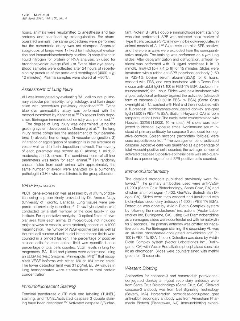
Wild-type and floxed VEGF alleles yield bands of differentsizes. To determine the efficiency of SPC-Cre mediatedrecombination in vivo, transgene positive founders weremated with Z/EG reporter mice, in which Cre-mediated ex-cision of a stop codon leads to cell type-specific expressionof enhanced GFP.22,23 Expression of GFP, indicating SPC-Cre transgene expression, was observed in the small air-ways and air sacs during the perinatal period (data notshown). In adult mice, site-specific recombination wasfound in 71 � 8% of type II cells, as shown by GFP-proSPCdouble staining (Figure 2A). In SPC-VEGF-KO mice, re-duced epithelial expression of VEGF in the alveolar spacewas demonstrated by in situ hybridization (Figure 2B). VEGFmRNA-positive cells (per optical field) were 10.8 � 1.4% inwild-type animals and 2.9 � 1.1% in SPC-VEGF-KO mice(P � 0.001). Quantitative real-time RT-PCR showed a sig-nificantly reduced expression in SPC-VEGF-KO mice incomparison with wild-type littermates (P � 0.0002) (Figure2C). At the protein level, wild-type animals were character-ized by strong staining of bronchial epithelial cells andmoderate to diffuse staining of vascular endothelial cells.Type II epithelial cells were intensely VEGF-positive.Alveolar macrophages were occasionally positive. InSPC-VEGF-KO animals, few type II alveolar epithelialcells were VEGF-positive, while alveolar macrophagesshowed positive VEGF staining; airway cell staining waspreserved (Figure 2D). VEGF-positive cells (per opticalfield) were 24.9 � 2.5% in wild-type animals and 9.4 �2.2% in SPC-VEGF-KO mice (P � 0.001) (Figure 2E).
SPC-VEGF-KO mice (7- to 10-week-old) showed normalbody weight. The lungs showed normal appearance and
proSPC and GFP staining
VEGF in situ hybridizationWT SPC-VEGF KO
VEGF immunostaining
VEGF gene expression in lung tissue
WT SPC-VEGF KO0
1
2
3
VE
GF
rel
ativ
eex
pre
ssio
n le
vel
*
WT SPC-VEGF KO WT SPC-VEGF KO
0
10
20
30
% o
f V
EG
F-p
osi
tive
cel
ls/
op
tica
l fi
eld
*
VEGF-positive cells in lung sections
proSPC GFP Merge
A
B C
D E
Figure 2. Selective deletion of VEGF in type IIalveolar epithelial cells. A: Green fluorescentprotein (GFP)-proSurfactant Protein C (proSPC)double staining, to determine the efficiency ofSPC-promoter driven Cre-recombinase system intype II cells. GFP is stained with fluoresceinisothiocyanate (green) and SPC with Cy3 (red).Nuclei are counterstained blue with 4,6-dia-midino-2-phenylindole. Expression of GFP indi-cates cell-specific recombination and is seen inthe majority of proSPC-positive Type II cells.Original magnification �200. B: In situ hybrid-ization shows lack of VEGF mRNA expression inthe alveolar wall of SPC-VEGF knockout (KO)animals. One VEGF positive cell in the KO groupis to show the “leakiness” of the system. Arrowsindicate VEGF-positive cells. Original magnifica-tion �1000. C: VEGF gene expression in lungtissue evaluated by quantitative Real-Time RT-PCR,showing a significantly reduced expression in SPC-VEGF-KO mice (n� 15) in comparison with wild-type animals (n � 12). *P � 0.0002. D: VEGFimmunostaining, showing much weaker stainingin the alveolar walls in SPC-VEGF KO mice incomparison with wild-type littermates. Nucleiwere counterstained with hematoxylin. Arrowsindicate VEGF-positive type II cells, arrowheadsindicate VEGF-positive macrophages. Originalmagnification �1000. E: Quantification of VEGF-positive cells, which was significantly lower inSPC-VEGF-KO mice (n � 3) in comparison withwild-type littermates (n � 3). *P � 0.001.
VEGF in Acute Lung Injury 1729AJP April 2010, Vol. 176, No. 4
histology. We performed physiological assessments of dy-namic lung compliance, arterial blood gas analyses, andBAL for total and differential cell counts. No significant dif-ferences were found between SPC-VEGF-KO and wild-typemice in regard to blood gas analyses and BAL counts (datanot shown). However, dynamic lung compliance was sig-nificantly higher (123 � 42 vs. 96 � 24 �l/cm H2O; P �0.015) and vascular permeability as measured by Evansblue dye assay was significantly lower (12.2 � 2.6 vs.19.0 � 3.9, % of dose administered; P � 0.043) in SPC-VEGF-KO mice compared with wild-type animals.
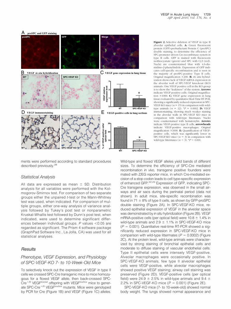
The 28- to 32-Week-Old SPC-VEGF-KO MiceDevelop Spontaneous Emphysema
Morphological and physiological studies were also con-ducted on 28- to 32-week-old SPC-VEGF-KO animalsand wild-type littermates. At histological analysis, airspace enlargement was seen (Figure 3A), and increasedlung volume was evident in some cases even on grossexamination (Figure 3B). The morphometric analysis withthe linear intercept method showed significant airspaceenlargement (lower mean linear intercept), compatiblewith emphysema, in SPC-VEGF-KO animals in compari-son with wild-type mice (P � 0.0420) (Figure 3C). Thedynamic lung compliance measurement confirmed highervalues in SPC-VEGF-KO animals in comparison with wild-type littermates (P � 0.004) (Figure 3D).
Acute Lung Injury Was Ameliorated inSPC-VEGF-KO 7- to 10-Week-Old Mice
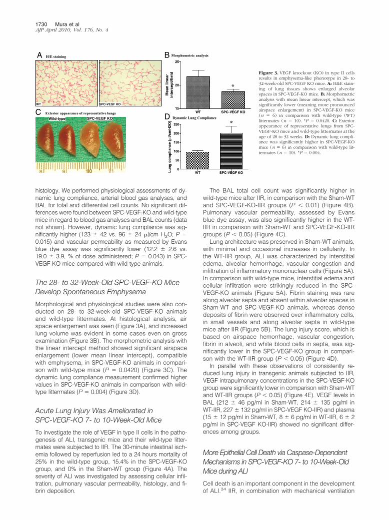
To investigate the role of VEGF in type II cells in the patho-genesis of ALI, transgenic mice and their wild-type litter-mates were subjected to IIR. The 30-minute intestinal isch-emia followed by reperfusion led to a 24 hours mortality of25% in the wild-type group, 15.4% in the SPC-VEGF-KOgroup, and 0% in the Sham-WT group (Figure 4A). Theseverity of ALI was investigated by assessing cellular infil-tration, pulmonary vascular permeability, histology, and fi-brin deposition.
The BAL total cell count was significantly higher inwild-type mice after IIR, in comparison with the Sham-WTand SPC-VEGF-KO-IIR groups (P � 0.01) (Figure 4B).Pulmonary vascular permeability, assessed by Evansblue dye assay, was also significantly higher in the WT-IIR in comparison with Sham-WT and SPC-VEGF-KO-IIRgroups (P � 0.05) (Figure 4C).
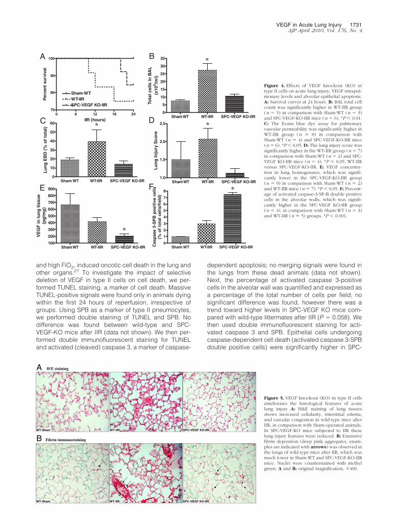
Lung architecture was preserved in Sham-WT animals,with minimal and occasional increases in cellularity. Inthe WT-IIR group, ALI was characterized by interstitialedema, alveolar hemorrhage, vascular congestion andinfiltration of inflammatory mononuclear cells (Figure 5A).In comparison with wild-type mice, interstitial edema andcellular infiltration were strikingly reduced in the SPC-VEGF-KO animals (Figure 5A). Fibrin staining was rarealong alveolar septa and absent within alveolar spaces inSham-WT and SPC-VEGF-KO animals, whereas densedeposits of fibrin were observed over inflammatory cells,in small vessels and along alveolar septa in wild-typemice after IIR (Figure 5B). The lung injury score, which isbased on airspace hemorrhage, vascular congestion,fibrin in alveoli, and white blood cells in septa, was sig-nificantly lower in the SPC-VEGF-KO group in compari-son with the WT-IIR group (P � 0.05) (Figure 4D).
In parallel with these observations of consistently re-duced lung injury in transgenic animals subjected to IIR,VEGF intrapulmonary concentrations in the SPC-VEGF-KOgroup were significantly lower in comparison with Sham-WTand WT-IIR groups (P � 0.05) (Figure 4E). VEGF levels inBAL (212 � 46 pg/ml in Sham-WT, 214 � 135 pg/ml inWT-IIR, 227 � 132 pg/ml in SPC-VEGF KO-IIR) and plasma(15 � 12 pg/ml in Sham-WT, 8 � 6 pg/ml in WT-IIR, 6 � 2pg/ml in SPC-VEGF KO-IIR) showed no significant differ-ences among groups.
More Epithelial Cell Death via Caspase-DependentMechanisms in SPC-VEGF-KO 7- to 10-Week-OldMice during ALI
Cell death is an important component in the developmentof ALI.34 IIR, in combination with mechanical ventilation
H/E staining
SPC-VEGF KOWild-type
Morphometric analysis
Exterior appearance of representative lungs
WT
Dynamic Lung ComplianceWT SPC-VEGF KO
15
20
25
Mea
n li
nea
rin
terc
ept/
fiel
d
*
SPC-VEGF KO
WT SPC-VEGF KO0
50
100
150
200
250
Lu
ng
co
mp
lian
ce (μ
l/cm
H2O
) *
A B
DC
Figure 3. VEGF knockout (KO) in type II cellsresults in emphysema-like phenotype in 28- to32-week-old SPC-VEGF KO mice. A: H&E stain-ing of lung tissues shows enlarged alveolarspaces in SPC-VEGF-KO mice. B: Morphometricanalysis with mean linear intercept, which wassignificantly lower (meaning more pronouncedairspace enlargement) in SPC-VEGF-KO mice(n � 6) in comparison with wild-type (WT)littermates (n � 10). *P � 0.0420. C: Exteriorappearance of representative lungs from SPC-VEGF-KO mice and wild-type littermates at theage of 28 to 32 weeks. D: Dynamic lung compli-ance was significantly higher in SPC-VEGF-KOmice (n � 6) in comparison with wild-type lit-termates (n � 10). *P � 0.004.
1730 Mura et alAJP April 2010, Vol. 176, No. 4
and high FiO2, induced oncotic cell death in the lung andother organs.27 To investigate the impact of selectivedeletion of VEGF in type II cells on cell death, we per-formed TUNEL staining, a marker of cell death. MassiveTUNEL-positive signals were found only in animals dyingwithin the first 24 hours of reperfusion, irrespective ofgroups. Using SPB as a marker of type II pneumocytes,we performed double staining of TUNEL and SPB. Nodifference was found between wild-type and SPC-VEGF-KO mice after IIR (data not shown). We then per-formed double immunofluorescent staining for TUNELand activated (cleaved) caspase 3, a marker of caspase-
dependent apoptosis; no merging signals were found inthe lungs from these dead animals (data not shown).Next, the percentage of activated caspase 3-positivecells in the alveolar wall was quantified and expressed asa percentage of the total number of cells per field; nosignificant difference was found, however there was atrend toward higher levels in SPC-VEGF KO mice com-pared with wild-type littermates after IIR (P � 0.058). Wethen used double immunofluorescent staining for acti-vated caspase 3 and SPB. Epithelial cells undergoingcaspase-dependent cell death (activated caspase 3-SPBdouble positive cells) were significantly higher in SPC-
A B
D
FE
Sham WT WT-IIR SPC-VEGF KO-IIR0
5
10
15
20
25
30
35
Tota
l cel
ls in
BA
L(x
105 /m
l)
*
Sham WT WT-IIR SPC-VEGF KO-IIR1.0
1.5
2.0
2.5
Lu
ng
Inju
ry S
core
*
Sham WT WT-IIR SPC-VEGF KO-IIR100
200
300
400
500
600
700
800
900
VE
GF
in lu
ng
tis
sue
(pg
/mg
)
*
Sham WT WT-IIR SPC-VEGF KO-IIR0
1
2
3
4
5
6
7
8
Cas
pas
e 3-
SP
B p
osi
tive
cel
ls(%
of
tota
l ce
lls/
fiel
d)
*Sham WT WT-IIR SPC-VEGF KO-IIR
0
10
20
30
40
50
60
Lu
ng
EB
D (
% o
f to
tal) *C
0 6 12 18 2470
80
90
100
Sham-WTWT-IIRSPC-VEGF KO-IIR
IIR (hours)
Per
cent
sur
viva
l
Figure 4. Effects of VEGF knockout (KO) intype II cells on acute lung injury, VEGF intrapul-monary levels and alveolar epithelial apoptosis.A: Survival curves at 24 hours. B: BAL total cellcount was significantly higher in WT-IIR group(n � 5) in comparison with Sham-WT (n � 3)and SPC-VEGF-KO-IIR mice (n � 6). *P � 0.01.C: The Evans blue dye assay for pulmonaryvascular permeability was significantly higher inWT-IIR group (n � 8) in comparison withSham-WT (n � 4) and SPC-VEGF-KO-IIR mice(n � 6). *P � 0.05. D: The lung injury score wassignificantly higher in the WT-IIR group (n � 7)in comparison with Sham-WT (n � 2) and SPC-VEGF KO-IIR mice (n � 4). *P � 0.05, WT-IIRversus SPC-VEGF-KO-IIR. E: VEGF concentra-tion in lung homogenates, which was signifi-cantly lower in the SPC-VEGF-KO-IIR group(n � 9) in comparison with Sham-WT (n � 2)and WT-IIR mice (n � 7). *P � 0.05. F: Percent-age of activated caspase-3/SP-B double positivecells in the alveolar walls, which was signifi-cantly higher in the SPC-VEGF KO-IIR group(n � 4), in comparison with Sham-WT (n � 3)and WT-IIR (n � 5) groups. *P � 0.001.
SPC-VEGF KO-IIRWT-IIRWT-Sham
WT-Sham WT-IIR SPC-VEGF KO-IIR
H/E staining
Fibrin immunostaining
A
B
Figure 5. VEGF knockout (KO) in type II cellsameliorates the histological features of acutelung injury A: H&E staining of lung tissuesshows increased cellularity, interstitial edema,and vascular congestion in wild-type mice afterIIR, in comparison with Sham-operated animals.In SPC-VEGF-KO mice subjected to IIR theselung injury features were reduced. B: Extensivefibrin deposition (deep pink aggregates, exam-ples are indicated with arrows) was observed inthe lungs of wild-type mice after IIR, which wasmuch lower in Sham-WT and SPC-VEGF-KO-IIRmice. Nuclei were counterstained with methylgreen. A and B: original magnification, �400.
VEGF in Acute Lung Injury 1731AJP April 2010, Vol. 176, No. 4
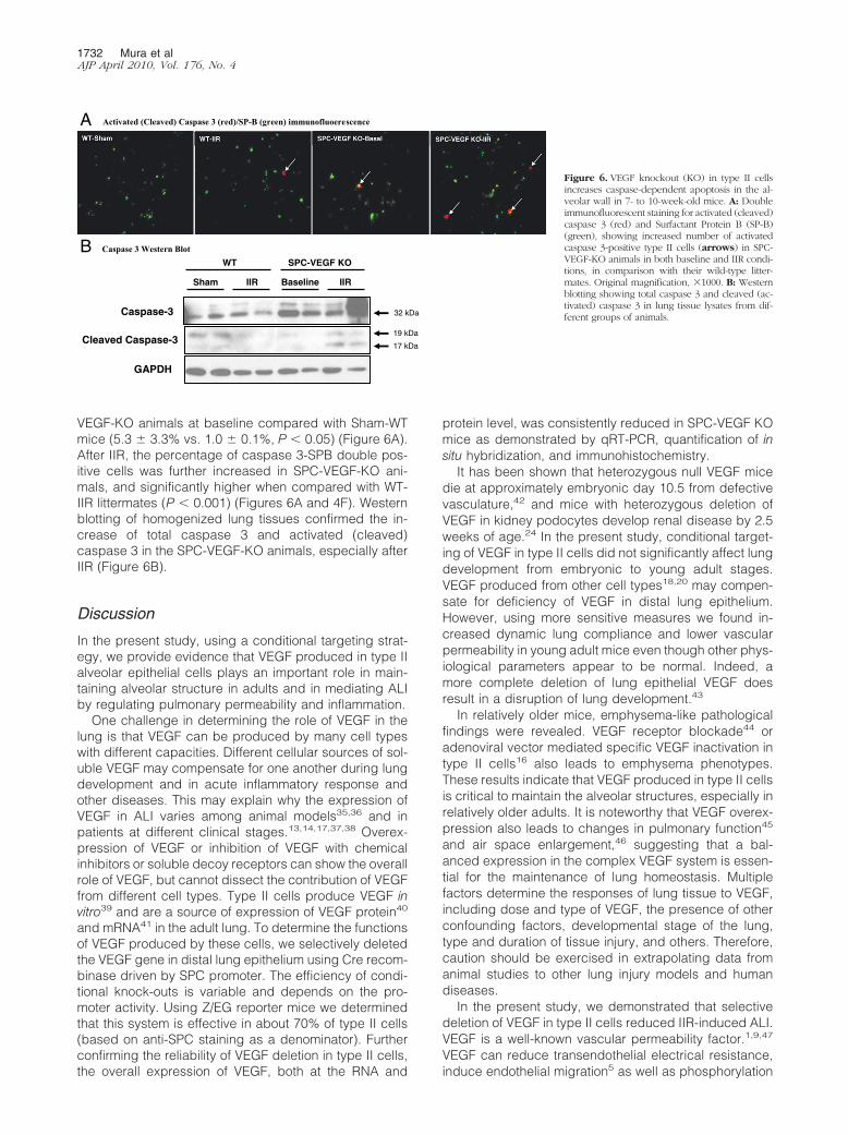
VEGF-KO animals at baseline compared with Sham-WTmice (5.3 � 3.3% vs. 1.0 � 0.1%, P � 0.05) (Figure 6A).After IIR, the percentage of caspase 3-SPB double pos-itive cells was further increased in SPC-VEGF-KO ani-mals, and significantly higher when compared with WT-IIR littermates (P � 0.001) (Figures 6A and 4F). Westernblotting of homogenized lung tissues confirmed the in-crease of total caspase 3 and activated (cleaved)caspase 3 in the SPC-VEGF-KO animals, especially afterIIR (Figure 6B).
Discussion
In the present study, using a conditional targeting strat-egy, we provide evidence that VEGF produced in type IIalveolar epithelial cells plays an important role in main-taining alveolar structure in adults and in mediating ALIby regulating pulmonary permeability and inflammation.
One challenge in determining the role of VEGF in thelung is that VEGF can be produced by many cell typeswith different capacities. Different cellular sources of sol-uble VEGF may compensate for one another during lungdevelopment and in acute inflammatory response andother diseases. This may explain why the expression ofVEGF in ALI varies among animal models35,36 and inpatients at different clinical stages.13,14,17,37,38 Overex-pression of VEGF or inhibition of VEGF with chemicalinhibitors or soluble decoy receptors can show the overallrole of VEGF, but cannot dissect the contribution of VEGFfrom different cell types. Type II cells produce VEGF invitro39 and are a source of expression of VEGF protein40
and mRNA41 in the adult lung. To determine the functionsof VEGF produced by these cells, we selectively deletedthe VEGF gene in distal lung epithelium using Cre recom-binase driven by SPC promoter. The efficiency of condi-tional knock-outs is variable and depends on the pro-moter activity. Using Z/EG reporter mice we determinedthat this system is effective in about 70% of type II cells(based on anti-SPC staining as a denominator). Furtherconfirming the reliability of VEGF deletion in type II cells,the overall expression of VEGF, both at the RNA and
protein level, was consistently reduced in SPC-VEGF KOmice as demonstrated by qRT-PCR, quantification of insitu hybridization, and immunohistochemistry.
It has been shown that heterozygous null VEGF micedie at approximately embryonic day 10.5 from defectivevasculature,42 and mice with heterozygous deletion ofVEGF in kidney podocytes develop renal disease by 2.5weeks of age.24 In the present study, conditional target-ing of VEGF in type II cells did not significantly affect lungdevelopment from embryonic to young adult stages.VEGF produced from other cell types18,20 may compen-sate for deficiency of VEGF in distal lung epithelium.However, using more sensitive measures we found in-creased dynamic lung compliance and lower vascularpermeability in young adult mice even though other phys-iological parameters appear to be normal. Indeed, amore complete deletion of lung epithelial VEGF doesresult in a disruption of lung development.43
In relatively older mice, emphysema-like pathologicalfindings were revealed. VEGF receptor blockade44 oradenoviral vector mediated specific VEGF inactivation intype II cells16 also leads to emphysema phenotypes.These results indicate that VEGF produced in type II cellsis critical to maintain the alveolar structures, especially inrelatively older adults. It is noteworthy that VEGF overex-pression also leads to changes in pulmonary function45
and air space enlargement,46 suggesting that a bal-anced expression in the complex VEGF system is essen-tial for the maintenance of lung homeostasis. Multiplefactors determine the responses of lung tissue to VEGF,including dose and type of VEGF, the presence of otherconfounding factors, developmental stage of the lung,type and duration of tissue injury, and others. Therefore,caution should be exercised in extrapolating data fromanimal studies to other lung injury models and humandiseases.
In the present study, we demonstrated that selectivedeletion of VEGF in type II cells reduced IIR-induced ALI.VEGF is a well-known vascular permeability factor.1,9,47
VEGF can reduce transendothelial electrical resistance,induce endothelial migration5 as well as phosphorylation
Activated (Cleaved) Caspase 3 (red)/SP-B (green) immunofluoerescence
Caspase-3
Cleaved Caspase-3
GAPDH
32 kDa
19 kDa
17 kDa
WT SPC-VEGF KO
Sham IIR Baseline IIR
Caspase 3 Western Blot
A
B
Figure 6. VEGF knockout (KO) in type II cellsincreases caspase-dependent apoptosis in the al-veolar wall in 7- to 10-week-old mice. A: Doubleimmunofluorescent staining for activated (cleaved)caspase 3 (red) and Surfactant Protein B (SP-B)(green), showing increased number of activatedcaspase 3-positive type II cells (arrows) in SPC-VEGF-KO animals in both baseline and IIR condi-tions, in comparison with their wild-type litter-mates. Original magnification, �1000. B: Westernblotting showing total caspase 3 and cleaved (ac-tivated) caspase 3 in lung tissue lysates from dif-ferent groups of animals.
1732 Mura et alAJP April 2010, Vol. 176, No. 4
of VE-cadherin and E-cadherin, and alter the actin stressfibers in pulmonary endothelial cells.6 Using a ventilatedpulmonary ischemia model in isolated ferret lungs, it hasbeen shown that pulmonary vascular barrier dysfunction isassociated with a rapid increase in VEGF expression.48
Similar results have been reported in a unilateral lung isch-emia model.36 In the present study, 24 hours after IIR,transgenic mice showed significantly lower pulmonary vas-cular permeability and attenuated interstitial edema. More-over, the chemotactic activity of VEGF may contribute tomonocyte activation and migration.9 Lack of type II epithe-lial cell-derived VEGF may reduce the recruitment of inflam-matory cells into the lung. Our results strongly support thehypothesis that VEGF locally produced in the distal lung epi-thelial cells is important for the pathogenesis of ALI/ARDS.
Despite improvement in ALI, some mortality was ob-served even in SPC-VEGF KO animals. This can be ex-plained by our previous observation of severe decreaseof blood arterial pressure, systemic inflammation, andmultiorgan dysfunction and cell death occurring in the IIRmodel in mice.28
Cell death is one of the underlying mechanisms ofALI.34 Although apoptosis is involved in ALI, it is not theonly form of cell death. Other types of cell death, such ascaspase-independent cell death, oncosis, autophagyand necrosis are more pro-inflammatory.34 We previouslydemonstrated that the prevalent pattern of cell death inIIR-induced ALI at 4 hours is oncosis.27 In the presentstudy, we found that the number of caspase 3/SPB dou-ble positive cells was increased in the SPC-VEGF-KOmice after IIR. These results suggest that the lack ofconstitutive VEGF signaling may induce type II cells toundergo caspase-dependent cell death, which may alsocontribute to the increased dynamic lung complianceseen in these animals. It is well-known that VEGF is animportant survival factor for endothelial cells.49 Recently,we28 and others19 have found that VEGF is also importantfor alveolar epithelial cells to survive. Knock-down of VEGFexpression with short interfering RNA resulted in death ofhuman lung epithelial cells in culture, and decreased VEGFin lung tissue was associated with increased death of typeII cells in vivo.28 On the other hand, the total number ofTUNEL-positive, as well as TUNEL/SPB-positive cells,showed no significant difference between wild-type andSPC-VEGF-KO mice after IIR. It has been shown thatTUNEL staining may reflect DNA injury, apoptotic50 or ne-crotic cell death.51,52 Since the total TUNEL staining andTUNEL�/SPB� cells did not change significantly, the totalamount of type II cell death may be similar in both wild-typeand SPC-VEGF KO mice during ALI, but the mechanisms oftype II cell death were switched toward caspase 3-depen-dent apoptosis in SPC-VEGF KO animals.
In conclusion, we provided evidence that VEGF pro-duced by type II cells is important to maintain the alveolarstructure, especially in adulthood. Results from targetedblockade of VEGF in type II cells point to a complex rolefor VEGF in acute lung injury as an important factorcontributing to the acute inflammatory response and pul-monary edema on the one hand, and as a protectivefactor for the alveolar epithelial barrier, on the other.
Therefore, caution should be taken when consideringVEGF as a therapeutic target for ALI/ARDS.
Acknowledgments
We gratefully acknowledge Dr. Susan E. Quaggin (De-partment of Medicine, University of Toronto) for her con-tinuous support and insightful advice on this researchproject. We also thank Dr. Jing Xu, Dr. Huadong Zhu, andZhihong Yun (University Health Network, Toronto, Can-ada) and Dr. Vera Eremina (The Samuel Lunenfeld Re-search Institute, Mount Sinai Hospital, Toronto, Canada)for their technical assistance.
References
1. Ferrara N, Gerber HP, LeCouter J: The biology of VEGF and itsreceptors. Nat Med 2003, 9:669–676
2. Gille H, Kowalski J, Li B, LeCouter J, Moffat B, Zioncheck TF, PelletierN, Ferrara N: Analysis of biological effects and signaling properties ofFlt-1 (VEGFR-1) and KDR (VEGFR-2). A reassessment using novelreceptor-specific vascular endothelial growth factor mutants. J BiolChem 2001, 276:3222–3230
3. Brown KR, England KM, Goss KL, Snyder JM, Acarregui MJ: VEGFinduces airway epithelial cell proliferation in human fetal lung in vitro.Am J Physiol Lung Cell Mol Physiol 2001, 281:L1001�L1010
4. Barleon B, Hauser S, Schollmann C, Weindel K, Marme D, Yayon A,Weich HA: Differential expression of the two VEGF receptors flt andKDR in placenta and vascular endothelial cells. J Cell Biochem 1994,54:56–66
5. Becker PM, Verin AD, Booth MA, Liu F, Birukova A, Garcia JG:Differential regulation of diverse physiological responses to VEGF inpulmonary endothelial cells. Am J Physiol Lung Cell Mol Physiol 2001,281:L1500�L1511
6. Godzich M, Hodnett M, Frank JA, Su G, Pespeni M, Angel A, HowardMB, Matthay MA, Pittet JF: Activation of the stress protein responseprevents the development of pulmonary edema by inhibiting VEGFcell signaling in a model of lung ischemia-reperfusion injury in rats.FASEB J 2006, 20:1519–1521
7. Paul R, Zhang ZG, Eliceiri BP, Jiang Q, Boccia AD, Zhang RL, Chopp M,Cheresh DA: Src deficiency or blockade of Src activity in mice providescerebral protection following stroke. Nat Med 2001, 7:222–227
8. van Bruggen N, Thibodeaux H, Palmer JT, Lee WP, Fu L, Cairns B,Tumas D, Gerlai R, Williams SP, van Lookeren Campagne M, FerraraN: VEGF antagonism reduces edema formation and tissue damageafter ischemia/reperfusion injury in the mouse brain. J Clin Invest1999, 104:1613–1620
9. Mura M, dos Santos CC, Stewart D, Liu M: Vascular endothelialgrowth factor and related molecules in acute lung injury. J ApplPhysiol 2004, 97:1605–1617
10. Kaner RJ, Ladetto JV, Singh R, Fukuda N, MatthayMA, Crystal RG: Lungoverexpression of the vascular endothelial growth factor gene inducespulmonary edema. Am J Respir Cell Mol Biol 2000, 22:657–664
11. Lee CG, Link H, Baluk P, Homer RJ, Chapoval S, Bhandari V, Kang MJ,Cohn L, Kim YK, McDonald DM, Elias JA: Vascular endothelial growthfactor (VEGF) induces remodeling and enhances TH2-mediated sensi-tization and inflammation in the lung. Nat Med 2004, 10:1095–1103
12. Hamada N, Kuwano K, Yamada M, Hagimoto N, Hiasa K, Egashira K,Nakashima N, Maeyama T, Yoshimi M, Nakanishi Y: Anti-vascularendothelial growth factor gene therapy attenuates lung injury andfibrosis in mice. J Immunol 2005, 175:1224–1231
13. Ware LB, Kaner RJ, Crystal RG, Schane R, Trivedi NN, McAuley D,Matthay MA: VEGF levels in the alveolar compartment do not distin-guish between ARDS and hydrostatic pulmonary oedema. Eur RespirJ2005, 26:101–105
14. Thickett DR, Armstrong L, Millar AB: A role for vascular endothelialgrowth factor in acute and resolving lung injury. Am J Respir Crit CareMed 2002, 166:1332–1337
15. Tuder RM, Zhen L, Cho CY, Taraseviciene-Stewart L, Kasahara Y,
VEGF in Acute Lung Injury 1733AJP April 2010, Vol. 176, No. 4
Salvemini D, Voelkel NF, Flores SC: Oxidative stress and apoptosisinteract and cause emphysema due to vascular endothelial growthfactor receptor blockade. Am J Respir Cell Mol Biol 2003, 29:88–97
16. Tang K, Rossiter HB, Wagner PD, Breen EC: Lung-targeted VEGFinactivation leads to an emphysema phenotype in mice, J ApplPhysiol 2004, 97:1559–1566; discussion 1549
17. Kaner RJ, Crystal RG: Compartmentalization of vascular endothelialgrowth factor to the epithelial surface of the human lung. Mol Med2001, 7:240–246
18. Corne J, Chupp G, Lee CG, Homer RJ, Zhu Z, Chen Q, Ma B, Du Y,Roux F, McArdle J, Waxman AB, Elias JA: IL-13 stimulates vascularendothelial cell growth factor and protects against hyperoxic acutelung injury. J Clin Invest 2000, 106:783–791
19. Roberts JR, Perkins GD, Fujisawa T, Pettigrew KA, Gao F, Ahmed A,Thickett DR: Vascular endothelial growth factor promotes physicalwound repair and is anti-apoptotic in primary distal lung epithelial andA549 cells. Crit Care Med 2007, 35:2164–2170
20. Shifren JL, Doldi N, Ferrara N, Mesiano S, Jaffe RB: In the humanfetus, vascular endothelial growth factor is expressed in epithelialcells and myocytes, but not vascular endothelium: implications formode of action. J Clin Endocrinol Metab 1994, 79:316–322
21. Souza DG, Soares AC, Pinho V, Torloni H, Reis LF, Teixeira MM, DiasAA: Increased mortality and inflammation in tumor necrosis factor-stimulated gene-14 transgenic mice after ischemia and reperfusioninjury. Am J Pathol 2002, 160:1755–1765
22. Novak A, Guo C, Yang W, Nagy A, Lobe CG: Z/EG, a double reportermouse line that expresses enhanced green fluorescent protein uponCre-mediated excision. Genesis 2000, 28:147–155
23. Chalfie M, Tu Y, Euskirchen G, Ward WW, Prasher DC: Green fluo-rescent protein as a marker for gene expression. Science 1994,263:802–805
24. Eremina V, Sood M, Haigh J, Nagy A, Lajoie G, Ferrara N, Gerber HP,Kikkawa Y, Miner JH, Quaggin SE: Glomerular-specific alterations ofVEGF-A expression lead to distinct congenital and acquired renaldiseases. J Clin Invest 2003, 111:707–716
25. dos Santos CC, Han B, Andrade CF, Bai X, Uhlig S, Hubmayr R, TsangM, Lodyga M, Keshavjee S, Slutsky AS, Liu M: DNA microarray analysisof gene expression in alveolar epithelial cells in response to TNFalpha,LPS, and cyclic stretch. Physiol Genomics 2004, 19:331–342
26. Yao H, Edirisinghe I, Yang SR, Rajendrasozhan S, Kode A, Caito S,Adenuga D, Rahman I: Genetic ablation of NADPH oxidase enhancessusceptibility to cigarette smoke-induced lung inflammation and em-physema in mice. Am J Pathol 2008, 172:1222–1237
27. Mura M, Andrade CF, Han B, Seth R, Zhang Y, Bai XH, Waddell TK,Hwang D, Keshavjee S, Liu M: Intestinal ischemia-reperfusion-in-duced acute lung injury and oncotic cell death in multiple organs.Shock 2007, 28:227–238
28. Mura M, Han B, Andrade CF, Seth R, Hwang D, Waddell TK,Keshavjee S, Liu M: The early responses of VEGF and its receptorsduring acute lung injury: implication of VEGF in alveolar epithelial cellsurvival. Crit Care 2006, 10:R130
29. Swaisgood CM, French EL, Noga C, Simon RH, Ploplis VA: Thedevelopment of bleomycin-induced pulmonary fibrosis in mice defi-cient for components of the fibrinolytic system. Am J Pathol 2000,157:177–187
30. Ginsberg HS, Horswood RL, Chanock RM, Prince GA: Role of earlygenes in pathogenesis of adenovirus pneumonia. Proc Natl Acad SciUSA 1990, 87:6191–6195
31. Han B, Mura M, Andrade CF, Okutani D, Lodyga M, dos Santos CC,Keshavjee S, Matthay M, Liu M: TNFalpha-induced long pentraxinPTX3 expression in human lung epithelial cells via JNK. J Immunol2005, 175:8303–8311
32. Abadie Y, Bregeon F, Papazian L, Lange F, Chailley-Heu B, ThomasP, Duvaldestin P, Adnot S, Maitre B, Delclaux C: Decreased VEGFconcentration in lung tissue and vascular injury during ARDS. EurRespir J2005, 25:139–146
33. Krajewski S, Zapata JM, Krajewska M, VanArsdale T, Shabaik A,Gascoyne RD, Reed JC: Immunohistochemical analysis of in vivopatterns of TRAF-3 expression, a member of the TNF receptor-asso-ciated factor family. J Immunol 1997, 159:5841–5852
34. Tang PS, Mura M, Seth R, Liu M: Acute lung injury and cell death: howmany ways can cells die? Am J Physiol Lung Cell Mol Physiol 2008,294:L632�L641
35. Fehrenbach A, Pufe T, Wittwer T, Nagib R, Dreyer N, Pech T, Petersen
W, Fehrenbach H, Wahlers T, Richter J: Reduced vascular endothe-lial growth factor correlates with alveolar epithelial damage afterexperimental ischemia and reperfusion. J Heart Lung Transplant2003, 22:967–978
36. Kazi AA, Lee WS, Wagner E, Becker PM: VEGF, fetal liver kinase-1,and permeability increase during unilateral lung ischemia. Am JPhysiol Lung Cell Mol Physiol 2000, 279:L460�L467
37. Maitre B, Boussat S, Jean D, Gouge M, Brochard L, Housset B, AdnotS, Delclaux C: Vascular endothelial growth factor synthesis in theacute phase of experimental and clinical lung injury. Eur Respir J2001, 18:100–106
38. Thickett DR, Armstrong L, Christie SJ, Millar AB: Vascular endothelialgrowth factor may contribute to increased vascular permeability inacute respiratory distress syndrome. Am J Respir Crit Care Med2001, 164:1601–1605
39. Pham I, Uchida T, Planes C, Ware LB, Kaner R, Matthay MA, ClericiC: Hypoxia upregulates VEGF expression in alveolar epithelial cells invitro and in vivo. Am J Physiol Lung Cell Mol Physiol 2002,283:L1133�L1142
40. Fehrenbach H, Kasper M, Haase M, Schuh D, Muller M: Differentialimmunolocalization of VEGF in rat and human adult lung, and inexperimental rat lung fibrosis: light, fluorescence, and electron mi-croscopy. Anat Rec 1999, 254:61–73
41. Tuder RM, Flook BE, Voelkel NF: Increased gene expression for VEGFand the VEGF receptors KDR/Flk and Flt in lungs exposed to acute or tochronic hypoxia. Modulation of gene expression by nitric oxide. J ClinInvest 1995, 95:1798–1807
42. Ferrara N, Carver-Moore K, Chen H, Dowd M, Lu L, O’Shea KS,Powell-Braxton L, Hillan KJ, Moore MW: Heterozygous embryoniclethality induced by targeted inactivation of the VEGF gene. Nature1996, 380:439–442
43. Yamamoto H, Yun EJ, Gerber HP, Ferrara N, Whitsett JA, Vu TH:Epithelial-vascular cross talk mediated by VEGF-A and HGF signalingdirects primary septae formation during distal lung morphogenesis.Dev Biol 2007, 308:44–53
44. Kasahara Y, Tuder RM, Taraseviciene-Stewart L, Le Cras TD, AbmanS, Hirth PK, Waltenberger J, Voelkel NF: Inhibition of VEGF receptorscauses lung cell apoptosis and emphysema. J Clin Invest 2000,106:1311–1319
45. Bhandari V, Choo-Wing R, Chapoval SP, Lee CG, Tang C, Kim YK,Ma B, Baluk P, Lin MI, McDonald DM, Homer RJ, Sessa WC, Elias JA:Essential role of nitric oxide in VEGF-induced, asthma-like angio-genic, inflammatory, mucus, and physiologic responses in the lung.Proc Natl Acad Sci USA 2006, 103:11021–11026
46. Le Cras TD, Spitzmiller RE, Albertine KH, Greenberg JM, Whitsett JA,Akeson AL: VEGF causes pulmonary hemorrhage, hemosiderosis,and air space enlargement in neonatal mice. Am J Physiol Lung CellMol Physiol 2004, 287:L134�L142
47. Lee CG, Yoon HJ, Zhu Z, Link H, Wang Z, Gwaltney JM, Landry M,Elias JA: Respiratory syncytial virus stimulation of vascular endothe-lial cell growth factor/vascular permeability factor. Am J Respir CellMol Biol 2000, 23:662–669
48. Becker PM, Alcasabas A, Yu AY, Semenza GL, Bunton TE: Oxygen-independent upregulation of vascular endothelial growth factor andvascular barrier dysfunction during ventilated pulmonary ischemia inisolated ferret lungs. Am J Respir Cell Mol Biol 2000, 22:272–279
49. Gerber HP, McMurtrey A, Kowalski J, Yan M, Keyt BA, Dixit V, FerraraN: Vascular endothelial growth factor regulates endothelial cell sur-vival through the phosphatidylinositol 3�-kinase/Akt signal transduc-tion pathway. Requirement for Flk-1/KDR activation. J Biol Chem1998, 273:30336–30343
50. Albertine KH, Plopper CG: DNA oxidation or apoptosis: will the realculprit of dna damage in hyperoxic lung injury please stand up? Am JRespir Cell Mol Biol 2002, 26:381–383
51. Wang X, Ryter SW, Dai C, Tang ZL, Watkins SC, Yin XM, Song R, ChoiAM: Necrotic cell death in response to oxidant stress involves theactivation of the apoptogenic caspase-8/bid pathway. J Biol Chem2003, 278:29184–29191
52. Bhandari V, Choo-Wing R, Lee CG, Zhu Z, Nedrelow JH, ChuppGL, Zhang X, Matthay MA, Ware LB, Homer RJ, Lee PJ, Geick A,de Fougerolles AR, Elias JA: Hyperoxia causes angiopoietin 2-medi-ated acute lung injury and necrotic cell death. Nat Med 2006,12:1286–1293
1734 Mura et alAJP April 2010, Vol. 176, No. 4