Embed Size (px)
Citation preview
Functional genomics of Lactobacillus caseiestablishment in the gutHélène Licandro-Serauta,b,c, Hélène Scorneca, Thierry Pédronb,c, Jean-François Cavina, and Philippe J. Sansonettib,c,d,1
aUnité Mixte de Recherche A 02102 Procédés Alimentaires et Microbiologiques, AgroSup Dijon/Université de Bourgogne, 21000 Dijon, France; bUnité dePathogénie Microbienne Moléculaire and cInstitut National de la Santé et de la Recherche Médicale U786, Institut Pasteur, 75724 Paris Cedex 15, France;and dChaire de Microbiologie et Maladies Infectieuses, Collège de France, 75005 Paris, France
Contributed by Philippe J. Sansonetti, June 25, 2014 (sent for review April 1, 2014)
Although the composition of the gut microbiota and its symbioticcontribution to key host physiological functions are well estab-lished, little is known as yet about the bacterial factors thataccount for this symbiosis. We selected Lactobacillus casei asa model microorganism to proceed to genomewide identificationof the functions required for a symbiont to establish colonizationin the gut. As a result of our recent development of a transposon-mutagenesis tool that overcomes the barrier that had preventedL. casei randommutagenesis, we developed a signature-tagged mu-tagenesis approach combining whole-genome reverse genetics us-ing a set of tagged transposons and in vivo screening using therabbit ligated ileal loop model. After sequencing transposon in-sertion sites in 9,250 random mutants, we assembled a library of1,110 independent mutants, all disrupted in a different gene, thatprovides a representative view of the L. casei genome. By deter-mining the relative quantity of each of the 1,110 mutants beforeand after the in vivo challenge, we identified a core of 47 L. caseigenes necessary for its establishment in the gut. They are involvedin housekeeping functions, metabolism (sugar, amino acids), cellwall biogenesis, and adaptation to environment. Hence we providewhat is, to our knowledge, the first global functional genomicsanalysis of L. casei symbiosis.
commensalism | Lactic acid bacteria
The pioneering studies that led to the characterization of thegut microbiota were reviewed in 2001 (1). These studies and
recent investigations have revealed mutualistic functions (2),including a barrier effect against allogenic microbes (3), fer-mentation of complex sugars (4, 5), and maturation and ho-meostasis of the immune system (6). Recent metagenomic studieshave revealed an extraordinary diversity of genes constituting thegut microbiome (7), opening the way to correlative studies linkingmicrobiome diversity, homeostasis, and diseases (5, 8, 9).In parallel, some representative species, i.e., “model sym-
bionts,” now are being studied functionally (10). As it was donefor pathogens, it is essential to develop the cellular microbiologyof symbionts and particularly to identify the genes required fortheir establishment and persistence in the gut. Transcriptomicprofiling identified up-regulated genes linked to metabolicfunctions, stress responses, and pili synthesis during early colo-nization (11–13). Comparative genomics among Lactobacilliidentified strain-specific candidate genes for extended coloniza-tion: In Lactobacillus rhamnosus, persistence was attributed to anspaCBA locus encoding LPXTG-like pilins (14), and in Lacto-bacillus johnsonii it was attributed to specific glycosyltransferases,a phosphotransfer system, and a protease (15). Otherwise, afunctional in vivo screening based on the expression of a ge-nomic library of Bacteroides fragilis identified a locus encodingpolysaccharide utilization as essential for stable colonizationof murine colonic crypts (16). Alternatively, colonization ofgerm-free mice with a collection of random mutants of Bac-teroides thetaiotaomicron followed by deep sequencing showedthat mutants unable to synthesize vitamin B12 were impairedin gut colonization (17).
Lactobacillus spp. pioneer initial gut colonization (18), andthey participate in the gut immunological and nutritional sym-biosis. Because of our permanent exposure to Lactobacillus spp.,and particularly to Lactobacillus casei, as components of dairyproducts, tools are required to decipher its symbiosis with thegut. Genetic manipulation of lactic bacteria often is problematicbecause of their natural resistance to numerous antibiotics andthe lack of dedicated genetic tools and efficient transformationprocedures (19). We recently developed a transposon-mutagen-esis tool, the Pjunc-TpaseIS1223 system (20), which overcomes thebarrier to random mutagenesis in Lactobacillus casei. Using thistool, we adapted a signature-tagged mutagenesis (STM) approach(21) to L. casei that combined whole-genome reverse genetics usinga set of tagged transposons with an in vivo screening in the rabbitligated ileal loop model identifying mutants impaired in gut es-tablishment. The term “establishment” qualifies the early steps ofcolonization explored by this model. After sequencing the 1,110independent mutants obtained in this study, we identified a core of47 L. casei genes belonging to five major functional groups thatare required for its establishment in the gut.
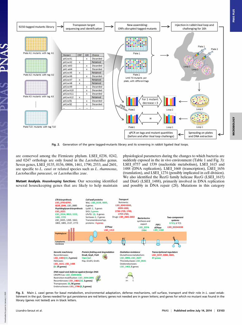
ResultsGeneration of a Library of L. casei Tagged Mutants. To generatea large library of L . casei tagged mutants and to proceed to STM,tagged derivatives of the Pjunc-TpaseIS1223 transposable vectorwere generated using 70 DNA tags previously used for Salmo-nella typhimurium STM (21). For each tag, among the ∼5,000integrants obtained per transformation, clones were selectedrandomly and assembled in 96-well plates. Thus, a library of9,250 tagged mutants labeled with 70 different tags was gener-ated. To extend the contribution of STM, we introduced real-timePCR, rather than dot-blot analysis, to allow relative quantificationof bacteria in addition to their detection.
Significance
Lactobacillus casei, a food bacterium recognized for its bene-ficial effects, was selected as a model microorganism to pro-ceed to genomewide identification of the functions requiredfor a symbiont to establish colonization in the gut. We recentlyhave developed a mutagenesis tool that overcomes the barrierthat prevented L. casei random mutagenesis. After identifying9,250 mutations, we assembled a library of 1,110 mutants dis-rupted in different genes and tested them for their ability tocolonize an in vivo model, the rabbit ligated ileal loop. With thisglobal functional genomic analysis of L. casei symbiosis (the first,to our knowledge), we identified a core of 47 L. casei genesnecessary for its establishment in the gut.
Author contributions: H.L.-S., T.P., and P.J.S. designed research; H.L.-S., H.S., and T.P.performed research; H.L.-S., H.S., T.P., J.-F.C., and P.J.S. analyzed data; and H.L.-S., T.P.,J.-F.C., and P.J.S. wrote the paper.
The authors declare no conflict of interest.1To whom correspondence should be addressed. Email: [email protected].
This article contains supporting information online at www.pnas.org/lookup/suppl/doi:10.1073/pnas.1411883111/-/DCSupplemental.
www.pnas.org/cgi/doi/10.1073/pnas.1411883111 PNAS | Published online July 14, 2014 | E3101–E3109
MICRO
BIOLO
GY
PNASPL
US
Dow
nloa
ded
by g
uest
on
Janu
ary
2, 2
020
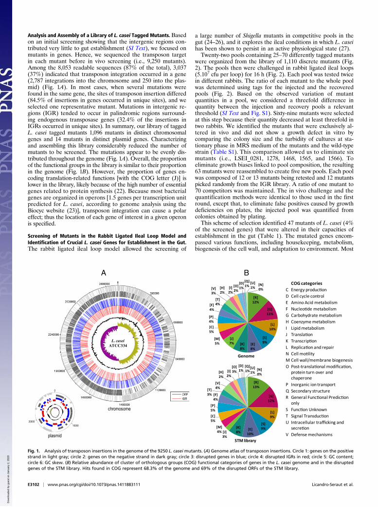
Analysis and Assembly of a Library of L. casei Tagged Mutants. Basedon an initial screening showing that the intergenic regions con-tributed very little to gut establishment (SI Text), we focused onmutants in genes. Hence, we sequenced the transposon targetin each mutant before in vivo screening (i.e., 9,250 mutants).Among the 8,053 readable sequences (87% of the total), 3,037(37%) indicated that transposon integration occurred in a gene(2,787 integrations into the chromosome and 250 into the plas-mid) (Fig. 1A). In most cases, when several mutations werefound in the same gene, the sites of transposon insertion differed(84.5% of insertions in genes occurred in unique sites), and weselected one representative mutant. Mutations in intergenic re-gions (IGR) tended to occur in palindromic regions surround-ing endogenous transposase genes (32.4% of the insertions inIGRs occurred in unique sites). In summary, our library of taggedL. casei tagged mutants 1,096 mutants in distinct chromosomalgenes and 14 mutants in distinct plasmid genes. Characterizingand assembling this library considerably reduced the number ofmutants to be screened. The mutations appear to be evenly dis-tributed throughout the genome (Fig. 1A). Overall, the proportionof the functional groups in the library is similar to their proportionin the genome (Fig. 1B). However, the proportion of genes en-coding translation-related functions [with the COG letter (J)] islower in the library, likely because of the high number of essentialgenes related to protein synthesis (22). Because most bacterialgenes are organized in operons [1.5 genes per transcription unitpredicted for L. casei, according to genome analysis using theBiocyc website (23)], transposon integration can cause a polareffect; thus the location of each gene of interest in a given operonis specified.
Screening of Mutants in the Rabbit Ligated Ileal Loop Model andIdentification of Crucial L. casei Genes for Establishment in the Gut.The rabbit ligated ileal loop model allowed the screening of
a large number of Shigella mutants in competitive pools in thegut (24–26), and it explores the ileal conditions in which L. caseihas been shown to persist in an active physiological state (27).Twenty-two pools containing 25–70 differently tagged mutants
were organized from the library of 1,110 discrete mutants (Fig.2). The pools then were challenged in rabbit ligated ileal loops(5.107 cfu per loop) for 16 h (Fig. 2). Each pool was tested twicein different rabbits. The ratio of each mutant to the whole poolwas determined using tags for the injected and the recoveredpools (Fig. 2). Based on the observed variation of mutantquantities in a pool, we considered a threefold difference inquantity between the injection and recovery pools a relevantthreshold (SI Text and Fig. S1). Sixty-nine mutants were selectedat this step because their quantity decreased at least threefold intwo rabbits. We identified the mutants that were exclusively al-tered in vivo and did not show a growth defect in vitro bycomparing the colony size and the turbidity of cultures at sta-tionary phase in MRS medium of the mutants and the wild-typestrain (Table S1). This comparison allowed us to eliminate sixmutants (i.e., LSEI_0281, 1278, 1468, 1565, and 1566). Toeliminate growth biases linked to pool composition, the resulting63 mutants were reassembled to create five new pools. Each poolwas composed of 12 or 13 mutants being retested and 12 mutantspicked randomly from the IGR library. A ratio of one mutant to70 competitors was maintained. The in vivo challenge and thequantification methods were identical to those used in the firstround, except that, to eliminate false positives caused by growthdeficiencies on plates, the injected pool was quantified fromcolonies obtained by plating.This scheme of selection identified 47 mutants of L. casei (4%
of the screened genes) that were altered in their capacities ofestablishment in the gut (Table 1). The mutated genes encom-passed various functions, including housekeeping, metabolism,biogenesis of the cell wall, and adaptation to environment. Most
COG categoriesC Energy produc onD Cell cycle controlE Amino Acid metabolismF Nucleo de metabolismG Carbohydrate metabolismH Coenzyme metabolismI Lipid metabolismJ Transla onK Transcrip onL Replica on and repairN Cell mo lityM Cell wall/membrane biogenesisO Post-transla onal modifica on,
protein turn over andchaperone
P Inorganic ion transportQ Secondary structureR General Func onal Predic on
onlyS Func on UnknownT Signal Transduc onU Intracellular trafficking and
secre onV Defense mechanisms
L. caseiATCC334
[R]12%
[G]11%
[L]10%
[S]9%[E]
8%[K]8%
[J]7%
[M]5%
[C]5%
[P]4%
[F]4%
[T]4%
[V]3%
[H]2%
[I]2%
[O]2%
[D]1%
[Q]1%
[U]1%
[N]0%
Genome
[R]13%
[G]12%
[L]9%
[S]9%[E]
10%
[K]8%[J]
3%
[M]4%
[C]5%
[P]5%
[F]4%
[T]3%
[V]4%
[H]2%
[I]2%
[O]3%
[D]1%
[Q]1%
[U]1%
[N]0%
STM library
0
2000010000
chromosome
plasmid
A B
Fig. 1. Analysis of transposon insertions in the genome of the 9250 L. caseimutants. (A) Genome atlas of transposon insertions. Circle 1: genes on the positivestrand in light gray; circle 2: genes on the negative strand in dark gray; circle 3: disrupted genes in blue; circle 4: disrupted IGRs in red; circle 5: GC content;circle 6: GC skew. (B) Relative abundance of cluster of orthologous groups (COG) functional categories of genes in the L. casei genome and in the disruptedgenes of the STM library. Hits found in COG represent 68.3% of the genome and 69% of the disrupted ORFs of the STM library.
E3102 | www.pnas.org/cgi/doi/10.1073/pnas.1411883111 Licandro-Seraut et al.
Dow
nloa
ded
by g
uest
on
Janu
ary
2, 2
020
are conserved among the Firmicute phylum. LSEI_0238, 0242,and 0247 orthologs are only found in the Lactobacillus genus.Seven genes, LSEI_0135, 0156, 0806, 1461, 1790, 2553, and 2601,are specific to L. casei or related species such as L. rhamnosus,Lactobacillus paracasei, or Lactobacillus zeae.
Mutant Analysis. Housekeeping functions. Our screening identifiedseveral housekeeping genes that are likely to help maintain
physiological parameters during the changes to which bacteria aresuddenly exposed in the in vivo environment (Table 1 and Fig. 3):LSEI_0757 and 1539 (nucleotide metabolism), LSEI_1615 and1488 (DNA replication), LSEI_1668 (transcription), LSEI_1656(translation), and LSEI_1274 (possibly implicated in cell division).We also identified the RecG family helicase RecG (LSEI_1615)and DinG (LSEI_1488), primarily involved in DNA replicationand possibly in DNA repair (28). Mutations in this category
Fig. 2. Generation of the gene tagged-mutants library and its screening in rabbit ligated ileal loops.
Bacteriocinssynthesis and
exportLSEI_2374-
2384GTPase LSEI_0219-0220
LSEI_1313
F0F1ATPase
LSEI_1160
TransportNutrients:
LSEI_0242-0243,0999-1000,
1738-1739, 1743,1759-1760,
Drugs: LSEI_2601-2602
Oxidation resistanceGlutathione metabolism:LSEI_0893, LSEI_2627Thioreductases:LSEI_0541Oxidoreductases:LSEI_1463, 8 genes
Protein folding and degradationDnaK, GrpE, FtsHClpLClpCHsp, GroES, GroEL
Cytoplasmicmembrane
LTA biosynthesisLSEI_0793-0797,0238, 2546, LSEI_0885PeptidoglycanbiosynthesisLSEI_0221LSEI_0314, 0810, 1152,1481, 1725LSEI_0435, 1269, 1662, 1802, 1805, 2137, 2773
Cellwall proteinsWxL: LSEI_0158, 0605,6 genesLysM: 2, 3 genesSH3: 2 genesLPxTG: 13, 6 genesSortases:3, 2 genesTransmembrane hyp.proteins: 4 genes
DNA repair and defense against foreign DNACRISPR-Cas: LSEI_0349-0356Restric on modifica on: LSEI_2094-2095Recombinases:LSEI_1403 (10, 2 genes)Transposases:33, 56 genesEndonucleases:LSEI_1790 (0, 5 genes)
Genetic machineryRecombinases:LSEI_1403 (10, 2 genes)Helicases:LSEI_1615, LSEI_1488(9, 14 genes)
Two-componentsystems
9 TCS, 6 TCS
Transcriptional regulatorsLSEI_0247, 0394, 0681,47 genes
Pep doglycan
-SH
-SH
-S
-S
Fig. 3. Main L. casei genes for basal metabolism, environmental adaptation, defense mechanisms, cell surface, transport and their role in L. casei estab-lishment in the gut. Genes needed for gut persistence are red letters; genes not needed are in green letters; and genes for which no mutant was found in thelibrary (genes not tested) are in black letters.
Licandro-Seraut et al. PNAS | Published online July 14, 2014 | E3103
MICRO
BIOLO
GY
PNASPL
US
Dow
nloa
ded
by g
uest
on
Janu
ary
2, 2
020
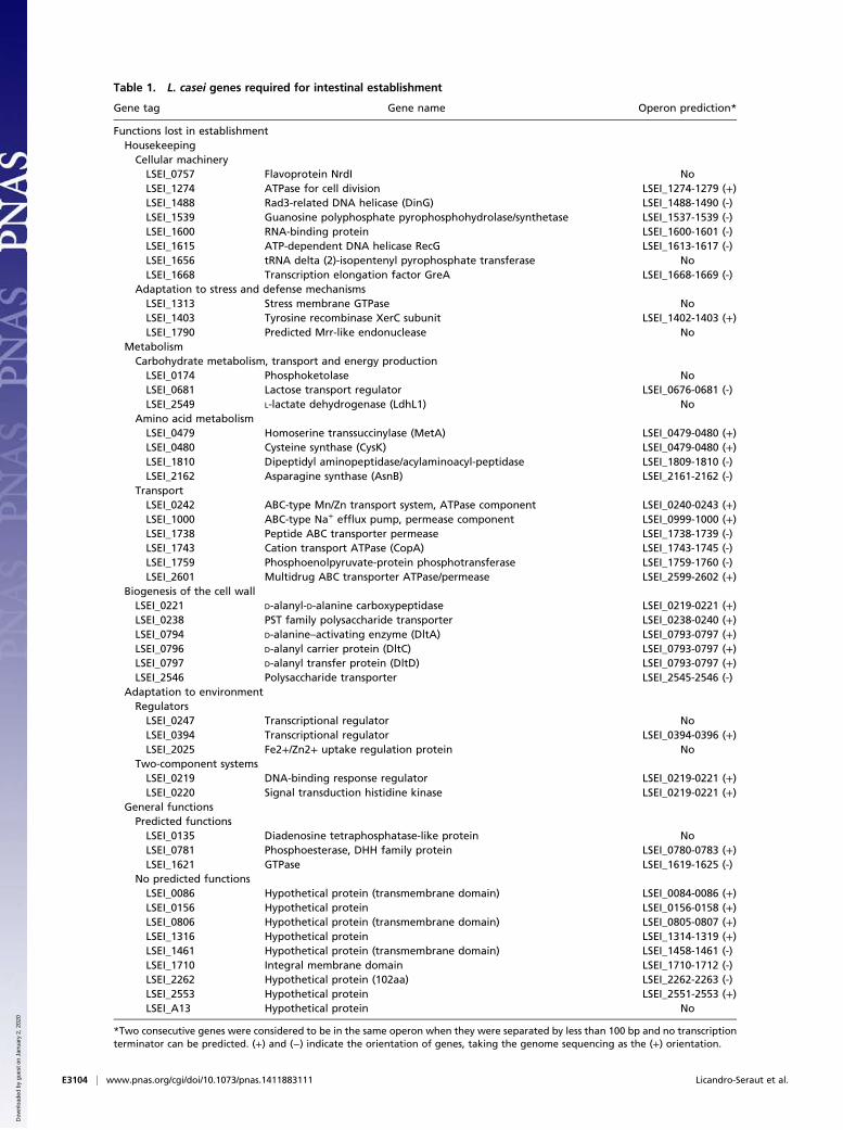
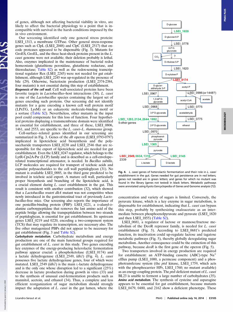
Table 1. L. casei genes required for intestinal establishment
Gene tag Gene name Operon prediction*
Functions lost in establishmentHousekeeping
Cellular machineryLSEI_0757 Flavoprotein NrdI NoLSEI_1274 ATPase for cell division LSEI_1274-1279 (+)LSEI_1488 Rad3-related DNA helicase (DinG) LSEI_1488-1490 (-)LSEI_1539 Guanosine polyphosphate pyrophosphohydrolase/synthetase LSEI_1537-1539 (-)LSEI_1600 RNA-binding protein LSEI_1600-1601 (-)LSEI_1615 ATP-dependent DNA helicase RecG LSEI_1613-1617 (-)LSEI_1656 tRNA delta (2)-isopentenyl pyrophosphate transferase NoLSEI_1668 Transcription elongation factor GreA LSEI_1668-1669 (-)
Adaptation to stress and defense mechanismsLSEI_1313 Stress membrane GTPase NoLSEI_1403 Tyrosine recombinase XerC subunit LSEI_1402-1403 (+)LSEI_1790 Predicted Mrr-like endonuclease No
MetabolismCarbohydrate metabolism, transport and energy production
LSEI_0174 Phosphoketolase NoLSEI_0681 Lactose transport regulator LSEI_0676-0681 (-)LSEI_2549 L-lactate dehydrogenase (LdhL1) No
Amino acid metabolismLSEI_0479 Homoserine transsuccinylase (MetA) LSEI_0479-0480 (+)LSEI_0480 Cysteine synthase (CysK) LSEI_0479-0480 (+)LSEI_1810 Dipeptidyl aminopeptidase/acylaminoacyl-peptidase LSEI_1809-1810 (-)LSEI_2162 Asparagine synthase (AsnB) LSEI_2161-2162 (-)
TransportLSEI_0242 ABC-type Mn/Zn transport system, ATPase component LSEI_0240-0243 (+)LSEI_1000 ABC-type Na+ efflux pump, permease component LSEI_0999-1000 (+)LSEI_1738 Peptide ABC transporter permease LSEI_1738-1739 (-)LSEI_1743 Cation transport ATPase (CopA) LSEI_1743-1745 (-)LSEI_1759 Phosphoenolpyruvate-protein phosphotransferase LSEI_1759-1760 (-)LSEI_2601 Multidrug ABC transporter ATPase/permease LSEI_2599-2602 (+)
Biogenesis of the cell wallLSEI_0221 D-alanyl-D-alanine carboxypeptidase LSEI_0219-0221 (+)LSEI_0238 PST family polysaccharide transporter LSEI_0238-0240 (+)LSEI_0794 D-alanine–activating enzyme (DltA) LSEI_0793-0797 (+)LSEI_0796 D-alanyl carrier protein (DltC) LSEI_0793-0797 (+)LSEI_0797 D-alanyl transfer protein (DltD) LSEI_0793-0797 (+)LSEI_2546 Polysaccharide transporter LSEI_2545-2546 (-)
Adaptation to environmentRegulators
LSEI_0247 Transcriptional regulator NoLSEI_0394 Transcriptional regulator LSEI_0394-0396 (+)LSEI_2025 Fe2+/Zn2+ uptake regulation protein No
Two-component systemsLSEI_0219 DNA-binding response regulator LSEI_0219-0221 (+)LSEI_0220 Signal transduction histidine kinase LSEI_0219-0221 (+)
General functionsPredicted functions
LSEI_0135 Diadenosine tetraphosphatase-like protein NoLSEI_0781 Phosphoesterase, DHH family protein LSEI_0780-0783 (+)LSEI_1621 GTPase LSEI_1619-1625 (-)
No predicted functionsLSEI_0086 Hypothetical protein (transmembrane domain) LSEI_0084-0086 (+)LSEI_0156 Hypothetical protein LSEI_0156-0158 (+)LSEI_0806 Hypothetical protein (transmembrane domain) LSEI_0805-0807 (+)LSEI_1316 Hypothetical protein LSEI_1314-1319 (+)LSEI_1461 Hypothetical protein (transmembrane domain) LSEI_1458-1461 (-)LSEI_1710 Integral membrane domain LSEI_1710-1712 (-)LSEI_2262 Hypothetical protein (102aa) LSEI_2262-2263 (-)LSEI_2553 Hypothetical protein LSEI_2551-2553 (+)LSEI_A13 Hypothetical protein No
*Two consecutive genes were considered to be in the same operon when they were separated by less than 100 bp and no transcriptionterminator can be predicted. (+) and (−) indicate the orientation of genes, taking the genome sequencing as the (+) orientation.
E3104 | www.pnas.org/cgi/doi/10.1073/pnas.1411883111 Licandro-Seraut et al.
Dow
nloa
ded
by g
uest
on
Janu
ary
2, 2
020
of genes, although not affecting bacterial viability in vitro, arelikely to affect the bacterial physiology to a point that is in-compatible with survival in the harsh conditions imposed by thein vivo environment.Our screening identified only one general stress protein:
LSEI_1313, a membrane GTPase. Other general stress-responsegenes such as ClpL (LSEI_2048) and ClpC (LSEI_2517) that en-code proteases appeared to be dispensable (Fig. 3). Mutants forGroES, GroEL, and the three heat-shock proteins present in the L.casei genome were not available; their deletion probably is lethal.Also, enzymes implicated in the maintenance of bacterial redoxhomeostasis (glutathione peroxidase, glutathione reductase, andthioreductase; Table S2) as well as the redox-sensing transcrip-tional regulator Rex (LSEI_2245) were not needed for gut estab-lishment, although LSEI_2245 was up-regulated in the presence ofbile (29). Otherwise, bacteriocin production (LSEI_2374-2384,four mutants) is not essential during this step of establishment.Biogenesis of the cell wall. Cell wall-associated proteins have beenfavorite targets in Lactobacillus–host interactions (30). L. caseiis one of the Lactobacillus species containing the largest set ofgenes encoding such proteins. Our screening did not identifymutants for a gene encoding a known cell wall protein motif(LPxTG, LysM) or an eukaryotic molecule-binding motif ora sortase (Table S2). Nevertheless, other mutants in the inputpool could compensate for this loss of function. Four hypothet-ical proteins displaying a transmembrane domain were identifiedas essential for establishment, and three of these, LSEI_0806,1461, and 2553, are specific to the L. casei–L. rhamnosus group.Cell-surface–related genes identified in our screening are
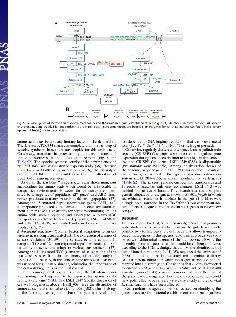
summarized in Fig. 3. Genes of the dlt operon (LSEI_0793-0797)implicated in lipoteichoic acid biosynthesis and the poly-saccharide transporters LSEI_0238 and LSEI_2546 that are re-sponsible for the export of lipoteichoic acid are needed for gutestablishment. Even the LSEI_0247 regulator, which belongs to theLytR-Cps2A-Psr (LCP) family and is described as a cell-envelope–related transcriptional attenuator, is needed. In Bacillus subtilis,LCP molecules are required for transport of teichoic acids andcapsular polysaccharides to the cell wall peptidoglycan (31). Nomutant is available LSEI_0885, in the third gene predicted to beinvolved in teichoic acid export. A mature cell wall, particularlyproper biosynthesis and branching of the lipoteichoic acid, isa crucial element during L. casei establishment in the gut. Thisresult is consistent with another contribution (32), which showedthat a Lactobacillus reuteri dltA mutant was not competitive whennewly introduced in the gastrointestinal tract of previously Lacto-bacillus-free mice. Our screening also reports the importance ofone penicillin-binding protein (PBP): LSEI_0221, a D-alanyl-D-alanine carboxypeptidase that removes the last amino acid of thepeptide bridge allowing the transpeptidation between two strandsof peptidoglycan, is essential for gut establishment. Its upstreamgenes LSEI_0219 and 0220, encoding a two-component system(TCS) that may regulate its transcription, are necessary also. Thefive other mutagenized PBPs did not appear to be necessary forgut establishment (Fig. 3 and Table S2).Carbohydrate metabolism. Carbohydrate metabolism and energyproduction are one of the main functional groups required forgut establishment of L. casei in this study. Two genes encodingkey enzymes of the energy-producing heterolactic fermentationpathway appear crucial: a phosphoketolase (LSEI_0174) anda lactate dehydrogenase (LSEI_2549, ldh1) (Fig. 4). L. caseipossesses five lactate dehydrogenase genes, four of which weremutated. LSEI_2549 (ldh1) is the main L-lactate dehydrogenaseand is the only one whose disruption led to a significant (25%)decrease in lactate production during growth in vitro (33) andto the synthesis of unusual end-fermentation products such asmannitol, acetoin, and ethanol (34). Thus, a complete and lessefficient reorganization of sugar metabolism should stronglyimpact the adaptation of L. casei in the gut lumen, where the
concentration and variety of sugars is limited. Conversely, thepyruvate kinase, which is a key enzyme in sugar metabolism, isdispensable for establishment, indicating that L. casei can bypassthis step, probably by synthesizing oxaloacetate as an inter-mediate between phosphoenolpyruvate and pyruvate (LSEI_1820and then LSEI_1855) (Table S2).LSEI_0681, a regulator of lactose or mannose/fructose me-
tabolism of the DeoR repressor family, is needed for L. caseiestablishment (Fig. 5). According to LSEI_0681’s predictedfunction, its inactivation could up-regulate lactose and tagatosemetabolic pathways (Fig. 5), thereby globally deregulating sugarmetabolism. Another consequence could be the extinction of thispathway, because deoR is the first gene of the operon (Fig. 5).Two transporters involved in energy production are required
for establishment: an ATP-binding cassette (ABC)-type Na+
efflux pump (LSEI_1000, a permease component) and a phos-photransferase system (the ptsI kinase, LSEI_1759, which actswith the phosphocarrier HPr, LSEI_1760; no mutant available)as an energy coupling protein. The ptsI-deficient mutant of L. caseiBL23 is unable to ferment a large number of carbohydrates (35).Amino acid metabolism. The synthesis of cysteine and asparagineappears to be essential for gut establishment, because mutantsLSEI_0479, 0480, and 2162 show a deficient phenotype. These
D-ribulose 5-P
D-glycer-aldhehyde 3-P
pyruvate
ethanol
acetyl-P
L-lactate D-lactate
6-P D-gluconate
D-glucose 6-P
CO2
D-glucose
LSEI_1659
LSEI_0778
D-xylulose P
?
LSEI_1620
LSEI_0174
LSEI_0996 (or 1448?)
LSEI_2549, 0634, 1310, 2339
LSEI_0145
acetateacetyl-CoA
acetaldehyde
2-P D-glycerate
1,3-biP D-glycerate
3-P D-glycerate
phosphoenol-pyruvate
LSEI_0967
LSEI_0968
LSEI_1293, 2134, 2644, 1992, 5 other genes
LSEI_0970
LSEI_1365
LSEI_0775
LSEI_0775
Fig. 4. L. casei genes of heterolactic fermentation and their role in L. caseiestablishment in the gut. Genes needed for gut persistence are in red letters;genes not needed are in green letters; and genes for which no mutant wasfound in the library (genes not tested) in black letters. Metabolic pathwayswere annotated using Kyoto Eencyclopedia of Genes and Genome analysis (72).
Licandro-Seraut et al. PNAS | Published online July 14, 2014 | E3105
MICRO
BIOLO
GY
PNASPL
US
Dow
nloa
ded
by g
uest
on
Janu
ary
2, 2
020
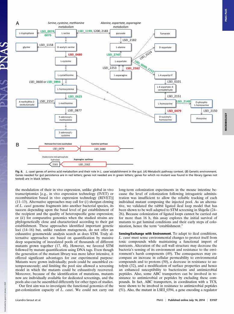
amino acids may be a strong limiting factor in the ileal milieu.The L. casei ATCC334 strain can complete only the last step ofcysteine synthesis; hence it is auxotrophic for this amino acid.Conversely, mutations in genes for tryptophane, alanine, andthreonine synthesis did not affect establishment (Fig. 6 andTable S2). The cysteine synthase activity of the enzyme encodedby LSEI_0480 was demonstrated experimentally (36). BecauseLSEI_0479 and 0480 form an operon (Fig. 6), the phenotypeof the LSEI_0479 mutant could stem from an alteration ofLSEI_0480 transcription alone.As do all the Lactobacillus species, L. casei shows numerous
auxotrophies for amino acids which would be unfavorable incompetitive environments. However, this deficiency is compen-sated by a large set of peptidases (27 genes) and ABC trans-porters predicted to transport amino acids or oligopeptides (37).Among the 15 mutated peptidase/protease genes, LSEI_1810,a dipeptidase predicted to be secreted, is needed for establish-ment. It may have a high affinity for peptides containing limitingamino acids, such as cysteine and asparagine. Also two ABCtransporters predicted to transport peptides, LSEI_0242-0243and LSEI_1738-1739, are needed and could compensate auxo-trophies (Fig. 3).Environmental adaptation. Optimal bacterial adaptation to an en-vironment is strongly associated with the expression of a series ofsensors/regulators (38, 39). The L. casei genome contains 16complete TCS and 124 transcriptional regulators contributing toits ability to sense and adapt to various environments (37).Among the 10 mutated TCS (a mutant of at least one of thetwo genes was available in our library) (Table S2), only theLSEI_0219-0220 TCS, in the same genetic locus as a PBP gene,was needed for gut establishment, reinforcing the importance ofthe cell wall biogenesis in the ileal context.Three transcriptional regulators among the 50 whose genes
were mutagenized appeared to be required for optimal estab-lishment of L. casei (Table S2): LSEI_0247 (see the discussion ofcell wall biogenesis, above), LSEI_0394 (see the discussion ofamino acids metabolism, above), and LSEI_2025, which belongsto the ferric uptake regulator (Fur) family, a family of metal
ion-dependent DNA-binding regulators that can sense metalions (i.e., Fe2+, Zn2+, Ni2+, or Mn2+) or hydrogen peroxide.Otherwise, regularly clustered, interspaced, short palindromic
repeats (CRISPR)-Cas genes were reported to regulate geneexpression during host–bacteria interaction (40). In this screen-ing, the CRISPR-Cas locus (LSEI_0349-0356) is dispensable(two mutants were available). Among the six endonucleases ofthe genome, only one gene, LSEI_1790, was needed, in contrastto the two genes needed in the type I restriction modificationsystem (LSEI_2094-2095, a mutant available for each gene)(Table S2). The L. casei genome encodes 105 transposases and13 recombinases, but only one recombinase (LSEI_1403) wasneeded for gut establishment. This recombinase could supportgenetic adaptation to the gut, as shown in Bacillus fragilis in whichrecombinases modulate its surface in the gut (41). Moreover,a single point mutation in the EnvZ/OmpR two-component sys-tem affects the expression of more than 100 genes in Escherichiacoli (42).
DiscussionHere we report the first, to our knowledge, functional genome-wide study of L. casei establishment in the gut. It was madepossible by a technological breakthrough that allows transposon-based mutagenesis in this species (20). This approach was com-bined with differential tagging of the transposon, allowing theassembly of mutant pools that then could be challenged in vivo,according to the STM technique that allows the identification ofloss-of-function mutants (43, 44). We sequenced the entire set of9,250 mutants obtained in this study and assembled a libraryof 1,110 unique mutants in which the tagged transposon had in-tegrated into a discrete gene. Considering that L. casei is expectedto encode 2,929 genes (45), with a putative set of at least 400essential genes (46, 47), one can consider that more than half ofthe genome was mutagenized. Because transposon insertions couldhave polar effect, one can safely state that nearly all the nonvitalL. casei functions have been affected.Our random mutagenesis method focused on identifying the
genes necessary for bacterial establishment in the gut regardless
tagatose 6-P
D-galactose 6-P
lactose 6-P
CO2
lactose
LSEI_1781, LSEI_A05
LSEI_A04
tagatose 1,6-bisP
LSEI_0679+0680
LSEI_0677 (or LSEI_1348)
Lactose and galactose metabolism
glycerone P D-glycer-aldhehyde 3-P
LSEI_0678, 2597
LSEI_0681Regula on
(-)
D-fructose 6-P
D-fructose 1,6-bisP D-fructose 1-P
D-fructose
LSEI_1348,2655
D-mannose 6-P
LSEI_0292
LSEI_0324-0326, 0401-0405, 0559-0561, 1347,2736-2738, 2740-2741, 2776-2778, 2827-2729
LSEI_1364
D-mannose
LSEI_2045
Fructose and mannose metabolism
LSEI_0328, 0432, 2704
pyruvate
D-glycer-aldhehyde 3-P glycerone P
glycolysis
outside
inside
Lactose transport regulator
Galac. 6-P isomerase
Galactose 6-P isomerase
Tagatose-1,6-bisP aldolase
Tagatose-6-P kinase Hyp. protein
LSEI_0676 LSEI_0677 LSEI_0678 0679 0680 LSEI_0681
A
B
Fig. 5. L. casei genes of lactose and mannose metabolism and their role in L. casei establishment in the gut. (A) Metabolic pathway context. (B) Geneticenvironment. Genes needed for gut persistence are in red letters; genes not needed are in green letters; genes for which no mutant was found in the library(genes not tested) are in black letters.
E3106 | www.pnas.org/cgi/doi/10.1073/pnas.1411883111 Licandro-Seraut et al.
Dow
nloa
ded
by g
uest
on
Janu
ary
2, 2
020
the modulation of their in vivo expression, unlike global in vivotranscriptomics [e.g., in vivo expression technology (IVET) orrecombination based in vivo expression technology (RIVET)](11–13). Alternative approaches may call for (i) shotgun cloningof L. casei genome fragments into another bacterial species, itssuccess depending upon the basal level of gut establishment ofthe recipient and the quality of heterospecific gene expression;or (ii) for comparative genomics when the studied strains arephylogenetically close and characterized according to their gutestablishment. These approaches identified important geneticloci (14–16) but, unlike random mutagenesis, do not offer anexhaustive genomewide analysis search as does STM. Truly al-ternative approaches are based on quantification by massivedeep sequencing of inoculated pools of thousands of differentmutants grown together (17, 48). However, we favored STMfollowed by mutant quantification using DNA tags. Even thoughthe generation of the mutant library was more labor intensive, itoffered significant advantages for our experimental purpose:Mutants were grown individually; pools could be assembled ex-temporaneously; and limiting the pool size allowed a screeningmodel in which the mutants could be exhaustively recovered.Moreover, because of the identification of mutations, mutantsnow are individually available for dedicated screenings, and thepools also can be assembled differently for other types of studies.Our first aim was to investigate the functional genomics of the
gut-colonization capacity of L. casei. We could not carry out
long-term colonization experiments in the mouse intestine be-cause the level of colonization following intragastric adminis-tration was insufficient to allow the reliable tracking of eachindividual mutant composing the injected pool. As an alterna-tive, we validated the rabbit ligated ileal loop model that hasbeen shown to be well adapted to STM screening in Shigella (24–26). Because colonization of ligated loops cannot be carried outfor more than 16 h, this assay explores the initial survival ofmutants to gut luminal conditions and their early steps of colo-nization, hence the term “establishment.”
Sensing/Exchange with Environment. To adapt to ileal conditions,L. casei must sense environmental changes to protect itself fromtoxic compounds while maintaining a functional import ofnutrients. Alteration of the cell wall structure may decrease thebacteria’s sensing of its environment and resistance to the envi-ronment’s harsh components (49). The consequences may en-compass an increase in cellular permeability to environmentalcompounds and to protons (50), a decrease in resistance to au-tolysis (32), and a modification of surface properties and hencean enhanced susceptibility to bacteriocins and antimicrobialpeptides. Also, some ABC transporters can be involved in re-sistance to antimicrobial or peptides by excluding these com-pounds. In fact, ABC transporters, in coordination with a TCS,were shown to be involved in resistance to antimicrobial peptides(51). Also, the mutant in LSEI_0394, a gene encoding a regulator
L-cystathionine
4-methylthio 2-oxobutanoate
S-adenosylL-methioinine
L-cysteine
O-acetylL-serine
L-serine
LSEI_0480
L-homocysteine
xx
L-methionine
LSEI_0625
S-adenosylL-cysteine
LSEI_2157
LSEI_0877
x
L-asparagine
L-aspartate
L-alanine
pyruvate
LSEI_2182
LSEI_2747
LSEI_2253 LSEI_2162
D-aspartate
Serine, cysteine, methioninemetabolism
Alanine, aspartate, asparagine metabolism
L-4-aspartyl-P
L-4-aspartate-4-semialdehyde
LSEI_0101
L-homoserine
LSEI_2151
O-succinylL-homoserine
LSEI_0479
LSEI_0600 or LSEI_0894 x
O-phosphoL-homoserine
LSEI_2149
threonine
LSEI_2150
LSEI_1199, 1200, 2183L-tryptophane
LSEI_0074, 0075
glycineLSEI_1158
fumarate
Homoserine trans-succinylase Cysteine synthase
Asparagine synthaseDiadenosine tetraphosphate
hydrolase
2161
LSEI_0480LSEI_0479
LSEI_2162
A
B
Fig. 6. L. casei genes of amino acid metabolism and their role in L. casei establishment in the gut. (A) Metabolic pathway context. (B) Genetic environment.Genes needed for gut persistence are in red letters; genes not needed are in green letters; genes for which no mutant was found in the library (genes nottested) are in black letters.
Licandro-Seraut et al. PNAS | Published online July 14, 2014 | E3107
MICRO
BIOLO
GY
PNASPL
US
Dow
nloa
ded
by g
uest
on
Janu
ary
2, 2
020
of the AcrR family that is located in an operon encoding an ABCtransporter (LSEI_0395-0396) is impaired in its capacity of es-tablishment, whereas the disruption of this transporter had noimpact on gut establishment. It is likely that LSEI_0394 encodesa repressor of this transporter, as is the case for AcrR in E. colion the multidrug efflux pump acrAB (52). Therefore, derepres-sion of the transporter becomes a handicap for the bacterium inthe gut lumen. The control of exchanges with the ileal environ-ment is fundamental for L. casei establishment to capture a max-imum of nutrients without unbalancing the bacterial cell contentor permitting the entrance of toxic molecules.
Sugar Metabolism. Previous transcriptomic studies carried out inLactobacillus plantarum and L. johnsonii while colonizing the gutshowed an up-regulation of a large set of genes related to car-bohydrate transport and metabolism, indicating a global re-cruitment of sugar-utilization enzymes for energy supply anda change in carbohydrate-utilization pathways to adapt to sugarlimitation (15, 53, 54). In L. plantarum, energy production frommaltose, melibiose, and lactose is activated, as is the import ofmannose and cellobiose in the cecum of monocolonized mice(54). Our results are fully consistent with these data. Also, inB. thetaiotaomicron, the transcription of numerous genes implicatedin polysaccharide degradation is activated in the gut to degradeglycans that the competitive flora (Bacteroides longum orL. casei) cannot to metabolize (55). Symbionts thus adapt theirprofile of substrate utilization to the availability of these sub-strates in the ileal milieu and in response to the presence of othersymbionts, suggesting the need to define the composition of theresident microbiota further in future studies.
Metabolism and Link with Stress/Oxidation. While initiating colo-nization, pathogens encounter potentially bactericidal compo-nents of the intestinal fluid (i.e., bile, lysozyme, trypsin) and alsoa strong host response, especially highly reactive oxidative stressmolecules, and must respond accordingly to survive. However, itis not clear how much oxidative stress is imposed onto symbiontsas they establish in the gut. This screening identified only onegeneral stress protein and no gene encoding factors of the spe-cific response to oxidative stress.However, cysteine synthase is involved in establishment. Con-
cerning sulfured amino acids, competition for nutrients andduring the establishment of a commensal was described for B.thetaiotaomicron, particularly the need for vitamin B12 (17), anessential cofactor for methionine biosynthesis for most bacterialspecies, although not for L. casei, according to our genomeanalysis. Cysteine also constitutes a pool of sulfured moleculesimplicated in redox regulation of the host intestine and of bac-terial cells. The main tandem compounds that permit redoxhomeostasis are cysteine/cystine, glutathione/glutathione disul-fide, and thioredoxin/thioredoxin disulfide (56). Glutathione issynthesized from cysteine in epithelial cells and in some bacteria.L. casei is able to use it in complement to thioredoxin to controlits redox balance but is unable to synthesize it (57). BecauseL. casei also is auxotrophic for cysteine, it is strongly dependent onthe ileal content. Moreover, cysteine is known as the most lim-iting nonessential amino acid in human cells (58). Thus, L. caseimust compete with epithelial cells and the endogenous flora forcysteine and glutathione. Because mutants in genes involved inthe redox balance, particularly thioreductases, were not affected,we hypothesize that during establishment L. casei needs cysteinefor nutrition rather than for the maintenance of its redox bal-ance. This notion is consistent with the evidence for nutritionalcompetition, particularly for cysteine, observed between in-tracellular pathogens and their hosts (59). Although it ofteninvolves essential amino acids, nutritional competition also caninvolve other amino acids, e.g., asparagine, which appears tobe decisive for the virulence of Lactococcus garvieae (60) and
Francisella tularensis (61). Thus, asparagine could be anothernutritional requirement for which L. casei must compete withits host.Iron limitation is a major signal in the virulence of mucosal
pathogens. In most pathogens Fur proteins act as central regu-lators for successful colonization and virulence. They controlgenes involved in iron homeostasis and protection against re-active oxygen species damage (62) in Shigella (63), Salmonella(64), Vibrio cholerae (65), Pseudomonas (66), Listeria (67), andHelicobacter pylori (68). Therefore it is likely that L. casei isstrongly challenged by iron-limiting conditions in the gut, partic-ularly given the high levels of lactoferrin in intestinal secretions.It also is possible that the protein regulating Fe2+/Zn2+ uptake(LSEI_2025) allows scavenging of other essential metals andprovides L. casei protection against reactive oxygen species, al-though the latter benefit is unlikely because L. casei does notappear to activate oxidative stress defenses in the ileal context.Identification of copA (LSEI_1743), an ATPase predicted to beresponsible for translocating copper, silver, and cadmium ionsacross biological membranes, emphasizes the importance ofmetal import. Accordingly, in Enterococcus hirae, copA supportsbacterial survival in extremely low-copper environments (69). InL. plantarum, copA expression was highly up-regulated in theconventional mouse gut, and a copA mutant showed decreasedcolonization capacity (70, 71).In conclusion, we identified 47 mutations affecting establish-
ment that we organized into five functional groups: basic physio-logical processes (housekeeping), metabolism, cell wall biogenesis,environmental adaptation, and a remaining group of genes ofunknown function. In summary, most genes linked to bacterialestablishment are conserved among Firmicutes. In consequence,our library provides major information regarding the coloni-zation potential of other Firmicutes. Regarding L. casei, wewill better characterize the major pathways controlling bacte-rial establishment. We now are in a position to study the im-pact of controlled microbiota on the establishment of thismodel symbiont. Our annotated library also is available to studyother phenotypes, particularly the identification of L. caseieffectors involved in immune and metabolic functions in theircolonized host.
Materials and MethodsDesign of the L. casei Random Mutant Library. For STM, 70 DNA tags, pre-viously used for Salmonella typhimurium STM (21) were individually clonedinto the EcoRI site of pVI110 to generate 70 differently tagged transposablevectors (Fig. S2 and Tables S3 and S4). The tagged mutant library in L. caseiwas obtained using the Pjunc-TpaseIS1223 system as recently described (20)and was ordered in pools of 70 mutants. After the transposon insertion siteswere identified by by individual sequencing, mutants were reassembled toconstitute a library of 1,110 gene mutants.
Screening for Bacterial Establishment. Each pool of mutants was challenged inrabbit ileal loops as previously described (25, 26) with the following mod-ifications. In each loop, 0.5 mL of bacterial suspension was injected (5 × 10⁷
cfu per loop). Challenges were carried out over 16 h. The whole intestinalloop was homogenized, diluted, and spread on agar plates to obtain iso-lated colonies and to proceed to DNA isolation. Quantitative PCR was usedto measure the proportion of each tag corresponding to each mutant ininjected and recovered pools. All mutants displaying at least a threefolddecrease in quantity between injection and recovery were selected.
Detailed experimental procedures are described in SI Text.
ACKNOWLEDGMENTS. We thank Ellen Arena for careful reading of andpertinent suggestions regarding the manuscript, Cyril Iaconelli for advice indata processing, Christoph Tang for providing DNA tags, and the PasteurInstitute Micro-organism Collection for the L. casei type strain (CIP 107868,ATCC 334). This work and a postdoctoral fellowship (to H.L.-S.) were supportedby an Advanced Grant of the European Research Council HOMEOEPITH,Grant Agreement ERC-232798 (to P.J.S.). P.J.S. is a Howard Hughes MedicalInstitute Senior Scholar. H.S. was supported by a doctoral fellowship fromthe Ministère de l’Enseignement Supérieur et de la Recherche.
E3108 | www.pnas.org/cgi/doi/10.1073/pnas.1411883111 Licandro-Seraut et al.
Dow
nloa
ded
by g
uest
on
Janu
ary
2, 2
020
1. Savage DC (2001) Microbial biota of the human intestine: A tribute to some pio-neering scientists. Curr Issues Intest Microbiol 2(1):1–15.
2. O’Hara AM, Shanahan F (2006) The gut flora as a forgotten organ. EMBO Rep 7(7):688–693.3. Brandtzaeg P (2013) Gate-keeper function of the intestinal epithelium. Benef Mi-
crobes 4(1):67–82.4. Bocci V (1992) The neglected organ: Bacterial flora has a crucial immunostimulatory
role. Perspect Biol Med 35(2):251–260.5. Tremaroli V, Bäckhed F (2012) Functional interactions between the gut microbiota
and host metabolism. Nature 489(7415):242–249.6. Eberl G, Boneca IG (2010) Bacteria and MAMP-induced morphogenesis of the immune
system. Curr Opin Immunol 22(4):448–454.7. Qin J, et al.; MetaHIT Consortium (2010) A human gut microbial gene catalogue es-
tablished by metagenomic sequencing. Nature 464(7285):59–65.8. Packey CD, Sartor RB (2009) Commensal bacteria, traditional and opportunistic
pathogens, dysbiosis and bacterial killing in inflammatory bowel diseases. Curr OpinInfect Dis 22(3):292–301.
9. Arumugam M, et al.; MetaHIT Consortium (2011) Enterotypes of the human gut mi-crobiome. Nature 473(7346):174–180.
10. Chow J, Lee SM, Shen Y, Khosravi A, Mazmanian SK (2010) Host-bacterial symbiosis inhealth and disease. Adv Immunol 107:243–274.
11. O’Connell Motherway M, et al. (2011) Functional genome analysis of Bifidobacteriumbreve UCC2003 reveals type IVb tight adherence (Tad) pili as an essential and con-served host-colonization factor. Proc Natl Acad Sci USA 108(27):11217–11222.
12. Bron PA, Grangette C, Mercenier A, de Vos WM, KleerebezemM (2004) Identificationof Lactobacillus plantarum genes that are induced in the gastrointestinal tract ofmice. J Bacteriol 186(17):5721–5729.
13. Walter J, et al. (2003) Identification of Lactobacillus reuteri genes specifically inducedin the mouse gastrointestinal tract. Appl Environ Microbiol 69(4):2044–2051.
14. Kankainen M, et al. (2009) Comparative genomic analysis of Lactobacillus rhamnosusGG reveals pili containing a human- mucus binding protein. Proc Natl Acad Sci USA106(40):17193–17198.
15. Denou E, et al. (2008) Identification of genes associated with the long-gut-persistencephenotype of the probiotic Lactobacillus johnsonii strain NCC533 using a combinationof genomics and transcriptome analysis. J Bacteriol 190(9):3161–3168.
16. Lee SM, et al. (2013) Bacterial colonization factors control specificity and stability ofthe gut microbiota. Nature 501(7467):426–429.
17. Goodman AL, et al. (2009) Identifying genetic determinants needed to establisha human gut symbiont in its habitat. Cell Host Microbe 6(3):279–289.
18. Karlsson CL, Molin G, Cilio CM, Ahrné S (2011) The pioneer gut microbiota in humanneonates vaginally born at term-a pilot study. Pediatr Res 70(3):282–286.
19. Fang F, O’Toole PW (2009) Genetic tools for investigating the biology of commensallactobacilli. Front Biosci (Landmark Ed) 14:3111–3127.
20. Licandro-Seraut H, et al. (2012) Development of an efficient in vivo system (Pjunc-TpaseIS1223) for random transposon mutagenesis of Lactobacillus casei. Appl EnvironMicrobiol 78(15):5417–5423.
21. Hensel M, et al. (1995) Simultaneous identification of bacterial virulence genes bynegative selection. Science 269(5222):400–403.
22. Commichau FM, Pietack N, Stülke J (2013) Essential genes in Bacillus subtilis: A re-evaluation after ten years. Mol Biosyst 9(6):1068–1075.
23. Caspi R, et al. (2010) The MetaCyc database of metabolic pathways and enzymes andthe BioCyc collection of pathway/genome databases. Nucleic Acids Res 38(Databaseissue):D473–D479.
24. Sansonetti PJ, et al. (1983) Alterations in the pathogenicity of Escherichia coli K-12after transfer of plasmid and chromosomal genes from Shigella flexneri. Infect Im-mun 39(3):1392–1402.
25. Marteyn B, et al. (2010) Modulation of Shigella virulence in response to availableoxygen in vivo. Nature 465(7296):355–358.
26. West NP, et al. (2005) Optimization of virulence functions through glucosylation ofShigella LPS. Science 307(5713):1313–1317.
27. Oozeer R, Mater DD, Goupil-Feuillerat N, Corthier G (2004) Initiation of proteinsynthesis by a labeled derivative of the Lactobacillus casei DN-114 001 strain duringtransit from the stomach to the cecum in mice harboring human microbiota. ApplEnviron Microbiol 70(12):6992–6997.
28. McRobbie AM, et al. (2012) Staphylococcus aureus DinG, a helicase that has evolvedinto a nuclease. Biochem J 442(1):77–84.
29. Alcántara C, Zúñiga M (2012) Proteomic and transcriptomic analysis of the responseto bile stress of Lactobacillus casei BL23. Microbiology 158(Pt 5):1206–1218.
30. Kleerebezem M, et al. (2010) The extracellular biology of the lactobacilli. FEMS Mi-crobiol Rev 34(2):199–230.
31. Kawai Y, et al. (2011) A widespread family of bacterial cell wall assembly proteins.EMBO J 30(24):4931–4941.
32. Walter J, et al. (2007) D-alanyl ester depletion of teichoic acids in Lactobacillus reuteri100-23 results in impaired colonization of the mouse gastrointestinal tract. EnvironMicrobiol 9(7):1750–1760.
33. Rico J, Yebra MJ, Pérez-Martínez G, Deutscher J, Monedero V (2008) Analysis of ldhgenes in Lactobacillus casei BL23: Role on lactic acid production. J Ind Microbiol Bio-technol 35(6):579–586.
34. Viana R, Yebra MJ, Galán JL, Monedero V, Pérez-Martínez G (2005) Pleiotropic effects oflactate dehydrogenase inactivation in Lactobacillus casei. Res Microbiol 156(5-6):641–649.
35. Viana R, et al. (2000) Enzyme I and HPr from Lactobacillus casei: Their role in sugartransport, carbon catabolite repression and inducer exclusion.Mol Microbiol 36(3):570–584.
36. Bogicevic B, Berthoud H, Portmann R, Meile L, Irmler S (2012) CysK from Lactobacilluscasei encodes a protein with O-acetylserine sulfhydrylase and cysteine desulfurizationactivity. Appl Microbiol Biotechnol 94(5):1209–1220.
37. Cai H, Thompson R, Budinich MF, Broadbent JR, Steele JL (2009) Genome sequenceand comparative genome analysis of Lactobacillus casei: Insights into their niche-associated evolution. Genome Biol Evol 1:239–257.
38. Sturme MH, Francke C, Siezen RJ, de Vos WM, KleerebezemM (2007) Making sense ofquorum sensing in lactobacilli: A special focus on Lactobacillus plantarum WCFS1.Microbiology 153(Pt 12):3939–3947.
39. Krell T, et al. (2010) Bacterial sensor kinases: Diversity in the recognition of environ-mental signals. Annu Rev Microbiol 64:539–559.
40. Sampson TR, Saroj SD, Llewellyn AC, Tzeng YL, Weiss DS (2013) A CRISPR/Cas systemmediates bacterial innate immune evasion and virulence. Nature 497(7448):254–257.
41. Coyne MJ, Weinacht KG, Krinos CM, Comstock LE (2003) Mpi recombinase globallymodulates the surface architecture of a human commensal bacterium. Proc Natl AcadSci USA 100(18):10446–10451.
42. Giraud A, et al. (2008) Dissecting the genetic components of adaptation of Escherichiacoli to the mouse gut. PLoS Genet 4(1):e2.
43. Saenz HL, Dehio C (2005) Signature-taggedmutagenesis: Technical advances in a negativeselection method for virulence gene identification. Curr Opin Microbiol 8(5):612–619.
44. West NP, Sansonetti PJ, Frankel G, Tang CM (2003) Finding your niche: What has beenlearnt from STM studies on GI colonization. Trends Microbiol 11(7):338–344.
45. Makarova K, et al. (2006) Comparative genomics of the lactic acid bacteria. Proc NatlAcad Sci USA 103(42):15611–15616.
46. Acevedo-Rocha CG, Fang G, Schmidt M, Ussery DW, Danchin A (2013) From essentialto persistent genes: A functional approach to constructing synthetic life. TrendsGenet 29(5):273–279.
47. Juhas M, Eberl L, Glass JI (2011) Essence of life: Essential genes of minimal genomes.Trends Cell Biol 21(10):562–568.
48. Langridge GC, et al. (2009) Simultaneous assay of every Salmonella Typhi gene usingone million transposon mutants. Genome Res 19(12):2308–2316.
49. Jordan S, Hutchings MI, Mascher T (2008) Cell envelope stress response in Gram-positive bacteria. FEMS Microbiol Rev 32(1):107–146.
50. Boyd DA, et al. (2000) Defects in D-alanyl-lipoteichoic acid synthesis in Streptococcusmutans results in acid sensitivity. J Bacteriol 182(21):6055–6065.
51. Revilla-Guarinos A, et al. (2013) Characterization of a regulatory network of peptideantibiotic detoxification modules in Lactobacillus casei BL23. Appl Environ Microbiol79(10):3160–3170.
52. Routh MD, Su CC, Zhang Q, Yu EW (2009) Structures of AcrR and CmeR: Insight intothe mechanisms of transcriptional repression and multi-drug recognition in the TetRfamily of regulators. Biochim Biophys Acta 1794(5):844–851.
53. Denou E, et al. (2007) Gene expression of commensal Lactobacillus johnsonii strainNCC533 during in vitro growth and in the murine gut. J Bacteriol 189(22):8109–8119.
54. Marco ML, et al. (2009) Lifestyle of Lactobacillus plantarum in the mouse caecum.Environ Microbiol 11(10):2747–2757.
55. Sonnenburg JL, Chen CT, Gordon JI (2006) Genomic and metabolic studies of theimpact of probiotics on a model gut symbiont and host. PLoS Biol 4(12):e413.
56. Circu ML, Aw TY (2012) Intestinal redox biology and oxidative stress. Semin Cell DevBiol 23(7):729–737.
57. SerataM, Iino T, Yasuda E, Sako T (2012) Roles of thioredoxin and thioredoxin reductase inthe resistance to oxidative stress in Lactobacillus casei. Microbiology 158(Pt 4):953–962.
58. Young VR (1994) Adult amino acid requirements: The case for a major revision incurrent recommendations. J Nutr 124(8, Suppl):1517S–1523S.
59. Abu Kwaik Y, Bumann D (2013) Microbial quest for food in vivo: ‘Nutritional viru-lence’ as an emerging paradigm. Cell Microbiol 15(6):882–890.
60. Menéndez A, Fernández L, Reimundo P, Guijarro JA (2007) Genes required for Lac-tococcus garvieae survival in a fish host. Microbiology 153(Pt 10):3286–3294.
61. Gesbert G, et al. (2014) Asparagine assimilation is critical for intracellular replicationand dissemination of Francisella. Cell Microbiol 16(3):434–449.
62. Troxell B, Hassan HM (2013) Transcriptional regulation by Ferric Uptake Regulator(Fur) in pathogenic bacteria. Front Cell Infect Microbiol 3:59.
63. Africa LA, Murphy ER, Egan NR, Wigley AF, Wing HJ (2011) The iron-responsiveFur/RyhB regulatory cascade modulates the Shigella outer membrane protease IcsP.Infect Immun 79(11):4543–4549.
64. Leclerc JM, Dozois CM, Daigle F (2013) Role of the Salmonella enterica serovar TyphiFur regulator and small RNAs RfrA and RfrB in iron homeostasis and interaction withhost cells. Microbiology 159(Pt 3):591–602.
65. Mey AR, Wyckoff EE, Kanukurthy V, Fisher CR, Payne SM (2005) Iron and fur regu-lation in Vibrio cholerae and the role of fur in virulence. Infect Immun 73(12):8167–8178.
66. Cornelis P, Matthijs S, Van Oeffelen L (2009) Iron uptake regulation in Pseudomonasaeruginosa. Biometals 22(1):15–22.
67. McLaughlin HP, et al. (2012) A putative P-type ATPase required for virulence andresistance to haem toxicity in Listeria monocytogenes. PLoS ONE 7(2):e30928.
68. Pich OQ, Merrell DS (2013) The ferric uptake regulator of Helicobacter pylori: A criticalplayer in the battle for iron and colonization of the stomach. FutureMicrobiol 8(6):725–738.
69. Solioz M, Stoyanov JV (2003) Copper homeostasis in Enterococcus hirae. FEMS Mi-crobiol Rev 27(2-3):183–195.
70. Marco ML, Bongers RS, de Vos WM, Kleerebezem M (2007) Spatial and temporalexpression of Lactobacillus plantarum genes in the gastrointestinal tracts of mice.Appl Environ Microbiol 73(1):124–132.
71. Bron PA, Meijer M, Bongers RS, de Vos WM, Kleerebezem M (2007) Dynamics ofcompetitive population abundance of Lactobacillus plantarum ivi gene mutants infaecal samples after passage through the gastrointestinal tract of mice. J Appl Mi-crobiol 103(5):1424–1434.
72. Kanehisa M, Goto S, Sato Y, Furumichi M, Tanabe M (2012) KEGG for integration andinterpretation of large-scale molecular data sets. Nucleic Acids Res 40(Database issue):D109–D114.
Licandro-Seraut et al. PNAS | Published online July 14, 2014 | E3109
MICRO
BIOLO
GY
PNASPL
US
Dow
nloa
ded
by g
uest
on
Janu
ary
2, 2
020