Embed Size (px)
Citation preview
Adjuvant effects of Lactobacillus casei added to a renutrition dietin a malnourished mouse model
PAOLA GAUFFIN CANO1, GRACIELA AGÜERO3 AND GABRIELA PERDIGON1,2
1. Centro de Referencias para Lactobacilos (CERELA). Chacabuco 145 (4000). Tucumán. Argentina.2. Cátedra de Inmunología, Instituto de Microbiología. Facultad de Bioquímica, Química y Farmacia. Universidad Nacional
de Tucumán.3. Cátedra de Análisis Clínico, Instituto de Bioquímica Aplicada. Facultad de Bioquímica, Química y Farmacia. Universidad
Nacional de Tucumán.
Key words: malnutrition, Lactobacillus casei, mucosal immunity, gut barrier, renutrition diet.
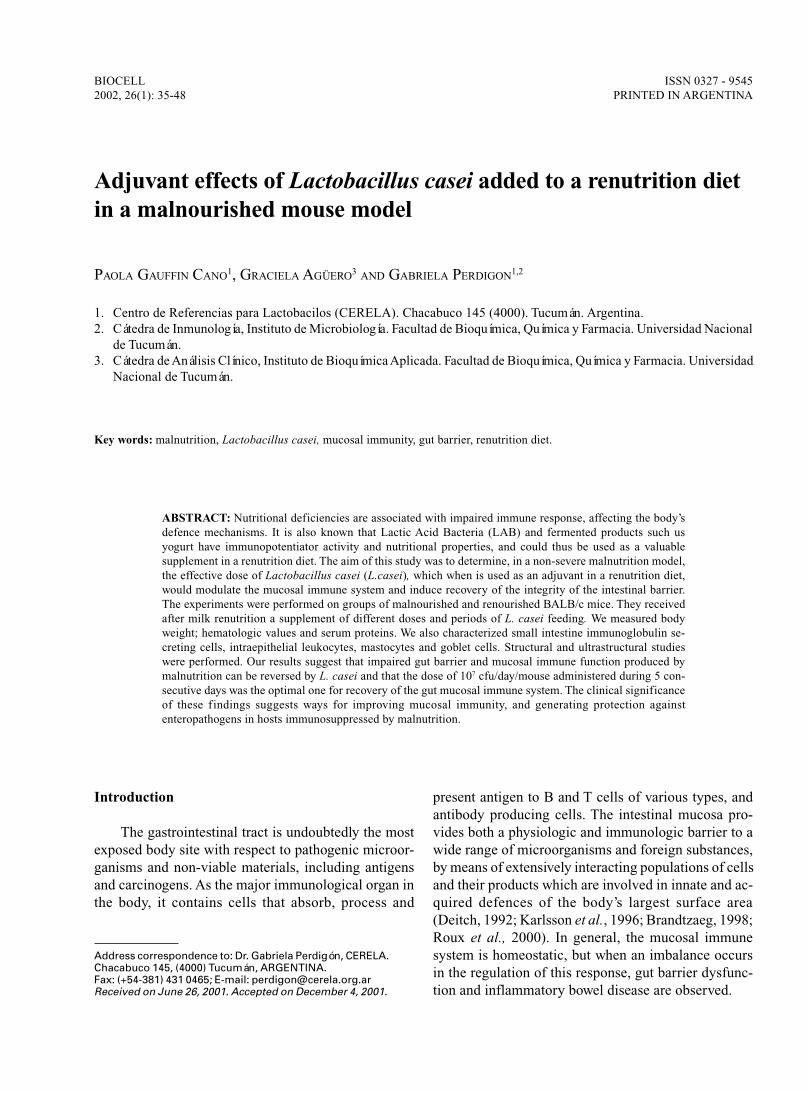
ABSTRACT: Nutritional deficiencies are associated with impaired immune response, affecting the body’sdefence mechanisms. It is also known that Lactic Acid Bacteria (LAB) and fermented products such usyogurt have immunopotentiator activity and nutritional properties, and could thus be used as a valuablesupplement in a renutrition diet. The aim of this study was to determine, in a non-severe malnutrition model,the effective dose of Lactobacillus casei (L.casei), which when is used as an adjuvant in a renutrition diet,would modulate the mucosal immune system and induce recovery of the integrity of the intestinal barrier.The experiments were performed on groups of malnourished and renourished BALB/c mice. They receivedafter milk renutrition a supplement of different doses and periods of L. casei feeding. We measured bodyweight; hematologic values and serum proteins. We also characterized small intestine immunoglobulin se-creting cells, intraepithelial leukocytes, mastocytes and goblet cells. Structural and ultrastructural studieswere performed. Our results suggest that impaired gut barrier and mucosal immune function produced bymalnutrition can be reversed by L. casei and that the dose of 107 cfu/day/mouse administered during 5 con-secutive days was the optimal one for recovery of the gut mucosal immune system. The clinical significanceof these findings suggests ways for improving mucosal immunity, and generating protection againstenteropathogens in hosts immunosuppressed by malnutrition.
BIOCELL2002, 26(1): 35-48
ISSN 0327 - 9545PRINTED IN ARGENTINA
Introduction
The gastrointestinal tract is undoubtedly the mostexposed body site with respect to pathogenic microor-ganisms and non-viable materials, including antigensand carcinogens. As the major immunological organ inthe body, it contains cells that absorb, process and
present antigen to B and T cells of various types, andantibody producing cells. The intestinal mucosa pro-vides both a physiologic and immunologic barrier to awide range of microorganisms and foreign substances,by means of extensively interacting populations of cellsand their products which are involved in innate and ac-quired defences of the body’s largest surface area(Deitch, 1992; Karlsson et al., 1996; Brandtzaeg, 1998;Roux et al., 2000). In general, the mucosal immunesystem is homeostatic, but when an imbalance occursin the regulation of this response, gut barrier dysfunc-tion and inflammatory bowel disease are observed.
Address correspondence to: Dr. Gabriela Perdigón, CERELA.Chacabuco 145, (4000) Tucumán, ARGENTINA.Fax: (+54-381) 431 0465; E-mail: [email protected] on June 26, 2001. Accepted on December 4, 2001.
PAOLA GAUFFIN CANO et al.36
The relationship between nutrition, developmentand immunity has been the focus of increasing researchin the last two decades. Thus, the function of many cellsin the immune system relies on metabolic pathways thatin turn depend upon various nutrients as critical cofac-tors, which in turn affects body defense mechanisms(Mac Dermott, 1993).
It is now established that nutritional deficiency iscommonly associated with an impaired immune re-sponse, particularly cell-mediated immunity, phagocytefunction, cytokine production, secretory antibody re-sponse, antibody affinity, and the complement system(Chandra and Wadhwa, 1993; Chandra, 1997). Non-specific mechanisms that include intestinal flora, ana-tomical barriers (mucosa and epithelium), secretorysubstances such as lysozymes and mucus are affectedby malnutrition (Sherman et al., 1985). In fact, malnu-trition is the commonest cause of immunodeficiency inundeveloped countries. It is suggested that impairedsystemic and mucosal immunity contribute to the in-creased frequency and severity of intestinal infectionseen in undernourished individuals.
In protein-energy malnutrition (PEM), most of thehost defence mechanisms are breached depending onthe severity of protein deficiency relative to energy, al-lowing microbes to invade and produce clinical infec-tion, which is more severe and prolonged (Chandra,1996; Scrimshaw and Sangiovanni, 1997).
That the detrimental effects of severe malnutritionon the mucosal immune system can be reversed withadequate renutrition is of further clinical importance.Similarity, refeeding rapidly restores the morphology andfunction of intestine resulting in repair of gut atrophyand normalization of intestinal permeability (Castillo etal., 1991; Poullain et al., 1991; Boza et al., 1999). Milkand milk products represent important sources of dietaryproteins for humans, and these proteins are known to serveas a source of biologically active peptides. It was welldemonstrated that the presence of a number of growthfactors and hormones in the milk of various species in-cluding human and bovine, have beneficial effects on thehost and so it suggests its potential use in the renutritionprocess (Koldovsky, 1989; Meisel and Bockelmann, 1999;Matar et al., 2000).
The gut microflora is an important constituent ofthe intestinal mucosa barrier and this has led to the con-cept of probiotic therapy, i.e. the application of poten-tially beneficial microorganisms (Fuller, 1992). LABwith probiotic activity were used in various fermentedfoods since antiquity. Probiotic is a microbial dietaryadjuvant that beneficially affects the host physiology
by modulating mucosal and systemic immunity, as wellas improving nutritional and microbial balance in theintestine tract. The probiotic spectrum of activity canbe divided into nutritional, physiological, and antimi-crobial effects. LAB are also potential adjuvants, andtheir oral administration triggers mucosal and systemicimmune responses. Various effects ascribed to LAB aresummarized as follows: improvement of the nutritionalquality of food and feed; stimulation of vitamin syn-thesis and enzyme production; stabilization of gut mi-croflora and competitive exclusion of enteric pathogens;enhancement of innate host defences by production ofantimicrobial substances; reduction of serum choles-terol by assimilation mechanisms; decreased risk ofcolon cancer by detoxification of carcinogens and tu-mor suppression by modulation of cell-mediated im-munity (Gerritse et al., 1990; Perdigón et al., 1990;1994; Naidu et al., 1999). Probiotics can be used asinnovative tools for treating dysfunctions of the gutmucosal barrier including acute gastroenteritis, foodallergy, and inflammatory bowel disease.
In our laboratory it was demonstrated that L. caseiwas able to induce a secretory immune response, whichwas related to the dose administered to healthy mice(Perdigón et al., 1991). However when we used an ex-perimental model of severe malnutrition, we showed thatyogurt and L. casei were not able to increase the pro-tective intestinal mechanisms to the same levels foundin the well-nourished host (Perdigón et al., 1995; Agüeroet al., 1996; Perdigón and Oliver, 2000). Therefore, inorder to avoid harmful effect at intestinal level by anoverstimulation of the immune system we recommendedthe use of LAB in the malnourished mice after mucosalrecovery by adequate refeeding.
The aim of this study was to determine, in a non-severe malnutrition experimental model, the effectivedose of L. casei used as adjuvant in a renutrition dietrequired to modulate the mucosal immune system andto repair the integrity of intestinal barrier.
Material and Methods
Diets
The protein-free diet was supplemented in order tofulfill nutritional requirements. It was composed anddesigned as follows: vitamin mixture (2.2%) (ICN,Biomedicals, Inc-1263 South Chillicothe Road, Aurora,Ohio 44202), salt mixture (4%) (ICN, Biomedicals, Inc-1263 South Chillicothe Road, Aurora, Ohio 44202), andcorn oil (5%).
Lactobacillus casei AS ADJUVANT IN MALNUTRITION 37
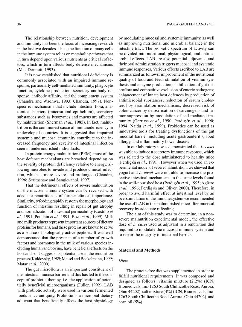
TABLE 1.
Effect of different doses and periods of Lactobacillus casei feeding onbody weight of malnourished and renourished mice.
Body Weight (g)1 ± SD
Days of feeding
2 5 7
Test groups M-N Re7d M-N Re7d M-N Re7d
Lactobacillus casei 106 10.1± 1.3 10.7± 0.9 12.1± 0.9 12.3± 1.5 12.1± 1.1 16.6± 1.7
(cfu/day/mouse) 107 10.4± 13 13.0± 1.5 11.4± 1.0 12.8± 0.7 13.3± 0.7 20.0± 1.62*
108 9.8 ± 1.1 11.9± 02 9.7± 0.9 19.0± 1.62* 12.0± 1.0 21.0± 1.62**
Values for well-nourished groups= 25.8±1.5 g; M-N control groups= 10.4±1.23** g and Re7d control groups= 12.1± 1.53** g
M-N: malnourished; Re7d: renourished.1Values are means ± SD of 10 mice per group. 2Significantly different from Re7d control. 3Significantly different of well-nourished control. *P<0.05. **P<0.01 (ANOVA and Student’s Test)
Animals
Weaning BALB/c mice obtained from closedcolony of the breeding unit kept at CERELA werehoused in cages at 23°C with a 12 h light/dark periods.They had free access to protein-free diet supplementedwith vitamins, minerals and essentials fat free acid for21 days. At the end of this period, the animals that hadlost about 35-55% of their weight compared with thewell-nourished controls were selected for the experi-ments. The mice were split into two groups. One groupwas the malnourished (M-N) and the other was refeedingwith 10%-non-fat-milk as the source of protein (NFM)for 7 days; this was the renourished group (Re7d). Com-parative assays between M-N and Re7d animals wereaffected under identical conditions.
Experiments were performed with 10 animals forweight body determinations and 5 animals for the otherassays.
Microorganism feeding procedure
The bacterial strain used for experiments was Lac-tobacillus casei var casei CRL 431 from CERELA cul-ture collection. It was cultured for 8 h at 37°C in MRS(De Man et al., 1960) broth (Oxoid Ltd., U.S.A.) andharvested by centrifugation at 5,000 rpm for 10 min,and washed three times with sterile saline solution. Micewere daily fed with culture of LAB at a concentrationof 106, 107 and 108 colony forming units (cfu)/day/ani-
mal for 2, 5 or 7 consecutive days. The cultures wereadministered at 20% v/v in the drinking water to M-Ngroups and in NFM 10% to Re7d group.
We used three control groups: well nourished whichreceived conventional diet (W-N), M-N (21 days pro-tein-free diet) and Re7d controls (mal-nourished micerenourished for 7 days with 10% NFM).
Body weight determinations
It was determined at the end of each treatment pe-riod in groups of 10 mice and expressed as grams
Hematological and serum total protein determinations
They were performed in all experimental and con-trol groups. Peripheral blood was recovered by cardiacpuncture. The hematocrit (HTO) and number of leuko-cytes were determined by the hematocytometric method.PMN and lymphocytes populations were differentiatedon smears stained with Giemsa solution. The total pro-tein concentration was determined using Bradford tech-nique (1976).
Tissue sections
The small intestine was removed at the end of eachtreatment period and processed by modified Saint-Maries’s technique (1962). Briefly, tissues were fixedin 95% ethanol for 24 hours at 4°C, and then were de-
PAOLA GAUFFIN CANO et al.38
hydrated in 3 changes of absolute alcohol and clearedby passing through 3 consecutive baths of xylene at 4°Cfor 45 min each one. The tissue was embedded in paraf-fin at 56°C for 3 to 6 h. Sectioning was carried out asusual, and tissue sections (3-4 µm) were placed on glassslides.
Number of IgA, IgG and IgM secreting cell determina-tions
The number of IgA, IgG and IgM secreting B cellswere determined on histological slices from small intes-
tine by direct immunofluorescence technique. It was per-formed using the respective monospecifc antibodies (α,γ and µ-chain specific) conjugated with fluoresceinisothiocyanate (FITC) (Sigma, Saint Louis, Missouri63103, USA). Histological samples were incubated with0.1 ml of different antibodies at 1/60 dilutions for IgA,and 1/100 for both IgG and IgM for 30 min at 37°C inhumidified chamber. Then they were washed three timeswith 0.01M-phosphaste-buffered saline (PBS), pH 7.2.The slices were mounted in glycerol:PBS. The numberof positive fluorescent cells was expressed as number ofsecreting cells per 10 fields (magnification 100X).
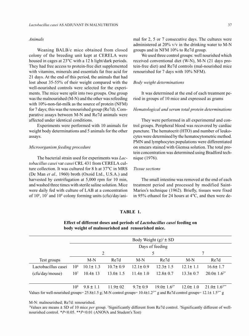
TABLE 2.
Effect of different doses and periods of Lactobacillus casei feeding onthe hematological values of malnourished and renourished mice.
Test and Hematological parameters1
control groups Hematocrit (%) White cells/µl PMN (%) Lymphocytes (%)Days of
L. casei M-N Re7d M-N Re7d M-N Re7d M-N Re7dfeeding
106 2 41± 0.9 45± 1.3 3500± 1062 3300± 100 19± 0.52 29± 0.83 81± 1.32 71± 1.53
(cfu/day/mouse) 5 43± 1.1 39± 0.5 3800± 912 3500± 130 34± 1.82 40± 1.03 66± 1.92 60± 1.53
7 38± 0.8 40± 0.8 3900± 1002 3900± 893 19± 1.02 44± 1.53 81± 1.32 56± 1.63
107 2 40± 0.9 43± 1.1 3900± 802 3600± 903 30± 22 20± 0.5 70± 1.72 80± 1.0 (cfu/day/mouse) 5 40± 1.2 39± 1.5 3390± 107 3400± 100 24± 0.92 18± 0.9 76± 1.52 82± 0.9
7 37± 0.9 44± 0.9 3100± 802 3800± 1203 19± 2.02 30± 1.23 81± 1.12 70± 1.03
108 2 43± 1.0 42± 1.0 3550± 1032 3150± 110 23± 1.52 30± 1.23 77± 22 70± 0.93
(cfu/day/mouse) 5 40± 0.9 43± 1.7 3780± 902 3600± 1003 40± 1.72 35± 0.63 60± 0.52 65± 1.23
7 39± 1.1 45± 1.0 3850± 100 4000± 1003 20± 1.92 35± 1.03 80± 1.12 65± 1.13
M-N and Re7d controls 42±1.04 39±0.94 2800±1064 3200±1064 11±0.9 20±1.5 89±1.1 80±1.4
W-N control 55±10 4800±100 15±1 82±0.9
M-N: malnourished. Re7d: renourished. W-N: well-nourished.1Values are means of n = 5 per group ± SD. 2Significantly different from M-N control (P<0.01). 3Significantly different fromRe7d control (P<0.01). 4Significantly different from W-N control (P<0.01). (ANOVA and Student’s Test)
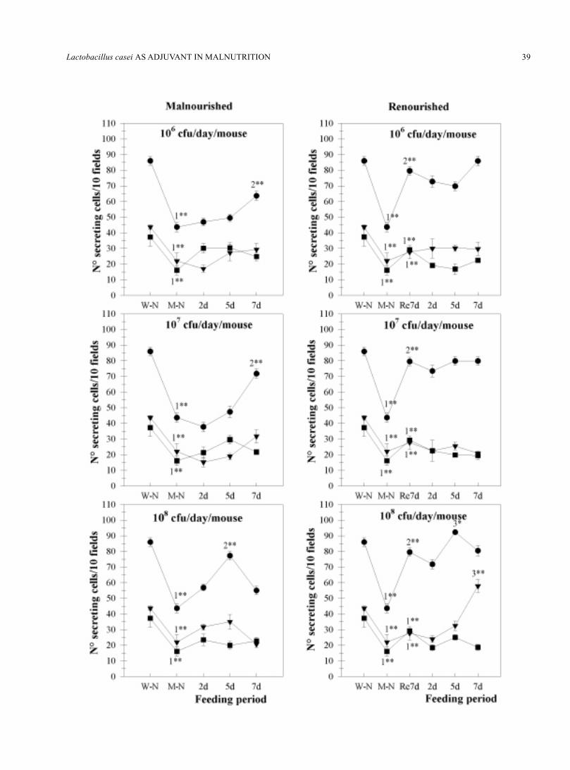
FIGURE 1. Number of IgA, IgG and IgM secreting cells present in the small intestine of malnour-ished and renourished mice treated with different doses and periods of Lactobacillus casei feed-ing, by immunofluorescent test.The values are means ± SD of two small intestine’s sections of each of 5 experimental and con-trol mice.W-N: well-nourished control. M-N: malnourished control. Re7d: renourished control. 1Significantdifferences with W-N control (P<0.01). 2Significant differences with M-N control (P<0.01). 3Sig-nificant differences with Re7d control. *P<0.05, **P<0.01 (ANOVA and Student’s Test).●: IgA + cells; ■: IgG + cells; ▼: IgM + cells.
Lactobacillus casei AS ADJUVANT IN MALNUTRITION 39

PAOLA GAUFFIN CANO et al.40
Number of intraepithelial leukocytes (IEL), mast cells,and goblet cells
Histological slices were stained with Hematoxilin-eosin for IEL. For mastocytes and goblet cells sliceswere stained with 1% Alcian Blue 8GX (Merck, F.R.Germany, Darmstadt D-6100) in 3% acetic acid - 0.5%Safranin O (Sigma, Saint Louis, Missouri 63103, USA)in 0.01M HCl. This was performed as described byKoretou (1988). The number of cells was expressed per10 fields (magnification 100X).
Preparation of samples for electronic microscopy forultrastructural studies
At the end of each treatment period, the mice weresacrificed by cervical dislocation. Peyer’s patches andsmall intestine were carefully removed. Tissues werefixed in 40% formaldehyde and 10% glutaraldehyde inphosphate buffer, pH 7.2. Specimens were then washedin sodium phosphate buffer and fixed in 10% OsO
4,
dehydrated in ethanol, cleared in propylene oxide andfinally embedded in low-viscosity medium. Thin sec-tions were stained with saturated uranyl acetate in 50%ethanol and 4% citrate. Sections were examined by trans-mission electron microscopy and the micrographs wereproduced at 4,900 or 12,800 magnification. Specimensof control mice were processed in the same way.
Statistical analysis
Data were summarized using descriptive statisticssuch as the mean and standard deviations by Student’stest. Statistical comparisons of the treatment versus con-trol groups were analyzed by ANOVA test.
Results
Body weight
The body weight of mice from the M-N and Re7dcontrol groups was significantly lower (P<0.01) thanthat of mice from the well-nourished group. The oralsupplementation with different doses of L. casei to M-N groups did not produce a significant difference withrespect to M-N control. When the effect of L. casei treat-ment on the body weight of Re7d group was examined,all the doses studied produced significant increases at7 days of feeding, and this was also observed on the 5th
day of supplementation of the diet with 108 cfu/day/
mouse compared with Re7d control group (Table 1).
Hematological values and serum total proteins
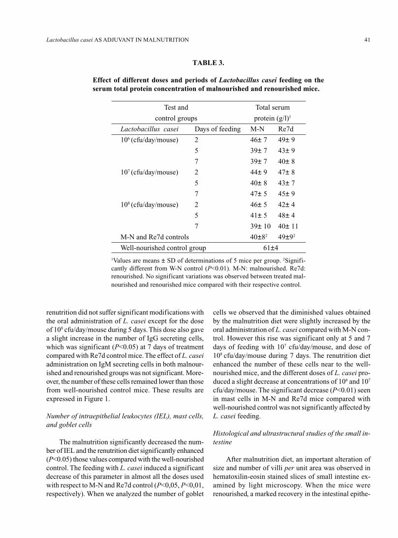
When we studied the effect of the oral administra-tion of L. casei on the hematocrit values we did notobserve a significant variation with regard to the re-spective controls, which were significantly lower whencompared to well-nourished controls. The values ofwhite cells increased significantly with almost all thedoses of L. casei compared to M-N and Re7d controlrespectively; however, this increase in no case reachedthe values obtained for well-nourished controls (Table2). We studied the percentages of PMN and lympho-cytes (L) by counting 100 cells of each type in stainedblood smears. It showed that the malnutrition diet in-duced a slight decrease in the percentage of PMN andproportional increase of L, while the renutrition withmilk produced a slight modification of those cells com-pared with the well-nourished mice (see Table 2). Theresults show variations on the percentage of PMN andL after the supplementation of the diet with L. casei.We observed in the group of M-N significant increasesof the PMN in all the doses used. However, the concen-tration of 107 cfu/day/mouse during 5 days of feedingin the Re7d groups did not induce significant variationsof this parameter. The variations of percentage of L werealways proportional to those obtained with PMN. Thedeterminations of total serum proteins are shown inTable 3. In malnourished mice the level of total pro-teins was significantly decreased (P<0.01) with respectto well-nourished control; the renourished control hada slight increase of this parameter but this did not reachthose of well-nourished control mice. When mice werefed with varying doses of L. casei differences were notobserved.
Number of IgA, IgG and IgM secreting cells
As shown in Figure 1 the malnutrition produced aremarkable decrease in the number of IgA secreting cellsassociated with the lamina propria of the small intes-tine, and the renutrition diet enhanced the IgA+ cellsnear to the values of well-nourished control mice. Whenwe analyzed the effect of oral supplementation of dietwith different doses of L. casei to M-N groups, L. caseiinduced a significant (P<0.01) increase in the numberof IgA+ cells compared with M-N control at 7 days oftreatment for 106 and 107 cfu/day/mouse, and 5 days ofadministration of 108 cfu/day/mouse when compared tothe M-N control. The values of IgA+ cells obtained by
Lactobacillus casei AS ADJUVANT IN MALNUTRITION 41
TABLE 3.
Effect of different doses and periods of Lactobacillus casei feeding on theserum total protein concentration of malnourished and renourished mice.
Test and Total serum
control groups protein (g/l)1
Lactobacillus casei Days of feeding M-N Re7d
106 (cfu/day/mouse) 2 46± 7 49± 9
5 39± 7 43± 9
7 39± 7 40± 8
107 (cfu/day/mouse) 2 44± 9 47± 8
5 40± 8 43± 7
7 47± 5 45± 9
108 (cfu/day/mouse) 2 46± 5 42± 4
5 41± 5 48± 4
7 39± 10 40± 11
M-N and Re7d controls 40±82 49±92
Well-nourished control group 61±4
1Values are means ± SD of determinations of 5 mice per group. 2Signifi-cantly different from W-N control (P<0.01). M-N: malnourished. Re7d:renourished. No significant variations was observed between treated mal-nourished and renourished mice compared with their respective control.
renutrition did not suffer significant modifications withthe oral administration of L. casei except for the doseof 108 cfu/day/mouse during 5 days. This dose also gavea slight increase in the number of IgG secreting cells,which was significant (P<0.05) at 7 days of treatmentcompared with Re7d control mice. The effect of L. caseiadministration on IgM secreting cells in both malnour-ished and renourished groups was not significant. More-over, the number of these cells remained lower than thosefrom well-nourished control mice. These results areexpressed in Figure 1.
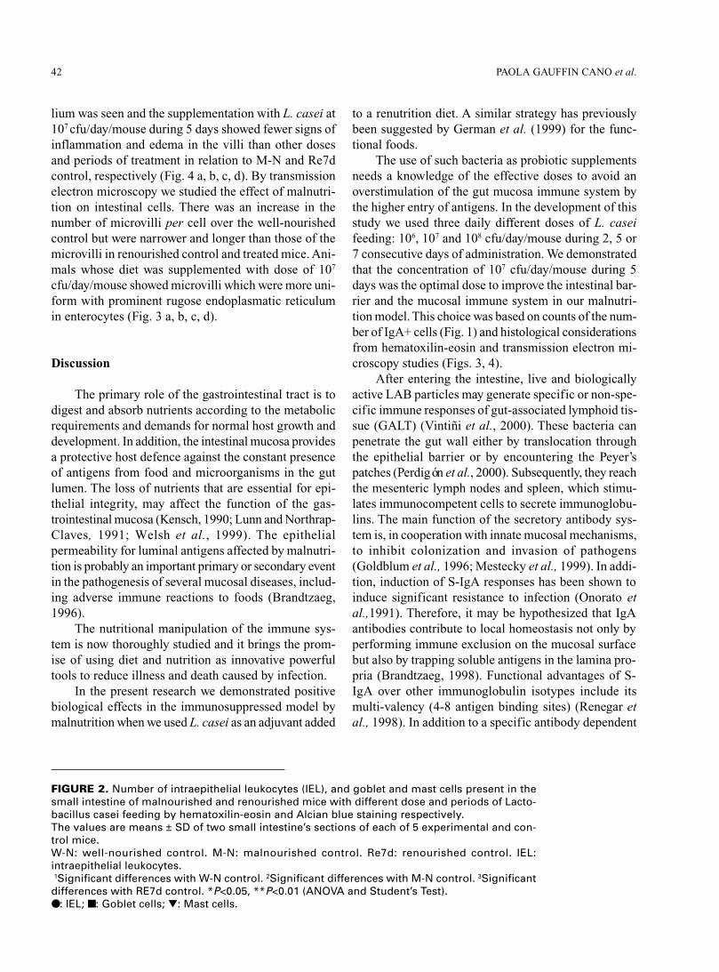
Number of intraepithelial leukocytes (IEL), mast cells,and goblet cells
The malnutrition significantly decreased the num-ber of IEL and the renutrition diet significantly enhanced(P<0.05) those values compared with the well-nourishedcontrol. The feeding with L. casei induced a significantdecrease of this parameter in almost all the doses usedwith respect to M-N and Re7d control (P<0,05, P<0,01,respectively). When we analyzed the number of goblet
cells we observed that the diminished values obtainedby the malnutrition diet were slightly increased by theoral administration of L. casei compared with M-N con-trol. However this rise was significant only at 5 and 7days of feeding with 107 cfu/day/mouse, and dose of108 cfu/day/mouse during 7 days. The renutrition dietenhanced the number of these cells near to the well-nourished mice, and the different doses of L. casei pro-duced a slight decrease at concentrations of 106 and 107
cfu/day/mouse. The significant decrease (P<0.01) seenin mast cells in M-N and Re7d mice compared withwell-nourished control was not significantly affected byL. casei feeding.
Histological and ultrastructural studies of the small in-testine
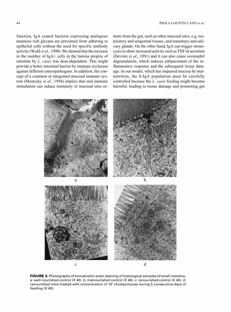
After malnutrition diet, an important alteration ofsize and number of villi per unit area was observed inhematoxilin-eosin stained slices of small intestine ex-amined by light microscopy. When the mice wererenourished, a marked recovery in the intestinal epithe-
PAOLA GAUFFIN CANO et al.42
lium was seen and the supplementation with L. casei at107 cfu/day/mouse during 5 days showed fewer signs ofinflammation and edema in the villi than other dosesand periods of treatment in relation to M-N and Re7dcontrol, respectively (Fig. 4 a, b, c, d). By transmissionelectron microscopy we studied the effect of malnutri-tion on intestinal cells. There was an increase in thenumber of microvilli per cell over the well-nourishedcontrol but were narrower and longer than those of themicrovilli in renourished control and treated mice. Ani-mals whose diet was supplemented with dose of 107
cfu/day/mouse showed microvilli which were more uni-form with prominent rugose endoplasmatic reticulumin enterocytes (Fig. 3 a, b, c, d).
Discussion
The primary role of the gastrointestinal tract is todigest and absorb nutrients according to the metabolicrequirements and demands for normal host growth anddevelopment. In addition, the intestinal mucosa providesa protective host defence against the constant presenceof antigens from food and microorganisms in the gutlumen. The loss of nutrients that are essential for epi-thelial integrity, may affect the function of the gas-trointestinal mucosa (Kensch, 1990; Lunn and Northrap-Claves, 1991; Welsh et al., 1999). The epithelialpermeability for luminal antigens affected by malnutri-tion is probably an important primary or secondary eventin the pathogenesis of several mucosal diseases, includ-ing adverse immune reactions to foods (Brandtzaeg,1996).
The nutritional manipulation of the immune sys-tem is now thoroughly studied and it brings the prom-ise of using diet and nutrition as innovative powerfultools to reduce illness and death caused by infection.
In the present research we demonstrated positivebiological effects in the immunosuppressed model bymalnutrition when we used L. casei as an adjuvant added
to a renutrition diet. A similar strategy has previouslybeen suggested by German et al. (1999) for the func-tional foods.
The use of such bacteria as probiotic supplementsneeds a knowledge of the effective doses to avoid anoverstimulation of the gut mucosa immune system bythe higher entry of antigens. In the development of thisstudy we used three daily different doses of L. caseifeeding: 106, 107 and 108 cfu/day/mouse during 2, 5 or7 consecutive days of administration. We demonstratedthat the concentration of 107 cfu/day/mouse during 5days was the optimal dose to improve the intestinal bar-rier and the mucosal immune system in our malnutri-tion model. This choice was based on counts of the num-ber of IgA+ cells (Fig. 1) and histological considerationsfrom hematoxilin-eosin and transmission electron mi-croscopy studies (Figs. 3, 4).
After entering the intestine, live and biologicallyactive LAB particles may generate specific or non-spe-cific immune responses of gut-associated lymphoid tis-sue (GALT) (Vintiñi et al., 2000). These bacteria canpenetrate the gut wall either by translocation throughthe epithelial barrier or by encountering the Peyer’spatches (Perdigón et al., 2000). Subsequently, they reachthe mesenteric lymph nodes and spleen, which stimu-lates immunocompetent cells to secrete immunoglobu-lins. The main function of the secretory antibody sys-tem is, in cooperation with innate mucosal mechanisms,to inhibit colonization and invasion of pathogens(Goldblum et al., 1996; Mestecky et al., 1999). In addi-tion, induction of S-IgA responses has been shown toinduce significant resistance to infection (Onorato etal.,1991). Therefore, it may be hypothesized that IgAantibodies contribute to local homeostasis not only byperforming immune exclusion on the mucosal surfacebut also by trapping soluble antigens in the lamina pro-pria (Brandtzaeg, 1998). Functional advantages of S-IgA over other immunoglobulin isotypes include itsmulti-valency (4-8 antigen binding sites) (Renegar etal., 1998). In addition to a specific antibody dependent
FIGURE 2. Number of intraepithelial leukocytes (IEL), and goblet and mast cells present in thesmall intestine of malnourished and renourished mice with different dose and periods of Lacto-bacillus casei feeding by hematoxilin-eosin and Alcian blue staining respectively.The values are means ± SD of two small intestine’s sections of each of 5 experimental and con-trol mice.W-N: well-nourished control. M-N: malnourished control. Re7d: renourished control. IEL:intraepithelial leukocytes. 1Significant differences with W-N control. 2Significant differences with M-N control. 3Significantdifferences with RE7d control. *P<0.05, **P<0.01 (ANOVA and Student’s Test).●: IEL; ■: Goblet cells; ▼: Mast cells.
Lactobacillus casei AS ADJUVANT IN MALNUTRITION 43
PAOLA GAUFFIN CANO et al.44
function, IgA coated bacteria expressing analogousmannose rich glycans are prevented from adhering toepithelial cells without the need for specific antibodyactivity (Wold et al., 1990). We showed that the increasein the number of IgA+ cells in the lamina propria ofintestine by L. casei was dose-dependent. This mightprovide a better intestinal barrier by immune exclusionagainst different enteropathogens. In addition, the con-cept of a common or integrated mucosal immune sys-tem (Mestecky et al., 1994) implies that oral immunestimulation can induce immunity in mucosal sites re-
mote from the gut, such as other mucosal sites, e.g. res-piratory and urogenital tissues, and mammary and sali-vary glands. On the other hand, IgA can trigger mono-cytes to show increased activity such as TNF-α secretion(Devière et al., 1991) and it can also cause eosinophildegranulation, which induces enhancement of the in-flammatory response and the subsequent tissue dam-age. In our model, which has impaired mucosa by mal-nutrition, the S-IgA population must be carefullycontrolled because the L. casei feeding might becomeharmful, leading to tissue damage and promoting gut
FIGURE 3. Photographs of hematoxilin-eosin staining of histological samples of small intestine.a: well-nourished control (X 40). b: malnourished control (X 40). c: renourished control (X 40). d:renourished mice treated with concentration of 107 cfu/day/mouse during 5 consecutive days offeeding (X 40).
a b
c d

Lactobacillus casei AS ADJUVANT IN MALNUTRITION 45
FIGURE 4. Transmission electron microphotographs of epithelial cell from smallintestine. a: well-nourished control (X 4,900). b: malnourished control (X 12,800).c: renourished control (X 12,800). d: renourished mice treated with concentrationof 107 cfu/day/mouse during consecutive 5 days of feeding (X 12,800).
barrier compromise. The locally produced IgA is prob-ably crucial for the immunological homeostasis withinthe lamina propria because this isotype lacks potent ef-fector functions such as classical complement activa-tion (Brandtzaeg et al., 1993). The increase of the num-ber of IgG producing cells observed with some dosesof L. casei feeding would indicate an inflammatory re-sponse because the mucosal IgG and IgM antibodiesactivate complement and may thereby impair the sur-face epithelium and enhancement of intestinal perme-ability.
Within the normal intestinal epithelium there is alarge population of leucocytes, which are mostly ac-counted by lymphocytes (intraepithelial lymphocytes,IELs). It has been proposed the IEL have a number ofdifferent roles including: surveillance of the intestinalepithelial layer for the detection of microbial pathogens;removal of damaged or transformed epithelial cells;maintenance of epithelial integrity via secretion oftrophic factors important for epithelial cell growth anddifferentiation; and the regulation of local cell medi-ated or humoral immune responses. (Aranda et al., 1999).
a b
c d
PAOLA GAUFFIN CANO et al.46
The retention of T cells within the intra-epithelial spaceis very efficient and stable under normal conditions(Famularo et al., 1997). Several studies suggest thatadhesion molecules and chemokine receptors that areselectively expressed on IELs are important for inter-actions with epithelial cells, such as the binding ofintegrin α
E β
7 to E-cadherin, a ligand expressed on all
epithelial cells. Finally these interactions of adhesionmolecules on IEL with ligands on epithelial cells maymodulate the functional response of either cell type(Agace et al., 2000). The decrease observed in the num-ber of IEL after L. casei feeding could be due to that itinduces recovering of intestinal epithelium and wouldbe more selective for retaining different immune cells.So, the administration of L. casei might have more in-fluence on epithelial cells than immune cells, and like-wise it could avoid a powerful immune activation ofIEL that can induce villus atrophy. By optic and trans-mission electron microscopy we showed that the supple-mentation of diet induces recovery of epithelial cellsand mucosa, and we think it could influence thefunctionly of these cells.
It has been recently shown that T cells and mastcells share common homing/adhesion receptors, whichsuggests that they use similar migration pathways. Fur-thermore, these cells colocalize in the gastrointestinalmucosa, and it is possible that they act in concert toachieve an immune response (Smith and Weis, 1996).Mast cells contain a large number of bioactive media-tors such as histamine and proteases and leukotrienes.However, recent work shows that mast cells can alsoplay a critical role in innate immunity to bacterial in-fection and that mast cells and basophils can be acti-
vated by viral proteins (Wedemeyer et al., 2000). Wedid not observe significant variations when we studiedthe number of mast cells in the lamina propria aftersupplementation of the diet. It is important to inhibitthe inappropriate activation of these cells because mu-cosal mast cells, together with eosinophils, T cells andother effector cells, constitute a network which can or-chestrate protective immunity or harmful inflammation.
Goblet cells, present in both small and large intes-tine, release mucus granules from the apical cytoplasmafter an inflammatory stimulus. Mucus secretion is trig-gered by direct stimulation by immune complexes andchemical agents and indirect stimulation by mediatorsreleased by histamine and lymphokines (Snyder andWalker, 1994). Therefore, the increase of the numberof goblet cells observed in some doses of L. casei ad-ministration could indicate an inflammatory response.
In conclusion this study showed that impaired gutbarrier and mucosal immune function by malnutritioncan be reversed by L. casei used as an oral adjuvant ofrenutrition diet. The clinical significance of these find-ings will be important, in particular whether the chosendose, as well as improving mucosal immunity, may alsoinduce protection against enteropathogens.
Acknowledgements
This paper was supported by grants from ConsejoNacional de Investigaciones Científicas y Técnicas(CONICET), Argentina PIP 5011 and from Consejo deInvestigaciones Universidad Nacional de Tucumán(CIUNT), Argentina 26/D/127.
References
AGACE W, HIGGINS J, SADASIVAN B, BRENNER M, PARKER C (2000). T-Lymphocyte-epithelial-cell interactions: integrenin αE
(CD103) β7, LEEP-CAM and chemokines. Curr Opin Cell Biol 12: 563-568.
AGÜERO G, SANCHEZ S, FERNANDEZ S, ALLORI C, P DE RUIZ HOLGADO A, PERDIGON G (1996). Administration of yoghurtor Lactobacillus casei to malnourished mice: Comparative effect on lymphoid cells and mucosal reconditioning of the intestine.Food Agricul Immunol 8: 229-238.
ARANDA R, SYDORA B, KRONENBERG M (1999). Intraepithelial lymphocytes: function. In: Mucosal Immunology, Chapter 26. PLOgra, ME Lamm, J Brenenstock, and JR McGhee, Eds. Academic press, Inc. San Diego, USA, pp. 429-437.
BOZA J, MOËNNOZ D, VUICHOUD J, JARRET A, GAUDARD-DE-WECK D, FRITSCHÉ R, DONNET A, SCHIFFRIN E,PERRUISSEAU, BALLÈVRE O (1999). Food deprivation and refeeding influence growth, nutrient retention and functional recov-ery of rats. J Nutr 129: 1340-1346.
BRADFORD M (1976). A rapid and sensitive method for quantification of microgram quantities of protein utilizing the principles ofprotein dye binding. Anal Biochem 72: 248-254.
BRANDTZAEG P (1996). History of oral tolerance and mucosal immunity. Ann NY Acad Sci. 778: 1-27.BRANDTZAEG P (1998). Development and basic mechanisms of human gut immunity. Nutrition Reviews, 56, Nº1: S5- S18.BRANDTZAEG P, HALSTENSEN T, HVATUM M, KVALE D, SCOTT H (1993). The serologic and mucosal immunologic basis of
celiac disease. In: Immunophysiology of the Gut. A. Walker, P. Harmatz and B. Wershil, Eds. Academic press, Inc. San Diego,USA, pp. 295-333.
Lactobacillus casei AS ADJUVANT IN MALNUTRITION 47
CASTILLO R, FENG J, STEVENSON D, KWONG L (1991). Altered maturation of small intestine function in the absence of intra-luminal nutrients rapid normalization with refeeding. Am J Clin Nutr 53: 558-561.
CHANDRA R, WADHWA M (1993). Nutritional deficiencies and intestinal mucosal immunity. In: Immunophysiology of the Gut. AWalker, P Harmatz and B Wershil, Eds. Academic Press, Inc. San Diego, USA, pp.389-399.
CHANDRA R (1996). Nutrition, immunity and infection: from basic knowledge of dietary manipulation of immune responses to practi-cal application of ameliorating suffering and improving survival. Proc Natl Acad Sci 93: 14304-14307.
CHANDRA R (1997). Nutrition and the immune system: an introduction. Am J Clin Nutr 66: 460S-3S.DE MAN J ROGOSA M, SHARPE ME (1960). A medium for the cultivation of lactobacilli. J Applied Bacteriol 23: 130-155.DEITCH E (1992). Multiple organ failure. Pathophisiology and potential future therapy. Ann Surg 216:117-134.DEVIÈRE J, VAERMAN J-P, CONTENT J, DENYS C, SCHANDENE L, VANDENBUSSCHE P, SIBILLE Y, DUPONT E (1991). IgA
triggers tumor necrosis factor a secretion by monocytes: a study in normal subjects and patients with alcoholic cirrhosis. Hepatology13: 670-22.
FAMULARO G, MORETTI S, MARCELLINI S, DE SIMONE C (1997). Stimulation of immunity by probiotics. In: Probiotics 2:Applications and practical aspects. R. Fuller, Eds. Chapman & Hall, London, pp. 133-161.
FULLER R (1992). History and development of probiotics. In: Probiotics. The Scientific Basis. R Fuller, Ed. Chapman and Hall, London,pp. 2-8.
GERMAN B, SCHIFFRIN E, REINIERO R, MOLLET B, PTEIFER A, NEESER J-R (1999). The development of functional foods:lessons from the gut. Tibtech 17: 492-499.
GERRITSE, K, POSNO M, SCHELLEKENS M, BOERSNA W, CLAASSEN E (1990). Oral administration of TNP-Lactobacillus con-jugates in mice: a model for evaluation of mucosal and systemic immune response and memory formation elicited by transformedlactobacilli. Res Microbiol 141: 955-962.
GOLDBLUM R, HANSON L, BRANDTZAEG P (1996). The mucosal defense system. In: Immunologic disorders in infants & children,4th Ed. ER Stiehm, Ed. WB Saunders Co. Philadelphia, pp. 159-99.
KARLSSON L, CASTAÑO A, PETERSON P (1996). Principles of antigen processing and presentation. In: Essentials of mucosalimmunology. MF Kagnoff and H Kiyono, Eds. Academic Press, Inc. San Diego. USA, pp. 3-28.
KENSCH G (1990). Malnutrition, infection and immune function. In: The malnourished child. H Suskind and L Lewinter-Suskind, Eds.Raven Press, Vevey, Switzerland. pp. 37-55.
KOLDOVSKY O (1989). Hormones in milk: their possible physiological significance for the neonate. In: Gastroenterology and Nutri-tion in Infancy. E. Lebenthal, Ed. Raven press. New York, pp. 97-119.
KORETOU O (1988). Relationship between the staining property of mast cell granule with alcian blue-safranin O and toluidine blue O,and the content of mast cell protease I in the granule of rat peritoneal mast cell. Acta Histochem Cytochem 21 (1): 25-32.
LUNN P, NORTHRAP-CLAVES C (1991). Intestinal permeability, mucosal injury, and growth faltering in Gambian infants. Lancet 338:907-056.
MAC DERMOTT R (1993). Effect of nutritional factors and the microenvironment on mucosal immune function. In: Immunophysiologyof the Gut. A Walker, P Harmatz and B Wershil, Eds. Academic Press, Inc. San Diego, USA, pp. 365-371.
MATAR C, GOULET J, BERNIER R, BROCHU E (2000). Bioactive peptides from fermented foods: their role in the immune system. In:Probiotics 3: Immunomodulation by the gut microflora and probiotics. R. Fuller and G. Perdigón, Eds. Kluwer Academic Publisher.London, pp. 193-212.
MEISEL H, BOCKELMANN W (1999). Bioactive peptides encrypted in milk proteins: proteolytic activation and thropho-functionalproperties. Antonie Van Leeuwenhocek 76: 207-215.
MESTECKY J, ABRAHAM R, OGRA P (1994). Common mucosal immune system and strategies for the development of vaccineseffective at the mucosal surfaces. In: Handbook of Mucosal Immunology, Chapter 31. P Ogra, M Lamm, and J McGhee, Eds.Academic Press, Inc, New York, NY. pp 357-372.
MESTECKY S, RUSSELL M, ELSON C (1999). Intestinal IgA: novel views on its function in the defence of the largest mucosal surface.Gut 44: 2-5.
NAIDU A, BIDLACK W, CLEMENS R (1999). Probiotic spectra of lactic acid bacteria (LAB). Crit Rev Food Sci Nutr 38 (1): 13-126.ONORATO L, MODLIN J, MCBEAN A, THOMAS M, LOSONSKY G, BERNIER R (1991). Mucosal immunity induced by enhanced-
potency inactivated and oral polio vaccines. J Infec Dis 163: 1-6.PERDIGÓN G, DE MACIAS M, ALVAREZ S, OLIVER G, PESCE DE RUIZ HOLGADO A (1990). Prevention of gastrointestinal
infection using immunobiological methods with milk fermented with Lactobacillus casei and Lactobacillus acidophillus. J DairyRes, 57: 255-264.
PERDIGÓN G, ALVAREZ S, PESCE DE RUIZ HOLGADO A (1991). Immunoadjuvant activity of oral Lactobacillus casei: influence ofdose on the secretory immune response and protective capacity in intestinal infections. J Dairy Res 58: 485-496.
PERDIGON G, RACHID M, DE BUDEGUER M, VALDEZ J (1994). Effect of yogurt feeding on the small and large intestine associatedlymphoid cells in mice. J Dairy Res 61: 553-562.
PERDIGÓN G, AGÜERO G, ALVAREZ S, DE ALLORI C, PESCE DE RUIZ HOLGADO A (1995). Effect of viable Lactobacillus caseifeeding on immunity of the mucosae and intestinal microflora in malnourished mice. Milchwissenschaft 50: 125-145.
PERDIGON G, MEDINA M, VINTIÑI E, VALDEZ J (2000). Intestinal pathway of internalization of lactic acid bacteria and gut mucosalimmunostimulation. International J of Immunoph Pharmacol 13 (3): 141-150.
PERDIGON G, OLIVER G (2000). Modulation of the immune response of the immunosuppressed host by probiotics. In: Probiotics 3:Immunomodulation by the gut microflora and probiotics. R. Fuller and G. Perdigón, Eds. Kluwer Academic Publisher. London, pp.148-175.
PAOLA GAUFFIN CANO et al.48
POULLAIN M, CEZARD J, MARCHÉ C, MACRY J, ROGER L, GRASSET E, BROYART J (1991). Effects of dietary whey proteins,their peptides or amino-acids on the ileal mucosa of normally fed and starved rats. Clin Nutr 11: 48-53.
RENEGAR K, JACKSON G, MESTECKY J (1998). In vitro comparison of the biologic activities of monoclonal monomeric IgA ,polymeric IgA and secretory IgA. J immunol 60: 1219-1223.
ROUX M, LOPEZ M, FLORIN-CHRISTENSEN A (2000). Mucosal immunity.In: Probiotics 3: Immunomodulation by the gut microf-lora and probiotcs. R. Fuller and G. Perdigón, Eds. Kluwer Academic Publisher. London, pp. 12-28
SAINT-MARIE G (1962). A paraffin embedding technique for studies employing immunoflorescence. J Histochem Cytochem 10: 250-256.
SCRIMSHAW N, SANGIOVANNI J (1997). Synergism of nutrition, infection and immunity: an overview. Am J Clin Nutr 66: 464S-77S.SHERMAN P, FORSTNER J, ROOMI N, KHATRI I, FORSTNER, G (1985). Mucin depletion in the intestine of malnourished rats. Am
J Physiol 248 G418-23.SMITH T, WEIS J (1996). Mucosal T cells and mast cells share common adhesion receptors. Immunol Today 17 (2): 60-63.SNYDER J, WALKER W (1994). Structure and function of intestinal mucin: development aspects. Int Arch Allergy Appl Immunol, 92:
351-356.VINTIÑI E, ALVAREZ S, MEDINA M, MEDICI M, de BUDEGUER MV, PERDIGON G (2000). Gut mucosal immunostimulation by
lactic acid bacteria. Biocell 24(3): 223-232.WEDEMEYER J, TSAI M, GALLI SJ (2000). Roles of mast CEIs and basophils in innate and acquired immunity. Curr Opin Immunol
12: 624-631.WELSH F, FARMERY S, MacLENNAN K, SHERIDAN M, BARCLAY G, GUILLOU P, REYNOLDS J (1999). Gut barrier function in
malnourished patients. Gut 42: 396-401.WOLD A, MESTECKY J, TOMANA M, KOBATA A, OHBAYASHI H ENDO T, EDEN CS (1990). Secretory immunoglobulin A carries
oligosaccharide receptors for Escherichia coli type 1 fimbrial lectin. Infect Immun 58: 3073-3077.