Embed Size (px)
Citation preview
7/28/2019 Function of Protein in Immune System
http://slidepdf.com/reader/full/function-of-protein-in-immune-system 1/8
Function of protein in immune system
1. The immune response features a specialized array of cells and proteins.
2. Self is distinguished from nonself by the display of peptides on cell surfaces.
3. Antibodies have two identical antigen-binding sites.
4. Antibodies bind tightly and specifically to antigen.
5. The antibody-antigen interaction is the basis for a variety of important analytical procedures.
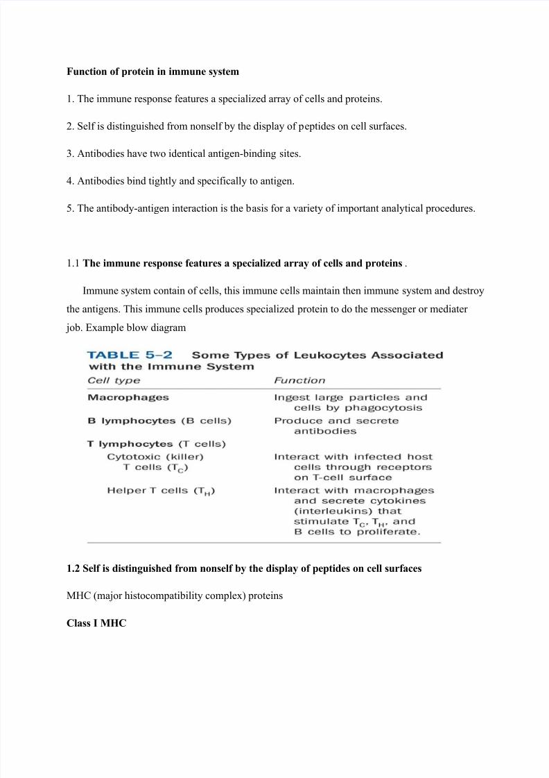
1.1 The immune response features a specialized array of cells and proteins.
Immune system contain of cells, this immune cells maintain then immune system and destroy
the antigens. This immune cells produces specialized protein to do the messenger or mediater
job. Example blow diagram
1.2 Self is distinguished from nonself by the display of peptides on cell surfaces
MHC (major histocompatibility complex) proteins
Class I MHC
7/28/2019 Function of Protein in Immune System
http://slidepdf.com/reader/full/function-of-protein-in-immune-system 2/8
Each individual produces up to 6 class I MHC variants. Bind and display peptides derived from
cellular proteins. Recognition targets of the T-cell receptors of the TC cells.
Class II MHC
Occur on the surfaces of macrophages and B lymphocytes. Each human produces up to 12
variants. Bind and display peptides derived from external proteins. Recognition targets of the T-
cell receptors of the TH cells.
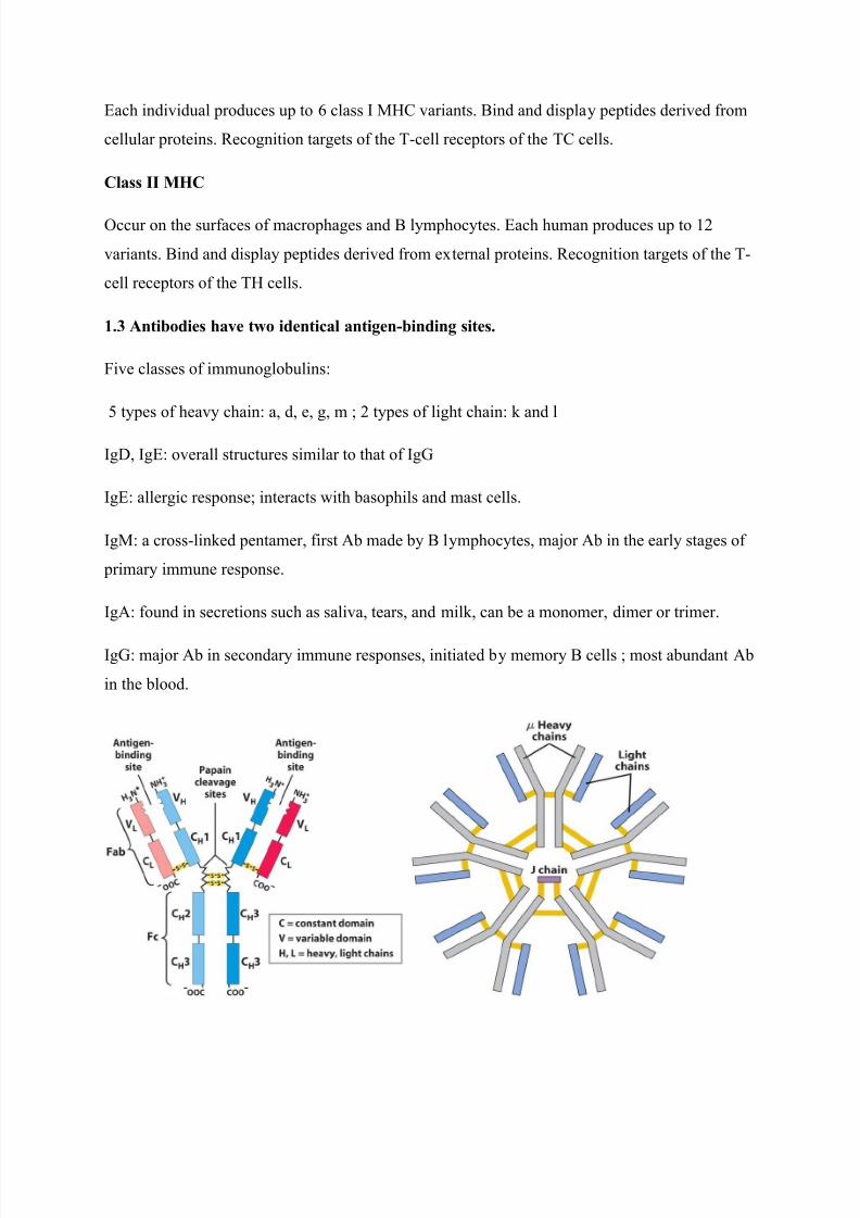
1.3 Antibodies have two identical antigen-binding sites.
Five classes of immunoglobulins:
5 types of heavy chain: a, d, e, g, m ; 2 types of light chain: k and l
IgD, IgE: overall structures similar to that of IgG
IgE: allergic response; interacts with basophils and mast cells.
IgM: a cross-linked pentamer, first Ab made by B lymphocytes, major Ab in the early stages of
primary immune response.
IgA: found in secretions such as saliva, tears, and milk, can be a monomer, dimer or trimer.
IgG: major Ab in secondary immune responses, initiated by memory B cells ; most abundant Ab
in the blood.

7/28/2019 Function of Protein in Immune System
http://slidepdf.com/reader/full/function-of-protein-in-immune-system 3/8
1.4 The antibody-antigen interaction is the basis for a variety of important analytical procedures.
Two types of antibodies preparations are in use:
Polyclonal antibodies
Monoclonal antibodies: by Köhler and Milstein, 1975
Practical uses of antibodies:
Affinity column
ELISA (enzyme-linked immunosorbent assay)
Immunoblot assay (Western blot)
Muscle Contraction & The Sliding Filament Theory
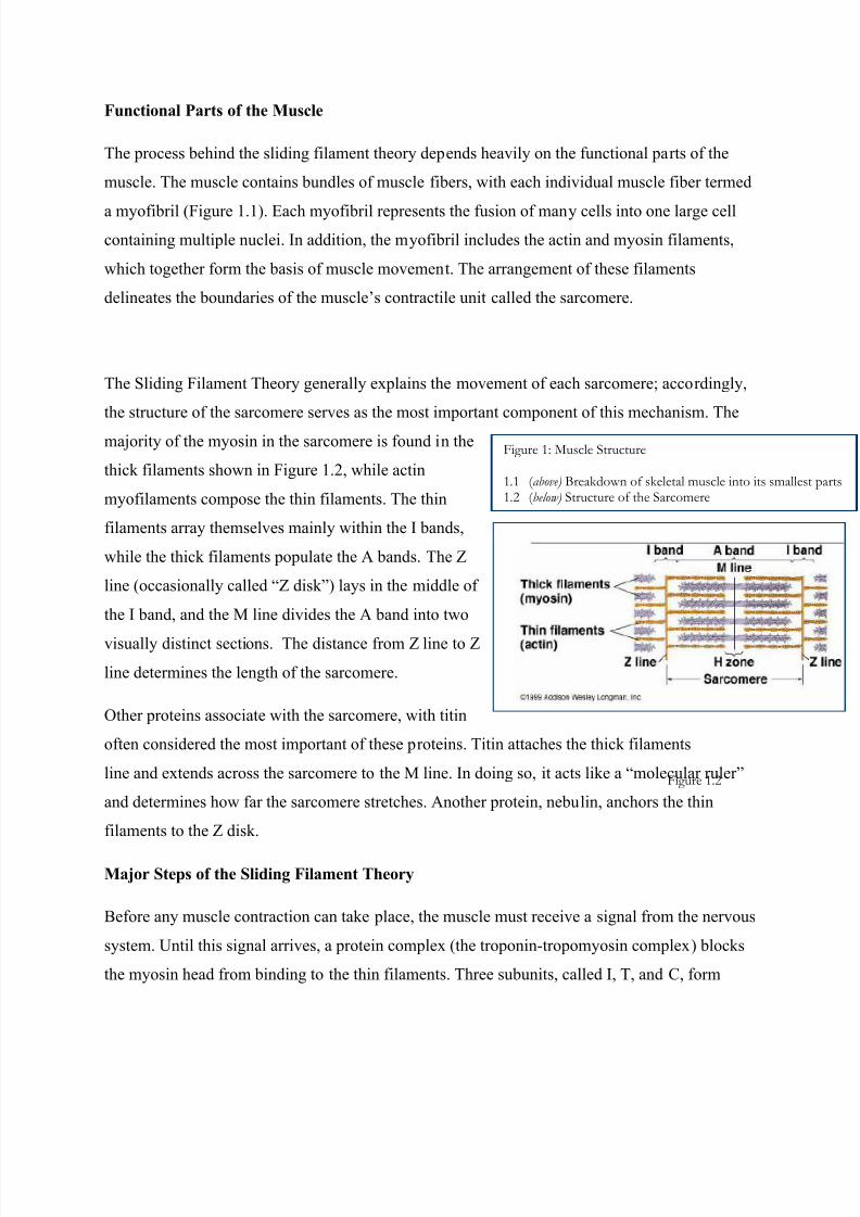
The Sliding Filament Theory explains the basis of skeletal muscle contraction and
specifically deals with the movement of myosin heads along an
actin fiber in the sarcomere. The first step to understanding this
theory stems from an understanding of the functional parts of
the muscle and its terminology. Once the components of the
system are known, the mechanical process of muscle
contraction follows naturally. Accordingly, this document
outlines the parts of the muscle before providing a simplified
explanation of the mechanism behind muscle contraction.
7/28/2019 Function of Protein in Immune System
http://slidepdf.com/reader/full/function-of-protein-in-immune-system 4/8
7/28/2019 Function of Protein in Immune System
http://slidepdf.com/reader/full/function-of-protein-in-immune-system 5/8
troponin. Troponin I prevents the actual binding of the myosin head, while Troponin T connects
troponin to tropomyosin. Meanwhile, Troponin C awaits the signal from the nervous system,
which releases calcium ions (Ca2+
) into the sarcoplasmic reticulum surrounding the myofibril.
Once released, the Ca2+
binds to Troponin C, and a conformational change in troponin allows for
muscle contraction to occur.
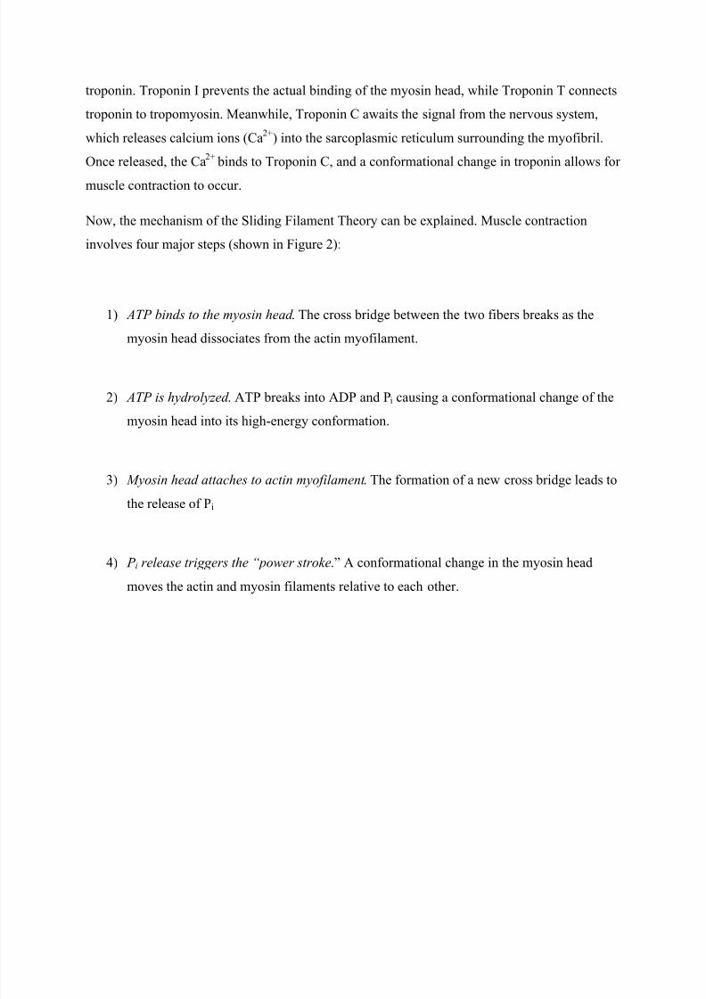
Now, the mechanism of the Sliding Filament Theory can be explained. Muscle contraction
involves four major steps (shown in Figure 2):
1) ATP binds to the myosin head . The cross bridge between the two fibers breaks as the
myosin head dissociates from the actin myofilament.
2) ATP is hydrolyzed . ATP breaks into ADP and Pi causing a conformational change of the
myosin head into its high-energy conformation.
3) Myosin head attaches to actin myofilament . The formation of a new cross bridge leads to
the release of Pi
4) P i release triggers the “power stroke.” A conformational change in the myosin head
moves the actin and myosin filaments relative to each other.
7/28/2019 Function of Protein in Immune System
http://slidepdf.com/reader/full/function-of-protein-in-immune-system 6/8
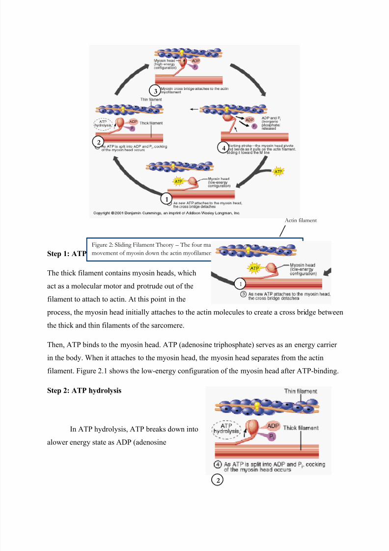
Step 1: ATP binds to the myosin head
The thick filament contains myosin heads, which
act as a molecular motor and protrude out of thefilament to attach to actin. At this point in the
process, the myosin head initially attaches to the actin molecules to create a cross bridge between
the thick and thin filaments of the sarcomere.
Then, ATP binds to the myosin head. ATP (adenosine triphosphate) serves as an energy carrier
in the body. When it attaches to the myosin head, the myosin head separates from the actin
filament. Figure 2.1 shows the low-energy configuration of the myosin head after ATP-binding.
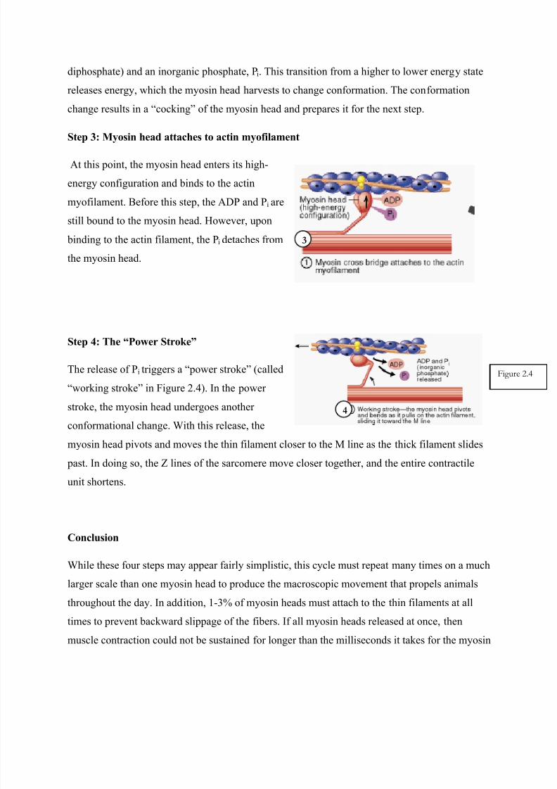
Step 2: ATP hydrolysis
In ATP hydrolysis, ATP breaks down into
alower energy state as ADP (adenosine
1
2
3
4
Figure 2: Sliding Filament Theory – The four major steps that result in the ultimate
movement of myosin down the actin myofilament.
1
Actin filament
2
7/28/2019 Function of Protein in Immune System
http://slidepdf.com/reader/full/function-of-protein-in-immune-system 7/8
diphosphate) and an inorganic phosphate, Pi. This transition from a higher to lower energy state
releases energy, which the myosin head harvests to change conformation. The conformation
change results in a “cocking” of the myosin head and prepares it for the next step.
Step 3: Myosin head attaches to actin myofilament
At this point, the myosin head enters its high-
energy configuration and binds to the actin
myofilament. Before this step, the ADP and P i are
still bound to the myosin head. However, upon
binding to the actin filament, the Pi detaches from
the myosin head.
Step 4: The “Power Stroke”
The release of Pi triggers a “power stroke” (called
“working stroke” in Figure 2.4). In the power
stroke, the myosin head undergoes another
conformational change. With this release, the
myosin head pivots and moves the thin filament closer to the M line as the thick filament slides
past. In doing so, the Z lines of the sarcomere move closer together, and the entire contractile
unit shortens.
Conclusion
While these four steps may appear fairly simplistic, this cycle must repeat many times on a much
larger scale than one myosin head to produce the macroscopic movement that propels animals
throughout the day. In addition, 1-3% of myosin heads must attach to the thin filaments at all
times to prevent backward slippage of the fibers. If all myosin heads released at once, then
muscle contraction could not be sustained for longer than the milliseconds it takes for the myosin
3
Figure
4

7/28/2019 Function of Protein in Immune System
http://slidepdf.com/reader/full/function-of-protein-in-immune-system 8/8
head to bind ATP, hydrolyze it, bind to the actin fiber, and trigger the power stroke. Therefore,
this process occurs in staggered, multiple waves to create effective muscle contraction. Overall,
the Sliding Filament Theory successfully explains the mechanism behind skeletal muscular
contraction, at the microscopic level of a single sarcomere.