Embed Size (px)
Citation preview

radonmedicalimaging.com
WV Office 866-723-6698
VA Office 800-722-1991
We focus on people, innovative products, and service excellence
Fujifilm Persona CT
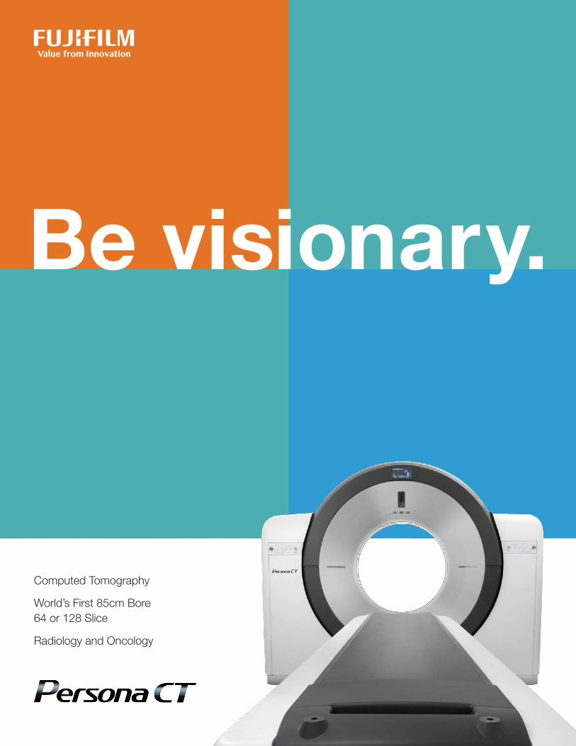
Computed Tomography
World’s First 85cm Bore 64 or 128 Slice
Radiology and Oncology

Introducing Persona CT
Visionary Performance Embrace the best.• Advancedautomateddoseoptimizationbydesign• Synchronizeddose-loweringacquisitionfeatures• Refinedimageprocessingwithartificialintelligence
People-first Design A workflow that works for you.• 85cmborebringsaddedcomforttoeveryexamandpatient• Fastacquisitionenhancespatienttreatmentandexperiences• Easyoperationimprovesworkflow,speed,andaccuracy
Reliability in Action Experience you can rely on.• Exceptionalimagequalityanddosereductionmadepossiblebylownoiseacquisitionsystemandintelligentimageprocessing
• Simplifieddesign,engineeredtomaximizereliabilityandstreamlinemaintenance
Flexible Scalability Flex however you see fit.• AdvancedapplicationspoweredbyFujifilm’sworld-classSynapse3Dtechnology
• Versatilityandprotocolstoexpandtothelatestgrowingprocedures
• Amazingspecialized3Dtoolsforcoronary,brain,respiratory,orthopedics,wholebodyandmore
Oncology Care
Specifications
Contents

Persona CT: advanced CT offering high-level solutions without compromise.
PersonaCTistheresultofanexcitingcollaborationbetweentwotrustedleadersinmedicalimagingworldwide.FujifilmandAnalogichavecometogethertodeliveranewworld-classCTsolution.ThePersonaCTisuniquelyengineeredtohelpcliniciansachieveconsistentlybetterclinical,operationalandfinancialexperiences.
Fujifilmhasmorethan85yearsofmedicalimagingexperience,deliveringbreakthroughinnovationsinmedicalimagingwithasolidreputationforimagequality,dosereduction,diagnosticvisualization,andreliability.
Analogicbringsmorethan50yearsofexperienceasaleadingproviderofdiverseimagingtechnologiesandasanOEMtoCTsystemsandcomponentsofmostoftheleadingbrandsonthemarkettoday.
Withanunrelentingdrivetodiscoverwhat’snext,wearecommittedtocontinuallydeliveringvaluefrominnovation.Ouradvancedimagingtechnologiesenableandempowerphysicianstoseemore,tohelpmakethemostaccuratediagnosestodeterminethebestpathsforward.
Weareproudtointroduceourlatestcontributiontoradiologyandoncologytreatmentplanning,deliveringinnovationsinCTsystemdesignandperformanceyouhavecometoexpectfromFujifilm.
IntroducingPersonaCT.Anocompromise,wellthoughtout,scalablesolution,designedtosimplifyeverystepinthecareandtreatmentofyourpatients.
•Bigperformancewithastreamlinedsmallfootprint•High-sensitivityimageacquisitiontechnologies•Refinedintelligentimageprocessing•Performanceandreliabilityyoucantrust
Yourpatientsandteamdependonyou.Youcanrestassuredthatwe’reworkingwithyoutostreamlineworkflowandenhanceconfidenceforbothpatientandprovider,everystepoftheway.
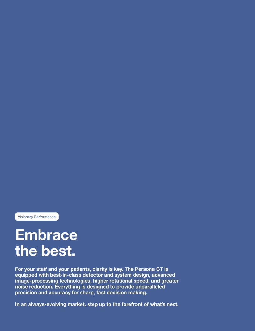
Embrace the best.For your staff and your patients, clarity is key. The Persona CT is equipped with best-in-class detector and system design, advanced image-processing technologies, higher rotational speed, and greater noise reduction. Everything is designed to provide unparalleled precision and accuracy for sharp, fast decision making.
In an always-evolving market, step up to the forefront of what’s next.
VisionaryPerformance
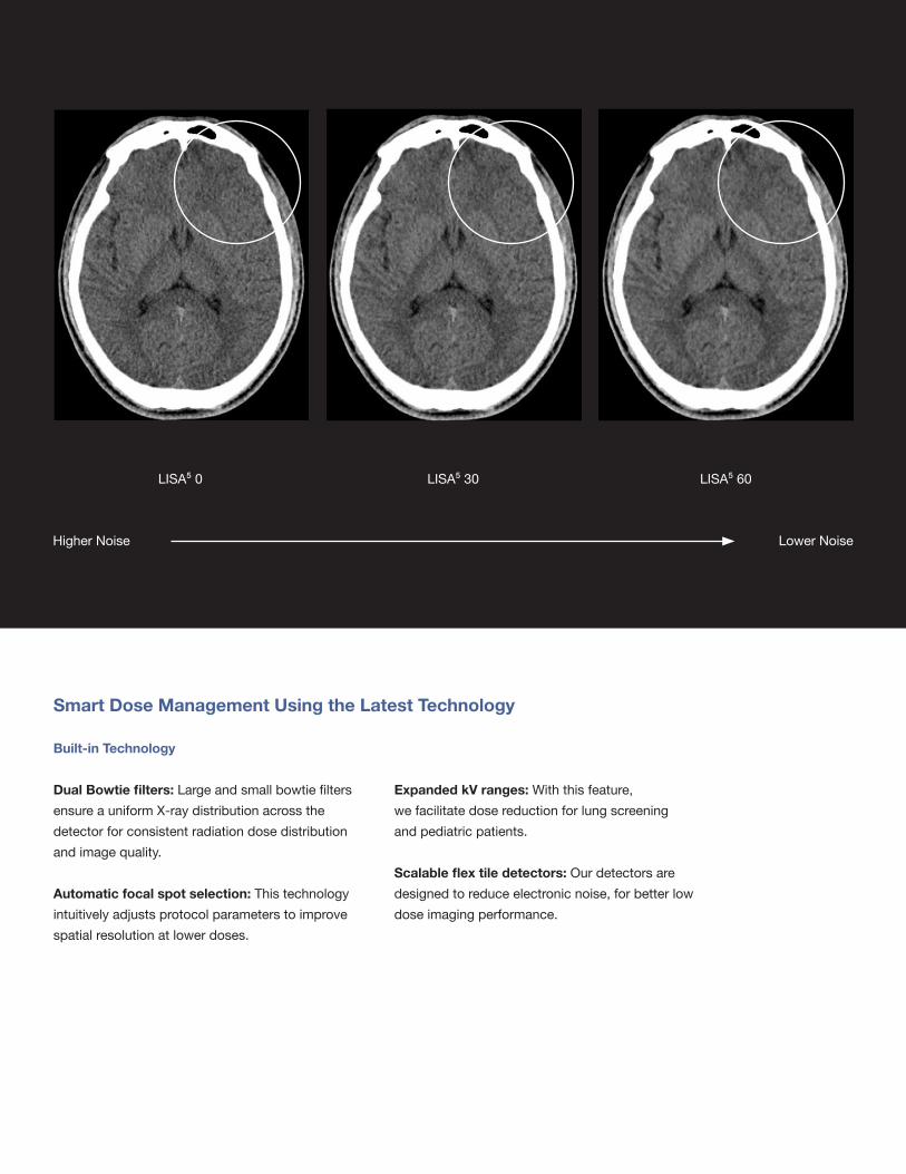
Smart Dose Management Using the Latest Technology
Built-in Technology
Dual Bowtie filters:LargeandsmallbowtiefiltersensureauniformX-raydistributionacrossthedetectorforconsistentradiationdosedistributionandimagequality.
Automatic focal spot selection:Thistechnologyintuitivelyadjustsprotocolparameterstoimprovespatialresolutionatlowerdoses.
Expanded kV ranges:Withthisfeature,wefacilitatedosereductionforlungscreeningandpediatricpatients.
Scalable flex tile detectors:Ourdetectorsaredesignedtoreduceelectronicnoise,forbetterlowdoseimagingperformance.
LISA⁵0
HigherNoise LowerNoise
LISA⁵60LISA⁵30
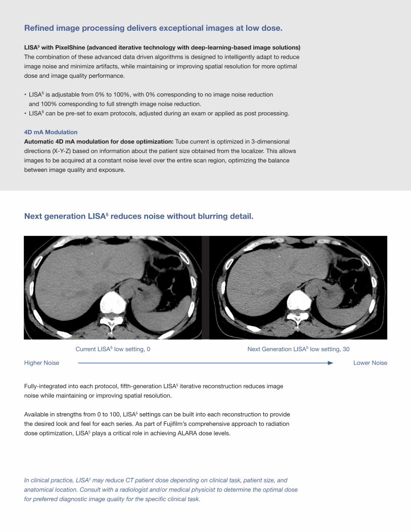
CurrentLISA⁵lowsetting,0
HigherNoise LowerNoise
NextGenerationLISA⁵lowsetting,30
In clinical practice, LISA5 may reduce CT patient dose depending on clinical task, patient size, and anatomical location. Consult with a radiologist and/or medical physicist to determine the optimal dose for preferred diagnostic image quality for the specific clinical task.
Next generation LISA5 reduces noise without blurring detail.
Fully-integratedintoeachprotocol,fifth-generationLISA5iterativereconstructionreducesimagenoisewhilemaintainingorimprovingspatialresolution.
Availableinstrengthsfrom0to100,LISA5settingscanbebuiltintoeachreconstructiontoprovidethedesiredlookandfeelforeachseries.AspartofFujifilm’scomprehensiveapproachtoradiationdoseoptimization,LISA5playsacriticalroleinachievingALARAdoselevels.
Refined image processing delivers exceptional images at low dose.
LISA5 with PixelShine (advanced iterative technology with deep-learning-based image solutions) Thecombinationoftheseadvanceddatadrivenalgorithmsisdesignedtointelligentlyadapttoreduceimagenoiseandminimizeartifacts,whilemaintainingorimprovingspatialresolutionformoreoptimaldoseandimagequalityperformance.
• LISA⁵isadjustablefrom0%to100%,with0%correspondingtonoimagenoisereductionand100%correspondingtofullstrengthimagenoisereduction.
• LISA⁵canbepre-settoexamprotocols,adjustedduringanexamorappliedaspostprocessing.
4D mA ModulationAutomatic 4D mA modulation for dose optimization: Tubecurrentisoptimizedin3-dimensionaldirections(X-Y-Z)basedoninformationaboutthepatientsizeobtainedfromthelocalizer.Thisallowsimagestobeacquiredataconstantnoiselevelovertheentirescanregion,optimizingthebalancebetweenimagequalityandexposure.

Dose Information Display and Distribution: Managingpatientdoseinformationiscritical;thesystemsimplifiesthatinformationtransfer.SimpleDoseReportsavesthedataasasecondarycaptureandsendstoPACS,whileDICOMDoseSRsendsthedoseinformationasastructuredreport.
A workflow that works for you.Your patients and your needs are front and center. Persona CT is engineered to provide a premium experience for your patients, raising the bar for comfort and safety. The system’s highly intuitive interface is designed for easy and precise operation, helping your imaging staff perform at their best.
People-firstDesign

High diagnostic imaging in a single breath hold.
Routine imaging with high speed sub-millimeter scanning: Thesystemiscapableofusingsub-millimeterslicesforhigh-speed,whole-bodyscans—somethingtraditional64or128sliceCTmachineshavetroubledoing.PersonaCTgeneratessubmillimeterhighresolutionimagesinanydimensionwithasinglebreathhold.
•Coveragerangesupto1900mm•Wholebodyimagesinjust16seconds
High Speed Scanning for better patient care: Witha40mmwidthdetectoranduniqueHQ3Dreconstructionalgorithm,thePersonaCTachieveshigh-speedscansevenwhenusingapitchof1.25.Fasterscansmeanlessburdenonpatients,especiallythosethathavedifficultyholdingtheirbreathorposition.
• 320mmcoveragescansin2.7seconds• 570mmThoraco-abdominalscansin5seconds

Maximize your imaging opportunities without taking up more space.
Open, Yet CompactClassleadingboresizecalmspatients,whileanunconventionallycompactfootprintfacilitatesinstallationintoexistingrooms.
Operator-friendly Designedwithyourtechnologistinmind.ThePersonaCTcomeswithsleekcustomizedstandalonegantrycontrols,alarge27-inchtouchscreendisplaywithallthecontrolsandinformationyouneedinasingleview.Thecontrollerisdirectlyintegratedwiththekeyboard,furtherconsolidatingeverythingintoonestreamlinedexperience.
Inyourhigh-stakesenvironment,efficiencyiseverything.Ourintuitiveuserinterface,withQuickEntrymode,fewerbuttons,andlargericons,speedsupyourprocesssignificantly.
Simple SitingThePersonaCTonlyhas4mainsystemmodules;gantry,patienttable,powerdistributionmodule,andoperationconsole.This,combinedwithourfewer-than-average100componentcount,achievesanimpressivelycompactfootprint.
85
50
cm wide bore
cm full FOV
PACSDose SR
Simple Dose Report
Chest Thoraco-abdominal
85
50
cm wide bore
cm full FOV
PACSDose SR
Simple Dose Report
Chest Thoraco-abdominal
HQ 3D algorithm satisfies both High-Speed Scans and High Image Quality: ThePersonaCTusesHighQuality3Dreconstructionsoftware.This3Dreconstructionalgorithmautomaticallyoptimizestherangeofacquisitiondata,creatinghighqualityimageswithlessartifacts—evenwithahigh-pitchscan.
Extends the potential of your service, without redundancy.

Best-in-class bore size for enhanced patient comfort.Ourbore-and-tabletoppairing,boththewidestintheindustryat85and49cmrespectively,cancomfortablyaccommodatevirtuallyanypatient.

Experience you can rely on.We’re the trusted partner at your side. With over 85 years of imaging expertise and best-in-class designs, Fujifilm has the technical know-how and service to support you. Our technologies create sharp images, simplified workflow, patient comfort, and reliability.
ReliabilityinAction

See the Fujifilm difference.Unprecedented image quality with advanced image processing and engineering design.
Additive Approach: Filtered Back Projection vs. LISA5 + PixelShineTheLISA⁵+PixelShinedeeplearningimagesolutionensuresthatPersonaCT’sscansarepreciseandhighresolutionwithaslittlenoiseaspossible,comparedtothetraditionalFilterBackProjectionmethod.
LISA⁵5mm PixelShine5mm+
ConventionalFBP5mm PixelShine5mmLISA⁵5mmvs. +

Old Gantry
PowerLinkPowerLinknon-contactpoweranddatatransferensuresthatPersonaCTishighlyreliable,easilyserviced,lightweight,andconsolidatedtolowermaintenancecosts.
Non-contact power & data transferPowerLinktechnologycompletelyeliminatesrelianceonoutdatedcarbonbrushesfoundinconventionalCTdesigns.Conventionalbrushescontributetoelectronicfailureandrequirefrequentmaintenance.PersonaCTeliminatestheseconcerns,insteademployinganon-contactdesigndatatransfer.
Experience the Fujifilm difference.Unprecedentedperformanceandreliabilitywithadvancedengineeringdesign.Engineeredfornoisereductionandreliability.
Modular DesignFewerfieldreplaceableunits(100inPersonaCTvs.200-500incompetitors)inthemachinemeanshigheruptime,shorterlearningcurveforservicepersonnelandfasterserviceability.

Plan PossibilitiesThePersonaCTincludesvariousplansforpartscoverageandservice,keepingyouruniqueneedsfrontandcenter.Weofferbothcomprehensiveplansandàlacarteoptionsforcomponentparts,toensurethatyou’regettingexactlywhatyouneed.
Dependable ServiceOurplanscomewithfactorytrained,highlyresponsivecustomerserviceteams,remotemonitoringcapability,anda12-monthwarranty.YoucanrestassuredthatyourplanwillcoverallaspectsofthePersonaCT’sstreamlinedcomponentdesign,makingmaintenanceeasyandaffordable.*
*with the exception of X-ray tubes and patient accessories
With fewer major parts and only 100 components, installation and maintenance are far more streamlined and hassle-free.
New Gantry

Flex however you see fit.From general radiology to radiation treatment planning, the scalable flexibility of Persona CT has you covered. Persona CT’s modular design can flex to fit a wide range of clinical needs from pediatrics, to bariatrics, to oncology, with added comfort, efficient workflow and better performance. We provide maximum adaptability to your ever-changing caseloads and budget.
FlexibleScalability
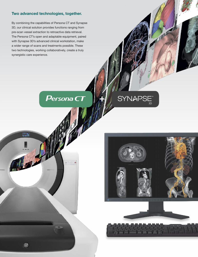
Two advanced technologies, together.
BycombiningthecapabilitiesofPersonaCTandSynapse3D,ourclinicalsolutionprovidesfunctionsrangingfrompre-scanvesselextractiontoretroactivedataretrieval.ThePersonaCT’sopenandadaptableequipment,pairedwithSynapse3D’sadvancedclinicalworkstation,makeawiderrangeofscansandtreatmentspossible.Thesetwotechnologies,workingcollaboratively,createatrulysynergisticcareexperience.
+
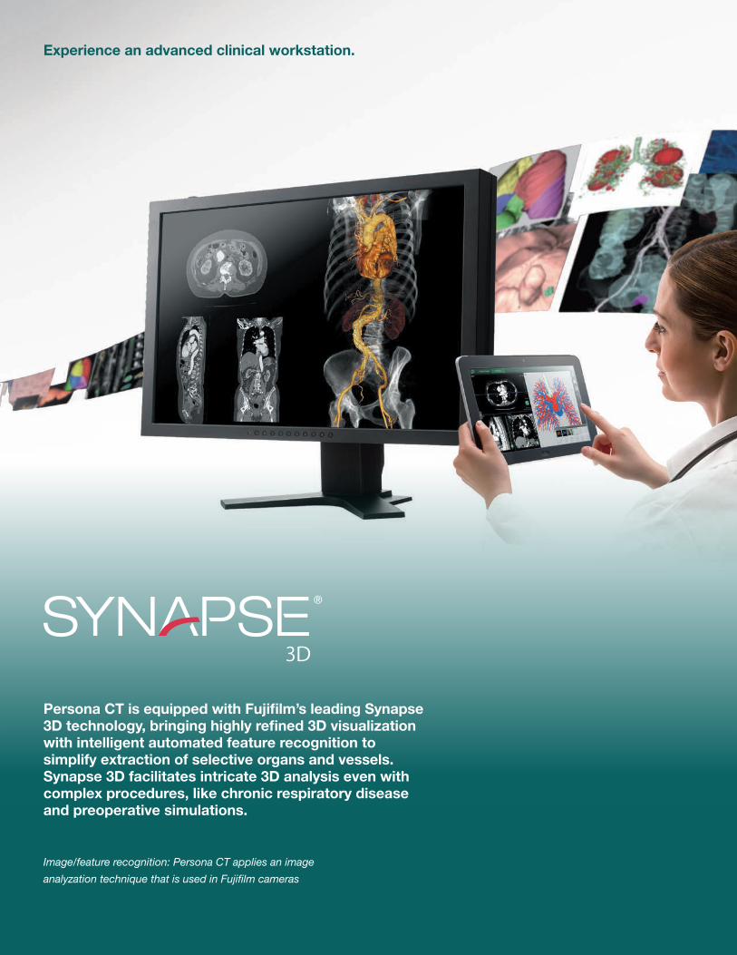
Persona CT is equipped with Fujifilm’s leading Synapse 3D technology, bringing highly refined 3D visualization with intelligent automated feature recognition to simplify extraction of selective organs and vessels. Synapse 3D facilitates intricate 3D analysis even with complex procedures, like chronic respiratory disease and preoperative simulations.
Image/feature recognition: Persona CT applies an image analyzation technique that is used in Fujifilm cameras
Experience an advanced clinical workstation.
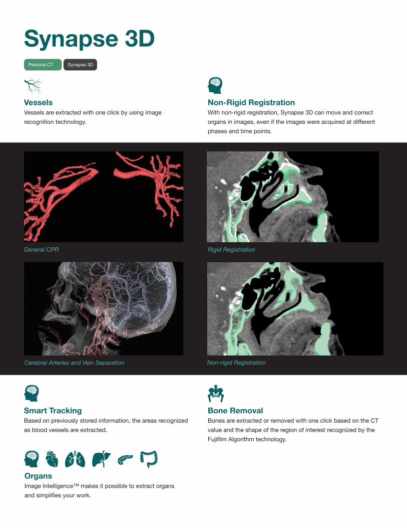
Non-Rigid RegistrationWithnon-rigidregistration,Synapse3Dcanmoveandcorrectorgansinimages,eveniftheimageswereacquiredatdifferentphasesandtimepoints.
Rigid Registration
Non-rigid Registration
Bone RemovalBonesareextractedorremovedwithoneclickbasedontheCTvalueandtheshapeoftheregionofinterestrecognizedbytheFujifilmAlgorithmtechnology.
General CPR
Cerebral Arteries and Vein Separation
VesselsVesselsareextractedwithoneclickbyusingimagerecognitiontechnology.
Smart TrackingBasedonpreviouslystoredinformation,theareasrecognizedasbloodvesselsareextracted.
OrgansImageIntelligence™makesitpossibletoextractorgansandsimplifiesyourwork.
Synapse 3DPersona CT Synapse3D

BrainVessel Extraction & Separation Withjustasingleclick,operatorscanachieveclearvesselextractionandseparationofcerebralarteryandveinthroughthetechnologyinPersonaCTandSynapse3D.
Technology for Precise Subtraction:
•OrbitSynchronizationinHelicalScan(Thetwoorbitsofhelicalscansbeforeandaftercontrastcanbesynchronized,whichimprovestheaccuracyofsubtractionimage.)
•Non-rigidRegistration(Non-rigidregistrationallowsnaturaladjustmentofbodypartsthatotherwiseisdifficultinlinearregistration.)
Subtraction
PersonaCT Synapse3D
Directional Control of RadiationPersonaCTallowsyoutocontrolthedirectionoftheradiationmoreprecisely,incorporatingatiltinggantryangleupto30°.Inturn,it’seasiertoavoidartifactsfromteethandpotentialdosetolensesoftheeyes.
PersonaCT Synapse 3D
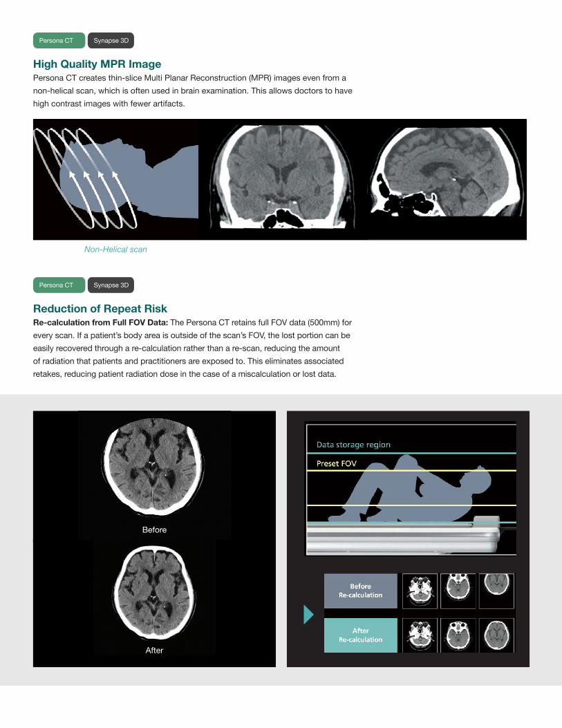
Reduction of Repeat RiskRe-calculation from Full FOV Data:ThePersonaCTretainsfullFOVdata(500mm)foreveryscan.Ifapatient’sbodyareaisoutsideofthescan’sFOV,thelostportioncanbeeasilyrecoveredthroughare-calculationratherthanare-scan,reducingtheamountofradiationthatpatientsandpractitionersareexposedto.Thiseliminatesassociatedretakes,reducingpatientradiationdoseinthecaseofamiscalculationorlostdata.
High Quality MPR ImagePersonaCTcreatesthin-sliceMultiPlanarReconstruction(MPR)imagesevenfromanon-helicalscan,whichisoftenusedinbrainexamination.Thisallowsdoctorstohavehighcontrastimageswithfewerartifacts.
Non-Helical scan
PersonaCT Synapse 3D
PersonaCT Synapse 3D
Before
After
Advanced Iterative Reconstruction with Noise ReductionPatented breakthrough deep-learning-based software dramatically reduces noise in CT Scans.
LISA5 (Low-dose Iterative Solution Approach) with PixelShine:Appliesadvancednoisereductionanditerativereconstructiontoreduceimagenoiseandartifactswhilemaintaininghighimagequalityatlowdoses.Theartificialneuralnetworkalgorithmsignificantlyenhancesdetailinlowdoseandunderpenetratedimages.Thecombinationofadvancediterativereconstruction,noisereductionandpixel-enhancementalgorithmshelpreduce
imagenoisewhilemaintainingorimprovingspatialresolution.TheLISA⁵Weightingpercentageisappliedthroughadjustableincrementsfrom0%to100%strengthimagenoisereduction.Weightingcanbepre-setinprotocols,adjustedduringanactiveexam,orevenappliedaspostprocessing.
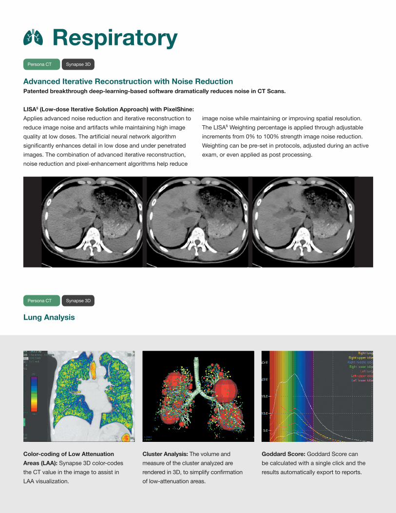
Respiratory
Color-coding of Low Attenuation Areas (LAA):Synapse3Dcolor-codestheCTvalueintheimagetoassistinLAAvisualization.
Cluster Analysis:Thevolumeandmeasureoftheclusteranalyzedarerenderedin3D,tosimplifyconfirmationoflow-attenuationareas.
Goddard Score:GoddardScorecanbecalculatedwithasingleclickandtheresultsautomaticallyexporttoreports.
Lung Analysis
Persona CT Synapse3D
PersonaCT Synapse 3D
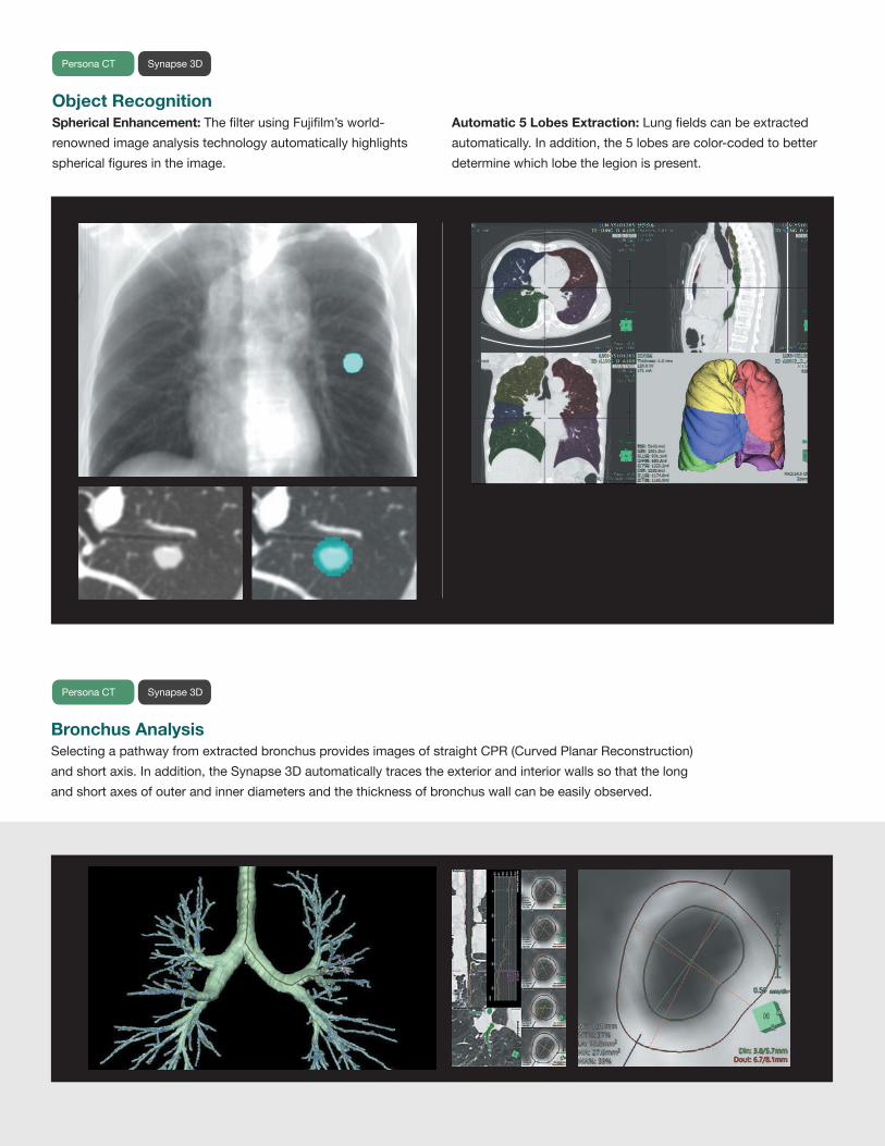
Object Recognition Spherical Enhancement: ThefilterusingFujifilm’sworld-renownedimageanalysistechnologyautomaticallyhighlightssphericalfiguresintheimage.
Automatic 5 Lobes Extraction:Lungfieldscanbeextractedautomatically.Inaddition,the5lobesarecolor-codedtobetterdeterminewhichlobethelegionispresent.
Bronchus Analysis SelectingapathwayfromextractedbronchusprovidesimagesofstraightCPR(CurvedPlanarReconstruction)andshortaxis.Inaddition,theSynapse3Dautomaticallytracestheexteriorandinteriorwallssothatthelongandshortaxesofouterandinnerdiametersandthethicknessofbronchuswallcanbeeasilyobserved.
Persona CT Synapse3D
Persona CT Synapse3D

High Quality 3D Reconstruction High Speed Scanning with HQ 3D Reconstruction Method: HQ3DReconstruction(HighQuality3DReconstruction)method,whichisaunique3Dreconstructionalgorithm,optimizestherangeofacquisitiondatatobereconstructed.Byutilizingthedataacrossthewholedetectoreffectively,ahighqualityimagewithfewerartifactscanbeobtainedevenwithahigh-pitchscan.
4DdosemodulationdynamicallyadjustsmAinX,Y,andZdirectionsthroughoutthedurationofthescanwiththegoalofprovidingconsistentimagequalityandlowestachievabledoses.Tubecurrentdynamicallychangesbasedonthesizeofpatientandorgans.
Whole Body & Vessels
FeldKamp Method(Conventional3Dimagereconstruction)
CORE Method(New3Dimagereconstruction)
225 mA
50 mA
184 mA
PersonaCT Synapse 3D
PersonaCT Synapse 3D
2.7 sec 5 sec320mm 570mm
Automatic 4D mA Modulation for Dose Optimization

Torous Enhancement FilterThecolonareacanbeobservedinafillet-viewformat.Withtheenhancementfilter,theprotrusionsonthecolonwallcanmoreeasilybeidentified.
Virtual Endoscopy Synapse3Dautomaticallyextractsthecolontoprovideavirtualendoscopyimage.Thisallowsinspectionoftheperipheralregionofalimentarycanal,theregionbeyondthebronchusarea,andgivesanoptiontohavecolondiagnosiswithouttheinvasivepatientprocedure.
Colon
Vessel Analysis
Easy CPR image creation:SelectionofvesselstoapplyCurvedPlanarReconstruction(CPR)isavailablebyclickingastartingandendpointwhichcreatesaStraightreconstructionoftheidentifiedvessel.StraightCPR,stretchCPRandcross-sectionviewscaneasilybegenerated.
Aneurysm Analysis:Measurementresultofaneurysmandvesseldiametersuchasthoracicaorticaneurysm(TAA)andabdominalaorticaneurysm(AAA)canbeprovided.Virtualimplantationofstentgraftsallowsclinicianstomakeinformedchoicesaboutwhichdevicetousethroughsimulation.
Lowdose1.3mSv IntelliIPLEVEL7
Persona CT Synapse3D
Persona CT Synapse3D Persona CT Synapse3D

Multi-angle Multiplanar Reformation (MPR) for Vertebral Observation Reconstructioncanbeperformedatmultipleanglesalongeachvertebraldisctoeasilyobservetheconditionofvertebralbodiesanddiscs.Standardtemplatesforreconstructionpositionandanglesareprovidedforcervicalandlumbarspinebutarbitraryselectionisalsoavailable.
Bone SeparationAdvancedobjectrecognitiontechnologycombinedwithcolordelineationofcomponentsassistsinvisualizationofboneseparationandjoints.
OrthopedicsPersonaCT Synapse 3D
Persona CT Synapse3D
Oncology CareWhat if there was a way to streamline and consolidate the high-stakes oncology care process, from first scan to last treatment?

Persona CT Oncology Options:
• 85cmCT•AAPMTG66TableforRTPPlanning*• TreatmentPlanningSoftware• AlignmentPositioningLasers• 4DRespiratoryGating*• Synapse3DforAdvancedVisualization
Persona CT from Fujifilm answers that need.
OurCTsolutionisequippedwithbest-in-classsoftwareandtechnologyintegration,anddesignedwithoncologypatientsandpractitionersinmind.FromLAPlaserpositioningtotheworld’sfirst85cmCTbore(64or128slice),PersonaCTisoptimizedtoensurepatientscanbeimagedintheoptimaltreatmentposition,whilemaintainingthepositionalandaccuracyrequirementsofradiationoncology.
Whole body coverage with 190cm scan length for patients over 6’ 2” tall (standard).
* Oncology options currently unavailable in the US
Oncology Care

Laser SystemsPrecisepatientmarking,accurateplanningandexactpositioningarekeyfactorsforasuccessfultreatment.PersonaCTisavailablewithleadingLaserSystemsthathelpwitheachoftheseareas:
•markingtheappropriateanatomicallocation• referencepointmarking• finalisocentermarking• fieldmarkingLasersystemsareavailablewithmultipleconfigurationsandmountingoptionstomeetanyroomrequirement.
4D Gating Capabilities*• AmplitudeBinning&Phase-BasedImagingprovideadvancedmotionmanagementandradiationtherapyplanning
• FlexibleoptionsofgatingdevicesfromVarianRPMtoBellowsStandardRespiratoryGatingDevices
• ProspectiveAxial• ProspectiveHelical•RetrospectiveHelical•Rapid10PhaseReconstructioninlessthan2minutes• TumorMotionandPhaseinformationforpatientmarkingusing4DMIPS
Synapse 3D Capabilities•CoronaryAnalysis• PET/CTFusion• LiverAnalysis• LungAnalysis•HeadAnalysis• 2Dand3DFatAnalysis• VesselAnalysis 85cm Bore Solutions that Matter• EquippedwithdedicatedRadiationOncologyprotocols.• Increaseddetectorcoverageandimprovedrotationspeedresultin4xfasterscanswithsameresolution,enhancingpatientcomfort.
• Improvedacquisitionspeedalongwiththe4cmdetectorcoveragehelpswithpatientswhohavedifficultywithlyingstill,DeepInspirationBreathHolds(DIBH),andrespiratorystudies.
•MaximizeROI-PersonaCTtranslatesdirectlytotheRadiologyDepartment,minimizinglearningcurvesandmaximizingflexibility.
* Currently unavailable in the US

Capabilities The Fujifilm Difference
Fujifilm 85cm Large Bore CTSimulator
Standard Bore Multislice CT
16/32 Slice 85cm CT Simulator
Radiation Therapy Planning suitable
System is designed with RTP in mind
Fujifilm 85cm Large Bore
Matches Arc of Linear Accelerator for maximum positional accuracy and more precise treatment planning
Large eFOV (60cm+)* Extend ability to do surface measurements
PowerLink Technology System reliability and reduced service costs with no brush blocks
49cm Wide Comfort Pallet Tabletop
Widest tabletop in the industry for enhanced patient comfort and safety
0.4 sec Rotation Speed Faster rotation time to improve temporal resolution especially for 4D acquisitions
200 msec Temporal Resolution
One of the fastest in this segment
4D Respiratory Gating Technology*
Advanced motion management with amplitude binning for more precise dose planning
4cm Detector Coverage
Wider coverage equals faster scan times
Metal Artifact Reduction*
Metal Artifact Reduction (MAR) is a projection-based reconstruction technique that restores anatomical details often obscured by metal in conventional image reconstruction. MAR automatically identifies areas affected by highly-attenuating objects and reconstructs improved images for increased clinical confidence. Next generation metal artifact reduction dramatically removes and/or reduces the streak artifact created by metal.
64 and 128 slice configurations
World’s First 64 and 128 slice configuration in Large Bore environment with 85cm Bore
TG-66 Compliant Table*
Meets AAPM TG66 requirements for positional accuracy and table movement
Rapid Image Acquisition up to 42ips
Image Quality High High High
Medium Medium HighSystem Price
Rapid reconstruction rate with Real-time Image Preview to streamline workflow
The Fujifilm Difference
*These items are works in progress: not commercially available

Gantry
User Interface Features
HIS/RIS/PACS Integration Services
Clinical Application Packages (standard)
Clinical Application Packages (optional)
X-ray Tube
X-Ray Generator
X-Ray Detection
Scan Parameters
Image Quality
Image Reconstruction
Patient Table
Radiation Dose Management
System Console
PatientAperture:85cmScanFieldofView:50cmGantryTilt:±30°ScanTimes(360°):CT64/128:0.4,0.5,0.67,1
GantryDimensionsHxWxD:79.5x98.1x42.3inInputPowerRequirements:380-400/480VAC,3phase,50/60Hz
ClinicalProtocolDrivenwithOverridesTouchscreenWorkflowAutomatedwithTaskProtocolsAutomaticMPRReconstruction
Auto-ArchiveAdvancedHelpSecondWorkstation(optional)withSynapse3D
DICOM3.0ModalityWorkListSCUIHEProfilesforRadiationDoseStructuredReports
MPRMiP/MiniP3DShadedSurfaceDisplay
3DVolumeRenderingBolusTrigger/DynamicEvaluationCTAngiography
3DVolumeRenderingBolusTrigger/DynamicEvaluationCTAngiographyVirtualColonographyCTPerfusion
LungNoduleAnalysisCardiacGating/TriggeringCoronaryArteryCalciumScoringBoneMineralDensityImageFusion
AnodeHeatCapacity:CT64/128:8MHUMaximumHeatDissipation:CT64/128:931kHU/minute,OiltoAir
Power:Effective105kWwithLISA⁵kVRange:80,100,120,140kVpCurrentRange:10to660mA
NumberofDetectorElementsinX-Direction:57,344elements(64x896)NumberofDASRows:64SliceDoublingTechnique:Optional128slice
DetectorWidth(Zdimension):CT64/128:40mmatiso-centerMaximumHelicalScanTime:120secondsMaximumScanVolume:190cm
AcquiredSliceThickness:64or128sliceof0.625mmDisplayedSliceThickness:0.625,1.25,2.5,5,7.5,10mmScanModes:Localizer,Axial,Helical,Cine
SpatialResolution:17.2Lp/cmat0%MTF
Maxreconstructionrate:Upto42imagespersecondReal-timeImagePreviewIterativeBoneCorrection
VerticalTravelRange:48cmto101cm(18.9into39.8in)HorizontalScannableRange:190cm(74.8in)withextensionsHorizontalTravelSpeed:Upto180mm(7.1in)/sPatientWeightMaximum:225kg(500lbs)orOptional300kg(660lbs)
HorizontalAccuracy:±0.5mmTablePalletWidth:49cm(19.3in)TableWidth:78cm(30.7in)IncludedAccessories:TabletopSlickerPad,HeadHolder&Pad,FootExtensionBoard&SlickerPad,PatientRestraints,IVPole&Holder,KneePad
PediatricProtocolsLISA5withPixelShine4DmAmodulationProtocolDrivenDualBowtieFilters
AutomaticFocalSpotSelectionExpandedkVRangesScalableFlexTileDetectorsNEMAXR-29
DisplayMonitor:One(1)27”FullHDFlatPanelTouchscreenDisplayMatrix:1920x1080DisplayDimensionsHxWxD:26.5”x16.5”x2.0”Workstation:Inteli7,Windows
SystemMemory:16GBytesSystemHardDisk:2TBytesImageDataStorage:DVD>250,000ImagesSystemArchivalStorage:DVD
Technology Specs
System Specifications
Specifications

FujifilmMedicalSystemsUSA,Inc.Corporationisarecognizedleaderinadvancedimagingtechnologies.ContactyourFujifilmrepresentativeformoreinformation.Technicalspecificationssubjecttochangewithoutnotice. XBUSCT0001
#VisionaryCT
FujifilmMedicalSystemsU.S.A.,Inc.www.fujimed.com866-879-0006©2019FujifilmMedicalSystemsU.S.A.,Inc.