Embed Size (px)
Citation preview

From DEPARTMENT OF PHYSIOLOGY AND PHARMACOLOGY Karolinska Institutet, Stockholm, Sweden
ENDOGENOUS KYNURENIC ACID AND SCHIZOPHRENIA – PHYSIOLOGICAL AND PHARMACOLOGICAL ASPECTS
By
Lilly Schwieler
Stockholm 2006

Published and printed by Karolinska University Press Box 200, SE-171 77 Stockholm, Sweden © Lilly Schwieler, 2006 ISBN 91-7140-704-9

To my surprise but to Kristina Ikelbergs wisdom

ABSTRACT
Kynurenic acid is a glutamate receptor antagonist with a preferential action at the glycine/D-serine site of the N-methyl-D-aspartic acid (NMDA) receptor. The compound is a metabolite of tryptophan and is synthesized in astrocytes. Previous studies have shown increased levels of kynurenic acid in the CSF and post mortem in the prefrontal cortex of patients with schizophrenia. The aim of the present thesis was to further investigate in the rat the physiological significance of kynurenic acid as well as its importance for the pathophysiology of schizophrenia. Studies include a disclosure of a prostaglandin-mediated regulation of kynurenic acid synthesis and analysis of the role of the compound in the regulation of firing activity of dopamine neurons in ventral tegmental area (VTA). Furthermore, an interaction of the antipsychotic drug clozapine with endogenous kynurenic acid is analyzed and the significance of kynurenic acid for behavior is studied using prepulse inhibition (PPI) methodology. Systemic administration of diclofenac and indomethacin, inhibitors with a preferential selectivity for cyclooxygenase (COX)-1, was associated with an increased formation of kynurenic acid in brain, whereas meloxicam and parecoxib, selective COX-2 inhibitors, decreased brain kynurenic acid formation. Both the elevation and the lowering in brain kynurenic acid levels following administration of the COX inhibitors were effectively prevented by the prostaglandin E1/E2 analog misoprostol. Pharmacological manipulation of kynurenic acid synthesis with COX inhibitors thus enabled us to study a role of the compound in the control of firing of midbrain dopamine neurons. An increase in brain kynurenic acid concentration (by 150-300%), induced by indomethacin, increased firing rate and burst firing activity of VTA dopamine neurons whereas a reduction in brain kynurenic acid concentration (by 39-44%) elicited by parecoxib, was associated with a clear-cut reduction in firing activity of these neurons. Thus, endogenous brain concentrations of kynurenic acid appear to be of critical physiological importance for maintaining neuronal activity of VTA dopamine neurons. In the next series of experiments we investigated the effects of clozapine and haloperidol on VTA dopamine neurons in rats with attenuated NMDA receptor function induced by increased levels of endogenous brain kynurenic acid. Here, clozapine, in contrast to haloperidol, was found to interact with the NMDA receptor complex. We propose a novel mechanism of action of the atypical antipsychotic drug clozapine, i.e. stimulation of the glycine/D-serine site of the NMDA receptor. Elevated levels of endogenous kynurenic acid induced by systemic administration of kynurenine or PNU 156561A were associated with a disruption in PPI, an effect that could be reversed by antipsychotic drugs. It is proposed that kynurenic acid acts as an endogenous modulator of the PPI response. Taken together, the results of the present thesis suggest that endogenous kynurenic acid in the brain is involved in the physiological regulation of glutamate neurotransmission. Hereby, this endogenous NMDA receptor antagonist may participate in the pathophysiology of schizophrenia.

LIST OF PUBLICATIONS
I. Schwieler L, Erhardt S, Erhardt C, Engberg G (2005). Prostaglandin-mediated
control of rat brain kynurenic acid synthesis – opposite action by COX-1 and COX-2 isoforms. Journal of Neural Transmission 112;863-872.
II. Schwieler L, Erhardt S, Nilsson L, Linderholm K, Engberg G (2006). Effects of
COX-1 and COX-2 inhibitors on the firing of rat midbrain dopaminergic neurons -possible involvement of endogenous kynurenic acid. Synapse 5;290-298.
III. Schwieler L, Erhardt S. (2003). Inhibitory action of clozapine on rat ventral
tegmental area dopamine neurons following increased levels of endogenous kynurenic acid. Neuropsychopharmacology 28;1770-1777.
IV. Schwieler L, Engberg G, Erhardt S (2004) Clozapine modulates midbrain
dopamine neuron firing via interaction with the NMDA receptor complex. Synapse 52;114-122.
V. Erhardt S, Schwieler L, Emanuelsson C, Geyer M (2004). Endogenous kynurenic
acid disrupts prepulse inhibition. Biological Psychiatry 56;255-260.

TABLE OF CONTENTS
1 INTRODUCTION.......................................................................................... 1
1.1 Schizophrenia ...................................................................................... 1
1.2 Pharmacological treatment of schizophrenia ...................................... 2
1.3 The dopamine system of the brain ...................................................... 4
1.3.1 Dopamine pathways...................................................................................... 4
1.3.2 Electrophysiology of midbrain dopamine neurons ............................................. 6
1.3.3 Afferent regulation of dopamine neurons in Ventral Tegmental Area............... 7
1.3.4 The dopamine hypothesis of schizophrenia....................................................... 9
1.4 The glutamatergic system ..................................................................10
1.4.1 NMDA receptors....................................................................................... 11
1.4.2 The glutamate deficiency theory of schizophrenia ............................................ 12
1.5 Kynurenic acid....................................................................................13
1.5.1 The kynurenine pathway ............................................................................. 14
1.5.2 Regulation of kynurenic acid synthesis .......................................................... 16
1.5.3 Mechanism of action and physiological significance......................................... 17
1.5.4 The kynurenine pathway is activated by immune stimulation ......................... 18
1.6 Cyclooxygenases and the central nervous system...............................19
1.7 The kynurenic acid hypothesis of schizophrenia............................... 20
2 SPECIFIC AIMS OF THE STUDY.............................................................. 22
3 MATERIALS AND METHODS................................................................... 23
3.1 Animals.............................................................................................. 23
3.2 Drugs ................................................................................................. 23
3.3 Pretreatment with PNU 156561A (paper III and V) ........................... 24
3.4 In vivo electrophysiology ................................................................... 25
3.4.1 Anesthesia and surgery................................................................................ 25
3.4.2 Maintenance of general anesthesia ................................................................ 25
3.4.3 Preparation of recording electrode.................................................................. 26
3.4.4 Extracellular single cell recording ................................................................. 26
3.4.5 Identification of VTA dopamine neurons..................................................... 27
3.4.6 Electrophysiological characteristics of dopaminergic neurons ............................ 27
3.4.7 Drug administration and experimental protocol............................................. 27
3.4.8 Influence of anesthesia.................................................................................. 28
3.4.9 Data analysis ............................................................................................. 29

3.5 Prepulse inhibition .............................................................................29
3.5.1 Apparatus ................................................................................................. 29
3.5.2 Experimental protocol................................................................................. 30
3.6 Analysis of kynurenic acid.................................................................. 31
3.6.1 Sample preparation..................................................................................... 31
3.6.2 High performance liquid chromatography...................................................... 31
3.6.3 Chemicals................................................................................................... 32
3.7 Statistical analysis...............................................................................32
3.7.1 Biochemical and electrophysiological studies (paper I-IV) ............................... 32
3.7.2 Behavioral studies, PPI (paper V)............................................................... 33
4 RESULTS AND DISCUSSION.....................................................................34
4.1 Effects of cyclooxygenase inhibitors on kynurenic acid synthesis and
on neuronal activity of midbrain dopamine neurons (paper I and II) 34
4.1.1 Effects of cyclooxygenase inhibitors on kynurenic acid formation in the rat brain..
(Paper I) .................................................................................................... 34
4.1.2 Effects of cyclooxygenase-1 and cyclooxygenase-2 inhibitors on neuronal firing of
rat midbrain dopaminergic neurons (Paper II)............................................... 36
4.1.3 Involvement of endogenous kynurenic acid in the effects of cyclooxygenase inhibitors
on dopamine firing (paper II)....................................................................... 38
4.2 The atypical antipsychotic drug clozapine affects the neuronal activity
of midbrain dopamine neurons by interfering with glutamatergic
mechanisms (Paper III and IV). ........................................................40
4.2.1 Effects of antipsychotic drugs on midbrain dopamine firing in rats with elevated
levels of endogenous brain kynurenic acid (paper III)................................................... 40
4.2.2 Clozapine modulates midbrain dopamine neurons via interaction with the
NMDA receptor (paper IV) ................................................................................... 41
4.3 Increased levels of endogenous kynurenic acid disrupt prepulse
inhibition - a behavioral model for schizophrenia (paper V)..............43
5 GENERAL DISCUSSION.............................................................................46
6 ACKNOWLEDGEMENTS ........................................................................... 51
7 REFERENCES..............................................................................................53

LIST OF ABBREVIATIONS
AMPA amino-3-hydroxy-5-methyl-4-isoxazole propionic acid
ANOVA analyses of variance
COX cyclooxygenase
CNS central nervous system
EPS extrapyramidal side effects
GABA γ-aminobutyric acid
HIV human immunodeficiency virus
IDO indoleamine 2,3-dioxygenase
ISH interspike time interval histogram
KAT kynurenine aminotransferase
LTP long term potentiation
mGluR metabotropic glutamate receptor
NAD nicotinamide adenine dinucleotide
NMDA N-methyl-D-aspartate
NSAID nonsteroidal anti-inflammatory drugs
PCP phencyclidine
PET positron emission tomography
PG prostaglandin
PPI prepulse inhibition
TDO tryptophan 2,3-dioxygenase
VTA ventral tegmental area

Introduction
1
1 INTRODUCTION
1.1 Schizophrenia
Schizophrenia is a serious mental illness which impairs both functioning and personality,
and therefore impacts almost every aspect of life. This mental disease, which affects
approximately 1% of the world’s population (Carpenter and Buchanan, 1994; Jablensky,
1997) strikes people early in life just when they are about to achieve adulthood (Almeida et
al., 1995). Even though the outcome is variable and some patients do well, the majority
will experience a lifetime of disability and about 10% of the affected individuals will
eventually commit suicide (Black and Fisher, 1992; Meltzer, 2002). As a result,
schizophrenia is not only associated with substantial emotional burden for the families of
those affected but is also a tremendous economic cost to society in terms of healthcare
and lost productivity.
There are currently no biological markers for schizophrenia and therefore its
diagnosis relies solely upon clinical assessment of each patient’s symptoms. However, the
clinical presentation of schizophrenia is very complex with numerous different symptoms
that vary from patient to patient. To guide clinicians and investigators to survey signs and
symptoms in a standardized manner, criterion-based systems for diagnosing have been
developed during the past several decades. One of the most widely used is the fourth
edition of the American Psychiatric Association’s Diagnostic and Statistical Manual (DSM-
IV). In this criterion-based system symptoms of schizophrenia are divided into three broad
clusters: positive symptoms, negative symptoms and cognitive impairment (Andreasen,
1995). The positive symptoms (or psychotic symptoms) involve the loss of contact with
reality, including false beliefs (delusions), perceptual experience not shared by others
(hallucinations), bizarre behavior and disorganized speech. The manifestation of psychotic
symptoms is not constant in presence or intensity, but tends to fluctuate over time (Green
et al., 2000). The negative symptoms reflect diminishment or absence of emotional and
behavioral processes. Common negative symptoms are lack of pleasure (anhedonia),
withdrawal from social contacts, amotivation, apathy, and reduced quantity or content of
speech (alogia). Negative symptoms are associated with poor psychosocial functioning
(Tek et al., 2001) and are usually more pervasive and fluctuate less over time than
psychotic symptoms (Fenton and McGlashan, 1991). Patients with schizophrenia also
suffer from cognitive impairments such as impaired attention, memory and executive
functions (e.g. the ability to plan, initiate and regulate goal directed behavior). The majority

Lilly Schwieler
2
of the patients show a reduction in cognitive abilities at the initial onset of psychoses
(Saykin et al., 1994).
The complexity of schizophrenia has made the exploration of the underlying cause
of the disease extremely difficult. However, data from family and twin studies show that
the rates of schizophrenia are higher among relatives of people with the disease than in the
general population, which indicates that genetic factors contribute to the disease. Although
several putative susceptibility genes for schizophrenia have been identified, the risk
conferred by any single gene seems to be small. In addition, the degree of concordance for
schizophrenia among monozygotic twins (sharing 100% of the same genes) is
approximately 50% (Farmer et al., 1987; Cardno et al., 1999), indicating that epigenetic and
environmental factors may also be important for development of the disease.
Environmental risks that have been associated with schizophrenia include biological and
psychosocial factors such as urban upbringing, social isolation, migration, drug abuse,
season of birth, and obstetric complications (Susser, 2002). Furthermore, recent
epidemiological studies reveal that an immunologic component may also play a part in the
etiology of schizophrenia (Sperner-Unterweger, 2005). For instance, prenatal infections
during the second trimester are associated with an increased risk of schizophrenia (see
Brown, 2006). This includes infection with influenza virus (Mednick et al., 1988; Cannon
et al., 1996; Limosin et al., 2003; Brown et al., 2004), rubella (Brown et al., 2001) poliovirus
(Suvisaari et al., 1999) and herpes simplex virus (Buka et al., 2001). It has also been
postulated that retroviruses may be important in the etiopathogenesis in schizophrenia
since retroviral sequences (HERV-W) have been found in CSF from these patients
(Karlsson et al., 2001). These results may indicate that several types of infections may
increase the risk of schizophrenia, tentatively through some common immunological
mechanism.
1.2 Pharmacological treatment of schizophrenia
The first useful treatment for schizophrenia was discovered by the French suregon Henri
Laborit in the beginning of the 1950s. When searching for an antihistaminic that could
calm patients before surgery, he found by serendipity a new compound with an
exceptional tranquilizing property. Laborit hypothesized that a drug that calmed patients
approaching surgery might also be useful for patients with psychiatric symptoms. Acting
on this suggestion, John Delay and Pierre Deniker found within a year that the compound,
chlorpromazine, had remarkable beneficial effects in patients suffering from psychotic

Introduction
3
illnesses (Delay et al., 1952). Chlorpromazine revolutionized psychiatry and in particular it
represented a major breakthrough in the treatment of schizophrenia. However, it soon
became clear that chlorpromazine and other drugs that followed its introduction, like
haloperidol, induced extrapyramidal side effects (EPS) e.g. abnormal involuntary
movements. Furthermore, although these drugs were useful against positive symptoms,
they were found to have little or no effect against negative and cognitive symptoms (Delay
et al., 1952; Seeman and Lee, 1975; Creese et al., 1976; Palao et al., 1994; see King 1998).
The mechanism of action of these classical antipsychotic drugs was at this time unknown
and it took almost a decade before Carlsson and Lindqvist suggested that haloperidol and
chlorpromazine block monoamine receptors (Carlsson and Lindqvist, 1963). A few years
later, the clinical efficacy of typical antipsychotics was shown to be due to their ability to
block dopamine D2 receptors in the central nervous system (CNS; Seeman and Lee 1975;
Creese et al., 1976). The introduction of positron emission tomography (PET) made
visualization and quantification of radiolabelled D2 receptors possible and the exact
receptor occupancy following antipsychotic treatment could be estimated. These studies
revealed that, for typical antipsychotics, receptor occupancy in the striatum of at least 70%
was required for antipsychotic efficacy while occupancy above 80% potentially increased
the risk of EPS (Farde et al., 1988; 1992; Farde and Nordström, 1993; Nordström et al
1993; Kapur et al., 1996).
One of the most effective antipsychotic drugs used today is clozapine, which has a
remarkable efficacy in treatment-resistant schizophrenia (Kane et al., 1988; 2001; Pickar et
al., 1992). It is classified as an atypical antipsychotic drug due to its very low incidence of
EPS associated with its use (Claghorn et al., 1987; Coward et al., 1989). Clozapine has also
been shown to be superior in ameliorating both negative and cognitive symptoms, in
comparison to classical antipsychotics (Kane et al., 1988; see Baldessarini and
Frankenburg, 1991; see Meltzer and McGurk, 1999). Despite these advantages, clinical use
of the drug has been limited by a low but significant risk of potentially fatal agranulocytosis
(Idänpään-Heikkilä et al., 1977; see Krupp and Barnes, 1992). The receptor pharmacology
of clozapine is complex since the compound binds to many receptors in the brain.
Clozapine interacts not only with all dopamine receptors (the D4 subtype showing the
highest affinity: 19 nM, Van Tol et al., 1991), but also with other metabotropic receptors
e.g. those for serotonin (5HT1 and 5HT2, Canton et al., 1990), acetylcholine (Snyder et al.,
1974), noradrenaline (see Coward, 1992), and histamine (see Coward, 1992; see Brunello et
al., 1995). Various pharmacological explanations have been put forward in an attempt to
explain the unique clinical profile of clozapine. These include a relative dopamine D4

Lilly Schwieler
4
selectivity (Seeman et al., 1998) or a preferential action of clozapine on the mesolimbic
dopamine system as compared to the nigrostriatal dopamine system (Andén and Stock,
1973; Bartholini, 1976; Chiodo and Bunney, 1983; 1985; Moghaddam and Bunney, 1990).
Moreover, since clozapine is a potent serotonin 5-HT2-receptor antagonist, it has been
suggested that concurrent 5-HT2 and dopamine D2 receptor antagonism may contribute to
its atypical profile (see Deutch et al., 1991; Meltzer and Nash, 1991; Ichikawa et al., 2001).
Another potential explanation for the preferential efficacy of clozapine might be its ability
to potentiate glutamatergic neurotransmission. Thus, clozapine has been shown to
modulate the response to N-methyl-D-aspartate (NMDA) in frontal cortex slices in vitro
(Arvanov et al., 1997; Chen et al., 2002, Jardemark et al., 2003) and to prevent
phencyclidine (PCP)-induced NMDA receptor hyperactivity in vivo (Arvanov and Wang,
1999). Furthermore, studies utilizing microdialysis indicate that the systemic administration
of clozapine increases extracellular levels of glutamate (Daly and Moghaddam, 1993;
Yamamoto et al., 1994). However, the pharmacology underlying the unique antipsychotic
action of clozapine is still obscure.
The superiority of clozapine over classic antipsychotic drugs has stimulated
researchers to develop novel compounds with similar therapeutic profiles but without
clozapine’s severe side effects. This has resulted in a new generation of drugs (e.g.
olanzapine, quetiapine, risperidone, sertindole) that are widely used in the treatment of
schizophrenia. However, the safety advantage of the atypical drugs has been questioned
because of their ability to induce potentially important adverse side effects such as weight
gain and altered glucose and lipid metabolism, resulting in increased risk of cardiovascular
dysfunction (see Gardner et al., 2005). Furthermore, several studies have shown that the
majority of patients discontinue treatment due to inefficacy or intolerable side effects
(Lieberman et al., 2005). Although the new generation of antipsychotic drugs represents a
step forward, they are far from ideal. Obviously, further research is required in order to
develop more effective, safe and well-tolerated drugs for the treatment of schizophrenia.
1.3 The dopamine system of the brain
1.3.1 Dopamine pathways
The dopamine system constitutes a very small fraction of the several billions of neurons in
the brain (see Williams and Herrup, 1988; see Prakash and Wurst, 2006). Nevertheless, this
system has been shown to be a key modulator in an astonishing array of human behaviors.

Introduction
5
This includes physical movement, motivation and control of emotions (e.g. reward and
reinforcement), cognitive functions and endocrine regulation.
Figure 1. Dopaminergic pathways in the human brain. Abbreviations: Am= amygdala; Hip= hippocampus; Hyp= hypothalamus; NAC= nucleus accumbens; P= pituitary gland; PFC= prefrontal cortex; Sep= septum; SN= substantia nigra; Str=striatum; Th=thalamus; VTA= ventral tegmental area (Modified from Rang et al., 1999)
Extensive research has revealed that several clusters of cells in the midbrain form
long nerve fibers. On the basis of their efferent projections, these have been divided into
three major dopamine pathways (Fig. 1). The cell bodies of these dopamine pathways are
located in the substantia nigra (A9), the ventral tegmental area of Tsai (VTA; A10) and in
the arcuate and periventricular hypothalamic nuclei (A12, A14, see also Dahlström and
Fuxe, 1964). The dopamine neurons of the substantia nigra primarily innervate the dorsal
part of striatum i.e. the caudate and the putamen, comprising the nigrostriatal dopamine
system. This pathway is important for regulation of movements, and degeneration of these
neurons causes Parkinson’s disease. In addition, inhibition of this system by antipsychotic
drugs is associated with EPS. The efferent projections from the VTA have a more diverse
distribution, innervating both cortical and subcortical structures of the brain. Based on the
different projections this system is further subdivided into the mesolimbic and the mesocortical
dopamine systems. The mesolimbic pathway projects to several limbic areas of the brain,
including the nucleus accumbens, the amygdala, the septal area, and the olfactory tubercle,
whereas the mesocortical dopamine system most densely projects to the prefrontal cortex
(Dahlström and Fuxe, 1964; Andén et al., 1966; Ungerstedt, 1971; Björklund and Lindvall,
1984). The mesolimbic and mesocortical dopamine systems are crucial for several
important behavioral functions such as motivation, control of emotions, and cognition,
functions that are frequently disturbed in patients suffering from schizophrenia. Finally,
the tuberohypophysial dopamine system, which is involved in endocrine control, originates in

Lilly Schwieler
6
the hypothalamus and projects to the pituitary stalk. Inhibition of this system by
antipsychotic drugs causes an increased prolactin secretion, which is responsible for side
effects such as sexual dysfunction, infertility, gynecomastia, and galactorrhea.
The effects of dopamine are mediated through dopamine receptors that are
divided into two main subgroups. These subgroups are classified as D1-like (D1 and D5
receptors) that stimulate adenylyl cyclase, and D2-like (D2, D3, D4 receptors) which
inhibit adenylyl cyclase (see Jaber et al., 1996). The dopamine receptors differ in their
pharmacological characteristics as well as in their anatomical distribution in the human
brain. The D1 and D2-like receptors can also be located both pre- and postsynaptically.
Presynaptic D2-like receptors function as autoreceptors that provide an important
inhibitory feedback mechanism for dopamine neurons. Thus, activation of these
autoreceptors leads to increased potassium conductance that ultimately hyperpolarizes the
plasma membrane of dopaminergic neurons, making the cell less excitable and reducing
dopamine release (West and Grace, 2002). However, this inhibitory feedback mechanism
can be disrupted by agents that block D2 receptors. Blockade of these autoreceptors will
thus lead to increased dopamine synthesis and release, which is followed by a
compensatory increase in the firing rate of the dopaminergic neurons (see Cooper et al.,
2003).
1.3.2 Electrophysiology of midbrain dopamine neurons
The electrophysiological analysis of dopamine neurons started with a paper from 1973
where the physiological, neurochemical, and histochemical characteristics of
catecholaminergic neurons were described (Coyle, 1973). Since then, substantial progress
has been made in which the electrophysiology of these neurons has been dissected in
preparations ranging from in vitro recordings of isolated neurons to activity recordings in
freely behaving primates. These studies have provided substantial insight into the firing
characteristics of dopamine neurons and knowledge about their different firing patterns.
Dopamine neurons displays two basic modes of firing: a relatively slow irregular single
spike firing mode or, alternatively, a burst firing mode with short interspike intervals where
the spike amplitude progressively decreases and the spike duration increases (see Wang,
1981; Grace and Bunney, 1984, 1984a; Clark and Chiodo, 1988). Burst firing has been
shown to be of crucial importance for transmitter release. Thus, a change from single spike
mode to burst firing mode has been associated with an increased release of dopamine in
terminal areas (Gonon, 1988), whereas extermination of burst firing will reduce the

Introduction
7
dopamine terminal efflux (Nissbrandt et al., 1994). The functional relevance of burst firing
is not fully understood but studies have shown a correlation between burst firing and
specific behaviors. For instance, increased burst firing of dopamine neurons has been
associated with the presentation of a novel visual or auditory stimulus to rats (Freeman et
al., 1985). Furthermore, experiments with monkeys show that short phasic alternation in
firing of dopamine neurons is coupled to reward-related tasks (Schultz et al., 1993; Schultz,
1998; Schultz et al., 2000). In freely moving rats it has been shown that over 90% of VTA
dopamine neurons exhibit burst firing (Freeman and Bunney, 1987). However, sensory
input is not a determinant factor for burst firing of dopamine neurons, since 50 to 73% of
midbrain dopamine neurons emit bursts in anesthetized rats (Grace and Bunney, 1984a;
Grenhoff et al., 1988; Clark and Chiodo, 1988). This indicates that the bursting mechanism
is intact also when sensory input is limited (e.g. under anesthesia).
1.3.3 Afferent regulation of dopamine neurons in Ventral Tegmental Area
Spontaneous burst firing does not occur in brain slice preparations, where most sources of
organized synaptic input are lacking. It is therefore generally believed that the firing pattern
of dopamine neurons is driven by afferent inputs (Sanghera et al., 1984; Grace and Onn,
1989; Seutin et al., 1990; Johnson et al., 1992). Several different afferents project to the
VTA dopamine neurons, and they utilize a variety of neurotransmitters, including
glutamate, γ-aminobutyric acid (GABA), serotonin, acetylcholine, and noradrenalin.
Inhibitory GABAergic and stimulatory glutamatergic inputs to the VTA seem to be of
major importance for the regulation of dopaminergic VTA neurons. Thus, in the VTA
approximately 15 to 20% of the total neuronal population is the intrinsic GABAergic
interneurons (Beart and McDonald, 1980; see Kalivas et al., 1993; Van Bockstaele and
Pickle, 1995). Also extrinsic GABAergic sources, including neurons that project from areas
such as nucleus accumbens and frontal cortex have been demonstrated (Walaas and
Fonnum, 1980; Yim and Mogenson, 1980; see Kalivas et al., 1993). The intrinsic
GABAergic neurons of the VTA not only synapse to dopamine neurons in the VTA
(O’Brien and White, 1987; Bayer and Pickel, 1991) but also send afferent fibers to the
prefrontal cortex and the nucleus accumbens (Fig. 2, see Kalivas et al., 1993; Carr and
Sesack, 2000). The influence of GABA on dopamineric neurons is exerted via both
GABAA and GABAB receptors. In vivo electrophysiological studies have shown that
stimulation of GABAB receptors by baclofen is associated with a reduction of midbrain
dopamine cell firing (Erhardt et al., 1998; 2002). In accordance, local application of

Lilly Schwieler
8
baclofen into the VTA induces a
cessation of dopaminergic firing activity
(see Lacey, 1993), which results in a
decreased release of dopamine (Klitenick
et al., 1992; Yoshida et al., 1994;
Westernick et al., 1996; 1998; Xi and
Stein, 1999; Giorgetti et al., 2002).
Furthermore, systemic administration of
a selective GABAB receptor antagonist
(SCH 50911) has been shown to stimulate
dopamine cell firing (Erhardt et al.,
1999). These results are in concordance
with a recent study where local administration of a GABAB receptor antagonist increased
local dopamine release (Giorgetti et al., 2002), suggesting a tonic inhibitory control of VTA
dopamine neurons by these receptors.
With regard to the GABAA receptors, several in vivo electrophysiological studies
have shown that agonists acting at these receptors induce a paradoxical increase of firing
activity of midbrain dopamine neurons (MacNeil et al., 1978; Grace and Bunney, 1979;
Waszczak and Walters, 1980; Erhardt and Engberg, 2002). The excitatory effect of
GABAA agonists has been suggested to be due to a disinhibition of dopamine neurons
(Grace and Bunney, 1979; Waszczak et al., 1980). However, a recent study proposed that
the excitatory action of GABAA antagonists on midbrain dopamine neurons is mediated
via release of the excitatory transmitter glutamate (Erhardt and Engberg, 2000).
The major excitatory input to the VTA stems from glutamatergic innervations
from prefrontal cortex (Carr and Sesack, 2000) and the pedunculopontine nucleus
(Charara et al., 1996). These glutamatergic afferents have been found to be of critical
importance for the induction of burst firing activity. Thus, stimulation or inactivation of
the prefrontal cortex increases or decreases burst firing activity of VTA dopamine
neurons, respectively (Murase et al., 1993). The glutamatergic afferents from prefrontal
cortex establish contacts with GABA interneurons and with GABAergic cells that project
to the nucleus accumbens. Furthermore, prefrontal cortex afferents also synapse with
dopaminergic cells in VTA that project back to prefrontal cortex (Carr and Sesack, 2000a).
Prefrontal cortex may thus affect the activity of VTA dopamine neurons both directly, by
the monosynaptic projections onto dopamine neurons in VTA, and indirectly, by
influencing GABAergic interneurons that synapse to dopamine neurons that in turn

Introduction
9
project to the nucleus accumbens (Fig. 2). The excitatory effects of glutamate can be
mediated by both ionotropic and metabotropic glutamate receptors (see section 1.4).
However, during the past few years, extensive research has been focusing on the
significance of the ionotropic glutamate NMDA receptor for regulation of dopamine
neurons. Stimulation of these receptors has been shown to profoundly induce burst firing
of midbrain dopamine neurons (Johnson et al., 1992; Chergui et al., 1993). Furthermore,
the systemic administration of non-competitive NMDA receptor antagonist, e.g. MK-801
or PCP, is associated with an increased burst firing of midbrain dopamine neurons (French
et al., 1993; Murase et al., 1993a; French, 1994) as well as increased release of dopamine
(Schmidt et al., 1996; Yan et al., 1997; Kretschmer, 1999). This paradoxical activation has
been suggested to be due to a disinhibition of dopamine neurons via GABAergic
interneurons (see Adell and Artigas, 2004) since systemic administration of PCP and MK-
801 inhibits the activity of these neurons (Zhang et al., 1993).
In summary, burst firing activity of midbrain dopamine neurons, seems to be
regulated by both excitatory glutamate- and inhibitory GABA-containing afferents where
concomitant GABAB- and NMDA receptor stimulation provides important contribution
for a particularly effective regulation of firing in vivo.
1.3.4 The dopamine hypothesis of schizophrenia
The dopamine hypothesis of schizophrenia has for several decades been the cornerstone
of schizophrenia research. As first formulated, this hypothesis proposed that a
hyperactivity of the dopamine system was responsible for the psychotic symptoms
displayed by these patients (Carlsson and Lindqvist, 1963). This idea originated from the
findings that the effect of antipsychotic drugs correlated with the degree of D2 receptor
occupancy (Seeman and Lee, 1975; Creese et al., 1976) and that dopamine-releasing drugs
can induce a state of psychosis (Snyder 1973; see Angrist and van Kammen, 1984). This
indirect pharmacological evidence has recently found further support in studies where new
imaging techniques have been used (see Laruelle et al., 2005). Thus, studies have reported
that dopamine synthesis in striatum is increased when patients are experiencing psychotic
symptoms (Reith et al., 1994; Lindström et al., 1999) and that the synaptic level of dopa-
mine at baseline is higher in patients experiencing their first episode of schizophrenia and
during illness exacerbation (Abi-Dargham et al., 2000). It has also been shown that
amphetamine-induced release of dopamine is increased in schizophrenic patients compa-
red with healthy controls (Laruelle et al., 1996; Breier et al., 1997; Abi-Dargham et al.,
1998).

Lilly Schwieler
10
Upon closer examination, the simplest form of the dopamine hypothesis has proven to
have shortcomings and cannot fully account for the pathophysiology of this complex
disorder. More recently, increased attention focused on the negative and cognitive
symptoms in schizophrenia and their resistance to D2 receptor antagonism (King, 1998;
Breier, 1999) has led to a reformulation of the classical dopamine hypothesis. These
symptoms are suggested to arise from a deficit in dopamine transmission at D1 receptors
in the prefrontal cortex (see Davis et al., 1991). Thus, both hypo and hyperfunctioning of
brain dopamine systems might occur simultaneously, although in different brain regions.
The positive symptoms of schizophrenia may in this regard be due to an excess of
subcortical dopamine functions involving D2 receptors whereas the negative and cognitive
symptoms are likely related to a deficit of dopamine in prefrontal cortex. However, the
mechanism(s) by which such a dopamine imbalance might emerge in the brain of patients
with schizophrenia remains unknown.
1.4 The glutamatergic system
Glutamate is one of the ordinary 20 amino acids that take parts in typical metabolic
functions like energy production and protein synthesis. It was therefore hard to believe
that a compound with so many functions and present virtually everywhere in the body
could play a critical role as neurotransmitter. Today it is widely acknowledged that
glutamate is one of the primary excitatory neurotransmitters in the mammalian brain.
Glutamate is involved in most aspects of normal brain function including cognition,
memory and learning. However, massive activation of glutamate receptors may result in
cell dysfunction and even cell death. From this it follows that glutamate is essential for
brain functioning but at the same time also highly neurotoxic and must therefore be strictly
controlled. In the brain, astrocytes, which surround glutamatergic neurons, are thought to
be of major importance for regulating the extracellular concentration of glutamate. Thus,
astrocytes are capable of taking up glutamate and storing it as glutamine, which then cycles
back to the nerve terminals where it can be converted back to glutamate (see Danbolt,
2001). This process is referred to as the glutamate-glutamine cycle, and is important
because it allows glutamate to be inactivated by astrocytes and transported back to neurons
in an inactive (non-toxic) form.
The receptors that mediate the effects of glutamate are divided into two broad
families named metabotropic and ionotropic receptors. The metabotropic glutamate
receptors (mGluR) are linked to G proteins and operate through a second messenger

Introduction
11
system. To date, eight receptors from this family have been cloned (mGluR1 through
mGluR8); they are divided into three groups based on second messenger coupling and
ligand sensitivity. The ionotropic receptors, which are ligand-gated ion channel receptors,
are differentiated into three groups originally named after reasonably selective agonists:
amino-3-hydroxy-5-methyl-4-isoxazole propionic acid (AMPA), kainate, and NMDA. The
AMPA receptor is widely expressed in the brain and appears to serve as synaptic receptor
for fast excitatory synaptic transmission mediated by glutamate. AMPA receptors are
composed of a combination of GluR1-4 subunits and work often in concert with NMDA
receptors. The kainate receptor is comprised of combinations of GluR5–7 and KA1–2
subunits and has a much more restricted distribution in the brain compared to AMPA and
NMDA receptors. The endogenous agonists for all glutamate receptors are generally
believed to be glutamate and/or aspartate, but other alternatives such as cysteic and
homocysteic acid have also been proposed.
1.4.1 NMDA receptors
The NMDA receptor, which is widely
distributed throughout the entire brain,
is comprised of seven subunits, NR1,
NR2 A-D and NR3 A-B, which are all
products of separate genes. Electro-
physiological studies have demon-
strated that the minimal requirement
for a functional NMDA receptor is
likely to be a tetramer composed of two
NR1 (incorporating a strychnine-
insensitive glycine-binding site) and two
NR2 subunits (incorporating the
glutamate-recognition site). In some
receptors an NR3 subunit can
substitute one of the NR2 subunits. Thus, the NMDA receptor is a quite complex
receptor, which has several distinct binding-sites for endogenous ligands (Fig. 3). A unique
property of the NMDA receptor is that opening of the channel requires that both
glutamate and glycine act in concert for receptor activation. In addition, recent studies
strongly support a role for D-serine as an endogenous co-agonist at the glycine site of the
Lilly Schwieler
12
NMDA receptor in vivo (Snyder et al., 2000; Mothetet al., 2000). It has been claimed that
the glycine/D-serine site might be constantly occupied and therefore physiologically silent
(Obrenovitch et al., 1997). However, this view has been reconsidered since several studies
have demonstrated that this site is not saturated in vivo (see Danysz and Parsons, 1998).
The availability of glycine and D-serine is thus of critical importance for optimal NMDA
receptor functioning. Activation of the NMDA receptor is not only dependent on ligand
stimulation but is also restricted by a voltage-dependent Mg2+-block. This means that
NMDA receptors are activated only after depolarization of the cell membrane, e.g. by
AMPA receptor activation, which relieves the Mg2+-blockade. The ion channel of the
NMDA receptor is permeable to both K+ and Na+ but has a particular high permeability
for Ca2+, which acts as a second messenger to activate intracellular signaling cascades.
Thus, increased Ca2+ influx due to high glutamatergic activity on NMDA receptors results
in a permanent increase in synaptic efficacy known as long term potentiation (LTP, see
Lynch, 2004), a process associated with learning and memory. However, excessive Ca2+
influx due to activation of extrasynaptic NMDA receptors causes neural injury and
ultimately death through a pathologic process known as excitotoxcity (see Arundine et al.,
2003).
1.4.2 The glutamate deficiency theory of schizophrenia
In 1980 Kim and colleagues reported that schizophrenic patients had low levels of
glutamate in the CSF (Kim et al., 1980). Based on their findings they introduced the
hypothesis that there is a dysfunction of glutamatergic neurons in schizophrenia. This new
theory was not well received since the findings could not be replicated in subsequent
studies, and because the knowledge of the glutamatergic system was to limited at this time.
However, since then evidence has been accumulating which supports the idea that
schizophrenia might be associated with a persistent dysfunction of glutamate transmission
involving NMDA receptors. The strongest line of evidence for a NMDA receptor
hypofunction in schizophrenia comes from clinical observations that NMDA receptor
antagonists, e.g. PCP (angel dust) or ketamine, have psychotomimetic properties (Luby et
al., 1959; Itil et al., 1967; Adler et al., 1999). Thus, these drugs (collectively known as
“dissociative anesthetics”) induce both positive and negative symptoms in healthy subjects
and schizophrenic patients. Furthermore, untreated patients with schizophrenia show
greater sensitivity than normal individuals to the psychotomimetic properties (Lahti et al.,
2001), which may indicate that these drugs affect a system that is already vulnerable in

Introduction
13
patients with schizophrenia. Both PCP and ketamine antagonize the NMDA receptor non-
competitively by binding to a site within the NMDA receptor ion channel (Fig. 3).
However, different compounds that antagonize other ligand-binding sites of the NMDA
receptor, such as the glycine site or the glutamate recognition site, also have
psychotomimetic properties (Kristense et al., 1992; Grotta et al., 1995; Yenari et al., 1998;
Albers et al., 1999). The mechanism by which NMDA receptor antagonists induce
schizophrenia-like symptoms is not well understood. However, a growing body of animal
and human studies suggests that NMDA receptor hypofunction might be related to the
dopaminergic imbalance suggested to account for schizophrenia symptoms (see section
1.3.4). Thus, electrophysiological studies have shown that systemic administration of
NMDA receptor antagonists is associated with increased firing rate and burst firing activity
of midbrain dopamine neurons (French et al., 1993; Murase et al., 1993a; French, 1994;
Erhardt et al., 2001; Erhardt and Engberg, 2002) as well as an enhancement of
amphetamine-induced dopamine release in striatum (Miller and Abercrombie, 1996;
Kegeles et al., 2000; see Laruelle et al., 2005). Furthermore, subchronic administration of
PCP to rats has been found not only to increase dopamine release in mesolimbic areas
(Jentsch et al., 1998; see Jentsch and Roth 1999) but also to reduce dopamine release in the
prefrontal cortex (Jentsch et al., 1997; 1998a, b). Notably, chronic recreational ketamine
users display a selective up-regulation of D1 receptors in the dorsolateral prefrontal cortex
(Narendran et al., 2005). Since dopamine transmission at D1 receptors has been shown to
be important for optimal prefrontal cortex performance (see Goldman-Rakic et al., 2000)
the up-regulation of these receptors is suggested to be secondary to a drug-induced deficit
in prefrontal dopamine functioning.
Altogether, mounting evidence suggests that an altered dopamine function in
schizophrenia may be a consequence of a NMDA receptor hypofunction. However, at
present there is no generally accepted explanation for how as NMDA dysfunction in
schizophrenia may arise.
1.5 Kynurenic acid
The amino acid tryptophan was discovered in Cambridge, England by Hopkins and Cole
in 1901. Only three years later Ellinger was able to isolate one of its major metabolites,
kynurenic acid, from the urine of two cocker spaniel dogs that had been on a strict
tryptophan diet (Ellinger, 1904). Kynurenic acid was one of the first tryptophan
metabolites to be reported. A substantial body of research has since then revealed that the

Lilly Schwieler
14
generation of kynurenic acid from tryptophan involves a cascade of enzymatic steps, which
together have been named the “kynurenine pathway” after the key compound kynurenine.
Several metabolites of this pathway, including kynurenic acid, have later been shown to be
involved in many diverse physiological and pathological processes.
1.5.1 The kynurenine pathway
In mammals, most of the
tryptophan ingested is
metabolized via the kynu-
renine pathway (major en-
zymes are illustrated in Fig.
4) leading to either the
biosynthesis of nicotina-
mide adenine dinucleotide
(NAD) or a complete
oxidation of the amino
acid (see Hayaishi, 1993).
It is well known that more
than 95% of tryptophan
from food is metabolized
along this pathway,
whereas only a tiny amo-
unt of dietary trypto-phan
is converted to the well-
known neurotransmitter
serotonin (Wolf, 1974; Peters, 1991). The kynurenine pathway is initiated by the oxidative
opening of the indole ring of tryptophan by indoleamine 2,3-dioxygenase (IDO, Hayaishi,
1976) or the more specific enzyme tryptophan 2,3-dioxygenase (TDO, most active in the
liver, Hayaishi et al., 1957). This enzymatic step converts tryptophan to formyl kynurenine,
which is rapidly and almost completely degraded to L-kynurenine by kynurenine formylase
(Mehler and Knox, 1950; Gál and Scherman, 1978). L-kynurenine in turn serves as a
substrate of several distinct enzymes: I) kynureninase (yields anthranilic acid), II)
kynurenine 3-hydroxylase (yields 3-hydroxykynurenine), and III) kynurenine
aminotransferase (KAT; yields kynurenic acid, see Moroni, 1999). Since kynurenine 3-

Introduction
15
hydroxylase has the highest affinity for kynurenine it is suggested that this enzyme under
normal conditions metabolizes most of the available kynurenine (Bender and McCreanor,
1982; see Moroni 1999). Both anthranilic acid and 3-hydroxykynurenine can be
metabolized to 3-hydroxyanthranilic acid that subsequently can be converted to the
neurotoxic compound quinolinic acid (Stone and Perkins 1981). Finally, quinolinic acid is
then metabolized through several steps with adenosine triphosphate or NAD as end
products (see Hayaishi, 1993).
All enzymes involved in the peripheral degradation of tryptophan to kynurenic acid
were already known and well characterized in the early 1970s. However, the cellular
localization of the enzymes of the kynurenine pathway in the brain was only investigated
once the neurobiological significance of the metabolites from this pathway was recognized.
Thus, all enzymes of the catabolic cascade are present in the brain (Swartz et al., 1990;
Guidetti et al., 1995) but their cerebral activity is much lower than in the peripheral organs
(see Stone, 1993). Almost all of the enzymes in brain have been confirmed to be identical
with those in peripheral organs except for the enzymes that yield kynurenic acid.
Compared to peripheral organs, which express several enzymes for the metabolism of
kynurenic acid, only two such enzymes appear to exist in the brain; these are termed KAT
I and KAT II (Okuno et al., 1991; Buchli et al., 1995; Guidetti et al., 1997). In the brain
KAT II has been found to be responsible for most of the de novo formation of kynurenic
acid (Guidetti et al., 1997). This makes sense since KAT II operates best at physiological
pH and preferentially recognizes L-kynurenine as a substrate whereas KAT I has a much
higher pH optimum (9.9 to 10) and shows relatively little substrate specificity (Schmidt et
al., 1993). Recently, a third member of the KAT family was found in mammals and is
suggested to belong to the same subfamily as KAT I (Yu et al., 2006). KAT III shares
several features with KAT I including similar genomic structure and expression in multiple
tissues such as brain, kidney, liver, heart, and lung. The KAT III enzyme has been shown
to be up-regulated in KAT II knock-out mice but further studies are needed to clarify its
biological functions in mammals.
The cerebral metabolism of kynurenic acid is driven mainly by blood-borne L-
kynurenine, which easily enters the brain via the large neutral amino acid transporter
(Fukui et al., 1991). In contrast, peripheral sources of kynurenic acid do not significantly
contribute to the cerebral contents of the compound since kynurenic acid, due to its polar
structure, is almost completely unable to pass the blood brain barrier under normal
physiological conditions (Fukui et al., 1991; see Scharfman et al., 2000). L-kynurenine that
reaches the brain is rapidly taken up by astrocytes and microglia. A small amount of L-

Lilly Schwieler
16
kynurenine is also actively transported into neurons by a Na+ dependent mechanism
(Speciale and Schwarcz, 1990). The L-kynurenine degradation process is functionally
different in astrocytes and microglia. Thus, astrocytes do not appear to contain kynurenine
3-hydroxylase and therefore favor synthesis of kynurenic acid (Guillemine et al., 2001; Kiss
et al., 2003), whereas microglia cells harbor very little KAT activity and preferentially form
metabolites of the quinolinic acid branch of the pathway (Guillemine et al., 2001; Lehrman
et al., 2001). In fact, astrocytes have been found to be more capable of catabolizing
quinolinic acid than producing it (Guillemine et al., 2001). Thus, astrocytes have been
suggested to protect neurons by degrading quinolinic acid and by producing kynurenic acid
that antagonizes the neurotoxic effect induced by quinolinic acid. Accumulated kynurenic
acid in the astrocytes is readily liberated into the extracellular milieu (Swartz et al., 1990;
Gramsbergen et al., 1997). So far, no catabolic enzyme or re-uptake mechanism for
kynurenic acid has been detected in glial cells or neurons. Thus, the only known
mechanism for clearing it from the extracellular space is the probenecid-sensitive carrier
system that transports kynurenic acid out of the brain (Moroni et al., 1988). In accordance,
inhibition of this system by probenecid is associated with a substantial elevation of brain
kynurenic acid concentration in rats (Miller et al., 1992).
1.5.2 Regulation of kynurenic acid synthesis
The formation of kynurenic acid is preferentially determined by the intracellular
concentration of L-kynurenine (see Schwarcz and Pellicciari, 2002). Thus, systemic
injections of kynurenine results in elevated levels of brain kynurenic acid in both rats
(Swartz et al., 1990; Wu et al., 1992) and primates (Jauch et al., 1993). Furthermore, the
generation of kynurenic acid is critically influenced by additional modulatory factors. For
example, in the brain, the extracellular concentration of kynurenic acid is decreased by
depolarizing agents like K+ or veratridine (Gramsbergen et al., 1991; Wu et al., 1992). A
reduction of kynurenic acid is also seen during compromised cellular energy metabolism
(hypoglycemia, Gramsbergen et al., 1997) an effect probably related to inhibition of the
uptake of kynurenine into astrocytes (Hodgkins and Schwarcz, 1998). This effect is
reversed by administration of e.g. pyruvate, which stimulates cellular energy metabolism
(Hodgkins and Schwarcz, 1998). In addition, pyruvate and 2-oxoglutarate serve as co-
substrates for KAT and subsequently stimulate the production of kynurenic acid
(Hodgkins et al., 1999). Furthermore, the activity of KAT is probably influenced by the
intracellular concentration of amino acids, such as glutamine and phenylalanine, which are

Introduction
17
competitive substrates of KAT I, and KAT II (Urbanska et al., 1997; Battaglia et al., 2000).
Taken together, all these regulating mechanisms indicate that intricate machinery has
evolved to regulate the extracellular levels of kynurenic acid.
1.5.3 Mechanism of action and physiological significance
More than twenty years ago Perkins and Stone (1982) described kynurenic acid as a
neuroinhibitory compound that affects NMDA receptors. Subsequent studies have
revealed that kynurenic acid in low concentrations acts as a non-competitive antagonist of
the NMDA receptor by binding to the strychnine-insensitive glycine recognition site
(IC50~8-15 µM; Ganong and Cotman, 1986; Birch et al., 1988; Kessler et al., 1989; Parsons
et al., 1997). At higher concentration the compound is able to block the agonist
recognition site of all ionotropic glutamate receptors (NMDA, IC50 ~200-500 µM;
AMPA/Kainate, IC50 mM; see Stone 1993). A recent study showed that low
concentrations of kynurenic acid also block α7* nicotinic receptors (IC50~7 µM; Hilmas et
al., 2001).
The physiological role of kynurenic acid in brain has long been a matter of
controversy since whole brain kynurenic acid concentrations in rodents are far below those
required to affect NMDA receptors, a fact shown by in vitro studies (Stone, 1993; Parsons
et al., 1997; Urenjak and Obrenovitch, 2000). Research over the past decade, however, has
provided mounting evidence that moderately increased levels of brain kynurenic acid are
sufficient to inhibit excitatory synaptic function. In fact, elevation of brain kynurenic acid
concentration is associated with a number of physiological effects, such as sensory
perception, control of seizures, and prevention of ischemic or excitotoxic neural
degeneration (Hajos and Engberg, 1990, 1990a; Nozaki and Beal, 1992; Carpenedo et al.,
1994; Wu et al., 2000). Furthermore, it has been found that when the levels of endogenous
kynurenic acid in the brain are elevated pharmacologically, the result is a pronounced
excitation of midbrain dopamine neurons (Erhardt et al., 2001, 2003; Erhardt and
Engberg, 2002), an effect that mimics the action of other NMDA receptor antagonists, e.g.
phencyclidine (French et al., 1993; French, 1994) and MK-801 (Zhang et al., 1992).
Altogether, these studies show that a moderate increase of endogenous brain kynurenic
acid is sufficient to reduce the activity of excitatory synapses in spite of the low
concentration of the compound in rat whole-brain. One possible explanation for this
discrepancy could be that kynurenic acid is synthesized in and released from astrocytes
(Guillemin et al., 2001; Kiss et al., 2003), which are known to communicate dynamically

Lilly Schwieler
18
with glutamatergic neurons via intimate synaptic junctions (Coyle and Schwarcz, 2000;
Newman, 2003; Hertz and Zielke, 2004; Araque and Perea, 2004). Thus, the concentration
of kynurenic acid within the synaptic cleft is likely to be substantially higher than is inferred
from the total concentration in homogenates of brain tissue.
1.5.4 The kynurenine pathway is activated by immune stimulation
In the late 1970s Hayaishi and co-workers discovered that immune stimulating agents such
as influenza virus induced de novo synthesis of IDO (Hayaishi and Yoshida, 1978), the first
regulatory step of the kynurenine pathway (Hayaishi, 1976). Subsequently it was found that
interferon-γ and other pro-inflammatory cytokines induce tryptophan breakdown via the
kynurenine pathway (Yoshida et al., 1986, Carlin et al., 1987). The biological significance of
this response to immune stimulation is not fully understood. However, it is suggested to be
a biostatic defense mechanism for reducing the local supply of tryptophan needed for
replication of intracellular pathogens (Pfefferkorn, 1984; see Moffet and Namboodiri,
2003). Furthermore, kynurenine metabolites have been found to possess remarkable
immunomodulatory properties. Thus, several metabolites of the quinolinic branch of the
kynurenine pathway suppress the proliferation of activated T cells, hereby affecting the
organism’s tolerance to non-harmful antigens (see Schwarcz, 2004; see Mellor, 2005). The
immune-activated metabolism of tryptophan has been shown to accelerate the entire
enzymatic cascade of the kynurenine pathway. In line with this, interferon-γ induces
increased expression of KAT I and KAT II and this increase correlates with an increased
synthesis and release of kynurenic acid by astrocytes (Guillemine et al., 2001). One
example of a disease with sustained immunoactivation and subsequently increased IDO
expression is human immunodeficiency virus (HIV). Recent studies have revealed an
increased accumulation of kynurenic acid in cerebrospinal fluid (CSF, Atlas et al., 2006)
and brain tissue from these patients (Baran et al., 2000) as well as increased activity of both
KAT I and KAT II (Baran et al., 2000). In addition, elevated kynurenic acid metabolism
has been observed in diseases such as chronic inflammatory bowel disease (Forrest et al.,
2002), Alzheimer’s disease (Baran et al., 1999), and schizophrenia (Erhardt et al., 2001a;
Schwarcz et al., 2001; Nilsson et al., 2005).

Introduction
19
1.6 Cyclooxygenases and the central nervous system
Cyclooxygenase (COX) is the key enzyme in the conversion of arachidonic acid to
prostaglandins (PGs). Three isoforms of COX have been identified, all of which are
present in the brain (see Smith et al., 2000; Chandrasekharan et al., 2002). The first of the
COX isoforms to be isolated, named COX-1, is constitutively expressed in most tissues
and responsible for the production of PGs with general “housekeeping” functions,
whereas COX-2 is an inducible enzyme responsible for the synthesis of PGs during
inflammation. In the brain, however, both isoforms appears to have constitutive functions
(Yamagata et al., 1993). A third form, COX-3, has recently been described and is a product
of the COX-1 gene (Chandrasekharan et al., 2002). Of the three isoforms, COX-3 is the
one most expressed in cerebral cortex but the functional role of this isoform is still
relatively unknown. The synthesis of PG by COX is interrupted by agents collectively
known as nonsteroidal anti-inflammatory drugs (NSAIDs). These drugs are historically
divided into two groups; I) classical NSAIDs (introduced before 1995) inhibit both COX-
1 and -2, but in general bind more tightly to COX-1, and II) the newly developed selective
COX-2 inhibitors (see Smith et al., 2000). NSAIDs are frequently used to reduce
inflammation, pain, and fever. Furthermore, long-term use of these drugs has been found
to effectively reduce the risk of fatal thrombotic events, as well as development of
Alzheimer’s disease (see Kaufmann et al., 1997).
Experiments with selective COX inhibitors (see DeWitt, 1999), as well as studies
of knockout mice that lack either COX-1 or -2 (see Langenbach et al., 1999) have
established that each COX isoform in brain is responsible for its own particular
physiological functions. Furthermore, synthesis of prostaglandins via COX-2 has been
observed in cells that also express COX-1 (Shinohara et al., 1999; see Smith et al., 2000),
indicating that the activity of COX-1 and -2 are segregated within cells. Factors that have
been suggested to permit the two COX isoforms to signal independently are their
differential intracellular localization (Morita et al., 1995; see Kaufmann et al., 1997) and
expression pattern (Smith et al., 1996). In addition, different kinetic properties permit
COX-2 to compete more effectively for newly released arachidonic acid when the both
isoforms are expressed in the same cell (Swinney et al., 1997; Chen et al., 1999). The
potential role of the different COX isoforms in brain has been extensively studied in
recent years and these studies have revealed that COX is involved in both normal and
abnormal nerve activity. Thus, COX-2 has been suggested to affect NMDA receptor
dependent synaptic activity such as neural plasticity and memory consolidation (see

Lilly Schwieler
20
Minghetti, 2004). Furthermore, COX-2 is rapidly up-regulated after seizures or ischemia
and growing evidence indicates that this enzyme mediates parts of the glutamate
neurotoxicity following activation of NMDA receptors (Planas et al., 1995; Nogawa et al.,
1997; Nakayama et al., 1998). Previous studies also support a link between COX activity
and the dopamine system. Thus, COX-1 inhibitors antagonize catalepsy induced by
dopamine receptor antagonists in rodents (Ono et al., 1992; Naidu and Kulkarni, 2002;
Ross et al., 2002) and, in addition, the NSAID indomethacin is shown to potentiate the
stimulatory action of morphine on dopaminergic neuronal activity (Melis et al., 2000).
1.7 The kynurenic acid hypothesis of schizophrenia
Multiple lines of clinical and experimental evidence indicate that the core symptoms of
schizophrenia result from hypofunction of the NMDA receptor (see section 1.4.3).
However, there is at present no generally accepted explanation for how this NMDA
dysfunction may arise. Several findings indicate, though, that kynurenic acid may be of
pathophysiological significance for the induction of schizophrenic symptoms. Thus,
patients with schizophrenia display elevated levels of kynurenic acid in the CSF (Erhardt et
al., 2001a, Nilsson et al., 2005) and in the prefrontal cortex postmortem (Schwarcz et al.,
2001). Furthermore, it was recently shown that the expression of TDO, which is the first
regulatory step in the kynurenine pathway (Hayaishi, 1976), is increased in the postmortem
prefrontal cortex from patients with schizophrenia (Miller et al., 2004). At low
concentrations kynurenic acid preferentially blocks the glycine/D-serine site of the
NMDA receptor (Ganong and Cotman, 1986; Birch et al., 1988; Kessler et al., 1989;
Parsons et al., 1997). Interestingly, other compounds that block this site have been shown
to induce schizophrenia-like symptoms in humans (Albers et al., 1999). Moreover, clinical
studies in patients with schizophrenia have also revealed that glycine or D-serine, which
stimulate the glycine/D-serine site of the NMDA receptor, ameliorate negative symptoms
when added to conventional antipsychotics (see Touminen et al., 2005), findings pointing
towards a dysfunction of the NMDA receptor in this disease.
According to several in vivo electrophysiological studies, kynurenic acid might have
physiological significance in the brain. Thus, a moderate (4 fold) increase of whole brain
kynurenic acid is associated with a marked activation of rat midbrain dopamine neurons,
including increased firing rate and burst firing activity (Erhardt et al., 2001, 2003; Erhardt
and Engberg, 2002). The actions of elevated levels of kynurenic acid on midbrain
dopamine neurons show in this regard striking similarities to the actions of systemic

Introduction
21
administration of other NMDA receptor antagonists, e.g. PCP (French et al., 1993;
French, 1994) and MK-801 (Zhang et al., 1992) on these neurons. Elevated levels of
kynurenic acid might thus be responsible for the dysfunctioning of the dopamine system
that has been suggested to elicit the symptoms of schizophrenia.

Lilly Schwieler
22
2 SPECIFIC AIMS OF THE STUDY
1. To study a prostaglandin-mediated control of kynurenic acid synthesis in rat brain. 2. To examine the physiological significance of endogenous brain kynurenic acid for
neuronal activity of dopamine neurons in ventral tegmental area.
3. To study whether the antipsychotic drugs clozapine and haloperidol affect the neuronal activity of dopamine neurons in the ventral tegmental area by interfering with glutamatergic mechanisms.
4. To examine behavioral effects of elevated brain levels of kynurenic acid in rats.

Materials and Methods
23
3 MATERIALS AND METHODS
3.1 Animals
Experiments were performed on male Sprague-Dawley rats (Scanbur BK, Sollentuna,
Sweden). The animals were housed in groups of five or four, and were given free access
to food (R34 rat chow) and water. Environmental conditions were checked daily. The
rats were maintained under constant temperature (25oC), and 40-60% humidity in a
room with a regulated 12 h light/dark cycle. For electrophysiological (paper II-IV) and
biochemical (paper I) experiments the rats were housed in a daily cycle of lights on at
06.00 PM. For behavioral experiments (paper V) the rats were housed in a room with
regulated, reversed 12-h light/dark cycle with lights off at 07.00 AM and lights on at
07.00 PM. The rats were kept in their respective light/dark conditions for two weeks
prior to experiments to adjust their diurnal rhythm. In the behavioral experiments, rats
were handled once daily for the 2 weeks preceding the experiments to reduce any
subsequent handling stress. The rats in paper I-IV weighed 200-330 g on the day of the
experiments whereas the rats in paper V weighed 250-500 g. Experiments were approved
by and performed in accordance with the guidelines of the Ethical Committee of
Northern Stockholm, Sweden, and every effort was made to minimize the number of
animals used and their suffering.
3.2 Drugs
Cyclooxygenase inhibitors
Diclofenac COX-1>COX-2 inhibitor, Novartis, Sweden.
Indomethacin COX-1>COX-2 inhibitor A/S Dumex Ltd. Copenhagen,
Denmark.
Ketorolac Selective COX-1 inhibitor, Hoffmann-La Roche AG, Basel,
Switzerland.
Meloxicam Selective COX-2 inhibitor, Sigma, St. Louis, MO, USA.
Parecoxib Selective COX-2 inhibitor, Pharmacia & Upjohn, Sweden,
and Pharmacia, Buckinghamshire, Great Britain.

Lilly Schwieler
24
Glutamatergic drugs
D-cycloserine Partial NMDA/glycine site agonist, Sigma, St. Louis, MO,
USA.
L-701,324 Selective NMDA/glycine site antagonist, Sigma, St. Louis, MO,
USA.
MK 801 Non-competitive NMDA receptor antagonist, Merck Sharp &
Dohme, West Point, PA, USA.
Antipsychotic drugs
Clozapine Atypical antipsychotic drug, Sigma, St. Louis, MO, USA.
Haloperidol Traditional antipsychotic drug, Janssen Pharmaceutical, Beerse,
Belgium.
Other drugs
Apomorphine Dopamine agonist, Merck, Darmstadt, Germany.
β-cyclodextrin Sigma, St. Louis, MO, USA.
Chloral hydrate General anesthetic drug, Merck, Darmstadt, Germany.
L-kynurenine Precursor of kynurenic acid, Sigma, St. Louis, MO, USA.
Misoprostol Prostaglandin E1/E2 agonist kindly donated by Pharmacia &
Upjohn, Sweden.
MLA α7* nicotinic receptor antagonist, Sigma, St. Louis, MO, USA.
PNU 156561A [(R,S)-2-amino-4-oxo-4(3´-f´-dichlorophenyl)butanoic
acid], kynurenine 3-hydroxylase inhibitor, kindly donated
by Dr. C. Speciale, Pharmacia & Upjohn, Milano, Italy.
3.3 Pretreatment with PNU 156561A (paper III and V)
To elevate levels of endogenous brain kynurenic acid, rats were pretreated with PNU
156561A (dissolved in 10% b-cyclodextrin) i.v. 5 h before electrophysiological experiments
(40 mg/kg) and 4 h before behavioral studies (10 mg/kg). This drug inhibit the enzyme
kynurenine 3-hydroxylase and thereby shunts the synthesis of kynurenine towards
kynurenic acid. For intravenous (i.v.) injection, the rat was placed in an adjustable
restrainer consisting of a Plexiglas tube with a sliding piston to adjust the space for the rat
(rat from 180 g up to 320 g). An elastic rubber band was clamped to the base of the tail to

Materials and Methods
25
restrict venous blood flow, using a pair of curved forceps, and the tail was immersed in a
beaker of tempered water to dilate the veins. A temporary cannula (0.5 × 1.6 mm needle)
was inserted into a lateral tail vein, a successful insertion was recognized when blood
leaked out through the cannula, and the rubber band was removed. A syringe containing
PNU 156561A or vehicle was inserted to the needle and approximately 1 mL was carefully
injected. After drug administration, the cannula was removed and the rats were placed
individually in a Plexiglas cage.
3.4 In vivo electrophysiology
3.4.1 Anesthesia and surgery
Rats were weighed and thereafter anesthetized with 8% chloral hydrate intraperitoneally
(400 mg/kg, i.p.). For the best induction of anesthesia, the rats were left to fall asleep in
a quiet environment for approximately 10 min. If the degree of anesthesia was not
satisfactory by the end of this period additional 8% chloral hydrate (200 mg/kg, i.p.) was
administered. The rat was placed onto a heating pad to maintain its body temperature at
37oC. It was subsequently mounted onto the earbars of a conventional stereotaxic frame
(David Kopf Instruments, Tujunga, CA, USA), so that the skull was set in a horizontal
plane, and the nose was secured using a clamp at the front of the frame. An elastic
rubber band was clamped to the base of the tail to restrict venous blood flow, using a
pair of curved forceps, and the tail was immersed in a beaker of tempered water to dilate
the veins. A cannula was inserted into a lateral tail vein and secured with strips of
adhesive. A syringe containing 0.9% NaCl was connected to the cannula and 0.5-1 mL
was injected into the rat to assure that the cannula was in a proper position. An incision
was made with a scalpel from the nose bridge along the centre of the head to its base
and the skull surface was exposed. For recordings from the VTA a burr hole of
approximately 3 mm in diameter was drilled immediately anterior to lambda and lateral
to the midline on the right side of the skull. The dura was carefully removed using a
needle and a pair of tweezers.
3.4.2 Maintenance of general anesthesia
The level of anesthesia during the experiments was determined from the response
following a hind paw pinching and by observing the breathing pattern. When required,

Lilly Schwieler
26
additional 8% chloral hydrate was administered through the tail vein to maintain a stable
level of surgical anesthesia. In the first series of experiments in paper II, where the effects
of COX inhibitors on firing were recorded during a period of 45 min, 8% chloral hydrate
(20 mg/kg, i.p.) was regularly administrated every fifth minute to maintain a stable level of
anesthesia.
3.4.3 Preparation of recording electrode
Single barrel recording electrodes were prepared from glass capillaries (Harvard
Apparatus, inner diameter 1.16 mm) pulled in a vertical electrode puller (Narishige,
Japan) set at 14.5 amperes. The electrode was filled with 0.5 M sodium acetate saturated
with Pontamine Sky Blue and the tip was broken under a microscope to a diameter of
approximately 1–2 µm. The in vitro impedance of the electrode was measured in a
microelectrode tester. For VTA-recordings, the impedances were typically 5-8 MΩ
measured at 135 Hz in 0.9% saline.
3.4.4 Extracellular single cell recording
The recording electrode was secured onto a hydraulic microdrive (David Kopf
Instruments, Tujunga, CA, USA) mounted to the stereotaxic instrument. The
coordinates for lambda were visually estimated, placing the tip of the electrode just
above lambda without touching the skull, and the coordinates for VTA were set in
relation to that. According to the stereotaxic coordinates from the atlas of Paxinos and
Watson (1998) VTA was set to approximately 3 mm anterior to lambda and 0.7 mm
lateral to the midline. The recording electrode was vertically lowered until its tip touched
the brain surface, which caused a signal on the oscilloscope. Using the stereotaxic
instrument the electrode was lowered into the brain to a depth of approximately 7 mm.
From this point the electrode was lowered slowly, using the hydraulic micro drive, into
VTA where dopamine neurons were found 7.5-8.5 mm from the brain surface. Single
unit potentials from identified dopamine neurons were passed through a high input
impedance amplifier and filters. The impulses were discriminated from background noise
and fed into a computer, and simultaneously displayed on a digital storage oscilloscope,
monitored on an audio monitor and on a strip chart recorder (Gould).

Materials and Methods
27
3.4.5 Identification of VTA dopamine neurons
After each experiment the recording site was histologically verified (paper III, IV) except
in those experiments where brains were used for the analysis of kynurenic acid (paper
II). In the experiments where histological verification was not performed, the
dopaminergic neurons were mainly identified based upon their typical neurophysiological
characteristics (see below). To further confirm that recordings had been made on
dopamine neurons only, the inhibitory action of a single dose of the dopamine agonist
apomorphine was verified at the end of the experiments, when appropriate.
3.4.6 Electrophysiological characteristics of dopaminergic neurons
The dopamine neurons were identified using the electrophysiological characteristics
previously described for midbrain dopamine neurons (Wang, 1981; Grace and Bunney,
1984, 1984a) including:
1. A biphasic (positive-negative) or triphasic (positive-negative-positive) waveform,
often with a prominent inflection in the initial phase.
2. An action potential of long duration (2.5-4.0 msec), giving a characteristic low-pitched
sound on the audio monitor.
3. A slow, irregular firing pattern with a frequency between 1 and 9 Hz, sometimes
alternating with bursts of high frequency spike activity. A decreasing amplitude of spikes
within each burst was also observed.
3.4.7 Drug administration and experimental protocol
Drugs were administered via the lateral tail vein or i.p. into the abdomen before or
during the electrophysiological experiments. The basal activity was recorded for
approximately 3 min before drug administration. From the basal activity median values
of firing rate, percentage of spikes fired in bursts and variation coefficients (see section
3.4.9) were estimated and used as the control/predrug activity. The effect of a drug on
firing was assessed by comparing the median basal activity with the median values of
firing rate, percentage of spikes fired in bursts and variation coefficients following each
incremental dose administered. The firing activity was recorded from only one VTA
dopamine neuron in each rat in all studies with exception of the second series of

Lilly Schwieler
28
experiments in paper II. Here, three to thirteen cells were analyzed in each rat to
estimate basal electrophysiological characteristics following 3-4 h drug pretreatment. In
these experiments, the electrode was moved 0.1 mm between each track in a pattern that
ensured that cells were recorded from only once. The number of spontaneously firing
dopamine cells in VTA was calculated from the numbers of cells found per track.
In the third paper, rats were pretreated with PNU 156561A (40mg/kg, i.v., 5-7h) in
order to increase endogenous levels of kynurenic acid. Previous studies have shown that
such treatment is associated with a 3-5-fold elevation of brain kynurenic acid
concentrations (Erhardt and Engberg, 2000, 2002; Erhardt et al., 2000, 2001, b, 2002a),
as well as an increase in firing rate and percentage of spikes fired in bursts of midbrain
dopamine neurons (Erhardt and Engberg, 2002; Erhardt et al., 2001). In this study,
efforts were made to avoid recording of dopamine neurons with very high frequency and
burst firing activity, i.e. basal firing rate over 10 Hz and basal percentage of spikes fired
in bursts over 90%. Furthermore, in line with a previous study (Erhardt and Engberg,
2002) 25% of all VTA dopamine neurons in rats with elevated levels of kynurenic acid
occasionally showed depolarization-block characteristics. Such neurons were excluded
from pharmacological analyses.
3.4.8 Influence of anesthesia
The influence of anesthetics as a confounding factor on neuronal activity and afferent
responsiveness remains unclear and care must be taken in the interpretation of results
obtained in anesthetized animals to infer a normal function. The firing patterns of
dopamine neurons in anesthetized, paralyzed and freely moving rats are similar in some
respects but different in others; for example, higher burst frequencies are found in freely
moving rats compared to anesthetized and paralyzed rats (Freeman and Bunney, 1987;
see Overton and Clark, 1997; Hyland et al., 2002). Previous reports have shown that the
primary metabolite of chloral hydrate, trichloroethanol (presumably responsibly for the
depressant effects of chloral hydrate in the nervous system, Breimer, 1977), impairs
NMDA receptor activation (Peoples and Weight, 1998; Scheibler et al., 1999).
Furthermore, it has recently been shown that chloral hydrate decreases extracellular
levels of synaptically released glutamate in the striatum (Kreuter et al., 2004). These
influences on the glutamatergic system may interact with our experiments and also with
the effects of elevated levels of kynurenic acid on dopamine cell firing. However, since
all rats were anesthetized with chloral hydrate using the same routines throughout the

Materials and Methods
29
experiments the differences found between control rats and drug treated rats should not
be attributable to the anesthesia.
3.4.9 Data analysis
The distribution of spikes was analyzed on-line with a Macintosh computer. The software
used for the analysis of firing was written in-house using a high level object orientated
programming language called “G” (Lab VIEW; National Instruments, Austin, TX, USA).
The software was designed to sample and analyze the intervals of an arbitrary number of
TTL pulses (corresponding to spikes passing through the discriminating filter) using a time
resolution of one ms. An interspike interval was designated as the time (in ms) elapsed
between the rising edges of two sequential TTL pulses. In order to avoid artifacts in the
sampling procedure, the spike analyzer was set to ignore time intervals below 20 ms. The
onset of a burst was determined as an interspike interval shorter than 80 ms and the
termination of a burst by the next interval longer than 160 ms (Grace and Bunney, 1984,
1984a). The software program also sorted the intervals of recorded spikes and divided
them into 3-ms bins and displayed the results as an interspike time interval histogram
(ISH) with regard to the number of intervals corresponding to each bin. The intervals were
analyzed with regard to the number of bursts that occurred during a 100-spike sampling
period along with a calculation of the percentage of spikes fired in bursts. Firing rate,
percentage of spikes fired in bursts and variation coefficients (calculated as the ratio
between the standard deviation and the mean interval of an ISH and used as a measure of
the regularity of firing (Werner and Mountcastle, 1963)) were expressed as the median of at
least three consecutive ISHs.
3.5 Prepulse inhibition
3.5.1 Apparatus
Two startle chambers were used for measuring the startle response (SR-LAB, San Diego
Instruments, San Diego, CA, USA). Each chamber consisted of a Plexiglas cylinder (9 cm
diameter) mounted on a frame, housed within a ventilated chamber (39 × 38 × 58 cm).
Sudden movements within the cylinder were detected by a piezoelectric accelerometer
attached below the cylinder. A loudspeaker (Radio Shack Supertweeter) mounted 24 cm

Lilly Schwieler
30
above the cylinder provided the broadband background noise and acoustic stimuli.
Presentations of the acoustic stimuli were controlled by the SR-LAB software and
interface system, which also rectified, digitized (0-4095), and recorded responses from the
accelerometer. As described previously (Mansbach et al., 1988), sound levels [dB(A) scale]
and accelerometer sensitivities within each chamber were calibrated regularly and found to
remain constant over the test period.
3.5.2 Experimental protocol
To elevate levels of endogenous brain kynurenic acid, rats were pretreated with PNU
156561A (dissolved in 10% β-cyclodextrin) i.v. 4 h before testing or kynurenine (100
mg/kg, i.p.) 60 min before testing. Control rats received vehicle either i.v. 4 h or i.p. 60
min before testing. Animals pretreated with kynurenine or vehicle were treated with an
injection of saline, clozapine (7.5 kg/mg, i.p) or haloperidol (0.2 mg/kg, i.p.) 30 min
before testing in the startle chambers. All drug combinations were balanced across the
two startle chambers. The experimental session consisted of a 5 min acclimatization
period to a 65 dB background noise (continuous throughout the session), followed by a
20 min acoustic prepulse inhibition (PPI) test session. Two days before any drug testing,
animals were pre-exposed to the chambers and a testing session. The purpose of the pre-
exposure was to acclimatize the animals to the testing chambers and startle/prepulse
stimuli and to base-line match the groups for subsequent testing (groups were matched
for equivalent mean startle magnitude and percent PPI, as defined below). In the main
experimental session, a background noise (65 dB) was presented alone for 5 min and
then continued throughout the remainder of the session. The test session used in all of
the experiments contained five different trial types and had a duration of 20 min: a
“pulse-alone” trial in which a 40 ms 120 dB broadband burst was presented; three
“prepulse + pulse” trials in which 20 ms pulses that were either 3, 6, or 12 dB above the
background noise were presented 100 ms before the onset of the 120 dB pulse; and a
“no stimulus” trial, which included only the background noise. All trial types were
presented several times in a pseudorandom order for 60 trials (twelve “pulse-alone”
trials, ten each of the remaining “prepulse + pulse” trials and eight “no stimulus” trials).
Five “pulse-alone” trials, which were not included in the calculation of PPI values, were
presented at the beginning of the test session to achieve a relatively stable level of startle
reactivity for the remainder of the session (based on the observation that the most rapid
habituation of the startle reflex occurs within the first few presentations of the startling

Materials and Methods
31
stimulus (Geyer et al., 1990)). In addition, five “pulse-alone” trials at the end of the
session were also excluded. An average of 15 s (ranging from 9 to 21 s) separated
consecutive trials. The whole session lasted approximately 24 min. The baseline session
used to familiarize rats with the testing procedure consisted of 24 trials (eighteen 120 dB
“pulse-alone” and six “prepulse + pulse” trials with a 12 dB prepulse intensity).
3.6 Analysis of kynurenic acid
3.6.1 Sample preparation
Immediately after each electrophysiological or behavioral experiment the rats were killed
by decapitation. In paper I, the rats were killed by asphyxiation with CO2 before
decapitation. The brains were then rapidly taken out and stored at -70°C for subsequent
analysis of kynurenic acid. The brains were sonicated with an equal weight of
homogenization medium (perchloric acid 0.4 M, Na2S2O5 0.1%, and ethylenediaminetetra-
acetate (EDTA, 0.05%). The samples were centrifuged at 20,000 g for 5 min and
approximately 40 µL perchloric acid (70%) was added to the supernatant. Thereafter the
supernatant was centrifuged twice and stored at -70°C. Before analysis, all samples were
defrosted and centrifuged at 20,000 g for 5 min.
3.6.2 High performance liquid chromatography
Kynurenic acid is a stable compound and is not degraded even by repeated thawing (Heyes
and Quearry, 1990). It was analyzed with an isocratic reversed-phase high-performance
liquid chromatography (HPLC) system, including a dual piston, high liquid delivery pump
(Bischoff, Leonberg, Germany), a ReproSil-Pur C18 column (4 × 150 mm, Dr Maisch
GmbH, Ammerbuch, Germany) and a fluorescence detector (Jasco Ltd, Hachioji City,
Japan) with an excitation wavelength of 344 nm and an emission wavelength of 398 nm
(18 nm bandwidth). A mobile phase of 50 mM sodium acetate pH 6.20 (adjusted with
acetic acid) and 7.0% acetonitrile was pumped through the reversed-phase column at a
flow rate of 0.5 mL/min. Samples of 30 µl or 50 µl were manually injected (Rheodyne,
Rhonert Park, CA, USA). Zinc acetate (0.5 M, not pH adjusted) was delivered postcolumn
by a peristaltic pump (P-500, Pharmacia, Uppsala, Sweden) at a flow rate of 10 mL/h. The
signals from the fluorescence detector were transferred to a computer for analysis with

Lilly Schwieler
32
Datalys Azur (Grenoble, France). The retention time of kynurenic acid was approximately
13 min, and the detection limit of the method was approximately 0.125 pmol (signal:
noise ratio 5:1; see Fig. 5). Initially the sensitivity of the fluorescence method was
evaluated by injection of a standard mixture of kynurenic acid, with concentrations from
1.25 nM to 60 nM. This resulted in a standard plot, which was used to relate the heights of
the peaks in the chromatogram to the correct concentration of kynurenic acid in the
samples.
Figure 5. Chromatograms illustrating the measurement of kynurenic acid in rat whole brain
3.6.3 Chemicals
Zinc acetate and kynurenic acid, Sigma, St. Louis, MO, USA. Sodium acetate, Riedel-de
Haen, Germany. Perchloric acid, Kebo Lab, Stockholm, Sweden. Acetonitrile, Labasco,
Partille, Sweden.
3.7 Statistical analysis
3.7.1 Biochemical and electrophysiological studies (paper I-IV)
Firing rate, variation coefficient, burst firing activity and estimation of kynurenic acid levels
are expressed as mean ±SEM. Significance was assumed for all values where p<0.05. Due
to the relatively few observations in each group no evidence of a normal distribution of
measurements was obtained. Therefore, statistically significant differences were established
using the following nonparametric tests: Kruskal-Wallis analysis of variance followed by
Wilcoxon signed rank test or Mann-Whitney U-test. Adjusted Bonferroni Comparison was

Materials and Methods
33
occasionally used to correct for the α level due to multiple tests. Statistical analyses were
performed using GraphPad Prism version 4.
3.7.2 Behavioral studies, PPI (paper V)
For each “pulse-alone” and prepulse + pulse trial, the startle response to the 120 dB burst
was recorded. Two measures were then calculated from these data for each animal. First,
startle magnitudes were calculated as the average response to the “pulse-alone” trials
within each of the four blocks and analyzed with mixed-design analyses of variance
(ANOVAs), with block as the repeated measure and pretreatment and/or treatment as
between-subject factors. For simplicity of presentation, data from the first and last blocks
of five “pulse-alone” trials are not presented, because the startle data from the middle two
blocks when PPI was assessed were representative of the treatment effects, and no reliable
effects on startle habituation were observed. Second, the amount of PPI was calculated as
a percentage score for each prepulse + pulse trial type: %PPI = 100 - ([(startle response
for prepulse + pulse trial)/(startle response for “pulse-alone” trial)] × 100). All data were
first analyzed in a four-factor ANOVA including blocks (first and second halves of the
session) and trial types as repeated measures and pretreatment and treatment as between-
subject factors. When the blocks factor did not interact with another factor, only the three-
factor ANOVA (pretreatment, treatment, and trial types) is reported. The main effect of
prepulse intensity was always significant and is not reported specifically. Post hoc
comparisons of means were carried out with Tukey’s test. Statistically significant
differences regarding estimation of kynurenic acid concentrations were established using
Kruskal-Wallis analysis of variance followed by Mann-Whitney U-test. Significance was
assumed for all values where p<0.05.

Lilly Schwieler
34
4 RESULTS AND DISCUSSION
4.1 Effects of cyclooxygenase inhibitors on kynurenic acid synthesis and on neuronal activity of midbrain dopamine neurons (paper I and II)
In the initial studies, a putative prostaglandin-mediated control of kynurenic acid
synthesis was examined. For this purpose, the effects of drugs with different selectivity
for COX-1 and COX-2 on endogenous levels of brain kynurenic acid as well as on firing
activity of midbrain dopamine neurons were investigated in rats.
4.1.1 Effects of cyclooxygenase inhibitors on kynurenic acid formation in the rat brain (Paper I)
COX is the key enzyme in the conversion of arachidonic acid to prostaglandins. Three
isoforms of COX have been identified, all of which are present in the brain (see Smith et
al., 2000; Chandrasekharan et al., 2002). Recently, the NSAID diclofenac, which interacts
with both COX-1 and COX-2, was found to increase rat brain kynurenic acid formation
(Edwards et al., 2000). In this study we analyzed whether this previously reported effect
of diclofenac is related to prostaglandins and how drugs with various selectivity for
different COX isoforms modulate the synthesis of rat brain kynurenic acid.

Results and Discussion
35
We found that administration of the non-selective COX-inhibitor diclofenac (50 mg/kg,
i.p., 3.5 h; n=7) or administration of indomethacin (50 mg/kg, i.p., 3.5 h; n=7), a drug
with preferential selectivity for COX-1 (Warner et al., 1999), was associated with an
increased formation of brain kynurenic acid (Fig. 6a). Ketorolac (10 mg/kg, i.p., 3.5 h;
n=7), a highly selective COX-1 inhibitor (Warner et al., 1999) which essentially lacks
COX-2 inhibitory effects, showed only a tendency to increase concentrations of brain
kynurenic acid (Fig. 6a). However, the discrepancy between ketolorac and indomethacin
in this regard may tentatively be ascribed to ketorolac’s poor ability to pass the blood
brain barrier (Jett et al., 1999). Alternatively, our results may indicate that inhibition of
both COX-1 and COX-2 is a prerequisite to elevate brain kynurenic acid. Compared to
non-selective COX inhibitors, COX-2 selective inhibitors showed an opposite action on
rat brain kynurenic acid formation. Thus, both parecoxib (25 mg/kg, i.p., 3.5 h; n=6)
and meloxicam (5 mg/kg, i.p., 3.5 h; n=7) significantly decreased brain kynurenic acid
concentration (Fig. 6a).
The detailed mechanisms by which COX inhibitors modulate the synthesis of
brain kynurenic acid is ambiguous, especially in view of the diverse effects of NSAIDs
with COX-1 inhibitory actions and selective COX-2 inhibitors. Since both the elevation
and the lowering of brain kynurenic acid levels following administration of the COX
inhibitors (indomethacin 50 mg/kg, i.p., 3.5 h; n=10 or parecoxib 25 mg/kg, i.p., 3.5 h;
n=7), were effectively prevented by pretreatment with the prostaglandin E1/E2 analog
misoprostol (500+500 µg/kg, s.c., 1.5 and 3 h, Fig. 6b) it appears that prostaglandins
tonically modulate kynurenic acid metabolism in a complicated counteractive way
depending on the COX isoforms involved. However, elevation of brain kynurenic acid by
kynurenine (200 mg/kg, i.p., 2 h; n=6, 126.5 ±12.2 nM), the immediate precursor of
kynurenic acid, was not prevented by co-administration of misoprostol (500+500 µg/kg,
s.c., 1.5 and 3 h; n=6, 127.7 ±28.4 nM). This indicates per se that the prostaglandin
mediated control of brain kynurenic acid synthesis is not executed at the level of KAT, the
enzyme responsible for the conversion of kynurenine to kynurenic acid. Rather,
prostaglandins tonically modulate brain kynurenic acid metabolism by controlling, directly
or indirectly, the synthesis of kynurenine. Indeed, previous studies have shown that
NSAIDs increase activity of the first and rate-limiting enzyme for the kynurenine pathway,
tryptophan-2,3-dioxygenase in rats (Franzone et al., 1980; see McCormack, 1994). Given
the opposite actions on brain kynurenic acid levels by NSAIDs with different selectivity
for COX-1 and COX-2 one may suggest that the two forms of COX are modulating
kynurenic acid synthesis at distinctly different levels of the kynurenine pathway. Thus,

Lilly Schwieler
36
although both forms of COX are functionally similar and catalyze transformation of
arachidonic acid to the same inflammatory mediators, prostaglandins may tonically induce
a facilitatory or inhibitory action on the synthesis of brain kynurenic acid depending on the
metabolic step of the kynurenine metabolism that is affected.
4.1.2 Effects of cyclooxygenase-1 and cyclooxygenase-2 inhibitors on neuronal firing of rat midbrain dopaminergic neurons (Paper II)
In the present in vivo electrophysiological study we investigate whether alteration of
endogenous levels of rat brain kynurenic acid, induced by drugs with various selectivity for
the COX isoforms, influences firing characteristics of VTA dopamine neurons. For this
purpose, the COX-1 inhibitor indomethacin and the selective COX-2 inhibitor parecoxib
were used, which increase and decrease brain kynurenic acid concentration, respectively.
In the first series of experiments, the activity of VTA dopamine neurons was
monitored before, during and after administration of the COX inhibitors. These results
reveal that i.v. administration of the selective COX-2 inhibitor parecoxib (25 mg/kg, n=9),
which decreased the concentration of whole brain kynurenic acid (by 39%, table 1), was
associated with an inhibition of VTA dopamine neurons. Thus, firing rate and burst firing
activity were significantly decreased within 10-20 min after administration and continued
to decrease time-dependently throughout the 45 minute period of observation (Fig. 7).
In analogy, intravenous administration of indomethacin, a COX-1 preferential inhibitor
(50 mg/kg, n=13) which increased brain kynurenic acid concentration (by 150%, table 1),
produced the opposite effects on the firing of VTA dopamine neurons as compared with
parecoxib. Firing rate and burst firing activity were significantly increased within 10-20 min
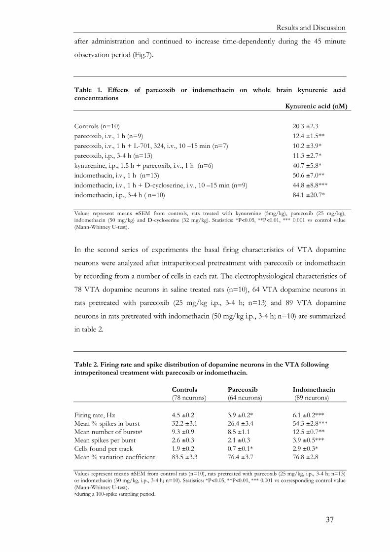
Results and Discussion
37
after administration and continued to increase time-dependently during the 45 minute
observation period (Fig.7).
Table 1. Effects of parecoxib or indomethacin on whole brain kynurenic acid concentrations Kynurenic acid (nM) Controls (n=10) 20.3 ±2.3 parecoxib, i.v., 1 h (n=9) 12.4 ±1.5** parecoxib, i.v., 1 h + L-701, 324, i.v., 10 –15 min (n=7) 10.2 ±3.9* parecoxib, i.p., 3-4 h (n=13) 11.3 ±2.7* kynurenine, i.p., 1.5 h + parecoxib, i.v., 1 h (n=6) 40.7 ±5.8* indomethacin, i.v., 1 h (n=13) 50.6 ±7.0** indomethacin, i.v., 1 h + D-cycloserine, i.v., 10 –15 min (n=9) 44.8 ±8.8*** indomethacin, i.p., 3-4 h ( n=10) 84.1 ±20.7* Values represent means ±SEM from controls, rats treated with kynurenine (5mg/kg), parecoxib (25 mg/kg), indomethacin (50 mg/kg) and D-cycloserine (32 mg/kg). Statistics: *P<0.05, **P<0.01, *** 0.001 vs control value (Mann-Whitney U-test). In the second series of experiments the basal firing characteristics of VTA dopamine
neurons were analyzed after intraperitoneal pretreatment with parecoxib or indomethacin
by recording from a number of cells in each rat. The electrophysiological characteristics of
78 VTA dopamine neurons in saline treated rats (n=10), 64 VTA dopamine neurons in
rats pretreated with parecoxib (25 mg/kg i.p., 3-4 h; n=13) and 89 VTA dopamine
neurons in rats pretreated with indomethacin (50 mg/kg i.p., 3-4 h; n=10) are summarized
in table 2.
Table 2. Firing rate and spike distribution of dopamine neurons in the VTA following intraperitoneal treatment with parecoxib or indomethacin. Controls Parecoxib Indomethacin (78 neurons) (64 neurons) (89 neurons) Firing rate, Hz 4.5 ±0.2 3.9 ±0.2* 6.1 ±0.2*** Mean % spikes in burst 32.2 ±3.1 26.4 ±3.4 54.3 ±2.8*** Mean number of burstsa 9.3 ±0.9 8.5 ±1.1 12.5 ±0.7** Mean spikes per burst 2.6 ±0.3 2.1 ±0.3 3.9 ±0.5*** Cells found per track 1.9 ±0.2 0.7 ±0.1* 2.9 ±0.3* Mean % variation coefficient 83.5 ±3.3 76.4 ±3.7 76.8 ±2.8 Values represent means ±SEM from control rats (n=10), rats pretreated with parecoxib (25 mg/kg, i.p., 3-4 h; n=13) or indomethacin (50 mg/kg, i.p., 3-4 h; n=10). Statistics: *P<0.05, **P<0.01, *** 0.001 vs corresponding control value (Mann-Whitney U-test). aduring a 100-spike sampling period.

Lilly Schwieler
38
Results from these experiments show that pretreatment with the selective COX-2 inhibitor
parecoxib decreased the firing rate and tended to decrease the burst firing activity of VTA
dopamine neurons. In addition, the number of spontaneously active VTA dopamine
neurons decreased after pretreatment with parecoxib, as shown by the decreased number
of cells found per track. The opposite effect on the basal firing activity was found in rats
pretreated with indomethacin. Thus, mean firing rate and mean percent spikes fired in
bursts of VTA dopamine neurons were significantly increased. In addition, this
pretreatment also increased the number of spontaneously active VTA dopamine neurons.
Intraperitoneal administration of indomethacin and parecoxib in these experiments
resulted in an increase and a decrease in whole brain kynurenic acid concentrations,
respectively (table 1).
4.1.3 Involvement of endogenous kynurenic acid in the effects of cyclooxygenase inhibitors on dopamine firing (paper II)
In the following experiments the mechanism underlying the effects of COX inhibitors
on the neuronal firing of midbrain dopamine neurons were more thoroughly
investigated. The excitatory and inhibitory actions on midbrain dopamine neurons
exerted by indomethacin and parecoxib, respectively, were correlated to their opposite
effects on the formation of the NMDA/glycine receptor antagonist kynurenic acid. Such
a relationship per se strongly supports an involvement of kynurenic acid in the effects of
COX-inhibitors on VTA dopamine firing. In further support, the excitation of midbrain
dopamine neurons observed after systemic administration of indomethacin strikingly
resembles the excitatory action induced by acute or chronic elevation of rat brain
kynurenic acid (Erhardt et al., 2001; Erhardt and Engberg, 2002; Nilsson et al., 2005a).
We therefore investigated if drugs acting selectively at the NMDA/glycine receptor
interact with the actions of indomethacin and parecoxib on VTA dopamine neurons.
Systemic administration of the partial NMDA/glycine receptor agonist D-
cycloserine (2-32 mg/kg; n=9) was per se associated with a slight dose-dependent increase
in firing rate and burst firing activity. In rats pretreated with indomethacin (50 mg/kg,
i.v., 1 h; n=9) systemic administration of D-cycloserine (2-32 mg/kg), significantly
decreased the firing rate and percent spikes fired in burst. This result resembles that of a
previous study where increased levels of brain kynurenic acid, induced by PNU
156651A, converted the excitatory action of D-cycloserine into a pure inhibitory
response (Erhardt and Engberg, 2002).

Results and Discussion
39
Systemic administration of the selective NMDA/glycine site antagonist L-701,324
(Grimwood et al., 1995) was per se associated with a dose-dependent increase in firing rate
and burst firing activity. Somewhat surprisingly, parecoxib pretreatment (25 mg/kg i.v., 1
h), enhanced the excitatory action of L-701,324 (0.06–2 mg/kg, i.v; n=7). Although the
mechanism behind this phenomenon is obscure it may be related to a reduced occupancy
of NMDA/glycine sites due to the decrease in endogenous kynurenic acid concentration.
Even more convincing results for an involvement of kynurenic acid in the action of
parecoxib on VTA dopamine neurons are the experiments that used kynurenine, the
immediate precursor of kynurenic acid. Thus, pretreatment with kynurenine (5 mg/kg i.p.,
30 min before parecoxib administration; n=5), prevented the reduction of brain kynurenic
acid levels by parecoxib (table 1), and simultaneously antagonized the inhibitory action of
the drug on the neuronal firing of the VTA dopamine neurons (Fig. 7). This effect of
kynurenine should conclusively link the action of parecoxib on VTA dopamine neurons to
the kynurenine pathway.
Altogether, the findings of the present study suggest that the actions of COX-
inhibitors on neuronal firing of VTA dopamine neurons are linked to their effects on the
synthesis of endogenous brain kynurenic acid. Furthermore, such a regulatory effect by
kynurenic acid on VTA dopamine neurons should be attributed to its antagonism at the
glycine site of the NMDA receptor as also indicated by the interaction with D-cycloserine
and L-701,324. Recent studies though show that kynurenic acid, apart from being an
antagonist at the NMDA/glycine-site, also blocks the α7* nicotinic receptor (Hilmas et al.,
2001). Such an action of kynurenic acid appears, however, unlikely to account for the
presently observed effects on VTA dopamine neurons, since administration of the
selective α7* nicotinic receptor antagonist, MLA was previously shown not to influence
the firing of VTA dopamine neurons (Schilström et al., 2003; see paper IV).
In conclusion, this study reveals that endogenous brain concentrations of
kynurenic acid are of critical importance for the tonic control of neuronal activity of
midbrain dopamine neurons. Thus, a reduction in brain kynurenic acid levels is associated
with a decrease in activity of VTA dopamine neurons and it stands clear that endogenous
kynurenic acid serves to maintain the firing activity of these neurons.

Lilly Schwieler
40
4.2 The atypical antipsychotic drug clozapine affects the neuronal activity of midbrain dopamine neurons by interfering with glutamatergic mechanisms (Paper III and IV).
4.2.1 Effects of antipsychotic drugs on midbrain dopamine firing in rats with elevated levels of endogenous brain kynurenic acid (paper III)
In this in vivo electrophysiological study, we investigate the effects of the atypical
antipsychotic drug clozapine and the classic antipsychotic drug haloperidol on VTA
dopamine neurons following increased levels of endogenous brain kynurenic acid. Brain
kynurenic acid concentration was pharmacologically elevated by administration of the
kynurenine 3-hydroxylase inhibitor PNU 156561 A. This compound blocks an
alternative pathway for the precursor kynurenine, and thereby shunts the synthesis of
kynurenine towards kynurenic acid (see section 3.3). This treatment is associated with
hyperdopaminergia (Erhardt et al., 2001; Erhardt and Engberg, 2002) that may
tentatively mimic a condition similar to that occurring in schizophrenia.
Acutely administered haloperidol (0.05-0.8 mg/kg, i.v.; n=7) or clozapine (1.25-
10 mg/kg, i.v.; n=11) in control rats was associated with increased firing rate and burst
firing activity of VTA dopamine neurons. These findings are in agreement with previous
in vivo electrophysiological studies (White and Wang, 1983; Tung et al., 1991; Gessa et al.,
2000) and it is generally accepted that antipsychotic drugs increase dopamine cell firing
rate by blockade of somatodendritic dopamine autoreceptors (Pucak and Grace, 1994;
1996). However, pretreatment with PNU 156561A (40mg/kg, i.v., n=8, 5-7 h) was
found not only to antagonize the increase in firing rate and percentage of spikes fired in
burst of VTA dopamine neurons induced by clozapine (1.25-10 mg/kg,

Results and Discussion
41
i.v.), but also to reverse the action of clozapine, leading to a significant decrease in
percentage of spikes fired in burst and a tendency to reduced firing rate (Fig. 8a,b). This
reduction in burst firing activity was observed in seven of eight dopamine neurons,
irrespective of their predrug basal characteristics. Furthermore, five of eight spon-
taneously bursting neurons were converted to non-bursting neurons by clozapine.
The effects of haloperidol on VTA dopamine neurons were clearly potentiated
after pretreatment with PNU 156561A, in contrast to those of clozapine, since haloperidol
forced all dopamine neurons recorded from into a depolarization block already after
administration of 0.1 mg/kg haloperidol (n=4).
We suggest that an increased dopaminergic transmission, as induced by elevated
kynurenic acid levels, may promote haloperidol to induce depolarization block by its
potent antagonism at somatodendritic dopamine D2 receptors. In contrast to haloperidol,
the inhibitory action of clozapine could not be explained in terms of antagonism of
dopamine receptors but rather by an interaction with the glycine site of NMDA receptor
or, alternatively, of the α7* nicotinic receptor. Thus, the present data do not allow any
definitive conclusion regarding the mechanism behind the inhibitory action of clozapine
on VTA dopamine neurons. Hypothetically, the effect could be mediated by I) activation
of presynaptic α7* nicotinic receptors located on glutamatergic afferents (McGehee et al.,
1995; Wonnacott et al., 2000) leading to a potentially increased glutamate release by
clozapine, II) activation of postsynaptic α7* nicotinic receptors, located on cell soma or
dendrites of VTA dopamine neurons, or III) displacement of kynurenic acid at
postsynaptic NMDA receptors. In paper IV these questions were more thoroughly
investigated by utilizing the NMDA receptor antagonist MK 801, the antagonist at the
glycine site of the NMDA receptor L-701,324 and the α7* nicotinic receptor antagonist
MLA.
4.2.2 Clozapine modulates midbrain dopamine neurons via interaction with the NMDA receptor (paper IV)
Administration of the NMDA receptor antagonist MK 801 alone (1 mg/kg, i.v., n = 8)
increased the firing rate (57%) and tended to increase the percentage of spikes fired in
bursts (from 16% to 32%) of VTA dopamine neurons. This treatment antagonized the
excitatory action of clozapine (1.25-10 mg/kg, i.v; n=8, Fig. 9a,b) pointing to an
interaction between clozapine and glutamatergic NMDA receptors. In line with this
view, pretreatment with L-701,324 (1-8 mg/kg, i.v; n=8) which per se was associated with

Lilly Schwieler
42
increased firing rate and percentage of spikes fired in burst, not only antagonized, but
also reversed the excitatory action of clozapine on VTA dopamine neurons, leading to a
decrease in firing rate and in burst firing activity (Fig. 9a,b and 10). These results are
indicative of an interaction of clozapine with the glycine site of the NMDA receptor.
Pretreatment with the α7* nicotinic receptor antagonist MLA (6 mg/ kg, i.p., 10 min;
n=7), did not affect spont-aneous firing of VTA dopamine neurons nor the ability of
clozapine to increase
burst firing activity of
the same neurons.
However, MLA slightly
antagonized the increase
in firing rate by
clozapine (Fig. 9a,b) as
observed in control rats.
Therefore, one cannot

Results and Discussion
43
fully exclude that α7* nicotinic receptors may play a role in the inhibition in firing rate of
VTA dopamine neurons by clozapine.
Altogether, these data provide pharmacological evidence that clozapine interferes
with the NMDA receptor. Thus, not only the regulation of basal firing of VTA
dopamine neurons is related to blockade of the glycine site of the NMDA receptor, it
also appears that the interaction between elevated levels of kynurenic acid and the
response of these neurons to clozapine is related to the same site. Tentatively, clozapine
could be an agonist at the glycine site of the NMDA receptor or an inhibitor of the
glycine transporter, resulting in increased synaptic levels of glycine. In support of the
latter theory it was recently shown that clozapine inhibits rat brain synaptosomal glycine
transport (Javitt et al., 2005). A putative interaction with the glycine site of the NMDA
receptor may account for clozapine’s unique clinical efficacy and suggest the glycine site
of the NMDA receptor as a target for novel antipsychotic drugs.
4.3 Increased levels of endogenous kynurenic acid disrupt prepulse inhibition - a behavioral model for schizophrenia (paper V)
Previous studies have shown that a moderate elevation of endogenous brain kynurenic
acid dramatically increases the firing activity of midbrain dopamine neurons (Erhardt et
al., 2001; Erhardt and Engberg, 2002), thereby mimicking the dopaminergic hyperactivity
believed to be one underlying cause of schizophrenia. Obviously, an important question
is whether increased levels of endogenous brain kynurenic acid are able to induce
symptoms similar to those observed in schizophrenia. Patients with schizophrenia have
long been considered to have an impaired ability to filter out extraneous stimuli in the
environment that would interfere with attention and information processing (Venables,
1960; McGhei and Chapman, 1961; Freedman et al., 1987; Geyer and Braff, 1987).
Prepulse inhibition is a behavioral model that reflects such sensory gating deficits in
these patients (c.f., introduction). We therefore examined the effects of elevated
endogenous brain kynurenic acid on PPI in rats.
Elevation of endogenous brain kynurenic acid concentration was achieved through
administration of kynurenine (100 mg/kg, i.p., 1 h; n=13), the precursor of kynurenic acid,
or by administration of the kynurenine 3-hydroxylase inhibitor PNU 156561A (10 mg/kg,
i.v., 4 h; n=9, table 3). A four-fold increase in brain kynurenic acid levels by these drugs
was found to significantly reduce PPI compared to saline-treated control rats (n=28, Fig.
11).

Lilly Schwieler
44
Table 3. Mean whole brain concentration of kynurenic acid Kynurenic acid (nM) Controls 27 ±5 nM, n =28 PNU 156561A 106 ±38 nM**, n = 9 (10 mg/kg, i.v., 4 h) Kynurenine 124 ±12 nM***, n = 13 (100 mg/kg, i.p., 60 min) Saline + Haloperidol 19 ±3 nM***, n = 11 (Saline + 0.2 mg/kg, i.p., 30 min) Saline + Clozapine 20 ±3 nM***, n = 12 (Saline + 7.5 mg/kg, i.p., 30 min) Kynurenine + Haloperidol 183 ±26 nM***, n = 8 (100 mg/kg, i.p., 60 min + 0.2 mg/kg, i.p., 30 min) Kynurenine + Clozapine 236 ±40 nM***, n = 12 (100 mg/kg, i.p., 60 min + 7.5 mg/kg, i.p., 30 min) Kynurenine + Probenecid (100 mg/kg, i.p., 60 min + 200 mg/kg, i.p., 120 min) 1691 ±44 nM***, n = 7 Statistics: **P<0.01, *** 0.001 vs control value (Mann-Whitney U-test). The second finding of the present study is that the reduced PPI as induced by elevated
brain levels of kynurenic acid can be restored by administration of the antipsychotic
drugs haloperidol
(0.2 mg/kg, i.p., 30
min; n=8) or cloza-
pine (7.5 mg/kg,
i.p., 30 min; n=12).
The present
findings, that incre-
aseed brain kynu-
renic acid disrupts
PPI, are in line with
previous studies in
which 7-chlorokyn-
urenic acid, a potent
synthetic analog of
kynurenic acid (Le-hmann et al., 1993; Wu et al., 1997), reduced PPI when administered
locally into the nucleus accumbens (Kretschmer and Koch, 1997; 1998). In a previous
study, it was also shown that pharmacological elevation of hippocampal kynurenic acid
concentration disrupts auditory sensory gating in rats, another measure of gating that is

Results and Discussion
45
deficient in patients with schizophrenia (Freedman et al., 1987). Since kynurenic acid
may interact with both nicotinic and glutamatergic receptors, the ability of kynurenic acid
to modulate PPI could be due to interaction with any of these systems. However, the
present results do not allow any definitive conclusion regarding the mechanism by which
kynurenic acid disrupts PPI.
In conclusion, this study shows that elevated levels of endogenous kynurenic acid
are associated with a disruption in PPI, an effect that can be reversed by antipsychotic
drugs. These findings not only reveal that kynurenic acid serves as an endogenous
modulator of PPI, they also provide further support for the notion that the compound
participates in the pathophysiology of schizophrenia.

Lilly Schwieler
46
5 GENERAL DISCUSSION
It is generally accepted that the mesolimbic and mesocortical dopamine systems of the
brain are involved in the mediation of symptoms in schizophrenia. However, growing
evidence suggests that the underlying cause of the disease lies elsewhere and is related to a
glutamatergic hypofunction. This view is largely based on the observation that NMDA
receptor antagonists such as PCP and ketamine produce symptoms that resemble both
positive and negative symptoms of schizophrenia (Luby et al., 1959; Itil et al., 1967; Adler
et al., 1999). Indeed, multiple lines of evidence from experimental and clinical studies using
agonists acting at the modulatory glycine/D-serine site of NMDA receptor further link
schizophrenia with an attenuated glutamatergic neurotransmission (see Coyle and Tsai,
2004). However, direct evidence for a reduced functioning of the NMDA receptor, e.g.
from ligand binding studies, is surprisingly sparse and even inconsistent. Recent studies
from our laboratory suggest that elevated levels of kynurenic acid would account for the
glutamatergic hypofunction that is related to schizophrenia. Thus, this endogenous
antagonist at the glycine/D-serine site of the NMDA receptor has been shown to be
elevated in the CSF (Erhardt et al., 2001a; Nilsson et al., 2005) as well as in the post
mortem brain (Schwarcz et al., 2001) from patients with schizophrenia. In addition, a
recent study shows that the expression of tryptophan 2,3-dioxygenase is increased in the
frontal cortex of schizophrenics (Miller et al., 2004), further indicating a disturbance in the
kynurenine pathway in these patients. In line with a putative role of kynurenic acid in
schizophrenia electrophysiological studies show that pharmacologically elevated levels of
the compound are associated with hyperactivity of midbrain dopaminergic neurons
(Erhardt et al., 2001; Erhardt and Engberg 2002). The results of the present thesis add
further evidence to the hypothesis that kynurenic acid is involved in the pathophysiology
of schizophrenia. Thus, a decrease in brain kynurenic acid levels is associated with a
reduced dopaminergic activity, offering indisputable evidence that the compound is a
significant modulator of glutamatergic neurotransmission. In addition, utilizing the PPI
technique, we offer behavioral support for a role of kynurenic acid in sensory gating, a
phenomenon that has been proposed to underlie the disturbances in filtering sensory
information seen in patients with schizophrenia. Finally, we propose a novel mechanism of
action of the atypical antipsychotic drug clozapine, i.e. stimulation of the glycine/D-serine
site of the NMDA receptor. Such an action of the drug would directly interfere with a
modulation of the NMDA receptor by kynurenic acid.

General Discussion
47
The present thesis also describes a prostaglandin-mediated regulation of kynurenic
acid formation. Thus, administration of COX-1 inhibitors was associated with an increased
formation of kynurenic acid in brain. Unexpectedly, COX-2 selective inhibitors had the
opposite action on rat brain kynurenic acid. The disparate effects of COX-1 and COX-2
inhibitors in this regard are puzzling; however, one might suggest that the two COX
isoforms modulate kynurenic acid synthesis at distinctly different steps in the kynurenine
pathway. A regulation of kynurenic acid by inflammatory mediators is interesting given the
possible involvement of immunological mechanisms in schizophrenia, as suggested by
several epidemiological studies (c.f. Introduction). Indeed, it is well established that
immune stimulation profoundly increases the metabolism of tryptophan via the
kynurenine pathway (see Moffet and Namboodiri, 2003). Thus, interferon-γ and other pro-
inflammatory cytokines have been shown to increase kynurenine, the immediate precursor
of kynurenic acid, by inducing IDO expression (Yoshida et al., 1986; Carlin et al., 1987).
Notably, a recent study from our laboratory shows that acute psychotic symptoms in HIV-
1 infected patients are associated with increased levels of kynurenic acid in CSF (Atlas et
al., 2006). Thus, it is possible that kynurenic acid in this regard serves as a functional link
between immunological processes and development of psychiatric symptoms.
The physiological role of brain kynurenic acid has long been a matter of
controversy since whole brain kynurenic acid concentrations in rodents (nM range) are far
below those required to affect NMDA receptors (µM range), as shown by in vitro studies
(Stone, 1993; Parsons et al., 1997; Urenjak and Obrenovitch, 2000). One possible
explanation for this discrepancy could be that kynurenic acid is synthesized and released
from astrocytes (Guillemin et al., 2001; Kiss et al., 2003), which are known to
communicate dynamically with glutamatergic neurons via intimate synaptic junctions
(Coyle and Schwarcz, 2000; Newman, 2003; Hertz and Zielke, 2004; Araque and Perea,
2004). Thus, the concentration of kynurenic acid within the synaptic cleft could be
substantially higher than is inferred from the total concentration in homogenates of brain
tissue. This is also supported by our electrophysiological studies utilizing COX inhibitors: a
decreased endogenous brain kynurenic acid concentration, as induced by a selective COX-
2 inhibitor, was associated with a clear-cut reduction in firing rate and burst firing activity
of VTA dopamine neurons. Thus, endogenous brain concentrations of kynurenic acid
appear to be of critical importance for maintaining firing activity of VTA dopamine
neurons. Such a modulatory function of the compound should not only be of major
importance for the functioning of the mesocorticolimbic dopamine pathway but also for
glutamatergic neurotransmission in general. Hence, one might speculate that the

Lilly Schwieler
48
compound affects a broad range of fundamental processes related to NMDA receptor
functions such as neurodevelopment and synaptic plasticity as well as cognition, behavior
and sensory perception.
The activation of midbrain dopamine neurons by kynurenic acid is in all
probability related to the glycine/D-serine site of the NMDA receptor. The increase in
firing rate and burst firing activity of midbrain dopamine neurons induced by elevated
levels of endogenous kynurenic acid or systemic administration of other NMDA receptor
antagonists (French et al., 1992, 1993; Zhang et al., 1992) has previously been suggested to
be due to an inhibition of a tonic GABAergic input that normally dampens neuronal
activity of midbrain dopamine neurons (Erhardt et al., 2002). In support of this view,
GABAergic interneurons, as compared to glutamatergic neurons, display a particular
vulnerability to NMDA receptor antagonists (Grunze et al., 1996; Li et al., 2002). Thus, the
action of COX-1 inhibitors on VTA dopamine neurons would be mediated via the same
mechanism. In analogy, decreasing the kynurenic acid concentration, by use of the
selective COX-2 inhibitor, should decrease firing of VTA dopamine neurons by
promoting GABAergic transmission. Recently, kynurenic acid was suggested to antagonize
also α7* nicotinic receptors at low concentrations (Hilmas et al., 2001). However, it is
unlikely that such an action of kynurenic acid could account for changes in firing rate of
midbrain dopamine neurons observed in the present study. Thus, blockade of the α7*
nicotinic receptor with MLA does not affect the activity of VTA dopamine neurons
(Schilström et al., 2003; paper IV). Moreover, stimulation of α7* nicotinic receptor is, if
anything, associated with an increase of burst firing of VTA dopamine neurons
(Schilström et al., 2003).
From our experiments we could see that endogenous brain kynurenic acid is of critical
importance for firing activity of midbrain dopamine neurons. Conversely, several studies
have shown that stimulation of the dopamine system changes the levels of kynurenic acid
in brain. Thus, agents that elevate synaptic dopamine concentration (e.g. amphetamine, L-
dopa) as well as drugs that stimulate dopamine receptors (D1 and D2), significantly
decrease the levels of kynurenic acid in rat brain (Poeggeler et al., 1998; Rassoulpour et al.,
1998; Wu et al., 2001). The mechanism by which dopamine down-regulates this
compound is not fully clarified but it has been proposed that the decrease in kynurenic
acid levels may be caused by stimulation of astrocytic dopamine receptors (Wu et al.,
2002). Such a direct regulation of kynurenic acid metabolism by dopamine may tentatively
indicate the existence of a feedback mechanism aiming at lowering levels of kynurenic acid,
which in turn would dampen dopaminergic activity and further dopamine release.

General Discussion
49
An important question is whether increased levels of kynurenic acid are able to
induce symptoms similar to those observed in schizophrenia. Patients with schizophrenia
have long been considered to have an impaired ability to filter out extraneous stimuli in the
environment that would interfere with attention and information processing (Venables,
1960; McGhei and Chapman, 1961; Freedman et al., 1987; Geyer and Braff, 1987).
Prepulse inhibition is a behavioral model that reflects such sensory gating deficits in these
patients. The cross-species nature of PPI enables the use of animal models to investigate
behavioral phenomena that are similar to the deficits seen in schizophrenia. As shown by
such studies, administration of NMDA receptor antagonists (e.g. PCP, MK 801, ketamine)
can disrupt PPI in rodents (Geyer et al., 1990; Mansbach and Geyer, 1988) and in primates
(Javitt and Lindsley, 2001; Linn and Javitt, 2001). In the present thesis we found that
elevated levels of endogenous kynurenic acid are associated with a disruption in PPI, an
effect that could be reversed by antipsychotic drugs. In line with our results, a previous
study has shown that pharmacological elevation of hippocampal kynurenic acid
concentration disrupts auditory sensory gating in rats, another measure of gating that is
deficient in patients with schizophrenia (Freedman et al., 1987). Since kynurenic acid
interacts with both cholinergic and glutamatergic receptors, the ability of kynurenic acid to
modulate PPI could theoretically be due to interaction with any of these system. Although
the present results do not allow any definitive conclusion regarding the mechanism by
which kynurenic acid disrupts PPI we suggest that this compound serves as an
endogenous modulator of PPI.
If NMDA receptor hypofunction is involved in the pathophysiology of
schizophrenia, then interventions directed at enhancing NMDA receptor function should
reduce symptoms. However, it is unlikely that agents that stimulate the recognition site of
the NMDA receptor would be suitable candidates in this regard, because of their tendency
to produce excessive excitotoxicity (c.f. Introduction). Instead, clinical trials have focused
on agents that stimulate the modulatory glycine/D-serine site of the NMDA receptor.
Interestingly, clinical treatment augmentation studies have shown that agents acting at the
glycine site of the NMDA receptor, such as glycine, D-serine and the partial agonist D-
cycloserine, further ameliorate symptoms when used in combination with conventional
antipsychotic drugs (Javitt et al., 1994; Tsai et al., 1998; Heresco-Levy et al., 1998, 1999;
Evins et al., 2002; see Coyle and Tsai, 2004), but not when added to clozapine (Goff et al.,
1996, 1999). Indeed, when D-cycloserine was added to the treatment regimen of patients
maintained on clozapine, a significant dose-related exacerbation of negative symptoms was
revealed (Goff et al., 1996, 1999). This may be related to the ability of clozapine itself to

Lilly Schwieler
50
increase the NMDA receptor activity to a level that prevents further enhancement by D-
serine and glycine treatment. Such a profile of clozapine would also explain why co-
administration of D-cycloserine even produces exacerbation of the negative symptoms due
to its partial antagonistic action. Indeed, the findings of the present thesis suggest that
administration of clozapine leads to a stimulation of the modulatory glycine/D-serine site
of the NMDA receptor. Tentatively, clozapine could either directly stimulate the
glycine/D-serine of the NMDA receptor or act as an inhibitor of the glycine transporter,
resulting in increased synaptic levels of glycine. In support of the latter theory, recent
experimental studies have shown that clozapine, but neither haloperidol nor risperidone,
inhibits glycine transporters in cortical synaptosomes (Javitt et al., 2005). A stimulatory
action of clozapine on NMDA receptors may explain the clinical profile of the drug, i.e. a
therapeutic action also on the negative symptoms of the disease since these symptoms also
are induced by NMDA receptor antagonists (c.f. Introduction).
Altogether, the present thesis provides evidence that endogenous brain kynurenic
acid participates in the pathophysiology of schizophrenia. Further support for this view
comes from clinical reports on the side effects of NSAIDs. Thus, several studies have
reported that traditional NSAIDs with an inhibitory action on COX-1 and that increase
kynurenic acid levels (especially indomethacin), induce psychiatric side effects such as
psychosis, delusions and cognitive dysfunctions (see Hoppmann et al., 1991; see Jiang and
Chang, 1999; Tharumaratnam et al., 2000; Clunie et al., 2003). If the psychiatric side effects
of NSAIDs that interfere with COX-1 could be attributed to increased levels of kynurenic
acid, then selective COX-2 inhibitors producing decreased brain kynurenic acid levels
would be beneficial in the treatment of schizophrenia. Actually, the selective COX-2
inhibitor celecoxib was recently shown to display antipsychotic effects in patients with an
acute exacerbation of schizophrenia with regard to positive, negative (Müller et al., 2002)
as well as some cognitive symptoms (Müller et al., 2004) when added to conventional
antipsychotic treatment. Thus, the diverse effects on brain kynurenic acid metabolism
exerted by COX inhibitors with selectivity for different COX isoforms may provide a
rational explanation not only for the psychiatric side effects induced by non-selective COX
inhibitors but also for the antipsychotic properties of the COX-2 selective agent celecoxib.

Acknowledgements
51
6 ACKNOWLEDGEMENTS
I would like to express my deepest gratitude to all people, who in different ways have supported me during the work of this thesis at Department of Physiology and Pharmacology, Karolinska Institutet. In particular I would like to thank: Professor Göran Engberg, my supervisor, for believing in me and for always being encouraging and optimistic. Especially, I would like to thank you for creating an open and creative atmosphere that leads to interesting discussions and exchange of knowledge that made me grow from student to scientist. Also, I would like to thank you for giving me the opportunity to present my data at several important international congresses and for all travel memories never to be forgotten. Docent Sophie Erhardt, my co-supervisor and friend during life, for not listening at me when I thought there was something fishy when you first told me to start research, for later on introduce me to the field and for giving me the opportunity to chare all your knowledge about performing research. Kristina Ikelberg, my former college and friend at Danderyds Hospital who repeatedly told me to use my capacity and who gave me my first and most important grant. Your words of encouragement were liberating! Linda Nilsson, co-author and friend, I’m going to miss our nice discussions concerning soccer, science and life in general. How fun it was in the car from San Diego to California, up in the mountain into the dessert and down in Grand Canyon. Memories for life! Klas Linderholm, for invaluable help with experiments in study II and for unforgettable travel company in USA but most of all for demonstrating “joie de vivre” by keeping your dreams floating. Present and former members of the Engberg-Erhardt research group, Carolina Emanuelsson for invaluable help with the PPI study, Lotta Erhardt Co-author and for good assistance at lab. Carola Sundberg and Alexandra Andersson, and especially thanks to the rookies Maria Holtze and Elin Olsson who kindly helped me with references. Björn Schilström, for always sharing happenings in life and for the fancy discussion about dopamine we had during our walk in Grand Canyon. Lotta Wiker and Åsa Konradsson for fun company at congresses and for sharing the situation of being a PhD student. Elham Rosstami, I will never forget our crazy visit in Itasca and Sara Johansson for sharing the experience of being a PhD-student and for always being interested in my work. Bertil Hållsten not only for financial support but also for showing grate interest in our work.

Lilly Schwieler
52
Janet Holmén for your professional revision of my thesis and for your great work with the Lucia choir. Professor Stefan Eriksson and professor Bertil Fredholm, present and former head of the department of Physiology and Pharmacology, for providing good working conditions. The Torgny Svensson and Kent Jardemark group; Monica Marcus, Anna Malmerfeldt, Sabina de Villiers, Ann-chatrine Samuelsson, Amani Eltayb, NinaLindblom for interesting discussions now and then especially during Wednesday coffee breaks and during lunch at Hjulet. All members of the Urban Ungerstedt´s group, Urban for interesting discussions and for introducing me to the microdialysis technique, Neda Rajamand, Åse Elfving, Johan Ungerstedt, Michel Goiny I miss you all a lot! All the staff at the animal department, administration office as well as Margareta Westling, Ulla Röberg-Kisch, Monica Pace-Sjögren, Hans Gustavsson, Eva Näsström, Hasse Svensson, for making my work much easier. Mother and Kevork for supporting me during life and for teaching me the importance of keeping my dreams alive. David and your wonderful family; Josefin, Samuel, Edwin and Emelia. Brother I am so impressed of your never failing effort to enjoy every moment of life what so ever. Sussi my lovely sister for always being my best friend in life and for illustrating psychosis at the front page. How would life be without the Wiklund family… LESS! Rolf and Maj-Britt for always giving support whenever needed and for all marvelous dinners now and then. Roger for inviting me to Skellefteå Hockey and for your positive attitude. Anette for keeping me company during life when Wiklunds have to sleep. and Mats, my true love in life, for all the encouragement, confidence, and love you bring to me, Jojo and Bella for bringing joy and happiness into my life and Hampus for giving me the opportunity to experience crazy love.

References
53
7 REFERENCES
Abi-Dargham A, Gil R, Krystal J, Baldwin RM,
Seibyl JP, Bowers M, van Dyck CH, Charney DS, Innis RB, Laruelle M 1998. Increased striatal dopamine transmission in schizophrenia: confirmation in a second cohort. Am J Psychiatry 155:761-767.
Abi-Dargham A, Rodenhiser J, Printz D, Zea-Ponce
Y, Gil R, Kegeles LS, Weiss R, Cooper TB, Mann JJ, Van Heertum RL, Gorman JM, Laruelle M 2000. Increased baseline occupancy of D2 receptors by dopamine in schizophrenia. Proc Natl Acad Sci U S A 97:8104-8109.
Adell A, Artigas F 2004. The somatodendritic release
of dopamine in the ventral tegmental area and its regulation by afferent transmitter systems. Neurosci Biobehav Rev 4:415-431.
Adler CM, Malhotra AK, Elman I, Goldberg T,
Egan M, Pickar D, Breier A 1999. Comparison of ketamine-induced thought disorder in healthy volunteers and thought disorder in schizophrenia. Am J Psychiatry 156:1646-1649.
Albers GW, Clark WM, Atkinson RP, Madden K,
Data JL, Whitehouse MJ 1999. Dose escalation study of the NMDA glycine-site antagonist licostinel in acute ischemic stroke. Stroke 30:508-513.
Almeida OP, Howard RJ, Levy R, David AS 1995.
Psychotic states arising in late life (late paraphrenia) psychopathology and nosology. Br J Psychiatry 2:205-214.
Andén NE, Dahlström A, Fuxe K, Larsson K,
Olsson L, Ungerstedt U 1966. Ascending neurons to the telencephalon and diencephalon. Act Physiol Scand 67:313-326.
Anden NE, Stock G 1973. Effect of clozapine on
the turnover of dopamine in the corpus striatum and in the limbic system. J Pharm Pharmacol 25: 346-348.
Andreasen NC 1995. Symptoms, signs, and diagnosis
of schizophrenia. Lancet 346:477-481. Angrist B, van Kammen DP 1984. CNS stimulants as
tools in the study of schizophrenia. Trends Neurosci 7:388-390.
Araque A, Perea G 2004. Glial modulation of
synaptic transmission in culture. Glia 3:241-248. Arundine M, Tymianski M 2003. Molecular mech-
anisms of calcium-dependent neurodegeneration in excitotoxicity. Cell Calcium 5:325-337.
Arvanov VL, Liang X, Schwartz J, Grossman S, Wang RY 1997. Clozapine and haloperidol modulate N-methyl-D-aspartate- and non-N-methyl-D-aspartate receptor-mediated neurotransmission in rat prefrontal cortical neurons in vitro. J Pharmacol Exp Ther 283: 226-234.
Arvanov VL, Wang RY 1999. Clozapine, but not
haloperidol, prevents the functional hyperactivity of N-methyl-D-aspartate receptors in rat cortical neurons induced by subchronic administration of phencyclidine. J Pharmacol Exp Ther 289:1000-1006.
Atlas A, Gisslen M, Nordin C, Lindström L,
Schwieler L 2006. Acute psychotic symptoms in HIV-1 infected patients are associated with increased levels of kynurenic acid in cerebrospinal fluid. Brain Behav Immun DOI:10.1016-/j.bbi.2006.02.005.
Baldessarini RJ, Frankenburg FR 1991. Clozapine A
novel antipsychotic agent. N Engl J Med Mar 324:746-754.
Baran H, Jellinger K, Deecke L 1999. Kynurenine
metabolism in Alzheimer's disease. J Neural Transm 106:165-181.
Baran H, Hainfellner JA, Kepplinger, B, Mazal PR,
Schmid H, Budka H 2000. Kynurenic acid metabolism in the brain of HIV-1 infected patients. J Neural Transm 107:1127-1138.
Bartholini G 1976. Differential effect of neuroleptic
drugs on dopamine turnover in the extrapyramidal and limbic system. J Pharm Pharmacol 28: 429-433.
Battaglia G, Rassoulpour A, Wu HQ, Hodgkins PS,
Kiss C, Nicoletti F, Schwarcz R 2000. Some metabotropic glutamate receptor ligands reduce kynurenate synthesis in rats by intracellular inhibition of kynurenine aminotransferase II. J Neurochem 75:2051-2060.
Bayer VE, Pickel VM 1991. GABA-labeled
terminals form proportionally more synapses with dopaminergic neurons containing low densities of tyrosine hydroxylase-immunoreactivity in rat ventral tegmental area. Brain Res 559:44-55.
Beart PM, McDonald D 1980. Neurochemical
studies of the mesolimbic dopaminergic pathway: somatodendritic mechanisms and GABAergic neurones in the rat ventral tegmentum. J Neurochem 34:1622-1629.

Lilly Schwieler
54
Bender DA, McCreanor GM 1982. The preferred route of kynurenine metabolism in the rat. Biochim Biophys Acta 717:56-60.
Birch PJ, Grossman CJ, Hayes AG 1988. Kynurenic
acid antagonises responses to NMDA via an action at the strychnine-insensitive glycine receptor. Eur J Pharmacol 154: 85-87.
Björklund A, Lindvall O 1984. Dopamine
containing systems in the CNS. In: Handbook of chemical neuroanatomy. Vol. 2: Classical transmitters in the CNS, Part I (Björklund A and Hökfelt T, eds) pp. 55-122.
Black DW, Fisher R 1997. Mortality in DSM-IIIR
schizophrenia. Schizophr Res 7:109-116. Breier A 1999. Cognitive deficit in schizophrenia
and its neurochemical basis. Br J Psychiatry Suppl 37:16-18.
Breier A, Su TP, Saunders R, Carson RE,
Kolachana BS, de Bartolomeis A, Weinberger DR, Weisenfeld N, Malhotra AK, Eckelman WC, Pickar D 1997. Schizophrenia is associated with elevated amphetamine-induced synaptic dopamine concentrations: Evidence from a novel positron emission tomography method. Proc Nat Acad Sci USA 94:2569-2574.
Breimer DD 1977. Clinical pharmacokinetics of
hypnotics. Clin Pharmacokinet 2:93-109. Brown AS, Begg MD, Gravenstein S, Schaefer CA,
Wyatt RJ, Bresnahan M, Babulas VP, Susser ES 2004. Serologic evidence of prenatal influenza in the etiology of schizophrenia. Arch Gen Psychiatry 61:774-780.
Brown AS, Cohen P, Harkavy-Friedman J, Babulas
V, Malaspina D, Gorman JM, Susser ES 2001. Prenatal rubella, premorbid abnormalities, and adult schizophrenia. Biol Psychiatry 49:473-486.
Brown AS 2006. Prenatal Infection as a Risk Factor
for Schizophrenia. Schizophr Bull 32:200-202. Brunello N, Masotto C, Steardo L, Markstein R,
Racagni G 1995. New insights into the biology of schizophrenia through the mechanism of action of clozapine. Neuropsychopharmacology 13:177-213.
Buchli R, Alberati-Giani D, Malherbe P, Kohler C,
Broger C, Cesura AM 1995. Cloning and functional expression of a soluble form of kynurenine/alpha-aminoadipate aminotransfe-rase from rat kidney. J Biol Chem 270:29330-29335.
Buka SL, Tsuang MT, Torrey EF, Klebanoff MA,
Bernstein D, Yolken RH 2001. Maternal
infections and subsequent psychosis among offspring. Arch Gen Psychiatry 58:1032-1037.
Cannon M, Cotter D, Coffey VP, Sham PC, Takei N,
Larkin C, Murray RM, O'Callaghan E 1996. Prenatal exposure to the 1957 influenza epidemic and adult schizophrenia: a follow-up study. Br J Psychiatry 168:368-371.
Canton H, Verriele L, Colpaert FC 1990. Binding of
typical and atypical antipsychotics to 5-HT1C and 5-HT2 sites: clozapine potently interacts with 5-HT1C sites. Eur J Pharmacol 191: 93-96.
Cardno AG, Marshall EJ, Coid B, Macdonald AM,
Ribchester TR, Davies NJ, Venturi P, Jones LA, Lewis SW, Sham PC, Gottesman II, Farmer AE, McGuffin P, Reveley AM, Murray RM 1999. Heritability estimates for psychotic disorders: the Maudsley twin psychosis series. Arch Gen Psychiatry 56:162-168.
Carlin JM, Borden EC, Sondel PM, Byrne GI 1987.
Biologic-response-modifier-induced indoleamine 2,3-dioxygenase activity in human peripheral blood mononuclear cell cultures. J Immunol 139:2414-2418.
Carlsson A, Lindqvist M 1963. Effect of
chlorpromazine or haloperidol on formation of 3-methoxytyramine and normetanephrine in mouse brain. Acta Pharmacol Toxicol 20:140-144.
Carpenedo R, Chiarugi A, Russi P, Lombardi G,
Carla V, Pellicciari R, Mattoli L, Moroni F 1994. Related Inhibitors of kynurenine hydroxylase and kynureninase increase cerebral formation of kynurenate and have sedative and anticonvulsant activities. Neuroscience 61:237-243.
Carpenter WT Jr, Buchanan RW 1994.
Schizophrenia. N Engl J Med 330:681-690. Carr DB, Sesack SR 2000. GABA-containing neurons
in the rat ventral tegmental area project to the prefrontal cortex. Synapse 38:114-123.
Carr DB, Sesack SR 2000a. Projections from the rat
prefrontal cortex to the ventral tegmental area: target specificity in the synaptic associations with mesoaccumbens and mesocortical neurons. J Neurosci 20:3864-3873.
Chandrasekharan NV, Dai H, Roos KL, Evanson
NK, Tomsik J, Elton TS, Simmons DL 2002. COX-3, a cyclooxygenase-1 variant inhibited by acetaminophen and other analgesic/antipyretic drugs: cloning, structure, and expression. Proc Natl Acad Sci USA 99: 13926-13931.
Charara A, Smith Y, Parent A 1996. Glutamatergic
inputs from the pedunculopontine nucleus to midbrain dopaminergic neurons in primates:

References
55
Phaseolus vulgaris-leucoagglutinin anterograde labeling combined with postembedding glutamate and GABA immunohistochemistry. J Comp Neurol 364:254-266.
Chen L, Yang CR 2002. Interaction of dopamine D1
and NMDA receptors mediates acute clozapine potentiation of glutamate EPSPs in rat prefrontal cortex. J Neurophysiol 87:2324-2336.
Chen W, Pawelek TR, Kulmacz RJ 1999.
Hydroperoxide dependence and cooperative cyclooxygenase kinetics in prostaglandin H synthase-1 and -2. J Biol Chem 274:20301-20306.
Chergui K, Charlety PJ, Akaoka H, Saunier CF,
Brunet JL, Buda M, Svensson TH, Chouvet G 1993. Tonic activation of NMDA receptors causes spontaneous burst discharge of rat midbrain dopamine neurons in vivo. Eur J Neurosci 5:137-44.
Chiodo LA, Bunney BS 1983. Typical and atypical
neuroleptics: differential effects of chronic administration on the activity of A9 and A10 midbrain dopaminergic neurons. J Neurosci 3:1607-1619.
Chiodo LA, Bunney BS 1985. Possible mechanisms
by which repeated clozapine administration differentially affects the activity of two subpopulations of midbrain dopamine neurons. J Neurosci 5: 2539-2544.
Claghorn J, Honigfeld G, Abuzzahab FS Sr, Wang
R, Steinbook R, Tuason V, Klerman G 1987. The risks and benefits of clozapine versus chlorpromazine. J Clin Psychopharmacol 7: 377-384.
Clark D, Chiodo LA 1988. Electrophysiological and
pharmacological characterization of identified nigrostriatal and mesoaccumbens dopamine neurons in the rat. Synapse 2:474-485.
Clunie M, Crone LA, Klassen L, Yip R 2003.
Psychiatric side effects of indomethacin in parturients. Canadian Journal of Anaesthesia 50:586-588.
Cooper JR, Bloom FB, Roth RH 2003. The
biochemical basis of neuropharmacology. Oxford University Press, Inc, New York.
Coward DM 1992. General pharmacology of
clozapine. Br J Psychiatry Suppl 17:5-11. Coward DM, Imperato A, Urwyler S, White TG
1989. Biochemical and behavioural properties of clozapine. Psychopharmacol 99:S6-12.
Coyle JT, Schwarcz R 2000. Mind glue: implications
of glial cell biology for psychiatry. Arch Gen Psychiatry 57:90-93.
Coyle JT 1973. The development of catechol-aminergic neurons of the central nervous syst-em. Neurosci Res (N Y) 5:35-52.
Coyle JT, Tsai G 2003. The NMDA receptor glycine
modulatory site: a therapeutic target for improving cognition and reducing negative symptoms in schizophrenia. Psychopharmacology (Berl) 174:32-38.
Creese I, Burt DR, Snyder SH 1976. Dopamine
receptor binding predicts clinical and pharmacological potencies of antischizophrenic drugs. Science 192:481-483.
Dahlström A, Fuxe K 1964. Evidence for the
existence of monoamine-containing neurons in the central nervous system. I. Demonstration of monoamines in the cell bodies of brain stem neurons. Acta Physiol Scand 62:1-55.
Daly DA, Moghaddam B 1993. Actions of clozapine
and haloperidol on the extracellular levels of excitatory amino acids in the prefrontal cortex and striatum of conscious rats. Neurosci Lett 152:61-64.
Danbolt NC 2001. Glutamate uptake. Prog
Neurobiol 65:1-105. Danysz W, Parsons AC 1998. Glycine and N-
methyl-D-aspartate receptors: physiological significance and possible therapeutic applications. Pharmacol Rev 50:597-664.
Davis KL, Kahn RS, Ko G, Davidson M 1991.
Dopamine in schizophrenia: a review and reconceptualization. Am J Psychiatry 148:1474-1486.
Delay J, Deniker P, Harl M 1952. Utilisation en
thérapeutique psychiatrique d’une phenothiazine centrale elective. Ann Méd Psychol (Paris) 110:112-117.
Deutch AY, Moghaddam B, Innis RB, Krystal JH,
Aghajanian GK, Bunney BS, Charney DS 1991. Mechanisms of action of atypical antipsychotic drugs. Implications for novel therapeutic strategies for schizophrenia. Schizophr Res 4:121-156.
DeWitt DL 1999. Cox-2-selective inhibitors: the new
super aspirins. Mol Pharmacol 55:625-631. Edwards SR, Mather LE, Lin Y, Power I, Cousins MJ
2000. Glutamate and kynurenate in the rat central nervous system following treatments with tail ischaemia or diclofenac. Journal of Pharmacy and Pharmacology 52: 59-66.
Ellinger A 1904. Die entstehung der Kynurensaure.
Z Physiol Chem 43:325.

Lilly Schwieler
56
Erhardt S, Andersson B, Nissbrandt H, Engberg G 1998. Inhibition of firing rate and changes in the firing pattern of nigral dopamine neurons by gamma-hydroxybutyric acid (GHBA) are specifically induced by activation of GABA(B) receptors. Naunyn Schmiedeberg´s Arch Pharmacol 357:611-619.
Erhardt S, Blennow K, Nordin C, Skogh E,
Lindström LH, Engberg G 2001a. Kynurenic acid levels are elevated in the cerebrospinal fluid of patients with schizophrenia. Neurosci Lett 13:96-98.
Erhardt S, Engberg G 2002. Increased phasic
activity of dopaminergic neurones in the rat ventral tegmental area following pharmacol-ogically elevated levels of endogenous kynurenic acid. Acta Physiol Scand 175:45-53.
Erhardt S, Engberg G (2000). Excitation of nigral
dopamine neurons by the GABA-A-receptor agonist muscimol is mediated via a release of glutamate. Life Sciences 67: 1901-1911.
Erhardt S, Mathé JM, Engberg G, Svensson TH
2002. GABA(B) receptor mediated modulation of the firing pattern of ventral tegmental area dopamine neurons in vivo. Naunyn-Schmiedeberg´s Arch Pharmacol 365:173-180.
Erhardt S, Nissbrandt H, Engberg G 1999.
Activation of nigral dopamine neurons by the selective GABA(B)-receptor antagonist SCH 50911. J Neural Transm 106:383-394.
Erhardt S, Öberg H, Engberg G (2001b).
Pharmacologically elevated levels of endogenous kynurenic acid prevent nicotine-induced activation of nigral dopamine neurons. Naunyn-Schmiedeberg´s Arch Pharmacol 363:21-27.
Erhardt S, Schwieler L, Engberg G 2002a.
Excitatory and inhibitory responses of dopamine neurons in the ventral tegmental area to nicotine. Synapse 43:227-237.
Erhardt S, Schwieler L, Engberg G 2003. Kynurenic
acid and schizophrenia. Adv Exp Med Biol 527:155-165.
Erhardt S. Öberg H, Mathé JM, Engberg G 2001.
Elevation of endogenous kynurenic acid levels activates nigral dopamine neurons. Amino Acids 20:353-362.
Evins AE, Amico E, Posever TA, Toker R, Goff
DC 2002. D-Cycloserine added to risperidone in patients with primary negative symptoms of schizophrenia. Schizophr Res 56:19-23.
Farde L, Nordstrom AL 1993. PET examination of
central D2 dopamine receptor occupancy in relation to extrapyramidal syndromes in patients
being treated with neuroleptic drugs. Psychopharmacol Ser 10:94-100.
Farde L, Nordstrom AL, Wiesel FA, Pauli S,
Halldin C, Sedvall G 1992. Positron emission tomographic analysis of central D1 and D2 dopamine receptor occupancy in patients treated with classical neuroleptics and clozapine. Relation to extrapyramidal side effects. Arch Gen Psychiatry 49:538-544.
Farde L, Wiesel FA, Halldin C, Sedvall G 1988.
Central D2-dopamine receptor occupancy in schizophrenic patients treated with antipsychotic drugs. Arch Gen Psychiatry 45:71-76.
Farmer AE, McGuffin P, Gottesman II 1987. Twin
concordance for DSM-III schizophrenia. Scrutinizing the validity of the definition. Arch Gen Psychiatry 44:634-641.
Fenton WS, McGlashan TH 1991. Natural history of
schizophrenia subtypes. II. Positive and negative symptoms and long-term course. Arch Gen Psychiatry 48:978-986.
Forrest CM, Youd P, Kennedy A, Gould SR,
Darlington LG, Stone TW 2002. Purine, kynurenine, neopterin and lipid peroxidation levels in inflammatory bowel disease. J Biomed Sci 9:436-442.
Franzone JS, Cravanzola C, Reboani MC 1980.
Influence of non-steroidal anti-inflammatory compounds on the hepatic tryptophan pyrrolase activity in hypo- and hyperthyroid rats. Arch Int Pharmacodyn Ther 245: 156-165.
Freedman R, Adler LE, Gerhardt GA, Waldo M,
Baker N, Rose GM, Drebing C, Nagamoto H, Bickford-Wimer P, Franks R 1987. Neurobiological studies of sensory gating in schizophrenia. Schizophr Bull 13:669-678.
Freeman AS, Bunney BS 1987. Activity of A9 and
A10 dopaminergic neurons in unrestrained rats: further characterization and effects of apomorphine and cholecystokinin. Brain Res 405:46-55.
Freeman AS, Meltzer LT, Bunney BS 1985. Firing
properties of substantia nigra dopaminergic neurons in freely moving rats. Life Sci 36:1983-1994.
Freedman R, Adler LE, Gerhardt GA, Waldo M,
Baker N, Rose GM 1987. Neurobiological studies of sensory gating in schizophrenia. Schizophr Bull 13:669–677.
French ED 1994. Phencyclidine and the midbrain
dopamine system: electrophysiology and behavior. Neurotoxicol Teratol 16:355-362.

References
57
French ED, Mura A, Wang T 1993. MK-801, phencyclidine (PCP), and PCP-like drugs increase burst firing in rat A10 dopamine neurons: comparison to competitive NMDA antagonists. Synapse 13:108-116.
Fukui S, Schwarcz R, Rapoport SI, Takada Y, Smith
QR 1991. Blood-brain barrier transport of kynurenines: implications for brain synthesis and metabolism. J Neurochem 56:2007-2017.
Gál EM, Sherman AD 1978. Synthesis and
metabolism of L-kynurenine in rat brain. J Neurochem 30:607-613.
Ganong AH, Cotman CW 1986. Kynurenic acid and
quinolinic acid act at N-methyl-D-aspartate receptors in the rat hippocampus. J Pharmacol Exp Ther 236:293-299.
Gardner DM, Baldessarini RJ, Waraich P 2005.
Modern antipsychotic drugs: a critical overview. CMAJ 172:1703-1711.
Gessa GL, Devoto P, Diana M, Flore G, Melis M,
Pistis M 2000. Dissociation of haloperidol, clozapine, and olanzapine effects on electrical activity of mesocortical dopamine neurons and dopamine release in the prefrontal cortex. Neuropsychopharmacology 22:642-649.
Geyer MA, Braff DL 1987. Startle habituation and
sensorimotor gating in schizophrenia and related animal models. Schizophr Bull 4:643-668.
Geyer MA, Swerdlow NR, Mansbach RS, Braff DL
1990. Startle response models of sensorimotor gating and habituation deficits in schizophrenia. Brain Res Bull 25:485-498.
Giorgetti M, Hotsenpiller G, Froestl W, Wolf ME
2002. In vivo modulation of ventral tegmental area dopamine and glutamate efflux by local GABA(B) receptors is altered after repeated amphetamine treatment. Neuroscience 109:585-595.
Goff DC, Tsai G, Levitt J, Amico E, Manoach D,
Schoenfeld DA, Hayden DL, McCarley R, Coyle JT 1999. A placebo-controlled trial of D-cycloserine added to conventional neuroleptics in patients with schizophrenia. Arch Gen Psychiatry 56:21-27.
Goff DC, Tsai G, Manoach DS, Flood J, Darby
DG, Coyle JT 1996. D cycloserine added to clozapine for patients with schizophrenia. Am J Psychiatry 153:1628-1630.
Goldman-Rakic PS, Muly EC 3rd, Williams 2000.
GVD(1) receptors in prefrontal cells and circuits. Brain Res Brain Res Rev 31:295-301.
Gonon FG 1988. Nonlinear relationship between impulse flow and dopamine released by rat midbrain dopaminergic neurons as studied by in vivo electrochemistry. Neuroscience 24:19-28.
Grace AA, Bunney BS 1984. The control of firing
pattern in nigral dopamine neurons: single spike firing. J Neurosci 4:2866-2876.
Grace AA, Bunney BS 1984a. The control of firing
pattern in nigral dopamine neurons: burst firing. J Neurosci 4:2877-2890.
Grace AA, Bunney BS 1979. Paradoxical GABA
excitation of nigral dopaminergic cells: indirect mediation through reticulata inhibitory neurons. Eur J Pharmacol 59:211-218.
Grace AA, Onn SP 1989. Morphology and
electrophysiological properties of immunocytochemically identified rat dopamine neurons recorded in vitro. J Neurosci 9:3463-3481.
Gramsbergen JB, Hodgkins PS, Rassoulpour A,
Turski WA, Guidetti P, Schwarcz R 1997. Brain-specific modulation of kynurenic acid synthesis in the rat. J Neurochem 69:290-298.
Gramsbergen JB, Turski WA, Schwarcz R 1991.
Brain-specific control of kynurenic acid production by depolarizing agents. Adv Exp Med Biol 294:587-590.
Green MF, Kern RS, Braff DL, Mintz J 2000.
Neurocognitive deficits and functional outcome in schizophrenia: are we measuring the "right stuff"? Schizophr Bull 26:119-136.
Grenhoff J, Ugedo L, Svensson TH 1988. Firing
patterns of midbrain dopamine neurons: differences between A9 and A10 cells. Acta Physiol Scand 134:127-132.
Grimwood S, Kulagowski JJ, Mawer IM, Rowley M,
Leeson PD, Foster AC 1995. Allosteric modulation of the glutamate site on the NMDA receptor by four novel glycine site antagonists. Eur J Pharmacol 290:221–226.
Grotta J, Clark W, Coull B, Pettigrew LC, Mackay
B, Goldstein LB, Meissner I, Murphy D, LaRue L 1995. Safety and tolerability of the glutamate antagonist CGS 19755 (Selfotel) inpatients with acute ischemic stroke. Results of a phase IIa randomized trial. Stroke 26:602-605.
Grunze HC, Rainnie DG, Hasselmo ME, Barkai E,
Hearn EF, McCarley RW, Greene RW 1996. NMDA-dependent modulation of CA1 local circuit inhibition. J Neurosci 16:2034-43.

Lilly Schwieler
58
Guidetti P, Eastman CL, Schwarcz R 1995. Metabolism of [5-3H]kynurenine in the rat brain in vivo: evidence for the existence of a functional kynurenine pathway. J Neurochem 65:2621-2632.
Guidetti P, Okuno E, Schwarcz R 1997. Characterization of rat brain kynurenine aminotransferases I and II. J Neurosci Res 50:457-465.
Guillemin GJ, Kerr SJ, Smythe GA, Smith DG,
Kapoor V, Armati PJ, Croitoru J, Brew BJ 2001. Kynurenine pathway metabolism in human astrocytes: a paradox for neuronal protection. J Neurochem 78:842-853.
Hajos M, Engberg G 1990. Kynurenic acid blocks
chemogenic nociception. J Pharm Pharmacol 42:373-374.
Hajos M, Engberg G 1990a. A role of excitatory
amino acids in the activation of locus coeruleus neurons following cutaneous thermal stimuli. Brain Res 521:325-328.
Hayaishi O 1976. Properties and function of
indoleamine 2,3-dioxygenase. J Biochem (Tokyo). 79:13P-21P.
Hayaishi O, Rothberg S, Mehler AH, Saito Y 1957.
Studies on oxygenases; enzymatic formation of kynurenine from tryptophan. J Biol Chem 229:889-896.
Hayaishi O, Yoshida R 1978. Specific induction of
pulmonary indoleamine 2,3-dioxygenase by bacterial lipopolysaccharide. Ciba Found Symp 6-8:199-203.
Hayaishi O 1993. My life with tryptophan--never a
dull moment. Protein Sci 2:472-475. Heresco-Levy U, Javitt DC, Ermilov M, Silipo G,
Shimoni J 1998. Double-blind, placebo-controlled, crossover trial of D-cycloserine adjuvant therapy for treatment-resistant schizophrenia. Int J Neuropsychopharmacol 1:131-135.
Heresco-Levy U, Javitt DC, Ermilov M, Mordel C,
Silipo G, Lichtenstein M 1999. Efficacy of high-dose glycine in the treatment of enduring negative symptoms of schizophrenia. Arch Gen Psychiatry 56:29-36.
Hertz L, Zielke HR 2004. Astrocytic control of
glutamatergic activity: astrocytes as stars of the show. Trends Neurosci 27:735-743.
Heyes MP, Quearry BJ 1990. Quantification of
kynurenic acid in cerebrospinal fluid: effects of systemic and central L-kynurenine administration. J Chromatogr 530:108-115.
Hilmas C, Pereira EF, Alkondon M, Rassoulpour A, Schwarcz R, Albuquerque EX 2001. The brain metabolite kynurenic acid inhibits alpha7 nicotinic receptor activity and increases non-alpha7 nicotinic receptor expression: physiopathological implications. J Neurosci 21:7463-7473.
Hilmas C, Pereira EF, Alkondon M, Rassoulpour A, Schwarcz R, Albuquerque EX 2001. The brain
metabolite kynurenic acid inhibits alpha7 nicotinic receptor activity and increases non-alpha7 nicotinic receptor expression: physiopathological implications. J Neurosci 21:7463-7473.
Hodgkins PS, Schwarcz R 1998. Interference with
cellular energy metabolism reduces kynurenic acid formation in rat brain slices: reversal by lactate and pyruvate. Eur J Neurosci 10:1986-1994.
Hodgkins PS, Wu HQ, Zielke HR, Schwarcz R 1999.
2-Oxoacids regulate kynurenic acid production in the rat brain: studies in vitro and in vivo. J Neurochem 72:643-651.
Hopkins FG, Cole A 1901. A contribution to the
chemistry of proteids. Part i. A preliminarystudy of a hitherto undescribed product of tryptic digestion. J Physiol 27:48-58.
Hoppmann RA, Peden JG, Ober SK 1991. Central
nervous system side effects of nonsteroidal anti-inflammatory drugs. Aseptic meningitis, psychosis, and cognitive dysfunction. Arch Int Med 151: 1309-1313.
Hyland BI, Reynolds JN, Hay J, Perk CG, Miller R
2002. Firing modes of midbrain dopamine cells in the freely moving rat. Neuroscience 114:475-92.
Ichikawa J, Ishii H, Bonaccorso S, Fowler WL,
O'Laughlin IA, Meltzer HY 2001. 5-HT(2A) and D(2) receptor blockade increases cortical DA release via 5-HT(1A) receptor activation: a possible mechanism of atypical antipsychotic-induced cortical dopamine release. J Neurochem 76:1521-1531.
Idänpään-Heikkilä J, Alhava E, Olkinuora M, Palva
IP 1977. Agranulocytosis during treatment with chlozapine. Eur J Clin Pharmacol 11:193-198.
Itil T, Keskiner A, Kiremitci N, Holden JMC 1967.
Effects of phencyclidine in chronic schizo-phrenics. Can Psychiatr Assoc J 12:209-212.
Jaber M, Robinson SW, Missale C, Caron MG 1996.
Dopamine receptors and brain function. Neuropharmacology 35:1503-1519.
Jablensky A 1997. The 100-year epidemiology of
schizophrenia. Schizophr Res 28:111-125.

References
59
Jardemark KE, Ninan I, Liang X, Wang RY 2003. Protein kinase C is involved in clozapine's facilitation of N-methyl-D-aspartate- and electrically evoked responses in pyramidal cells of the medial prefrontal cortex. Neuroscience 118:501-512.
Jauch DA, Sethy VH, Weick BG, Chase TN,
Schwarcz R 1993. Intravenous administration of L-kynurenine to rhesus monkeys: effect on quinolinate and kynurenate levels in serum and cerebrospinal fluid. Neuropharmacology 32:467-472.
Javitt DC, Zylberman I, Zukin SR, Heresco-Levy U,
Lindenmayer JP 1994. Amelioration of negative symptoms in schizophrenia by glycine. Am J Psychiatry 151:1234-1236.
Javitt DC, Duncan L, Balla A, Sershen H 2005.
Inhibition of system A-mediated glycine transport in cortical synaptosomes by therapeutic concentrations of clozapine: implications for mechanisms of action. Mol Psychiatry 10:275-287.
Javitt DC, Lindsley RW 2001. Effects of
phencyclidine on prepulse inhibition of acoustic startle response in the macaque. Psychopharmacology (Berl) 156:165-168.
Jentsch JD, Dazzi L, Chhatwal JP, Verrico CD,
Roth RH 1998b. Reduced prefrontal cortical dopamine, but not acetylcholine, release in vivo after repeated, intermittent phencyclidine administration to rats. Neurosci Lett 258:175-178.
Jentsch JD, Elsworth JD, Taylor JR, Redmond DE
Jr, Roth RH 1998a. Dysregulation of mesoprefrontal dopamine neurons induced by acute and repeated phencyclidine administration in the nonhuman primate: implications for schizophrenia. Adv Pharmacol 42:810-814.
Jentsch JD, Roth RH 1999. The neuropsy-
chopharmacology of phencyclidine: from NMDA receptor hypofunction to the dopamine hypothesis of schizophrenia. Neuropsychop-harmacology 20:201-225.
Jentsch JD, Taylor JR, Roth RH 1998. Subchronic
phencyclidine administration increases mesoli-mbic dopaminergic system responsivity and augments stress- and psychostimulant-induced hyperlocomotion. Neuropsychopharmacology 19:105-113.
Jentsch JD, Tran A, Le D, Youngren KD, Roth RH
1997. Subchronic phencyclidine administration reduces mesoprefrontal dopamine utilization and impairs prefrontal cortical-dependent cognition in the rat. Neuropsychopharmacology 17:92-99.
Jett MF, Ramesha CS, Brown CD, Chiu S, Emmett C, Voronin T, Sun T, O'Yang C, Hunter JC, Eglen RM, Johnson RM 1999. Characterization of the analgesic and anti-inflammatory activities of ketorolac and its enantiomers in the rat. J Pharmacol Exp Ther 288:1288-1297.
Jiang HK, Chang DM 1999. Non-steroidal antiinfl-
ammatory drugs with adverse psychiatric reacti-ons: five case reports. Clin Rheumatol 18: 339-45.
Johnson SW, Seutin V, North RA 1992. Burst firing
in dopamine neurons induced by N-methyl-D-aspartate: role of electrogenic sodium pump. Science 258:665-667.
Kalivas PW, Churchill L, Klitenick MA 1993.
GABA and enkephalin projection from the nucleus accumbens and ventral pallidum to the ventral tegmental area. Neuroscience 57:1047-1060.
Kane J, Honigfeld G, Singer J, Meltzer H 1988.
Clozapine for the treatment-resistant schizophrenic. A double-blind comparison with chlorpromazine. Arch Gen Psychiatry 45:789-796.
Kane JM, Marder SR, Schooler NR, Wirshing WC,
Umbricht D, Baker RW, Wirshing DA Safferman A, Ganguli R, McMeniman M, Borenstein M 2001. Clozapine and haloperidol in moderately refractory schizophrenia: a 6-month randomized and double-blind comparison. Arch Gen Psychiatry 58:965-672.
Kapur S, Remington G, Jones C, Wilson A, DaSilva
J, Houle S, Zipursky R 1996. High levels of dopamine D2 receptor occupancy with low-dose haloperidol treatment: a PET study. Am J Psychiatry 153:948-950.
Karlsson H, Bachmann S, Schroder J, McArthur J,
Torrey EF, Yolken RH 2001. Free in PMC Retroviral RNA identified in the cerebrospinal fluids and brains of individuals with schizophrenia. Proc Natl Acad Sci U S A 98:4634-4639.
Kaufmann WE, Andreasson KI, Isakson PC, Worley
PF 1997. Cyclooxygenases and the central nervous system. Prostaglandins 54:601-624.
Kegeles LS, Abi-Dargham A, Zea-Ponce Y,
Rodenhiser-Hill J, Mann JJ, Van Heertum RL, Cooper TB, Carlsson A, Laruelle M 2000. Modulation of amphetamine-induced striatal dopamine release by ketamine in humans: implications for schizophrenia. Biol Psychiatry 48:627-640.
Kessler M, Terramani T, Lynch G, Baudry M 1989.
A glycine site associated with N-methyl-D-aspartic acid receptors: characterization and

Lilly Schwieler
60
identification of a new class of antagonists. J Neurochem 52:1319-1328.
Kim JS, Kornhuber HH, Schmid-Burgk W,
Holzmuller B 1980. Low cerebrospinal fluid glutamate in schizophrenic patients and a new hypothesis on schizophrenia. Neurosci Lett 20:379-382.
King DJ 1998. Drug treatment of the negative
symptoms of schizophrenia. European Neuropsychopharmacology 8:33-42.
Kiss C, Ceresoli-Borroni G, Guidetti P, Zielke CL,
Zielke HR, Schwarcz R 2003. Kynurenate production by cultured human astrocytes. J Neural Transm 110:1-14.
Klitenick MA, DeWitte P, Kalivas PW 1992.
Regulation of somatodendritic dopamine release in the ventral tegmental area by opioids and GABA: an in vivo microdialysis study. J Neurosci 12:2623-2632.
Kretschmer BD, Koch M 1997. Role of the
strychnine-insensitive glycine binding site in the nucleus accumbens and anterodorsal striatum in sensorimotor gating: a behavioral and microdialysis study. Psychopharmacology 130:131-138.
Kretschmer BD, Koch M, 1998. The ventral
pallidum mediates disruption of prepulse inhibition of the acoustic startle response induced by dopamine agonists, but not by NMDA antagonists. Brain Res 798:204-210.
Kretschmer BD 1999. Modulation of the
mesolimbic dopamine system by glutamate: role of NMDA receptors. J Neurochem 73:839-848.
Kreuter JD, Mattson BJ, Wang B, You ZB, Hope BT
2004. Cocaine-induced Fos expression in rat striatum is blocked by chloral hydrate or urethane. Neuroscience 127:233-242.
Kristensen JD, Svensson B, Gordh T Jr. 1992. The
NMDA-receptor antagonist CPP abolishes neurogenic 'wind-up pain' after intrathecal administration in humans. Pain 51:249-253.
Krupp P, Barnes P 1992. Clozapine-associated
agranulocytosis: risk and aetiology. Br J Psychi-atry 7:38-40.
Lacey MG 1993. Neurotransmitter receptors and
ionic conductances regulating the activity of neurones in substantia nigra pars compacta and ventral tegmental area. Prog Brain Res 99:251-276.
Lahti AC, Weiler MA, Tamara Michaelidis BA,
Parwani A, Tamminga CA 2001. Effects of ketamine in normal and schizophrenic
volunteers. Neuropsychopharmacology 25:455-467.
Langenbach R, Loftin CD, Lee C, Tiano H 1999.
Cyclooxygenase-deficient mice. A summary of their characteristics and susceptibilities to inflammation and carcinogenesis. Ann N Y Acad Sci 889:52-61.
Laruelle M, Abi-Dargham A, van Dyck CH, Gil R, D'Souza CD, Erdos J, McCance E, Rosenblatt W, Fingado C, Zoghbi SS, Baldwin RM, Seibyl JP, Krystal JH, Charney DS, Innis RB 1996. Single photon emission computerized tomo-graphy imaging of amphetamine-induced dopamine release in drug-free schizophrenic subjects. Proc Natl Acad Sci USA 93:9235-9240.
Laruelle M, Frankle WG, Narendran R, Kegeles LS,
Abi-Dargham A 2005. Mechanism of action of antipsychotic drugs: from dopamine D(2) receptor antagonism to glutamate NMDA facilitation. Clin Ther 27 Suppl A:S16-24.
Lehmann JC, Procureur D, Wood PL 1993. 7-
Chlorokynurenate prevents NMDA-induced and kainate-induced striatal lesions. Brain Res 620:1-6.
Lehrmann E, Molinari A, Speciale C, Schwarcz R
2001. Immunohistochemical visualization of newly formed quinolinate in the normal and excitotoxically lesioned rat striatum. Exp Brain Res 141:389-397.
Li Q, Clark S, Lewis DV, Wilson WA 2002. NMDA
receptor antagonists disinhibit rat posterior cingulate and retrosplenial cortices: a potential mechanism of neurotoxicity. J Neurosci 22:3070-3080.
Lieberman JA, Stroup TS, McEvoy JP, Swartz MS,
Rosenheck RA, Perkins DO, Keefe RS, Davis SM, Davis CE, Lebowitz BD, Severe J, Hsiao JK 2005. Effectiveness of Antipsychotic Drugs in Patients with Chronic Schizophrenia. N Engl J Med 353:1290-1223.
Limosin F, Rouillon F, Payan C, Cohen JM, Strub N
2003. Prenatal exposure to influenza as a risk factor for adult schizophrenia. Acta Psychiatr Scand 107:331-335.
Lindström LH, Gefvert O, Hagberg G, Lundberg T,
Bergström M, Hartvig P, Långström B 1999. Increased dopamine synthesis rate in medial prefrontal cortex and striatum in schizophrenia indicated by L-(beta-11C) DOPA and PET. Biol Psychiatry 46:681-688.
Linn GS, Javitt DC 2001. Phencyclidine (PCP)-induced deficits of prepulse inhibition in monkeys. Neuroreport 12:117-120.

References
61
Luby ED, Cohen BD, Rosenbaum G, Gottlieb JS, Kelly R 1959. Study of a new schizophreno-mimetic drug - Seernyl. Arch Neurol Psychiat 81:363-369.
Lynch MA 2004. Long-term potentiation and
memory. Physiol Rev 84:87-136. MacNeil D, Gower M, Szymanska I 1978. Response
of dopamine neurons in substantia nigra to muscimol. Brain Res 154:401-403.
Mansbach RS, Geyer MA, Braff DL 1988.
Dopaminergic stimulation disrupts sensorimotor gating in the rat. Psychopharmacology 94:507-514.
McCormack K 1994. The spinal actions of
nonsteroidal anti-inflammatory drugs and the dissociation between their anti-inflammatory and analgesic effects. Drugs 47:28-45.
McGehee DS, Heath MJ, Gelber S, Devay P, Role
LW 1995. Nicotine enhancement of fast excitatory synaptic transmission in CNS by presynaptic receptors. Science 269:1692-1696.
Mcghie A, Chapman J 1961. Disorders of attention
and perception in early schizophrenia. Br J Med Psychol 34:103-116.
Mednick SA, Machon RA, Huttunen MO, Bonett D
1988. Adult schizophrenia following prenatal exposure to an influenza epidemic. Arch Gen Psychiatry 45:189-192.
Mehler AH, Knox WE 1950. The conversion of
tryptophan to kynurenine in liver. II. The enzymatic hydrolysis of formylkynurenine. J Biol Chem 187:431-438.
Melis M, Diana M, Gessa GL 2000. Cyclo-
oxygenase-inhibitors increase morphine effects on mesolimbic dopamine neurons. Eur J Pharmacol 387: R1-3.
Mellor A 2005. Indoleamine 2,3 dioxygenase and
regulation of T cell immunity. Biochem Biophys Res Commun 338:20-24.
Meltzer HY, McGurk SR 1999. The effects of
clozapine, risperidone, and olanzapine on cognitive function in schizophrenia. Schizoph Bull 25:233-255.
Meltzer HY, Nash JF 1991. Effects of antipsychotic
drugs on serotonin receptors. Pharmacol Rev 43:587-604.
Meltzer HY 2002. Suicidality in schizophrenia: a
review of the evidence for risk factors and treatment options. Curr Psychiatry Rep 4:279-283.
Miller CL, Llenos IC, Dulay JR, Barillo MM, Yolken RH, Weis S 2004. Expression of the kynurenine pathway enzyme tryptophan 2,3-dioxygenase is increased in the frontal cortex of individuals with schizophrenia. Neurobiol Dis 15:618-629.
Miller DW, Abercrombie ED 1996. Effects of MK-
801 on spontaneous and amphetamine-stimulated dopamine release in striatum measured with in vivo microdialysis in awake rats. Brain Res Bull 40:57-62.
Miller JM, MacGarvey U, Beal MF 1992. The effect
of peripheral loading with kynurenine and probenecid on extracellular striatal kynurenic acid concentrations. Neurosci Lett 46:115-118.
Minghetti L 2004. Cyclooxygenase-2 (COX-2) in
inflammatory and degenerative brain diseases. J Neuropathol Exp Neurol 63:901-910.
Moffet JR Namboodiri MA 2003. Tryptophan and
the immune response. Immunol. Cell Biol 81:247-265.
Moghaddam B, Bunney BS 1990. Acute effects of
typical and atypical antipsychotic drugs on the release of dopamine from prefrontal cortex, nucleus accumbens, and striatum of the rat: an in vivo microdialysis study. J Neurochem 54:1755-1760.
Morita I, Schindler M, Regier MK, Otto JC, Hori T,
DeWitt DL, Smith WL 1995. Different intracellular locations for prostaglandin endoperoxide H synthase-1 and -2. J Biol Chem 270:10902-10908.
Moroni F 1999. Tryptophan metabolism and brain
function: focus on kynurenine and other indole metabolites. Eur J Pharmacol 375:87-100.
Moroni F, Russi P, Lombardi G, Beni M, Carlà V 1988. Presence of kynurenic acid in the mammalian brain. J Neurochem 51:177-180.
Mothet JP, Parent AT, Wolosker H, Brady RO Jr,
Linden DJ, Ferris CD, Rogawski MA, Snyder SH 2000. D-serine is an endogenous ligand for the glycine site of the N-methyl-D-aspartate receptor. Proc Natl Acad Sci U S A 97:4926-4931.
Müller N, Riedel M, Scheppach C, Brandstatter B,
Sokullu S, Krampe K, Ulmschneider M, Engel RR, Moller HJ, Schwarz MJ 2002. Beneficial antipsychotic effects of celecoxib add-on therapy compared to risperidone alone in schizophrenia. Am J Psychiat 159:1029-1034
Muller N, Riedel M, Schwarz MJ, Engel RR 2005.
Clinical effects of COX-2 inhibitors on cognition in schizophrenia. Eur Arch Psychiatry Clin Neurosci 255:149-151.

Lilly Schwieler
62
Murase S, Grenhoff J, Chouvet G, Gonon FG, Svensson TH 1993. Prefrontal cortex regulates burst firing and transmitter release in rat mesolimbic dopamine neurons studied in vivo. Neurosci Lett 157:53-56.
Murase S, Mathe JM, Grenhoff J, Svensson TH
1993a. Effects of dizocilpine (MK-801) on rat midbrain dopamine cell activity: differential actions on firing pattern related to anatomical localization. J Neural Transm Gen Sect 91:13-25.
Naidu PS, Kulkarni SK 2002. Differential effects of cyclooxygenase inhibitors on haloperidol-induced catalepsy. Progr Neuropsych-opharmacol Biol Psychiat 26:819-822.
Nakayama M, Uchimura K, Zhu RL, Nagayama T,
Rose ME, Stetler RA, Isakson PC, Chen J, Graham SH 1998. Cyclooxygenase-2 inhibition prevents delayed death of CA1 hippocampal neurons following global ischemia. Proc Natl Acad Sci U S A 95:10954-10959.
Narendran R, Frankle WG, Keefe R, Gil R,
Martinez D, Slifstein M, Kegeles LS, Talbot PS, Huang Y, Hwang DR, Khenissi L, Cooper TB, Laruelle M, Abi-Dargham A 2005. Altered prefrontal dopaminergic function in chronic recreational ketamine users. Am J Psychiatry 162:2352-2359.
Newman EA, 2003. New roles for astrocytes:
regulation of synaptic transmission. Trends Neurosci 26:536-542.
Nilsson LK, Linderholm KR, Engberg G, Paulson L,
Blennow K, Lindstrom LH, Nordin C, Karanti A, Persson P, Erhardt S 2005. Elevated levels of kynurenic acid in the cerebrospinal fluid of male patients with schizophrenia. Schizophr Res 80:315-322.
Nilsson LK, Linderholm KR, Erhardt S 2005a.
Subchronic treatment with kynurenine and probenecid: effects on prepulse inhibition and firing of midbrain dopamine neurons. J Neural Transm Aug 5; DOI: 10.1007/s00702-005-0343-z
Nissbrandt H, Elverfors A, Engberg G 1994.
Pharmacologically induced cessation of burst activity in nigral dopamine neurons: significance for the terminal dopamine efflux. Synapse 17:217-224.
Nogawa S, Zhang F, Ross ME, Iadecola C 1997.
Cyclo-oxygenase-2 gene expression in neurons contributes to ischemic brain damage. J Neurosci 17:2746-2755.
Nordstrom AL, Farde L, Wiesel FA, Forslund K,
Pauli S, Halldin C, Uppfeldt G 1993. Central D2-dopamine receptor occupancy in relation to antipsychotic drug effects: a double-blind PET
study of schizophrenic patients. Biol Psychiatry 33:227-235.
Nozaki K, Beal MF 1992. Neuroprotective effects of
L-kynurenine on hypoxia-ischemia and NMDA lesions in neonatal rats. J Cereb Blood Flow Metab 12:400-407.
Obrenovitch TP, Hardy AM, Urenjak J 1997. High
extracellular glycine does not potentiate N-methyl-D-aspartate-evoked depolarization in vivo. Brain Res 746:190-194.
O'Brien DP, White FJ 1987. Inhibition of non-
dopamine cells in the ventral tegmental area by benzodiazepines: relationship to A10 dopamine cell activity. Eur J Pharmacol 142:343-354.
Okuno E, Nakamura M, Schwarcz R 1991. Two
kynurenine aminotransferases in human brain. Brain Res 542:307-312.
Ono N, Abiru T, Sugiyama K, Kamiya H 1992.
Influences of cyclooxygenase inhibitors on the cataleptic behavior induced by haloperidol in mice. Prostaglandins Leukot Essent Fatty Acids 46:59-63.
Overton PG, Clark D 1997. Burst firing in midbrain
dopaminergic neurons. Brain Res Brain Res Rev 25:312-34.
Palao DJ, Arauxo A, Brunet M, Marquez M,
Bernardo M, Ferrer J, Gonzalez-Monclus E 1994. Positive versus negative symptoms in schizophrenia: response to haloperidol. Prog Neuropsychopharmacol Biol Psychiatry 18:155-164.
Parsons CG, Danysz W, Quack G, Hartmann S,
Lorenz B, Wollenburg C, Baran L, Przegalinski E, Kostowski W, Krzascik P, Chizh B, Headley PM 1997. Novel systemically active antagonists of the glycine site of the N-methyl-D-aspartate receptor: electrophysiological, biochemical and behavioral characterization. J Pharmacol Exp Ther 283:1264-1275.
Paxinos G, Watson C 1998. The rat brain in
stereotaxic coordinates. 4th edn Academic Press, New York.
Peoples RW, Weight FF 1998. Inhibition of
excitatory amino acid-activated currents by trichloroethanol and trifluoroethanol in mouse hippocampal neurones. Br J Pharmacol 124:1159-1164.
Perkins MN, Stone TW 1982. An iontophoretic
investigation of the actions of convulsant kynurenines and their interaction with the endogenous excitant quinolinic acid. Brain Res 247:184-187.

References
63
Peters JC 1991. Tryptophan nutrition and metabolism: an overview. Adv Exp Med Biol 294:345-358.
Pfefferkorn ER 1984. Interferon gamma blocks the
growth of Toxoplasma gondii in human fibroblasts by inducing the host cells to degrade tryptophan. Proc Natl Acad Sci USA 81:908-912.
Pickar D, Owen RR, Litman RE, Konicki E,
Gutierrez R, Rapaport MH 1992. Clinical and biologic response to clozapine in patients with schizophrenia. Crossover comparison with fluphenazine. Arch Gen Psychiatry 49:345-53.
Planas AM, Soriano MA, Rodriguez-Farre E, Ferrer I
1995. Induction of cyclooxygenase-2 mRNA and protein following transient focal ischemia in the rat brain. Neurosci Lett 200:187-190.
Poeggeler B, Rassoulpour A, Guidetti P, Wu HQ,
Schwarcz R 1998. Dopaminergic control of kynurenate levels and N-methyl-D-aspartate toxicity in the developing rat striatum. Dev Neurosci 20:146-153.
Prakash N, Wurst W 2006. Development of
dopaminergic neurons in the mammalian brain. Cell Mol Life Sci 63:187-206.
Pucak ML, Grace AA 1994. Evidence that
systemically administered dopamine antagonists activate dopamine neuron firing primarily by blockade of somatodendritic autoreceptors. J Pharmacol Exp Ther 271: 1181-1192.
Pucak ML, Grace AA 1996. Effects of haloperidol
on the activity and membrane physiology of substantia nigra dopamine neurons recorded in vitro. Brain Res 713:44-52.
Rassoulpour A, Wu HQ, Poeggeler B, Schwarcz R
1998. Systemic d-amphetamine administration causes a reduction of kynurenic acid levels in rat brain. Brain Res 802:111-8.
Reith J, Benkelfat C, Sherwin A, Yasuhara Y,
Kuwabara H, Andermann F, Bachneff S, Cumming P, Diksic M, Dyve SE, Etienne P, Evans AC, Lal S, Shevell M, Savard G, Wong DF, Chouinard G, Gjedde A 1994. Elevated Dopa Decarboxylase Activity in Living Brain of Patients with Psychosis Proceedings of the National Academy of Sciences of the United States of America 91:11651-11654.
Ross BM, Brooks RJ, Lee M, Kalasinsky KS, Vorce
SP, Seeman M, Fletcher PJ, Turenne SD 2002. Cyclooxygenase inhibitor modulation of dopamine-related behaviours. Eur Jour Pharmacol 23:141-151.
Sanghera MK, Trulson ME, German DC 1984. Electrophysiological properties of mouse dopamine neurons: in vivo and in vitro studies.
Neuroscience 12:793-801. Saykin AJ, Shtasel DL, Gur RE, Kester DB, Mozley
LH, Stafiniak P, Gur RC 1994. Neuropsychological deficits in neuroleptic naive patients with first-episode schizophrenia. Arch Gen Psychiatry 51:124-131.
Scharfman HE, Goodman JH, Schwarcz R 2000.
Electrophysiological effects of exogenous and endogenous kynurenic acid in the rat brain: studies in vivo and in vitro. Amino Acids 19:283-297.
Scheibler P., Kronfeld A., Illes P., Allgaier C 1999.
Trichloroethanol impairs NMDA receptor function in rat mesencephalic and cortical neurones. Eur J Pharmacol 366:R1-2.
Schilström B, Rawal N, Mameli-Engvall M, Nomikos
GG, Svensson TH. 2003. Dual effects of nicotine on dopamine neurons mediated by different nicotinic receptor subtypes. Int J Neuropsychopharmacol 6:1-11.
Schmidt CJ, Fadayel GM 1996. Regional effects of
MK-801 on dopamine release: effects of competitive NMDA or 5-HT2A receptor blockade. J Pharmacol Exp Ther 277:1541-9.
Schmidt W, Guidetti P, Okuno E, Schwarcz R 1993.
Characterization of human brain kynurenine aminotransferases using [3H]kynurenine as a substrate. Neuroscience 55:177-184.
Schultz W 1998. Predictive reward signal of
dopamine neurons. J Neurophysiol 80:1-27. Schultz W, Apicella P, Ljungberg T 1993.
Responses of monkey dopamine neurons to reward and conditioned stimuli during successive steps of learning a delayed response task. J Neurosci 13:900-913.
Schultz W, Tremblay L, Hollerman JR 2000.
Reward processing in primate orbitofrontal cortex and basal ganglia. Cereb Cortex 10:272-84.
Schwarcz R, Pellicciari R 2002. Manipulation of
brain kynurenines: glial targets, neuronal effects, and clinical opportunities. J Pharmacol Exp Ther 303:1-10.
Schwarcz R, Rassoulpour A, Wu HQ, Medoff D,
Tamminga CA, Roberts RC 2001. Increased cortical kynurenate content in schizophrenia. Biol Psychiatry 50:521-530.
Schwarcz R 2004. The kynurenine pathway of
tryptophan degradation as a drug target. Curr Opin Pharmacol 4:12-7.

Lilly Schwieler
64
Seeman P, Corbett R, Van Tol HH 1998. Dopamine D4 receptors may alleviate antipsychotic-induced parkinsonism. Adv Pharmacol 42:478-482.
Seeman P, Lee T 1975. Antipsychotic drugs: direct
correlation between clinical potency and presynaptic action on dopamine neurons. Science 188:1217-1219.
Seutin V, Verbanck P, Massotte L, Dresse A 1990.
Evidence for the presence of N-methyl-D-aspartate receptors in the ventral tegmental area of the rat: an electrophysiological in vitro study. Brain Res 514:147-150.
Shinohara H, Balboa MA, Johnson CA, Balsinde J,
Dennis EA 1999. Related Regulation of delayed prostaglandin production in activated P388D1 macrophages by group IV cytosolic and group V secretory phospholipase A2s. J Biol Chem 274:12263-122638.
Smith WL, DeWitt DL, Garavito RM 2000.
Cyclooxygenases: structural, cellular, and molecular biology. Annu Rev Biochem 69:145-182
Smith WL, DeWitt DL, Garavito RM 2000.
Cyclooxygenases: structural, cellular, and molecular biology. Annu Rev Biochem 69:145-182.
Smith WL, Garavito RM, DeWitt DL 1996.
Prostaglandin endoperoxide H synthases (cyclooxygenases)-1 and -2. J Biol Chem 271:33157-160.
Snyder SH 1973. Amphetamine psychosis: a
“model" schizophrenia mediated by catechol-amines. Am J Psychiatry 130:61-67.
Snyder SH, Greenberg D, Yamumura HI 1974.
Antischizophrenic drugs: affinity for muscarinic cholinergic receptor sites in the brain predicts extrapyramidal effects. J Psychiatr Res 11:91-95.
Snyder SH, Kim PM 2000. D-amino acids as
putative neurotransmitters: focus on D-serine. Neurochem Res 25:553-560.
Speciale C, Schwarcz R 1990. Uptake of kynurenine into rat brain slices. J Neurochem 54:156-163.
Sperner-Unterweger B 2005. Immunological
aetiology of major psychiatric disorders: evidence and therapeutic implications. Drugs. 65:1493-1520.
Stone TW 1993. Neuropharmacology of quinolinic
and kynurenic acids. Pharmacol Rev 45:309-379. Stone TW, Perkins MN 1981. Quinolinic acid: a
potent endogenous excitant at amino acid receptors in CNS. Eur J Pharmacol 72:411-412.
Susser, Ezra S 2002. Epidemiology of Schizophrenia.
Publisher: Cambridge University Press.
Suvisaari J, Haukka J, Tanskanen A, Hovi T, Lonnqvist J 1999. Association between prenatal exposure to poliovirus infection and adult schizophrenia. Am J Psychiatry 156:1100-1102.
Swartz KJ, During MJ, Freese A, Beal MF 1990.
Cerebral synthesis and release of kynurenic acid: an endogenous antagonist of excitatory amino acid receptors. J Neurosci 10:2965-2973.
Swinney DC, Mak AY, Barnett J, Ramesha CS 1997.
Differential allosteric regulation of prostaglandin H synthase 1 and 2 by arachidonic acid. J Biol Chem 272:12393-12398
Tharumaratnam D, Bashford S, Khan SA 2000.
Indomethacin induced psychosis. Postgrad Med J 76:736-737
Tek C, Kirkpatrick B, Buchanan RW 2001. A five-
year followup study of deficit and nondeficit schizophrenia. Schizophr Res 49:253-260.
Tsai G, Yang P, Chung LC, Lange N, Coyle JT
1998. D-serine added to antipsychotics for the treatment of schizophrenia. Biol Psychiatry 44:1081-1089.
Tung CS, Grenhoff J, Svensson TH 1991.
Kynurenate blocks the acute effects of haloperidol on midbrain dopamine neurons recorded in vivo. J Neural Transm Gen Sect 84:53-64.
Tuominen HJ, Tiihonen J, Wahlbeck K 2005.
Glutamatergic drugs for schizophrenia: a systematic review and meta-analysis. Schizophr Res 72:225-234.
Ungerstedt U 1971. Stereotaxic mapping of the
monoamine pathways in the rat brain. Acta Physiol Scand Suppl 367:1-48.
Urbanska EM, Kocki T, Saran T, Kleinrok Z, Turski
WA 1997. Impairment of brain kynurenic acid production by glutamate metabotropic receptor agonists. Neuroreport 8:3501-3505.
Urenjak J, Obrenovitch TP 2000. Kynurenine 3-
hydroxylase inhibition in rats: effects on extracellular kynurenic acid concentration and N-methyl-D-aspartate-induced depolarisation in the striatum. J Neurochem 75:2427-433.
Van Bockstaele EJ, Pickel VM 1995. GABA-
containing neurons in the ventral tegmental area project to the nucleus accumbens in rat brain. Brain Res 682:215-221.
Van Tol HH, Bunzow JR, Guan HC, Sunahara RK,

References
65
Seeman P, Niznik HB, Civelli O 1991. Cloning of the gene for a human dopamine D4 receptor with high affinity for the antipsychotic clozapine. Nature 350:610-614.
Venables PH 1960. The effect of auditory and visual
stimulation on the skin potential response of schizophrenics. Brain 83:77-92.
Walaas I, Fonnum F 1980. Biochemical evidence for
gamma-aminobutyrate containing fibres from the nucleus accumbens to the substantia nigra and ventral tegmental area in the rat. Neuroscience 5:63-72.
Wang RY 1981. Dopaminergic neurons in the rat
ventral tegmental area; I. Identification and characterization. Brain Res Rev 3:123-140.
Wang RY 1981. Dopaminergic neurons in the rat
ventral tegmental area; I. Identification and characterization. Brain Res Rev 3:123-140.
Warner TD, Giuliano F, Vojnovic I, Bukasa A,
Mitchell JA, Vane JR 1999. Nonsteroid drug selectivities for cyclo-oxygenase-1 rather than cyclo-oxygenase-2 are associated with human gastrointestinal toxicity: a full in vitro analysis. Proc Natl Acad Sci USA 96:7563-7568.
Waszczak BL, Eng N, Walters JR 1980. Effects of
muscimol and picrotoxin on single unit activity of substantia nigra neurons. Brain Res 188:185-197.
Waszczak BL, Walters JR 1980. Intravenous GABA
agonist administration stimulates firing of A10 dopaminergic neurons. Eur J Pharmacol 66:141-144.
Werner G, Mountcastle VB 1963. The variability of
central neural activity in a sensory system and its implification for the central reflection of sensory events. J Neurophysiol 26:958-977.
West AR, Grace AA 2002. Opposite influences of
endogenous dopamine D1 and D2 receptor activation on activity states and electrophysiological properties of striatal neurons: studies combining in vivo intracellular recordings and reverse microdialysis. J Neurosci 22:294-304.
Westerink BH, Enrico P, Feimann J, De Vries JB
1998. The pharmacology of mesocortical dopamine neurons: a dual-probe microdialysis study in the ventral tegmental area and prefrontal cortex of the rat brain. J Pharmacol Exp Ther 285:143-154.
Westerink BH, Kwint HF, deVries JB 1996. The
pharmacology of mesolimbic dopamine neurons: a dual-probe microdialysis study in the ventral tegmental area and nucleus accumbens
of the rat brain. J Neurosci 16:2605-2611. White FJ, Wang RY 1983. Differential effects of
classical and atypical antipsychotic drugs on A9 and A10 dopamine neurons. Science 221:1054-1057.
Williams RW, Herrup K 1988. The control of neuron number. Annu Rev Neurosci 11:423-453.
Wolf. H, 1974. Studies on tryptophan metabolism in
man. Scand J Clin Lab Invest 136: 1-186. Wonnacott S, Kaiser S, Mogg A, Soliakov L, Jones
IW 2000. Presynaptic nicotinic receptors modulating dopamine release in the rat striatum. Eur J Pharmacol 393:51-58.
Wu HQ, Guidetti P, Goodman JH, Varasi M,
Ceresoli-Borroni G, Speciale C, Scharfman HE, Schwarcz R 2000. Kynurenergic manipulations influence excitatory synaptic function and excitotoxic vulnerability in the rat hippocampus in vivo. Neuroscience 97:243-251.
Wu HQ, Rassoulpour A, Schwarcz R 2002. Effect of
systemic L-DOPA administration on extracellular kynurenate levels in the rat striatum. J Neural Transm 109:239-249.
Wu HQ, Salituro FG, Schwarcz R 1997. Enzyme-
catalyzed production of the neuroprotective NMDA receptor antagonist 7-chlorokynurenic acid in the rat brain in vivo. Eur J Pharmacol 319:13-20.
Wu HQ, Ungerstedt U, Schwarcz R 1992.
Regulation of kynurenic acid synthesis studied by microdialysis in the dorsal hippocampus of unanesthetized rats. Eur J Pharmacol 213:375-380.
Xi ZX, Stein EA 1999. Baclofen inhibits heroin self-
administration behavior and mesolimbic dopamine release. J Pharmacol Exp Ther 290:1369-1374.
Yamagata K, Andreasson KI, Kaufmann WE, Barnes
CA, Worley PF 1993. Expression of a mitogen-inducible cyclooxygenase in brain neurons: regulation by synaptic activity and glucocorticoids. Neuron 11:371-386
Yamamoto BK, Pehek EA, Meltzer HY 1994. Brain
region effects of clozapine on amino acid and monoamine transmission. J Clin Psychiatry 55 Suppl B:8-14.
Yan QS, Reith ME, Jobe PC, Dailey JW 1997.
Dizocilpine (MK-801) increases not only dopamine but also serotonin and norepinephrine transmissions in the nucleus accumbens as measured by microdialysis in freely moving rats. Brain Res 765:149-158.

Lilly Schwieler
66
Yenari MA, Bell TE, Kotake AN, Powell M, Steinberg GK 1998. Dose escalation safety and tolerance study of the competitive NMDA antagonist selfotel (CGS 19755) in neurosurgery patients. Clin Neuropharmacol 21:28-34.
Yim CY, Mogenson GJ 1980. Effect of picrotoxin
and nipecotic acid on inhibitory response of dopaminergic neurons in the ventral tegmental area to stimulation of the nucleus accumbens. Brain Res 199:466-473.
Yoshida M, Yokoo H, Tanaka T, Emoto H, Tanaka
M 1994. Opposite changes in the mesolimbic dopamine metabolism in the nerve terminal and cell body sites induced by locally infused baclofen in the rat. Brain Res 636:111-114.
Yoshida R, Oku T, Imanishi J, Kishida T, Hayaishi O
1986. Interferon: a mediator of indoleamine 2,3-
dioxygenase induction by lipopolysaccharide, poly(I) X poly(C), and pokeweed mitogen in mouse lung. Arch Biochem Biophys 249:596-604.
Yu P, Li Z, Zhang L, Tagle DA, Cai T 2006.
Characterization of kynurenine aminotransferase III, a novel member of a phylogenetically conserved KAT family. Gene 365:111-118.
Zhang J, Chiodo LA, Freeman AS 1993. Effects of
phencyclidine, MK-801 and 1,3-di(2-tolyl)guanidine on non-dopaminergic midbrain neurons. Eur J Pharmacol 230:371-374.
Zhang J, Chiodo LA, Freeman AS 1992.
Electrophysiological effects of MK-801 on rat nigrostriatal and mesoaccumbal dopaminergic neurons. Brain Res 590:153-163.