Embed Size (px)
Citation preview
REPORT DOCUMENTATION PAGE Form Approved
OMB No. 0704-0188 Public reporting burden for this collection of information is estimated to average 1 hour per response, including the time for reviewing instructions, searching existing data sources, gathering and maintaining the data needed, and completing and reviewing this collection of information. Send comments regarding this burden estimate or any other aspect of this collection of information, including suggestions for reducing this burden to Department of Defense, Washington Headquarters Services, Directorate for Information Operations and Reports (0704-0188), 1215 Jefferson Davis Highway, Suite 1204, Arlington, VA 22202-4302. Respondents should be aware that notwithstanding any other provision of law, no person shall be subject to any penalty for failing to comply with a collection of information if it does not display a currently valid OMB control number. PLEASE DO NOT RETURN YOUR FORM TO THE ABOVE ADDRESS.1. REPORT DATE (DD-MM-YYYY)
2. REPORT TYPE
3. DATES COVERED (From - To)
4. TITLE AND SUBTITLE
5a. CONTRACT NUMBER
5b. GRANT NUMBER 5c. PROGRAM ELEMENT NUMBER
6. AUTHOR(S) 5d. PROJECT NUMBER 5e. TASK NUMBER 5f. WORK UNIT NUMBER
7. PERFORMING ORGANIZATION NAME(S) AND ADDRESS(ES)
8. PERFORMING ORGANIZATION REPORT NUMBER
9. SPONSORING / MONITORING AGENCY NAME(S) AND ADDRESS(ES) 10. SPONSOR/MONITOR’S ACRONYM(S) 11. SPONSOR/MONITOR’S REPORT NUMBER(S) 12. DISTRIBUTION / AVAILABILITY STATEMENT
13. SUPPLEMENTARY NOTES
14. ABSTRACT
15. SUBJECT TERMS
16. SECURITY CLASSIFICATION OF: 17. LIMITATION OF ABSTRACT
18. NUMBER OF PAGES
19a. NAME OF RESPONSIBLE PERSON
a. REPORT
b. ABSTRACT
c. THIS PAGE
19b. TELEPHONE NUMBER (include area code)
Standard Form 298 (Re . 8-98)vPrescribed by ANSI Std. Z39.18
08-04-2015 Master of Military Studies Research Paper September 2014 - April 2015
Things Don't Just Go Back to Normal: The Implications of Antenatal and PostpartumPhysiology and Morphology for the Resumption of Fitness Testing
N/A
N/A
N/A
Carlson, Katharine E., Major, USMC N/A
N/A
N/A
USMC Command and Staff CollegeMarine Corps University2076 South StreetQuantico, VA 22134-5068
N/A
N/A N/A
N/A
Approved for public release; distribution is unlimited.
None
The US Marine Corps currently gives Marines six months following their 42-day postpartum convalescent leave, or about sevenand a half months, to rehabilitate before resuming fitness testing. However, this time period is inadequate to regain sufficientmuscular strength and endurance in the transversus abdominis and pelvic floor muscles to enable safe fitness testing due tothe high-impact and/or high-intensity of the constituent test events and the sustained repetitive nature of the abdominal crunchevent. Pregnancy-induced musculoskeletal changes, to include hormone-induced ligament relaxation, persist for months afterchildbirth and have substantial ramifications for abdominal and pelvic floor muscles. If postpartum women do not return in agraduated, progressive manner to high-impact activities and activities that increase intra-abdominal pressure, such as trunkflexion and heavy lifting, they are more prone to incidence or recurrence of diastasis recti, abdominal fascia herniation,incontinence, and/or pelvic organ prolapse, all of which affect long-term health and readiness. The current timeline forresuming fitness testing should be extended to no earlier than 9 months postpartum. Additionally, postpartum Marines shouldbe referred to physical therapy programmatically so that they can undergo rehabilitation following childbirth with a physicaltherapist who has women’s health training and experience.
Marine Corps; postpartum rehabilitation; fitness test, PFT, CFT; diastasis recti; pelvic floor; pregnancy; physiology; morphology
Unclassified Unclassified UnclassifiedUU 56
Marine Corps University/Command a
(703) 784-3330 (Admin Office)
United States Marine Corps Command and Staff College
Marine Corps University 2076 South Street
Marine Corps Combat Development Command Quantico, Virginia 22134-5068
MASTER OF MILITARY STUDIES
Things Don't Just Go Back to Normal: The Implications of Antenatal and Postpartum Physiology and Morphology for the
Resumption of Fitness Testing
SUBMITTED IN PARTIAL FULFILLMENT OF THE REQUIREMENTS FOR THE DEGREE OF
MASTER OF MILITARY STUDIES
MAJOR KATHARINE E. CARLSON
A Y 14-15
Oral Defense Commi ~ ~ember: ---=L="-:_,' ,-'---'--'c '-)-JS'------.IL/)-'-' _5___:__:i.../-----'c,'---..,--'--=s-=--·o_v,'---'r'--l,--'-"-/) ____ _ Approved: ~c;;;;~ Date: ~ g- /JA?.ri/
(/ ,ZO))
ii
Executive Summary
Title: Things Don’t Just Go Back to Normal: The Implications of Antenatal and Postpartum Physiology and Morphology for the Resumption of Fitness Testing Author: Major Katharine E. Carlson, United States Marine Corps Thesis: Current US Marine Corps policy regarding postpartum fitness testing does not afford Marines sufficient time to recover properly or provide appropriate training resources to facilitate return to full duty activities prior to resuming fitness testing. Discussion: Pregnancy induces a number of physiological and morphological changes, including hematological, cardiovascular, respiratory, and musculoskeletal changes, as well as changes to other systems within the body. While beginning or maintaining at least a moderate exercise program during the antenatal period maintains or improves postpartum cardiovascular fitness, musculoskeletal changes, to include hormone-induced ligament relaxation, persist after childbirth and have substantial ramifications for abdominal and pelvic floor muscles. Abdominal muscle changes, which persist even at 6 months postpartum, correlate with abdominal functional deficits, and ligament relaxation makes the pelvic floor more prone to damage. Microtrauma of pregnancy from the increased weight of the fetus and following childbirth due to non-optimal strategies for transferring loads through the pelvis and macrotrauma sustained during second stage labor and vaginal delivery can cause pelvic floor dysfunction. Abdominal and pelvic floor changes affect women regardless of age; body mass index; weight gain during pregnancy; baby’s weight at birth; mode of delivery; and exercise training level before, during, and after pregnancy. The Marine Corps currently gives Marines six months following their 42-day postpartum convalescent leave, or approximately seven and a half months, to rehabilitate before it requires them to resume fitness testing. However, seven and a half months is not enough time to regain sufficient muscular strength and endurance in the transversus abdominis and pelvic floor muscles to enable safe fitness testing due to the high-impact and/or high-intensity of the constituent test events and the sustained repetitive nature of the trunk flexion event (i.e., abdominal crunch). If postpartum women do not return in a graduated and progressive manner to high-impact activities and activities that increase intra-abdominal pressure, such as trunk flexion and heavy lifting, they are more prone to incidence or recurrence of diastasis recti, abdominal fascia herniation, urinary and fecal incontinence, and/or pelvic organ prolapse. These substantial injuries affect long-term health and readiness of postpartum women. Additionally, the resource best suited to postpartum rehabilitation is physical therapy. Specifically, physical therapists certified in women’s health, and to a lesser degree in sports, are qualified to develop an individualized rehabilitation program that addresses core musculature deficits. Conclusion: The current timeline for resumption of fitness testing should be extended to no earlier than 9 months postpartum. Additionally, postpartum Marines should receive a referral for physical therapy either prior to childbirth or upon discharge from their birthing facility following delivery so that they can undergo rehabilitation with a physical therapist who has women’s health training and experience.
iii
DISCLAIMER
THE OPINIONS AND CONCLUSIONS EXPRESSED HEREIN ARE THOSE OF THE INDIVIDUAL STUDENT AUTHOR AND DO NOT NECESSARILY REPRESENT THE
VIEWS OF EITHER THE MARINE CORPS COMMAND AND STAFF COLLEGE OR ANY OTHER GOVERNMENTAL AGENCY. REFERENCES TO THIS STUDY SHOULD
INCLUDE THE FOREGOING STATEMENT.
QUOTATION FROM, ABSTRACTION FROM, OR REPRODUCTION OF ALL OR ANY PART OF THIS DOCUMENT IS PERMITTED PROVIDED PROPER
ACKNOWLEDGEMENT IS MADE.
iv
Table of Contents
DISCLAIMER ............................................................................................................................... iii
LIST OF ILLUSTRATIONS .......................................................................................................... v
PREFACE ...................................................................................................................................... vi
INTRODUCTION .......................................................................................................................... 1
CURRENT MARINE CORPS POLICIES ..................................................................................... 2
ANTENATAL PHYSIOLOGY AND MORPHOLOGY ............................................................... 8
POSTPARTUM PHYSIOLOGY AND MORPHOLOGY ........................................................... 14
PHYSIOLOGICAL/MORPHOLOGICAL IMPLICATIONS FOR POSTPARTUM REHABILITATION ..................................................................................................................... 22
RECOVERY RESOURCES ......................................................................................................... 29
RECONCILING POLICY WITH BIOLOGY.............................................................................. 32
CONCLUSION ............................................................................................................................. 35
APPENDIX A: POSTPARTUM RECOVER RESOURCES ..................................................... 37
BIBLIOGRAPHY ......................................................................................................................... 38

v
Illustrations
Figure 1. Abdominal muscle diagram………………………………………………………11

vi
Preface
Do you ever wonder why we do what we do as Marines, particularly regarding policy and
standing operating procedures? Sometimes the answer is, “because we have always done it that
way,” which I would argue is not really an answer. Sometimes there is an answer, but it makes
no sense at all. And sometimes the answer becomes logical once you understand the whole
picture or how decision-makers are weighing the complex variables involved. But what would
the answer be if you started from scratch—if you examined the problem afresh? I aim to do just
that for the question, “how long after childbirth does it take for a postpartum Marine to recover
and rehabilitate sufficiently to be able safely to resume fitness testing?” While I apply this
question to Marines, my findings can be applied across the services and could serve to inform
policy across the Department of Defense. If the military services genuinely want to manage their
talent and optimize readiness, they need to invest in their members. While what biology says
about the recovery and rehabilitation process may not be convenient in the short-term, aligning
policy with biology serves long-term service objectives.
In terms of acknowledgements, I thank, first and foremost, my husband, Kent, and my
daughter, Finley, for their patience and understanding. They gave me the time that I needed to
examine my research question comprehensively and to compose this paper. I thank Angelique
Ruiz, Physical Therapy Department Head at the John H. Bradley Branch Health Clinic,
Quantico, Virginia, for motivating me to pursue this research topic and for confirming the
viability of my concern regarding proper postpartum rehabilitation. I thank Dr. C. Doug
McKenna, Dean of Academics, Command and Staff College, Marine Corps University, for
embracing my research proposal and offering to serve as my mentor. He also helped me develop
and refine my research question, which shaped my approach to this paper, and he provided
vii
thoughtful critique throughout my research and writing process. I thank Major Misty Posey,
USMC, in the Marine Corps Force Integration Office for encouraging me and championing my
research. She provided venues and contacts that enabled me to socialize what I have learned and
my associated recommendations. Brian McGuire, Physical Readiness Programs Officer,
Training and Education Command, served as a sounding board and identified potential gaps in
my research. He also provided information regarding the Marine Corps orders review and
modification process and regarding the Marine Corps Physical Fitness Program. When I was
trying to answer the question of what, if any, medical information informed the Marine Corps’
current policy, Brian McGuire directed me to Captain Vincent L. DeCicco, USN, Director of
Clinical Programs, Health Services, Headquarters Marine Corps. Captain DeCicco discussed the
rationale and justification for the current policy with me. Captain Michele Weinstein, USN,
Physical Therapy Department Head at the David R. Ray Branch Health Clinic, Quantico,
Virginia, provided me valuable information regarding the physical therapy field within the Navy
and across the military. She also discussed the different physical therapy specialties and their
prevalence within the military. Additionally, I thank Cindy Evans, Interlibrary Loan Technician
at the Library of the Marine Corps, Quantico, Virginia, for responding to my numerous
interlibrary loan requests. She was consistently responsive and obtained the requested material
quickly. Her role was particularly important, because without her help I would have needed to
commute to the National Library of Medicine in Bethesda, Maryland more frequently to access
relevant journal articles. I also thank the Leadership Communication Skills Center staff for their
instruction and guidance and their constant willingness to answer questions.
Because most of the relevant medical or physical therapy studies did not extend
longitudinally beyond 6 months postpartum, I sought out women’s health physical therapists
viii
who have signification postpartum rehabilitation experience to inform my paper with their
clinical observations. Dianne Edmonds, Director and Founder of The Pregnancy Centre in
Australia, provided extensive documentation and personally invested herself in my project. I
thank her for all the time and energy she dedicated to sending me information and answering my
questions. She also raised my awareness regarding the role of the pelvic floor and the
importance of pelvic floor rehabilitation. Additionally, Dianne reached out to Judith Thompson,
a continence and women’s health physical therapist who has published extensively on pelvic
floor dysfunction, and Marianne Ryan, a prominent prenatal and postpartum physical therapist
and clinical director in New York City, who both provided additional input and feedback. I
thank Marianne Ryan for discussing the role of hormones in postpartum rehabilitation and for
giving me an advance copy of her book, Baby Bod®, which puts much of the current body of
knowledge into layman’s terms.
This paper represents the product of the collective effort of those acknowledged above, as
well as others who are not listed by name. Thank you for supporting my work.
1
Introduction
Prior to January 6, 2015, US Marine Corps* policy required postpartum women to resume
fitness testing no later than six months after return to full duty following the 42-day postpartum
convalescent leave period. While the modified policy now states that women will resume fitness
testing no earlier than six months after returning to full duty, it still assumes that approximately
seven and a half months is long enough to undergo postpartum rehabilitation and resume fitness
testing safely. Although seven and a half months may seem like a sufficient period to recover,
the physiological and morphological changes that a woman undergoes during the antenatal and
postpartum periods are extensive, regardless of the woman’s fitness level before pregnancy or
the activity level she is able to maintain through the antenatal period. Even with the recent
modification, current Marine Corps policy regarding postpartum fitness testing does not afford
Marines sufficient time to recover properly or provide appropriate recovery resources to facilitate
a return to full duty activities prior to resuming fitness testing.
To make this assertion, one must first review any current Marine Corps policy documents
regarding postpartum Marines and fitness testing requirements. Then one must consider the
comprehensive physiological and morphological changes the human body undergoes during
pregnancy and the postpartum process through which the body returns to prepregancy function
and form. One then can discern the requisite rehabilitation steps to address the associated
changes and to resume fitness activities safely and progressively. Only after analyzing this data
collectively can one reconcile Marine Corps policy with the informed rehabilitation process and
recommend policy modifications.
* All further uses of “Marine Corps” will refer to the US Marine Corps unless otherwise indicated.
2
Current Marine Corps Policies
The Marine Corps has two policy documents that codify service-level postpartum
physical fitness guidance and requirements: Marine Corps Order (MCO) 5000.12E with change
1-2, Marine Corps Policy Concerning Pregnancy and Parenthood, dated December 8, 2004, and
MCO 6100.13 with change 1, Marine Corps Physical Fitness Program (MCPFP), dated August
1, 2008. These orders are managed by Administration and Resource Management, Headquarters
Marine Corps (HQMC). Marine administrative message (MARADMIN) 005/15, recently
published on January 6, 2015, promulgates a second change to MCO 6100.13, which alters the
time period during which a postpartum Marine is required to resume fitness testing.
The Manpower Plans and Policy Division at Manpower & Reserve Affairs has
proponency for MCO 5000.12E with change 1-2, hereafter referred to as the pregnancy and
parenthood order. Training and Education Command under Marine Corps Combat Development
Center has proponency for MCO 6100.13 with change 1, hereafter called the MCPFP. A given
proponent is responsible for reviewing the orders for which it has proponency at least every two
years. Upon review, the proponent may determine whether the order requires revision, change,
or no change. In addition to regular programmatic review, a proponent may be tasked to review
an order outside the normal review schedule should the need arise. The proponent typically
announces changes to orders via MARADMIN or all Marine Corps activities message
(ALMAR).1
The parenthood and pregnancy order is the Marine Corps’ primary policy document
regarding pregnant and postpartum service members.* While the majority of the order provides
* MCO 5000.12E W/CH 1-2 also pertains to Marines considering adoption of an infant or child and to male single parent Marines; however, these aspects of the policy are not relevant to the subject under consideration.
3
administrative guidance regarding limitations and responsibilities for relevant parties such as
health care providers, commanding officers, and the pregnant service member, it only provides
limited policy information applicable to postpartum women. In listing responsibilities of the
pregnant Marine, the order states that, following delivery, service members will participate in an
exercise program to prepare for the physical fitness test (PFT) as soon as is medically authorized.
Postpartum Marines are also required to take the PFT and conform to service body composition
standards no later than six months after being returned to full duty by a health care provider,
which is normally immediately upon completion of the 42-day postpartum convalescent leave
period.*, 2 The policy also states that a health care provider may grant a postpartum Marine
additional time to prepare for fitness testing and conform to body composition standards in the
case of “unique medical circumstances.”†, 3
Although the order provides the timeline of no later than six months after return to full
duty in paragraph 4.a.(6) regarding individual responsibilities and reiterates this timeline in
paragraph 5.b.(7) on notification procedures, paragraph 9.a.(1) concerning general limitations
exempts the postpartum Marine from routine physical training and the PFT for six months
following return to full duty.4 This incongruity was addressed indirectly by the second change to
the MCPFP promulgated in MARADMIN 005/15, but it has not been resolved explicitly in the
pregnancy and parenthood order itself.
In terms of educational resources, the pregnancy and parenthood order states that
commanding officers will provide appropriate training to all Marines regarding the contents of
the order and on various services available to support Marines in making family life decisions.
* In providing this direction, pregnancy and parenthood order refers the reader to the MCPFP order, which was MCO P6100.12 at the time. † No examples of “unique medical circumstances” are provided in the order.
4
The order also encourages Marines to seek counseling and support regarding pregnancy and
parenthood from Marine Corps Community Services (MCCS) and medical treatment facilities
staffs as well as chaplains.5
The MCPFP establishes procedures for program management and details requirements
regarding combat conditioning, remedial conditioning, the PFT, and the combat fitness test
(CFT). Although the order waives Marines from completing the PFT and CFT upon
confirmation of pregnancy, the MCPFP dictates that Marines will participate in a medically
approved exercise program throughout pregnancy and the postpartum period. The order then
states that as soon as possible following delivery, postpartum Marines should resume physical
conditioning.6
In Chapter 1, paragraph 6.f. regarding medical considerations specific to
pregnancy/postpartum, the MCPFP order requires Marines to complete the PFT or CFT,
depending on the semi-annual period during which the test is administered, no later than six
months after return to full duty. The return to full duty normally coincides with completion of a
42-day postpartum convalescent leave period.* In the case of complicated pregnancies or
deliveries, a health care provider may afford the Marine additional recovery time to return to full
duty and complete the requisite semi-annual fitness test.7 In Chapter 1, after paragraph 6.f.(2)
states that postpartum Marines will fulfill the PFT or CFT requirement no later than six months
after return to full duty, paragraph 7.a.(4) grants fitness testing exemption to postpartum Marines
for six months following return to full duty.8 This incongruity parallels the inconsistency
presented in the pregnancy and parenthood order.
* The Marine Corps currently does not have a reliable means to determine the percentage of postpartum Marines who are granted convalescent leave that exceeds 42 days. Starting upon discharge from the birthing facility, 42 days is the standard length of postpartum leave regardless of delivery method (i.e., vaginal or caesarian section).
5
While these two paragraphs seem contradictory to some degree, the aforementioned
MARADMIN 005/15, Change 2 to the MCPFP, clarifies the policy through its modification of
Chapter 1, paragraph 6.f.(2). This change replaces “no later than” with “no earlier than” six
months after returning to full duty.9 The Marine then has the remainder of the semi-annual
period during which the six-month period expires to fulfill the fitness testing requirement per the
MCPFP. The rationale for the change, as stated in the MARADMIN, is that current policy at the
time of issuance related to postpartum PFT/CFT requirements was inconsistent with the period
afforded female Marines to meet other standards, specifically body composition,* and with the
pregnancy and parenthood order. While the Marine Corps’ Training and Education Command
presents this change as an extension of the time period provided to postpartum Marines, in
actuality it only serves to clarify the inconsistency inherent in the previous iteration of the order
by confirming that the fitness testing exemption granted in Chapter 1, paragraph 7.a.(4) was
indeed the intended policy. Also, while the presented rationale indicates that the change creates
parity between the MCPFP and the pregnancy and parenthood order, the change does no such
thing, because the incongruity is also inclusive in the pregnancy and parenthood order and has
not been concurrently clarified or updated. Regardless, the MARADMIN does clarify the
exemption timeline in the MCPFP and presumes agreement across other policy documents.
The current Marine Corps policy regarding postpartum resumption of fitness testing is
based on input from Health Services, Headquarters Marine Corps. This office, and specifically
the Medical Officer to the Marine Corps, is responsible for advising the Commandant of the
Marine Corps (CMC) and the headquarters staff on all matters regarding healthcare.10 In
* Female Marines are not eligible for assignment to the body composition program (BCP) or the military appearance program (MAP) during pregnancy, during the 42-day postpartum convalescent leave period, and for six months following return to full duty per MCO 6110.3 with change 1, Marine Corps Body Composition and Military Appearance Program.
6
providing input regarding postpartum fitness testing, the Health Services staff consulted current
American College of Obstetricians and Gynecologists (ACOG) guidelines.11 Informed by the
ACOG Committee Opinion entitled “Exercise during Pregnancy and the Postpartum Period,”
Health Services staff indicated that “by the [sixth] week postpartum the physiological effects of
pregnancy have resolved.” As such, Health Services staff assessed that having six months
following return to full duty to “achieve sufficient physical fitness to successfully complete a
[physical fitness assessment] is physiologically feasible.”12 That is to say, because the effects of
pregnancy are resolved within six weeks following childbirth, six months is enough time to
regain sufficient fitness to pass a PFT or CFT.
The MCPFP also provides guidance regarding postpartum physical training. Specifically,
the order informs commanders and officers-in-charge (OIC) that postpartum Marines require a
progressive training routine to return to prepregnancy fitness levels.13 The order also indicates
remedial conditioning program (RCP) assignment as one tool available to commanders to assist
postpartum Marines. RCP is designed to provide tailored, supervised fitness training to improve
Marine fitness and appearance levels that have been degraded for any reason, an example of
which is pregnancy.14 RCP will be discussed further in the “Recovery Resources” section of this
paper.
Understanding the requisite test events described in the MCPFP is equally important to
knowing the timeline requirements for postpartum exercise and fitness testing resumption.
Chapter 2 describes the PFT, and Chapter 3 describes the CFT. The PFT consists of a pull-up or
flexed-arm hang event, an abdominal crunch event, and a 3.0-mile run. While no sequence is
prescribed by the MCPFP, the events are typically conducted in the order listed above. For the
pull-up/flexed-arm hang event, female Marines have the option to perform a maximum set of
7
complete “dead hang”* pull-ups or to execute a flexed-arm hang for as long as possible but for
up to 70 seconds. The flexed-arm hang requires the Marine to hang from the pull-up bar starting
with her chin above the bar. Hang time continues to accrue as long as the Marine maintains
some degree of arm flexion. In order to pass the PFT, the Marine must perform at least three
pull-ups or hang for at least 15 seconds. The abdominal crunch event requires the Marine to
execute as many abdominal crunches as possible within a 2-minute time limit. Maximum points
are awarded if the Marine completes 100 abdominal crunches, but the Marine must perform at
least 40 crunches to pass the PFT. For the 3.0-mile run, the Marine must run the prescribed
distance on a measured course over reasonably level ground as quickly as possible. Women
achieve maximum points for the event if they run the course in 21:00 minutes or less and pass if
they run the course in 36:00 minutes or less. Achieving the minimum passing score in each
event is not sufficient to receive an overall passing score unless the Marine is at least 46 years
old. Although individual events scores are performance-based irrespective of age, the minimum
passing score is lower for older age groups.15
The CFT consists of a movement to contact event, an ammunition lift event, and a
maneuver under fire event. The Marine wears camouflage utilities and boots for the test.† The
movement to contact event requires a Marine to run 880 yards (805 meters) over reasonably
level ground in as little time as possible. The ammunition lift requires a Marine to lift a 30-
pound ammunition can from shoulder height to overhead as many times as possible within a 2-
minute time limit. The maneuver under fire event is a 300-yard (274-meter) shuttle run that
* “Dead hang” refers to the fact that the Marine’s arms must be fully extended in the starting position and at the end of each repetition and that the Marine must avoid using a pendulum-like motion to enhance the ability to perform the pull-up. † The utility blouse is optional for the movement to contact, mandatory for the maneuver under fire, and not permitted for the ammunition lift.
8
includes a variety of combat-related tasks: high crawls, a buddy drag, a buddy carry,
ammunition resupply, a grenade throw, and agility running. For the buddy drag and carry, a
Marine’s buddy must be within 10 pounds of the Marine’s weight and within 6 inches of the
Marine’s height. The ammunition resupply requires a Marine to carry two 30-pound ammunition
cans for two legs of the shuttle run. Maximum and minimum event performance criteria depend
on the age group and sex of the Marine executing the test.16 As apparent from the descriptions of
the requisite PFT and CFT events, both tests require high levels of cardiovascular fitness and
total-body muscular strength and endurance.
Antenatal Physiology and Morphology
The antenatal, or prenatal, period extends from fertilization through birth, or parturition,
and thus includes labor and delivery. During the antenatal period, which typically lasts 40
weeks, the human body undergoes profound physiological, or functional, and morphological, or
structural, changes in order to accommodate the needs of the developing fetus. The changes that
are most relevant to physical fitness considerations are hematological, cardiovascular,
respiratory, and musculoskeletal. Although these types of changes are facilitated by hormonal
changes, endocrine changes will not be discussed as they are outside the scope of this thesis.
Hematological and cardiovascular changes are closely related, as hematological changes
affect the blood and cardiovascular changes relate to the heart and circulatory system. In terms
of hematological changes, maternal blood volume begins to increase during the first trimester,
plasma volume expanding by approximately 15 percent from prepregnancy levels during the 12-
week period. Overall, blood volume expansion during pregnancy averages 40 to 45 percent
above prepregnancy levels but can as much as double. Because plasma volume changes exceed
that of blood cell volume increases, whole blood viscosity decreases.17 Cardiac functional
9
changes become apparent during the first eight weeks of pregnancy. Cardiac output increases as
early as the fifth week due to reduced systemic vascular resistance and increased heart rate.
These changes enable maternal cardiovascular integrity to be maintained while meeting the
demands of the fetus.18
The respiratory system undergoes substantial antenatal adaptations that largely improve
lung function.19 Over the course of pregnancy, the diaphragm rises approximately 4 centimeters
(1.6 inches) as the uterus expands. Although the thoracic circumference also increases by
approximately 6 centimeters (2.4 inches), this increase is not enough to prevent a reduction of
residual lung volumes. Despite the reduced lung volumes, the total lung capacity remains
unchanged, or if it changes, it decreases less than 5 percent at full term. As pregnancy advances,
the respiratory rate remains unchanged, but tidal volume and resting minute ventilation
significantly* increase. Tidal volume is the amount of air displaced between normal inhalation
and exhalation. Resting minute ventilation is the amount of air inhaled or exhaled per minute.20
The increase in tidal volume delivers more oxygen into the lungs, and because of hematological
changes, it facilitates an increase in total oxygen-carrying capacity. Oxygen consumption
increases approximately 20 percent during pregnancy and 40 to 60 percent during labor.
Regardless, oxygen delivery clearly exceeds the oxygen requirements of pregnancy.21
Cardiovascular fitness refers to the ability of the heart and lungs to supply oxygen-rich
blood to muscle tissue and the muscles’ ability to use oxygen to release energy for movement.
As such, cardiovascular fitness encompasses hematological, cardiovascular, and respiratory
considerations. The cardiovascular status of a normal pregnant woman is similar in many ways
to that of a trained non-pregnant woman during exercise: expanded blood volume and increased
* Within this paper, any form of the word “significant” denotes statistical significance. Similarly, “insignificant” denotes statistical insignificance.
10
flow rates. When physically fit women maintain their exercise program during pregnancy, the
pregnancy-induced cardiovascular adaptations overlay the preexisting training adaptations with
at least an additive effect.22 This effect means that women can maintain high cardiovascular
fitness levels during the antenatal period by maintaining an appropriately strenuous exercise
program including aerobic and strength training.23 Additionally, in the case of sedentary women,
initiating a regular exercise regimen during pregnancy improves maternal cardiovascular fitness
without identifiable risk to mother or fetus.24 Beyond maintaining or improving cardiovascular
fitness, exercising through pregnancy reduces fat deposition and physical discomfort during
pregnancy. It also shortens active labor; decreases the need for medical intervention during labor
and delivery; and increases incidence of uncomplicated, spontaneous delivery.25
Perhaps the most drastic musculoskeletal adaptation involves increased peripheral joint
laxity, which generally increases over the course of pregnancy. Although such increased joint
mobility is commonly considered to be associated with the protein hormone relaxin, researchers
have not yet attributed ligament relaxation to a specific pregnancy hormone or a combination
thereof.* Regardless of the cause, peripheral joint laxity during the antenatal period is a reality.26
Effects of ligament relaxation extend to the viscera, or internal organs, because they are
supported by ligaments and fascial connections. In particular, the uterine ligaments are
distended up to four times their normal length during pregnancy.27 The connective tissue
softening that leads to the increased laxity also has ramifications for muscle function, specifically
that of abdominal and pelvic floor muscles.
* A study published by Marnach, et al., in 2003 found that increased joint laxity does not correlate with maternal serum levels of estradiol, progesterone, or relaxin. Along with their findings, Marnach, et al., describe the controversy that exists in the literature regarding the four primary serum hormones (cortisol, estradiol, progesterone, and relaxin) and the development of joint laxity or ligament relaxation.
11
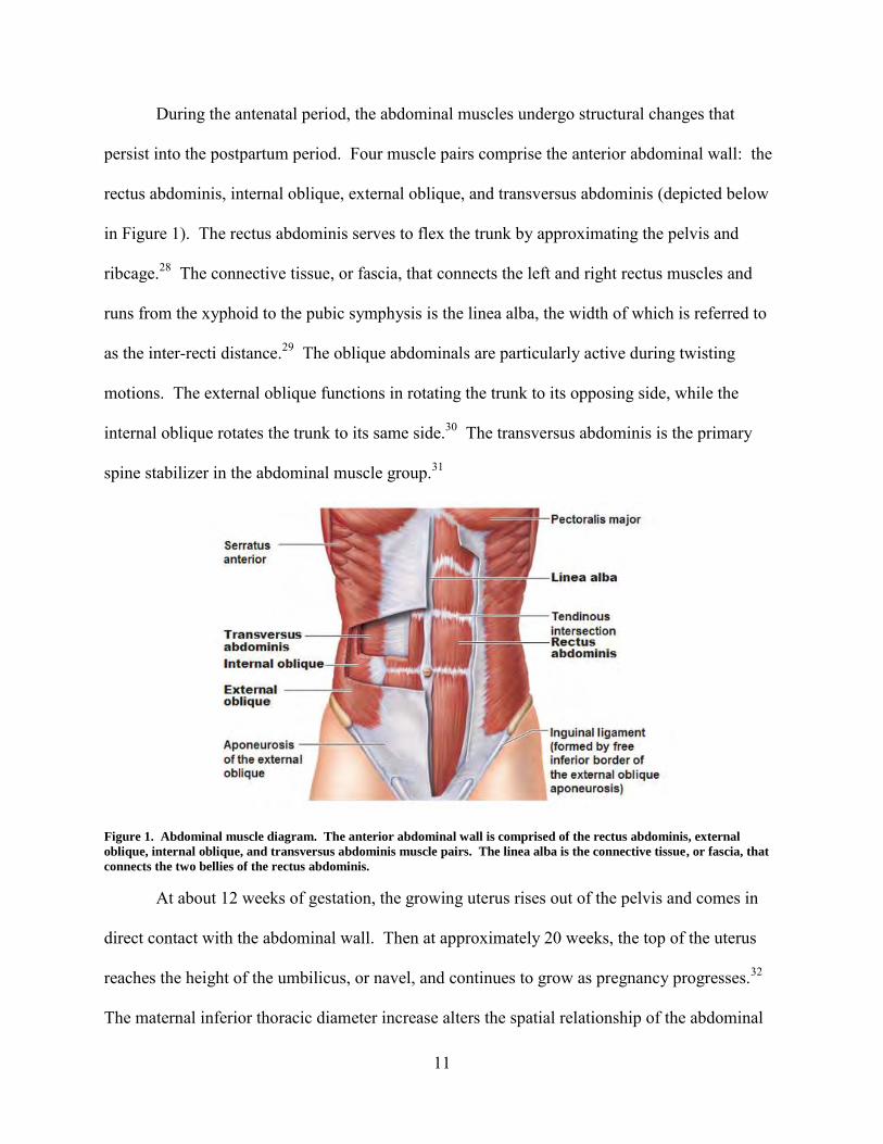
During the antenatal period, the abdominal muscles undergo structural changes that
persist into the postpartum period. Four muscle pairs comprise the anterior abdominal wall: the
rectus abdominis, internal oblique, external oblique, and transversus abdominis (depicted below
in Figure 1). The rectus abdominis serves to flex the trunk by approximating the pelvis and
ribcage.28 The connective tissue, or fascia, that connects the left and right rectus muscles and
runs from the xyphoid to the pubic symphysis is the linea alba, the width of which is referred to
as the inter-recti distance.29 The oblique abdominals are particularly active during twisting
motions. The external oblique functions in rotating the trunk to its opposing side, while the
internal oblique rotates the trunk to its same side.30 The transversus abdominis is the primary
spine stabilizer in the abdominal muscle group.31
Figure 1. Abdominal muscle diagram. The anterior abdominal wall is comprised of the rectus abdominis, external
oblique, internal oblique, and transversus abdominis muscle pairs. The linea alba is the connective tissue, or fascia, that
connects the two bellies of the rectus abdominis.
At about 12 weeks of gestation, the growing uterus rises out of the pelvis and comes in
direct contact with the abdominal wall. Then at approximately 20 weeks, the top of the uterus
reaches the height of the umbilicus, or navel, and continues to grow as pregnancy progresses.32
The maternal inferior thoracic diameter increase alters the spatial relationship of the abdominal
12
muscle attachments and increases the distance between muscle attachments, resulting in muscle
lengthening. To be more specific, in the case of the rectus abdominis, absolute and normalized
muscle lengths increase from 18 to 38 weeks of gestation.33 This increase likely reflects muscle
fiber length increase;34 however, the rectus abdominis does exhibit thinning during pregnancy.35
The muscle also widens. With an increase in length and width, the cross-sectional area
increases.36 In addition to dimensional changes specific to the individual muscles, the rectus
abdominis muscle-pair separation increases above, at, and below the umbilicus. The abdominal
separation 4.5 centimeters (1.8 inches) below the umbilicus increases between gestational weeks
18 and 30, while the separation at and above the umbilicus continues to increase from 18 to 38
weeks.37 In conjunction with this muscle separation, the linea alba, which is the connective
tissue between the rectus muscles, widens.38 The rectus abdominis muscle curves around the
abdominal protuberance as pregnancy progresses instead of maintaining the normal vertical
orientation. The resulting altered line of action likely contributes to antenatal abdominal muscle
functional deficits. 39 Specifically, women demonstrate a diminished ability to stabilize the
pelvis against resistance between 30 and 38 weeks of gestation.40 Such structural changes and
decreased functional abilities occur irrespective of maintaining an antenatal aerobic exercise
regimen.41
An inter-recti distance of greater than 2 centimeters at a given assessment point with
respect to the umbilicus is generally considered diastasis recti.42 In a study of six primigravid*
subjects with a single fetus who regularly conducted aerobic exercise throughout their
pregnancies, Gilleard and Brown observed abdominis muscle separation widths at, above, and
below the umbilicus that exceeded 2 centimeters at 38 weeks gestation in all subjects.43
* “Primigravid” means pregnant for the first time.
13
Although the small sample size may preclude this rate of diastasis recti from adequately
representing the broader maternal population, a more recent study published in February 2015 by
Mota, et al., found diastasis recti at 2 centimeters (0.8 inches) below the center of the umbilicus
in 100 percent of the subject population of 84 primigravid women at 35 weeks gestation.44
Although Mota, et al., only measured at one location along the linea alba, their sample size and
the reliability of their measuring techniques make the findings applicable to the broader maternal
population.
The pelvic floor includes the structures that comprise the bottom of the abdominal
canister. It is a three-dimensional structure made up of several muscles and an extensive,
complex fascial support system.45 The primary functions of the pelvic floor are to support the
weight of the abdominal and pelvic organs, maintain intra-abdominal pressure, allow voluntary
control of urination and defecation, and enable sacrum and coccyx flexion. In women, the pelvic
floor also facilitates fetal movement through the birth canal. The pelvic floor’s ability to perform
these functions effectively depends on constituent muscle fiber integrity and muscle tone
maintenance.46 From 20 weeks gestation, pelvic floor muscle strength exhibits a decline that
may interfere with muscle function.47 Additionally, fascial tissue stretches and may tear slowly
over time as the weight of the fetus increases.48
Pelvic floor muscles are placed under substantial stress during delivery. Ultrasound and
magnetic resonance imaging show major pelvic floor muscle injuries in 20 to 26 percent of
women following vaginal delivery.49 One specific pelvic floor muscle that is part of the levator
ani muscle group is known to stretch over three times its resting length during the second stage
of labor. More than one third of women who deliver vaginally also experience levator avulsion,
which means that the muscle is separated from its insertion. Increased maternal age increases the
14
risk of avulsion. Avulsion is associated with highly significant reduction in muscle strength.
This deficit can also lead to bladder prolapse, a condition where the bladder descends from its
normal position.50 Vaginal delivery also results in partial denervation of pelvic floor muscles in
80 percent of women. Women who experienced long, active second stage labor showed greater
evidence of denervation. Damage to the nerve to the levator ani may result in atrophy of the
denervated muscle, thereby placing more stress on the pelvic fascia. Such increased fascial
stress may result in organ prolapse or separation of the fascia over time.51 The majority of direct
pelvic floor fascia injuries are thought to occur during vaginal delivery. Pelvic floor fascia may
detach or tear during deliver, thereby destabilizing the pelvic floor.52
Pelvic floor muscle and fascia integrity and function are also essential to the preservation
of continence, particularly during tasks that increase intra-abdominal pressure. If pelvic floor
muscles are weak, if they do not contract at the right time, or if the pelvic floor fascia is lax or
torn, the pelvic floor will be unable to control urine loss.53
Collectively, the hematological, cardiovascular, respiratory, and musculoskeletal changes
a woman undergoes during the antenatal period are considerable. Discussion of these changes
clearly shows the extent to which pregnancy changes physiology and morphology across the
body’s systems. Such structural and functional changes accumulate over the gestational period
to accommodate the developing fetus and its needs. Naturally, this 40-week process is not
reversed or resolved immediately upon childbirth.
Postpartum Physiology and Morphology
The puerperium is the period following delivery during which most pregnancy-induced
maternal anatomical and physiological changes return to the nonpregnant state. While
puerperium duration varies, it generally is considered to last until 6 weeks postpartum.54
15
Although most physiological adaptations return to a nonpregnant state during puerperium, many
cardiovascular changes persist for months following delivery, and some changes, such as those
to pelvic musculature and cardiac remodeling, persist for years.55 Additionally, a return to
nonpregnant physiology does not necessarily equate to a return to nulliparous* morphology; a
number of maternal characteristics, beyond those discussed in this paper, do not return to
nulliparous form.
Uterine involution, or shrinkage, begins immediately after delivery. The uterus and its
inner membrane, the endometrium, return roughly to pregravid size by eight weeks following
delivery; however, after each successive delivery, the uterus remains slightly larger following
involution than it was before that pregnancy.56 In conjunction with involution, postpartum
uterine discharge, called lochia, begins as a flow of blood that diminishes in volume. It has a
reduced blood component within 3 to 4 days postpartum but continues as a brownish or pink
discharge with a median duration of 22 to 27 days.57
Between natural blood loss during delivery and through lochia and increased postpartum
urine production, blood volume typically returns to nonpregnant levels by one week after
delivery. After the first 2 days postpartum, systemic vascular resistance and blood pressure
begin to rise steadily to normal values.58 Deep vein vessel size significantly reduces and venous
flow velocity concomitantly increases during the puerperium. Cardiac output peaks at 24 weeks’
gestation and after delivery slowly returns to nonpregnant values. However, even after 1 year
postpartum, cardiac output remains significantly higher than prepregnant values in both
primiparous and multiparous† women.59
* The term “nulliparous” refers to a woman who has not borne a child. † “Primiparous” refers to a woman who has borne one child, and “multiparous” means having given birth to more than one child.
16
Regarding cardiovascular fitness, women who are sedentary during the antenatal period
consistently have lower maximal oxygen uptake (VO2max) postpartum than they did
prepregnancy.60 However, through moderate exercise, a woman can maintain peak ventilation
and absolute maximal aerobic capacity. In fact, the combination of pregnancy and physical
training can even improve maximal aerobic capacity.61 Well-conditioned women who maintain
at least a moderate exercise regimen, comprised of three or more 30-minute aerobic sessions
weekly, during pregnancy and postpartum have a small but statistically significant increase in
postpartum VO2max.62 The enhanced training effect of pregnancy becomes most apparent six
months to one year after delivery.63
The increased joint laxity that peaks in the third trimester persists into the postpartum
period and remains at near third trimester level even at 6 weeks postpartum.64 While joint laxity
does not resolve immediately, joint strengthening usually completes within three to five months
of childbirth.65 The exception to this joint strengthening timeline is lactating women, who
remain under the influence of pregnancy hormones for as long as they are breastfeeding. In the
case of nursing women, they may see persistent joint laxity for up to three months following
lactation termination.66 Although the body of research regarding postpartum connective tissue
compromise is extremely limited, numerous physical therapists both within the United States and
abroad have observed persistent ligament relaxation in their patients that follows the timeline
described above.67
The abdominal muscles experience immediate change following delivery. Upon
childbirth, the effective total rectus abdominis muscle length increases suddenly when the stretch
from the uterus is removed. The sudden increase in effective length affects the rectus abdominis’
ability to produce tension, and thus contributes to functional deficits. 68 The cross-sectional area
17
of the rectus abdominis returns to values similar to nulliparous values after approximately 8
weeks postpartum. Although muscle width remains significantly different from nulliparous
values at 12 months postpartum, width steadily declines by 8 weeks and plateaus at some point
between 6 and 12 months postpartum. Because cross-sectional area is a function of length and
width, a combined consideration of cross-sectional area and width resolution timelines implies
that muscle length returns to prepregnancy values between 8 weeks and 6 months postpartum.
While the rectus abdominis steadily thickens over the course of the 12-month postpartum period,
the muscle remains significantly thinner than it was prepregnancy.69 The reduction in muscle
thickness manifests itself as a decrease in concentric and eccentric trunk flexion strength that is
observed at least through 6 months postpartum.70
The rectus abdominis muscle separation widths at, above, and below the umbilicus
narrow between 38 weeks of gestation and 4 weeks postpartum to widths observed between 22
and 26 weeks of gestation.71 The ability of the abdominal muscles to generate torque remains
reduced at 8 weeks postpartum; therefore, exercises that require significant torque production are
unsuitable. This means that trunk flexion activities such as sit-ups are contraindicated.
Additionally, the ability to stabilize the pelvis against resistance remains reduced postpartum.
This reduced capacity is problematic because correct performance of most abdominal exercises
requires stabilization of the pelvis.72
Even by 6 months postpartum, the inter-recti distance does not return to prepregnacy
values. When Liaw, et al., compared inter-recti distance measurements of postpartum women to
nulliparous age-matched counterparts in a control group, they found that inter-recti distances,
both above and below the umbilicus, in women at 6 months postpartum exceed that of their age-
matched counterparts, who are without previous pregnancy. Postpartum women exhibit
18
significant reduction in inter-recti distance at the upper margin of the umbilical ring and 2.5
centimeters above the umbilicus between 7 weeks postpartum and 6 months postpartum;
however, the change below the umbilicus over the same period is insignificant.73 In addition to
the study by Liaw, et al., a study by Coldron, et al., shows that inter-recti distance does not return
to control values at 12 months postpartum but the value plateaus by 6 months.74
In terms of abdominal muscle function, muscle strength and static endurance improves
between 7 weeks postpartum and 6 months postpartum; however, after 6 months, postpartum
women still exhibit less strength and endurance than their nulliparous counterparts.
Additionally, the increased postpartum inter-recti distance measurements correlate negatively
with strength and endurance of trunk flexors and rotators. In particular, the reduction in the
inter-recti distance between 7 weeks and 6 months postpartum was associated with an
improvement in trunk flexor strength.
Collectively, these findings suggest that the incomplete recovery of the linea alba may
create a functional deficit that reduces the ability of the abdominal musculature to generate
force.75 In other words, the inter-recti distance in postpartum women does not recover to
nulliparous values even at 6 months postpartum, and that increased linea alba width correlates
with abdominal functional deficit. Both structural and functional deficits persist to at least six
months after childbirth. The findings of Liaw, et al., coupled with the findings of Coldron, et al.,
that indicate persistent decreased muscle thickness, increased muscle width, and increased inter-
recti distance at 12 months postpartum imply that the postpartum mechanical disadvantage
induced by pregnancy may not resolve.76 The results of Liaw, et al., are more appropriate to
consider than many other studies that examine inter-recti distance because Liaw, et al., followed
participants for 6 months postpartum, considered a larger sample size, and used ultrasound
19
imaging to measure inter-recti distance at several locations to more objectively quantify
diastasis.
In the Liaw, et al., study, the postpartum and nulliparous women had not received any
abdominal muscle training or engaged in any other regular exercise within the previous six
months. Additionally, the postpartum women did not engage in abdominal muscle training or
regular exercise during the length of the study; they merely performed activities consistent with
daily living.77 One might argue that such study participant conditions negates the applicability of
the findings to a postpartum Marine population, whose members would be engaged in some
degree of regular exercise assuming their medical condition allows it; however, this is not the
case. In fact, because the Liaw, et al., study investigates the natural recovery of inter-recti
distance and abdominal muscle function, it can actually be applied more broadly. Since the
study is unencumbered by variables specific to a given exercise regimen, studies regarding the
effects of exercise on inter-recti distance can be overlaid on the Liaw, et al., findings. A
systematic review published in March 2014, which comprehensively reviewed studies that
examined the effects of exercise on diastasis recti and were published no later than July 31, 2012,
indicated that the degree to which abdominal exercise reduces postpartum diastasis recti is
unclear. The review, however, does state that transversus abdominis muscle activation could
potentially protect the linea alba and help prevent or reduce diastasis recti, because activation
and exercise of the transversus abdominis draws the bellies of the rectus abdominis together and
increases fascial tension.78 Regardless of whether abdominal exercise can reduce the inter-recti
distance, multimodal* physical therapy treatment has enabled postpartum women to restore
* Multimodal treatment is treatment that involves a combination of approaches and may include general education; strengthening, kinesthetic, and functional exercises; muscle reeducation; and posture correction.
20
optimal strategies for transferring loads through the abdominal canister irrespective of diastasis
recti closure.79
As discussed in the preceding antenatal section, the vast majority, if not all, pregnant
women have diastasis recti by 35 weeks gestation. At 6 to 8 weeks postpartum and also at 12 to
14 weeks postpartum, the percentage of women exhibiting diastasis recti declines to around 50
percent. After six months, 39 percent of the postpartum population has diastasis recti.80 One
may hypothesize that certain factors predispose a woman to experiencing diastasis recti;
however, Mota, et al., did not identify any significant risk factors associated with the presence of
diastasis recti at 6 months postpartum. Specifically, the following variables showed no
significant correlation with the presence of diastasis recti: age; body mass index (BMI) before
pregnancy or at 6 months postpartum; weight gain during pregnancy; joint hypermobility; baby’s
weight at birth, abdominal circumference at 35 weeks gestation; or exercise training level
(defined as at least three times weekly) before, during, and after pregnancy.81 These findings
were comparable to the one other study that examined similar variables.82 Such studies illustrate
that diastasis recti, as well as less significant persistent rectus abdominis separation, occurs
irrespective of the woman’s fitness level before pregnancy or the activity level she is able to
maintain through the antenatal period. Abdominal separation also occurs irrespective of BMI or
weight gain. Whether a woman is physically active and able to remain so and regardless of the
extent of her weight gain, she will experience this pregnancy-induced change and the
corresponding functional deficit. Increased parity, however, does seem to increase the risk of
diastasis recti.83 This means that women are more likely to have diastasis recti upon each
subsequent pregnancy.
21
Postpartum pelvic floor damage can result from the trauma of second stage labor and
vaginal delivery or from microtrauma over prolonged periods of time. Microtrauma can occur
during pregnancy from the increased weight of the fetus and/or following childbirth due to non-
optimal strategies for transferring loads through the pelvis.84 Although women who give birth
via caesarean section do not experience trauma resulting from vaginal delivery and experience
reduced, if any, second stage labor trauma, they can still be afflicted by microtrauma.
Pelvic floor dysfunction includes urinary incontinence, anal incontinence, and pelvic
organ prolapse.85 In the case of urinary incontinence, on average 30 percent of women with
vaginal deliveries experience the dysfunction compared to 15 percent of women who delivered
by caesarian section. At 1 year postpartum, 23 percent of women who deliver vaginally and 10
percent of those delivering via caesarian section experience stress urinary incontinence. Six
years following delivery, the prevalence of urinary leakage was comparable or slightly higher
than at 1 year postpartum.86 Incidence of anal or fecal incontinence is much lower and generally
is limited to women with anal sphincter rupture; incidence of rupture is of primary significance
rather than mode of delivery.87 Pelvic organ prolapse is associated significantly with vaginal
delivery, age over 30 years at delivery, and increased parity.88
Urinary incontinence is the most prevalent symptom of pelvic floor dysfunction. The
most common form of urinary incontinence is stress urinary incontinence, which is the
involuntary loss of urine during an effort or physical exertion or on sneezing and coughing.89
Studies that require high adherence to a strength training protocol and close follow-up indicate
that pelvic floor muscle training after delivery can prevent and treat urinary incontinence.*, 90
* The performance of pelvic floor exercises during pregnancy also has been shown to have a protective effect against postpartum urinary incontinence for 6 weeks and 3 months postpartum. Lemos, et al., 878.
22
Although pelvic floor muscle training can treat pelvic floor dysfunction and successfully reduce
symptoms, pelvic floor muscle training necessitates the ability to perform properly a pelvic floor
muscle contraction. An effective pelvic floor muscle contraction is one that increases urethral
closure pressure without a substantial Valsalva effort; it involves an inward lift and squeeze
around the urethra with resultant closure, stabilization, and resistance to downward movement.91
However, in a population exhibiting urinary incontinence, fecal incontinence, or prolapse, only
38 percent of women could perform an optimal pelvic floor muscle contraction. When
attempting to perform a lifting contraction, 43 percent of women depressed the levator plate and
19 percent exhibited no change in levator plate position.92 This observation means that elevation
of the levator plate is not the automatic response to verbal instruction to contract the pelvic floor.
Additionally, women who depress the levator plate when attempting to perform a lifting
contraction are actually weakening the pelvic ligaments and thereby contributing to the
pathology of stress urinary incontinence and pelvic organ prolapse.93 Such responses
demonstrate the importance of education even in women who believe they know how to perform
a correct pelvic floor muscle contraction.
Physiological/Morphological Implications for Postpartum Rehabilitation
During the puerperium, or approximately the first six weeks following delivery, women
may start frequent sessions of sustained exercise and gradually increase exercise volume, i.e.,
intensity and duration, over time. Many women resume some form of exercise within two weeks
after childbirth. If a woman experienced caesarean birth or traumatic vaginal birth, she may
need to wait four weeks or longer before resuming exercise. During this initial stage, exercise
should serve to provide relaxation and enhance well-being. While the body is undergoing its
greatest extent of recovery from pregnancy and childbirth, progress in an exercise regimen is
23
irrelevant. Walking is the ideal exercise, but other forms of low-impact cardiovascular exercise
such as swimming or cycling may also be permissible depending on the woman’s birthing
circumstances. Physicians recommend exercising at least three days a week; however, five times
a week is ideal. If a woman’s exercise does not provide relaxation or promote well-being or if it
results in exercise-associated pain or heavy bleeding, she should revise her exercise program to
reduce its frequency or volume. Additionally, the woman must ensure she maintains proper
hydration and obtains adequate rest. The three absolute contraindications to exercise during the
puerperium are heavy bleeding, pain, and infection or abscess. Women should also reduce breast
discomfort by nursing or relieving engorgement prior to exercise and ensuring adequate breast
support during exercise. While not complete contraindications to exercise, urine leakage and
increased pelvic pressure during exercise are cues that a given activity is too intense.94
As previously discussed, women who are sedentary in the antenatal period, either by
choice or by medical necessity, undergo cardiovascular detraining during their sedentary period.
Conversely, women who maintain at least a moderate exercise regimen can maintain or increase
their absolute maximal aerobic capacity.95 In either case, resumption of aerobic exercise
following childbirth does not present inherent cardiovascular risk. Women should start with
light- to moderate-intensity* aerobic exercise in terms of perceived exertion, particularly in the
case of deconditioned women, and gradually increase exercise intensity and duration following
the guidelines in the preceding paragraph. Because research supports the value of discontinuous
exercise patterns, women may accumulate bouts of aerobic activity lasting at least 10 minutes
throughout the course of the day and still obtain well-being and health benefits.96
* Light intensity, as described by the American College of Sports Medicine (ACSM), equates to Borg rating of perceived exertion (RPE) 9-11 (very light to fairly light); moderate intensity corresponds to Borg RPE 12-13 (fairly light to somewhat hard).
24
Although a given activity may be safe in terms of cardiovascular risk, not all aerobic
activities or anaerobic exercises are advisable in the immediate postpartum period for other
reasons. High-impact activities such as running and jumping can place undo strain on weakened
pelvic floor muscles and lax pelvic floor fascia, thereby leading to or exacerbating pelvic floor
dysfunction. High-intensity, high-impact activities such as interval training, hill training, or
activities that involve rapid direction change place even greater strain on pelvic floor structures.97
The most suitable aerobic exercise for at least the first 3 weeks postpartum is walking.
Once the flow of lochia has subsided, a woman can resume swimming or low intensity water
aerobics. Once she has healed sufficiently to be comfortable sitting on a bicycle seat, a woman
may resume cycling. Specifically in the case of cycling, the seat provides support to the pelvic
floor. After the routine 6-week postnatal physical examination, provided the outcome is
favorable, a woman smay safely begin low impact aerobics training. During the third month
postpartum, the woman ideally should be evaluated by a physical therapist and subjected to
abdominal muscle and pelvic floor testing before introducing high-impact exercise. Generally, at
approximately 4 months postpartum it should be safe to introduce high-impact exercise. This
statement means that at approximately 4 months postpartum a woman can start running. It does
not mean that she has sufficient pelvic floor and abdominal muscle strength to begin sprinting
and conducting high-intensity interval training. Upon introducing high-impact activities, the
woman should reduce the intensity of her training or change the nature of her activity if she leaks
urine or senses pelvic floor muscle weakness or bouncing. Once she can engage in a given
activity without experiencing pelvic floor weakness or dysfunction, she may gradually increase
the intensity of her activity.98 By 9 months postpartum, the woman should be able to conduct
25
intense interval training and hill training without experiencing feelings of weakness in the pelvic
floor.99
This prescribed guidance and the associated timeline regarding resumption of
cardiovascular training assumes initiation of a graduated, progressive abdominal and pelvic floor
muscle training program during the immediate postpartum period. In addition to accounting for
pregnancy-induced and labor- and delivery-induced changes in abdominal musculature and
pelvic floor structures, these guidelines consider the 3- to 5-month timeline for postpartum joint
laxity resolution.
In terms of muscular fitness, the woman should begin performing transverse abdominal
contractions and pelvic floor exercises shortly after delivery. After her 6-week postnatal check-
up, again presuming favorable outcome, she may resume resistance training with light weights.
She must be able to maintain good posture and form throughout a given activity.100 When an
exercise is difficult, the individual performing the exercise may not follow correct performance
techniques.101 Additionally, she should not lift anything so heavy or perform a task so
challenging that it requires straining or breath holding.102 Straining or holding one’s breath
increases intra-abdominal pressure, thereby stressing the core musculature,* including the
transversus abdominis and the pelvic floor muscles, that manages such pressure. In addition to
beginning with lighter weights, she should be sure to lift her pelvic floor before exerting effort
and relax the pelvic floor afterwards. She should remain mindful of pelvic floor and transversus
abdominis fatigue and reduce repetitions or increase rest as required. Reducing the depth of
lunges or squats to keep hips higher than knees and using supported or seated positions also
helps protect the pelvic floor as core muscle fitness improves. Over time, the woman may
* The core muscles include the diaphragm, the multifidus (extends up the spine), the transversus abdominis, and the pelvic floor.
26
gradually increase weight, number of repetitions at a given weight, and depths of squats or
lunges. She also will be able gradually to introduce more demanding core exercises.103
To increase safely the degree of impact, intensity, or difficulty associated with
cardiovascular or muscular fitness training, the postpartum woman must engage in a graduated
abdominal and pelvic floor muscle training program. The typical abdominal exercise regimen
predominantly involves exercises that involve trunk flexion, such as sit-ups or crunches. Such
rehabilitation programs are problematic because they preferentially train the rectus abdominis
and neglect the deeper abdominal muscles that function as stabilizers and help manage intra-
abdominal pressure. The increased activity of the rectus abdominis during trunk flexion
exercises overrides the static holding capacity of transversus abdominis and internal oblique
muscles. Furthermore, the higher the level of speed at which trunk flexion exercises are
performed, the greater the reduction of abdominal stabilizing function.104 Additionally, the trunk
flexion exercises relied upon in traditional abdominal exercise programs increase intra-
abdominal pressure.105 When activities that increase intra-abdominal pressure, such as trunk
flexion or heavy lifting, are introduced before the deep abdominal and pelvic floor muscles are
adequately strengthened, the woman’s inability to engage those muscles to control the pressure
increase can lead to incidence or recurrence of diastasis recti, abdominal fascia herniation,
urinary or fecal incontinence, and/or pelvic organ prolapse.106
In order to train the deep core muscles, the transversus abdominis and pelvic floor should
be trained progressively prior to introducing exercises that involve rectus abdominis contraction.
Over time and following a graduated abdominal and pelvic floor muscle training program,
postpartum women can learn to engage the transversus abdominis and pelvic floor and maintain
the engagement as they lift their head and shoulders from a hook-lying position. With
27
progressive training, women can lift their head and shoulders higher, do more repetitions, and
move their hands to positions that make the activity more demanding.107 Presuming a woman
starts her transversus abdominal and pelvic floor training during the immediate postpartum
period, she should be safe to introduce a crunch or sit-up activity at approximately 6 months
postpartum.108
The PFT and CFT clearly include high-impact events. Additionally, due to the way in
which events are evaluated, every test event is high-intensity. The participant must execute
maximum repetitions, either without or within a time limit, and the participant must cover
prescribed distances or perform prescribed activities in as little time as possible.109 Such impact
and intensity place substantial demands on the transversus abdominis and pelvic floor. Within
the PFT, executing a maximum set of crunches in a 2-minute period is particularly demanding
because it requires the Marine to perform high repetitions of crunches and to perform those
repetitions at a high rate of speed. Even in isolation, these two requirements, high numbers of
repetitions and high rates of speed, reduce the ability of the transverse abdominis and oblique
muscles to perform a stabilizing function.
Proper rehabilitation prior to resuming fitness testing is important particularly in the case
of the PFT crunch event. As discussed in the “Postpartum Physiology and Morphology” section,
the inter-recti distance in postpartum women does not recover to nulliparous values even at 6
months postpartum, and the increased linea alba width correlates with abdominal functional
deficit. While the degree to which abdominal exercise can reduce the inter-recti distance is
unclear,110 physical therapy treatment can enable postpartum women to restore optimal strategies
for transferring loads through the abdominal canister irrespective of diastasis recti closure.111
28
Trunk flexion activities are specifically contraindicated during the immediate postpartum period,
and they remain unsuitable until the transversus abdominis and pelvic floor are sufficiently
strengthened to contain the increase in intra-abdominal pressure associated with the activity.
Approximately 6 months postpartum is a reasonable point to expect the majority of women to be
able to introduce a crunch activity. “Introduce” means that the woman is doing the activity for
the first time. The postpartum woman will then have to build up progressively to performing
more sets throughout the day and higher numbers of repetitions within a given set before she
safely will be able to continue the activity for a 2-minute time period at a high rate of speed.
Although no one specific research study answers the question of how long it takes to
progress sufficiently before a postpartum woman is physically capable of safely executing a
maximum set of crunches in 2 minutes, physical therapists suggest approximately 9 months as
sufficient based on their clinical observations.112 Ideally, each woman should be evaluated
individually by a women’s health physical therapist and cleared to undergo fitness testing based
on her individual progress.113 However, having a prescribed timeline that captures the majority
of the postpartum population provides a general guideline to postpartum women and health care
providers and forces a trigger to assess whether women are making sufficient progress towards
rehabilitation, if they need more time (i.e., an extension before resuming testing), or if their
medical situation precludes further service. The 9-month timeline recommendation affords the
majority of postpartum women sufficient time to rehabilitate in order to perform the crunch event
safely.
Antenatal and postpartum physiology and morphology have substantial implications for
postpartum rehabilitation. In the case of postpartum Marines, these implications mean that
although postpartum Marines generally return to full duty at 6 weeks postpartum, they have not
29
sufficiently recovered to be able to perform the complete spectrum of full duty activities. They
must progressively introduce those activities that are more demanding, particularly for core
musculature. The rehabilitation process also determines at what point postpartum Marines are
ready to resume fitness testing safely. If a woman begins a graduated abdominal and pelvic floor
muscle training program by 6 weeks postpartum, she should be able to perform safely high-
intensity, high-impact activities and execute properly repetitive trunk flexion activities by 9
months postpartum.
Recovery Resources
By and large, pregnant and postpartum women do not know what they do not know
regarding postpartum rehabilitation. While MCCS provides limited pregnancy and parenthood
training, it does not cover material specific to postpartum rehabilitation. Military hospitals hold
voluntary classes that women can attend at 28 weeks and 36 weeks gestation, and they provide
breastfeeding training, but these sessions do not discuss maternal care as it pertains to
postpartum fitness. As a result, anything a woman learns during pregnancy or after delivery
about postpartum rehabilitation is self-taught. However, because she does not know what she
does not know, she may have difficulty discerning the accuracy or relevancy of self-acquired
information.
Beyond pregnant and postpartum Marines, many commanding officer and OICs,
particularly those who have never been pregnant, do not understand antenatal or postpartum
physiology and morphology. Those who do understand pregnancy-induced changes still may
lack an appreciation of the implications such changes have for postpartum rehabilitation. For
commanding officers and OICs who have experienced pregnancy, that experience, in and of
itself, does not inherently confer any expertise regarding safe or proper postpartum rehabilitation.
30
While the MCPFP informs commanders and OICs that postpartum Marines require a progressive
training routine to return to prepregnancy fitness levels, the order provides no further information
or additional details.114 In terms of educating Marines, the pregnancy and parenthood order only
requires commanders to provide training on the contents of the order itself.115 As such,
commanders have no requirement or means to educate themselves on postpartum rehabilitation
considerations. Because they generally lack education regarding postpartum rehabilitation, most
commanders are unable to serve as an educational resource to postpartum Marines. Senior
enlisted leaders and other staff non-commissioned officers have the same educational deficits.
Per the MCPFP, as mentioned in the “Current Marine Corps Policies” section, a unit’s
RCP provides tailored physical training for Marines whose fitness level has been degraded due to
pregnancy, among other causes. The unit’s assigned combat conditioning instructor (CCI) or
command physical training representative (CPTR) develops RCP training plans in accordance
with Marine Corps Reference Publication (MCRP) 3-02A, Marine Physical Readiness Training
for Combat and based on organizational mission essential tasks, applicable occupation-specific
requirements, and individual identified deficiencies.116 While the CCI should have attended a
CCI course, CCIs and CPTRs are required by the MCPFP only to review MCRP 3-02A and the
MCPFP order itself.117 Neither the CCI course, the reference publication, nor the order provides
training or education sufficient to qualify the CCI or CPTR to rehabilitate postpartum Marines.118
As such, the RCP is not an appropriate recovery resource regardless of how well-intentioned the
responsible CCI or CPTR may be.
In addition to CCIs and CPTRs, most Semper Fit personal trainers are similarly
unqualified to rehabilitate postpartum Marines despite their certification as personal trainers.
The Semper Fit program does not maintain expertise in postpartum rehabilitation within its
31
personal trainer staff. A given personal trainer may choose independently to pursue education
specific to postpartum rehabilitation, but the personal trainers have no requirement or incentive
to do so. Additionally, personal trainers who pursue pregnancy and postpartum fitness training
via continuing education may only have to attend a weekend seminar, many of which focus
predominantly on pregnancy fitness and neglect or barely cover postpartum concerns.* As such,
having a personal trainer with genuine expertise in postpartum rehabilitation is the exception
rather than the norm at a given installation.
Physical therapy is the resource best suited to postpartum rehabilitation. Although
pregnancy and childbirth should not be conflated with injury, vaginal delivery should be treated
comparably to a major sports injury.119 Additionally, caesarian section, although common, is not
minor surgery. The vast majority of bases and stations have physical therapy clinics. Although a
small percentage of civilian physical therapists are board-certified, approximately 60 to 70
percent of military physical therapists are board-certified. Most board-certified military physical
therapists specialize in either orthopaedics or sports, but the military does have some women’s
health specialists. A physical therapist who is certified in women’s health is best suited to assess
the degree of abdominal separation and pelvic floor damage, as well as any joint or alignment
issues, and to determine individual functional deficits; however, sports specialists have a
component of their certification on understanding women in sports that would qualify them to
conduct a similar evaluation. Such evaluation enables the physical therapist to develop an
individualized progressive graduated rehabilitation program. If a given physical therapist at a
* The book provided for Aerobics and Fitness Association of America’s Perinatal Fitness weekend workshop has 84 pages, 2 of which address postpartum fitness. Aerobics and Fitness Association of America. Perinatal Fitness: Fitness during the Childbearing Year—Pregnancy, Birth, and Postpartum. Sherman Oaks, CA: Aerobics and Fitness Association of America, 2005, workshop manual.
32
particular military treatment facility is presented with a postpartum case that exceeds their level
of expertise, the physical therapist has the ability to refer that patient to a local civilian facility.120
Reconciling Policy with Biology
This section is entitled “Reconciling Policy with Biology” because the Marine Corps can
reconcile policy with biology, but biology cannot adapt to fit policy. Bypassing biology, while a
potential option, is not a viable option. Policy should not define pregnancy or motherhood as
incompatible with service.
As stated in the “Current Marine Corps Policies” section, the current policy regarding
resumption of fitness testing is based on the assertion that the physiological effects of pregnancy
have resolved by the sixth week postpartum and that it is physiologically feasible to achieve
sufficient physical fitness over the course of a 6-month period to complete a fitness test
successfully.121 This assertion is flawed in a couple of ways. First, the ACOG guidelines that
informed the current policy actually state that “many of the physiologic and morphologic
changes of pregnancy persist 4-6 weeks postpartum.”122 That statement does not equate to the
current policy’s justification that all of the physiological effects of pregnancy resolve by 6 weeks
postpartum. Not only does the ACOG not assert such complete resolution, but the section on
“Postpartum Physiology and Morphology” clearly shows that the effects of pregnancy do not
resolve by 6 weeks postpartum. Moreover, some changes persist permanently, while others have
yet to resolve by 6 or 12 months postpartum. Second, because of the flawed assumption
regarding the first 6 weeks postpartum, the assertion that 6 additional months is enough time to
regain sufficient fitness assumes that the postpartum Marine’s baseline at 6 weeks postpartum is
merely a detrained state rather than a state of physiological and morphological deficit that must
be progressively overcome.
33
As shown through the sections that detailed antenatal and postpartum physiology and
morphology and that discussed their implications for postpartum rehabilitation, six months
following return to full duty is an insufficient length of time to recover properly and resume
fitness testing safely. Rather, the current timeline for resumption of fitness testing should be
extended to no earlier than 9 months postpartum.
However, nine months is a sufficient length of time to rehabilitate only if women have
access to appropriate resources to facilitate their recovery. The Marine’s primary care manager,
obstetrician, or another health care provider should refer the postpartum Marine for physical
therapy either prior to childbirth or upon discharge from the hospital or birthing facility
following delivery. This referral timeline would enable the woman to schedule an initial
appointment shortly after discharge. Having an initial evaluation prior to return to full duty
would enable the physical therapist to conduct an initial assessment of any pelvic floor damage
and provide instruction to the postpartum Marine on the performance of pelvic floor and
transversus abdominis exercises to be conducted during the immediate postpartum period before
she returns to work. Ideally, the physical therapist should be a women’s health specialist;
however, a sports specialist does have a component regarding understanding women in sports.
Whether military or civilian, physical therapists who work in military care facilities but lack
women’s health or sports board-certification should receive women’s health training specifically
regarding postpartum or pelvic physical therapy so that they can adequately rehabilitate
postpartum patients.
Although not a direct requisite for reconciling policy with biology, education plays an
important role in policy implementation. As discussed in the “Recovery Resources” section,
pregnant and postpartum Marines and Marine leaders have limited resources available to provide
34
education regarding the implications of antenatal and postpartum physiology and morphology for
physical training and fitness testing. In the case of pregnant and postpartum Marines, the
physical therapist to whom she is referred can provide adequate education, training, and
guidance to direct and support her rehabilitation. To address the lack of educational resources
for commanders and other Marine leaders, adding a brief explanatory paragraph to the MCPFP
in order to better clarify and describe the postpartum rehabilitation process would be a relatively
simple way to provide more information and a greater degree of expectations management
without levying an additional training requirement. Currently, the MCPFP only includes the
following addendum sentence to serve as a caveat at the end of paragraph 6.f.(2) in Chapter 1:
“Commanders/OICs should be attentive that Marines returning to full duty status following
pregnancy will require a progressive training routine in returning to the level of fitness
experience prior to pregnancy.”123 This sentence is inadequate in terms of providing
commanders and Marine leaders an appreciation of the rehabilitation process and should be
replaced with a separate numbered subparagraph within paragraph 6.f. that concisely explains
why postpartum fitness must be graduated and progressive. The addition to the order should also
indicate that although the postpartum Marine typically returns to full duty following her
convalescent leave, she is not yet ready to perform the complete spectrum of full duty activities,
particularly combat conditioning or physical training activities. If a commander or Marine leader
desires more extensive education regarding postpartum rehabilitation, that leader can consult a
qualified physical therapist. Additionally, socialization of postpartum rehabilitation concerns by
Marine leaders and Marine mothers will raise service-wide awareness over time.
Despite the extent of information currently available regarding postpartum rehabilitation,
reconciling policy with biology should remain an iterative process. In the case of endocrine
35
changes, the current body of research does not examine the degree to which the influence of
pregnancy hormones persists following delivery. Particularly in the case of lactating women,
research has not considered the impact of the hormones that maintain lactation on ligament
relaxation. Once further studies regarding joint laxity in lactating women are conducted, the
Marine Corps will have the responsibility to reexamine postpartum fitness testing guidelines to
ensure that they are still relevant to breastfeeding mothers. While breastfeeding women likely
can progress safely along the recommended 9-month postpartum rehabilitation timeline, any
extended joint laxity persistence in their case makes graduated rehabilitation and mastery of
optimal load transfer strategies even more essential to long-term recovery.
Conclusion
Things do not just go back to normal following childbirth, and that reality has compelling
implications for resumption of fitness testing. Although no one specific research study answers
the question of how much time a postpartum woman needs to rehabilitate sufficiently before she
is physically capable of safely completing a fitness test, an analysis of the body of research,
reported clinical observations, and the rehabilitation process clearly shows the insufficiency of
current Marine Corps policy. Six months following return to full duty, or approximately seven
and a half months, is not enough time to regain sufficient muscular strength and endurance,
particularly in transversus abdominis and pelvic floor musculature, to be able to perform high-
intensity, high-impact activities and sustained repetitive trunk flexion activities. Postpartum
Marines should, however, be able safely to complete a fitness test at 9 months postpartum as
long as they begin a graduated physical therapy program shortly after delivery to regain core
strength. Like all other Marines, postpartum Marines are athletes, but they need time and
appropriate resources to be able to rehabilitate properly. If they are not afforded sufficient time
36
or appropriate resources to rehabilitate properly, they risk incidence or recurrence of diastasis
recti, abdominal fascia herniation, urinary and fecal incontinence, and/or pelvic organ prolapse.
If the Marine Corps makes the nine-month short-term investment in its postpartum Marines, it
will improve the long-term health and readiness of its postpartum population.
The Marine Corps has a responsibility to pursue genuine reconciliation of policy to
biology in matters where biology is compatible with continued service. This paper’s
comprehensive review and analysis presents a compelling case for its two primary policy
recommendations. However, if Marine decision-makers deem this presentation insufficiently
compelling or inadequate to inform a policy change, the Marine Corps has a responsibility to
pursue an objective study of its own pregnant and postpartum populations to augment the current
body of research.* Whether Marine decision-makers embrace this paper’s thesis, the proponents
of the MCPFP and the pregnancy and parenthood order need to continue the revisions they began
when Training and Education Command wrote MARADMIN 005/15 in order to obtain parity
within and across the two orders.†
* To date, no studies have been published that in any way examine postpartum rehabilitation in Marine populations. Three studies related to fitness test performance have been published on postpartum military populations; two of these looked at Air Force sample populations and one at an Army population. These three studies in no way measured any variables that would assess whether women had rehabilitated sufficiently or improved core musculature function adequately to ensure safe testing. Nicole H. Armitage and Denise A. Smart, “Changes in Air Force Fitness Measurements Pre- and Post-Childbirth,” Military Medicine 177, no. 12 (December 2012): 1519-1523; Nicole H. Armitage, Billie M. Severtsen, Roxanne Vandemause, and Denise A. Smart, “Training for the Air Force Fitness Assessment: The Experience of Postpartum Women,” Military Medicine 179, no. 7 (July 2014): 766-772; and Stacy Usher Weina, “Effects of Pregnancy on the Army Physical Fitness Test,” Military Medicine 171, no. 6 (June 2006): 534-537. † Currently, the pregnancy and parenthood order is undergoing revision by the Manpower Plans and Policy Division at Manpower & Reserve Affairs.
37
APPENDIX A: POSTPARTUM RECOVER RESOURCES
The resources listed below provide general information and are not a substitute for individualized medical advice or prescribed treatment. Ryan, Marianne. Baby Bod®: Turn Flab into Fab in 12 Weeks Flat. Edited by Deborah
Grandinetti. Forthcoming. Marianne Ryan is a prominent prenatal and postpartum physical therapist and clinical
director in New York, NY. This book’s release is tentatively scheduled for late April 2015. Although this book has an informal title, it is the product of thorough research by the author. Ryan successfully communicates technical concepts and explanations in layman’s terms so that they can be widely understood and applied.
The Pregnancy Centre. “Postnatal Exercises.” The Pregnancy Centre, last modified 2015.
http://www.thepregnancycentre.com.au/post-pregnancy/exercise/postnatal-exercises. This website provides graduated core exercise levels developed by Dianne Edmonds,
Director and Founder of The Pregnancy Centre in Australia. The exercises that comprise each level include photos and detailed descriptions. The levels are loosely tied to weeks postpartum ranging from 0-24 weeks. In addition to the timeline, each subsequent level’s page provides guidelines to help determine individual readiness to advance to that given level.
38
Notes
1 Brian McGuire (Physical Readiness Programs Officer, Marine Air-Ground Task Force Training and Education Standards Division, Training and Education Command), interviewed by author, January 28, 2015 at 1000. 2 Commandant of the Marine Corps, Marine Corps Policy Concerning Pregnancy and Parenthood, MCO 5000.12E W/CH 1-2, December 8, 2004., 3, 10, http://www.marines.mil/Portals/59/Publications/MCO%205000.12E%20W%20CH%201-2.pdf. 3 CMC, Pregnancy and Parenthood, 3. 4 CMC, Pregnancy and Parenthood, 3, 5, 8. 5 CMC, Pregnancy and Parenthood, 3-4. 6 Commandant of the Marine Corps, Marine Corps Physical Fitness Program, MCO 6100.13 W/CH 1, August 1, 2008, 1-8, http://www.marines.mil/Portals/59/Publications/MCO%206100.13%20W_CH%201.pdf. 7 CMC, MCPFP, 1-8. 8 CMC, MCPFP, 1-8, 1-12. 9 Commanding General, Training and Education Command, Change 2 to MCO 6100.13 Marine Corps Physical Fitness Program, MARADMIN 005/15, January 6, 2015, http://www.marines.mil/News/Messages/MessagesDisplay/tabid/13286/Article/172743/change-2-to-mco-610013-marine-corps-physical-fitness-program.aspx. 10 “Health Services, Headquarters Marine Corps,” Marines: The Official Website of the United States Marine Corps, accessed on February 25, 2015, http://www.hqmc.marines.mil/Agencies/HealthServices/DirectorofPsychologicalHealth.aspx. 11 Prior to providing input, CAPT Vincent L. DeCicco, Director of Clinical Programs, Health Services, Headquarters Marine Corps additionally consulted current guidelines from the American Academy of Pediatrics (AAP), American Academy of Family Physicians (AAFP), and US Preventative Services Task Force (USPSTF); however, only the ACOG provided information specific to the postpartum period. CAPT Vincent L. DeCicco, USN, Director of Clinical Programs, Health Services, Headquarters Marine Corps, “Review and Update of MCO 5000.12E (Marine Corps Policy Concerning Pregnancy and Parenthood),” information memorandum excerpt, April 24, 2014. 12 CAPT Vincent L. DeCicco, information memorandum excerpt. 13 CMC, MCPFP, 1-8. 14 CMC, MCPFP, 4-1. 15 CMC, MCPFP, 2-2 to 2-7. 16 CMC, MCPFP, 3-2 to 3-8. 17 Gary F. Cunningham, et al., ed., Williams Obstetrics, 24th ed. (New York: McGraw-Hill Education, 2014), 55. 18 Cunningham, et al., 58. 19 James F. Clapp III and Catherine Cram, Exercising Through Your Pregnancy, 2nd ed. (Omaha, NB: Addicus Books, Inc., 2012), nook book, 13-14. 20 Cunningham, et al., 62. 21 Cunningham, et al., 63. 22 Clapp III and Cram, 10.
39
23 Kristin Reimers Kardel, “Effects of Intense Training during and after Pregnancy in Top-Level Athletes,” Scandinavian Journal of Medicine and Science in Sports 15, no. 2 (April 2005): 84. 24 Clapp III and Cram, 62. 25 Clapp III and Cram, 53-61. 26 M. L. Marnach, et al., “Characterization of the Relationship Between Joint Laxity and Maternal Hormones in Pregnancy,” Obstetrics and Gynecology 101, no. 2 (February 2003): 332-335. 27 Diane G. Lee and Linda-Joy Lee, The Pelvic Girdle: An Integration of Clinical Expertise and Research, 4th ed. (London: Churchill Livingstone, 2011), 138. 28 C. M. Norris, “Abdominal Muscle Training in Sport,” British Journal of Sports Medicine 27, no. 1 (1993): 19, http://bjsm.bmj.com/. 29 Diane G. Lee, Linda-Joy Lee, and L. McLaughlin, “Stability, Continence and Breathing: The Role of Fascia Following Pregnancy and Delivery,” Journal of Bodywork and Movement Therapies 12, no. 4 (October 2008): 334, http://ncbi.nlm.gov/pubmed/19083692. 30 Norris, 20. 31 Richard S. Jemmett, Spinal Stabilization: The New Science of Back Pain, 2nd ed. (Canada: Libris Hubris Publishing, 2013), 14. 32 Lih-Jiun Liaw, et al., “The Relationships Between Inter-recti Distance Measured by Ultrasound Imaging and Abdominal Muscle Function in Postpartum Women: A 6-Month Follow-up Study,” Journal of Orthopaedic & Sports Physical Therapy 41, no. 6 (June 2011): 441, www.jospt.org. 33 Wendy L. Gilleard and J. Mark M. Brown, “Structure and Function of the Abdominal Muscles in Primigravid Subjects During Pregnancy and the Immediate Postbirth Period,” Physical Therapy 76, no. 7 (July 1996): 755, http://ptjournal.apta.org/content/76/7/750. 34 Gilleard and Brown, 758. 35 Yvonne Coldron, et al., “Postpartum Characteristics of Rectus Abdominis on Ultrasound Imaging,” Manual Therapy 13, no. 2 (May 2008): 116-117. 36 Coldron, et al., 116-117. 37 Gilleard and Brown, 756. 38 Gilleard and Brown, 758. 39 Gilleard and Brown, 757. 40 Gilleard and Brown, 757. 41 Gilleard and Brown, 761. 42 D. R. Benjamin, A. T. M. van de Water, and C. L. Peiris, “Effects of Exercise on Diastasis of the Rectus Abdominis Muscle in the Antenatal and Postnatal Periods: A Systematic Review,” Physiotherapy 100, no. 1 (March 2014): 1. 43 Gilleard and Brown, 756. 44 P. G. Mota, et al., “Prevalence and Risk Factors of Diastasis Recti Abdominis from Late Pregnancy to 6 Months Postpartum, and Relationship with Lumbo-Pelvic Pain,” Manual Therapy 20, no. 1 (February 2015): 202. 45 Diane G. Lee and Linda-Joy Lee, 38. 46 Dot Stables and Jean Rankin, ed., Physiology in Childbearing: With Anatomy and Related Biosciences, 3rd ed. (New York: Baillière Tindall, 2010), 356.
40
47 Andrea Lemos, et al., “Do Perineal Exercises during Pregnancy Prevent the Development of Urinary Incontinence? A Systematic Review,” International Journal of Urology 15 (2008): 875. 48 Lee, Lee, and McLaughlin, 337. 49 Siv Mørkved and Kari Bø, “Effect of Pelvic Floor Muscle Training during Pregnancy and after Childbirth on Prevention and Treatment of Urinary Incontinence: A Systematic Review,” British Journal of Sports Medicine 48, no. 4 (February 2014): 1, http://bjsm.bmj.com/. 50 Lee and Lee, 133-134. 51 Lee and Lee, 141-142. 52 Lee and Lee, 134-137. 53 Lee and Lee, 139-140. 54 Cunningham, et al., 668 and Steven G. Gabbe, et al., Obstetrics: Normal and Problem Pregnancies, 6th ed. (Philadelphia, PA: Saunders, 2012), 517. 55 Gabbe, et al., 517. 56 Cunningham, et al., 669 and Gabbe, et al., 517-518. 57 Cunningham, et al., 670 and Gabbe, et al., 518. 58 Cunningham, et al., 671. 59 Gabbe, et al., 520. 60 Kardel, 84. 61 Clapp III and Cram, 18. 62 James F. Clapp III and Eleanor Capeless, “The VO2max of Recreational Athletes before and after Pregnancy,” Medicine and Science in Sports and Exercise 23, no. 10 (October 1991): 1131. 63 Clapp III and Cram, 18. 64 Marnach, et al., 332. 65 Cunningham, et al., 71. 66 Marianne Ryan, Baby Bod®: Turn Flab into Fab in 12 Weeks Flat, ed. by Deborah Grandinetti, forthcoming, chapter 5, pages 7-8. 67 Ryan, chapter 5, page 7. 68 Gilleard and Brown, 758. 69 Coldron, et al., 116-117. 70 Coldron, et al., 120. 71 Gilleard and Brown, 756. 72 Gilleard and Brown, 761. 73 Liaw, et al., 441. 74 Coldron, et al., 116-117. 75 Liaw, et al., 442. 76 Coldron, et al., 120. 77 Liaw, et al., 436. 78 Benjamin, van de Water, and Peiris, 7. 79 Lee, Lee, and McLaughlin, 336. 80 Mota, et al., 202; Gail S. Bursch, “Interrater Reliability of Diastasis Recti Abdominis Measurement,” Physical Therapy 67 (July 1987): 1078-1079, http://ptjournal.apta.org/content/67/7/1077; Jill Schiff Boissonnault and Mary Jo Blaschak, “Incidence of Diastasis Recti Abdominis during the Childbearing Year,” Physical Therapy 68, no. 7 (July 1988): 1085, http://ptjournal.apta.org/content/68/7/1082; and T. M. Spitznagle, F. C.
41
Leong, and L. R. Van Dillen, “Prevalence of Diastasis Recti Abdominis in a Urogynecological Patient Population,” International Urogynecology Journal and Pelvic Floor Dysfunction 18, no. 3 (March 2007): 321-328. 81 Mota, et al., 203. 82 Mota, et al., 203; Coldron, et al., 120; Benjamin, van de Water, and Peiris, 4; and R. Hannaford and J. Tozer, “An Investigation of the Incidence, Degree and Possible Predisposing Factors of Rectus Diastasis in the Immediate Postpartum Period,” The Journal of the National Obstetrical and Gynaecological Special Group of the Australian Physiotherapy Association 4, no. 2 (1985): 29-34. 83 Volkan Turan, et al., “Prevalence of Diastasis Recti Abdominis in the Population of Young Multiparous Adults in Turkey,” Ginekologia Polska 82, no. 11 (November 2011): 819 and Hannaford and Tozer, 29-34. 84 Lee and Lee, 141. 85 Guri Rørtveit and Yngvild S. Hannestad, “Association between Mode of Delivery and Pelvic Floor Dysfunction,” Tidsskrift for den Norske Legeforening 134, no. 19 (October 2014): 1848. http://tidsskriftet.no/pdf/pdf2014/1848-52eng.pdf. 86 Rørtveit and Hannestad, 1849. 87 Rørtveit and Hannestad, 1850-1851. 88 C. Glazener, A. Elders, C. MacArthur, R. J. Lancashire, P. Herbison, S. Hagen, N. Dean, C. Bain, P. Toozs-Hobson, K. Richard, A. McDonald, G. McPherson, D. Wilson, “Childbirth and Prolapse: Long-Term Associations with the Symptoms and Objective Measurement of Pelvic Organ Prolapse,” BJOG: An International Journal of Obstetrics & Gynaecology 120, no. 2 (January 2013): 164, http://onlinelibrary.wiley.com/doi/10.1111/1471-0528.12075/pdf. 89 Mørkved and Bø, 1. 90 Mørkved and Bø, 11. 91 Judith A. Thompson and Peter B. O’Sullivan, “Levator Plate Movement during Voluntary Pelvic Floor Muscle Contraction in Subjects with Incontinence and Prolapse: A Cross-Sectional Study and Review,” International Urogynecology Journal and Pelvic Floor Dysfunction 14, no. 2 (June 2003): 84 and Judith A. Thompson, et al., “Differences in Muscle Activation Patterns during Pelvic Floor Muscle Contraction and Valsalva Manoeuvre,” Neurourology and Urodynamics 25, no. 2 (2006): 148. 92 Thompson and O’Sullivan, 85-86. 93 Thompson and O’Sullivan, 87. 94 Clapp III and Cram, 101-108. 95 Clapp III and Cram, 18. 96 Carol Ewing Garber, et al., “Quantity and Quality of Exercise for Developing and Maintaining Cardiorespiratory, Musculoskeletal, and Neuromotor Fitness in Apparently Healthy Adults: Guidance for Prescribing Exercise,” ACSM position stand, Medicine & Science in Sports & Exercise 43, no. 7 (July 2011): 1336, 1339, 1344, 1348, http://journals.lww.com/acsm-msse/Fulltext/2011/07000/Quantity_and_Quality_of_Exercise_for_Developing.26.aspx. 97 Continence Foundation of Australia, “Pelvic Floor Safe Cardio Exercises,” Pelvic Floor First, last modified 2013, http://www.pelvicfloorfirst.org.au/pages/what-are-pelvic-floor-safe-exercises.html and Dianne Edmonds (Director and Founder of The Pregnancy Centre in Australia), e-mail message to author, January 13, 2015.
42
98 Continence Foundation of Australia, “Pelvic Floor Safe Cardio Exercises” and Continence Foundation of Australia, “Returning to Sport or Exercise after the Birth,” Pelvic Floor First, last modified 2013, http://www.pelvicfloorfirst.org.au/pages/returning-to-sport-or-exercise-after-the-birth.html. 99 Dianne Edmonds (Director and Founder of The Pregnancy Centre in Australia), e-mail message to author, January 13, 2015 and Dianne Edmonds, “Erica’s Story,” The Pregnancy Centre, last modified 2015. http://www.thepregnancycentre.com.au/post-pregnancy/stories/erica-s-story. 100 Continence Foundation of Australia, “Returning to Sport or Exercise after the Birth.” 101 Gilleard and Brown, 761. 102 Continence Foundation of Australia, “Returning to Sport or Exercise after the Birth.” 103 Continence Foundation of Australia, “Pelvic Floor Safe Resistance Exercises,” Pelvic Floor First, last modified 2013, http://www.pelvicfloorfirst.org.au/pages/pelvic-floor-safe-resistance-exercises.html. 104 Deborah Wohlfahrt, Gwendolen Jull, and Carolyn Richardson, “The Relationship between the Dynamic and Static Function of Abdominal Muscles,” Australian Physiotherapy 39, no. 1 (1993): 12. 105 C. M. Norris, “Abdominal Muscle Training in Sport,” British Journal of Sports Medicine 27, no. 1 (1993): 24, http://bjsm.bmj.com/. 106 Stables and Rankin, 356. 107 Dianne Edmonds, Postnatal Abdominal Rehabilitation (New South Wales, Australia: Australian Fitness Network, 2012), 5, www.fitnessnetwork.com.au/cecs. 108 Dianne Edmonds (Director and Founder of The Pregnancy Centre in Australia), e-mail message to author, January 5, 2015 and Judith Thompson (continence and women’s health physical therapist), e-mail to author, March 2, 2015. 109 CMC, MCPFP, 2-2 to 2-4, 3-2 to 3-8. 110 Benjamin, van de Water, and Peiris, 7. 111 Lee, Lee, and McLaughlin, 336. 112 The author chose to consult two Australian physical therapists, Dianne Edmonds and Judith Thompson, because Australia is acknowledged, even by American physical therapists, to emphasize postpartum rehabilitation better than the United States. The author also consulted American physical therapists in the course of her research. Dianne Edmonds (Director and Founder of The Pregnancy Centre in Australia), e-mail message to author, January 5, 2015; Dianne Edmonds (Director and Founder of The Pregnancy Centre in Australia), e-mail message to author, January 13, 2015; and Judith Thompson (continence and women’s health physical therapist), e-mail to author, March 2, 2015. 113 Dianne Edmonds (Director and Founder of The Pregnancy Centre in Australia), e-mail message to author, January 5, 2015 and Marianne Ryan (prenatal and postpartum physical therapist and Clinical Director, Marianne Ryan Physical Therapy, New York, NY), interviewed by author, February, 19, 2015 at 1500. 114 CMC, MCPFP, 1-8. 115 CMC, Pregnancy and Parenthood, 3-4. 116 CMC, MCPFP, 1-2, 4-1. 117 CMC, MCPFP, 2, 1-2.
43
118 CMC, MCPFP and Commanding General, Marine Corps Combat Development Command, Marine Physical Readiness Training for Combat, MCRP 3-02A, June 16, 2004, https://ia600808.us.archive.org/18/items/milmanual-mcrp-3-02a-marine-physical-readiness-training-for-combat/mcrp_3-02a_marine_physical_readiness_training_for_combat.pdf. 119 Mørkved and Bø, 1. 120 CAPT Michele Weinstein, USN (Department Head, Physical Therapy Department, David R. Ray Branch Health Clinic, The Basic School, Quantico, VA), interviewed by author, February 9, 2015 at 0700. CAPT Weinstein is board-certified in sports. 121 CAPT Vincent L. DeCicco, information memorandum excerpt. 122 American College of Obstetricians and Gynecologists, “Exercise during Pregnancy and the Postpartum Period,” ACOG Committee Opinion, no. 267, January 2002, reaffirmed 2009, 3, http://www.acog.org/Resources-And-Publications/Committee-Opinions/Committee-on-Obstetric-Practice/Exercise-During-Pregnancy-and-the-Postpartum-Period. 123 CMC, MCPFP, 1-8.
44
Bibliography
Aerobics and Fitness Association of America. Perinatal Fitness: Fitness during the Childbearing Year—Pregnancy, Birth, and Postpartum. Sherman Oaks, CA: Aerobics and Fitness Association of America, 2005, workshop manual.
American College of Obstetricians and Gynecologists. “Exercise during Pregnancy and the
Postpartum Period.” ACOG Committee Opinion, no. 267, January 2002, reaffirmed 2009. http://www.acog.org/Resources-And-Publications/Committee-Opinions/Committee-on-Obstetric-Practice/Exercise-During-Pregnancy-and-the-Postpartum-Period.
Armitage, Nicole H. and Denise A. Smart. “Changes in Air Force Fitness Measurements Pre-
and Post-Childbirth.” Military Medicine 177, no. 12 (December 2012): 1519-1523. Armitage, Nicole H., Billie M. Severtsen, Roxanne Vandemause, and Denise A. Smart.
“Training for the Air Force Fitness Assessment: The Experience of Postpartum Women.” Military Medicine 179, no. 7 (July 2014): 766-772.
Benjamin, D. R., A. T. M. van de Water, and C. L. Peiris. “Effects of Exercise on Diastasis of
the Rectus Abdominis Muscle in the Antenatal and Postnatal Periods: A Systematic Review.” Physiotherapy 100, no. 1 (March 2014): 1-8.
Boissonnault, Jill Schiff and Mary Jo Blaschak. “Incidence of Diastasis Recti Abdominis during
the Childbearing Year.” Physical Therapy 68, no. 7 (July 1988): 1082-1086. http://ptjournal.apta.org/content/68/7/1082.
Bursch, S. Gail. “Interrater Reliability of Diastasis Recti Abdominis Measurement.” Physical
Therapy 67 (July 1987): 1077-1079. http://ptjournal.apta.org/content/67/7/1077. Clapp, James F., III, and Eleanor Capeless. “Cardiovascular Function before, during, and after
the First and Subsequent Pregnancies.” The American Journal of Cardiology 80, no. 11 (December 1, 1997): 1469-1473.
Clapp, James F., III, and Eleanor Capeless. “The VO2max of Recreational Athletes before and
after Pregnancy.” Medicine and Science in Sports and Exercise 23, no. 10 (October 1991): 1128-1133.
Clapp, James F., III, and Catherine Cram. Exercising Through Your Pregnancy. 2nd ed.
Omaha, NB: Addicus Books, Inc., 2012, nook book. Coldron, Yvonne, Maria J. Stokes, Di J. Newham, and Katy Cook. “Postpartum Characteristics
of Rectus Abdominis on Ultrasound Imaging.” Manual Therapy 13, no. 2 (May 2008): 112-121.
45
Commandant of the Marine Corps. Marine Corps Body Composition and Military Appearance Program. MCO 61100.3 W/CH 1, August 8, 2008. http://www.marines.mil/Portals/59/Publications/MCO%206110.3%20W%20CH%201.pdf.
Commandant of the Marine Corps. Marine Corps Physical Fitness Program. MCO 6100.13
W/CH 1, August 1, 2008. http://www.marines.mil/Portals/59/Publications/MCO%206100.13%20W_CH%201.pdf.
Commandant of the Marine Corps. Marine Corps Policy Concerning Pregnancy and
Parenthood. MCO 5000.12E W/CH 1-2, December 8, 2004. http://www.marines.mil/Portals/59/Publications/MCO%205000.12E%20W%20CH%201-2.pdf.
Commanding General, Marine Corps Combat Development Command. Marine Physical
Readiness Training for Combat. MCRP 3-02A, June 16, 2004. https://ia600808.us.archive.org/18/items/milmanual-mcrp-3-02a-marine-physical-readiness-training-for-combat/mcrp_3-02a_marine_physical_readiness_training_for_combat.pdf.
Commanding General, Training and Education Command. Change 2 to MCO 6100.13 Marine
Corps Physical Fitness Program. MARADMIN 005/15, January 6, 2015. http://www.marines.mil/News/Messages/MessagesDisplay/tabid/13286/Article/172743/change-2-to-mco-610013-marine-corps-physical-fitness-program.aspx.
Continence Foundation of Australia. “Pelvic Floor Safe Cardio Exercises.” Pelvic Floor First,
last modified 2013. http://www.pelvicfloorfirst.org.au/pages/what-are-pelvic-floor-safe-exercises.html.
Continence Foundation of Australia. “Pelvic Floor Safe Resistance Exercises.” Pelvic Floor
First, last modified 2013. http://www.pelvicfloorfirst.org.au/pages/pelvic-floor-safe-resistance-exercises.html.
Continence Foundation of Australia. “Returning to Sport or Exercise after the Birth.” Pelvic
Floor First, last modified 2013. http://www.pelvicfloorfirst.org.au/pages/returning-to-sport-or-exercise-after-the-birth.html.
Continence Foundation of Australia. “Signs of a Pelvic Floor Problem.” Pelvic Floor First, last
modified 2013. http://www.pelvicfloorfirst.org.au/pages/how-can-i-tellif-i-have-a-pelvic-floor-problem.html.
Cunningham, F. Gary, Kenneth J. Leveno, Steven L. Bloom, Catherine Y. Spong, Jodi S. Dashe,
Barbara L. Hoffman, Brian M. Casey, and Jeanne S. Sheffield, ed. Williams Obstetrics. 24th ed. New York: McGraw-Hill Education, 2014.
46
DeCicco, CAPT Vincent L., USN, Director of Clinical Programs, Health Services, Headquarters Marine Corps. “Review and Update of MCO 5000.12E (Marine Corps Policy Concerning Pregnancy and Parenthood),” information memorandum excerpt, April 24, 2014.
Edmonds, Dianne. Postnatal Abdominal Rehabilitation. New South Wales, Australia:
Australian Fitness Network, 2012. www.fitnessnetwork.com.au/cecs. Gabbe, Steven G, Jennifer R. Niebyl, Joe Leigh Simpson, Mark B. Landon, Henry L. Galan, Eric
R. M. Jauniaux, and Deborah A. Driscoll. Obstetrics: Normal and Problem Pregnancies. 6th ed. Philadelphia, PA: Saunders, 2012.
Garber, Carol Ewing, Bryan Blissmer, Michael Deschenes, Barry A. Franklin, Michael J.
Lamonte, I-Min M. D. Lee, David C. Nieman, David P. Swain. “Quantity and Quality of Exercise for Developing and Maintaining Cardiorespiratory, Musculoskeletal, and Neuromotor Fitness in Apparently Healthy Adults: Guidance for Prescribing Exercise.” ACSM position stand. Medicine & Science in Sports & Exercise 43, no. 7 (July 2011): 1334-1359. http://journals.lww.com/acsm-msse/Fulltext/2011/07000/Quantity_and_Quality_of_Exercise_for_Developing.26.aspx.
Gilleard, Wendy L. and J. Mark M. Brown. “Structure and Function of the Abdominal Muscles
in Primigravid Subjects During Pregnancy and the Immediate Postbirth Period.” Physical Therapy 76, no. 7 (July 1996): 750-762. http://ptjournal.apta.org/content/76/7/750.
Glazener, C., A. Elders, C. MacArthur, R. J. Lancashire, P. Herbison, S. Hagen, N. Dean, C.
Bain, P. Toozs-Hobson, K. Richard, A. McDonald, G. McPherson, D. Wilson. “Childbirth and Prolapse: Long-Term Associations with the Symptoms and Objective Measurement of Pelvic Organ Prolapse.” BJOG: An International Journal of Obstetrics & Gynaecology 120, no. 2 (January 2013): 161-168. http://onlinelibrary.wiley.com/doi/10.1111/1471-0528.12075/pdf.
Hannaford R. and J. Tozer. “An Investigation of the Incidence, Degree and Possible
Predisposing Factors of Rectus Diastasis in the Immediate Postpartum Period.” The Journal of the National Obstetrical and Gynaecological Special Group of the Australian Physiotherapy Association 4, no. 2 (1985): 29-34.
“Health Services, Headquarters Marine Corps.” Marines: The Official Website of the United
States Marine Corps, accessed on February 25, 2015. http://www.hqmc.marines.mil/Agencies/HealthServices/DirectorofPsychologicalHealth.aspx.
Jemmett, Richard S. Spinal Stabilization: The New Science of Back Pain. 2nd ed. Canada:
Libris Hubris Publishing, 2013.
47
Kardel, Kristin Reimers. “Effects of Intense Training during and after Pregnancy in Top-Level Athletes.” Scandinavian Journal of Medicine and Science in Sports 15, no. 2 (April 2005): 79-86.
Lee, Diane G. and Linda-Joy Lee. The Pelvic Girdle: An Integration of Clinical Expertise and
Research. 4th ed. London: Churchill Livingstone, 2011. Lee, Diane G., Linda-Joy Lee, and L. McLaughlin. “Stability, Continence and Breathing: The
Role of Fascia Following Pregnancy and Delivery.” Journal of Bodywork and Movement Therapies 12, no. 4 (October 2008): 333-348. http://ncbi.nlm.gov/pubmed/19083692.
Lemos, Andrea, Ariani Impieri de Souza, Ana Laura Carneiro Gomes Ferreira, José Natal
Figueiroa, and José Eulálio Cabral-Filho. “Do Perineal Exercises during Pregnancy Prevent the Development of Urinary Incontinence? A Systematic Review.” International Journal of Urology 15 (2008): 875-880.
Liaw, Lih-Jiun, Miao-Ju Hsu, Chien-Fen Liao, Mei-Fang Liu, and Ar-Tyan Hsu. “The
Relationships Between Inter-recti Distance Measured by Ultrasound Imaging and Abdominal Muscle Function in Postpartum Women: A 6-Month Follow-up Study.” Journal of Orthopaedic & Sports Physical Therapy 41, no. 6 (June 2011): 435-443. www.jospt.org.
Marnach, M. L., K. D. Ramin, P. S. Ramsey, S. W. Song, J. J. Stensland, and K. N. An.
“Characterization of the Relationship Between Joint Laxity and Maternal Hormones in Pregnancy.” Obstetrics and Gynecology 101, no. 2 (February 2003): 331-335.
Mørkved, Siv and Kari Bø. “Effect of Pelvic Floor Muscle Training during Pregnancy and after
Childbirth on Prevention and Treatment of Urinary Incontinence: A Systematic Review.” British Journal of Sports Medicine 48, no. 4 (February 2014): 299-310. http://bjsm.bmj.com/.
Mota, P. G., A. G. Pascoal, Al Carita, and K. Bø. “Prevalence and Risk Factors of Diastasis
Recti Abdominis from Late Pregnancy to 6 Months Postpartum, and Relationship with Lumbo-Pelvic Pain.” Manual Therapy 20, no. 1 (February 2015): 200-205.
Norris, C. M. “Abdominal Muscle Training in Sport.” British Journal of Sports Medicine 27, no.
1 (1993): 19-27. http://bjsm.bmj.com/. “Pregnancy Postpartum Physical Training.” United States Army Public Health Command.
http://phc.amedd.army.mil/topics/healthyliving/al/pages/armypregnancypostpartumphysicaltrainingprogram.aspx.
Rørtveit, Guri and Yngvild S. Hannestad. “Association between Mode of Delivery and Pelvic
Floor Dysfunction.” Tidsskrift for den Norske Legeforening 134, no. 19 (October 2014): 1848-1852. http://tidsskriftet.no/pdf/pdf2014/1848-52eng.pdf.
48
Ryan, Marianne. Baby Bod®: Turn Flab into Fab in 12 Weeks Flat. Edited by Deborah Grandinetti. Forthcoming.
Stables, Dot and Jean Rankin. ed. Physiology in Childbearing: With Anatomy and Related
Biosciences. 3rd ed. New York: Baillière Tindall, 2010. “Specialist Certification.” American Board of Physical Therapy Specialties, last modified
January 26, 2015. http://www.abpts.org/Certification/. Spitznagle, T. M., F. C. Leong, and L. R. Van Dillen. “Prevalence of Diastasis Recti Abdominis
in a Urogynecological Patient Population.” International Urogynecology Journal and Pelvic Floor Dysfunction 18, no. 3 (March 2007): 321-328.
The Pregnancy Centre. “Erica’s Story.” The Pregnancy Centre, last modified 2015.
http://www.thepregnancycentre.com.au/post-pregnancy/stories/erica-s-story. Thompson, Judith A. and Peter B. O’Sullivan. “Levator Plate Movement during Voluntary
Pelvic Floor Muscle Contraction in Subjects with Incontinence and Prolapse: A Cross-Sectional Study and Review.” International Urogynecology Journal and Pelvic Floor Dysfunction 14, no. 2 (June 2003): 84-88.
Thompson, Judith A., Peter B. O’Sullivan, N. Kathryn Briffa, and Patricia Neumann.
“Differences in Muscle Activation Patterns during Pelvic Floor Muscle Contraction and Valsalva Manoeuvre.” Neurourology and Urodynamics 25, no. 2 (2006): 148-155.
Turan, Volkan, Cagdas Colluoglu, Turkyilmaz Esengul, and Korucuoglu Umit. “Prevalence of
Diastasis Recti Abdominis in the Population of Young Multiparous Adults in Turkey.” Ginekologia Polska 82, no. 11 (November 2011): 817-821.
U. S. Army Public Health Command. U. S. Army Pregnancy and Postpartum Physical Training:
Implementation Guide. TG 255A, June 2010. http://phc.amedd.army.mil/PHC%20Resource%20Library/TG255A0610.pdf.
Weina, Stacy Usher. “Effects of Pregnancy on the Army Physical Fitness Test.” Military
Medicine 171, no. 6 (June 2006): 534-537. Wohlfahrt, Deborah, Gwendolen Jull, and Carolyn Richardson. “The Relationship Between the
Dynamic and Static Function of Abdominal Muscles.” Australian Physiotherapy 39, no. 1 (1993): 9-13.