Embed Size (px)
Citation preview
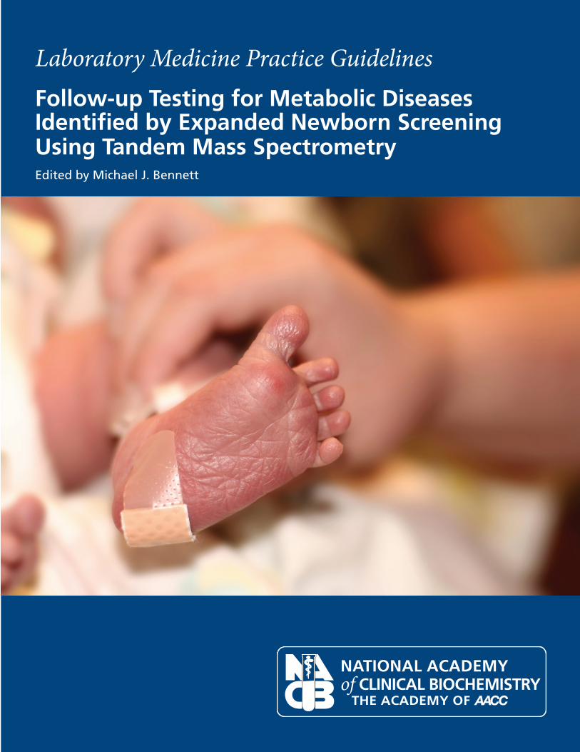
Laboratory Medicine Practice GuidelinesFollow-up Testing for Metabolic DiseasesIdentified by Expanded Newborn ScreeningUsing Tandem Mass SpectrometryEdited by Michael J. Bennett
NACB_LMPG_Newborn_cover.indd 1 11/23/09 1:32:04 PM
The National Academy of Clinical Biochemistry
Presents
LABORATORY MEDICINE PRACTICE GUIDELINES
FOLLOW-UP TESTING FOR METABOLICDISEASES IDENTIFIED BY EXPANDED
NEWBORN SCREENING USING TANDEM MASS SPECTROMETRY
EDITED BYMichael J. Bennett
NACB Committee Members
Michael J. Bennett, PhD, FRCPath, FACB, ChairUniversity of Pennsylvania and Children’s Hospital of Philadelphia, PA
Piero Rinaldo, MD, PhD, FACMGMayo Clinic, Rochester, MN
Ronald J.Whitley, PhD, FACBUniversity of Kentucky Medical Center, Lexington, KY
William J. Rhead, MD, PhD, FACMGMedical College of Wisconsin and Children’s Hospital of Wisconsin, Milwaukee, WI
W. Harry Hannon, PhDCenters for Disease Control and Prevention, Atlanta, GA
Dennis J. Dietzen, PhD, FACBWashington University and St Louis Children’s Hospital, St Louis, MO
Uttam C. Garg, PhD, FACBUniversity of Missouri, Kansas City School of Medicine and Children’s Mercy Hospital, Kansas City, MO
Stanley F. Lo, PhD, FACBMedical College of Wisconsin and Children’s Hospital of Wisconsin, Milwaukee, WI
Copyright © 2009 by the American Association for Clinical Chemistry, Inc. All rights reserved.
Single copies for personal use may be printed from authorized Internet sources such as the NACB’s home page(http://www.aacc.org/members/nacb/LMPG/Pages/default.aspx), provided it is printed in its entirety, including this notice. Printing ofselected portions of the document is also permitted for personal use, provided the user also prints and attaches the title page and coverpages to the selected reprint or otherwise clearly identifies the reprint as having been produced by the NACB. Otherwise, this documentmay not be reproduced in whole or in part, stored in a retrieval system, translated into another language, or transmitted in any form withoutexpress written permission of the National Academy of Clinical Biochemistry. Such permission may be requested from NACB, 1850 KStreet, Suite 625, Washington, DC, 20006-2213. Permission will ordinarily be granted, provided the NACB logo and the following noticeappear prominently at the front of the document: Reproduced (translated) with permission of the National Academy of ClinicalBiochemistry, Washington, DC.
This document (product ID 5232) was approved by the National Academy of Clinical Biochemistry Board of Directors in November 2008.The NACB is the Academy of the American Association for Clinical Chemistry.
Table of Contents
Overview v
1. Evidence-Based Rationale for Expanded Newborn Screening 1
2. Pre-Analytical, Analytical, and Post-Analytical Issues Related toTandem Mass Spectrometry as a Tool for Expanded Newborn Screening 9
a. Measurement of Amino Acids 9b. Measurement of Acylcarnitines 16c. Measurement of Organic Acids 22
3. Follow-Up of Positive Screens 31a. General Requirements of the Follow-Up Process 31b. General Recommendations 31c. Disease-Specific Follow-Up Testing for Primary Targets 32d. Secondary Targets 32
4. Patient Outcomes From Expanded Newborn Screening 43
5. Future Directions in Expanded Newborn Screening for Metabolic Disorders 47a. Lysosomal Storage Disorders 47b. Tyrosinemia Type 1 47
Acknowledgment 50
iv
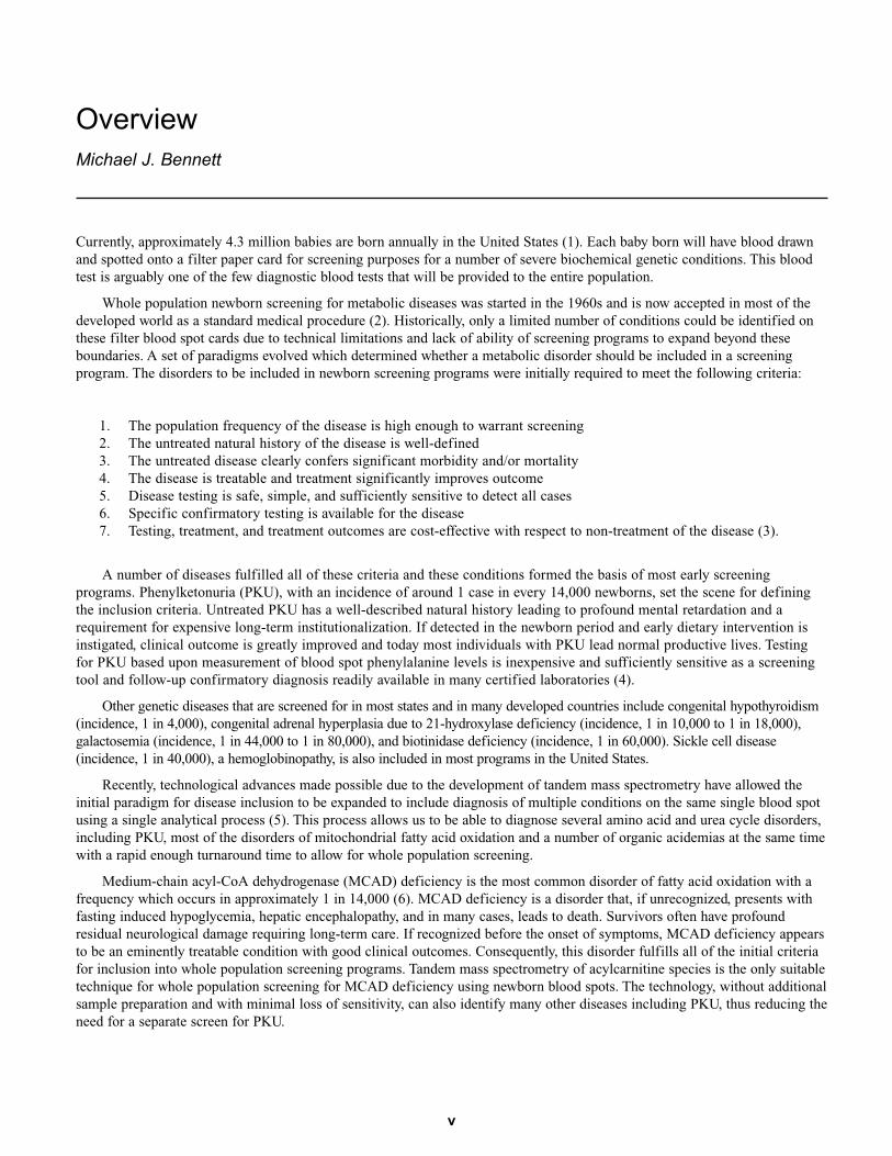
Overview
Michael J. Bennett
v
Currently, approximately 4.3 million babies are born annually in the United States (1). Each baby born will have blood drawnand spotted onto a filter paper card for screening purposes for a number of severe biochemical genetic conditions. This bloodtest is arguably one of the few diagnostic blood tests that will be provided to the entire population.
Whole population newborn screening for metabolic diseases was started in the 1960s and is now accepted in most of thedeveloped world as a standard medical procedure (2). Historically, only a limited number of conditions could be identified onthese filter blood spot cards due to technical limitations and lack of ability of screening programs to expand beyond theseboundaries. A set of paradigms evolved which determined whether a metabolic disorder should be included in a screeningprogram. The disorders to be included in newborn screening programs were initially required to meet the following criteria:
1. The population frequency of the disease is high enough to warrant screening2. The untreated natural history of the disease is well-defined3. The untreated disease clearly confers significant morbidity and/or mortality4. The disease is treatable and treatment significantly improves outcome5. Disease testing is safe, simple, and sufficiently sensitive to detect all cases6. Specific confirmatory testing is available for the disease7. Testing, treatment, and treatment outcomes are cost-effective with respect to non-treatment of the disease (3).
A number of diseases fulfilled all of these criteria and these conditions formed the basis of most early screeningprograms. Phenylketonuria (PKU), with an incidence of around 1 case in every 14,000 newborns, set the scene for definingthe inclusion criteria. Untreated PKU has a well-described natural history leading to profound mental retardation and arequirement for expensive long-term institutionalization. If detected in the newborn period and early dietary intervention isinstigated, clinical outcome is greatly improved and today most individuals with PKU lead normal productive lives. Testingfor PKU based upon measurement of blood spot phenylalanine levels is inexpensive and sufficiently sensitive as a screeningtool and follow-up confirmatory diagnosis readily available in many certified laboratories (4).
Other genetic diseases that are screened for in most states and in many developed countries include congenital hypothyroidism(incidence, 1 in 4,000), congenital adrenal hyperplasia due to 21-hydroxylase deficiency (incidence, 1 in 10,000 to 1 in 18,000),galactosemia (incidence, 1 in 44,000 to 1 in 80,000), and biotinidase deficiency (incidence, 1 in 60,000). Sickle cell disease (incidence, 1 in 40,000), a hemoglobinopathy, is also included in most programs in the United States.
Recently, technological advances made possible due to the development of tandem mass spectrometry have allowed theinitial paradigm for disease inclusion to be expanded to include diagnosis of multiple conditions on the same single blood spotusing a single analytical process (5). This process allows us to be able to diagnose several amino acid and urea cycle disorders,including PKU, most of the disorders of mitochondrial fatty acid oxidation and a number of organic acidemias at the same timewith a rapid enough turnaround time to allow for whole population screening.
Medium-chain acyl-CoA dehydrogenase (MCAD) deficiency is the most common disorder of fatty acid oxidation with afrequency which occurs in approximately 1 in 14,000 (6). MCAD deficiency is a disorder that, if unrecognized, presents withfasting induced hypoglycemia, hepatic encephalopathy, and in many cases, leads to death. Survivors often have profoundresidual neurological damage requiring long-term care. If recognized before the onset of symptoms, MCAD deficiency appearsto be an eminently treatable condition with good clinical outcomes. Consequently, this disorder fulfills all of the initial criteriafor inclusion into whole population screening programs. Tandem mass spectrometry of acylcarnitine species is the only suitabletechnique for whole population screening for MCAD deficiency using newborn blood spots. The technology, without additionalsample preparation and with minimal loss of sensitivity, can also identify many other diseases including PKU, thus reducing theneed for a separate screen for PKU.
Although some of the other conditions that can be diagnosed using this technology (to be described in the succeedingsections of this document) appear to be less frequent in the population, they are identifiable simultaneously with no increasein analytical time. As a result of the introduction of tandem mass spectrometry into the newborn screening arena, thenumbers of diseases that are potentially identifiable have expanded considerably. Some of these conditions are rare. Some are regarded as having unproven treatability. The sensitivities for detection of some conditions are not always idealand some of these conditions do not necessarily fulfill the initial criteria for inclusion in a whole population screeningprogram. Thus, necessitating a change in the way in which we approach candidacy for inclusion in these programs.
In addition, the increased numbers of different metabolic diseases that are identifiable by tandem mass spectrometry alsoincreases the complexity of testing required for confirmation of diagnosis. Confirmatory testing is a critical component of thewhole process, which may involve additional metabolite measurement, enzyme assay, or molecular testing. We recognize theimportance of establishing guidelines for systematic, consistent, and appropriate disease confirmation in the clinical laboratoryand also guidelines for monitoring efficacy of therapeutic intervention and patient well-being.
At the time of publication of this document, approximately 98% of all babies born in the United States are provided withexpanded newborn screening for metabolic diseases by tandem mass spectrometry. MCAD deficiency is now mandated in 46states and the District of Columbia, required but not yet implemented in one state, and offered but not mandated in two additionalstates (7; accessed 09/19/2008). It is highly likely that the process will be taken up by the few remaining states or other providerswill provide the service for MCAD deficiency for all babies born in those states before the end of the present decade. In addition,tandem mass spectrometry is being utilized for multiple additional metabolic conditions, which vary by state.
The National Academy of Clinical Biochemistry (NACB) is the American Association for Clinical Chemistry’s scientific academy. An important activity of the NACB is to develop laboratory medicine practice guidelines to assist clinical and laboratorypractice decisions concerning the diagnosis of specific diseases. NACB recognizes that there is a strong need for evaluation of howthese newborn screening laboratory services are provided, and of equal importance, how procedures for adequate follow-up testingshould proceed. Screening programs, by definition, should be developed with the highest degree of sensitivity, such that there arefewest possible false-negative cases (missed diagnoses). Follow-up testing should provide the highest degree of specificity so thatfalse positives from the screening process are removed and only true-positive cases are eventually given a diagnosis.
The NACB has convened a panel of experts to evaluate the data supporting the role of expanded newborn screening, to determine optimal methods and performance characteristics for performing the testing, and for optimizing confirmatoryfollow-up testing procedures for positive screens.
Specific recommendations in this NACB guideline are based whenever possible on relevant published information in thepeer-reviewed medical and scientific literature, and from surveys and guidelines produced by other medical academic groupsand organizations including the American College of Medical Genetics and the Centers for Disease Control. The strength ofthe supporting data for each recommendation is determined using the scoring criteria adopted from the US PreventativeServices Task Force Recommendations for Preventative Services.
Strength of Recommendations (Modified from US Preventive Services Task Force Recommendations for Preventive Services)
A. The NACB strongly recommends adoption; there is good evidence that it improves important health outcomes andconcludes that benefits substantially outweigh harms.
B. The NACB recommends adoption; there is at least fair evidence that it improves important health outcomes and concludesthat benefits outweigh harms.
C. The NACB recommends against adoption; there is evidence that it is ineffective or that harms outweigh benefits.
I. The NACB concludes that the evidence is insufficient to make recommendations; evidence that it is effective is lacking, ofpoor quality, or conflicting and the balance of benefits and harms cannot be determined.
NACB grades the quality of the overall evidence on a 3-point scale:
I. Evidence includes consistent results from well-designed, well-conducted studies in representative populations.
II. Evidence is sufficient to determine effects, but the strength of the evidence is limited by the number, quality, orconsistency of the individual studies; generalizability to routine practice; or indirect nature of the evidence.
III. Evidence is insufficient to assess the effects on health outcomes because of limited number or power of studies, importantflaws in their design or conduct, gaps in the chain of evidence, or lack of information.
vi Overview
REFERENCES
1. http://www.cdc.gov/nchs/data/nvsr/nvsr56/nvsr56_12.pdf (accessed 09/19/08)2. Guthrie R. The origin of newborn screening. Screening, 1; 5–15, 19923. Wilson JMG, Jungner G. Principles of screening for disease. Geneva: World Health Organization, 19684. National Institutes of Health. Phenylketonuria (PKU): screening and management. NIH consensus statement 17(3); 1–33, 20005. Chace DH, Kalas TA, Naylor EW. Use of tandem mass spectrometry for multianalyte screening of dried blood specimens from
newborns. Clin Chem 49; 1797–1817, 20036. Grosse SD, Khoury MJ, Greene CL, Crider KS, Pollitt RJ. The epidemiology of medium-chain acyl-CoA dehydrogenase
deficiency: an update. Genet Med 8; 205–212, 20067. http://genes-r-us.uthscsa.edu/ (accessed 09/19/08)
Overview vii

In 2000, the American Academy of Pediatrics (AAP) NewbornScreening Task Force released a report entitled “NewbornScreening: A Blueprint for the Future – A Call for a NationalAgenda on State Newborn Screening Programs” (1). Tandemmass spectrometry (MS/MS) was mentioned once (p. 395) in the body of the voluminous report, and was recog-nized correctly as an example of technological advances like-ly to have a significant impact on the sensitivity, specificity,and scope of newborn screening. However, the positive mes-sage was mitigated by a concern described as “the ability todetect individuals with metabolic conditions for which thereare no effective treatments at this time.” In retrospect, this con-servative assessment is not surprising in view of the fact thatwhen the report was written 100% of US births were screenedfor fewer than 10 conditions (2), and only a small proportion(7%; MA, ME, NC, SC, WI) was tested for medium-chainacyl-CoA dehydrogenase (MCAD) deficiency using MS/MS.Seven years is a relatively short period of time in public healthpolicy making, so it is remarkable that currently the situationhas changed to 98% and 83% of US births being tested for apanel of >20 and >30 conditions, respectively (2). As of April2008, 98% of US newborns are screened for MCAD deficien-cy. There is no doubt this unprecedented evolution has beendriven by a combination of factors, including public pressure,political action, and increasing attention of mass media to theissue of newborn screening expansion, yet the publication ofprospectively collected evidence that has taken place in recentyears must be recognized as a primary driver of the ongoingforward progress (2, 4–9). In particular, a critical contributioncame as a report from an expert panel assembled by theAmerican College of Medical Genetics (ACMG) (10–11). Thiseffort was commissioned by the Maternal and Child HealthBureau through a contract from the Health Resources andServices Administration (HRSA) to outline a process for thedefinition, among others outcomes, of a panel of conditions tobe recommended for universal and uniform inclusion in statenewborn screening programs. The expert panel identified apanel of 29 conditions, a list routinely referred to as the uni-form panel. Twenty of the primary conditions are screened forby MS/MS analysis of amino acids and acylcarnitines, andselected ratios (5, 12). An additional 25 conditions, 22 of themalso detected by MS/MS, were identified in a cohort of so-called secondary targets. Most of them are part of the differential
diagnosis of one or more conditions in the core panel. The con-sideration given to the secondary targets has been controver-sial because, with few exceptions, their incidence, naturalhistory, prospective screening experience, and effectiveness oftreatment have not yet been defined (13). However, a definingcharacteristic of a multiplex platform like MS/MS is the needto perform an elaborate differential diagnosis for most of themetabolites detectable in the amino acid and acylcarnitine pro-files (2, 5, 14–15). It seems therefore reasonable to underscorethe importance of developing tools for better confirmatorytesting and differential diagnosis of all detectable conditions,rather than debating the artificial exclusion of one or more rareconditions that are detected anyway in a profile mode, at noadditional cost, and could be misinterpreted as false positivesof a better-known condition (16).
Since the publication of the ACMG report, it has becomea reality to regard this panel of conditions as the establishedtarget of newborn screening by MS/MS, and we concur withsuch approach even though it is increasingly apparent thatthere are additional conditions potentially detectable by analysisof the same amino acid and acylcarnitine markers (17–20).This observation suggests a need in the near future to updatethe list of secondary targets, and possibly upgrade a few of theexisting ones to a status of primary target on the basis of newevidence obtained after the implementation of expandedscreening by MS/MS (21–22).
To date, a multiplex platform for the simultaneousscreening at birth of at least 42 metabolic disorders should beconsidered an accepted standard of care, of which a fullnationwide implementation is likely to be completed verysoon (16). Therefore, it is important to shift our collectiveattention and evaluation of evidence from quantity (howmany conditions) to quality (how well we screen for) issues,particularly the monitoring of objective metrics and thedefinition of targets of adequate performance (2).
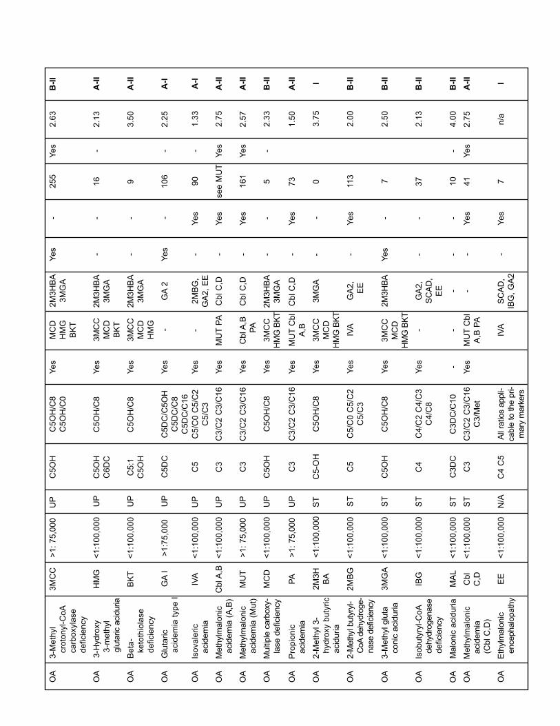
Table 1 summarizes the 42 conditions included in thepanel recommended by ACMG plus three additional condi-tions, which are examples of the additional findings to beencountered by MS/MS testing. In addition to an updated listof primary markers and informative ratios, this Table also cov-ers a few emerging aspects of newborn screening by MS/MS,namely the possibility of detecting maternal cases (i.e., anabnormal result of the screening is secondary to a maternal
Chapter 1
Evidence-Based Rationale for Expanded
Newborn Screening
Piero Rinaldo, Ronald J. Whitley, William J. Rhead, and W. Harry Hannon
1
biochemical phenotype; 23–24) and of observing interferencecaused by diet, drugs, modality of treatment, and prematurity.Artifacts may be either true elevations of a marker or interfer-ence by isobaric compounds (25). Another element of Table 1is the current status of second tier test availability, an elementof increasing importance and recognition in the definition ofacceptable targets of performance by MS/MS or any othermultiplex platform (2, 26–27). The ability to verify an abnor-mal result of the primary screening without a recall of the new-born is appealing and likely to increase cost effectiveness(28–30), not to mention the benefit of preventing unnecessarydistress of the newborn's family (31). As another, indirect frameof reference, Table 1 includes the number of true-positive casesincluded as of May 2007 in a cumulative database assembledby a HRSA regional collaborative project entitled “LaboratoryQuality Improvement of Newborn Screening by MS/MS” (32).To date, 38 US states and 33 laboratories in 20 countries areactively contributing data to this database. The rationale hereis to gauge how likely it will be to collect enough cases with-in a reasonable period of time to provide an objective, trulyevidence-based evaluation of each condition.
Finally, Table 1 shows a summary of validation scoresderived from the ACMG report (11). At least two experts ratedon a 1 to 4 scale the evidence in support of, or against, theinclusion of a given condition in the recommended panel forfour elements (condition, tests, diagnosis, and treatment), accord-ing to the levels of evidence defined by the American Academyof Pediatrics (AAP) Steering Committee on Quality Improvementand Management (33). The numeric values presented in Table1 are the average of these scores (11). This informal assessmentof a prior evidence review process was included simply as apoint of reference for the strength of the recommendations ofthis report, shown in the far right column of Table 1, which arebased on current LMPG criteria, modified from the USPreventive Services Task Force Recommendations forPreventive Service (33). Seven conditions received an A-I rat-ing, the highest possible, 31 of the remaining 35 conditionsincluded in the ACMG panel are recommended for adoptionalthough the available evidence at this time is limited, or indi-rect. Four conditions, all but one not yet detected prospective-ly in a patient by newborn screening, should be re-evaluatedregularly to verify that lack of detection could be at least in parta function of improperly set cutoff values.
REFERENCES
1. American Academy of Pediatrics, Newborn Screening Task Force.(2000) Serving the family from birth to the medical home: new-born screening a blueprint for the future. Pediatrics 106:389–427.
2. Rinaldo P, Zafari S, Tortorelli S, Matern D. (2006) Making thecase for objective performance metrics in newborn screeningby tandem mass spectrometry. MRDD Research Reviews 12:255–261.
3. National Newborn Screening and Genetics Resource Center(NNSGRC). (2006) US national newborn screening informationsystem. Available at http://genes-r-us.uthscsa.edu. (accessed09/19/08)
2 Expanded Newborn Screening Using Tandem Mass Spectrometry
4. Zytkovicz TH, Fitzgerald EF, Marsden D, et al. (2001) Tandemmass spectrometric analysis for amino, organic, and fatty aciddisorders in newborn dried blood spots: a two-year summaryfrom the New England Newborn Screening Program. ClinChem 47:1945–1955.
5. Chace DH, Kalas TA, Naylor EW. 2003. Use of tandem massspectrometry for multianalyte screening of dried blood specimensfrom newborns. Clin Chem 49:1797–1817.
6. Schulze A, Lindner M, Kohlmuller D, et al. 2003. Expandednewborn screening for inborn errors of metabolism by electro-spray ionization-tandem mass spectrometry: results, outcome,and implications. Pediatrics. 111:1399–406.
7. Wilcken B, Wiley V, Hammond J, et al. 2003. Screening newbornsfor inborn errors of metabolism by tandem mass spectrometry.N Engl J Med 348:2304–2312.
8. Hoffmann GF, von Kries R, Klose D, et al. 2004. Frequencies ofinherited organic acidurias and disorders of mitochondrial fattyacid transport and oxidation in Germany. Eur J Pediatr163:76–80.
9. Frazier DM, Millington DS, McCandless SE, et al. 2006. Thetandem mass spectrometry newborn screening experience inNorth Carolina: 1997–2005. J Inherit Metab Dis 29:76–85.
10. Watson MS, Mann MY, Lloyd-Puryear MA, Rinaldo P, HowellRR [editors]. (2006) Newborn screening: Toward a uniformscreening panel and system [Executive summary]. Genet Med8(Supplement):1S–11S.
11. Watson MS, Lloyd-Puryear MA, Mann MY, Rinaldo P, HowellRR [editors]. (2006) Newborn screening: Toward a uniformscreening panel and system [Main report]. Genet Med 8(Supplement):12S–252S.
12. Rinaldo P, Hahn SH, Matern D. 2005. Inborn errors of aminoacid, organic acid, and fatty acid metabolism. In: Burtis CA,Ashwood ER, Bruns DE, eds. Tietz Textbook of ClinicalChemistry and Molecular Diagnostics, 4th ed., W.B. Saunders,pp. 2207–2247.
13. Botkin JR, Clayton EW, Fost NC, et al. 2006. Newborn screeningtechnology: proceed with caution. Pediatrics 117:1793–1799.
14. Rinaldo P, Tortorelli S, Matern M. 2004. Recent developmentsand new applications of tandem mass spectrometry in newbornscreening. Curr Opini Pediatr 16:427–432.
15. Sweetman L, Millington DS, Therrell BL, et al. 2006. Namingand counting disorders (conditions) included in newborn screen-ing panels. Pediatrics 117(Pt 2):S308–314.
16. Howell RR. 2006. We need expanded newborn screening.Pediatrics 117:1800–1805.
17. Merinero B, Perez-Cerda C, Ruiz Sala P, Ferrer I, et al. (2006)Persistent increase of plasma butyryl/isobutyrylcarnitine con-centrations as marker of SCAD defect and ethylmalonicencephalopathy. J Inherit Metab Dis 29:685.
18. Garcia-Cazorla A, Rabier D, Touati G, Chadefaux-Vekemans B,et al. (2006) Pyruvate carboxylase deficiency: metabolic charac-teristics and new neurological aspects. Ann Neurol 59:121–127.
19. Tan ES, Wiley V, Carpenter K, Wilcken B. (2007) Non-ketotichyperglycinemia is usually not detectable by tandem mass spec-trometry newborn screening. Mol Genet Metab 90:446–448.
20. Carrozzo R, Dionisi-Vici C, Steuerwald U, Lucioli S, et al.(2007) SUCLA2 mutations are associated with mild methyl-malonic aciduria, Leigh-like encephalomyopathy, dystonia anddeafness. Brain 130:862–874.
21. Lorey F, Enns G, Cederbaum S, Crombez E, et al. (2007) ShouldCobalamin C (Cbl C) be a core target? Evidence for increasedprevalence, detection, and effectiveness of treatment intervention
in California newborn screening. Proceedings of the 2007Newborn Screening and Genetic Testing Symposium,Association of Public Health Laboratories, Minneapolis (MN, p. 36. Also available at http://www.aphl.org.
22. Watson MS. (2006) Current status of newborn screening: decision-making about the conditions to include in screeningprograms. Ment Retard Dev Disabil Res Rev 12:230–235.
23. Gibson KM, Bennett MJ, Naylor EW, Morton DH. (1998) 3-Methylcrotonyl-coenzyme A carboxylase deficiency inAmish/Mennonite adults identified by detection of increasedacylcarnitines in blood spots of their children. J Pediatr.132:519–523.
24. Schimmenti LA, Crombez EA, Schwahn BC, Heese BA, et al.(2007) Expanded newborn screening identifies maternal pri-mary carnitine deficiency. Mol Genet Metab 90:441–445.
25. Abdenur JE, Chamoles NA, Guinle AE, Schenone AB, et al.(1998) Diagnosis of isovaleric acidaemia by tandem mass spec-trometry: false positive result due to pivaloylcarnitine in a new-born screening programme. J Inherit Metab Dis 21:624–630.
26. Lacey JM, Minutti CZ, Magera MJ, Tauscher AL, et al. (2004)Improved specificity of newborn screening for congenital adre-nal hyperplasia by second tier steroid profiling using tandemmass spectrometry. Clin Chem 50:621–625.
Evidence-Based Rationale for Expanded Newborn Screening 3
27. Magera MJ, Gunawardena ND, Hahn SH, Tortorelli S, et al.(2006) Rapid quantitative determination of succinylacetone indried blood spots by liquid chromatography tandem mass spec-trometry. Mol Genet Metab 88:16–21.
28. Pandor A, Eastham J, Beverley C, Chilcott J, et al. (2004) Clinicaleffectiveness and cost-effectiveness of neonatal screening forinborn errors of metabolism using tandem mass spectrometry: asystematic review. Health Technol Assess 8:1–121.
29. Pandor A, Eastham J, Chilcott J, Paisley S, et al. (2006)Economics of tandem mass spectrometry screening of neonatalinherited disorders. Int J Technol Assess Health Care 22:321–326.
30. Aaron E, Carroll AE, Downs SM. (2006) Comprehensive cost-utility analysis of newborn screening strategies. Pediatrics117:S287–S295.
31. Waisbren SE, Albers S, Amato S, et al. (2003) Effect of expandednewborn screening for biochemical genetic disorders on childoutcomes and parental stress. JAMA 290:2564–2572.
32. Rinaldo P., Zafari S. (2006) Progress report on expanded new-born screening outcomes: The Region 4 MS/MS collaborativeproject [abstract]. Annual Clinical Genetics Meeting, Programand Abstracts, p. 161 (#335). See also www.region4genetics.org
33. Marcuse EK, Shiffman RN. (2004) Classifying recommenda-tions for clinical practice guidelines. Pediatrics 114:874–877.
AA
Citru
llin
em
ia
type I
I
CIT
II<
1:1
00,0
00
ST
Cit
Cit/A
rgY
es
AS
AC
ITC
ITII
,
PC
--
29
-2
.71
B-I
I
<1:1
00,0
00
AA
Benig
n H
yper-
phenyla
lanin
e-
mia
H-P
HE
ST
Phe
Phe/T
yr
Yes
PK
UH
-PH
E,
BIO
PT
(BS
) &
(RE
G)
--
38
5-
n/a
A-I
Group
Condition (inborn errors of amino
acid, fatty acid, and organic acid
metabolism)
ACMG code
US incidence
ACMG panel
Primary marker(s)
Informative ratios
Interpretation requires differential
diagnosis
Primary or other conditions with
same marker(s)
Secondary or other conditions
with same markers
Maternal cases detected by NBS
Potential interference by diet,
drugs, prematurity
No. of cases
Available 2nd tier test
Evidence level in 2006 ACMG
report (1–4)
Evaluation of evidence (LMPG
criteria)
(1)
(1)
(2)
(3)
(4)
(5)
(6)
(6)
(6)
(7)
(8)
(9)
(10
)(1
1)
(12
)
AA
Arg
inin
osuccin
ic
acid
em
ia
AS
A<
1:1
00,0
00
UP
Cit
Cit/A
rgY
es
CIT
CIT
II,
PC
--
13
-2
.50
B-I
I
AA
Citru
llin
em
iaC
IT<
1:1
00,0
00
UP
Cit
Cit/A
rgY
es
AS
AC
ITII
,
PC
--
73
-3
.00
B-I
I
AA
Hom
ocystinuria
(CB
S d
efici-
ency)
HC
Y<
1:1
00,0
00
UP
Met
Met/
Phe
Yes
-M
ET
-Y
es
24
Yes
2.0
0B
-II
AA
Maple
syru
p
(urine)
dis
ease
MS
UD
<1:1
00,0
00
UP
Val
Ile+
Leu
Val/P
he
(Ile
+Leu)/
Phe
(Ile
+Leu)/
Ala
--
--
Yes
99
Yes
2.1
3A
-II
AA
Phenylk
eto
nuria
PK
U>
1:
25,0
00
UP
Phe
Phe/T
yr
Yes
-H
-PH
E,
BIO
PT
(BS
) &
(RE
G)
Yes
Yes
60
8-
2.0
0A
-I
AA
Tyro
sin
em
ia
type I
TY
R I
<1:1
00,0
00
UP
Tyr
SU
AC
Tyr/
Cit
Yes
-T
YR
II,
TY
R I
II
-Y
es
48
Yes
1.9
4A
-II
AA
Arg
inin
em
iaA
RG
<1:1
00,0
00
ST
Arg
--
--
--
7-
3.5
0B
-II
AA
Defe
cts
of
bio
pte
rin c
ofa
c-
tor
bio
synth
esis
BIO
PT
(BS
)
<1:1
00,0
00
ST
Phe
Phe/T
yr
Yes
PK
UH
-PH
E,
BIO
PT
(RE
G)
-Y
es
3-
2.0
0A
-I
FA
OD
ienoyl re
duc-
tase d
eficie
ncy
DE
-
RE
D
<1:1
00,0
00
ST
C10:2
C10:2
/C10
--
--
-1
-4
.00
I
FA
OV
ery
long-c
hain
acyl-C
oA
dehydro
genase
deficie
ncy
VLC
AD
>1:7
5,0
00
UP
C14:2
C14:1
C14
C14:1
/C16
Yes
-G
A2
--
16
4-
2.5
8A
-II
AA
Dis
ord
ers
of
bio
pte
rin c
ofa
c-
tor
regenera
tion
BIO
PT
(RE
G)
<1:1
00,0
00
ST
Phe
Phe/T
yr
Yes
PK
UH
-PH
E,
BIO
PT
(BS
)
-Y
es
4-
2.5
0A
-I
AA
Hyper-
meth
ionin
em
ia
ME
T<
1:1
00,0
00
ST
Met
Met/
Phe
Yes
HC
Y-
-Y
es
24
Yes
1.7
5B
-II
AA
Tyro
sin
em
ia
type I
I
TY
R I
I<
1:1
00,0
00
ST
Tyr
Tyr/
Cit
Yes
TY
R I
TY
R I
II-
Yes
18
Yes
2.3
8B
-II
AA
Tyro
sin
em
ia
type I
II
TY
R I
II<
1:1
00,0
00
ST
Tyr
Tyr/
Cit
Yes
TY
R I
TY
R I
I-
Yes
1Y
es
3.6
3B
-II
AA
Non k
eto
tic
hyperg
lycin
e-
mia
NK
HG
<1:1
00,0
00
N/A
Gly
Gly
/Ala
--
--
-2
4-
n/a
I
AA
Pyru
vate
carb
oxyla
se
deficie
ncy
PC
<1:1
00,0
00
N/A
Cit
Cit/A
rgY
es
AS
AC
ITC
ITII
--
2-
n/a
I
FA
OC
arn
itin
e u
pta
ke
defe
ct
CU
D<
1:1
00,0
00
UP
C0
(C0+
C2+
C3
+C
16+
C18:1
)/
CIT
Yes
GA
I,
3M
CC
(mat)
-Y
es
Yes
60
-2
.25
A-I
I
FA
OLong-c
hain
3-O
H a
cyl-C
oA
dehydro
genase
deficie
ncy
LC
HA
D>
1:7
5,0
00
UP
C16:1
-OH
C16-O
H
C18:1
-OH
C18-O
H
C16-O
H/C
16
Yes
TF
P-
-Y
es
82
-2
.75
A-I
I
FA
OM
ediu
m-c
hain
acyl-C
oA
dehydro
genase
deficie
ncy
MC
AD
>1:2
5,0
00
UP
C6 C
8
C10:1
C10
C8/C
2 C
8/C
10
Yes
-G
A2,
MC
KA
T
-Y
es
80
7-
1.6
3A
-I
FA
OT
rifu
nctional
pro
tein
deficie
ncy
TF
P<
1:1
00,0
00
UP
C16:1
-OH
C16-O
H
C18:1
-OH
C18-O
H
C16-O
H/C
16
Yes
LC
HA
D-
-Y
es
Se
e
LC
HA
D
-3
.50
A-I
I
(Con
tinue
d)
FA
OC
arn
itin
e p
alm
i-
toyl-tr
ansfe
rase
Ia d
eficie
ncy (
L)
CP
TIa
<1:1
00,0
00
ST
C0 (
hig
h)
C16 (
low
)
C18 (
low
)
C0/(
C16+
C18)
--
--
-3
3-
3.7
5B
-II
FA
OC
arn
itin
e p
alm
i-
toyl-tr
ansfe
rase
II d
eficie
ncy
CP
TII
<1:1
00,0
00
ST
C16 C
18:2
C18:1
C18
C0/(
C16+
C18)
Yes
-C
AC
--
20
-3
.38
B-I
I
FA
OG
luta
ric
acid
em
ia
type I
I
GA
2<
1:1
00,0
00
ST
C4-C
18
satu
rate
d
and u
nsat-
ura
ted
specie
s
All r
atios
applicable
to
the p
rim
ary
mark
ers
Yes
MC
AD
,
GA
I, I
VA
SC
AD
,
IBG
, E
E
--
38
-3
.38
B-I
I
FA
OM
ediu
m-c
hain
keto
acyl-C
oA
dehydro
genase
deficie
ncy
MC
KA
T<
1:1
00,0
00
ST
C8
C8/C
2 C
8/C
10
Yes
MC
AD
,
GA
2
--
Yes
04
.00
I
FA
OS
hort
-chain
acyl-C
oA
dehydro
genase
deficie
ncy
SC
AD
>1:
75,0
00
ST
C4
C4/C
2 C
4/C
3
C4/C
8
Yes
GA
II,
IBG
, E
E
-2
51
2.6
3I/
C-I
I*
FA
OC
arn
itin
e/
acyl-carn
itin
e
translo
case
deficie
ncy
CA
CT
<1:1
00,0
00
ST
C16 C
18:2
C18:1
C18
C0/(
C16+
C18)
Yes
CP
TII
--
52
.58
B-I
I
FA
OM
ediu
m/s
hort
-
chain
3-O
H
acyl-C
oA
dehydro
genase
deficie
ncy
M/S
CH
AD
>1:1
00,0
00
ST
C4-O
H-
--
--
-0
-I
4.0
0
Group
Condition (inborn errors of amino
acid, fatty acid, and organic acid
metabolism)
ACMG code
US incidence
ACMG panel
Primary marker(s)
Informative ratios
Interpretation requires differential
diagnosis
Primary or other conditions with
same marker(s)
Secondary or other conditions
with same markers
Maternal cases detected by NBS
Potential interference by diet,
drugs, prematurity
No. of cases
Available 2nd tier test
Evidence level in 2006 ACMG
report (1–4)
Evaluation of evidence (LMPG
criteria)
(1)
(1)
(2)
(3)
(4)
(5)
(6)
(6)
(6)
(7)
(8)
(9)
(10
)(1
1)
(12
)
OA
Eth
ylm
alo
nic
encephalo
path
y
EE
<1:1
00,0
00
N/A
C4 C
5A
ll r
atios a
ppli-
cable
to the p
ri-
mary
mark
ers
IVA
SC
AD
,
IBG
, G
A2
-Y
es
7n
/aI
OA
Meth
ylm
alo
nic
acid
em
ia
(Cbl C
,D)
Cbl
C,D
<1:1
00,0
00
ST
C3
C3/C
2 C
3/C
16
C3/M
et
Yes
MU
TC
bl
A,B
PA
--
Yes
41
Yes
2.7
5A
-II
OA
Malo
nic
acid
uria
MA
L<
1:1
00,0
00
ST
C3D
CC
3D
C/C
10
--
--
-1
0-
4.0
0B
-II
OA
Isobuty
ryl-C
oA
dehydro
genase
deficie
ncy
IBG
<1:1
00,0
00
ST
C4
C4/C
2 C
4/C
3
C4/C
8
Yes
-G
A2,
SC
AD
,
EE
--
37
2.1
3B
-II
OA
3-M
eth
yl glu
ta
conic
acid
uria
3M
GA
<1:1
00,0
00
ST
C5O
HC
5O
H/C
8Y
es
3M
CC
MC
D
HM
G B
KT
2M
3H
BA
Yes
-7
2.5
0B
-II
OA
2-M
eth
yl buty
ryl-
CoA
dehydro
ge-
nase d
eficie
ncy
2M
BG
<1:1
00,0
00
ST
C5
C5/C
0 C
5/C
2
C5/C
3
Yes
IVA
GA
2,
EE
-Y
es
113
2.0
0B
-II
OA
2-M
eth
yl 3-
hydro
xy b
uty
ric
acid
uria
2M
3H
BA
<1:1
00,0
00
ST
C5-O
HC
5O
H/C
8Y
es
3M
CC
MC
D
HM
G B
KT
3M
GA
--
03
.75
I
OA
Pro
pio
nic
acid
em
ia
PA
>1:
75,0
00
UP
C3
C3/C
2 C
3/C
16
Yes
MU
TC
bl
A,B
Cbl C
,D-
Yes
73
1.5
0A
-II
OA
Multip
le c
arb
oxy-
lase d
eficie
ncy
MC
D<
1:1
00,0
00
UP
C5O
HC
5O
H/C
8Y
es
3M
CC
HM
G B
KT
2M
3H
BA
3M
GA
--
5-
2.3
3B
-II
OA
3-M
eth
yl
cro
tonyl-C
oA
carb
oxyla
se
deficie
ncy
3M
CC
>1:
75,0
00
UP
C5O
HC
5O
H/C
8
C5O
H/C
0
Yes
MC
D
HM
G
BK
T
2M
3H
BA
3M
GA
Yes
-2
55
Yes
2.6
3B
-II
OA
3-H
ydro
xy
3-m
eth
yl
glu
taric a
cid
uria
HM
G<
1:1
00,0
00
UP
C5O
H
C6D
C
C5O
H/C
8Y
es
3M
CC
MC
D
BK
T
2M
3H
BA
3M
GA
--
16
-2
.13
A-I
I
OA
Beta
-
keto
thio
lase
deficie
ncy
BK
T<
1:1
00,0
00
UP
C5:1
C5O
H
C5O
H/C
8Y
es
3M
CC
MC
D
HM
G
2M
3H
BA
3M
GA
--
93
.50
A-I
I
OA
Glu
taric
acid
em
ia t
ype I
GA
I>
1:7
5,0
00
UP
C5D
CC
5D
C/C
5O
H
C5D
C/C
8
C5D
C/C
16
Yes
-G
A2
Yes
-1
06
-2
.25
A-I
OA
Isovale
ric
acid
em
ia
IVA
<1:1
00,0
00
UP
C5
C5/C
0 C
5/C
2
C5/C
3
Yes
-2M
BG
,
GA
2,
EE
-Y
es
90
-1
.33
A-I
OA
Cbl A
,B<
1:1
00,0
00
UP
C3
C3/C
2 C
3/C
16
Yes
MU
TP
AC
bl C
,D-
Yes
se
e M
UT
Yes
2.7
5A
-II
OA
Meth
ylm
alo
nic
acid
em
ia (
Mut)
MU
T>
1:
75,0
00
UP
C3
C3/C
2 C
3/C
16
Yes
Cbl A
,B
PA
Cbl C
,D-
Yes
16
1Y
es
2.5
7A
-II
Meth
ylm
alo
nic
acid
em
ia (
A,B
)
8 Expanded Newborn Screening Using Tandem Mass Spectrometry
Table 1. Legend
1. Nomenclature and abbreviations from reference 11 (ACMG main report, Table 1, pp. 5S–6S). PC is a disorder of the
gluconeogenesis pathway listed here because of the potential diagnosis by finding of elevated citrulline
2. From reference 11 (ACMG main report, Fact sheets, pp. 127S–215S)
3. From reference 11 (ACMG main report, Table 7–8, pp.37S–38S). UP, uniform panel (primary targets); ST, secondary targets;
N/A, condition not included in the panel
4. Abbreviations according to reference 13, with modifications. Informative results are higher than normal unless indicated
otherwise (low)
5. The selection of these ratios are derived from the cumulative experience of the Region 4 collaborative project (Laboratory
quality improvement of newborn screening by MS/MS; see www.region4genetics.org), and should be regarded merely as a
suggestion.
6. From reference 11 (ACMG main report, Table 6, p. 37S). (mat) indicates maternal cases
7. Reported evidence of abnormal NBS results by newborn screening caused by a primary, previously undiagnosed maternal
condition
8. Artifacts include secondary elevations of informative markers due to diet and/or drug therapy. Artifacts may be either true
elevations or interference by isobaric compounds
9. Number of true positive cases included as May 2007 in the database of the Region 4 collaborative project (Laboratory quality
improvement of newborn screening by MS/MS; see www.region4genetics.org). This information is provided as an approximated
assessment of the evidence being gathered as result of the collaborative project, with no assumptions of estimated prevalence.
10. A 2nd tier test is considered available when it is performed on a punch of the dried blood spot specimen analyzed by
MS/MS, without notification/recall of the newborn
11. From reference 11 (ACMG main report, Appendix 1, pp. 67S–76S). Values represent the average of 7 to 12 scores for a
given condition. n/a, not available
12. Strength of recommendation, modified from reference 33: US Preventive Services Task Force Recommendations for
Preventive Service.
*Committee was unable to reach a consensus on SCAD deficiency
Follow-up (confirmatory) testing of positive newbornscreens requires a combination of additional methodologies,which may be more specific or more sensitive than the bloodspot tandem mass spectrometric process that is used forwhole population screening. This may include plasma orserum acycarnitine analysis by tandem mass spectrometry,plasma amino acid analysis by ion-exchange chromatogra-phy, and urine organic acid or acylglycine analysis by gaschromatography mass spectrometry. Although measurementof these metabolites has been available for many years, fewguidelines for appropriate use of the analytical tools havebeen developed. Consequently, we have broken down eachof the analyses for individual consideration. This hasresulted in apparent redundancy in some instances but theCommittee decided that this was an essential component fordeveloping clear guidelines. A summary of the recommenda-tions for each of the analytes precedes more detaileddiscussion of the recommendations. A number of appendicesfor each section are also listed.
MEASUREMENT OF AMINO ACIDS
Chapter 2
Pre-Analytical, Analytical, and Post-Analytical
Issues Related to Follow-Up Testing of Positive
Newborn Screens
Ronald J. Whitley, W. Harry Hannon, Dennis J. Dietzen, and Piero Rinaldo
9
Summary of Recommendations
Pre-Analytical Issues/Quality Requirements
1. Plasma (sodium or lithium heparin) is the preferredspecimen type.
2. Urine amino acid analysis should be discouraged as afirst-tier investigation.
3. Specimen collection requirements should be estab-lished by the laboratory and made available to referringphysicians upon request.
4. Specimens spotted and dried on filter paper should betransported or mailed to the testing laboratory atambient temperature. All other specimens should beplaced on ice and promptly transported to the labora-tory for processing and frozen storage.
Analytical Issues/Quality Requirements
5. The director of the testing laboratory should be aboard-certified doctoral scientist or physician withspecialized training and/or experience in biochemicalgenetics.
6. Known concentrations of non-isotopic amino acidreference calibrators should be prepared in anappropriate aqueous matrix.
7. For ion-exchange chromatography, two differentcompounds eluting in important parts of the chro-matogram should be used as internal standards. Fortandem mass spectrometry, stable-isotope amino acidinternal standards should be used when possible.
8. Specimens should be deproteinized prior to analysis.9. Chemical derivitization of amino acids is required
for detection (e.g., ion-exchange chromatography).10. Chemical derivitization of amino acids is recom-
mended to enhance assay sensitivity and specificity(e.g., MS/MS).
11. Amino acids should be analyzed quantitatively by a reliable technique, such as automated cation-exchange liquid chromatography.
12. Amino acids should be analyzed quantitatively by areliable technique, such as electrospray ionizationtandem mass spectrometry.
13. Identification of amino acids by ion-exchange chro-matography should primarily be based on chro-matographic retention time, and retention timerelative to an internal standard. Quantitation shouldbe based on the recovery of the internal standard ineach specimen compared to the recovery of theinternal standard in the calibrators.
14. Qualitative screening methods, such as thin-layerchromatography (TLC), should not be used foramino acid analysis.
15. At least two control mixtures should be analyzeddaily to monitor the ongoing performance of theanalytic process.
10 Expanded Newborn Screening Using Tandem Mass Spectrometry
16. Age-matched reference intervals (normal ranges)for reported amino acids should be established orverified by the testing laboratory for the populationbeing investigated.
17. For analytes regulated by Centers for Medicare andMedicaid Services (CMS), the laboratory must par-ticipate in a CMS-approved provider proficiencytesting (PT) program. Currently, amino acids arenot regulated analytes. If the analyte is not regulated,the laboratory must have a mechanism for verifyingthe accuracy and reliability of its test at least twotimes per year. Participation in a formal PTprovider’s service may satisfy this requirement inthe absence of a CMS-approved PT provider. The requirement may also be satisfied by an inter-laboratory sample exchange program and/or a custom-designed process within the laboratory. The laboratory must document performance, corrective and preventive actions, and maintainaccurate records. PT samples must be handled in amanner identical to the unknown samples.Successful PT or performance assessment schemesare a condition of laboratory accreditation.
Post-Analytical Issues/Quality Requirements
18. Interpretation of test results should be based on rel-ative amino acid levels, pattern recognition, andcorrelation of positive and negative findings.
19. Test reports should include appropriate patient andspecimen information, test results, and clinicalinterpretation.
20. Substances that have the potential to interfere withthe analysis should be identified and taken intoaccount during interpretation.
Pre-Analytical Issues/Quality Requirements
Type of Specimen
Recommendation: Plasma (sodium or lithium heparin) is thepreferred specimen type. Serum is generally considered to beless suitable for amino acid analysis.
Comments/Specific Examples: Gel tubes are acceptable.Thrombin-activated tubes are not recommended. Serum maybe used but is less ideal; serum specimens generally clot at roomtemperature, a process than can lead to artifacts from deamina-tion, conversion of arginine to ornithine by red blood cellarginase, release of oligopeptides, and loss of sulfur-containingamino acids due to protein binding (1). Dried blood spotscollected on filter paper cards can be used. Cerebral spinal fluid(CSF) is useful in the diagnosis of nonketotic hyperglycemiaand other disorders. Analysis of a simultaneous plasma speci-men allows calculation of CSF/plasma amino acid ratios (2).
Strength of recommendation: A
Quality of Evidence: I
Type of Specimen
Recommendation: Urine amino acid analysis should be dis-couraged as a first-tier investigation.
Comments/Specific Examples: Amino acid concentrations aremore variable in urine than in plasma due to factors such as renalfunction and greater interferences from medications. Analysis ofurine is primarily indicated for the diagnosis of disorders affect-ing renal transport (eg, cystinuria, renal Fanconi syndrome) (3, 4).
Strength of recommendation: A
Quality of Evidence: I
Specimen Collection and Processing
Recommendation: Specimen collection requirements shouldbe established by the laboratory and made available to refer-ring physicians upon request.
Comments/Specific Examples: Hemolysis should be avoidedwhen collecting blood specimens, since red blood cells andleukocytes contain high levels of certain amino acids (e.g.,glycine, taurine, aspartic acid and glutamic acid). Plasma orserum must be promptly separated from cells (avoid collect-ing buffy coat material) and refrigerated (<4 hours) or frozenas soon as possible. In infants and newborns, blood shouldbe collected immediately before the next scheduled feeding(2 to 3 hours after last meal) (5, 6). A random urine collec-tion is satisfactory, since urine creatinine is used for normal-ization. Collection of a 24-hour urine specimen (keptrefrigerated during collection) is rarely needed unless aspecific disorder is suspected for which plasma in notinformative, or unless urine analysis is valuable for thedifferential diagnosis. Collection of a 24-hour urine shouldbe avoided when a patient is acutely ill, especially pediatricpatients. Urine collections should avoid fecal contaminationand the addition of preservatives. Urine specimens that arecontaminated with bacteria (pH > 7 and/or nitrite positive)should be rejected. Urines should be mixed as soon as pos-sible after collection, and aliquots should be immediatelyfrozen to prevent loss of some amino acids (7). CSF shouldbe collected in tubes without preservatives or anticoagulantsfree of blood contamination. CSF should be centrifuged toremove and separate cellular material, and the supernatantshould be frozen immediately.
Strength of recommendation: A
Quality of Evidence: I
Specimen Transport and Storage
Recommendation: Specimens spotted and dried on filterpaper should be transported or mailed to the testing laboratory
Pre-Analytical, Analytical, and Post-Analytical Issues Related to Follow-Up Testing of Positive Newborn Screens 11
at ambient temperature. All other specimens should be placedon ice and promptly transported to the laboratory for process-ing and frozen storage.
Comments/Specific Examples: Amino acid degradation islargely arrested when specimens are frozen at −20°C for twomonths or at −80°C for longer periods. Glutamine andasparagine may gradually disappear even in frozen samples,with concomitant increases in glutamic acid and aspartic acid.Specimens that are analyzed at a distant testing laboratory shouldbe transported on dry ice and kept frozen until analysis (8).
Strength of recommendation: A
Quality of Evidence: I
Analytical Issues/Quality Requirements
Calibrators
Recommendation: Known concentrations of non-isotopic amino acid reference calibrators should be prepared inan appropriate aqueous matrix. Performance characteristics(eg, linear range, analytical measurement range, lower limit ofdetection, imprecision, and accuracy) should be determinedfor all clinically informative amino acids, when possible.
Comments/Specific Examples: Reference calibrators are com-mercially available, either individual or pre-mixed, for all aminoacids. It may be useful to include amino acids that are not usuallypresent in physiologic specimens, such as alloisoleucine (9).
Strength of recommendation: A
Quality of Evidence: I
Internal Standards
Recommendation: For ion-exchange chromatography, twodifferent compounds eluting in important parts of the chro-matogram should be used as internal standards. For tandemmass spectrometry, stable-isotope amino acid internal stan-dards should be used when possible.
Comments/Specific Examples: Typical internal standards forion-exchange chromatography include aminoethylcysteine andglucosaminic acid, which are used to correct for any variation inthe operating conditions of the analyzer over time. Stable-isotopeinternal standards (individual or pre-mixed) are available fortandem mass spectrometry from commercial source (seeAppendix, Table 1 for a typical list). Internal standards should beadded to all specimens, including calibrators and controls (10).
Strength of recommendation: A
Quality of Evidence: I
Specimen Preparation – Deproteinization/Extraction
Recommendation: Specimens should be deproteinized priorto analysis.
Comments/Specific Examples: For methods employing ion-exchange chromatography, a common method of specimendeproteinization is mixing the specimen and internal stan-dards(s) with a concentrated acid, such as sulfosalicylic acid ortrichloroacetic acid, in order to precipitate proteins and otherlarge molecules. The pH of the supernatants or filtrates shouldbe monitored and adjusted if necessary. The supernatant contain-ing the water soluble amino acids can be stored at 4°C for upto 3 days (11). For methods employing tandem mass spectrom-etry, liquid-liquid and solid-phase extraction procedures arefrequently used. Methanol is a common extraction solvent thatalso serves to deproteinize plasma samples.
Strength of recommendation: A
Quality of Evidence: I
Specimen Preparation—Derivitization & Ion ExchangeChromatography
Recommendation: Chemical derivitization of amino acids isrequired for detection (eg, ion-exchange chromatography).
Comments/Specific Examples: Derivitization of amino acidsfor ion-exchange chromatography can be accomplished eitherpre-column with o-phthalaldehyde or phenylisothiocyanate orpost-column using ninhydrin (12). Post-column ninhydrinderivitization is preferable since it involves minimal samplehandling and produces more consistent results (13).
Strength of recommendation: A
Quality of Evidence: I
Specimen Preparation—Derivitization & Tandem MassSpectrometry
Recommendation: Chemical derivitization of amino acids isrecommended to enhance assay sensitivity and specificity.
Comments/Specific Examples: Typically, amino acids arederivitized to their butyl esters using hydrogen chloride inbutanol and heating at 65°C for 15 minutes. Butylation ofamino acids is a useful step to improve detection limits andminimize ion suppression effects. Direct analysis of aminoacids without chemical derivitization is also possible (14).
Strength of recommendation: B
Quality of Evidence: II
Detection/Data Acquisition
Recommendation: Amino acids should be analyzed quantita-tively by a reliable technique such as automated cation-exchange column liquid chromatography.
Comments/Specific Examples: Ion-exchange chromatogra-phy is the most common method of amino acid separation andanalysis. Several autosampler/ion exchange/detector configu-rations are commercially available. Most systems can resolve
12 Expanded Newborn Screening Using Tandem Mass Spectrometry
and quantitate about 40 amino acid peaks in a 2- to 4-houranalytical run. High purity reagents are essential, and pH iscritical to resolution (15–17).
Strength of recommendation: A
Quality of Evidence: I
Detection/Data Acquisition (Tandem Mass Spectrometry)
Recommendation: Amino acids should be analyzed quantita-tively by a reliable technique, such as electrospray ionizationtandem mass spectrometry.
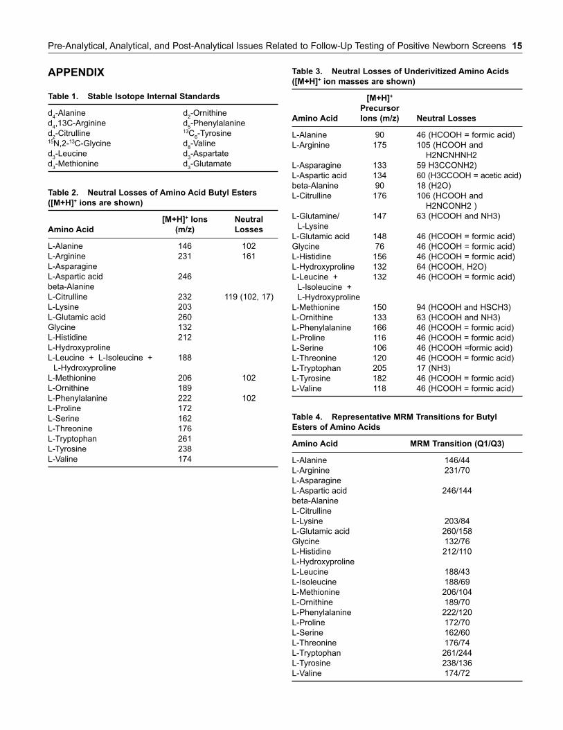
Comments/Specific Examples: Tandem mass spectrometryis typically used to measure specific amino acids for newbornscreening. However, tandem mass spectrometry is increasinglyused to measure amino acids quantitatively for diagnosis andtherapy assessment. For newborn screening, most acids aremeasured in the full-scan mode using a neutral loss of mass tocharge (m/z) 102. For selected amino acids not adequatelydetected using a neutral loss scan, selected reaction monitoringscans can be used. For example, arginine can be detected usinga neutral loss of m/z 161, citrulline and ornithine using a neu-tral loss of m/z 119, and glycine using a neutral loss of m/z 56.In the neutral-loss mode for analysis, all precursors sharing acommon neutral fragment are detected (18). The precursormolecular weight [M+H]+ ions, corresponding to amino acidbutyl esters, are listed in the Appendix, Table 2. Neutral lossesof underivitized amino acids are listed in the Appendix, Table 3.For MRM analysis, appropriate parent-product ion pairs areidentified for each reported amino acid. Representative MRMtransitions for derivitized and underivitized amino acids arelisted in the Appendix (Tables 4 and 5, respectively). MRMallows optimization of experimental parameters for eachamino acid individually, providing optimum sensitivity andselectivity.
Strength of recommendation: A
Quality of Evidence: I
Chromatogram Analysis and Quantitation (Ion-Exchange Chromatography)
Recommendation: Identification of amino acids by ion-exchange chromatography should primarily be based on chro-matographic retention time, and retention time relative to aninternal standard. Quantitation should be based on the recov-ery of the internal standard in each specimen compared withthe recovery of the internal standard in the calibrators.
Comments/Specific Examples: Peaks which fall within pre-determined limits of known amino acid retention timesshould be identified. Chromatograms should also be inspectedfor atypical peaks (eg, alloisoleucine, homocitrulline). Ifninhydrin is used for detection, signal ratios at two wave-lengths (570 nm and 440 nm) should be determined for properidentification of individual amino acids and for detection of
co-eluting interferences (19). For quantitation, calibrationcurves can be constructed for each amino acid by plottingthe ratio of the peak height (or area) of the amino acid to thepeak height (or area) of the corresponding internal standardversus calibrator concentrations.
Strength of recommendation: A
Quality of Evidence: I
Qualitative Analysis
Recommendation: Qualitative screening methods, such asthin-layer chromatography (TLC), should not be used foramino acid analysis.
Comments/Specific Examples: TLC is suitable only fordetection of gross abnormalities. Quantitative analysis ofplasma, urine, or CSF using more sensitive methods is rec-ommended for the diagnosis and monitoring of disorderscharacterized by abnormal amino acids (3).
Strength of recommendation: A
Quality of Evidence: I
Quality Control
Recommendation: At least two control materials should be ana-lyzed daily to monitor the ongoing performance of the analyticprocess.
Comments/Specific Examples: Controls should verify assayperformance at relevant decision points. One control shouldcontain analyte concentrations above the upper reference limit(ie, positive control), and a second control should be below theupper reference limit (ie, negative control). A third controlmight contain analyte concentrations at or near the upper ref-erence limit. Valid acceptable ranges for each analyte shouldbe established for each control material. External controlsshould be run with each new lot number/shipment of analyti-cally critical reagents, after major preventive maintenance, orafter change of a critical instrument component. If patientspecimens are run in daily batches, these materials should beanalyzed with each batch. Internal standard responses for eachspecimen may also serve as a quality control check (20).
Strength of recommendation: A
Quality of Evidence: I
Reference Intervals
Recommendation: Age-matched reference intervals (normalranges) for reported amino acids should be established or ver-ified by the testing laboratory for the population being investi-gated.
Comments/Specific Examples: Reference intervals of severalamino acids are characteristically age dependent, thereby requir-ing test results be compared with a properly defined age group.
Pre-Analytical, Analytical, and Post-Analytical Issues Related to Follow-Up Testing of Positive Newborn Screens 13
Comparison of pediatric results to normative ranges derivedfrom an adult population is not appropriate. If a formal referenceinterval study is not possible, then the testing laboratory shouldcarefully evaluate published data for its own reference ranges(21–24).
Strength of recommendation: A
Quality of Evidence: I
External Quality Assessment
Recommendation: For analytes regulated by Centers forMedicare and Medicaid Services (CMS), the laboratory mustparticipate in a CMS approved–provider PT program. Currently,amino acids are not regulated analytes. If the analyte is not reg-ulated, the laboratory must have a mechanism for verifying theaccuracy and reliability of its test at least two times a year.Participation in a formal proficiency testing (PT) provider’sservice may satisfy this requirement in the absence of a CMS-approved PT provider. The requirement may also be satisfied byan interlaboratory sample exchange program and/or a custom-designed process within the laboratory. The laboratory mustdocument performance, corrective and preventive actions, andmaintain accurate records. PT samples must be handled in amanner identical to the unknown samples. Successful PT or per-formance assessment schemes are a condition of laboratoryaccreditation.
Comments/Specific Examples: PT challenges should includequantitation of amino acids and an assessment of the laboratory’scapability to recognize disease biomarkers and profiles (25–27).The selected PT program or process must use test challenges inthe same matrix as the patient sample. The College of AmericanPathologists (CAP) offers a PT program for amino acid profileanalysis in a plasma matrix. The Newborn Screening QualityAssurance Program at the Centers for Disease Control andPrevention (CDC) offers dried blood spot PT and quality controlprograms for amino acids measured for identification of newborndisorders. Also an amino acid quality assurance program usinglyophilized, spiked human serum is available from ERNDIM (theEuropean Research Network for evaluation and improvement ofscreening, Diagnosis and treatment of Inherited disorders ofMetabolism).
Strength of recommendation: A
Quality of Evidence: I
Post-Analytical Issues/Quality Requirements
Test Interpretation
Recommendation: Interpretation of test results should bebased on relative amino acid levels, pattern recognition, andcorrelation of positive and negative findings.
Comments/Specific Examples: Disease states are often char-acterized by a pattern of elevated amino acids in comparison to
age-matched reference ranges. Amino acid elevation(s) or over-all profiles should be evaluated in the context of clinical findingsand/or additional test results. Interpretation of abnormal findingscan be difficult and requires considerable knowledge not only ofinherited metabolic disorders, but also of numerous physiologi-cal factors that affect amino acid concentrations (28–29).
Strength of recommendation: A
Quality of Evidence: I
Patient Reports
Recommendation: Test reports should include appropriatepatient and specimen information, test results, and clinicalinterpretation.
Comments/Specific Examples: Laboratory reports should bedesigned to convey patient results effectively to a non-expertphysician. This includes documentation of the analyticalmethod used and clinical interpretation of the test results. Thepurpose for performing the test should also be described (eg,screening follow-up). Identification of all relevant amino acidsshould be listed, and quantitative results should be reportedwith appropriate age-matched reference intervals. A detailedinterpretive report of abnormal results should include anoverview of the significance of the test results, correlation toavailable clinical information and/or additional test results,differential diagnosis, and recommendations for further confir-matory biochemical testing (eg, enzyme assay, molecularanalysis). When no abnormalities are detected, test results canbe reported qualitatively (3).
Strength of recommendation: A
Quality of Evidence: I
Interferences/Artifacts
Recommendation: Substances that have the potential tointerfere with the amino acid analysis should be identified andtaken into account during interpretation.
Comments/Specific Examples: Many medications anddietary artifacts affect test results (eg, increase in glycine dueto valproate therapy) (1). Bacterial contamination enhancesconversion of glutamine and asparagine to glutamic and aspar-tic acids, and conversion of cystathionine to homocystine,mimicking homocystinuria; decreases of glycine, alanine, pro-line, and other amino acids may be seen in urine. Tryptophanmay be lost due to deproteinization, while delayed depro-teinization may cause loss of disulfide-containing amino acids.The same effect occurs during clotting, making serum unsuit-able for these amino acids. Hemolysis and/or contamination ofplasma and serum with blood cells may lead to increased lev-els of several amino acids (30).
Strength of recommendation: A
Quality of Evidence: I
16. Long CL, Geiger JW. Automatic analysis of amino acids: effectof resin cross-linking and operational variables on resolution.Anal Bioch, 1969; 29:265–83.
17. Lee PL, Slocum RH. A high-resolution method for amino acidanalysis of physiological fluids containing mixed disulfides.Clin Chem 1988; 34:719–23.
18. Chase DH, Kalas TA, Naylor EW. Use of tandem mass spectom-etry for multianalyte screening of dried blood specimens formnewborns. Clin Chem 2003; 49:1797–1817.
19. Williams AP. General problems associated with the analysis ofamino acids by automated in-exchange chromatography. JChromatogr 1986; 373:175–90.
20. CLSI. Statistical quality control for quantitative measurements:principles and definitions - second edition; approved guidelineC24-A3. Wayne, PA: CLSI, 1996.
21. Bremer HJ, Duran M, Kamerling JP, Przyrembel H, WadmanSK. Disturbances of Amino Acid Metabolism: ClinicalChemistry and Diagnosis. Baltimore: Urban and Schwarzenberg,1981.
22. Rinaldo P, Hahn SH, Matern D. (2005) Inborn errors of aminoacid, organic acid, and fatty acid metabolism. In: Burtis CA,Ashwood ER, Bruns DE, eds. Tietz Textbook of ClinicalChemistry and Molecular Diagnostics, 4th ed., ElsevierSaunders, St. Louis (Missouri), pp. 2207–2247.
23. Clayton BE, Jenkins P, Round RM. Paediatric ChemicalPathology: Clinical Tests and Reference Ranges. Oxford:Blackwell Scientific Publications, 1980.
24. Parvy P, Bardet J, Rabier D, Kamoun P. Age-related referencevalues for free amino acids in first morning urine specimens.Clin Chem 1988; 34:2092–5.
25. 42 Code of Federal Regulations (CFR) 493.901, April 23, 2007.26. Rattenbury JM, Townsend JC. Establishment of an external
quality-assessment scheme for amino acid analyses: resultsfrom assays of samples distributed during two years. Clin Chem1990; 36:217–24.
27. Parvy P, Bardet J, Rabier D, Gasquet M, Kamoun P. Intra- andinter-laboratory quality control for assay of amino acids in bio-logical fluids: 14 years of the French experience. Clin Chem1993; 39:1831–6.
28. Briddon A, Oberholzer VG. Plasma amino acid patterns in crit-ically ill children. J Inher Metab Dis. 1986; 900:254–6.
29. Scriver CR, Sly WS, Childs B, Beaudet AL, Valle D, Kinzer KW,Vogelstein B, eds). The Metabolic and Molecular Bases ofInherited Disease, 8th edition. McGraw-Hill, New York, 2001.
30. Ananth N. Laboratory generated artifacts in plasma amino acidquantitation. Online J Health Allied Scs. 2002; 3:4–7.
REFERENCES
1. Shih VE. Amino Acid Analysis. In: Blau N, Duran M,Blaskovics ME, Gibson KM (Eds): Physician’s Guide to theLaboratory Diagnosis of Metabolic Diseases, Second Edition,Springer, Berlin, 2003, pp. 11–26.
2. Tada K. Non-ketotic hyperglycinemia. In: Fernandes J,Saudubray J-M, Tada K, eds. Inborn Metabolic Diseases.Berlin: Springer-Verlag, 1990 323–9.
3. American College of Medical Genetics. F: Clinical biochemicalgenetics. In: Standards and Guidelines for Clinical GeneticsLaboratories. 2006 http://www.acmg.net/Pages/ACMG_Activities/ stds-2002/f.htm (accessed 09/19/08).
4. Tsai MY, Marshall JG, Josephson MW. Free amino acid analy-sis of untimed and 24-h urine samples compared. Clin Chem1980; 26:1804–8.
5. Perry T, Hansen S. Technical pitfalls leading to errors in the quan-titation of plasma amino acids. Clin Chim Acta 1969; 25: 53–8.
6. Sahai S, Uhlhaas S. Stability of amino acids in human plasma.Clin Chem Acta 1985; 148:225–9.
7. Levy HL, Madaigan PM, Kum A. Fecal contamination in urineamino acid screening. Artifactual cause of hyperaminoaciduria.Am J Clin Pathol 1969; 51:765–8.
8. Walker V, Mills GA. Quantitative methods for amino acid analy-sis in biological fluids. Ann Clin Biochem 1995; 32: 28-57.
9. Ambler RP. Standards and accuracy in amino acid analysis. In:Rattenbury JM, ed. Amino Acid Analysis. Chichester: EllisHorwood Ltd, 1981:119–37.
10. Armstrong MD, Stave U. A study of plasma free amino acid lev-els. I. A study of factors affecting validity of amino acid analy-ses. Metabolism 1973:22:549–60.
11. Hubbard RW, chambers JG, Sanchez A, Slocum R, Lee P.Amino acid analysis of plasma: studies in sample preparation. JChromatogr 1988; 431:163–9.
12. Ersser RS, Davey JF. Liquid chromatographic analysis of aminoacids in physiological fluids: recent advances. Med Lab sci1991; 48:59–71.
13. Deyl Z. Profiling of amino acids in body fluids and tissues bymeans of liquid chromatography. J Chromatogr 1986; 379: 177–250.
14. Chase DH. Mass spectrometry in the clinical laboratory. ChemRev 2001; 101:445–477.
15. Moodie IM, Shephard GS, Labadarios D. A review of quantita-tive ion eschange, high performance liquid and gas chromato-graphic analyses of amino acids in physiological fluids. J HighResol Chromatogr 1989; 12:509–16.
14 Expanded Newborn Screening Using Tandem Mass Spectrometry
Pre-Analytical, Analytical, and Post-Analytical Issues Related to Follow-Up Testing of Positive Newborn Screens 15
APPENDIX
Table 1. Stable Isotope Internal Standards
d4-Alanine
d4,13C-Arginine
d2-Citrulline
15N,2-13C-Glycine
d3-Leucine
d3-Methionine
Table 2. Neutral Losses of Amino Acid Butyl Esters
([M+H]+ ions are shown)
[M+H]+ Ions Neutral
Amino Acid (m/z) Losses
L-Alanine 146 102
L-Arginine 231 161
L-Asparagine
L-Aspartic acid 246
beta-Alanine
L-Citrulline 232 119 (102, 17)
L-Lysine 203
L-Glutamic acid 260
Glycine 132
L-Histidine 212
L-Hydroxyproline
L-Leucine + L-Isoleucine + 188
L-Hydroxyproline
L-Methionine 206 102
L-Ornithine 189
L-Phenylalanine 222 102
L-Proline 172
L-Serine 162
L-Threonine 176
L-Tryptophan 261
L-Tyrosine 238
L-Valine 174
d2-Ornithine
d5-Phenylalanine
13C6-Tyrosine
d8-Valine
d3-Aspartate
d3-Glutamate
Table 3. Neutral Losses of Underivitized Amino Acids
([M+H]+ ion masses are shown)
[M+H]+
Precursor
Amino Acid Ions (m/z) Neutral Losses
L-Alanine 90 46 (HCOOH = formic acid)
L-Arginine 175 105 (HCOOH and
H2NCNHNH2
L-Asparagine 133 59 H3CCONH2)
L-Aspartic acid 134 60 (H3CCOOH = acetic acid)
beta-Alanine 90 18 (H2O)
L-Citrulline 176 106 (HCOOH and
H2NCONH2 )
L-Glutamine/ 147 63 (HCOOH and NH3)
L-Lysine
L-Glutamic acid 148 46 (HCOOH = formic acid)
Glycine 76 46 (HCOOH = formic acid)
L-Histidine 156 46 (HCOOH = formic acid)
L-Hydroxyproline 132 64 (HCOOH, H2O)
L-Leucine + 132 46 (HCOOH = formic acid)
L-Isoleucine +
L-Hydroxyproline
L-Methionine 150 94 (HCOOH and HSCH3)
L-Ornithine 133 63 (HCOOH and NH3)
L-Phenylalanine 166 46 (HCOOH = formic acid)
L-Proline 116 46 (HCOOH = formic acid)
L-Serine 106 46 (HCOOH =formic acid)
L-Threonine 120 46 (HCOOH = formic acid)
L-Tryptophan 205 17 (NH3)
L-Tyrosine 182 46 (HCOOH = formic acid)
L-Valine 118 46 (HCOOH = formic acid)
Table 4. Representative MRM Transitions for Butyl
Esters of Amino Acids
Amino Acid MRM Transition (Q1/Q3)
L-Alanine 146/44
L-Arginine 231/70
L-Asparagine
L-Aspartic acid 246/144
beta-Alanine
L-Citrulline
L-Lysine 203/84
L-Glutamic acid 260/158
Glycine 132/76
L-Histidine 212/110
L-Hydroxyproline
L-Leucine 188/43
L-Isoleucine 188/69
L-Methionine 206/104
L-Ornithine 189/70
L-Phenylalanine 222/120
L-Proline 172/70
L-Serine 162/60
L-Threonine 176/74
L-Tryptophan 261/244
L-Tyrosine 238/136
L-Valine 174/72
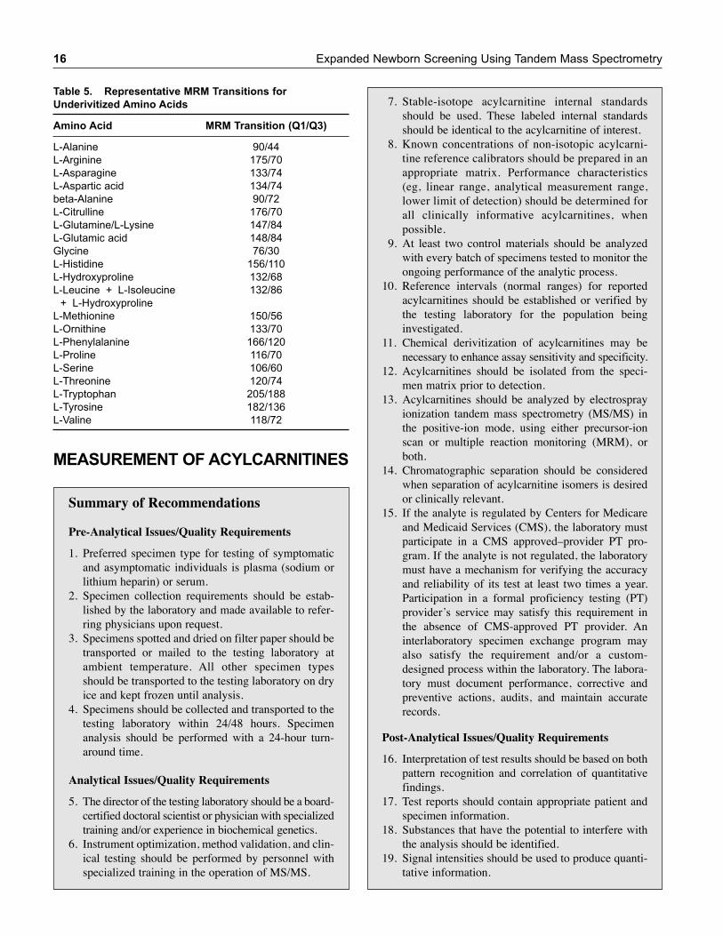
16 Expanded Newborn Screening Using Tandem Mass Spectrometry
MEASUREMENT OF ACYLCARNITINES
Table 5. Representative MRM Transitions for
Underivitized Amino Acids
Amino Acid MRM Transition (Q1/Q3)
L-Alanine 90/44
L-Arginine 175/70
L-Asparagine 133/74
L-Aspartic acid 134/74
beta-Alanine 90/72
L-Citrulline 176/70
L-Glutamine/L-Lysine 147/84
L-Glutamic acid 148/84
Glycine 76/30
L-Histidine 156/110
L-Hydroxyproline 132/68
L-Leucine + L-Isoleucine 132/86
+ L-Hydroxyproline
L-Methionine 150/56
L-Ornithine 133/70
L-Phenylalanine 166/120
L-Proline 116/70
L-Serine 106/60
L-Threonine 120/74
L-Tryptophan 205/188
L-Tyrosine 182/136
L-Valine 118/72
Summary of Recommendations
Pre-Analytical Issues/Quality Requirements
1. Preferred specimen type for testing of symptomaticand asymptomatic individuals is plasma (sodium orlithium heparin) or serum.
2. Specimen collection requirements should be estab-lished by the laboratory and made available to refer-ring physicians upon request.
3. Specimens spotted and dried on filter paper should betransported or mailed to the testing laboratory atambient temperature. All other specimen typesshould be transported to the testing laboratory on dryice and kept frozen until analysis.
4. Specimens should be collected and transported to thetesting laboratory within 24/48 hours. Specimenanalysis should be performed with a 24-hour turn-around time.
Analytical Issues/Quality Requirements
5. The director of the testing laboratory should be a board-certified doctoral scientist or physician with specializedtraining and/or experience in biochemical genetics.
6. Instrument optimization, method validation, and clin-ical testing should be performed by personnel withspecialized training in the operation of MS/MS.
7. Stable-isotope acylcarnitine internal standardsshould be used. These labeled internal standardsshould be identical to the acylcarnitine of interest.
8. Known concentrations of non-isotopic acylcarni-tine reference calibrators should be prepared in anappropriate matrix. Performance characteristics(eg, linear range, analytical measurement range,lower limit of detection) should be determined forall clinically informative acylcarnitines, whenpossible.
9. At least two control materials should be analyzedwith every batch of specimens tested to monitor theongoing performance of the analytic process.
10. Reference intervals (normal ranges) for reportedacylcarnitines should be established or verified bythe testing laboratory for the population beinginvestigated.
11. Chemical derivitization of acylcarnitines may benecessary to enhance assay sensitivity and specificity.
12. Acylcarnitines should be isolated from the speci-men matrix prior to detection.
13. Acylcarnitines should be analyzed by electrosprayionization tandem mass spectrometry (MS/MS) inthe positive-ion mode, using either precursor-ionscan or multiple reaction monitoring (MRM), orboth.
14. Chromatographic separation should be consideredwhen separation of acylcarnitine isomers is desiredor clinically relevant.
15. If the analyte is regulated by Centers for Medicareand Medicaid Services (CMS), the laboratory mustparticipate in a CMS approved–provider PT pro-gram. If the analyte is not regulated, the laboratorymust have a mechanism for verifying the accuracyand reliability of its test at least two times a year.Participation in a formal proficiency testing (PT)provider’s service may satisfy this requirement inthe absence of CMS-approved PT provider. Aninterlaboratory specimen exchange program mayalso satisfy the requirement and/or a custom-designed process within the laboratory. The labora-tory must document performance, corrective andpreventive actions, audits, and maintain accuraterecords.
Post-Analytical Issues/Quality Requirements
16. Interpretation of test results should be based on bothpattern recognition and correlation of quantitativefindings.
17. Test reports should contain appropriate patient andspecimen information.
18. Substances that have the potential to interfere withthe analysis should be identified.
19. Signal intensities should be used to produce quanti-tative information.
Pre-Analytical, Analytical, and Post-Analytical Issues Related to Follow-Up Testing of Positive Newborn Screens 17
Pre-Analytical Issues/Quality Requirements
Type of Specimen
Recommendation: Preferred specimen type for testing ofsymptomatic and asymptomatic individuals is plasma (sodiumor lithium heparin) or serum.
Comments/Specific Examples: Gel tubes are acceptable.Dried blood spots collected on filter paper cards can be used(1–4). Urine is less frequently used but may be valuable ininvestigation of inconclusive/borderline urine organic acidand plasma acylcarnitine profiles (eg, detecting glutaricacademia type I, biotinidase deficiency, and isobutyryl-CoAdehydrogenase deficiency) (5). Post-mortem screening canbe performed on dried blood spots or bile (4). Cell-freesupernatant of amniotic fluid can be used for prenatal diag-nosis of selected inborn errors of metabolism.
Strength of recommendation: A
Quality of Evidence: I
Specimen Collection and Processing
Recommendation: Specimen collection requirements shouldbe established by the laboratory and made available to refer-ring physicians upon request.
Comments/Specific Examples: Acylcarnitines are unstable,and plasma/serum should be separated from cells and frozen assoon as possible. Typical specimen testing volumes are listedin the Appendix Table 1. Clinical information is needed forappropriate interpretation. Additional required informationincludes age, sex, diet (eg, total parenteral nutrition therapy),drug therapy, and family history (6). No strong evidence ofdiurnal variation, so specimens can be collected at any time ofday. Acylcarnitine levels vary with patient age (7).
Strength of recommendation: B
Quality of Evidence: II
Specimen Transport and Storage
Recommendation: Specimens spotted and dried on filter papershould be transported or mailed to the testing laboratory at ambi-ent temperature. All other specimen types should be transportedto the testing laboratory on dry ice and kept frozen until analysis.
Comments/Specific Examples: Compounds are stable indefi-nitely when stored at –80°C. Long-term storage at room temper-ature causes progressive losses, with degradation of short-chainacylcarnitines occurring faster than long-chain species (6, 8).
Strength of recommendation: B
Quality of Evidence: II
Emergency Specimen Collection
Recommendation: Specimens should be collected and trans-ported to the testing laboratory as quickly as possible. Specimenanalysis should be performed within 24 hours or fewer.
Comments/Specific Examples: In life-threatening situations,where an abnormality is thought to be likely (either fromfamily history, results of preliminary investigations orclinical presentation), there should be no delay in specimentransport and/or delay in specimen analysis. At the earliestopportunity, the testing laboratory should be contacted todiscuss appropriate investigations. If possible, specimensshould be collected before death (9).
Strength of recommendation: B
Quality of Evidence: II
Analytical Issues/Quality Requirements
Laboratory Director
Recommendation: The director of the testing laboratoryshould be a board-certified doctoral scientist or physician withspecialized training and/or experience in biochemical genetics.
Comments/Specific Examples: The laboratory director isresponsible for the technical and scientific oversight of thetesting laboratory and for the interpretation and reporting ofresults. At a minimum, the director should meet the person-nel standards for high complexity as defined by CLIA ’88.Board certification in clinical chemistry, clinical pathology,and/or biochemical genetics is recommended (10).
Strength of recommendation: A
Quality of Evidence: I
Testing Personnel
Recommendation: Instrument optimization, method valida-tion, and clinical testing should be performed by personnelwith specialized training in the operation of MS/MS.
Comments/Specific Examples: Operators of MS/MS instru-ments should hold a minimum of a Bachelor of Sciencedegree in a laboratory science or medical technology.Appropriate training and ongoing competency requirementsfor testing personnel performing acylcarnitine analysisshould be established and documented (15).
Strength of recommendation: A
Quality of Evidence: I
Stable-Isotope Internal Standards
Recommendation: Stable isotope acylcarnitines internalstandards should be used. These labeled internal standardsshould be identical to the acylcarnitine of interest.
Comments/Specific Examples: Stable isotope internal stan-dards (individual and pre-mixed) are available from a varietyof commercial sources. Typical internal standards for a com-plete carnitine/acylcarnitine profile are listed in Appendix(Table 2). Commercial suppliers should provide concentration
18 Expanded Newborn Screening Using Tandem Mass Spectrometry
and stability data. If an acylcarnitine standard is not availablein labeled form, then a close mass homologue should be sub-stituted. For example, d3-C16 can be used to estimate the con-centrations of C16, C16:1, C16OH, and C16:1OH (assumesacylcarnitines of same chain length have similar performancecharacteristics). Labeled internal standards should be validatedby determining recoveries from weighed, non-isotopicacylcarnitine calibrators. Labeled internal standards should beanalyzed with each specimen (6).
Strength of recommendation: A
Quality of Evidence: I
Calibrators
Recommendation: Known concentrations of non-isotopicacylcarnitine reference calibrators should be prepared in anappropriate matrix. Performance characteristics (eg, linearrange, analytical measurement range, lower limit of detec-tion) should be determined for all clinically informativeacylcarnitines, when possible.
Comments/Specific Examples: Reference calibrators(Appendix, Table 3) are not available for all acylcarnitines,particularly hydroxyl and unsaturated species. Quantitationof these compounds should be extrapolated by application ofthe calibration for the nearest species of similar structure.Performance characteristics should be verified on a regularbasis as specified by regulatory agencies. The matrix usedfor calibrators should be free of endogenous carnitine andacylcarnitines (6).
Strength of recommendation: A
Quality of Evidence: I
Specimen Preparation – Derivitization
Recommendation: Chemical derivitization of acylcarnitinesis recommended to enhance assay sensitivity and specificity.
Comments/Specific Examples: Typically, acylcarnitines arederivitized to their butyl esters using hydrogen chloride inbutanol and heating at 65°C for 15 minutes (11, 12, 14). Directanalysis of acylcarnitines without derivitization is also possi-ble, and alternative derivitization methods including methyla-tion are occasionally used (13).
Strength of recommendation: B
Quality of Evidence: II
Specimen Preparation – Deproteinization/Extraction
Recommendation: Acylcarnitines should be isolated fromthe specimen matrix prior to detection.
Comments/Specific Examples: Liquid-liquid and solid-phase extraction procedures are frequently used. Methanol is acommon extraction solvent which also serves to deproteinize
plasma samples. Blood spots are dry samples, and a certain degreeof accuracy and precision is lost because of variations in bloodvolumes in the disks and in extraction efficiency (12, 16).
Strength of recommendation: A
Quality of Evidence: I
Detection/Data Acquisition
Recommendation: Acylcarnitines should be analyzed byelectrospray ionization tandem mass spectrometry (MS/MS) inthe positive-ion mode, using either precursor-ion scan or mul-tiple reaction monitoring (MRM), or both.
Comments/Specific Examples: In the precursor-ion modefor analysis of butylated derivatives, all precursors of productsm/z 85 within a mass range between 200 to 500 m/z arerecorded. The parent ions (molecular weight [M+H]+ ions),corresponding to the butyl ester derivatives, are listed in theAppendix, Table 4. Advantages of the precursor-ion full scanmode include evaluation of all potential acylcarnitines,detection of common drug artifacts, presence of interferingcompounds, and evidence of poor derivitization. For MRManalysis, appropriate parent-product ion pairs are identified foreach reported acylcarnitine. Representative MRM transitionsfor underivitized acylcarnitines are listed in the Appendix,Table 5. MRM allows optimization of experimental parametersfor each acylcarnitine individually, providing optimumsensitivity and selectivity. In addition to being more specificand thus more precise, MRM may also allow for selectivemeasurement of particular acylcarnitines (6, 7, 12).
Strength of recommendation: A
Quality of Evidence: I
Chromatographic Separation
Recommendation: Liquid chromatographic separationshould be considered when separation of acylcarnitine isomersis desired or clinically relevant.
Comments/Specific Examples: Accurate determination ofindividual acylcarnitine isomers by MS/MS requires chro-matographic separation (6, 17).
Strength of recommendation: B
Quality of Evidence: II
Quality Control
Recommendation: At least two control materials should beanalyzed with every batch of specimens tested to monitor theongoing performance of the analytic process.
Comments/Specific Examples: One control should containanalyte concentrations above the upper reference limit (ie, pos-itive control), and a second control should be below the upperreference limit (ie, negative control). A third control might
Pre-Analytical, Analytical, and Post-Analytical Issues Related to Follow-Up Testing of Positive Newborn Screens 19
contain analyte concentrations at or near the upper referencelimit. Valid acceptable ranges should be established for eachcontrol material (18).
Strength of recommendation: A
Quality of Evidence: I
Reference Intervals
Recommendation: Reference intervals (normal ranges) forreported acylcarnitines should be established or verified by thetesting laboratory for the population being investigated.
Comments/Specific Examples: Reference intervals must beage matched. Comparison of pediatric results to normativeranges derived from an adult population is not appropriate.If a formal reference interval study is not possible, then thetesting laboratory should carefully evaluate published datafor its own reference ranges (8, 14).
Strength of recommendation: A
Quality of Evidence: I
External Quality Assessment
Recommendation: If the analyte is regulated by Centers forMedicare and Medicaid Services (CMS), the laboratory mustparticipate in a CMS approved–provider PT program.Currently, acylcarnitines are not regulated analytes. If the ana-lyte is not regulated, the laboratory must have a mechanism forverifying the accuracy and reliability of its test at least twotimes a year. Participation in a formal proficiency testing (PT)provider’s service may satisfy this requirement in the absenceof a CMS-approved PT provider. An interlaboratory sampleexchange program may also satisfy the requirement and/or acustom-designed process within the laboratory. The laboratorymust document performance, corrective and preventiveactions, and maintain accurate records. PT samples must behandled in a manner identical to the unknown samples.Successful PT or performance assessment schemes are a con-dition of laboratory accreditation.
Comments/Specific Examples: Proficiency testing (PT)challenges should include quantitation of acylcarnitines and anassessment of the laboratory’s capability to recognize diseasebiomarkers and profiles. The selected PT program or processmust use test challenges in the same matrix as the patient sam-ple. The College of American Pathologists (CAP) offers a PTprogram for acylcarnitine profile analysis in a plasma matrix.The Newborn Screening Quality Assurance Program at theCenters for Disease Control and Prevention (CDC) offers driedblood spot PT and quality control programs for acylcarnitinesmeasured for identification of newborn disorders. Also a qual-itative blood spot (35 to 50mL) acycarnitine quality assuranceprogram is available mfrom ERNDIM (the European ResearchNetwork for evaluation and improvement of screening,
Diagnosis and treatment of Inherited disorders of Metabolism;19, 20).
Strength of recommendation: A
Quality of Evidence: I
Post-Analytical Issues/Quality Requirements
Test Interpretation
Recommendation: Interpretation of test results should bebased on both pattern recognition and correlation of quantita-tive findings.
Comments/Specific Examples: Disease states are often char-acterized by a pattern of elevated acylcarnitines in comparisonto age-matched reference ranges (see Appendix, Table 6).Calculation of ratios (eg, C8/C10 ratio in MCAD deficiency)may also be useful in the interpretation of abnormal results (6, 21, 22).
Strength of recommendation: A
Quality of Evidence: I
Patient Reports
Recommendation: Test reports should contain appropriatepatient and specimen information, test results, and clinicalinterpretation.
Comments/Specific Examples: Interpretation of abnormalresults should include an overview of the significance of thetest results, quantitative results with reference ranges, a corre-lation of available clinical information, elements of differentialdiagnosis, and recommendations for additional biochemicaltesting (23).
Strength of recommendation: A
Quality of Evidence: I
Interferences/Artifacts
Recommendation: Substances that have the potential tointerfere with the analysis should be identified.
Comments/Specific Examples: Several drug interferencesare known to induce false positive test results (eg, pivalic acid,valproic acid, cefotaxime). Other exogenous compounds (eg,drug metabolites, food additives, intravenous fluids containingdextrose) may result in the appearance of peaks with m/z val-ues in close proximity to, or overlapping with, acylcarnitinesof diagnostic significance. Dietary artifacts are also possible,usually related to food intake enriched with fatty acids (eg,ketogenic diet, MCT oil; 7, 24, 25).
Strength of recommendation: A
Quality of Evidence: I
20 Expanded Newborn Screening Using Tandem Mass Spectrometry
REFERENCES
1. Van Hove JL, Zhang W, Kahler SG, Roe CR, Chen YT, Terada N,et al. Medium-chain acyl-CoA dehydrogenase (MCAD) defi-ciency: diagnosis by acylcarnitine analysis in blood. Am J HumGenet 1993; 52:958–66.
2. Chace DH, Hillman SL, Vanhove JLK, Naylor EW. Rapid diag-nosis of MCAD deficiency – quantitative analysis of octanoyl-carnitine and other acylcarnitines in newborn blood sports bytandem mass spectrometry. Clin Chem 1997; 43:2106–13.
3. Millington DS, Chace DH. Carnitine and acylcarnitines in meta-bolic disease diagnosis and management. In: Desiderio DM(ed). Mass Spectrometry: Clinical and Biomedical Applications.Volume 1. New York: Plenum Press; 1992, pp 199–219.
4. Chace DH, DiPerna JC, Mitchell BL, Sgroi B, et al.Electrospray tandem mass spectrometry for analysis ofacylcarnitines in dried postmortem blood specimens collectedat autopsy from infants with unexplained cause of death. ClinChem 2001; 47:1166–1182.
5. Tortorelli S, Hahn SH, Cowan TM, Brewster TG et al. The uri-nary excretion of glutarylcarnitine is an informative tool in thebiochemical diagnosis of glutaric acidemia type I. Mol GenetMetab 2005; 84:137–143.
6. Rinaldo P, Cowan T, Matern D. Acylcarnitine profile analysis:American College of Medical Genetics Standards and Guidelinesfor Clinical Genetic Laboratories. Genet Med 2008; 10:151–156
7. Millington DS. Tandem mass spectrometry in clinical diagnosis.In: Blau N, Duran M, Blaskovics ME, Gibson KM (eds)Physician’s guide to the laboratory diagnosis of metabolic dis-eases. Springer, Berlin, 2nd edition, 2003, pp 57–75.
8. Rinaldo P, Hahn S, Matern D. Appendix 55. Inborn errors of aminoacid, organic acid, and fatty acid metabolism. In: CA Burtis, ERAshwood and DE Bruns (Eds.), Tietz Textbook of ClinicalChemistry, Fourth Edition, Elsevier Saunders, St. Louis, MO, 2006.
9. Green A, Morgan I, Gray J. Neonatology and LaboratoryMedicine. ACB Venture Publications. London, 2003.
10. American College of Medical Genetics. B. Personnel Policies. In:Standards and Guidelines for Clinical Genetics Laboratories. 2006http://www.acmg.net/Pages/ACMG_Activities/stds-2002/f.htm
11. Chace DH. Mass spectrometry in the clinical laboratory. ChemRev. 2001; 101:445–477.
12. Chace DH, Kalas TA, Naylor EW. Use of tandem mass spec-trometry for multianalyte screening of dried specimens fromnewborns. Clin Chem 2003; 49:1797–1817.
13. Nagy N, Takáts Z, Pollreisz F, Szabó T, Vékey K. Direct tandemmass spectrometric analysis of amino acids in dried blood spotswithout chemical derivatization for neonatal screening; RapidCommunications in Mass Spectrometry, 2003; 17:983–990.
14. Matern D. Acylcarnitines including in vitro loading tests. In:Blau N, Duran M, Gibson KM (eds) Laboratory guide to themethods in biochemical genetics. Berlin: Springer-Verlag, 2008;pp 171–206.
15. Center for Disease Control and Prevention. Using tandem massspectrometry for metabolic disease screening among newborns:a report of a work group. MMWR 2001; 50 (No. RR-3):1–40.
16. Chace DH, Kalas TA, Naylor EW. The application of tandemmass spectrometry to neonatal screening for inherited disordersof intermediary metabolism. Annu Rev Genomics Hum Genet2002; 3:17–45.
17. Ghoshal AK, Guo T, Sukhova N, Soldin SJ. Rapid measurementof plasma acylcarnitines by liquid chromatography-tandem massspectrometry. Clin Chim Acta 2005; 358:104–112.
18. CLSI. Statistical quality control for quantitative measurements:principles and definitions – third edition; approved guidelineC24-A3. Wayne, PA:CLSI, 2006.
19. 42 Code of Federal Regulations (CFR) 493.901, April 23, 2007.20. European Research Network for evaluation and improvement of
screening, Diagnosis and treatment of Inherited disorders ofMetabolism” (ERNDIM) see http://www.erndimqa.nl/
21. Chace DH, Kalas TA. A biochemical perspective on the use oftandem mass spectrometry for newborn screening and clinicaltesting. Clin Biochem 2005; 38:296–309.
22. Sweetman L, Millington DS, Therrell BL, Hannon WH et al.Naming and counting disorders (conditions) included in new-born screening panels. Pediatrics. 2006; 117(5 Pt 2):S308–S314.
23. American College of Medical Genetics. F: Clinical biochemicalgenetics. In: Standards and Guidelines for Clinical GeneticsLaboratories. 2006 http://www.acmg.net/Pages/ACMG_Activities/ stds-2002/f.htm
24. Abdenur JE, Chamoles NA, Guinle AE, Schenone AB et al.Diagnosis of isovaleric acidaemia by tandem mass spectrom-etry: false positive result due to pivaloylcarnitine in anewborn screening programme. J Inherit Metab Dis. 1998;21:624–630.
25. Vianey-Saban C, Boyer S, Levrat V, Cheillan D, et al.Interference of Cefotaxime in plasma acylcarnitine profile mim-icking an increase of 3-hydroxypalmitoleylcarnitine (C16:1-OH)using butyl esters. J Inherit Metab Dis 2004; 27(Suppl. 1):94.

Pre-Analytical, Analytical, and Post-Analytical Issues Related to Follow-Up Testing of Positive Newborn Screens 21
Table 1. Typical Sample Testing Volumes
Sample Type Testing Volume
Amniotic fluid (cell-free supernatant) 20 μL
Amniocyte culture medium 50 μL*
Bile (liquid specimen) 20 μL**
Bile dried spot (postmortem) one 1/8” punch**
Blood dried spot (neonatal,
postmortem) one 1/8” or 3/16” punch
Cell culture medium 50 μL*
Plasma/serum 20 μL
Urine Variable; standardized
to creatinine
*Spotted on filter paper. **May require dilution if profile is abnormal.
Table 2. Stable Isotope Internal Standards
d9-free carnitine (d
9-C0)
d3-Acetylcarnitine (d
3-C2)
d3-Propionylcarnitine (d
3-C3)
d3-Butyrlcarnitine (d
3-C4)
d9-Isovalerylcarnitine (d
9-C5)
d3-Hexanoylcarnitine (d
3-C6)
d3-Octanoylcarnitine (d
3-C8)
d3-Decanoylcarnitine (d
3-C10)
d3-Dodecanoylcarnitine (Lauroyl) (d
3-C12)
d3-Myristoylcarnitine (Tetradecanoyl) (d
3-C14)
d3-Palimitoylcarnitine (Hexadecanoyl) (d
3-C16)
d3-Octadecanoylcarnitine (Stearoyl) (d
3-C18)
d6-Glutarylcarnitine (d
6-C5DC)
Table 3. Reference Calibrators
Free carnitine (C0)
Acetylcarnitine (C2)
Propionylcarnitine (C3)
Butyrlcarnitine (C4)
Isovalerylcarnitine (C5)
Hexanoylcarnitine (C6)
Octanoylcarnitine (C8)
Decanoylcarnitine (C10)
Lauroylcarnitine (C12)
Myristoylcarnitine (C14)
Palimitoylcarnitine (C16)
Octadecanoylcarnitine (C18)
Glutarylcarnitine (C5DC)
Table 4. [M+H]+ Ions of Acylcarnitine-Butyl-Esters
Acyl-carnitine Chain Length [M+H]+
Acetyl- C2 260
Acrylyl- C3:1 272
Propionyl- C3 274
Formiminoglutamate (FIGLU) 287
Isobutyryl-/Butyryl- C4 288
Tiglyl- C5:1 300
Isovaleryl-/2-methylbutyryl- C5 302
3-hydroxybutyryl C4-OH 304
Hexanoyl- C6 316
3-hydroxyisovaleryl- C5-OH 318
2-methyl-3-hydroxybutyryl-
Heptanoyl- C7 330
3-hydroxyhexanoyl- C6-OH 332
Octenoyl- C8:1 342
Octanoyl- C8 344
Malonyl- C3-DC 360
Decadienoyl- C10:2 368
Decenoyl- C10:1 370
Decanoyl- C10 372
Methylmalonyl- C4-DC 374
3-hydroxydecenoyl- C10:1-OH 386
Glutaryl-/3-hydroxydecanoyl- C5-DC/ C10-OH 388
Dodecenoyl- C12:1 398
Dodecanoyl- (lauroyl-) C12 400
3-hydroxydodecenoyl- C12:1-OH 414
3-hydroxydodecanoyl- C12-OH 416
Tetradecadienoyl- C14:2 424
Tetradecenoyl- C14:1 426
Tetradecanoyl- (myristoyl-) C14 428
3-hydroxytetradecenoyl- C14:1-OH 442
3-hydroxytetradecenoyl- C14-OH 444
Hexadecenoyl- C16:1 454
Hexadecanoyl- (palmitoyl-) C16 456
3-hydroxyhexadecenoyl- C16:1-OH 470
3-hydroxyhexadecanoyl- C16-OH 472
Octadecadienoyl- C18:2 480
Octadecenoyl- C18:1 482
Octadecanoyl- (stearyl-) C18 484
3-hydroxyoctadecadienoyl- C18:2-OH 496
3-hydroxyoctadecenoyl- C18:1-OH 498
3-hydroxyoctadecanoyl- C18-OH 502
Table 5. Representative Multiple Reaction Monitoring
(MRM) Transitions for Underivitized Acylcarnitines
Acyl-carnitine Chain Length MRM Transition
Acetyl- C2 204/85
Propionyl- C3 218/85
Isobutyryl-/Butyryl- C4 232/85
Isovaleryl-/2-methylbutyryl- C5 246/85
Hexanoyl- C6 260/85
Octanoyl- C8 288/85
Decanoyl- C10 316/85
Dodecanoyl- (lauroyl-) C12 344/85
Tetradecanoyl- (myristoyl-) C14 372/85
Hexadecanoyl- (palmitoyl-) C16 400/85
APPENDIX
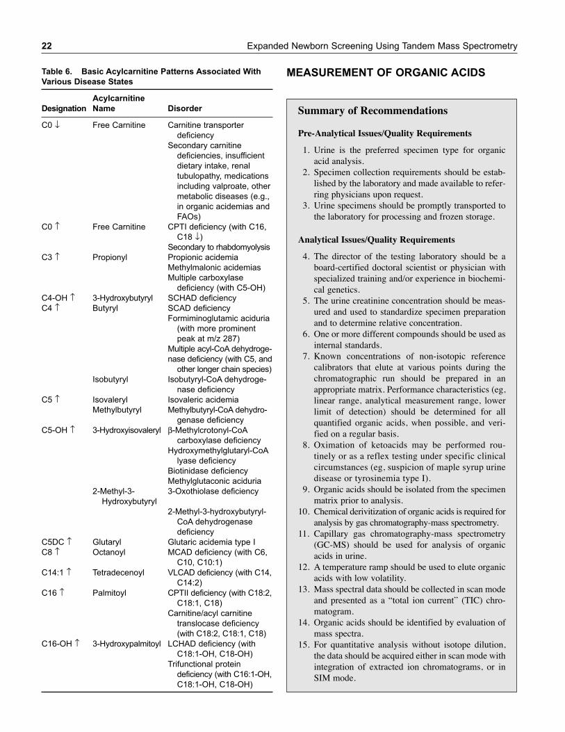
22 Expanded Newborn Screening Using Tandem Mass Spectrometry
Table 6. Basic Acylcarnitine Patterns Associated With
Various Disease States
Acylcarnitine
Designation Name Disorder
C0 ↓ Free Carnitine Carnitine transporter
deficiency
Secondary carnitine
deficiencies, insufficient
dietary intake, renal
tubulopathy, medications
including valproate, other
metabolic diseases (e.g.,
in organic acidemias and
FAOs)
C0 ↑ Free Carnitine CPTI deficiency (with C16,
C18 ↓)
Secondary to rhabdomyolysis
C3 ↑ Propionyl Propionic acidemia
Methylmalonic acidemias
Multiple carboxylase
deficiency (with C5-OH)
C4-OH ↑ 3-Hydroxybutyryl SCHAD deficiency
C4 ↑ Butyryl SCAD deficiency
Formiminoglutamic aciduria
(with more prominent
peak at m/z 287)
Multiple acyl-CoA dehydroge-
nase deficiency (with C5, and
other longer chain species)
Isobutyryl Isobutyryl-CoA dehydroge-
nase deficiency
C5 ↑ Isovaleryl Isovaleric acidemia
Methylbutyryl Methylbutyryl-CoA dehydro-
genase deficiency
C5-OH ↑ 3-Hydroxyisovaleryl β-Methylcrotonyl-CoA
carboxylase deficiency
Hydroxymethylglutaryl-CoA
lyase deficiency
Biotinidase deficiency
Methylglutaconic aciduria
2-Methyl-3- 3-Oxothiolase deficiency
Hydroxybutyryl
2-Methyl-3-hydroxybutyryl-
CoA dehydrogenase
deficiency
C5DC ↑ Glutaryl Glutaric acidemia type I
C8 ↑ Octanoyl MCAD deficiency (with C6,
C10, C10:1)
C14:1 ↑ Tetradecenoyl VLCAD deficiency (with C14,
C14:2)
C16 ↑ Palmitoyl CPTII deficiency (with C18:2,
C18:1, C18)
Carnitine/acyl carnitine
translocase deficiency
(with C18:2, C18:1, C18)
C16-OH ↑ 3-Hydroxypalmitoyl LCHAD deficiency (with
C18:1-OH, C18-OH)
Trifunctional protein
deficiency (with C16:1-OH,
C18:1-OH, C18-OH)
MEASUREMENT OF ORGANIC ACIDS
Summary of Recommendations
Pre-Analytical Issues/Quality Requirements
1. Urine is the preferred specimen type for organicacid analysis.
2. Specimen collection requirements should be estab-lished by the laboratory and made available to refer-ring physicians upon request.
3. Urine specimens should be promptly transported tothe laboratory for processing and frozen storage.
Analytical Issues/Quality Requirements
4. The director of the testing laboratory should be aboard-certified doctoral scientist or physician withspecialized training and/or experience in biochemi-cal genetics.
5. The urine creatinine concentration should be meas-ured and used to standardize specimen preparationand to determine relative concentration.
6. One or more different compounds should be used asinternal standards.
7. Known concentrations of non-isotopic referencecalibrators that elute at various points during thechromatographic run should be prepared in anappropriate matrix. Performance characteristics (eg,linear range, analytical measurement range, lowerlimit of detection) should be determined for allquantified organic acids, when possible, and veri-fied on a regular basis.
8. Oximation of ketoacids may be performed rou-tinely or as a reflex testing under specific clinicalcircumstances (eg, suspicion of maple syrup urinedisease or tyrosinemia type I).
9. Organic acids should be isolated from the specimenmatrix prior to analysis.
10. Chemical derivitization of organic acids is required foranalysis by gas chromatography-mass spectrometry.
11. Capillary gas chromatography-mass spectrometry(GC-MS) should be used for analysis of organicacids in urine.
12. A temperature ramp should be used to elute organicacids with low volatility.
13. Mass spectral data should be collected in scan modeand presented as a “total ion current” (TIC) chro-matogram.
14. Organic acids should be identified by evaluation ofmass spectra.
15. For quantitative analysis without isotope dilution,the data should be acquired either in scan mode withintegration of extracted ion chromatograms, or inSIM mode.
Pre-Analytical, Analytical, and Post-Analytical Issues Related to Follow-Up Testing of Positive Newborn Screens 23
16. Isotope ratio mass spectometry using stable-isotopelabeled internal standards should be used for accu-rate quantitation of selected organic acids.
17. A quality control program based on the analysis ofnormal and abnormal controls should be imple-mented and performed with patient specimens.
18. For quantitative reporting, reference intervals (nor-mal ranges) for organic acids should be establishedor verified by the testing laboratory for the popula-tion being investigated.
19. For analytes regulated by Centers for Medicare andMedicaid Services (CMS), the laboratory must par-ticipate in a CMS approved–provider PT program.Currently, organic acids are not regulated analytes.If the analyte is not regulated, the laboratory musthave a mechanism for verifying the accuracy andreliability of its test at least two times a year.Participation in a formal proficiency testing (PT)provider’s service may satisfy this requirement inthe absence of a CMS-approved PT provider. Aninterlaboratory sample exchange program may alsosatisfy the requirement and/or a custom-designedprocess within the laboratory. The laboratory mustdocument performance, corrective and preventiveactions, and maintain accurate records. PT samplesmust be handled in a manner identical to the unknownsamples. Successful PT or performance assessmentschemes are a condition of laboratory accreditation.
Post-Analytical Issues/Quality Requirements
20. Interpretation of organic acid profiles should bebased on pattern recognition and correlation of pos-itive and negative findings.
21. Test reports should include appropriate patient andspecimen information, test results, and clinicalinterpretation.
22. Substances that have the potential to interfere withthe organic acid analysis should be identified andtaken into account during interpretation.
Pre-Analytical Issues/Quality Requirements
Type of Specimen
Recommendation: Urine is the preferred specimen type fororganic acid analysis.
Comments/Specific Examples: In rare instances, organic acidscan be analyzed in heparinized plasma, cerebral spinal fluid(CSF), or vitreous humor. Collection of body fluids other thanurine is indicated when a urine specimen cannot be obtained (eg,post-mortem). Quantitation of specific CSF metabolites hasbeen reported to be helpful in identifying organic acid disordersin some patients (eg, “cerebral” lactic acidemias, disorders ofbiotin metabolism, glutaryl-CoA deficiency; 1–5).
Strength of recommendation: A
Quality of Evidence: I
Specimen Collection and Processing
Recommendation: Specimen collection requirements shouldbe established by the laboratory and made available to refer-ring physicians upon request.
Comments/Specific Examples: Any random or timed urinespecimen may be collected.
Ideally, urine specimens should be obtained during the timeof acute illness and metabolic decompensation (eg, firsturine void after emergency admission), since abnormalmetabolite levels often decrease to near-normal concentra-tions if specimens are collected only when the patient isapparently healthy. Urine specimens should be collected inclean containers without preservatives, and samples shouldbe frozen as soon as possible after collection (4). Urinespecimens collected and dried on filter paper strips can alsobe used (6).
Strength of recommendation: A
Quality of Evidence: I
Specimen Transport and Storage
Recommendation: Liquid urine specimens should bepromptly transported to the laboratory for processing andfrozen storage.
Comments/Specific Examples: Urine organic acids are sta-ble for very long periods of time (several years) if stored at−20°C or colder. Freezing is not necessary if analysis isimmediate. Specimens may be stored refrigerated for 24hours after collection, but urines stored at ambient (room)temperature cause are unacceptable. Specimens that areanalyzed at a distant testing laboratory should be shippedon dry ice via overnight courier. If freezing is not possible,or if dry ice is not available, then preservatives (eg, sodiumazide, thymol, chloroform) may be added to prevent bacte-rial growth. Plasma, CSF, or vitreous humor should befrozen soon after collection (4). Specimens dried on filterpaper strips may be transported or mailed to the testing lab-oratory at ambient temperature (7).
Strength of recommendation: A
Quality of Evidence: I
Analytical Issues/Quality Requirements
Specimen Preparation
Recommendation: The urine creatinine concentration shouldbe measured and used to standardize specimen preparation andto determine relative concentration.
24 Expanded Newborn Screening Using Tandem Mass Spectrometry
Comments/Specific Examples: A volume of thawed, thor-oughly mixed urine equivalent to a constant amount of creati-nine is used. Typically, a volume of urine corresponding to0.25 mg (2.2 μmol) of creatinine is used. The recommendedminimum volume is 0.5 μL and the maximum is 5.0 mL (8).
Strength of recommendation: A
Quality of Evidence: I
Internal Standard
Recommendation: One or more different compounds shouldbe used as internal standards.
Comments/Specific Examples: The internal standardshould not be a metabolite that might be detected in normal orpathologic urine, nor should it co-chromatograph with signif-icant metabolites. Examples of internal standards includeheptadecanoic acid, 2-phenylbutyric acid, and tropic acid.The final concentration of internal standard should be cho-sen to generate a peak on the total ion chromatogram that isclearly recognizable (9).
Strength of recommendation: A
Quality of Evidence: I
Calibrators/Calibration
Recommendation: Known concentrations of non-isotopicreference calibrators that elute at various points during thechromatographic run should be prepared in an appropriatematrix. Performance characteristics (eg, linear range, analyti-cal measurement range, lower limit of detection) should bedetermined for all quantified organic acids, when possible, andverified on a regular basis.
Comments/Specific Examples: To generate calibrationcurves, calibration mixtures consisting of known standardcompounds (eg, 10 to 15 analytes) should be prepared and car-ried through the entire sample preparation procedure. The useof standards not carried through the extraction procedure isdiscouraged since they do not control for different extractionefficiencies of various acids. Concentrations of reference cali-brators should be corrected for endogenous organic acids pres-ent in the urine matrix (9).
Strength of recommendation: A
Quality of Evidence: I
Oximation
Recommendation: Oximation of ketoacids may be per-formed routinely, or as a reflex testing under specific clinicalcircumstances (eg, suspicion of maple syrup urine disease ortyrosinemia type I).
Comments/Specific Examples: The addition of an oximatingreagent, such as hydroxylamine hydrochloride, serves to stabilize
alpha-keto acids that are present in urine. Important ketoacidsinclude pyruvate, succinylacetone, and certain intermediates ofbranched chain amino acid metabolism (eg, 2-ketoisocaproicacid, 2-keto-3-methylvaleric, and 2-ketoisovaleric acids presentin maple syrup urine disease; 1).
Strength of recommendation: B
Quality of Evidence: II
Specimen Extraction
Recommendation: Organic acids should be isolated from thespecimen matrix prior to analysis.
Comments/Specific Examples: Liquid-liquid and solid-phase extraction procedures are frequently used. In a typicalanalysis, urine plus internal standard is acidified to pH 1-2and extracted into an equal volume of an organic solvent.Ethyl acetate is most commonly used. For greatest efficiency,the specimen may be extracted up to five times. The additionof saturating amounts of sodium chloride prior to the extrac-tion process may reduce the extraction of urea, which caninterfere with the identification of other organic acids (10).Alternatively, the addition of urease prior to extractionreduces urea interference (11). Solid phase ion-exchangechromatography can also be used for specimen extraction(12). The addition of ammonia prior to evaporation mayavoid loss of volatile acids.
Strength of recommendation: A
Quality of Evidence: I
Derivitization
Recommendation: Chemical derivitization of organic acidsis required for analysis by gas chromatography-mass spec-trometry.
Comments/Specific Examples: Most databases for organicacid spectra are based upon spectra generated procedure,organic acids in dried urine extracts are mixed with N5O5-bis-(trimethylsilyl) trifluoroacetamide with 1% trimethylchlorosi-lane (BSTFA/TCMS) and heated at 80°C for 30 minutes. Apossible alternative to TMS derivitization is tert-butyl-dimethylsilylation (3).
Strength of recommendation: A
Quality of Evidence: I
Gas Chromatography-Mass Spectrometry
Recommendation: Capillary gas chromatography-massspectrometry (GC-MS) should be used for analysis of organicacids in urine.
Comments/Specific Examples: The only acceptable method ofanalysis for urine organic acids is GC-MS. Gas chromatographyalone without positive identification of compounds of complex
Pre-Analytical, Analytical, and Post-Analytical Issues Related to Follow-Up Testing of Positive Newborn Screens 25
profiles should be discouraged. Tandem mass spectrometrymay be used for analysis of targeted compounds but is not rec-ommended for screening. Several GC-MS instrument configu-rations are commercially available. Mass spectra are obtainedusing electron impact (EI) ionization using quadrupole filtermass spectrometers (2).
Strength of recommendation: A
Quality of Evidence: I
Column
Recommendation: Capillary GC columns should be used toseparate organic acids.
Comments/Specific Examples: A variety of capillary GCcolumns are used to separate organic acids with equivalentefficiency. Columns are typically 15 to 30 meters in length, 0.2to 0.5 mm in internal diameter, and coated with a 0.1 to 1.0 μmlayer of a liquid stationary phase (eg, OV-1, OV-17, DB-1, andSE-30). Sample injection onto the column should be in thesplit mode with a 1 to 3 μL injection and a split ratio of at least1:15 to prevent column overload (4).
Strength of recommendation: A
Quality of Evidence: I
Column Temperature
Recommendation: A temperature ramp should be used toelute organic acids with low volatility.
Comments/Specific Examples: The initial oven temperature,hold time, rate of temperature ramp, highest temperature, andfinal fold time will determine the total run time, which is typ-ically 30 to 60 minutes (4).
Strength of recommendation: A
Quality of Evidence: I
Data Acquisition—Qualitative Analysis
Recommendation: Mass spectral data should be collected inscan mode and presented as a total ion current (TIC) chro-matogram.
Comments/Specific Examples: Data should be acquired inscan mode with a full-scale scan every 0.5 seconds. The rangeof ions scanned should be from m/z 50 to m/z 500 to 550. Theabscissa (x-axis) of the TIC chromatogram represents the elu-tion times of separated or co-eluting peaks from the gas chro-matograph. The ordinate (y-axis) is the relative abundance ofeach compound in the analyzed specimen. Four variables deter-mine the final appearance of the chromatogram: the volume ofurine used for extraction, the final volume after derivitization,the injection volume, and the injection mode (split ratio; 4).
Strength of recommendation: A
Quality of Evidence: I
Compound Identification
Recommendation: Organic acids should be identified byevaluation of mass spectra.
Comments/Specific Examples: Compound identification isessential to diagnosing disorders of organic acid metabolism. Theidentification of each individual peak relies primarily on compar-isons of mass spectral patterns with those of pure reference com-pounds. Spectral match should be greater than 80% in thepresence of a known co-chromatographing peak in order to pro-vide positive identification. A limited number of commerciallibraries containing mass spectra of TMS derivatives of organicacids are available, but laboratories should also build their ownin-house libraries based on experience and availability of organ-ic acid standards and samples from patients with known organicacidurias. The methods and criteria for peak identification mustbe documented by the laboratory. Evaluation of chromato-graphic retention times, or relative retention times to an internalstandard, is also useful, particularly for correct recognition ofisomers or structurally similar compounds. Each chromatogramshould be examined carefully for very small peaks which couldindicate a clinically significant abnormality (eg, orotic acid,hexanoylglycine, 4-hydroxybutyrate, 3-hydroxyglutarate,succinylacetone). Attention should also be paid to areas of thechromatogram where complete or partial overlap of peaks mightaffect interpretation (see the Appendix Table 1 for a list of criti-cal overlaps; 4). One hundred of the nearly 500 organic acids thathave been identified in urine are listed in the Appendix (Table 2).
Strength of recommendation: A
Quality of Evidence: I
Data Acquisition –Quantitative Analysis Without IsotopeDilution
Recommendation: For quantitative analysis without isotopedilution, the data should be acquired either in scan mode withintegration of extracted ion chromatograms, or in selected ionmonitoring (SIM) mode.
Comments/Specific Examples: SIM is the standard methodfor compound specific analysis with improved sensitivity.Quantitation of organic acid concentrations should be per-formed in relation to known reference calibrators using at leastone internal standard. Calibration curves should be con-structed by plotting the ratio of the peak area of a (unique)organic acid ion to the peak area of an ion specific to the inter-nal standard versus calibrator concentrations (13). Methods oforganic acid analysis vary among testing laboratories, andquantitative values are often not comparable. Quantitativeanalysis is of value when monitoring therapy in some condi-tions, for instance, to evaluate the effects of vitamin B12 inpatients with methylmalonic academia resulting from cobal-amin defects. For this purpose, the data needs to be consistentfrom laboratory to laboratory to provide portability of result-ing. Quantitative organic acid analysis is not yet sufficientlyportable in the absence of isotope-labeled internal standards asevaluated from data accumulated by the College of American
26 Expanded Newborn Screening Using Tandem Mass Spectrometry
Pathologists where the coefficient of variation of quantitativedata is unacceptably high. At this time, the strongest recom-mendation for quantitative analysis without isotope dilution isto perform the analysis in only one laboratory for patientmonitoring purposes (14).
Strength of recommendation: B
Quality of Evidence: II
Data Acquisition – Quantitative Analysis with IsotopeDilution
Recommendation: Isotope ratio mass spectometry using sta-ble isotope–labeled internal standards should be used for accu-rate quantitation of selected organic acids.
Comments/Specific Examples: In some cases, methods ofhigher sensitivity and specificity may be needed to over-come the limitations of standard organic acid analysis, par-ticularly for the investigation of non-acutely ill patientswhose organic acid concentrations are lower than 10μmol/mol creatinine. Data collection for this purpose shouldbe in the selected ion monitoring mode using at least twoions for both internal standards and native compound. GCMS methods that use SIM and stable-isotope labeled internalstandard are often used for quantitative analysis ofacylglycines, methyl malonic acid (MMA), and orotic acid.In some high-volume testing laboratories, liquid chromatog-raphy tandem mass spectrometry (LC-MS/MS) methods arereplacing GC-MS procedures (eg, determination of plasmaand urine MMA; 5).
Strength of recommendation: A
Quality of Evidence: I
Quality Control
Recommendation: A quality control program based on theanalysis of normal and abnormal controls should be imple-mented and performed with patient specimens.
Comments/Specific Examples: Quality control samplesmust use or simulate the urine matrix of the patient testsamples as close as possible. Quality control samples must behandled in a manner identical to that used for patient samplesin order to accurately reflect the performance of the assay. Onecontrol should contain analyte concentrations above the upperreference limit (ie, positive control), and a second controlshould be below the upper reference limit (ie, negative control).An optional third control might contain analyte concentrationsat or near the upper reference limit. Valid acceptable ranges foreach analyte should be established for each control material.The internal standard(s) in each specimen serves as a qualitycontrol check for each specimen (15).
Strength of recommendation: A
Quality of Evidence: I
Reference Intervals
Recommendation: For quantitative reporting, referenceintervals (normal ranges) for organic acids should be estab-lished or verified by the testing laboratory for the populationbeing investigated.
Comments/Specific Examples: Reference intervals of severalorganic acids in urine are characteristically age dependent,thereby requiring that quantitative results be compared with aproperly defined age group. If a formal reference interval studyis not possible, then the testing laboratory should carefullyevaluate published data for its own reference ranges.Quantitation of urine organic acids is nearly always expressedrelative to the concentration of creatinine (2, 4, 8).
Strength of recommendation: A
Quality of Evidence: I
External Quality Assessment
Recommendation: For analytes regulated by Centers forMedicare and Medicaid Services (CMS), the laboratory mustparticipate in a CMS approved–provider PT program.Currently, organic acids are not regulated analytes. If the ana-lyte is not regulated, the laboratory must have a mechanism forverifying the accuracy and reliability of its test at least twotimes a year. Participation in a formal proficiency testing (PT)provider’s service may satisfy this requirement in the absenceof a CMS-approved PT provider. An interlaboratory sampleexchange program may also satisfy the requirement and/or acustom-designed process within the laboratory. The laboratorymust document performance, corrective and preventive actions,and maintain accurate records. PT samples must be handled ina manner identical to the unknown samples. Successful PT orperformance assessment schemes are a condition of laboratoryaccreditation.
Comments/Specific Examples: Proficiency testing (PT)challenges should evaluate analytical proficiency andprovide an assessment of the laboratory’s capability torecognize disease biomarkers and profiles. The selected PTprogram or process must use test challenges in the samematrix as the patient sample. The College of AmericanPathologists (CAP) offers a PT program for organic acids ina human urine matrix. The European Research Network forevaluation and improvement of screening, Diagnosis andtreatment of Inherited disorders of Metabolism (ERNDIM)offers a quality assurance program for quantitative organicacids in urine (lyophilized spiked human urine). TheNewborn Screening Quality Assurance Program at theCenters for Disease Control and Prevention (CDC) coversthe dried blood spot matrix, which is the routine matrix fordetection of newborn screening disorders (16).
Strength of recommendation: A
Quality of Evidence: I
Pre-Analytical, Analytical, and Post-Analytical Issues Related to Follow-Up Testing of Positive Newborn Screens 27
Post-Analytical Issues/Quality Requirements
Interpretation of Organic Acid Profiles
Recommendation: Interpretation of organic acid profilesshould be based on pattern recognition of abnormal organicacids and correlation of positive and negative findings.
Comments/Specific Examples: Meaningful interpretation oforganic acid profiles is based on the overall pattern of organicacids present in abnormal quantities, not on individual abnor-mal values. Diagnostic specificity of organic acids may varyconsiderably, and the disease pattern may not always be obvi-ous. For example, the organic acid profile in fatty acid oxida-tion disorders can appear normal when the patient isasymptomatic, yet can be vastly abnormal during an acuteepisode. Experience in interpreting both qualitative and quan-titative reports is essential. The rarity of some organicacidurias means that very few laboratories have a great depthof experience. Accurate interpretation also depends on theavailability of clinical information, especially family historyand relevant diet and drug history. Informative profiles maynot always be detected in disorders where excretion of diag-nostic organic acids depend on the residual activity of thedefective enzyme, the dietary load of precursors, and the ana-bolic status of a patient. An abnormal organic acid profile isnot sufficient to establish conclusively a diagnosis, and confir-mation by an independent method is recommended wheneverpractical (eg, acylcarnitine and amino acid analyses, in vitroenzyme assay, molecular analysis; 8, 16).
Strength of recommendation: A
Quality of Evidence: I
Patient Reports
Recommendation: Test reports should include appropriatepatient and specimen information, test results, and clinicalinterpretation.
Comments/Specific Examples: Identification of all relevantcompounds should be provided on the report. Some laborato-ries provide extensive quantitative reports, but there is noevidence that this format is clinically useful. Quantitativereports should be reported with appropriate age-matchedreference intervals. When no clinically significant abnormali-ties are detected, the report may be interpreted in qualitativeterms (eg, “normal”, “unremarkable” or “no specific abnor-mality detected”). When abnormal results are detected, adetailed interpretive report should include an overview of thesignificance of the test results, a correlation of availableclinical information, elements of a differential diagnosis,recommendations for additional confirmatory biochemicaltesting, and contact information for the reporting laboratory (5).
Strength of recommendation: A
Quality of Evidence: I
Interferences/Artifacts
Recommendation: Substances that have the potential tointerfere with the organic acid analysis should be identifiedand taken into account during interpretation.
Comments/Specific Examples: A number of different condi-tions that influence organic acid results. Dietary factors such astotal parenteral nutrition, medium-chain triglyceride supple-mentation, and ketogenic diets produce significant artifactualchanges. Medications, such as valproic acid, aspirin, and manyothers, can produce a wide variety of artifactual peaks. Manyof these conditions should not hinder analysis provided thetesting laboratory is provided a patient history that includesdiet (eg, total parenteral nutrition) and drug therapy. The mostcommon artifactual changes are due to bacterial contamination,which can be prevented by proper handling of specimens (18).
Strength of recommendation: A
Quality of Evidence: I
REFERENCES
1. Chalmers RA, Lawson AM. Analytical chemistry, biochemistryand diagnosis of the organic acidurias. In Organic Acids in Man.Chapman and Hall, London, 1982.
2. Hoffmann G, Feyh P. Organic acid analysis. In: Blau N, DuranM, Blaskovics ME, Gibson KM (eds). Physician’s Guide to theLaboratory Diagnosis of Metabolic Diseases, Second Edition,Springer, pp. 27–44.
3. Sweetman L. Organic acid analysis. In: Techniques inDiagnostic Human Biochemical Genetics. Hommes FA, ed.Wiley-Liss, New York, 1991; 143–176.
4. Bennett M. Recommendations for the measurement of urineorganic acids. In: Sherwin JE, Lockitch G, Rosenthal P, et al(au). Laboratory Medicine Practice Guidelines: Maternal-FetalRisk Assessment and Reference Values in Pregnancy. NationalAcademy Clinical Biochemistry, Washington, DC, 2006;59–62.
5. American College of Medical Genetics. F: Clinical biochemicalgenetics. In: Standards and Guidelines for Clinical GeneticsLaboratories. 2006 http://www.acmg.net/Pages/ACMG_Activities/ stds-2002/f.htm
6. Lehotay DC, Clarke JTR. Organic acidurias and related abnor-malities. Crit Rev Clin Lab Sci 32:377–429, 1995.
7. Suh JW, Lee SH, Chung BC. GC-MS determination of organicacids with solvent extraction after cation-exchange chromatog-raphy. Clin Chem 1997; 43:2256–2261.
8. Rinaldo P, Hahn S, Matern D. Appendix 55. Inborn errors of aminoacid, organic acid, and fatty acid metabolism. In: CA Burtis, ERAshwood and DE Bruns (Eds.), Tietz Textbook of ClinicalChemistry, Fourth Edition, Elsevier Saunders, St. Louis, MO, 2006.
9. Duez P, Kumps A, Mardens Z. GC-MS profiling of urinaryorganic acids evaluated as a quantitative method. Clin Chem1996; 42:1609–1615.
10. Mardens Y, Kumps A, Planchon C, Wurth C. Comparison of twoextraction procedures for urinary organic acids prior to gaschromatography–mass spectrometry. J Chromatogr 1992;577:341–346.
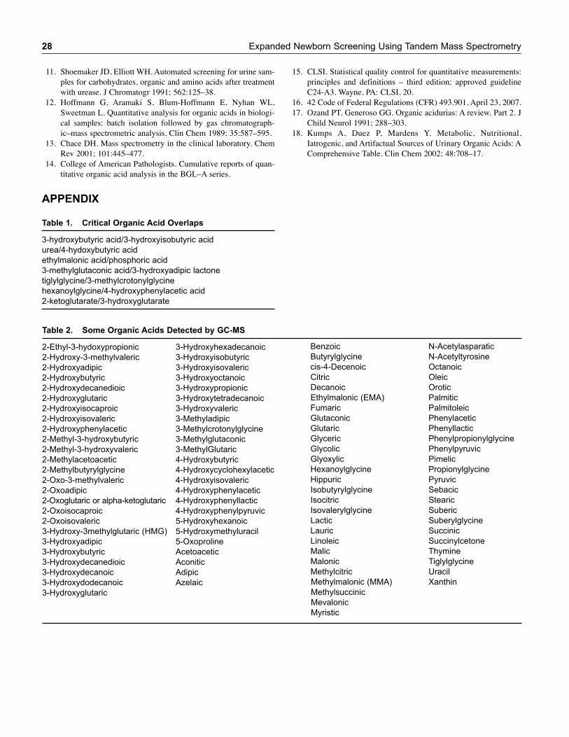
APPENDIX
Table 1. Critical Organic Acid Overlaps
3-hydroxybutyric acid/3-hydroxyisobutyric acid
urea/4-hydoxybutyric acid
ethylmalonic acid/phosphoric acid
3-methylglutaconic acid/3-hydroxyadipic lactone
tiglylglycine/3-methylcrotonylglycine
hexanoylglycine/4-hydroxyphenylacetic acid
2-ketoglutarate/3-hydroxyglutarate
Table 2. Some Organic Acids Detected by GC-MS
2-Ethyl-3-hydoxypropionic 3-Hydroxyhexadecanoic
2-Hydroxy-3-methylvaleric 3-Hydroxyisobutyric
2-Hydroxyadipic 3-Hydroxyisovaleric
2-Hydroxybutyric 3-Hydroxyoctanoic
2-Hydroxydecanedioic 3-Hydroxypropionic
2-Hydroxyglutaric 3-Hydroxytetradecanoic
2-Hydroxyisocaproic 3-Hydroxyvaleric
2-Hydroxyisovaleric 3-Methyladipic
2-Hydroxyphenylacetic 3-Methylcrotonylglycine
2-Methyl-3-hydroxybutyric 3-Methylglutaconic
2-Methyl-3-hydroxyvaleric 3-MethylGlutaric
2-Methylacetoacetic 4-Hydroxybutyric
2-Methylbutyrylglycine 4-Hydroxycyclohexylacetic
2-Oxo-3-methylvaleric 4-Hydroxyisovaleric
2-Oxoadipic 4-Hydroxyphenylacetic
2-Oxoglutaric or alpha-ketoglutaric 4-Hydroxyphenyllactic
2-Oxoisocaproic 4-Hydroxyphenylpyruvic
2-Oxoisovaleric 5-Hydroxyhexanoic
3-Hydroxy-3methylglutaric (HMG) 5-Hydroxymethyluracil
3-Hydroxyadipic 5-Oxoproline
3-Hydroxybutyric Acetoacetic
3-Hydroxydecanedioic Aconitic
3-Hydroxydecanoic Adipic
3-Hydroxydodecanoic Azelaic
3-Hydroxyglutaric
Benzoic N-Acetylasparatic
Butyrylglycine N-Acetyltyrosine
cis-4-Decenoic Octanoic
Citric Oleic
Decanoic Orotic
Ethylmalonic (EMA) Palmitic
Fumaric Palmitoleic
Glutaconic Phenylacetic
Glutaric Phenyllactic
Glyceric Phenylpropionylglycine
Glycolic Phenylpyruvic
Glyoxylic Pimelic
Hexanoylglycine Propionylglycine
Hippuric Pyruvic
Isobutyrylglycine Sebacic
Isocitric Stearic
Isovalerylglycine Suberic
Lactic Suberylglycine
Lauric Succinic
Linoleic Succinylcetone
Malic Thymine
Malonic Tiglylglycine
Methylcitric Uracil
Methylmalonic (MMA) Xanthin
Methylsuccinic
Mevalonic
Myristic
28 Expanded Newborn Screening Using Tandem Mass Spectrometry
11. Shoemaker JD, Elliott WH. Automated screening for urine sam-ples for carbohydrates, organic and amino acids after treatmentwith urease. J Chromatogr 1991; 562:125–38.
12. Hoffmann G, Aramaki S, Blum-Hoffmann E, Nyhan WL,Sweetman L. Quantitative analysis for organic acids in biologi-cal samples: batch isolation followed by gas chromatograph-ic–mass spectrometric analysis. Clin Chem 1989; 35:587–595.
13. Chace DH. Mass spectrometry in the clinical laboratory. ChemRev 2001; 101:445–477.
14. College of American Pathologists. Cumulative reports of quan-titative organic acid analysis in the BGL–A series.
15. CLSI. Statistical quality control for quantitative measurements:principles and definitions – third edition; approved guidelineC24-A3. Wayne, PA: CLSI, 20.
16. 42 Code of Federal Regulations (CFR) 493.901, April 23, 2007.17. Ozand PT, Generoso GG. Organic acidurias: A review. Part 2. J
Child Neurol 1991; 288–303.18. Kumps A, Duez P, Mardens Y. Metabolic, Nutritional,
Iatrogenic, and Artifactual Sources of Urinary Organic Acids: AComprehensive Table. Clin Chem 2002; 48:708–17.
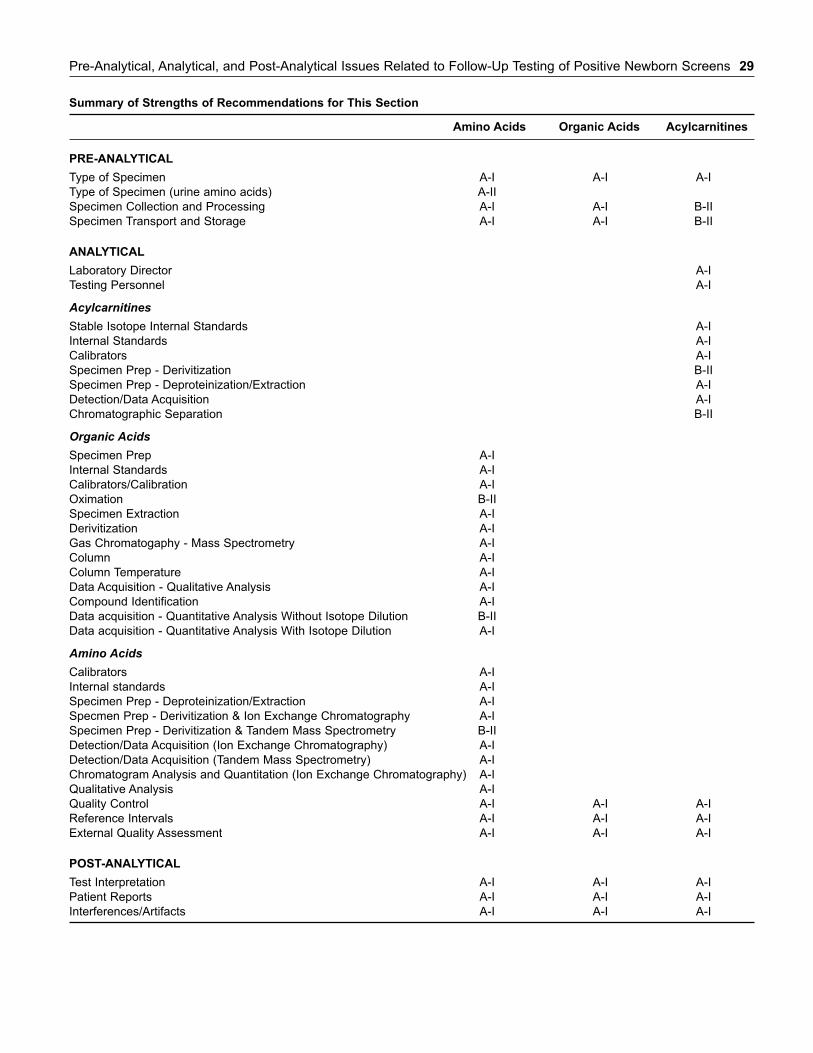
Pre-Analytical, Analytical, and Post-Analytical Issues Related to Follow-Up Testing of Positive Newborn Screens 29
Summary of Strengths of Recommendations for This Section
Amino Acids Organic Acids Acylcarnitines
PRE-ANALYTICAL
Type of Specimen A-I A-I A-I
Type of Specimen (urine amino acids) A-II
Specimen Collection and Processing A-I A-I B-II
Specimen Transport and Storage A-I A-I B-II
ANALYTICAL
Laboratory Director A-I
Testing Personnel A-I
AcylcarnitinesStable Isotope Internal Standards A-I
Internal Standards A-I
Calibrators A-I
Specimen Prep - Derivitization B-II
Specimen Prep - Deproteinization/Extraction A-I
Detection/Data Acquisition A-I
Chromatographic Separation B-II
Organic AcidsSpecimen Prep A-I
Internal Standards A-I
Calibrators/Calibration A-I
Oximation B-II
Specimen Extraction A-I
Derivitization A-I
Gas Chromatogaphy - Mass Spectrometry A-I
Column A-I
Column Temperature A-I
Data Acquisition - Qualitative Analysis A-I
Compound Identification A-I
Data acquisition - Quantitative Analysis Without Isotope Dilution B-II
Data acquisition - Quantitative Analysis With Isotope Dilution A-I
Amino AcidsCalibrators A-I
Internal standards A-I
Specimen Prep - Deproteinization/Extraction A-I
Specmen Prep - Derivitization & Ion Exchange Chromatography A-I
Specimen Prep - Derivitization & Tandem Mass Spectrometry B-II
Detection/Data Acquisition (Ion Exchange Chromatography) A-I
Detection/Data Acquisition (Tandem Mass Spectrometry) A-I
Chromatogram Analysis and Quantitation (Ion Exchange Chromatography) A-I
Qualitative Analysis A-I
Quality Control A-I A-I A-I
Reference Intervals A-I A-I A-I
External Quality Assessment A-I A-I A-I
POST-ANALYTICAL
Test Interpretation A-I A-I A-I
Patient Reports A-I A-I A-I
Interferences/Artifacts A-I A-I A-I

With the rapid expansion of newborn screening to includemore newborns and more pathologic conditions comes anadditional pool of individuals presenting to primary careproviders, community hospitals, or tertiary care genetic centersfor follow-up evaluation. With the exception of conditions thathave historically been included in screening programs, thispopulation presents with potential disorders that have notpreviously been encountered with great frequency in asympto-matic patients. For example, in the symptomatic phase, ureacycle disorders would present in the context of severe hyper-ammonemic encephalopathy, methylmalonic aciduria withacute acidosis and encephalopathy, and medium-chain acylCoA dehydrogenase (MCAD) deficiency with hypoketotichypoglycemia and liver failure. Metabolic findings in acutecircumstances are usually pronounced and enable unequivocaldiagnosis. In today’s newborn screening environment, there isa high likelihood that neonates will present in an asymptomaticclinical phase of the illness requiring that laboratory tests andthose that interpret them detect ever more subtle metabolicabnormalities.
In this context, the biochemical genetics laboratory playsa pivotal role in recommending and interpreting initial studiesas well as dictating the necessity and type of further testing.The focus of this Chapter, therefore, is not on the diagnosticaccuracy of newborn screening per se, but on the sensitivityand specificity of the sophisticated testing applied to infantsreferred for follow-up of a positive newborn screening result.Primary literature and expert opinion has guided the testingrecommendations to follow. The rarity of these disorders gen-erally precludes large, randomized studies of diagnostic accu-racy. An exhaustive review of the literature, primarily casereports and series, were reviewed for evidence of biochemicalabnormalities in affected, generally asymptomatic children.
GENERAL REQUIREMENTS OF THE
FOLLOW-UP PROCESS
Abnormal screening results should be followed up promptly toavoid morbidity and mortality. The protocol for initial testing ofblood spots varies among screening laboratories. A commonlyemployed protocol for newborn screening by MS/MS is illus-trated in Figure 3-1. A sample is tested using MS/MS with aspecific cut-off. If the sample value is below the cut-off, nofurther testing is done and the result is reported as negative.
When the value of a particular marker or set of markersexceeds the requisite cut-off, a second sample from theoriginal blood spot is tested. If the second value is below thecut-off, the result is reported as negative and no further testingis done. If the value is above the cut-off, the case is categorizedas low risk or high risk depending on the concentration of themarker. In the low risk category, another sample from theinfant is collected and tested without the involvement of agenetic specialist due to the reasonable likelihood that theresult is false positive.
If the result falls into a high-risk category, the primary carephysician and a specialist are contacted and immediate follow-up commences. The follow-up includes clinical evaluation andconfirmatory testing, the subject of this Chapter. Primary carephysicians may be the first persons to be contacted and shouldbe familiar with the initial management and follow-up. Theyshould also be prepared to explain the positive results to thefamily and make arrangement for the referral.
Several desirable attributes characterize the role of thelaboratory in the referral of the newborn. First, laboratory per-sonnel should be among the first responders notified about thereferral. Biochemical genetics laboratories typically operate inbatch mode during normal working hours. Procedures oftenrequire many hours or even days to complete. Early notifica-tion will facilitate prompt laboratory analysis. Second, testingshould be properly pointed to the suspected disorder(s).Children will be referred on the basis of an elevated marker.Only those tests that provide relevant, timely contribution tothe narrowing the differential diagnosis should be undertaken.Recommendations for these pointed investigations are presentedin Table 3-1, which is organized by disorder. The discussionthat follows considers the scope of other disorders that shouldbe dictated by each of the markers in the screen. Results oflaboratory analyses should be interpreted promptly by an expe-rienced laboratory director in consultation with the clinicalteam responsible for clinical assessment of the newborn.Finally, the follow-up circle should be closed by providingresults to newborn screening centers to enhance understandingof the diagnostic performance of screening tests.
GENERAL RECOMMENDATIONS
For the reasons outlined above, we recommend adoption of thefollowing practices with the highest level of evidence: A-I.
Chapter 3
Follow-Up of Positive Screening Results
Dennis J. Dietzen, Uttam C. Garg, Stanley F. Lo, and William J. Rhead
31
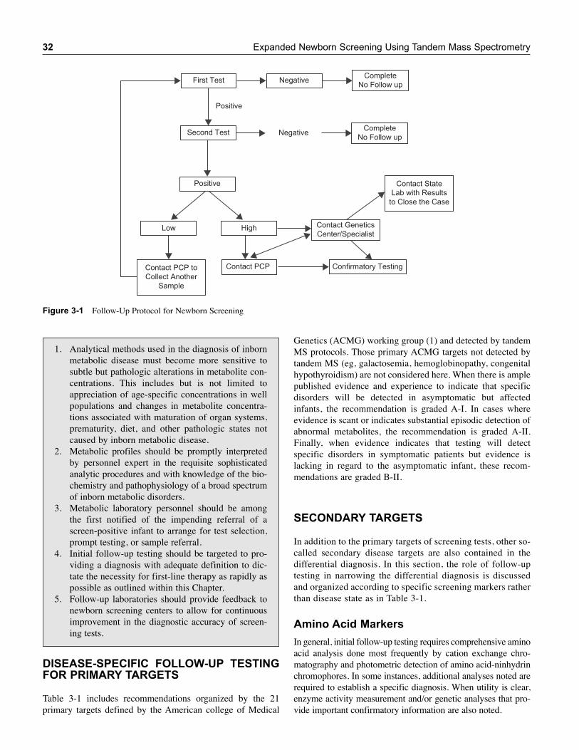
32 Expanded Newborn Screening Using Tandem Mass Spectrometry
Genetics (ACMG) working group (1) and detected by tandemMS protocols. Those primary ACMG targets not detected bytandem MS (eg, galactosemia, hemoglobinopathy, congenitalhypothyroidism) are not considered here. When there is amplepublished evidence and experience to indicate that specificdisorders will be detected in asymptomatic but affectedinfants, the recommendation is graded A-I. In cases whereevidence is scant or indicates substantial episodic detection ofabnormal metabolites, the recommendation is graded A-II.Finally, when evidence indicates that testing will detectspecific disorders in symptomatic patients but evidence islacking in regard to the asymptomatic infant, these recom-mendations are graded B-II.
SECONDARY TARGETS
In addition to the primary targets of screening tests, other so-called secondary disease targets are also contained in thedifferential diagnosis. In this section, the role of follow-uptesting in narrowing the differential diagnosis is discussedand organized according to specific screening markers ratherthan disease state as in Table 3-1.
Amino Acid Markers
In general, initial follow-up testing requires comprehensive aminoacid analysis done most frequently by cation exchange chro-matography and photometric detection of amino acid-ninhydrinchromophores. In some instances, additional analyses noted arerequired to establish a specific diagnosis. When utility is clear,enzyme activity measurement and/or genetic analyses that pro-vide important confirmatory information are also noted.
Negative
Negative
Positive
Positive
Contact PCP Confirmatory Testing
CompleteNo Follow up
Contact GeneticsCenter/Specialist
First Test
CompleteNo Follow upSecond Test
Low High
Contact PCP toCollect Another
Sample
Contact StateLab with Results
to Close the Case
Figure 3-1 Follow-Up Protocol for Newborn Screening
1. Analytical methods used in the diagnosis of inbornmetabolic disease must become more sensitive tosubtle but pathologic alterations in metabolite con-centrations. This includes but is not limited toappreciation of age-specific concentrations in wellpopulations and changes in metabolite concentra-tions associated with maturation of organ systems,prematurity, diet, and other pathologic states notcaused by inborn metabolic disease.
2. Metabolic profiles should be promptly interpretedby personnel expert in the requisite sophisticatedanalytic procedures and with knowledge of the bio-chemistry and pathophysiology of a broad spectrumof inborn metabolic disorders.
3. Metabolic laboratory personnel should be amongthe first notified of the impending referral of ascreen-positive infant to arrange for test selection,prompt testing, or sample referral.
4. Initial follow-up testing should be targeted to pro-viding a diagnosis with adequate definition to dic-tate the necessity for first-line therapy as rapidly aspossible as outlined within this Chapter.
5. Follow-up laboratories should provide feedback tonewborn screening centers to allow for continuousimprovement in the diagnostic accuracy of screen-ing tests.
DISEASE-SPECIFIC FOLLOW-UP TESTING
FOR PRIMARY TARGETS
Table 3-1 includes recommendations organized by the 21primary targets defined by the American college of Medical
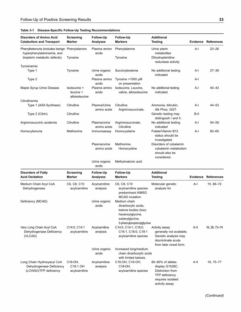
Follow-Up of Positive Screening Results 33
Table 3-1 Disease-Specific Follow-Up Testing Recommendations
Disorders of Amino Acid Screening Follow-Up Follow-Up Additional
Catabolism and Transport Marker Analyses Markers Testing Evidence References
Phenylketonuria (includes benign Phenylalanine Plasma amino Phenylalanine Urine pterin A-I 23–26
hyperphenylalaninemia, and acids metabolites
biopterin metabolic defects) Tyrosine Tyrosine Dihydropteridine
reductase activity
Tyrosinemia
Type 1 Tyrosine Urine organic Succinylacetone No additional testing A-I 27–39
acids indicated
Type 2 Plasma amino Tyrosine >1000 µM A-I
acids on presentation
Maple Syrup Urine Disease Isoleucine + Plasma amino Isoleucine, Leucine, No additional testing A-I 40–43
leucine + acids valine, alloisoleucine indicated
alloisoleucine
Citrullinemia
Type 1 (ASA Synthase) Citrulline Plasma/Urine Citrulline Ammonia, bilirubin, A-I 44–53
amino acids Argininosuccinate, Alk Phos, GGT.
Type 2 (Citrin) Citrulline Genetic testing may B-II
distinguish I and II.
Argininosuccinic acidemia Citrulline Plasma/urine Argininosuccinate, No additional testing A-I 54–59
amino acids Citrulline indicated
Homocytsinuria Methionine Immunoassay Homocysteine Folate/Vitamin B12 A-I 60–65
status should be
investigated.
Plasma/urine Methionine, Disorders of cobalamin
amino acids Homocystine cobalamin metabolism
should also be
considered.
Urine organic Methylmalonic acid
acids
Disorders of Fatty Screening Follow-Up Follow-Up Additional
Acid Oxidation Marker Analyses Markers Testing Evidence References
Medium Chain Acyl CoA C6, C8, C10 Acylcarnitine C6, C8, C10 Molecular genetic A-I 15, 66–72
Dehydrogenase acylcarnitine analysis acylcarnitine species analysis for
predominant A985G
MCAD mutation
Deficiency (MCAD) Urine organic Medium chain
acids dicarboxylic acids,
ketone bodies (low)
hexanoylglycine,
suberylglycine,
3-phenylpropionylglycine
Very Long Chain Acyl CoA C14:0, C14:1 Acylcarnitine C14:0, C14:1, C16:0, Activity assay A-II 16, 26, 73–74
Dehydrogenase Deficiency acylcarnitine analysis C16:1, C18:0, C18:1 generally not available.
(VLCAD) acylcarnitine species Genetic analysis may
discriminate acute
from later onset form.
Urine organic Increased long/medium
acids chain dicarboxylic acids
with limited ketosis.
Long Chain Hydroxyacyl CoA C16-OH, Acylcarnitine C16-OH, C18-OH, 80–90% of alleles A-II 18, 75–77
Dehydrogenase Deficiency C18:1 OH analysis C18-OH, display G1528C.
(LCHAD)/TFP deficiency acylcarnitine acylcarnitine species Distinction from
TFP deficiency
requires isolated
activity assay
(Continued)
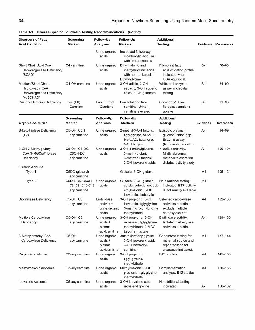
34 Expanded Newborn Screening Using Tandem Mass Spectrometry
Table 3-1 Disease-Specific Follow-Up Testing Recommendations (Cont’d)
Disorders of Fatty Screening Follow-Up Follow-Up Additional
Acid Oxidation Marker Analyses Markers Testing Evidence References
Urine organic Increased 3-hydroxy-
acids dicarboxylic aciduria
with limited ketosis
Short Chain Acyl CoA C4 carnitine Urine organic Ethylmalonic and Fibroblast fatty B-II 78–83
Dehydrogenase Deficiency acids methylsuccinic acids acid oxidation profile
(SCAD) with normal ketosis. indicated when
Butyrylglycine UOA equivocal.
Medium/Short Chain C4-OH carnitine Urine organic 3-OH adipic, 3-OH White cell enzyme B-II 84–90
Hydroxyacyl CoA acids sebacic, 3-OH suberic assay, molecular
Dehydrogenase Deficiency acids. 3-OH glutarate testing
(M/SCHAD)
Primary Carnitine Deficiency Free (C0) Free + Total Low total and free Secondary? Low B-II 91–93
Carnitine Carnitine carnitine. Urine fibroblast carnitine
carnitine elevated uptake
Screening Follow-Up Follow-Up Additional
Organic Acidurias Marker Analyses Markers Testing Evidence References
B-ketothiolase Deficiency C5-OH, C5:1 Urine organic 2-methyl-3-OH butyric, Episodic plasma A-II 94–99
(T2) acylcarnitine acids tiglylglycine, AcAc, 2 glucose, anion gap.
MeAcAC, butanone, Enzyme assay
3-OH butyric (fibroblast) to confirm.
3-OH-3-Methylglutaryl C5-OH, C6-DC, Urine organic 3-OH-3-methylglutaric, <100% sensitivity. A-II 100–104
CoA (HMGCoA) Lyase C6OH-DC acids 3-methylglutaric, Mildly abnormal
Deficiency acylcarnitine 3-methylglutaconic, metabolite excretion
3-OH isovaleric acids dictates activity study
Glutaric Aciduria
Type 1 C5DC (glutaryl) Glutaric, 3-OH glutaric A-I 105–121
acylcarnitine
Type 2 C5DC, C5, C5OH, Urine organic Glutaric, 2-OH glutaric, No additional testing A-I
C6, C8, C10-C16 acids adipic, suberic, sebacic indicated. ETF activity
acylcarnitine ethylmalonic, 3-OH is not readily available.
isovaleric, isobutyric
Biotinidase Deficiency C5-OH, C3 Biotinidase 3-OH propionic, 3-OH Selected carboxylase A-I 122–130
acylcarnitine activity + isovaleric, tiglylglycine, activities + biotin to
urine organic 3-methycrotonylglycine exclude multiple
acids methylcitrate carboxylase def.
Multiple Carboxylase C5-OH, C3 Urine organic 3-OH propionic, 3-OH Biotinidase activity. A-II 129–136
Deficiency acylcarnitine acids + isovaleric, tiglylglycine Isolated carboxylase
plasma methylcitrate, 3-MCC activities + biotin.
acylcarnitine (glycine), lactate
3-Methylcrotonyl CoA C5-OH Urine organic 3methylcrotonylglycine Concurrent testing for A-I 137–144
Carboxylase Deficiency acylcarnitine acids + 3-OH isovaleric acid. maternal source and
plasma 3-OH isovaleryl- repeat testing for
acylcarnitine carnitine. clearance indicated.
Propionic acidemia C3-acylcarnitine Urine organic 3-OH propionic, B12 studies. A-I 145–150
acids tiglyl-glycine,
methylcitrate
Methylmalonic acidemia C3-acylcarnitine Urine organic Methylmalonic, 3-OH Complementation A-I 150–155
acids propionic, tiglylglycine, analysis. B12 studies
methylcitrate
Isovaleric Acidemia C5-acylcarnitine Urine organic 3-OH isovaleric acid, No additional testing
acids isovaleryl glycine indicated A-II 156–162
Follow-Up of Positive Screening Results 35
Phenylalanine. Elevated phenylalanine is primarilycaused by defects in phenylalanine hydroxylase (classicphenylketonuria) but may be benign or due to defects in thesynthesis or recycling of tetrahydrobiopterin, a cofactor neces-sary for conversion of phenylalanine to tyrosine and thesynthesis of various neurotransmitters. Assessment of plasmaamino acids showing elevated phenylalanine and normal orreduced tyrosine confirms the diagnosis of hyperphenylala-ninemia and dictates dietary phenylalanine restriction. Furtherinvestigations (eg, urine pterin metabolites) not typically avail-able at referral centers are required to assess the integrity oftetrahydrobiopterin metabolism and the utility of tetrahydro-biopterin (BH4) supplementation.
Tyrosine. Elevated tyrosine indicates disrupted tyrosinecatabolism at the level of fumarylacetoacetate hydrolase (type I), tyrosine aminotransferase (type II), or the very raredeficiency of hydroxyphenylpyruvate dioxygenase (type III).Tyrosine elevations in Type II disease are pronounced whileelevations in Type I and Type III disease may be marginal.Screening results are confounded by transient elevations intyrosine due to slow maturation of the liver catabolic pathway,particularly in premature infants (2–4). Both plasma amino acidsand urine organic acids are indicated for follow-up. Urine spec-imens from affected neonates will uniformly contain the tyrosinemetabolites 4-hydroxyphenyllactate and 4-hydrox-yphenylpyruvate. The additional finding of succinylacetone inurine is pathognomonic for Type I disease. Type II disease ischaracterized by massive elevations of plasma tyrosine(>1,000 μM). Biochemical evidence for the very rare Type IIIdisease includes demonstration of 4-hydroxycyclo-hexylaceticacid in urine and the unusual amino acid, hawkinsin, in serumand/or urine (5).
Leucine. Screening techniques utilizing tandem MS do notdistinguish leucine from isoleucine, alloisoleucine, or hydrox-yproline. Leucine may also be modestly elevated in fastingketosis. All these possibilities must be considered in follow-upanalysis. Plasma amino acid analysis that distinguishes each ofthe branched-chain amino acids (leucine, isoleucine,alloisoleucine, and valine) from each other and from hydroxypro-line is essential. Modest elevations of branched chain aminoacids and ketosis are consistent with a catabolic state. The detec-tion of alloisoleucine along with significant elevations of thebranched-chain amino acids is diagnostic of MSUD.Demonstration of elevated branched-chain keto-acids (α-ketoisovaleric, α-ketoisocaproic, and α-ketomethylvaleric acids)is not necessary to establish the diagnosis. Hydroxyprolinemia isprobably a benign disorder (6). Confirmation of MSUD byamino acid analysis dictates prompt dietary therapy.
Citrulline. Citrulline is the product of the ornithine tran-scarbamoylase reaction and a substrate for argininosuccinate(ASA) synthase. Elevation of citrulline indicates deficiency ofASA synthase (citrullinemia) or a deficiency of ASA lyase.Comprehensive amino acid analysis is necessary to distinguishthese disorders. Detection of ASA by standard LC/ninhydrinamino acid analyzers is not straightforward. It appears as threespecies with distinct retention times: two cyclic anhydrides andthe uncyclized tricarboxylic acid (7, 8). Analysis of both serum
and urine is indicated. In early stages of these disorders, eleva-tions can be appreciated in urine before plasma elevations arereadily apparent. Citrulline is also elevated due to deficiency ofcitrin, the mitochondrial glutamate-aspartate antiporter. In addi-tion to hyperammonemia, this disorder is characterized byhyperbilirubinemia associated with cholestasis. Citrulline,along with lysine, alanine, and proline, may also be elevated inpyruvate carboxylase deficiency (9). Profound lactic acidemiaand hypoglycemia should prompt consideration of this disorder.
Methionine. Methionine is primarily used as a marker todetect deficiency of cystathionine-β-synthetase (CBS). Methionineis one step removed from the primary substrate for CBS, homo-cysteine. Follow-up testing should therefore include plasmaamino acid analysis and plasma homocysteine. The latter can berapidly done with widely available immunoassays developed toassess risk for cardiovascular disease. Elevated homocysteine isdetected in plasma amino acid analysis as its oxidized homod-imer, homocystine. Immunoassays for detection of homocys-teine are available on most automated chemistry platforms.Methionine measurement by LC/ninhydrin based techniquescan be confounded by co-elution of homocitrulline (found, forexample, in HHH syndrome) (10). This cause of elevatedmethionine should be excluded in all cases of elevated methion-ine. In less frequent situations, plasma methionine may be elevat-ed without elevation of homocystine or homocysteine. Causes ofthis profile include deficiencies of methionine adenosyltransferase(11), guanidinoacetate methyltransferase (12), and adenosylho-mocysteine hydrolase (13). Mild elevation of methionine mayalso occur due to hyperalimenation or liver disease.
Acylcarnitine Markers
Urine organic acid analysis plays a major role in the initialfollow-up testing of most suspected inborn errors marked byspecific acylcarnitines. In many instances, however, diagnosticurinary organic acid patterns are only evident during episodesof disease exacerbation. Recognition of subtle abnormalities isa particular challenge for testing the asymptomatic infant withabnormal newborn screening results. Plasma acylcarnitineanalyses are less subject to episodic fluctuation and are, there-fore, also recommended during the initial follow-up of all pos-itive screens, which indicate elevations of specific carnitineesters. Again, when utility is clear, complementation studies,enzyme activity analyses, and genetic studies are noted.
Medium chain, saturated acylcarnitine species (C8, C6,C10). Of these three species, C8 is elevated to the greatestextent. The differential diagnosis for this screening result islimited to MCAD deficiency. Follow-up analyses shouldinclude a confirmatory plasma acylcarnitine profile and urineorganic acid analysis. Abnormalities in the acylcarnitine profilea r etypically constant whereas abnormalities of urine organic acidsmay be episodic and evident only during acute illness. Affectedneonates excrete abnormal quantities of medium chain dicar-boxylic acids (adipic, suberic, sebacic) with inappropriatelylow ketone bodies. Several abnormal glycine conjugates are also reliably detected in most organic acid protocols.
36 Expanded Newborn Screening Using Tandem Mass Spectrometry
These include hexanoylglycine, suberylglycine, and 3-phenyl-propionylglycine. The latter compound, derived from gut bac-terial metabolism, is typically not found in urine from neonateswith limited bacterial flora. Interpretation of urine organic acidprofiles can be complicated by fasting and by dietary consump-tion of medium chain triglyceride. The concentration of ketonesin urine is an important consideration in follow-up urine organicacid analysis. MCAD deficiency inhibits most but not all keto-sis so the urine from affected individuals is relatively hypoke-totic in the context of dicarboxylic aciduria. The absence ofurine ketones does not exclude the presence of ketones inblood, as significant ketonemia (3 to 5 mM) may exist before evidence of ketosis appears in the urine (14). MCADdeficiency is reliably confirmed by molecular genetic testingfor the A985G mutation. This mutation accounts for 63% ofmutated alleles in cases detected by newborn screening (15).
C14:1-carnitine. Elevations of this marker suggest defi-ciency of very long chain acyl CoA dehydrogenase deficiency(VLCAD). Like potential MCAD deficiency, appropriatefollow-up includes plasma acylcarnitine analysis and urineorganic acid analysis. The acylcarnitine profile contains eleva-tions of saturated and monounsaturated C14, C16, and C18species. Urine organic acids in affected individuals containboth long and medium chain dicarboxylic acids with inappro-priately low ketone bodies present. Unlike MCAD deficiency,biochemical findings may not adequately confirm diagnosis.In these circumstances, fibroblast fatty acid oxidation studiesor molecular genetic analysis may be required. The former arenot readily available and are time consuming. Genetic analysisfor VLCAD deficiency is often more accessible and helpful indiscriminating severe, neonatal onset disease caused by trun-cating mutations that result in little functional protein fromlater onset, milder disease caused by single amino acidchanges, insertions, or deletions (16). Western blots forVLCAD protein may also provide some indication of severity.
C16-OH. Long chain hydroxyacyl CoA dehydrogenase(LCHAD) is one of three activities associated with themitochdondrial trifunctional protein (TFP). TFP consists oflong chain enoyl CoA hydratase and long chain ketoacylthiolase activities in addition to LCHAD. First-line follow-uptesting should include plasma acylcarnitine analysis and urineorganic acid analysis. For confirmation, the former containselevated levels of long chain monocarboxylic (chain length>10) hydroxyacylcarnitine species and the latter is notable formedium and long chain (C6 to C14) hydroxydicarboxylicaciduria and C6 to C10 dicarboxylic aciduria. The G1528Cmutation of the α subunit of TFP is found in 80% to 90% of allmutant alleles (17). Isolated LCHAD deficiency may be distin-guished from generalized TFP deficiency by activity measure-ment but does not alter treatment. The presence of fetalLCHAD carries a high risk of obstetrical complications suchas acute fatty liver of pregnancy and the Hemolysis, ElevatedLiver enzymes and Low Platelets (HELLP) syndrome (18).
C4 carnitine. Confirmatory testing for elevated C4 carni-tine requires distinction among three disorders: isobutyrylCoA dehydrogenase (IBDH) deficiency, short chain acyl CoAdehydrogenase (SCAD) deficiency, and ethylmalonic acid
(EMA) encephalopathy. IBDH deficiency is caused by a defectin the terminal steps of valine catabolism, SCAD deficiency bya defect in terminal oxidation of fatty acids and EMAencephalopathy by a defect in the ETHE1 gene, the function ofwhich is poorly defined (19). Urine organic acid analysis is themost effective means to differentiate these disorders. IBDHdeficiency is distinguished from the others by elevated excre-tion of isobutyric acid and the absence of ethylmalonic acid.Increased ethylmalonic acid is observed in both SCADdeficiency and EMA encephalopathy. In the latter condition,however, increased EMA excretion is associated withincreased excretion of isovalerylglycine. In classic cases of theformer, increased EMA excretion is often observed along withelevated excretion of methylsuccinic acid. Genetic analyses ofeach of the causative genes are available but the occurrence ofprevalent mutations and genotype/phenotype correlations arenot well known. Fibroblast fatty acid oxidation studies mayclarify the role of SCAD in a potential defect. Direct enzymaticactivity measurements of IBDH are complicated by overlap-ping substrate specificities of other short-branched chain CoAdehydrogenases. The function of the ETHE1 product is notknown so enzymatic activity measurement is not yet possible.
C4-OH carnitine. The differential diagnosis of elevatedC4-OH carnitine is limited to the poorly characterizedmedium/short-chain acyl CoA dehydrogenase (M/SCHAD)deficiency. Urine organic acid analysis revealing elevatedexcretion of both hydroxylated and non-hydroxylated C6-C10dicarboxylic acids strongly suggests this disorder. Activitymeasurement in tissue or fibroblast cultures is of limited avail-ability but may augment the diagnosis.
Low C0 (free) carnitine. Primary carnitine deficiency resultsfrom the inability to reabsorb carnitine from the glomerular fil-trate. Diagnosis is confirmed by the presence of low free andtotal carnitine in blood and inappropriately high concentrationsin urine. Interpretation of results in the newborn period may becomplicated by maternal status. High maternal carnitine intakemay mask deficiency in the newborn and maternal deficiencymay, in turn, cause secondary deficiency in the newborn.Interpretation requires adequate dietary history. Transporteractivity measurement and genetic (OCTN2) (20) analysis are notreadily available but are essential for diagnostic confirmation anddifferentiation from secondary causes of tubular carnitine loss.
C5 carnitine. The differential diagnosis for elevated C5-carnitine includes deficiencies of isovaleryl CoA dehydroge-nase, 2-methylbutyryl CoA dehydrogenase (short/branchedchain CoA dehydrogenase) or administration of antibioticscontaining pivalic (2,2-dimethylpropionic) acid (21).Discrimination between these possibilities is accomplished byurine organic acid analysis. Isovaleric acidemia is character-ized by increased excretion of isovalerylglycine and 3-OHisovaleric acid. 2-methylbutyryl CoA dehydrogenase defi-ciency is characterized by elevated urine concentrations of 2-methylbutyrylglycine in the absence of isovalerylglycine.The absence of both of these acylglycines suggests a falsepositive screening result.
C5DC carnitine. Isolated elevation of this species suggeststhe deficiency of the conversion of glutaryl CoA to crotonyl
Follow-Up of Positive Screening Results 37
CoA by glutaryl CoA dehydrogenase (glutaric aciduria type I).In follow-up testing, the presence of elevated concentrations ofglutaric acid, 3-OH glutaric acid and in some patients, gluta-conic acid is diagnostic for this disorder. The absence of thesecompounds generally excludes this diagnosis. However, in thepresence of other clinical abnormalities associated with glutaricaciduria type I (eg, macrocephaly) measurement of fibroblastglutaryl CoA dehydrogenase activity or analysis of the corre-sponding gene may be indicated to definitively exclude thisdiagnosis. C5DC is also elevated in glutaric aciduria type II(also known as multiple acyl CoA dehydrogenation deficiencyMADD). Type II is distinguished from Type I glutaric aciduriaby elevation of multiple additional acylcarnitine species andurine organic acids. Elevated acylcarnitine species include C5,C5OH, C6, C8, and C10 to C16 carnitine esters. Urine organic acids in glutaric aciduria type II include 2-OH ratherthan 3-OH glutaric acid, medium chain dicarboxylic acids, andethylmalonic, 3-OH isovaleric, and isobutyric acids.
C5-OH carnitine. Elevated blood concentrations of thisspecies dictate a broad differential diagnosis that includes defi-ciencies of 3-methylcrotonylcarboxylase (MCC), 3-hydroxy-3-methylglutaryl (HMG)-CoA lyase, β-ketothiolase (T2),2-methyl-3-hydroxybutyryl CoA dehydrogenase (MHBD), 3-methylglutaconyl CoA hydratase (MGA), biotinidase, orholocarboxylase synthetase. Initial discrimination between thesedisorders is accomplished by analysis of urine organic acids. 3-MCC deficiency is characterized by excretion of 3-methylcrotonylglycine and variable amounts of 3-methyl-crotonic acid, and 3-OH isovaleric acid. A large number offalse positive screening results have been attributed totransplacental transfer of 3-methylcrotonyl-carnitine fromaffected mothers. It is necessary to distinguish 3-MCC defi-ciency from errors affecting propionyl CoA, acetyl CoA, andpyruvate carboxylase. In so-called multiple carboxylase defi-ciency, urine specimens contain elevated concentrations of lac-tate, 3-OH propionic, methylcitrate, tiglylglycine, and 3-OHisovaleric acid in addition to 3-methylcrotonylglycine. Directassay of biotinidase, holocarboxylase synthetase, or isolatedcarboxylase activities is required to clarify the etiology of thedisorder. HMG CoA lyase deficiency and thiolase deficiencyare disorders of ketone body metabolism. In the former condi-tion, urine specimens contain elevated amounts of HMG, itsimmediate precursor, 3-methylglutaconic acid, and 3-OH iso-valeric acid. The latter condition affects both ketone body uti-lization and isoleucine catabolism. Urine contains largequantities of ketone bodies in the context of normoglycemia,2-methyl-3-hydroxybutyric acid, and tiglylglycine. 2-methy-lacetoacetate and butanone may be variably detected becauseof their volatility. Finally, 3-methylglutaconic aciduria is char-acterized by excretion of large amounts of 3-methylglutaconicand 3-OH isovaleric acids.
C3 carnitine. Isolated elevation of C3 carnitine suggests adisorder in the anaplerotic pathway of propionic acid to its disposition as succinate in the citric acid cycle. Potentialdisturbances in this pathway include propionyl CoAcarboxylase, methylmalonyl CoA mutase, or multiple errors in
cobalamin metabolism. Again, urine organic acids play a majorrole in the initial distinction. Propionic acid is not routinelydetected in urine organic acid analysis but propionylglycine anda less volatile metabolite, 3-OH propionic acid are detected. Athigh concentrations, 3-OH propionyl CoA acid may outcompeteacetyl CoA in the citrate synthase reaction and form methylci-trate. Further upstream, tiglylglycine may also be detected.Methylmalonyl CoA mutase deficiency is notable for the hyper-excretion of methylmalonic acid in addition to 3-OH propionate,methylcitrate, and tiglylglycine. Methylmalonic aciduria may becaused by mutations in methylmalonyl CoA mutase (mut0 ormut−) but also by various disturbances in cobalamin metabolism.Plasma homocysteine measurement is useful in defining the roleof cobalamin metabolism. Methylmalonic aciduria without ele-vation of homocysteine may be caused by defective synthesis ofadenosylcobalamin, the cofactor for methylmalonyl CoA mutase(cbl A and B subtype). These subtypes are often responsive tovitamin B12 administration. Methylmalonic aciduria, with ele-vated plasma homocysteine, suggests a defect in the formation ofadenosylcobalamin and methylcobalamin, the latter being a req-uisite cofactor for the conversion of homocysteine to methionine.These disorders belong to the cbl C, D, or F subtypes. Fibroblastcomplementation analysis is required for definitive classificationof the metabolic defect (22).
REFERENCES
1. Newborn screening: toward a uniform screening panel and system.Genet. Med. 8 Suppl 1:1S–252S.: 1S–252S, 2006
2. Levy HL, Shih VE, Madigan PM, MacCready RA: Transienttyrosinemia in full-term infants. JAMA. 209:249–250, 1969
3. Avery ME, Clow CL, Menkes JH, Ramos A, Scriver CR, Stern L,Wasserman BP: Transient tyrosinemia of the newborn: dietaryand clinical aspects. Pediatrics. 39:378–384, 1967
4. Rice DN, Houston IB, Lyon IC, Macarthur BA, Mullins PR,Veale AM, Guthrie R: Transient neonatal tyrosinaemia. J Inherit.Metab Dis. 12:13–22, 1989
5. Niederwieser A, Matasovic A, Tippett P, Danks DM: A new sulfuramino acid, named hawkinsin, identified in a baby with transienttyrosinemia and her mother. Clin. Chim. Acta. 76:345–356, 1977
6. Pelkonen R, Kivirikko KI: Hydroxyprolinemia: an apparentlyharmless familial metabolic disorder. N. Engl. J Med. 283:451–456,1970
7. Canepari S, Carunchio V, Girelli AM, Messina A: Determination ofargininosuccinate lyase in human serum by ion-pair reversed phaseliquid chromatography. Biomed. Chromatogr. 9:171–174, 1995
8. Gallagher JV, Rifai N, Conry J, Soldin SJ: Role of the clinicallaboratory in evaluation of argininosuccinate lyase deficiency.Clin. Chem. 37:1384–1389, 1991
9. Coude FX, Ogier H, Marsac C, Munnich A, Charpentier C,Saudubray JM: Secondary citrullinemia with hyperammonemiain four neonatal cases of pyruvate carboxylase deficiency.Pediatrics. 68:914, 1981
10. Al Dirbashi OY, Al Hassnan ZN, Rashed MS: Determination ofhomocitrulline in urine of patients with HHH syndrome by liquidchromatography tandem mass spectrometry. Anal. Bioanal. Chem.386:2013–2017, 2006
38 Expanded Newborn Screening Using Tandem Mass Spectrometry
11. Gaull GE, Tallan HH: Methionine adenosyltransferase deficien-cy: new enzymatic defect associated with hypermethioninemia.Science. 186:59–60, 1974
12. Stockler S, Isbrandt D, Hanefeld F, Schmidt B, von Figura K:Guanidinoacetate methyltransferase deficiency: the first inbornerror of creatine metabolism in man. Am. J Hum. Genet. 58:914–922, 1996
13. Kaminska JE, Fox IH: Decreased S-adenosylhomocysteinehydrolase in inborn errors of purine metabolism. J Lab Clin.Med. 96:141–147, 1980
14. Ferrier B, Martin M, Janbon B, Baverel G: Transport of beta-hydroxybutyrate and acetoacetate along rat nephrons: a microp-uncture study. Am. J Physiol. 262:F762–F769, 1992
15. Andresen BS, Dobrowolski SF, O’Reilly L, Muenzer J,McCandless SE, Frazier DM, Udvari S, Bross P, Knudsen I,Banas R, Chace DH, Engel P, Naylor EW, Gregersen N: Medium-chain acyl-CoA dehydrogenase (MCAD) mutations identified byMS/MS-based prospective screening of newborns differ fromthose observed in patients with clinical symptoms: identificationand characterization of a new, prevalent mutation that results inmild MCAD deficiency. Am. J Hum. Genet. 68:1408–1418, 2001
16. Andresen BS, Olpin S, Poorthuis BJ, Scholte HR, Vianey-SabanC, Wanders R, Ijlst L, Morris A, Pourfarzam M, Bartlett K,Baumgartner ER, deKlerk JB, Schroeder LD, Corydon TJ, LundH, Winter V, Bross P, Bolund L, Gregersen N: Clear correlation ofgenotype with disease phenotype in very-long-chain acyl-CoAdehydrogenase deficiency. Am. J Hum. Genet. 64:479–494, 1999
17. Gregersen N, Andresen BS, Bross P: Prevalent mutations infatty acid oxidation disorders: diagnostic considerations. Eur. JPediatr. 159 Suppl 3:S213–8.:S213–S218, 2000
18. Sims HF, Brackett JC, Powell CK, Treem WR, Hale DE,Bennett MJ, Gibson B, Shapiro S, Strauss AW: The molecularbasis of pediatric long chain 3-hydroxyacyl-CoA dehydrogenasedeficiency associated with maternal acute fatty liver of pregnan-cy. Proc. Natl. Acad. Sci. U.S.A. 92:841–845, 1995
19. Tiranti V, D’Adamo P, Briem E, Ferrari G, Mineri R, Lamantea E,Mandel H, Balestri P, Garcia-Silva MT, Vollmer B, Rinaldo P,Hahn SH, Leonard J, Rahman S, Dionisi-Vici C, Garavaglia B,Gasparini P, Zeviani M: Ethylmalonic encephalopathy is causedby mutations in ETHE1, a gene encoding a mitochondrialmatrix protein. Am. J Hum. Genet. 74:239–252, 2004
20. Nezu J, Tamai I, Oku A, Ohashi R, Yabuuchi H, Hashimoto N,Nikaido H, Sai Y, Koizumi A, Shoji Y, Takada G, Matsuishi T,Yoshino M, Kato H, Ohura T, Tsujimoto G, Hayakawa J,Shimane M, Tsuji A: Primary systemic carnitine deficiency iscaused by mutations in a gene encoding sodium ion-dependentcarnitine transporter. Nat. Genet. 21:91–94, 1999
21. Abdenur JE, Chamoles NA, Guinle AE, Schenone AB, FuertesAN: Diagnosis of isovaleric acidaemia by tandem mass spectrom-etry: false positive result due to pivaloylcarnitine in a newbornscreening programme. J Inherit. Metab Dis. 21:624–630, 1998
22. Qureshi AA, Rosenblatt DS, Cooper BA: Inherited disorders ofcobalamin metabolism. Crit Rev. Oncol. Hematol. 17:133–151,1994
23. Chace DH, Millington DS, Terada N, Kahler SG, Roe CR,Hofman LF: Rapid diagnosis of phenylketonuria by quantitativeanalysis for phenylalanine and tyrosine in neonatal blood spotsby tandem mass spectrometry. Clin. Chem. 39:66–71, 1993
24. Chace DH, Sherwin JE, Hillman SL, Lorey F, Cunningham GC:Use of phenylalanine-to-tyrosine ratio determined by tandem massspectrometry to improve newborn screening for phenylketonuria
of early discharge specimens collected in the first 24 hours.Clin. Chem. 44:2405–2409, 1998
25. Ceglarek U, Muller P, Stach B, Buhrdel P, Thiery J, Kiess W:Validation of the phenylalanine/tyrosine ratio determined bytandem mass spectrometry: sensitive newborn screening forphenylketonuria. Clin. Chem. Lab Med. 40:693–697, 2002
26. Spiekerkoetter U, Sun B, Zytkovicz T, Wanders R, Strauss AW,Wendel U: MS/MS-based newborn and family screening detectsasymptomatic patients with very-long-chain acyl-CoA dehydro-genase deficiency. J Pediatr. 143:335–342, 2003
27. Tuchman M, Freese DK, Sharp HL, Whitley CB, Ramnaraine ML,Ulstrom RA, Najarian JS, Ascher N, Buist NR, Terry AB: Persistentsuccinylacetone excretion after liver transplantation in a patient withhereditary tyrosinaemia type I. J Inherit. Metab Dis. 8:21–24, 1985
28. Laine J, Salo MK, Krogerus L, Karkkainen J, Wahlroos O,Holmberg C: The nephropathy of type I tyrosinemia after livertransplantation. Pediatr. Res. 37:640–645, 1995
29. Holme E, Lindstedt S: Neonatal screen for hereditary tyrosi-naemia type I. Lancet. 340:850, 1992
30. Holme E, Lindstedt S: Tyrosinaemia type I and NTBC (2-(2-nitro-4-trifluoromethylbenzoyl)-1,3-cyclohexanedione). J Inherit. Metab Dis. 21:507–517, 1998
31. Kvittingen EA, Halvorsen S, Jellum E: Deficient fumarylace-toacetate fumarylhydrolase activity in lymphocytes and fibrob-lasts from patients with hereditary tyrosinemia. Pediatr. Res.17:541–544, 1983
32. Gagne R, Lescault A, Grenier A, Laberge C, Melancon SB,Dallaire L: Prenatal diagnosis of hereditary tyrosinaemia: meas-urement of succinylacetone in amniotic fluid. Prenat. Diagn.2:185–188, 1982
33. Allard P, Grenier A, Korson MS, Zytkovicz TH: Newbornscreening for hepatorenal tyrosinemia by tandem mass spec-trometry: analysis of succinylacetone extracted from driedblood spots. Clin. Biochem. 37:1010–1015, 2004
34. Tallab TM: Richner-Hanhart syndrome: importance of earlydiagnosis and early intervention. J Am. Acad. Dermatol.35:857–859, 1996
35. Rehak A, Selim MM, Yadav G: Richner-Hanhart syndrome(tyrosinaemia-II) (report of four cases without ocular involve-ment). Br. J Dermatol. 104:469–475, 1981
36. Pelet B, Antener I, Faggioni R, Spahr A, Gautier E: Tyrosinemiawithout liver or renal damage with plantar and palmar keratosisand keratitis (hypertyrosinemia type II). Helv. Paediatr. Acta.34:177–183, 1979
37. Goldsmith LA, Kang E, Bienfang DC, Jimbow K, Gerald P,Baden HP: Tyrosinemia with plantar and palmar keratosis andkeratitis. J Pediatr. 83:798–805, 1973
38. Garibaldi LR, Siliato F, de M, I, Scarsi MR, Romano C:Oculocutaneous tyrosinosis. Report of two cases in the samefamily. Helv. Paediatr. Acta. 32:173–180, 1977
39. Chitayat D, Balbul A, Hani V, Mamer OA, Clow C, Scriver CR:Hereditary tyrosinaemia type II in a consanguineous AshkenaziJewish family: intrafamilial variation in phenotype; absence ofparental phenotype effects on the fetus. J Inherit. Metab Dis.15:198–203, 1992
40. Strauss KA, Morton DH: Branched-chain KetoacylDehydrogenase Deficiency: Maple Syrup Disease. Curr. Treat.Options. Neurol. 5:329–341, 2003
41. Morton DH, Strauss KA, Robinson DL, Puffenberger EG,Kelley RI: Diagnosis and treatment of maple syrup disease: astudy of 36 patients. Pediatrics. 109:999–1008, 2002
Follow-Up of Positive Screening Results 39
42. Chace DH, Hillman SL, Millington DS, Kahler SG, Roe CR,Naylor EW: Rapid diagnosis of maple syrup urine disease inblood spots from newborns by tandem mass spectrometry. Clin.Chem. 41:62–68, 1995
43. Schadewaldt P, Bodner-Leidecker A, Hammen HW, Wendel U:Significance of L-alloisoleucine in plasma for diagnosis ofmaple syrup urine disease. Clin. Chem. 45:1734–1740, 1999
44. Whelan DT, Brusso T, Spate M: Citrullinemia: phenotypic vari-ations. Pediatrics. 57:935–941, 1976
45. McMurray WC, Mohyuddin F, Rathbun JC: Citrullinuria. Rev.Neuropsiquiatr. 27:287–93.:287–293, 1964
46. Johnson DW: Analysis of amino acids as formamidene butylesters by electrospray ionization tandem mass spectrometry.Rapid Commun. Mass Spectrom. 15:2198–2205, 2001
47. Stadler S, Gempel K, Bieger I, Pontz BF, Gerbitz KD, BauerMF, Hofmann S: Detection of neonatal argininosuccinate lyasedeficiency by serum tandem mass spectrometry. J Inherit. MetabDis. 24:370–378, 2001
48. Marsden D: Expanded newborn screening by tandem massspectrometry: the Massachusetts and New England experience.Southeast Asian J Trop. Med. Public Health. 34 Suppl 3:111–4.:111–114, 2003
49. Ohura T, Kobayashi K, Tazawa Y, Nishi I, Abukawa D,Sakamoto O, Iinuma K, Saheki T: Neonatal presentation ofadult-onset type II citrullinemia. Hum. Genet. 108:87–90, 2001
50. Tazawa Y, Kobayashi K, Ohura T, Abukawa D, Nishinomiya F,Hosoda Y, Yamashita M, Nagata I, Kono Y, Yasuda T, YamaguchiN, Saheki T: Infantile cholestatic jaundice associated with adult-onset type II citrullinemia. J Pediatr. 138:735–740, 2001
51. Tamamori A, Okano Y, Ozaki H, Fujimoto A, Kajiwara M,Fukuda K, Kobayashi K, Saheki T, Tagami Y, Yamano T:Neonatal intrahepatic cholestasis caused by citrin deficiency:severe hepatic dysfunction in an infant requiring liver transplan-tation. Eur. J Pediatr. 161:609–613, 2002
52. Kobayashi K, Sinasac DS, Iijima M, Boright AP, Begum L, LeeJR, Yasuda T, Ikeda S, Hirano R, Terazono H, Crackower MA,Kondo I, Tsui LC, Scherer SW, Saheki T: The gene mutated inadult-onset type II citrullinaemia encodes a putative mitochon-drial carrier protein. Nat. Genet. 22:159–163, 1999
53. Saheki T, Kobayashi K: Mitochondrial aspartate glutamatecarrier (citrin) deficiency as the cause of adult-onset type IIcitrullinemia (CTLN2) and idiopathic neonatal hepatitis(NICCD). J Hum. Genet. 47:333–341, 2002
54. Batshaw ML, Berry GT: Use of citrulline as a diagnostic mark-er in the prospective treatment of urea cycle disorders. J Pediatr.118:914–917, 1991
55. Sander J, Janzen N, Sander S, Steuerwald U, Das AM, Scholl S,Trefz FK, Koch HG, Haberle J, Korall H, Marquardt I, KorenkeC: Neonatal screening for citrullinaemia. Eur. J Pediatr.162:417–420, 2003
56. Kleijer WJ, Garritsen VH, Linnebank M, Mooyer P, HuijmansJG, Mustonen A, Simola KO, Arslan-Kirchner M, Battini R,Briones P, Cardo E, Mandel H, Tschiedel E, Wanders RJ, KochHG: Clinical, enzymatic, and molecular genetic characterizationof a biochemical variant type of argininosuccinic aciduria: pre-natal and postnatal diagnosis in five unrelated families. J Inherit.Metab Dis. 25:399–410, 2002
57. Widhalm K, Koch S, Scheibenreiter S, Knoll E, Colombo JP,Bachmann C, Thalhammer O: Long-term follow-up of 12patients with the late-onset variant of argininosuccinic acidlyase deficiency: no impairment of intellectual and psychomo-tor development during therapy. Pediatrics. 89:1182–1184, 1992
58. Zytkovicz TH, Fitzgerald EF, Marsden D, Larson CA, Shih VE,Johnson DM, Strauss AW, Comeau AM, Eaton RB, Grady GF:Tandem mass spectrometric analysis for amino, organic, andfatty acid disorders in newborn dried blood spots: a two-yearsummary from the New England Newborn Screening Program.Clin. Chem. 47:1945–1955, 2001
59. Chace DH, Kalas TA, Naylor EW: Use of tandem mass spec-trometry for multianalyte screening of dried blood specimensfrom newborns. Clin. Chem. 49:1797–1817, 2003
60. Gaustadnes M, Ingerslev J, Rutiger N: Prevalence of congenitalhomocystinuria in Denmark. N. Engl. J Med. 340:1513, 1999
61. Sokolova J, Janosikova B, Terwilliger JD, Freiberger T, KrausJP, Kozich V: Cystathionine beta-synthase deficiency inCentral Europe: discrepancy between biochemical and molecu-lar genetic screening for homocystinuric alleles. Hum. Mutat.18:548–549, 2001
62. Mudd SH, Skovby F, Levy HL, Pettigrew KD, Wilcken B,Pyeritz RE, Andria G, Boers GH, Bromberg IL, Cerone R, .:The natural history of homocystinuria due to cystathioninebeta-synthase deficiency. Am. J Hum. Genet. 37:1–31, 1985
63. Refsum H, Fredriksen A, Meyer K, Ueland PM, Kase BF: Birthprevalence of homocystinuria. J Pediatr. 144:830–832, 2004
64. Fowler B, Jakobs C: Post- and prenatal diagnostic methodsfor the homocystinurias. Eur. J Pediatr. 157 Suppl2:S88–93.:S88–S93, 1998
65. Chace DH, Hillman SL, Millington DS, Kahler SG, Adam BW,Levy HL: Rapid diagnosis of homocystinuria and otherhypermethioninemias from newborns’ blood spots by tandemmass spectrometry. Clin. Chem. 42:349–355, 1996
66. Rhead WJ: Newborn screening for medium-chain acyl-CoAdehydrogenase deficiency: a global perspective. J Inherit. MetabDis. 29:370–377, 2006
67. Rinaldo P, O’Shea JJ, Coates PM, Hale DE, Stanley CA,Tanaka K: Medium-chain acyl-CoA dehydrogenase deficien-cy. Diagnosis by stable-isotope dilution measurement of uri-nary n-hexanoylglycine and 3-phenylpropionylglycine. N.Engl. J Med. 319:1308–1313, 1988
68. Chace DH, DiPerna JC, Mitchell BL, Sgroi B, Hofman LF,Naylor EW: Electrospray tandem mass spectrometry foranalysis of acylcarnitines in dried postmortem blood speci-mens collected at autopsy from infants with unexplained causeof death. Clin. Chem. 47:1166–1182, 2001
69. Chace DH, Hillman SL, van Hove JL, Naylor EW: Rapid diag-nosis of MCAD deficiency: quantitative analysis of octanoyl-carnitine and other acylcarnitines in newborn blood spots bytandem mass spectrometry. Clin. Chem. 43:2106–2113, 1997
70. van Hove JL, Zhang W, Kahler SG, Roe CR, Chen YT, TeradaN, Chace DH, Iafolla AK, Ding JH, Millington DS: Medium-chain acyl-CoA dehydrogenase (MCAD) deficiency: diagnosisby acylcarnitine analysis in blood. Am. J Hum. Genet.52:958–966, 1993
71. Clayton PT, Doig M, Ghafari S, Meaney C, Taylor C, LeonardJV, Morris M, Johnson AW: Screening for medium chain acyl-CoA dehydrogenase deficiency using electrospray ionisationtandem mass spectrometry. Arch. Dis. Child. 79:109–115, 1998
72. Pourfarzam M, Morris A, Appleton M, Craft A, Bartlett K:Neonatal screening for medium-chain acyl-CoA dehydrogenasedeficiency. Lancet. 358:1063–1064, 2001
73. Wood JC, Magera MJ, Rinaldo P, Seashore MR, Strauss AW,Friedman A: Diagnosis of very long chain acyl-dehydrogenasedeficiency from an infant’s newborn screening card. Pediatrics.108:E19, 2001
40 Expanded Newborn Screening Using Tandem Mass Spectrometry
74. Brown-Harrison MC, Nada MA, Sprecher H, Vianey-Saban C,Farquhar J, Jr., Gilladoga AC, Roe CR: Very long chain acyl-CoA dehydrogenase deficiency: successful treatment of acutecardiomyopathy. Biochem. Mol. Med. 58:59–65, 1996
75. Matern D, Strauss AW, Hillman SL, Mayatepek E, MillingtonDS, Trefz FK: Diagnosis of mitochondrial trifunctional proteindeficiency in a blood spot from the newborn screening card bytandem mass spectrometry and DNA analysis. Pediatr. Res.46:45–49, 1999
76. van Hove JL, Kahler SG, Feezor MD, Ramakrishna JP, Hart P,Treem WR, Shen JJ, Matern D, Millington DS: Acylcarnitinesin plasma and blood spots of patients with long-chain 3-hydrox-yacyl-coenzyme A dehydrogenase defiency. J Inherit. MetabDis. 23:571–582, 2000
77. Rinaldo P, Matern D, Bennett MJ: Fatty acid oxidation disorders.Annu. Rev. Physiol. 64:477–502.:477–502, 2002
78. Tein I, Haslam RH, Rhead WJ, Bennett MJ, Becker LE, VockleyJ: Short-chain acyl-CoA dehydrogenase deficiency: a cause ofophthalmoplegia and multicore myopathy. Neurology. 52:366–372, 1999
79. Corydon MJ, Vockley J, Rinaldo P, Rhead WJ, Kjeldsen M,Winter V, Riggs C, Babovic-Vuksanovic D, Smeitink J, De JongJ, Levy H, Sewell AC, Roe C, Matern D, Dasouki M, GregersenN: Role of common gene variations in the molecular pathogen-esis of short-chain acyl-CoA dehydrogenase deficiency. Pediatr.Res. 49:18–23, 2001
80. Young SP, Matern D, Gregersen N, Stevens RD, Bali D, LiuHM, Koeberl DD, Millington DS: A comparison of in vitroacylcarnitine profiling methods for the diagnosis of classicaland variant short chain acyl-CoA dehydrogenase deficiency.Clin. Chim. Acta. 337:103–113, 2003
81. Coates PM, Hale DE, Finocchiaro G, Tanaka K, Winter SC:Genetic deficiency of short-chain acyl-coenzyme A dehydroge-nase in cultured fibroblasts from a patient with muscle carnitinedeficiency and severe skeletal muscle weakness. J Clin. Invest.81:171–175, 1988
82. Bhala A, Willi SM, Rinaldo P, Bennett MJ, Schmidt-Sommerfeld E, Hale DE: Clinical and biochemical characteriza-tion of short-chain acyl-coenzyme A dehydrogenase deficiency.J Pediatr. 126:910–915, 1995
83. Koeberl DD, Young SP, Gregersen NS, Vockley J, Smith WE,Benjamin DK, Jr., An Y, Weavil SD, Chaing SH, Bali D,McDonald MT, Kishnani PS, Chen YT, Millington DS: Raredisorders of metabolism with elevated butyryl- and isobutyryl-carnitine detected by tandem mass spectrometry newbornscreening. Pediatr. Res. 54:219–223, 2003
84. Bennett MJ, Spotswood SD, Ross KF, Comfort S, Koonce R,Boriack RL, Ijlst L, Wanders RJ: Fatal hepatic short-chain L-3-hydroxyacyl-coenzyme A dehydrogenase deficiency: clinical,biochemical, and pathological studies on three subjects with thisrecently identified disorder of mitochondrial beta-oxidation.Pediatr. Dev. Pathol. 2:337–345, 1999
85. Bennett MJ, Russell LK, Tokunaga C, Narayan SB, Tan L,Seegmiller A, Boriack RL, Strauss AW: Reye-like syndromeresulting from novel missense mutations in mitochondrialmedium- and short-chain l-3-hydroxy-acyl-CoA dehydroge-nase. Mol. Genet. Metab. 89:74–79, 2006
86. Molven A, Matre GE, Duran M, Wanders RJ, Rishaug U,Njolstad PR, Jellum E, Sovik O: Familial hyperinsulinemichypoglycemia caused by a defect in the SCHAD enzyme ofmitochondrial fatty acid oxidation. Diabetes. 53:221–227, 2004
87. Yang SY, He XY, Schulz H: 3-Hydroxyacyl-CoA dehydrogenaseand short chain 3-hydroxyacyl-CoA dehydrogenase in humanhealth and disease. FEBS J. 272:4874–4883, 2005
88. Clayton PT, Eaton S, Aynsley-Green A, Edginton M, Hussain K,Krywawych S, Datta V, Malingre HE, Berger R, van dB, I:Hyperinsulinism in short-chain L-3-hydroxyacyl-CoA dehydro-genase deficiency reveals the importance of beta-oxidation ininsulin secretion. J Clin. Invest. 108:457–465, 2001
89. Luo MJ, Mao LF, Schulz H: Short-chain 3-hydroxy-2-methyla-cyl-CoA dehydrogenase from rat liver: purification and charac-terization of a novel enzyme of isoleucine metabolism. Arch.Biochem. Biophys. 321:214–220, 1995
90. Treacy EP, Lambert DM, Barnes R, Boriack RL, Vockley J,O’brien LK, Jones PM, Bennett MJ: Short-chain hydroxyacyl-coenzyme A dehydrogenase deficiency presenting as unex-pected infant death: A family study. J Pediatr. 137:257–259,2000
91. Treem WR, Stanley CA, Finegold DN, Hale DE, Coates PM:Primary carnitine deficiency due to a failure of carnitine trans-port in kidney, muscle, and fibroblasts. N. Engl. J Med.319:1331–1336, 1988
92. Wilcken B, Wiley V, Sim KG, Carpenter K: Carnitine trans-porter defect diagnosed by newborn screening with electrospraytandem mass spectrometry. J Pediatr. 138:581–584, 2001
93. Tein I, De Vivo DC, Bierman F, Pulver P, De Meirleir LJ,Cvitanovic-Sojat L, Pagon RA, Bertini E, Dionisi-Vici C,Servidei S, .: Impaired skin fibroblast carnitine uptake in primarysystemic carnitine deficiency manifested by childhood carnitine-responsive cardiomyopathy. Pediatr. Res. 28:247–255, 1990
94. Fukao T, Zhang GX, Sakura N, Kubo T, Yamaga H, Hazama A,Kohno Y, Matsuo N, Kondo M, Yamaguchi S, Shigematsu Y,Kondo N: The mitochondrial acetoacetyl-CoA thiolase (T2)deficiency in Japanese patients: urinary organic acid and bloodacylcarnitine profiles under stable conditions have subtle abnor-malities in T2-deficient patients with some residual T2 activity.J Inherit. Metab Dis. 26:423–431, 2003
95. Fukao T, Scriver CR, Kondo N: The clinical phenotype and out-come of mitochondrial acetoacetyl-CoA thiolase deficiency(beta-ketothiolase or T2 deficiency) in 26 enzymatically provedand mutation-defined patients. Mol. Genet. Metab. 72:109–114,2001
96. Renom G, Fontaine M, Rolland MO, Duprey J, Degand PM,Dobbelaere D: A new case of 2-methylacetoacetyl-CoA thiolasedeficiency? J Inherit. Metab Dis. 23:751–753, 2000
97. Gibson KM, Feigenbaum AS: Phenotypically mild presentationin a patient with 2-methylacetoacetyl-coenzyme A (beta-keto)thiolase deficiency. J Inherit. Metab Dis. 20:712–713, 1997
98. Fontaine M, Briand G, Ser N, Armelin I, Rolland MO,Degand P, Vamecq J: Metabolic studies in twin brothers with2-methylacetoacetyl-CoA thiolase deficiency. Clin. Chim.Acta. 255:67–83, 1996
99. Aramaki S, Lehotay D, Sweetman L, Nyhan WL, Winter SC,Middleton B: Urinary excretion of 2-methylacetoacetate, 2-methyl-3-hydroxybutyrate and tiglylglycine after isoleucineloading in the diagnosis of 2-methylacetoacetyl-CoA thiolasedeficiency. J Inherit. Metab Dis. 14:63–74, 1991
100. Faull KF, Bolton PD, Halpern B, Hammond J, Danks DM: Theurinary organic acid profile associated with 3-hydroxy-3-methylglutaric aciduria. Clin. Chim. Acta. 73:553–559, 1976
101. Gibson KM, Breuer J, Nyhan WL: 3-Hydroxy-3-methylglu-taryl-coenzyme A lyase deficiency: review of 18 reportedpatients. Eur. J Pediatr. 148:180–186, 1988
Follow-Up of Positive Screening Results 41
102. Wilkinson SP, Wysocki SJ, Hahnel R: Diagnosing 3-hydroxy-3-methylglutaric aciduria. Clin. Chem. 32:1615–1616, 1986
103. Wysocki SJ, Hahnel R: 3-Hydroxy-3-methylglutaryl-coenzymea lyase deficiency: a review. J Inherit. Metab Dis. 9:225–233, 1986
104. Barash V, Mandel H, Sella S, Geiger R: 3-Hydroxy-3-methyl-glutaryl-coenzyme A lyase deficiency: biochemical studies andfamily investigation of four generations. J Inherit. Metab Dis.13:156–164, 1990
105. Kyllerman M, Skjeldal OH, Lundberg M, Holme I, Jellum E,von Dobeln U, Fossen A, Carlsson G: Dystonia and dyskinesiain glutaric aciduria type I: clinical heterogeneity and therapeuticconsiderations. Mov Disord. 9:22–30, 1994
106. Kolker S, Christensen E, Leonard JV, Greenberg CR, BurlinaAB, Burlina AP, Dixon M, Duran M, Goodman SI, Koeller DM,Muller E, Naughten ER, Neumaier-Probst E, Okun JG,Kyllerman M, Surtees RA, Wilcken B, Hoffmann GF, BurgardP: Guideline for the diagnosis and management of glutaryl-CoAdehydrogenase deficiency (glutaric aciduria type I). J Inherit. Metab Dis. 30:5–22, 2007
107. Morton DH, Bennett MJ, Seargeant LE, Nichter CA, Kelley RI:Glutaric aciduria type I: a common cause of episodicencephalopathy and spastic paralysis in the Amish of LancasterCounty, Pennsylvania. Am. J Med. Genet. 41:89–95, 1991
108. Merinero B, Perez-Cerda C, Font LM, Garcia MJ, Aparicio M,Lorenzo G, Martinez PM, Garzo C, Martinez-Bermejo A,Pascual C, I, .: Variable clinical and biochemical presentation ofseven Spanish cases with glutaryl-CoA-dehydrogenase defi-ciency. Neuropediatrics. 26:238–242, 1995
109. Kolker S, Garbade SF, Greenberg CR, Leonard JV, SaudubrayJM, Ribes A, Kalkanoglu HS, Lund AM, Merinero B, WajnerM, Troncoso M, Williams M, Walter JH, Campistol J, Marti-Herrero M, Caswill M, Burlina AB, Lagler F, Maier EM,Schwahn B, Tokatli A, Dursun A, Coskun T, Chalmers RA,Koeller DM, Zschocke J, Christensen E, Burgard P, HoffmannGF: Natural history, outcome, and treatment efficacy in childrenand adults with glutaryl-CoA dehydrogenase deficiency. Pediatr.Res. 59:840–847, 2006
110. Hoffmann GF, Bohles HJ, Burlina A, Duran M, Herwig J, LehnertW, Leonard JV, Muntau A, Plecko-Starting FK, Superti-Furga A,.: Early signs and course of disease of glutaryl-CoA dehydroge-nase deficiency. J Inherit. Metab Dis. 18:173–176, 1995
111. Hedlund GL, Longo N, Pasquali M: Glutaric acidemia type 1.Am. J Med. Genet. C. Semin. Med. Genet. 142:86–94, 2006
112. Bjugstad KB, Goodman SI, Freed CR: Age at symptom onsetpredicts severity of motor impairment and clinical outcome ofglutaric acidemia type 1. J Pediatr. 137:681–686, 2000
113. Strauss KA, Puffenberger EG, Robinson DL, Morton DH: TypeI glutaric aciduria, part 1: natural history of 77 patients. Am. JMed. Genet. C. Semin. Med. Genet. 121:38–52, 2003
114. Christensen E, Ribes A, Merinero B, Zschocke J: Correlation ofgenotype and phenotype in glutaryl-CoA dehydrogenase defi-ciency. J Inherit. Metab Dis. 27:861–868, 2004
115. Lindner M, Kolker S, Schulze A, Christensen E, Greenberg CR,Hoffmann GF: Neonatal screening for glutaryl-CoA dehydroge-nase deficiency. J Inherit. Metab Dis. 27:851–859, 2004
116. Dusheiko G, Kew MC, Joffe BI, Lewin JR, Mantagos S, TanakaK: Recurrent hypoglycemia associated with glutaric aciduriatype II in an adult. N. Engl. J Med. 301:1405–1409, 1979
117. Jakobs C, Sweetman L, Wadman SK, Duran M, Saudubray JM,Nyhan WL: Prenatal diagnosis of glutaric aciduria type II bydirect chemical analysis of dicarboxylic acids in amniotic fluid.Eur. J Pediatr. 141:153–157, 1984
118. Poplawski NK, Ranieri E, Harrison JR, Fletcher JM: Multipleacyl-coenzyme A dehydrogenase deficiency: diagnosis by acyl-carnitine analysis of a 12-year-old newborn screening card. J Pediatr. 134:764–766, 1999
119. Russell AP, Schrauwen P, Somm E, Gastaldi G, Hesselink MK,Schaart G, Kornips E, Lo SK, Bufano D, Giacobino JP, Muzzin P,Ceccon M, Angelini C, Vergani L: Decreased fatty acid beta-oxi-dation in riboflavin-responsive, multiple acylcoenzyme A dehy-drogenase-deficient patients is associated with an increase inuncoupling protein-3. J Clin. Endocrinol. Metab. 88:5921–5926,2003
120. Yamaguchi S, Shimizu N, Orii T, Fukao T, Suzuki Y, Maeda K,Hashimoto T, Previs SF, Rinaldo P: Prenatal diagnosis andneonatal monitoring of a fetus with glutaric aciduria type II dueto electron transfer flavoprotein (beta-subunit) deficiency.Pediatr. Res. 30:439–443, 1991
121. Yamaguchi S, Orii T, Suzuki Y, Maeda K, Oshima M,Hashimoto T: Newly identified forms of electron transferflavoprotein deficiency in two patients with glutaric aciduriatype II. Pediatr. Res. 29:60–63, 1991
122. Wallace SJ: Biotinidase deficiency: presymptomatic treatment.Arch. Dis. Child. 60:574–575, 1985
123. Haagerup A, Andersen JB, Blichfeldt S, Christensen MF:Biotinidase deficiency: two cases of very early presentation.Dev. Med. Child Neurol. 39:832–835, 1997
124. Wolf B, Norrgard K, Pomponio RJ, Mock DM, McVoy JR,Fleischhauer K, Shapiro S, Blitzer MG, Hymes J: Profoundbiotinidase deficiency in two asymptomatic adults. Am. J Med.Genet. 73:5–9, 1997
125. Wolf B, Pomponio RJ, Norrgard KJ, Lott IT, Baumgartner ER,Suormala T, Ramaekers VT, Coskun T, Tokatli A, Ozalp I,Hymes J: Delayed-onset profound biotinidase deficiency. J Pediatr. 132:362–365, 1998
126. Heard GS, Wolf B, Jefferson LG, Weissbecker KA, Nance WE,McVoy JR, Napolitano A, Mitchell PL, Lambert FW, LinyearAS: Neonatal screening for biotinidase deficiency: results of a1-year pilot study. J Pediatr. 108:40–46, 1986
127. Wolf B, Heard GS, Weissbecker KA, McVoy JR, Grier RE,Leshner RT: Biotinidase deficiency: initial clinical features andrapid diagnosis. Ann. Neurol. 18:614–617, 1985
128. Wolf B, Heard GS, Jefferson LG, Proud VK, Nance WE,Weissbecker KA: Clinical findings in four children withbiotinidase deficiency detected through a statewide neonatalscreening program. N. Engl. J Med. 313:16–19, 1985
129. Suormala TM, Baumgartner ER, Wick H, Scheibenreiter S,Schweitzer S: Comparison of patients with complete and partialbiotinidase deficiency: biochemical studies. J Inherit. MetabDis. 13:76–92, 1990
130. Hoffman TL, Simon EM, Ficicioglu C: Biotinidase deficiency:the importance of adequate follow-up for an inconclusive new-born screening result. Eur. J Pediatr. 164:298–301, 2005
131. Yang X, Aoki Y, Li X, Sakamoto O, Hiratsuka M, Kure S, TaheriS, Christensen E, Inui K, Kubota M, Ohira M, Ohki M, KudohJ, Kawasaki K, Shibuya K, Shintani A, Asakawa S, MinoshimaS, Shimizu N, Narisawa K, Matsubara Y, Suzuki Y: Structure ofhuman holocarboxylase synthetase gene and mutation spectrumof holocarboxylase synthetase deficiency. Hum. Genet.109:526–534, 2001
132. Suormala T, Fowler B, Duran M, Burtscher A, Fuchshuber A,Tratzmuller R, Lenze MJ, Raab K, Baur B, Wick H,Baumgartner R: Five patients with a biotin-responsive defect inholocarboxylase formation: evaluation of responsiveness to
42 Expanded Newborn Screening Using Tandem Mass Spectrometry
biotin therapy in vivo and comparative biochemical studies invitro. Pediatr. Res. 41:666–673, 1997
133. Suormala T, Wick H, Bonjour JP, Baumgartner ER: Rapid dif-ferential diagnosis of carboxylase deficiencies and evaluationfor biotin-responsiveness in a single blood sample. Clin. Chim.Acta. 145:151–162, 1985
134. Sakamoto O, Suzuki Y, Li X, Aoki Y, Hiratsuka M, Holme E,Kudoh J, Shimizu N, Narisawa K: Diagnosis and molecularanalysis of an atypical case of holocarboxylase synthetase defi-ciency. Eur. J Pediatr. 159:18–22, 2000
135. Sakamoto O, Suzuki Y, Li X, Aoki Y, Hiratsuka M, Suormala T,Baumgartner ER, Gibson KM, Narisawa K: Relationshipbetween kinetic properties of mutant enzyme and biochemicaland clinical responsiveness to biotin in holocarboxylase syn-thetase deficiency. Pediatr. Res. 46:671–676, 1999
136. Aoki Y, Li X, Sakamoto O, Hiratsuka M, Akaishi H, Xu L,Briones P, Suormala T, Baumgartner ER, Suzuki Y, Narisawa K:Identification and characterization of mutations in patients withholocarboxylase synthetase deficiency. Hum. Genet. 104:143–148,1999
137. Ficicioglu C, Payan I: 3-Methylcrotonyl-CoA carboxylase defi-ciency: metabolic decompensation in a noncompliant childdetected through newborn screening. Pediatrics. 118:2555–2556,2006
138. Visser G, Suormala T, Smit GP, Reijngoud DJ, Bink-BoelkensMT, Niezen-Koning KE, Baumgartner ER: 3-methylcrotonyl-CoA carboxylase deficiency in an infant with cardiomyopathy,in her brother with developmental delay and in their asympto-matic father. Eur. J Pediatr. 159:901–904, 2000
139. Stadler SC, Polanetz R, Maier EM, Heidenreich SC, Niederer B,Mayerhofer PU, Lagler F, Koch HG, Santer R, Fletcher JM,Ranieri E, Das AM, Spiekerkotter U, Schwab KO, Potzsch S,Marquardt I, Hennermann JB, Knerr I, Mercimek-MahmutogluS, Kohlschmidt N, Liebl B, Fingerhut R, Olgemoller B, MuntauAC, Roscher AA, Roschinger W: Newborn screening for 3-methylcrotonyl-CoA carboxylase deficiency: population heterogeneity of MCCA and MCCB mutations and impact onrisk assessment. Hum. Mutat. 27:748–759, 2006
140. Layward EM, Tanner MS, Pollitt RJ, Bartlett K: Isolated biotin-resistant 3-methylcrotonyl-CoA carboxylase deficiency present-ing as a Reye syndrome-like illness. J Inherit. Metab Dis.12:339–340, 1989
141. Rolland MO, Divry P, Zabot MT, Guibaud P, Gomez S, LachauxA, Loras I: Isolated 3-methylcrotonyl-CoA carboxylasedeficiency in a 16-month-old child. J Inherit. Metab Dis.14:838–839, 1991
142. Murayama K, Kimura M, Yamaguchi S, Shinka T, Kodama K:Isolated 3-methylcrotonyl-CoA carboxylase deficiency in a 15-year-old girl. Brain Dev. 19:303–305, 1997
143. van Hove JL, Rutledge SL, Nada MA, Kahler SG, MillingtonDS: 3-Hydroxyisovalerylcarnitine in 3-methylcrotonyl-CoAcarboxylase deficiency. J Inherit. Metab Dis. 18:592–601, 1995
144. Koeberl DD, Millington DS, Smith WE, Weavil SD, Muenzer J,McCandless SE, Kishnani PS, McDonald MT, Chaing S, BoneyA, Moore E, Frazier DM: Evaluation of 3-methylcrotonyl-CoAcarboxylase deficiency detected by tandem mass spectrometrynewborn screening. J Inherit. Metab Dis. 26:25–35, 2003
145. Dhondt JL, Farriaux JP, Amedee MO, Cartigny B, Sweetman L,Duran M: Antenatal diagnosis of propionic acidaemia bymethylcitrate determination. J Inherit. Metab Dis. 4:49–50, 1981
146. Aramaki S, Lehotay D, Nyhan WL, MacLeod PM, Sweetman L:Methylcitrate in maternal urine during a pregnancy with a fetus
affected with propionic acidaemia. J Inherit. Metab Dis. 12:86–88,1989
147. Merinero B, DelValle JA, Jimenez A, Garcia MJ, Ugarte M,Solaguren R, Lopez O, Condado I: Late onset type of propionicacidaemia: case report and biochemical studies. J Inherit. MetabDis. 4:71–72, 1981
148. Wolf B, Paulsen EP, Hsia YE: Asymptomatic propionyl CoAcarboxylase deficiency in a 13-year-old girl. J Pediatr.95:563–565, 1979
149. Naylor EW, Chace DH: Automated tandem mass spectrometryfor mass newborn screening for disorders in fatty acid, organicacid, and amino acid metabolism. J Child Neurol. 14 Suppl1:S4–8.:S4–S8, 1999
150. Chace DH, DiPerna JC, Kalas TA, Johnson RW, Naylor EW:Rapid diagnosis of methylmalonic and propionic acidemias:quantitative tandem mass spectrometric analysis of propionyl-carnitine in filter-paper blood specimens obtained fromnewborns. Clin. Chem. 47:2040–2044, 2001
151. Horster F, Hoffmann GF: Pathophysiology, diagnosis, and treat-ment of methylmalonic aciduria-recent advances and new chal-lenges. Pediatr. Nephrol. 19:1071–1074, 2004
152. Baumgarter ER, Viardot C: Long-term follow-up of 77 patientswith isolated methylmalonic acidaemia. J Inherit. Metab Dis.18:138–142, 1995
153. Matsui SM, Mahoney MJ, Rosenberg LE: The natural history ofthe inherited methylmalonic acidemias. N. Engl. J Med.308:857–861, 1983
154. Coulombe JT, Shih VE, Levy HL: Massachusetts MetabolicDisorders Screening Program. II. Methylmalonic aciduria.Pediatrics. 67:26–31, 1981
155. la Marca G, Malvagia S, Pasquini E, Innocenti M, Donati MA,Zammarchi E: Rapid 2nd-tier test for measurement of 3-OH-propionic and methylmalonic acids on dried blood spots: reduc-ing the false-positive rate for propionylcarnitine duringexpanded newborn screening by liquid chromatography-tandemmass spectrometry. Clin. Chem. 53:1364–1369, 2007
156. Ensenauer R, Vockley J, Willard JM, Huey JC, Sass JO, EdlandSD, Burton BK, Berry SA, Santer R, Grunert S, Koch HG,Marquardt I, Rinaldo P, Hahn S, Matern D: A common mutationis associated with a mild, potentially asymptomatic phenotypein patients with isovaleric acidemia diagnosed by newbornscreening. Am. J Hum. Genet. 75:1136–1142, 2004
157. Vockley J, Parimoo B, Tanaka K: Molecular characterization offour different classes of mutations in the isovaleryl-CoA dehy-drogenase gene responsible for isovaleric acidemia. Am. JHum. Genet. 49:147–157, 1991
158. Budd MA, Tanaka K, Holmes LB, Efron ML, Crawford JD,Isselbacher KJ: Isovaleric acidemia. Clinical features of a newgenetic defect of leucine metabolism. N. Engl. J Med.277:321–327, 1967
159. Mayatepek E, Kurczynski TW, Hoppel CL: Long-term L-carnitinetreatment in isovaleric acidemia. Pediatr. Neurol. 7:137–140, 1991
160. de Sousa C, Chalmers RA, Stacey TE, Tracey BM, Weaver CM,Bradley D: The response to L-carnitine and glycine therapy inisovaleric acidaemia. Eur. J Pediatr. 144:451–456, 1986
161. Duran M, van Sprang FJ, Drewes JG, Bruinvis L, Ketting D,Wadman SK: Two sisters with isovaleric acidaemia, multipleattacks of ketoacidosis and normal development. Eur. J Pediatr.131:205–211, 1979
162. Tanaka K, Orr JC, Isselbacher KJ: Identification of beta-hydrox-yisovaleric acid in the urine of a patient with isovalericacidemia. Biochim. Biophys. Acta. 152:638–641, 1968
Assessing patient outcomes from expanded newborn screeningis difficult and requires solid data on the following points (1–7).The epidemiology and natural history of the disease should bewell understood, including clinical progression from latent toovert disease. There should also be a clear disease marker in thelatent, preclinical phase and there should be effective treatmentfor patients identified through either newborn screening or earlysymptomatology. All other cost effective primary preventionmeasures should have been implemented and recognized asinadequate to reduce the morbidity and mortality due to the con-dition. There should be effective treatment for patients identifiedthrough early detection and clear evidence that early treatmentleads to better outcomes. Treatment should be consensus based,using the best clinical evidence to institute appropriate andeffective early treatments (8–10). Obviously, optimal evidencecomes from high-quality randomized controlled trials provingthat screening for the disorder is effective in reducing mortalityor morbidity; such trials are extremely difficult to perform in thenewborn screening context (2, 4). Clearly, the benefits of newbornscreening should outweigh the physical, medical, and psycho-logical harm caused by the initial test, follow-up diagnosticprocedures, and treatment.
However, meeting these conditions for all the disordersscreened for via tandem mass spectrometry newborn screeningis problematic. A number of these disorders, including maplesyrup urine disease, the severe form of glutaric acidemia, type2, propionic acidemia, and several of the methylmalonicacidemias can present with catastrophic presentations veryearly in life, with clinically severe metabolic decompensationsat age 2 days to five days, well before the 5 to 7 days requiredin many programs for completion of tandem mass spectrome-try newborn screening in the reference laboratory. The rarity ofthese disorders, and the genetic and biochemical heterogeneityof the methylmalonic acidemias in particular, render assess-ment of patient outcomes and treatment effectiveness verydifficult for these diseases (2, 6, 10, 11, 12).
In addition, other disorders, such as type 1 tyrosinemiamay be very difficult to confirm using our current tandem massspectrometry screening technology, although new technologiesare being developed to address these difficulties (13). At theother extreme, a variety of disorders, notably, short chain acyl-CoA dehydrogenase deficiency (SCAD), 2-methylbutyryl-CoA dehydrogenase deficiency, and 3-methylcrotonyl-CoA
carboxylase deficiency, have far milder and more variableclinical phenotypes than thought at their first identifications inthe past (1–4, 8, 10, 13). Indeed, the available evidence derivedfrom prospective detection of SCAD deficiency demonstratesa significant majority of patients showing no apparentevidence of disease after short- and medium-term follow-up.Furthermore, there is as yet no clear understanding that signsand symptoms in patients diagnosed with SCAD deficiencyare related to the defect. This Committee is in agreement thatoutcomes measurement will be difficult to evaluate given thelack of clinical consensus and acknowledge the opinions thatsome groups have recommended the exclusion of SCADdeficiency from the panel of conditions targeted by newbornscreening programs (14, 15). But the Committee did not reacha consensus on grading or strength of evidence. Therefore, wehave graded SCAD as I/C-II. However, elevated levels of C4acylcarnitine are also associated with isobutyryl-CoAdehydrogenase deficiency, and ethylmalonic encephalopathy,disorders for which outcomes are severe or not yet established.Reporting of elevated C4 acylcarnitine is still recommended.
With these concerns in mind, only a handful of disordersscreened for by tandem mass spectrometry meet the criteria ofclinical effectiveness as measured by improved patientoutcomes in neonatally detected patients. Arranged indescending order of the strength of evidence, these diseasesinclude medium chain acyl-CoA dehydrogenase deficiency(MCAD: A-I; 16–18), maple syrup urine disease (MSUD: A-I; 19), glutaric acidemia type 1 (GA-1: A-I; 20–23), and the so-called “classical” organic acidemias, propionic acidemia,methylmalonic acidemia, and isovaleric acidemia (A- II; 11).Cost effectiveness and health care use analyses confirm theutility of newborn screening for these disorders (1, 3–7). Theremaining disorders have lower quality of evidence and carryweaker recommendations. In fact, as noted above, severaldisorders, such as short chain acyl-CoA dehydrogenase and 2-methylbutyryl-CoA dehydrogenase deficiencies, are suffi-ciently mild and clinically benign that they could be consid-ered for removal from tandem mass spectrometry newbornscreening panels. Accumulation, pooling, and analysis of datafrom the tandem mass spectrometry newborn screeningprograms around the world will lead to firmer and betterevidence based conclusions concerning these conditions withweaker recommendations in the years to come.
Chapter 4
Patient Outcomes From Early Diagnosis by Expanded
Newborn Screening
William J. Rhead, Stanley F. Lo, and Piero Rinaldo
43
REFERENCES
1. Schoen EJ, Baker JC, Colby CJ, Trinh T. Cost-Benefit Analysisof Universal Tandem Mass Spectrometry for NewbornScreening. Pediatrics 2002, 110(4):781–786.
2. Pollitt RJ. International Perspectives on Newborn Screening. J Inherit Metab Dis 2006, 29:390–396.
3. Insinga RP, Laessig RH, Hoffman GL. Newborn Screening withTandem Mass Spectrometry: Examining its Cost-Effectivenessin the Wisconsin Newborn Screening Panel. J Peds 2002,141(4):524–531.
4 Pandor A, Eastham J, Beverley C, Chilcott J, Paisley S. ClinicalEffectiveness and Cost-Effectiveness of Neonatal Screening forInborn Errors of Metabolism Using Tandem Mass Spectrometry:A Systematic Review. Health Technology Assessment 2004,8:12 (Executive Summary).
5. Haas M, Chaplin M, Joy P, Wiley V, Black C, Wilcken B.Healthcare Use and Costs of Medium-chain Acyl-CoADehydrogenase Deficiency in Australia: Screening Versus NoScreening. J Pediatr 2007, 151:121–6.
6. Cipriano LE, Rupar CA, Zaric GS. The Cost-Effectiveness ofExpanding Newborn Screening for up to 21 Inherited MetabolicDisorders Using Tandem Mass Spectrometry: Results from aDecision-Analytic Model. Value in Health 2007, 10(2):83–97.
7. van der Hilst CS, Derks TGJ, Reijngoud DJ, Smit PA,TenVergert EM. Cost-Effectiveness of Neonatal Screening forMedium Chain Acyl-CoA Dehydrogenase Deficiency: TheHomogeneous Population of the Netherlands. J Pediatr 2007,151:115–20.
8. McCandless SE. A Primer on Expanded Newborn Screening byTandem Mass Spectrometry. Prim Care Clin Office Pract 2004,31:583–604.
9. Schulz A, Lindner M, Kohlmuller D, Olgemuller K, MayatepekE, Hoffmann GF. Expanded Newborn Screening for InbornErrors of Metabolism by Electrospray Ionization-Tandem MassSpectromery: Results, Outcome, and Implications. Pediatr 2003,111(6):1399–1406.
10. Green NS, Dolan SM, Murray TH. Newborn Screening:Complexities in Universal Genetic Testing. Amer J Pub Health2006, 96(11):1955–58.
11. Dionsi-Vici C, Deodato F, Roschinger W, Rhead WJ, Wilcken B.‘Classical’ Organic Acidurias, Propionic Aciduria, MethylmalonicAciduria and Isovaleric Aciduria: Long-Term Outcome andEffects of Expanded Newborn Screening Using Tandem MassSpectrometry. J Inherit Metab Dis 2006, 29:383–389.
12. Waisbren SE. Newborn Screening for Metabolic Disorders.JAMA 2006, 296(8):993–995.
44 Expanded Newborn Screening Using Tandem Mass Spectrometry
13. Turgeon C, Magera MJ, Allard P, Tortorelli S, Gavrilov D,Oglesbee D, Raymond K, Rinaldo P, Matern D. Combinednewborn screening for succinylacetone, amino acids, and acyl-carnitines in dried blood spots. Clin Chem 54, 657–664, 2008.
14. Wilcken B. The Consequences of Extended Newborn ScreeningProgrammes: Do We Know Who Needs Treatment? J InheritMetab Dis, Online, February 2008.
15. van Maldegem BT, Duran M, Wanders RJA, Niezen-KoningKE, Hogeveen M, Ijlst L, Waterham HR, Wijburg FA. Clinical,Biochemical, and Genetic Heterogeneity in Short-Chain Acyl-Coenzyme A Dehydrogenase Deficiency. JAMA 2006,296(8):943–52.
16. Nennstiel-Ratzel U, Arenz S, Maier EM, Knerr I, Baukotter J,Roschinger W, Liebl B, Hadron HB, Roscher AA, von Kries R.Reduced Incidence of Severe Metabolic Crisis or Death inChildren with Medium Chain Acyl-CoA DehydrogenaseDeficiency Homozygous for c.985A>G Identified by NeonatalScreening. Mol Genet Met 2005, 85:157–159.
17. Wilcken B, Haas M, Joy P, Wiley V, Chaplin M, Black C,Fletcher J, McGill J, Boneh A. Outcome of Neonatal Screeningfor Medium-Chain Acyl-CoA Dehydrogenase Deficiency inAustralia: A Cohor Study. Lancet 2007, 369(9555):37–42.
18. Grosse SD, Khoury MJ, Greene CL, Crider KS, Pollitt RJ. TheEpidemiology of Medium Chain Acyl-CoA DehydrogenaseDeficiency: An Update. Genet Med 2006, 8(4):205–212.
19. Simon E, Fingerhut R, Baumkotter J, Konstantopoulou V,Ratschmann R, Wendel U. Maple Syrup Urine Disease:Favourable Effect of Early Diagnosis by Newborn Screening onthe Neonatal Course of the Disease. J Inherit Metab Dis 2006,29:532–537.
20. Lindner M, Ho S, Fang-Hoffmann GF, Kolker S. NeonatalScreening for Glutaic Aciduria Type I: Strategies to Proceed. J Inherit Metab Dis 2006, 29:378–382.
21. Strauss KA, Puffenberger EG, Robinson DL, Morton DH. TypeI Glutaric Aciduria, Part 1: Natural History of 77 Patients. Am JMed Gen (Semin. Med. Genet.) 2003, 121C:38–52.
22. Hsieh CT, Hwu WL, Huang YT, Huang AC, Wang SF, Hu MH,Chien YH. Early Detection of Glutaric Aciduria Type I byNewborn Screening in Taiwan. J Formos Med Assoc 2008,107(2):139–144.
23. Kolker S, Garbade SF, Boy N, Maier EM, Meissner T,Muhlhausen C, Hennermann JB, Lucke T, Haberle J,Baumkotter J, Haller W, Muller E, Zschocke J, Burgard P,Hoffmann G. Decline of Acute Encephalopathic Crises inChildren with Glutaryl-CoA Dehydrogenase DeficiencyIdentified by Newborn Screening in Germany. PediatricResearch 2007, 62(3):357–363.
APPENDIX
Table 1. List of Conditions, Abbreviations, and Levels of Evidence
Condition Abbreviation
1 Medium Chain Acyl CoA Dehydrogenase Deficiency MCAD A-I
2 Phenylketonuria (inc. benign hyper PHE, & BH4 defects) PKU A-I
3 Biotinidase Deficiency BIOT B-II
4 Congenital Adrenal Hyperplasia CAH A-I
5 Isovaleric acidemia IVA A-II
6 Very Long Chain AcylCoA Dehydrogenase Deficiency VLCAD A-II
7 Maple Syrup Urine Disease MSUD A-I
8 Long Chain Hydroxy AcylCoA Dehydrogenase Deficiency LCHAD B-II
9 Glutaric Acidemia (all forms) GA
Type 1 GA1 A-I
Type 2 GA2 B-II
10 HMGCoA Lyase Deficiency HMG A-II
11 Trifunctional Protein Deficiency TFP A-II
12 Multiple Carboxylase Deficiency MCD B-III
13 Methylmalonic Acidemia (all forms) MMA A-II
Mutase MUT A-II
Cbl CBL A-II
14 Homocystinuria HCY B-III
15 3-Methylcrotonyl CoA Carboxylase Deficiency MCC C-II
16 Propionic Acidemia PA A-II
17 Primary Carnitine Deficiency PCD B-II
18 Thiolase Deficiency KT B-II
19 Citrullinemia CIT B-III
20 Argininosuccinic Acidemia ASA B-III
21 Tyrosinemia (all forms) TYR
Type 1 TYR 1 B-III
Type 2 TYR 2 B-III
Type 3 TYR 3 B-III
22 Short Chain AcylCoA Dehydrogenase Deficiency SCAD C-II/I∗
23 Medium/Short Chain Hydroxyacyl Coa DH deficiency SCHAD B-III
∗Committee unable to reach a consensus on SCAD deficiency
Patient Outcomes From Early Diagnosis by Expanded Newborn Screening 45

The present status of expanded newborn screening for metabolicdiseases comprises a panel of 29 recommended conditions,referred to as the uniform panel, for which sufficient evidenceis available for inclusion in a whole population screen. Twentyof these conditions can presently be diagnosed using tandemmass spectrometry. An additional 25 conditions (22 detectableby tandem mass spectrometry (MS/MS) comprise a secondarycohort for which evidence is less strong. The paradigm shifttowards comprehensive screening using multiple reaction mon-itoring (MRM) analysis (multiple conditions identified on asingle analytical run) has provided a basis for the developmentof MS/MS methods for early detection of many additionalmetabolic diseases. Often treatment options are not as welldefined as for the uniform panel, but for some conditions bettertreatment options appear to be improving outcomes. Most ofthese treatments have not yet generated sufficient prospectiveevidence to make recommendations at this time. Therefore, formost of the conditions outlined in this section, the strength ofthe evidence is insufficient to make recommendations.However, MS/MS methods based on newborn blood spots areconstantly being developed to diagnose additional conditionsand groups of metabolic diseases. In many instances thisprovides “proof of principle”, and presently, we cannot justifyinclusion in a whole population screening program. Given therapid speed of implementation of MS/MS, it is likely that addi-tional conditions will be added in the future and that update ofthis Laboratory Medicine Practice Guideline will becomenecessary. This section addresses the emerging evidence fordeveloping tandem MS methodology for multiple additionalconditions and evaluates the human clinical evidence for out-come measures based on presently available treatment options.
LYSOSOMAL STORAGE DISORDERS
The natural history of most lysosomal storage disorders is thatof progressive and irreversible neurodegeneration with variablevisceral and connective tissue abnormalities. Some of the“milder” forms of these diseases, such as the non-neuronopathicform of Gaucher disease and Fabry disease, result in chronicdebilitating disease that progresses throughout adulthood,necessitates multiple hospital admissions, and can result inpremature death. Diagnosis has historically been based upon
observation of clinical phenotype and complex enzyme meas-urement using tissues such as white blood cell preparations orcultured skin fibroblasts. Consequently, these conditions wereconsidered to be not amenable to whole population analysis.Recently, many lysosomal enzymes have been shown to haveremarkable stability when collected and stored on newbornscreening cards (1–7). A method has been developed for thesimultaneous measurement of a number of lysosomal enzymesusing tandem MS and multiple reaction monitoring (8–13).
At the same time there are increasing numbers of treat-ment options for some of these conditions that early data fromclinical trials of symptomatically diagnosed patients suggestsimproves the outcome. Enzyme replacement therapy (ERT)has now been available for the milder, later, presenting non-neuronopathic form of Gaucher disease for more than 10 yearsand shows promise. There are no studies demonstrating thera-peutic benefit in individuals diagnosed with this form ofGaucher disease in the newborn period but the availability of ascreening tool is likely to provide an opportunity for study ofpresymptomatic individuals. Despite the lack of evidence toshow that there is a clinical benefit to early newborn diagno-sis, the state of New York has already introduced mass screen-ing for Krabbe disease based on tandem mass spectrometry(National Newborn Screening and Genetics Resource Center,accessed April 14, 2008).
Table 2 highlights additional groups of conditions forwhich diagnostic methods based on blood spot analysis anddirect flow injection tandem mass spectrometry have beenidentified but not yet considered for whole population screen-ing. The longer term potential for additional diseases to beincluded into statewide and national whole population screen-ing programs appears to be limited only the availability ofsufficient data on diagnostic sensitivity and specificity and bythe availability of sufficient numbers of mass spectrometersand appropriately trained operators.
TYROSINEMIA TYPE 1
The diagnosis of hepatorenal tyrosinemia (tyrosinemia type 1)poses particular problems using present paradigms. Althoughincluded in the uniform panel, there are many causes of falselyelevated blood spot tyrosine levels including prematurity and/or
Chapter 5
Future Directions in Expanded Newborn Screening for
Metabolic Diseases by Tandem Mass Spectrometry
Michael J. Bennett, Dennis J. Deitzen, William J. Rhead, and Uttam C. Garg
47
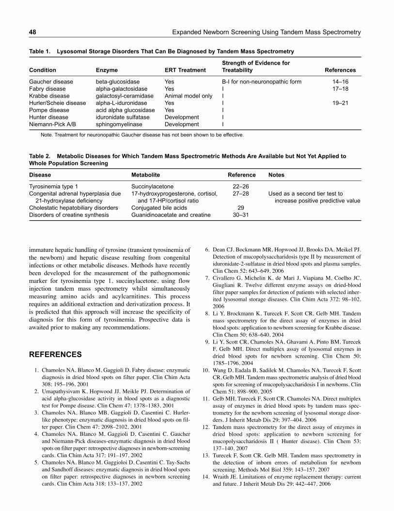
immature hepatic handling of tyrosine (transient tyrosinemia ofthe newborn) and hepatic disease resulting from congenitalinfections or other metabolic diseases. Methods have recentlybeen developed for the measurement of the pathognomonicmarker for tyrosinemia type 1, succinylacetone, using flowinjection tandem mass spectrometry whilst simultaneouslymeasuring amino acids and acylcarnitines. This processrequires an additional extraction and derivatization process. Itis predicted that this approach will increase the specificity ofdiagnosis for this form of tyrosinemia. Prospective data isawaited prior to making any recommendations.
REFERENCES
1. Chamoles NA, Blanco M, Gaggioli D. Fabry disease: enzymaticdiagnosis in dried blood spots on filter paper. Clin Chim Acta308; 195–196, 2001
2. Umapathysivam K, Hopwood JJ, Meikle PJ. Determination ofacid alpha-glucosidase activity in blood spots as a diagnostictest for Pompe disease. Clin Chem 47; 1378–1383, 2001
3. Chamoles NA, Blanco MB, Gaggioli D, Casentini C. Hurler-like phenotype: enzymatic diagnosis in dried blood spots on fil-ter paper. Clin Chem 47; 2098–2102, 2001
4. Chamoles NA, Blanco M, Gaggioli D, Casentini C. Gaucherand Niemann-Pick diseases-enzymatic diagnosis in dried bloodspots on filter paper: retrospective diagnoses in newborn-screeningcards. Clin Chim Acta 317; 191–197, 2002
5. Chamoles NA, Blanco M, Gaggioloi D, Casentini C. Tay-Sachsand Sandhoff diseases: enzymatic diagnosis in dried blood spotson filter paper: retrospective diagnoses in newborn screeningcards. Clin Chim Acta 318; 133–137, 2002
48 Expanded Newborn Screening Using Tandem Mass Spectrometry
6. Dean CJ, Bockmann MR, Hopwood JJ, Brooks DA, Meikel PJ.Detection of mucopolysaccharidosis type II by measurement ofiduronidate-2-sulfatase in dried blood spots and plasma samples.Clin Chem 52; 643–649, 2006
7. Civallero G, Michelin K, de Mari J, Viapiana M, Coelho JC,Giugliani R. Twelve different enzyme assays on dried-bloodfilter paper samples for detection of patients with selected inher-ited lysosomal storage diseases. Clin Chim Acta 372; 98–102,2006
8. Li Y, Brockmann K, Turecek F, Scott CR, Gelb MH. Tandemmass spectrometry for the direct assay of enzymes in driedblood spots: application to newborn screening for Krabbe disease.Clin Chem 50; 638–640, 2004
9. Li Y, Scott CR, Chamoles NA, Ghavami A, Pinto BM, TurecekF, Gelb MH. Direct multiplex assay of lysosomal enzymes indried blood spots for newborn screening. Clin Chem 50;1785–1796, 2004
10. Wang D, Eadala B, Sadilek M, Chamoles NA, Turecek F, ScottCR, Gelb MH. Tandem mass spectrometric analysis of dried bloodspots for screening of mucopolysaccharidosis I in newborns. ClinChem 51; 898–900, 2005
11. Gelb MH, Turecek F, Scott CR, Chamoles NA. Direct multiplexassay of enzymes in dried blood spots by tandem mass spec-trometry for the newborn screening of lysosomal storage disor-ders. J Inherit Metab Dis 29; 397–404, 2006
12. Tandem mass spectrometry for the direct assay of enzymes indried blood spots: application to newborn screening formucopolysaccharidosis II ( Hunter disease). Clin Chem 53;137–140, 2007
13. Turecek F, Scott CR. Gelb MH. Tandem mass spectrometry inthe detection of inborn errors of metabolism for newbornscreening. Methods Mol Biol 359; 143–157, 2007
14. Wraith JE. Limitations of enzyme replacement therapy: currentand future. J Inherit Metab Dis 29; 442–447, 2006
Table 1. Lysosomal Storage Disorders That Can Be Diagnosed by Tandem Mass Spectrometry
Strength of Evidence for
Condition Enzyme ERT Treatment Treatability References
Gaucher disease beta-glucosidase Yes B-I for non-neuronopathic form 14–16
Fabry disease alpha-galactosidase Yes I 17–18
Krabbe disease galactosyl-ceramidase Animal model only I
Hurler/Scheie disease alpha-L-iduronidase Yes I 19–21
Pompe disease acid alpha glucosidase Yes I
Hunter disease iduronidate sulfatase Development I
Niemann-Pick A/B sphingomyelinase Development I
Note. Treatment for neuronopathic Gaucher disease has not been shown to be effective.
Table 2. Metabolic Diseases for Which Tandem Mass Spectrometric Methods Are Available but Not Yet Applied to
Whole Population Screening
Disease Metabolite Reference Notes
Tyrosinemia type 1 Succinylacetone 22–26
Congenital adrenal hyperplasia due 17-hydroxyprogesterone, cortisol, 27–28 Used as a second tier test to
21-hydroxylase deficiency and 17-HP/cortisol ratio increase positive predictive value
Cholestatic hepatobiliary disorders Conjugated bile acids 29
Disorders of creatine synthesis Guanidinoacetate and creatine 30–31
Future Directions in Expanded Newborn Screening for Metabolic Diseases by Tandem Mass Spectrometry 49
15. Starzyk K, Richards S, Yee J, Smith SE, Kingma W. The long-term international safety experience of imiglucerase therapy forGaucher disease. Mol Genet Metab 90; 157–163, 2007
16. Sidransky E, LaMarca ME, Ginns EI. Therapy for Gaucherdisease: don’t stop thinking about tomorrow. Mol Genet Metab90; 122–125, 2007 (commentary on reference 15)
17. Schiffmann R, Askari H, Timmons M, Robinson C, Benko W,brady RO, Ries M. weekly enzyme replacement therapy mayslow decline of renal function in patients with Fabry diseasewho are on long-term biweekly dosing. J Am Soc Nephrol 18;1576–1583, 2007
18. Germain DP, Waldeck S, Banikazemi M, Bushinsky DA,Charrow J, Desnick RJ, Lee P, Loew T, Vedder AC, AbichandaniR, Wilcox WR, Guffon N. Sustained long-term renal stabiliza-tion after 54 months of agalsidase beta therapy in patients withFabry disease. J Am Soc Nephrol 18; 1547–1557, 2007
19. Thomas JA, Jacobs S, Kierstein J, Van Hove J. Outcome afterthree years of laronidase enzyme replacement therapy in apatient with Hurler syndrome. J Inherit Metab Dis 29; 762, 2006
20. Sifuentes M, Dorosshow R, Hoft R, Mason G, Walot I,Diament M, Ojkazaki S, Huff K, Cox GF, Swiedler SJ,Kakkis ED. A follow up study of MPSI patients treatedwithlaronidase enzyme replacement therapy for 6 years.Mol Genet Metab 90; 171–180, 2007
21. Tokic V, Barisic I, Huzjak N, Petkovic G, Fumic K, Pasche E.Enzyme replacement therapy in two patients with an advancedsevere (Hurler) phenotype of mucopolysaccharidosis I. Eur JPediatr 166; 727–732, 2007
22. Allard P, Grenier A, Korson MS, Zytkovicz TH. Newbornscreening for hepatorenal tyrosinemia by tandem mass spec-trometry: analysis of succinylacetone extracted from driedblood spots. Clin Biochem 37; 1010–1015, 2004
23. Sander J, Janzen N, Peter M, sander S, Steuerwald U, HoltkampU, Schwahn B, Mayatepek E, Trefz FK, Das AM. Newbornscreening for hepatorenal tyrosinemia: tandem mass spectro-metric quantification of succinylacetone. Clin Chem 52,482–487, 2006
24. Magera MJ, Gunawardena ND, Hahn SH, Tortorelli S, MitchellGA, Goodman SI, Rinaldo P, Matern D. Quantitative determina-tion of succinylacetone in dried blood spots for newborn screen-ing of tyrosinemia type 1. Mol genet Metab 88; 16–21, 2006
25. Johnson DW, Gerace R, Ranieri E, Trinh MU, Fingerhut R.Analysis of succinylacetone, as a Girard T derivative, in urineand dried blood spots by flow injection electrospray ionizationtandem mass spectrometry. Rapid Commun Mass Spectrom 21;59–63, 2007
26. Turgeon C, Magera MJ, Allard P, Tortorelli S, Gavrilov D,Oglesbee D, Raymond K, Rinaldo P, Matern D. Combinednewborn screening for succinylacetone, amino acids, and acyl-carnitines in dried blood spots. Clin Chem 54, 657–664, 2008
27. Minutti CZ, Lacey JM, Magera MJ, Hahn SH, McCann M,Schulze A, Cheillan D, Dorche C, Chace DH, Lymp JF,Zimmerman D, Rinaldo P, Matern D. Steroid profiling by tandemmass spectrometry improves the positive predictive value ofnewborn screening for congenital adrenal hyperplasia. J ClinEndocrinol Metab 89; 3687–3693, 2004
28. Lacy JM, Minutti CZ, Magera MJ, Tauscher AL, Casetta B,McCann M, Lymp J. Hahn SH, Rinaldo P, Matern D. Improvedspecificity of newborn screening for congenital adrenal hyper-plasia by second-tier steroid profiling using tndem mass spec-trometry. Clin Chem 50; 621–625, 2004
29. Mushtaq I, Logan S, Morris M, Johnson AW, Wade AM, KellyD, Clayton PT. Screening of newborn infants for cholestatichepatobiliary disease with tandem mass spectrometry. Brit MedJ 319; 471–477, 1999
30. Bodamer OA, Bloesch SM, Gregg AR, Stockler-Ipsiroglu S,O’Brien WE. Analysis of guanidinoacetate and creatine by isotopedilution tandem mass spectrometry. Clin Chim Acta 308;173–178, 2001
31. Cardicci C, Santagata S, Leuzzi V, Carducci C, Artiola C,Giovanniello T, Battini R, Antonozzi I. Quantitative determinationof guanidinoacetate and creatine in dried blood spots by flow-injection analysis-electrospray tandem mass spectrometry. ClinChim Acta 364; 180–187, 2006

50
Acknowledgment
The committee would like to acknowledge the input on future trends in this area from Dr Joseph Muenzer, MD, PhD, The Universityof North Carolina, Chapel Hill, NC.