Embed Size (px)
Citation preview
REVIEW ARTICLEpublished: 29 October 2014
doi: 10.3389/fphys.2014.00418
Carotid body, insulin, and metabolic diseases: unravelingthe linksSílvia V. Conde1*, Joana F. Sacramento1, Maria P. Guarino1,2, Constancio Gonzalez3, Ana Obeso3,
Lucilia N. Diogo1, Emilia C. Monteiro1 and Maria J. Ribeiro1
1 CEDOC, Centro Estudos Doenças Crónicas, NOVA Medical School, Faculdade de Ciências Médicas, Universidade Nova de Lisboa, Lisboa, Portugal2 Health Research Unit - UIS, School of Health Sciences, Polytechnic Institute of Leiria, Leiria, Portugal3 Departamento de Bioquímica y Biología Molecular y Fisiología, Facultad de Medicina, Instituto de Biología y Genética Molecular, Consejo Superior de
Investigaciones Científicas, Ciber de Enfermedades Respiratorias, CIBERES, Instituto de Salud Carlos III, Universidad de Valladolid, Valladolid, España
Edited by:
Rodrigo Iturriaga, PontificiaUniversidad Católica de Chile, Chile
Reviewed by:
Giovanni Solinas, University ofGothenburg, SwedenJ. Thomas Cunningham, Univerity ofNorth Texas Health Science Center,USA
*Correspondence:
Sílvia V. Conde, CEDOC, CentroEstudos Doenças Crónicas,Faculdade de Ciências Médicas,Universidade Nova de Lisboa,Campo Mártires da Pátria, 130, RuaCamara Pestana, n◦6, 6A, Edificio IIPiso 3, 1169-056 Lisboa, Portugale-mail: [email protected]
The carotid bodies (CB) are peripheral chemoreceptors that sense changes in arterialblood O2, CO2, and pH levels. Hypoxia, hypercapnia, and acidosis activate the CB, whichrespond by increasing the action potential frequency in their sensory nerve, the carotidsinus nerve (CSN). CSN activity is integrated in the brain stem to induce a panoplyof cardiorespiratory reflexes aimed, primarily, to normalize the altered blood gases, viahyperventilation, and to regulate blood pressure and cardiac performance, via sympatheticnervous system (SNS) activation. Besides its role in the cardiorespiratory control the CBhas been proposed as a metabolic sensor implicated in the control of energy homeostasisand, more recently, in the regulation of whole body insulin sensitivity. Hypercaloric dietscause CB overactivation in rats, which seems to be at the origin of the developmentof insulin resistance and hypertension, core features of metabolic syndrome and type2 diabetes. Consistent with this notion, CB sensory denervation prevents metabolic andhemodynamic alterations in hypercaloric feed animal. Obstructive sleep apnea (OSA) isanother chronic disorder characterized by increased CB activity and intimately relatedwith several metabolic and cardiovascular abnormalities. In this manuscript we reviewin a concise manner the putative pathways linking CB chemoreceptors deregulation withthe pathogenesis of insulin resistance and arterial hypertension. Also, the link betweenchronic intermittent hypoxia (CIH) and insulin resistance is discussed. Then, a final sectionis devoted to debate strategies to reduce CB activity and its use for prevention andtherapeutics of metabolic diseases with an emphasis on new exciting research in themodulation of bioelectronic signals, likely to be central in the future.
Keywords: carotid body, chronic intermittent hypoxia, insulin resistance, metabolic dysfunction, obstructive sleep
apnea
THE CAROTID BODIESThe carotid bodies (CB) are peripheral chemoreceptors locatedbilaterally in the bifurcation of the common carotid arterythat classically sense changes in arterial blood such as low O2
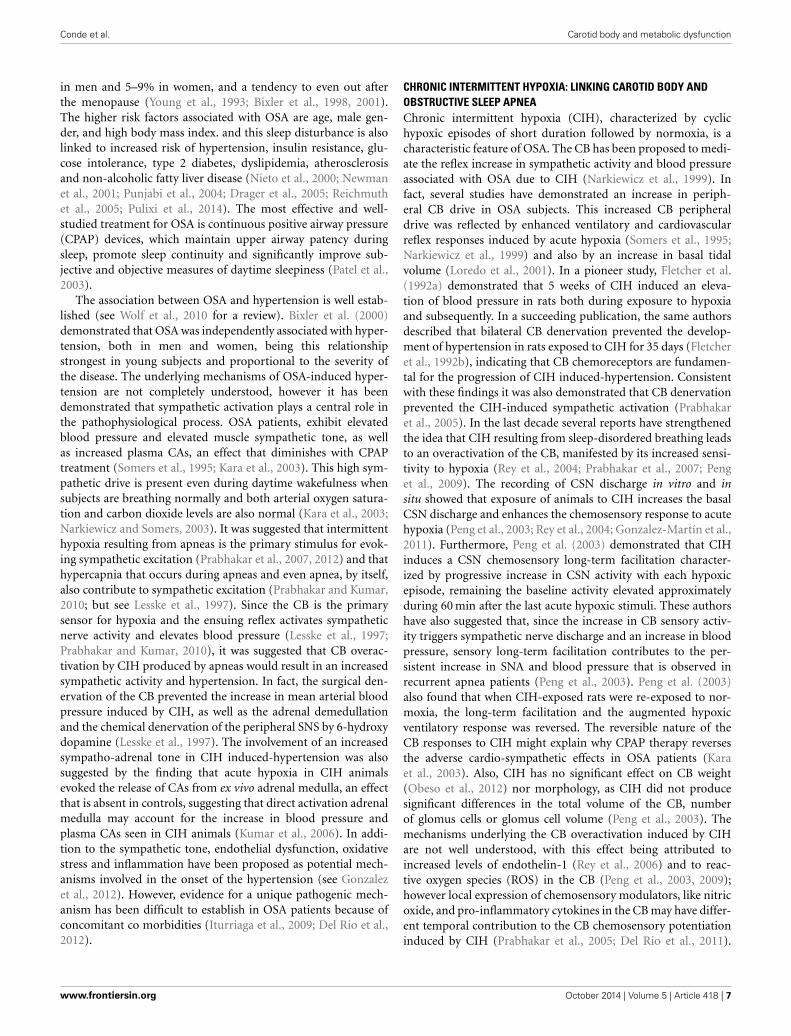
(hypoxia), high CO2 (hypercapnia), and low pH (acidosis).Hypoxia and acidosis/hypercapnia activate the CB, inducing anincrease in the frequency of discharge in the nerve endings ofits sensorial nerve, the carotid sinus nerve (CSN). The CSNactivity is integrated in the nucleus solitary tract to induce a myr-iad of respiratory reflexes aimed to normalize the altered bloodgases, via hyperventilation (Gonzalez et al., 1994), and to regu-late blood pressure and cardiac performance via an increase inthe activity of the sympathetic branch of the autonomic nervoussystem (SNS) (Marshall, 1994) (see Figure 1). The chemorecep-tor cells, also known as glomus or type I cells, are the maincellular constituent of the CB and are generally accepted as itschemosensory unit. These cells, which are derived of the neuralcrest, contain several classical neurotransmitters including, cat-echolamines [CA; dopamine (DA), and norepinephrine (NE)],
serotonin, ACh, neuropeptides (substance P and enkephalins)and adenosine (Ado) and ATP (Gonzalez et al., 1994; Zhang et al.,2000; Rong et al., 2003; Buttigieg and Nurse, 2004; Conde andMonteiro, 2004; Conde et al., 2012a). All these substances, theiragonists and antagonists are capable of modifying, inhibiting orstimulating CSN activity. In addition to chemoreceptor cells, theCB also possesses type II cells, or sustentacular cells and it hasbeen proposed that they are adult neural stem cells sustainingneurogenesis in vivo in response to physiological stimuli, likechronic hypoxia, and acting in paracrine signaling during hypoxia(Pardal et al., 2007; Piskuric and Nurse, 2013).
ROLE OF CAROTID BODY IN METABOLISMEVIDENCES FOR A ROLE OF CAROTID BODY IN GLUCOSEHOMEOSTASISThe idea of a physiological role of the CB on the control of glu-cose metabolism was first suggested by Petropavlovskaya in the50’s. In this pioneer study it was shown that the stimulation ofthe CB induces a reflex hyperglycemia, an effect that is mediated
www.frontiersin.org October 2014 | Volume 5 | Article 418 | 1
Conde et al. Carotid body and metabolic dysfunction
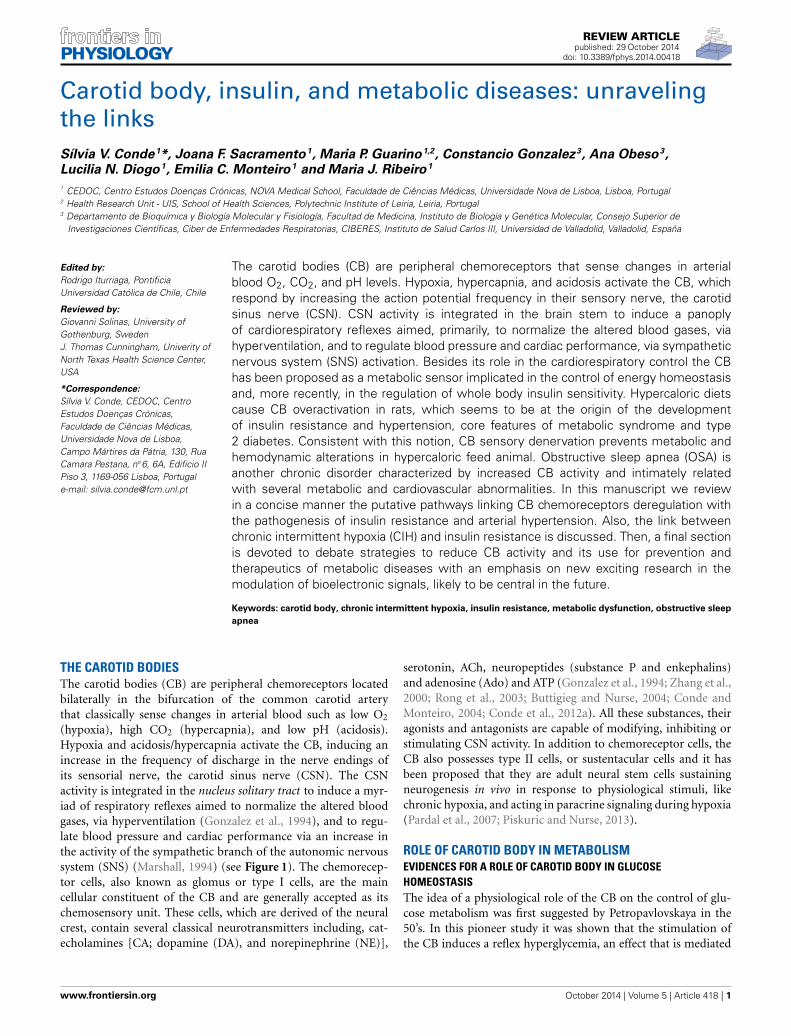
FIGURE 1 | Schematic representation of the chemoreflexes elicited by the carotid bodies. (A) Representation of important mechanism involved in thereflex-responses elicited by the carotid body. (B) Stimulation of the carotid body is capable of produce cardiovascular, respiratory, endocrine, and renal responses.
by the adrenal medulla, since it was not observed in adrenalec-tomized animals (Petropavlovskaya, 1953). Twenty five years later,Alvarez-Buylla and de Alvarez-Buylla (1988) confirmed thoseresults by demonstrating that the pharmacological stimulation ofthe CB with cyanide (NaCN) produced an increase in hepatic glu-cose output in cats, this reflex response being eliminated by bilat-eral adrenalectomy or by surgical removal of the neurohypophysis(Alvarez-Buylla et al., 1997). Also, it was shown that changes inblood concentration in the CB-CSN, superfused in vivo, modifybrain glucose retention, suggesting that chemosensory activity inthe CSN controls brain glucose metabolism (Alvarez-Buylla andde Alvarez-Buylla, 1994). In parallel with the increase in hepaticglucose output, one would expect an increase in plasma insulinlevels to ensure an adequate glucose utilization by the peripheraltissues and, in fact, stimulation of CBs by corconium, a nicoti-nomimetic agent, caused a rise in circulating insulin that wasreversed by CSN resection (Anichkov and Tomilina, 1962). Lateron, Koyama et al. (2000) demonstrated that CB plays an impor-tant role in glucose homeostasis in vivo, since dogs that have theirCB resected presented lower arterial glucagon in basal conditionsand reduced glucagon and cortisol levels during insulin-inducedhypoglycemia, together with a marked decrease in endogenoushepatic glucose production in response to hypoglycemia, and
with an increase in insulin sensitivity, independent of blood glu-cose level. These last results suggested for the first time that CBresection affects the response to moderate hyperinsulinemia andtherefore, that the CB may play a role in glucose homeostasis thatis not related with the hypoglycemic counterregulatory response.
The results obtained by Koyama et al. (2000) were sup-ported by clinical studies where it was demonstrated that, therate of glucose infusion necessary to maintain glucose levels ina hyperinsulinemic-hypoglycemic clamp was significantly higherduring hyperoxia than in normoxia (Wehrwein et al., 2010). Inthe same study, the authors also observed that hyperoxia, whichblunts CB activity, decreased the release of counter-regulatoryhormones such as adrenaline, cortisol, glucagon and growthhormone, which seems to indicate that the CB play an impor-tant role in neuroendocrine responses during hypoglycemia(Wehrwein et al., 2010). However, the absence of adequate con-trols in hyperinsulinemic-euglycemic conditions in this studydoes not allow assigning the effects to the hyperinsulinemia perse or to hypoglycemia. In another clinical study designed todetermine whether hypo- and hyperglycaemia modulate the ven-tilatory responses to hypoxia, it was shown that hypoglycemia, aswell as hyperglycemia, produced an increase in ventilation andin the hypoxic ventilatory response, being the latter accompanied
Frontiers in Physiology | Integrative Physiology October 2014 | Volume 5 | Article 418 | 2
Conde et al. Carotid body and metabolic dysfunction
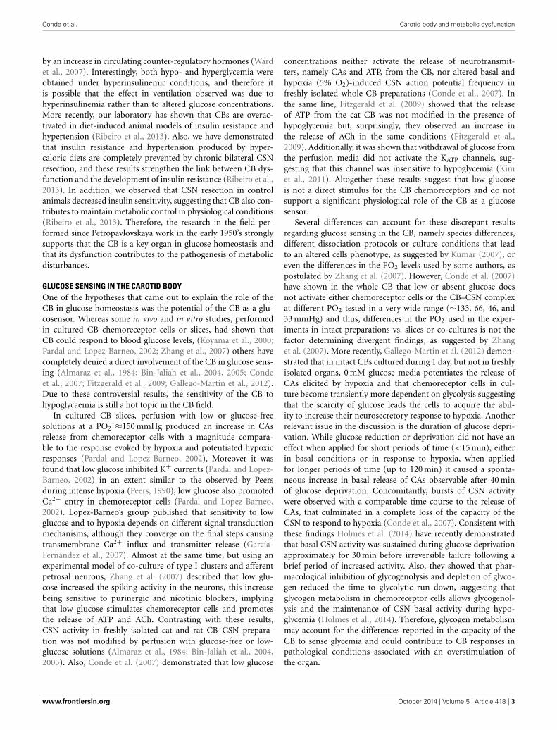
by an increase in circulating counter-regulatory hormones (Wardet al., 2007). Interestingly, both hypo- and hyperglycemia wereobtained under hyperinsulinemic conditions, and therefore itis possible that the effect in ventilation observed was due tohyperinsulinemia rather than to altered glucose concentrations.More recently, our laboratory has shown that CBs are overac-tivated in diet-induced animal models of insulin resistance andhypertension (Ribeiro et al., 2013). Also, we have demonstratedthat insulin resistance and hypertension produced by hyper-caloric diets are completely prevented by chronic bilateral CSNresection, and these results strengthen the link between CB dys-function and the development of insulin resistance (Ribeiro et al.,2013). In addition, we observed that CSN resection in controlanimals decreased insulin sensitivity, suggesting that CB also con-tributes to maintain metabolic control in physiological conditions(Ribeiro et al., 2013). Therefore, the research in the field per-formed since Petropavlovskaya work in the early 1950’s stronglysupports that the CB is a key organ in glucose homeostasis andthat its dysfunction contributes to the pathogenesis of metabolicdisturbances.
GLUCOSE SENSING IN THE CAROTID BODYOne of the hypotheses that came out to explain the role of theCB in glucose homeostasis was the potential of the CB as a glu-cosensor. Whereas some in vivo and in vitro studies, performedin cultured CB chemoreceptor cells or slices, had shown thatCB could respond to blood glucose levels, (Koyama et al., 2000;Pardal and Lopez-Barneo, 2002; Zhang et al., 2007) others havecompletely denied a direct involvement of the CB in glucose sens-ing (Almaraz et al., 1984; Bin-Jaliah et al., 2004, 2005; Condeet al., 2007; Fitzgerald et al., 2009; Gallego-Martin et al., 2012).Due to these controversial results, the sensitivity of the CB tohypoglycaemia is still a hot topic in the CB field.
In cultured CB slices, perfusion with low or glucose-freesolutions at a PO2 ≈150 mmHg produced an increase in CAsrelease from chemoreceptor cells with a magnitude compara-ble to the response evoked by hypoxia and potentiated hypoxicresponses (Pardal and Lopez-Barneo, 2002). Moreover it wasfound that low glucose inhibited K+ currents (Pardal and Lopez-Barneo, 2002) in an extent similar to the observed by Peersduring intense hypoxia (Peers, 1990); low glucose also promotedCa2+ entry in chemoreceptor cells (Pardal and Lopez-Barneo,2002). Lopez-Barneo’s group published that sensitivity to lowglucose and to hypoxia depends on different signal transductionmechanisms, although they converge on the final steps causingtransmembrane Ca2+ influx and transmitter release (García-Fernández et al., 2007). Almost at the same time, but using anexperimental model of co-culture of type I clusters and afferentpetrosal neurons, Zhang et al. (2007) described that low glu-cose increased the spiking activity in the neurons, this increasebeing sensitive to purinergic and nicotinic blockers, implyingthat low glucose stimulates chemoreceptor cells and promotesthe release of ATP and ACh. Contrasting with these results,CSN activity in freshly isolated cat and rat CB–CSN prepara-tion was not modified by perfusion with glucose-free or low-glucose solutions (Almaraz et al., 1984; Bin-Jaliah et al., 2004,2005). Also, Conde et al. (2007) demonstrated that low glucose
concentrations neither activate the release of neurotransmit-ters, namely CAs and ATP, from the CB, nor altered basal andhypoxia (5% O2)-induced CSN action potential frequency infreshly isolated whole CB preparations (Conde et al., 2007). Inthe same line, Fitzgerald et al. (2009) showed that the releaseof ATP from the cat CB was not modified in the presence ofhypoglycemia but, surprisingly, they observed an increase inthe release of ACh in the same conditions (Fitzgerald et al.,2009). Additionally, it was shown that withdrawal of glucose fromthe perfusion media did not activate the KATP channels, sug-gesting that this channel was insensitive to hypoglycemia (Kimet al., 2011). Altogether these results suggest that low glucoseis not a direct stimulus for the CB chemoreceptors and do notsupport a significant physiological role of the CB as a glucosesensor.
Several differences can account for these discrepant resultsregarding glucose sensing in the CB, namely species differences,different dissociation protocols or culture conditions that leadto an altered cells phenotype, as suggested by Kumar (2007), oreven the differences in the PO2 levels used by some authors, aspostulated by Zhang et al. (2007). However, Conde et al. (2007)have shown in the whole CB that low or absent glucose doesnot activate either chemoreceptor cells or the CB–CSN complexat different PO2 tested in a very wide range (∼133, 66, 46, and33 mmHg) and thus, differences in the PO2 used in the exper-iments in intact preparations vs. slices or co-cultures is not thefactor determining divergent findings, as suggested by Zhanget al. (2007). More recently, Gallego-Martin et al. (2012) demon-strated that in intact CBs cultured during 1 day, but not in freshlyisolated organs, 0 mM glucose media potentiates the release ofCAs elicited by hypoxia and that chemoreceptor cells in cul-ture become transiently more dependent on glycolysis suggestingthat the scarcity of glucose leads the cells to acquire the abil-ity to increase their neurosecretory response to hypoxia. Anotherrelevant issue in the discussion is the duration of glucose depri-vation. While glucose reduction or deprivation did not have aneffect when applied for short periods of time (<15 min), eitherin basal conditions or in response to hypoxia, when appliedfor longer periods of time (up to 120 min) it caused a sponta-neous increase in basal release of CAs observable after 40 minof glucose deprivation. Concomitantly, bursts of CSN activitywere observed with a comparable time course to the release ofCAs, that culminated in a complete loss of the capacity of theCSN to respond to hypoxia (Conde et al., 2007). Consistent withthese findings Holmes et al. (2014) have recently demonstratedthat basal CSN activity was sustained during glucose deprivationapproximately for 30 min before irreversible failure following abrief period of increased activity. Also, they showed that phar-macological inhibition of glycogenolysis and depletion of glyco-gen reduced the time to glycolytic run down, suggesting thatglycogen metabolism in chemoreceptor cells allows glycogenol-ysis and the maintenance of CSN basal activity during hypo-glycemia (Holmes et al., 2014). Therefore, glycogen metabolismmay account for the differences reported in the capacity of theCB to sense glycemia and could contribute to CB responses inpathological conditions associated with an overstimulation ofthe organ.
www.frontiersin.org October 2014 | Volume 5 | Article 418 | 3
Conde et al. Carotid body and metabolic dysfunction
IS INSULIN A STIMULUS FOR CB ACTIVATION?A large body of literature supports a role for the central nervoussystem in insulin-induced sympathoexcitation, as the injectionof insulin on arcuate nucleus and paraventricular nucleus hasbeen shown to produce an increase in spinal sympathetic out-flow, mediated by dorsal hypothalamus and rostral ventrolateralmedulla (for a review see Dampney, 2011). However, this effectcannot be exclusively assigned to a centrally-mediated mecha-nism, since the injection of insulin into the carotid artery ofanesthetized dogs produces an increase in blood pressure andsympathetic activity higher than the systemic insulin administra-tion, being the effect abolished by ganglionic blockade (Peredaet al., 1962). These results were the first to suggest a role forthe peripheral nervous system in insulin-mediated sympatheticactivity. During the evaluation of a putative direct role of the CBin glucose sensing, Bin-Jaliah et al. (2004) observed that insulininfusion, used to produce hypoglycemia, increased minute venti-lation and the rate of O2 consumption (VO2), an effect that wastotally mediated by the CB, since CSN denervation blunted it.The same authors demonstrated afterwards that insulin-inducedhypoglycemia was associated with a significantly increase in CO2
chemosensitivity, an effect that was mediated by the CB, since theeffect was lost in animals that had their CSN resected (Bin-Jaliahet al., 2005). Since in vitro hypoglycemia was incapable of mod-ifying basal CSN activity (Bin-Jaliah et al., 2004; Conde et al.,2007) and blunted the response of CSN to hypercapnia (Bin-Jaliah et al., 2005) the elevation of ventilation observed in vivoby Bin-Jaliah’s group was somehow surprising (Bin-Jaliah et al.,2004, 2005) and the hypothesis of being an indirect consequenceof systemic hypoglycemia related to some other undeterminedsubstance had to be considered. To pursue this hypothesis, ourgroup has been dedicated to investigate whether insulin itself iscapable of stimulating the CB and of eliciting a neurosecretoryresponse. We have demonstrated the presence of insulin recep-tors in the rat CB by western-blot and its phosphorylation inresponse to insulin (Ribeiro et al., 2013). The presence of insulinreceptors was also confirmed on finding that isolated whole CBsincubated with insulin accumulate more 2-deoxiglucose than the
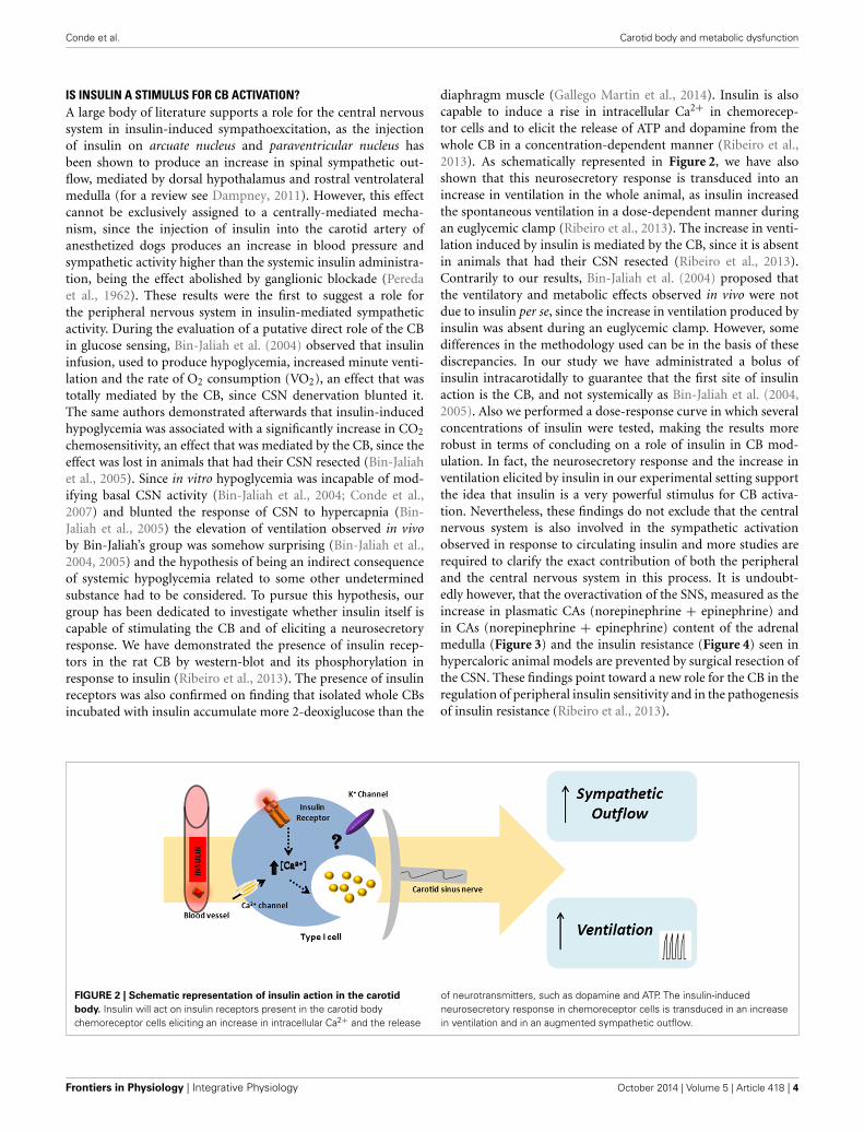
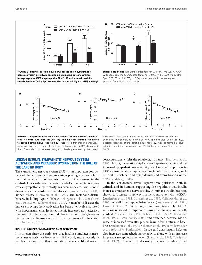
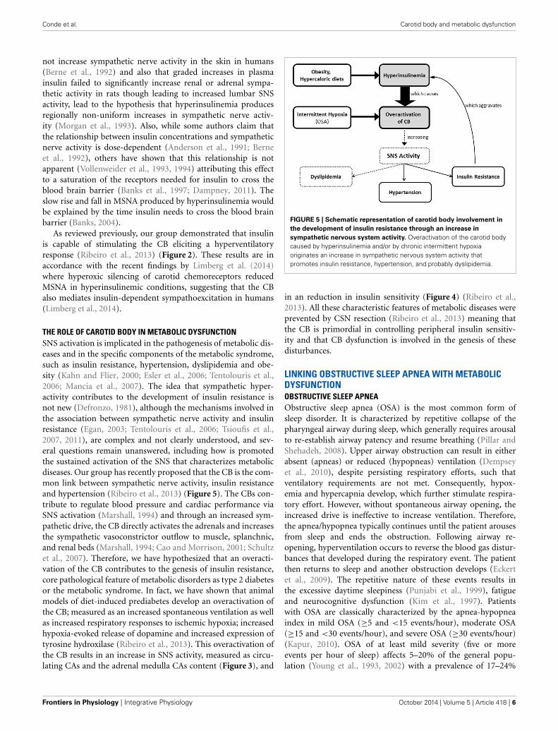
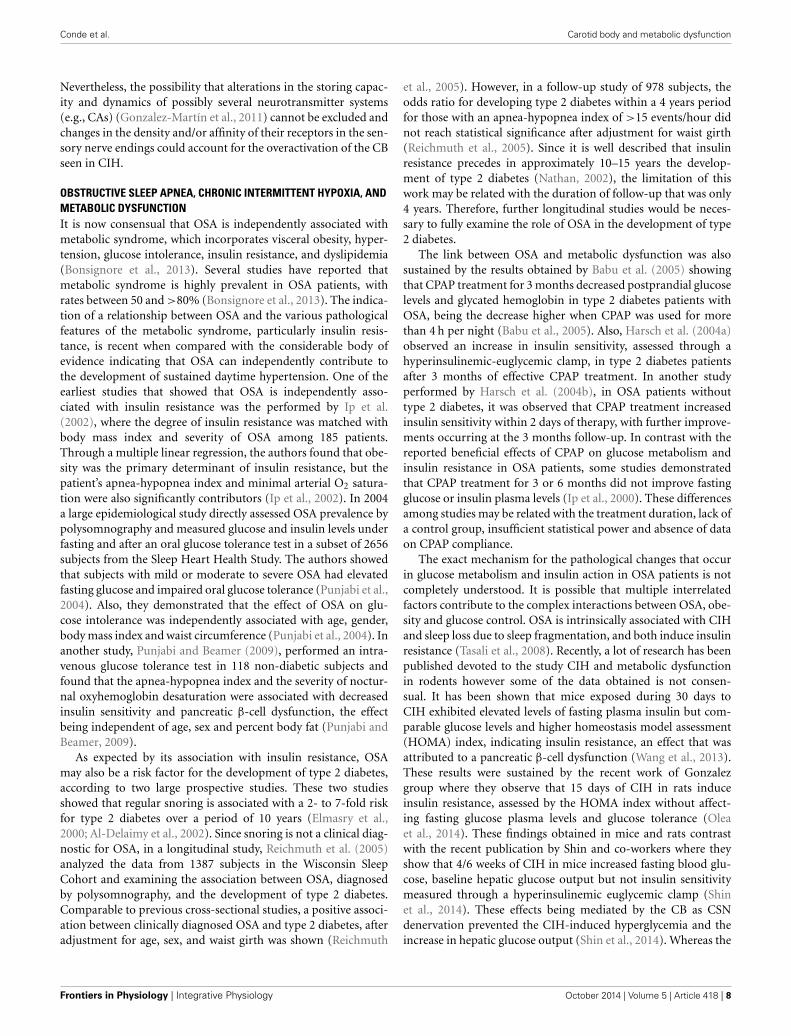
diaphragm muscle (Gallego Martin et al., 2014). Insulin is alsocapable to induce a rise in intracellular Ca2+ in chemorecep-tor cells and to elicit the release of ATP and dopamine from thewhole CB in a concentration-dependent manner (Ribeiro et al.,2013). As schematically represented in Figure 2, we have alsoshown that this neurosecretory response is transduced into anincrease in ventilation in the whole animal, as insulin increasedthe spontaneous ventilation in a dose-dependent manner duringan euglycemic clamp (Ribeiro et al., 2013). The increase in venti-lation induced by insulin is mediated by the CB, since it is absentin animals that had their CSN resected (Ribeiro et al., 2013).Contrarily to our results, Bin-Jaliah et al. (2004) proposed thatthe ventilatory and metabolic effects observed in vivo were notdue to insulin per se, since the increase in ventilation produced byinsulin was absent during an euglycemic clamp. However, somedifferences in the methodology used can be in the basis of thesediscrepancies. In our study we have administrated a bolus ofinsulin intracarotidally to guarantee that the first site of insulinaction is the CB, and not systemically as Bin-Jaliah et al. (2004,2005). Also we performed a dose-response curve in which severalconcentrations of insulin were tested, making the results morerobust in terms of concluding on a role of insulin in CB mod-ulation. In fact, the neurosecretory response and the increase inventilation elicited by insulin in our experimental setting supportthe idea that insulin is a very powerful stimulus for CB activa-tion. Nevertheless, these findings do not exclude that the centralnervous system is also involved in the sympathetic activationobserved in response to circulating insulin and more studies arerequired to clarify the exact contribution of both the peripheraland the central nervous system in this process. It is undoubt-edly however, that the overactivation of the SNS, measured as theincrease in plasmatic CAs (norepinephrine + epinephrine) andin CAs (norepinephrine + epinephrine) content of the adrenalmedulla (Figure 3) and the insulin resistance (Figure 4) seen inhypercaloric animal models are prevented by surgical resection ofthe CSN. These findings point toward a new role for the CB in theregulation of peripheral insulin sensitivity and in the pathogenesisof insulin resistance (Ribeiro et al., 2013).
FIGURE 2 | Schematic representation of insulin action in the carotid
body. Insulin will act on insulin receptors present in the carotid bodychemoreceptor cells eliciting an increase in intracellular Ca2+ and the release
of neurotransmitters, such as dopamine and ATP. The insulin-inducedneurosecretory response in chemoreceptor cells is transduced in an increasein ventilation and in an augmented sympathetic outflow.
Frontiers in Physiology | Integrative Physiology October 2014 | Volume 5 | Article 418 | 4
Conde et al. Carotid body and metabolic dysfunction
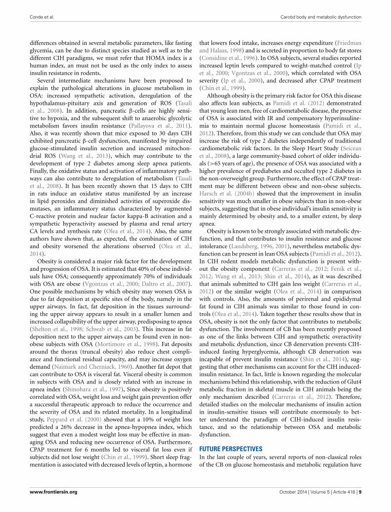
FIGURE 3 | Effect of carotid sinus nerve resection on sympathetic
nervous system activity, measured as circulating catecholamines
[norepinephrine (NE) + epinephrine (Epi)] (A) and adrenal medulla
catecholamines (NE + Epi) content (B), in control, high fat (HF) and high
sucrose (HSu) diet rats. Bars represent mean ± s.e.m. Two-Way ANOVAwith Bonferroni multicomparison tests; ∗p < 0.05, ∗∗∗p < 0.001 vs. control;#p < 0.05, ##p < 0.01, ###p < 0.001 vs. values within the same group(adapted from Ribeiro et al., 2013).
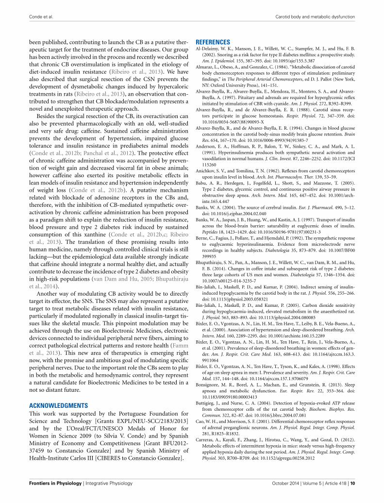
FIGURE 4 | Representative excursion curves for the insulin tolerance
test in control (A), high fat (HF) (B), and high fat animals submitted
to carotid sinus nerve resection (C) rats. Note that insulin sensitivity,expressed by the constant of the insulin tolerance test (KITT) decrease inthe HF animals, this decrease being completely prevented by the bilateral
resection of the carotid sinus nerve. HF animals were achieved bysubmitting the animals to a HF diet (45% lipid-rich diet) during 21 days.Bilateral resection of the carotid sinus nerve (C) was performed 5 daysprior to submitting the animals to HF diet (adapted from Ribeiro et al.,2013).
LINKING INSULIN, SYMPATHETIC NERVOUS SYSTEMACTIVATION AND METABOLIC DYSFUNCTION: THE ROLE OFTHE CAROTID BODYThe sympathetic nervous system (SNS) is an important compo-nent of the autonomic nervous system playing a major role inthe maintenance of homeostasis due to its involvement in thecontrol of the cardiovascular system and of several metabolic pro-cesses. Sympathetic overactivity has been associated with severaldiseases, such as cardiovascular diseases (Graham et al., 2004),kidney disease (Converse et al., 1992), and metabolic distur-bances, including type 2 diabetes (Huggett et al., 2003; Grassiet al., 2005, 2007; Kobayashi et al., 2010). In metabolic diseases theincrease in sympathetic activation has been attentively associatedwith hyperinsulinemia, hyperleptinemia increased non-esterifiedfree fatty acids, inflammation, and obesity among others, howeverthe precise mechanisms remain to be unequivocally elucidated(Lambert et al., 2010).
INSULIN-INDUCED SYMPATHETIC OVERACTIVATIONIt is known since the early 80’s that insulin stimulates sympa-thetic nerve activity (Rowe et al., 1981) and, more recently, ithas been shown that this stimulation occurs at blood insulin
concentrations within the physiological range (Hausberg et al.,1995). In fact, the relationship between hyperinsulinemia and theincreased sympathetic nerve activity lead Landsberg to propose in1986 a causal relationship between metabolic disturbances, suchas insulin resistance and dyslipidemia, and overactivation of theSNS (Landsberg, 1986).
In the last decades several reports were published, both inanimals and in humans, supporting the hypothesis that insulinincreases sympathetic nerve activity. In humans insulin has beenshown to increase muscle sympathetic nerve activity (MSNA)(Anderson et al., 1991; Scherrer et al., 1993; Vollenweider et al.,1993) as well as norepinephrine levels (Anderson et al., 1991;Lambert et al., 2010) in euglycemic conditions. The MSNAresponse observed in response to insulin administration is bothgradual (Anderson et al., 1991; Scherrer et al., 1993; Vollenweideret al., 1993, 1994; Banks, 2004) and sustained because MSNAremains increased even after plasma insulin levels return to base-line (Anderson et al., 1991; Scherrer et al., 1993; Vollenweideret al., 1993, 1994; Banks, 2004). In rats and dogs, insulin infusionalso increases sympathetic nerve activity along with an increasein plasma norepinephrine levels (Liang et al., 1982; Tomiyamaet al., 1992). However, the discovery that insulin infusion did
www.frontiersin.org October 2014 | Volume 5 | Article 418 | 5
Conde et al. Carotid body and metabolic dysfunction
not increase sympathetic nerve activity in the skin in humans(Berne et al., 1992) and also that graded increases in plasmainsulin failed to significantly increase renal or adrenal sympa-thetic activity in rats though leading to increased lumbar SNSactivity, lead to the hypothesis that hyperinsulinemia producesregionally non-uniform increases in sympathetic nerve activ-ity (Morgan et al., 1993). Also, while some authors claim thatthe relationship between insulin concentrations and sympatheticnerve activity is dose-dependent (Anderson et al., 1991; Berneet al., 1992), others have shown that this relationship is notapparent (Vollenweider et al., 1993, 1994) attributing this effectto a saturation of the receptors needed for insulin to cross theblood brain barrier (Banks et al., 1997; Dampney, 2011). Theslow rise and fall in MSNA produced by hyperinsulinemia wouldbe explained by the time insulin needs to cross the blood brainbarrier (Banks, 2004).
As reviewed previously, our group demonstrated that insulinis capable of stimulating the CB eliciting a hyperventilatoryresponse (Ribeiro et al., 2013) (Figure 2). These results are inaccordance with the recent findings by Limberg et al. (2014)where hyperoxic silencing of carotid chemoreceptors reducedMSNA in hyperinsulinemic conditions, suggesting that the CBalso mediates insulin-dependent sympathoexcitation in humans(Limberg et al., 2014).
THE ROLE OF CAROTID BODY IN METABOLIC DYSFUNCTIONSNS activation is implicated in the pathogenesis of metabolic dis-eases and in the specific components of the metabolic syndrome,such as insulin resistance, hypertension, dyslipidemia and obe-sity (Kahn and Flier, 2000; Esler et al., 2006; Tentolouris et al.,2006; Mancia et al., 2007). The idea that sympathetic hyper-activity contributes to the development of insulin resistance isnot new (Defronzo, 1981), although the mechanisms involved inthe association between sympathetic nerve activity and insulinresistance (Egan, 2003; Tentolouris et al., 2006; Tsioufis et al.,2007, 2011), are complex and not clearly understood, and sev-eral questions remain unanswered, including how is promotedthe sustained activation of the SNS that characterizes metabolicdiseases. Our group has recently proposed that the CB is the com-mon link between sympathetic nerve activity, insulin resistanceand hypertension (Ribeiro et al., 2013) (Figure 5). The CBs con-tribute to regulate blood pressure and cardiac performance viaSNS activation (Marshall, 1994) and through an increased sym-pathetic drive, the CB directly activates the adrenals and increasesthe sympathetic vasoconstrictor outflow to muscle, splanchnic,and renal beds (Marshall, 1994; Cao and Morrison, 2001; Schultzet al., 2007). Therefore, we have hypothesized that an overacti-vation of the CB contributes to the genesis of insulin resistance,core pathological feature of metabolic disorders as type 2 diabetesor the metabolic syndrome. In fact, we have shown that animalmodels of diet-induced prediabetes develop an overactivation ofthe CB; measured as an increased spontaneous ventilation as wellas increased respiratory responses to ischemic hypoxia; increasedhypoxia-evoked release of dopamine and increased expression oftyrosine hydroxilase (Ribeiro et al., 2013). This overactivation ofthe CB results in an increase in SNS activity, measured as circu-lating CAs and the adrenal medulla CAs content (Figure 3), and
FIGURE 5 | Schematic representation of carotid body involvement in
the development of insulin resistance through an increase in
sympathetic nervous system activity. Overactivation of the carotid bodycaused by hyperinsulinemia and/or by chronic intermittent hypoxiaoriginates an increase in sympathetic nervous system activity thatpromotes insulin resistance, hypertension, and probably dyslipidemia.
in an reduction in insulin sensitivity (Figure 4) (Ribeiro et al.,2013). All these characteristic features of metabolic diseases wereprevented by CSN resection (Ribeiro et al., 2013) meaning thatthe CB is primordial in controlling peripheral insulin sensitiv-ity and that CB dysfunction is involved in the genesis of thesedisturbances.
LINKING OBSTRUCTIVE SLEEP APNEA WITH METABOLICDYSFUNCTIONOBSTRUCTIVE SLEEP APNEAObstructive sleep apnea (OSA) is the most common form ofsleep disorder. It is characterized by repetitive collapse of thepharyngeal airway during sleep, which generally requires arousalto re-establish airway patency and resume breathing (Pillar andShehadeh, 2008). Upper airway obstruction can result in eitherabsent (apneas) or reduced (hypopneas) ventilation (Dempseyet al., 2010), despite persisting respiratory efforts, such thatventilatory requirements are not met. Consequently, hypox-emia and hypercapnia develop, which further stimulate respira-tory effort. However, without spontaneous airway opening, theincreased drive is ineffective to increase ventilation. Therefore,the apnea/hypopnea typically continues until the patient arousesfrom sleep and ends the obstruction. Following airway re-opening, hyperventilation occurs to reverse the blood gas distur-bances that developed during the respiratory event. The patientthen returns to sleep and another obstruction develops (Eckertet al., 2009). The repetitive nature of these events results inthe excessive daytime sleepiness (Punjabi et al., 1999), fatigueand neurocognitive dysfunction (Kim et al., 1997). Patientswith OSA are classically characterized by the apnea-hypopneaindex in mild OSA (≥5 and <15 events/hour), moderate OSA(≥15 and <30 events/hour), and severe OSA (≥30 events/hour)(Kapur, 2010). OSA of at least mild severity (five or moreevents per hour of sleep) affects 5–20% of the general popu-lation (Young et al., 1993, 2002) with a prevalence of 17–24%
Frontiers in Physiology | Integrative Physiology October 2014 | Volume 5 | Article 418 | 6
Conde et al. Carotid body and metabolic dysfunction
in men and 5–9% in women, and a tendency to even out afterthe menopause (Young et al., 1993; Bixler et al., 1998, 2001).The higher risk factors associated with OSA are age, male gen-der, and high body mass index. and this sleep disturbance is alsolinked to increased risk of hypertension, insulin resistance, glu-cose intolerance, type 2 diabetes, dyslipidemia, atherosclerosisand non-alcoholic fatty liver disease (Nieto et al., 2000; Newmanet al., 2001; Punjabi et al., 2004; Drager et al., 2005; Reichmuthet al., 2005; Pulixi et al., 2014). The most effective and well-studied treatment for OSA is continuous positive airway pressure(CPAP) devices, which maintain upper airway patency duringsleep, promote sleep continuity and significantly improve sub-jective and objective measures of daytime sleepiness (Patel et al.,2003).
The association between OSA and hypertension is well estab-lished (see Wolf et al., 2010 for a review). Bixler et al. (2000)demonstrated that OSA was independently associated with hyper-tension, both in men and women, being this relationshipstrongest in young subjects and proportional to the severity ofthe disease. The underlying mechanisms of OSA-induced hyper-tension are not completely understood, however it has beendemonstrated that sympathetic activation plays a central role inthe pathophysiological process. OSA patients, exhibit elevatedblood pressure and elevated muscle sympathetic tone, as wellas increased plasma CAs, an effect that diminishes with CPAPtreatment (Somers et al., 1995; Kara et al., 2003). This high sym-pathetic drive is present even during daytime wakefulness whensubjects are breathing normally and both arterial oxygen satura-tion and carbon dioxide levels are also normal (Kara et al., 2003;Narkiewicz and Somers, 2003). It was suggested that intermittenthypoxia resulting from apneas is the primary stimulus for evok-ing sympathetic excitation (Prabhakar et al., 2007, 2012) and thathypercapnia that occurs during apneas and even apnea, by itself,also contribute to sympathetic excitation (Prabhakar and Kumar,2010; but see Lesske et al., 1997). Since the CB is the primarysensor for hypoxia and the ensuing reflex activates sympatheticnerve activity and elevates blood pressure (Lesske et al., 1997;Prabhakar and Kumar, 2010), it was suggested that CB overac-tivation by CIH produced by apneas would result in an increasedsympathetic activity and hypertension. In fact, the surgical den-ervation of the CB prevented the increase in mean arterial bloodpressure induced by CIH, as well as the adrenal demedullationand the chemical denervation of the peripheral SNS by 6-hydroxydopamine (Lesske et al., 1997). The involvement of an increasedsympatho-adrenal tone in CIH induced-hypertension was alsosuggested by the finding that acute hypoxia in CIH animalsevoked the release of CAs from ex vivo adrenal medulla, an effectthat is absent in controls, suggesting that direct activation adrenalmedulla may account for the increase in blood pressure andplasma CAs seen in CIH animals (Kumar et al., 2006). In addi-tion to the sympathetic tone, endothelial dysfunction, oxidativestress and inflammation have been proposed as potential mech-anisms involved in the onset of the hypertension (see Gonzalezet al., 2012). However, evidence for a unique pathogenic mech-anism has been difficult to establish in OSA patients because ofconcomitant co morbidities (Iturriaga et al., 2009; Del Rio et al.,2012).
CHRONIC INTERMITTENT HYPOXIA: LINKING CAROTID BODY ANDOBSTRUCTIVE SLEEP APNEAChronic intermittent hypoxia (CIH), characterized by cyclichypoxic episodes of short duration followed by normoxia, is acharacteristic feature of OSA. The CB has been proposed to medi-ate the reflex increase in sympathetic activity and blood pressureassociated with OSA due to CIH (Narkiewicz et al., 1999). Infact, several studies have demonstrated an increase in periph-eral CB drive in OSA subjects. This increased CB peripheraldrive was reflected by enhanced ventilatory and cardiovascularreflex responses induced by acute hypoxia (Somers et al., 1995;Narkiewicz et al., 1999) and also by an increase in basal tidalvolume (Loredo et al., 2001). In a pioneer study, Fletcher et al.(1992a) demonstrated that 5 weeks of CIH induced an eleva-tion of blood pressure in rats both during exposure to hypoxiaand subsequently. In a succeeding publication, the same authorsdescribed that bilateral CB denervation prevented the develop-ment of hypertension in rats exposed to CIH for 35 days (Fletcheret al., 1992b), indicating that CB chemoreceptors are fundamen-tal for the progression of CIH induced-hypertension. Consistentwith these findings it was also demonstrated that CB denervationprevented the CIH-induced sympathetic activation (Prabhakaret al., 2005). In the last decade several reports have strengthenedthe idea that CIH resulting from sleep-disordered breathing leadsto an overactivation of the CB, manifested by its increased sensi-tivity to hypoxia (Rey et al., 2004; Prabhakar et al., 2007; Penget al., 2009). The recording of CSN discharge in vitro and insitu showed that exposure of animals to CIH increases the basalCSN discharge and enhances the chemosensory response to acutehypoxia (Peng et al., 2003; Rey et al., 2004; Gonzalez-Martín et al.,2011). Furthermore, Peng et al. (2003) demonstrated that CIHinduces a CSN chemosensory long-term facilitation character-ized by progressive increase in CSN activity with each hypoxicepisode, remaining the baseline activity elevated approximatelyduring 60 min after the last acute hypoxic stimuli. These authorshave also suggested that, since the increase in CB sensory activ-ity triggers sympathetic nerve discharge and an increase in bloodpressure, sensory long-term facilitation contributes to the per-sistent increase in SNA and blood pressure that is observed inrecurrent apnea patients (Peng et al., 2003). Peng et al. (2003)also found that when CIH-exposed rats were re-exposed to nor-moxia, the long-term facilitation and the augmented hypoxicventilatory response was reversed. The reversible nature of theCB responses to CIH might explain why CPAP therapy reversesthe adverse cardio-sympathetic effects in OSA patients (Karaet al., 2003). Also, CIH has no significant effect on CB weight(Obeso et al., 2012) nor morphology, as CIH did not producesignificant differences in the total volume of the CB, numberof glomus cells or glomus cell volume (Peng et al., 2003). Themechanisms underlying the CB overactivation induced by CIHare not well understood, with this effect being attributed toincreased levels of endothelin-1 (Rey et al., 2006) and to reac-tive oxygen species (ROS) in the CB (Peng et al., 2003, 2009);however local expression of chemosensory modulators, like nitricoxide, and pro-inflammatory cytokines in the CB may have differ-ent temporal contribution to the CB chemosensory potentiationinduced by CIH (Prabhakar et al., 2005; Del Rio et al., 2011).
www.frontiersin.org October 2014 | Volume 5 | Article 418 | 7
Conde et al. Carotid body and metabolic dysfunction
Nevertheless, the possibility that alterations in the storing capac-ity and dynamics of possibly several neurotransmitter systems(e.g., CAs) (Gonzalez-Martín et al., 2011) cannot be excluded andchanges in the density and/or affinity of their receptors in the sen-sory nerve endings could account for the overactivation of the CBseen in CIH.
OBSTRUCTIVE SLEEP APNEA, CHRONIC INTERMITTENT HYPOXIA, ANDMETABOLIC DYSFUNCTIONIt is now consensual that OSA is independently associated withmetabolic syndrome, which incorporates visceral obesity, hyper-tension, glucose intolerance, insulin resistance, and dyslipidemia(Bonsignore et al., 2013). Several studies have reported thatmetabolic syndrome is highly prevalent in OSA patients, withrates between 50 and >80% (Bonsignore et al., 2013). The indica-tion of a relationship between OSA and the various pathologicalfeatures of the metabolic syndrome, particularly insulin resis-tance, is recent when compared with the considerable body ofevidence indicating that OSA can independently contribute tothe development of sustained daytime hypertension. One of theearliest studies that showed that OSA is independently asso-ciated with insulin resistance was the performed by Ip et al.(2002), where the degree of insulin resistance was matched withbody mass index and severity of OSA among 185 patients.Through a multiple linear regression, the authors found that obe-sity was the primary determinant of insulin resistance, but thepatient’s apnea-hypopnea index and minimal arterial O2 satura-tion were also significantly contributors (Ip et al., 2002). In 2004a large epidemiological study directly assessed OSA prevalence bypolysomnography and measured glucose and insulin levels underfasting and after an oral glucose tolerance test in a subset of 2656subjects from the Sleep Heart Health Study. The authors showedthat subjects with mild or moderate to severe OSA had elevatedfasting glucose and impaired oral glucose tolerance (Punjabi et al.,2004). Also, they demonstrated that the effect of OSA on glu-cose intolerance was independently associated with age, gender,body mass index and waist circumference (Punjabi et al., 2004). Inanother study, Punjabi and Beamer (2009), performed an intra-venous glucose tolerance test in 118 non-diabetic subjects andfound that the apnea-hypopnea index and the severity of noctur-nal oxyhemoglobin desaturation were associated with decreasedinsulin sensitivity and pancreatic β-cell dysfunction, the effectbeing independent of age, sex and percent body fat (Punjabi andBeamer, 2009).
As expected by its association with insulin resistance, OSAmay also be a risk factor for the development of type 2 diabetes,according to two large prospective studies. These two studiesshowed that regular snoring is associated with a 2- to 7-fold riskfor type 2 diabetes over a period of 10 years (Elmasry et al.,2000; Al-Delaimy et al., 2002). Since snoring is not a clinical diag-nostic for OSA, in a longitudinal study, Reichmuth et al. (2005)analyzed the data from 1387 subjects in the Wisconsin SleepCohort and examining the association between OSA, diagnosedby polysomnography, and the development of type 2 diabetes.Comparable to previous cross-sectional studies, a positive associ-ation between clinically diagnosed OSA and type 2 diabetes, afteradjustment for age, sex, and waist girth was shown (Reichmuth
et al., 2005). However, in a follow-up study of 978 subjects, theodds ratio for developing type 2 diabetes within a 4 years periodfor those with an apnea-hypopnea index of >15 events/hour didnot reach statistical significance after adjustment for waist girth(Reichmuth et al., 2005). Since it is well described that insulinresistance precedes in approximately 10–15 years the develop-ment of type 2 diabetes (Nathan, 2002), the limitation of thiswork may be related with the duration of follow-up that was only4 years. Therefore, further longitudinal studies would be neces-sary to fully examine the role of OSA in the development of type2 diabetes.
The link between OSA and metabolic dysfunction was alsosustained by the results obtained by Babu et al. (2005) showingthat CPAP treatment for 3 months decreased postprandial glucoselevels and glycated hemoglobin in type 2 diabetes patients withOSA, being the decrease higher when CPAP was used for morethan 4 h per night (Babu et al., 2005). Also, Harsch et al. (2004a)observed an increase in insulin sensitivity, assessed through ahyperinsulinemic-euglycemic clamp, in type 2 diabetes patientsafter 3 months of effective CPAP treatment. In another studyperformed by Harsch et al. (2004b), in OSA patients withouttype 2 diabetes, it was observed that CPAP treatment increasedinsulin sensitivity within 2 days of therapy, with further improve-ments occurring at the 3 months follow-up. In contrast with thereported beneficial effects of CPAP on glucose metabolism andinsulin resistance in OSA patients, some studies demonstratedthat CPAP treatment for 3 or 6 months did not improve fastingglucose or insulin plasma levels (Ip et al., 2000). These differencesamong studies may be related with the treatment duration, lack ofa control group, insufficient statistical power and absence of dataon CPAP compliance.
The exact mechanism for the pathological changes that occurin glucose metabolism and insulin action in OSA patients is notcompletely understood. It is possible that multiple interrelatedfactors contribute to the complex interactions between OSA, obe-sity and glucose control. OSA is intrinsically associated with CIHand sleep loss due to sleep fragmentation, and both induce insulinresistance (Tasali et al., 2008). Recently, a lot of research has beenpublished devoted to the study CIH and metabolic dysfunctionin rodents however some of the data obtained is not consen-sual. It has been shown that mice exposed during 30 days toCIH exhibited elevated levels of fasting plasma insulin but com-parable glucose levels and higher homeostasis model assessment(HOMA) index, indicating insulin resistance, an effect that wasattributed to a pancreatic β-cell dysfunction (Wang et al., 2013).These results were sustained by the recent work of Gonzalezgroup where they observe that 15 days of CIH in rats induceinsulin resistance, assessed by the HOMA index without affect-ing fasting glucose plasma levels and glucose tolerance (Oleaet al., 2014). These findings obtained in mice and rats contrastwith the recent publication by Shin and co-workers where theyshow that 4/6 weeks of CIH in mice increased fasting blood glu-cose, baseline hepatic glucose output but not insulin sensitivitymeasured through a hyperinsulinemic euglycemic clamp (Shinet al., 2014). These effects being mediated by the CB as CSNdenervation prevented the CIH-induced hyperglycemia and theincrease in hepatic glucose output (Shin et al., 2014). Whereas the
Frontiers in Physiology | Integrative Physiology October 2014 | Volume 5 | Article 418 | 8
Conde et al. Carotid body and metabolic dysfunction
differences obtained in several metabolic parameters, like fastingglycemia, can be due to distinct species studied as well as to thedifferent CIH paradigms, we must refer that HOMA index is ahuman index, an must not be used as the only index to assessinsulin resistance in rodents.
Several intermediate mechanisms have been proposed toexplain the pathological alterations in glucose metabolism inOSA: increased sympathetic activation, deregulation of thehypothalamus-pituitary axis and generation of ROS (Tasaliet al., 2008). In addition, pancreatic β-cells are highly sensi-tive to hypoxia, and the subsequent shift to anaerobic glycolyticmetabolism favors insulin resistance (Pallayova et al., 2011).Also, it was recently shown that mice exposed to 30 days CIHexhibited pancreatic β-cell dysfunction, manifested by impairedglucose-stimulated insulin secretion and increased mitochon-drial ROS (Wang et al., 2013), which may contribute to thedevelopment of type 2 diabetes among sleep apnea patients.Finally, the oxidative status and activation of inflammatory path-ways can also contribute to deregulation of metabolism (Tasaliet al., 2008). It has been recently shown that 15 days to CIHin rats induce an oxidative status manifested by an increasein lipid peroxides and diminished activities of superoxide dis-mutases, an inflammatory status characterized by augmentedC-reactive protein and nuclear factor kappa-B activation and asympathetic hyperactivity assessed by plasma and renal arteryCA levels and synthesis rate (Olea et al., 2014). Also, the sameauthors have shown that, as expected, the combination of CIHand obesity worsened the alterations observed (Olea et al.,2014).
Obesity is considered a major risk factor for the developmentand progression of OSA. It is estimated that 40% of obese individ-uals have OSA; consequently approximately 70% of individualswith OSA are obese (Vgontzas et al., 2000; Daltro et al., 2007).One possible mechanisms by which obesity may worsen OSA isdue to fat deposition at specific sites of the body, namely in theupper airways. In fact, fat deposition in the tissues surround-ing the upper airway appears to result in a smaller lumen andincreased collapsibility of the upper airway, predisposing to apnea(Shelton et al., 1998; Schwab et al., 2003). This increase in fatdeposition next to the upper airways can be found even in non-obese subjects with OSA (Mortimore et al., 1998). Fat depositsaround the thorax (truncal obesity) also reduce chest compli-ance and functional residual capacity, and may increase oxygendemand (Naimark and Cherniack, 1960). Another fat depot thatcan contribute to OSA is visceral fat. Visceral obesity is commonin subjects with OSA and is closely related with an increase inapnea index (Shinohara et al., 1997), Since obesity is positivelycorrelated with OSA, weight loss and weight gain prevention offera successful therapeutic approach to reduce the occurrence andthe severity of OSA and its related mortality. In a longitudinalstudy, Peppard et al. (2000) showed that a 10% of weight losspredicted a 26% decrease in the apnea-hypopnea index, whichsuggest that even a modest weight loss may be effective in man-aging OSA and reducing new occurrence of OSA. Furthermore,CPAP treatment for 6 months led to visceral fat loss even ifsubjects did not lose weight (Chin et al., 1999). Short sleep frag-mentation is associated with decreased levels of leptin, a hormone
that lowers food intake, increases energy expenditure (Friedmanand Halaas, 1998) and is secreted in proportion to body fat stores(Considine et al., 1996). In OSA subjects, several studies reportedincreased leptin levels compared to weight-matched control (Ipet al., 2000; Vgontzas et al., 2000), which correlated with OSAseverity (Ip et al., 2000), and decreased after CPAP treatment(Chin et al., 1999).
Although obesity is the primary risk factor for OSA this diseasealso affects lean subjects, as Pamidi et al. (2012) demonstratedthat young lean men, free of cardiometabolic disease, the presenceof OSA is associated with IR and compensatory hyperinsuline-mia to maintain normal glucose homeostasis (Pamidi et al.,2012). Therefore, from this study we can conclude that OSA mayincrease the risk of type 2 diabetes independently of traditionalcardiometabolic risk factors. In the Sleep Heart Study (Seiceanet al., 2008), a large community-based cohort of older individu-als (>65 years of age), the presence of OSA was associated with ahigher prevalence of prediabetes and occulted type 2 diabetes inthe non-overweight group. Furthermore, the effect of CPAP treat-ment may be different between obese and non-obese subjects.Harsch et al. (2004b) showed that the improvement in insulinsensitivity was much smaller in obese subjects than in non-obesesubjects, suggesting that in obese individual’s insulin sensitivity ismainly determined by obesity and, to a smaller extent, by sleepapnea.
Obesity is known to be strongly associated with metabolic dys-function, and that contributes to insulin resistance and glucoseintolerance (Landsberg, 1996, 2001), nevertheless metabolic dys-function can be present in lean OSA subjects (Pamidi et al., 2012).In CIH rodent models metabolic dysfunction is present with-out the obesity component (Carreras et al., 2012; Fenik et al.,2012; Wang et al., 2013; Shin et al., 2014), as it was describedthat animals submitted to CIH gain less weight (Carreras et al.,2012) or the similar weight (Olea et al., 2014) in comparisonwith controls. Also, the amounts of perirenal and epididymalfat found in CIH animals was similar to those found in con-trols (Olea et al., 2014). Taken together these results show that inOSA, obesity is not the only factor that contributes to metabolicdysfunction. The involvement of CB has been recently proposedas one of the links between CIH and sympathetic overactivityand metabolic dysfunction, since CB denervation prevents CIH-induced fasting hyperglycemia, although CB denervation wasincapable of prevent insulin resistance (Shin et al., 2014), sug-gesting that other mechanisms can account for the CIH induced-insulin resistance. In fact, little is known regarding the molecularmechanisms behind this relationship, with the reduction of Glut4metabolic fraction in skeletal muscle in CIH animals being theonly mechanism described (Carreras et al., 2012). Therefore,detailed studies on the molecular mechanisms of insulin actionin insulin-sensitive tissues will contribute enormously to bet-ter understand the paradigm of CIH-induced insulin resis-tance, and so the relationship between OSA and metabolicdysfunction.
FUTURE PERSPECTIVESIn the last couple of years, several reports of non-classical rolesof the CB on glucose homeostasis and metabolic regulation have
www.frontiersin.org October 2014 | Volume 5 | Article 418 | 9
Conde et al. Carotid body and metabolic dysfunction
been published, contributing to launch the CB as a putative ther-apeutic target for the treatment of endocrine diseases. Our grouphas been actively involved in the process and recently we describedthat chronic CB overstimulation is implicated in the etiology ofdiet-induced insulin resistance (Ribeiro et al., 2013). We havealso described that surgical resection of the CSN prevents thedevelopment of dysmetabolic changes induced by hypercalorictreatments in rats (Ribeiro et al., 2013), an observation that con-tributed to strengthen that CB blockade/modulation represents anovel and unexploited therapeutic approach.
Besides the surgical resection of the CB, its overactivation canalso be prevented pharmacologically with an old, well-studiedand very safe drug: caffeine. Sustained caffeine administrationprevents the development of hypertension, impaired glucosetolerance and insulin resistance in prediabetes animal models(Conde et al., 2012b; Panchal et al., 2012). The protective effectof chronic caffeine administration was accompanied by preven-tion of weight gain and decreased visceral fat in obese animals;however caffeine also exerted its positive metabolic effects inlean models of insulin resistance and hypertension independentlyof weight loss (Conde et al., 2012b). A putative mechanismrelated with blockade of adenosine receptors in the CBs and,therefore, with the inhibition of CB-mediated sympathetic over-activation by chronic caffeine administration has been proposedas a paradigm shift to explain the reduction of insulin resistance,blood pressure and type 2 diabetes risk induced by sustainedconsumption of this xanthine (Conde et al., 2012b,c; Ribeiroet al., 2013). The translation of these promising results intohuman medicine, namely through controlled clinical trials is stilllacking—but the epidemiological data available strongly indicatethat caffeine should integrate a normal healthy diet, and actuallycontribute to decrease the incidence of type 2 diabetes and obesityin high-risk populations (van Dam and Hu, 2005; Bhupathirajuet al., 2014).
Another way of modulating CB activity would be to directlytarget its effector, the SNS. The SNS may also represent a putativetarget to treat metabolic diseases related with insulin resistance,particularly if modulated regionally in classical insulin-target tis-sues like the skeletal muscle. This pinpoint modulation may beachieved through the use on Bioelectronic Medicines, electronicdevices connected to individual peripheral nerve fibers, aiming tocorrect pathological electrical patterns and restore health (Fammet al., 2013). This new area of therapeutics is emerging rightnow, with the promise and ambitious goal of modulating specificperipheral nerves. Due to the important role the CBs seem to playin both the metabolic and hemodynamic control, they representa natural candidate for Bioelectronic Medicines to be tested in anot so distant future.
ACKNOWLEDGMENTSThis work was supported by the Portuguese Foundation forScience and Technology [Grants EXPL/NEU-SCC/2183/2013]and by the L’Oreal/FCT/UNESCO Medals of Honor forWomen in Science 2009 (to Sílvia V. Conde) and by SpanishMinistry of Economy and Competitiveness [Grant BFU2012-37459 to Constancio Gonzalez] and by Spanish Ministry ofHealth-Institute Carlos III [CIBERES to Constancio Gonzalez].
REFERENCESAl-Delaimy, W. K., Manson, J. E., Willett, W. C., Stampfer, M. J., and Hu, F. B.
(2002). Snoring as a risk factor for type II diabetes mellitus: a prospective study.Am. J. Epidemiol. 155, 387–393. doi: 10.1093/aje/155.5.387
Almaraz, L., Obeso, A., and Gonzalez, C. (1984). “Metabolic dissociation of carotidbody chemoreceptors responses to different types of stimulation: preliminaryfindings,” in The Peripheral Arterial Chemoreceptors, ed D. J. Pallot (New York,NY: Oxford University Press), 141–151.
Alvarez-Buylla, R., Alvarez-Buylla, E., Mendoza, H., Montero, S. A., and Alvarez-Buylla, A. (1997). Pituitary and adrenals are required for hyperglycemic reflexinitiated by stimulation of CBR with cyanide. Am. J. Physiol. 272, R392–R399.
Alvarez-Buylla, R., and de Alvarez-Buylla, E. R. (1988). Carotid sinus recep-tors participate in glucose homeostasis. Respir. Physiol. 72, 347–359. doi:10.1016/0034-5687(88)90093-X
Alvarez-Buylla, R., and de Alvarez-Buylla, E. R. (1994). Changes in blood glucoseconcentration in the carotid body-sinus modify brain glucose retention. BrainRes. 654, 167–170. doi: 10.1016/0006-8993(94)91585-7
Anderson, E. A., Hoffman, R. P., Balon, T. W., Sinkey, C. A., and Mark, A. L.(1991). Hyperinsulinemia produces both sympathetic neural activation andvasodilation in normal humans. J. Clin. Invest. 87, 2246–2252. doi: 10.1172/JCI115260
Anichkov, S. V., and Tomilina, T. N. (1962). Reflexes from carotid chemoreceptorsupon insulin level in blood. Arch. Int. Pharmacodyn. Ther. 139, 53–59.
Babu, A. R., Herdegen, J., Fogelfeld, L., Shott, S., and Mazzone, T. (2005).Type 2 diabetes, glycemic control, and continuous positive airway pressure inobstructive sleep apnea. Arch. Intern. Med. 165, 447–452. doi: 10.1001/arch-inte.165.4.447
Banks, W. A. (2004). The source of cerebral insulin. Eur. J. Pharmacol. 490, 5–12.doi: 10.1016/j.ejphar.2004.02.040
Banks, W. A., Jaspan, J. B., Huang, W., and Kastin, A. J. (1997). Transport of insulinacross the blood-brain barrier: saturability at euglycemic doses of insulin.Peptides 18, 1423–1429. doi: 10.1016/S0196-9781(97)00231-3
Berne, C., Fagius, J., Pollare, T., and Hjemdahl, P. (1992). The sympathetic responseto euglycaemic hyperinsulinaemia. Evidence from microelectrode nerverecordings in healthy subjects. Diabetologia 35, 873–879. doi: 10.1007/BF00399935
Bhupathiraju, S. N., Pan, A., Manson, J. E., Willett, W. C., van Dam, R. M., and Hu,F. B. (2014). Changes in coffee intake and subsequent risk of type 2 diabetes:three large cohorts of US men and women. Diabetologia 57, 1346–1354. doi:10.1007/s00125-014-3235-7
Bin-Jaliah, I., Maskell, P. D., and Kumar, P. (2004). Indirect sensing of insulin-induced hypoglycaemia by the carotid body in the rat. J. Physiol. 556, 255–266.doi: 10.1113/jphysiol.2003.058321
Bin-Jaliah, I., Maskell, P. D., and Kumar, P. (2005). Carbon dioxide sensitivityduring hypoglycaemia-induced, elevated metabolism in the anaesthetized rat.J. Physiol. 563, 883–893. doi: 10.1113/jphysiol.2004.080085
Bixler, E. O., Vgontzas, A. N., Lin, H. M., Ten Have, T., Leiby, B. E., Vela-Bueno, A.,et al. (2000). Association of hypertension and sleep-disordered breathing. Arch.Intern. Med. 160, 2289–2295. doi: 10.1001/archinte.160.15.2289
Bixler, E. O., Vgontzas, A. N., Lin, H. M., Ten Have, T., Rein, J., Vela-Bueno, A.,et al. (2001). Prevalence of sleep-disordered breathing in women: effects of gen-der. Am. J. Respir. Crit. Care Med. 163, 608–613. doi: 10.1164/ajrccm.163.3.9911064
Bixler, E. O., Vgontzas, A. N., Ten Have, T., Tyson, K., and Kales, A. (1998). Effectsof age on sleep apnea in men: I. Prevalence and severity. Am. J. Respir. Crit. CareMed. 157, 144–148. doi: 10.1164/ajrccm.157.1.9706079
Bonsignore, M. R., Borel, A. L., Machan, E., and Grunstein, R. (2013). Sleepapnoea and metabolic dysfunction. Eur. Respir. Rev. 22, 353–364. doi:10.1183/09059180.00003413
Buttigieg, J., and Nurse, C. A. (2004). Detection of hypoxia-evoked ATP releasefrom chemoreceptor cells of the rat carotid body. Biochem. Biophys. Res.Commun. 322, 82–87. doi: 10.1016/j.bbrc.2004.07.081
Cao, W. H., and Morrison, S. F. (2001). Differential chemoreceptor reflex responsesof adrenal preganglionic neurons. Am. J. Physiol. Regul. Integr. Comp. Physiol.281, R1825–R1832.
Carreras, A., Kayali, F., Zhang, J., Hirotsu, C., Wang, Y., and Gozal, D. (2012).Metabolic effects of intermittent hypoxia in mice: steady versus high-frequencyapplied hypoxia daily during the rest period. Am. J. Physiol. Regul. Integr. Comp.Physiol. 303, R700–R709. doi: 10.1152/ajpregu.00258.2012
Frontiers in Physiology | Integrative Physiology October 2014 | Volume 5 | Article 418 | 10
Conde et al. Carotid body and metabolic dysfunction
Chin, K., Shimizu, K., Nakamura, T., Narai, N., Masuzaki, H., Ogawa, Y.,et al. (1999). Changes in intra-abdominal visceral fat and serum leptinlevels in patients with obstructive sleep apnea syndrome following nasalcontinuous positive airway pressure therapy. Circulation 100, 706–712. doi:10.1161/01.CIR.100.7.706
Conde, S. V., and Monteiro, E. C. (2004). Hypoxia induces adenosine releasefrom the rat carotid body. J. Neurochem. 89, 1148–1156. doi: 10.1111/j.1471-4159.2004.02380.x
Conde, S. V., Monteiro, E. C., Rigual, R., Obeso, A., and Gonzalez, C. (2012a).Hypoxic intensity: a determinant for the contribution of ATP and adenosineto the genesis of carotid body chemosensory activity. J. Appl. Physiol. 112,2002–2010. doi: 10.1152/japplphysiol.01617.2011
Conde, S. V., Nunes da Silva, T., Gonzalez, C., Mota Carmo, M., Monteiro, E. C.,and Guarino, M. P. (2012b). Chronic caffeine intake decreases circulating cat-echolamines and prevents diet-induced insulin resistance and hypertension inrats. Br. J. Nutr. 107, 86–95. doi: 10.1017/S0007114511002406
Conde, S. V., Obeso, A., and Gonzalez, C. (2007). Low glucose effects on ratcarotid body chemoreceptor cells’ secretory responses and action potential fre-quency in the carotid sinus nerve. J. Physiol. 585, 721–730. doi: 10.1113/jphys-iol.2007.144261
Conde, S. V., Ribeiro, M. J., Obeso, A., Rigual, R., Monteiro, E. C., and Gonzalez,C. (2012c). Chronic caffeine intake in adult rat inhibits carotid body sensitiza-tion produced by chronic sustained hypoxia but maintains intact chemoreflexoutput. Mol. Pharmacol. 82, 1056–1065. doi: 10.1124/mol.112.081216
Considine, R. V., Sinha, M. K., Heiman, M. L., Kriauciunas, A., Stephens, T.W., Nyce, M. R., et al. (1996). Serum immunoreactive-leptin concentrationsin normal-weight and obese humans. N. Engl. J. Med. 334, 292–295. doi:10.1056/NEJM199602013340503
Converse, R. L., Jacobsen, T. N., Toto, R. D., Jost, C. M. T., Cosentino, F., Fouad-Tarazi, F., et al. (1992). Sympathetic overactivity in patients with chronic renalfailure. N. Engl. J. Med. 327, 1912–1918. doi: 10.1056/NEJM199212313272704
Daltro, C., Gregorio, P. B., Alves, E., Abreu, M., Bomfim, D., Chicourel, M. H., et al.(2007). Prevalence and severity of sleep apnea in a group of morbidly obesepatients. Obes. Surg. 17, 809–814. doi: 10.1007/s11695-007-9147-6
Dampney, R. A. L. (2011). Arcuate nucleus – a gateway for insulin’s action onsympathetic activity. J. Physiol. 589, 2109–2110. doi: 10.1113/jphysiol.2011.208579
Defronzo, R. A. (1981). Insulin and renal sodium handling: clinical implications.Int. J. Obes. 5(Suppl. 1), 93–104.
Del Rio, R., Moya, E. A., and Iturriaga, R. (2011). Differential expression of pro-inflammatory cytokines, endothelin-1 and nitric oxide synthases in the ratcarotid body exposed to intermittent hypoxia. Brain Res. 1395, 74–85. doi:10.1016/j.brainres.2011.04.028
Del Rio, R., Moya, E. A., and Iturriaga, R. (2012). Contribution of inflammation oncarotid body chemosensory potentiation induced by intermittent hypoxia. Adv.Exp. Med. Biol. 758, 199–205. doi: 10.1007/978-94-007-4584-1_28
Dempsey, J. A., Veasey, S. C., Morgan, B. J., and O’Donnell, C. P. (2010).Pathophysiology of sleep apnea. Physiol. Rev. 90, 47–112. doi: 10.1152/phys-rev.00043.2008
Drager, L. F., Bortolotto, L. A., Lorenzi, M. C., Figueiredo, A. C., Krieger, E. M.,and Lorenzi-Filho, G. (2005). Early signs of atherosclerosis in obstructive sleepapnea. Am. J. Respir. Crit. Care Med. 172, 613–618. doi: 10.1164/rccm.200503-340OC
Eckert, D. J., Malhotra, A., and Jordan, A. S. (2009). Mechanisms of apnea. Prog.Cardiovasc. Dis. 51, 313–323. doi: 10.1016/j.pcad.2008.02.003
Egan, B. M. (2003). Insulin resistance and the sympathetic nervous system. Curr.Hypertens. Rep. 5, 247–254. doi: 10.1007/s11906-003-0028-7
Elmasry, A., Janson, C., Lindberg, E., Gislason, T., Tageldin, M. A., and Boman, G.(2000). The role of habitual snoring and obesity in the development of diabetes:a 10-year follow-up study in a male population. J. Intern. Med. 248, 13–20. doi:10.1046/j.1365-2796.2000.00683.x
Esler, M., Straznicky, N., Eikelis, N., Masuo, K., Lambert, G., and Lambert, E.(2006). Mechanisms of sympathetic activation in obesity-related hypertension.Hypertension 48, 787–796. doi: 10.1161/01.HYP.0000242642.42177.49
Famm, K., Litt, B., Tracey, K. J., Boyden, E. S., and Slaoui, M. (2013). Drug dis-covery: a jump-start for electroceuticals. Nature 496, 159–161. doi: 10.1038/496159a
Fenik, V. B., Singletary, T., Branconi, J. L., Davies, R. O., and Kubin, L.(2012). Glucoregulatory consequences and cardiorespiratory parameters in rats
exposed to chronic-intermittent hypoxia: effects of the duration of exposure andlosartan. Front. Neurol. 3:51. doi: 10.3389/fneur.2012.00051
Fitzgerald, R. S., Shirahata, M., Chang, I., and Kostuk, E. (2009). The impact ofhypoxia and low glucose on the release of acetylcholine and ATP from the incu-bated cat carotid body. Brain Res. 1270, 39–44. doi: 10.1016/j.brainres.2009.02.078
Fletcher, E. C., Lesske, J., Behm, R., Miller, C. C., Stauss, H., and Unger, T. (1992b).Carotid chemoreceptors, systemic blood pressure, and chronic episodic hypoxiamimicking sleep apnea. J. Appl. Physiol. 72, 1978–1984.
Fletcher, E. C., Lesske, J., Qian, W., Miller, C. C., and Unger, T. (1992a). Repetitive,episodic hypoxia causes diurnal elevation of blood pressure in rats. Hypertension19, 555–561. doi: 10.1161/01.HYP.19.6.555
Friedman, J. M., and Halaas, J. L. (1998). Leptin and the regulation of body weightin mammals. Nature 395, 763–770. doi: 10.1038/27376
Gallego-Martin, T., Fernandez-Martinez, S., Rigual, R., Obeso, A., and Gonzalez,C. (2012). Effects of low glucose on carotid body chemoreceptor cell activitystudied in cultures of intact organs and in dissociated cells. Am. J. Physiol. CellPhysiol. 302, C1128–C1140. doi: 10.1152/ajpcell.00196.2011
Gallego Martin, T., Olea, E., Gonzalez, C., and Yubero, S. (2014). Interactionbetween intermittent hypoxia and high fat diet to generate oxidative stress,sympathetic hyperactivity, insulin resistance, and systemic hypertension. Proc.Physiol. Soc. 31:SA097.
García-Fernández, M., Ortega-Sáenz, P., Castellano, A., and López-Barneo, J.(2007). Mechanisms of low-glucose sensitivity in carotid body glomus cells.Diabetes 56, 2893–2900. doi: 10.2337/db07-0122
Gonzalez, C., Almaraz, L., Obeso, A., and Rigual, R. (1994). Carotid bodychemoreceptors: From natural stimuli to sensory discharges. Physiol. Rev. 74,829–898.
Gonzalez, C., Yubero, S., Gomez-Niño, M. A., Agapito, T., Rocher, A., Rigual, R.,et al. (2012). Some reflections on intermittent hypoxia. Does it constitute thetranslational niche for carotid body chemoreceptor researchers? Adv. Exp. Med.Biol. 758, 333–342. doi: 10.1007/978-94-007-4584-1_45
Gonzalez-Martín, M. C., Vega-Agapito, M. V., Conde, S. V., Castañeda, J.,Bustamante, R., Olea, E., et al. (2011). Carotid body function and ventilatoryresponses in intermittent hypoxia. Evidence for anomalous brainstem inte-gration of arterial chemoreceptor input. J. Cell. Physiol. 226, 1961–1969. doi:10.1002/jcp.22528
Graham, L. N., Smith, P. A., Huggett, R. J., Stoker, J. B., Mackintosh, A. F.,and Mary, D. A. S. G. (2004). Sympathetic drive in anterior and inferioruncomplicated acute myocardial infarction. Circulation 109, 2285–2289. doi:10.1161/01.CIR.0000129252.96341.8B
Grassi, G., Dell’oro, R., Quarti-Trevano, F., Scopelliti, F., Seravalle, G., Paleari,F., et al. (2005). Neuroadrenergic and reflex abnormalities in patients withmetabolic syndrome. Diabetologia 48, 1359–1365. doi: 10.1007/s00125-005-1798-z
Grassi, G., Seravalle, G., Quarti-Trevano, F., Scopelliti, F., Dell’oro, R., Bolla, G.,et al. (2007). Excessive sympathetic activation in heart failure with obesityand metabolic syndrome: characteristics and mechanisms. Hypertension 49,535–541. doi: 10.1161/01.HYP.0000255983.32896.b9
Harsch, I. A., Schahin, S. P., Brückner, K., Radespiel-Tröger, M., Fuchs, F. S., Hahn,E. G., et al. (2004a). The effect of continuous positive airway pressure treatmenton insulin sensitivity in patients with obstructive sleep apnoea syndrome andtype 2 diabetes. Respiration 71, 252–259. doi: 10.1159/000077423
Harsch, I. A., Schahin, S. P., Radespiel-Tröger, M., Weintz, O., Jahreiss, H., Fuchs,F. S., et al. (2004b). Continuous positive airway pressure treatment rapidlyimproves insulin sensitivity in patients with obstructive sleep apnea syndrome.Am. J. Respir. Crit. Care Med. 169, 156–162. doi: 10.1164/rccm.200302-206OC
Hausberg, M., Mark, A. L., Hoffman, R. P., Sinkey, C. A., and Anderson, E.A. (1995). Dissociation of sympathoexcitatory and vasodilator actions ofmodestly elevated plasma insulin levels. J. Hypertens. 13, 1015–1021. doi:10.1097/00004872-199509000-00012
Holmes, A. P., Turner, P. J., Carter, P., Leadbeater, W., Ray, C. J., Hauton, D.,et al. (2014). Glycogen metabolism protects against metabolic insult to preservecarotid body function during glucose deprivation. J. Physiol. 592, 4493–4506.doi: 10.1113/jphysiol.2014.276105
Huggett, R. J., Scott, E. M., Gilbey, S. G., Stoker, J. B., Mackintosh, A. F., andMary, D. A. S. G. (2003). Impact of Type 2 diabetes mellitus on sympa-thetic neural mechanisms in hypertension. Circulation 108, 3097–3101. doi:10.1161/01.CIR.0000103123.66264.FE
www.frontiersin.org October 2014 | Volume 5 | Article 418 | 11
Conde et al. Carotid body and metabolic dysfunction
Ip, M. S., Lam, B., Ng, M. M., Lam, W. K., Tsang, K. W., and Lam, K. S. (2002).Obstructive sleep apnea is independently associated with insulin resistance. Am.J. Respir. Crit. Care Med. 165, 670–676. doi: 10.1164/ajrccm.165.5.2103001
Ip, M. S., Lam, K. S., Ho, C., Tsang, K. W., and Lam, W. (2000). Serum leptinand vascular risk factors in obstructive sleep apnea. Chest 118, 580–586. doi:10.1378/chest.118.3.580
Iturriaga, R., Moya, E. A., and Del Rio, R. (2009). Carotid body potentiationinduced by intermittent hypoxia: implications for cardiorespiratory changesinduced by sleep apnoea. Clin. Exp. Pharmacol. Physiol. 36, 1197–1204. doi:10.1111/j.1440-1681.2009.05213.x
Kahn, B. B., and Flier, J. S. (2000). Obesity and insulin resistance. J. Clin. Invest.106, 473–481. doi: 10.1172/JCI10842
Kapur, V. K. (2010). Obstructive sleep apnea: diagnosis, epidemiology, and eco-nomics. Respir. Care 55, 1155–1167.
Kara, T., Narkiewicz, K., and Somers, V. K. (2003). Chemoreflexes – physiologyand clinical implications. Acta Physiol. Scand. 177, 377–384. doi: 10.1046/j.1365-201X.2003.01083.x
Kim, D., Kim, I., Papreck, J. R., Donnelly, D. F., and Carroll, J. L. (2011).Characterization of an ATP-sensitive K+ channel in rat carotid body glomuscells. Respir. Physiol. Neurobiol. 177, 247–255. doi: 10.1016/j.resp.2011.04.015
Kim, H. C., Young, T., Matthews, C. G., Weber, S. M., Woodward, A. R., andPalta, M. (1997). Sleep-disordered breathing and neuropsychological deficits.A population-based study. Am. J. Respir. Crit. Care Med. 156, 1813–1819. doi:10.1164/ajrccm.156.6.9610026
Kobayashi, D., Takamura, M., Murai, H., Usui, S., Ikeda, T., Inomata, J.-I., et al.(2010). Effect of pioglitazone on muscle sympathetic nerve activity in type 2diabetes mellitus with alpha-glucosidase inhibitor. Auton. Neurosci. 158, 86–91.doi: 10.1016/j.autneu.2010.05.003
Koyama, Y., Coker, R. H., Stone, E. E., Lacy, D. B., Jabbour, K., Williams, P. E., et al.(2000). Evidence that carotid bodies play an important role in glucoregulationin vivo. Diabetes 49, 1434–1442. doi: 10.2337/diabetes.49.9.1434
Kumar, G. K., Rai, V., Sharma, S. D., Ramakrishnan, D. P., Peng, Y. J., Souvannakitti,D., et al. (2006). Chronic intermittent hypoxia induces hypoxia-evoked cate-cholamine efflux in adult rat adrenal medulla via oxidative stress. J. Physiol. 575,229–239. doi: 10.1113/jphysiol.2006.112524
Kumar, P. (2007). How sweet it is: sensing low glucose in the carotid body. J. Physiol.578:627. doi: 10.1113/jphysiol.2006.126250
Lambert, G. W., Straznicky, N. E., Lambert, E. A., Dixon, J. B., and Schlaich, M.P. (2010). Sympathetic nervous activation in obesity and the metabolic syn-drome - causes, consequences and therapeutic implications. Pharmacol. Ther.126, 159–172. doi: 10.1016/j.pharmthera.2010.02.002
Landsberg, L. (1986). Diet, obesity and hypertension: an hypothesis involvinginsulin, the sympathetic nervous system, and adaptive thermogenesis. Q. J. Med.61, 1081–1090.
Landsberg, L. (1996). Obesity and the insulin resistance syndrome. Hypertens Res.19(Suppl. 1), S51–S55. doi: 10.1291/hypres.19.SupplementI_S51
Landsberg, L. (2001). Insulin-mediated sympathetic stimulation: role in the patho-genesis of obesity-related hypertension (or, how insulin affects blood pres-sure, and why). J. Hypertens. 19, 523–528. doi: 10.1097/00004872-200103001-00001
Lesske, J., Fletcher, E. C., Bao, G., and Unger, T. (1997). Hypertension causedby chronic intermittent hypoxia–influence of chemoreceptors and sympa-thetic nervous system. J. Hypertens. 15, 1593–1603. doi: 10.1097/00004872-199715120-00060
Liang, C., Doherty, J. U., Faillace, R., Maekawa, K., Arnold, S., Gavras, H., et al.(1982). Insulin infusion in conscious dogs. Effects on systemic and coronaryhemodynamics, regional blood flows, and plasma catecholamines. J. Clin. Invest.69, 1321–1336. doi: 10.1172/JCI110572
Limberg, J. K., Taylor, J. L., Dube, S., Basu, R., Basu, A., Joyner, M. J., et al. (2014).Role of the carotid body chemoreceptors in baroreflex control of blood pres-sure during hypoglycaemia in humans. Exp. Physiol. 99, 640–650. doi: 10.1113/expphysiol.2013.076869
Loredo, J. S., Clausen, J. L., Nelesen, R. A., Ancoli-Israel, S., Ziegler, M. G., andDimsdale, J. E. (2001). Obstructive sleep apnea and hypertension: are periph-eral chemoreceptors involved? Med. Hypotheses 56, 17–19. doi: 10.1054/mehy.2000.1086
Mancia, G., Bousquet, P., Elghozi, J. L., Esler, M., Grassi, G., Julius, S., et al. (2007).The sympathetic nervous system and the metabolic syndrome. J. Hypertens. 25,909–920. doi: 10.1097/HJH.0b013e328048d004
Marshall, J. M. (1994). Peripheral chemoreceptors and cardiovascular regulation.Physiol. Rev. 74, 543–594.
Morgan, D. A., Balon, T. W., Ginsberg, B. H., and Mark, A. L. (1993). Nonuniformregional sympathetic nerve responses to hyperinsulinemia in rats. Am. J. Physiol.264, R423–R427.
Mortimore, I. L., Marshall, I., Wraith, P. K., Sellar, R. J., and Douglas, N. J. (1998).Neck and total body fat deposition in nonobese and obese patients with sleepapnea compared with that in control subjects. Am. J. Respir. Crit. Care Med. 157,280–283. doi: 10.1164/ajrccm.157.1.9703018
Naimark, A., and Cherniack, R. M. (1960). Compliance of the respiratory systemand its components in health and obesity. J. Appl. Physiol. 15, 377–382.
Narkiewicz, K., and Somers, V. K. (2003). Sympathetic nerve activity in obstruc-tive sleep apnoea. Acta Physiol. Scand. 177, 385–390. doi: 10.1046/j.1365-201X.2003.01091.x
Narkiewicz, K., van de Borne, P. J., Pesek, C. A., Dyken, M. E., Montano,N., and Somers, V. K. (1999). Selective potentiation of peripheral chemore-flex sensitivity in obstructive sleep apnea. Circulation 99, 1183–1189. doi:10.1161/01.CIR.99.9.1183
Nathan, D. M. (2002). Clinical practice. Initial management of glycemia in type 2diabetes mellitus. N. Engl. J. Med. 347, 1342–1349. doi: 10.1056/NEJMcp021106
Newman, A. B., Nieto, F. J., Guidry, U., Lind, B. K., Redline, S., Pickering, T. G.,et al. (2001). Relation of sleep-disordered breathing to cardiovascular diseaserisk factors: the sleep heart health study. Am. J. Epidemiol. 154, 50–59. doi:10.1093/aje/154.1.50
Nieto, F. J., Young, T. B., Lind, B. K., Shahar, E., Samet, J. M., Redline, S., et al.(2000). Association of sleep-disordered breathing, sleep apnea, and hyperten-sion in a large community-based study. Sleep heart health study. JAMA 283,1829–1836. doi: 10.1001/jama.283.14.1829
Obeso, A., Gaytan, S., Olea, E., Gonzalez-Martin, M. C., Pasaro, M. R., andGonzalez, C. (2012). Carotid body function in animal models of obstructivesleep apnea. Acta Physiologica 206(Suppl. 693), O143.
Olea, E., Agapito, M. T., Gallego-Martin, T., Rocher, A., Gomez-Niño, A., Obeso, A.,et al. (2014). Intermittent hipoxia and diet-induced obesity: effects on oxidativestatus, sympathetic tone, plasma glucose and insulin levels and arterial pressure.J. Appl. Physiol. (1985) 117, 706–719. doi: 10.1152/japplphysiol.00454.2014
Pallayova, M., Lazurova, I., and Donic, V. (2011). Hypoxic damage to pancreaticbeta cells–the hidden link between sleep apnea and diabetes. Med. Hypotheses77, 930–934. doi: 10.1016/j.mehy.2011.08.016
Pamidi, S., Wroblewski, K., Broussard, J., Day, A., Hanlon, E. C., Abraham, V., et al.(2012). Obstructive sleep apnea in young lean men: impact on insulin sensitivityand secretion. Diabetes Care 35, 2384–2389. doi: 10.2337/dc12-0841
Panchal, S. K., Wong, W. Y., Kauter, K., Ward, L. C., and Brown, L. (2012).Caffeine attenuates metabolic syndrome in diet-induced obese rats. Nutrition28, 1055–1062. doi: 10.1016/j.nut.2012.02.013
Pardal, R., and Lopez-Barneo, J. (2002). Low glucose-sensing cells in the carotidbody. Nat. Neurosci. 5, 197–198. doi: 10.1038/nn812
Pardal, R., Ortega-Sáenz, P., Durán, R., and López-Barneo, J. (2007). Glia-like stemcells sustain physiologic neurogenesis in the adult mammalian carotid body. Cell131, 364–377. doi: 10.1016/j.cell.2007.07.043
Patel, S. R., White, D. P., Malhotra, A., Stanchina, M. L., and Ayas, N. T. (2003).Continuous positive airway pressure therapy for treating sleepiness in a diversepopulation with obstructive sleep apnea: results of a meta-analysis. Arch. Intern.Med. 163, 565–571. doi: 10.1001/archinte.163.5.565
Peers, C. (1990). Hypoxic suppression of K+ currents in type I carotid body cells:selective effect on the Ca2+-activated K+ current. Neurosci. Lett. 119, 253–256.doi: 10.1016/0304-3940(90)90846-2
Peng, Y. J., Nanduri, J., Yuan, G., Wang, N., Deneris, E., Pendyala, S., et al.(2009). NADPH oxidase is required for the sensory plasticity of the carotidbody by chronic intermittent hypoxia. J. Neurosci. 29, 4903–4910. doi:10.1523/JNEUROSCI.4768-08.2009
Peng, Y. J., Overholt, J. L., Kline, D., Kumar, G. K., and Prabhakar, N. R. (2003).Induction of sensory long-term facilitation in the carotid body by intermittenthypoxia: Implications for recurrent apneas. Proc. Natl. Acad. Sci. U.S.A. 100,10073–10078. doi: 10.1073/pnas.1734109100
Peppard, P. E., Young, T., Palta, M., Dempsey, J., and Skatrud, J. (2000).Longitudinal study of moderate weight change and sleep-disordered breathing.JAMA 284, 3015–3021. doi: 10.1001/jama.284.23.3015
Pereda, S. A., Eckstein, J. W., and Abboud, F. M. (1962). Cardiovascular responsesto insulin in the absence of hypoglycemia. Am. J. Physiol. 202, 249–252.
Frontiers in Physiology | Integrative Physiology October 2014 | Volume 5 | Article 418 | 12
Conde et al. Carotid body and metabolic dysfunction
Petropavlovskaya, A. A. (1953). “Reflex hyperglycemia,” in The Pharmacology ofNew Therapeutic Substances (Leningrad: Medgiz), 30–32.
Pillar, G., and Shehadeh, N. (2008). Abdominal fat and sleep apnea: the chicken orthe egg? Diabetes Care 31(Suppl. 2), S303–S309. doi: 10.2337/dc08-s272
Piskuric, N. A., and Nurse, C. A. (2013). Expanding role of ATP as a versatile mes-senger at carotid and aortic body chemoreceptors. J. Physiol. 591, 415–422. doi:10.1113/jphysiol.2012.234377
Prabhakar, N. R., Dick, T. E., Nanduri, J., and Kumar, G. K. (2007). Systemic, cel-lular and molecular analysis of chemoreflex-mediated sympathoexcitation bychronic intermittent hypoxia. Exp. Physiol. 92, 39–44. doi: 10.1113/expphys-iol.2006.036434
Prabhakar, N. R., and Kumar, G. K. (2010). Mechanisms of sympathetic activationand blood pressure elevation by intermittent hypoxia. Respir. Physiol. Neurobiol.174, 156–161. doi: 10.1016/j.resp.2010.08.021
Prabhakar, N. R., Kumar, G. K., and Peng, Y. J. (2012). Sympatho-adrenal acti-vation by chronic intermittent hypoxia. J. Appl. Physiol. 113, 1304–1310. doi:10.1152/japplphysiol.00444.2012
Prabhakar, N. R., Peng, Y. J., Jacono, F. J., Kumar, G. K., and Dick, T. E. (2005).Cardiovascular alterations by chronic intermittent hypoxia: importance ofcarotid body chemoreflexes. Clin. Exp. Pharmacol. Physiol. 32, 447–449. doi:10.1111/j.1440-1681.2005.04209.x
Pulixi, E. A., Tobaldini, E., Battezzati, P. M., D’Ingianna, P., Borroni, V., Fracanzani,A. L., et al. (2014). Risk of obstructive sleep apnea with daytime sleepiness isassociated with liver damage in non-morbidly obese patients with nonalcoholicfatty liver disease. PLoS ONE 9:e96349. doi: 10.1371/journal.pone.0096349
Punjabi, N. M., and Beamer, B. A. (2009). Alterations in glucose disposal insleep-disordered breathing. Am. J. Respir. Crit. Care Med. 179, 235–240. doi:10.1164/rccm.200809-1392OC
Punjabi, N. M., O’Hearn, D. J., Neubauer, D. N., Nieto, F. J., Schwartz, A. R., Smith,P. L., et al. (1999). Modeling hypersomnolence in sleep-disordered breathing.A novel approach using survival analysis. Am. J. Respir. Crit. Care Med. 159,1703–1709. doi: 10.1164/ajrccm.159.6.9808095
Punjabi, N. M., Shahar, E., Redline, S., Gottlieb, D. J., Givelber, R., and Resnick,H. E. (2004). Sleep-disordered breathing, glucose intolerance, and insulinresistance: the sleep heart health study. Am. J. Epidemiol. 160, 521–530. doi:10.1093/aje/kwh261
Reichmuth, K. J., Austin, D., Skatrud, J. B., and Young, T. (2005). Association ofsleep apnea and type II diabetes: a population-based study. Am. J. Respir. Crit.Care Med. 172, 1590–1595. doi: 10.1164/rccm.200504-637OC
Rey, S., Del Rio, R., Alcayaga, J., and Iturriaga, R. (2004). Chronic intermit-tent hypoxia enhances cat chemosensory and ventilatory responses to hypoxia.J. Physiol. 560, 577–586. doi: 10.1113/jphysiol.2004.072033
Rey, S., Del Rio, R., and Iturriaga, R. (2006). Contribution of endothelin-1 to theenhanced carotid body chemosensory responses induced by chronic intermit-tent hypoxia. Brain Res. 1086, 152–159. doi: 10.1016/j.brainres.2006.02.082
Ribeiro, M. J., Sacramento, J. F., Gonzalez, C., Guarino, M. P., Monteiro, E. C.,and Conde, S. V. (2013). Carotid body denervation prevents the developmentof insulin resistance and hypertension induced by hypercaloric diets. Diabetes62, 2905–2916. doi: 10.2337/db12-1463
Rong, W., Gourine, A. V., Cockayne, D. A., Xiang, Z., Ford, A. P. D. W., Spyer,M., et al. (2003). Pivotal role of Nucleotide P2X2 receptor subunit of theATP-gated ion channel mediating ventilatory responses to hypoxia. J. Neurosci.23, 11315–11321.
Rowe, J. W., Young, J. B., Minaker, K. L., Stevens, A. L., Pallotta, J., and Landsberg,L. (1981). Effect of insulin and glucose infusions on sympathetic nervoussystem activity in normal man. Diabetes 30, 219–225. doi: 10.2337/diab.30.3.219
Scherrer, U., Vollenweider, P., Randin, D., Jéquier, E., Nicod, P., and Tappy,L. (1993). Suppression of insulin induced sympathetic activation andvasodilation by dexamethasone in humans. Circulation 88, 388–394. doi:10.1161/01.CIR.88.2.388
Schultz, H. D., Li, Y. L., and Ding, Y. (2007). Arterial chemoreceptors andsympathetic nerve activity: implications for hypertension and heart failure.Hypertension 50, 6–13. doi: 10.1161/HYPERTENSIONAHA.106.076083
Schwab, R. J., Pasirstein, M., Pierson, R., Mackley, A., Hachadoorian, R., Arens, R.,et al. (2003). Identification of upper airway anatomic risk factors for obstructivesleep apnea with volumetric magnetic resonance imaging. Am. J. Respir. Crit.Care Med. 168, 522–530. doi: 10.1164/rccm.200208-866OC
Seicean, S., Kirchner, H. L., Gottlieb, D. J., Punjabi, N. M., Resnick, H., Sanders, M.,et al. (2008). Sleep-disordered breathing and impaired glucose metabolism innormal-weight and overweight/obese individuals: the sleep heart health study.Diabetes Care 31, 1001–1006. doi: 10.2337/dc07-2003
Shelton, K. E., Woodson, H., Gay, S., and Suratt, P. M. (1998). Pharyngeal fat inobstructive sleep apnea. Am. Rev. Respir. Dis. 148, 462–466. doi: 10.1164/ajr-ccm/148.2.462
Shin, M. K., Yao, Q., Jun, J. C., Bevans-Fonti, S., Yoo, D. Y., Han, W., et al. (2014).Carotid body denervation prevents fasting hyperglycemia during chronicintermittent hypoxia. J. Appl. Physiol. (1985) 117, 765–776. doi: 10.1152/japplphysiol.01133.2013
Shinohara, E., Kihara, S., Yamashita, S., Yamane, M., Nishida, M., Arai, T., et al.(1997). Visceral fat accumulation as an important risk factor for obstruc-tive sleep apnoea syndrome in obese subjects. J. Intern. Med. 241, 11–18. doi:10.1046/j.1365-2796.1997.63889000.x
Somers, V. K., Dyken, M. E., Clary, M. P., and Abboud, F. M. (1995). Sympatheticneural mechanisms in obstructive sleep apnea. J. Clin. Invest. 96, 1897–1904.doi: 10.1172/JCI118235
Tasali, E., Mokhlesi, B., and Van Cauter, E. (2008). Obstructive sleep apnea andtype 2 diabetes: interacting epidemics. Chest 133, 496–506. doi: 10.1378/chest.07-0828
Tentolouris, N., Liatis, S., and Katsilambros, N. (2006). Sympathetic system activityin obesity and metabolic syndrome. Ann. N.Y. Acad. Sci. 1083, 129–152. doi:10.1196/annals.1367.010
Tomiyama, H., Kushiro, T., Abeta, H., Kurumatani, H., Taguchi, H., Kuga, N.,et al. (1992). Blood pressure response to hyperinsulinemia in salt-sensitiveand salt-resistant rats. Hypertension 20, 596–600. doi: 10.1161/01.HYP.20.5.596
Tsioufis, C., Kordalis, A., Flessas, D., Anastasopoulos, I., Tsiachris, D.,Papademetriou, V., et al. (2011). Pathophysiology of resistant hypertension: therole of sympathetic nervous system. Int. J. Hypertens. 2011:642416. doi: 10.4061/2011/642416
Tsioufis, C., Thomopoulos, K., Dimitriadis, K., Amfilochiou, A., Tousoulis, D.,Alchanatis, M., et al. (2007). The incremental effect of obstructive sleepapnoea syndrome on arterial stiffness in newly diagnosed essential hyper-tensive subjects. J. Hypertens. 25, 141–146. doi: 10.1097/HJH.0b013e32801092c1
van Dam, R. M., and Hu, F. B. (2005). Coffee consumption and risk oftype 2 diabetes: a systematic review. JAMA 294, 97–104. doi: 10.1001/jama.294.1.97
Vgontzas, A. N., Papanicolaou, D. A., Bixler, E. O., Hopper, K., Lotsikas, A., Lin,H. M., et al. (2000). Sleep apnea and daytime sleepiness and fatigue: relationto visceral obesity, insulin resistance, and hypercytokinemia. J. Clin. Endocrinol.Metab. 85, 1151–1158. doi: 10.1210/jcem.85.3.6484
Vollenweider, P., Randin, D., Tappy, L., Jéquier, E., Nicod, P., and Scherrer, U.(1994). Impaired insulin-induced sympathetic neural activation and vasodi-lation in skeletal muscle in obese humans. J. Clin. Invest. 93, 2365–2371. doi:10.1172/JCI117242
Vollenweider, P., Tappy, L., Randin, D., Schneiter, P., Jéquier, E., Nicod, P., et al.(1993). Differential effects of hyperinsulinemia and carbohydrate metabolismon sympathetic nerve activity and muscle blood flow in humans. J. Clin. Invest.92, 147–154. doi: 10.1172/JCI116542
Wang, N., Khan, S. A., Prabhakar, N. R., and Nanduri, J. (2013). Impairment ofpancreatic β-cell function by chronic intermittent hypoxia. Exp. Physiol. 98,1376–1385. doi: 10.1113/expphysiol.2013.072454
Ward, D. S., Voter, W. A., and Karan, S. J. (2007). The effects of hypo- and hypergly-caemia on the hypoxic ventilatory response in humans. J. Physiol. 582, 859–869.doi: 10.1113/jphysiol.2007.130112
Wehrwein, E. A., Basu, R., Basu, A., Curry, T. B., Rizza, R. A., and Joyner,M. J. (2010). Hyperoxia blunts counterregulation during hypoglycaemia inhumans: possible role for the carotid bodies? J. Physiol. 588, 4593–4601. doi:10.1113/jphysiol.2010.197491
Wolf, J., Hering, D., and Narkiewicz, K. (2010). Non-dipping pattern of hyperten-sion and obstructive sleep apnea syndrome. Hypertens. Res. 33, 867–871. doi:10.1038/hr.2010.153
Young, T., Palta, M., Dempsey, J., Skatrud, J., Weber, S., and Badr, S. (1993). Theoccurrence of sleep-disordered breathing among middle-aged adults. N. Engl. J.Med. 328, 1230–1235. doi: 10.1056/NEJM199304293281704
www.frontiersin.org October 2014 | Volume 5 | Article 418 | 13
Conde et al. Carotid body and metabolic dysfunction
Young, T., Peppard, P. E., and Gottlieb, D. J. (2002). Epidemiology of obstructivesleep apnea: a population health perspective. Am. J. Respir. Crit. Care Med. 165,1217–1239. doi: 10.1164/rccm.2109080
Zhang, M., Buttigieg, J., and Nurse, C. (2007). Neurotransmitter mechanismsmediating low-glucose signaling in co-cultures and fresh tissue slices of ratcarotid body. J. Physiol. 575, 735–750. doi: 10.1113/jphysiol.2006.121871
Zhang, M., Zhong, H., Vollmer, C., and Nurse, C. A. (2000). Co-releaseof ATP and ACh mediates hypoxic signalling at rat carotid bodychemoreceptors. J. Physiol. 525, 143–158. doi: 10.1111/j.1469-7793.2000.t01-1-00143.x
Conflict of Interest Statement: The authors declare that the research was con-ducted in the absence of any commercial or financial relationships that could beconstrued as a potential conflict of interest.
Received: 11 September 2014; accepted: 10 October 2014; published online: 29 October2014.Citation: Conde SV, Sacramento JF, Guarino MP, Gonzalez C, Obeso A, Diogo LN,Monteiro EC and Ribeiro MJ (2014) Carotid body, insulin, and metabolic diseases:unraveling the links. Front. Physiol. 5:418. doi: 10.3389/fphys.2014.00418This article was submitted to Integrative Physiology, a section of the journal Frontiersin Physiology.Copyright © 2014 Conde, Sacramento, Guarino, Gonzalez, Obeso, Diogo, Monteiroand Ribeiro. This is an open-access article distributed under the terms of the CreativeCommons Attribution License (CC BY). The use, distribution or reproduction in otherforums is permitted, provided the original author(s) or licensor are credited and thatthe original publication in this journal is cited, in accordance with accepted academicpractice. No use, distribution or reproduction is permitted which does not comply withthese terms.
Frontiers in Physiology | Integrative Physiology October 2014 | Volume 5 | Article 418 | 14