Embed Size (px)
Citation preview
Received 03/16/2020 Review began 03/18/2020 Review ended 03/20/2020 Published 03/22/2020
© Copyright 2020Aboujaoude et al. This is an open accessarticle distributed under the terms of theCreative Commons Attribution LicenseCC-BY 4.0., which permits unrestricteduse, distribution, and reproduction in anymedium, provided the original author andsource are credited.
Five Years Follow-up of a Spontaneous Eruptionof an Impacted Mandibular Premolar Associatedwith a Dentigerous Cyst Treated byMarsupializationSamia Aboujaoude , Maryse Ziade , Georges Aoun
1. Pediatric Dentistry and Public Dental Health, Lebanese University, Beirut, LBN 2. Oral Surgery, Lebanese University,Beirut, LBN 3. Oral Medicine and Maxillofacial Radiology, Lebanese University, Beirut, LBN
Corresponding author: Georges Aoun, [email protected]
AbstractDentigerous cysts (DC) are developmental odontogenic cysts associated with impacted or partially eruptedteeth; they can occur at any location of the jaw. Being generally asymptomatic, they are fortuitouslydiscovered when radiographs are taken to investigate a tooth eruption failure. In this report, we present acase of a 10-year-old girl presented with the absence of the right second mandibular premolar and retentionof the right second primary molar. After clinical and radiological examinations a preliminary diagnosis ofthe DC was made and confirmed later histopathologically. The lesion was treated by marsupialization toallow eruption of the affected tooth and followed up for five years with no evidence of recurrence.
Categories: DentistryKeywords: dentigerous cyst, marsupialization, follow-up
IntroductionA dentigerous cyst (DC) is a developmental odontogenic cyst associated with impacted or partially eruptedteeth [1]. It is considered the second most common cyst of the oral cavity after the radicular cyst [2-3].
A DC can occur at any location of the jaw but it is commonly seen in relation to mandibular third molarsfollowed by the maxillary canines and the maxillary third molars [3-6]. Its formation is described as a resultof fluid accumulation between the enamel reduced epithelium and the developing tooth crown [7].
Clinically, patients with DC are usually asymptomatic unless the cyst becomes secondarily infected [2,7].Thus, most of the DCs are discovered fortuitously when radiographs are taken to investigate a tooth eruptionfailure [7].
Radiographically, most DCs present as a well-defined unilocular radiolucent lesion arising at thecementoenamel junction and surrounding the crown of an impacted tooth [2,4,6].
Many cysts and tumors with radiological appearances related to an embedded tooth may constitute adifferential diagnosis challenge for DC. Dental follicle remains the most prominent condition; it can be ruledout as, contrary to DC’s size, it does not exceed 3-4 mm [8]. Odontogenic keratocyst and unicysticameloblastoma can also be considered; however, a difference exists between these two lesions and DCconsidering the attachment point to the embedded tooth [9].
Treatment modalities of DC are enucleation and decompression/marsupialization; however, despite thefavorable prognosis of DC whatever the surgical technique is, some important factors must be considered forthe treatment plan, such as DC size and proximity to anatomic structures, the patient’s age, and thepossibility of saving the involved tooth [2,6,10-12]. Therefore, marsupialization in pediatric dentistry waspreferred based on the higher tooth eruption potential in children with teeth open apices [12-13].
This report describes a case of a DC associated with a second right mandibular premolar of a 10-year-old girltreated by marsupialization and followed up for five years.
Case PresentationA 10-year-old girl presented to our specialized dental office, along with her parents, complaining from painwhen chewing on the right side. Medical and physical examinations revealed a healthy girl with no extra-oral findings. Intra-orally, the right second mandibular premolar was absent, with retention of the rightsecond primary molar. On palpation, moderate pain was felt at the vestibule of the region. The overlyingmucosa was normal in color and texture (Figure 1).
1 2 3
Open Access CaseReport DOI: 10.7759/cureus.7370
How to cite this articleAboujaoude S, Ziade M, Aoun G (March 22, 2020) Five Years Follow-up of a Spontaneous Eruption of an Impacted Mandibular PremolarAssociated with a Dentigerous Cyst Treated by Marsupialization . Cureus 12(3): e7370. DOI 10.7759/cureus.7370
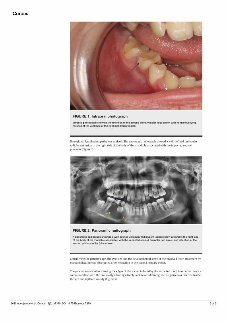
FIGURE 1: Intraoral photographIntraoral photograph showing the retention of the second primary molar (blue arrow) with normal overlyingmucosa of the vestibule of the right mandibular region
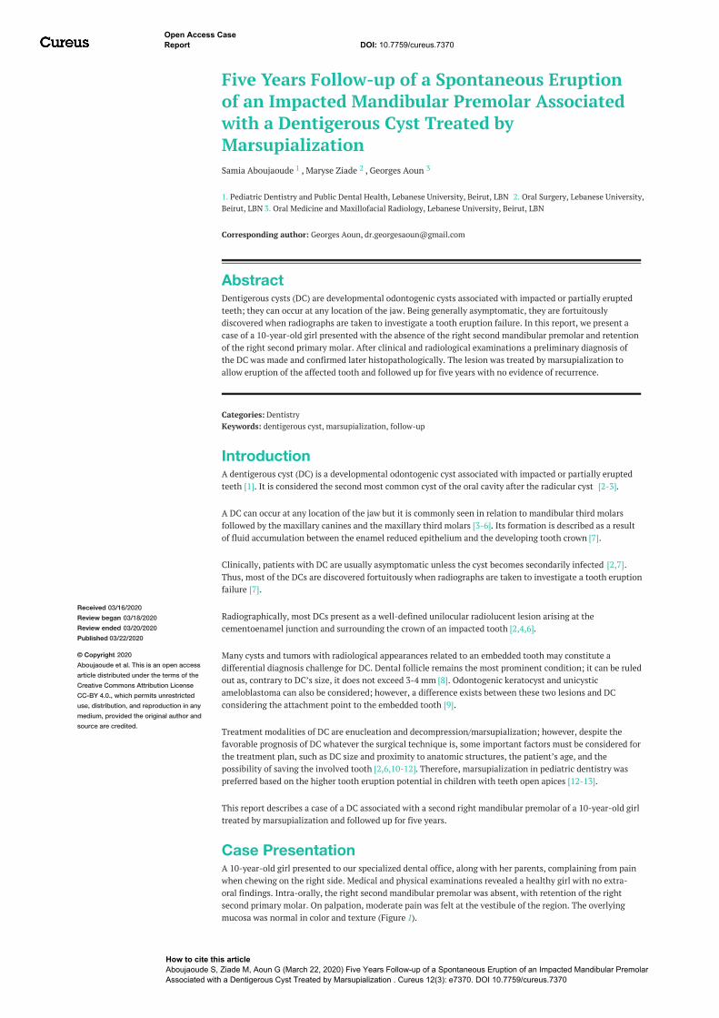
No regional lymphadenopathy was noticed. The panoramic radiograph showed a well-defined unilocularradiolucent lesion in the right side of the body of the mandible associated with the impacted secondpremolar (Figure 2).
FIGURE 2: Panoramic radiographA panoramic radiograph showing a well-defined unilocular radiolucent lesion (yellow arrows) in the right sideof the body of the mandible associated with the impacted second premolar (red arrow) and retention of thesecond primary molar (blue arrow)
Considering the patient’s age, the cyst size and the developmental stage of the involved tooth treatment bymarsupialization was effectuated after extraction of the second primary molar.
The process consisted in suturing the edges of the socket induced by the extracted tooth in order to create acommunication with the oral cavity allowing a freely continuous draining; sterile gauze was inserted insidethe site and replaced weekly (Figure 3).
2020 Aboujaoude et al. Cureus 12(3): e7370. DOI 10.7759/cureus.7370 2 of 6
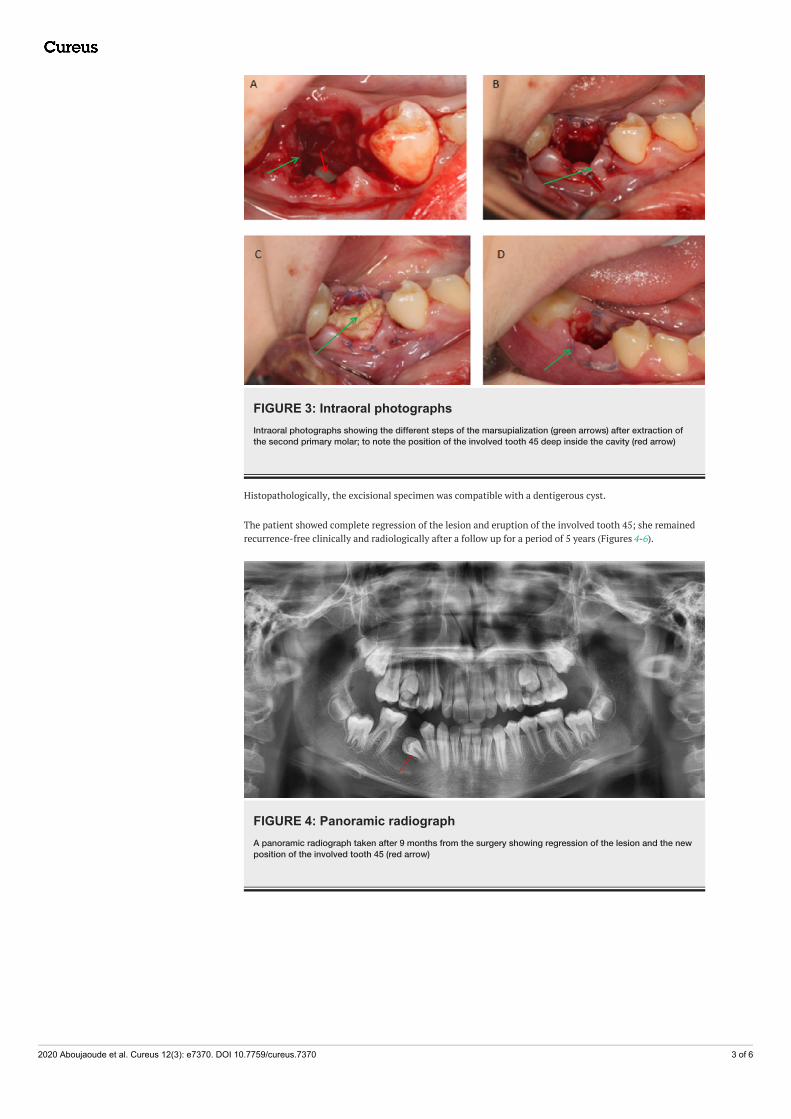
FIGURE 3: Intraoral photographsIntraoral photographs showing the different steps of the marsupialization (green arrows) after extraction ofthe second primary molar; to note the position of the involved tooth 45 deep inside the cavity (red arrow)
Histopathologically, the excisional specimen was compatible with a dentigerous cyst.
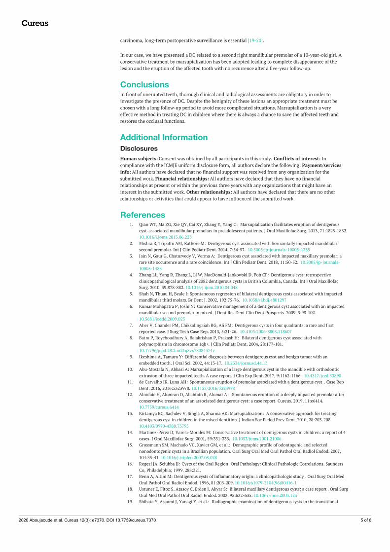
The patient showed complete regression of the lesion and eruption of the involved tooth 45; she remainedrecurrence-free clinically and radiologically after a follow up for a period of 5 years (Figures 4-6).
FIGURE 4: Panoramic radiographA panoramic radiograph taken after 9 months from the surgery showing regression of the lesion and the newposition of the involved tooth 45 (red arrow)
2020 Aboujaoude et al. Cureus 12(3): e7370. DOI 10.7759/cureus.7370 3 of 6
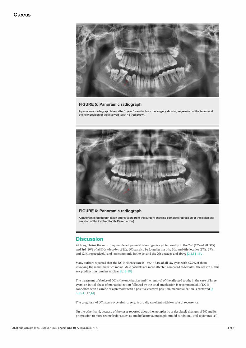
FIGURE 5: Panoramic radiographA panoramic radiograph taken after 1 year 8 months from the surgery showing regression of the lesion andthe new position of the involved tooth 45 (red arrow).
FIGURE 6: Panoramic radiographA panoramic radiograph taken after 5 years from the surgery showing complete regression of the lesion anderuption of the involved tooth 45 (red arrow)
DiscussionAlthough being the most frequent developmental odontogenic cyst to develop in the 2nd (23% of all DCs)and 3rd (20% of all DCs) decades of life, DC can also be found in the 4th, 5th, and 6th decades (17%, 17%,and 12 %, respectively) and less commonly in the 1st and the 7th decades and above [2,4,14-16].
Many authors reported that the DC incidence rate is 14% to 34% of all jaw cysts with 45.7% of theminvolving the mandibular 3rd molar. Male patients are more affected compared to females; the reason of thissex predilection remains unclear [4,16-18].
The treatment of choice of DC is the enucleation and the removal of the affected tooth; in the case of largecysts, an initial phase of marsupialization followed by the total enucleation is recommended. If DC isconnected with a canine or a premolar with a positive eruptive position, marsupialization is preferred [2-3,10-11,12,14].
The prognosis of DC, after successful surgery, is usually excellent with low rate of recurrence.
On the other hand, because of the cases reported about the metaplastic or dysplastic changes of DC and itsprogression to more severe lesions such as ameloblastoma, mucoepidermoid carcinoma, and squamous cell
2020 Aboujaoude et al. Cureus 12(3): e7370. DOI 10.7759/cureus.7370 4 of 6
carcinoma, long-term postoperative surveillance is essential [19-20].
In our case, we have presented a DC related to a second right mandibular premolar of a 10-year-old girl. Aconservative treatment by marsupialization has been adopted leading to complete disappearance of thelesion and the eruption of the affected tooth with no recurrence after a five-year follow-up.
ConclusionsIn front of unerupted teeth, thorough clinical and radiological assessments are obligatory in order toinvestigate the presence of DC. Despite the benignity of these lesions an appropriate treatment must bechosen with a long follow-up period to avoid more complicated situations. Marsupialization is a veryeffective method in treating DC in children where there is always a chance to save the affected teeth andrestores the occlusal functions.
Additional InformationDisclosuresHuman subjects: Consent was obtained by all participants in this study. Conflicts of interest: Incompliance with the ICMJE uniform disclosure form, all authors declare the following: Payment/servicesinfo: All authors have declared that no financial support was received from any organization for thesubmitted work. Financial relationships: All authors have declared that they have no financialrelationships at present or within the previous three years with any organizations that might have aninterest in the submitted work. Other relationships: All authors have declared that there are no otherrelationships or activities that could appear to have influenced the submitted work.
References1. Qian WT, Ma ZG, Xie QY, Cai XY, Zhang Y, Yang C: Marsupialization facilitates eruption of dentigerous
cyst-associated mandibular premolars in preadolescent patients. J Oral Maxillofac Surg. 2013, 71:1825-1832.10.1016/j.joms.2013.06.223
2. Mishra R, Tripathi AM, Rathore M: Dentigerous cyst associated with horizontally impacted mandibularsecond premolar. Int J Clin Pediatr Dent. 2014, 7:54-57. 10.5005/jp-journals-10005-1235
3. Jain N, Gaur G, Chaturvedy V, Verma A: Dentigerous cyst associated with impacted maxillary premolar: arare site occurrence and a rare coincidence. Int J Clin Pediatr Dent. 2018, 11:50-52. 10.5005/jp-journals-10005-1483
4. Zhang LL, Yang R, Zhang L, Li W, MacDonald-Jankowski D, Poh CF: Dentigerous cyst: retrospectiveclinicopathological analysis of 2082 dentigerous cysts in British Columbia, Canada. Int J Oral MaxillofacSurg. 2010, 39:878-882. 10.1016/j.ijom.2010.04.048
5. Shah N, Thuau H, Beale I: Spontaneous regression of bilateral dentigerous cysts associated with impactedmandibular third molars. Br Dent J. 2002, 192:75-76. 10.1038/sj.bdj.4801297
6. Kumar Mohapatra P, Joshi N: Conservative management of a dentigerous cyst associated with an impactedmandibular second premolar in mixed. J Dent Res Dent Clin Dent Prospects. 2009, 3:98-102.10.5681/joddd.2009.025
7. Aher V, Chander PM, Chikkalingaiah RG, Ali FM: Dentigerous cysts in four quadrants: a rare and firstreported case. J Surg Tech Case Rep. 2013, 5:21-26. 10.4103/2006-8808.118607
8. Batra P, Roychoudhury A, Balakrishan P, Prakash H: Bilateral dentigerous cyst associated withpolymorphism in chromosome 1qh+. J Clin Pediatr Dent. 2004, 28:177-181.10.17796/jcpd.28.2.m21q8vx78084374v
9. Ikeshima A, Tamura Y: Differential diagnosis between dentigerous cyst and benign tumor with anembedded tooth. J Oral Sci. 2002, 44:13-17. 10.2334/josnusd.44.13
10. Abu-Mostafa N, Abbasi A: Marsupialization of a large dentigerous cyst in the mandible with orthodonticextrusion of three impacted teeth. A case report. J Clin Exp Dent. 2017, 9:1162-1166. 10.4317/jced.53890
11. de Carvalho IK, Luna AH: Spontaneous eruption of premolar associated with a dentigerous cyst . Case RepDent. 2016, 2016:5323978. 10.1155/2016/5323978
12. Alnofaie H, Alomran O, Ababtain R, Alomar A : Spontaneous eruption of a deeply impacted premolar afterconservative treatment of an associated dentigerous cyst: a case report. Cureus. 2019, 11:e6414.10.7759/cureus.6414
13. Kirtaniya BC, Sachdev V, Singla A, Sharma AK: Marsupialization: A conservative approach for treatingdentigerous cyst in children in the mixed dentition. J Indian Soc Pedod Prev Dent. 2010, 28:203-208.10.4103/0970-4388.73795
14. Martínez-Pérez D, Varela-Morales M: Conservative treatment of dentigerous cysts in children: a report of 4cases. J Oral Maxillofac Surg. 2001, 59:331-333. 10.1053/joms.2001.21006
15. Grossmann SM, Machado VC, Xavier GM, et al.: Demographic profile of odontogenic and selectednonodontogenic cysts in a Brazilian population. Oral Surg Oral Med Oral Pathol Oral Radiol Endod. 2007,104:35-41. 10.1016/j.tripleo.2007.05.028
16. Regezi JA, Sciubba JJ: Cysts of the Oral Region. Oral Pathology: Clinical Pathologic Correlations. SaundersCo, Philadelphia; 1999. 288:321.
17. Benn A, Altini M: Dentigerous cysts of inflammatory origin: a clinicopathologic study . Oral Surg Oral MedOral Pathol Oral Radiol Endod. 1996, 81:203-209. 10.1016/s1079-2104(96)80416-1
18. Ustuner E, Fitoz S, Atasoy C, Erden I, Akyar S: Bilateral maxillary dentigerous cysts: a case report . Oral SurgOral Med Oral Pathol Oral Radiol Endod. 2003, 95:632-635. 10.1067/moe.2003.123
19. Shibata Y, Asaumi J, Yanagi Y, et al.: Radiographic examination of dentigerous cysts in the transitional
2020 Aboujaoude et al. Cureus 12(3): e7370. DOI 10.7759/cureus.7370 5 of 6

dentition. Dentomaxillofac Radiol. 2004, 33:17-20. 10.1259/dmfr/2414836320. Vasiapphan H, Christopher PJ, Kengasubbiah S, Shenoy V, Kumar S, Paranthaman A: Bilateral dentigerous
cyst in impacted mandibular third molars: a case report. Cureus. 2018, 10:3691. 10.7759/cureus.3691
2020 Aboujaoude et al. Cureus 12(3): e7370. DOI 10.7759/cureus.7370 6 of 6