Embed Size (px)
Citation preview

ContEntS
Page
Printed by: Airlangga University Press. (139/09.10/AUP-B5E). Kampus C Unair, Jln. Mulyorejo Surabaya 60115, Indonesia. Telp. (031) 5992246, 5992247, Telp./Fax. (031) 5992248. E-mail: [email protected]. Ijin penerbit: No. 0787/SK/Dir. PK/SIT/1969. Accredited No. 48/DIKTI/Kep/2006.
Volume 43 Number 2 April 2010 ISSN 1978 - 3728
Dental Journal MajalahKedokteranGigi
1. Erythema multiforme as the result of taking carbamazepine Maharani laillyza apriasari and M. Jusri .................................................................................... 49–53
2. Tensile bond strength of hydroxyethyl methacrylate dentin bonding agent on dentin surface at various drying techniques
Kun ismiyatin ................................................................................................................................... 54–57
3. The efficacy of honey solution as plaque reducing agent dewi nurul M, indria rizki S, indriani S, Masyitoh, and auerkari Ei ..................................... 58–61
4. Optimum dose of 2-hydroxyethyl methacrylate based bonding material on pulp cells toxicity Widya Saraswati .............................................................................................................................. 62–66
5. Ankylosis of the temporomandibular joint and mandibular growth disturbance caused by neglected condylar fracture in childhood
Endrajana ........................................................................................................................................ 67–71
6. The effect of monofluorophosphate implant in white rat mothers towards the level of fluor in the incisors of the young babies (Rattus-rattus)
Widjijono .......................................................................................................................................... 72–75
7. Early removal of odontoma resulting in spontaneous eruption of the impacted teeth achmad harijadi ............................................................................................................................. 76–80
8. Dental modifications: a perspective of Indonesian chronology and the current applications rusyad adi Suriyanto and toetik Koesbardiati ............................................................................ 81–90
9. Enamel defect of deciduous teeth in small gestational age children Willyanti S Syarif, roosje r. oewen, Sjarif h. Effendi and Bambang Sutrisna ....................... 91–96
10. Integrated orofacial therapy in chronic rhinosinusitis management for children with sleep bruxism
haryono utomo ................................................................................................................................ 97–101
11. The apical leakage of mineral trioxide agregate as the retrograde filling material with various mixing agents
Ema Mulyawati ................................................................................................................................ 102–106
Daftar isi
72
Vol. 43. No. 2 June 2010
Research Report
The effect of monofluorophosphate implant in white rat mothers towards the level of fluor in the incisors of their young babies (Rattus-rattus)
WidjijonoDepartment of Biomaterial Faculty of Dentistry, Gadjah Mada UniversityYogyakarta - Indonesia
abstract
Background: Fluoridehasbeenwidelyusedinthepreventionofdentalcariesforalongtime.Topreventdentalcaries,fluoridemustbeinducedinlowamountathighfrequency.Inducingitthroughimplantationprocessevenmakeslowreleaseofsmallconcentrationoffluoride.Purpose:Theaimofthisresearchwastoanalyzewhethertheinductionofmonofluorophosphate(MFP)implantintothewhiteratmothersaffectstheleveloffluorideintheincisorsoftheiryoungbabies.Method:Theobjectsoftheresearchweretwentywhiteratmothersintwodaysofpregnancywhichthenweredividedintofourgroups(n=5).First,thosemothershavebeeninducedwithimplantundertheirbackskinuntiltheirbornyoungbabiesintheageof35days(n=5).TheleveloffluorideintheincisorsofthoseyoungbabiesthenismeasuredwithPotentiometer.TheobtaineddatawerefinallyanalyzedwithOne-WayANOVAtestandcontinuedbywithLSDtest(p=0.05).result: Theresultofthisresearchshowedthatthemeansofthefluoridelevelintheincisorsofthosebabiesdividedintothosefourgroupsinserieswereabout11956.16±201.35ppb(K),27328.04±234.56ppb(P1),37267.21±248.86ppb(P2),and18103.50±267.11ppb(P3).TheresultofANOVAtestthenshowedthattheinductionofvariousMFPimplantlevelssignificantlyaffectedtheleveloffluorideintheincisorsofthebabies.ThemeandifferencesamongthetreatmentgroupsafterbeingtestedwithLSD0.05werealsosignificant.Conclusion: ThefindingconfirmthatthesignificantincreasingoftheoptimalfluorideretentionintheincisorsofwhiteratbabiescanbeachievedwiththeinductionoffluoridewithMFPionsimplantinabout52.98mg.
Key words: MFPimplant,fluoride,theincisorsofwhiterats
abstrak
latar Belakang: Pencegahankariesgigimenggunakansenyawafluortelahbanyakdilakukandanberlangsungdalamjangkawaktulama.Pemberianfluordalamjumlahrendahdanfrekuensitinggimerupakanpemenuhankebutuhanpencegahankariesgigi.Pemberiandengancaraimplantasimemberikankeluaranfluorjumlahkecildanwaktulama.tujuan: Penelitianinibertujuanuntukmengetahuiapakahinduktikusyangdiberiimplan-MFPberpengaruhterhadapkandunganfluorgigiserianaktikus.Metode:Subjekpenelitianadalah20ekorinduktikusputihbunting2haridibagi4kelompok(n=5).Indukdiberiimplanpadabawah-kulitpunggunghinggaanaktikuslahirdanpadaumur35hari(n=5).KandunganfluorpadagigiserianaktikusdiukurmenggunakanPotensiometer.Data yang diperoleh dianalisis dengan Anova 1 jalur dilanjutkan uji LSD (p=0,05). Hasil: penelitian menunjukkan rerata fluorgigiserianaktikusberturut-turutsebesar:11956,16±201,35ppb(K),27328,04±234.56ppb(P1),37267,21±248.86ppb(P2),dan18103,50±267,11ppb(P3).hasil: AnavamembuktikanbahwaadapengaruhbermaknaakibatvariasikadarMFPdalamimplanterhadapkandunganfluorgigianaktikus.BedarerataantarkelompokperlakuandiujidenganLSD0,05memperlihatkanperbedaanbermaknapadasemuakelompok.Kesimpulan: PenelitiandapatdisimpulkanbahwakenaikansecarabermaknaterhadapretensifluoroptimaldalamgigiseritikusputihpadapemberianfluoridasimenggunakanimplandenganmuatanMFP:52,98mg.
Kata kunci:Implan-MFP,fluor,gigiseritikusputih
Correspondence: Widjijono, c/o: Bagian Biomaterial, Fakultas Kedokteran Gigi Universitas Gadjah Mada. Jl. Denta I, Sekip Utara Yogyakarta 55281. E-mail: [email protected]

73Widjijono: The effect of monofluorophosphate implant
introduction
The prevention of dental caries with chemical application using fluoride has already been done. However, the prevention of dental caries must be conducted in the long term process started from the formation of tooth until they get maturation. The prevention of dental caries even is supposed to be conducted minimally until the age of twelve. Unfortunately, the prevention of dental caries using fluoride given orally cannot be well controlled since it can cause dental fluorosis. Therefore, the alternative way as such implantation process is needed to control the application of fluoride especially to prevent fluorosis.
The main usage effects of fluoride on body actually can be particularly seen in calcificated tissues since fluoride has narrow therapeutic window. Fluoride at low concentration level can increase the crystalinity of teeth and bones, while at the high concentration level it can cause abnormality in calcificated tissues, such as fluorosis.1 Induction of fluoride at low concentration with high frequency of application can prevent dental caries.2 In other words, the daily controlled of fluoride intake will effectively decrease dental caries and fluorosis risks.3 Induction of fluoride through implantation will cause fluoride released in small amount and in long term.
Sodium-monofluorophosphate (natrium-monofosfat, MFP) considered as potential fluoride that can be used as anti-caries with low toxicity which is not only more effective than NaF, but also can be absorbed faster without being affected by calcium ions.3 MFP is chosen because it has anti-caries, its toxicity about one third lower than that in NaF,4 it still can be degraded by alkali or acid phosphate through hydrolysis process,5 and by the substitution of the structure of phosphate hydrogen on hydroxy apatite with monofluorophosphate ion.6 The dissolution of MFP will 20 times faster if the level of calcium ions is so high to make calcium-monofluorophosphate compound that easily dissolved.7
In recent years, various products of medicines modified with controlled fluoride release have actually been developed for several reasons. The first one is because of their affectivity and efficiency. Another one is to solve the impracticality caused by the routine of the long term treatment. The various use of polymer in dental health has widely developed.8 The selection of polymer type, poly-dl-lactic acids (PLA), in implant materials is aimed to obtain fluoride carriers through controlled releasing. Besides that, PLA in the form of monolith has also several characteristics, such as small degradation constant, bio-erosion, and low permeability.9
Fluoride induced orally makes the dissolved fluoride be absorbed and diffused in a simple way through digestive system, especially gastro-intestinal tract.10 Inducing through the implant of endoderm is directly absorbed and diffused into blood vessel. The reason is because fluoride induced orally must diffuse through first pass of metabolism channel. As a result, none of fluoride intake
can be absorbed. On the other side, MFP implant induced in the mothers of young rats makes fluoride intake can easily be absorbed by those mothers and be diffused into blood. Some of the fluoride diffused into those mothers’ blood then will be induced into placenta and will be functioned as the source of fluoride for their fetuses. Placenta also functions as partial barrier when the amount of fluoride is suddenly increased. There is a direct correlation between the amount of serum in mothers and in the placenta of their fetuses. The amount of fluoride in placenta is 75% bigger than that in their mothers.11
The previous researches actually have already pointed out that fluoride in the form of MFP implant can be distributed homogeneously in the form of monolith (bar), and fluoride release can be controlled seven days after the implantation.12 The aim of this study was to analyze the amount of fluoride in the incisors of the white rat babies whose mothers were induced with various levels of MFP in the form of poly-dl-lactate acid implant.
material and method
The objects of the research are twenty Wistar white rat mothers (Rattus-rattus) in two days of pregnancy. The main materials used in this study, are natrium-monofluorophosphat (MFP) (Na@211, Australia), poly-dl-lactate acid (Poly Science, USA) and formalin.
MFP implant consists of MFP and PLA with the ratio 20:80. In other words, MFP implant is made based on the ratio of MFP to poly-dl-lactate acid, 20:80 (b/b mg). The implant was made according to Beck et al. method.13 For MFP implant (P1), there are two kinds of solution. Solution I is made by dissolving 26.49 mg of MFP to 3 ml of methanol, meanwhile solution II is made by dissolving 105.96 mg of PLA to 7 ml of chloroform and methilen chloride mixed with the ratio 1:1. Solution I and solution II then are mixed together until they become homogenous. Organic solvent then is steamed with dryer at temperature of 50° C and stirred until the dough becomes plastic. Afterwards, the dough is poured into die and pressed by hydraulic pressure about 50 kgf/cm2 for 1 minute. Similarly, implants (P2), (P3) and implant (K) are made of MFP which in series are about 52.98 mg, 264.9 mg and 0 mg (Table 1).
table �. Implants K, P1, P2 and P3 with their MFP-PLA level ratio
Implants MFP(mg) PLA (mg)
Implant K 0 100
Implant P1 26.49 105.96
Implant P2 52.98 211.92
Implant P3 264.9 1059.6
74 Dent. J. (Maj. Ked. Gigi), Vol. 43. No. 2 June 2010: 72-75
Twenty white rat mothers in two days of pregnancy are divided into four groups, group 1 with controlled implant treatment without MFP (K), group 2 with implant P1, group 3 with implant P2, and group 4 with implant P3. Implantation process then is conducted on the back skin of those mothers. That skin must be cleanly shaved before being disinfected. The incision of the skin of their back is conducted for about 0.5 cm long. After that, the blunt dissection is conducted along the implant, 1 cm from the incision edge. Next, the implant is inserted into the cavity, and the wound of the incision then needs silk-thread stitches. Those mothers then must be on normal ad libitum diet modified without fluoride. After those mothers delivered their babies, the incisors of their 35 day old babies must be extracted under anesthetic. The extracted teeth are measured by cutting them into small pieces, and they then are dissolved in 2.5 ml of 65% HNO3. Afterwards, 1 ml of the dental solution is taken as the sample which then is mixed with 9 ml of 0.5M HClO4 and 10 ml TISAB II. Specific Potentiometer of Fluoride ions connected with digital TPS (Titralizer Action Electrode, Calomel Refference) is used to determine the level of fluoride based on the interpolation mechanism related with the potential difference measured with the standard solution curve. The data is analyzed by using one way variant analysis in order to analyze the effect of MFP implant variants towards the level of fluoride in the incisors of those white rat babies, meanwhile and the mean of the difference of fluoride level in the incisors among treatment groups is tested with LSD 0.05.
result
This research finds that the means of fluoride level in the incisors of those white rat babies is as the following table 2.
table ��. The mean and standard deviation of fluoride level in the incisors of white rats
Groups Mean± SD (ppb)
K 11956.16 ± 201.35
P1 27328.04 ± 234.56
P2 37267.21 ± 248.86
P3 18103.50 ± 267.11
K: Control, P1: Sample with 26.96 mg MFP group, P2: Sample with 52.98 mg MFP group, P3: Sample with 264.9 mg MFP group
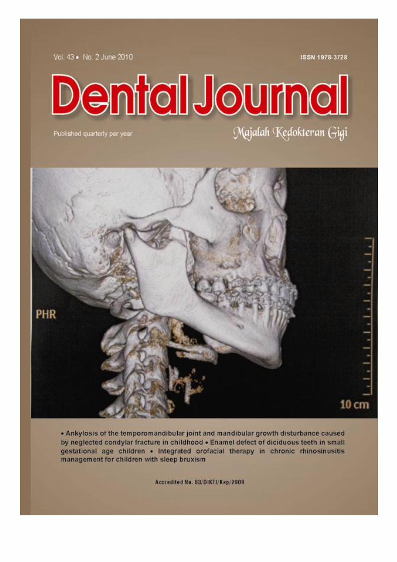
Graphically, it also shows that the increasing of fluoride is not linear, but at certain levels it can reach the optimum absorption level. At the increasing level above the optimum one, the amount of fluoride absorbed is decreased (Figure 1).
The level of fluoride in the incisors of those white rat babies classified into three treatment groups, P1, P2 and
P3, induced with implant is gradually increased, in series about 2.3 times, 3.2 times, and 1.5 times bigger than the controlled ones. It means that the biggest increasing of the amount of fluoride occurs in the treatment group induced with implant P2 (52.98 mg of MFP).
05000
10000150002000025000300003500040000
M F P - 0M F P - 26M F P - 58M F P - 264
figure �. Graph of fluoride level in the incisor of thirty-five day old white rat babies (ppb).
table ��. The result of LSD0.05 test between the controlled group and the treatment groups, P1, P2 and P3
Groups K P1 P2 P3
K - 15371.878* 25311.056* 6147.338*
P1 - 9939.178* 9224.540*
P2 - 19163.718*
P3 -
Note: *) Significant difference (p<0.05) All of the treatment groups are significantly different from the controlled ones
Through the statistic analysis using SPSS 13.0 for Window with one way ANOVA, moreover, it is also known that F is about 10675.56, bigger than F in the table. This result indicates that the variants of MFP implant level significantly affect the level of fluoride in the incisors of those white rat babies. The difference of means among those treatment groups tested with LSD 0.05 also shows that there is significant difference in those treatment groups (table 3).
discussion
The use of MFP implant is aimed to obtain the lower and controlled fluoride release. Polylactate-natrium monofluorophosphate implant in the bar shape even can be dispersed in homogeneous way. In other words, by using MFP implant the controlled fluoride release occurred seven days after implantation.12 The purpose of selecting PLA is to obtain fluoride carriers with controlled disposal. PLA in the form of monolith actually has several characteristics, such as: small degradation constant, bio-erosion, and low permeability.9 Thus, if the reason of the use of MFP is because of anti-caries activities in MFP with its toxicity about one third lower than that in NaF,3 it still can be degraded by alkali or acid phosphate

75Widjijono: The effect of monofluorophosphate implant
through hydrolysis process,5 and by the substitution of the structure of phosphate hydrogen on hydroxy apatite with monofluorophosphate ion.6
The average of fluoride contained on the incisors of those thirty-five day old white rat babies and the binding of fluoride ion on the teeth, actually depends on the level of fluoride in their blood plasma. The increasing of fluoride level in the certain availability of plasma will increase the amount of fluoride bound on teeth. The increasing of the availability in their blood plasma which is more than the optimal one can decrease the binding of dental fluor (Figure 1). P2 implant has the average of plasma fluoride level, about 125.61± 23.30 ppb,12 which is not only appropriate with the level of blood fluoride, about 0.1–0.2 ppm,10 but is also close to the level of therapeutic-hypothetic in blood plasma of society with the level of consumed water about 1 ppm. Moreover, the variants of fluoride level in their incisors caused by the increasing amount of fluoride that can also be explained by the opinion of Fejerkov et al.,1 stating that fluoride has narrow therapeutic window mechanism, which means that at the low dosage its influence is not significant, but at the high dosage it can disrupt the growth of dental structure. Fluoride with low dosage (such as in P1 implant), will increase dental crystalinity. It is possibly caused by the reaction of the substitution of isoionic F with hydroxyl structure in dental apatite which then can form apatite fluoride or hydroxy-fluor apatite. The forming of fluor apatite crystal is also supported by a research conducted by Monjoetal.,15 stating that the inducing of implant with fluoride modification in bones can not only make fluoride modulate the forming of osteogenic marker, but also increase the density of bones located in interface part between implant and bones. The surface of implant modified with fluoride can increase osseointegration in the early stage of recovery.16
On the other hand, fluoride in the high dosage (such as in P3 implant) can cause disruption during the growth of enamel affecting in enamel organic and inorganic components which then causes either hypoplasia of permanent teeth10 or partial resistance in proteinase that has responsibility for breaking enamel protein. As a result, it can either decelerate protein disposal during maturation or cause the disruption of calcification or fluorosis.14 It means that the disruption in disposal process of protein substituted with mineral can relatively decrease the amount
of fluoride released from the disrupted tissue. Therefore, it can be concluded that the optimal resistance of fluoride in the incisors of white rat babies is significantly increased during fluoridation using implant with MFP ions, about 52.98 mg.
references
1. Fejerkov O, Richards A, DenBasten P. The effect of fluoride on the tooth Mine-ralization. In: Ekstrand J, Burt BA, editors. Fluoride in dentistry. Copenhagen: Munksgaard; 1996; p. 112–47.
2. Hargreaves JA. Water fluoridation and fluoride supplementation: Consideration for future. J Dent Res 1990; 69(Spec Iss): 775–70.
3. Cremer HD, Buttner W. Absorption of fluoride. In: Fluoride and human health. Geneva: WHO; 1970. p. 84–5.
4. White WE. Monofluorophosphate–its beginning. Caries Res 1983; 17(Suppl 1): 2–8.
5. Pearce EIF. Biochemistry of monofluorophosphate. Caries Res 1983; 17(Suppl 1): 21–35.
6. Ingram GS. The reaction of monofluorophosphate with apatite. Caries Res 1972; 6: 1–15.
7. Ericsson Y. Monofluorophosphate physiology: General considerations. Caries Res 1983; 17(Suppl 1): 46–55.
8. Shargel L, Yu ABC. 1985. Biofarmasetika dan farmakokinetika terapan. Edisi 2. Surabaya: Penerbit Universitas Airlangga (AUP); 1988. p. 454–56.
9. Pitt CG, Schindler A. The design of controlled drug delivery system based on biodegradable polymers. In: Hafez ESE, van Os WAA, editors. Biodegradable and delivery system for contraception. Boston: GK Hall Medical Pub; 1980. p. 27.
10. Cole AS, Eastoe JE. Biochemistry and oral biology. Singapore: Toppan Co Ltd; 1977. p. 123.
11. Ekstrand J. Fluoride metabolism. In: Ekstrand J, Burt BA, editors. Fluoride in dentistry. Copenhagen: Munksgaard; 1996. p. 112–47.
12. Widjijono. Penggunaan implan polilaktat-natrium monofluorofosfat dengan kajian availabilitas fluor sediaan, biokompatilitas dan bioavailabilitas fluoride dalam darah dan gigi pada tikus putih. Disertasi. Surabaya: Program Pascasarjana Universitas Airlangga; 2001. p. 115.
13. Beck LR, Cowsar DR, Lewis DH. Systemic and Local delivery of contraceptive steroids using biodegradable microcapsules. In: Hafez ESE, van Os WAA, editors Biodegradable and delivery system for contraception. Boston: GK Hall Medical Pub; 1980. p. 63–82.
14. Bawden JW, Crenshaw MA, Wright GT, LeGeros RZ. Consideration of possible biologic mechanism of fluorosis. J Dent Res 1995; 74(7): 1349–52.
15. Monjo M, Lamolle SF, Lyngstadaas SP, Ronold HJ, Ekkingsen JE. In vivo expression of osteogenic marker and bone mineral density at the surface of fluoride-modified titanium implants. Biomaterials 2008; 29(28): 3771–80.
16. Berglundh T, Abrahamsson I, Albouy JP, Linde J, Bone healing at implants with a fluoride-modified surface: an experimental research in dogs. Clinical Oral Implants Research 2007; 18(2): 147–52.