Embed Size (px)
Citation preview


FIRST AID
NBDE PART IThird Edition
DEREK M. STEINBACHER, DMD, MD, FAAPAssistant ProfessorPlastic and Reconstructive SurgeryDirector of Craniofacial SurgeryYale University School of MedicineNew Haven, Connecticut
STEVEN R. SIERAKOWSKI, DMD, MDScDiplomate, American Board of PeriodontologyPrivate practicePhiladelphia, Pennsylvania
New York / Chicago / San Francisco / Lisbon / London / Madrid / Mexico City
Milan / New Delhi / San Juan / Seoul / Singapore / Sydney / Toronto
FORTHE®
Copyright © 2012 by The McGraw-Hill Companies, Inc. All rights reserved. Except as permitted under the United States Copyright Act of 1976, no part of this publication may be reproduced or distributed in any form or by any means, or stored in a database or retrieval system, without the prior written permission of the publisher.
ISBN: 978-0-07-177359-1
MHID: 0-07-177359-2
The material in this eBook also appears in the print version of this title: ISBN: 978-0-07-176904-4, MHID: 0-07-176904-8.
All trademarks are trademarks of their respective owners. Rather than put a trademark symbol after every occurrence of a trademarked name, we use names in an editorial fashion only, and to the benefi t of the trademark owner, with no intention of infringement of the trademark. Where such designations appear in this book, they have been printed with initial caps.
McGraw-Hill eBooks are available at special quantity discounts to use as premiums and sales promotions, or for use in corporate training programs. To contact a representative please e-mail us at [email protected].
Previous editions copyright © 2009, 2007 by The McGraw-Hill Companies, Inc.
First Aid for the® is a registered trademark of The McGraw-Hill Companies, Inc.
NOTICE
Medicine is an ever-changing science. As new research and clinical experience broaden our knowledge, changes in treatment and drug therapy are required. The authors and the publisher of this work have checked with sources believed to be reliable in their efforts to provide information that is complete and generally in accord with the standards accepted at the time of publication. However, in view of the possibility of human error or changes in medical sciences, neither the authors nor the publisher nor any other party who has been involved in the preparation or publication of this work warrants that the information contained herein is in every respect accurate or complete, and they disclaim all responsibility for any errors or omissions or for the results obtained from use of the information contained in this work. Readers are encouraged to confirm the information contained herein with other sources. For example and in particular, readers are advised to check the product information sheet included in the package of each drug they plan to administer to be certain that the information contained in this work is accurate and that changes have not been made in the recommended dose or in the contraindications for administration. This recommendation is of particular importance in connection with new or infrequently used drugs.
TERMS OF USE
This is a copyrighted work and The McGraw-Hill Companies, Inc. (“McGraw-Hill”) and its licensors reserve all rights in and to the work. Use of this work is subject to these terms. Except as permitted under the Copyright Act of 1976 and the right to store and retrieve one copy of the work, you may not decompile, disassemble, reverse engineer, reproduce, modify, create derivative works based upon, transmit, distribute, disseminate, sell, publish or sublicense the work or any part of it without McGraw-Hill’s prior consent. You may use the work for your own noncommercial and personal use; any other use of the work is strictly prohibited. Your right to use the work may be terminated if you fail to comply with these terms.
THE WORK IS PROVIDED “AS IS.” McGRAW-HILL AND ITS LICENSORS MAKE NO GUARANTEES OR WARRANTIES AS TO THE ACCURACY, ADEQUACY OR COMPLETENESS OF OR RESULTS TO BE OBTAINED FROM USING THE WORK, INCLUDING ANY INFORMATION THAT CAN BE ACCESSED THROUGH THE WORK VIA HYPERLINK OR OTHERWISE, AND EXPRESSLY DISCLAIM ANY WARRANTY, EXPRESS OR IMPLIED, INCLUDING BUT NOT LIMITED TO IMPLIED WARRANTIES OF MERCHANTABILITY OR FITNESS FOR A PARTICULAR PURPOSE. McGraw-Hill and its licensors do not warrant or guarantee that the functions contained in the work will meet your requirements or that its operation will be uninterrupted or error free. Neither McGraw-Hill nor its licensors shall be liable to you or anyone else for any inaccuracy, error or omission, regardless of cause, in the work or for any damages resulting therefrom. McGraw-Hill has no responsibility for the content of any information accessed through the work. Under no circumstances shall McGraw-Hill and/or its licensors be liable for any indirect, incidental, special, punitive, consequential or similar damages that result from the use of or inability to use the work, even if any of them has been advised of the possibility of such damages. This limitation of liability shall apply to any claim or cause whatsoever whether such claim or cause arises in contract, tort or otherwise.

DE DICATION
To our families and loved ones who supported us through this endeavor,
and
To those mentors who sparked our enthusiasm for learning.
Derek M. Steinbacher, DMD, MD, FAAP
Steven R. Sierakowski, DMD, MDSc

This page intentionally left blank

v
Contributing Authors vii
Acknowledgments ix
How to Contribute x
How to Use This Book xi
Introduction xiii
SECT ION I ANATOM IC SC I E NC ES 1
Chapter 1 Gross Anatomy 3
Chapter 2 General Histology 153
Chapter 3 Oral Histology 195
Chapter 4 Developmental Biology 215
SECT ION I I BIOC H E M ISTRY–PHYSIOLOGY 247
Chapter 5 Physical-Chemical Principles 249
Chapter 6 Biological Compounds 257
Chapter 7 Metabolism 277
Chapter 8 Molecular Biology 291
Chapter 9 Membranes 301
Chapter 10 Neurophysiology 305
Chapter 11 Muscle Physiology 337
Chapter 12 Circulatory and Cardiac Physiology 345
Chapter 13 Respiratory Physiology 369
Chapter 14 Renal, Fluid, and Acid-Base Physiology 383
Chapter 15 Gastrointestinal Physiology 397
Chapter 16 Nutrition 407
Chapter 17 Endocrine Physiology 413
CONTENTS

vi
SECT ION I I I M IC ROBIOLOGY–PATHOLOGY 439
Chapter 18 Microbiology 441
Chapter 19 Oral Microbiology and Pathology 489
Chapter 20 Reactions to Tissue Injury 497
Chapter 21 Immunology and Immunopathology 509
Chapter 22 Systemic Pathology 527
Chapter 23 Neoplasia 599
SECT ION IV DE NTAL ANATOMY AN D OCC LUSION 629
Chapter 24 Tooth Morphology 631
Chapter 25 Pulp Morphology 653
Chapter 26 Calcification and Eruption 657
Chapter 27 Occlusion and Function 661
Chapter 28 Tooth Anomalies 673
Index 677

vii
CONTRIBUTING AUTHORS
Jordan BowerTemple University Kornberg School of Dentistry, Class of 2013
Tommy BurkHarvard School of Dental Medicine, Class of 2013
Austin EckardHarvard School of Dental Medicine, Class of 2013
Raha Ghafurian, DMD, MSEdUniversity of Pennsylvania School of Dental Medicine, 2010 Yale Pediatric Dental Fellow
Virgina HogsettHarvard School of Dental Medicine, Class of 2013
Grace KimHarvard School of Dental Medicine, Class of 2013
Sarah KrygowskiHarvard School of Dental Medicine, Class of 2013
Katie McCafferty Harvard School of Dental Medicine, Class of 2013
Veronica MitkoHarvard School of Dental Medicine, Class of 2013
Marc D. Thomas, DDSUCLA School of Dentistry, 2008Yale Pediatric Dental Fellow
Janelle Tonn, DDSLoma Linda School of Dentistry, 2010Yale Pediatric Dental Fellow

This page intentionally left blank

ix
ACKNOWLEDGMENTS
Thanks to our publisher, McGraw-Hill, for making this third edition areality. We extend sincere gratitude to our editor, Catherine Johnson, for hercontinued support and guidance. Thanks also to Peter Boyle for supervisingthe production process and to the graphics team for their excellent renderingof anatomic illustrations.
Derek M. Steinbacher, DMD, MD, FAAPSteven R. Sierakowski, DMD, MDSc

x
We welcome your comments, suggestions, ideas, corrections, or other submissions to help with this andfuture editions of First Aid for the NBDE Part I. What information can you contribute?
■ Test-taking tips and study strategies for the exam■ Suggestions for mnemonics, diagrams, figures, and tables■ Critiques related to the content or arrangement of facts contained herein■ Other sources of study material you find useful
Contributions used will result in a personal acknowledgment in the next edition of this book.
If you wish to contribute, e-mail your entries or suggestions to the following address:
Please include your name, address, e-mail address, and school affiliation.
NOTE TO CONTRI BUTORS
All contributions become property of the authors and are subject to editorial manipulation. In the eventthat similar or duplicate entries are received, only the first entry received will be used. Please include areference to a standard textbook to verify the factual data.
HOW TO CONTRIBUTE

xi
HOW TO USE THIS BOOK
We feel that this book is an organized and resourceful guide to help you prepare for the NBDE Part I.
It is recommended that you use this book as early as possible, preferably in conjunction with your basic sci-ence and dental anatomy curriculum. The information contained within this book highlights the majorfacts and concepts commonly used in the NBDE Part I. Determine your own study strategy using not onlythis book, but also the materials you find most appropriate: concise review texts, the Internet, or your ownclass notes.
First Aid for the NBDE Part I is not meant to be a comprehensive review text; nor is it meant to be used as asubstitute for a lack of studying during the first two years of dental school. As you study each topic, refer tothe corresponding section in this book for a concise review or self-test. You may even want to make yourown notes in the margins or highlight particular facts.
Some of the information found in the NBDE Part I is repeated in different sections. Many of the Key Factsin this book will guide you in cross-referencing a particular topic to another section. Use this to test yourselfand integrate these facts into your body of knowledge.
During the last week before the exam, review the topics about which you feel the most unsure. The tablesand figures in this book will help you keep the heavily-tested information fresh in your memory.
As soon as possible after you take the exam, we recommend that you review the book to help us create aneven better fourth edition. Tell us what information should be revised, included, or removed. You may evensend in your own annotated book.

This page intentionally left blank

xiii
INTRODUCTION
The National Board Dental Examination (NBDE) Part I is the first of two national standardized examina-tions administered by the Joint Commission on National Dental Examinations. Its purpose is to provide statedental licensing boards an objective method of evaluating the qualifications of prospective applicants. TheNBDE Part I is intended to assess competency in the preclinical dental and basic biomedical sciences.
WHY DO WE LL ON TH E EXAM?
Achieving a passing score is a necessary requisite for both graduation from a dental school accredited bythe Commission on Dental Accreditation, and for obtaining dental licensure in the United States.Although each state reserves the authority to use the scores from the NBDE as a requisite to fulfill itswritten examination requirement, all 50 states, the District of Columbia, Puerto Rico, and the VirginIslands use the NBDE for their licensing protocol. Additionally, each jurisdiction may have its ownminimum NBDE Part I score requirement in order to obtain a dental license. It is essential that thefuture dentist be aware of the licensure requirements for each state in which he or she wishes to practice.A score of 85 or above is adequate in every state.
The NBDE Part I scores are also a major criterion used to gauge the quality of students applying to post-doctoral training programs. Since this examination is usually taken after the second year of dental school,it is often the only objective method of comparing soon-to-be graduates for such programs. Performing wellon the NBDE Part I gives the student more bargaining potential when applying for these programs, aswell as positions in academic, military, or private practice settings. In short, it behooves you to earn agood score on the NBDE Part I. Much diligence and a targeted study strategy, using materials like thisbook, will help you accomplish this goal.
STRUCTU RE OF TH E EXAM I NATION
The NBDE Part I is administered at Prometric Test Centers throughout the United States, its territories,and Canada. Once your application has been approved by the Joint Commission office, you will be notifiedby mail to contact Prometric to register for the examination. You have 12 months after application approvalto take the examination. A list of test centers can be found at www.prometric.com. The fee for the examis $260.
The exam consists of two test sections of 200 questions each, for a total of 400 questions. The questionsare in the form of multiple-choice test items, each with four to five possible answer choices. Approximately80% of the questions are individual discipline-based items, while 20% are testlet questions based on a patient-centered scenario. All of the test items are evenly distributed in four broad categories:
■ Anatomic Sciences■ Microbiology and Pathology■ Biochemistry and Physiology■ Dental Anatomy and Occlusion

xiv
The total testing time is seven hours, allowing 3.5 hours per section with an optional one-hour (maximum)break between sections. There is a 15-minute tutorial at the start of the exam and a post-examinationsurvey to complete when you are finished.
SCORI NG OF TH E EXAM I NATION
The NBDE Part I is scored on a scale of 49 to 99. Scores are graded against a predetermined standard andare not based on a curve. The total number of questions you answered correctly (your raw score) deter-mines your scaled score. There is no penalty for answering a question incorrectly. Furthermore, up to 15%of the questions may be discarded, as they may be used for other evaluation purposes. A scaled score of 75for each section is considered the minimum passing score for the NBDE Part I.
Your score report will be mailed approximately three to four weeks after the examination. You will receivea total of five scores: the overall standard score and the raw scores (total number correct answers out ofthe total number of questions) for each of the four subject areas. The report also gives the nationalaverages for each of the scores. The dean of your dental school will also receive a copy of your scores.Requesting additional copies of score reports is possible upon written request and is necessary for bothpostdoctoral program and state licensure applications.
REG ISTE RI NG FOR TH E EXAM
An active or former dental student is eligible for the NBDE Part I after his or her dental school certifies,either by signature or electronic approval, that the student has successfully completed all subjects in-cluded in Part I. For US and Canadian students, the signature of the dean or school designee is the onlyrequirement. For graduates of international dental schools, Educational Credential Evaluators, Inc.(ECE) must verify their official dental school transcripts.
You can request an application in writing, by telephone, or online. For more information, or to request aCandidate’s Guide, contact the address below:
The Joint Commission on National Dental ExaminationsAmerican Dental Association
211 East Chicago Avenue, 6th FloorChicago, IL 60611
(312) 440-2678www.ada.org
PRE PARATION FOR TH E EXAM I NATION
The best preparation for the NBDE Part I is, of course, performing as well as possible in your preclinical dentalschool courses. The examination will address each of the subjects you have encountered during years oneand two of dental school, although sometimes more or less depending on the specifics of your curriculum.Make a concerted effort to master the material when it is initially presented at your school. Be organized.Keep your notes for each course and have them arranged by topic or theme. Be aware of the subjectscovered on NBDE Part I and create your study sheets accordingly. Use this book as an outline or anumbrella under which you can incorporate the pertinent details of each individual dental school course.For instance, when taking dental school microbiology in first year, cross-reference the subject matter
xv
with the outlines and material presented here in First Aid. In this way you will create organized studymaterials that will be easy to reference when you go back to study for the NBDE Part I.
If you are now just a few months away from taking the NBDE Part I and have not prepared in the mannerjust mentioned over the past two years, do not fret. The major obstacle is the disparate location of informa-tion. There is a lot of material to cover, but what is most important is that all facts are compiled in oneplace. Choose your study materials and then cross-reference and combine information pertaining toeach topic in one location. Try to decide on a study method that has worked for you. This could be notecards, subject outlines, or categorical maps. The process of organizing the information in one place willforce you to learn it. A combination approach that we find helpful is looking at the broad categories oftopics with study sheets or maps and focusing on the particular details with note cards or lists. For example,when studying microbiology it is useful to first divide bacteria generally into classes based, say, on Gramstain (outline this on one sheet), then prepare flashcards with more specifics, e.g., organism names on oneside and pertinent descriptive details on the other.
If you are only days or weeks away from the exam you need to do some self-reflection. Now that the examis computerized you can postpone the date of the exam with relative ease. If, for some reason, you cannotpush back the exam you must focus on quickly hammering out the high-yield facts. Recognition andrecall are essential for last-minute cramming. Go through lists of buzzwords. Try to organize these indifferent categories if possible. Visualize their interrelationships. If you are in a private place, it may helpto talk aloud. The mantra is: “repetition to create recognition and recall.”
STU DY TACTICS
It is ideal to plan well ahead and orchestrate your exam date at a convenient time. You should allow forample time to address all subject areas and review thoroughly. As mentioned, the best situation is toconcurrently use the First Aid book as a study aid while taking each preclinical course. You will want tocommit to memory all of the topic headings and buzzwords. This is made more tangible by linkingconcepts to associations and looking at the interrelationships of different ideas.
Set up a schedule at least one to two months ahead of time. The laxity of the schedule depends on yourcircumstances. If you are very busy during the spring semester of your second year, you may want toarrange for a longer study phase with less information covered each day. Alternatively, your school mayset aside dedicated NBDE study time. You can plan to complete most of your rigorous studying duringthis period and concentrate on your dental school classes up until then. If you know you are weak in aparticular subject area, it would be prudent to spend some extra time reviewing that topic earlier in thesemester.
Do not jeopardize a healthy lifestyle at the expense of studying. It is important to eat well, exercise regu-larly, and get ample sleep each night. All of these things will allow you to be maximally efficient duringyour studying. Feeling good and maintaining yourself personally will help your mind absorb all of themeticulous information required. When studying, take regular breaks. Some advocate a breather every45 minutes or so. You should take a 15-minute break at least every hour-and-a-half to two hours. Get upand stretch. Have a snack. Take a power nap. Do something to distract yourself from preclinical dentaland basic science courses.
An important tenet to follow is to start your studying with areas that you find most difficult or that willrequire the most time to master. Set your schedule with time allotted for each subject and goals you wantto achieve with each session. You may want to allow two weeks to study pathology and each night youwill cover a subset thereof––e.g., cellular injury. At the end of each session it is a good idea to test

xvi
yourself; either with released test questions or with questions you derive on your own during the courseof studying. Equally important is taking time to review. Before each new study session, take 15–20 minutesto review the material you have learned last time. Your study timeline should allow for one to two weeksprior to the exam for a comprehensive review. You can use your cross-referenced summary study sheetsor lists and take old examinations. Taking loads of old exams the last week is very helpful. For one, youget into the groove of taking eight hours worth of questions in a single day. Second, you will gain familiaritywith the writing style of NBDE questions. Lastly, you will be able to witness your deficiencies and taketime to work them out.
Lore has it that the day prior to the exam should be reserved for leisurely activity and getting a goodnight’s rest. True, you should engage in some stress-relieving activity and be sure to go to bed early. How-ever, it is completely reasonable to review material for a few hours that day. This may mean reading overyour review sheets for each subject, listing aloud high-yield buzzwords, or going through the practicequestions that you got wrong. Do not try to learn brand new material or complete a rigorous study sessionduring these final hours!
In the end, try to relax. It is only an exam and, if anything happens to go wrong, it is not the end of theworld. The vast majority of students pass on their first try. If you have a bad day––and we all have hadone––the exam will be there for you to take again. It may set you back some money, but spend sometime preparing and you will make it through dental school perfectly fine and go into the field you havealways imagined.
DAY OF TH E EXAM
Be sure to set your alarm with plenty of time allowed. You need to perform your morning routine in anunhurried fashion. Do some light stretching and eat a healthy breakfast. Plan on arriving at the test sitewith 10–15 minutes to spare. Bring ample snacks with you. We would advise that you take a break betweenat least every other section. Use this time to go to the restroom, eat a banana, or breathe some fresh air.Save time to take at least a 25-minute lunch break.
During the exam, relax. Read each question carefully from start to finish. Do not jump to the answerchoices until you have read the entire question. As you process the question in your mind you should bethinking about the possible answers. Try and deduce what the answer should be and see if it matcheswith the available choices. If you look at the answer choices prior to fully processing the question, youare apt to be swayed by the “trap answers.” Go with your first instinct. If you are not sure about the cor-rect answer do not worry. Mark the question so you can return to it later. Do not get bogged down andspend 10 minutes on a single question. Move on to the next question and continue until you have gonethrough all the items for a single pass. Then you can return to the questions you have marked. Pay atten-tion to the clock. You want to divide your marked questions by the available time. Go through with yourbest choice or best guess and be done. By all means, answer every question––there is no penalty forwrong answers.
You have studied hard and prepared well. This book will help guide your studying. Good luck.

� Gross Anatomy
� General Histology
� Oral Histology
� Developmental Biology
1
S E C T I O N 1
Anatomic Sciences

This page intentionally left blank

3
C H A P T E R 1
Gross Anatomy
Head: Cranial Anatomy and Osteology 6
CRANIUM 6
CRANIAL FOSSAE 8
FACE AND VISCEROCRANIUM 12
SCALP 21
MENINGES 22
PTERYGOID PLEXUS OF VEINS 25
VENTRICULAR SYSTEM 25
BLOOD-BRAIN BARRIER 27
INTRACRANIAL CIRCULATION 27
Oral Cavity and Pharynx 29
ORAL CAVITY 29
TONGUE 30
PALATE 36
PHARYNX 39
Nasolacrimal Apparatus 42
LACRIMAL GLAND 42
Mastication and TMJ 47
MASTICATION 47
TEMPOROMANDIBULAR JOINT (TMJ) 50
Neck Anatomy 54
CERVICAL VERTEBRAE 54
LAYERS AND FASCIA OF THE NECK 55
TRIANGLES OF THE NECK 58
SCM, TRAPEZIUS MUSCLES 61
HYOID BONE 61
SUPRA- AND INFRAHYOID MUSCLES 62
CERVICAL PLEXUS (OF NERVES) 63
CERVICAL PLEXUS 63
PHRENIC NERVE 64
BLOOD SUPPLY TO FACE 65
EXTERNAL CAROTID ARTERY 65

VENOUS DRAINAGE FROM THE FACE 68
LYMPH NODES IN THE FACE 69
THYROID 71
PARATHYROID GLANDS 72
LARYNX 72
RESPIRATORY SYSTEM 75
Brachial Plexus and Upper Extremities 76
AXILLA 76
BRACHIAL PLEXUS 77
LIMB MUSCLES AND FUNCTIONS BY JOINT 79
External Thorax and Abdomen 81
STERNUM 81
CLAVICLE 81
RIBS 81
INTERCOSTAL SPACE 81
MUSCLES OF RESPIRATION 82
ABDOMINAL REGIONS 83
RECTUS SHEATH 84
BREAST 84
DERMATOMES 85
REFLEXES 86
FEMORAL TRIANGLE 86
Thoracic and Abdominal Viscera 86
BODY CAVITIES 86
LUNG 87
HEART AND GREAT VESSELS 88
ATRIA 89
VENTRICLES 89
VEINS OF THE HEART 91
MEDIASTINUM 91
THYMUS 92
AORTA 92
AZYGOUS SYSTEM 94
SPLANCHNIC NERVES 95
SUPERIOR VENA CAVA 95
INFERIOR VENA CAVA 96
PORTAL VEIN 96
PORTAL TRIAD 97
LYMPHATIC SYSTEM 98
PERITONEUM 98
MESENTERY 98
PERITONEAL LIGAMENTS 99
GASTROINTESTINAL TRACT 100
STOMACH 101
LIVER 103
SPLEEN 104
4

GALLBLADDER 104
SMALL INTESTINE 105
LARGE INTESTINE 105
RETROPERITONEAL STRUCTURES 106
POSTERIOR ABDOMINAL MUSCLES 107
PANCREAS 107
ADRENAL GLAND 107
URINARY SYSTEM 108
KIDNEY 108
PELVIC CAVITY 109
INGUINAL CANAL 109
Neuroanatomy 110
NERVOUS SYSTEM 110
BRAIN 110
CRANIAL NERVES 114
CN II—OPTIC NERVE 120
CNS III, IV, VI—OCULOMOTOR, TROCHLEAR, ABDUCENS 121
CN V—TRIGEMINAL NERVE 125
FACIAL REFLEXES (INVOLVING CN V) 132
CN VII-FACIAL NERVE 132
CN VIII—VESTIBULOCOCHLEAR 137
CN IX—GLOSSOPHARYNGEAL 139
CN X—VAGUS 141
CN XI—ACCESSORY 143
CN XII—HYPOGLOSSUS 143
SPINAL CORD 145
PERIPHERAL NERVOUS SYSTEM 147
5

ANAT
OM
IC S
CIEN
CES
GROS
S AN
ATO
MY
� H EAD: C RAN IAL ANATOMY AN D OSTEOLOGY
Cranium
■ The neurocranium encloses the brain and the viscerocranium comprisesthe face.
ANTERIOR SKULL
See Figure 1–1 for the anterior aspect of the skull.
Neurocranium Viscerocranium
Frontal bone Maxillae (2, then fuse)Parietal bones (2) Nasal bones (2)Temporal bones (2) Zygomatic bones (2)Occipital bone Palatine bones (2)Sphenoid bone Lacrimal bones (2)Ethmoid bone Inferior conchae (2)
VomerMandibleHyoid
There are four unpaired
bones of the neurocranium:
ethmoid, sphenoid, frontal,
occipital.
Bones of the viscerocranium
(except the mandibular
condyle) form by
intramembranous growth.
F I G U R E 1 – 1 . Anterior aspect of the skull.
6
Frontal eminence
Superciliary arch
Supraorbital margin and foramen
Nasomaxillary suture
Zygomatic process of frontal bone
Frontal process of maxilla
Middle nasal conchaZygomatic facial foramen
Zygomatic maxillary sutureZygomatic bone
Maxilla
Inferior nasal concha
Alveolar process of maxilla
Posterior border of ramus of mandible
Alveolar process of mandibleAngle of mandible
Mental foramenBase of mandible
Mental tubercle
Vertex
Frontal bone
Superior temporal line
Glabella
Frontonasal suture
Nasal bone
Internasal suture
Orbital cavity
Infraorbital margin
Perpendicular plate of ethmoidInfraorbital foramenAnterior nasal aperture (piriform)Vomer
Anterior nasal spineCanine fossaIntermaxillary suture
Mandible
Mental protuberance
Frontal process of zygomatic bone

7
F I G U R E 1 – 2 . Lateral aspect of the skull.
LATERAL SKULL
See Figure 1–2 for the lateral aspect of the skull.
POSTERIOR SKULL
See Figure 1–3 for the posterior aspect of the skull.
F I G U R E 1 – 3 . Posterior aspect of the skull.
Petrous temporal bone forms
the floor of the middle cranial
fossa and separates middle
and posterior cranial fossae.
Middle meningeal artery is
located in middle cranial fossa
(exits foramen spinosum).
Superior temporal line
Inferior temporal line
Parietomastoid suture
Inferior nuchal lineOccipital condyle
Mandible
Mastoid foramen inoccipitomastoid suture
Sagittal suture
Parietal bone
LambdaLambdoid suture
Squamous portion ofoccipital bone
Superior nuchal line
External occipitalprotuberance and inion
Mastoid processStyloid processExternal occipital crest
Lingula
Internal oblique ridge/mylohyoid line
The pterion is considered the
weakest part of the skull.
Deep to the pterion runs the
middle meningeal, which may
be damaged with trauma to
the pterion.
Coronal sutureSquamous part
Frontal eminence
Articular eminence/tubercleZygomatic arch
Greater wing of sphenoidZygomatic process
(of frontal bone)Frontal process
(of zygomatic bone)Crest of lacrimal bone
Nasal boneFrontal process
(of maxilla)Zygomaticofacial foramen
Coronoid process
Alveolar process
Ramus
Alveolar part
Mental foramen
Mental tubercle
External oblique ridge
Canine fossa
Superior and inferiortemporal lines
Condyle
Parietal eminence
Lambdoid suture
Postglenoid tubercle
Suprameatal spine
Supramastoid crestMastoid part
Superior nuchal line
Tympanic part
Mastoid process
Neck
Angle
Inion or externaloccipitalprotuberance
Bregma (intersection ofcoronal and sagittal sutures)
Styloid process
Pterion
Body
ANATOM
IC SCIENCES
GROSS ANATOM
Y

8
ANAT
OM
IC S
CIEN
CES
GROS
S AN
ATO
MY
Cranial Fossae
See Figure 1–4.
Anterior Cranial Fossa Middle Cranial Fossa Posterior Cranial Fossa
Formed by Orbital plates of the frontal Greater wings of Squamous and mastoid(bones) bone, cribriform plates the sphenoid bone portion of temporal
of the ethmoid bone, and and petrous and bones and occipital bonesmall wings of the squamous portions sphenoid bone of the temporal bones
Contents Frontal lobes Temporal lobes Occipital lobesCribriform plate Pituitary Brain stemForamen cecum Optic foramen CerebellumCrista galli Superior orbital fissure Internal acoustic meatus
Carotid canal Jugular foramenTrigeminal ganglion Foramen magnumForamen rotundum Hypoglossal canalForamen ovaleForamen spinosum
See Figures 1–4 and 1–5.

9
ANATOM
IC SCIENCES
GROSS ANATOM
Y
Important Cranial Foramina
Foramina Bone(s) Contents Passed
Foramen cecum Frontal and ethmoid Emissary veinGreater palatine foramen Palatine Greater palatine nerve, artery, veinLesser palatine foramen Palatine Lesser palatine nerve, artery, veinIncisive canal Maxilla Nasopalatine nerveSupraorbital foramen Frontal Supraorbital nerve, artery, veinInfraorbital foramen Sphenoid and maxilla Infraorbital nerve (V2), artery, and
veinOptic canal Sphenoid Optic nerve (II) and ophthalmic
arterySuperior orbital fissure Sphenoid (between greater Oculomotor (III), trochlear (IV),
and lesser wings) abducens (VI), trigeminal (V1–lacrimal, frontal, and nasociliary nerves), and superiorophthalmic vein
Inferior orbital fissure Sphenoid, maxilla V2, infraorbital vessels, (leads to infraorbital ascending branches of
foramen) sphenopalatine ganglionForamen rotundum Sphenoid V2Foramen ovale Sphenoid V3, parasympathetic fibers from
CN IX via lesser petrosal nerve,accessory meningeal artery
Foramen spinosum Sphenoid Middle meningeal artery and vein
Petrotympanic fissure Temporal Chorda tympani, anteriortympanic artery
Foramen lacerum Temporal and sphenoid Greater and deep petrosal nerveand parasympathetic fibers fromCN VII via nervus intermedius
Internal acoustic meatus Temporal (petrous) VII and VIIIStylomastoid foramen Temporal Facial nerve (VII)Jugular foramen Temporal and occipital IJV, glossopharyngeal (IX),
vagus (X), and spinal accessory (XI) nerves
Foramen magnum Occipital Medulla oblongata/spinal cord, vertebral arteries, spinal accessory nerve
Mandibular foramen Mandible Inferior alveolar nerve,artery, vein
Mental foramen Mandible Mental nerve, artery,and vein

ANAT
OM
IC S
CIEN
CES
GROS
S AN
ATO
MY
F I G U R E 1 – 4 . Internal skull.
INTERNAL SKULL
See Figure 1–4 for the internal skull base.
10
Foramen cecumFrontal crest
Crista galliAnterior ethmoidal foramina
Anterior cranial fossaPosterior ethmoidal foramina
Foramen rotundumAnterior clinoid process
Optic canal
Tuberculum sellae
Hypophyseal fossa
Dorsum sellae
Posterior clinoid process
ClivusHypoglossal canal
Internal acoustic meatus
Jugular tubercleJugular foramen
Sulcus of sigmoid sinus
Foramen magnum
Posterior cranial fossa
Internal occipital crest
Internal occipital protuberance
Cribriform plate of ethmoidExternal laminaOrbital part of frontal boneInternal laminaDiploe
Lesser wing of sphenoid
Superior orbital fissure
Greater wing of sphenoid
Foramen lacerumForamen ovaleForamen spinosum
Trigeminal impressionPetrooccipital fissureSulcus of superior petrosal sinus
Middle cranial fossaExternal aperture vestibularaqueductMastoid foramen
Sulcus of transverse sinus

11
ANATOM
IC SCIENCES
GROSS ANATOM
Y
CRANIAL BASE
See Figure 1–5 for the cranial base.
F I G U R E 1 – 5 . Cranial base.
Canine tooth
Incisor teethIncisive fossa
Premolar teethMedian palatine suture
Alveolar process of maxillaPalatine process of maxillaTransverse palatine suture
Horizontal plate of palatine bonePosterior nasal spine
Posterior nasal apertureVomer
Pterygoid hamulusMedial pterygoid plate
Foramen lacerumApex of petrous portion of temporal bone
Pharyngeal tubercleForamen spinosum
Basilar part of occipital boneCarotid canal
Occipital condyleJugular foramen
Jugular processCondylar fossa
Occipital bone
External occipital crest
External occipital protuberance
Foramen magnum
Infraorbital foramenMaxillaZygomatic boneMolar teethZygomatic archGreater palatine foraminaLesser palatine foraminaZygomatic process of temporal bonePterygoid fossaSphenoid boneLateral pterygoid plateForamen ovaleArticular tubercleMandibular fossa
Temporal bone
External acoustic meatusStyloid processMastoid processStylomastoid foramenMastoid notch
Mastoid foramen
Inferior nuchal line

12
ANAT
OM
IC S
CIEN
CES
GROS
S AN
ATO
MY
Face and Viscerocranium
BONES OF THE ORBIT
The orbit comprises seven bones (Figure 1–6):
■ Frontal■ Maxilla■ Zygoma■ Ethmoid ■ Sphenoid■ Lacrimal■ Palatine
See the chart Important Cranial Foramina (p. 9).
Component Function
Ethmoid Cribriform plate Olfactory foramina.Crista galli Attaches to falx cerebri.Lateral plates Contain ethmoid sinuses,
lamina papyracea, superiorand middle nasal conchae.
Perpendicular plate Superior part of nasal septum.
Sphenoid Hollow body Sella turcica and sphenoidal sinuses.Greater wings Lateral orbital wall and roof of
infratemporal fossa.Lesser wings Optic canal, superior orbital
fissue, anterior clinoid processMedial and lateral Lateral pterygoid plate is attachment
pterygoid plates for both medial and lateral pterygoidmuscles; medial pterygoid plateends as a hamulus (tensor velipalatine muscle hooks around this).
ETHMOID AND SPHENOID BONES
■ Single (unpaired) midline, bilaterally symmetric bones.■ Contribute both to the neurocranium and viscerocranium.
The greater wing of sphenoid
contains three foramina:
rotundum, ovale, spinosum
(middle cranial fossa).
The inferior nasal conchae
is its own bone.
The ophthalmic artery
(a branch of the internal
carotid artery, ICA) is the
major blood supply to the
orbit and eye. It enters the
orbit with the optic nerve via
the optic canal.

ANATOM
IC SCIENCES
GROSS ANATOM
Y
F I G U R E 1 – 6 . The orbit, frontal view.
F I G U R E 1 – 7 . The orbit, sagittal view.
ZYGOMA
The zygomatic bone is also referred to as the malar bone or cheekbone.
■ Located in the upper and lateral part of the face.■ Prominence of the cheek.■ Part of the lateral wall and floor of the orbit.■ Parts of the temporal and infratemporal fossae.■ Articulates with the maxilla (anteriorly), temporal bone (posteriorly), and
frontal bone (superiorly).
ORBITAL CONTENTS (FIGURE 1–7)
13
The upper orbital septum is
continuous with the levator
palpebrae superioris and the
lower orbital septum is
continuous with the tarsal
plate.
Frontal bone
Supraorbital notch
Anterior ethmoidal foramen
Nasal bone
Optic canal
Ethmoid bone
Lacrimal fossa
Anterior lacrimal crest
Posterior lacrimal crest
Depression for lacrimal gland
Lesser wing of sphenoid bone
Superior orbital fissureGreater wing of sphenoid bone
Palatine bone
Inferior orbital fissure
Infraorbital groove
Zygomaticofacial foramen
Zygomatic bone
Infraorbital foramen
Maxilla
Posterior ethmoidal foramen
Trochlear pit
PeriorbitaScleraLevator palpebrae superioris muscleSuperior rectus muscle
Retina
Optic nerveOrbital fat bodyInferior rectus muscle
Choroid
Maxillary sinus
Orbital portion of orbicularisoculi muscles
Orbital septum
Superior fornix,conjunctival sac
Superior tarsal muscleInsertion of levator palpebrae
superioris muscleLens
Tarsus of upper eyelidCornea of eyeball
Bulbar conjunctivaIris
Tarsus of lower eyelidInferior fornix, conjunctival sac
Ciliary ringOra serrata
Orbital septum
Inferior oblique muscle

14
ANAT
OM
IC S
CIEN
CES
GROS
S AN
ATO
MY
ZYGOMATIC ARCH
■ Formed by temporal process of zygomatic bone and zygomatic process oftemporal bone.
■ Temporalis muscle passes deep to zygomatic arch.■ Masseter muscle originates from the zygoma and zygomatic arch.
MAXILLA
■ Upper jaw.■ Consists of a body and four processes: zygomatic, frontal, alveolar, and
palatine.■ Forms boundaries of three cavities.
■ Roof of the mouth (palate).■ Floor and lateral wall of the nose.■ Floor of the orbit.
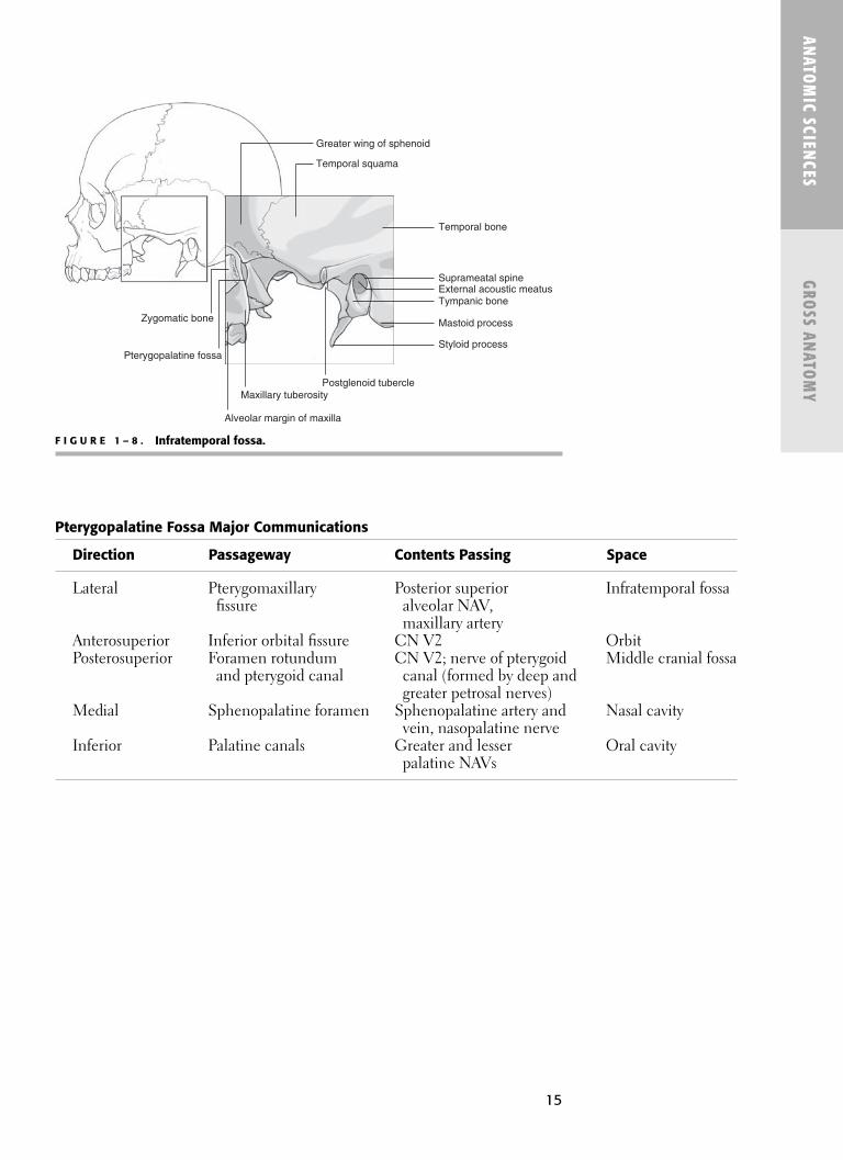
■ Forms two fossae (See Table 1–1).■ Infratemporal (See Figure 1–8).■ Pterygopalatine.
■ Forms two fissures.■ Infraorbital. ■ Pterygomaxillary.
T A B L E 1 – 1 . Infratemporal and Pterygopalatine Fossae
BOUNDARIES
FOSSA ANTERIOR POSTERIOR MEDIAL LATERAL ROOF FLOOR CONTENTS
Infratemporal Posterior Temporal Lateral Mandibular Greater Medial Temporalis and
fossa maxilla bone pterygoid ramus wing of pterygoid pterygoid muscles
(articular plate sphenoid muscle Maxillary artery
tubercle) (sphenoid) (with (superior (and branches,
and foramen surface eg, middle
spenoidal ovale where meningeal)
spine of →CN V3) inserts into Pterygoid plexus
the mandible) of veins
spenoid Mandibular
bone nerve (V3)
Chorda tympani (VII)
Otic ganglion (IX)
Pterygopalatine Maxilla Pterygoid Nasal Infratemporal Greater Pyramidal Pterygopalatine
fossa plates fossa fossa wing of process of (3rd) part of
sphenoid; palatine maxillary artery
opens into bone; and its branches
inferior inferior end Maxillary nerve (V2)
orbital contains Nerve of pterygoid
fissure palatine canal
canals Pterygopalatine
ganglion and
branchesa
aFor pterygopalatine ganglion, see the parasympathetic ganglia chart.
15
ANATOM
IC SCIENCES
GROSS ANATOM
Y
F I G U R E 1 – 8 . Infratemporal fossa.
Pterygopalatine Fossa Major Communications
Direction Passageway Contents Passing Space
Lateral Pterygomaxillary Posterior superior Infratemporal fossafissure alveolar NAV,
maxillary arteryAnterosuperior Inferior orbital fissure CN V2 OrbitPosterosuperior Foramen rotundum CN V2; nerve of pterygoid Middle cranial fossa
and pterygoid canal canal (formed by deep andgreater petrosal nerves)
Medial Sphenopalatine foramen Sphenopalatine artery and Nasal cavityvein, nasopalatine nerve
Inferior Palatine canals Greater and lesser Oral cavitypalatine NAVs
Greater wing of sphenoid
Temporal squama
Mastoid processZygomatic bone
Pterygopalatine fossa
Postglenoid tubercleMaxillary tuberosity
Alveolar margin of maxilla
Styloid process
Tympanic boneExternal acoustic meatusSuprameatal spine
Temporal bone

ANAT
OM
IC S
CIEN
CES
GROS
S AN
ATO
MY
HARD PALATE
The palate forms the roof of the oral cavity and the floor of the nasal cavity.
■ Maxilla are palatal processes (anterior two-thirds).■ Palatine bones are horizontal palates (posterior one-third).■ Pterygoid plates of the sphenoid articulate with the maxillary tuberosity
(posterior palate).
Palatal Foramen
■ Incisive foramen (Scarpa, midline; Stenson, lateral)—descending palatinevessels and the nasopalatine nerves (of V2)—anterior palatal block.
■ Greater and lesser palatine foramen—descending palatine vessels and ante-rior palatine nerve (of V2)—site of palatal anesthetic block.
F I G U R E 1 – 9 . Lateral scheme of the pterygopalatine fossa to show the entrances and exits.
Reproduced, with permission, from Liebgott B. The Anatomical Basis of Dentistry. Toronto:
BC Decker, 1986.
Nasal Cavity
Boundary Contributing Structures
Floor Hard palate (maxilla and palatine bones)Roof Cribriform plate of ethmoid, anterior body of sphenoid, nasal spine of frontal
bone, nasal bones, lateral nasal cartilagesLateral wall Nasal, ethmoid, sphenoid, maxilla, palatine, and inferior conchal bonesMedial wall Nasal septumExternal nose Two nasal bones, nasal cartilages
See Figure 1–9 for lateral scheme of the pterygopalatine fossa.
16
Pterygoid canal
Foramen lacerum
Foramen rotundum
Middle cranial fossa
Pterygoidprocess
Lesser & greaterpalatine foramina palate
Posterior superioralveolar foramina
maxillary sinus
Maxilla
Sphenopalatine foramen nasal cavity
Inferior orbital fissure orbit
Infraorbital groove canal,and forament face

17
ANATOM
IC SCIENCES
GROSS ANATOM
Y
NASAL CAVITY
See Figure 1–10.
■ Sensory innervation is from branches of V2.■ Nasopalatine■ Infraorbital■ Greater palatine
■ Some sensory branches are from V1 (ophthalmic division).■ Anterior ethmoidal nerve
■ Parasympathetic to secretory glands supplied by branches of the ptery-gopalatine ganglion.
■ Olfactory epithelium (roof of the nasal cavity) is innervated by the olfac-tory nerve (I).
■ Blood supply—sphenopalatine branch of maxillary artery, anterior eth-moidal branch of ophthalmic artery, and septal branch of superior labialbranch of facial artery.
■ Superior nasal conchae and upper third of septum contain yellowish olfac-tory mucosa.
F I G U R E 1 – 1 0 . Lateral nasal cavity and hard palate.
Conchae Meatuses
Superior and middle (ethmoid Areas below each conchae are bone) inferior (its own bone) the superior, middle, and inferior
meatuses, respectivelyIncrease air turbulence for Drainage points for sinuses and
warming, filtering, olfaction nasolacrimal apparatus
CN I (olfactory nerve) projects
to the primary olfactory cortex
(pyriform cortex).
Crista galli
Sphenoethmoidal recess
Semilunar hiatus
Medial pterygoid plate
Horizontal plate of palatine bone
Pterygoid hamulus
Choanae (posterior nasalaperature)
Sphenoidal sinus
Hypophyseal fossa(sella turcica)
Sphenopalatine foramen
Frontal sinus
Nasal part offrontal bone
Superior nasal concha
Nasal bone
Middle nasal concha
Middle nasal meatus
Inferior nasal concha
Inferior nasal meatusAnterior nasal spine
Incisive canal
Palatine processof maxilla
Anterior nasal aperture
Cribriform plate of ethmoid bone

ANAT
OM
IC S
CIEN
CES
GROS
S AN
ATO
MY
Most cases of epistaxis arise
from this area.
18
NASAL SEPTUM
The nasal septum comprises five bones and one cartilage (See Figure 1–11).
■ Vertical plate of ethmoid■ Vomer■ Nasal crest of maxilla and palatine bones■ Nasal crest of sphenoid bones■ Septal cartilage
The maxillary sinus is lined by
the Schneiderian membrane
which is pseudo-stratified
columnar epithelium.
F I G U R E 1 – 1 1 . The cartilaginous and bony components of the nasal septum.
KIESSELBACH’S PLEXUS
■ This plexus is the anastomosis of five arteries:■ Sphenopalatine artery (from maxillary artery)■ Greater palatine artery (from maxillary artery)■ Superior labial artery (from facial artery)■ Anterior ethmoid artery (from opthalmic artery)■ Lateral nasal branches from facial artery
PARANASAL SINUSES (FIGURE 1–12)
■ Frontal■ Maxillary■ Ethmoid ■ Sphenoid
Nasal spine of frontal boneNasal bone
Vertical plate of ethmoidLateral nasal cartilage Nasal crest of sphenoid bone
Vomer
Nasal crest palatine bone
Septal cartilage
Nasal crest of maxilla

19
ANATOM
IC SCIENCES
GROSS ANATOM
Y
System Location of Drainage
Nasolacrimal apparatus Inferior meatus (below inferior concha)
Frontal sinuses Middle meatus: Hiatus semilunaris (below middle concha)
Maxillary sinuses Middle meatus: Ostium (below middle concha) (within hiatus semilunaris)
Ethmoid sinuses: Middle meatus■ Anterior Hiatus semilunaris■ Middle Ethmoidal bullae■ Posterior Superior meatus
Sphenoid sinuses Sphenoethmoidal recess of nasal cavity
F I G U R E 1 – 1 2 . Paranasal sinuses.
The middle meatus contains
openings for the frontal sinus,
anterior and middle
ethmoidal sinuses, and
maxillary sinuses.
A surgical approach to
pituitary gland is via the
sphenoid sinus.
Ethmoidal cells
Middle nasal meatusMaxillary hiatus
Maxillary sinusVomer
Inferior nasal concha
Alveolar process of maxilla
Maxillary molar tooth
Orbital surface of greaterwing of sphenoid
Orbital portion of frontal bone
Temporal bone
Zygomatic bone
Middle nasal conchaInferior nasal meatus
Crista galli
Superior orbital fissurePerpendicular lamina of ethmoid bone
Inferior orbital fissure
Palatine process of maxilla

20
ANAT
OM
IC S
CIEN
CES
GROS
S AN
ATO
MY
EXTERNAL NOSE (FIGURE 1–13)
■ Nasal bones■ Septal cartilages■ Lateral cartilages■ Alar cartilages
MANDIBLE
■ Largest and strongest bone of the face.■ Lower jaw: houses lower teeth.■ Consists of:
■ Body (curved, horizontal).■ Rami (two perpendicular portions).
■ Body and rami unite at angle (nearly 90 degrees).■ Coronoid process (attachment of temporalis muscle).■ Condyle.
See Section Mastication and TMJ of this chapter for information on temporo-mandibular joints and the muscles of mastication.
FORAMINA
Mandibular Foramen
■ Is located on the medial side of the ramus (just below lingula), midwaybetween anterior and posterior borders of the ramus.
■ It passes■ Inferior alveolar nerve (IAN) (of V3).■ Inferior alveolar artery and vein.
The mandibular canal
traverses the mandibular
body and opens anteriorly at
the mental foramen.
F I G U R E 1 – 1 3 . The nose.
Cartilage of the external
nose is hyaline.
Nasal bone
Lateral nasal cartilage
Nasal septal cartilage
Greater alar cartilagesLesser alar cartilages

Mental Foramen
■ Is located below the second premolar on each side.■ It passes
■ Mental nerve; the inferior alveolar nerve exits as the mental nerve. Itsupplies skin and mucous membrane of the mental region.
■ Incisive branch, which supplies the pulp chambers of the anterior teethand adjacent mucous membrane.
Scalp (Figure 1–14)
A mnemonic device to remember the components of the scalp is:
■ Skin■ Connective tissue■ Aponeurosis (galea aponeurotica, epicranial aponeurosis)■ Loose connective tissue■ Periosteum
ANATOM
IC SCIENCES
GROSS ANATOM
Y
21
F I G U R E 1 – 1 4 . The scalp.
The lingula is a tongue-
shaped projection above the
mandibular foramen where
the sphenomandibular
ligament attaches.
Epidermis
Diploic vein
Cerebrum
Pia mater
Subarachnoid spaceArachnoidDura materInner compact layer of bone
Diploe
Outer compact layer of bone
Superficial fasciaDermis
PeriosteumLoose areolar tissue
Aponeurotic layer

22
ANAT
OM
IC S
CIEN
CES
GROS
S AN
ATO
MY
Meninges
Meninge Space Description
Epidural space Potential space between periosteum of inner surface of skull and dura; middlemeningeal artery is in this location.
Dura mater Tough membranous, outermost layer, continuous with the periosteum within the skull; forms the venous sinuses in the cranial cavity. Endosteal layer is continuous with cranial periosteum.
Meningeal layer folds between brain.
Subdural Between dura and arachnoid; space bridging veins and cranial
venous sinuses are located here.
Arachnoid Weblike (spiderlike) lattices interposed between dura and pia; does not follow the sulci; bridges them.
Subarachnoid Between arachnoid and pia; is filled space with CSF; cerebral circulation is here
(circle of Willis).This is the space entered with a
lumbar puncture (“spinal tap”).
Pia mater Layer adherent to the brain;spinal cord follows the sulci.
Meningitis is inflammation
of the meninges. For more
information, see
“Systemic Pathology,”
Chapter 22.
Blood surrounds themeninges in the followingways:
■ Epidural hematomainvolves the middlemeningeal artery.
■ Subdural hematomainvolves a bridging vein.
■ Subarachnoidhemorrhage ofteninvolves a rupturedaneurysm (eg, anteriorcommunicating artery).
DURAL FOLDS
See Figure 1–15 for the folds of the dura mater.
Fold Description
VerticalFalx cerebri Vertical, midline.
Separates the cerebral hemispheres.Forms the superior and inferior sagittal sinuses.
Falx cerebelli Separates cerebellar hemispheres.Contains occipital sinus.
HorizontalTentorium cerebelli Separates cerebral hemispheres (occipital lobes) from cerebellum below.
Contains straight, transverse, and superior petrosal sinuses.Uncus (medial parahippocampal gyrus); amygdala lies beneath; herniatesbelow tentorium.
Diaphragma sella Roof of the sella turcica. Small hole allows passage of the pituitary stalk.
23
ANATOM
IC SCIENCES
GROSS ANATOM
Y
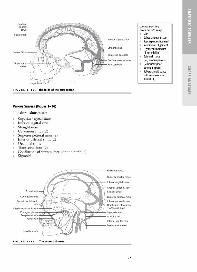
VENOUS SINUSES (FIGURE 1–16)
The dural sinuses are:
■ Superior sagittal sinus■ Inferior sagittal sinus■ Straight sinus■ Cavernous sinus (2)■ Superior petrosal sinus (2)■ Inferior petrosal sinus (2)■ Occipital sinus ■ Transverse sinus (2)■ Confluence of sinuses (torcular of herophile)■ Sigmoid
F I G U R E 1 – 1 5 . The folds of the dura mater.
F I G U R E 1 – 1 6 . The venous sinuses.
Lumbar puncture (from outside to in): ■ Skin■ Subcutaneous tissue■ Supraspinous ligament■ Interspinous ligament■ Ligamentum flavum
(if not midline)■ Epidural space
(fat, venous plexus)■ (Subdural space—
potential space)■ Subarachnoid space
with cerebrospinalfluid (CSF)
Inferior sagittal sinus
Superiorsagittal
sinus
Tentorium cerebelli
Falx cerebri
Straight sinus
Confluence of sinuses
Falx cerebelli
Frontal sinus
Diaphragmasellae
Emissary veins
Superior sagittal sinus
Inferior sagittal sinus
Greater vertebral vein
Straight sinus
Superior petrosal sinus
Inferior petrosal sinus
Transverse sinus
Occipital vein
Sigmoid sinus
Confluence of sinuses
Internal jugular vein
Deep cervical vein
Facial vein
Maxillary vein
Deep facial veinPterygoid plexus
Inferior ophthalmic vein
Cavernous sinus
Superior ophthalmicvein
Frontal vein

24
ANAT
OM
IC S
CIEN
CES
GROS
S AN
ATO
MY
CAVERNOUS SINUS (FIGURES 1–17 AND 1–18)
F I G U R E 1 – 1 7 . Cavernous sinus.
Reproduced, with permission, from Bhushan V, et al. First Aid for the USMLE Step 1. New
York: McGraw-Hill, 2003. Adapted from Stobo J, et al. The Principles and Practice of Medicine,
23rd ed. Stamford, CT: Appleton & Lange, 1996:277.
Tributaries of Dural Sinuses
Emissary veins Drain scalp into dural sinuses.Diploic veins Drain the diploe of the skull into dural sinuses.Meningeal veins Drain meninges into dural sinuses.
Drainage of the head/brain
is via the internal jugular
vein (IJV).
*IJV forms from the inferior
petrosal and sigmoid sinuses. Location Connections Contents Description
Middle cranial Anterior: Superior Lateral wall: Route of fossa (on either and inferior CNs III, IV, infection to side of sella ophthalmic veins, V1, V2 brain (eg, turcica) pterygoid plexus zygomycosis)
of veins (via facial vein)
Posterior: Superior Running through Cavernousand inferior cavernous sinus: sinus thrombosispetrosal, CN VI (see Chapter 22,intercavernous ICA Systemicsinus Pathology)
Ophthalmic veins(superior and inferior)can communicate with thecavernous sinus. Becausethere are no valves,retrograde flow occurs.
The superior petrosal sinus
connects the cavernous and
sigmoid sinuses.
CN VI is the smallest and most
medial nerve in the cavernous
sinus and will be the first
nerve affected by an infection.
Hypothalamus Third ventricle
Pituitary stalkDiaphragma sellae
Pituitary gland
Oculomotor nerve (III)
Trochlear nerve (IV)
Sella turcicaAbducens nerve (VI)
Ophthalmic nerve (V1)
Maxillary nerve (V2)
Optic chiasm
Cavernous sinus
Internal carotid artery
Sphenoid bone Sphenoidalsinus
Nasopharynx

25
ANATOM
IC SCIENCES
GROSS ANATOM
Y
Pterygoid Plexus of Veins
F I G U R E 1 – 1 8 . Cavernous sinus and its communications.
Location Receives Drains
Located in the Venous branches Maxillary vein posteriorlyinfratemporal fossa corresponding Deep facial vein
Surrounds the with those of the into the facialmaxillary artery maxillary artery vein anteriorly
Associated with the pterygoid muscles
Ventricular System
This system is lined with ependymal cells. It consists of these parts: lateral ven-tricle, interventricular foramen, third ventricle, cerebral aqueduct, fourth ventri-cle (releases CSF into subarachnoid space). See Figure 1–19.
Ventricle Nearby Anatomical Structure
Lateral ventricle Caudate nucleusLateral ventricle (inferior horn) HippocampusThird ventricle HypothalamusFloor of fourth ventricle Pons
The abducens nerve is
most likely affected from a
laterally expanding
pituitary tumor because
it is medially located
within the cavernous sinus.
The deep facial vein connects
the anterior facial vein and
pterygoid plexus.
The choroid plexus and
ventricular system regulate
intracranial pressure.
Superior ophthalmic vein.
Inferior ophthalmic vein.
Facial vein(anterior)
Petrosal sinuses
Pteygoid plexus(of veins)
Emissary vein
Maxillary veinSuperficial temporal veinRetromandibular vein
Deepfacial vein
Cavernous sinus

ANAT
OM
IC S
CIEN
CES
GROS
S AN
ATO
MY
CSF CIRCULATION
■ The CSF flows from lateral ventricles (produced in choroids plexus)through the ventricular system to the subarachnoid space, where it entersthe venous circulation.
■ Pathway
Lateral ventricles ↓
Foramen of Monro↓
Third ventricle↓
Cerebral aqueduct↓
Fourth ventricle↓
Foramina of Magendie and Luschka (exits ventricular system intosubarachnoid space)
↓Bathes the cisterns in the subarachnoid space
↓Arachnoid granulations protrude into the superior sagittal sinus and
empty CSF into the venous circulation.
Ependymal cells can also
produce CSF.
Hydrocephalus resultsfrom excess buildup of CSF.
■ Noncommunicating—obstruction in theventricular system(eg, blocked cerebralaqueduct).
■ Communicating—obstruction insubarachnoid space.
F I G U R E 1 – 1 9 . The ventricular system.
Reproduced, with permission, from Waxman SG. Clinical Neuroanatomy, 25th ed. New York:
McGraw-Hill, 2003.
26
Interventricularforamen (of Monro)
Anterior(frontal)horn
Third ventricle
Inferior(temporal) horn
Fourthventricle
Cerebralaqueduct
Posterior(occipital)horn
Lateralventricles
Foramina of Luschka are
Lateral aperatures
Foramina of Magendie are
Medial aperatures.

27
ANATOM
IC SCIENCES
GROSS ANATOM
YIntracranial Circulation
■ Blood is supplied to the brain via many arteries.
CIRCLE OF WILLIS
See Figures 1–20 and 1–21.
■ Contents:■ Posterior cerebral artery■ Posterior communicating artery■ Internal carotid artery ■ Anterior cerebral artery■ Anterior communicating artery
■ Four arteries contribute: vertebral arteries (2) and carotid arteries (2)
Blood-brain barrier is absent
in hypothalamus, pineal
gland, area postrema (of
fourth ventricle), and areas
near third ventricle.
Vascular-Endothelial Arachnoid Blood-CSF Barrier Barrier Barrier
CSF produced in choroid Tight junctions Arachnoid cells form plexus in ventricles. between a barrier, preventing
Choroid plexus epithelial endothelial cells substances from dural cells are joined by tight vessel from diffusingjunctions, allowing in toward brain.selective passage.
Circle of Willis
Feeder Arteries Branches Supplies
Right and left internal 1. Anterior cerebral artery Medial aspect of frontal carotid arteries (branch from right and left and parietal lobes.
ICAs and communicate via anterior communicating artery).
2. Middle cerebral artery Anterior temporal lobes(continuation of ICA). and cortex of insula.
Basilar artery which Posterior cerebral artery Occipital cortex (visual area).arises from convergence (connects to middle cerebral of right and left vertebral artery via the posterior arteries. communicating artery).
Blood-Brain Barrier
The blood-brain barrier (BBB) consists of three parts:

28
ANAT
OM
IC S
CIEN
CES
GROS
S AN
ATO
MY
F I G U R E 1 – 2 0 . Major cerebral arteries.
Reproduced, with permission, from Waxman SG. Clinical Neuroanatomy, 25th ed. New York:
McGraw-Hill, 2003.
F I G U R E 1 – 2 1 . Circle of Willis and principal arteries of the brain stem.
Reproduced, with permission, from Waxman SG. Clinical Neuroanatomy, 25th ed. New York:
McGraw-Hill, 2003.
The ophthalmic artery is
a branch of the ICA
(follows optic nerve through
optic foramen into orbit);
it gives off the anterior
ethmoidal branch that
supplies the nasal cavity.
Vertebral arteries are
branches of the subclavian
artery.
The ICA has no branches in
the neck.
Aorta
Left subclavianartery
Left commoncarotid artery
Left internalcarotid artery
Left vertebralartery
Basilar artery
Left posteriorcerebral artery
Left anteriorcerebral artery
Carotid siphon
Left middlecerebral artery
Left vertebral arteryAnterior spinal artery
Posterior inferiorcerebellar artery
Anterior inferiorcerebellar artery
Superiorcerebellar artery
Basilar arterywith pontinebranches
Posteriorcommunicatingartery
Middle cerebral artery
Internal carotid artery
Anterior cerebral artery
Anterior communicating artery
Posteriorcerebral artery

MIDDLE CEREBRAL ARTERY
■ Largest branch of the ICA.■ If blocked, it causes the most ischemic injury.■ Leticulostriate arteries, branches of the MCA, are often involved in stroke,
are thin-walled, and can rupture.
� ORAL CAVITY AN D PHARYNX
Oral Cavity (Figure 1–22)
ANATOM
IC SCIENCES
GROSS ANATOM
Y
Components Description
Oral vestibule Slitlike space between lips and cheeks and the facial surfaces of teethand gingivae.
Oral cavity proper Space posterior and medial to dental arches (deep to lingual surfaces of teeth).Posterior termination is palatoglossal arch.Roof is the palate.Tongue occupies this space at rest with mouth closed.
F I G U R E 1 – 2 2 . The mouth of the oral cavity.
29
Frenulum
Upper lip
Transverse palatine ridges
Palatine glands
Opening of parotid duct
CheekBuccinator muscle
Soft palatePalatopharyngeal arch
Palatoglossal arch
Oral portion of pharynx
Palatine tonsil
Dorsum of tongue
Lower lip
Frenulum
Greater palatine artery
Greater palatine nerve
Tendon of tensor veli palatini muscle
Pterygoid hamulus
Levator veli palatini muscleBuccinator muscle
Superior pharyngeal constrictor
Uvulae muscle
Pterygomandibular raphePalatopharyngeus muscle
Oral vestibule

30
ANAT
OM
IC S
CIEN
CES
GROS
S AN
ATO
MY
Tongue
Function Innervation
Motor CN XIISensation CN V3, IX, XTaste CN VII, IX, X
GENERAL SENSATION OF THE TONGUE
See Figure 1–23 for the innervation of the tongue, which is mediated by thesecranial nerves (CNs):
■ V3■ IX■ X
TASTE SENSATION OF THE TONGUE
The sense of taste is mediated by the following CNs:
■ VII■ IX■ X
Brain stem Thalamic Location Nerve Pathway Nucleus Nucleus
Anterior 2/3 CN VII Solitary tract Nucleus of VPM Gustatory cortexChorda tympani solitary tract next to the
nerve travels (gustatory somatosensoryvia lingual nucleus) representation ofnerve (of V3) the tongueto the (frontal-parietal geniculate operculum, ganglion insula)
Posterior 1/3 CN IX Solitary tract Nucleus of VPM Gustatory cortexsolitary tract next to the(gustatory somatosensorynucleus) representation of
the tongue(frontal-parietaloperculum,insula
Epiglottis CN X Solitary tract Nucleus of VPM Gustatory cortexsolitary tract next to the(gustatory somatosensorynucleus) representation of
the tongue(frontal-parietaloperculum,insula)
The tongue is derived from the
first four pharyngeal arches
and is innervated by
associated nerves of those
arches: arch 1 (V), arch 2 (VII),
arch 3 (IX), and arch 4 (X).
Damage to right or left CN XII
will cause the tongue to
deviate to the side of the
lesion.

CHORDA TYMPANI NERVE
■ This nerve is part of the structure of the facial nerve (CN VII).
COURSE
■ Nucleus of solitary tract (accepts taste fibers).■ Superior salivatory nucleus (parasympathetic to submandibular, sublin-
gual glands).■ Chorda tympani nerve arises from the geniculate ganglion.■ Emerges from petrotympanic fissure.■ Crosses the medial surface of the tympanic membrane.■ Joins the lingual nerve (of V3) in the infratemporal fossa.
COMPONENTS
■ Taste (pathway): See Figure 1–24.■ Anterior two-thirds of tastebuds.■ Chorda tympani (travels with lingual nerve).■ Cell bodies are located in the geniculate ganglion (within facial canal
or petrous temporal).■ Preganglionic parasympathetic
■ Synapse in submandibular ganglion.
INFERIOR SURFACE OF THE TONGUE
■ Lingual frenulum: vertical fold in the midline.■ Plica fimbriata: fold of mucous membrane, lateral to the frenulum.■ Wharton’s and Rivian ducts: openings of the submandibular and sublin-
gual glands.■ (Blood supply of the tongue: see external carotid artery.)
ANATOM
IC SCIENCES
GROSS ANATOM
Y
F I G U R E 1 – 2 3 . Sensory innervation of the tongue.
Reproduced, with permission, from Waxman SG. Clinical Neuroanatomy, 25th ed. New York:
McGraw-Hill, 2003.
31
Salt
Sweet
VII (VA)
Sour
Bitter
IX (VA)
Epiglottis
V (SA)
IX (SA)

32
ANAT
OM
IC S
CIEN
CES
GROS
S AN
ATO
MY
F I G U R E 1 – 2 4 . Diagram of taste pathways.
Reproduced, with permission, from Waxman SG. Clinical Neuroanatomy, 25th ed. New York:
McGraw-Hill, 2003.
Tastebud Typea Description
Filiform papillae Rough texture of tongue; found in rows; avascular; most numerous papillae of tongue;do not contain tastebuds.
Fungiform papillae Mushroom-shaped; scattered among filiform papillae; usually contain tastebuds.
Circumvallate papillae Seven to nine large circular structures withtastebuds; serous-only salivary glands within(von Ebner’s glands).
Foliate papillae On lateral surface of tongue in ridges; rudimentary and nonfunctional.
aListed from smallest to largest.
TASTEBUDS
All tastebuds except filiform
are vascular.
Nucleus ofsolitary tract(and parabrachialnucleus)
Solitary tract
Nerve VII(via chordatympani, nervusintermedius)Nerve IX
Mediallemniscus
Insula
Postcentralgyrus
Thalamus(VPM nuclei)

33
ANATOM
IC SCIENCES
GROSS ANATOM
Y
OTHER SURFACE COMPONENTS OF THE TONGUE
See Figure 1–25 for the dorsal view of the tongue.
■ Foramen cecum■ Upper part of thyroglossal duct
■ Sulcus terminalis■ Lingual tonsils (See the section “Waldeyer’s Ring.”)■ Glands
■ Mucous (back, front, and sides) ■ Serous (posteriorly) ■ Anterior lingual glands (mixed seromucous glands)
LYMPHATIC DRAINAGE OF THE TONGUE
See Figure 1–26 for an illustration of the lymph nodes of the tongue. Also seelymphatic drainage of head and neck.
MUSCLES CONTROLLING THE TONGUE (FIGURE 1–27)
Bony Attachments
■ Genial tubercles■ Styloid process■ Hyoid bone
F I G U R E 1 – 2 5 . The tongue, dorsal view.
Palatine tonsil
Epiglottis
Vallecula of epiglottisMedian glossoepiglottic fold
Lingual tonsil and follicles
Foramen cecum
Circumvallate papillae
Foliate papillae
Filiform papillae
Medial lingual sulcus
Fungiform papillae
Apex of tongue
Sulcus terminalis
Palatoglossus muscle
Lateral margin of tongue

34
ANAT
OM
IC S
CIEN
CES
GROS
S AN
ATO
MY
F I G U R E 1 – 2 7 . Muscles controlling the tongue.
F I G U R E 1 – 2 6 . Lymph drainage of the tongue.
All tongue muscles, except
palatoglossus, are innervated
by CN XII.
The muscles attaching to
genial tubercles are the
genioglossus and the
geniohyoid.
The tongue’s blood supply
is via lingual artery; veins
drain into IJV.
Superior deep cervical lymph nodes
Submental lymph nodes
Submandibularlymph nodes
Inferior deepcervical lymph nodes
Styloid process
Palatoglossus muscle
Palatine tonsil
Dorsum of tongue
Genioglossus muscleMandible
Geniohyoid muscle Lingual artery
Vein
Styloglossus muscle
Stylopharyngeus muscle
Stylohyoid ligament
Stylohyoid muscle
Submandibular ganglion
Hyoglossus muscle
Hyoid bone

35
ANATOM
IC SCIENCES
GROSS ANATOM
Y
EXTRINSIC MUSCLES
INTRINSIC MUSCLES
These muscles lie within the tongue itself.
Muscle Origin Insertion Action Innervation
Genioglossus Genial tubercles Inferior: Hyoid Protrude tongue XIISuperior: Tongue
Hyoglossus Hyoid (body, Side of tongue, Depress tongue, XIIgreater cornu) medial to pull down sides
styloglossus (retracts)
Styloglossus Styloid process Side of tongue Pull tongue up XIIand back
Palatoglossus Anterior soft palate Side and dorsum Pull tongue up and X (pharyngealof tongue back (toward palate) plexus)
■ Longitudinals (superior Underneath mucosa; shorten the tongueand inferior) (both); make dorsum concave (superior);
make dorsum convex (inferior).■ Transversus Arise from median fibrous septum and pass
laterally; narrows and elongates the tongue.■ Verticalis Flattens and broadens the tongue.
SPEAKING SOUNDS
■ “La-la” CN XII moves tongue against roof of mouth.■ “Mi-mi” CN VII moves lips.■ “Kuh-kuh” CN X raises the palate.
RELATIONSHIP OF LINGUAL ARTERY, VEIN, AND SUBMANDIBULAR DUCT
The relationships to the hyoglossus muscle are:
■ Medial to the hyoglossus.■ Lingual artery
■ Lateral to the hyoglossus.■ Lingual vein■ Lingual nerve■ Submandibular duct■ Hypoglossal nerve

ANAT
OM
IC S
CIEN
CES
GROS
S AN
ATO
MY
36
F I G U R E 1 – 2 8 . Branches of the lingual artery.
See Figure 1–28 for branches of the lingual tongue.
Palate
■ Roof of oral cavity, floor of nasal cavity.
INNERVATION
■ Motor■ Pharyngeal plexus (except tensor veli palatine, CN V3).
■ Sensory■ CN V2
■ Greater palatine nerve is located posteriorly; it travels anteriorly.■ Nasopalatine nerves are located anteriorly; they join the greater palatine
posteriorly.
BLOOD SUPPLY
■ Third part of the maxillary artery (branch of ECA).■ Greater palatine artery travels with nerve and vein from the greater and
lesser palatine foramina.■ Sphenopalatine vessels travel with nasopalatine nerves from the inci-
sive foramen.
The palatal salivary glands
are mostly mucous, located
beneath the
mucous membrane of hard
and soft palates, and
contribute to oral fluid.
Apex of tongue
Superior longitudinal muscle of tongueGenioglossus muscle
Mandible
Geniohyoid muscle
Deep lingual artery
Mylohyoid muscle
Sublingual artery
Thyrohyoid muscle
Cricothyroid membrane
Lamina of cricoid cartilage
Hyoglossus muscle
Dorsal lingual branchesFacial artery
External carotid artery
Lingual artery
Superior thyroid artery
Stylohyoid ligament
Suprahyoid branch
Inferior constrictor of the pharynx
Palatoglossus muscle
Styloglossus muscleSuperior constrictor of the pharynx
Palatine tonsil
Internal carotid artery
Stylopharyngeus muscle
Middle constrictor of the pharynx

37
ANATOM
IC SCIENCES
GROSS ANATOM
Y
Tonsil Location Description
Pharyngeal tonsils Nasopharynx (posterior wall No lymph, sinuses, nor crypts(adenoids) and roof) Surrounded in part by connective tissue and
in part by epitheliumPalatine tonsils In isthmus of fauces (between Reach maximum size during childhood then diminish
palatoglossal and palatopharyngeal Contain crypts and lymphoid follicles (No sinuses)folds) on either side of the Covered partly by connective tissue, posterior oropharynx partly by epithelium
Lingual tonsils Dorsum of tongue (posteriorly) Lymphoid follicles, each with a single crypt
(See Peyers patches in GI section).
Hard Palate Soft Palate (Muscles)
Maxillary bone Palatopharyngeus(palatine processes) Palatoglossus
Palatine bones Levator veli palatini(horizontal plates) Tensor veli palatini
UvularCovered by keratinized mucosa Covered by nonkeratinized mucosa
(with rugae anteriorly) SubmucosaPalatal salivary glands Anterior zone of palatal submucosa
(beneath mucosa) contains fatPosterior zone contains mucous glandsPalatal aponeurosis: Fibrous connective
tissue of soft palate (muscles beneath)
UVULA
■ Suspended from soft palate.■ Bifid uvula results from incomplete fusion of palatine shelves.■ Unilateral damaged pharyngeal plexus causes uvula to deviate to contralat-
eral side. Contraction on intact side pulls it to functional side.
FAUCES
■ The fauces are between anterior and posterior pillars, and they house thepalatine tonsils.
Pillar Muscle Muscle Function
Anterior pillar Palatoglossus Draws tongue and soft (palatoglossal fold) palate closer together
(with swallowing)Narrows isthmus of fauces
Posterior pillar Palatopharyngeus Elevates pharynx(palatopharyngeal fold) Helps close the nasopharynx
Aids in swallowing
TONSILS
The soft palate attaches to the
tongue by the glossopalatine
(palatoglossal) arches and to
the pharynx by the
palatopharyngeal arches.
Remember: Most muscles of
the soft palate attach to the
palatal aponeurosis.

38
ANAT
OM
IC S
CIEN
CES
GROS
S AN
ATO
MY
WALDEYER’S RING
Ring of lymphoid tissue
■ Lingual tonsil (inferiorly)■ Palatine tonsils (faucial tonsils) (laterally)■ Nasopharyngeal tonsils (adenoids) (superiorly)The tensor and levator veli
palatini muscles prevent food
from entering the nasopharynx.
(See Figure 1–29 for
parasagittal view of the soft
palate and pharynx).
Muscle Origin Insertion Action Innervation
Tensor veli Greater wing of Wraps around pterygoid Tenses palate, opens CN V3palatini sphenoid hamulus to insert onto auditory tube with
(scaphoid fossa), midline palatal mouth openinglateral cartilage aponeurosis (with of auditory tube contralateral fibers)
Levator veli Inferior petrous Palatine aponeurosis Elevates/raises palate CN Xpalatini temporal bone, (during swallowing)
medial auditory tube
F I G U R E 1 – 2 9 . Parasagittal view of soft palate and pharynx.
Tensor versus Levator Veli Palatini
Torus tubariusPharyngeal opening of auditory tube
Cartilage of auditory tube
Muscular uvula
Ascending palatine branch of facial arteryTensor veli palatini muscle
Third cervical vertebra
Palatoglossus muscleDorsum of tongue
Tonsillar fossa
Pharyngobasilar fascia
Levator veli palatini muscle
Palatopharyngeus muscle
Superior constrictor of pharynx
Middle constrictor of pharynx
Axis
Dens
Epiglottis
Salpingopharyngeus muscle

39
ANATOM
IC SCIENCES
GROSS ANATOM
Y
Pharynx
See Figures 1–30 and 1–31 for views of the throat and Table 1–2 for locationsand descriptions.
■ Pharynx is behind nasal and oral cavities.■ Pharynx shares conduit to larynx and esophagus.
PHARYNGEAL MUSCLES
Muscle Origin Insertion Action Innervation
Constrictors
Superior Pterygomandibular Midline Contract in waves Sensory:raphe and pharyngeal raphe (to propel food). CN Xpharyngeal (posteriorly); Motor:tubercle; pharyngeal CN XI (via X)mylohyoid line tubercle.of mandible.
Middle Hyoid bone Midline pharyngeal Contract in waves Sensory:(greater and raphe (posteriorly). (to propel food). CN Xlesser horns). Motor:
CN XI (via X)Inferior Thyroid and cricoid Midline pharyngeal Contract in waves Sensory:
cartilages. raphe. (to propel food). CN XMotor:CN XI (via X)
Cricopharyngeus Lower fibers of Midline raphe. Constant contraction Sensory:inferior constrictor. (serves as UES). CN X
Motor:CN XI (via X)
Longitudinal muscles
Palatopharyngeus Palatal Posterolateral Raise pharynx and CN XI (via X) aponeurosis. pharynx. larynx during (pharyngeal
swallowing. plexus)Salpingopharyngeus Cartilage of Palatopharyngeus Elevate nasopharynx, CN XI (via X)
eustachian tube. muscle. open auditory tube, (pharyngeal equalize pressure plexus)between pharynxand auditory canal.
Stylopharyngeus Styloid process. Pass between superior Raise pharynx and CN IXand middle larynx during constrictors; swallowing.blend with palatopharyngeus.
The pterygomandibular raphe
is the meeting point of the
buccinator muscle and the
superior pharyngeal
constrictor.

40
ANAT
OM
IC S
CIEN
CES
GROS
S AN
ATO
MY
F I G U R E 1 – 3 0 . External view of the pharynx.
F I G U R E 1 – 3 1 . Posterior view of the soft palate and pharynx.
Buccinator muscle
Tensor veli palatini muscleLevator veli palatini muscle
Pterygomandibular raphe
Hyoglossus muscleMylohyoid muscle
Anterior belly of digastricmuscle
Sternohyoid muscleThyrohyoid membrane
Thyrohyoid muscle
Thyroid cartilage
Cricothyroid muscle
Pharyngobasilar fascia
Superior constrictor of pharynxStyloglossus muscleStylopharyngeus muscleStylohyoid ligamentPosterior belly of digastric muscle
Middle constrictor of pharynxStylohyoid muscle
Superior belly of omohyoid muscle
Inferior constrictor of pharynx
Esophagus
Pharyngeal tonsil
Nasal septum
Nasal pharynx
UvulaPalatine tonsil
Posterior nasal apertureMiddle nasla concha
Cartilage of auditory tube
Inferior conchaTensor veli palatini muscle
Salpingopharyngeus muscleSuperior constrictor of pharynx
Uvular musclePalatopharyngeus muscle
Base of tongue
Epiglottis
Palatopharyngeus muscleAryepiglottic fold
Cuneiform tubercleStylopharyngeus muscle
Oblique arytenoid muscleCorniculate tubercle
Piriform recessPosterior cricoarytenoid muscle
Oral pharynx
Lateral glossoepiglottic fold
Laryngeal pharynx
Muscular coat of esophagus

41
ANATOM
IC SCIENCES
GROSS ANATOM
Y
T A B L E 1 – 2 . Pharynx
LOCATION DESCRIPTION
Nasopharynx Above the soft palate; Auditory tube connects
continuous with nasal nasopharynx with middle ear.
passages; soft palate
and uvula form anterior wall.
Oropharynx Extends from anterior Communicates with oral cavity
pillars to larynx. via fauces; food and air from
mouth; air and sinus drainage
from nasopharynx; contains
palatine and lingual tonsils.
Laryngopharynx Area inferior to oropharynx Allows food into esophagus;
containing entrance to air into larynx.
both larynx and esophagus.
Stylopharyngeus muscle is
the only muscle supplied by
CN IX. This muscle is a
landmark for finding the
nerve.
Food can get caught in the
vallecula or pyriform recesses.
Sensory information goes to
the swallowing center
(nucleus ambiguous in
medulla oblongata).
Nucleus ambiguous sends
motor information (SVE), via
CNs IX, X, XI, XII, to facilitate
swallowing.
Pharyngeal plexus = CNs IX,
X, XI. Provides innervation
to constrictors, palatoglossus,
palatopharyngeus, and
cricopharyngeus.
SWALLOWING (THREE PHASES)
Oral Phase
■ Moisten food■ Masticate food (CN V)■ Tongue assumes trough shape (CN XII)■ Voluntary posterior movement of food bolus
Pharygeal Phase
■ Close Nasopharynx■ Tense vensor veli palatini (CN V)■ Raise velum: palatoglossal, levator veli palatini (CN X)
■ Elevate pharynx and hyoid■ Stylopharyngeus, salpingopharyngeus, palatopharyngeus (CN IX, X)
■ Close pharynx■ Retroversion of the epiglottis, adduction of vocal cords■ Bolus passes through pharynx
■ Wavelike contraction of superior, middle, and inferior pharyngeal con-strictor muscles (CN X, XI)
■ Relaxation of cricopharyngeus
Esophageal Phase
■ Esophageal peristalsis (CN X)■ Relaxation phase
■ Larynx, pharynx, and hyoid return to initial position
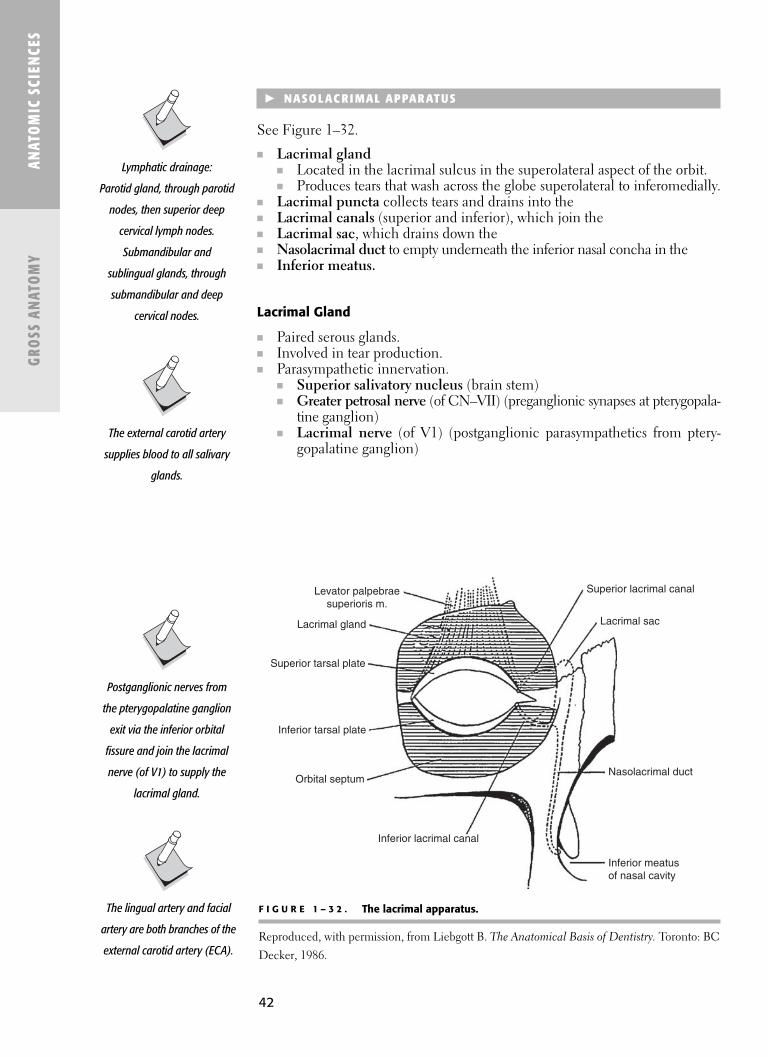
42
ANAT
OM
IC S
CIEN
CES
GROS
S AN
ATO
MY
F I G U R E 1 – 3 2 . The lacrimal apparatus.
Reproduced, with permission, from Liebgott B. The Anatomical Basis of Dentistry. Toronto: BC
Decker, 1986.
� NASOLAC RI MAL APPARATUS
See Figure 1–32.
■ Lacrimal gland ■ Located in the lacrimal sulcus in the superolateral aspect of the orbit.■ Produces tears that wash across the globe superolateral to inferomedially.
■ Lacrimal puncta collects tears and drains into the■ Lacrimal canals (superior and inferior), which join the■ Lacrimal sac, which drains down the■ Nasolacrimal duct to empty underneath the inferior nasal concha in the■ Inferior meatus.
Lacrimal Gland
■ Paired serous glands.■ Involved in tear production.■ Parasympathetic innervation.
■ Superior salivatory nucleus (brain stem)■ Greater petrosal nerve (of CN–VII) (preganglionic synapses at pterygopala-
tine ganglion)■ Lacrimal nerve (of V1) (postganglionic parasympathetics from ptery-
gopalatine ganglion)
Postganglionic nerves from
the pterygopalatine ganglion
exit via the inferior orbital
fissure and join the lacrimal
nerve (of V1) to supply the
lacrimal gland.
The lingual artery and facial
artery are both branches of the
external carotid artery (ECA).
Lymphatic drainage:
Parotid gland, through parotid
nodes, then superior deep
cervical lymph nodes.
Submandibular and
sublingual glands, through
submandibular and deep
cervical nodes.
The external carotid artery
supplies blood to all salivary
glands.
Levator palpebraesuperioris m.
Lacrimal gland
Superior tarsal plate
Inferior tarsal plate
Orbital septum
Inferior lacrimal canal
Superior lacrimal canal
Lacrimal sac
Nasolacrimal duct
Inferior meatusof nasal cavity

ANATOM
IC SCIENCES
GROSS ANATOM
Y
43
SALIVARY GLANDS
BloodGland Location Secretion Duct Innervation Supply
Parotid gland Parotid region, Serous Stenson’s Preganglionic: Glandular(See Figure 1–33.) between (parotid duct) Lesser petrosal branches
posterior goes anterior along nerve (of CN IX). of superficialborder of lateral masseter, Synapse: Otic temporalmandibular rolls over anterior ganglion. artery andramus and border of masseter Postganglionic: transverseanterior border and pierces cheek, Leaves otic ganglion, facial artery.of SCM. (Below buccinator muscle; travels with and anterior empties next to auriculotemporalto ear.) maxillary second nerve (of V3)
molar. to parotid.Submandibular Superficial Serous Wharton’s Preganglionic: Glandulargland (See portion and (submandibular Chorda tympani branches Figure 1–34.) Between lateral mucous duct) (of VII). of the
aspect of continues Synapse: facial mylohyoid forward from Submandibular artery.muscle and deep portion of ganglion.submandibular gland, crosses Postganglionic:fossa of lingual nerve Leaves ganglionmandible (near the and passes (submandibular sublingual gland), to gland.triangle). and empties
Deep portion: into oral cavityIn floor of mouth at the sublingualbetween base of caruncle (papilla) tongue and (next to sublingual gland sublingual(between mylohyoid frenulum; behindand hyoglossus) lower central
incisors).Sublingual Floor of mouth Mucous Sublingual Preganglionic: Glandular
gland (See medial to (Rivian) ducts Chorda tympani branchesFigure 1–35.) sublingual fossa open directly (of VII). of lingual
of mandible into oral cavity Synapse: artery(above mylohyoid through openings Submandibular (sublingual muscle). in sublingual fold; ganglion. artery).
anterior ducts Postganglionic:open medially Leaves ganglioninto submandibular and passes duct (the single to gland.Bartholin’s duct empties into submandibular duct at sublingual papilla).
(Adapted, with permission, from Liebgott B. The Anatomical Basis of Dentistry. Toronto: BC Decker, 1986:346.)

44
ANAT
OM
IC S
CIEN
CES
GROS
S AN
ATO
MY
MINOR SALIVARY GLANDS
Gland Location Secretion
Labial and buccal Labial and buccal Mucous only minor salivary glands mucosavon Ebner’s glands At base of circumvallate Serous only saliva
papillae (rinse food away from papilla)
Glands of Anterior lingual Mixed serous-mucous Blandin-Nuhn (anterior lingual glands)
The submandibular gland
emits the highest volume of
salivary fluid per day.
The parotid gland is second
in volume to submandibular
gland.
Going from largest to
smallest, the major salivary
glands go from serous to
mixed to mostly mucous
(parotid, submandibular, and
sublingual, respectively).
F I G U R E 1 – 3 3 . Schematic of the innervation of the parotid gland by the glossopharyngealnerve.
Light gray: Preganglionic parasympathetic nerves leave the brain stem with the glosopharyn-
geal nerve (CN IX) and run with the lesser petrosal nerve to the otic ganglion.
Dark gray: Postganglionic parasympathetic nerves travel with the auriculotemporal branch of
CN V3 and then the facial nerve to reach the parotid gland. (Reproduced, with
permission, from Lalwani AK (ed). Current Diagnosis & Treatment in Otolaryngology–Head &
Neck Surgery. New York: Lange Medical Books/McGraw-Hill, 2004.)
Parotid and von Ebner’s
glands are the only glands to
secrete serous-only saliva.
CN V
CN V3
CN VII
CN IX
Lesser petrosal nerve(parasympathetic fibersfrom CN IX via tympanic nerve)
Auriculotemporal nerve(branch of V3, caries parasympathtic fibersfrom CN IX via lesser petrosal nerve)
Parotid gland
Otic ganglion

45
ANATOM
IC SCIENCES
GROSS ANATOM
Y
F I G U R E 1 – 3 4 . Schematic of the innervation of the submandibular and sublingualglands by the facial nerve.
Light gray: Preganglionic parasympathetic nerves leave the brain stem with the facial nerve (CN
VII) and run with the chorda tympani and the lingual branch of CN V3 to the submandibular
ganglion.
Dark gray: Postganglionic parasympathetic nerves travel either directly to the
submandibular gland or back to the lingual branch of CN V3 to the sublingual gland.
(Reproduced, with permission, from Lalwani, AK (ed). Current Diagnosis & Treatment in
Otolaryngology–Head & Neck Surgery. New York: Lange Medical Books/McGraw-Hill, 2004.)
F I G U R E 1 – 3 5 . The sublingual gland.
CN V
CN V3
CN VII
Chorda tympani(branch of CN VII)
Lingual nerve(branch of V3)
Submandibular ganglion
Submandibular gland
Sublingual gland
Oral mucous membraneSublingual ducts
Submandibular gland (deep part)
Submandibular gland(superficial part)
Sublingual caruncleSubmandibular duct
Sublingual gland
Mylohyoid muscle
Anterior belly ofdigastric muscle

46
ANAT
OM
IC S
CIEN
CES
GROS
S AN
ATO
MY
Space Boundaries Contents
Parotid Superficial: Skin. Parotid glandDeep: Styloid process. Facial nerveSuperior and inferior: External carotid artery
Parotid capsule. (and terminal branches)Posterior: Retromandibular vein
Sternocleidomastoid.Anterior: Posterior
mandibular ramus, stylomandibular ligament.
Submandibular Lateral: Superficial fascia, Submandibular glandbody of mandible. Lymph nodes
Medial: Mylohyoid muscle. Hypoglossal nerveSuperior: Mylohyoid line. Nerve to mylohyoidInferior: Hyoid bone. Facial artery
Sublingual Lateral: Mandibular body. Sublingual glandMedial: Base of tongue. Deep submandibular gland and ductSuperior: Floor of mouth mucosa. Lingual nerve (and submandibular ganglion)Inferior: Mylohyoid. Lingual artery
Hypoglossal nerveTonsillar Anterior: Palatoglossus. Palatine tonsil
Posterior: Palatopharyngeus. Glossopharyngeal nerveLateral: Pharyngobasilar fascia. Tonsillar and ascending palatine Medial: Oropharyngeal mucosa. branches of the facial artery
Masticator Lateral: Lateral side of cervical Mandibular ramus and TMJfascia, masseter fascia, and Muscles of masticationtemporalis fascia (all blend) Mandibular nerveto superior temporal line. Maxillary artery
Medial: Deep fascia—deep to Pterygoid plexus of veinsmandibular ramus and Chorda tympani nervemedial pterygoid.
Posterior: Stylomandibular ligament.Anterior: Anterior mandibular ramus,
temporalis tendon (part of thefascia blends with buccopha-ryngeal fascia of buccinator).
Superior: Limited by roof of infratemporal fossa and superioraspect of temporalis muscle.
Parapharyngeal Anterior: Neck viscera. Carotid sheathPosterior: Vertebral column. Deep cervical chain of lymph nodesLateral: Sternocleidomastoid muscle.
Retropharyngeal Posterior aspect of parapharyngeal space space.
Extends from the skull base to the superior mediastinum component.
Fascial Spaces (see Figure 1–36)

47
ANATOM
IC SCIENCES
GROSS ANATOM
YF I G U R E 1 – 3 6 . Buccal, masticator, temporal, and pterygomandibular spaces, space of body of the mandible,and parotid, submaxillary, submental, submandibular, and sublingual spaces of the face and jaws.
� MASTICATION AN D TM J
Mastication
■ Mastication is the process of biting and chewing food to make it soft enoughto swallow.
■ Muscles of mastication, which are all controlled by CN V3:■ Masseter■ Temporalis■ Medial and lateral pterygoids
■ Accessory muscles of mastication include the supra- and infrahyoids.■ Tongue and buccinator are essential for controlling the food bolus and pro-
pelling it posteriorly for swallowing (CNs XII, VII, respectively).
The lateral pterygoid muscles
protrude the mandible and
move it toward the contralateral
side. For example, a left
subcondylar fracture will deviate
the mandible to the left because
only the right lateral pterygoid is
functional.
PharynxInternal jugular vein
Parts ofsubmandibularspace
Retromandibular veinParotid gland
Superior pharyngeal constrictorMandible
Inferior alveolar nerveLateral pharyngeal space
Sublingual
Submandibular
Internal carotid artery
Retropharyngeal space
Masticator spaceMedial pterygoid muscle
Masseter muscleTongue
Sublingual gland
Genioglossus
Geniohyoid muscleSubmandibular gland
Mylohyoid muscleAnterior belly of digastric muscle
Platysma
Mandible

48
ANAT
OM
IC S
CIEN
CES
GROS
S AN
ATO
MY
F I G U R E 1 – 3 7 . The masseter muscle.
Muscles of Mastication (Figures 1–37, 1–38, and 1–39)
Muscle Origin Insertion Action
Masseter Superficial: Zygomatic Superficial: Angle of Elevation process of maxilla, mandible. Retrusion (deep and anterior 2/3 zygomatic arch. posterior fibers)
Deep: Zygomatic arch Deep: Lateral ramus. Ipsilateral excursion(inner posterior 1/3).
Temporalis Curvilinear lower Medial coronoid, anterior Elevation (anterior andtemporal line. ramus (passing deep superior fibers)
Temporal fossa. to zygomatic arch). Retrusion (posterior fibers)Temporal fascia. Ipsilateral excursion
Medial pterygoid Medial side of lateral Medial side of mandibular Elevationpterygoid plate. angle. Protrusion
Contralateral excursionLateral pterygoid Superior: Greater Superior: Articular capsule Protrusion
wing of the sphenoid. and disc. DepressionInferior: Lateral side of Inferior: Anterior Contralateral excursion
lateral pterygoid plate. condylar neck.
Temporalis
Levator labii superiorisalaeque nasi muscle
Levator labii superioris muscleZygomaticus minor muscleZygomaticus major muscle
Orbicularis oris muscle
Depressor anguli oris muscleDepressor labii inferioris muscle
Mentalis muscle
Zygomatic arch
External acoustic meatus
Condylar neck
Mastoid process
Styloid process
Buccinator muscle
Masseter muscle
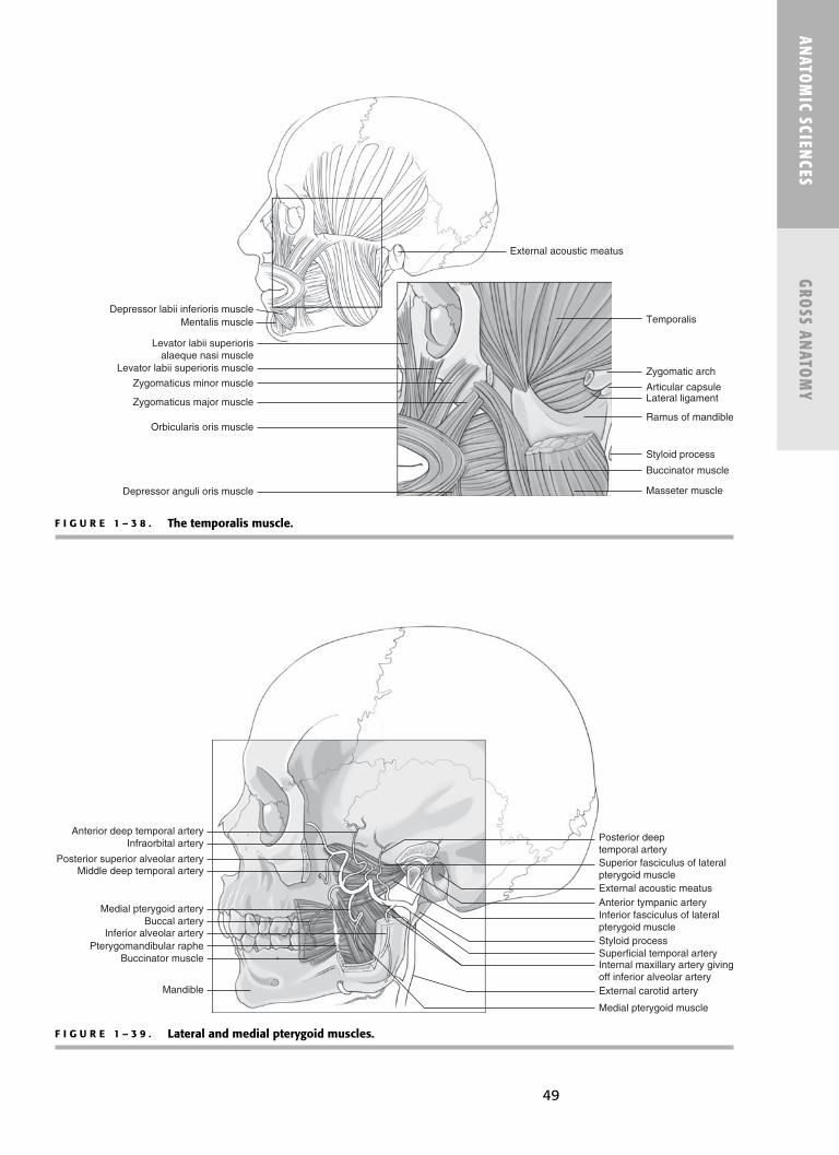
49
ANATOM
IC SCIENCES
GROSS ANATOM
Y
F I G U R E 1 – 3 8 . The temporalis muscle.
F I G U R E 1 – 3 9 . Lateral and medial pterygoid muscles.
Levator labii superiorisalaeque nasi muscle
Temporalis
Levator labii superioris muscle
Zygomaticus minor muscle
Zygomaticus major muscle
Orbicularis oris muscle
Depressor anguli oris muscle
Mentalis muscleDepressor labii inferioris muscle
Zygomatic arch
Articular capsule
Ramus of mandible
Styloid process
Buccinator muscle
Masseter muscle
External acoustic meatus
Lateral ligament
Posterior deeptemporal artery
Anterior deep temporal artery
Superior fasciculus of lateralpterygoid muscleExternal acoustic meatusAnterior tympanic arteryInferior fasciculus of lateralpterygoid muscle
Internal maxillary artery givingoff inferior alveolar arteryExternal carotid artery
Medial pterygoid muscle
Styloid processSuperficial temporal artery
Infraorbital artery
Posterior superior alveolar arteryMiddle deep temporal artery
Medial pterygoid arteryBuccal artery
Inferior alveolar arteryPterygomandibular raphe
Buccinator muscle
Mandible

50
ANAT
OM
IC S
CIEN
CES
GROS
S AN
ATO
MY
Function Muscles Involved
Opening Lateral pterygoids, suprahyoids, infrahyoidsClosing Temporalis, masseter, medial pterygoidsProtrusion Pterygoids (medial and lateral)Retrusion Temporalis, masseters (deep)Excursion Ipsilateral masseter, temporalis; contralateral
pterygoids (medial and lateral)
The masseter and medial
pterygoid muscles form a sling
at the angle of mandible; both
elevate/close the mandible
and stabilize it laterally.
Temporomandibular Joint (TMJ)
■ Bilateral synovial joint (diarthrodial).■ Upper compartment: translation■ Lower compartment: rotation
■ Articulation between mandibular condyles and skull base on both sides.(Other articulation is between maxillary and mandibular teeth.)
COMPONENTS
■ Moving from superior to inferior:■ Glenoid/mandibular fossa (of temporal bone)■ Articular cartilage■ Disc/meniscus■ Condylar cartlilage cap■ Condyle of mandible
■ Articular capsule surrounds joint, with synovium internally.■ Ligaments stabilize.
BONY COMPONENTS
■ Condyle of the mandible■ Elliptically shaped with long axis oriented mediolaterally.■ Posterior condyle is rounded and convex.■ Anteroinferior aspect is concave.
■ Glenoid/mandibular fossa (of temporal bone)■ Concave.
■ Articular eminence■ Anterior part of glenoid fossa (squamous temporal bone). ■ Articular eminence (tubercle) is convex.
■ Articular sufaces■ Glenoid fossa and condyle are lined with dense fibrocartilage. ■ Not hyaline cartilage like most synovial joints.
Articular Disc (Meniscus)
■ Fibrocartilaginous biconcave disc. ■ Lies between articular surfaces of condyle and mandibular fossa.
Palpate posterior aspect of
mandibular condyle through
external auditory meatus and
lateral aspect in front of the
external auditory meatus
(anterior to the tragus).
The lateral pterygoid attaches
to neck of the condyle,
capsule, and articular disc of
TMJ.
Damage to the disc, where the
lateral pterygoid inserts, can
render the lateral pterygoid
nonfunctional on that side.
(The mandible will deviate to
the damaged side)
MANDIBULAR FUNCTIONS FOR MASTICATION

51
■ Divides disc space into superior and inferior compartments.■ Attaches peripherally to the capsule and anteriorly to the lateral pterygoid
muscle.■ Attaches to medial and lateral poles of the condyle via collateral ligaments.■ Regions
■ Thin intermediate zone.■ Thick anterior and posterior bands.■ Posterior band is contiguous with the posterior attachment tissues (bil-
aminar zone).■ Bilaminar zone is vascular, innervated tissue (role in allowing condyle
to move forward).
Articular Capsule
■ Fibrous capsule that surrounds the TMJ. ■ Attaches superiorly to the glenoid fossa (tubercle of articular eminence).■ Attaches inferiorly to the condylar neck.■ Synovium
■ Lines the internal surface of the joint capsule.■ Secretes synovial fluid for joint lubrication. ■ Does not cover the articular surfaces or articular disc.
TMJ IMAGING
Bony Structures of TMJ
■ Panorex x-ray■ CT scan ■ Plain films
Soft Tissue of TMJ
■ MRI (magnetic resonance imaging)■ Especially the position of the articular disc
NERVES OF THE TMJ
TMJ receives only sensory innervation (motor is to the muscles).
■ Auriculotemporal nerve (of V3) conducts primary innervation to the TMJ. ■ Transmits pain in capsule and periphery of disc.■ Also provides parasympathetics to the parotid gland via lesser petrosal
nerve (CN IX).■ Nerve to masseter (of V3)
■ A few sensory fibers to anterior part of TMJ.■ Posterior deep temporal nerve (of V3)
■ Also supplies anterior part of TMJ.
ANATOM
IC SCIENCES
GROSS ANATOM
Y
MRI uses a magnetic field
to alter energy levels of water
in tissues. The advantage of
MRI is the lack of x-ray
radiation exposure, with no
harmful effects being shown.

52
ANAT
OM
IC S
CIEN
CES
GROS
S AN
ATO
MY
LIGAMENTS OF THE TMJ
■ TMJ ligaments stabilize the mandible.
TMJ Noises
Noise Description
Click With anterior disc displacement:First clickOpening: The disc clicks over the anteriorly
moving condyle (condyle clicks past the thickposterior band of articular disc).
Second clickClosing: Condyle moves posteriorly past the disc.Can also hear this click with lateral excursion to the
contralateral side (as the disc is anteromedially located and the condyle is moving medially).
Crepitus Associated with osteoarthritis of the condyle (degenerative disease).
Dull thud With self-reducing subluxation of the condyle.
Ligament Description
Temporomandibular ligament This ligament has two parts: the outer (lateral ligament) oblique portion (prevents posterior
and inferior displacement) and theinner horizontal portion (preventslateral or mesial displacement).
Accessory ligamentsSphenomandibular Remnant of Meckel’s cartilage.
ligament Thickened fibrous band connecting spine of sphenoid with lingula of mandible.
Stylomandibular Connects styloid process toligament mandibular angle.
TMJ DISLOCATION
■ TMJ can be dislocated anteriorly only (known as “lockjaw”).■ Luxation: requires assistance for reduction.■ Subluxation: auto-reduces.
TMJ DISC PLACEMENT
■ Usually occurs anteromedially.■ Collateral ligaments loosen or tear, allowing the lateral pterygoid to pull
the disc anteromedially.
To reduce luxated TMJ, apply
pressure inferiorly and
posteriorly.
Stand behind patient; thumbs
on patient’s occlusal surfaces,
fingers below chin; press
thumbs inferiorly while fingers
close the mandible; condylar
head then slides back into
articular fossa.

53
TMJ MOVEMENTS
■ Six mandibular movements:■ Protrusion■ Retrusion■ Opening■ Closing■ Medial and lateral excursions
■ Normal mandibular range of motion (ROM) is 50 mm opening, 10 mmprotrusively and laterally.
■ See Posselt’s envelope of motion.
Hinge-Type Rotation
■ With small movements (lower compartment).
Translation
■ With larger movements (upper compartment).■ Slides forward out of the mandibular fossa.
See Figure 1–40 for mandibular condyle and meniscus.See Figure 1–41 for mandibular capsule, temporomandibular ligament, andsphenomandibular ligament.
ANATOM
IC SCIENCES
GROSS ANATOM
Y
F I G U R E 1 – 4 0 . The mandibular condyle and meniscus.
Intermediate zone
Posterior band
Posterior temporal attachmentBilaminar region
Anterior temporal attachmentAnterior mandibular attachment
Head of condyle
Posterior mandibular attachment
Posterior wall of capsule
Posterior boundary of capsule
Mandible
Lateral pterygoid muscle
Anterior band

ANAT
OM
IC S
CIEN
CES
GROS
S AN
ATO
MY
F I G U R E 1 – 4 1 . Mandibular capsule, temporomandibular ligament, and sphenomandibular ligament.
54
� N EC K ANATOMY
Cervical Vertebrae
■ C1–C7■ C1 atlas (no vertebral body)■ C2 axis (dens or odontoid process)
■ Transverse foramen allow passage of the vertebral artery (from subclavian;forms basilar).
ATLANTO-OCCIPITAL JOINT
■ Articulation between C1 (atlas) superior facets and the occipital condylesof the skull.■ Allows to nod head yes (flexion and extension).
ATLANTO-AXIAL JOINT
■ Articulation between C1 vertebrae (atlas) inferior facets and the C2 verte-brae (axis) superior facets.■ Allows to shake head no (pivot).
External acoustic meatus
Articular capsule
Lateral ligamentStyloid process
Stylomandibular ligament
Mandible
Auriculotemporal nerve
Middle meningeal artery
Articular capsule
Mandibular division ofthe trigeminal nerve
Maxillary arteryLingual nerve
Inferior alveolar artery
Inferior alveolar nerveMylohyoid groove
Submandibular fovea
Styloid process
Mylohyoid artery
Sphenomandibular ligamentStylomandibular ligament
Mylohyoid nerve

55
ANATOM
IC SCIENCES
GROSS ANATOM
Y
Layers and Fascia of the Neck
Layer Contents
SkinSubcutaneous tissue (superficial Cutaneous nerves, blood and lymphatic vessels, fat,
cervical fascia). platysma (anterolaterally).Deep cervical fascia
(muscular fascia)■ Investing Sternocleidomastoid and trapezius muscles and
submandibular and parotid glands■ Pretracheal Thyroid gland, larynx, pharynx, and esophagus■ Prevertebral Vertebrae and deep cervical muscles
Attachments
Description Superiorly Inferiorly
The most superficial Superior nuchal line (occipital bone) Manubrium (sternum)deep fascial layer. Mastoid process (temporal bone) Clavicles
Splits into superficial and Zygomatic arches Acromions and spines deep layers to invest the Inferior border of mandible of scapulaSCM and trapezius. Hyoid bone
Splits to enclose Spinous processes of submandibular gland. cervical vertebrae
Splits to form fibrous capsuleof parotid gland.
PLATYSMA MUSCLE
See Figure 1–42.
■ Innervated by cervical branch of CN VII; blends with orbicularis oris.■ Superficial to the deep cervical fascia.
F I G U R E 1 – 4 2 . The platysma muscle.
Investing Layer of Deep Cervical Fascia
MandibleOrbicularis oris
Depressor labii inferioris
Mental protuberance
Platysma muscle

56
PRETRACHEAL LAYER (OF DEEP CERVICAL FASCIA)
See Figure 1–43.
■ Extends from hyoid bone to thorax (blends with fibrous pericardium).■ Has a thin muscular layer enveloping infrahyoid muscles.■ Visceral layer encloses thyroid gland, trachea, esophagus.
■ Visceral layer is continuous with buccopharyngeal fascia.■ Blends laterally with carotid sheaths.
CAROTID SHEATH
See Figure 1–44.
■ Extends from base of cranium to root of neck.■ Blends with investing and pretracheal fascia anteriorly.■ Blends with prevertebral fascia posteriorly.
Contents
■ Common carotid artery■ Internal jugular vein■ CN X■ Also associated with:
■ Lymph nodes■ Carotid sinus nerve■ Sympathetic nerves
PREVERTEBRAL LAYER (OF DEEP CERVICAL FASCIA)
■ Runs from investing layers in both sides of the lateral neck; splits toenclose the thyroid.
■ Superiorly: Attaches to laryngeal cartilages.
ANAT
OM
IC S
CIEN
CES
GROS
S AN
ATO
MY
The prevertebral layer of deep
cervical fascia is why the
thyroid moves with laryngeal
movements.
F I G U R E 1 – 4 3 . The pretracheal layer.
Modified, with permission, from White JS. USMLE Road Map Gross Anatomy. New York:
McGraw-Hill, 2003.
Trachea
Pretracheal layer
Alar fasciaEsophagus
Investinglayer
Thyroid gland
Prevertebrallayer

57
ANATOM
IC SCIENCES
GROSS ANATOM
Y
■ Inferiorly: Fuses with perichondrium■ Posteriorly: Fuses with anterior longitudinal ligament
RETROPHARYNGEAL SPACE
See Figure 1–45.
■ Between pharynx (buccopharyngeal fascia) and prevertebral fascia ■ Communicates with mediastimum (concern for infection spread, Ludwig’s
angina)
F I G U R E 1 – 4 5 . The retropharyngeal space.
Modified, with permission, from White JS. USMLE Road Map Gross Anatomy. New York:
McGraw-Hill, 2003.
F I G U R E 1 – 4 4 . The carotid sheath.
Modified, with permission, from White JS. USMLE Road Map Gross Anatomy. New York:
McGraw-Hill, 2003.
Carotid sheath Common carotid artery
Vagus nerve
Internal jugular vein
Prevertebral fasciaBuccopharyngeal fascia
Retropharyngeal space
Carotid sheath
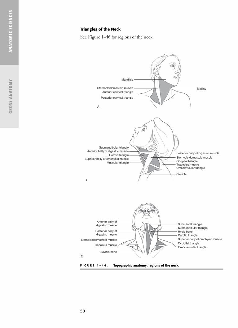
58
Triangles of the Neck
See Figure 1–46 for regions of the neck.
ANAT
OM
IC S
CIEN
CES
GROS
S AN
ATO
MY
F I G U R E 1 – 4 6 . Topographic anatomy: regions of the neck.
Midline
Mandible
Posterior cervical triangle
Sternocleidomastoid muscleAnterior cervical triangle
A
Submandibular triangle
Posterior belly of digastric muscleAnterior belly of digastric muscle
Carotid triangleSuperior belly of omohyoid muscle
Muscular triangle
Sternocleidomastoid muscleOccipital triangleTrapezius muscleOmoclavicular triangle
Clavicle
B
Anterior belly ofdigastric muscle Submental triangle
Posterior belly ofdigastric muscle
Sternocleidomastoid muscle
Trapezius muscle
Clavicle bone
Submandibular triangleHyoid boneCarotid triangleSuperior belly of omohyoid muscle
Occipital triangleOmoclavicular triangle
C

ANATOM
IC SCIENCES
GROSS ANATOM
Y
59
POSTERIOR TRIANGLE
Boundaries
Anterior Posterior Inferior Floor Roof Contents
SCM (post Trapezius Clavicle Splenius capitus Skin Exterior jugular border) (anterior border) (middle 1/3) Levator scapulae Superficial vein
Scalene muscles fascia, platysma, Cervical plexus, Deep investing lesser occipital nerve,fascia of neck great auricular nerve;
CN XI, phrenic nerveSubclavian vein, artery, brachial plexus
ANTERIOR TRIANGLE
Boundaries
Anterior Posterior Inferior Floor Roof Contents
Neck SCM (anterior Inferior border Pharynx, larynx, Skin Infrahyoid, suprahyoid midline border) of mandible thyroid Superficial fascia muscles
Platysma Common, internal,Deep investing external carotid
fascia arteriesInternal, external
jugular veinRetromandibular veinCNs X, XI, XII, cervical
plexus
SUBSETS OF THE ANTERIOR TRIANGLE
Submandibular Triangle (see Figure 1–47)
Boundaries
Inferior Superior Floor Roof Contents
Bellies of Inferior Mylohyoid Skin Submandibular digastric border of Hyoglossus Superficial gland (Figure 1–48)
mandible fascia Submandibular Platysma lymph nodesDeep fascia Hypoglossal nerve
Mylohyoid nerveLingual and facial
arteries and veins
Submandibular triangle
contains two glands
(or nodes), two nerves,
two arteries, and two veins.

60
ANAT
OM
IC S
CIEN
CES
GROS
S AN
ATO
MY
F I G U R E 1 – 4 7 . The submandibular triangle.
F I G U R E 1 – 4 8 . The submandibular gland.
Posterior belly of digastric muscle
Hypoglossal nerve
Stylohyoid muscle
Facial vein
Hyoid bone
Facial artery
Hyoglossus muscle
Anterior belly ofdigastric muscle
Submandibular gland
Lingual veinLingual artery
Superior pharyngeal constrictor
Mylohyoid muscle
Anterior belly ofdigastric muscle
Hyoglossus
Submandibular gland (superficial part)
Stylohyoid
Posterior belly of digastric
Longus capitis

61
ANATOM
IC SCIENCES
GROSS ANATOM
Y
Boundaries Superior belly of omohyoid, SCM, midline of neckContents Infrahyoid strap muscles
Carotid Triangle
Boundaries Superior belly of omohyoid, posterior bellyof digastric, SCM
Contents Common carotid artery, internal jugular vein, CNs X, XI, XII, cervical plexus
Submental Triangle
Boundaries Right and left anterior bellies of the digastric,body of hyoid
Contents Mylohyoid muscle (midline raphe)
Muscular Triangle
SCM, Trapezius Muscles
Origin Insertion Action Innervation
SCM Manubrium, Mastoid Bilateral: Flexes CN XImedial process, neck.one-third superior Unilateral: Pullsclavicle nuchal line head to shoulder,
(lateral) turns head toopposite side.
Trapezius Thoracic and Scapula Bilateral: Extends CN XIcervical spines (spine and head.
Ligamentum acromion) Unilateral: Chinnucha Lateral up to opposite
Superior nuchal one-third side; elevate line clavicle acromion (rotates,
elevates scapula and clavicle).
With damage to CN XI (spinal
accessory), in the posterior
triangle, you cannot raise an
arm above horizontal and
cannot shrug shoulder
(because of the trapezius).
Hyoid Bone
■ U-shaped floating bone (from second and third branchial arches).■ Composed of body, greater and lesser horns (cornua).
Torticollis (head tilts to
affected side) results from
injury to SCM.
Three infrahyoid muscles
innervated by ansa cervicalis:
sternohyoid, sternothyroid,
omohyoid (SOS).
Thyrohyoid innervated by C1
via CXII.

62
Supra- and Infrahyoid Muscles (see Figure 1–49)
ANAT
OM
IC S
CIEN
CES
GROS
S AN
ATO
MY
Ligaments Muscles (Innervation)
Stylohyoid ligament Mylohyoid (V3)Hypoepiglottic ligament Anterior digastric (V3)
Posterior digastric (VII)Stylohyoid (VII)Hypoglossus (XII)Geniohyoid (C1 fibers via CN XII)Omohyoid (ansa cervicalis)Sternohyoid (ansa cervicalis)Thyrohyoid (C1 fibers via CN XII)
HYOID ATTACHMENTS
Muscle Origin Insertion Action Innervation
SuprahyoidsDigastric (anterior) Intermediate Anterior Raises hyoid CN V3 (nerve to
tendon mandible mylohyoid)(digastric fossa)
Digastric (posterior) Temporal bone Intermediate Raises hyoid CN VII(digastric notch) tendon
Mylohyoid Medial mandible Median raphe; Raises hyoid, base CN V3 (nerve to(mylohyoid line) body of hyoid of tongue, floor mylohyoid)
of mouthGeniohyoid Genial tubercles Body of hyoid Raises hyoid C1 fibers via
(mandible) (pulls hyoid forward CN XIIto open pharynx)
Stylohyoid Styloid process Hyoid Raises hyoid CN VII(greater horn)
InfrahyoidsOmohyoid Superior belly: Superior belly: Depresses hyoid Ansa cervicalis
Intermediate Hyoid (body, lower and larynxtendon surface)
Inferior belly: Superior belly:Superior scapula Intermediate tendon
Sternohyoid Manubrium of Hyoid Depresses hyoid Ansa cervicalissternum (lower surface) and larynx
Sternothyroid Manubrium of Thyroid cartilage Depresses larynx Ansa cervicalissternum (oblique line)
Thyrohyoid Thyroid cartilage Hyoid (body and Depresses hyoid C1 fibers via (oblique line) greater horn) and larynx CN XII
Supra- and Infrahyoid Muscles
Connections to styloidprocess (of temporalbone):
■ Stylomandibularligament
■ Stylopharyngeusmuscle
■ Stylohyoid muscle■ Styloglossus muscle
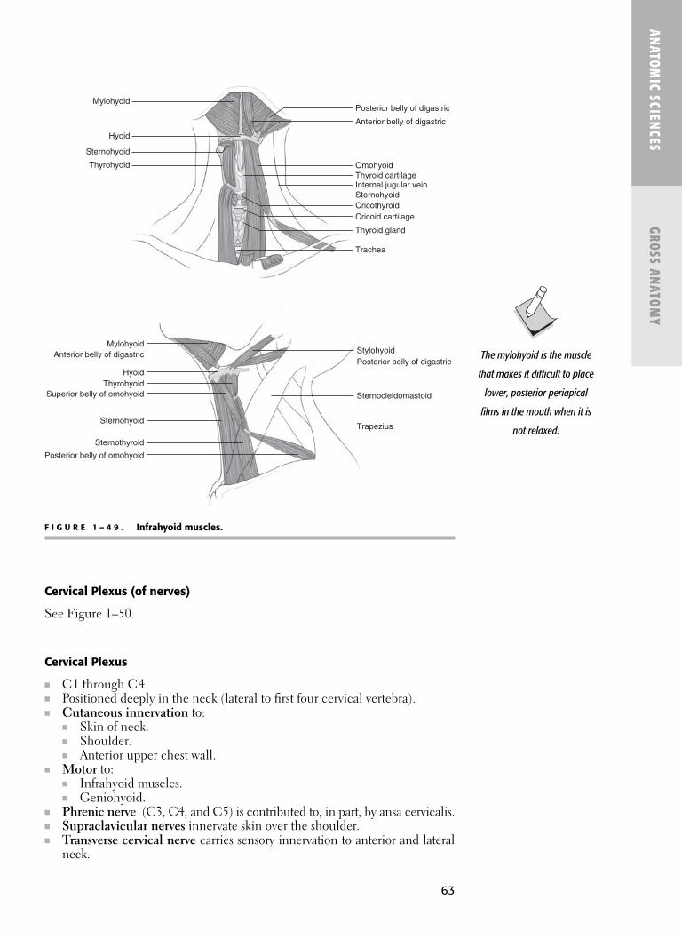
63
ANATOM
IC SCIENCES
GROSS ANATOM
Y
F I G U R E 1 – 4 9 . Infrahyoid muscles.
The mylohyoid is the muscle
that makes it difficult to place
lower, posterior periapical
films in the mouth when it is
not relaxed.
Cervical Plexus (of nerves)
See Figure 1–50.
Cervical Plexus
■ C1 through C4■ Positioned deeply in the neck (lateral to first four cervical vertebra).■ Cutaneous innervation to:
■ Skin of neck.■ Shoulder.■ Anterior upper chest wall.
■ Motor to:■ Infrahyoid muscles.■ Geniohyoid.
■ Phrenic nerve (C3, C4, and C5) is contributed to, in part, by ansa cervicalis.■ Supraclavicular nerves innervate skin over the shoulder.■ Transverse cervical nerve carries sensory innervation to anterior and lateral
neck.
Stylohyoid
Mylohyoid
MylohyoidAnterior belly of digastric
HyoidThyrohyoid
Superior belly of omohyoid
Sternohyoid
Sternothyroid
Posterior belly of omohyoid
Posterior belly of digastric
Sternocleidomastoid
Trapezius
Hyoid
Thyrohyoid
Sternohyoid
Anterior belly of digastric
Posterior belly of digastric
OmohyoidThyroid cartilageInternal jugular veinSternohyoidCricothyroidCricoid cartilage
Thyroid gland
Trachea

64
ANAT
OM
IC S
CIEN
CES
GROS
S AN
ATO
MY
F I G U R E 1 – 5 0 . The cervical plexus.
Reproduced, with permission, from White JS. USMLE Road Map Gross Anatomy. New York:
McGraw-Hill, 2003.
ANSA CERVICALIS (MOTOR)
■ Motor division of cervical plexus.■ Comes from C1 (runs with CN XII), C2, C3.■ Innervates:
■ Infrahyoids (except thyrohyoid, which is innervated by C1).■ Geniohyoid (moves hyoid anteriorly to hold open the pharynx).
Branches of C2, C3 Loop (Sensory)
Nerve Supplies
Lesser occipital nerve (C2) Skin of neck and scalp (posterosuperior to auricle)Great auricular nerve (C2, C3) Skin over parotid gland, posterior aspect of auricle,
area from angle of mandible to mastoidTransverse cervical nerve (C2, C3) Skin of anterior triangle
Phrenic Nerve
■ C3, C4, C5 “keeps the diaphragm alive.”■ Contains motor, sensory, and sympathetic nerve fibers.■ Sole motor innervation to the diaphragm.The terminal branches of the
ECA are the maxillary and
superficial temporal arteries.
See innervation of external
ear. Remember, four nerves
are involved.
Greater occipital n.(dorsal ramus of C2)
Accessory n.(CN XI)
3
4
5
6
Trapezius m.
Supraclavicular nn.
Lesser occipital n.
Great auricular n.
Cervical br. offacial n. (CN VII)
Transverse cervical n.
Sternocleidomastoid m.

65
ANATOM
IC SCIENCES
GROSS ANATOM
Y
Blood Supply to Face
See Figure 1–51 for arteries of the head and neck.
■ Blood is supplied to the face via the external carotid branches.
External Carotid Artery
Course
■ Branches from the common carotid artery at the level of the upper borderof thyroid cartilage.
■ Gives off branches and continues to the substance of the parotid glandwhere it ends as:■ Maxillary artery.■ Superficial temporal branch (superiorly).
In addition to the lingual
artery, the tongue receives
some blood supply from the
tonsillar branch of the facial
artery and the ascending
pharyngeal artery.
F I G U R E 1 – 5 1 . Arteries of the head and neck.
Reproduced, with permission, from Langman J, Woerdeman MW. Atlas of Medical Anatomy. Philadelphia: WB Saunders
Company, 1978.
Superficial temporal artery
Post. auricular artery
Occipital artery
Ascending pharyngeal artery
Internal carotid artery
Vertebral artery
Right subclavian artery
Brachiocephalic trunk
Common carotid artery
Sup. thyroid artery
External carotid artery
Lingual artery
Facial artery
Maxillary artery
Infraorbital artery
Transverse facial artery
Middle meningeal artery

66
ANAT
OM
IC S
CIEN
CES
GROS
S AN
ATO
MY
Supply
■ Supplies most of head and neck (except the brain).■ Face■ Thyroid■ Salivary glands■ Tongue■ Jaws■ Teeth
■ Carotid sinus (baroreception for blood pressure).■ At the common carotid bifurcation.
■ Carotid body (chemoreception including O2, CO2, pH, and temperature). ■ Posterior to the bifurcation of the CCA.
See also the Physiology section in Chapter 12.
BRANCHES OF THE EXTERNAL CAROTID
■ The mnemonic to remember the branches of the external carotid artery isSALFOPSM: Some Anatomists Like Football, Others Prefer SoccerMatches.
The anterior hard palate is
supplied by the nasopalatine
artery, a branch of the
sphenopalatine, after it exits
the incisive foramen.
Epistaxis (nosebleed) occurs
because of the anastomoses
in the nasal septum
(Kiesselbach’s plexus,
comprising five arteries).
The mnemonic to remember
the branches of the facial
artery is TAGS ISLA.
The terminal branch of facial
artery is the angular artery.
The facial artery supplies the
muscles of facial expression.
Branch Supplies
Superior thyroid Thyroid gland, gives off SCM branch and the superior laryngeal artery
Ascending palatine Soft palate, eustachian tube, palatine tonsils,levator veli palatini muscle
Lingual TongueFacial Face and submandibular glandOccipital Pharynx and suboccipital trianglePosterior auricular Posterior scalpSuperficial temporal Parotid gland, auricle, temple, scalpMaxillary Infratemporal fossa, nasal cavity
LINGUAL ARTERY
■ Originates from the ECA at the greater horn of hyoid bone (in the carotidtriangle).
■ Supplies:■ Tongue.■ Sublingual gland.■ Floor of mouth.
■ Branches:■ Dorsal lingual.■ Suprahyoid.■ Sublingual arteries (to sublingual gland).
■ Ends as the deep lingual artery ■ Between the genioglossus and hyoglossus muscles.
■ Passes deep to hyoglossus muscle, then supplies tongue
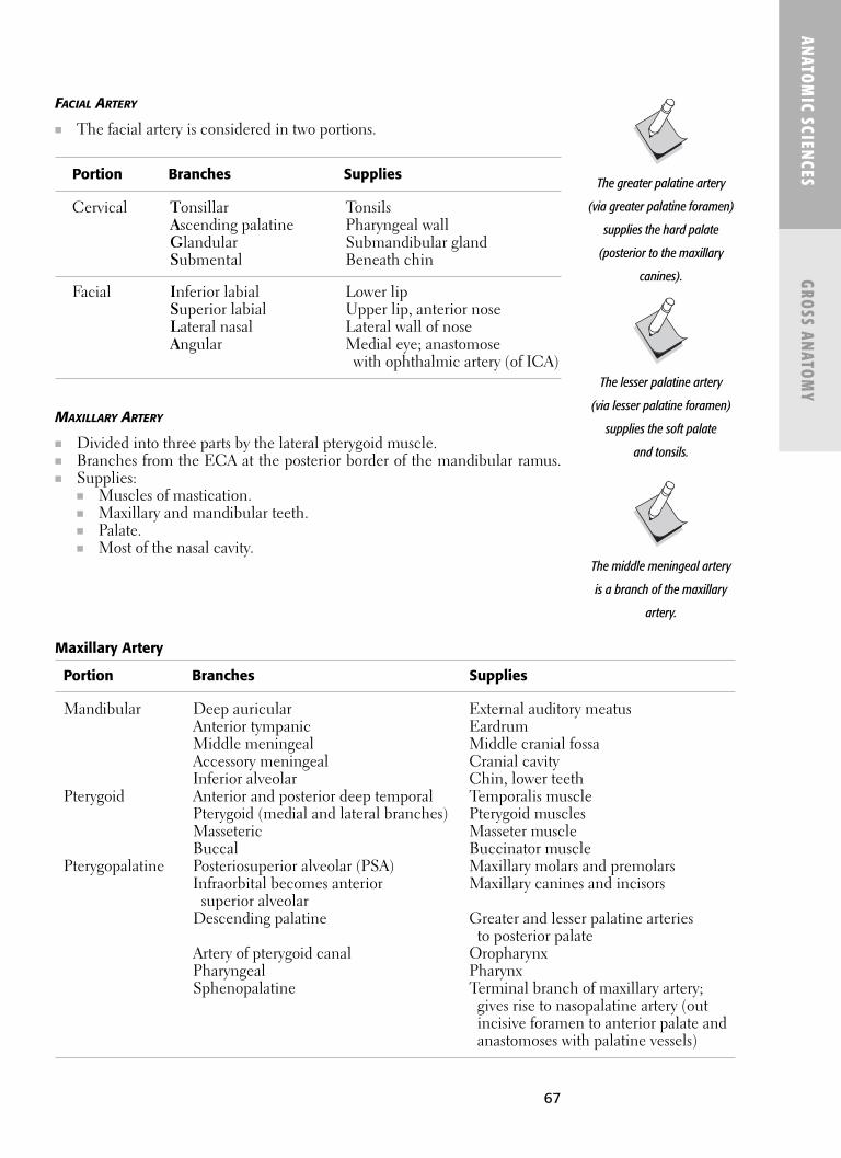
67
ANATOM
IC SCIENCES
GROSS ANATOM
Y
FACIAL ARTERY
■ The facial artery is considered in two portions.
The greater palatine artery
(via greater palatine foramen)
supplies the hard palate
(posterior to the maxillary
canines).
The lesser palatine artery
(via lesser palatine foramen)
supplies the soft palate
and tonsils.
The middle meningeal artery
is a branch of the maxillary
artery.
Maxillary Artery
Portion Branches Supplies
Mandibular Deep auricular External auditory meatusAnterior tympanic EardrumMiddle meningeal Middle cranial fossaAccessory meningeal Cranial cavityInferior alveolar Chin, lower teeth
Pterygoid Anterior and posterior deep temporal Temporalis musclePterygoid (medial and lateral branches) Pterygoid musclesMasseteric Masseter muscleBuccal Buccinator muscle
Pterygopalatine Posteriosuperior alveolar (PSA) Maxillary molars and premolarsInfraorbital becomes anterior Maxillary canines and incisors
superior alveolarDescending palatine Greater and lesser palatine arteries
to posterior palateArtery of pterygoid canal OropharynxPharyngeal PharynxSphenopalatine Terminal branch of maxillary artery;
gives rise to nasopalatine artery (outincisive foramen to anterior palate andanastomoses with palatine vessels)
MAXILLARY ARTERY
■ Divided into three parts by the lateral pterygoid muscle.■ Branches from the ECA at the posterior border of the mandibular ramus.■ Supplies:
■ Muscles of mastication.■ Maxillary and mandibular teeth.■ Palate.■ Most of the nasal cavity.
Portion Branches Supplies
Cervical Tonsillar TonsilsAscending palatine Pharyngeal wallGlandular Submandibular glandSubmental Beneath chin
Facial Inferior labial Lower lipSuperior labial Upper lip, anterior noseLateral nasal Lateral wall of noseAngular Medial eye; anastomose
with ophthalmic artery (of ICA)

68
ANAT
OM
IC S
CIEN
CES
GROS
S AN
ATO
MY
Venous Drainage from the Face
See Figure 1–52.
FACIAL VENOUS DRAINAGE
See Figure 1–53.
SUPERFICIAL TEMPORAL VEIN
■ Drains scalp and side of head.■ Merges with maxillary vein and plunges into parotid gland.
MAXILLARY VEIN
■ Forms from the pterygoid plexus of veins, which is the connection of deepsystem with superficial venous drainage.
RETROMANDIBULAR VEIN
■ Formed by the superficial temporal and maxillary veins.■ Divides at the angle of the mandible into anterior and posterior branches.
EXTERNAL JUGULAR VEIN
■ Formed by the posterior auricular and retromandibular veins.■ Crosses the SCM.■ Drains the
Remember: The scalp can
drain superficially
(via superficial temporal and
posterior auricular veins) or
deeply (via emissary veins).
F I G U R E 1 – 5 2 . Veins of the head and neck.
Reproduced, with permission, from Langman J, Woerdeman MW. Atlas of Medical Anatomy.
Philadelphia: WB Saunders Company, 1978.
Superficial temporal venous network
Superficial temporal vein
Post. auricular vein
Retromandibular vein
Occipital vein
Externaljugular vein
Superficial cervical vein
Transverse cervical vein
Suprascapular vein
Subclavian vein
Right brachiocephalic vein
Inf. thyroid vein
Ant. jugular vein (cut)
Internal jugular vein
Sup. thyroid vein
Lingual vein
Maxillary vein
Facial vein
Transverse facial vein
Angular vein
Supratrochlear vein
Supraorbital vein

69
■ Skin.■ Parotid gland.■ Muscles of the face and neck.
■ Empties into the subclavian vein.
FACIAL VEIN (ANTERIOR FACIAL VEIN)
■ Forms from the angular vein (which itself forms from the supraorbital andsupratrochlear veins).
■ Receives infraorbital and deep facial veins.■ Enters into the IJV (directly or by way of the common facial vein).
COMMON FACIAL VEIN
■ Formed by the anterior facial and retromandibular veins.
INTERNAL JUGULAR VEIN
■ Arises from the sigmoid and inferior petrosal sinuses.■ Drains the dural venous sinuses.■ Exits the skull via the jugular foramen (along with CNs XI, X, IX).■ Descends the neck in the carotid sheath.■ Merges with the subclavian vein to form:
■ Large brachiocephalic vein (behind the sternoclavicular joint).■ Left and right brachiocephalic veins form the superior vena cava.
DANGER TRIANGLE OF THE FACE
■ Area where superficial facial veins communicate with deep system (duralsinuses).■ Base: Upper lip/anterior maxilla.■ Apex: Infraorbital region.
Lymph Nodes in the Face
See Figures 1–54 and 1–55.See the section “Tongue” for illustration of tongue lymph drainage.
ANATOM
IC SCIENCES
GROSS ANATOM
Y
F I G U R E 1 – 5 3 . Facial venous drainage.
Important:
Veins of the head and neck
contain no valves.
Because there are no valves,
retrograde flow can allow
infection to spread via the
deep facial vein, pterygoid
plexus, and ophthalmic veins
to the cavernous sinus.
The deep cervical nodes form
the jugular lymph trunk
(along IJV).
Posteriorauricular vein
Superficial temporal vein
Pterygoid plexus of veins
Anterior facialvein
Common facial vein
Drains into internal jugular vein
Internal jugular veinExternal jugular vein
Joins with the subclavian veinto form brachiocephalic vein
Retromandibular vein
Anteriorretromandibular
vein
Posteriorretromandibular
vein

70
ANAT
OM
IC S
CIEN
CES
GROS
S AN
ATO
MY
F I G U R E 1 – 5 4 . Lymphatic drainage of the face. M, mastoid nodes: O, occipital nodes;P, parotid nodes; SMd, submandibular nodes; SMe, submental nodes.
Reproduced, with permission, from Liebgott B. The Anatomical Basis of Dentistry. Toronto: BC
Decker, 1986.
The jugular lymph trunkempties into either
■ The thoracic duct (on left) or the rightlymphatic duct (whichthen empties into thebrachiocephalic vein).
■ Example:■ Infection of lower lip
■ Enters bloodstream at rightbrachiocephalicvein.
■ Infection of abdomen ■ Enters at left
brachiocephalicvein.
F I G U R E 1 – 5 5 . Lymph nodes in the face.
Jugulodigastric node
Deep cervical (jugular trunk)
SMe
Jugulo-omohyoid node
Retropharyngeal
Post nasal cavity Nasopharynx Soft palate Middle ear Ext. aud. meatus
Jugulodigastric node
Jugulo-omohyoid node
Superficialring of nodes
Deep cervicallymph nodes
Deep nodes
Submandibular
Submental nodes Cheek Nose, paranasal sinuses Upper lip Maxillary teeth Mandibular teeth (except incisors) Facial gingivae Mucosa/gingivae of hard palate Posterior floor of mouth Tongue (ant 2/3 not tip)
Submental
Tip of tongue Floor of mouth (beneath tongue tip) Mandibular incisors and assoc. gingivae Center part of lower lip Skin of chin
Parotid
• Superficial Eyelids Temporal region Cheek Auricle • Deep Middle ear Ext. auditory meatus Soft palate Post. nasal cavity
Mastoid
Scalp Auricle
Occipital
Scalp

71
Thyroid
See Figure 1–56.
■ H-shaped gland at laryngotracheal junction at anterior neck.■ Two lobes (right and left) joined by isthmus.■ Largest endocrine gland.■ Produces/secretes
■ Thyroid hormone. ■ Calcitonin.
ANATOM
IC SCIENCES
GROSS ANATOM
Y
F I G U R E 1 – 5 6 . Thyroid gland.
■ Blood supply: See the following chart.■ Nerve supply
■ Glandular branches of cervical ganglia of sympathetic trunk.
Follicular Cells
■ Produce thyroglobulin (tyrosine-containing). ■ Stored in colloid.■ Precursor to T3 (triiodothyronine) and T4 (thyroxine) (involved in basal
metabolic rate).
Hyoid
Pyramidal lobe
Cricothyroid
Left lobe
Isthmus
Trachea
Cricothyroid membrane
Cricoid cartilage
Superior thyroid vein
Superior thyroid artery
Superior laryngeal arteryThyrohyoid membrane
Thyroid cartilage
Right lobe
Common carotid
Internal jugular vein
Inferior thyroid veins
T3 is about 20 times more
potent than T4 at increasing
cellular metabolism.
Calcitonin acts to “tone down”
blood calcium levels.

72
Parafollicular (C) Cells
■ Produce calcitonin. ■ Lower blood calcium and phosphate.
Thyroglossal Duct
■ Connects thyroid gland to tongue development.■ Foramen cecum at base of tongue is proximal remnant.
THYROID BLOOD SUPPLY AND DRAINAGE
ANAT
OM
IC S
CIEN
CES
GROS
S AN
ATO
MY
The vocal cords attach
anteriorly at the lamina of
thyroid cartilage and
posteriorly at the vocal
process of arytenoid
cartilages.
Artery Branch of Vein Drains to
Superior thyroid artery ECA Superior thyroid vein IJV— — Middle thyroid vein IJVInferior thyroid artery Thyrocervical trunk Inferior thyroid vein Brachiocephalic vein
Parathyroid Glands
■ Small pea-shaped organs.■ Two superior parathyroids develop from the 4th pharyngeal pouch.■ Two inferior parathyroids develop from the 3rd pharyngeal pouch.■ Encased in the posterior surface of the thyroid.■ Blood supply
■ Superior thyroid artery (from ECA) (to superior part of gland).■ Inferior thyroid artery (from thyrocervical trunk) (to inferior part of gland).
■ Innervation■ Postganglionic sympathetic fibers of superior cervical ganglion.
■ Produces parathyroid hormone (PTH).■ Regulates calcium and phosphate metabolism.■ Deficiency of PTH → decrease Ca2+ can cause tetany.
Larynx
■ The larynx is responsible for voice production.
LARYNGEAL SKELETON
■ Nine cartilages (three single, three paired)
Important: There is no middle
thyroid artery; however, up to
10% of the population also
receives a blood supply via
the thyroid ima artery.

ANATOM
IC SCIENCES
GROSS ANATOM
Y
73
EPIGLOTTIS
■ Thin leaf-shaped cartilage, covered with mucous membrane, at the root ofthe tongue.
■ Flap closing over the larynx when swallowing.■ Median glossoepiglottic fold (1). ■ Lateral glossoepiglottic folds (2).■ Valecullae (2) (lie between the glossoepiglottic folds).■ Taste buds (CN X) are located on superior surface of epiglottis.
VESTIBULAR FOLDS
■ False vocal folds.■ No role in vocalization.■ Protective.
VOCAL FOLDS
■ True vocal cords. ■ Composed of vocal ligament and vocalis muscle.■ Involved in sound production.■ Sphincter of respiratory tract.
GLOTTIS
■ Composed of vocal fold and rima glottis.■ Rima glottis is the opening between vocal folds.
LARYNGEAL MUSCLES
EXTRINSIC MUSCLES
Muscle Group Function
Suprahyoids and stylopharyngeus muscle Raise larynx.Infrahyoids Depress larynx.
Cartilage Description
Thyroid Largest; forms the laryngeal prominence (“Adam’s apple”); superior thyroid notch; superior surface is attached to hyoid by thyrohyoid membrane
Cricoid Only laryngeal cartilage to form a complete ring around the airway.
Epiglottic Fibrocartilage; gives flexibility to epiglottis.Arytenoid (2) Vocal cord attaches posteriorly.Corniculate (2)Cuneiform (2)
All intrinsic laryngeal muscles
are supplied by the recurrent
laryngeal nerve except the
cricothyroid muscle, which is
innervated by the external
laryngeal nerve (a branch of
the superior laryngeal nerve).
The cricothyroid membrane is
incised at the midline
for an emergent
cricothyroidotomy/
tracheotomy. The cricothyroid
space is entered (inferior to
rima glottis where aspirated
objects are often lodged or
laryngospasm is occurring).

74
ANAT
OM
IC S
CIEN
CES
GROS
S AN
ATO
MY
LARYNGEAL NERVES
See also Vagus.
RECURRENT LARYNGEAL NERVE
See Figure 1–57.
■ Is a branch of the vagus nerve (CN X). ■ Comes off the vagus in the mediastinum and ascends to the larynx in the
tracheoesophageal groove.
INTRINSIC MUSCLES
Muscle Origin Insertion Action Innervation
Cricothyroid Anterolateral Thyroid cartilage Stretches and tenses External cricoid cartilage (inferior margin and vocal fold laryngeal nerve
inferior horn)
Posterior Cricoid cartilage Muscular process of Abducts vocal fold Recurrent cricoarytenoid (posterior laminae) arytenoid cartilage laryngeal nerve
Lateral Cricoid cartilage Muscular process of Adducts vocal fold Same as abovecricoarytenoid (arch) arytenoid cartilage
Thyroarytenoid Thyroid cartilage Muscular process of Relaxes vocal fold Same as above(posterior surface) arytenoid cartilage
Transverse and Arytenoid cartilage Opposite arytenoids Closes rima glottides Same as aboveoblique arytenoids cartilage
Vocalis Depression between Vocal ligament and Relaxes posterior Same as abovelaminae of thyroid vocal process of vocal ligament cartilage arytenoids cartilage Tenses anterior
vocal ligament Antagonist of
cricothyroid muscle
F I G U R E 1 – 5 7 . Course of the recurrent laryngeal nerve (posterior view).
Reproduced, with permission, from Bhushan V, et al. First Aid for the USMLE Step 1. New York:
McGraw-Hill, 2003.
Injured nerves (eg, after
thyroid or neck surgery) cause
hoarseness.
75
■ Has close association with thyroid gland and inferior thyroid artery. ■ Left recurrent laryngeal nerve wraps around aortic arch (ligamentum
arteriosum).■ Right recurrent laryngeal nerve wraps around right subclavian.■ Motor innervation: Involves all intrinsic muscles of larynx (via the inferior
laryngeal branch) except cricothyroid.■ Sensory innervation: Involves laryngeal mucosa below vocal folds and upper
trachea.
SUPERIOR LARYNGEAL NERVE
■ Branch of the vagus nerve arising just after exit from jugular foramen.■ Passes through thyrohyoid membrane.■ External branch: Innervates cricothyroid muscle (tenses vocal cords, increas-
ing pitch).■ Internal branch: Provides sensory information to laryngeal mucosa above
the vocal folds.
Respiratory System
■ Nasal cavity■ Pharynx■ Larynx■ Trachea■ Bronchi■ Bronchioles■ Alveoli
TRACHEA
■ Air tube lined by C-shaped tracheal rings.■ Lined with respiratory epithelium.■ Formed from hyaline cartilage.■ ~10 cm long.■ ~2.5 cm diameter.■ Extends from:
■ C5–C6 level (base of larynx—starts as ligamentous attachment tocricoid) to Sternal angle (of Louis) T4/5 level (second rib level).
■ Branches into left and right mainstem bronchi (at Carina).
Branching Pattern
Trachea↓
Primary bronchi: two mainstem bronchi (one per lung)↓
Secondary bronchi: five lobar bronchi (three in right lung; two in left lung) ↓
Tertiary bronchi↓
Terminal bronchioles↓
Respiratory bronchioles↓
Alveoli
ANATOM
IC SCIENCES
GROSS ANATOM
Y
76
� BRAC H IAL PLEXUS AN D U PPE R EXTRE M ITI ES
Axilla
ANAT
OM
IC S
CIEN
CES
GROS
S AN
ATO
MY
Contents
■ Axillary vessels■ Brachial plexus■ Biceps brachii (both heads)■ Coracobrachialis
AXILLARY ARTERY
■ Continuation of subclavian artery. ■ Named axillary artery as it passes lateral border of first rib.
■ Travels close with the medial cord of the brachial plexus.■ Aneurysm can compress the medial cord.
■ Passes posterior to pectoralis minor muscle.■ Becomes brachial artery.
■ As it passes inferior border of teres major and enters the arm.
BRACHIAL ARTERY
■ Continuation of axillary artery in the arm.■ Immediately medial to tendon of biceps brachii at elbow.
AXILLARY VEIN
■ Formed from the brachial veins and the basilic vein at the inferior borderof teres major.
■ Changes name to subclavian vein at the lateral border of the first rib.
CEPHALIC VEIN
■ Drains into the axillary vein.■ Found in deltopectoral groove.
■ Located between deltoid and pectoralis major.■ Drains superficial arm.
■ Drains blood from the radial side of arm.
Brachial and basilic veinsmerge to form the axillary vein.
Profunda brachii artery arisesfrom brachial artery in
proximal arm.
Boundaries
Medial Upper 4–5 ribsIntercostal musclesSerratus anterior muscle
Lateral HumerusCoracobrachialis and biceps musclesWithin intertubercular groove
Posterior Subscapularis Teres majorLatissimus dorsi
Anterior Pectoralis major and minorSubclavius muscles
Base Axillary fasciaSkin
The subclavian arteries supply
the upper extremities.

77
BRACHIAL VEIN
■ Drains blood from the deep arm.
BASILIC VEIN
■ Drains blood from the superficial arm.
SCALENE MUSCLES
■ There are three scalene muscles per side: ■ Anterior■ Middle■ Posterior
■ Phrenic nerve and subclavian vein pass on the surface of the anterior scalene.■ Brachial plexus and subclavian artery pass between the anterior and middle
scalene.
See Figure 1–58 for the dorsal scapular artery.
Brachial Plexus
■ The brachial plexus innervates the shoulder girdle and upper limb (ventralrami C5–T1).
OTHER BRANCHES OF THE BRACHIAL PLEXUS
See Figure 1–59.
ANATOM
IC SCIENCES
GROSS ANATOM
Y
F I G U R E 1 – 5 8 . Scalene muscles.
Thoracic outlet syndromeresults from compression of
the lower trunk of the brachialplexus and the subclavian
artery between the anteriorand middle scalene.
Middle scalene muscleAscending cervical artery
Dorsal scapular artery
Brachial plexus
Suprascapular artery
Third portion of subclavian artery
Inferior thyroid artery
Transverse cervical arteryVertebral artery
Thyrocervical trunkRight common carotid artery
Internal thoracic artery
Anterior scalene muscle

78
ANAT
OM
IC S
CIEN
CES
GROS
S AN
ATO
MY
Major Terminal Nerves of the Brachial Plexus
Nerve Course Motor Sensory
Median (C5–T1) Lateral and medial cords Forearm flexors: Anterior arm(from lateral and Passes between two Flexor carpi radialis Lateral palm (radial 2/3;medial cords) heads of pronator teres Palmaris longus thumb side)
Enters carpal tunnel in Pronators Lateral 31/2 digits (thumb, wrist between: Digital flexors 2, 3, including nailbeds)
Palmaris longus Thenar musclesFlexor carpi radialis Lateral two lumbricals (thumb side)
Ulnar (C8–T1) Medial cord Flexors of wrist and fingers Medial arm/forearmBehind medial (including flexor carpi ulnaris) Medial palm
epicondyle (elbow) Ulnar two lumbricals Medial 11/2 digit (pinky)Between flexor carpi Interosseous muscles
ulnaris and flexor digitorum profundus (wrist)
Superficial to flexor retinaculum in wrist
Radial (C6–C8) Posterior cord Extensors of arm/forearm: Posterior armMusculospiral groove ■ Triceps Forearm hand
(posterior humerus) ■ Brachioradialis (not fingers)Passes between ■ Extensor carpi radialis
brachioradialis and ■ Extensor carpi ulnarisbrachialis muscles ■ Extensors of wrist/fingers
■ Adductor pollicus■ Supinator■ Triceps brachii (medial
and lateral head) [BEAST]
Axillary Comes off in axilla Deltoid, teres minor, Skin of lower 1/2 of deltoidtriceps brachii (long head)
Musculocutaneous Arm flexors Anterolateral arm (C5–C7) ■ Biceps brachii Forearm
■ Coracobrachialis■ Brachialis
F I G U R E 1 – 5 9 . The brachial plexus.
Reproduced, with permission, from White JS. USMLE Road Map Gross Anatomy. New York: McGraw-Hill, 2003.
Suprascapular n.
Lateral pectoral n.Cords
DivisionsLateral cord
Posterior cord
Medial cord
Musculocutaneous n.
Branches
Median n.5
6
6
56
78 81
78
1
Axillary n.
Radial n.
Ulnar n. Medial brachial cutaneous n.
Subscapular nn.(upper and lower)
Medial pectoral n.
Long thoracic n.
TrunksThoracodorsal n.(middle subscapular n.)
Medial antibrachial cutaneous n.
Posterior division fibers
Anterior division fibers Dorsal scapular n.
Upper trunk
Middle trunk
Lower trunk
C5
Roots:
C6
C7
C8
T1

79
ANATOM
IC SCIENCES
GROSS ANATOM
Y
Summary of Brachial Plexus Actions by Roots
Root(s) Action(s)
C5 ShoulderFlexionAbductionLateral rotation ElbowFlexion
C5, C6 Elbow■ Extension
C6, C7 Wrist■ Flexion
C7, C8 Shoulder■ ExtensionElbow■ PronationFingers■ Extension
Nerve Supplies
Dorsal scapular (C4–C5) RhomboidsUpper/lower subscapular (C5–C6) Subscapularis, teres majorLong thoracic (C5–C7) Serratus anteriorSuprascapular (C4–C6) SupraspinatusThoracodorsal (C6–C8) Infraspinatus
Glenohumeral jointLatissimus dorsi
The hypothenar (pinky)
region of the palm is supplied
by the ulnar nerve.
The posterior cord gives rise
to the radial and axillary
nerves.
The thenar (thumb) region
of the hand and nailbeds
of thumb, index, and middle
fingers (and half of ring
finger) are supplied by
palmar digital nerves of the
median nerve.
The radial nerve is the “great
extensor nerve.” It is motor
to all muscles of the posterior
arm. It is the most often
injured nerve in a
mid-humeral shaft fracture
because it runs in the radial
(spiral) groove of the
humerus. Radial nerve deficit
includes wrist drop, inability to
make a tight fist, and sensory
deficit to the radial hand and
lower part of thumb and digits
2 and 3 (and half of 4).
Muscle(s) Movement
Pectoralis major FlexionDeltoidBicepsCoracobrachialisTriceps ExtensionTeres majorPectoralis majorDeltoidLatissimus dorsiDeltoid AbductionSupraspinatus
Limb Muscles and Functions by Joint
MOVEMENT OF THE ARM AT THE SHOULDER
■ This movement involves the glenohumeral joint.

80
ANAT
OM
IC S
CIEN
CES
GROS
S AN
ATO
MY
Muscle(s) Movement
Pectoralis major AdductionLatissimus dorsiTeres majorTricepsCoracobrachialisTeres major Medial rotationPectoralis majorLatissimus dorsiSubscapularisDeltoidInfraspinatus Lateral rotationTeres minorDeltoid
MOVEMENT OF THE ARM AT THE ELBOW
■ This movement involves the humeroulnar joint.
Muscle(s) Movement
Biceps FlexionBrachialisCoracobrachialisTriceps ExtensionAnconeus
MOVEMENT OF THE HAND AT THE ELBOW
■ This movement involves the radioulnar joint.
Muscle(s) Movement
Pronator quadratus, pronator teres PronationSupinator, biceps Supination
The interosseous membrane
holds radius and ulna
together.
Axillary nerves are often
injured by falling on an
outstretched arm when the
glenohumeral joint is
dislocated because the nerve
runs immediately inferior to
the glenohumeral joint.
One can damage the lower
part of the brachial plexus if
the arm is abruptly extended
above the head (eg, when
grabbing a fixed object to
slow a fall, as with a skier
grabbing a tree).
The long thoracic (nerve of
Bell) supplies the serratus
anterior (C5, C6, C7, or “wings
of heaven”).

81
ANATOM
IC SCIENCES
GROSS ANATOM
Y
ROTATOR CUFF
■ Supraspinatus: Abduction of arm at shoulder.■ Infraspinatus: External roation of arm at shoulder.■ Teres minor: External rotation of arm at shoulder.■ Subscapularis: Internal rotation of arm at shoulder.
� EXTE RNAL THORAX AN D ABDOM E N
Sternum
■ Anterior rib articulation (upper seven ribs articulate directly with sternum).■ Three parts (from superior to inferior):
■ Manubrium (articulates with clavicles and first ribs). ■ Body (articulates with ribs 2–7). ■ Xiphoid process.
■ Jugular notch (suprasternal notch).■ Superior border of manubrium.
■ Angle of Louis (sternal angle).■ Articulation of manubrium and body at second rib.
Clavicle
■ S-shaped bone. ■ Articulates with:
■ Acromion of scapula (laterally).■ Manubrium of sternum (medially).
■ Posterior dislocation at sternoclavicular joint can damage:■ Trachea.■ Subclavian vessels.■ Nerves to arm (brachial plexus).
■ Subclavius muscle helps prevent fractured clavicle from doing this damage.■ Clavicle forms by membranous bone formation.
Ribs
■ Twelve total:■ Ribs 1–7 are true: Attach directly to sternum (via costal cartilage).■ Ribs 8–10 are false: Attach indirectly to sternum (via costal cartilage of
rib above).■ Ribs 11–12 are floating: Do not attach to sternum at all.
Intercostal Space
■ The intercostal NVB runs on the inferior surface of the rib.■ Going from the closest undersurface of the rib inferiorly:
■ Vein■ Intercoastal vein drains into the hemiazygos and azygos veins.
■ Artery■ Anterior intercostal artery arises from internal thoracic artery.■ Posterior intercostals arteries arise from thoracic aorta.
■ Nerve■ Nerve is most exposed; least protected by costal groove.■ Nerve at angle of rib is at inferior surface in costal groove, so you
can anesthetize here.■ Intercostals muscles are involved in respiration.
The mnemonic for the muscles
of the rotator cuff is SITS.
If one stabs the fourth
intercostal space near sternal
border, the right ventricle is
injured.
If one fractures the tenth and
eleventh ribs, the spleen is
injured.
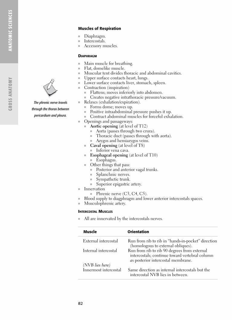
82
Muscles of Respiration
■ Diaphragm.■ Intercostals.■ Accessory muscles.
DIAPHRAGM
■ Main muscle for breathing.■ Flat, domelike muscle.■ Muscular tent divides thoracic and abdominal cavities.■ Upper surface contacts heart, lungs.■ Lower surface contacts liver, stomach, spleen.■ Contraction (inspiration)
■ Flattens; moves inferiorly into abdomen.■ Creates negative intrathoracic pressure/vacuum.
■ Relaxes (exhalation/expiration).■ Forms dome; moves up. ■ Positive intraabdominal pressure pushes it up.■ Contract abdominal muscles for forceful exhalation.
■ Openings and passageways:■ Aortic opening (at level of T12)
■ Aorta (passes through two crura).■ Thoracic duct (passes through with aorta).■ Azygos and hemiazygos veins.
■ Caval opening (at level of T8)■ Inferior vena cava.
■ Esophageal opening (at level of T10) ■ Esophagus.
■ Other things that pass:■ Posterior and anterior vagal trunks.■ Splanchnic nerves.■ Sympathetic trunk.■ Superior epigastric artery.
■ Innervation■ Phrenic nerve (C3, C4, C5).
■ Blood supply to diagphragm and lower anterior intercostals spaces.■ Musculophrenic artery.
INTERCOSTAL MUSCLES
■ All are innervated by the intercostals nerves.
ANAT
OM
IC S
CIEN
CES
GROS
S AN
ATO
MY
The phrenic nerve travels
through the thorax between
pericardium and pleura.
Muscle Orientation
External intercostal Run from rib to rib in “hands-in-pocket” direction(homologous to external obliques).
Internal intercostal Run from rib to rib 90 degrees from external intercostals; continue toward vertebral column as posterior intercostal membrane.
(NVB lies here)Innermost intercostal Same direction as internal intercostals but the
intercostal NVB lies in between.

83
ACCESSORY MUSCLES OF RESPIRATION
■ Sternocleidomastoid.■ Scalenes.■ Subcostals.■ Transversus thoracis.■ These latter two are innervated by intercostals nerves.
ANATOM
IC SCIENCES
GROSS ANATOM
Y
Muscles of the Thorax
Muscle Origin Insertion Action Innervation
Pectoralis major Medial half of clavicle; Greater tubercular Medially rotates, Lateral and medial sternum and costal crest (crest leading flexes, and adducts pectoral nervescartilages 1–6 downward from humerus (C6–C8 and T1)
greater tubercle)Pectoralis minor Anterior ends Coracoid process Protracts and depresses Lateral and medial
of ribs 3–5 glenoid of scapula pectoral nerves(C8 and T1)
Subclavius Anterior end Underside of clavicle Protracts and depresses Nerve to subclaviusof rib 1 clavicle (C5–C6)
MUSCLES OF THE PECTORAL GIRDLE
■ Serratus anterior■ Pectoralis minor■ Subclavius■ Trapezius■ Levator scapulae■ Rhomboideus major■ Rhomboideus minor
Abdominal Regions
■ Umbilical: Central around the umbilicus.■ Lumbar: Right and left of umbilical region.■ Epigastric: Midline region above umbilicus (subxiphoid, area of stomach).■ Hypochondriac: Left and right of epigastric regions (beneath rib cage).■ Hypogastric (pubic): Midline below umbilicus.■ Iliac (inguinal): Right and left of hypogastric region.

ANAT
OM
IC S
CIEN
CES
GROS
S AN
ATO
MY
84
Abdominal Muscles
Muscle Origin Insertion Action Innervation
External oblique Superficial aspect Iliac crest, linea alba Increases abdominal Lower intercostal of lower 8 ribs pressure nerves
Internal oblique Lumbodorsal fascia, Costal cartilages Same as above Lower intercostals,iliac crest, inguinal of last 3 ribs, iliohypogastric, ligament linea alba ilioinguinal nerves
Transversus Internal surface of Linea alba Same as above Lower intercostals, abdominis lower 6 costal iliohypogastric, and
cartilages, ilioinguinal nerveslumbodorsalfascia, iliac crest, inguinal ligament
Rectus Pubic symphysis External surface Increases abdominal Lower intercostal abdominis xiphoid process, pressure, flex nerves
external surface vertebral columncostal cartilages 5–7
POINTS
■ External oblique fibers (like external intercostals) ■ Run in a “hands-in-pocket” fashion. ■ Obliquely; lateral to medial going superior to inferior.
■ Internal oblique fibers (like internal intercostals fibers) ■ Run opposite (perpendicular) to external oblique fibers.■ Obliquely from lateral to medial going inferior to superior.
■ Cremaster muscle ■ Derived from internal oblique muscle.
Rectus Sheath
■ Aponeurotic sheath covering the rectus muscle.■ Anterior rectus sheath
■ Formed from the tendinous continuations of the external oblique andinternal oblique.
■ Posterior rectus sheath ■ Formed from the aponeurosis from the internal oblique and tranverse
abdominis.■ Linea alba
■ Midline fusion of the anterior and posterior rectus sheath. ■ At the midline of two bellies of the rectus.
Breast
■ Mammary glands. ■ Modified sweat glands.■ Lie in superficial fascia.
Fascia
■ Covers muscles.■ Attaches to nearby
bones by blending withcovering periosteum.

ANATOM
IC SCIENCES
GROSS ANATOM
Y
■ Myoepithelial cells (star-shaped) ■ Encircle some of the secretory cells.■ Force secretion toward the ducts.
■ Suspensory ligaments (Cooper’s ligaments) ■ Strong fibrous processes. ■ Run from dermis to deep layer of superficial fascia through breast.■ Support the breasts.■ Can cause dimpling of overlying skin and nipple retraction in breast
cancer.■ Innervation:
■ Fourth intercostal nerve (T4) (to the nipple).■ Blood supply:
■ Lateral thoracic (branch of axillary). ■ Internal thoracic arteries.
■ Lymph drainage.■ To lymph nodes (LNs) in axilla.
Dermatomes
See Figure 1–60. A dermatome is an area of skin supplied by a single nerve.
■ CN V: Head and face.■ CN V, VII, IX, X: Ear.■ C1: Does not supply a dermatome.■ C2 (greater occipital nerve): Posterior scalp (posterior half of skull cap).■ C3: Neck.■ C4: Low collar.■ T4: Nipple■ T7: Xiphoid.■ T10: Umbilicus.■ L1: Inguinal ligament.■ L4: Knee caps.■ S2, S3, S4: Erection, sensation of penile and anal zones.
85
F I G U R E 1 – 6 0 . Areas of skin supplied by a single nerve.
Reproduced, with permission, from Bhushan V, et al. First Aid for the USMLE Step 1. New York:
McGraw-Hill, 2003.
Cranial nervedermatomes do notoverlap.
■ Spinal nervedermatomes overlapby as much as 50%.
■ May require loss ofthree spinal nerves toproduce anesthesia inmiddle dermatome.
■ For example, lesion todorsal root of T7, T8,and T9 to producenumbness of T8dermatomaldistribution.
C3
C6
C2
V3V2
V1
C8
C8
C7
L5
C6
S2
S3
L4
C4
C5T1
T4
T6
T10
T8
L1

86
ANAT
OM
IC S
CIEN
CES
GROS
S AN
ATO
MY
Reflexes
■ C5, C6: Biceps■ C7: Triceps■ L4: Patella■ S1: Achilles
Femoral Triangle
See Figure 1–61. The femoral triangle has the following boundaries:
■ Lateral: Sartorius.■ Medial: Adductor longus.■ Superior: Inguinal ligament.■ Floor: Iliopsoas and pectineus.■ Femoral: NVB.■ From lateral to medial: Nerve, artery, vein.
� THORAC IC AN D ABDOM I NAL VISC E RA
Body Cavities
F I G U R E 1 – 6 1 . Femoral triangle.
Reproduced, with permission, from Bhushan V, et al. First Aid for the USMLE Step 1. New York:
McGraw-Hill, 2003.
Posterior Anterior
Cranial: Contains brain. ThoracicSpinal: Pericardial cavity: Surrounds heart■ Contains spinal cord. Pleural (right and left)■ These two cavities communicate ■ Surrounds each lung.
via foramen magnum. ■ Mediastinum lies between two■ Lined by meninges. pleural cavities.■ Bathed in CSF.
Abdominopelvic abdominal■ Stomach■ Spleen■ Liver■ Gallbladder■ Pancreas■ Small and large intestinesPelvic cavity■ Rectum■ Urinary bladder
Adductorlongusmuscle
Sartoriusmuscle
Femoral triangle
Inguinalligament

87
Lung
See Figure 1–62.
■ Housed in left and right pleural cavities.■ Visceral pleura: Covers the lungs.■ Parietal pleura: Covers the thoracic cavity.■ Pleural cavity.
■ Between the two layers of pleura.■ Filled with serous fluid.
ANATOM
IC SCIENCES
GROSS ANATOM
Y
Right Lung Left Lung
Three lobes Two lobes ■ Superior ■ Superior■ Middle ■ Inferior■ Inferior LingulaThree lobar bronchi ■ Tongue-shaped part of superior lobe
(secondary bronchi) corresponding to middle Ten bronchial segments lobe of right lung
(tertiary bronchi) Two lobar bronchi (secondary bronchi)One bronchial artery Eight bronchial segments (tertiary bronchi) Larger capacity than left lung Cardiac notch
■ Medial side of the superiorlobe of left lung
Two bronchial arteries
Describes where thepulmonary arterysits in relation tothe bronchus:
RALS
RightAnteriorLeftSuperior
Aspiration occurs more often
in the right lung (apical
aspect of right lower lobe)
because the right mainstem
bronchus is straighter, shorter,
and larger than the left. The
left mainstem takes a more
acute angle.LUNG HILUM (ROOT)
■ Pulmonary artery■ Pulmonary vein■ Bronchus
OTHER STRUCTURES PASSING INTO THE HILUM
Bronchial Arteries
■ Supply the lung tissues with oxygen.■ Also pass into the hilum.■ Follow the bronchial tree.
See the “Physiology” section
in Chapter 13 for
Hering-Breuer and cough
reflexes.
F I G U R E 1 – 6 2 . Anterior and posterior view of the lungs.
Reproduced, with permission, from Bhushan V, et al. First Aid for the USMLE Step 1. New York: McGraw-Hill, 2003.
Horizontalfissure
Obliquefissure
Obliquefissure
Posterior viewAnterior view
R L L R
Sup. lobe Sup. lobe
Sup. lobe Sup. lobe
Mid. lobe
Inf.lobe
Inf.lobe
Inf.lobe
Inf.lobe
4th rib
T2
Lingula

88
Branches of the Vagus Nerve
■ Also pass into the root of the lung.
Heart and Great Vessels
See Figure 1–63.
ANAT
OM
IC S
CIEN
CES
GROS
S AN
ATO
MY
From Body (Deoxy) From Lungs (Oxy)
Chamber Right atrium Left atriumValve Tricuspid valve Mitral valveChamber Right ventricle Left ventricleValve Pulmonic valve Aortic valve
To lungs To body
F I G U R E 1 – 6 3 . Heart and great vessels.
Reproduced, with permission, from Way LW, Doherty GM (eds). Current Surgical Diagnosis
and Treatment, 11th ed. New York: McGraw-Hill, 2002.
VALVES
SEMILUNAR
■ Each has three semilunar cusps.■ No chordae tendineae or papillary muscles are associated with semiluar
valves.■ Pulmonic valve
■ Right ventricle (RV) to pulmonary arteries. ■ At left sternal border, second intercostal space.
■ Aortic valve■ Left ventricle (LV) to aorta.■ Over right sternal border, second intercostal space.
The superior lobe of the left
lung has a lingula.
Leftventricle
Pulmonaryartery
Ventricularseptum
Aorta

89
ANATOM
IC SCIENCES
GROSS ANATOM
Y
ATRIOVENTRICULAR
■ Tricuspid valve ■ Right atrium (RA) to right ventricle (RV). ■ Over left sternal border, fifth intercostal space.
■ Mitral valve■ Left atrium (LA) to left ventricle (LV ).■ Over fifth intercostal space, midclavicular line.
Atria
RIGHT ATRIUM
■ Incoming deoxygenated blood from vena cavae.
■ Fossa ovalis■ Depression remnant of the foramen ovale.■ Lies on atrial septum (interatrial); dividing the left and right atria.■ Anulis ovalis is the upper margin of the fossa.
■ Crista terminalis■ Vertical ridge between vena cavae orifices.■ Sinoatrial (SA) node is located here. ■ Junction of sinus venosus and the heart in the developing embryo.
■ Sulcus terminalis■ Vertical groove on external heart represents crista terminalis (internally).
■ Pectinate muscles■ Radiate from crista terminalis to atrial appendage.
■ Right auricle■ Appendage of right atrium.
■ Conduction system■ SA node.■ AV node.■ Both nodes innervated by parasympathetic fibers from CN X and sym-
pathetic fibers from T1–T4.
LEFT ATRIUM
■ Incoming blood from pulmonary veins.
Ventricles
RIGHT VENTRICLE
■ Responsible for the pulmonic circulation.■ Pumps deoxygenated blood through the pulmonic valve to pulmonary
artery and lungs for oxygenation.■ Trabeculae carnae.
■ Ridges of cardiac muscle in the ventricles.■ Chordae tendinae.
■ Thin tendinous cords passing from valve cusps to papillary muscles.■ Papillary muscles.
■ Anchor chordae tendinae to heart wall.
LEFT VENTRICLE
■ Apex of the heart. ■ Auscultate fifth intercostals space at midclavicular line.
■ Responsible for the systemic circulation.
The chordae tendinae and
papillary muscles do not help
the AV valves close. They
prevent the valve from
everting back into the atria.
See the “Pathology”section in Chapter 19.Heart strain andhypertrophy secondaryto increased resistanceto flow:
■ Left heart■ Coarctation of the
aorta■ Systemic
hypertension■ Right heart■ Pulmonary
hypertension

90
ANAT
OM
IC S
CIEN
CES
GROS
S AN
ATO
MY
■ Receives oxygenated blood from the lungs and pumps it out to the restof the body.
■ Out the aortic valve to aorta.■ Thicker than RV because of higher afterload from systemic circulation.■ Same internal features as RV.
PERICARDIUM
■ Tough, double-walled covering of the heart ■ Innervated by the phrenic nerve■ Parietal pericardium: Outer fibrous■ Visceral pericardium: Inner serous
CORONARY ARTERIES
See Figure 1–64.
■ First branches of the aorta. ■ Coronary ostia located within aortic valve leaflets.■ Drain to coronary sinus which drains back to RA.
■ Coronary arteries fill during diastole.■ Blockage; thrombus on disrupted plaque can lead to:
■ Ischemia (angina). ■ Infarction (heart attack).
The pericardial cavity lies
between the two layers of
pericardium.
Vessel Area Supplied
Left circumflex LV lateral wallLeft anterior descending artery (LAD) Anterior wall LV
Interventricular septumRight coronary artery (RCA) Right ventricle
AV nodePosterior, inferior walls of LV
F I G U R E 1 – 6 4 . Coronary arteries.
Reproduced, with permission, from Bhushan V, et al. First Aid of the USMLE Step 1. New York:
McGraw-Hill, 2003; Adapted from Ganong WF. Review of Medical Physiology, 19th ed.
Stamford CT: Appleton & Lange, 1999.
Acute marginal artery
Left main coronary artery (LCA)
Circumflex artery (CFX)
Left anterior descendingartery (LAD)
Posterior descending artery (PD)
Right coronary artery (RCA)

91
Veins of the Heart
■ Cardiac veins lie superficial to the arteries.■ Coronary sinus
■ Continuation of great cardiac vein.■ Opens into the right atrium between the IVC and the tricuspid orifice.■ All veins drain into the coronary sinus except the anterior cardiac
veins (drain directly into the right atrium).
Mediastinum
■ The mediastinum is the area medial to lungs in the thorax.
ANATOM
IC SCIENCES
GROSS ANATOM
Y
The superior vena cava
(SVC), the inferior vena cava
(IVC), and coronary sinus all
empty deoxygenated blood
into the RA.
The thymus is located in both
the anterior and superior
mediastinum.
The inferior mediastinum
is split into anterior, middle,
and posterior.
Mediastinum Location Contents
Anterior Anterior to Thymuspericardium Connective tissue
Lymph nodesBranches of internal
thoracic artery
Middle Within pericardium PericardiumHeartRoots of great vesselsPhrenic nerve
Posterior Posterior to Thoracic ductpericardium Descending aorta
Azygous veinHemiazygous veinEsophagusVagus nervesSplanchnic nervesLymph nodes
Superior Above manubriosternal Thoracic ductjunction (T4) Ascending aorta
Aortic archBranches of aortic archDescending aortaSVCBrachiocephalic veinsThymusTracheaEsophagusCardiac nerveLeft recurrent
laryngeal nerve The thymus has no afferent
lymphatics or lymphatic
nodules.
Zinc is the most important
element for immunity
(involved in almost all aspects
of immunity).

Thymus
FUNCTION
■ A primary lymphoid organ. ■ Along with spleen, tonsils, lymph nodes, Peyer’s patches.
■ Master organ of immunogenesis in young.■ Proliferation and maturation of T cells (cell-mediated immunity).
■ Releases factors important in development of immune system:■ Thymopoietin.■ Thymosin.■ Thymic humoral factor.■ Thymic factor.
■ Zinc, vitamin B6, vitamin C are important for thymic hormones.
ANATOMY
■ Located inferior to thyroid, ventral/anterior to heart.■ Deep to sternum in superior mediastinum.■ Two lobes (soft, pinkish).■ Inner medulla
■ Lymphocytes.■ Hassall’s corpuscles (epithelial vestiges with unknown function).
■ Blood supply■ Internal thoracic artery.■ Inferior thyroid arteries.
■ Nerve supply■ Vagus nerve. ■ Phrenic nerve.
Aorta
See Figure 1–65.
■ Descending thoracic aorta extends from T4 to T12.■ Descending abdominal aorta extends from T12 to L4.
ANAT
OM
IC S
CIEN
CES
GROS
S AN
ATO
MY
All lymphoid organs arederived exclusively frommesenchyme except forthe thymus.Remember: The thymushas a double embryologicorigin:
■ Mesenchyme■ Lymphocytes
(hematopoieticstem cells)
■ Endoderm■ Hassall’s corpuscles
(epithelium)■ Derived from
the thirdpharyngealpouch.
Thymoma is associated
with myasthenia gravis.
F I G U R E 1 – 6 5 . The thoracic aorta and its branches.
Reproduced, with permission, from Liebgott B. The Anatomical Basis of Dentistry. Toronto: BC
Decker, 1986.
Remember: There is one
brachiocephalic artery, two
brachiocephalic veins.
92
Remember: The ligamentum
arteriosum connects the aortic
arch to the pulmonary trunk
and is a vestige of the ductus
arteriosis.

ANATOM
IC SCIENCES
GROSS ANATOM
Y
■ Aorta terminates around L4 when it divides into left and right commoniliac arteries (supplying lower limbs and pelvic viscera).
93
Parts Major Branches Supplies
Ascending Right and left Heartcoronary arteries
Aortic arch Brachiocephalic Head, neck, upper limbsartery
Left common carotidLeft subclavian
Descending Posterior intercostals Posterior thorax, diaphragmthoracic aorta
Descending Unpaired: Abdominal visceraabdominal aorta Celiac trunk
SMAIMA
Paired:RenalGonadal
COMMON CAROTID
■ Supplies head and neck.■ Bifurcates into external and internal carotid arteries at superior border of
the thyroid cartilage.
See the “Neck” section for branches.
SUBCLAVIAN ARTERY
■ Supplies the upper extremity.■ Continues as the axillary artery past the inferior margin of the first rib.
BRANCHES OF SUBCLAVIAN ARTERY
■ Vertebral (forms basilar—circle of Willis)■ Thyrocervical■ Internal thoracic■ Costocervical
INTERNAL THORACIC ARTERY
■ Runs underneath the sternum.■ Gives off anterior intercostals (meets posterior intercostals off the aorta).■ Extends inferiorly as superior epigastric artery.
SUPERIOR EPIGASTRIC ARTERY
■ Continuation of the internal thoracic artery.■ Enters rectus sheath and supplies rectus muscle.■ Anastomoses with inferior epigastric artery (branch of the external iliac artery)
near the umbilicus.
If the celiac trunk is blocked,
blood can still reach the
foregut by way of
anastomoses between the
superior pancreaticoduodenal
artery (a branch of the
gastroduodenal) and the
inferior pancreaticoduodenal
(a branch of the SMA).
The third part of duodenum
passes anterior to the IMA.
Remember: The right renal
artery is longer than the left
renal artery and the left
common carotid is longer
than the right common
carotid.

94
ABDOMINAL AORTA
ANAT
OM
IC S
CIEN
CES
GROS
S AN
ATO
MY
Midline Branches
Branch Branches Structures Supplied
Celiac Common hepatic, Foregut: Stomach, liver,splenic, left gastric spleen, and upper half
of pancreas/duodenumSMA Inferior pancreaticoduodenal, Midgut: Head of
intestinal (ileal, jejunal), pancreas, lowerright and middle colic arteries duodenum, rest of small
intestine, ascendingand transverse colon
IMA Superior rectal, sigmoid, Hindgut: Descendingleft colic arteries and sigmoid colon and
upper 2/3rd of rectum
Anatomy Formed by Drains to
Ascends through Right ascending lumbar vein Empties into the aortic orifice of Right subcostal vein superior venadiaphragm cava (SVC)
Lies within posterior mediastinum
Leaves a notch in the right lung as it passes over the hilum
Azygous System
See Figure 1–66.
■ The azygous system drains the thoracic wall.
Remember:
The right superior intercostal
vein drains into the azygos
vein.
The left superior intercostal
vein drains into left
brachiocephalic vein.
F I G U R E 1 – 6 6 . The azygous system of veins and the thoracic duct.
Reproduced, with permission, with Liebgott B. The Anatomical Basis of Dentistry. Toronto: BC
Decker, 1986.
95
Splanchnic Nerves
■ Sympathetic nerves to the abdominal viscera. ■ Counteract vagal/parasympathetic inputs.
■ Arise from thoracic ganglia ■ T5–T12.■ All pass through the diaphragm.
■ Preganglionic sympathetic fibers pass through ganglia of the sympathetictrunk without synapsing.■ This is the exception to the short preganglionic, long postganglionic.■ They synapse with small ganglia in the tissues/effector organs which
give off short postganglionics.■ Distribute to smooth muscle and glands of viscera.
ANATOM
IC SCIENCES
GROSS ANATOM
Y
Hemiazygos Vein (Left) Accessory Hemiazygos Vein (Right)(Inferior Hemiazygos Vein) (Superior Hemiazygos Vein)
Formed by: Formed by:■ Left ascending lumbar vein ■ 4th to 8th intercostal veins■ Left subcostal vein ■ Empties into the azygos vein■ Empties into the azygos vein
Nerve Components Innervates
Greater splanchnic T5–T9 Synapse at cervical plexus nerve (foregut)
Lesser splanchnic T10, T11 Synapse with superior nerve mesenteric plexus; aorticorenal
ganglion (midgut)Least splanchnic T12 Inferior mesenteric plexus/renal
nerve plexus (hindgut)
Superior Vena Cava
■ Drains the ■ Head.■ Neck.■ Upper extremities.■ Upper chest.
■ No valve.■ Formed by left and right brachiocephalic veins (merging in the superior
mediastinum).■ Brachiocephalic veins are formed from
■ Internal jugular vein.■ Subclavian vein.
■ Azygos vein empties into SVC.■ SVC empties into RA.
SVC Syndrome
■ Example: Compressionof SVC with lungcancer
■ Dyspnea■ Facial swelling and
flushing■ Jugular venous
distention■ Mental status changes

96
Inferior Vena Cava
■ Larger than SVC.■ Rudimentary, nonfunctioning valve.■ Drains
■ Thorax.■ Abdomen.■ Lower extremities.
■ Receives blood from the hepatic veins.■ Empties into RA.
PAIRED BRANCHES OF THE IVC
ANAT
OM
IC S
CIEN
CES
GROS
S AN
ATO
MY
Branch Structures Supplied
Suprarenal Adrenal glandsRenal KidneysGonadal Testes/ovariesLumbar Lumbar epaxial muscles
Portal Vein (Figure 1–67)
■ Transmits venous blood from the abdominal viscera (with absorbed sub-stances) to the liver and, via the portal system, to the heart.
Portal vein → hepatic sinusoids → central vein → hepatic veins → IVC.The portal vein arises from:
■ Splenic vein■ Drains:
■ Spleen.■ Stomach (L and R gastroepiploic veins, R and L gastric veins).■ Pancreas (pancreatic vein).■ Gallbladder (cystic vein).
Remember: The left gonadal
vein drains into left renal vein.
The right gonadal vein
empties directly into the IVC.
The left renal vein is longer
than the right and the right
renal artery is longer than the
left. This is due to the
positions of the vena cava
and aorta, respectively.
F I G U R E 1 – 6 7 . Anatomic relationship of portal vein and branches.
Reproduced, with permission, from Way LW, Doherty, GM (eds). Current Surgical Diagnosis
and Treatment, 11th ed. New York: McGraw-Hill, 2002.
A pelvic kidney may be
supplied by the common iliac
artery.
Liver
Pancreas
Portalvein
Left gastric vein
Right gastric vein
Splenic vein
Inferior mesentericvein
Superior mesentericvein
Remember: There is a
celiac artery but no celiac
vein.

■ Superior mesenteric vein■ Drains
■ Small intestine.■ Cecum.■ Ascending and transverse colon.
■ Inferior mesenteric vein ■ Enters into the splenic vein, the SMV, or crotch of the two.■ Drains:
■ Transverse colon distal to L colic flexure (splenic flexure).■ Descending colon.■ Rectum.
Portal Triad
■ Consists of:■ Portal vein.■ Hepatic artery.■ Bile duct.
■ Lies on free edge of lesser omentum (anterior to epiploic foramen ofWinslow).
■ Portal vein is posterior while hepatic artery and bile duct are anterior.■ Portal vein carries twice as much blood as the hepatic artery.
VENOUS ANASTOMOSES
See Figure 1–68.
ANATOM
IC SCIENCES
GROSS ANATOM
Y
F I G U R E 1 – 6 8 . Portal systemic anastomoses.
Reproduced, with permission, from Bhushan V, et al. First Aid for the USMLE Step 1. New York:
McGraw-Hill, 2003.
Portal blood can also get to
the IVC by the azygos and
hemiazygos veins.
Portocaval shunt can be
created by anastomosing the
splenic vein and renal vein
(a portal component and a
systemic venous component).
97
Superiormesenteric v.
Gastroesophageal vv.
Superior-middle/inferior rectal vv.
Gastrorenal-splenorenal vv.
Paraumbilical v.
IVC
Systemic venousPortal venous
Portal v.
L. gastric v.
Retro-peritonealparavertebralvv.
Splenic v.
1
2
3
4
5

98
ANAT
OM
IC S
CIEN
CES
GROS
S AN
ATO
MY
Lymphatic System
CISTERNA CHYLI
■ Located in the para-aortic region below the diaphragm at level of T12.■ Drains lymph from
■ Abdomen.■ Pelvis.■ Inguinal region.■ Lower extremities.
■ Continuous superiorly with the thoracic duct.
THORACIC DUCT
■ Continuation of the dilated cisterna chyli.■ Located in posterior and superior mediastinum.■ Drains lymph from
■ Lower limbs.■ Abdomen.■ Chest wall.
■ Empties into venous system at junction of the subclavian and internaljugular vein (on the left side only).
RIGHT LYMPHATIC DUCT
■ Drains■ Right arm.■ Right side of chest.■ Right side of head.
■ Empties into the junction of the right internal jugular vein and right subcla-vian veins (formation of the right brachiocephalic vein).
Peritoneum
■ Membrane lining the interior of the abdominopelvic cavity.■ Consists of:
■ Single layer of simple squamous mesothelium. ■ Thin layer of irregular connective tissue.
■ Visceral peritoneum■ Covers organs.
■ Parietal peritoneum■ Covers body walls.
■ Peritoneal fluid■ Fills the peritoneal space/cavity (space between these two serous layers).
Mesentery
■ Double-layer fold of peritoneum pushed into peritoneal cavity.■ Suspends most organs of abdominoperitoneal cavity (holds them in place).■ Allows path for blood vessels and nerves.
Occlusion of lymph supply
(eg, cancer) leads to
intractable edema.
Aside from the right arm,
chest, and right head, the
rest of the body—including the
right leg—is drained by the
thoracic duct.
Important: The diaphragm
divides the thoracic cavity and
abdominal cavity.
Pleura is the analog to
peritoneum in the pleural
cavity.
The pericardioperitoneal
canal embryologically
connects the thoracic and
peritoneal cavities.

99
Peritoneal Ligaments
■ Either a mesentery or omentum.■ Named double fold of the peritoneum and connects
■ Two organs or ■ Organ to the abdominal wall.
■ Are associated with the stomach, jejunum, ileum, appendix, transverse colon,sigmoid colon, spleen, liver, and gallbladder.
ANATOM
IC SCIENCES
GROSS ANATOM
Y
Embryonic Mesentery Adult Mesentery
Dorsal mesogastrium Greater omentumOmental bursa
Dorsal mesoduodenum Disappears (duodenum lies retroperitoneal)Pleuropericardial membrane Pericardium
Contribution to diaphragmVentral mesentery Falciform ligament
Ligamentum teresLesser omentum (hepatogastric,
hepatoduodenal ligaments)
The greater omentum begins
at its attachment to the
greater curvature of the
stomach, drapes over
(apronlike) the small and
large intestines, and loops
back to insert on the
transverse colon.
Ligament Description
Splenorenal Connects spleen to posterior abdominal wall.ligament Contains splenic artery, vein, and tail of pancreas.
Passes from spleen to parietal peritoneum on theanterior surface of the kidney.
Separates the greater peritoneal sac from the leftportion of the lesser peritoneal sac.
Gastrosplenic Part of dorsal mesogastrium between greater curve ofligament stomach and spleen.
Separates greater peritoneal sac from left portion oflesser peritoneal sac.
Incise to gain access to left side of lesser peritoneal sac.Gastrocolic Part of greater omentum, between greater curvature of
ligament stomach and transverse colon.Contains gastroepiploic arteries.
Gastroduodenal Between lesser curve of stomach and duodenum.ligament
Gastrohepatic Part of lesser omentum between liver and lesser curvature.ligament Separates greater peritoneal sac and right part of lesser
peritoneal sac.Contains no significant blood vessels (so may be incised
for access).Hepatoduodenal Part of lesser omentum
ligament Connects liver to first part of duodenum.Contains common bile duct, proper hepatic artery (withbrachial cystic artery), and portal vein.
Separates greater peritoneal sac from right part of lesserperitoneal sac.
Forms anterior portion of epiploic foramen.Falciform Connects liver to anterior abdominal wall.
ligament Contains ligamentum teres (remnant of umbilical vein).
Epiploic Foramen ofWinslow
■ Inlet to lesser sac.■ Posterior to the free
edge of the lesseromentum(hepatoduodenalligament).
■ Portal triad is locatedover top (anteriorly).

100
ANAT
OM
IC S
CIEN
CES
GROS
S AN
ATO
MY
GI Arterial Segment Derivatives Supply Innervation
Foregut Esophagus Celiac trunk Vagal Stomach parasympatheticsDuodenum (1st part) Thoracic nerveLiver Greater splanchnicGallbladder sympatheticsPancreas
Midgut Duodenum Superior Vagal (2nd–4th parts) mesenteric parasympatheticsJejunum artery ThoracicIleum splanchnicAppedix sympatheticAscending colon nerveTransverse colon
(to splenic flexure)
Hindgut Transverse colon Inferior Pelvic splanchnic (distal to splenic mesenteric nerve (S2–S4) flexure) artery parasympathetic
Descending colon Lumbar splanchnicSigmoid colon sympathetic nerveRectum
See histology for the
myenteric nervous system.
Gastrointestinal Tract
See Figure 1–69 for the cross section of the gastrointestinal tract.
F I G U R E 1 – 6 9 . Cross section of the gastrointestinal tract.
Reproduced, with permission, from Bhushan V, et al. First Aid for the USMLE Step 1. New York: McGraw-Hill, 2003. Adapted
from McPhee S, et al. Pathophysiology of Disease: An Introduction to Clinical Medicine, 3rd ed. New York: McGraw-Hill, 2000.
Lamina propria (support)
Submucosa (support)
Muscularis mucosae(mucosal motility)
Villus (increase ofmucosal surface)
Submucosal plexus(Meissner’s; controls secretions)
Myenteric plexus(Auerbach’s; controlsmotility)
Gland in submucosal layer (secretions)
Inner circular
layer (motility)
Mesentery (bindingof digestive tract to
abdominal wall)
Outer longitudinal muscle layer (motility)
Serosa(support)
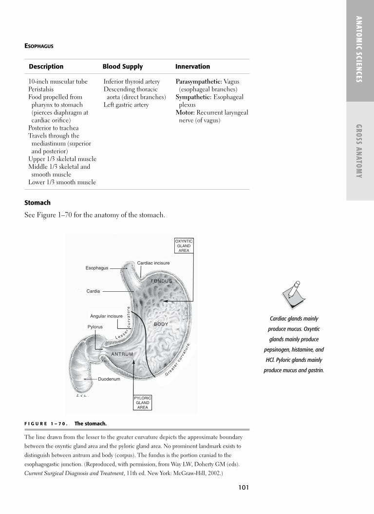
101
ESOPHAGUS
ANATOM
IC SCIENCES
GROSS ANATOM
Y
Description Blood Supply Innervation
10-inch muscular tube Inferior thyroid artery Parasympathetic: Vagus Peristalsis Descending thoracic (esophageal branches)Food propelled from aorta (direct branches) Sympathetic: Esophageal
pharynx to stomach Left gastric artery plexus(pierces diaphragm at Motor: Recurrent laryngealcardiac orifice) nerve (of vagus)
Posterior to tracheaTravels through the
mediastinum (superior and posterior)
Upper 1/3 skeletal muscleMiddle 1/3 skeletal and
smooth muscleLower 1/3 smooth muscle
Stomach
See Figure 1–70 for the anatomy of the stomach.
F I G U R E 1 – 7 0 . The stomach.
The line drawn from the lesser to the greater curvature depicts the approximate boundary
between the oxyntic gland area and the pyloric gland area. No prominent landmark exists to
distinguish between antrum and body (corpus). The fundus is the portion craniad to the
esophagogastic junction. (Reproduced, with permission, from Way LW, Doherty GM (eds).
Current Surgical Diagnosis and Treatment, 11th ed. New York: McGraw-Hill, 2002.)
OXYNTICGLANDAREA
PYLORICGLANDAREA
Duodenum
Pylorus
Angular incisure
EsophagusCardiac incisure
FUNDUS
BODY
ANTRUM
Cardia
L e s s er
cu
rva
tur e
Gr e
a t er
cur v
at u
r e
Cardiac glands mainly
produce mucus. Oxyntic
glands mainly produce
pepsinogen, histamine, and
HCl. Pyloric glands mainly
produce mucus and gastrin.

SPHINCTERS OF THE STOMACH
■ Cardiac/lower esophageal sphincter■ Composed of:■ Internal esophageal sphincter
(intrinsic esophageal muscle). ■ External sphincter (crura of diaphragm).■ Pyloric sphincter■ Outlet of stomach into duodenum.
ANAT
OM
IC S
CIEN
CES
GROS
S AN
ATO
MY BLOOD SUPPLY TO STOMACH (FIGURE 1–71)
Pyloric stenosis in children
causes violent, nonbilious
projectile vomiting.
Artery Description
Splenic artery Direct branch of celiac trunkSupplies:■ Spleen■ StomachBranches:■ Short gastrics to fundus■ Left gastroepiploic to left
greater curvatureLeft gastric artery Direct branch of celiac trunk.
Supplies:■ Left lesser curvature of stomach■ Inferior esophagus
Right gastric artery Branch of gastroduodenal(a branch of the hepatic artery proper off the celiac trunk)
Supplies:■ Right lesser curvature of the
stomachRight gastroepiploic Branch of gastroduodenal
Supplies:■ Right 1/2 greater curvature
of stomach■ Gastric ulcer likely bleeds from this
102
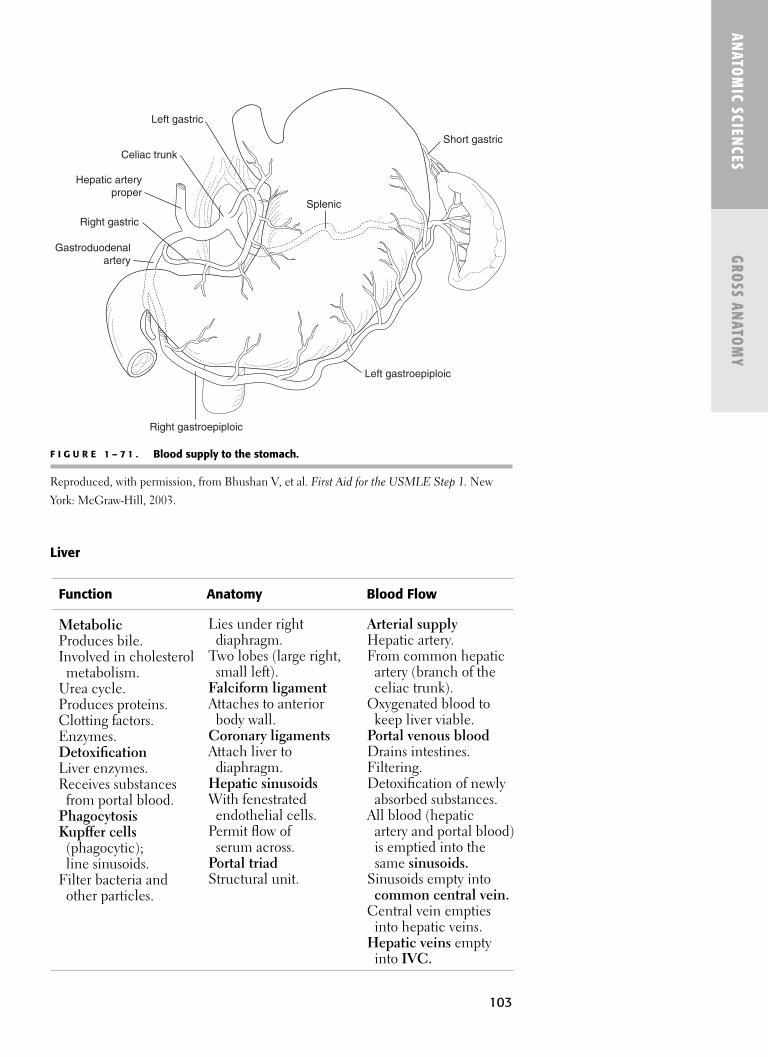
103
ANATOM
IC SCIENCES
GROSS ANATOM
Y
Liver
F I G U R E 1 – 7 1 . Blood supply to the stomach.
Reproduced, with permission, from Bhushan V, et al. First Aid for the USMLE Step 1. New
York: McGraw-Hill, 2003.
Function Anatomy Blood Flow
MetabolicProduces bile.Involved in cholesterol
metabolism.Urea cycle.Produces proteins.Clotting factors.Enzymes.DetoxificationLiver enzymes.Receives substances
from portal blood.PhagocytosisKupffer cells
(phagocytic);line sinusoids.
Filter bacteria andother particles.
Lies under right diaphragm.
Two lobes (large right,small left).
Falciform ligamentAttaches to anterior
body wall.Coronary ligamentsAttach liver to
diaphragm.Hepatic sinusoidsWith fenestrated
endothelial cells.Permit flow of
serum across.Portal triadStructural unit.
Arterial supplyHepatic artery.From common hepatic
artery (branch of theceliac trunk).
Oxygenated blood tokeep liver viable.
Portal venous bloodDrains intestines.Filtering.Detoxification of newly
absorbed substances.All blood (hepatic
artery and portal blood) is emptied into the same sinusoids.
Sinusoids empty intocommon central vein.
Central vein emptiesinto hepatic veins.
Hepatic veins emptyinto IVC.
Splenic
Left gastric
Short gastric
Left gastroepiploic
Right gastroepiploic
Gastroduodenalartery
Right gastric
Hepatic arteryproper
Celiac trunk

104
ANAT
OM
IC S
CIEN
CES
GROS
S AN
ATO
MY
Spleen
■ Develops from mesenchymal cells (of the mesentery attached to primitivestomach).■ Does not develop from the primitive gut (ie, forgut, midgut, hindgut).
■ Lies in the left hypochondrium (of the abdominal cavity).■ Between the stomach and diaphragm.
■ Ovoid organ.■ Size of a fist.■ Functions:
■ Blood reservoir.■ Phagocytosis (foreign particles and red blood cell [RBC] senescence).■ Production of mononuclear leukocytes.
■ Blood supply: Splenic artery (from celiac trunk).■ Venous drainage: Splenic vein (part of portal system).■ Innervation: From celiac plexus.■ Histology.
■ White pulp.■ Lymphocytes located around splenic artery branches.
■ Red pulp.■ Blood-filled sinusoids.■ Phagocytic cells (macrophages, monocytes).■ Lymphocytes.■ Plasma cells.
Gallbladder
■ Pouch on inferior surface of liver.■ Stores and concentrates bile.■ Bile is produced by the liver.■ Common bile duct formed by:
■ Cystic duct (from gallbladder).■ Hepatic duct (from liver).
■ Common bile duct empties into duodenum.■ Sphincter of Oddi (ampulla of Vater) (on duodenal papilla).
■ Relaxes:■ Cholecystokinin; fat into intestine.■ Gallbladder contracts ■ Bile released into duodenum for fat emulsion.
■ Contracts:■ No cholecystokinin; intestine is empty.■ Forcing bile up the cystic duct to the gallbladder for storage.
■ Blood supply: Cystic artery (branch of right hepatic).■ Innervation: Vagal fibers (celiac plexus).■ Lymph drains into cystic lymph node, then hepatic nodes, then celiac
nodes.
The gallbladder does not
contain a submucosa (the
stomach and small and large
bowel do).
Bile is composed of bile salts,
pigments, cholesterol, and
lecithin. Bile serves to emulsify
fats, and fat-soluble vitamins
(A, D, E, K).

105
ANATOM
IC SCIENCES
GROSS ANATOM
Y
Large Intestine
■ Extends from ileocecal valve to anus.■ Site of fluid and electrolyte reabsorption.■ Lack villi (unlike small intestine).■ Does not secrete enzymes or have enzymes along brush border (unlike
small intestine).■ Has goblet cells, absorptive cells, microvilli (see Chapter 2).
Segment Blood Supply Description
Duodenum Superior pancreaticoduodenal C-shaped.artery (indirect Celiac branch) Surrounds pancreas.
Inferior pancreaticoduodenal Shortest and widest part of artery (of SMA) small intestine.
Part of it is retroperitoneal.Connects stomach to jejunum.Common bile duct and pancreatic duct emptyinto duodenum atduodenal papilla.
Contains Brunners glands (submucosal glands) secreting mucous.
Jejunum SMA—intestinal branches Valves of Kerckring (plicae circulares).
Most villi (for greatest absorption).
Thickest muscular wall (of any small intestinesegment; for peristalsis).
Ileum SMA—intestinal branches Peyer’s patches.and ileocolic branch Vitamin B12 and bile salt
absorption.More goblet cells andmesenteric fat than jejunum.
No plicae circulares inlower lieum.
Enzymes present inintestinal villi:
■ Carbohydrate■ Maltase■ Sucrase ■ Lactase
■ Protein■ Aminopeptidase■ Dipeptidase■ Activating pancreatic
enzymes■ Enterokinase
■ Convertstrypsinogen totrypsin.
■ Trypsinactivates otherpancreaticenzymes.
Small Intestine
Descending and sigmoid
colon, rectum, and anus are
part of the hindgut, so they
are supplied by the pelvic
splanchnic nerves rather
than the vagus.

106
ANAT
OM
IC S
CIEN
CES
GROS
S AN
ATO
MY
PEYER’S PATCHES
■ Subepithelial, nonencapsulated lymphoid tissue (like tonsils).■ Located in ileum (small intestine).■ Limit numbers of harmful bacteria.
Retroperitoneal Structures (Figure 1–72)
■ Not suspended from mesenteries. ■ Plastered against posterior body wall, behind the peritoneum.
Segment Blood Supply Description
Cecum SMA; cecal branches, “Blind sac”appendicular artery Ileocecal valve enters(to appendix) Vermiform appendix
Lymphoid tissue
Colon 1/4 length of small intestine■ Ascending SMA—Ileocolic, right colic Larger diameter than small ■ Transverse SMA—Middle colic intestine■ Descending IMA—Left colic Teniae coli (3 smooth muscle■ Sigmoid IMA—Sigmoid bands)
Haustra (pouches created by teniae)
Epiploic appendages(fat globules on serosal surface)
Ascending and descendingcolon are retroperitoneal
Rectum IMA—Superior rectal artery From sigmoid to anusStraight
■ Middle rectal arteries Located with in pelvic cavity(of hypogastric) No teniae
No HaustraNo epiploic appendages
Anal canal Internal iliac— Last 3–4 cm of rectum■ Inferior rectal Internal sphincter (involuntary)
(of internal pudendal) External sphincter (volunatary)
Each part of the colonalternates betweenretroperitoneal andintraperitoneal.
■ Ascending colon(retroperitoneal)
■ Transverse colon(intraperitoneal)
■ Descending colon(retroperitoneal)
■ Sigmoid(intraperitoneal)
■ Rectum(retroperitoneal)
F I G U R E 1 – 7 2 . Retroperitoneal structures.
Reproduced, with permission, from Bhushan V, et al. First Aid for the USMLE Step 1. New York: McGraw-Hill, 2003.
Perirenalspace
Transversalis fascia
Peritoneum1
23
67
5
4
1. Duodenum (2nd, 3rd, 4th parts)2. Descending colon3. Ascending colon4. Kidney and ureters5. Pancreas (except tail)3. Aorta7. IVCAdrenal glands and rectum
(not shown in diagram)

107
ANATOM
IC SCIENCES
GROSS ANATOM
Y
Posterior Abdominal Muscles
Muscle Innervation
Psoas major and minor Lumbar plexusQuadratus lumborum Lumbar plexusIliacus Femoral nerve
Pancreas
■ Lobulated gland.■ Extends from curve of duodenum to spleen.■ Retroperitoneal (except for small portion of tail).■ Head (from ventral bud)
■ Lies in the C (concavity) of the duodenum.■ Located to right of midline (anterior to origin of SMA).■ Extends from L1–3.
■ Body: Extends across; splenic vein lies underneath.■ Tail: Abuts spleen hilum (intraperitoneal, covered by lienorenal ligament).
PANCREATIC DUCTS
■ Duct of Wirsung (main pancreatic duct) ■ Begins at tail and joins common bile duct to form ampulla of Vater
(hepatopancreatic ampulla), where it empties into duodenum.■ Santorini’s duct (accessory pancreatic duct)
■ Opens separately into duodenum (when present).
Adrenal Gland
■ Triangular glands.■ Located in adipose tissue on superior aspect of kidneys.
ADRENAL CORTEX
■ Develops from mesoderm (unlike medulla).
ADRENAL MEDULLA
■ Modified nervous tissue (like postganglionic sympathetic cells).■ Develops from neuroectoderm.
■ Neural crest cells differentiate into medullary cells (chromaffin cells).■ Releases
■ Epinephrine.■ Norepinephrine.
■ Same effect as direct sympathetic stimulation but lasts longer.■ Can live without adrenal medulla because postganglionic cells provide same
function and will compensate for the loss.
Bile and pancreatic enzymes
are released at ampulla of
Vater into the second part of
duodenum.
Retroperitoneal pnuemonic:
RAKE A CUPID
Rectum
Aorta
Kidneys
Esophagus
Adrenal glands
Colon (ascending/descending)
Ureters
Pancreas
Inf. vena cava
Duodenum (distal 3/4th)

108
Urinary System
■ Consists of:■ Kidneys (2).■ Ureters (2).■ Urinary bladder.■ Urethra.
■ Lined with transitional epithelium.■ Parasympathetic fibers from pelvic splanchnic nerve.■ Kidneys, ureters, and bladder are all retroperitoneal.
Kidney
See Figure 1–73.
■ Retroperitoneal.■ Upper portion protected by ribs 11 and 12.■ Left kidney attached to the spleen by the lienorenal ligament.
ANAT
OM
IC S
CIEN
CES
GROS
S AN
ATO
MY
F I G U R E 1 – 7 3 . Structure of the kidney.
Reproduced, with permission, from Bhushan V, et al. First Aid for the USMLE Step 1. New York: McGraw-Hill, 2003. (Adapted
from McPhee S, et al. Pathophysiology of Disease: An Introduction to Clinical Medicine, 3rd ed. New York, McGraw-Hill, 2000.)
As ureter enters pelvic cavity
it crosses over the top of the
common iliac artery (at its
point of bifurcation) and dives
deep into the pelvis.
Cortex
Medullarypyramids(papillae)
Glomerulus
Bowman'scapsule
Afferentarteriole
Interlobarartery and vein
Medulla
Collectingducts
Vasarecta
Renal arteryand vein
Proximaland distal convolutedtubules
Efferent arteriole
Afferent arteriole––blood flow to the glomerulus
Maculadensa
Basement membrane
Mesangial cells
Podocytes(visceral layer)
Bowman's capsule
Efferent arteriole––blood flow out of glomerulus
Interlobularartery and
veinPelvis
Ureter

ANATOM
IC SCIENCES
GROSS ANATOM
Y
URETER
■ Long slender muscular tubes (peristalse).■ Transports urine from kidney to urinary bladder.■ Travels in retroperitoneal location.
URETHRA
■ Passes urine from bladder to outside.■ Female: 4 cm; opens into vestibule (between clitoris and vagina).■ Male: 20 cm; travels through penis; also conveys semen.
URINARY BLADDER
■ Distensible sac in pelvic cavity; posterior to pubic symphysis.■ Holds urine.
Pelvic Cavity
■ From anterior to posterior: ■ Bladder■ Uterus■ Rectum
■ Females: Paired ovaries and single uterus■ Vesicouterine pouch
■ Between bladder and uterus■ Rectouterine pouch ■ Between rectum and uterus
■ Males: Paired ductus deferens and seminal vesicles, and single prostate■ Rectovesicle pouch
■ Between rectum and bladder
Inguinal Canal
■ Females: Round ligament of the uterus.■ Fibromuscular band attached to the uterus extending from fallopian tube,
through inguinal canal to labia majora.■ Males: Spermatic cord.
■ Suspends the testes within the scrotum.
SPERMATIC CORD
109
Females are more prone to
urinary infections because
of the shorter urethra.
The inguinal canal is larger in
males than females.
Layers Contents
External spermatic fascia Ductus deferensCremaster muscle Testicular arteryInternal spermatic fascia Pampiniform plexusLoose areolar tissue Vessels and nerves of ductus deferensRemnants of the processus
vaginalis

110
■ Ductus (Vas) deferens: Conveys epididymis from testis to ejaculatory duct.■ Ejaculatory duct (ductus deferens + seminal vesicle ducts): Empties into
prostatic urethra.■ Pampiniform plexus: Form the testicular vein.
■ Left testicular vein drains into left renal vein.■ Right testicular vein drains into the IVC.
DEFECATION
■ Mediated by pelvic and pudendal nerves.■ Internal sphincter relaxes
■ Pelvic nerve (senses distention then reflexively relaxes internal sphincter).■ External anal sphincter contracts
■ Pudendal nerve. ■ Conscious urge to defecate is sensed.
� N E U ROANATOMY
Nervous System
■ Peripheral nervous system = Somatic.■ Autonomic system = Visceral.■ Myenteric nervous system = GI, intrinsic.
Brain
See the “Embryology” section in Chapter 4 for derivation.
CEREBRUM
See Figure 1–74 for functions of the cerebral cortex.
■ Is 80% of brain mass.■ Has four paired lobes within two cerebral hemispheres.■ Commands higher function.■ See also the section “Homunculus” in Chapter 10.
ANAT
OM
IC S
CIEN
CES
GROS
S AN
ATO
MY
Central nervous system =
Brain + spinal cord.
Component Description
Cerebral cortex Gray matter externally (6 layers)Cerebral medulla Internal white matter (myelinated)Corpus callosum White matter tracts; connect the two hemispheres
Remember: SEVEN-UP
Seminiferous tubules
Epididymis
Vas deferens
Ejaculatory duct
(Nothing)
Urethra
Penis
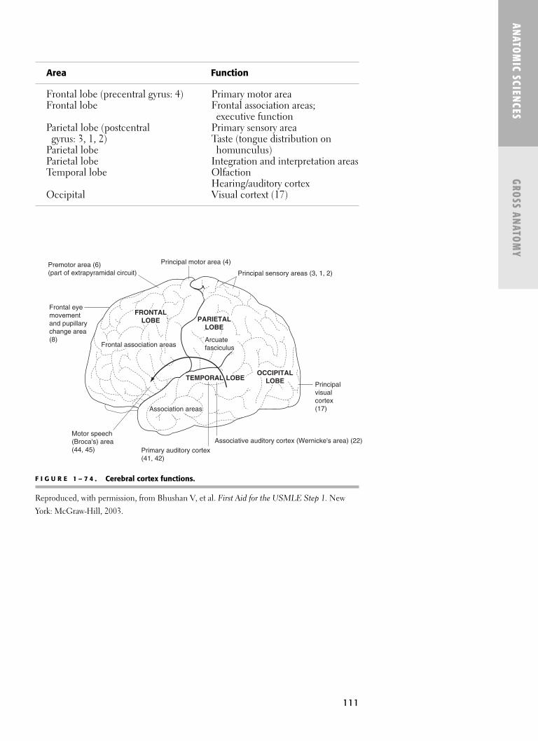
111
ANATOM
IC SCIENCES
GROSS ANATOM
Y
Area Function
Frontal lobe (precentral gyrus: 4) Primary motor areaFrontal lobe Frontal association areas;
executive functionParietal lobe (postcentral Primary sensory area
gyrus: 3, 1, 2) Taste (tongue distribution onParietal lobe homunculus)Parietal lobe Integration and interpretation areasTemporal lobe Olfaction
Hearing/auditory cortexOccipital Visual cortext (17)
F I G U R E 1 – 7 4 . Cerebral cortex functions.
Reproduced, with permission, from Bhushan V, et al. First Aid for the USMLE Step 1. New
York: McGraw-Hill, 2003.
Premotor area (6)(part of extrapyramidal circuit)
Principal motor area (4)
Principal sensory areas (3, 1, 2)
Principalvisualcortex(17)
Frontal eye movementand pupillary change area(8)
Motor speech(Broca's) area(44, 45)
TEMPORAL LOBE
Frontal association areasArcuatefasciculus
Association areas
OCCIPITAL
LOBE
PARIETAL
LOBE
FRONTAL
LOBE
Associative auditory cortex (Wernicke's area) (22)Primary auditory cortex(41, 42)

ANAT
OM
IC S
CIEN
CES
GROS
S AN
ATO
MY
■ Basal nuclei modulate motor activity first initiated from area 4: precentralgyrus of frontal lobe.
DIENCEPHALON
The diencephalon consists of:
■ Thalamus.■ Hypothalamus.■ Epithalamus.
■ Pineal gland is located within epithalamus.■ Releases melatonin (as does hypothalamus); plays a role in:
■ Circadian rhythms/sleep—wake cycle.■ Body temperature regulation.■ Appetite.
■ Pituitary gland (See Chapters 2 and 17).
General Nuclei Functions
Thalamus Sensory relay station Lateral geniculate VisualMedial geniculate AuditoryVentral posterior Proprioception, pressure,
lateral (VPL) touch, vibrationVentral posterior Facial sensation
medial (VPM) (including pain)Vental anterior/ventral Motor
lateralHypothalamus Body homeostasis Ventromedial nucleus Satiety center (hyperphagia
with bilateral destruction; decreases urge to eat with stimulation)
Lateral nucleus Hunger center (starvation with destruction; increase eating with stimulation)
Septal nucleus Aggressive behaviorSuprachiasmatic nucleus Circadian rhythms; input
from retinaSupraoptic nucleus Water balance; ADH, oxytocin
production
Remember: Diencephalon
contributes to Rathke’s pouch
(forming part of posterior
pituitary).
Function Components
Coordinates muscle movement Purkinje cells: Project to deep cerebellar nucleiMaintains equilibrium (which then project out of cerebellum)
and posture Climbing fibers: Afferents to cerebellumReceives proprioceptive Golgi cell bodies
inputs and position sense Granule cells(eg, dorsal columns) Mossy fibers: All afferents to cerebellum
(except climbing fibers)
CEREBELLUM
112

113
BRAIN STEM
See Figure 1–75 for a ventral view of the brain stem.
■ Connects cerebrum and spinal cord (fiber tracts to and from spinal cordpass).
■ Embryologically is the midbrain + hindbrain.■ Cranial nerves (3–12) originate from brain stem.■ Basic life functions:
■ Respiration.■ Swallowing.■ Heart rate.■ Arousal.
ANATOM
IC SCIENCES
GROSS ANATOM
Y
Segment Comments
Midbrain Auditory and visual reflexPons Relay station
Respiratory centerMedulla oblongata Relay station
Respiratory, cardiac, vasomotor centersReflexes: coughing, gagging, swallowing,vomiting
F I G U R E 1 – 7 5 . Ventral view of the brain stem with cranial verves.
Reproduced, with permission, from Waxman SG. Clinical Neuroanatomy. 25th ed. McGraw-
Hill, 2003.
All cranial nerves (except I
and II) originate from the
brain stem.
Tracts: Olfactory, Optic; no
nuclei for CNs I and II.
Olfactorybulb andpeduncle
Cranial nervesII
IIIIVV
VIVII
VIIIIXX
XIXII
Frontal pole
Temporal pole
Pituitarygland
Mamillarybodies
Trigeminalganglion
Cerebello-pontineangle
Cerebellum

114
ANAT
OM
IC S
CIEN
CES
GROS
S AN
ATO
MY
Cranial Nerves
■ Olfactory (I)■ Optic (II)■ Oculomotor (III)■ Troclear (IV)■ Trigeminal (V)■ Abducens (VI)■ Facial (VII)■ Vestibulocochlear/auditory (VIII)■ Glossopharyngeal (IX)■ Vagus (X)■ Spinal accessory (XI)■ Hypoglossal (XII)
CRANIAL NERVE COMPONENTS
See Table 1–3 for a complete chart of the cranial nerves.
■ Sensory: I, II, VIII■ Motor: III, IV, VI, XI, XII■ Sensory and motor: V, VII, IX, X
IMPORTANT BRAIN STEM NUCLEI
Nuclei (CN) Function
Efferent Receive higher Superior Salivation(motor/ levels information salivatory (submandibular, parasympathetic) (eg, cortical) then nucleus (VII) sublingual
project efferent glands),neurons to end Glandular organs (via ganglia secretionin the case of parasympathetics).
Inferior salivatory Salivationnuclei (IX) (parotid)
Nucleus Swallowingambiguous(IX, X)
Afferent Receives sensory Nucleus of the Taste(sensory) information and solitary tract
relays this to the (VII, IX, X)thalamus and cortex.
All cranial nerve motornuclei have unilateralcorticonuclearconnections except:
■ CN VII: Upper thirdmuscles of facialexpression havebilateral innervation.
■ CN XII: Genioglossusmuscles have bilateralmotor innervation.
All cranial nerve sensory
nuclei have unilateral
representation except hearing.
Hearing is bilateral; you can’t
go deaf in one ear from a
stroke to the unilateral
temporal area.
The mnemonic for the cranial
nerves is: On Old Olympus'
Towering Top a Finely Vested
German Viewed A Hawk.
Remember: Taste to the
anterior 2/3 of the tongue is
CN VII via the chorda tympani
and the posterior 1/3 by
CN IX. CN X caries taste fibers
from the palate.

115
ANATOM
IC SCIENCES
GROSS ANATOM
Y
Characteristic Cranial Nerves
General afferent Sensory V, VII, IX, XProprioceptive V, VII
Special afferent I, II, VII, VIII, IX, XI(special sense)
Voluntary efferent III, IV, V, VI, VII, IX,(motor to skeletal muscle) X, XI, XII
Involuntary efferent III, VII, IX, X(parasympathetic to smooth muscle or glands)
Preganglionic
parasympathetics are provided
by CNS III (ciliary ganglion),
VII (pterygomandibular and
submandibular ganglia), IX
(otic ganglion), and X (small
terminal ganglia).
PARASYMPATHETIC GANGLIA (AND CRANIAL NERVES)
Parasympathetic Sympathetic Ganglion Location Root Root Distribution
Ciliary Lateral to optic CN III Internal carotid Ciliary musclenerve plexus Spincter pupillae
DilatorPupillaeTarsal muscles
Pterygopalatine Pterygopalatine Greater petrosal Internal carotid Lacrimal gland fossa (on branch (CN VII) N. of plexus Glands in palate of V2) pterygoid canal and nose
Otic Just distal to Lesser petrosal Plexus associated Parotid glandForamen ovale (CN IX) (and with middle (on trunk of V3) tympanic branch meningeal artery
of CN IX)
Submandibular On Hyoglossus Chorda tympani Plexus on facial Submandibular muscle (on Lingual (CN VII) by way of artery Sublingual small n. of V3) lingual nerve salivary glands
SYMPATHETIC GANGLIA OF THE HEAD AND NECK
Ganglia Location Comments
Superior cervical Lies between ICA Largest and responsible for ganglia and IJV at level most sympathetic fibers to head
of T1–T2. and neck (cell bodies of postganglionics are here); thesepostganglionics give rise to the plexuses in the parasympathetic ganglia chart.
Middle cervical At level of cricoid Related to loop of inferior ganglia cartilage. thyroid artery.
Inferior cervical C7 vertebral level. Fused to first thoracic ganglia sympathetic ganglion to
forma stellate ganglion.
Preganglionics come from the
T1–2 (ciliospinal center of
Budge) where they have
descended from the brain
stem.
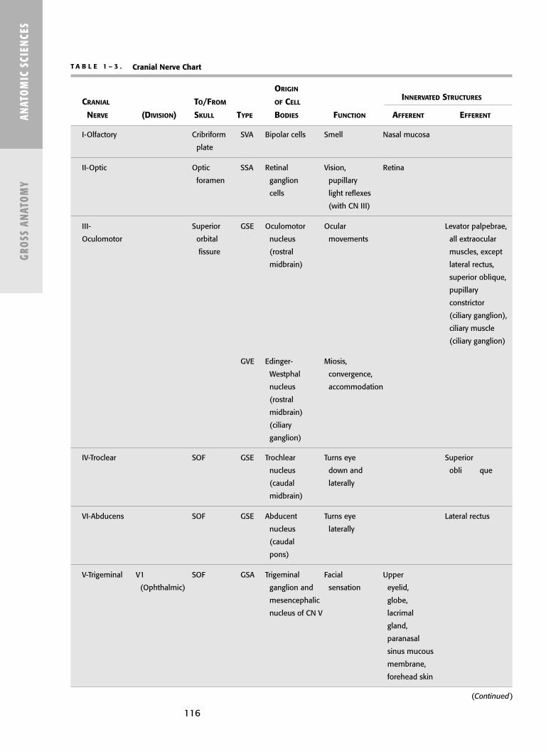
116
ANAT
OM
IC S
CIEN
CES
GROS
S AN
ATO
MY
T A B L E 1 – 3 . Cranial Nerve Chart
ORIGIN
CRANIAL TO/FROM OF CELLINNERVATED STRUCTURES
NERVE (DIVISION) SKULL TYPE BODIES FUNCTION AFFERENT EFFERENT
I-Olfactory Cribriform SVA Bipolar cells Smell Nasal mucosa
plate
II-Optic Optic SSA Retinal Vision, Retina
foramen ganglion pupillary
cells light reflexes
(with CN III)
III- Superior GSE Oculomotor Ocular Levator palpebrae,
Oculomotor orbital nucleus movements all extraocular
fissure (rostral muscles, except
midbrain) lateral rectus,
superior oblique,
pupillary
constrictor
(ciliary ganglion),
ciliary muscle
(ciliary ganglion)
GVE Edinger- Miosis,
Westphal convergence,
nucleus accommodation
(rostral
midbrain)
(ciliary
ganglion)
IV-Troclear SOF GSE Trochlear Turns eye Superior
nucleus down and obli que
(caudal laterally
midbrain)
VI-Abducens SOF GSE Abducent Turns eye Lateral rectus
nucleus laterally
(caudal
pons)
V-Trigeminal V1 SOF GSA Trigeminal Facial Upper
(Ophthalmic) ganglion and sensation eyelid,
mesencephalic globe,
nucleus of CN V lacrimal
gland,
paranasal
sinus mucous
membrane,
forehead skin
(Continued)

ANATOM
IC SCIENCES
GROSS ANATOM
Y
V2 Foramen GSA Sensation Lower eyelid,
(Maxillary) rotundum mid-face skin,
upper lip,
nasopharynx,
maxillary sinus,
soft palate,
tonsils, hard
palate, upper
teeth
V3 Foramen GSA Sensation Anterior 2/3rd
(Mandibular) ovale of tongue
(general
not taste
sensation!),
temporoauricular
skin, lower
face, lower
teeth
SVE Motor Chewing, Muscles of
nucleus opening, mastication,
CN V swallowing tensor veli
(mid pons) palatini, tensor
tympani
VII-Facial Internal GSA Geniculate Sensation External ear,
acoustic ganglion soft palate,
meatus, (facial auditory
stylomastoid canal of tube
foramen temporal
bone)
SVA Geniculate Taste Anterior 2/3rd
ganglion tongue
(facial
canal)
SVE Facial Facial Ms. Facial
nucleus expression expression,
(caudal stylohyoid,
pons) postdigastric,
stapedius
T A B L E 1 – 3 . Cranial Nerve Chart (Continued)
ORIGIN
CRANIAL TO/FROM OF CELLINNERVATED STRUCTURES
NERVE (DIVISION) SKULL TYPE BODIES FUNCTION AFFERENT EFFERENT
117
(Continued)
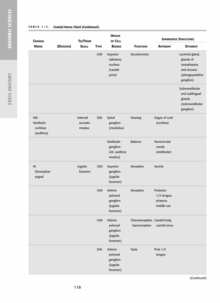
118
ANAT
OM
IC S
CIEN
CES
GROS
S AN
ATO
MY
GVE Superior Secretomotor Lacrimal gland,
salivatory glands of
nucleus nasopharynx
(caudal and sinuses
pons) (pterygopalatine
ganglion)
Submandibular
and sublingual
glands
(submandibular
ganglion)
VIII- Internal SSA Spiral Hearing Organ of corti
Vestibulo- acoustic ganglion (cochlea)
cochlear meatus (modiolus)
(auditory)
Vestibular Balance Semicircular
ganglion canals
(int. auditory (vestibular)
meatus)
IX- Jugular GSA Superior Sensation Auricle
Glossophar- foramen ganglion
yngeal (jugular
foramen)
GVA Inferior Sensation Posterior
petrosal 1/3 tongue,
ganglion pharynx,
(jugular middle ear
foramen)
GVA Inferior Chemoreception, Carotid body,
petrosal baroreception carotid sinus
ganglion
(jugular
foramen)
SVA Inferior Taste Post 1/3
petrosal tongue
ganglion
(jugular
foramen)
T A B L E 1 – 3 . Cranial Nerve Chart (Continued)
ORIGIN
CRANIAL TO/FROM OF CELLINNERVATED STRUCTURES
NERVE (DIVISION) SKULL TYPE BODIES FUNCTION AFFERENT EFFERENT
(Continued)

119
ANATOM
IC SCIENCES
GROSS ANATOM
Y
SVE Nucleus Motor for Stylopharyngeus
ambiguus swallowing
(rostral
medulla)
GVE Inferior Secretomotor Parotid gland
salivatory (via otic
nucleus ganglion and
(rostral aurioculotem-
medulla) poral branch
of V3), parasym-
pathetic path
of pharynx,
larynx
X-Vagus Jugular GSA Superior Sensation Posterior
foramen ganglion meninges,
(jugular external ear
foramen)
GVA Nodose Sensation Viscera of
ganglion pharynx,
(jugular larynx,
foramen) thoracic and
abdominal
viscera
(to left colic
flexure)
SVA Nodose Taste Laryngeal
ganglion additus and
(jugular epiglottis
foramen)
SVE Nucleus Motor Pharyngeal
ambiguus constrictors,
(medulla) palatopharyn-
geus, levator
palatini,
palatoglossus,
laryngeal ms
T A B L E 1 – 3 . Cranial Nerve Chart (Continued)
ORIGIN
CRANIAL TO/FROM OF CELLINNERVATED STRUCTURES
NERVE (DIVISION) SKULL TYPE BODIES FUNCTION AFFERENT EFFERENT
(Continued)

120
ANAT
OM
IC S
CIEN
CES
GROS
S AN
ATO
MY
GVE Dorsal Secretomotor Viscera of neck,
nucleus thoracic and
of CN X abdominal
(medulla) cavities (to left
colic flexure)
XI-Spinal Cranial Jugular SVE Nucleus Motor Cranial-ms
accessory foramen ambiguus larynx, pharynx,
(medulla) esophagus
Spinal Foramen Ventral Spinal—SCM,
magnum horn C1–C6 trapezius
XI-Hypoglossal Hypoglossal GVE Hypoglossal Motor All intrinsic and
canal nucleus extrinsic muscle
(medulla) of tongue (not
palatoglossus)
T A B L E 1 – 3 . Cranial Nerve Chart (Continued)
ORIGIN
CRANIAL TO/FROM OF CELLINNERVATED STRUCTURES
NERVE (DIVISION) SKULL TYPE BODIES FUNCTION AFFERENT EFFERENT
CN II—Optic Nerve
See Figure 1–76 and Table 1–3.See also the “Neurophysiology” section in Chapter 10 for lesions involving theoptic nerve.
Course
■ Ganglion cells of retina (converge at optic disc to form optic nerve).■ Leaves orbit via optic foramen/canal (sphenoid).■ Optic chiasm (two optic nerves unite at floor of diencephalons; anterior to
pituitary stalk).■ Optic tracts.■ Lateral geniculate nuclei (thalamus).■ Geniculocalcarine fibers (optic radiations). ■ Calcarine sulcus in the primary visual cortex (area 17) of the occipital lobe.
Important
■ Nasal side fibers (most medial) decussate and go with contralateral optictract.
■ Temporal hemiretina fibers stay ipsilateral.■ Left visual field = right optic tract.■ Right visual field = left optic tract (right visual field is interpreted on left
brain).

121
ANATOM
IC SCIENCES
GROSS ANATOM
Y
CNs III, IV, VI—Oculomotor, Trochlear, Abducens
See Figure 1–77.
■ Oculomotor nerve: Controls most of the extraocular muscles, the levatorpalpebrae superioris, and carries parasympathetic and sympathetic controlto the pupil.
■ Trochlear nerve: Is the smallest cranial nerve and the only cranial nervethat exits from the posterior surface of the brain stem.
F I G U R E 1 – 7 6 . The optic pathway.
The dotted lines represent nerve fibers that carry visual and papillary afferent impulses from the left half of the visual field.
(Reproduced, with permission, from Riordan-Eva P, Hoyt WF. Vaughan and Asbury’s General Ophthalmology, 17th ed.
New York: McGraw-Hill, 2008.)
Extraocular Movements
Muscle Nerve Movement
Medial rectus CN III Adduction (in)Superior rectus CN III Elevation and adductionInferior rectus CN III Depression and adductionInferior oblique CN III Elevation and abductionSuperior oblique CN IV Depression and abductionLateral rectus CN VI Abduction (out)

122
EYE ELEVATORS
See Figure 1–78 for a diagram of the eye muscle action.
■ Superior rectus: Adducts and elevates (only muscle that elevates fromabducted position).
■ Inferior oblique: Abducts and elevates (only muscle to elevate fromadducted position).
Pupillary Light Reflex
■ Direct and consensual papillary responses.■ Does not involve cortex.■ Shine light into one eye that eye (direct) pupil constricts as does the pupil
of the contralateral eye (consensual).
ANAT
OM
IC S
CIEN
CES
GROS
S AN
ATO
MY
F I G U R E 1 – 7 7 . The occulomotor, trochlear, and abducens nverves; ocular muscles.
Reproduced, with permission, from Waxman SG. Clinical Neuroanatomy, 25th ed. New York: McGraw-Hill, 2003.
Response Afferent Efferent
Direct response Optic nerve of eye tested CN III to the eye tested(ipsilateral) (ipsilateral)
Consensual response Optic nerve of eye CN III of opposite eyetested (ipsilateral) (contralateral)
Abducensnucleus
Trochlearnuclei
OculomotornucleusCiliary
ganglion
IIIIV
VI
To smooth (intraocular) muscles
To striated (extraocular) muscles
To striated(extraocular) muscles
Superior oblique
Superior rectus
Medial rectus
Constrictor pupillae
Inferior rectus
Inferior oblique
Lateralrectus
Levatorpalpebrae Nerves
Muscles

123
Pathway/Reflex Arc
See Figure 1–79.
■ Optic nerve■ Edinger-Westphal nucleus (midbrain) bilaterally■ Ciliary ganglia■ Short ciliary nerve (postganglionic)■ Constrictor pupillae muscle
ANATOM
IC SCIENCES
GROSS ANATOM
Y
F I G U R E 1 – 7 8 . Diagram of eye muscle action.
Reproduced, with permission, from Waxman SG. Clinical Neuroanatomy, 25th ed. New York: McGraw-Hill, 2003.
F I G U R E 1 – 7 9 . The path of the papillary light reflex.
Reproduced, with permission, from Waxman SG. Clinical Neuroanatomy. 25th ed. New York:
McGraw-Hill, 2003.
Lateralrectus
Lateralrectus
Superioroblique
Inferioroblique
Medialrectus
Superiorrectus
Superiorrectus
Inferiorrectus
Inferiorrectus
Right Left
Constrictorpupillaemuscle
Retinaganglioncell
Ciliaryganglion
Opticchiasm
Nerve III
Oculomotornucleus
Pretectal area
Posterior commissure

124
ACCOMMODATION AND CONVERGENCE
■ Gazing from distant to near, pupils constrict (accommodate), eyes movemidline (converge), lenses become more convex.
Pathway
■ Optic nerve■ Geniculate body■ Visual association cortex■ Frontal eye fields■ Edinger-Westphal nuclei and main oculomotor nuclei (both in midbrain)■ Constrictor pupillae muscle and medial recti, respectively
LESION
■ Blurred vision with a lesion to any of CN III, IV, VI.■ Ptosis (drooping eyelid) and dilated pupil with CN III injury (levator
palpebrae superioris and sphincter pupillae muscle).■ Lesion CN VI eye persistently directed toward nose (because of lateral rectus).■ See Table 1–4.
ANAT
OM
IC S
CIEN
CES
GROS
S AN
ATO
MY
T A B L E 1 – 4 . Common Lesions to CN III, IV, and VI
LESION DESCRIPTION
Ophthalmoplegia Internal: EOMs spared, selective loss of autonomically innervated sphincter and
ciliary muscles (because parasympathetic fibers are more peripheral, can be
compressed).
External: Paralysis of all EOMs (except superior oblique, rectus); sphincter and
ciliary muscles spared (as with DM neuropathy).
Internuclear Lesion within the medial longitudinal fasciculus—lose
ophthalmoplegia connection III, IV, VI. If lesion on right—look laterally to the right, both eyes move
look laterally to left, only left eye moves (right eye medial rectus does not pull
it medially); however with accommodation, both medial recti work.
Relative afferent Lesion in optic nerve (afferent limb of papillary light relflex,
(Marcus-Gunn) pupil eg, MS optic neuritis).
Swinging flashlight test—both pupils constrict when shine light in good eye;
quickly swing to contralateral eye and both pupils dilate.
Horner’s syndrome Lesion of oculosympathic pathway (sympathetics don’t come from CNs but run
with them; come from superior cervical ganglion, ciliospinal center of Budge);
miosis, ptosis, hemianhidrosis, apparent enophthalmos.
Argyll Robertson Think prostitute’s pupil—accommodates but does not react; also associated
pupil (pupillary with syphilis.
light-near dissociation) No miosis (papillary constriction) with either direct or consensual light; does
constrict with near stimulus (accommodation-convergence).
Occurs in syphilis and diabetes.

125
CN V—Trigeminal Nerve
■ Largest cranial nerve. ■ Trigeminal or gasserian ganglion—in the middle cranial fossa.■ Three divisions leave through foramina in the sphenoid bone.■ Sensory (GSA): Facial sensation (light touch; pain and temperature; pro-
prioception)■ Divisions V1, V2, V3.
■ Efferent (SVE): Motor to muscles of mastication, tensor veli palatini, tensortympani.■ Division V3.
■ No parasympathetic fibers are contained with the trigeminal nerve at its origin;other nerves distribute parasympathetic preganglionics by way of trigeminalbranches.■ Oculomotor (III).■ Facial nerve (VII).■ Glossopharyngeal nerve (IX).
TRIGEMINAL NERVE DIVISIONS
See Figure 1–80 for the trigeminal nerve and its branches.
ANATOM
IC SCIENCES
GROSS ANATOM
Y
V1 and V2 are purely sensory;
V3 is both sensory and motor.
Remember: The buccal nerve
of V3 provides sensation to
the cheek; whereas the buccal
branch
of CN VII is motor to the
buccinator muscle.
Division Sensory Motor
Ophthalmic (V1) Upper eyelid, cornea, N/Aconjunctiva, frontal sinus, upper nasal mucosa, forehead
Maxillary (V2) Lower eyelid, upper N/Acheek, lip, gums, palate, nose,tonsils, hard palate, upper teeth
Mandibular (V3) Tongue (general), Muscles of mastication, temporoauricular skin, tensor tympani, mylohyoid, lower face, lower teeth anterior belly of digastric,
tensor veli palatini

ANAT
OM
IC S
CIEN
CES
GROS
S AN
ATO
MY
Nerve Distribution
Lingual nerve General sensation: Anterior 2/3rd of tongue, floor of mouth, andmandibular lingual gingival.
Carries (from chorda tympani [VII]):Taste sensation: Anterior 2/3rd tongue.Preganglionic parasympathetics: To submandibular ganglion.
Auriculotemporal nerve Sensory: Front of ear, TMJ.Postganglionic parasympathetic: To parotid gland.
Inferior alveolar nerve Gives off nerve to mylohyoid and inferior dental plexus; terminatesas mental nerve.
Motor to mylohyoid.Sensory to teeth, skin of chin, lower lip.
Mental nerve Termination of inferior alveolar nerve.Sensory to skin of chin, skin, and mucous membrane of lower lip.
Motor branches Motor to muscles of mastication, anterior digastric, and so on.
IMPORTANT BRANCHES OF V3
F I G U R E 1 – 8 0 . The trigeminal nerve and its branches.
Reproduced, with permission, from Waxman SG. Clinical Neuroanatomy, 25th ed. New York: McGraw-Hill, 2003.
126
Otic ganglion
Chorda tympani nerve
Anterior belly of digastric muscle
Mylohyoid muscle
Mylohyoid nerve
Masseter muscle
Internal pterygoid muscle
Auriculotemporal nerve
Anterior and posteriordeep temporal nerves (to temporal muscle)
External pterygoid muscle
Mandibular nerve
Mesencephalicnucleus of V
Main sensory nucleus of V
Main motornucleus of V
Nucleus of spinal tract of V
Semilunarganglion
Ophthalmic nerve
Frontal nerveNasociliary nerve
Posterior ethmoidal nerveAnterior ethmoidal nerve
Supraorbital nerveFrontal branch of frontal nerve
Supratrochlear nerve
Ciliary ganglion
Internal nasalrami
Infraorbital nerve
External nasalrami
Nasal and labialrami of infraorbital nerve
Infratrochlear nerve
Anterior superioralveolar nerves
Submandibularganglion
Submandibular andsublingual glands
Mental nerve
Lacrimal
Pterygopalatineganglion
Buccinator nerve
Lingual nerve
Inferior alveolar nerve
Maxillary

127
INFERIOR ALVEOLAR NERVE BLOCK
■ Anesthetize the mandibular teeth. ■ Block this branch of V3 as it enters the mandibular foramen.
Needle Course (See Figure 1–81)
■ Pierces:■ Buccinator (between palatoglossal and palatopharyngeal folds).■ Lies lateral to medial pterygoid at the mandibular foramen.
■ If the needle penetrates too far posteriorly can hit parotid gland and CN VII:■ Ipsilateral facial paralysis.
ANATOM
IC SCIENCES
GROSS ANATOM
Y
The lingual nerve is found in
the pterygomandibular space
with the inferior alveolar
nerve, artery, and vein.
The lingual artery does not
run with the lingual nerve.
The lingual artery is medial
to the hyoglossus muscle,
whereas the lingual vein and
nerve are lateral to the
hyoglossus (as is the
submandibular duct and
hypoglossal nerve [XII]).
The submandibular duct is
crossed twice by the lingual
nerve.
If the lingual nerve is cut after
the chorda tympani joins, you
lose both taste and tactile
sensation.
F I G U R E 1 – 8 1 . Needle course.

ANAT
OM
IC S
CIEN
CES
GROS
S AN
ATO
MY
Nuclei Location Comments
Motor Lateral Motor information originates in the motor (area 4) and (masticatory) pons premotor (area 6) cortices.nucleus This information is modulated by the basal ganglia and
striatal motor systems then:Descends as corticobulbar tracts to the:Motor nucleus of the trigeminal where:LMNs then project past the trigeminal ganglion (without synapse) to the:Respective effector muscles.
Sensory Pons All sensory information from the face is relayed through nucleus VPM nucleus of thalamus (VPL for the body).
From here information relays to the:Somatosensory cortex (areas 3, 1, 2). ■ The facial segment of the sensory homunculus comprises a large
area of the lateral parietal lobe.
Spinal trigeminal Midpons to Pain/temperature from the face travel to the spinal trigeminal nucleus nucleus cervical cord (pain/temperature from body travel in the spinothalamic tract).
Mesencephalic Upper pons, Proprioceptive inputs are not located within the trigeminal nucleus midbrain ganglia (all other afferent cell bodies are).
border Proprioceptive inputs synapse in the mesencephalic nucleus.
Proprioceptive fibers from muscles and TMJ are found only in the mandibular division (V3).
The lingual nerve can be
damaged with third molar
extraction because it lies close
to the mandibular ramus in
the vicinity of the third molar.
TRIGEMINAL NUCLEI
■ There are four paired nuclei (both motor and sensory).
LINGUAL NERVE
Chorda Tympani Course Information (VII) Component
Lies deep to the lateral General sensation: Chorda tympani (of VII)pterygoid muscle, Anterior 2/3rd of contributes:anteromedial to the tongue, floor ■ Preganglionic inferior alveolar nerve of mouth, and parasympathetic(chorda tympani (from VII) mandibular lingual fibers to joins here). gingiva. submandibular
Runs between the medial ganglion.pterygoid and the ■ Taste fibers tomandibular ramus to anterior 2/3rdenter the side of the of tongue.tongue obliquely.
Continues forward between the hyoglossus and deep partof the submandibular gland.
Submandibular ganglion hangs from lingual nerve on top of hyoglossus muscle.
Finally, it runs across the submandibular duct to lie at the tongue tip (beneath the mucous membrane).
128

129
FACIAL SENSATION
CN V
■ All sensory information from the face is relayed through VPM nucleus ofthalamus; sensory information from the rest of the body is through the VPL.
■ From the thalamic nuclei (VPM or VPL), information relays to the somatosen-sory cortex (areas 3, 1, 2); the facial segment of the sensory homunculuscomprises a large area of the lateral parietal lobe.
■ Remember, parts of CNs VII and IX travel with trigeminospinal tract.■ All CN V afferent cell bodies are located within trigeminal ganglion except
proprioceptive inputs. ■ Mesencephalic nucleus of CN V is the only case where primary sensory
cell bodies are located within the CNS, rather than in ganglia.
ANATOM
IC SCIENCES
GROSS ANATOM
Y
1st-Order 2nd-Order 3rd-OrderModality Fiber Type Tract Neuron Neuron Neuron
Touch and A-beta fibers Dorsal Trigeminal Principal VPM projects pressure trigeminothalamic ganglion, sensory nucleus through
tract (via (principal of CN V projects posterior internal Meissner’s sensory to ipsilateral capsule to the and Pacini’s nucleus VPM (doesn’t face area ofcorpuscles) of CN V). cross over). somatosensory
cortex.Pain and A-delta Ventral Trigeminal From the spinal From VPM, project
temperature C-fibers trigeminothalamic ganglion; trigeminal via posterior tract then axons nucleus, ascend limb of posterior
descend in the and cross over capsule to thespinal trigeminal to the contralateral face area of tract and synapse VPM. somatosensorywith the spinal cortex.trigeminal nucleus.
Proprioception A-alpha fibers Spinocerebellar Afferents pass Mesencephalic Cerebellum(stretch and tract the trigeminal nucleus (withintendon ganglia (without the CNS).receptors in synapse). Motor nucleusmuscles of (many central mastication) fibers from
mesencephalicnucleus synapse here to create reflex arc).

FACIAL SENSATION
See Figure 1–82 for areas of facial sensation.See Figure 1–83 for cutaneous innervation of the face, scalp, and uricle.See Figure 1–84 for innervation of the external ear.
ANAT
OM
IC S
CIEN
CES
GROS
S AN
ATO
MY
Sensation in teeth can be
misinterpreted in ear
(because of the cross
innervation).
Herpes zoster often affects
V1 division.
Trigeminal neuralgia
(tic douloureux) can
affect V2 and V3.
F I G U R E 1 – 8 2 . Sensory distribution of CN V.
Reproduced, with permission, from White JS. USMLE Road Map Gross Anatomy. New York:
McGraw-Hill, 2003.
SENSORY INFORMATION IN THE FACE VERSUS BODY
Face Body
Touch and pressure Trigeminal main sensory Gracile and cuneate nucleinucleus Dorsal columns
Dorsal trigeminothalamic tract Medial lemniscusVPM VPL
Pain and temperature Spinal trigeminal nucleus Dorsal root ganglionVentral trigeminothalamic Lissauer’s tract
tract Spinothalamic tractVPM, periaqueductal grey, VPL, periaqueductal grey,
reticular formation reticular formationProprioception Mesencephalic nucleus Clarke’s nucleus
Spinocerebellar tract Spinocerebellar tractCerebellum Cerebellum
130
Ophthalmic(CN V1)
Maxillary(CN V2)
Mandibular(CN V3)

131
ANATOM
IC SCIENCES
GROSS ANATOM
Y
SENSATION OF EXTERNAL EAR
Nerve Distribution
Auriculotemporal nerve (V3) Anterior half of external ear canal and facial surfaceof upper part of auricle
Auricular branch of vagus (CN X) Posterior half of external ear canal (so stimulationcan cause reflex symptoms: eg, fainting, coughing, gagging).
Greater auricular nerve (C2, C3) Inferior auricle (anterior and posterior)Lesser occipital nerve (C2, C3) Cranial surface of upper auricle
F I G U R E 1 – 8 3 . Cutaneous innervation of the face, scalp, and auricle.
F I G U R E 1 – 8 4 . Innervation of the external ear.
Corneal reflex: Ifstimulating right eye:
■ Lesion R V1 neithereye blinks.
■ Lesion L V1 bilateralblink.
■ Lesion R VII only lefteye blinks (indirect).
■ Lesion L VII only righteye blinks (direct).
■ Levator palpebraesuperioris (CN III)keeps the eyelid open;lesion results in ptosis.
■ Orbicularis oculi (CN VII) closes eyelid;lesion results ininability to close, nocorneal reflex.
Auriculotemporal nerve
Greater occipital nerve
Lesser occipital nerve
Great auricular nerve
Supratrochlear nerve
Supraorbital nerve
Zygomaticotemporal nerve
Infratrochlear nerveZygomaticofacial nerve
External nasal nerve
Infraorbital nerve
Buccal nerve
Mental nerve
Auriculotemporal nerve
Anterior Posterior
Auricular branch of vagus
Greater auricular nerve
Lesser occipital nerve

132
ANAT
OM
IC S
CIEN
CES
GROS
S AN
ATO
MY
Reflex Description Afferent Efferent Comments
Corneal reflex Touch cornea V1 CN VII Pathwaywith cotton (orbicularis ■ Cornea (V1)causes both oculi) ■ Main sensory nucleus eyes to close of trigeminal
■ Medial longitudinal fasciculus
■ Main motor nucleus of facial nerve (CN VII)
■ Orbicularis oculi muscle■ Causes ipsilateral (direct)
and contralateral (indirect)eye blinking
Jaw jerk reflex Mouth open and V3— V3—motor Reflex is lost only with bilateral relaxed; tap chin proprioceptive CN V lesions (uncommon)lightly to get contraction/mouthclosure
Facial Reflexes (Involving CN V)
TRIGEMINAL LESIONS
Type Location Finding
Sensory Division V1, 2, 3 Deficits along distribution (pain, temperature,touch, pressure, proprioception)
Motor Division V3 only Temporalis and masseter muscles■ Ipsilateral weakness of jaw closure■ Ipsilateral open bitePterygoid muscle■ Weakness of jaw opening■ Deviation to ipsilateral side on openingDiminished/loss of reflexes
CN VII-Facial Nerve
See Figure 1–85.
Course
■ Originates in pons.■ Enters internal acoustic meatus.■ Passes through facial canal.■ Exits skull via stylomastoid foramen.■ Courses through the parotid gland (inadvertent deposition of anesthetic into
parotid can result in facial paresis).■ Motor branches.
Facial Nerve Branches
Mnemonic: To Zanzibar By
Motor Car
Temporal
Zygomatic
Buccal
Marginal Mandibular
Cervical

133
ANATOM
IC SCIENCES
GROSS ANATOM
Y
F I G U R E 1 – 8 5 . The facial nerve.
Reproduced, with permission, from Waxman SG. Clinical Neuroanatomy, 25th ed. New York: McGraw-Hill, 2003.
Components
■ Motor■ Sensory■ Secretomotor (parasympathetic):
■ Lacrimal gland■ Submandibular gland■ Sublingual gland
■ Special sense ■ Taste anterior two-thirds of tongue, floor of mouth, palate
Frontalis muscle
Lacrimal gland
Obicularis oculi
Zygomatic branch
Temporal branch
Obicularis oris
Buccinator
Risorius
Mentalis
Taste toanterior 2/3 of
tongue
Platysma muscle
Submandibular andsublingual glands
Submaxillary ganglion
Cervicofacial branch
Motor nerve
Occipitalis muscle
Stylomastoid foramen
Stapedius muscle
Geniculate ganglion
Nucleus of
solitary tract
Motor nucleus of VII
(Branchial efferent)
Superior
salivatory nucleus
Nervus intermedius
Superficial
petrosal nerve
Pterygopalatine
ganglion
Chorda tympani nerve
Sensory nerveParasympathetic nerve

134
FACIAL EXPRESSION
See Figure 1–86 for facial musculature.
ANAT
OM
IC S
CIEN
CES
GROS
S AN
ATO
MY
CN VII NUCLEI
Nuclei Comments
Main motor nucleus Upper face receives bilateral innervation; lower face receives unilateral innervation.
Muscles of facial expression, posterior belly of digastric, stylohyoid muscle, stapedius.
Superior salivatory Submandibular and sublingualnucleus glands.
Nucleus of the solitary tract (gustatory nucleus) Mediates taste.
F I G U R E 1 – 8 6 . Facial musculature.
Superior auricular muscle
Frontalis
Anterior auricular muscle
Occipitalis
Posterior auricular muscle
Sternocleidomastoid muscleSplenius muscle
Trapezius muscle
Orbicularis oculi
Compressor naris muscle
Levator labii superiorisalaeque nasi muscleLevator labii superioris
Zygomaticus minorZygomaticus majorOrbicularis orisDepressor labii inferioris
Depressor anguli orisMentalis
Platysma

135
ANATOM
IC SCIENCES
GROSS ANATOM
Y
IMPORTANT MUSCLES OF FACIAL EXPRESSION (ALL CONTROLLED BY CN VII)
Muscle Origin Insertion Action
Orbicularis Circumoral muscles, Lips and skin Whistleoris maxilla, mandible of lip Pulls lips against teeth,
protrudes lips
Depressor anguli Mandibular oblique line Angle of mouth Frownoris Pulls down angle of mouth
Zygomaticus Zygomatic bone Angle of mouth Smilemajor Pulls angle of mouth up
and back
Risorius Parotid and masseteric Angle of mouth Smilefascia Pulls angle of mouth
laterally
Orbicularis oculi Upper medial orbit, Encircle orbit; insert on Closes eyemedial palpebral medial palpebral,ligament, lacrimal bone ligament medial side
of lids, laterally in raphe
OTHER MUSCLES CONTROLLED BY CN VII
Muscle Origin Insertion Action Paralysis
Buccinator Alveolar process of Orbicularis Holds food on occlusal Food/saliva fall maxilla and mandible oris table (accessory between teeth Pterygomandibular muscle of mastication); and cheekraphe tenses cheek
(blowing, whistling)
Stapedius Neck Decreases vibration Hyperacusisof stapes of the stapes (decreases
perception of sound)
CN VII LESIONS
Lesion Clinical Finding Comments
Lower motor Ipsilateral paralysis/weakness If lesion proximal to greater petrosal andneuron lesion of upper and lower face; loss chorda tympani take off, patient
of corneal reflex (efferent limb). experiences:(Bell’s palsy: Acute 7th ■ Decreased taste (anterior 2/3rd).nerve palsy.) ■ Hyperacusis (due to stapedius
muscle paralysis).■ Decreased salivation on affected side
(though not clinically apparent if other salivary glands intact).
Upper motor Contralateral lower With a central lesion to the facial motor nucleusneuron lesion face weakness only. (eg, stroke), one can still raise eyebrows on affected
side (the side opposite the central lesion) becauseof bilateral innervation to muscles of upper face.
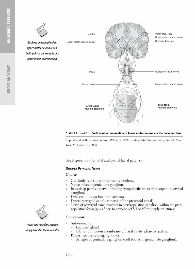
See Figure 1–87 for total and partial facial paralysis.
GREATER PETROSAL NERVE
Course
■ Cell body is at superior salivatory nucleus.■ Nerve arises at geniculate ganglion.■ Joins deep petrosal nerve (bringing sympathetic fibers from superior cervical
ganglion).■ Exits cranium via foramen lacerum.■ Enters pterygoid canal (as nerve of the pterygoid canal).■ Nerve of pterygoid canal synapses in pterygopalatine ganglion (within the ptery-
gopalatine fossa) (gives fibers to branches of V1 or V2 to supply structures).
Components
■ Autonomic to:■ Lacrimal gland.■ Glands of mucous membrane of nasal cavity, pharynx, palate.
■ Parasympathetic preganglionics ■ Synapse at geniculate ganglion (cell bodies in geniculate ganglion).
136
ANAT
OM
IC S
CIEN
CES
GROS
S AN
ATO
MY
F I G U R E 1 – 8 7 . Corticobulbar innervation of lower motor neurons in the facial nucleus.
Reproduced, with permission, from White JS. USMLE Road Map Neuroscience, 2nd ed. New
York: McGraw-Hill, 2008.
Facial and maxillary arteries
supply blood to the buccinator.
Stroke is an example of an
upper motor neuron lesion.
Bell’s palsy is an example of a
lower motor neuron lesion.
Cortex
Pons
Facial nerve
Partial facial
muscle paralysis
Total facial
muscle paralysis
Motor area, face
Upper motor neuron lesion
Upper motor neuron lesion
Nucleus of facial nerve
Lower motor neuron lesion
Corticobulbar tract

137
ANATOM
IC SCIENCES
GROSS ANATOM
Y
■ Sympathetics (postganglionic) ■ Pass through geniculate without synapsing.
■ Taste■ From palate via palatine nerves.■ Taste fibers pass through pterygopalatine ganglion and travel with
nerve of pterygoid canal.■ It reaches the greater petrosal nerve (the tractus solitarius and nucleus
of solitary tract in pons).
CN VIII—Vestibulocochlear
See Figure 1–88.
Course
■ Located within temporal bone, innervates:■ Cochlea (hearing).■ Semicircular canals and maculae (balance).
■ Passes internal acoustic meatus.■ Into brain stem at the junction pons and medulla, fibers terminate in:
■ Cochlear nucleus.■ Vestibular nuclear complex (near floor of fourth ventricle).
EAR ANATOMY
See Figure 1–89.
■ External ear—Receives sound waves■ Auricle ■ External auditory canal
■ Middle ear—tympanic cavity ■ Three ossicles:
■ Malleus (hammer)■ Incus (anvil)■ Stapes (stirrup)
■ Two muscles:■ Stapedius (VII)■ Tensor tympani (V3)
F I G U R E 1 – 8 8 . The vestibulocochlear nerve.
Reproduced, with permission, from Waxman SG. Clinical Neuroanatomy, 25th ed.
New York: McGraw-Hill, 2003.
Greater petrosal nerve is the
parasympathetic root of the
pterygopalatine ganglion.
The stapedius is the smallest
skeletal muscle in the body.
Spiral ganglionLabyrinth
Vestibular ganglion
Nerve VIII
Cochlear nucleiVestibular nuclei

138
■ Inner ear—composed of membranous and bony labyrinth■ Acoustic apparatus■ Vestibular apparatus■ Semicircular canals
MUSCLES IN MIDDLE EAR
■ The muscles in the middle dampen sounds; paralysis results in hyperacusis.
ANAT
OM
IC S
CIEN
CES
GROS
S AN
ATO
MY
F I G U R E 1 – 8 9 . External, middle, and internal ear.
Muscle Innervation
Tensor tympani V3Stapedius VII
EUSTACHIAN (AUDITORY) TUBE
■ Also called the pharyngotympanic tube.■ Connection between middle ear and pharynx. ■ Equalizes air pressure between the tympanic cavity and nasopharynx.
CN VIII LESIONS
HEARING LOSS
Presbycusis
■ Loss occurring gradually with aging because of changes in the inner ormiddle ear.
Middle ear communicates
posteriorly with the mastoid
cells and the mastoid antrum
via the aditus ad antrum.
Otitis media: Middle ear
infections; can extend to both
mastoid and nasopharynx via
eustachian tube.
Otitis externa: Infection of
ear canal (past external
auditory meatus).
AuricleEpitympanic recess
IntegumentFacial musculature
Squama of temporal bone
Superior ligament of the malleus
Head of malleus
Semicircular canals
IncusFacial nerve (VII)StapesInternal acoustic meatusVestibular nerveCochlear nerve
Cochlear duct
Tympanic membrane
Tensor tympani muscle
Concha
External acoustic meatus
Cartilages of external ear
Parotid glandFacial nerve (VII)
Levator veli palatini muscle
Styloid process
Internal jugular veinInternal carotid artery
Auditory (Eustachian) tube

139
■ Conductive loss: Thickening and stiffening of cochlear basilar membrane.■ Sensorineural loss: Loss of hair and/or nerve cells.
(See also hearing under special sense area.)
NYSTAGMUS
■ Involuntary rapid and repetitive movement of the eyes.■ Results from irritation to
■ Labyrinth.■ Vestibular nerve or nuclei.■ Cerebellum.■ Visual system.■ Cerebral cortex.
■ Rhythmic oscillations are slow to one side rapid to the opposite side.■ Defined by the direction of the rapid reflex movement.■ Usually horizontal, occasionally vertical or rotatory.
Cold Caloric Test
■ Instill water to the external auditory meatus stimulates nystagmus.■ Cool water fast component to side opposite.■ Warm water fast component toward stimulated side.
CN IX—Glossopharyngeal
See Figure 1–90.
Course
■ Originates in anterior surface of medulla oblongata (with X and XI). ■ Passes laterally in the posterior cranial fossa.■ Leaves skull via jugular foramen.
Components
■ Motor: Stylopharyngeus muscle.■ General sensory: Sensory cell bodies are within superior and inferior gan-
glia of CN IX.■ Mucosa of pharynx.
■ Posterior third of the tongue.■ Visceral sensory:
■ Taste perceived on posterior third of the tongue. ■ Gag reflex (afferent limb) (fauces).■ Chemo-, baroreception (afferent limb)—carotid body, carotid sinus.
■ Parasympathetic/secretomotor: Parotid via otic ganglion ■ Preganglionics.
■ Leave glossopharyngeal nerve as the tympanic nerve.■ Enter middle ear cavity.■ Contribute to tympanic plexus.■ Re-form as the lesser petrosal nerve. ■ Leave cranial cavity via foramen ovale.■ Enter otic ganglion.
■ Postganglionics.■ Carried by the auriculotemporal nerve (V3) to the parotid.
ANATOM
IC SCIENCES
GROSS ANATOM
Y
Central hearing connections
are bilateral, so a central
lesion will not cause deafness
in either ear.
Caloric test is test-ing the vestibulo-ocular reflex.
COWS
ColdOppositeWarmSame
The gag reflex is mediated by
CN IX (afferent-unilateral) and
CN X (efferent-bilateral).
Chemoreception: Carotid
body; oxygen tension
measurement.
Baroreception: Carotid sinus;
blood pressure changes.
Mediated CN IX (afferent) and
CN X (efferent).
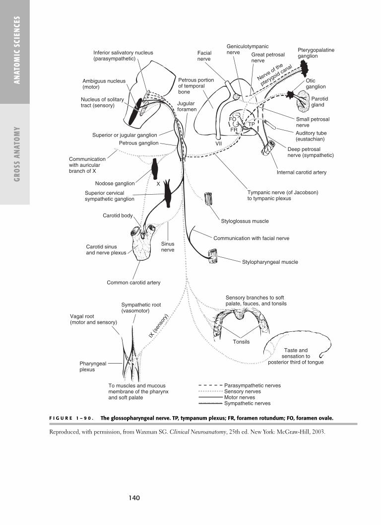
140
ANAT
OM
IC S
CIEN
CES
GROS
S AN
ATO
MY
F I G U R E 1 – 9 0 . The glossopharyngeal nerve. TP, tympanum plexus; FR, foramen rotundum; FO, foramen ovale.
Reproduced, with permission, from Waxman SG. Clinical Neuroanatomy, 25th ed. New York: McGraw-Hill, 2003.
Auditory tube(eustachian)
Deep petrosalnerve (sympathetic)
Internal carotid artery
Tympanic nerve (of Jacobson)to tympanic plexus
Styloglossus muscle
Communication with facial nerve
Stylopharyngeal muscle
Sensory branches to softpalate, fauces, and tonsils
Tonsils
Taste andsensation to
posterior third of tongue
To muscles and mucousmembrane of the pharynxand soft palate
Pharyngealplexus
Vagal root (motor and sensory)
Sympathetic root(vasomotor)
Common carotid artery
Carotid sinusand nerve plexus
Carotid body
Sinusnerve
Superior cervicalsympathetic ganglion
XNodose ganglion
Communicationwith auricularbranch of X
Petrous ganglion
Superior or jugular ganglion
Nucleus of solitarytract (sensory)
Ambiguus nucleus(motor)
Inferior salivatory nucleus(parasympathetic)
Jugularforamen
Petrous portionof temporalbone
Facialnerve
Geniculotympanicnerve Great petrosal
nerve
Nerve of the
pterygoid canal
Pterygopalatineganglion
Oticganglion
Parotidgland
Small petrosalnerve
FO
FRTP
VII
Parasympathetic nervesSensory nervesMotor nervesSympathetic nerves
IX(s
enso
ry)

141
ANATOM
IC SCIENCES
GROSS ANATOM
Y
CN X—Vagus
■ Cranial nerve with the widest distribution.■ Supplies the viscera of neck, thorax, and abdomen to left colic flexure.■ Course:
■ Leaves the medulla.■ Passes out jugular foramen.■ Descends neck in carotid sheath (behind internal and common carotid
arteries, and IJV).■ Posterior mediastinum (travels on the esophagus).■ Enters abdominal cavity with the esophagus.
Right Vagus Nerve Left Vagus Nerve
Crosses anterior surface of right Enters thorax in front of left subclavian subclavian artery. artery, behind left brachiocephalic vein.
Enters the thorax posterolateral to the Crosses the left side of the aortic arch (and is itself brachiocephalic trunk, lateral to the crossed by left phrenic nerve).trachea, and medial to the azygos vein. Passes behind left lung (contributing to
Passes posterior to the root of the right lung the pulmonary plexus).(contributing to the pulmonary plexus). Travels with the esophagus (contributes to
Travels with the esophagus (contributes esophageal plexus).to the esophageal plexus). Enters abdomen in front of esophagus (via the
Enters the abdomen behind the esophagus esophageal hiatus of the diaphragm).(via esophageal hiatus of diaphragm). Branches of esophageal plexus unite:
Branches of esophageal plexus unite:Posterior vagal trunk (posterior gastric Anterior vagal trunk (anterior gastric nerve—reaches
nerve—reaches posterior surface of stomach). anterior surface of stomach).
BRANCHES OF THE VAGUS IN THE HEAD AND NECK
See Figure 1–91 and Table 1–3.
Branch Description
Meningeal To dura.Auricular To auricle, posterior 1/2 of external auditory meatus.Pharyngeal Forms pharyngeal plexus.
Supplies:■ Muscles of pharynx (except stylopharyngeus [innervated by CN IX]).■ All the muscles of the soft palate (except tensor veli palatini,
innervated by NC V3).Superior laryngeal Travels with superior laryngeal artery.■ Internal laryngeal Pierces thyrohyoid membrane.■ External laryngeal Supplies mucous membranes of larynx above vocal fold.
Travels with superior thyroid artery.Supplies cricothyroid muscle.
Vagus nerves lose their
identity in the esophageal
plexus. The anterior gastric
nerve can be cut (vagotomy)
to reduce gastric secretion.

ANAT
OM
IC S
CIEN
CES
GROS
S AN
ATO
MY
F I G U R E 1 – 9 1 . The vagus nerve. J, jugular (superior) ganglion; N, nodose (inferior) ganglion.
Reproduced, with permission, from Waxman SG. Clinical Neuroanatomy, 25th ed. New York: McGraw-Hill, 2003.
142
Auricular branch toposterior auricleand part ofexternal meatus
Sensation tolower pharynx
Epiglottic andlingual rami
Inferior pharyngeal constrictor
Cricothyroid muscleGlottisRight recurrent laryngeal nerve
Left recurrent laryngeal nerve
Left vagus nerveAortic arch
Diaphragm
Stomach
Spleen
Pancreas
Left kidney
Small intestineRight kidney
Gallbladder
Liver
Esophageal plexus
Pulmonary plexus
Cardiac plexus
Cardiac nerves
Right subclavian artery
Esophagus
Arytenoid, thyroarytenoid,and cricoarytenoid muscles
Trapezius muscle
Sternocleidomastoid muscle
C4C3
Spinal roots of accessory nerveC2
C1
C5
Dorsal motor nucleus of vagus
Ambiguus nucleus
Nucleus of spinal tract of V
Nucleus of solitary tract
Meningeal branch toposterior fossa VII
IXX
XI
J
N
XII
Superior cervicalsympatheticganglion
Superiorlaryngeal nerve
Sensory nerves
Parasympathetic nervesMotor nerves
Pharyngealplexus
Celiacplexus
Muscles to palateand pharynx

ANATOM
IC SCIENCES
GROSS ANATOM
Y
CN X LESIONS
■ Parasympathetic disturbances■ Loss of gag reflex■ Dysphagia■ Dysphonia
CN XI—Accessory
See Figure 1–92 for a schematic illustration of the accessory nerve.
CN XI LESIONS
■ Paralysis of SCM: difficulty turning head to contralateral side.■ Paralysis of trapezius: Shoulder droop.
CN XII—Hypoglossus
See Figure 1–93.
■ The hypoglossal nerve is strictly a motor nerve.
Course
■ Leaves skull through hypoglossal canal (of the occipital bone) (medial tocarotid canal and jugular foramen).
■ Joined by C1 fibers from the cervical plexus soon after it leaves skull.■ Passes above hyoid bone on the lateral surface of the hyoglossus muscle
(deep to the mylohyoid).■ Loops around occipital artery.■ Passes between ECA and IJV.
F I G U R E 1 – 9 2 . Schematic illustration of the accessory nerve, viewed from below.
Reproduced, with permission, from Waxman SG. Clinical Neuroanatomy, 25th ed. New York:
McGraw-Hill, 2003.
The cardiac branches of the
vagus (form the cardiac
plexus) are preganglionic
parasympathetic nerves
that synapse with
postganglionic
parasympathetic nerves
in the heart.
143
The abdominal viscera below
the left colic flexure (and
genitalia and pelvic viscera)
are supplied by pelvic
splanchnic nerves
(parasympathetic
preganglionics).
Also with CN XII paralysis, the
tongue tends to fall back and
obstruct the airway
(genioglossus).
In addition to deviation to the
affected side (with damage
to CN XII and resultant
denervation atrophy),
dysarthria (inability to
articulate) can be experienced
by the patient.
Accessorynerve
Cranial portionof nerve XI(joins vagus nerve)Spinal portionof nerve XI
Sternocleido-mastoid muscle
Trapezius muscle(upper third)
C5
C1
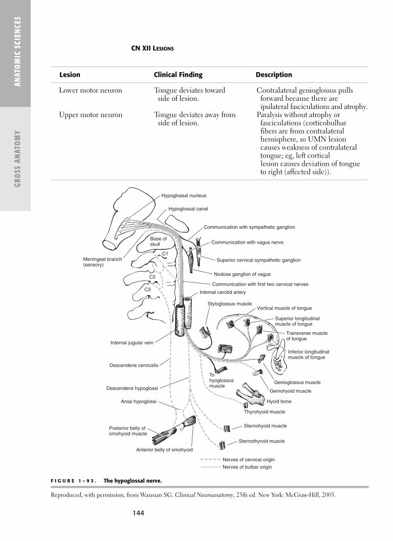
144
ANAT
OM
IC S
CIEN
CES
GROS
S AN
ATO
MY
CN XII LESIONS
Lesion Clinical Finding Description
Lower motor neuron Tongue deviates toward Contralateral genioglossus pullsside of lesion. forward because there are
ipsilateral fasciculations and atrophy.Upper motor neuron Tongue deviates away from Paralysis without atrophy or
side of lesion. fasciculations (corticobulbar fibers are from contralateral hemisphere, so UMN lesion causes weakness of contralateraltongue; eg, left cortical lesion causes deviation of tongueto right (affected side)).
F I G U R E 1 – 9 3 . The hypoglossal nerve.
Reproduced, with permission, from Waxman SG. Clinical Neuroanatomy, 25th ed. New York: McGraw-Hill, 2003.
Hypoglossal nucleus
Hypoglossal canal
Communication with sympathetic ganglion
Communication with vagus nerve
Superior cervical sympathetic ganglion
Nodose ganglion of vagus
Communication with first two cervical nerves
Internal carotid artery
Styloglossus muscleVertical muscle of tongue
Superior longitudinalmuscle of tongue
Transverse muscleof tongue
Inferior longitudinalmuscle of tongue
Genioglossus muscle
Geniohyoid muscle
Hyoid bone
Thyrohyoid muscle
Sternohyoid muscle
Sternothyroid muscle
Anterior belly of omohyoid
Posterior belly ofomohyoid muscle
Ansa hypoglossi
Descendens hypoglossi
Descendens cervicalis
Internal jugular vein
C3
C2
C1
Base ofskull
Meningeal branch(sensory)
Tohyoglossusmuscle
Nerves of cervical origin
Nerves of bulbar origin

Spinal Cord
■ Located in the spinal canal.■ Is 40–45 cm long.■ Continuation of the medulla oblongata.
■ Exits foramen magnum.■ Extends to L1–L2 (L3 in a child).
■ Occupies upper two-thirds of the vertebral canal.■ Conus medullaris
■ Spinal cord tapers at L1.■ Filum terminale
■ Prolongation of pia matter at apex attaches to coccyx. ■ Where dura and arachnoid fuse at about S2.
■ Cauda equina (“horse’s tail”) ■ Nerve roots extending down inferiorly below end of spinal cord.■ These exit via lumbar and sacral foramina.
■ Meninges (like brain)■ Dura■ Arachnoid (CSF in subarachnoid space)■ Pia
SPINAL CORD CROSS SECTION
See Figures 1–94 and 1–95.
■ Gray matter.■ Located centrally; H-shaped.■ Consists of unmyelinated nerve cell bodies.■ Anterior/ventral horn
■ Motor (efferent).■ Posterior/dorsal horn
■ Sensory (afferent).■ Dorsal root ganglion (cell bodies).
■ Intermediolateral horn■ Autonomic.
ANATOM
IC SCIENCES
GROSS ANATOM
Y
In the spinal cord, whitematter is peripheral and graymatter is central, the reverse
of the cerebral cortex.
CSF is located in thesubarachnoid space. Thisspace is entered during alumbar “tap” or puncture.
F I G U R E 1 – 9 4 . Schematic illustration of a cord segment with its roots, ganglia, andbranches.
Reproduced, with permission, from Waxman SG. Clinical Neuroanatomy, 25th ed. New York:
McGraw-Hill, 2003.
145
The spinal cord is protected bythe bony and ligamentouswalls of the vertebral canal
and CSF.
Cord segment Dorsal root
Dorsal root ganglion
Primary dorsal ramus
Primaryventral ramus
Sympatheticchain ganglion
Ventral rootlets
Communicatingramus
Spinal nerve
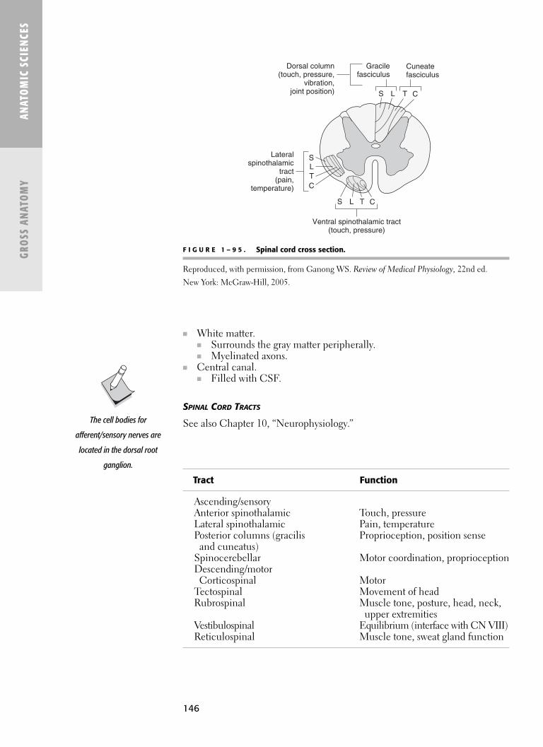
146
■ White matter.■ Surrounds the gray matter peripherally.■ Myelinated axons.
■ Central canal.■ Filled with CSF.
SPINAL CORD TRACTS
See also Chapter 10, “Neurophysiology.”
ANAT
OM
IC S
CIEN
CES
GROS
S AN
ATO
MY
F I G U R E 1 – 9 5 . Spinal cord cross section.
Reproduced, with permission, from Ganong WS. Review of Medical Physiology, 22nd ed.
New York: McGraw-Hill, 2005.
Tract Function
Ascending/sensoryAnterior spinothalamic Touch, pressureLateral spinothalamic Pain, temperaturePosterior columns (gracilis Proprioception, position sense
and cuneatus)Spinocerebellar Motor coordination, proprioceptionDescending/motor
Corticospinal MotorTectospinal Movement of headRubrospinal Muscle tone, posture, head, neck,
upper extremitiesVestibulospinal Equilibrium (interface with CN VIII)Reticulospinal Muscle tone, sweat gland function
The cell bodies for
afferent/sensory nerves are
located in the dorsal root
ganglion.
S
S
L
L
T
T
C
C
S L T C
Lateralspinothalamic
tract(pain,
temperature)
Ventral spinothalamic tract(touch, pressure)
Gracilefasciculus
Cuneatefasciculus
Dorsal column(touch, pressure,
vibration,joint position)

147
Peripheral Nervous System
■ Nerves outside the CNs.■ Consists of:
■ Cranial nerves (12): Leave the brain and pass out the skull foramina.■ Spinal nerves (31): Leave the spinal cord and pass out via the interver-
tebral foramina.■ 8 cervical spinal nerves (only 7 vertebrae).■ 12 thoracic.■ 5 lumbar.■ 5 sacral.■ 1 coccygeal.
■ Associated plexi and ganglia.■ Divided into
■ Afferent and efferent.■ Somatic and visceral.
■ Afferent (sensory)■ Somatic sensory (afferent).
■ From cutaneous and proprioceptive receptors.■ Visceral sensory (afferent).
■ From viscera.■ Efferent (motor)
■ Somatic motor (efferent). ■ Motor neurons to skeletal muscle.
■ Visceral motor (efferent) ■ Autonomic (parasympathetic > sympathetic) to viscera (eg, smooth
and cardiac muscle, glands, GI tract).
SPINAL NERVES
■ Comprise dorsal + ventral rami.■ Mixed nerves (motor + sensory).■ After they join as spinal nerve, they split into
■ Dorsal (posterior) rami.■ Ventral (anterior) rami.
■ Supplying both motor and sensory innervation to posterior and anteriorbody walls.
ANATOM
IC SCIENCES
GROSS ANATOM
Y
Spinal Nerve Root Location of Cell Body
Motor Ventral (anterior) Spinal cord (ventral horn-gray)Sensory Dorsal (posterior) Dorsal root ganglion (outside of cord)
PERIPHERAL NERVE
■ Bundle of nerve fibers.■ Formed from 1+ spinal nerve.■ Example: Musculocutaneous nerve
■ Formed from three spinal nerves (C5, C6, C7).
Remember histology:epinerium, perinerium,
endoneurium.
A peripheral nerve may be asubsegment of a cranial nerve;for example, the lingual nerveis part of the third division of
CN V (and it carries tastecomponents from CN VII).

148
ANAT
OM
IC S
CIEN
CES
GROS
S AN
ATO
MY
NERVE FIBER TYPES
Conduction Diameter Local AnestheticFiber Velocity (m/s) (lm) Function Myelin Sensitivity
A FiberA-α 70–120 12–20 Proprioception Y Least
MotorA-β 40–70 5–12 Sensory Y
Touch Pressure
A-γ 10–50 2–5 Muscle spindle YA-δ 6–30 2–5 Sharp pain Y
TemperatureTouch
B Fiber 3–15 <3 Preganglionic Yautonomic
C Fiber 0.5–2 0.4–1.2 Dull pain N MostTemperaturePostganglionic
autonomic
Data from Waxman SG. Clinical Neuroanatomy, 25th ed. New York: McGraw-Hill, 2003.
GANGLIA
■ Collections of cell bodies.■ Sensory versus autonomic.■ Sensory: Dorsal root ganglia.■ Autonomic: Sympathetic.
■ Sympathetic chain ganglia.■ Located on each sympathetic trunk, alongside vertebral column.■ 3 cervical (superior, middle, inferior).■ 12 thoracic.■ 4 lumbar.■ 4 sacral.
■ Parasympathetic■ Ciliary.■ Pterygopalatine.■ Submandibular.■ Otic.■ Celiac.■ Superior mesenteric.■ Inferior mesenteric.
PLEXUSES
■ Interdigitations of nerves (nerves joining neighboring nerves).■ Formed from anterior rami in cervical, brachial, lumbar, and sacral
regions.
Four major plexuses:
■ Cervical plexus (C1–C4).■ Brachial plexus (C5–T1).
Splanchnic nerves are
sympathetic nerves to the
viscera. They pass through the
sympathetic chain ganglia
without synapse (exceptions
to short preganglionic and
long postganglionic) and
synapse in the effector.

149
■ Lumbar plexus (L1–L4) .■ Formed in psoas muscle.■ Supplies lower abdomen and parts of lower limb.■ Main branches
■ Femoral nerve.■ Obturator nerve.
■ Sacral plexus (L4–L5 and S1–S4)■ Posterior pelvic wall in front of piriformis.■ Supplies lower back, pelvis, parts of thigh, leg, and foot.■ Main branches
■ Sciatic nerve (largest nerve in body).■ Gluteal nerve.■ Pelvic splanchnic nerve.
AUTONOMIC NERVOUS SYSTEM
See Figures 1–96 and 1–97 and Chapter 10, “Neurophysiology.”
■ Sympathetic■ Thoracolumbar■ “Fight or flight”
■ Parasympathetic■ Craniosacral.■ “Rest and digest.”■ All have afferent and efferent components.■ Efferent is the major portion.
■ General visceral efferent motor system (GVE)■ Involuntary.■ Controls and regulates smooth muscle, cardiac muscle, and glands.
ANATOM
IC SCIENCES
GROSS ANATOM
Y
F I G U R E 1 – 9 6 . Autonomic nervous system.
Reproduced, with permission, from Ganong WS. Review of Medical Physiology, 22nd ed. New York: McGraw-Hill, 2005.
From spinal cord,medulla, hypothalamus
Spinal nervesT1–T3
PostPre
To blood vessels,sweat glands
Sympathetic ganglion
Gray ramuscommunicans
WhiteRC
Collateral ganglionViscus
Cranial nerves–III, VII, IX, X
Pre
Brainstem
Cranial outflow
Sacral outflow
Post
Viscus
Pre
Post
Spinal nervesS2–S4
SYMPATHETIC DIVISION PARASYMPATHETIC DIVISION

150
ANAT
OM
IC S
CIEN
CES
GROS
S AN
ATO
MY
F I G U R E 1 – 9 7 . Sympathetic and parasympathetic divisions of the autonomic nervous system. Sympathetic preganglionicneurons are clustered in ganglia in the sympathetic chain alongside the spinal cord extending from the first thoracic spinalsegment to upper lumbar segments. Parasympathetic preganglionic neurons are located within the brain stem and in segmentsS2–S4 of the spinal cord. The major targets of autonomic control are shown here.
(Reproduced, with permission, from Kandel ER, Schwartz JH, Jessell TM. Principles of Neural Science, 4th ed. New York:
McGraw-Hill, 2000:964.)
Midbrain
Sympatheticdivision
Projections ofsympatheticdivision
Projections ofparasympatheticdivision
Parasympatheticdivision
Superior cervicalganglion
Ciliary ganglion
Otic ganglion
Esophagusand lungs
Stomach
Pancreas
Smallintestine
Large intestine,rectum
BladderPelvic nerveleading topelvic ganglia
Reproductiveorgans
Adrenal medulla
Bronchi
Heart
Eye
Lacrimal andsalivary glands
Pterygopalatine andsubmaxillary ganglion
III
VII
IX
X
Superiormesentericganglion
Sypatheticchain
Inferiormesentericganglion
Middle cervicalganglion
Inferior cervicalganglion
Celiac ganglion
PonsMedulla
Piloerectormuscle
Artery
Sweatglands
CervicalCranialnerves
Thoracic
Lumbar
Sacral
Liver
151
ANATOM
IC SCIENCES
GROSS ANATOM
Y
Pathway
Hypothalamus, reticular formation↓
Interomediolateral horn (cell body of preganglionic neuron)↓
Preganglionic neuron (short in sympathetic; long in parasympathetic)↓
Ganglion (outside of CNS)↓
Postganglionic neuron (long in sympathetic; short in parasympathetic)↓
Effector organ
Notes
■ Gray rami connect sympathetic trunk to every spinal nerve.■ White rami are limited to spinal cord segments between T1 and L2.■ Cell bodies of the visceral efferent preganglionic fibers (visceral branches
of sympathetic trunk) are located in the interomediolateral horn of thespinal cord.
■ Cell bodies of visceral afferent fibers are located in the dorsal root ganglia.
ENTERIC NERVOUS SYSTEM
See Chapter 15, “Gastrointestinal Physiology.”
Postganglionic autonomic fibers
are unmyelinated C-fibers.

This page intentionally left blank

C H A P T E R 2
General Histology
The Cell 156
PLASMA (CELL) MEMBRANE 156
CYTOPLASM 156
MEMBRANE-BOUND ORGANELLES 156
NON MEMBRANE-BOUND ORGANELLES 157
NUCLEUS 157
CELL SURFACE APPENDAGES 157
THE CELL CYCLE 158
CELL-TO-CELL CONTACTS 158
Epithelium 160
FUNCTIONS OF EPITHELIUM 160
CLASSIFICATION OF EPITHELIUM 160
Basement Membrane 161
FUNCTIONS OF BASEMENT MEMBRANE 161
COMPONENTS OF BASEMENT MEMBRANE 161
Connective Tissue 162
CLASSIFICATION OF CONNECTIVE TISSUE 162
CONNECTIVE TISSUE PROPER 162
CONNECTIVE TISSUE ATTACHMENTS 162
CELLS OF CONNECTIVE TISSUE 162
Glandular Tissue 163
TYPES OF GLANDS 163
CLASSIFICATION OF EXOCRINE GLANDS 163
STRUCTURE OF SALIVARY GLANDS 164
Cartilage 164
CARTILAGE MATRIX 164
SURFACE OF CARTILAGE 165
GROWTH OF CARTILAGE 165
Bone 165
FUNCTIONS OF BONE 166
BONE MATRIX 166
BONE FORMATION 166
153

154
BONE GROWTH 166
TYPES OF BONE 166
BONE SURFACES 167
BONE MARROW 167
BONE REMODELING 167
FRACTURE REPAIR 167
CALCIUM REGULATION 167
Joints 167
CLASSIFICATION OF JOINTS 167
TYPES OF SYNOVIAL JOINTS 168
SYNOVIAL JOINT COMPONENTS 168
Nervous Tissue 168
NEURONS 168
NEURON CLASSIFICATION 168
SYNAPSES 169
SUPPORTING CELLS 170
MYELINATION 170
Blood 171
FUNCTIONS 171
COMPONENTS 171
ERYTHROCYTES 172
LEUKOCYTES 172
HEMATOPOIESIS 172
Cardiovascular Tissue 173
ARRANGEMENT OF BLOOD VESSELS IN CIRCULATION 173
LAYERS OF BLOOD VESSEL WALLS 173
LAYERS OF THE HEART 174
CARDIAC CONDUCTION 174
Lymphatic System 175
LYMPH 175
FUNCTIONS OF THE LYMPHATIC SYSTEM 175
LYMPH DRAINAGE 175
Endocrine Glands 177
PITUITARY GLAND (HYPOPHYSIS) 177
THYROID GLAND 177
PARATHYROID GLANDS 177
PINEAL GLAND 177
ADRENAL (SUPRARENAL) GLANDS 179
Respiratory System 179
FUNCTIONS 179
DIVISIONS 180
ALVEOLI 180
Upper Digestive System 181
FUNCTIONS 181
LAYERS 181

155
ESOPHAGUS 181
STOMACH 181
SMALL INTESTINE 182
LARGE INTESTINE 183
GUT-ASSOCIATED LYMPHATIC TISSUE (GALT) 184
Lower Digestive System 184
LIVER 184
PORTAL TRIAD 184
LIVER LOBULES 184
HEPATOCYTES 184
HEPATIC SINUSOIDS 184
BILIARY TREE 185
BILE COMPOSITION 185
GALL BLADDER 186
PANCREAS 186
EXOCRINE PANCREAS 186
ENDOCRINE PANCREAS 186
Urinary System 187
COMPONENTS 187
FUNCTIONS OF KIDNEYS 187
COMPONENTS OF KIDNEYS 187
NEPHRON 187
URETHRA 189
Reproductive System 189
TESTIS 189
INTRA- AND EXTRA-TESTICULAR DUCT SYSTEM 189
PROSTATE GLAND 189
PENIS 190
OVARY 190
OVIDUCTS (FALLOPIAN TUBES) 190
UTERUS 190
VAGINA 190
MAMMARY GLANDS 190
Integument 190
FUNCTIONS OF SKIN 190
LAYERS OF SKIN 191
HAIR 192
SEBACEOUS GLANDS 192
Eye 192
LAYERS OF EYE 192
TEN LAYERS OF RETINA 194
OPTIC DISC 194
MACULA LUTEA 194

156
ANAT
OMIC
SCI
ENCE
SGE
NERA
L HI
STOL
OGY
156
� THE CELL
Plasma (Cell) Membrane
■ See also the section “Membranes” in Chapter 9.■ A fl uid, selectively permeable barrier consisting of an amphipathic phos-
pholipid bilayer that contains integral and peripheral proteins.
Cytoplasm
■ Site of cell-synthesizing activity. Contains the following:■ Organelles (See Figure 2–1)■ Cytoplasmic inclusions (metaplasm)
■ Glycogen■ Pigment granules: lipofuscin, melanin, etc■ Secretory granules■ Lipid droplets
■ Cytoplasmic matrix (cytosol)■ Ground substance
Membrane-Bound Organelles
■ Rough endoplasmic reticulum (rER): Protein synthesis for export outside of the cell. Studded with ribosomes.
F I G U R E 2 – 1 . Major cellular organelles.
Reproduced, with permission, from Ganong WS. Review of Medical Physiology, 22nd ed. New York: McGraw- Hill, 2005.
Secretory granules
Centrioles
Smoothendoplasmicreticulum
Golgiapparatus
Lipiddroplets
Roughendoplasmicreticulum
Lysosomes
Mitochondrion
Globular headsNucleolus
Nuclear envelope

157
ANATOMIC SCIENCES
GENERAL HISTOLOGY
157
■ Smooth endoplasmic reticulum (sER): Steroid synthesis (adrenal cortex and testes). Sequesters Ca2+ (skeletal and cardiac muscle). Lipid and glycogen metabolism (liver).
■ Golgi apparatus: Posttranslational modifi cation and packaging of proteins (adds oligosaccharides for glycoproteins, and sulfate groups for proteoglycans).Lysosome production.
■ Mitochondria: ATP production via the Krebs cycle and oxidative phospho-rylation. Have an inner and outer membrane. Contain their own cyclic DNA. Not present in erythrocytes.
■ Lysosomes: Digestion of microorganisms or other cellular components by various hydrolytic enzymes. Produced by the Golgi apparatus.
■ Peroxisomes: Elimination of H2O2 by various oxidative enzymes (catalase and peroxidase).
■ Endosomes: Vesicles formed as a result of phagocytosis.
Non Membrane-Bound Organelles
■ Microtubules: Provide cytoskeletal support, intracellular transport, and cellular movement. Composed of tubulin.■ Axoneme: Specialized group of microtubules found in cilia and fl agella
arranged in a “9 + 2” pattern.■ Centrioles: Provide microtubule organization. Form ends of mitotic spindle
during mitosis.■ Filaments
■ Microfi laments (actin, myosin, etc): Important for muscle contraction. Provide cellular movement or anchorage.
■ Intermediate fi laments (vimentin, desmin, cytokeratin, etc): Provide cytoskeletal support.
■ Basal bodies: Required for development of cilia. Derived from centrioles.■ Ribosomes: Protein synthesis for use within the cell. Composed of rRNA
and protein.
Nucleus
■ Nuclear membrane: Composed of inner and outer plasma membranes.■ Nucleoplasm: Ground substance of the nucleus.■ Chromatin: The complex of DNA and proteins (histones).
■ Euchromatin: Loosely arranged chromatin. Indicates nuclear activity.■ Heterochromatin: Highly condensed chromatin.
■ Nucleolus: Site of rRNA synthesis. Nonmembranous.■ Barr body: The repressed X chromosome found only in cells of females.
Appears as a dense chromatin mass adjacent to the nuclear membrane. Its presence (or absence) is used in sex identifi cation.
Cell Surface Appendages
■ Microvilli: Fingerlike structures of various lengths located on the apical surface of most epithelial cells. Core composed of microfi laments. Pro-vide increased cell surface area for absorption and transport of fl uids.
■ Stereocilia: Unusually long microvilli located only in the epididymis and sensory cells of the inner ear.
■ Cilia: Short, hairlike structures used for locomotion or movement of sub-stances along the cell membrane. Function in a synchronous, coordinated wave motion. Require basal bodies for development.
■ Flagella: Long, whiplike structures used for locomotion. Function in an undulating, snake-like motion. In humans, found only in spermatozoa.
“9 + 2” pattern: 9 pairs
(doublets) of microtubules
surrounding 2 central
microtubules.
Microtubules are found in
axonemes (of cilia and
fl agella), basal bodies,
centrioles, mitotic spindle,
elongating cell processes, and
cytoplasm.
The sex of an embryo (via
presence/absence of Barr
bodies) can be determined by
the 8th week.
Active cells (fi broblasts,
osteoblasts, etc) are
characterized by an
abundance of rER.

158
ANAT
OMIC
SCI
ENCE
SGE
NERA
L HI
STOL
OGY
158
The Cell Cycle
See Figure 2–2.
■ Mitosis: Produces two daughter cells with the same chromosome number (diploid, 2n) as the parent cell. All somatic cells except gametes undergo mitosis.
■ Interphase■ G1: First variable period of cellular growth.
■ G0: Period outside of the cell cycle of terminal differentiation.■ S: Period of DNA synthesis. Takes about 7 hours.■ G2: Second variable period of cellular growth.
■ Mitosis (karyokinesis)■ Prophase: Chromatin coils/condenses within the nucleus. Mitotic
spindle forms.■ Metaphase: Nuclear membrane and nucleoli disappear. Chromosomes
line up at the equatorial plate of the mitotic spindle.■ Anaphase: Chromosomes split to opposite poles of the cell.■ Telophase: Nuclear membrane forms around the chromosomes at
each pole. The chromosomes uncoil and nucleoli reappear. The cyto-plasm divides to form two daughter cells (cytokinesis).
■ Meiosis: Produces four daughter cells with half the chromosome num-ber (haploid, n) as the parent cell. Only gametes (spermatozoa and ova) undergo meiosis.
Cell-to-Cell Contacts
See Table 2–1.
■ Tight junction (zonula occludens): A beltlike junction that completely seals off the intercellular space between adjacent cells.
■ Intermediate junction (zonula adherens): A beltlike junction that leaves a 15–20 nm wide intercellular space between adjacent cells.
■ Desmosome (macula adherens): Provides strong but localized adhesion sites between adjacent cells. Composed of an attachment plaque on the cytoplasmic side of each adjoining cell surface, to which intermediate fi la-ments are anchored.
Cell turnover rate
(high → low):
Oral, epidermal, GI epithelial
↓
Smooth muscle, vascular
endothelial
↓
Skeletal muscle, cardiac
muscle
↓
Neurons
F I G U R E 2 – 2 . The cell cycle.
A junctional complex (See Figure 2–3) is a specialized junctioncommonly found in epithelial tissuesconsisting of the following (from apical → upper regions):
■ Tight junction■ Intermediate junction■ Desmosome
S
G1
G0G2
M
Protein and RNA syntheses
occur in all cell cycle phases
except mitosis.

159
ANATOMIC SCIENCES
GENERAL HISTOLOGY
159
F I G U R E 2 – 3 . Epithelial intercellular junctions. Note the relative location of each junction within the cell.
Reproduced, with permission, from Ganong WS. Review of Medical Physiology, 22nd ed. New York: McGraw- Hill, 2005.
T A B L E 2 – 1 . Comparison of Cell-to-Cell Contacts
CONTACT TYPE FUNCTION AREA OF CONTACT PRINCIPAL PROTEINS COMMON LOCATION
Tight junction (zonula
occludens)
Occlusion Cell-cell (band) Occludins, Claudins Various epithelium
Intermediate junction
(zonula adherens)
Adhesion Cell-cell (band) Actin, Cadherins Various epithelium
Desmosome (macula
adherens)
Adhesion Cell-cell (focal) Cadherins Epidermis and other epithelium
Hemidesmosome Adhesion Cell-ECM (focal) Integrins Epithelium of oral mucosa (JE),
skin, esophagus, vagina, and
cornea
Gap junction Communication Cell-cell (focal) Connexins Neurons, smooth and cardiac
muscle
Tightjunction(zonulaoccludens)
Zonulaadherens
Desmosomes
Gapjunctions
Hemidesmosome
■ Hemidesmosome: Provides strong but localized attachment of epithelial cells to connective tissue. Half of a desmosome (the attachment plaque is on the basal surface, facing the basement membrane).
■ Gap junction: Localized areas of free communication between adjacent cells. Enables passage of fl uids, ions, and small molecules.
Pemphigus: Autoimmunity
against desmosomal
attachments.
Pemphigoid: Autoimmunity
against hemidesmosomal
attachments.

160
ANAT
OMIC
SCI
ENCE
SGE
NERA
L HI
STOL
OGY
160
� EPITHELIUM
■ Epithelium covers all body surfaces, lines all body cavities, makes up the secretory component of glands, and serves as the receptor in various sensory organs.
Functions of Epithelium
■ Barrier■ Diffusion■ Absorption■ Secretory■ Transport■ Sensory
Classifi cation of Epithelium
■ By number of cell layers.■ Simple: One cell layer thick, all touching the basement membrane.■ Stratifi ed: Two or more cell layers thick, with only the deepest layer
touching the basement membrane.■ Pesudostratifi ed: One cell layer thick, all touching the basement
membrane, but not all reach the outer surface. Generally columnar.
The thinnest epithelium in the
oral cavity is the sublingual
mucosa.
Epithelium is classifi ed by cell
morphology and
arrangement, not by function.
See Table 2–2.
T A B L E 2 – 2 . Comparison of Epithelial Types
EPITHELIUM TYPE FUNCTION COMMON LOCATION
Simple squamous Barrier, diffusion Endothelium, mesothelium, lung
alveoli, Bowman’s capsule
Simple cuboidal Barrier, secretion, Small exocrine gland ducts
apsorption (thyroid follicles), renal tubules, OEE
Simple columnar Absorption, secretion, Stomach, small intestine, large
(often ciliated) transport intestine, gallbladder, IEE
Pseudostratifi ed columnar Absorption, secretion, Trachea, bronchi, nasopharynx,
(often ciliated) transport nasal cavity, paranasal sinuses
Stratifi ed squamous Barrier Epidermis, oral cavity, gingiva,
oropharynx, laryngopharynx,
esophagus, anus, vagina
Stratifi ed cuboidal Barrier, secretion Medium exocrine gland ducts
(sweat glands)
Stratifi ed columnar Barrier, transport Large exocrine gland ducts
(often ciliated) (salivary glands)
Transitional Barrier, distension Urinary bladder, urethra, ureters

161
ANATOMIC SCIENCES
GENERAL HISTOLOGY
161
■ By cell morphology.■ Squamous: Width > height.■ Cuboidal: Width = height.■ Columnar: Height > width.■ Transitional: Ranges from squamous to cuboidal. Distensible.
■ By location (almost always simple squamous).■ Endothelium: Lines all blood vessels.■ Mesothelium: Lines all closed body cavities.
� BASEMENT MEMBRANE
■ Connects the epithelial basal layer to underlying connective tissue.
Functions of Basement Membrane
■ Attachment■ Separation■ Filtration■ Scaffolding
Components of Basement Membrane
From epithelium → connective tissue (Figure 2–4)
■ Lamina lucida: Electron-clear layer■ Lamina densa (basal lamina): Product of epithelium
■ Type IV collagen■ Proteoglycans■ Laminin■ Fibronectin■ Anchoring fi brils (type VII collagen)
■ Reticular lamina: Product of CT■ Reticular fi bers (type III collagen)
Hemidesm-osomes Laminin
Basal lamina (type IV collagen)
Reticularfibrils (type III collagen)
Anchoringfibrils (type VIIcollagen)
Epithelial cell
F I G U R E 2 – 4 . Components providing attachment between epithelial cells and the underly-ing connective tissue.
Reproduced, with permission, from Ross MH, Romrell LJ, Kaye GI. Histology: A Text and Atlas. 3rd ed. Philadelphia: Williams & Wilkins; 1995.

162
ANAT
OMIC
SCI
ENCE
SGE
NERA
L HI
STOL
OGY
162
� CONNECTIVE TISSUE
Classifi cation of Connective Tissue
■ Connective tissue proper■ Loose CT■ Dense CT
■ Regular■ Irregular
■ Specialized CT■ Adipose tissue■ Blood■ Bone■ Cartilage■ Hematopoietic tissue■ Lymphatic tissue
■ Embryonic CT■ Mesenchyme■ Mucous CT
Connective Tissue Proper
■ Loose CT: Abundant ground substance with sparse fi bers and cells. Located under epithelial layers that cover body surfaces and line body cavities.
■ Dense CT: Greater fi ber concentration, which provides structural support.■ Irregular: Irregular arrangement of fi bers and cells. Makes up the
majority of dense CT. Found in dermis, submucosa of the GI tract, and fi brous capsules.
■ Regular: Ordered arrangement of fi bers and cells. Found in tendons,ligaments, and aponeuroses.
Connective Tissue Attachments
■ Ligament: Connects bone to bone■ Tendon: Connects muscle to bone■ Aponeurosis: A sheetlike tendon■ Sharpey’s fi ber: The portion of a ligament or tendon that inserts into bone
(or cementum)
Cells of Connective Tissue
■ Resident cell population■ Fibroblasts: Most common■ Myofi broblasts■ Adipocytes■ Macrophages (histiocytes)■ Mast cells■ Mesenchymal cells
■ Transient cell population■ Lymphocytes■ Neutrophils■ Monocytes■ Plasma cells■ Eosinophils■ Basophils
Almost all connective tissues
(CT) originate from
mesoderm; however, some
CTs of the head and neck
region derive from neural
crest ectoderm.
Consistency of CT:
■ Soft: Deeper layers of
skin, oral mucosa
■ Firm: Cartilage
■ Rigid: Bone
■ Fluid: Blood

163
ANATOMIC SCIENCES
GENERAL HISTOLOGY
163
Classifi cation of Exocrine Glands
See Figure 2–5.
Classifi cation Type Defi nition Examples
Cellularity Unicellular Single secretory cells Goblet cells Multicellular Multiple secretory cells Gastric pits
Secretion Merocrine Secretory product released Major salivary glands, pancreatic mechanism from secretory granules acinar cells Apocrine Secretory product is released Mammary glands, apocrine with cytoplasm sweat glands Holocrine Secretory product is released Sebaceous glands with portion of cell
Duct structure Simple Unbranched Sweat glands Compound Branched Major salivary glands, pancreas
Secretory unit Tubular Secretory portion shaped Intestinal glands like a tube Coiled Secretory portion shaped Eccrine sweat glands like a coiled tube Acinar Secretory portion shaped Sebaceous glands, mammary like a saclike dilation glands, gastric cardiac glands, pancreas Tubuloacinar Combination of tubular Major salivary glands and acinar
Secretion type Mucous Viscous secretion Sublingual salivary glands, goblet cells Serous Watery secretion Parotid salivary glands, von Ebner’s salivary glands, paneth cells, gastric chief cells Mixed Combination of mucous Submandibular salivary glands and serous
Types of Glands
Gland Type Function Examples Secretions
Exocrine Secrete products through Sweat, salivary, sebaceous, Sweat, saliva, sebum,ducts von Ebner’s glands digestive enzymes
Endocrine Secrete products into the Pituitary, thyroid, parathyroid, Various hormonesbloodstream (no ducts) adrenal, gonads, pineal glands
Paracrine Secrete products into Gastroenteropancreatic Various peptidesexracellular spaces that glands
affect other cells within the same epithelium
� GLANDULAR TISSUE
Types of Glands
■ There are three major types of glands.

164
ANAT
OMIC
SCI
ENCE
SGE
NERA
L HI
STOL
OGY
164
Structure of Salivary Glands
■ Comprised of lobes subdivided by CT septa.■ Lobe → lobules → terminal secretory units (acini or tubules).■ The structure is variable depending on the type of gland:
■ Terminal secretory unit: Contains cells that secrete the glycoprotein and watery elements of saliva. Lined by simple cuboidal epithelium.■ Serous cells: Secretory■ Mucous cells: Secretory■ Myoepithelial (basket) cells: Contractile
■ Intercalated duct: Transports saliva to larger ducts. Lined by simple cuboidal epithelium.
■ Striated duct: Modifi es salivary electrolytes (reabsorption of Na+/Cl−;secretion of K+/HCO3
−). Numerous elongated mitochondria give the “striated” appearance. Lined by simple low columnar epithelium.
■ Terminal excretory duct: Transports saliva to oral cavity. Lined by pseudostratifi ed columnar epithelium.
� CARTILAGE
■ An avascular connective tissue. Blood supply comes from perichondrium.■ Composed of chondrocytes, which reside in lacunae, surrounded by their
own specialized extracellular matrix (ECM). ■ Chondroblasts: The initial cartilagenic cells (prior to extensive matrix
formation).
Cartilage Matrix
■ Type II collagen■ Ground substance: Extremely hydrophilic
■ Glycosaminoglycans (GAGs)■ Hyaluronic acid
Serous glands (parotid) have
well-developed
intercalated and striated
ducts that modify their serous
secretions. Contrarily, mucous
glands (sublingual) have
poorly developed intercalated
and striated ducts.
Hyaline cartilage is the
precursor to bones that
develop via endochondral
ossifi cation.
Cartilage does not contain
calcium salts, so it does not
appear on radiographs.
F I G U R E 2 – 5 . Classifi cation of exocrine glands.
Reproduced, with permission, from Ross MH. Histology, 3rd ed. Lippincott Williams & Wilkins, 1995. (Based on Weiss L. Histology, 4th ed. McGraw- Hill, 1977.)
Simpletubular
Compoundtubular
Compoundtubuloacinar
Compoundacinar
Simple coiledtubular
Simplebranched tubular
Simplebranched acinar

165
ANATOMIC SCIENCES
GENERAL HISTOLOGY
165
■ Chondroitin sulfate■ Keratin sulfate
■ Proteoglycans
Surface of Cartilage
■ Perichondrium■ Inner cellular layer: Produces chondroblasts■ Outer fi brous layer: Provides protection
Growth of Cartilage
■ Appositional growth: New cartilage forms on the surface of existing cartilage. Must have perichondrium: fi broblastic cells from the inner perichondrium differentiate into chondroblasts, which secrete matrix.
■ Interstitial growth: New cartilage forms within existing cartilage. Chondro-cytes divide within their lacunae, enabling new matrix to be deposited.
Perichondrium covers all
cartilage except:
■ Fibrocartilage
■ Articular cartilage of joints
■ Nasal/costal cartilage
Types of Cartilage
CartilageECMComposition Perichondrium Function
Ability to Calcify Locations
Hyaline Closely packed, thin collagen fi bers
Some Provides pliability and resiliencePrecursor to endochondral bone formation
Yes Nose, trachea, bronchi, larynx, ribs (costal cartilage), articular surfaces of long bones
Elastic Collagen and elastic fi bers
Yes Provides elastic properties
No External ear, Eustachian tube, epiglottis
Fibrocartilage Dense collagen fi bers
No Withstands compression and tension
No Intervertebral disc, articular disc of TMJ, symphysis pubis, meniscus of knee
� BONE
■ A yellowish, mineralized, vascular tissue of varying degrees of density. ■ Osteoblasts: Produce osteoid (uncalcifi ed bone matrix), which is composed
of type I collagen and ground substance. Mature bone forms when the osteoid calcifi es.
■ During the clacifi cation process, osteoblasts become trapped in spaces within the mineralized matrix called lacunae. Here they differentiate into osteocytes, which are responsible for maintaining the bone matrix. They maintain nourishment via vascular tunnels within bone called canaliculi.
Hydroxyapatite
[Ca10(PO4 )6(OH)3 ] is the
predominant mineral found
in bone.

166
ANAT
OMIC
SCI
ENCE
SGE
NERA
L HI
STOL
OGY
166
Functions of Bone
■ Support■ Protection■ Movement■ Mineral storage: calcium, phosphorous■ Hematopoiesis: bone marrow
Bone Matrix
■ Organic■ Type I collagen■ Osteocalcin: Marker of bone formation■ Osteonectin: Binds Ca2+ and collagen■ Ground substance: GAGs, etc
■ Inorganic■ Hydroxyapatite
Bone Formation
■ Intramembranous ossifi cation: Mesenchymal cells differentiate into osteo- blasts, which secrete bone matrix within an established locus of loosely arranged collagen. This new matrix calcifi es, forming immature woven bone.Over time, osteoclastic resorption of woven bone occurs, and new osteoblastic matrix is deposited in a more tightly arranged manner. This matrix is then calcifi ed, forming mature bone of a higher strength. ■ Flat bones of skull, maxilla, mandibular body, clavicle.
■ Endochondral ossifi cation: A subperiosteal bony cuff forms around an already established locus of hyaline cartilage, which grows larger and sub-sequently causes the hypertrophy and death of the chondrocytes. The carti-lage matrix then becomes calcifi ed. Over time, osteoclastic resorption of the calcifi ed cartilage occurs, and new osteoblastic matrix is deposited, forming mature bone. ■ Long bones, vertebrae, mandibular condyles.
Bone Growth
■ Appositional growth: Occurs in both endochondral and intramembranous bone at any time.
■ Interstitial growth: Occurs in endochondral bone only until epiphyseal plates close.
Types of Bone
■ Cortical (compact): Composed of Haversian systems (osteons), which are composed of concentric bone matrix lamellae surrounding a central Hav-ersian canal that contains neurovascular bundles. Canaliculi connect to the central canal, providing nourishment to osteocytes. Osteons are con-nected to each other by Volkmann’s canals. The space between osteons is composed of previous osteonal lamellae, called interstitial lamellae.
■ Cancellous (spongy): Similar to cortical bone in that it is lamellar, but their confi guration and arrangement is less dense. Lamellae are arranged in thin spicules called trabeculae. If they are thick enough, they contain osteons. In between the trabeculae are marrow spaces of various sizes. Trabeculae follow lines of stress.
Initiation of bone
mineralization:
1. ↑ pores in collagen fi bers
2. Osteoblasts secrete matrix
vesicles
3. ↑ alkaline phosphatase
in osteoblasts and matrix
vesicles
4. Degradation of matrix
pyrophosphate, releasing
PO43-
Endochondral centers of
ossifi cation:
Primary: Diaphysis
Secondary: Epiphysis

167
ANATOMIC SCIENCES
GENERAL HISTOLOGY
167
Bone Surfaces
■ Periosteum: A fi brous connective tissue capsule that surrounds the outer surface of bone. Contains collagen, fi broblasts, and osteoprogenitor cells.
■ Endosteum: A one-cell thick layer of mostly osteoprogenitor cells that linesthe inner surface bone. Contains bone marrow.
Bone Marrow
■ Yellow marrow: Contains fat cells. The predominant marrow type in the maxilla and mandible.
■ Red marrow: Contains hematopoietic cells. Found in the mandibular ramus and condyles.
Bone Remodeling
■ Bone constantly remodels, a process involving both osteoclasts (resorption) and osteoblasts (matrix deposition). Osteoclasts are multinucleated giant cells that reside in resorption bays known as Howship’s lacunae. They pro-duce a large number of hydrolytic enzymes from their characteristic ruffl ed border. The protons lower the pH at the site of resorption, subsequently dissolv-ing the calcifi ed bone matrix. Collagenases and other proteases then digest the decalcifi ed bone matrix.
Fracture Repair
1. Blood clot formation2. Bridging callus formation3. Periosteal callus formation4. New endochondral bone formation
Calcium Regulation
■ Parathyroid hormone (PTH): Stimulates osteoclastic bone resorption(blood calcium). Secreted by the parathyroid gland.
■ Calcitonin: Inhibits osteoclastic bone resorption (↓ blood calcium). Secreted by the parafollicular cells of the thyroid gland.
� JOINTS
Classifi cation of Joints
■ By motion■ Synarthrosis: Immovable (cranial sutures)■ Amphiarthrosis: Slightly movable (symphysis pubis)■ Diarthrosis: Fully moveable (shoulder, hip, TMJ)
■ By connective tissue type■ Fibrous: Joined by fi brous CT
■ Suture (cranial sutures)■ Syndesmosis (radius-ulna, tibia-fi bula)■ Gomphosis (tooth socket)
■ Cartilaginous: Joined by cartilage■ Synchondrosis (epiphyseal plates of long bones)■ Symphysis (symphysis pubis)
■ Synovial: Freely movable. Lined by a synovial membrane■ Majority of joints (shoulder, hip, TMJ)
Bone marrow is contained
within the medullary spaces
of spongy bone.
Mature bone grows only
by appositional growth.
The TMJ is a synovial, diar-
throsis joint.
The alveolar socket is a
synarthrosis,
gomphosis joint.

168
ANAT
OMIC
SCI
ENCE
SGE
NERA
L HI
STOL
OGY
168
Types of Synovial Joints
■ Ball-and-socket: Hip, shoulder■ Gliding: Carpal bones■ Hinge: Elbow, knee■ Pivot: Atlas-axis■ Ellipsoidal: Radius-ulna■ Saddle: Metacarpal bones
Synovial Joint Components
■ Articular capsule (bursa): Composed of fi brous connective tissue. Sur-rounds the joint. Lined by synovial membrane.
■ Articular cartilage (meniscus): Layer of hyaline cartilage that covers the articular bone surfaces (except the TMJ and knee, which is composed of fi brocartilage).
■ Synovial cavity: Lies within the articular capsule and contains synovial fl uid.
■ Synovial membrane: Lines the articular capsule. Produces synovial fl uid.■ Synovial fl uid: Lubricates the articular cartilage.
� NERVOUS TISSUE
The hipbone (os coxa) is
formed by the fusion of the
ilium, ischium, and pubis.
The right and left hipbones
articulate anteriorly at the
symphysis pubis.
Neurons
■ Nerve cells that conduct electrical impulses (Figure 2–6).■ Perikaryon (cell body): Contains plasma membrane, cytoplasm,
nucleus, and Nissl bodies (rER).■ Axon: Conducts information away from cell body. Only one axon exists
per cell body.■ Dendrites: Conducts information toward the cell body. The number of
dendrites per cell is variable (none to many), depending on the type of neuron.
■ Cytoskeleton■ Actin■ Microtubules (dendrites)■ Neurofi laments (axons)
Neuron Classifi cation
■ By function■ Motor (efferent)■ Sensory (afferent)■ Mixed (both)
Neurons do not divide.
Two Major Divisions of Nervous Tissue
Division Origin Location Cell Body Clusters
CNS Neural tube Brain and spinal cord Nuclei ectoderm
PNS Neural crest Cranial and spinal Ganglia ectoderm nerves, ganglia, and nerve endings

169
ANATOMIC SCIENCES
GENERAL HISTOLOGY
169
Dendrites
Axon
Oligodendrocyte
Node of Ranvier
Motor end plate
Myelin
Myelin
Axon hillock
Nissl bodies
Perikaryon
Synapse
Dendrites
Recurrentaxonal branch
Schwann cell
Central nervous system
Peripheral nervous system
F I G U R E 2 – 6 . Schematic diagram of a motor neuron.
Reproduced, with permission, from Junqueira LC, Carneiro J. Basic Histology: Text & Atlas.11th ed. New York: McGraw-Hill; 2005.
■ By number of processes■ Unipolar: One process (axon only). Sensory neurons.■ Bipolar: Two processes (one axon and one dendrite). Retina and ganglia
of CN VIII.■ Multipolar: Three or more processes (one axon and many dendrites).
Motor and mixed neurons.
Synapses
■ Junctions that transmit impulses from one neuron to another, or from one neuron to an effector cell.
Interneurons form a
communicating network
between sensory and
motor neurons.

170
ANAT
OMIC
SCI
ENCE
SGE
NERA
L HI
STOL
OGY
170
■ Two types of synapses:■ Electrical: Synaptic cleft is a gap junction. Located in smooth and car-
diac muscle.■ Chemical: Synaptic cleft is a 20–30 nm intercellular space.
Supporting Cells
See Table 2–3.
■ Nonconducting cells that insulate neurons.
Myelination
See Table 2–4.
T A B L E 2 – 3 . Supporting Cells of Nervous Tissue
CELL TYPE FUNCTION
CNS (Glial Cells)
Astrocytes Scaffolding of BBB
■ Protoplasmic: Gray matter Regulation of metabolites
■ Fibrous: White matter
Oligodendrocytes Myelination
Microglia Phagocytosis
Ependymal cells Epithelium of ventricles of brain, spinal cord
Choroidal cells Secretion of CSF
PNS
Schwann cells Myelination
Surround nerve processes
Satellite cells Support
Surround nerve ganglia
Astrocytes:
■ Building blocks of BBB
■ May regulate vasocon-
striction and vasodilation
■ Communicate via gap
junctions using calcium
T A B L E 2 – 4 . Myelin-Producing Cells of the Nervous System
CELL TYPE DIVISION CELLS/NEURON MYELIN SHEATH
Oligodendrocyte CNS Multiple (as many as 50) Composed of several
internodal tongue-like
processes that wrap
around the axon
Schwann cell PNS 1 Composed of multiple
layers concentrically
wrapped around the axon

171
ANATOMIC SCIENCES
GENERAL HISTOLOGY
171
■ Myelin sheath: Lipid-rich layer that surrounds and insulates myelinated axons.
■ Neurilemma (sheath of Schwann): Layer of myelin cell cytoplasm that is contiguous with the myelin sheath.
■ Node of Ranvier: Unmyelinated junction between two myelin cells.■ Myelin formation begins before birth and during 1st year postnatally (see
Babinski refl ex).
� BLOOD
Functions
■ Transportation■ Buffering■ Thermoregulation
Components
See Figure 2–7.
F I G U R E 2 – 7 . Components of blood.
Blood(8% of total body
weight)
Plasma(55% of blood)Liquid portion
Formed elements(45% of blood)Cellular portion
Erythrocytes
Leukocytes
Platelets
Water(80% of plasma)
Protein(8% of plasma)
ElectrolytesBlood gasesHormonesEnzymes
Metabolic products
Albumin (55%)Regulate vascularosmotic pressure
Fibrinogen andclotting factors (7%)
Hemostasis
Globulins (38%)Immunoglobulins
Not all Schwann cells
produce myelin:
A fi bers: Myelinated − have
neurilemma and myelin.
C fi bers: Unmyelinated − only
have neurilemma.
Serum is blood plasma minus
fi brinogen and clotting factors.
Hematocrit is the percentage
of erythrocytes in a blood
sample. In males, the average
hematocrit is 45%; in females
it is 40%.

172
ANAT
OMIC
SCI
ENCE
SGE
NERA
L HI
STOL
OGY
172
Erythrocytes
■ Red blood cells. ■ Biconcave discs (7–10 μm in diameter).■ Contain hemoglobin. ■ No nucleus or mitochondria.■ Function: Transports O2 (and CO2).■ Lipid membrane contains lipoproteins and blood group surface markers (A,
B, O).■ Amount of bile pigment excreted by liver estimates the amount of erythro-
cyte destruction per day.
Leukocytes
See Table 2–5.
■ White blood cells. ■ Granulocytes: Contain cytoplasmic granules.
■ Neutrophils (60%)■ Eosinophils (5%)■ Basophils (1%)
■ Agranulocytes: Contain few cytoplasmic granules.■ Lymphocytes (30%)■ Monocytes (4%)
Hematopoiesis
See Figure 2–8.
■ Blood cell formation.■ All blood cells derive from one pluripotential stem cell.■ Hormones (erythropoietin, thrombopoietin, etc) and colony stimulating
factors (CSFs) regulate hematopoiesis.■ Occurs primarily in bone marrow, although some lymphocytes also develop
in lymphatic tissues.
Monocytes transform into
macrophages (histiocytes)
once they leave the blood-
stream and enter the sur-
rounding connective tissue.
Formed Elements
Comparison of Major Blood-Formed Elements
Formed Normal AmountElement Function Characteristics Average Lifespan Per mm3 Blood
Erythrocytes Transports O2 (99%) Biconcave discs 120 days 4–5 million Transports CO2 (30%) No nucleus No mitochondria Elastic
Platelets Hemostasis Cytoplasmic fragments 5–10 days 200,000–400,000 of megakaryocytes No nucleus
Leukocytes Immunoregulation Nucleated, but Variable 6,000–10,000 Infl ammation morphology is Phagocytosis variable Hypersensitivity

173
ANATOMIC SCIENCES
GENERAL HISTOLOGY
173
� CARDIOVASCULAR TISSUE
Arrangement of Blood Vessels in Circulation
Aorta → Large arteries → Medium arteries → Arterioles
Heart Capillaries
Vena cava ← Large veins ← Medium veins ← Venules
Layers of Blood Vessel Walls
From lumen → outermost layer
■ Tunica intima: Contains simple squamous epithelium (endothelium)resting on a basement membrane and subendothelial connective tissue.
The greatest drop in blood
pressure occurs at the
transition from arteries to
arterioles.
T A B L E 2 – 5 . Comparison of Leukocytes
NUCLEAR CYTOPLASMIC
LEUKOCYTE MORPHOLOGY GRANULES FUNCTION
Neutrophil 3–5 lobes Specifi c granules Phagocytosis
(lysozyme, etc) Acute infl ammation
Azurophilic granules
(peroxidases, etc)
Eosinophil 2 lobes Peroxidase Phagocytosis
Histaminase (parasites)
Arylsulfatase Chronic infl ammation
Basophil 2–3 lobes, Histamine Hypersensitivity
obscured by Seratonin (bind IgE)
dense granules Heparin sulfate
Lymphocyte Large, round Few azurophilic Immunoregulation
granules Chronic infl ammation
Monocyte U-shaped Few azurophilic Phagocytosis
granules Chronic infl ammation
F I G U R E 2 – 8 . Hematopoiesis.
Pluripotential stem cell
Lymphoid stem cell Myeloid stem cell
T cells B cells Granulocyte
Neutrophil Basophil Eosinophil Macrophage Erythrocyte Platelets
Monocyte Erythroblast Megakaryoblast
Larger arteries: More elastic
fi bers.
Smaller arteries: More smooth
muscle fi bers.

174
ANAT
OMIC
SCI
ENCE
SGE
NERA
L HI
STOL
OGY
174
Major Histologic Differences Among Blood Vessel Types
Blood Vessel Distinguishing Characteristics
Elastic arteries (aorta, pulmonary Thickest tunica media arteries, and their main branches) Elastic fi bers > smooth muscle Contain vasa vasora
Muscular arteries Thick tunica media Smooth muscle > elastic fi bers Some contain vasa vasora
Arterioles Thick tunica media Smooth muscle > elastic fi bers Directly affect arterial blood pressure
Capillaries Endothelial layer only One erythrocyte cell wide Slowest velocity of blood fl ow Enable gas and metabolite exchange via diffusion
Venules Thick tunica adventitia
Veins Thickest tunica adventitia Some contain valves Some contain vasa vasora
Sinusoids are fenestrated or
discontinuous capillaries in
the liver, spleen, and endo-
crine glands. They are larger
and more irregularly shaped
than capillaries to accommo-
date phagocytic cells of the
reticuloendothelial system.
Cardiac Conduction
■ Cardiac muscle maintains its own rhythmicity.■ The sinoatrial (SA) node is the “pacemaker” of the heart.■ Autonomic nerves only regulate the rate of cardiac impulses.■ SA node → AV node → bundle of His → purkinje fi bers.■ In sinus rhythm, every P wave is followed by QRS complex.
Present in all blood vessels and heart wall. Only arteries have an additional elastic layer here called the internal elastic membrane.
■ Tunica media: Contains smooth muscle and elastic fi bers. Much thicker in arteries.
■ Tunica adventitia: Connective tissue layer containing collagen and elasticfi bers. Much thicker in veins. Large vessels contain vasa vasora, which sup-ply the vessels themselves.
Layers of the Heart
From lumen → outermost layer
■ Endocardium: Same as tunica intima. Contains a simple squamous endothelium.
■ Myocardium: Thickest portion of the heart. Contains cardiac muscle.■ Pericardium: Outer layer of connective tissue and adipose tissue surrounded
by a simple squamous epithelium.
Tachycardia: >100 bpm
Bradycardia: <60 bpm

175
ANATOMIC SCIENCES
GENERAL HISTOLOGY
175
� LYMPHATIC SYSTEM
Lymph
■ A yellowish, plasmalike liquid that contains mostly lymphocytes.■ Once absorbed in various tissues throughout the body, it is carried through
a system of enclosed lymphatic vessels and ultimately drains into the venouscirculation.
■ Lymph is not pumped through its vessels, but relies on valves, gravity, and skeletal muscle contractions for its movement.
Functions of the Lymphatic System
■ Transportation of tissue fl uid (lymph) to the circulation■ Transportation of fat metabolites to the circulation■ Filtration of foreign agents in lymph nodes■ Provides immunological surveillance against pathogens
Lymph Drainage
■ Thoracic duct (drains majority of the body) → left subclavian vein■ Right lymphatic duct (drains upper right body regions) → right subclavian vein
COMPONENTS OF THE LYMPHATIC SYSTEM
■ Bone marrow■ Thymus■ Spleen■ Lymph■ Lymphatic vessels■ Lymph nodes■ Lymphatic nodules
■ Tonsils■ Appendix■ Peyer’s patches of the ileum
LYMPH NODES
■ Small, fi brous-encapsulated organs that fi lter lymph (Figure 2–9).■ Macrophages and lymphocytes process lymph in nodal cortical and trabe-
cular sinuses.■ Efferent vessels < afferent vessels.■ Widely distributed throughout the body, but concentrate in the axilla, groin,
mesenteries, and neck.■ Consist of:
■ External capsule: Trabeculae extend into cortex and medulla.■ Outer cortex: Germinal nodules. Contains B-cells.
■ 1° nodules: Small dormant clusters■ 2° nodules: Active germinal centers
■ Paracortex: Deeper cortex. Contains T-cells.■ Inner medulla: Medullary cords separated by medullary sinuses. Con-
tains B-cells and macrophages.
BONE MARROW
■ Site of B-cell maturation■ Contains pluripotent stem cells capable of differentiating into lymphocytes
or phagocytes
Most lymph is returned to
the blood at the junction of
the left internal jugular and
subclavian veins.
Pharyngeal tonsils and Peyer’s
patches are nonencapsulated
and subepithelial.
Lymph nodes are the only
lymphatic structures with both
efferent and afferent vessels.

176
ANAT
OMIC
SCI
ENCE
SGE
NERA
L HI
STOL
OGY
176
THYMUS
■ Site of T-cell maturation■ Grows in size from birth until puberty, then is reduced and replaced with
adipose tissue in adulthood■ Does not contain afferent lymphatic vessels■ Consists of:
■ External capsule: Trabeculae extend into cortex and medulla■ Outer cortex: Contains high concentration of T-cells■ Inner medulla: Thymic (Hassall’s) corpuscles. Contains epitheliore-
ticular cells
SPLEEN
■ Largest lymphatic organ.■ Does not develop from primitive gut (as do lungs, liver, pancreas, gallbladder,
stomach, esophagus, intestines). Instead, it develops from mesenchymal cells of the mesenchyme of the primitive stomach.
■ Site of lymphocyte proliferation, scavenging of large antigens and damaged erythrocytes, and fetal erythropoiesis.
■ Does not contain afferent lymphatic vessels.■ Consists of:
■ External capsule: Trabeculae extend into the pulp.■ White pulp: Splenic nodules. Contains B-cells.
■ Periarterial lymphatic sheath (PALS): Surrounds central artery within white pulp. Contains T-cells.
■ Red pulp: Splenic sinuses separated by splenic cords. Contains eryth-rocytes, macrophages, and lymphocytes.
F I G U R E 2 – 9 . Schematic diagram of a lymph node.
Reproduced, with permission, from Ross MH, Romrell LJ, Kaye GI. Histology: A Text and Atlas. 3rd ed. Philadelphia: Williams & Wilkins; 1995.
Artery
Vein
Afferentlymphaticvessels
Trabecula
Trabecularsinus
Subcapsularsinus
Postcapillaryvenules
Capsule
Trabecular sinus
Efferent lymphatic vessel
Medullary sinus
Medulla
T-cell rich area
B-cell rich area
Lymph nodules incortex
Paracortex

177
ANATOMIC SCIENCES
GENERAL HISTOLOGY
177
� ENDOCRINE GLANDS
Pituitary Gland (Hypophysis)
See Table 2–6.■ Pea-shaped organ located in the sella turcica of the sphenoid bone.■ Known as the “master endocrine gland.” Vital to life.■ Attached to the hypothalamus via infundibulum.■ Provides regulatory feedback to the hypothalamus.■ Blood supply: Inferior and superior hypophyseal arteries (branches in
ICA).■ Contains its own portal system.■ Composed of two distinct functional compartments:
■ Adenohypophysis: Anterior pituitary■ Neurohypophysis: Posterior pituitary
Thyroid Gland
■ Bilobed organ located anterolateral to the upper trachea, in the anterior triangle of the neck.
■ Surrounded by a connective tissue capsule.■ Numerous secretory follicles surround a gel-like colloid, which is com-
posed of inactive iodinated thyroglobulin.■ Composed of numerous secretory follicles surrounding a gel-like colloid
(inactive iodinated thyroglobulin).■ Active cells stain basophilic; inactive cells stain acidophilic.■ The normal T4:T3 ratio is 20:1.
A portal system has two
capillary beds. There are three
portal systems in the body:
hepatic, renal, pituitary.
Major Cellular Components of Thyroid Follicles
Cell Type Secretory Products Function
Follicular cells T4 (thyroxine) Regulate metabolism T3 (triiodothyronine) Regulate metabolism
Parafollicular cells Calcitonin ↓ blood calcium levels
Parathyroid Glands
See Table 2–7.
■ Small, ovoid organs arranged in pairs located in the thyroid connective tissue
■ Surrounded by their own connective tissue capsules■ Derive from the third and fourth pharyngeal pouches■ Regulate blood calcium and phosphate■ Innervation: Superior cervical ganglion (postganglionic sympathetic fi bers)■ Blood supply: Inferior and superior thyroid arteries
Pineal Gland
■ Small, cone-shaped gland located at back of 3rd ventricle of the brain.■ Regulates sleep-wake cycle. Adjusts to sudden changes in day length.■ Contains numerous neurotransmitters, including melatonin.
■ Hypothyroidism■ Cretinism
(children)■ Myxedema (adults)■ Hashimoto’s thy-
roiditis(autoimmune)
■ Hyperthyroidism■ Grave’s disease
(toxic goiter)
Effects of PTH
■ ↑ blood calciumlevels (reciprocalaction of calcitonin)
■ Stimulates bone resorption
■ ↑renal Ca2+ resorption (↓Ca2+ excretion)
■ ↓ renal PO43−
resorption (↑ PO43−
excretion)■ ↑ intestinal Ca2+
absorption
GH is the most abundant of the pituitary hormones.
ADH and oxytocin are synthe-sized in the hypothalamus and stored in the posterior
pituitary.

178
ANAT
OMIC
SCI
ENCE
SGE
NERA
L HI
STOL
OGY
178
T A B L E 2 – 6 . Functional Components of the Pituitary Gland
FUNCTIONAL EMBRYONIC SECRETORY
COMPONENT DERIVATION COMPONENTS PRODUCTS FUNCTION
Adenohypophysis Rathke’s pouch Pars distalis Somatotropes
(anterior pituitary) (oral ectoderm) Growth hormone (GH, General growth,
somatotropin) AA uptake,
protein synthesis,
carbohydrate and
fat breakdown
Lactotropes
Prolactin (�) Mammary gland
development
Gonadotropes
Follicle-stimulating hormone Milk production,
(FSH) Ovarian follicle maturation
(�), spermatogenesis (�)
Leutinizing hormone (LH) Stimulates sex steroid
secretion,
controls ovulation (�)
Corticotropes
Adrenocorticotropic Stimulates glucocorticoid
hormone (ACTH) (cortisol) secretion
Thyrotropes
Thyroid-stimulating Thyroxine secretion,
hormone (TSH) uptake of iodine
Pars intermedia Lipotropes
Lipotropic hormone (LPH) No known
human function
Pars tuberalis Gonadotropes See above
Neurohypophysis Infundibulum Pars nervosa Antidiuretic Water reabsorption
(posterior pituitary): (neurectoderm) hormone (ADH,
■ Contains vasopressin)
unmyelinated Oxytocin Uterine contractions,
nerves milk ejection
Infundibulum None
Median eminence None

179
ANATOMIC SCIENCES
GENERAL HISTOLOGY
179
T A B L E 2 – 7 . Major Parathyroid Cells
PARATHYROID CELL CYTOPLASM FUNCTION
Chief (principal) cell Clear Secretes PTH
Oxyphil cell Granules Unknown
Adrenal (Suprarenal) Glands
■ Triangular organs located just superior to the kidneys.■ Surrounded by a thick connective tissue capsule.■ Provides regulatory feedback to the pituitary and hypothalamus.■ Composed of two regions:
■ Outer cortex■ Inner medulla
Adrenal Embryologic SecretoryRegion Derivation Zones/Cells Products Examples
Outer Mesoderm Zona glomerulosa Mineralcorticoids Aldosterone cortex ■ Thin layer just beneath the capsule
Zona fasiculata Glucocorticoids Hydrocortisone■ Thick middle Cortisone
layer containing columns of cells
Zona reticularis Gonadocorticoids Sex steroids■ Innermost
layer containing cells in a network of connected cords
Inner Neural crest Chromaffi n Catecholamines Epinephrine medulla ectoderm cells Norepinephrine
� RESPIRATORY SYSTEM
]See Table 2–8.
Functions
■ Air conduction■ Air fi ltration■ Gas exchange
Zones of AdrenalCortex: GFR
■ G = Salt■ F = Sugar■ R = Sex

180
ANAT
OMIC
SCI
ENCE
SGE
NERA
L HI
STOL
OGY
180
Divisions
■ Conduction: Warms air, moistens, removes particles■ Nasal cavities■ Nasopharynx and oropharynx■ Larynx■ Trachea■ Bronchi
■ Respiration: Gas exchange■ Bronchioles■ Alveolar ducts■ Alveolar sacs■ Alveoli
Alveoli
■ Site of gas exchange■ Alveolar septum: Separates adjacent alveolar air spaces
T A B L E 2 – 8 . Major Respiratory Segments
RESPIRATORY
SEGMENT COMPONENTS EPITHELIUM
Nasal cavities Vestibule Stratifi ed squamous, kertinized
Respiratory segment Psuedostratifi ed ciliated
columnar
Olfactory segment Psuedostratifi ed ciliated
columnar
Pharynx Nasopharynx Pseudostratifi ed ciliated columnar
Oropharynx Stratifi ed squamous, nonkeratinized
Larynx Vocal folds (true vocal Stratifi ed squamous, nonkeratinized
cords)
Ventricular folds Pseudostratifi ed ciliated columnar
(false vocal cords)
Trachea C-shaped hyaline Pseudostratifi ed ciliated columnar
cartilage rings
Bronchi Primary Pseudostratifi ed columnar
Extrapulmonary Pseudostratifi ed columnar
Bronchioles Terminal Simple columnar
Respiratory Simple cuboidal
Alveoli Alveolar ducts Simple cuboidal
Alveolar sacs Simple cuboidal
Alveolar septa Simple squamous

181
ANATOMIC SCIENCES
GENERAL HISTOLOGY
181
ALVEOLAR EPITHELIAL CELL TYPES
Dust cells are alveolar
macrophages.
Alveolar Surface Pneumocyte Covered (%) Function Characteristic
Type I 95% Gas exchange Joined by tight junctions
Type II 5% Secretion Produce surfactant
� UPPER DIGESTIVE SYSTEM
Functions
■ Barrier■ Absorption■ Secretion
Layers
From inner → outer (Figure 1–69)
■ Mucosa■ Epithelium: Varies throughout the GI tract■ Lamina propria: Underlying CT and lymphatic tissue ■ Muscularis mucosae: Smooth muscle
■ Submucosa: Dense irregular CT, glands, submucosal plexus of unmyeli-nated nerves and ganglia
■ Muscularis externa: Smooth muscle■ Circular■ Longitudinal
■ Serosa■ Mesothelium: Simple squamous epithelium■ CT: Adipose tissue, vasculature, lymphatics
■ Adventitia: Loose CT
Esophagus
■ Function: Transports food from oropharynx → stomach■ Epithelium: Nonkeratinized stratifi ed squamous■ Glands: Mucous
■ Esophageal glands proper: Upper portion■ Esophageal cardiac glands: Lower portion■ Innervation: CN X
Stomach
■ Function: Mixing and partial digestion of food, producing chyme■ Epithelium: Simple columnar, renews every 3–5 days■ Lining
■ Rugae: Longitudinal folds along the lumen of the stomach that accom-modate expansion
■ Gastric pits: Microscopic depressions of the mucosal surface into which the gastric glands empty
■ Organization■ Cardiac: Junction of esophagus
Peristalsis is the waves of
smooth muscle contraction of
the muscularis externa that
propels GI contents along.

182
ANAT
OMIC
SCI
ENCE
SGE
NERA
L HI
STOL
OGY
182
■ Fundic: Body of stomach■ Pyloric: Junction of small intestine
■ Glands: None are submucosal. See Table 2–9■ Innervation: CN X (regulates emptying)
Small Intestine
See Table 2–10.
■ Function: Digestion of chyme and absorption■ Epithelium: Simple columnar, renews every 5–6 days■ Lining
■ Plicae circulares (valves of Kerckring): Transverse semilunar folds along the lumen of the small intestine, provide surface area. Mostnumerous at duodenum-jejunum junction.
■ Villi: Fingerlike projections on the plicae.■ Microvilli: Micro fi ngerlike projections on enterocytes, provide ↑ surface
area. Create “brush-border.”■ Organization
■ Duodenum: Shortest segment (25 cm)■ Has submucosal glands (Brunner’s glands)
■ Jejunum: Middle segment (2.5 m)■ More plicae and villi for increased absorption
■ Ileum: Longest segment (3.5 m)■ Has lymphoid tissue (Peyer’s patches)
■ Muscularis externa■ Myenteric (Auerbach’s) plexus: Located between two layers of smooth
muscle■ Muscular contractions
■ Peristalsis■ Segmentation: Local contractions
■ Glands■ Intestinal glands (crypts of Lieberkühn): Throughout small intestine
at base of villi■ Submucosal glands of Brunner: Only in duodenum
T A B L E 2 – 9 . Major Glands of the Stomach
GLAND LOCATION CELL TYPES SECRETION FUNCTION
Cardiac Cardiac stomach Cardiac mucosal cells Mucous
Fundic (gastric) Body of stomach Mucous neck cells Soluble mucous
Chief (zymogenic) Pepsinogen Converted to pepsin
cells via gastric HCl
Parietal (oxyntic) HCl ↓ stomach pH
cells Intrinsic factor Essential for vitamin B12
(a glycoprotein) absorption
Enteroendocrine (G) cells Gastrin Stimulates HCl secretion
from parietal cells
Pyloric Pyloric stomach Pyloric mucosal cells Viscous mucous
Epithelial cells that line
microvilli of “brush border”:
■ Enterocytes
■ Goblet cells
■ Enteroendocrine (APUD)
cells
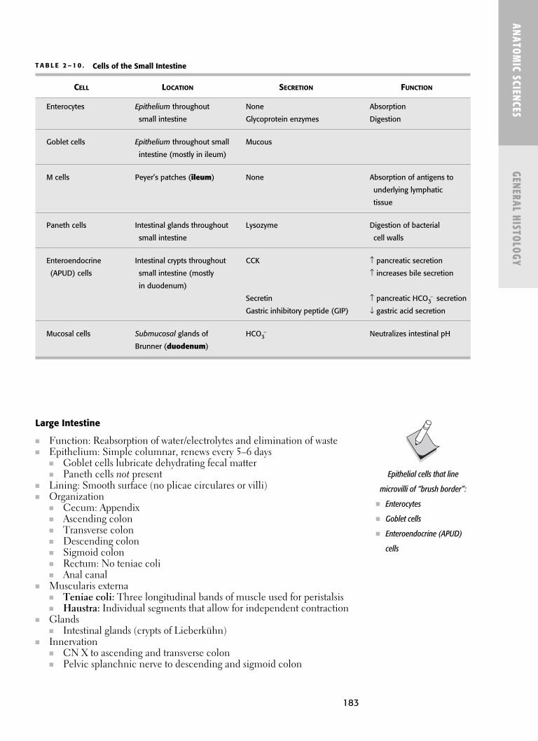
183
ANATOMIC SCIENCES
GENERAL HISTOLOGY
183
Large Intestine
■ Function: Reabsorption of water/electrolytes and elimination of waste■ Epithelium: Simple columnar, renews every 5–6 days
■ Goblet cells lubricate dehydrating fecal matter■ Paneth cells not present
■ Lining: Smooth surface (no plicae circulares or villi)■ Organization
■ Cecum: Appendix■ Ascending colon■ Transverse colon■ Descending colon■ Sigmoid colon■ Rectum: No teniae coli■ Anal canal
■ Muscularis externa■ Teniae coli: Three longitudinal bands of muscle used for peristalsis■ Haustra: Individual segments that allow for independent contraction
■ Glands■ Intestinal glands (crypts of Lieberkühn)
■ Innervation■ CN X to ascending and transverse colon■ Pelvic splanchnic nerve to descending and sigmoid colon
Epithelial cells that line
microvilli of “brush border”:
■ Enterocytes
■ Goblet cells
■ Enteroendocrine (APUD)
cells
T A B L E 2 – 1 0 . Cells of the Small Intestine
CELL LOCATION SECRETION FUNCTION
Enterocytes Epithelium throughout None Absorption
small intestine Glycoprotein enzymes Digestion
Goblet cells Epithelium throughout small Mucous
intestine (mostly in ileum)
M cells Peyer’s patches (ileum) None Absorption of antigens to
underlying lymphatic
tissue
Paneth cells Intestinal glands throughout Lysozyme Digestion of bacterial
small intestine cell walls
Enteroendocrine Intestinal crypts throughout CCK ↑ pancreatic secretion
(APUD) cells small intestine (mostly ↑ increases bile secretion
in duodenum)
Secretin ↑ pancreatic HCO3− secretion
Gastric inhibitory peptide (GIP) ↓ gastric acid secretion
Mucosal cells Submucosal glands of HCO3− Neutralizes intestinal pH
Brunner (duodenum)

184
ANAT
OMIC
SCI
ENCE
SGE
NERA
L HI
STOL
OGY
184
Gut-Associated Lymphatic Tissue (GALT)
■ Lamina propria (GI tract)■ Peyer’s patches (ileum)■ Lymphoid aggregates (large intestine and appendix)
� LOWER DIGESTIVE SYSTEM
Liver
■ Exocrine (via ducts)■ Bile
■ Endocrine (via bloodstream)■ Albumin■ Lipoproteins■ α and β globulins■ Prothrombin■ Fibronectin
Portal Triad
■ Hepatic artery■ Portal vein■ Bile duct
Liver Lobules
■ Hexagonal stacks of hepatocyte cords separated by anastamosing sinusoids■ Surround a central vein (into which sinusoids drain)■ Portal triads located at each corner
Hepatocytes
■ Nuclei: Often bi-nucleate (tetraploid)■ Cytoplasm: Acidophilic
■ Mitochondria■ Golgi bodies■ rER■ sER■ Peroxisomes
■ Catalase■ Alcohol dehydrogenase
■ Lysosomes■ Store iron
■ Glycogen deposits■ Lipid droplets
■ Life span: 5 months■ Capable of regeneration
Hepatic Sinusoids
■ Lined by a thin, discontinuous epithelium. No basement membrane.■ Gaps: Between epithelial cells■ Fenestrae: Within epithelial cells
■ Cell types■ Epithelial cells

185
ANATOMIC SCIENCES
GENERAL HISTOLOGY
185
■ Kupffer cells: Mononuclear macrophages■ Ito cells: Adipocytes in the space of Disse—store vitamin A
■ Perisinusoidal space (space of Disse): Site of exchange between blood and hepatocytes
Biliary Tree
See Figure 2–10.
■ Ductal system that transports bile from hepatocytes → gall bladder (via cystic duct) and duodenum (via common bile duct).
■ Canaliculi: Small canals formed by grooves in adjacent hepatocytes.■ Bile fl ow is opposite to that of blood fl ow (central vein → portal canal).■ Ampulla of Vater: Opening of common bile duct into the duodenum.■ Sphincters
■ Sphincter of Boyden: At common bile duct.■ Sphincter of Oddi: At ampulla of Vater.
Bile Composition
■ Water■ Electrolytes■ Cholesterol■ Lecithin■ Bile salts
■ Glycocholic acid■ Taurocholic acid
■ Bile pigments■ Bilirubin■ Biliverdin■ Glucuronide
Elevated serum bilirubin
results in jaundice.
Hepatocytes
Canaliculi
Canals of Hering
Interlobular bile ducts
Lobar ducts
Common hepatic duct (joins the cystic duct of the gall bladder)
Common bile duct
Duodenum (at the ampulla of vater)
F I G U R E 2 – 1 0 . Biliary fl ow.
Biliary fl ow is increased
primarily by CCK, but also by
secretin and gastrin.

186
ANAT
OMIC
SCI
ENCE
SGE
NERA
L HI
STOL
OGY
186
Gall Bladder
■ Function: Concentrates and stores bile■ Epithelium: Simple columnar
■ Contains microvilli■ Has lateral plications■ Rokitansky-Aschoff sinuses: Deep diverticula of the mucosa
■ No submucosa■ Lamina propria: Does not contain lymphatic vessels■ Glands
■ Mucin-secreting glands
Pancreas
■ Exocrine (via ducts)■ Digestive enzymes
■ Endocrine (via bloodstream)■ Glucagon■ Insulin■ Somatostatin
Exocrine Pancreas
■ Function: Produces digestive enzyme precursors (proenzymes), which are activated by trypsin in the lumen of the small intestine. Trypsinogen is fi rst activated to trypsin by enterokinase. ■ Trypsinogen■ Pepsinogen■ Amylase■ Lipase■ Deoxyribonuclease■ Ribonuclease
■ Components: Pancreatic acini■ Cell types: Centroacinar cells
Endocrine Pancreas
■ Function: Regulates blood glucose levels■ Components: Islets of Langerhans■ Cell types: See Table below
Enzyme production is
regulated by duodenal
secretin ( ↑ bicarbonate
secretion) and CCK
(↑ proenzyme secretion).
Pancreatic digestive enzymes
are activated only after they
reach the small intestine,
where trypsinogen is
converted to trypsin via
enterokinase. Trypsin then
converts the other inactive
digestive enzymes.
Major Cells of the Pancreatic Islets
Cell Type Major Product Function
Alpha Glucagon ↑ blood glucose levels↑ glycogenolysis, gluconeogenesis, and
hepatic lipase
Beta Insulin ↓ blood glucose levels↑ cellular glucose uptake, utilization,
and storage (as glycogen)
Delta Somatostatin Inhibits insulin and glucagon secretion
Somatostatin is also released
by the hypothalamus to inhibit
pituitary secretion of GH.
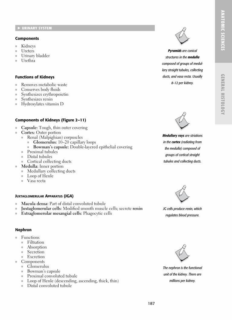
187
ANATOMIC SCIENCES
GENERAL HISTOLOGY
187
� URINARY SYSTEM
Components
■ Kidneys■ Ureters■ Urinary bladder■ Urethra
Functions of Kidneys
■ Removes metabolic waste■ Conserves body fl uids■ Synthesizes erythropoietin■ Synthesizes renin■ Hydroxylates vitamin D
Components of Kidneys (Figure 2–11)
■ Capsule: Tough, thin outer covering■ Cortex: Outer portion
■ Renal (Malpighian) corpuscles■ Glomerulus: 10–20 capillary loops■ Bowman’s capsule: Double-layered epithelial covering
■ Proximal tubules■ Distal tubules■ Cortical collecting ducts
■ Medulla: Inner portion■ Medullary collecting ducts■ Loop of Henle■ Vasa recta
JUXTAGLOMERULAR APPARATUS (JGA)
■ Macula densa: Part of distal convoluted tubule■ Juxtaglomerular cells: Modifi ed smooth muscle cells; secrete renin■ Extraglomerular mesangial cells: Phagocytic cells
Nephron
■ Functions ■ Filtration■ Absorption■ Secretion■ Excretion
■ Components■ Glomerulus■ Bowman’s capsule■ Proximal convoluted tubule■ Loop of Henle (descending, ascending, thick, thin)■ Distal convoluted tubule
Pyramids are conical
structures in the medulla
composed of groups of medul-
lary straight tubules, collecting
ducts, and vasa recta. Usually
8–12 per kidney.
Medullary rays are striations
in the cortex (radiating from
the medulla) composed of
groups of cortical straight
tubules and collecting ducts.
JG cells produce renin, which
regulates blood pressure.
The nephron is the functional
unit of the kidney. There are
millions per kidney.

188
ANAT
OMIC
SCI
ENCE
SGE
NERA
L HI
STOL
OGY
188
Medulla
Cortex
Section enlargedat right
Medullarypyramid
Renalpelvis
Ureter
Cortex
Medullaryrays
Papilla
Duct of Bellini
Ascendinganddescendinglimbs of loops of Henle
Collecting tubule
Medullary ray(contains only straighttubules)
Macula densa
Proximal convoluted tubule
Distal convoluted tubuleGlomerulusCapsule
Location of Length of Nephron Renal Corpuscles Loop of Henle
Cortical Outer cortex Short
Intermediate Middle cortex Medium
Juxtamedullary Base of medullary pyramid Long
TYPES OF NEPHRONS
F I G U R E 2 – 1 1 . Schematic diagram of a kidney and nephron structure.
Reproduced, with permission, from Ross MH, Romrell LJ, Kaye GI. Histology: A Text and Atlas. 3rd ed. Philadelphia: Williams & Wilkins; 1995.

189
ANATOMIC SCIENCES
GENERAL HISTOLOGY
189
Urethra
■ The male urethra is long (20 cm) and has three segments:■ Prostatic urethra: Travels from the urinary bladder through the pros-
tate gland. Widest and most dilatable.■ Membranous urethra: Travels from the end of the prostate through
the body wall to the penis. Shortest and least dilatable.■ Penile urethra: Travels through the length of the penis within the cor-
pus spongiosum. Longest and narrowest.■ The female urethra is short (3–5 cm), and extends from the urinary bladder
to the vaginal vestibule.
� REPRODUCTIVE SYSTEM
Testis
■ Produces sperm and steroids (testosterone).■ Tunica albuginea: Thick connective tissue capsule that covers the testis.■ Covered by a thick connective tissue capsule called the tunica albuginea.■ Composed of many lobules, each containing 1–4 seminiferous tubules,
the site of spermatogenesis.■ Epididymis: Sits on the posterior aspect of the testis. Contains the ductus
epididymis. Stores sperm. Epithelial cells have stereocilia, which increase surface area for nutrient absorption for sperm.
■ Seminal fl uid: Secretions from seminal vesicles and prostate.
Major Cellular Components of the Male Reproductive System
Cell Location Function
Leydig cell Seminiferous tubules Produces testosterone
Sertoli cell Seminiferous tubules Produces testicular fl uid
Sperm cell Produced in seminiferous Produces sperm tubules, but mature and are stored in the epididymis
Intra- and Extra-Testicular Duct System
Seminiferous tubules (testis)↓
Rete testis (testis)↓
Efferent ductules (testis)↓
Ductus epididymis (epididymis)↓
Ductus (vas) deferens (spermatic cord)-joins seminal vesicle duct↓
Ejaculatory duct (enters into urethra)
Prostate Gland
■ Surrounds the proximal urethra■ Secretes acid phosphatase, fi brinolysin, and citric acid
Spermatic cord:
■ Ductus (vas) deferens
■ Testicular artery and veins
■ Lymph vessels
■ Autonomic nerves

190
ANAT
OMIC
SCI
ENCE
SGE
NERA
L HI
STOL
OGY
190
Penis
■ Composed of three major masses of tissue surrounded by a dense fi broelasticcapsule (tunica albuginea):■ Corpora cavernosa: Two dorsal sections of erectile tissue■ Corpus spongiosum: One ventral section containing the urethra
Ovary
■ Elliptical organs supported by broad ligament of uterus.■ Produces ova and steroids (estrogen and progesterone).■ Composed of two regions:
■ Inner medulla: Contains vasculature, nerves, and CT.■ Outer cortex: Contains ovarian follicles, the site of oogenesis.
Oviducts (Fallopian Tubes)
■ Extend bilaterally from the ovaries to the uterus.■ Divided into four sections (from ovary → uterus):
■ Infundibulum: Just adjacent to the ovary. Contains fi ngerlike exten-sions called fi mbriae.
■ Ampulla: Longest section of the oviduct. Site of fertilization.■ Isthmus: Just adjacent to the uterus.■ Uterine: Opening into the uterus.
Uterus
■ Site of embryonic and fetal development.■ Composed of three layers:
■ Endometrium: Mucosal layer shed during menstruation.■ Myometrium: Thickest layer containing smooth muscle.■ Perimetrium: External layer containing connective tissue.
■ Contains uterine glands.■ The cervix is the lower section of the uterus, connecting it to the vagina. Its
mucosa is not shed during menstruation.
Vagina
■ Lined by nonkeratinized stratifi ed squamous epithelium.■ Does not contain glands.
Mammary Glands
■ Contain tubuloalveolar glands (produce milk), sebaceous glands (glands of Montgomery), and sweat glands.
■ Milk is produced by both merocrine and apocrine secretion.■ Lactation is regulated by sex hormones produced during pregnancy, and is
under the control of the pituitary and the hypothalamus.
� INTEGUMENT
Functions of Skin
■ Protection: Against physical, chemical, and biological agents■ Sensory: Touch, pain, pressure, etc■ Homeostasis: Regulates body temperature and water loss
Ovarian follicle development:
1. Primordial oocytes
(surrounded by
granulosa cells).
2. Multilayered theca interna
secretes estrogen.
3. Surrounding stromal cells
form theca externa.
4. Split in theca interna forms
Graafi an follicle.
Although both skin and
oral mucosa have stratifi ed
squamous epithelial surfaces
and subepithelial connective
tissues, there are some impor-
tant differences in terminology
and structures.

191
ANATOMIC SCIENCES
GENERAL HISTOLOGY
■ Synthesis: Produces vitamin D from UV light■ Excretion: Via sweat glands
Layers of Skin
■ Epidermis: Stratifi ed squamous keratinized epithelium (from innermost →outermost).■ Stratum basale (germinativum): Site of all epithelial mitotic activity
(contains melanocytes, keratinocytes, and other specialized cells). Least cytodifferentiated. Cuboidal to low columnar in shape.
■ Stratum spinosum: Spinous (prickle cell) layer. Cells have peripheral cytoplasmic processes. Langerhans cells often extend into this layer.
■ Stratum granulosum: Cells appear fl attened and may contain kera-tohyalin granules. Organelles are diminished.
■ Stratum lucidum: Clear cell layer found only in thick skin (palms,soles). Cells appear fl attened with few (if any) organelles.
■ Stratum corneum: Keratinized cell layer. Cells appear fl attened and pyknotic with few (if any) organelles.
■ Basement membrane: Links the epidermis and dermis via hemidesmosomes.■ Lamina lucida■ Lamina densa (basal lamina)
■ Type IV collagen■ Proteoglycans■ Laminin■ Fibronectin■ Anchoring fi brils (type VII collagen)
■ Reticular lamina■ Reticular fi bers (type III collagen)
■ Dermis: Thicker portion of skin. Dense CT, vasculature, lymphatics, nerves, sweat glands, sebaceous glands, hair follicles.■ Papillary layer: Thinner, cellular, less fi brous. Dermal papillae are the
projections that interdigitate with the epidermal rete pegs (ridges), cre-ating a variably tortuous epidermal-dermal interface. Contains bloodvessels that supply overlying epidermis.
■ Reticular layer: Thicker, fi brous, less cellular.■ Hypodermis: Loose CT, vasculature, lymphatics, and adipose tissue
(provides insulation, energy storage, and anchorage).
SPECIALIZED EPIDERMAL CELLS
■ Concentrated in the basal cell layer.■ Melanocytes: Produce melanin (pigment).■ Keratinocytes: Produce keratin (protection). Have well-developed tonofi brils
and desmosomes. ■ Langerhans cells: Antigen-presenting cells. Often extend into stratum
spinosum.■ Merkel cells: Touch-sensory cells (touch).
NEURONAL ENDINGS OF SKIN
■ Free nerve endings: Most abundant nerve endings. Detect touch, tempera-ture, pain. Extend into stratum granulosum. Not encapsulated.
■ Pacinian corpuscles: Pressure and vibration receptors.■ Meissner’s corpuscles: Touch receptors.■ Ruffi ni endings: Mechanoreceptors.
Epidermal layers (from innermost to outermost):
Bad Sprinters GetLeg Cramps:
BasaleSpinosumGranulosumLucidumCorneum

192
ANAT
OMIC
SCI
ENCE
SGE
NERA
L HI
STOL
OGY
Hair
■ Regulate body temperature■ Composed of keratinized cells■ Produced by hair follicles:
■ Bulb■ Internal root sheath■ External root sheath■ Arrector pili muscle
Types of Sweat Glands
Type Function Cells Secretion Location Innervation
Eccrine Regulates body Clear Sweat Entire body except SNS temperature Dark lips and parts of (parasympathetic) Myoepithelial external genitalia Duct
Apocrine Produces Myoepithelial Odorless Axilla, areola, nipple, SNS pheromones Duct serous circumanal region, (sympathetic) secretion external genitalia
Sebaceous Glands
■ Sebocytes: Secrete sebum, an oily substance that coats hair-covered skin.■ Outgrowths of external root sheaths of hair follicles.
� EYE
Layers of Eye
See Figure 2–12.From outermost → innermost:
■ Corneoscleral coat■ Cornea: Transparent portion (anterior 1/6) of eye.■ Limbus: Transitional zone between cornea and sclera.■ Sclera: “White” portion (posterior 5/6) of eye.
■ Uvea■ Choroid: Vascular layer. Dark brown color.■ Ciliary body: Smooth muscle (lens accommodation). See Figure 2–13.■ Iris: Smooth muscle (changes pupil diameter). Pigmented portion of eye.
■ Pupil: Central aperture of iris.■ Retina
■ Pigment epithelium: Melanin-containing cells.■ Neural retina: Rods and cones.
CHAMBERS
■ Anterior chamber: From cornea → iris■ Posterior chamber: From iris → lens■ Vitreous chamber: From lens → neural retina
Aqueous humor is the watery
fl uid within the anterior and
posterior chambers.
Vitreous humor is the
transparent watery gel within
the vitreous chamber.

193
ANATOMIC SCIENCES
GENERAL HISTOLOGY
F I G U R E 2 – 1 2 . General structure of the eye.
Anterior limiting membrane of corneaPosterior limiting membrane of cornea
Zonular fibers
Lens
Retina
Chorioid
Perichorioidal space
Intervaginal space
Optic nerve sheath
Fovea centralis
Optic disk
Sclera
Vitreous chamber of eye
Ciliary body
Posterior chamber of eye
Pars iridica retinae
Anterior chamber of eye
Superficial corneal epithelium
ConjunctivaCiliaris muscle
Sclera
Aqueous humor
Cornea
}
}
F I G U R E 2 – 1 3 . Ciliary zone.
Reproduced, with permission, from Riordan-Eva P, Hoyt WF. Vaughan and Asbury’s General Ophthalmology, 17th ed. New York: McGraw- Hill, 2008.
Pars iridica retinae
Posterior chamber of eye
Crystalline lens
Zonular fibers
Zonular space
Pupillary sphincter muscle
Superficial cornealepithelium
Cornea
Anterior chamber of eye(aqueous humor)
Iris
Anterior limiting membrane
Ora serrataSclera
Ciliary process ofciliary crown
Ciliary ring
Episclera
Ciliary muscle
Sclera venous sinus
Bulbar conjunctiva
Posterior limiting membrane
Tendon of medialrectus muscle

194
ANAT
OMIC
SCI
ENCE
SGE
NERA
L HI
STOL
OGY
Ten Layers of Retina
From outermost → innermost:
1. Pigment epithelium 2. Photoreceptor cells
■ Rods: Sensitive to light; contain rhodopsin■ Cones: Sensitive to color; contain iodopsin
3. External limiting membrane 4. Outer nuclear layer
■ Nuclei of rods and cones 5. Outer plexiform layer 6. Inner nuclear layer
■ Nuclei of horizontal, amacrine, bipolar, and Müller’s cells 7. Inner plexiform layer 8. Ganglion cell layer 9. Optic nerve fi bers10. Internal limiting membrane
Optic Disc
■ Collection of retinal ganglion cell nerve fi bers (axons) leaving eye as opticnerve. The right and left optic nerves meet at the optic chiasm.
■ Central artery and vein of the retina also exit here. The central artery (branch of ophthalmic artery) pierces the optic nerve and gains access to retina by emerging from center of optic disc and fans out.
■ Small blind spot on the retina; 3 mm to nasal side of macula.■ Only part of retina without rods or cones.
Macula Lutea
■ Just temporal to optic disc■ Responsible for detailed central vision (eg, reading)■ Fovea: Very center of macula. No blood vessels. Very high concentration of
cones. Area of sharpest vision.
Vitamin A is a source of
retinal, an essential
component of rods. Dietary
defi ciency of vitamin A results
in the inability to see in dim
light (“night blindness”).

C H A P T E R 3
Oral Histology
Soft Oral Tissues 196
ORAL MUCOSA 196
GINGIVA 196
DENTOGINGIVAL JUNCTION 199
Tooth and Supporting Tissues 202
DENTIN 202
ENAMEL 205
PULP 207
CEMENTUM 209
ALVEOLAR BONE 210
PERIODONTAL LIGAMENT (PDL) 211
195

196
ANAT
OM
IC S
CIEN
CES
ORA
L H
ISTO
LOGY
� SOFT ORAL TISSU ES
Oral Mucosa
LAYERS OF ORAL MUCOSA (SEE FIGURE 3–1)
From outermost → innermost:
■ Stratified squamous epithelium: Keratinized or nonkeratinized.■ Stratum corneum: Keratinized cell layer. Cells appear flattened and
pyknotic with few (if any) organelles.■ Stratum granulosum: Cells appear flattened and may contain keratohyalin
granules. Organelles are diminished.■ Stratum spinosum: Spinous (prickle cell) layer. Cells have peripheral
cytoplasmic processes. Langerhans cells often extend into this layer.■ Stratum basale (germinativum): Site of all epithelial mitotic activity.
Least cytodifferentiated. Cuboidal to low columnar in shape.■ Basement membrane: Contains type IV collagen and laminin. Epithelial
attachment to the basement membrane is mediated by hemidesmosomes.■ Subepithelial connective tissue: Contains collagen and elastic fibers,
ground substance, blood vessels, nerves, and inflammatory cells.■ Lamina propria
■ Papillary layer: Connective tissue papillae are the projections thatinterdigitate with the epithelial rete pegs (ridges), creating a vari-ably tortuous epithelial–connective tissue interface.
■ Reticular layer: Constitutes the remainder of the lamina propria.■ Submucosa. Present in areas of high compression. Often indistinguishable
with the lamina propria.
SPECIALIZED EPITHELIAL CELLS
■ Melanocytes: Produce melanin.■ Keratinocytes: Produce keratin, not present in all oral mucosa.■ Langerhans cells: Antigen-presenting cells. ■ Merkel cells: Touch-sensory cells.
TYPES OF ORAL MUCOSA
See Table 3–1.
■ All oral mucosa contains stratified squamous epithelium.
Gingiva
See Figures 3–2 and 3–3.
■ The fibrous, keratinized tissue that surrounds a tooth and is contiguouswith the periodontal ligament and oral mucosa.
■ Contains a stratified squamous keratinized epithelium with rete pegs. ■ Extends from the gingival margin (the most coronal portion of the gingiva)
to the mucogingival junction (MGJ). The MGJ separates the gingivafrom the alveolar mucosa.
■ Color ranges from pink to brown, depending on the amount of melaninexpression, which is correlated with cutaneous pigmentation.
Chronic mouth breathing
often results in pronounced
gingival erythema, especially
in the anterior regions.
Oral mucosa layers (fromoutermost to innermost):California Girls StringBikinis
■ Corneum■ Granulosum■ Spinosum■ Basale
Most specialized epithelial
cells concentrate in the basal
cell layer; however,
Langerhans cells may extend
into the stratum spinosum.

197
ANATOM
IC SCIENCES
ORAL H
ISTOLOGY
ZONES OF GINGIVA
■ Attached gingiva: Firmly bound to periosteum and cementum. About 40% ofthe adult population exhibits stippling of the attached gingiva, which is anorange-peel-like surface characteristic caused by the intersection of epithelialrete pegs.
F I G U R E 3 – 1 . Layers of oral mucosa.
Reproduced, with permission, from Ten Cate AR. Oral Histology: Development, Structure, and
Function. 4th ed. St Louis: Mosby; 1994.
Submucosa
Epithelium
Periosteum
Laminapropria
Bone
The free gingival groove
separates the attached and
unattached (marginal) gingiva.
The mucogingival junction
separates the gingiva from the
alveolar mucosa.

198
ANAT
OM
IC S
CIEN
CES
ORA
L H
ISTO
LOGY
F I G U R E 3 – 2 . Gingival anatomy.
Reproduced, with permission, from Carranza FA. Clinical Periodontology, 8th ed., page 60,
copyright 1996 by Elsevier.
Gingival sulcus
Free gingival grooveFree or marginal gingiva
Attached gingiva
Mucogingival junction
Alveolar mucosa
T A B L E 3 – 1 . The Three Major Types of Oral Epithelium
MUCOSA TYPE LOCATION EPITHELIUM SUBMUCOSA
Masticatory Gingiva Thick stratified squamous Indistinct
Keratinized (75% para)
May show stippling (40%)
Hard palate Thick stratified squamous Thin
Keratinized (mostly ortho)
Lining Soft palate Thin stratified squamous Thick
Nonkeratinized
Taste buds
Alveolar mucosa Thin stratified squamous Thick
Floor of the mouth Nonkeratinized
Buccal mucosa Thick stratified squamous Thin
Nonkeratinized
Lips Thin stratified squamous Thin
Keratinized (ortho and para)
Ventral tongue Thin stratified squamous Thin
Nonkeratinized
Specialized Dorsal tongue Thick stratified squamous Indistinct
Both keratinized and nonkeratinized
Taste buds
Denture abrasion may cause
the masticatory mucosa
(gingiva and hard palate) to
become more orthokeratinized.

199
ANATOM
IC SCIENCES
ORAL H
ISTOLOGY
■ Free (unattached, marginal) gingiva: Located coronal to the attachedgingiva and is separated from the tooth surface by the gingival sulcus.■ Interdental papilla: The triangular portion of the free gingiva located in
the interproximal embrasures between teeth just apical to their contactareas.
■ Gingival col: The depression in the interdental gingiva connectingthe buccal and lingual papilla immediately apical to the contactareas of adjacent teeth. Its epithelium is generally nonkeratinized,and its presence is based solely on the presence of an interdentalcontact.
Dentogingival Junction
■ The attachment of the gingiva to the tooth. ■ Consists of epithelial (epithelial attachment) and connective tissue
(connective tissue attachment) components.■ Forms as the oral epithelium fuses with the reduced enamel epithelium
(REE) during tooth eruption. As the tooth reaches its fully erupted position,cells of the junctional epithelium replace those of the REE.
DENTOGINGIVAL EPITHELIUM
■ Sulcular epithelium: Stratified squamous nonkeratinized epitheliumwithout rete pegs that extends from the gingival margin to the junctionalepithelium. It lines the gingival sulcus (Figure 3–4).
F I G U R E 3 – 3 . Healthy gingival anatomy.
Note the gingival stippling in the maxillary anterior region. Stippling occurs in about 40% of
the adult population.
Free gingivalgroove
Unattached(Marginal) gingiva Attached gingiva
Gingival papillae Alveolarmucosa
Mucogingivaljunction
Biologic width is defined as
the length of the
dentogingival junction. In
humans, the average width
of epithelial attachment is
0.97 mm and 1.07 mm for
the connective tissue
attachment, creating a mean
biologic width of 2.04 mm.
In the presence of inflammation
(eg, gingivitis), rete pegs will be
present in the sulcular and
junctional epithelia.

200
ANAT
OM
IC S
CIEN
CES
ORA
L H
ISTO
LOGY
■ Junctional epithelium: Stratified to single-layer (at its apical extent)nonkeratinized epithelium without rete pegs that adheres to the toothsurface at the base of the sulcus. It provides the epithelial attachment tothe tooth. Junctional epithelial cells have higher turnover rates and largerintercellular spaces than sulcular and gingival epithelia. It consists of twobasal laminae:■ External basal lamina: Attaches to underlying connective tissue as
elsewhere in the body.■ Internal basal lamina: Attaches to the cementum via hemidesmosomes.
It is unlike other basal laminae in that it does not contain type IVcollagen.
DENTOGINGIVAL CONNECTIVE TISSUE
■ Type I collagen makes up the bulk of the connective tissue.■ Other major components include fibroblasts, leukocytes, mast cells, elastic
fibers, proteoglycans, and glycoproteins.
F I G U R E 3 – 4 . Dentogingival epithelium. A: Gingival epithelium; B: Sulcular epithelium;C: Junctional epithelium. BL: Basal cell layer; SL: Spinous cell layer; FSL: Flattened spinouslayer; GL: Granular cell layer containing keratohyaline granules; KL: Keratinized layer.
Reproduced, with permission, from Rose LF, Mealey B, Genco R. Periodontics: Medicine,
Surgery, and Implants. St Louis: Mosby; 2004.
KL
FSL
SL
SL
BL
BLTooth
GL
SL
BL
A
AB
B
C
C
The subepithelial connective
tissue provides the cellular
signaling that determines
epithelial expression.

201
ANATOM
IC SCIENCES
ORAL H
ISTOLOGY
GINGIVAL FIBER GROUPS
See Figure 3–5.
■ Support the gingiva and aid in its attachment to alveolar bone and teeth.■ They are continuous with the PDL. ■ Resist gingival displacement.■ Found at the gingival level.■ Do not confuse these with PDL fibers!
■ Dentogingival fibers: Fan laterally from cementum into the adjacent CT.■ Alveologingival fibers: Fan coronally from the alveolar crest into the
adjacent CT.■ Dentoperiosteal fibers: Extend from cementum over the alveolar
crest, and turn apically to insert into the periosteum of the buccal sideof the alveolar bone.
■ Circumferential fibers: Surround the tooth in a circular fashion. Helpprevent rotational forces. Located in lamina propria of marginal gingiva.
The connective tissue
adjacent to the sulcular and
junctional epithelia generally
contains an increased
inflammatory infiltrate
compared to that adjacent to
the oral epithelium. PMNs
and other leukocytes
continually migrate between
these epithelial cells into the
sulcus, and account for a
significant portion of gingival
crevicular fluid (GCF) along
with plasma proteins,
epithelial cells, and bacteria.
F I G U R E 3 – 5 . The gingival fibers.
Enamel Gingiva
Mucogingival junction
Circumferential fibers
Alveologingival fibers
Alveolar mucosa
Connective tissue
Alveolar bone
Junctional epithelium
Cementoenamel junction
Dentin
Dentoperiosteal fibers
Dentogingival fibers
Cementum
Periodontal ligament

202
ANAT
OM
IC S
CIEN
CES
ORA
L H
ISTO
LOGY
Comparison of the Major Tissues of Teeth and the Periodontium
Enamel Dentin Cementum Alveolar Bone Pulp
Mineral 96% 70% 55% 50% <5%composition
Organic 4% 30% 45% 50% >95%composition
Embryologic Ectoderm Ectomesenchyme Ectomesenchyme Ectomesenchyme Ectomesenchymeorigin (neural crest) (neural crest) (neural crest) (neural crest)
Formative Ameloblast Odontoblast Cementoblast Osteoblast Fibroblastcell Mesenchymal
Formative IEE Dental papilla Dental follicle Dental follicle Dental papillacellsdifferentiated from
Tissue type Epithelial Connective Connective Connective Connective
Viability No Repair Repair Remodeling Yes
Incremental Neonatal Neonatal Resting Resting Nonelines Retzius Owen Reversal
(daily) von Ebner (daily)
Sensitivity No Yes No Yes Yes (pain)
Nutritive None Pulp PDL (diffusion) Vessels Vesselssupply (diffusion)
� TOOTH AN D SU PPORTI NG TISSU ES
Dentin
■ An elastic, avascular, mineralized tissue that is harder than bone but softerthan enamel
■ Color is generally yellowish
ORIGIN
■ Differentiated ectomesenchymal cells of the dental papilla
DENTINOGENESIS (SEE FIGURE 3–6)
■ Organization■ The odontoblasts become elongated and the organelles become polar-
ized by ameloblastic induction. ■ Mantle dentin formation
■ The dentin matrix formed by odontoblasts starts at the DEJ andprogresses inward toward the eventual pulp. The dentin matrix firstproduced by the odontoblasts consists largely of type I collagen andground substance, known as predentin.
■ As the odontoblasts retreat inward, they leave cytoplasmic extensionscalled odontoblastic processes (Tomes’ fibers) at the DEJ. These

203
ANATOM
IC SCIENCES
ORAL H
ISTOLOGY
processes are housed in dentinal tubules, which form channels fromthe DEJ/DCJ to the pulp.
■ The odontoblastic processes release matrix vesicles containing cal-cium, which crystallize and rupture. These crystals act as a nidus forthe formation of more hydroxyapatite (HA) crystals in and around theorganic dentin matrix.
■ This initial 150 μm of dentin in known as mantle dentin.■ Circumpulpal dentin formation
■ Once mantle dentin is formed, odontoblasts (with well-developed roughER and Golgi bodies) begin to secrete collagen fibrils perpendicularly tothe odontoblastic processes, as well as other organic substances such aslipids, phospholipids, and phosphoproteins.
■ Mineralization occurs by globular calcification by which globules ofHA fuse to form a calcified mass. Occasionally, the globules fail to fuse,leaving hypomineralized interglobular dentin in between.
■ The odontoblastic processes shrink in width, providing a space for a hy-permineralized peritubular dentin to form. If several adjacent dentinaltubules become occluded with this peritubular dentin, it takes on aglassy appearance and is called sclerotic dentin.
■ Most of the circumpulpal dentin produced is between the tubules,known as intertubular dentin.
■ Dead tracts are groups of necrotic odontoblastic processes within thedentinal tubules (often caused by trauma).
■ Reparative dentin formation■ Reparative dentin is formed only at specific sites of injury. ■ The types I and III collagens in its matrix are produced by differenti-
ated odontoblast-like cells from the pulp. The tubular pattern is oftendistorted due to an increased rate of its formation.
Coronal dentinal tubules
follow an S-shape (primary
curvature); radicular tubules
are generally straight. There
are more tubules
concentrated near the pulp
than the DEJ.
F I G U R E 3 – 6 . Dentinogenesis. A: Initial mantle dentin formation. Large-diameter collagen fibrils aredeposited by odontoblasts in the pre-existing ground substance of the dental papilla. B: Transition frommantle dentin formation. Odontoblasts now produce fine-diameter collagen, ground substance, and lipidat the mineralization front. C: Continuing dentinogenesis and circumpulpal dentin formation. Odonto-blasts now secrete intertubular and peritubular dentin.
Reproduced, with permission, from Ten Cate AR. Oral Histology: Development, Structure, and Function. 4th ed.
St Louis: Mosby; 1994.
A
Initial mantle dentin formation.Large-diameter collagen fibrilsare deposited by odontoblastsin the pre-existing groundsubstance of the dental papilla.
Transition from mantle dentinformation. Odontoblasts nowproduce fine-diameter collagen,ground substance, and lipid atthe mineralization front.
Continuing dentinogenesisand circumpulpal dentinformation. Odontoblasts nowsecrete intertubular andperitubular dentin.
B
Mantle
C
1 1
1
2
2
3
3
44
Mantle
HA = Ca10(PO4)6(OH)2
FA = Ca10(PO4 )6F2
204
ANAT
OM
IC S
CIEN
CES
ORA
L H
ISTO
LOGY
CLASSIFICATIONS OF DENTIN
■ By time of formation■ Mantle: The first 150 μm of dentin formed. Located closest to enamel
(CEJ) and cementum (CDJ)■ Circumpulpal: All dentin formed thereafter until tooth formation is
complete■ Reparative: Formed in response to trauma (eg, caries, restorations, attrition,
erosion, abrasion, etc)■ Sclerotic: Results from calcification of the dentinal tubules as one ages
over time. Helps prevent pulpal irritation■ By root completion
■ Primary: Formed before root completion. Tubules are most regular.■ Secondary: Formed after root completion, but not in response to trauma.■ Tertiary: Formed in response to trauma (eg, caries, restorations, attrition,
erosion, abrasion, etc). Tubules are least regular.■ By proximity to dentinal tubules
■ Peritubular: Hypermineralized dentin formed within the perimeter ofdentinal tubules as odontoblastic processes shrink.
■ Intertubular: Hypomineralized dentin located between the dentinaltubules. Makes up the bulk of the dentin formed.
■ Interglobular: Hypomineralized dentin located between improperlyfused HA globules.
■ By location■ Coronal
■ May contain hypomineralized interglobular dentin.■ May contain dead tracts.
■ Radicular■ May contain hypomineralized Tomes’ granular layer.
CROSS STRIATIONS AND INCREMENTAL LINES
■ Daily imbrication line of von Ebner: Daily periodic bands. ■ Contour lines of Owen: Wide rings produced by metabolic disturbances
during odontogenesis that run perpendicular to dentinal tubules.■ Neonatal line: A more pronounced contour line of Owen formed during
the physiologic trauma at birth.
EFFECTS OF AGING ON DENTIN
■ ↑ sclerotic dentin (↓ dentinal tubule diameter due to continued depositionof peritubular dentin).
■ ↑ reparative dentin formation.■ ↑ dead tracts.
CLINICAL IMPLICATIONS
■ Dentinal hypersensitivity: Can occur for several reasons:■ Myelinated nerve fibers have been found in dentinal tubules which
can be directly stimulated.■ Changes in dentinal tubule fluid pressures may affect pulpal nerve fibers
directly or may cause damage to odontoblasts, releasing inflammatorymediators in the pulp.
Odontoblasts move at a rate
of 4–8 mm/day.

205
ANATOM
IC SCIENCES
ORAL H
ISTOLOGY
DEFORMITIES OF DENTIN
■ Dentinogenesis imperfecta: Generally autosomal dominant defects indentin formation. Teeth exhibit an opalescent color and have bulb-shapedcrowns. The dentin is abnormally soft (allowing enamel to easily chip) andpulp chambers are often obliterated. ■ Type I: Often occurs with osteogenesis imperfecta (blue sclera is a
common finding).■ Type II: Not associated with osteogenesis imperfecta.■ Type III: Very rare form which exhibits multiple pulp exposures of the
primary dentition.■ Dentin dysplasia: Autosomal dominant defects in dentin formation and
pulp morphology. Tooth color is usually normal. Often called “rootlessteeth” because root dentin is usually affected more often than coronaldentin. Roots are short, blunt, or absent.
Enamel
■ The most calcified and brittle substance in the human body. ■ Color ranges from yellowish to grayish-white.■ Semitranslucent.
ORIGIN
■ Differentiated ectodermal cells of the inner enamel epithelium.
AMELOGENESIS
■ Organization■ The ameloblasts become elongated and the organelles become polarized
before the same occurs to odontoblasts.■ Formation (see Figure 3–7)
■ The enamel matrix produced by ameloblasts starts virtually perpendicularlyto the DEJ and progresses outward toward the eventual tooth surface. Theoldest enamel is located at the DEJ underlying a cusp or cingulum.
■ Ameloblastic activity starts immediately after mantle dentin formation. ■ As ameloblasts retreat, Tomes’ processes are formed around which
enamel matrix proteins are secreted, most of which are almost instantlypartially mineralized to form enamel matrix. This determines the struc-ture and morphology of the tooth.
■ Maturation■ Final mineralization occurs with inorganic ion influx and removal of
protein and water by cyclic ameloblastic activity, forming hydroxyap-atite (HA) crystals.
■ As the HA crystals accumulate, they are tightly stacked in elongatedunits called enamel rods (prisms). The rods are surrounded by a rodsheath and separated by an inter-rod substance that consists of HAcrystals aligned in a different direction than the rods themselves.
■ Each keyhole-shaped enamel rod is formed by four ameloblasts (onefor the head and three for the tail).
■ At cusp tips, the enamel rods appear twisted and intertwined in a formationknown as gnarled enamel.
Amelogenin constitutes
about 90% of the enamel
matrix protein secreted. Other
proteins include enamelin and
tuftelin.
Most enamel rods extend the
width of the enamel from the
DEJ to the outer enamel
surface.

206
ANAT
OM
IC S
CIEN
CES
ORA
L H
ISTO
LOGY
■ Protection■ When enamel maturation is complete, the outer enamel epithelium,
stratum intermedium, and stellate reticulum collapse onto theameloblastic layer, forming the reduced enamel epithelium (Nasmyth’smembrane). This is worn away soon after tooth eruption and quicklyreplaced by the salivary pellicle. Hemidesmosomes are also produced,which are critical in providing epithelial attachment to the tooth.
CROSS STRIATIONS AND INCREMENTAL LINES
■ Daily imbrication lines: Daily periodic bands.■ Striae of Retzius: More pronounced weekly periodic bands.
■ Shallow depressions, called perikymata, are formed on the enamel surfacewhere these lines reach the tooth surface. Perikymata disappear with agedue to attrition of the raised areas between them.
■ Neonatal line: A more apparent stria of Retzius formed during the physiologictrauma at birth.
■ Hunter–Schreger bands: Alternating light and dark zones produced only as anoptical phenomenon during light microscopy of longitudinal ground sections.
THE DENTINO–ENAMEL JUNCTION (DEJ)
■ Enamel tufts: Hypocalcified, fan-shaped enamel protein projecting a shortdistance into the enamel.
Enamel matrix is produced
at a rate of 4 mm/day.
Stratum
Mantle Dentin
Intermedium Stratum Intermedium
Structureless enamel
Mantle Dentin
1
2
F I G U R E 3 – 7 . Enamel formation. Structureless enamel initially forms across the straightface of each ameoloblast. As the ameloblasts retreat from the DEJ, Tomes’ processes areformed, and enamel matrix is secreted and mineralized at different orientations (1 and 2).
Reproduced, with permission, from Ten Cate AR. Oral Histology: Development, Structure,
and Function. 4th ed. St Louis: Mosby; 1994.
The DEJ is scalloped,
which provides more surface
area for enamel–dentin
adhesion.

207
ANATOM
IC SCIENCES
ORAL H
ISTOLOGY
■ Enamel lamellae: Hypocalcified enamel defects that can extend all the wayto the enamel surface. They generally consist of enamel protein or oraldebris.
■ Enamel spindles: Trapped odontoblastic processes in the enamel.
EFFECTS OF AGING ON ENAMEL
■ Attrition: Enamel wear by masticatory forces.■ Discoloration: Becomes darker as more dentin becomes visible.■ ↓ permeability: Enamel crystals have accepted more ions, especially fluoride.
CLINICAL IMPLICATIONS
■ Since enamel is translucent, its color depends on its thickness. The moretranslucent the enamel, the more yellow it appears because the underlyingdentin becomes more visible.
■ Tetracycline antibiotics can be incorporated into mineralizing tissues bychelation to divalent cations. This leads to a brownish-gray bandingwithin the enamel. Drugs from this family should not be given until aboutage 8 (about the time the second molar completes calcification).
DEFORMITIES OF ENAMEL
■ Amelogenesis imperfecta: Autosomal dominant or recessive enamel defectsof three basic types:■ Hypoplastic: Enamel has abnormal thickness or pitting, but normal
hardness. Defect in enamel matrix formation.■ Hypocalcified: Enamel has normal thickness, but is soft and chalky.
Defect in enamel mineralization.■ Hypomaturation: Enamel has normal thickness, but abnormal hardness.
Mild forms exhibit “snow-capped” incisal edges, while severe formslose their translucency. Defect in enamel maturation.
■ Enamel hypoplasia: Enamel is hard, but deficient in amount. Caused bydefective or altered enamel matrix formation. Can be acquired or develop-mentally-induced.■ Fluorosis: Enamel is selectively permeable to water and certain ions.
This allows fluoride (in drinking water and topical applications) toconcentrate in enamel apatite, forming fluorapatite, which is highlyresistant to acid dissolution. Fluoride concentrations greater than 5parts per million, however, affect ameloblastic enamel matrix secre-tion, leading to the appearance of enamel mottling and a brownishpigmentation.
■ Nutritional deficiencies: Deficiencies of vitamins A, C, and D andcalcium often lead to enamel pitting.
■ Infections: Febrile diseases at the time of amelogenesis can haltenamel formation, leaving bands of malformed surface enamel.
■ Congenital syphilis: Often causes incisors to look screwdriver-shaped(Hutchinson incisors) and molars to look globular (mulberry molars).
Pulp
■ The soft connective tissue that supports the dentin and is contained insidethe pulp chamber of the tooth.
■ Communicates to the periodontal tissues via the apical foramen and accessorycanals.
Fluoride:
■ Tasteless, odorless,
colorless
■ Excreted rapidly by kidneys
■ Deposited in calcified
tissues
■ Passes slowly through
placental barrier
Hutchinson’s triad ofcongenital syphilis:
■ Blindness (interstitial
keratitis)
■ Deafness (CN VIII injury)
■ Dental anomalies (notched
“Hutchinson” incisors,
mulberry molars,
peg-shaped laterals)
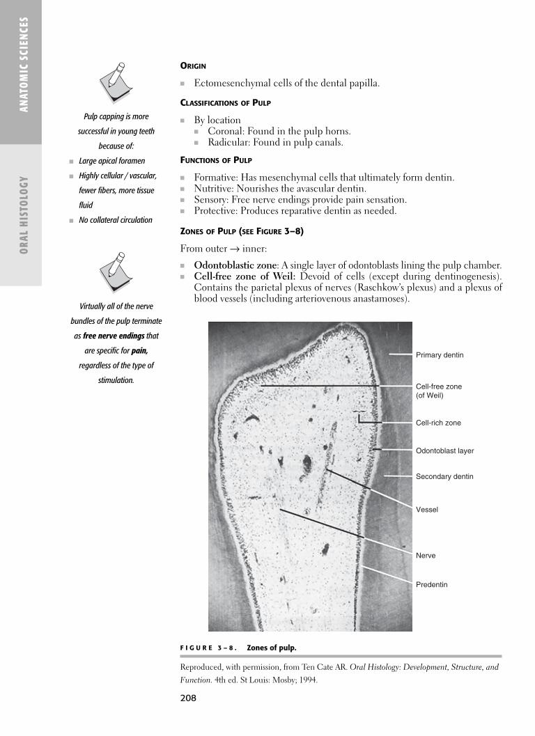
208
ANAT
OM
IC S
CIEN
CES
ORA
L H
ISTO
LOGY
ORIGIN
■ Ectomesenchymal cells of the dental papilla.
CLASSIFICATIONS OF PULP
■ By location■ Coronal: Found in the pulp horns.■ Radicular: Found in pulp canals.
FUNCTIONS OF PULP
■ Formative: Has mesenchymal cells that ultimately form dentin.■ Nutritive: Nourishes the avascular dentin.■ Sensory: Free nerve endings provide pain sensation.■ Protective: Produces reparative dentin as needed.
ZONES OF PULP (SEE FIGURE 3–8)
From outer → inner:
■ Odontoblastic zone: A single layer of odontoblasts lining the pulp chamber.■ Cell-free zone of Weil: Devoid of cells (except during dentinogenesis).
Contains the parietal plexus of nerves (Raschkow’s plexus) and a plexus ofblood vessels (including arteriovenous anastamoses).
F I G U R E 3 – 8 . Zones of pulp.
Reproduced, with permission, from Ten Cate AR. Oral Histology: Development, Structure, and
Function. 4th ed. St Louis: Mosby; 1994.
Primary dentin
Secondary dentin
Vessel
Nerve
Predentin
Cell-free zone(of Weil)
Cell-rich zone
Odontoblast layer
Virtually all of the nerve
bundles of the pulp terminate
as free nerve endings that
are specific for pain,
regardless of the type of
stimulation.
Pulp capping is more
successful in young teeth
because of:
■ Large apical foramen
■ Highly cellular / vascular,
fewer fibers, more tissue
fluid
■ No collateral circulation

209
ANATOM
IC SCIENCES
ORAL H
ISTOLOGY
■ Cell-rich zone: Contains fibroblasts and undifferentiated mesenchymalcells.
■ Pulp core: Contains fibroblasts, macrophages, leukocytes, blood andlymph vessels, myelinated (mostly Aδ) and unmyelinated (C) sympatheticnerve fibers, collagen types I and III (55:45 ratio), and ground substance.There are no elastic fibers.
PULP CALCIFICATIONS
■ Denticles (pulp stones): Concentric layers of mineralized tissue■ True: Surround dentinal tubules or odontoblastic processes.■ False: Surround dead cells or collagen fibers.■ Free: Located freely (unattached) in the pulp chamber.■ Attached: Attached to the pulp chamber wall.■ Interstitial: Embedded in the pulp chamber wall.
■ Dystrophic calcifications: Calcifications of collagen bundles or collagenfibers surrounding blood vessels and nerves.
EFFECTS OF AGING ON PULP
■ ↑ collagen fibers and calcifications.■ ↓ pulp chamber volume (due to continued dentin deposition), apical foramen
size, cellularity, vascularity, and sensitivity.
Cementum
■ An avascular tissue about 10 μm thick that covers the radicular dentin. ■ Composition most closely resembles bone.
ORIGIN
■ Differentiated ectomesenchymal cells of the dental follicle.
FUNCTIONS OF CEMENTUM
■ Support: Provides attachment for teeth (Sharpey’s fibers).■ Protection: Helps prevent root resorption during tooth movement.■ Formative: Continual apical cementum deposition accounts for continual
tooth eruption and movement.
CEMENTOGENESIS
■ During root formation, ectomesenchymal cells of the dental follicle migratethrough gaps in Hertwig’s epithelial root sheath and orient themselves alongradicular dentin. Here, they differentiate to cementoblasts and secrete ce-mentoid (cementum matrix).
■ As the cementoblasts retreat away from the dentin, the matrix is calcifiedand a new layer of cementum matrix is secreted. These layers form restinglines, which can be seen microscopically.
■ Cementoblasts may be trapped in their own matrix. When this occurs,they are known as cementocytes, which reside in lacunae. They receivenutrients via canaliculi that extend to the periodontal ligament.
■ Cementum is constantly produced at the apical portion of the root to ac-count for the continual eruption of teeth. Deposition of excessive cemen-tum is known as hypercementosis.

210
ANAT
OM
IC S
CIEN
CES
ORA
L H
ISTO
LOGY
CLASSIFICATIONS OF CEMENTUM
■ By formation■ Primary: First formed cementum. Covers coronal cementum, is acellular,
and consists of extrinsic collagen fibers.■ Secondary: Overlies primary cementum. Covers apical cementum,
may be either acellular or cellular, and consists of mixed collagen fibers. ■ By cellularity
■ Cellular: Contains cementocytes, cementoblasts, and cementoclasts.Most commonly found in apical areas of cementum.
■ Acellular: Devoid of cells. Most commonly found in coronal areas ofcementum.
■ By collagen fibers■ Intrinic fibers: Produced by cementoblasts. Arranged parallel to the
tooth surface.■ Extrinsic fibers: Produced by the PDL. Arranged perpendicular to the
tooth surface. As they become trapped in the cementum, they areknown as Sharpey’s fibers.
■ Mixed fibers: Combination of intrinsic and extrinsic fibers.
APPROXIMATION WITH ENAMEL AT THE CEJ
See Figure 3–9.
EFFECTS OF AGING ON CEMENTUM
■ ↑ cementum deposition.
CLINICAL IMPLICATIONS
■ Cementum enables orthodontic tooth movement because it is more resis-tant to resorption than alveolar bone.
Alveolar Bone
■ Alveolar bone: A general term to describe the bone in the maxilla andmandible which houses the teeth.
■ Interalveolar septum: The bony projection separating two alveoli. ■ Interradicular septum: Alveolar bone between the roots of multirooted
teeth.
F I G U R E 3 – 9 . Cementum morphology at the CEJ.
A. Cementum does not reach enamel; B. Cementum meets enamel; C. Cementum overlaps
enamel. Reproduced, with permission, from Carranza FA. Clinical Periodontology, 8th ed.
page 61, copyright 1996 by Elsevier.
D D D
A B C
5%–10% 30% 60%–65%
The only function of alveolar
bone is to support teeth.
During tooth development,
it grows in all dimensions to
accommodate the new
dentition. When a tooth is
lost, it continually resorbs
until complete alveolar ridge
atrophy occurs.

211
ORIGIN
■ Differentiated ectomesenchymal cells of the dental follicle.
COMPONENTS OF ALVEOLAR BONE
■ Alveolar bone proper: The thin layer of cortical bone that immediatelysurrounds the teeth and into which PDL fibers (Sharpey’s fibers) are em-bedded. It is also called bundle bone, lamina dura, or cribriform plate.
■ Supporting alveolar bone: The part of the alveolus that surrounds thealveolar bone proper. It consists of the following:■ Cortical bone (cortical plate): Forms the buccal and lingual outer surfaces
of the maxilla and mandible. It is generally thicker in the mandible and inposterior (molar) regions.
■ Cancellous bone (spongy bone, trabecular bone): Fills the area betweenthe cortical plates. It makes up the majority of alveolar bone.
CLINICAL IMPLICATIONS
■ The radiographic appearance of the lamina dura is determined as muchby the x-ray beam angulation as it is by its integrity.
■ The radiographic presence (or absence) of the crestal lamina dura has nocorrelation with periodontal attachment loss.
Periodontal Ligament (PDL)
See Figure 3–10.
■ A soft connective tissue located between the tooth and alveolar bone. ■ Approximately 0.2 mm wide but varies with tooth function and age.
ANATOM
IC SCIENCES
ORAL H
ISTOLOGY
The new alveolar bone
deposited during orthodontic
treatment is intramembranous
bone.
The periodontium is theattachment apparatus ofthe tooth. It consists of:
■ Cementum■ Alveolar bone proper■ PDL
F I G U R E 3 – 1 0 . The tooth and its periodontium.
Reproduced, with permission, from Liebgott B. The Anatomical Basis of Dentistry. Toronto: BC
Decker, 1986.
Enamel
Dentin
Pulp
Cementum
Root canal
Root apex
Gingiva
PDL
Alveolus

212
ORIGIN
■ Differentiated ectomesenchymal cells of the dental follicle.
FUNCTIONS OF THE PDL
■ Support: Provides attachment of the tooth to the alveolar bone.■ Formative: Contains cells responsible for formation of the periodontium.■ Nutritive: Contains a vascular network providing nutrients to its cells.■ Sensory: Contains afferent nerve fibers responsible for pain, pressure, and
proprioception.■ Remodeling: Contains cells responsible for remodeling of the periodontium.
CONTENTS OF THE PDL
■ Cells and cellular elements■ Fibroblasts: Most common cell of the PDL.■ Cementoblasts and cementoclasts.■ Osteoblasts and osteoclasts.■ Macrophages, mast cells, and eosinophils.■ Undifferentiated mesenchymal cells.■ Ground substance: Proteoglycans, glycosaminoglycans, glycoproteins,
and water (70%).■ Epithelial rests of Malassez: Remnants of HERS. Found closer to
cementum than alveolar bone.■ Cementicles: Calcified masses either attached or unattached to root
surfaces.■ Fibers (See Figure 3–11.)
■ Principal collagen fibers: Composed mostly of type I collagen, butalso type III collagen.■ Transseptal: Extend interproximally over the alveolar crest from the
cementum of one tooth to that of an adjacent tooth. Resist mesial-distal forces.
ANAT
OM
IC S
CIEN
CES
ORA
L H
ISTO
LOGY
F I G U R E 3 – 1 1 . The principal PDL fibers.
Transseptal
Alveolar crest
Interradicular
ObliqueApical
Horizontal
Orthodontic tooth movement
is possible because the PDL
actively responds to externally
applied forces.

213
■ Alveolar crest: Extend obliquely from cementum just apical to thejunctional epithelium to the alveolar crest. Resist vertical (intrusive/extrusive) forces.
■ Horizontal: Extend at right angles from cementum to alveolarbone. Resist lateral (tipping) and rotational forces.
■ Oblique: Extend obliquely from cementum to alveolar bone. Theyare the most abundant principal fibers. Main resistance to masticatory(intrusive and rotational) forces.
■ Apical: Extend from cementum to alveolar bone at root apices.Resist vertical (extrusive) forces.
■ Interradicular: Extend from radicular cementum to interradicularalveolar bone. Only present in multirooted teeth. Resist vertical(intrusive/extrusive) and lateral (tipping) forces.
■ Oxytalan fibers: Elastic-like fibers that run parallel to the tooth surface andbend to attach to cementum. They are largely associated with blood vessels.
■ Blood vessels (see Figure 3–12)■ The vasculature of the PDL arises from the maxillary artery. Vessels
can reach the PDL from various sources:■ Periosteal vessels: Branches from the periosteum. This is the primary
source of PDL vasculature.■ Apical vessels: Branches of the dental vessels that supply the apical
regions of the PDL.■ Transalveolar vessels: Branches of transseptal vessels that perforate
the alveolar bone proper.■ Anastomosing vessels of the gingiva.
■ Nerve fibers■ Arise from branches of the trigeminal nerve (CN V).
■ Free nerve endings: Transmit pain. Most abundant.■ Ruffini corpuscles: Provide mechanoreception.■ Coiled endings.■ Spindle endings.
ANATOM
IC SCIENCES
ORAL H
ISTOLOGY
Sharpey’s fibers are the
portions of the principal
fibers that insert into
cementum or alveolar bone
proper. They are thicker on
the alveolar bone side.
Blood vessels in the
interdental papilla
anastomose freely with
periodontal and interalveolar
vessels.
F I G U R E 3 – 1 2 . Gingival vasculature. Arterial branches extend from the apical aspectof the periodontal tissues though the PDL (1), alveolar bone (2), and along the periosteum(3), forming anastamoses and capillary loops in the gingival connective tissue.
Reproduced, with permission, from Rose LF, Mealey B, Genco R. Periodontics: Medicine,
Surgery, and Implants. St Louis: Mosby; 2004.
1 2 3
214
■ Lymphatics■ All drain to the submandibular lymph nodes, except mandibular incisors
which drain to the submental nodes.
EFFECTS OF AGING ON THE PDL
■ ↓ PDL width.■ ↓ cellularity and fiber content.
CLINICAL IMPLICATIONS
■ Teeth in hypofunction have a decreased PDL width with fibers arrangedparallel to the root.
■ Teeth in hyperfunction have an increased PDL width.
ANAT
OM
IC S
CIEN
CES
ORA
L H
ISTO
LOGY

C H A P T E R 4
Developmental Biology
Tooth Development 216
ODONTOGENESIS 216
General Embryology 219
GAMETOGENESIS 219
EMBRYOLOGY OF THE CENTRAL NERVOUS SYSTEM 229
EMBRYOLOGY OF THE CARDIAC SYSTEM 231
EMBRYOLOGY OF THE GASTROINTESTINAL SYSTEM 232
EMBRYOLOGY OF THE RENAL SYSTEM 234
Embryology of the Face and Phayrngeal Arches 236
BRANCHIAL ARCHES 237
PHARYNGEAL POUCHES 239
FORMATION OF THE FACE 240
MOUTH AND ORAL CAVITY 240
215

216
ANAT
OM
IC S
CIEN
CES
DEVE
LOPM
ENTA
L BI
OLO
GY
� TOOTH DEVE LOPM E NT
Odontogenesis
INITIATION
See Figure 4–1.
■ Starts at week 6 in utero.■ Underlying ectomesenchymal cells induce the overlying ectoderm (oral
epithelium) to proliferate, forming a localized thickening at the site ofeach tooth called the dental lamina.
■ Defects in this stage result in anodontia or supernumerary teeth.
BUD STAGE
See Figure 4–1.
■ Starts at week 8 in utero.■ Both ectodermal and ectomesenchymal cells proliferate, creating the
round shape of each tooth bud.
CAP STAGE
See Figure 4–1.
■ Starts at week 9 in utero.■ The enamel organ begins to form, which is composed of a single layer of
cells at the convex region (outer enamel epithelium, OEE) and the concaveregion (inner enamel epithelium, IEE). Between the two epithelial layers is
F I G U R E 4 – 1 . Odontogenesis.
It is the underlying
ectomesenchyme that
determines the type of tooth
to be formed.
Oral epithelium
Dental lamina
Dental papilla
Initiation stage Bud stage
Cap stage Bell stage
Oral epithelium
Dental laminaPermanent tooth bud
OEE
IEE
Cervical loop
Dental papilla
Dental follicle
Stellatereticulum
Enamelknot
Stratumintermedium
The tooth germ iscomposed of:
■ Enamel organ■ Dental papilla■ Dental follicle

217
ANATOM
IC SCIENCES
DEVELOPM
ENTAL BIOLOGY
the loosely arranged stellate reticulum. Some cells of the stellate reticulumbecome densely packed near the IEE and are known as the enamel knot.
■ The dental papilla is composed of condensed ectomesenchymal cellslocated within the concavity of the enamel organ.
■ The dental follicle (dental sac) is the capsulelike encasing of mesenchymesurrounding the enamel organ.
■ The succedaneous dental lamina begins to form adjacent to the primaryenamel organ.
■ Defects in this stage result in dens-in-dente, gemination, fusion, and tubercles.
BELL STAGE
See Figure 4–1.
■ Starts at week 11 in utero.■ Morphodifferentiation and histodifferentiation of specific cells occurs.■ The enamel organ is now well-defined and composed of the following:
■ OEE: Outermost layer of the enamel organ.■ IEE: Innermost layer of the enamel organ.■ Stratum intermedium: Forms directly lateral to the IEE.■ Stellate reticulum: Becomes more sparsely arranged due to increased
proteoglycan synthesis.■ The enamel knot disappears. ■ The cells of the IEE become tall and columnar (now called preameloblasts)
first.■ The cells of the dental papilla closest to the IEE become tall and columnar
(now called preodontoblasts) after the preameloblasts are formed.■ The dental lamina disintegrates. Its remnants are known as epithelial rests
of Serres.■ Defects in this stage result in dentinogenesis imperfecta, amelogenesis imper-
fecta, and macrodontia / microdontia.
APPOSITIONAL STAGE
■ Starts at week 14 in utero.■ The reduced enamel epithelium (REE) forms when the stellate reticulum
collapses, merging the OEE with the IEE.■ Odontoblasts secrete dentin matrix first.■ Ameloblasts secrete enamel matrix after dentin is first formed.■ Root formation (see below) begins.■ The dental papilla forms pulp tissue.■ The dental follicle forms cementum, alveolar bone, and PDL.■ Defects in this stage result in enamel dysplasia, concrescence, and enamel
pearls.
MINERALIZATION STAGE
■ Starts at 4–6 months in utero.■ Starts at the DEJ (where odontoblasts and ameloblasts first secrete matrix).■ Takes about 2 years to complete.
ROOT FORMATION
See Figure 4–2.
The differentiation of the IEE
cells to preameloblasts
induces the differentiation
of preodontoblasts from
mesenchymal cells of the
dental papilla.
Remember thechronological order ofenamel and dentinformation. Root, pulp, andperiodontium formationoccurs after these events.
1. Differentiation ofameloblasts
2. Differentiation ofodontoblasts
3. Deposition of dentinmatrix
4. Deposition of enamelmatrix
Korff’s fibers are the thick,
coarse bundles of collagen
fibers in the periphery of the
pulp associated with dentin
matrix formation.

218
ANAT
OM
IC S
CIEN
CES
DEVE
LOPM
ENTA
L BI
OLO
GY
■ Begins at the cervical loop (where the IEE and OEE join) after enamel isfirst formed.
■ As the cervical loop elongates, Hertwig’s epithelial root sheath (HERS) isformed, which shapes the root(s) and ultimately surrounds the majority ofthe dental papilla. Its most apical segment, the epithelial diaphragm, turnsmedially, ensuring that the root tapers as odontogenesis proceeds.
■ As root formation continues, the tooth erupts, leaving the epithelial diaphragmalways at the same location. This eventually forms the apical foramen.
■ As radicular dentin is formed, HERS begins to disintegrate, leaving behindpatches of epithelial cells called epithelial rests of Malassez. The collapseof HERS enables ectomesenchymal cells of the dental follicle to contactdentin and differentiate into the formative cells of the periodontium: cemen-toblasts (forming cementum), osteoblasts (forming alveolar bone proper),and fibroblasts (forming the PDL).
ERUPTION
See Figure 4–3.
■ As the tooth erupts into the oral cavity, the REE fuses with the oral epithe-lium, forming the dentogingival junction (epithelial attachment).
Enamel
Dentin
Dentalpapilla
Dentalfollicle HERS
IEE
OEE
REE
Odontoblasts Cementoblasts
Dentin
Epithelial restsof Malassez
Mesenchymalcells
F I G U R E 4 – 2 . Root formation.
REE REE JEJE
E E EOE
OE
Fusion of thereduced enamelepithelium (REE)
with the oralepithelium
Gradualreplacement
of the REE bythe junctional
epithelium
Final toothposition and
establishment ofa normal epithelial
attachment
F I G U R E 4 – 3 . Tooth eruption.
Remember theseepithelial remnants:
■ Serres: From dentallamina
■ Malassez: From HERS
It is believed that the epithelial
rests of Malassez are the
source of the epithelium of
odontogenic cysts.
Nasmyth’s membrane (the
primary enamel cuticle) is the
epithelial coating that covers
newly erupted teeth.

219
ANATOM
IC SCIENCES
DEVELOPM
ENTAL BIOLOGY
■ This later migrates apically along the tooth to its normal position in whichthe most apical cells of the JE are at the CEJ. A delay in this apical migrationis known as delayed (altered) passive eruption.
� G E N E RAL E M BRYOLOGY
Gametogenesis
■ See Figure 4–4 for stages of meiosis in sperm and egg.■ See Figure 4–5 for sperm development.■ See Figure 4–6 for mature sperm.■ See Figure 4–7 for oocyte maturation.
Sperm meiosis
Spermatogonium Oogonia46 chromosomes,diploid (2N)
46 chromosomes,diploid (2N)
Egg meiosisPrimitive germ cellsundergo a series of
MITOSES
Primaryspermatocyte
MEIOSISbegins with DNA duplication
Primaryoocyte
Secondaryoocyte
46sister
chromatids
46 sister chromatids
4N
2N
23chromosomes,haploid (1N)
Secondaryspermatocytes
23 sister chromatids 23
23 sister chromatids
23
23 23 23 23
23 23 23Meiosis complete
First meiotic division
Polar body
MatureOocyte
Polarbodies
Sperm
Spermatide
Meiosis not completeduntil fertilization occurs
23, 46 = number of chromosomes; 1N, 2N, 4N = number of chromatidcopies (N = 23)
Secondmeioticdivision
23chromosomes
F I G U R E 4 – 4 . Stages of meiosis in sperm and egg.
Reproduced, with permission, from Sweeney LJ. Basic Concepts in Embryology. New York:
McGraw-Hill, 1998.

220
ANAT
OM
IC S
CIEN
CES
DEVE
LOPM
ENTA
L BI
OLO
GY
FERTILIZATION
■ Capacitation and acrosomal reaction of sperm.■ Entry of spermatozoon.
■ Inhibition of polyspermy.■ Acrosome reaction: Sperm releases enzymes to penetrate outer surface
of egg.■ Cortical (zona) reaction: Sperm alters zona pellucida, preventing
other sperm from binding.■ Meiosis II occurs in oocyte.
■ Barr body (second polar body).■ Fusion of male and female pronuclei.
■ = Zygote.■ Restores diploid (46).
Secondary spermatocyte (2N)
Spermatid (N)
Spermatogonium 1° spermatocyte 2° spermatocyte Spermatid (diploid, 2N) (diploid, 4N) (two diploid (2N) cells) (four haploid [N] cells)
Primary spermatocyte (4N)
Junctional complex (tight junction) betweenSertoli cells forms blood-testis barrierSpermatogonium
Sertolicell
Basalcompartment
Adluminalcompartment
F I G U R E 4 – 5 . Sperm development.
Reproduced, with permission, from Bhushan V, Le T, Amin C. First Aid for the USMLE Step-1.
New York: McGraw-Hill, 2003.
Acrosome
Middlepiece
HeadTail
Neck
Nucleus
F I G U R E 4 – 6 . Mature sperm.
Reproduced, with permission, from Bhushan V, Le T, Amin C. First Aid of the USMLE Step-1.
New York: McGraw-Hill, 2003.
Acrosome contains a packet of
enzymes to penetrate and
fertilize the egg-acrosome
reaction.
Fertilization usually occurs in
the uterine tube. Implantation
normally occurs in the uterus;
if not, it is ectopic.
Acrosome is derived from
Golgi; the middle piece
contains numerous
mitochondria; the tail is
flagellum (9 x 2 + 2).
Meiosis II occurs only if
fertilization occurs.
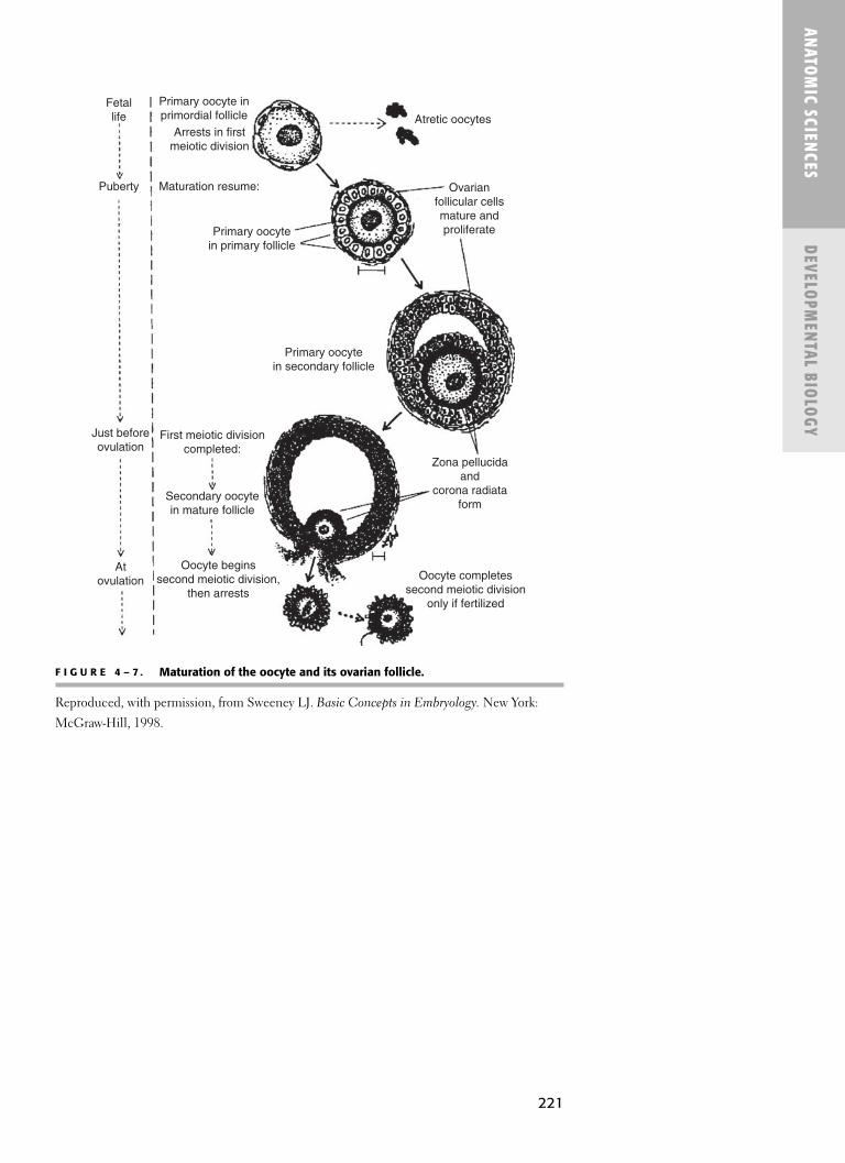
221
ANATOM
IC SCIENCES
DEVELOPM
ENTAL BIOLOGY
Fetallife
Puberty Maturation resume:
Just beforeovulation
Atovulation
Primary oocyte inprimordial follicle Atretic oocytes
Primary oocytein primary follicle
Ovarianfollicular cellsmature andproliferate
Primary oocytein secondary follicle
First meiotic divisioncompleted:
Secondary oocytein mature follicle
Oocyte beginssecond meiotic division,
then arrests
Zona pellucidaand
corona radiataform
Oocyte completessecond meiotic division
only if fertilized
Arrests in firstmeiotic division
F I G U R E 4 – 7 . Maturation of the oocyte and its ovarian follicle.
Reproduced, with permission, from Sweeney LJ. Basic Concepts in Embryology. New York:
McGraw-Hill, 1998.

222
ANAT
OM
IC S
CIEN
CES
DEVE
LOPM
ENTA
L BI
OLO
GY
F I G U R E 4 – 8 . Development of the embryo and its travels during week 1.
Reproduced, with permission, from Sweeney LJ. Basic Concepts in Embryology. New York:
McGraw-Hill, 1998.
Embryonic development on the way to the uterus
Cleavage divisions
First division Late morulaEarly morula Blastula
Uterine tube
Uterineendometrium
Cervix ofuterus
Zygote
Blastula begins to implantin uterine endometrium
Endometrialgland
Trophoblast
Syncytiotrophoblastinvades
uterine endometrium
Embryoblast(inner cell mass)
Maternalblood
vessels
Ectopic sitesof implantation:
1. Uterine tube2. Cervical opening of uterus3. Ovary4. Peritoneal cavity lining
4
Intestines
Peritoneal lining
1
13
∗Normal sites ofimplantation outlined
1
2
IMPLANTATION
See Figure 4–8.
■ Occurs by the end of the first week.■ Trophoblast cells invade endometrial epithelium.■ Trophoblast produces hCG.■ See Figure 4–9 for cleavage stages.
Sperm become motile in the
epididymis.

223
ANATOM
IC SCIENCES
DEVELOPM
ENTAL BIOLOGY
Zygote
Morula
Blastula
Firstcleavagedivision
Blastomere
Zona pellucida
Two cell stage
Cleavage divisions
Zona pellucidabreaks down
Blastula cavityTrophoblast:
outer cell mass
Embryoblast:inner cell mass
Cross-sectionof blastula:
F I G U R E 4 – 9 . Cleavage stages: morula and blastula.
Reproduced, with permission, from Sweeney LJ. Basic Concepts in Embryology. New York:
McGraw-Hill, 1998.
BILAMINAR DISC
■ Occurs in the second week.■ Epiblast (primary ectoderm).
■ Amniotic cavity.■ Ultimately gives rise to:
■ Ectoderm.■ Mesoderm.
■ Hypoblast (primary endoderm).■ Lining of yolk sac.
See Figure 4–10 for placentation.See Figure 4–11 for twinning.
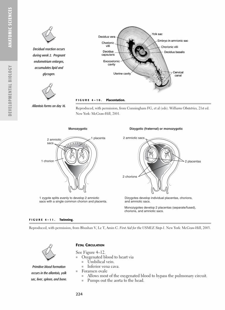
ANAT
OM
IC S
CIEN
CES
DEVE
LOPM
ENTA
L BI
OLO
GY
Monozygotic
1 zygote splits evenly to develop 2 amnioticsacs with a single common chorion and placenta.
1 chorion
2 amnioticsacs
1 placenta
Dizygotes develop individual placentas, chorions, and amniotic sacs.
Monozygotes develop 2 placentas (separate/fused),chorions, and amniotic sacs.
Dizygotic (fraternal) or monozygotic
2 placentas
2 amniotic sacs
2 chorions
F I G U R E 4 – 1 1 . Twinning.
Reproduced, with permission, from Bhushan V, Le T, Amin C. First Aid for the USMLE Step-1. New York: McGraw-Hill, 2003.
F I G U R E 4 – 1 0 . Placentation.
Reproduced, with permission, from Cunningham FG, et al (eds). Williams Obstetrics, 21st ed.
New York: McGraw-Hill, 2001.
FETAL CIRCULATION
See Figure 4–12.■ Oxygenated blood to heart via
■ Umbilical vein.■ Inferior vena cava.
■ Foramen ovale■ Allows most of the oxygenated blood to bypass the pulmonary circuit.■ Pumps out the aorta to the head.
Decidual reaction occurs
during week 2. Pregnant
endometrium enlarges,
accumulates lipid and
glycogen.
Allantois forms on day 16.
Primitive blood formation
occurs in the allantois, yolk
sac, liver, spleen, and bone.
224

225
ANATOM
IC SCIENCES
DEVELOPM
ENTAL BIOLOGY
■ Deoxygenated blood returned via ■ Superior vena cava. ■ Mostly pumped through the pulmonary artery and ductus arteriosus to the:
■ Feet.■ Umbilical arteries.
EMBRYOLOGIC BODY AXES
See Table 4–1 for germ layer derivatives.See Figure 4–13 for anatomic orientation of the embryo. See Figure 4–14 for germ layer derivatives. See Figure 4–15 for stages of fetal life.
Foramen Ovale
■ In fetal heart allowsblood to flow from RAto LA.
■ Bypass pulmonarycircuit .■ Because
oxygenated bloodcomes from themother/placenta.
■ Closes with fibrousconnective tissue tobecome fossa ovalis inthe adult.
Pulmonaryartery
Left atrium
Leftventricle
Aorta
Umbilicalarteries
Portalvein
Umbilicalvein
Inferiorvena cava
Superior venacava
Foramenovale
Right atrium
Right ventricle
Ductusarteriosus
Ductus venosus
Fromplacenta
To placenta
F I G U R E 4 – 1 2 . Fetal circulation.
Reproduced, with permission, from Hay WW, Hayward AR, Levin MJ, Sondheimer JM. (eds).
Current Pediatric Diagnosis & Treatment, 16th ed. New York: McGraw-Hill, 2003.
After birth the foramen ovale
closes to become the fossa
ovalis, the ductus arteriosis
closes to form the ligamentum
arteriosum, and the umbilical
vein closes to form the
ligamentum teres hepatis.

226
ANAT
OM
IC S
CIEN
CES
DEVE
LOPM
ENTA
L BI
OLO
GY
T A B L E 4 – 1 . Germ Layer Derivatives
ECTODERM DERIVATIVES
Epithelium of skin (superficial All nervous tissue: formed by neuroectoderm:
epidermis layer) Brain and spinal cord (neural tube)
All peripheral nerve tissue (neural crest)
ENDODERM DERIVATIVES
EPITHELIAL LININGS OF:
The gastrointestinal tract Hypoblast endoderm: Gametes migrate to gonads
Organs that form as buds
from the endoderm tube:
Pharyngeal gland derivatives*
Respiratory system
Digestive organs (liver, pancreas)
Terminal part of urogenital
systems
MESODERM DERIVATIVES
ALL CONNECTIVE TISSUES† ALL MUSCLE TYPES: EPITHELIAL LININGS OF:
General connective tissues Cardiac, skeletal, smooth Body cavities
Cartilage and bone Some organs:
Blood cells (red and white) Cardiovascular system
Reproductive and urinary
systems (most parts)
*Pharyngeal derivatives: palatine tonsils, thymus, thyroid, parathyroids.†Some connective tissues in the head are derived from neural crest.
Cranial
Dorsal
Lateral view
Caudal
Cranial
Ventral orfrontal view
Caudal
Ventral
F I G U R E 4 – 1 3 . Embryonic body axes.
Reproduced, with permission, from Sweeney LJ. Basic Concepts in Embryology. New York:
McGraw-Hill, 1998.

227
ANATOM
IC SCIENCES
DEVELOPM
ENTAL BIOLOGY
Somite formed into sclerotome,dermatome, and myotome regions
AB
Cells of each SOMITE regionmigrate separately to targetdestinations before forming
specific tissues:
Sclerotome cellsmigrate medially
to form bones(vertebrae and ribs)
Dermomyotomecells then migrate
Dermatome
Myotome
Intermediatemesoderm
Lateral platemesoderm expands
to form limb bud
Endoderm tube
Cross sections
In limbs
Withinbody
Myotomecells migrate to form
skeletal muscle
Dermatomecells migrate under ectoderm
to form connective tissue ofskin (dermis)
F I G U R E 4 – 1 4 . Dermatome, myotome, and sclerotome derivatives.
Reproduced, with permission, from Sweeney LJ. Basic Concepts in Embryology. New York:
McGraw-Hill, 1998.

228
ANATOM IC SC I E NC ESDEVE LOPM E NTAL B IOLOGY
Weeks
General Implantation
Brain
Face
Eyes
Ears
Diaphragm
Lungs
Heart
Intestines
Urinary tract
Genitalia
Axial skeleton
Limbs
1 2 3 4 5 6 7 8 9 12 16 20 24 28 32 36 38
Neural tubeHemispheres, cerebellum,ventricles, choroid plexus Temporal lobe, sulci, gyri; cellular migration, myelinization
Optic cups, lens, optic nerves, eyelids
Transverse septum, diaphragm
Vertebral cartilage,ossification centers
Buds, rays, webs, separate digits
Tracheoesophageal septum, bronchi, lobes
Primitive tube, great vessels, valves, chambers
Foregut, liver, pancreas, midgut
Genital folds, phallus; labioscrotal swelling
Mesonephric duct Glomeruli
Organogenesis Fetal growth
Pinnae
Abdominalwall; rotation
Metanephric duct, collecting system
Canals, cochlea, inner ears, ossicles
Lips, tongue, palate; cavitation, fusion
Penis,urethra, scrotum
Clitoris, labia
F I G U R E 4 – 1 5 . Stages of fetal life.
Reproduced, with permission, from Hay WW, Hayward AR, Levin MJ, Sondheimer JM. (eds). Current Pediatric Diagnosis & Treatment, 16th ed.
New York: McGraw-Hill, 2003.

229
ANATOM
IC SCIENCES
DEVELOPM
ENTAL BIOLOGY
Central canal
Marginal zone
Intermediate ormantle zone
Ventricular orependymal zone
Alar plate(Dorsal horn)
Basal plate(Ventral horn)
Notochord signals induce formation ofmotor neurons in ventral horn
Mantle zonenerve cell bodies
Marginal zonenerve processes
Ventral fissure
Week 4
Week 6
Alar plate neuronsreceive sensory
input
Basal plate motorneurons sendprocesses to
periphery
F I G U R E 4 – 1 6 . Development of regional specialization across the neural tube.
Reproduced, with permission, from Sweeney LJ. Basic Concepts in Embryology. New York:
McGraw-Hill, 1998.
Embryology of the Central Nervous System
See Figure 4–16.
■ Neural plate.■ Neural tube.■ Alar plate: Alar = sensory (posterior, dorsal).■ Basal plate: Basal = motor (anterior, ventral).■ See Figure 4–17 for formation of the nervous system.
The neural plate invaginates
to form neural groove with
neural folds on each side on
day 18 of embryonic life
(two neural folds fuseforming
the neural tube).
Neuroectoderm (ectoderm of
neural plate) gives rise to CNS
(brain and spinal cord).
Week 4 of embryologicdevelopment:
■ Neural tube is closed.■ Four pairs of branchial
arches are visibleexternally.
■ Characteristic C-shaped curvature ofembryo is appreciateddue to folding.
■ Upper and lower limbbuds appear.
Organogenesis occurs from
week 3 to 8.

230
ANAT
OM
IC S
CIEN
CES
DEVE
LOPM
ENTA
L BI
OLO
GY
EMBRYOLOGY OF THE BRAIN
F I G U R E 4 – 1 7 . Neuroectoderm forms the neural tube and neural crest in late week 3(A and B) and early week 4 (C).
Reproduced, with permission, from Sweeney LJ. Basic Concepts in Embryology. New York:
McGraw-Hill, 1998.
A
Dorsal views: Cross sections:
Cut edge of amnion
Primitive streakPrimitive node
Plane ofsection
Neural foldsB
C
Notochord
Neural plate
Ectoderm
Endoderm
Neuralgroove
Futureneural crest
Futureneural tube
Neural foldsnot yetfused
Neural foldsfused to formneural tube
Neural crestcells
Forming mesodermPrimitive streak
Embyologic Adult Division Subset Second Subset Derivative
Forebrain Prosencephalon Telencephalon Cerebrum, basal ganglia
Diencephalon ThalamusHypothalamusEpithalamusSubthalamusPosterior pituitary
(neurohypophysis)Midbrain Mesencephalon MidbrainHindbrain Metencephalon Pons
Myelencephalon Medulla oblongata
Brain stem = midbrain +
hindbrain
Brain stem = midbrain +
pons + medulla.

231
ANATOM
IC SCIENCES
DEVELOPM
ENTAL BIOLOGY
Gives Rise To
Neural crest Sensory ganglia (CNs V, VII, IX, X)Dorsal root ganglia (in peripheral nervous system)Schwann cells (peripheral)Melanocytes and odontoblastsEnterochromaffin cellsNeurons in parasympathetic and sympathetic
ganglia (adrenal medulla)Leptomeninges (pia and arachnoid)Parafollicular cells (C-cells) of parathyroid
Neuroepithelial Neuroblasts → neuronsEpendymal cells (lining ventricles, central canal)Glioblasts-astrocytes, oligodenrocytes (myelin in CNS)
Mesenchymal Microglia(mesoderm)
NEURAL TUBE CELLS
Aortic Arch Adult Analogue
1 Maxillary artery2 Hyoid artery
Stapedial artery3 Common carotid artery
ICA (first part)4 Aortic arch
Right subclavian (proximal)5 Involutes6 Pulmonary arteries (proximal)
Ductus arteriosus
Embryology of the Cardiac System
AORTIC ARCH DERIVATIVES
See Figure 4–18.
■ Aortic arch arteries■ Form during week 4.■ Arise from distal truncus arteriosus (aortic sac).■ Associated with corresponding pharyngeal arch.■ Connect to the paired dorsal aortae.
■ Dorsal aortae fuse in week 5 to form:■ Descending thoracic aorta.■ Abdominal aorta.
See Figure 4–19 for cardiac development. See Figure 4–20 for heart embryology.
The cardiovascular system is
derived from mesoderm.
Atrial Septal Defect
■ Results fromincomplete fusion ofseptum primum andseptum secundum.
■ Often asymptomaticuntil middle age.
■ Located near theforamen ovale.
Patent Foramen Ovale
■ Failure of completefusion of foramenovale.
■ Usually of nohemodynamicsignificance.
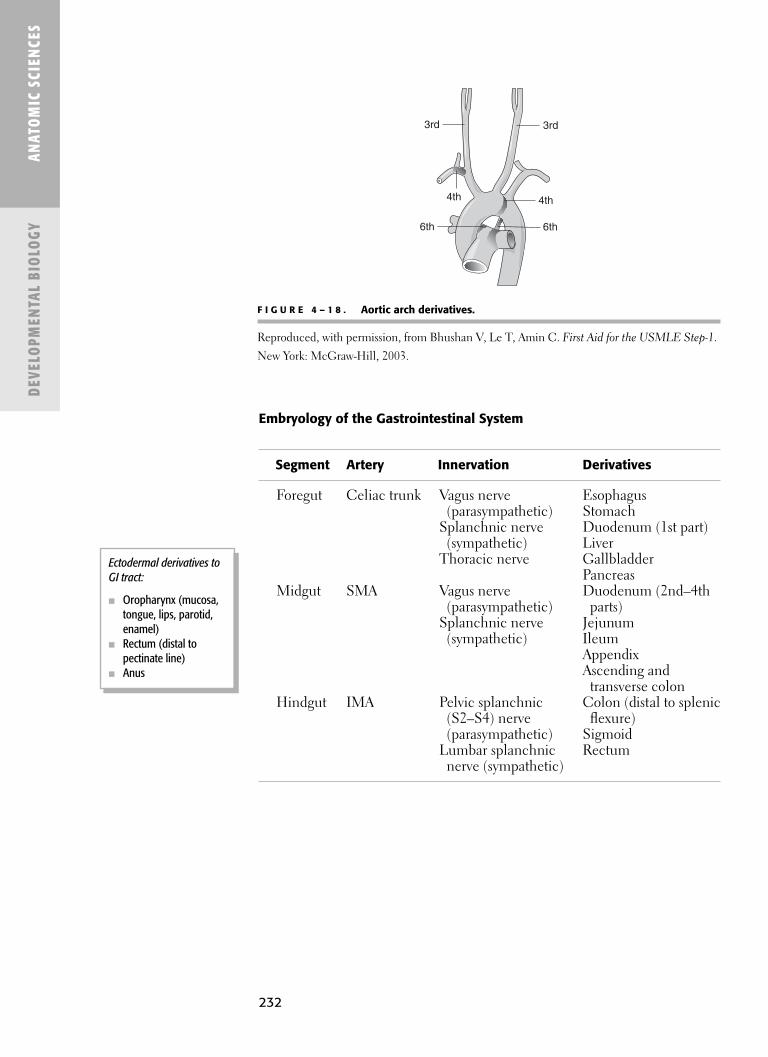
232
ANAT
OM
IC S
CIEN
CES
DEVE
LOPM
ENTA
L BI
OLO
GY
Ectodermal derivatives toGI tract:
■ Oropharynx (mucosa,tongue, lips, parotid,enamel)
■ Rectum (distal topectinate line)
■ Anus
Segment Artery Innervation Derivatives
Foregut Celiac trunk Vagus nerve Esophagus(parasympathetic) Stomach
Splanchnic nerve Duodenum (1st part)(sympathetic) Liver
Thoracic nerve GallbladderPancreas
Midgut SMA Vagus nerve Duodenum (2nd–4th (parasympathetic) parts)
Splanchnic nerve Jejunum(sympathetic) Ileum
AppendixAscending and
transverse colonHindgut IMA Pelvic splanchnic Colon (distal to splenic
(S2–S4) nerve flexure)(parasympathetic) Sigmoid
Lumbar splanchnic Rectumnerve (sympathetic)
Embryology of the Gastrointestinal System
4th 4th
3rd 3rd
6th 6th
F I G U R E 4 – 1 8 . Aortic arch derivatives.
Reproduced, with permission, from Bhushan V, Le T, Amin C. First Aid for the USMLE Step-1.
New York: McGraw-Hill, 2003.
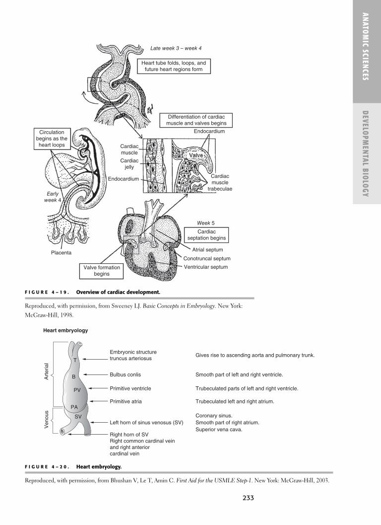
233
ANATOM
IC SCIENCES
DEVELOPM
ENTAL BIOLOGY
F I G U R E 4 – 1 9 . Overview of cardiac development.
Reproduced, with permission, from Sweeney LJ. Basic Concepts in Embryology. New York:
McGraw-Hill, 1998.
Late week 3 – week 4
Heart tube folds, loops, andfuture heart regions form
Differentiation of cardiacmuscle and valves begins
Endocardium
Cardiacmuscle
trabeculae
Week 5
Cardiacseptation begins
Atrial septum
Conotruncal septum
Ventricular septumValve formationbegins
Placenta
Earlyweek 4
Endocardium
Cardiacjelly
Cardiacmuscle
Circulationbegins as theheart loops
F I G U R E 4 – 2 0 . Heart embryology.
Reproduced, with permission, from Bhushan V, Le T, Amin C. First Aid for the USMLE Step-1. New York: McGraw-Hill, 2003.
Ven
ous
Art
eria
l T
B
PV
PA
SV
Embryonic structuretruncus arteriosus
Heart embryology
Gives rise to ascending aorta and pulmonary trunk.
Smooth part of left and right ventricle.
Trubeculated parts of left and right ventricle.
Trubeculated left and right atrium.
Bulbus conlis
Primitive atria
Primitive ventricle
Left horn of sinus venosus (SV)
Right horn of SVRight common cardinal veinand right anteriorcardinal vein
Coronary sinus.Smooth part of right atrium.Superior vena cava.

234
ANAT
OM
IC S
CIEN
CES
DEVE
LOPM
ENTA
L BI
OLO
GY
Embryology of the Renal System
Week of Structures Structure Development Formed
Pronephros 4 RegressesMesonephros 4 Mesonephric duct (gives rise to):
Ductus deferensEpididymisEjaculatory ductSeminal vesicleUreteric bud (forms):UreterRenal pelvisCalycesCollecting tubules
Metanephros 5 Adult kidney (from):Ureteric budMetanephric massKidney forms in pelvis but
“ascends” to abdomen with fetal growth
Ureters lengthen
Wolffian duct
(mesonephric duct).
Embryonic duct develops into
deferent duct, etc. in males;
obliterated in females.
Embryology of the Renal System
See Figure 4–21 for the three stages of kidney development.
■ From intermediate mesoderm■ Nephrogenic cord gives rise to:
■ Pronephros.■ Mesonephros.■ Metanephros.
See Figure 4–22 for embryology of the pancreas.

ANATOM
IC SCIENCES
DEVELOPM
ENTAL BIOLOGY
Pronephric kidney
Forms and degenerates during week 4
Nephric duct
Undifferentiatedintermediate mesoderm
Degenerating pronephric kidney
Nephric ductforms cranial
end ofmesonephric
duct
Mesonephrictubules form incranial-caudal
sequence
Mesonephric ductfuses with
hindgut cloaca
Mesonephric ductpersists as part of
male genital system
Metanephric kidney
Begins to form mid week 5
Ureteric bud forms frommesonephric duct
Metanephric cap ofintermediate mesoderm
Mesonephric kidney
Begins to form at the end of week 4;persists throughout month 2
Genitalintermediatemesoderm
Urogenital sinusforms from cloaca
F I G U R E 4 – 2 1 . The three stages of kidney development.
Reproduced, with permission, from Sweeney LJ. Basic Concepts in Embryology. New York:
McGraw-Hill, 1998.
235

236
ANAT
OM
IC S
CIEN
CES
DEVE
LOPM
ENTA
L BI
OLO
GY
F I G U R E 4 – 2 3 . Formation of neurocranial and viscerocranial regions of the head.
Reproduced, with permission, from Sweeney LJ. Basic Concepts in Embryology. New York:
McGraw-Hill, 1998.
Pharyngeal arches forming
Neural crest cells migrateinto pharyngeal arches
Viscerocranium formsaround endoderm tubefrom pharyngeal arch
tissues
5 weeks
4 weeks
3.5 weeks
Endoderm tubeforms mouth andpharynx linings
Viscerocranium
Nasalcavity
Neurocranium forms around cranial neural tube(future brain)
� E M BRYOLOGY OF TH E FAC E AN D PHAYRNG EAL ARC H ES
See Figure 4–23 for formation of the neurocranium and viscerocranium.
F I G U R E 4 – 2 2 . Embryology of the pancreas.
Reproduced, with permission, from Bhushan V, Le T, Amin C. First Aid for the USMLE Step-1.
New York: McGraw-Hill, 2003.

237
ANATOM
IC SCIENCES
DEVELOPM
ENTAL BIOLOGY
Branchial Arches
■ Rounded, mesodermal ridges (neural crest cells). ■ Form from proliferative activity of neural crest cells.■ Each arch contains nerve, artery, muscle, and cartilaginous bar.
■ Nerves (CNs V, VII, IX, X) are branchiomeric because they originatefrom the branchial arches.■ Not from somites. ■ Develop about week 4 of life.
■ At the end of week 4 the arches are well-defined and visible externally.■ During week 4 the first branchial arch divides into:
■ Mandibular process.■ Maxillary process.■ Weeks 5–6, arches are smaller, not seen on surface.■ Arches 1–3 play role in forming face and oral cavity.
■ Arch 1 ■ Mandible ■ Maxilla (most)
■ Arches 2 and 3 ■ Tongue
See Figure 4–24 for derivatives in the pharyngeal region.
Third arch
■ Hyoid bone■ Tongue formation
(root-posterior third)■ Hypobranchial
eminence
I(Mandibular)
II(Hyoid)
III
IV
IMaxillary a.
IIHyoid a.
stapedial a.
IIIInternal
carotid a.
IVR. subclaviana. and aorta
VTrigeminal
VIIFacial
IXGlosso-
pharyngeal
XVagus
Muscles ofmastication,
tensor tympani,mylohyoid,tensor veli
palatini, ant.belly digastric
Muscles offacial expres-
sion, stapedius,stylohyoid.post. bellydigastric
Stylo-pharyngeus
Pharyngealand laryngealmusculature
Malleus, incus,sphenomandibularligament, Meckel’s
cartilage
Stapes, styloidprocess, stylohyoid
ligament, lessercornu of hyoid,
part of body of hyoid
Archnumber
Aorticarch
Cranialnerve
Examples of branchiomeric
muscles
Skeletalderivatives
Greater cornu ofhyoid, part ofbody of hyoid
Laryngealcartilages
VI VI Pulmonary
arteriesand ductusarteriosis
XVagus andrecurrentlaryngeal
nerve
LarynxmusclesEXCEPT
cricothyroid
Cricoid,arytenoid and
corniculatecartilages
Branchial arch structures
Adultderivative of
branchialgroove
Externalauditorymeatus
Cervicalsinus
Externalear
I
II
III
IV
I
II
III
IV
Middle earauditory
tube
Adultderivative ofpharyngeal
pouchand liningstructures
Supra-tonsillarfossa
Thymus,inferior
parathyroidgland
Superiorparathyroid
gland,postbranchial
body
F I G U R E 4 – 2 4 . Derivatives in the pharyngeal region.
Reproduced, with permission, from Carlson BM. Patten’s Foundations of Embryology. 5th ed. New York: McGraw-Hill, 1988.

238
ANAT
OM
IC S
CIEN
CES
DEVE
LOPM
ENTA
L BI
OLO
GY
Nerve V
Nerve VII
Nerve IX
Nerve IX
Nerve X
Nerve X
B
Nerve VNerve VII
TemporalisOccipitalis
AI
II III IV
Fac
ial e
xpre
ssio
n
Auricularis
Stylopharyngeus
Posterior bellyof digastric
Pharyngeal muscles
C
Anterior bellyof digastric
MasseterStyloid processStylohyoid ligament
Greater hornof hyoid bone
Thyroid cartilage
Malleus
IncusStapes
Cricoid cartilage
D
Meckel’scartilage
Lesser hornof hyoid bone
F I G U R E 4 – 2 5 . Schematic diagram showing major derivatives of structures that constitute the branchial arches.
(A) 5-week embryo; (B–D) 4- to 5-month fetuses. The gray tones of structures in C and D corre-
spond to those of the branchial arches depicted in A.
Reproduced, with permission, from Carlson BM. Patten’s Foundations of Embryology. 5th ed.
New York: McGraw-Hill, 1988.
First arch
■ See Figure 4–25.■ Meckel’s cartilage
■ Model for themandible.
■ Dissolutes with onlyminor contribution toossification.
■ Mandibular process(forms mandible).
■ Maxillary process(forms maxilla,zygoma, squamoustemporal).
■ Tongue formation(anterior two-thirds).■ Tuberculum impar
(median tonguebud)
■ Lateral lingualswellings (2) (distaltongue buds)

239
ANATOM
IC SCIENCES
DEVELOPM
ENTAL BIOLOGY
Pharyngeal Pouches
See Figure 4–26.
■ Paired evaginations of pharyngeal endoderm that lines inner aspects ofbranchial arches
Pouch Structures
1 Tympanic membraneAuditory tubeMiddle ear cavity
2 Lymphatic nodulePalatine tonsil
3 Inferior parathyroid glandThymus
4 Superior parathyroid glandUltimobranchial body ■ Gives rise to thyroid parafollicular/C-cells■ Calcitonin
5 Rudimentary structure (becomes part of pouch 4)
Cervical sinus
Pharyngeal pouch 1
Parathyroid III
Thymus III
Postbranchial body
Parathyroid IV
Thymus IV(may be vestigial)
Faucial tonsil in wall ofpharyngeal pouch 2
Branchial groove 1(hyomandibular)
Tubotympanicrecess
Externalauditorymeatus
Foramencecum
Thyroidprimordium
Line of descentof thyroid
Thymus III
Thymus III
Parath. III
Parath. IV
Postbranchialbody
CAB
Thymus IV
Parath. IV
Thyroid
1
2
3
4
1
2
4
3
F I G U R E 4 – 2 6 . Diagrams showing the origin of the pharyngeal derivatives.
(A) The primary relationships of the several primordial to the pharyngeal pouches. (B) Course of
migration of some of the primordial from their place of origin. (C) Definitive relations of parathy-
roids, postbranchial body, thymus, and thyroid as they appear in a transverse section of the right
lobe of the thyroid taken above the level of the isthmus. Abbreviation: Parath., parathyroid.
Reproduced, with permission, from Carlson BM. Patten’s Foundations of Embryology. 5th ed.
New York: McGraw-Hill, 1988.

240
ANAT
OM
IC S
CIEN
CES
DEVE
LOPM
ENTA
L BI
OLO
GY
Formation of the Face
MANDIBLE
■ Two mandibular processes (branchial arch 1) merge.■ Medial ends merge at week 4.■ Mandibular processes → merge to form lower lip.
MAXILLA
■ Two maxillary processes (branchial arch 1) merge.■ Form upper cheek regions and most of upper lip.
FRONTAL NASAL PROCESSES
■ Form with growth of forebrain.■ Develop into forehead and nose.■ Two medial nasal processes merge. ■ Form philtrum of upper lip.
NASAL PLACODES
■ Thickened areas of specialized ectoderm. ■ Located on either side of the frontal nasal process.■ Give off elevations at their margins.■ Lateral nasal processes (2).
■ Form sides/alae of nose.■ Medial nasal processes (2).
■ Form bridge of nose.■ Nostrils.■ Upper lip philtrum.
Mouth and Oral Cavity
See Figure 4–27.
■ Begins as a slight depression on the stomodeum. ■ Located between branchial arch 1 and the forebrain.■ Buccopharyngeal membrane (oropharyngeal membrane)
■ Thin bilaminar membrane.■ Composed of ectoderm externally and endoderm internally.■ Separates the stomodeum from the primitive pharynx (until rupture).■ Ruptures around week 31/2 (~24 days). ■ Connecting the primitive mouth and primitive pharynx.
See Figure 4–28 for formation of the nasal cavities and the teeth.
FORMATION OF THE PALATE
See Figure 4–29.
The plane passing through theright and left anterior pillars
marks the separation betweenthe oral cavity and
oropharynx in the adult.
The vestibular lamina separatesthe lips and cheeks externally
and jaw structures internally inthe developing embryo.
The stomodeum is the primitivemouth; lined by ectoderm.
Nasal processes are proli-ferations of mesenchyme at themargins of the nasal placodes.The nasal placodes develop on
the lower part of the frontonasalprocess, on each side.
The mandible and maxilla,
except the mandibular
condyles, are formed by
intramembranous ossification.
Most of the upper lip is formed
by the maxillary processes.
The corner of the mouth is
formed by the fusion of the
maxillary and mandibular
processes.

241
ANATOM
IC SCIENCES
DEVELOPM
ENTAL BIOLOGY
Lateralviews
Frontal views
Earlyweek 4
Lateweek 4
Week 5
Week 6
Plane offrontal view
Ear
Eye
Formation of facialprominences
Frontonasal prominence
Arch 1 derivatives:Maxillary prominence
Mandibular prominence
Mandibular prominence
Oticplacode
Stomodeum
Frontonasal prominence
Maxillary prominence
Arch 2
Arch 3
Nasal placode derivatives:Medial nasalprominence
Lateral nasalprominence
Lensplacode
Medial nasalprominence
Lateral nasalprominence
Nasolacrimalgroove
Maxillaryprominence
Mandibularprominence
Mouth opening
Eye
Nasalpit
Nasalplacode
F I G U R E 4 – 2 7 . (A) Development of the face from facial prominences. (B) Developmentof the mature face and formation of cleft lip defects.
Reproduced, with permission, from Sweeney LJ. Basic Concepts in Embryology. New York:
McGraw-Hill, 1998.
Lateral cleft lip occurs becausethe maxillary and medial nasalprocesses fail to fuse. Cleft lipcan be unilateral or bilateral.

242
ANAT
OM
IC S
CIEN
CES
DEVE
LOPM
ENTA
L BI
OLO
GY
Final fusion of facial prominencesFrontalviews
Week 6
Week 7
Medial nasal prominences fuse togtherto form intermaxillary segment
Eye
Medial nasalprominences grow
down into stomodeumand fuse with maxillary
prominences
Frontonasal prominenceforms forehead, nose,philtrum of upper lip
Week 10
Week 7
Forehead
Nose
Philtrum
Maxillary prominencesform upper lip and cheeks
Mandibular prominencesform lower lip and face
Nasolacrimal groove
Cleft lip
Forms from failure of fusion ofmedial nasal prominenceswith maxillary prominences
Week 10
Complete bilateral cleft lips
Incompletefusion
Completefailure of
fusion
Incompleteunilateralcleft lip
Normalfusion
Each can occurunilaterally or bilaterally
F I G U R E 4 – 2 7 . (Continued)
PRIMARY PALATE
■ Forms from merging of the two median nasal processes.■ Becomes the premaxillary part of the maxilla.
■ Maxilla anterior to incisive foramen in adults.■ Contains central and lateral incisors.
SECONDARY PALATE
■ Forms from the two maxillary processes (lateral palatine processes).■ Forms hard and soft palates. ■ Extends from incisive foramen posteriorly.
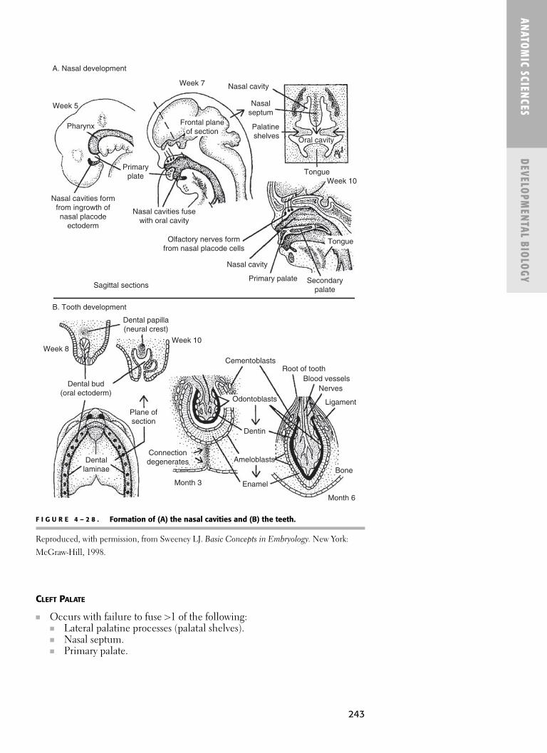
243
ANATOM
IC SCIENCES
DEVELOPM
ENTAL BIOLOGY
CLEFT PALATE
■ Occurs with failure to fuse >1 of the following:■ Lateral palatine processes (palatal shelves).■ Nasal septum.■ Primary palate.
A. Nasal development
B. Tooth development
Week 5
Week 7 Nasal cavity
Nasalseptum
Palatineshelves Oral cavity
TongueWeek 10
Tongue
Secondarypalate
Primary palate
Nasal cavity
Olfactory nerves formfrom nasal placode cells
Nasal cavities fusewith oral cavity
Primaryplate
Nasal cavities formfrom ingrowth ofnasal placode
ectoderm
Sagittal sections
Dental papilla(neural crest)
Week 8Week 10
Pharynx Frontal planeof section
Dental bud(oral ectoderm)
Plane ofsection
Dentallaminae
Connectiondegenerates
Enamel
Ameloblasts
Dentin
Odontoblasts
CementoblastsRoot of tooth
Blood vesselsNerves
Ligament
Bone
Month 3
Month 6
F I G U R E 4 – 2 8 . Formation of (A) the nasal cavities and (B) the teeth.
Reproduced, with permission, from Sweeney LJ. Basic Concepts in Embryology. New York:
McGraw-Hill, 1998.

244
ANAT
OM
IC S
CIEN
CES
DEVE
LOPM
ENTA
L BI
OLO
GY
Palate formation
Week 5Nasal
prominences
Maxillaryprominences
formpalatine shelves
Ventral views of roof of mouth
Intermaxillary segmentforms primary palate
Formingnasal septum
Week 7
Week 10
Week 5
Nasalprominences
Primary palate
Palatineshelves
formsecondary
palate
Frontal view
Cleft palate types
Failure offusion ofprimary
andscondarypalates;
combinedwith cleft lip
Failure offusion ofpalatineshelves
Cleftlip
Primary andsecondary
palates fuse
Hard palate
Soft palate
F I G U R E 4 – 2 9 . Normal formation of the mouth and palate and defects in palateformation.
Reproduced, with permission, from Sweeney LJ. Basic Concepts in Embryology. New York:
McGraw-Hill, 1998.
VARIATIONS OF CLEFT LIP AND PALATE
■ May involve uvula only, or extend through hard and soft portions of palate.■ Cleft uvula only.■ Unilateral cleft of secondary palate.■ Bilateral cleft of secondary palate.■ Complete unilateral cleft.
■ Lip.■ Alveolar process (of maxilla). ■ Primary palate.
■ Complete bilateral cleft (except secondary palate).■ Lip.■ Alveolar process (of maxilla). ■ Anterior palate. ■ Secondary palate (unilateral).
■ Complete bilateral cleft. ■ Lip.■ Alveolar process.■ Anterior (primary) palate.■ Posterior (secondary) palate.
Embryologically, the tongue
is derived from the first four
pharyngeal arches; and
innervation comes from
associated nerves of those
arches (arch 1, V; arch 2 VII;
arch 3 IX; arch 4, X).
Bifid tongue occurs because
of lack of fusion of distal
tongue buds (lateral
swellings).

245
ANATOM
IC SCIENCES
DEVELOPM
ENTAL BIOLOGY
TONGUE DEVELOPMENT
See Figure 4–30.
■ Begins in the first week with the formation of the tuberculum impar.■ Tuberculum impar.
■ Median, triangular elevation.■ Appears on floor of pharynx, just in front of foramen cecum.
Week 5 Pharyngeal arches form tongue
Somitic myotomes
Tonguemuscles
migrate intotongue
Plane of frontalsections
1 23
4
Pharynx
Frontalsections
Copula
Mouth
Arches 2–4contributions
to tongue:copula
Arch 1contributions:
Tuberculum imparLateral lingual
swellings
Posterior tongue:arches 3 and 4 overgrow arch 2
Foramen cecum
Anterior tongue (arch 1)
Week 5
Week 7
4
3
2
1
Epiglottal swellings
6
4
3
2
1
Week 6
Epiglottis
Posteriortongue
Foramencecum
Anterior tongue
Cranial nerves innervatetongue components
6
4
3
2
1
109
75
ArchesTongue muscles
12
F I G U R E 4 – 3 0 . Development of the tongue.
Reproduced, with permission, from Sweeney LJ. Basic Concepts in Embryology. New York:
McGraw-Hill, 1998.

246
ANAT
OM
IC S
CIEN
CES
DEVE
LOPM
ENTA
L BI
OLO
GY
Anterior Two-Thirds of Tongue
■ Lateral lingual swellings (distal tongue buds [2]).■ Form on each side of the tuberculum impar.■ From the proliferation of mesenchyme (arches 1, 2, 3).■ Swellings fuse to form the anterior two-thirds of tongue.
Posterior Third of Tongue:
■ Formed by two elevations.■ Copula (arch 2).■ Hypobranchial eminence (arch 3).

� Physical-ChemicalPrinciples
� Biological Compounds
� Metabolism
� Molecular Biology
� Membranes
� Neurophysiology
� Muscle Physiology
� Circulatory and CardiacPhysiology
� Respiratory Physiology
� Renal, Fluid, andAcid–Base Physiology
� GastrointestinalPhysiology
� Nutrition
� Endocrine Physiology
S E C T I O N 2
Biochemistry-Physiology
247

This page intentionally left blank

C H A P T E R 5
Physical-ChemicalPrinciples
Molecular Bonds 250
Water 250
CARBONIC ANHYDRASE 250
Laws of Thermodynamics 250
Calorimetry 250
Enzymology 250
ENZYMES 250
ENZYME MECHANICS 251
ENZYME KINETICS 252
ENZYME INHIBITION 254
ENZYME REGULATION 255
249

250
BIOC
HEM
ISTR
Y-PH
YSIO
LOGY
PHYS
ICAL
-CH
EMIC
ALPR
INCI
PLES
� MOLEC U LAR BON DS
■ Covalent bonds: Strong molecular interactions mediated by shared electrons.■ Noncovalent bonds: Weak, reversible molecular interactions.
■ Ionic bonds: Mediated by opposite electrostatic charges.■ Hydrogen bonds: Mediated by a shared hydrogen atom.■ Van der Waals bonds: A nonspecific attraction (occurs when any two
atoms are 3–4 Å apart).
� WATE R
■ Polar.■ Triangular.■ Highly cohesive.■ Excellent solvent for polar molecules.■ Weakens ionic and H-bonds.
Carbonic Anhydrase
See Figure 5–1.
■ Catalyzes the reaction between CO2 and H2O.■ Extremely fast enzyme.■ Located largely in erythrocytes and kidneys.■ A metalloenzyme: contains zinc.
Enthalpy is the heat content
of a system.
Entropy is the degree of
disorder of a system.
Most CO2 is transported in the
blood as bicarbonate.
F I G U R E 5 – 1 . Carbonic anhydrase reaction.
CO2 + H2O H2CO3 H+ + HCO3−
Carbonic anhydrase
Carbonic acid
� LAWS OF TH E RMODYNAM ICS
■ First law: The total energy of a closed system is conserved.■ Second law: The entropy of a closed system always increases.
� CALORI M ETRY
■ Direct calorimetry: Direct measurement of the amount of heat producedin a given system.
■ Indirect calorimetry: Measurement of the amount of heat produced interms of inhaled O2 and exhaled CO2.
� E N ZYMOLOGY
Enzymes
■ Highly specific catalysts for biochemical reactions. ■ Classified according to their mechanism of action.

251
BIOCHEM
ISTRY-PHYSIOLOGY
PHYSICAL-CHEM
ICALPRIN
CIPLES
■ Composed of proteins combined with nonprotein structures (eitherorganic or inorganic) that aid in their function: ■ Coenzymes■ Cofactors■ Prosthetic groups
DEFINITIONS (TABLE 5–1)
■ Coenzyme: Nonprotein portion of an enzyme.■ Apoenzyme: Protein portion of an enzyme. Catalytically inactive by itself.■ Haloenzyme: Complete, catalytically active enzyme.
■ Haloenzyme = Apoenzyme + Coenzyme■ Isozymes: Enzymes with subtle molecular differences that catalyze the
same reaction.
CLASSIFICATION
■ Oxidoreductases: Catalyze redox reactions.■ Transferases: Catalyze the transfer of functional groups.■ Hydrolases: Catalyze bond cleavage by hydrolysis.■ Isomerases: Catalyze a change in molecular structure.■ Lyases: Catalyze bond cleavage by elimination.■ Ligases: Catalyze the union of two molecules.
Enzyme Mechanics
INDUCED-FIT MODEL
See Figure 5–2.
■ Substrate-binding induces a conformational change in an enzyme.■ The energy produced by these changes enables the reactions to progress.
T A B L E 5 – 1 . Examples of Metallic Coenzymes of Various Enzymes
COENZYME ENZYME
Copper Cytochrome oxidase
Iron Catalase
Peroxidase
Magnesium Hexokinase
Glucose-6-phosphatase
Pyruvate kinase
Nickel Urease
Zinc Carbonic anhydrase
Alcohol dehydrogenase
Enzyme Classification:Over The HILL
Metals and B-complex
vitamins serve as the
majority of nonprotein
coenzymes.

252
BIOC
HEM
ISTR
Y-PH
YSIO
LOGY
PHYS
ICAL
-CH
EMIC
ALPR
INCI
PLES Enzyme Kinetics
SUBSTRATE CONCENTRATION
See Figures 5–3 and 5–4.
■ Increasing substrate concentration increases reaction rate only until theenzyme-binding sites are saturated.
■ Maximum reaction velocity (Vmax) is achieved when any further increasein substrate concentration does not increase reaction rate.
■ The Michaelis constant (Km) is the substrate concentration when the ini-tial reaction velocity (vi) is half of the maximum reaction velocity (Vmax).
■ Illustrated mathematically by the Michaelis–Menten equation.
GIBBS FREE ENERGY CHANGE (ΔG)
■ Determines reaction direction.■ If ΔGS > ΔGP, then ΔG will be negative and the reaction will proceed
spontaneously toward equilibrium.■ Equilibrium is attained when ΔG = 0.
A B
A B
A B
F I G U R E 5 – 2 . The induced-fit model of enzyme biomechanics.
Reproduced, with permission, from Murray RK. Harper’s Illustrated Biochemistry, 26th ed.
New York: McGraw-Hill, 2003.
vi =Vmax · [S]
Km + [S]
F I G U R E 5 – 3 . Michaelis–Menten equation.
Km reflects the affinity of the
enzyme for its substrate.
Vmax is directly proportional
to the substrate concentration.
Km is indirectly proportional to
enzyme affinity.
ΔG provides no information
about the reaction rate and is
independent of the path of the
reaction.
ΔG = ΔGP − ΔGS

253
BIOCHEM
ISTRY-PHYSIOLOGY
PHYSICAL-CHEM
ICALPRIN
CIPLES
■ Reactions are based on their ΔG (see Table 5–2).■ Exergonic■ Endergonic
REACTION DIRECTION
■ Determined by the ΔG
REACTION EQUILIBRIUM
Km
Vmax
Vmax/2
Vmax/2
vi
[S]
A
B
C
F I G U R E 5 – 4 . The effect of substrate concentration on reaction kinetics.
Reproduced, with permission, from Murray RK. Harper’s Illustrated Biochemistry, 26th ed.
New York: McGraw-Hill, 2003.
T A B L E 5 – 2 . Classification of Reactions Based on ΔG
REACTION TYPE ΔG ENERGY FLOW
Exergonic Negative Released
Endergonic Positive Required
A.
B. [C][D][Enz]
[A][B][Enz]Keq =
A + B + Enz C + D + Enz
■ A: Any reaction with enzyme present.■ B: Equilibrium constant of the reaction.■ Enzymes have no effect on reaction equilibrium.
REACTION RATE
■ Determined by the activation energy. ■ Attaining activation energy requires an increase in reactant kinetic energy.■ Kinetic energy is largely influenced by temperature and substrate concentration.■ Enzymes lower the activation energy of a reaction, accelerating the rate. ■ Influenced by five major factors. (See Table 5–3.)
Activation energy is the
energy needed to initiate
a reaction.

254
BIOC
HEM
ISTR
Y-PH
YSIO
LOGY
PHYS
ICAL
-CH
EMIC
ALPR
INCI
PLES
Enzyme Inhibition
COMPETITIVE INHIBITION
See Figure 5–5.
■ Inhibitor and substrate compete for the same binding site.■ Inhibition can be reversed with increased substrate concentration. ■ No effect on Vmax.■ Km is increased.
T A B L E 5 – 3 . Factors Influencing Reaction Rate
CONTRIBUTING FACTOR CHANGE IN FACTOR CHARACTERISTICS
pH Extreme changes can alter Enzymes function within an
the charged state of the optimal pH range.
enzyme or substrate.
Temperature An ↑ in temperature Extreme ↑ in temperature
will ↑ the reaction rate. can cause enzyme
denaturation.
Enzyme concentration An ↑ in enzyme concentration
will ↑ the reaction rate.
Inhibitor concentration An ↑ in inhibitor
concentration will ↓
the reaction rate.
Substrate concentration An ↑ in substrate Enzymes have a limited
concentration will ↑ number of active sites.
reaction rate only until the
enzyme-binding sites
are saturated.
[S]1
Km
1–K¢m1–
vi
1
Vmax
1
0
+ In
hibi
tor
No inhibitor
F I G U R E 5 – 5 . Competitive inhibition.
Reproduced, with permission, from Murray RK. Harper’s Illustrated Biochemistry, 26th ed.
New York: McGraw-Hill, 2003.
Kompetitive inhibition:
Km increases; Vmax does not
change.
Non-Kompetitive inhibition:
Km does Not change;
Vmax decreases.

255
BIOCHEM
ISTRY-PHYSIOLOGY
PHYSICAL-CHEM
ICALPRIN
CIPLES
NONCOMPETITIVE INHIBITION
See Figure 5–6.
■ Inhibitor and substrate bind simultaneously. ■ The two binding sites do not overlap.■ Inhibition cannot be reversed with increased substrate concentration. ■ Vmax is decreased.■ No effect on Km.
UNCOMPETITIVE INHIBITION
■ Inhibitor binds only after the substrate is bound first.■ The two binding sites do not overlap.■ Vmax is decreased.■ Km is decreased.
IRREVERSIBLE INHIBITION
■ Inhibitor irreversibly alters the molecular structure of an enzyme, prohibitingits continued activity.
Enzyme Regulation
COVALENT MODIFICATION
■ Reversible or irreversible enzymatic modification alters enzyme conforma-tion, thus affecting its activity.■ Phosphorylation (kinases)■ Dephosphorylation (phosphatases)■ Methylation (methyltransferases)
[S]1
Km
1–
V ¢max
1–
vi
1
Vmax
1
0
+ Inhib
itor
No inhibitor
F I G U R E 5 – 6 . Noncompetitive inhibition.
Reproduced, with permission, from Murray RK. Harper’s Illustrated Biochemistry, 26th ed.
New York: McGraw-Hill, 2003.
A noncompetitive inhibitor is
an allosteric inhibitor.
Aspirin irreversibly inhibits
cyclooxygenase by acetylating
serine.

256
BIOC
HEM
ISTR
Y-PH
YSIO
LOGY
PHYS
ICAL
-CH
EMIC
ALPR
INCI
PLES
ALLOSTERIC REGULATION
■ Allosteric enzyme: A regulatory enzyme that has both an active site (for thesubstrate) and an allosteric site (for the effector).
■ If the effector is present, it binds to the allosteric site causing a conforma-tional change to the active site, which then changes (increases or decreases)the enzymatic activity.
■ If no effector is present, the enzyme can still act on substrate normally (viathe active site) to produce product.
■ A form of feedback regulation in which an enzyme of a metabolic pathwayis controlled by the end product of that same pathway. (See Figure 5–7.)
■ Often catalyzes a committed step early in a metabolic pathway.■ Simple Michaelis–Menten kinetics are not followed.
PROTEASE ACTIVITY
See Figure 5–8.■ Proenzyme (zymogen): Catalytically inactive enzyme precursor.■ Proteases cleave the protein fragment (propeptide) of the zymogen, acti-
vating the enzyme.
(+/−)
Enz1 Enz2 Enz3A B C D
F I G U R E 5 – 7 . Feedback regulation.
Zymogens are enzymatically
inactive precursors of
proteolytic enzymes.
Enteropeptidase
Trypsinogen Trypsin
ThrombinFibrinogen Fibrin
F I G U R E 5 – 8 . Examples of protease activity.

C H A P T E R 6
Biological Compounds
Carbohydrates 258
CARBOHYDRATE STRUCTURE 258
STORAGE OF POLYSACCHARIDES 259
STRUCTURAL POLYSACCHARIDES 260
BACTERIAL POLYSACCHARIDES 262
COMPLEX CARBOHYDRATES 263
SALIVA 263
Proteins 264
AMINO ACIDS 264
PROTEIN STRUCTURE 266
PHYSIOLOGICALLY RELEVANT PROTEINS 267
Lipids 271
LIPID TRANSPORT 274
LIPID STORAGE 275
257

258
BIOC
HEM
ISTR
Y–PH
YSIO
LOGY
BIO
LOGI
CAL
COM
POUN
DS
� CARBOHYDRATES
Carbohydrate Structure
GLUCOSE
■ The most fundamental carbohydrate; required for carbohydrate metabo-lism, storage, and cellular structure.
■ If carbohydrates are not absorbed by dietary intake, they are generally con-verted to glucose in the liver.
CARBOHYDRATE CLASSIFICATION
■ Monosaccharides: The simplest carbohydrates. ■ Further classified by:
■ Number of carbon atoms: Trioses, tetroses, pentoses, hexoses, heptoses.■ Functional group: Aldoses (aldehyde) or ketoses (ketone) (Figures 6–1
and 6–2).■ Reducing sugars: Contain aldehyde groups that are oxidized to
carboxylic acids. For example: glucose, fructose, galactose, maltose,lactose.
■ Isomerism:■ D-form: Hydroxyl group on right. Most common form.■ L-form: Hydroxyl group on left.
■ Disaccharides: Glycosidic condensation of two monosaccharides. ■ Maltose → glucose + glucose; via maltase.■ Sucrose → glucose + fructose; via sucrase.■ Lactose → glucose + galactose; via lactase.
■ Oligosaccharides: Glycosidic condensation of 2–10 monosaccharides.■ Polysaccharides: Glycosidic condensation of >10 monosaccharides.
■ Mostly used as storage molecules or cellular structural components. ■ Can be linear or branched.
Only monosaccharides can be
absorbed in the small
intestine. Disaccharides are
hydrolyzed first by brush
border enzymes.
Sucrose is a non-reducing
sugar.
CAldehyde:
R
H O
C OKetone:
R′
R
F I G U R E 6 – 1 . Functional Groups.
OHH C
CHO
CH2OH
D-Glycerose
(D-glyceraldehyde)
OHH C
CHO
CH2OH
OHH C
D-Erythrose
OHH C
CHO
CH2OH
HHO C
HHO C
D-Lyxose
OHH C
CHO
CH2OH
OHH C
HHO C
D-Xylose
OHH C
CHO
CH2OH
HHO C
OHH C
D-Arabinose
OHH C
CHO
CH2OH
OHH C
OHH C
D-Ribose
OHH C
CHO
CH2OH
OHH C
OHH C
HHO C
D-Glucose
OHH C
CHO
CH2OH
HHO C
OHH C
HHO C
D-Mannose
OHH C
CHO
CH2OH
OHH C
HHO C
HHO C
D-Galactose
F I G U R E 6 – 2 . (A). Important aldoses.

BIOCHEM
ISTRY–PHYSIOLOGY
BIOLOGICAL CO
MPO
UNDS
259
Major Enzymes Important for Glycogenolysis
Enzyme Function
Glycogen phosphorylase Cleaves α-1,4 linkages
Glucantransferase Exposes α-1,6 branch points
Amylo-α-1,6 glucosidase Cleaves α-1,6 linkages
Storage of Polysaccharides
STARCH
■ A homopolymer of glucose linked by a–1, 4 glycosidic bonds. ■ The major glucose storage molecule in plants.■ Contains unbranched helical amylose (15–20%) and branched (a–1, 6)
amylopectin (80–85%).■ Amylases: The key enzymes in starch catabolism. (See Table below.)■ Dextrins: D-glucose polymer intermediates in starch hydrolysis. “Limit
dextrins” are the fragments that remain following hydrolysis.
Glycogen is stored in the liver.
Major Amylases Important for Starch Hydrolysis
Amylase Cleaves Hydrolysis Products
a-Amylase a-1,4 linkages (internal) Maltose + dextrins
b-Amylase a-1,4 linkages (from Maltosenonreducing ends)
g-Amylase a-1,4 linkages Glucose(glucamylase) a-1,6 linkages
GLYCOGEN
■ A homopolymer of glucose linked by a-1, 4 glycosidic bonds. ■ The major glucose storage molecule in animals.■ Contains numerous branch points via a-1, 6 glycosidic linkages.
F I G U R E 6 – 2 . (Continued) (B). Important ketoses.
Reproduced, with permission, from Murray RK. Harper’s Illustrated Biochemistry, 26th ed. McGraw-Hill, 2003.
O
HHO
OHH
OHH
C
C
C
C
C
OHH
CH2OH
CH2OH
D-Sedoheptulose
O
HHO
OHH
OHH C
C
C
C
CH2OH
CH2OH
D-Fructose
O
OHH
OHH C
C
C
CH2OH
CH2OH
D-Ribulose
O
HHO
OHH C
C
C
CH2OH
CH2OH
D-Xylulose
OC
CH2OH
CH2OH
Dihydroxyacetone
Isomaltase cleaves a-1, 6
branch points.
α-Amylase is secreted by
both the pancreas and the
parotid gland.

BIOC
HEM
ISTR
Y–PH
YSIO
LOGY
BIO
LOGI
CAL
COM
POUN
DS
260
Mucopolysaccharide Storage Diseases
Enzyme ClinicalDisease Deficiency Accumulation of Features Inheritance
Hurler’s a-L-iduronidase Heparan sulfate Developmental Autosomal syndrome Dermatan sulfate retardation recessive
Cornealclouding
GargoylismHunter’s L-iduronate Heparan sulfate Mild Hurler X-linked recessive
syndrome sulfatase Dermatan sulfate syndromeNo corneal
clouding
INULIN
■ A homopolymer of fructose.■ Highly water soluble. ■ Used to determine glomerular filtration rate (GFR).
Structural Polysaccharides
GLYCOSAMINOGLYCANS (GAGS)
See Table 6–1.
■ Heteropolymer chains containing repeating disaccharide units of an aminosugar (N-acetylglucosamine, N-acetylgalactosamine) and a uronic acid(glucaronic acid, iduronic acid).
■ The major structural polysaccharides of extracellular matrix (ECM), con-nective tissue (CT), and outer cell membrane surfaces.
■ Because they contain sulfate and carboxyl groups, GAGs are highly nega-tively charged and easily attract water, enabling them to cushion their sur-rounding structures.
■ Accumulation of various GAGs (due to enzyme deficiencies) results inseveral syndromic diseases.
Bacterial cell walls contain a
heteropolysaccharide
consisting of alternating
N-acetylglucosamine +
N-acetylmuramic acid
residues.
Glycogen Storage Diseases
Enzyme Glycogen Clinical Disease Deficiency Accumulates in Features Inheritance
von Gierke Glucose-6- Liver Hepatomegaly Autosomal disease phosphatase Kidney Renomegaly recessive(type I glycogenosis) Stunted growth
50% mortality
Pompe disease Lysosomal Lysosomes Hepatomegaly Autosomal (type II glycogenosis) glucosidase Heart Cardiomegaly recessive
Muscle hypotonia Skeletal muscle
McArdle Muscle glycogen Skeletal muscle Muscle cramping Autosomal syndrome phosphorylase Myoglobinuria recessive(type V glycogenosis)

261
BIOCHEM
ISTRY–PHYSIOLOGY
BIOLOGICAL CO
MPO
UNDS
T A B L E 6 – 1 . Major Glycosaminoglycans
GAG PROMINENT LOCATION COMPONENTS
Chondroitin sulfate Cartilage N-acetylgalactosamine +
■ Most abundant GAG Bone D-glucuronic acid
Tendon
Ligament
Heart valves
Hyaluronic acid ECM N-acetylglucosamine +
■ Most unique GAG Synovial fluid D-glucuronic acid
■ Shock absorbing Vitreous humor
■ Does not form a
proteoglycan
■ Does not contain sulfur
Heparan sulfate Basement membranes N-acetylglucosamine +
L-glucuronic acid
(or L-iduronic acid)
Heparin Mast cell granules N-acetylglucosamine +
■ Highly sulfated L-glucuronic acid
■ Anticoagulant (or L-iduronic acid)
Dermatan sulfate Skin N-acetylgalactosamine +
Blood vessels L-iduronic acid
Heart valves
Keratan sulfate Cornea N-acetylglucosamine +
Cartilage galactose
Bone
Hyaluronidase splits
hyaluronic acid:
■ Promotes depolymerizationof ECM
■ Lowers HA viscosity andincreases CT permeability
PROTEOGLYCANS (PGS)
■ Complex carbohydrates that have a central protein molecule to whichmany GAGs are attached in a radial (brush-like) pattern. (See Figure 6–3.)
■ 95% polysaccharide; 5% protein.■ Linkage of GAGs to the central protein involves a trisaccharide: 2 galactose +
1 xylose.■ Central protein is rich in serine and threonine.■ Located mostly in the ECM.
CELLULOSE
■ A homopolymer of b-D-glucopyranose linked by b-1, 4 bonds.■ Major component of plants.■ Cannot be digested by humans.

262
BIOC
HEM
ISTR
Y–PH
YSIO
LOGY
BIO
LOGI
CAL
COM
POUN
DS
CHITIN
■ A homopolymer of N-acetyl-D-glucosamine linked by b-1, 4 glycosidicbonds.
■ Major component of insect and crustacean exoskeletons.
Bacterial Polysaccharides
DEXTRAN
■ A homopolymer of glucose formed by the hydrolysis of sucrose via glucosyltransferase (dextran sucrase).
■ Produced by Streptococcus mutans.
LEVAN
■ A homopolymer of fructose formed by the hydrolysis of sucrose via levansucrase.
■ Increases adhesion of bacteria to tooth surfaces.■ Stored intracellularly as reserve nutrients for bacteria.
Sucrose Fructose + Glucan (Dextran)Glucosyl transferase
Hyaluronic acid
Link protein
Keratan sulfate
Chondroitin sulfate
Core protein
Subunits
F I G U R E 6 – 3 . Schematic representation of the proteoglycan aggrecan.
Cariogenic bacteria synthesize
glucans (dextrans) and
fructans (levans) from their
metabolism of dietary
sucrose, which aid in their
adherence to teeth.

BIOCHEM
ISTRY–PHYSIOLOGY
BIOLOGICAL CO
MPO
UNDS
263
Complex Carbohydrates
GLYCOPROTEINS
■ Proteins with covalently linked oligosaccharide (glycan) chains.■ Function: Structural components, transport molecules, enzymes, receptors,
and hormones. ■ Examples: Collagens, proteoglycans, immunoglobulins, selectins, fibronectin,
laminin, thyroid-stimulating hormone (TSH), and alkaline phosphatase.
GLYCOLIPIDS
■ Sphingolipids with attached carbohydrates. ■ Derived from ceramide.■ Commonly found on outer cell membrane surfaces, especially in brain
and other nervous tissues. ■ Examples: Gangliosides, galactosylceramide, and glucosylceramide.
Saliva
■ A hypotonic fluid with an average pH ranging from 6 to 7. ■ Salivary duct cells reabsorb Na+/Cl− in exchange for K+/HCO3
−.■ Contains mostly water, electrolytes, and organic factors.■ Elevated caries risk when salivary flow <0.7 mL/min.
Major Components of Saliva
Major SalivaryFunction Component Purpose
Antimicrobial Secretory IgA OpsonizationLysozyme Hydrolyzes peptidoglycanLactoperoxidase Catalyzes H2O2 oxidation
of bacterial substratesBuffering Bicarbonate Maintains pHCleansing Water Washes away debrisDigesting a-Amylase Hydrolyzes starchLubricating Mucins Coats food for swallowingDental integrity Calcium, phosphate Enamel mineralization
Glycoproteins Pellicle formation
Salivary Gland Primary Secretion
Parotid SerousSubmandibular MixedSublingual MucousMinor (except von Ebner) Mucous
SALIVARY SECRETIONS
■ Serous (watery) → contain a-amylase.■ Mucous (viscous) → contain mucins.
Salivary Glands and Their Primary Secretions
von Ebner’s salivary glands
are serous. Located in
circumvallate papillae of
tongue.
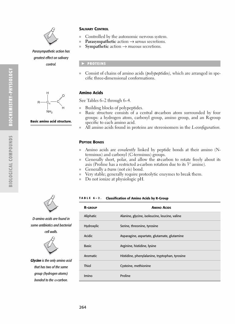
264
BIOC
HEM
ISTR
Y–PH
YSIO
LOGY
BIO
LOGI
CAL
COM
POUN
DS
T A B L E 6 – 2 . Classification of Amino Acids by R-Group
R-GROUP AMINO ACIDS
Aliphatic Alanine, glycine, isoleucine, leucine, valine
Hydroxylic Serine, threonine, tyrosine
Acidic Asparagine, aspartate, glutamate, glutamine
Basic Arginine, histidine, lysine
Aromatic Histidine, phenylalanine, tryptophan, tyrosine
Thiol Cysteine, methionine
Imino Proline
SALIVARY CONTROL
■ Controlled by the autonomic nervous system.■ Parasympathetic action → serous secretions.■ Sympathetic action → mucous secretions.
� PROTE I NS
■ Consist of chains of amino acids (polypeptides), which are arranged in spe-cific three-dimensional conformations.
Amino Acids
See Tables 6–2 through 6–4.
■ Building blocks of polypeptides. ■ Basic structure consists of a central a-carbon atom surrounded by four
groups: a hydrogen atom, carboxyl group, amino group, and an R-groupspecific to each amino acid.
■ All amino acids found in proteins are stereoisomers in the L-configuration.
PEPTIDE BONDS
■ Amino acids are covalently linked by peptide bonds at their amino (N-terminus) and carboxyl (C-terminus) groups.
■ Generally short, polar, and allow the a-carbon to rotate freely about itsaxis (Proline has a restricted a-carbon rotation due to its 3° amine).
■ Generally a trans (not cis) bond.■ Very stable; generally require proteolytic enzymes to break them.■ Do not ionize at physiologic pH.
Parasympathetic action has
greatest effect on salivary
control.
CαR
NH2
H
C
O
H
Basic amino acid structure.
Glycine is the only amino acid
that has two of the same
group (hydrogen atoms)
bonded to the a-carbon.
D-amino acids are found in
some antibiotics and bacterial
cell walls.

265
BIOCHEM
ISTRY–PHYSIOLOGY
BIOLOGICAL CO
MPO
UNDS
DISULFIDE BONDS
■ Strong, covalent bonds between thiol (–SH) group of two cysteine residuesthat stabilizes structure of proteins and prevent denaturation.
■ Abundant in insulin and Ig.
CLASSIFICATION
■ By R-group. (See Table 6–2.)■ By dietary necessity. (See Table 6–3.)■ By metabolic end product. (See Table 6–4.)
T A B L E 6 – 3 . Classification of Amino Acids by Dietary Necessity
DIETARY NECESSITY CHARACTERISTIC AMINO ACIDS
Essential (9) Must be obtained from dietary intake Phenylalanine
Dietary lack can result in negative Valine
nitrogen balance Threonine
Tryptophan
Isoleucine
Methionine
Histidine
Lysine
Leucine
Nonessential (11) All synthesized from glucose Arginine
in TCA cycle (from α-ketoacids, Aspartate
α-amino acids, transaminases, Asparagine
vitamin B6), except tyrosine, Alanine
which is derived from phenylalanine Cysteine
Glycine
Glutamate
Glutamine
Proline
Serine
Tyrosine
T A B L E 6 – 4 . Classification of Amino Acids by Metabolic End Product
METABOLIC CATEGORY AMINO ACIDS
Ketogenic Leucine, lysine
■ Yields acetyl-CoA
Glucogenic Arginine, aspartate, asparagine, alanine, cysteine, histidine,
■ Yields pyruvate methionine, glycine, glutamate, glutamine, proline, serine,
threonine, valine
Both Isoleucine, phenylalanine, tryptophan, tyrosine
Essential amino acids:
PriVaTe TIM HaLL.

266
BIOC
HEM
ISTR
Y–PH
YSIO
LOGY
BIO
LOGI
CAL
COM
POUN
DS
Protein Structure
■ Primary structure: The specific sequence of amino acids in a polypeptidechain. Each amino acid in the polypeptide chain is called a residue.
■ Secondary structure: The folding of portions of a polypeptide chain.■ a-Helix: Coiled configuration.■ b-Pleated sheet: Zigzag or pleated configuration.■ b-Turn: Reverse turns that link two sides of a b-pleated sheet.
AMINO ACID DERIVATIVES
■ Tryptophan → 5-hydroxytryptamine (serotonin) → Melatonin
■ Ribose-5-P → Histidine → Histamine
DEFECTS OF AMINO ACID METABOLISM
AspartateOxaloacetateLysine
Threonine
Asparagine
Methionine
PyruvateLeucine
Valine
Alanine
Isoleucine
Phenylalanine Tyrosine
T3, T4
Melanin
Dopa
Dopamine NE Epi
Disease Affected AA Deficiency Effects
Phenylketonuria Phenylalanine Phenylalanine Developmental (PKU) hydroxylase retardation, ↓ skin/
hair pigmentationNeed to take tyrosine
supplements
Albinism Tyrosine Tyrosinase Lack of melanin pigmentation
Alkaptonuria Tyrosine Homogentisic Excessive urinaryacid oxidase excretion of
homogentisic acid (causing black urine),pigmented sclera
Cystinuria Cysteine Renal Excessive urinaryreabsorption excretion of cysteine,of cysteine kidney stones
Protein structures are
determined by x-ray
diffraction analysis.
■
■
■
α-Ketoglutarate Glutamate
Arginine
Proline
Glutamine
Nitric oxide
3-phosphoglycerate Serine
Cysteine
Glycine Porphyrin Heme
Heme is a major component
of hemoglobin and myoglobin.
Epinephrine and
norepinephrine are produced
by the adrenal medulla. They
cause potent vasoconstriction
and bronchodilation.
Serotonin is released largely
by gastric mucosa and
platelets, causing
vasoconstriction. It is a potent
neurotransmitter in the CNS.
H1 receptors mediate type I
hypersensitivity.
H2 receptors mediate gastric
acid and pepsin secretion.
Histamine is released largely
by basophils and mast cells,
causing vasodilation and
bronchoconstriction.
Nitric oxide is released largely
by vascular endothelium,
causing vasodilation.
■
■

267
BIOCHEM
ISTRY–PHYSIOLOGY
BIOLOGICAL CO
MPO
UNDS
■ Tertiary structure: The overall three-dimensional conformation of apolypeptide. Each portion of the polypeptide that can perform a biochem-ical or physical function is called a domain.
■ Quarternary structure: The spatial arrangement of two or more polypep-tide chains. Each polypeptide is known as a subunit. Associated via non-covalent interactions.
Physiologically Relevant Proteins
COLLAGEN
See Figure 6–4 for collagen structure and Figure 6–5 for collagen synthesis.
■ 1/3 of body’s protein.■ Consists of three polypeptide a-chains wound around one another to form
a triple helix.■ Produced by many cells: fibroblasts, epithelial cells, odontoblasts, osteoblasts,
and chondrocytes. ■ It is the organic matrix in dentin and cementum.
Triple helix
2° structure
Amino acidsequence – Gly – X – Y – Gly – X – Y – Gly – X – Y –
F I G U R E 6 – 4 . Collagen structure.
Reproduced, with permission, from Murray RK. Harper’s Illustrated Biochemistry, 26th ed.
McGraw-Hill, 2003.
F I G U R E 6 – 5 . Collagen synthesis.
Reproduced, with permission, from Bhushan V. First Aid for the USMLE Step 1. McGraw-Hill,
2006:85.
Vitamin C is required for the
hydroxylation of proline and
lysine during collagen
synthesis.
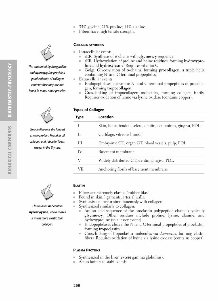
268
BIOC
HEM
ISTR
Y–PH
YSIO
LOGY
BIO
LOGI
CAL
COM
POUN
DS
■ 35% glycine; 21% proline; 11% alanine.■ Fibers have high tensile strength.
COLLAGEN SYNTHESIS
■ Intracellular events■ rER: Synthesis of a-chains with glycine-x-y sequence.■ rER: Hydroxylation of proline and lysine residues, forming hydroxypro-
line and hydroxylysine. Requires vitamin C.■ Golgi: Glycosylation of α-chains, forming procollagen, a triple helix
containing N- and C-terminal propeptides.■ Extracellular events
■ Endopeptidases cleave the N- and C-terminal propeptides of procolla-gen, forming tropocollagen.
■ Cross-linking of tropocollagen molecules, forming collagen fibrils.Requires oxidation of lysine via lysine oxidase (contains copper).
The amount of hydroxyproline
and hydroxylysine provide a
good estimate of collagen
content since they are not
found in many other proteins.
Tropocollagen is the longest
known protein. Found in all
collagen and reticular fibers,
except in the thymus.
Elastin does not contain
hydroxylysine, which makes
it much more elastic than
collagen.
Types of Collagen
Type Location
I Skin, bone, tendon, sclera, dentin, cementum, gingiva, PDL
II Cartilage, vitreous humor
III Embryonic CT, organ CT, blood vessels, pulp, PDL
IV Basement membrane
V Widely distributed CT, dentin, gingiva, PDL
VII Anchoring fibrils of basement membrane
ELASTIN
■ Fibers are extremely elastic, “rubber-like.”■ Found in skin, ligaments, arterial walls.■ Synthesis can occur simultaneously with collagen.■ Synthesized similarly to collagen:
■ Amino acid sequence of the proelastin polypeptide chain is typicallyglycine-x-y. Other residues include proline, lysine, alanine, andhydroxyproline (to a lesser extent).
■ Endopeptidases cleave the N- and C-terminal propeptides of proelastin,forming tropoelastin.
■ Cross-linking of tropoelastin molecules via desmosine, forming elastinfibers. Requires oxidation of lysine via lysine oxidase (contains copper).
PLASMA PROTEINS
■ Synthesized in the liver (except gamma globulins).■ Act as buffers to stabilize pH.

269
BIOCHEM
ISTRY–PHYSIOLOGY
BIOLOGICAL CO
MPO
UNDS
Major Plasma Proteins
PlasmaPlasma Protein Content Examples Function
Albumin 60% Albumin Maintains plasma osmotic pressureTransports various molecules (calcium,
copper, free fatty acids, bilirubin,steroid hormones, drugs)
Fibrinogen 4% Fibrinogen Hemostasis(Factor I)
a-Globulins 10% Lipoproteins (HDL) Transports cholesterol estersProthrombin HemostasisErythropoietin Erythrocyte synthesisAngiotensinogen Regulates blood pressurea2-Macroglobulin Protease inhibition
b-Globulins 10% Lipoproteins (LDL) Transports cholesterolTransferrin Transports copper and iron
g-Globulins 15% Immunoglobulins Antibodies
Complement <1% C3, C5, etc Bacterial cell lysisproteins Inflammation
IMMUNOGLOBULINS
See also Chapter 21, “Immunology and Immunopathology.”
HEMOGLOBIN
See Tables 6–5 and 6–6 and Figure 6–6.
■ Transports O2 in erythrocytes.
■ Each heme reversibly binds one molecule of O2 when the iron is in areduced ferrous (Fe2+) state.
■ Heme binds carbon monoxide (CO) with a greater affinity than O2.■ Hb binds ~15% of the CO2 carried in venous blood (the majority is
carried by bicarbonate). ■ Mutations of a and b subunits result in numerous hemoglobin types.
Hemoglobin
1 Globin
4 Heme
2 α-chains
2 β-chains
4 pyrrole rings
1 Fe2+
■

270
BIOC
HEM
ISTR
Y–PH
YSIO
LOGY
BIO
LOGI
CAL
COM
POUN
DS
T A B L E 6 – 5 . The Major Types of Hemoglobin
HB TYPE CHARACTERISTIC CAUSE OF ABNORMALITY
Hb A Normal Hb N/A
Hb F Fetal Hb N/A
Hb C Chronic anemia Lysine replaces glutamate
Hb H a-Thalassemia Defect of a chain genes
(composed of four b chains)
Hb M Methemoglobinemia Tyrosine replaces histidine
Hb S Sickle-cell anemia Valine replaces glutamate
T A B L E 6 – 6 . Comparison of Hemoglobin and Myglobin
GLOBIN LOCATION HEMES (NO.) O2 USAGE O2 AFFINITY
Hemoglobin Erythrocyte 4 Transport +
Myoglobin Muscle 1 Storage +++
N
N
N
NFe2+
O
–O
O–
O
F I G U R E 6 – 6 . Heme.
Reproduced, with permission, from Murray RK. Harper’s Illustrated Biochemistry, 26th ed.
New York: McGraw-Hill, 2003.
Methemoglobinemia is a
condition in which Fe2+
(ferrous) is oxidized to Fe3+
(ferric), which cannot bind
oxygen. Methemoglobin is
formed due to decreased
activity of methemoglobin
reductase—a side effect of
drugs (eg, sulfonamides) or a
hereditary phenotype of
increased hemoglobin M.
Heme is a cyclic structure
composed of four pyrrole
rings with a central iron atom.
MYOGLOBIN
■ Stores O2 in muscle.■ Similar structure to hemoglobin.■ Contains only one heme: can only associate with one O2 molecule.■ Has much higher affinity for O2 than hemoglobin.

271
BIOCHEM
ISTRY–PHYSIOLOGY
BIOLOGICAL CO
MPO
UNDS
� LI P I DS
STRUCTURE
■ Highly hydrophobic molecules.■ Soluble in nonpolar solvents such as chloroform, ether, and other organic
solvents.■ Functions:
■ Cellular structure■ Metabolism■ Transportation■ Storage
FATTY ACIDS
■ Basic building blocks of most lipids. ■ All are aliphatic (non-aromatic) carboxylic acids.■ Most are esters, although some exist as unesterified free fatty acids. ■ The carbon chain can be saturated or unsaturated.■ Have an even number of carbon atoms with a terminal carboxyl group.■ Most are nonessential (can be synthesized).■ Only a few are essential (found in vegetable oils and animal fats):
■ Linolenic acid: ω-3 fatty acid■ Linoleic acid: ω-6 fatty acid■ Oleic acid: ω-9 fatty acid■ Arachidonic acids
Oxygen-binding curves of hemoglobin and myoglobin.
Reproduced, with permission, from Murray RK. Harper’s Illustrated Biochemistry, 26th ed.
McGraw-Hill, 2003, as modified, with permission, from Scriver CR, et al. (ed.) The Molecular
and Metabolic Basis of Inherited Disease, 7th ed. New York: McGraw-Hill, 1995.
100
1400 20 40 60
Gaseous pressure of oxygen (mm Hg)
80 100 120
80
60
40
20
Per
cent
sat
urat
ion
Oxygenated bloodleaving the lungs
Myoglobin
Reduced bloodreturning from tissues
Hemoglobin
O
(CH2)n CβH2
OH
CωH3 CαH2 C
Basic fatty acid structure.
Shift to right (Bohr effect):
↑ CO2, acid, 2,3-DPG,
altitude, temp., metabolic
need; ↓ O2 affinity, pH.
Shift to left:
↓ CO2, acid, 2,3-DPG, temp.,
metabolic need; ↑ O2
affinity, pH.

272
BIOC
HEM
ISTR
Y–PH
YSIO
LOGY
BIO
LOGI
CAL
COM
POUN
DS
CLASSIFICATION
■ By number of double bonds.■ Saturated: No double bonds.■ Monounsaturated: One double bond, usually in “cis” configuarion.■ Polyunsaturated: Multiple double bonds.
LIPID TYPES
■ Triacylglycerols■ Phospholipids■ Steroids■ Eicosanoids
TRIACYLGLYCEROLS (TRIGLYCERIDES)
■ Consists of three fatty acids acylated to a glycerol molecule.■ Important source of energy.■ Stored in adipose tissue.■ Transported in the plasma by lipoproteins.■ Increased TGs linked to atherosclerosis, heart disease, and stroke.
PHOSPHOLIPIDS
■ Consists of two fatty acids acylated to two carbons of a glycerol moleculeand a phosphate group esterified to the third carbon.
■ Hydrophilic head (phosphate group); hydrophobic tail (fatty acids).■ Derive from phosphatate.■ Major constituents of cell and mitochondrial membranes.■ Precursors for second messengers and metabolic intermediates.■ Increased TGs linked to atherosclerosis, heart disease, and stroke.
C
O
O
O C
OR2
R2
O C
O
R1
2CH
3CH2
1CH2
P
O
O–
O–
C
O
O
O
R2
O C
O
R1
2CH
3CH2
1CH2
Basic triglyceride structure.
Basic phospholipid structure.
Three Major Types of Phospholipids
MajorPhospholipid Function Constituent
Lecithins Water-soluble Choline(phosphatidycholines) emulsifiers
Plasma membraneconstituent
Cephalins Nerve tissue components Ethanolamine(phosphatidylethanolamines)
Sphingomyelins Plasma membrane Ceramideconstituent Choline
Nerve tissueconstituent
Triglycerides are not found in
cell membranes.
Accumulation of
sphingomyelins causes
Niemann-Pick disease.

273
BIOCHEM
ISTRY–PHYSIOLOGY
BIOLOGICAL CO
MPO
UNDS
CHOLINE
■ N+(CH3)3-CH2-CH2-OH■ An essential nutrient.■ Found mostly in phospholipids.■ Found as lecithin in plasma cell membrane.■ Precursor of betaine, an osmolyte used by kidney to control water balance.■ Component of sphingomyelin, which forms myelin sheath. Insulates nerve
fibers and aids in rapid conduction on nerve impulses.■ Source of methyl group required for lipoprotein formation in liver.■ Necessary for acetylcholine formation.■ Active component of lung surfactant.
STEROIDS
■ Cholesterol is the most basic steroid.■ Major constituent of cell membranes and lipoproteins. ■ Commonly present as a cholesterol ester. ■ Conversion of HMG-CoA → mevalonate via HMG-CoA reductase is the
rate-limiting step.■ Precursor molecule for other steroids:
■ Bile salts■ Sex hormones■ Adrenocortical hormones■ Vitamin D
EICOSANOIDS
■ 20-carbon long polyunsaturated fatty acids.■ Derivatives of arachidonic acid.■ Phospholipase A2 (PLA2) releases arachidonic acid from plasma mem-
brane phospholipids upon hormone or cytokine stimulation or cellulardamage.
Eicosanoids
Formative Eicosanoid Pathway Examples Function
Prostanoids Cyclooxygenase Prostaglandins Vasodilation(COX) Inflammatory
PainGastric protection
Prostacyclins Vasodilation↓ platelet aggregation
Thromboxanes Vasoconstriction↑ platelet aggregation
Leukotrienes 5-Lipoxygenase Various Bronchoconstriction(5-LOX) leukotrienes Inflammatory
Lipoxins 15-Lipoxygenase Various lipoxins Anti-inflammatory(15-LOX)
Corticosteroids inhibit
phospholipase A2 (PLA2).
Prostaglandins:■ Modulate hormones■ Act locally■ Metabolized rapidly
Choline deficiency causes
abnormal fat metabolism and
can lead to fatty liver disease
and hepatic cirrhosis.
Aspirin and nonsteroidal anti-
inflammatory drugs (NSAIDs)
inhibit cyclooxygenase (COX).
Statin drugs inhibit HMG-CoA
reductase.

274
BIOC
HEM
ISTR
Y–PH
YSIO
LOGY
BIO
LOGI
CAL
COM
POUN
DS
Monolayer of mainlyamphipathic lipids
Core of mainlynonpolar lipids
Cholesterylester
Phospholipid
Peripheral apoprotein(eg, apo C)
Triacylglycerol
Integralapoprotein(eg, apo B)
Freecholesterol
F I G U R E 6 – 7 . Lipoprotein structure.
Reproduced, with permission, from Murray RK. Harper’s Illustrated Biochemistry, 26th ed.
McGraw-Hill, 2003.
Lipid Transport
LIPOPROTEINS
■ Transport lipids in blood plasma. ■ Composed of a nonpolar lipid core surrounded by a single layer of amphi-
pathic phospholipids and cholesterol (see Figure 6–7). ■ Characterized by the protein moiety embedded in their outer layer
(apoprotein).■ Contain triglycerides (16%), phospholipids (30%), cholesterol (14%), cho-
lesterol esters (36%), and free fatty acids (4%). ■ Choline is essential for the secretion of lipoproteins from hepatocytes,
especially very low density lipoproteins (VLDL).
LDL is the major carrier of
cholesterol in the blood. It is
taken up by cell-mediated
endocytosis.
HDL is produced de novo in
the liver.
Lipoxins15-LOX
5-LOXPlasma membrane PLA-2
Arachidonic acid Leukotrienes
Diet COXPGI-2 Prostacyclins
Prostaglandins
ThromboxanesTXA-2
■ Eicosanoid formation pathways.
Familial
hypercholesterolemia is
caused by an autosomal
dominant defect of the low-
density lipoprotein (LDL)
receptor, leading to increased
plasma LDL cholesterol and
atherosclerosis.

275
BIOCHEM
ISTRY–PHYSIOLOGY
BIOLOGICAL CO
MPO
UNDS
The Major Lipoproteins
Protein Major Lipid Carries Carries Lipoprotein Density (%) Content Lipid From Lipid To
Chylomicron + 1 Dietary TG Small intestine Extrahepatic tissues
Chylomicron ++ 7 Cholesterol Chylomicrons of Liverremnants extrahepatic tissues
VLDL ++ 10 Endogenous TG Liver Extrahepatic tissues
IDL (VLDL +++ 11 TG, Cholesterol VLDL of extrahepatic Liverremnants) tissues
LDL +++ 20 Cholesterol Liver Extrahepatic tissues
HDL ++++ 30–55 Cholesterol Extrahepatic tissues Liver
BILE SALTS
■ Aid in lipid absorption (emulsification and solubilization).■ Absorption via micelles (water-soluble complexes).■ Decrease surface tension of particles to break them into smaller sizes.
■ Formed from cholesterol in the liver.■ Almost exclusively absorbed in the ileum and returned to the liver via the
enterohepatic (portal) circulation.■ Those that are not reabsorbed are excreted.■ Surplus bile salts are stored in the gall bladder.■ Two major bile salts enter bile as glycine or taurine conjugates (occurs in
peroxisomes):■ Glycocholic acid: Glycine + cholic acid.■ Taurocholic acid: Taurine + cholic acid.
Lipid Storage
■ Most lipid triglycerides are stored in adipose tissue.
LIPID (LYSOSOMAL) STORAGE DISEASES
See Table 6–7.
■ Inherited disorders of the reticuloendothelial system.■ Caused by incomplete lysosomal breakdown of sphingolipids and mucopolysac-
charides within phagocytes, leading to their accumulation.■ Most are common to Ashkenazi Jewish ancestry.

276
BIOC
HEM
ISTR
Y–PH
YSIO
LOGY
BIO
LOGI
CAL
COM
POUN
DS
T A B L E 6 – 7 . Lipid Storage Diseases
DEFICIENT ACCUMULATED CLINICAL
DISEASE ENZYME LIPID FEATURES INHERITANCE
Gaucher’s b-Glucocerebrosidase Glucocerebrosides Splenomegaly Autosomal
disease Hepatomegaly recessive
Anemia
Skin pigmentation
(brown)
Most common
Niemann-Pick Sphingomyelinase Sphingomyelin Splenomegaly Autosomal
disease Hepatomegaly recessive
Cherry red spot
on macula
Rapidly fatal
Tay-Sachs Hexosaminidase A Gangliosides CNS degeneration Autosomal
disease Developmental recessive
retardation
Cherry red spot
on macula
Rapidly fatal
Fabry’s a-Galactosidase Ceramide Peripheral neuropathy X-linked
disease trihexoside Skin lesions recessive
(angiokeratomas)
Renal failure
Cardiovascular
Krabbe’s Galactocerbrosidase Galactocerebroside Peripheral Autosomal
disease neuropathy recessive
Optic atrophy
Developmental
retardation

C H A P T E R 7
Metabolism
Overview 278
RATE-LIMITING STEPS 279
ATP Production 279
Carbohydrate Metabolism 280
Lipid Metabolism 285
Protein Metabolism 286
Bacterial Metabolism 289
277

278
BIOC
HEM
ISTR
Y-PH
YSIO
LOGY
MET
ABO
LISM
� OVE RVI EW
See Figure 7–1 for summary of metabolic pathways.
KEY INTERMEDIATES
See Figure 7–2.
■ Glucose-6-phosphate■ Pyruvate■ Acetyl-CoA
F I G U R E 7 – 1 . Summary of metabolic pathways.
Reproduced, with permission, from Bhushan V, et al. First Aid for the USMLE Step 1. New York: McGraw-Hill, 2006.

279
BIOCHEM
ISTRY-PHYSIOLOGY
METABO
LISM
Rate-Limiting Steps (Table 7–1)
F I G U R E 7 – 2 . Common metabolic intermediates.
Glucose
Glucose-6-phosphate
Glycogen Pyruvate Ribose-5-phosphate
Lactate Acetyl-CoA
Cholesterol Ketone bodies
ATP HMG-CoA Fatty acids
OxaloacetateAlanine
T A B L E 7 – 1 . Rate-Limiting Enzymes of Major Metabolic Pathways
METABOLIC PATHWAY ENZYME
Glycolysis Phosphofructokinase (PFK)
Gluconeogenesis Fructose bisphosphatase-2
TCA cycle Isocitrate dehydrogenase
Glycogen synthesis Glycogen synthase
Glycogenolysis Glycogen phosphorylase
HMP shunt Glucose-6-phosphate dehydrogenase
(G6PD)
Urea cycle Carbamoyl phosphate synthetase
Fatty acid synthesis Acetyl-CoA carboxylase (ACC)
Fatty acid oxidation Carnitine acyltransferase
Ketogenesis HMG-CoA synthase
Cholesterol synthesis HMG-CoA reductase
� ATP PRODUCTION
See Table 7–2.
■ Substrate-level phosphorylation: ADP + Pi → ATP.■ Oxidative phosphorylation: Major source of ATP (aerobic).

280
BIOC
HEM
ISTR
Y-PH
YSIO
LOGY
MET
ABO
LISM
� CARBOHYDRATE M ETABOLISM
■ Determines the fate of glucose.■ ATP production per molecule of glucose. The table below assumes that
the NADH produced in glycolysis is carried into mitochondria via themalate–aspartate shuttle. If the glycerol-3-phosphate (G3P) shuttle is used,the net ATP production would be 36.
ATP Production per Molecule of Glucose
Metabolic Pathway ATP Produced
Glycolysis (cytosol)2 ATP consumed (by hexokinase and PFK) –24 ATP formed +42 NADH formed
Pryuvate → Acetyl-CoA (mitochondrial matrix)2 NADH formed
TCA Cycle (mitochondrial matrix)2 GTP formed +26 NADH formed2 FADH2 formed
ETC (inner mitochondrial membrane)10 NADH oxidized +302 FADH2 oxidized +4
+38
Each NADH yields ~3 ATP.
Each FADH2 yields ~2 ATP.
GLYCOLYSIS
■ Also called the Embden–Meyerhof Pathway. (See Figures 7–3.)■ Occurs in the cytosol, in the absence of O2.■ Converts glucose (as glucose-6-phosphate) → two molecules of pyruvate.■ Conversion of fructose-6-phosphate → fructose-1, 6-biphosphate via phos-
phofructokinase (PFK) is the rate-limiting step.
T A B L E 7 – 2 . Net ATP Production per Molecule of Glucose
PROCESS LOCATION PHOSPHORYLATION NET ATP PER GLUCOSE
Embden-Meyerhof Cytosol Substrate level 2
pathway
Entner-Doudoroff Cytosol Substrate level 1
pathway
TCA cycle Mitochondrial Substrate level 2 (as GTP)
matrix
ETC Inner mitochondrial Oxidative 32 (G3P shuttle)
membrane 34 (malate-aspartate shuttle)
Photophosphorylation
occurs in plants as a result of
photosynthesis. Also involves
an ETC.

281
BIOCHEM
ISTRY-PHYSIOLOGY
METABO
LISM
■ ATP-requiring reactions: ■ Hexokinase/Glucokinase: glucose → glucose-6-phosphate■ PFK: fructose-6-phosphate → fructose-1,6-bisphosphate
■ ATP-producing reactions:■ Phosphoglycerate kinase: 1,3-bisphosphoglycerate → 3-phosphoglycerate■ Pyruvate kinase: phosphoenolpyruvate → pyruvate
Glucose
ATP
ATP
ADP
Hexokinase
Glucose-6-phosphate
Fructose-6-phosphate
PFKADP
Fructose-1,6-biphosphate
2 NAD+ 4 ADP
4 ATP2 NADH
Pyruvate (2x)
F I G U R E 7 – 3 . Major reactions of glycolysis.
METABOLIC FATES OF PYRUVATE
Type of Reaction Enzyme Product Function
Oxidation Pyruvate Acetyl-CoA ATP productiondehydrogenase Fatty acid synthesis
Reduction Lactate Lactate Anaerobic glycolysisdehydrogenase
Carboxylation Pyruvate Oxaloacetate Gluconeogenesiscarboxylase Replenishes TCA
cycle
Transamination Alanine Alanine Amino acid synthesisaminotransferase
Because erythrocytes do not
have mitochondria, glycolysis
always ends in lactate. The
lactic acid is converted back to
glucose via the Cori cycle.
(see Fig 7–5).
Fluoride inhibits enolase, the
enzyme that converts
2-phosphoglycerate →phosphoenolpyruvate.
■ Stoichiometry of glycolysis:
Glucose + 2 Pi + 2 ADP + 2 NAD+ → 2 Pyruvate + 2 ATP + 2 NADH + 2 H+ + 2 H2O

282
BIOC
HEM
ISTR
Y-PH
YSIO
LOGY
MET
ABO
LISM
PYRUVATE DEHYDROGENASE
■ Pyruvate + NAD+ → Acetyl-CoA + CO2 + NADH■ Requires five cofactors:
■ Pyrophosphate (B1, thiamine)■ FAD (B2, riboflavin)■ NAD (B3, niacin)■ CoA (B5, pantothenate)■ Lipoic acid
THE LACTIC ACID CYCLE
■ Also called the Cori cycle (See Figure 7–4.)■ Occurs in the liver■ Prevents lactic acidosis■ Converts lactate → glucose, which is then reoxidized via glycolysis■ Provides quick ATP production during anaerobic glycolysis in muscle and
erythrocytes■ Results in net loss of 4 ATP per cycle
THE CITRIC ACID CYCLE
■ Also called the Krebs cycle and the tricarboxylic acid cycle. (See Figures 7–5.)■ Occurs in the mitochondrial matrix.■ Completes the metabolism of glucose.■ Oxidizes acetyl-CoA.■ Reduces NAD+ and FAD → NADH and FADH2, which are reoxidized in
the ETC to produce ATP.■ Tightly regulated by both ATP and NAD+.■ Stoichiometry of TCA cycle:
Acetyl-CoA + 3 NAD+ + FAD + Pi + GDP + 2 H2O → 2 CO2 + 3 NADH + FADH2 + GTP + 2 H+ + CoA
LIVER MUSCLE
PyruvateLactateLactate
Glycogen Glucose-6-phosphate
Pyruvate
Glucose-6-phosphate Glycogen
Urea
–NH2
Alanine
–NH2
Alanine
Tran
sam
inat
ion Transam
ination
Lactate
BLOOD
Pyruvate
Alanine
BLOOD
Glucose
F I G U R E 7 – 4 . The Cori cycle.
Reproduced, with permission, from Murray RK. Harper’s Illustrated Biochemistry, 26th ed. New York: McGraw-Hill, 2003.

283
BIOCHEM
ISTRY-PHYSIOLOGY
METABO
LISM
THE ELECTRON TRANSPORT CHAIN
■ Also called the respiratory chain (See Figure 7–5.)■ Occurs in the inner mitochondrial membrane.■ Produces ATP via oxidative phosphorylation of ADP.■ Reoxidizes NADH and FADH2 back → NAD+ and FAD as electrons flow
through a series of four cytochrome complexes of increasing redox poten-tial (along a proton gradient).
■ Cytochromes contain a central iron atom (similar to hemoglobin), whichcan exist in an oxidized ferric (Fe3+) state or a reduced ferrous (Fe2+) state.
■ Cytochromes receive electrons from the reduced form of coenzyme Q(ubiquinone).
■ Cytochromes carry electrons as flavins, iron-sulfur groups, hemes, andcopper ions.
F I G U R E 7 – 5 . The citric acid cycle and electron transport chain.
Reproduced, with permission, from Murray RK. Harper’s Illustrated Biochemistry, 26th ed.
New York: McGraw-Hill, 2003.
H2O
H2O
H2O
2H
H2O
H2O
Fp
Carbohydrate Protein Lipids
Acetyl-CoA (C2)
Citric acidcycle
H2OOxaloacetate (C4) Citrate (C6)
Cis-aconitate (C6)
Malate (C4)
2H
2H
2H
α-Ketoglutarate (C5)
CO2
CO2
Succinyl-CoA (C4)
NADSuccinate (C4)
Fumarate (C4)
P
P
P
P
Q
Cyt b
Cyt c
Cyt aa3
1/2 O2
Anaerobiosis(hypoxia, anoxia)
Oxidativephosphorylation
Isocitrate (C6)
Fp
P
Cyt
Respiratory chain
Flavoprotein
Cytochrome
High-energy phosphate

284
BIOC
HEM
ISTR
Y-PH
YSIO
LOGY
MET
ABO
LISM
THE PENTOSE PHOSPHATE PATHWAY
■ Also called the hexose monophosphate shunt.■ Stoichiometry of the pentose phosphate pathway (PPP):
■ Occurs in the cytosol.■ An alternative to glycolysis in the metabolism of glucose.■ Coverts glucose-6-phosphate → ribose-5-phosphate.■ Conversion of glucose-6-phosphate → 6-phosphogluconolactone via
glucose-6-phosphate dehydrogenase (G6PD) is the rate-limiting step.■ Produces ribose (for nucleotide synthesis) and NADPH (for fatty acid and
steroid synthesis).■ Not all cells use the PPP (most active in liver, adipose tissue, adrenal cortex,
thyroid, mammary gland, testis, and erythrocytes).
GLUCONEOGENESIS
■ Stoichiometry of gluconeogenesis:
■ Occurs mostly in the liver and kidneys■ Not a direct reversal of the glycolysis■ Converts amino acids → glucose or glycogen in states of carbohydrate need■ Clears lactic acid (from anaerobic glycolysis) and glycerol (from fatty acid
metabolism) ■ Under strict hormonal regulation
GLYCOGEN SYNTHESIS AND CATABOLISM
■ Enzymatic regulation of glycogen metabolism:
Glucose-6-phosphate + 2 NADP+ + H2O → Ribose-5-phosphate + 2 NADPH + 2 H+ + CO2
CYTOCHROME COMPLEXES OF THE ETC
Cytochrome Complex Enzyme Electron Transfer
Complex I NADH-Q reductase NADH → ubiquinone
Complex II Succinate-Q reductase FADH2 → ubiquinone■ Does not pump
protons
Complex III Cytochrome reductase Ubiquinone → cyt c
Complex IV Cytochrome oxidase Cyt c + H+ + O2 → H2O■ Requires copper ■ O2 is the final
e– receptor
(+) Glucagon, epinephrineGlycogen phosphorylase Phosphoglucomutase
Glycogen Glucose-1-phosphate
Glycogen synthase(+) Insulin
Glucose-6-phosphataseGlucose-6-phosphate Glucose
Glucokinase/Hexokinase
Pyruvate + 2 ATP + GTP + NADH + 2 H2O → Glucose-6-phosphate + 2 ADP + GDP + 3 Pi + NAD+ + H+
Coenzyme Q is also known as
ubiquinone.
No ATP is produced from the
PPP.
The NADPH produced from
the PPP helps to rid
erythrocytes of free radicals
and H2O2. G6PD deficiency
causes hemolytic anemia due
to a ↓ in NADPH production,
and subsequent ↑ of oxidizing
agents in RBCs.
Glycogen is a branched
polymer of glucose residues.

285
BIOCHEM
ISTRY-PHYSIOLOGY
METABO
LISM
■ Glycogen synthase: Key regulatory enzyme in its synthesis. Uses UDP-glucose and the nonreducing end of glycogen as its substrate.■ Glycogenin: Primer for glycogen synthase by catalyzing the addition of
glucose to itself.■ Glycogen phosphorylase: Key regulatory enzyme in its catabolism.■ Under strict hormonal regulation.
� LI P I D M ETABOLISM
FATTY ACID SYNTHESIS
See Figure 7–6.
■ Occurs in the cytosol of mostly hepatocytes. ■ The irreversible conversion of acetyl-CoA → malonyl-CoA via acetyl-CoA
carboxylase is the rate-limiting step.■ Conversion of acetyl-CoA → malonyl-CoA is the rate-limiting step.■ Citrate–malate shuttle transports acetyl groups from mitochondria to the
cytosol.
Glycerol kinaseGlycerol Glycerol-3-phosphate Glucose-6-phosphate
Triglyceride
Fatty acids Acetyl-CoA
Lipase: (+) glucagon, epinephrine; (−) insulin
F I G U R E 7 – 7 . Overview of triglyceride lipolysis.
Glucagon and epinephrine
stimulate glycogenolysis
and gluconeogenesis,
whereas insulin triggers
glycogen formation and
cellular glucose uptake.
Coenzyme A (CoA) is a
pantothenic acid (B5)-
containing coenzyme involved
in both fatty acid synthesis
and catabolism.
In humans, fatty acids cannot
be converted to glucose.
(+) Citrate, insulin(−) Glucagon, epinephrineAcetyl-CoA carboxylase
Glucose Acetyl-CoA Acylglycerols
Biotin, pantothenic acid
Malonyl-CoA Palmitate
F I G U R E 7 – 6 . Overview of fatty acid synthesis.
TRIGLYCERIDE LIPOLYSIS
See Figure 7–7.
■ Occurs in adipocytes.■ The glycerol is phosphorylated and ultimately oxidized in glycolysis.■ The free fatty acids are transported to the liver for b-oxidation.■ Triacyglycerol lipase is under strict hormone regulation.
b-OXIDATION
See Figure 7–8.
■ Occurs in the mitochondrial matrix of hepatocytes.■ Converts acyl-CoA → acetyl-CoA.■ Fatty acids are carried into the mitochondrial matrix by a carnitine-mediated
enzyme system.

286
BIOC
HEM
ISTR
Y-PH
YSIO
LOGY
MET
ABO
LISM
■ Under certain metabolic states (starvation, diabetes mellitus), much of theacetyl-CoA is converted to ketone bodies:■ Acetoacetate: synthesized by cleavage of HMG-CoA■ b-Hydroxybutyrate■ Acetone
■ Ketone bodies are a source of fuel in extrahepatic tissues such as skeletaland cardiac muscle.
■ Ketosis is the accumulation of ketone bodies leading to ketoacidosis anddiabetic coma.
Re-esterification
Fatty acid Acetyl-CoA TCA cycleCarnitineshuttle
b-OxidationHMG-CoA Ketone bodies
Cholesterol
Acyl-CoA synthetase
Acyl-CoA
F I G U R E 7 – 8 . Overview of β-oxidation.
α-Amino acid
α-Ketoglutarate
α-Keto acid
NH3 CO2
OXIDATIVEDEAMINATION
UREA CYCLE
L-Glutamate
TRANSAMINATION
Urea
F I G U R E 7 – 9 . Key reactions and intermediates in the formation of urea.
Reproduced, with permission, from Murray RK. Harper’s Illustrated Biochemistry, 26th ed.
New York: McGraw-Hill, 2003.
METABOLIC FATES OF ACETYL-COA
Fate Pathway
ATP production Via TCA cycle and the ETCCholesterol synthesis Via HMG-CoA reductaseKetone body synthesis Via HMG-CoA lyaseFatty acid synthesis Via malonyl-CoA
� PROTE I N M ETABOLISM
■ Both dietary and structural proteins are degraded daily to their amino acidconstituents by various proteases and peptidases. (See Figure 7–9.)
Of the three ketone bodies,
acetone is not used for energy
production. Its accumulation
can cause breath with a
fruity odor.

287
BIOCHEM
ISTRY-PHYSIOLOGY
METABO
LISM
TRANSAMINATION
■ Amino acids are used to synthesize other amino acids (and proteins) ormetabolic intermediates (pyruvate, acetyl-CoA, oxaloacetate, succinyl CoA,and a-ketoacids).
■ Aminotransferases (transaminases) cleave the a-amino nitrogen of mostamino acids, leaving hydrocarbon skeletons, which are degraded to the variousglucogenic and ketogenic intermediates.
■ Requires either pyridoxal phosphate or pyridoxamine phosphate, forms of pyri-doxine (vitamin B6), as a coenzyme.
■ The transamination of pyruvate to alanine yields either a-ketoglutarate oroxaloacetate.
■ See Figure 7–10 for key metabolic intermediates formed from the carbonskeletons of amino acids.
Pyruvate Transaminase Alanine
Glutamate or aspartate Pyridoxine α-Ketoglutarate or oxaloacetate
Glutamate
Citrate
cycle
Succinyl-CoACitrate
lleLeuTrp
AlaCysGlyHypSerThr
Pyruvate
Oxaloacetate
Acetoacetyl-CoA
Acetyl-CoA
Aspartate Asn
Leu, Lys,Phe, Trp,
Tyr
Fumarate
ArgHisGlnPro
lleMetVal
TyrPhe
α-Ketoglutarate
F I G U R E 7 – 1 0 . Key metabolic intermediates formed from the carbon skeletons ofamino acids.
Reproduced, with permission, from Murray RK. Harper’s Illustrated Biochemistry, 26th ed.
New York: McGraw-Hill, 2003.
Only lysine, serine, and
threonine are not
transaminated.

288
BIOC
HEM
ISTR
Y-PH
YSIO
LOGY
MET
ABO
LISM
OXIDATIVE DEAMINATION
■ An alternative to transamination in the metabolism of amino acids.■ Results in the formation of α-ketoacids (for energy) and ammonia (for
urea formation). ■ Oxidative deamination of glutamate:
■ The reaction is reversible but favors the formation of glutamate.■ Occurs mostly in the liver and kidneys.■ In humans, the vast majority of oxidative deamination derives from gluta-
mate; the major enzyme responsible is glutamate dehydrogenase.■ Other amino acids that can undergo oxidative deamination are asparagine,
histidine, serine, and threonine (Table 7–3).
Glutamate dehydrogenase
L-Glutamate + NAD(P)+ + H2O α-Ketoglutarate + NAD(P)H + NH4+ + H+
T A B L E 7 – 3 . Enzymes Involved in Oxidative Deamination
ENZYME DEAMINATES PRODUCES
Glutamate dehydrogenase Glutamate α-Ketoglutarate + NH4+
Histidinase Histidine Urocanic acid + NH4+
Serine dehydratase Serine Pyruvate + NH4+
Asparaginase Asparagine Aspartic acid + NH4+
MEDICALLY RELEVANT TRANSAMINASES
Also ↑ Levels MayTransaminase Known As Location Suggest
Aspartate Serum glutamate- Liver Acute hepatitisaminotransferase (AST) oxaloacetate Cardiac muscle Myocardial infarction
transaminase Skeletal muscle Acute renal disease(SGOT) Kidneys Acute hemolytic anemia
BrainRBCs
Alanine Serum glutamate- Mostly liver Acute hepatitisaminotransferase (ALT) pyruvate
transaminase(SGPT)
Glutamate is the only amino
acid that undergoes rapid
oxidative deamination.

289
BIOCHEM
ISTRY-PHYSIOLOGY
METABO
LISM
THE UREA CYCLE
■ Occurs in the cytosol and mitochondrial matrix of hepatocytes.■ Eliminates the ammonia (NH4
+) produced by oxidative deamination inthe form of urea (Figure 7–11).
■ Stoichiometry of the urea cycle:
■ The excess nitrogen is converted to urea and excreted by the kidneys.■ Complete block of the cycle leads to extensive ammonia accumulation.
For example, liver cirrhosis (eg, from alcoholism) results in ↓ carbamoylphosphate synthase. It is fatal since there is no alternative pathway.
� BACTE RIAL M ETABOLISM
ENTNER–DOUDOROFF PATHWAY
■ An alternative method of glycolysis to the Embden-Meyerhof or pentosephosphate pathways.
■ Used most commonly by aerobic bacteria.■ Converts glucose → pyruvate + glyceraldehyde-3-phosphate.■ Produces 1 ATP per glucose via substrate-level phosphorylation.
CO2 + NH4+ + 3 ATP + Aspartate + 2 H2O → Urea + 2 ADP + 2 Pi + AMP + PPi + Fumarate
Citrulline
Arginine
Urea
Arginase
H2O
To kidney
Fumarate
Aspartate
Argininosuccinatesynthetase
Ornithinetranscarbamoytase
2 ATP
2 ADP + Pi
CO2 + NH4+
ArgininosuccinateOmithineCytoplasm
Mitochondria
Carbamoylphosphate
Carbamoyl phosphatesynthetase IRate-limiting step
(Liver)NH2 From NH4
+
Urea
Argininosuccinase
C O From CO2
NH2 From aspartate
F I G U R E 7 – 1 1 . Key metabolic intermediates formed from the carbon skeletons of amino acids.
Reproduced, with permission, from Le T, Bhushan V, Vasan N. First Aid for the USMLE Step 1 2011. 21st ed.
New York: McGraw-Hill, 2011.
The urea cycle requires 3 ATP.
The amount of nonprotein
nitrogen in the blood is
primarily due to urea.

This page intentionally left blank

C H A P T E R 8
Molecular Biology
Nucleotides 292
Base Pairing 292
ORGANIZATION 292
BIOSYNTHESIS 293
Nucleic Acids 293
DNA 294
RNA 294
DNA Organization 295
NUCLEOSOMES 295
CHROMATIN 296
DNA Synthesis 296
Transcription and Translation 297
RNA SYNTHESIS (TRANSCRIPTION) 297
PROTEIN SYNTHESIS (TRANSLATION) 297
Mutations 298
POINT MUTATIONS 299
FRAMESHIFT MUTATIONS 299
REPEAT MUTATIONS 299
Clinical Considerations 299
REVERSE TRANSCRIPTASE 299
RECOMBINANT DNA TECHNOLOGY 299
POLYMERASE CHAIN REACTION (PCR) 299
BLOTTING TECHNIQUES 300
291

292
BIOC
HEM
ISTR
Y-PH
YSIO
LOGY
MO
LECU
LAR
BIO
LOGY
� N UC LEOTI DES
■ Building blocks of DNA and RNA. ■ Functions:
■ Protein synthesis■ Nucleic acid synthesis■ Signal transduction pathways
■ Composed of three basic compounds:■ Nitrogenous base
■ Purine■ Pyrimidine
■ Pentose sugar■ Deoxyribose■ Ribose
■ Phosphate group(s)
� BASE PAI R I NG
See Figure 8–1 for base pairing in DNA.
Organization
■ Purines pair with pyrimidines.■ Held together via hydrogen bonds.
■ Adenine—Thymine/Uracil (two H-bonds).■ Guanine—Cytosine (three H-bonds).
A nucleoside is a nucleotide
(sugar + base) without
esterified phosphate groups.
Purines:
Pure Ag (silver)Purines are Adenine and
Guanine.
Pyrimidines:
CUT the PY (pie)Cytosine, Uracil,
Thymine are thePyrimidines.
The strongest bonds(three hydrogenbonds) are betweenCytosine andGuanine, like CrazyGlue.
Adenine andThymine/Uracil areheld by two hydrogenbonds.
F I G U R E 8 – 1 . Base pairing in DNA.
Reproduced, with permission, from Murray, RK. Harper’s Illustrated Biochemistry, 26th ed.
New York: McGraw-Hill, 2003.
NN N
N
N
N
O
N N
O
O
CH3
O
H
H H
H
HN
N
H
H
Thymidine
Adenosine
Guanosine
CytosineH
N
N
N
N
N

BIOCHEM
ISTRY-PHYSIOLOGY
MO
LECULAR BIOLOGY
293
Biosynthesis
■ Nucleotide synthesis.
Comparison of Purines and Pyrimidines
Metabolic Catabolize Catabolic Nucleotide DNA Bases RNA Bases Defects to Defects
Purines Adenine (A) Adenine (A) Antifolate Uric acid GoutGuanine (G) Guanine (G) drugs Hyperuricemia
Anticancer G-6-P drugs deficiency
Pyrimidines Thymine (T) Uracil (U) UV light β-Alanine Rare (highly Cytosine (C) Cytosine (C) Methotrexate β-Amino- water soluble)
Other isobutyrateanticancerdrugs
Comparison of DNA and RNA
Nucleic Acid Strands Sugar Bases
DNA Double Deoxyribose A-T, G-CRNA Single Ribose A-U, G-C
DNA and RNA are
differentiated in the
laboratory by the Feulgen
reaction, which is specific for
deoxyribose.
ATP
Ribose-5-phosphate → 5-phosphoribosyl-1-pyrophosphate → Purines
(PRPP)
↓Orotic acid → Orotate monophosphate → Pyrimidines
■ Uric acid synthesis.
Purines → Xanthine → Uric acid Xanthine oxidase
� N UC LE IC AC I DS
■ Extremely polar and hydrophilic.■ The “backbone” consists of pentose sugars linked by phosphodiester
bonds at the third and fifth carbon atoms.■ The two polynucleotide chains are considered antiparallel and comple-
mentary (one chain runs in the 5′ → 3′ direction; the other runs in the3′ → 5′ direction).
Tetrahydrofolic acid (TFA):
■ Used by several enzymes
in purine/pyrimidine
synthesis.
■ Target for antimetabolites
(eg, methotrexate).
The catabolism of nucleotides
results in no ATP production.

294
BIOC
HEM
ISTR
Y-PH
YSIO
LOGY
MO
LECU
LAR
BIO
LOGY
Three Major Types of RNA
RNA Site of Synthesis Function Note
Ribosomal Nucleolus Major component Most prevalent(rRNA) of ribosomes RNA
Transfer Nucleus Carries amino acids Contains an anticodon(tRNA) from cytosol (complementary
to ribosomes to mRNA codons)
Messenger Nucleus Carries genetic code Least prevalent RNA.(mRNA) from DNA Contains codons
to ribosomes (complementary to DNA templateand tRNA anticodon)
F I G U R E 8 – 2 . The backbone structure of a single DNA strand. It is constant: deoxyribose sugars linked together viaphosphodiester bonds.
Reproduced, with permission, from Murray RK. Harper’s Illustrated Biochemistry, 26th ed. New York: McGraw-Hill, 2003.
O
H
H
H H
N
N N
NHG
NH2
O
H
CH2
O
H
H
NO
NC
NH2
HH
CH2
H
O
HH
H
H H
N
O
O
NHH3C
T
CH2
CH2
O
HH
H
H H
NH2
N
N N
NA
5�
3�
O
O
P
O
P
O
P
O
O
P
O
O
P
DNA
See Figure 8–2 for the backbone structure of a single DNA strand.
RNA
See Figure 8–3 for the backbone structure of a single RNA strand.The melting temperature of the
double helix is a function of
base composition: a higher G-C
content increases the melting
temperature and stability.

295
BIOCHEM
ISTRY-PHYSIOLOGY
MO
LECULAR BIOLOGY
� DNA ORGAN I ZATION
Nucleosomes
See Figures 8–4 and 8–5.
■ Consists of DNA wrapped around a histone octomer.■ Held by ionic bonds.
Histones are composed
largely of arginine and
lysine, which are positively
charged due to their free
amino group. They bond
tightly to the negatively
charged phosphate groups of
DNA.
O
HH
HO
H H
N
O
O
NHU
CH2
O
H
HO
H H
N
N N
NHG
NH2
O
H
CH2
5�
CH2
O
HH
HO
H H
NH2
N
N N
NA
3�
O
HO
H
NO
NC
NH2
HH
CH2
H
O
O
P
O
O
P
O
O
P
O
P
O
P
F I G U R E 8 – 3 . The backbone structure of a single RNA strand. It is constant: ribose sugars linked together viaphosphodiester bonds.
Reproduced, with permission, from Murray RK. Harper’s Illustrated Biochemistry, 26th ed. New York: McGraw-Hill, 2003.
F I G U R E 8 – 4 . Nucleosome structure.
Reproduced, with permission, from Murray RK. Harper’s Illustrated Biochemistry, 26th ed.
New York: McGraw-Hill, 2003.
Histone octamer
DNAHistoneH1

BIOC
HEM
ISTR
Y-PH
YSIO
LOGY
MO
LECU
LAR
BIO
LOGY
Chromatin
■ Consists of nucleosomes, enzymes, gene regulatory proteins (transcriptionfactors), and small amounts of RNA.
� DNA SYNTH ES IS
See Figure 8–6.
■ Helicase: Unwinds the DNA molecule.■ Topoisomerase: Secures the replication fork, where the two DNA strands
are separated into leading and lagging strands.
F I G U R E 8 – 5 . DNA organization.
Reproduced, with permission, from Murray RK. Harper’s Illustrated Biochemistry, 26th ed.
New York: McGraw-Hill, 2003.
296
Chromatin looks like beads
of nucleosomes on a string
of DNA.

297
■ DNA polymerase: Forms new complementary strands in the 5′ → 3′direction.■ Leading strand: Runs in the 3′ → 5′ direction. Synthesized continuously.■ Lagging strand: Runs in the 5′ → 3′ direction. Synthesized in segments
(Okazaki fragments).■ DNA ligase: Joins Okazaki fragments.■ Exonuclease: Removes the nuclueotide primer.■ DNA gyrase: Reforms the supercoiled structure once the replication fork
has passed.
� TRANSC RI PTION AN D TRANSLATION
See Figure 8–7.
RNA Synthesis (Transcription)
■ DNA is used as a template to form RNA. ■ Occurs in the nucleus.■ DNA is unwound and the replication fork is exposed.■ RNA polymerase binds to a promoter site on the DNA strand. ■ Synthesis occurs in the 5′ → 3′ direction. ■ Posttranscriptional modifications:
■ Addition of a 5′ cap and a 3′ poly(A) tail.■ RNA splicing:
■ Removal of introns (noncoding segments).■ Subsequent joining of exons (coding segments).
Protein Synthesis (Translation)
■ An mRNA template is used to determine the specific amino acid sequencefor polypeptide synthesis.
■ Occurs in the cytoplasm (in ribosomes). ■ A small ribosomal subunit binds to mRNA.
BIOCHEM
ISTRY-PHYSIOLOGY
MO
LECULAR BIOLOGY
F I G U R E 8 – 6 . DNA synthesis.
Reproduced, with permission, from Bhushan V et al. First Aid for the USMLE Step 1: 2006.
New York: McGraw-Hill, 2006.
70s ribosomes (50s + 30s) are
found in prokaryotic cells.
80s ribosomes (60s + 40s) are
found in eukaryotic cells.

298
■ Aminoacyl-tRNA synthetase: Adds each amino acid to tRNA. ■ The complementary anticodon of tRNA (carrying the first amino acid)
binds to the mRNA start codon. ■ A large ribosomal subunit attaches, forming a complete ribosome. ■ Synthesis occurs in the 5′ → 3′ direction until a stop codon is reached.
BIOC
HEM
ISTR
Y-PH
YSIO
LOGY
MO
LECU
LAR
BIO
LOGY
Posttranscrip-
tional modification
Posttranslational
modificationTranslation
DNA
Chain separation
Amino acidtRNAadenylate
tRNA-amino acid-adenylatecomplex
A3 A2 A1Peptide chain
Messenger RNA
Coding triplets for
A3
A4
A2A 4
A 1
Ribosome
Activatingenzyme
RNA strand formed
on DNA strand
(transcription)
F I G U R E 8 – 7 . Transcription and translation.
Reproduced, with permission, from Ganong WF. Review of Medical Physiology, 21st ed. New York: McGraw-Hill, 2003.
Important Codons
Codon Amino Acid Function
AUG Methionine (Met) Initiation
UAA, UAG, UGA None (Nonsense) Termination
� M UTATIONS
■ Caused by mutagenic chemicals, radiation, UV light, and some viruses.■ Due to failure of DNA repair mechanisms.

299
Point Mutations
■ Substitution of one base with another.■ Missense mutation: Results in a codon that causes an altered amino acid
sequence (eg, valine replaces glutamate causing sickle cell anemia).■ Nonsense mutation: Results in a stop codon that causes polypeptide
chain termination.■ Transverse mutation: A purine is replaced with a pyrimidine, or vice
versa (the purine–pyrimidine orientation is changed).■ Transition mutation: A purine is replaced with another purine, or a
pyrimidine is replaced with another pyrimidine (the purine–pyrimidineorientation is not changed).
Frameshift Mutations
■ Deletion or insertion of one or two base pairs, changing the reading frameof the DNA template and the amino acid sequence.
Repeat Mutations
■ Amplification of the sequence of three nucleotides.
� C LI N ICAL CONSI DE RATIONS
Reverse Transcriptase
■ Forms a complementary strand of DNA from the original RNA. ■ HIV contains only a single-stranded RNA molecule and its own reverse
transcriptase.
Recombinant DNA Technology
■ Restriction endonucleases: Cleave DNA at various points to allow addi-tion of various vectors, plasmids, cosmids, or bacteriophages.
■ DNA ligases: Join DNA fragments.■ DNA polymerase: Adds nucleotides.■ Exonucleases: Remove nucleotides.
Polymerase Chain Reaction (PCR)
■ Amplifies a target sequence of DNA.■ Extremely sensitive, selective, and fast.■ Used extensively in forensic medicine.■ Laboratory sequence:
1. Denaturation of the DNA sample into two strands2. Anneal primers to each strand3. Copy each strand by a heat-stable DNA polymerase4. Repeat cycle resulting in exponential amplification of sample
BIOCHEM
ISTRY-PHYSIOLOGY
MO
LECULAR BIOLOGY
AZT inhibits reverse
transcriptase function.
Wobble effect: Degeneracy
resides in the last nucleotide
of the triplet. Thus, several
codon-anticodon pairings
may result in the same amino
acid based solely upon the
3rd nucleotide.
UV light produces pyrimidine
dimers in DNA, which
interfere with replication and
transcription. These lesions
are typically removed by
exonuclease.
The first organism used for
DNA cloning was E. coli.

300
Blotting Techniques
■ Identify specific DNA/RNA/protein fragments within a given tissue sample
BIOC
HEM
ISTR
Y-PH
YSIO
LOGY
MO
LECU
LAR
BIO
LOGY
Blot Technique Identifies Probe
Southern DNA cDNA
Northern RNA cDNA
Western Protein Antibody
Southwestern Protein-DNA cDNA

C H A P T E R 9
Membranes
Plasma Membranes 302
Fluid Mosaic Model 302
Membrane Components 302
LIPIDS 302
PROTEINS 303
CARBOHYDRATES 303
Movement Through Membranes 303
MEMBRANE TRANSPORT 303
TRANSPORT PROTEINS 303
GLUCOSE TRANSPORTERS 304
301

302
BIOC
HEM
ISTR
Y-PH
YSIO
LOGY
MEM
BRAN
ES
� PLASMA M E M BRAN ES
■ Function as barriers, separating the contents of cells and organelles.■ Asymmetric sheetlike structures consisting of an outer and an inner
surface.■ Outer: More carbohydrates (glycoproteins, glycolipids) and
glycosphingolipids.■ Inner: More phospholipids.
■ Selectively permeable, enabling only small, nonpolar molecules (O2,CO2, etc) and water to easily pass.
■ Contain lipids, proteins, and carbohydrates in varying ratios. ■ Lipids and integral proteins generally interact noncovalently, allowing mol-
ecules to move freely within the membrane.
� FLU I D MOSAIC MODE L
See Figure 9–1 for the fluid mosaic model of plasma membrane structure.
� M E M BRAN E COM PON E NTS
Lipids
■ Form an amphipathic lipid bilayer suspended in water with hydrophilichead groups (on the outer and inner surfaces) and hydrophobic tail groups(on the inside of the bilayer).
■ Types of plasma membrane lipids:■ Phospholipids
■ Phosphoglycerides■ Phosphatidylcholine (lecithin)■ Phosphatidylethanolamine (cephalin)
Phospholipids constitute the
majority of membrane lipids.
Integral protein
Peripheral protein
Carbohydrate chainsOutside cell
Inside cell
Lipid
F I G U R E 9 – 1 . The fluid mosaic model of plasma membrane structure.
Reproduced, with permission, from Murray RK. Harper’s Illustrated Biochemistry, 26th ed. New York: McGraw-Hill, 2003.
Fluidity of plasma membrane:
■ Increases with higher
temperature
■ Decreases with lower
temperature.

BIOCHEM
ISTRY-PHYSIOLOGY
MEM
BRANES
303
■ Phosphatidylglycerol (cardiolipin)■ Phosphatidylserine■ Phosphatidyinositol
■ Sphingomyelin■ Glycosphingolipids
■ Galactoceramide■ Glucosylceramide■ Gangliosides
■ Cholesterol and other steroids■ See Figure 9–2 for membrane phospholipids.
Proteins
■ Function as receptors, transport channels, enzymes, antigens, and otherstructural components.
■ Two types of plasma membrane protein:■ Integral: Amphipathic proteins that are embedded within either one or
both (traverse the entire membrane) portions of the lipid bilayer. ■ Peripheral: Proteins that weakly bind to hydrophilic head groups on
the inner or outer membrane surfaces.
Carbohydrates
■ Attach to proteins and lipids only on the external surface of cell membranes.
� MOVE M E NT TH ROUG H M E M BRAN ES
Membrane Transport
See Figure 9–3 for the types of movement of substances through membranes.
Transport Proteins
■ Uniport: Transport of a single molecule in both directions.■ Coupled: Transport of one molecule depends on the presence of another
(different) molecule.■ Symport: Transports molecules in the same direction.■ Antiport: Transports molecules in opposite directions.
Polar head group(hydrophilic)
Apolar, hydrocarbon tails(hydrophobic)
S S SU
F I G U R E 9 – 2 . Membrane phospholipids. S: Saturated; U: Unsaturated.
Reproduced, with permission, from Murray RK. Harper’s Illustrated Biochemistry, 26th ed.
New York: McGraw-Hill, 2003.
Formation of lipid bilayer
arises via two opposing
forces:
■ Attractive: between
hydrocarbon chains (via
van der Waals forces).
■ Repulsive: between polar
head groups.

304
BIOC
HEM
ISTR
Y-PH
YSIO
LOGY
MEM
BRAN
ES
Membranetransport
Passive transport(− ΔG)
Active transport(+ ΔG)
Primary active
transport
(via directparticipation of
ATP)eg, Na+/K+
pump
Secondary
active
transport
(via change inion concentration
followingprimary AT)
Simple diffusion
(carrierindependent)eg, osmosis
Facilitated
diffusion
(carrierdependent,
exhibits saturationkinetics)
F I G U R E 9 – 3 . Outline of the types of movement of substances through membranes.
Glucose Transporters
■ Facilitative bidirectional transporters of glucose.■ GLUT-1: Erythrocytes, brain■ GLUT-2: Liver, pancreas■ GLUT-3: Neurons■ GLUT-4: Muscle, adipose tissue■ GLUT-5: Intestines, testes
Both active transport and
facilitative diffusion are
characterized by competitive
inhibition.
C H A P T E R 1 0
Neurophysiology
Central Nervous System 307
Peripheral Nervous System 307
Autonomic Nervous System 307
BRAIN 307
DIENCEPHALON 307
HYPOTHALAMUS 308
HEAT REGULATION 308
LIMBIC SYSTEM 309
Motor Control and Coordination 309
BASAL GANGLIA 309
CEREBELLUM 310
Brain stem 310
MIDBRAIN (MESENCEPHALON) 310
PONS 310
MEDULLA OBLONGATA 310
Peripheral Nervous System 311
Autonomic Nervous System 311
AUTONOMIC GANGLIA 311
PARASYMPATHETIC NERVOUS SYSTEM 312
SYMPATHETIC NERVOUS SYSTEM 312
Spinal Tracts 314
Somatosensory Pathways 316
SENSORY TRACTS (ASCENDING) 316
DESCENDING MOTOR TRACTS (EFFERENT OR DESCENDING PATHWAYS) 319
UPPER MOTOR NEURONS 319
CORTICOSPINAL TRACT (PYRAMIDAL SYSTEM) 319
EXTRAPYRAMIDAL SYSTEM 319
Spinal Cord Lesions 320
Neurophysiology 320
RESTING MEMBRANE POTENTIAL (ALL CELLS!) 320
ACTION POTENTIAL 321
305

306
REPOLARIZATION 321
HYPERPOLARIZATION 322
Local Anesthetics 322
LOCAL ANESTHETICS 322
Neurotransmitters 323
EXCITATORY 323
INHIBITORY 323
Summation 323
SPATIAL SUMMATION 323
TEMPORAL SUMMATION 323
Nerve Conduction 323
SALTATORY CONDUCTION 323
PROBLEMS WITH NERVE CONDUCTION 324
Synapse 325
NEURONAL EXCITABILITY 325
TYPES OF SYNAPSE 325
Neuromuscular Junction (NMJ) 326
ACETYLCHOLINE METABOLISM 327
Special Senses 327
VISION 327
Hearing 331
ANATOMY OF THE EAR 331
HEARING 332
Taste 333
Taste Buds 333
Smell 334
SMELL PATHWAY 334

BIOCHEM
ISTRY-PHYSIOLOGY
NEURO
PHYSIOLOGY
307
� C E NTRAL N E RVOUS SYSTE M
CNS = brain + spinal cord
� PE RI PH E RAL N E RVOUS SYSTE M
■ All nerves outside the brain and spinal cord:■ Cranial nerves.■ Spinal nerves.■ Nerve plexuses.■ Associated spinal and autonomic ganglia.
� AUTONOM IC N E RVOUS SYSTE M
■ ANS = nervous system involved in controlling involuntary functions.■ Sympathetic.■ Parasympathetic.
Brain
See embryology of the nervous system, Chapter 4––forebrain, midbrain,hindbrain.
CEREBRAL CORTEX
TWO CEREBRAL HEMISPHERES
■ Right and left.■ Connected by corpus callosum.
■ Thick white matter tract; nerve fibers.
FOUR LOBES PER HEMISPHERE
■ Frontal lobes.■ Control skilled motor behavior (precentral gyrus).
■ Parietal lobes. ■ Interpret somatosensory input (postcentral gyrus).
■ Occipital lobes. ■ Interpret visual input.
■ Temporal lobes. ■ Interpret auditory input.
Diencephalon
Thalamus + hypothalamus
Beyond the primary function
of each lobe, much of the
cerebral cortex works together
for associative and higher
order functions (including
ideation, language, and
thought).

308
THALAMUS
■ Ovoid mass of gray matter. ■ Ascending input (all sensory stimuli except olfactory) is relayed through
the thalamus to the cerebral cortex. ■ Descending output (from cortex) can also pass through/synapse within
thalamus.
Hypothalamus
■ Collection of nerve cells (nuclei).■ Lies subcortical (at base of cerebrum).■ Controls homeostatic processes.
■ Often associated with autonomic nervous system.■ Regulates:
■ Body temperature■ Appetite■ Water balance (thirst)■ Sexual activity■ Sleep■ Emotions■ Pituitary secretions: Releasing hormones to the pituitary gland (endocrine
system)■ Autonomic functions: GI and cardiac activity, etc
Heat Regulation
■ Controlled by the posterior hypothalamus. ■ Both heat generation and heat loss.
■ Goal is to keep human body temperature constant. ■ Heat gained = heat loss.
SHIVERING
■ Potent mechanism for heat production.■ When body core temperature drops the shivering reflex is triggered,
■ Causes fibrillation of muscle for heat production.
HEAT LOSS
■ When environmental temperature < body temperature,■ Want to produce more heat.
■ When exercise or warm environment, ■ Want to give off excess heat:
■ Vasodilation of skin vessels.■ ↑ sympathetic outflow to sweat glands.
BIOC
HEM
ISTR
Y-PH
YSIO
LOGY
NEU
ROPH
YSIO
LOGY
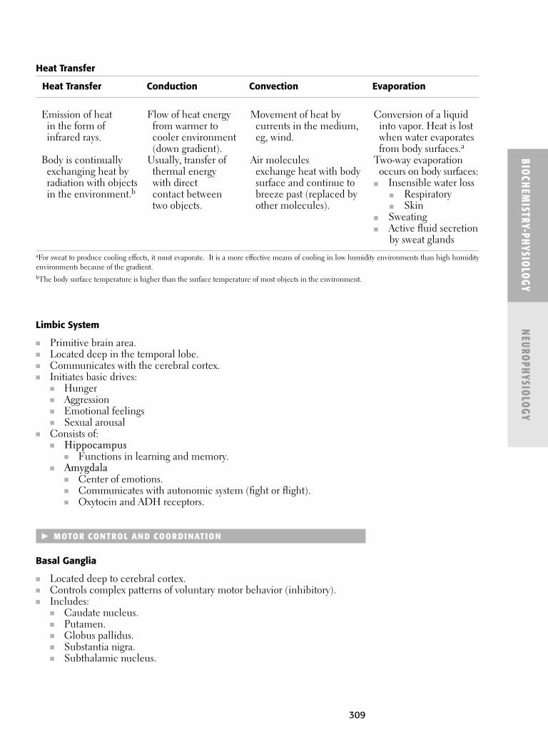
309
Limbic System
■ Primitive brain area. ■ Located deep in the temporal lobe.■ Communicates with the cerebral cortex.■ Initiates basic drives:
■ Hunger■ Aggression■ Emotional feelings■ Sexual arousal
■ Consists of:■ Hippocampus
■ Functions in learning and memory.■ Amygdala
■ Center of emotions.■ Communicates with autonomic system (fight or flight).■ Oxytocin and ADH receptors.
� MOTOR CONTROL AN D COORDI NATION
Basal Ganglia
■ Located deep to cerebral cortex.■ Controls complex patterns of voluntary motor behavior (inhibitory).■ Includes:
■ Caudate nucleus.■ Putamen.■ Globus pallidus.■ Substantia nigra.■ Subthalamic nucleus.
BIOCHEM
ISTRY-PHYSIOLOGY
NEURO
PHYSIOLOGY
Heat Transfer
Heat Transfer Conduction Convection Evaporation
Emission of heat Flow of heat energy Movement of heat by Conversion of a liquid in the form of from warmer to currents in the medium, into vapor. Heat is lost infrared rays. cooler environment eg, wind. when water evaporates
(down gradient). from body surfaces.aBody is continually Usually, transfer of Air molecules Two-way evaporation
exchanging heat by thermal energy exchange heat with body occurs on body surfaces: radiation with objects with direct surface and continue to ■ Insensible water loss in the environment.b contact between breeze past (replaced by ■ Respiratory
two objects. other molecules). ■ Skin■ Sweating■ Active fluid secretion
by sweat glandsaFor sweat to produce cooling effects, it must evaporate. It is a more effective means of cooling in low humidity environments than high humidityenvironments because of the gradient.bThe body surface temperature is higher than the surface temperature of most objects in the environment.

310
Cerebellum
■ Lies posteroinferior to cerebrum, superoposterior to brain stem.■ Morphologically divided:
■ Two lateral hemispheres.■ Middle portion (Vermis).
■ Functions (excitatory):■ Maintains muscle tone.■ Coordinates muscle movement.■ Controls balance.
Note: The basal ganglia and cerebellum modify movement on a minute-to-minute basis. The output of the cerebellum is excitatory, whereas that ofthe basal ganglia is inhibitory.
These two systems work together to achieve smooth, coordinated move-ment. Movement disorders result from aberration to each of these systems, eg,Parkinson’s (loss of DA neurons in basal ganglia), Huntington’s (loss of neuronsin basal ganglia, triple repeat disease), dysmetria, ataxia (see pathology).
� BRAI N STE M
Lies immediately inferior to cerebrum, just anterior to cerebellum.
Midbrain (Mesencephalon)
■ Connects dorsally with the cerebellum.■ Large voluntary motor nerve tracts pass through.■ Location of:
■ CN III, IV nuclei (connect with VI via MLF).■ Substantia nigra. (See also “Parkinson’s Disease” in Neuropathology)
Pons
■ Between the midbrain and medulla.■ Connects to the cerebellum posteriorly.■ Location of:
■ CN V, VI, VII, VIII nuclei.
Medulla Oblongata
■ Most inferior segment of vertebrate brain.■ Continues with the spinal cord below.■ Joins the spinal cord at foramen magnum.■ Contains:
■ Important regulatory centers:■ Area postrema—vomiting.■ Swallowing. ■ Cardiac.■ Vasomotor.■ Respiratory.
■ CN IX, X, XI, XII nuclei.
BIOC
HEM
ISTR
Y-PH
YSIO
LOGY
NEU
ROPH
YSIO
LOGY
Motor pathway:
Motor cortex (precentral gyrus)
↓
Upper motor neuron
↓
Internal capsule
↓
Corticospinal tract
↓(Cerebral peduncles –
midbrain)
↓(Pyramids – medulla – fibers
cross)
↓
Ventral horn (spinal cord)
↓
Lower motor neuron
↓
Muscle

311
� PE RI PH E RAL N E RVOUS SYSTE M
PNS is composed of:
■ Afferent neurons: From sensory receptors to CNS.■ Efferent neurons: From CNS to muscles, organs, and glands (and their
associated ganglia and plexuses).
BIOCHEM
ISTRY-PHYSIOLOGY
NEURO
PHYSIOLOGY
Subdivisions of the Peripheral Nervous System
Somatic Nervous System Autonomic Nervous System
12 pairs of cranial nerves. 2 subdivisions:31 pairs of spinal nerves. ■ Sympathetic.Both sensory and motor neurons. ■ Parasympathetic.Innervates skeletal muscle (voluntary). Action is largely involuntary.Motor: Controls:No synapse in peripheral ganglion. Glands (exocrine and endocrine).
■ Uses 1 efferent neuron from Cardiac muscle.the CNS to end-organ (LMN). Smooth muscle.
Sensory: Visceral organs.Synapse within dorsal root ganglion Not skeletal muscle.(peripheral) prior to CNS. Motor:
Modalities: Synapses within autonomic ganglion.■ Touch. ■ Uses 2 efferent neurons from ■ Movement (position sense). CNS to effector.■ Temperature. ■ Pain.
� AUTONOM IC N E RVOUS SYSTE M
Refer to the illustrations in the “Neuroanatomy” section.
BASIC ANATOMIC PATHWAY
Preganglionic neuron (within CNS). ↓
Ganglion (cell bodies of postganglionic neurons; outside of the CNS).↓
Postganglionic neuron (outside of the CNS).↓
Effector organ.
Autonomic Ganglia
■ Collections of cell bodies of the postganglionic neurons (unmyelinated). ■ Sympathetic: Sympathetic chain ganglia (near spinal cord) (paravertebral):
■ Short preganglionic neuron.■ Long postganglionic neuron.
■ Parasympathetic: Ganglia at, within organ (eg, celiac ganglion):■ Long preganglionic neuron.■ Short postganglionic neuron.
Remember: Both
parasympathetic and
sympathetic preganglionic
neurons are cholinergic
(release Ach). However,
parasympathetic
postganglionic neurons are
cholinergic (muscarinic
receptors); sympathetic
postganglionic neurons are
largely adrenergic (release
norepinephrine, which binds
to sympathetic receptors).
The exception is in sweat
glands and blood vessels in
skeletal muscle. Here the
postganglionic sympathetic
neurons are cholinergic
(release Ach at muscarinic
receptors).
Remember: Prevertebral
ganglia have longer
preganglionics.

312
Parasympathetic Nervous System
■ Craniosacral: Composed of:■ CN nuclei (ie, III-ciliary, VII- submandibular and pterygopalatine, IX-otic).■ S2–S4 (pelvic splanchnic).
■ Major nerve is the vagus nerve (CN X): Originates in medulla.■ Preganglionic neuron (myelinated)
■ Cholinergic (releases Ach).■ Binds nicotinic cholinergic receptors on postganglionic neurons (gan-
glia within effector).■ Postganglionic neuron (unmyelinated)
■ Cholinergic (releases Ach).■ Binds muscarinic cholinergic receptors in the tissue.
BIOC
HEM
ISTR
Y-PH
YSIO
LOGY
NEU
ROPH
YSIO
LOGY
Postganglionic Receptors
Nicotinic Muscarinic
N1 (NN) N2 (NM) Mstimulated by: nicotine stimulated by: muscurin blocked by: hexamethonium blocked by: atropine
Principal Autonomic Motor endplate Effector organs (includinglocation ganglia (NMJ) sweat glands under
■ Both symp and parasymp sympathetic control)preganglionics releaseAch on NN
Effect(s) Excitatory Excitatory Excitatory or inhibitory■ Nerve transmission ■ Muscle contraction blood vessels in■ Postganglionics skeletal muscle
are activated
NOTES
■ All preganglionic autonomic neurons (both sympathetic and parasympa-thetic) and all postganglionic parasympathetic neurons are cholinergic(use Ach as NT).
■ Cholinergic effects of preganglionic autonomic systems (at ganglia of sym-pathetic and parasympathetic systems) are excitatory.
■ Cholinergic effects of postganglionic parasympathetic fibers are either exci-tatory or inhibitory, depending on the end-organ (eg, parasympathetic fibersinnervating heart ↓ HR).
Sympathetic Nervous System
■ Thoracolumbar.■ Composed of spinal segments T1–L3.
■ Exerts widespread effect because of high ratio of postganglionic to pregan-glionic fibers.■ Each sympathetic preganglionic neuron branches extensively and
synapses with numerous postganglionic neurons.
Sympathetic Tone Only:adrenal medullasweat glandspilorector musclesblood vessels
Paravertebral ganglia→sympathetic chain versusPrevertebral ganglia →celiac, SMA, IMA, inferiorhypogastric

313
■ Preganglionic neuron (myelinated)■ Cholinergic (releases Ach).■ Binds nicotinic cholinergic receptors on postganglionic neurons (ganglia
within effector).■ Each preganglionic parasympathetic neuron synapses with many post-
ganglionic parasympathetic neurons.■ Postganglionic neuron (unmyelinated)
■ Adrenergic (releases NE).■ Binds adrenergic receptors in the tissue except sweat glands and skele-
tal muscle blood vessels.
ADRENERGIC RECEPTORS
■ Membrane receptor proteins.■ G-protein-coupled receptors.■ Located on autonomic effector organs.■ Bound by catecholamine ligands (epinephrine, norepinephrine).■ Norepinephrine (NE) stimulates mainly alpha receptors.■ Epinephrine stimulates both alpha and beta equally.
BIOCHEM
ISTRY-PHYSIOLOGY
NEURO
PHYSIOLOGY
Alpha- and Beta-Adrenergic Receptors
Alpha-1 (α-1) Alpha-2 (α-2) Beta-1 (β-1) Beta-2 (β-2)
Principal Vascular smooth Presynaptic Heart Skeletal musclelocation muscle: nerve Bronchial
terminals— smoothdecrease musclesynaptictransmission
■ Skin Platelets—plateletaggregation
■ Mucosa Fat cells—inhibitlipolysis
■ GI GI tract wall
Effect(s) Vasoconstriction Inhibition ↑ Heart rate Vasodilation (relaxation ↑ Contractility Bronchodilationor dilation)
Summary of Autonomic Effects
Sympathetic Parasympathetic
Adrenergic CholinergicPupils Mydriasis (dilate) Miosis (constrict)Salivation Thick (mucous) Watery (ready to eat)Bronchi Bronchodilation BronchoconstrictionHeart rate ↑ ↓Adrenal medulla Causes epi and NE release No effect
Gray ramuscommunicans—carryunmyelinated post-ganglionic sympatheticnerves to peripheralorgans.
Three cervical sympathetic
ganglia supply the head and
neck (superior, middle, and
inferior). These are
paravertebral ganglia.
Other ganglia of the SNS
include the celiac ganglion,
superior mesenteric ganglion,
and inferior mesenteric
ganglion.
White ramuscommunicans—allsympathetic preganglionicneurons enter theparavertebral chain viawhite ramus.
Monoamine oxidase (MAO) is
an enzyme that catalyzes
oxidative deamination of
monoamines (including NE,
serotonin, and epinephrine).
The deamination process ↑
breakdown (metabolism) of
excess NTs that accumulate at
postsynaptic terminals.

314
� SP I NAL TRACTS
■ See also the “Neuroanatomy” section. ■ Spinal cord is part of the CNS.■ White matter tracts:
■ Composed of myelinated nerve fibers bundles.■ Surround gray matter horns.
■ Form ascending and descending tracts.■ Axons of a tract have the same origin, termination, and function.■ Tracts are often named for origin and termination (eg, spinothalamic
tract).■ Tracts are sensory or motor (see the following chart).
BIOC
HEM
ISTR
Y-PH
YSIO
LOGY
NEU
ROPH
YSIO
LOGY
Sensory (Ascending) Motor (Descending)
Spinothalamic Corticospinal (pyramidal)■ Pain Extrapyramidal■ TemperatureDorsal column, medial lemniscus■ Touch■ Pressure■ Vibration■ Proprioception (position)
SENSORY PATHWAY
Receptor↓
Peripheral nerve (sensory fibers) ↓
Dorsal root ganglion (synapse) (primary neuron) ↓
Spinal cord ↓
Spinal tracts (spinothalamic or dorsal column, medial lemniscus) ↓
Brain stem nuclei (gracile/ cuneate nucleus in medulla; secondary neuron)↓
Thalamus (tertiary neuron; VPL)↓
Decussates after synapse↓
Cortex (postcentral gyrus of parietal lobe)
Tracts can be:
■ Ipsilateral: Axonsrun on same side ascell bodies.
■ Contralateral: Axonsrun on opposite side(have crossed over).
Spinal cord anatomy:■ Dorsal horn
■ Sensory.■ Receives fibers
from the dorsalroot ganglia.
■ Ventral (anterior) root■ Motor.

315
RECEPTORS
■ Receive information from internal or external environment.■ Send nerve impulses to CNS.■ Two broad types: According to location of stimuli.■ Exteroreceptors: Receive external stimuli (from body surface):
■ Touch.■ Pressure.■ Pain.■ Temperature.■ Light.■ Sound.
■ Interoreceptors (visceroreceptors): Receive input from internal environ-ment of body:■ Pressure.■ Pain.■ Chemical changes.
PROPRIORECEPTORS
■ Type of interoceptor. ■ Relays information concerning position of body parts (in space).■ Separate from visual input.
■ Kinesthetic sense■ Located in muscles, tendons, joints.■ Communicate with the vestibular apparatus.
BIOCHEM
ISTRY-PHYSIOLOGY
NEURO
PHYSIOLOGYNonencapsulated versus Encapsulated Receptors
Nonencapsulated Receptors Encapsulated Receptors (corpuscles)
Free nerve endings Meissner’s corpuscles—rapidly adaptingPain primarily, touch, pressure, tickle, Mechanoreceptors, allow 2-point
hot/cold. discrimination.Terminal ends have no myelin. In dermal papilla of skin. In epithelial cells, skin, cornea, alimentary Ovoid stack of Schwann cells.
tract, connective tissue, haversian system of bone, dental pulp.
Merkel’s disc—slow adapting Pacinian corpuscles (rapidly adapting)Tactile (touch, pressure) (fine detailed Vibration, pressure.
surface patterns).In hairless skin—fingertips, pressure. In dermis, subcutaneous tissue, ligaments, Found in basal layer. joints.
Concentric lamellae of flattened cells.
Hair follicle receptors Ruffini’s corpuscles (slow adapting)Mechanoreception—bending hair Stretch, continuous pressure states
stimulates, touch. In dermis of hairy skin.Fiber winds around hair. Large unmyelinated nerve fibers ending
within bundles of collagen fibers.
There are many types ofjoint receptors:■ Nonencapsulated
■ Free nerve endings:Pain.
■ Encapsulated ■ Pacinian: Vibration,
pressure.■ Ruffini: Stretch.
■ Neuromuscularspindles■ Stretch.
■ Neurotendons■ Tension.

316
� SOMATOSE NSORY PATHWAYS
Sensory Tracts (Ascending)
See Figure 10–1 for somatosensory pathways.
BIOC
HEM
ISTR
Y-PH
YSIO
LOGY
NEU
ROPH
YSIO
LOGY
Receptors Classified by Stimulus Type
Mechanoreceptor Thermoreceptor Nociceptor Chemoreceptor Photoreceptor
Stimulus Pressure Temperature Pain Chemicals Lightor stretch (inhaled, ingested,
or in blood)
Examples Pacinian corpuscles Free nerve Free nerve Taste receptors RetinaMuscle spindles endings endings Smell receptors ■ Rods(Golgi tendon organs) Osmoreceptors ■ ConesMeissner’s corpuscles Carotid body O2Hair cells receptors
Sensorynerve
Pain,cold,
warmth
Touch, light
Touch
MIDLINE
Postcentralgyrus
Nucleigracilis andcuneatus
Fasciculigracilis andcuneatus(dorsalcolumns)
Dorsalrootganglioncell
Thalamus (specificsensory relay nuclei)(VPL = body; VPM = head)
Thalamicradiation
Lateral spino-thalamic tract
Ventral spino-thalamic tract
Mediallemniscus
F I G U R E 1 0 – 1 . Somatosensory pathways.
Reproduced, with permission, from Ganong WF. Review of Medical Physiology, 22nd ed.
New York: McGraw-Hill, 2005.
Hair cells transduce senses of
hearing and balance.
Osmoreceptors and carotid
body O2 receptors monitor pH
and gas levels.

317
SPINOTHALAMIC TRACT (ANTEROLATERAL SYSTEM)
■ Lateral spinothalamic tract: Transmits pain and temperature.■ Anterior spinothalamic tract: Transmits light touch.
PATHWAY
Sensory nerve ↓
Dorsal horn of spinal cord gray matter (travel in Lissauers tract and thensynapse on secondary neurons)
↓Cross to opposite side of cord via anterior white commisure (decussate at level
enters cord) via Lissaure’s tract; secondary neuron↓
Ascend contralateral spinal cord (through anterior and lateral white mattercolumns/tracts)
↓Thalamus (tertiary neuron, in VPL)
↓Somatosensory cortex (parietal lobe)
DORSAL COLUMN, MEDIAL LEMNISCUS SYSTEM
■ Conveys touch, pressure, and vibration.
PATHWAY
Sensory nerve ↓
Dorsal horn of spinal cord gray matter ↓
Ascend ipsilateral spinal cord ↓
Posterior columns: Fasciculus gracilis (fibers from lower extremities) andFasciculus cuneatis (fibers from upper extremities)
↓Synapse in medulla (nucleus gracilis and cuneatis)
↓Internal arcuate fibers decussate and ascend contralateral brain stem in medial
lemniscus↓
Thalamus ↓
Somatosensory cortex
BIOCHEM
ISTRY-PHYSIOLOGY
NEURO
PHYSIOLOGY

318
SOMATOSENSORY CORTEX (POSTCENTRAL GYRUS)
See Figures 10–2 and 10–3 for somatosensory cortex.
■ Representation is homunculus (representation of body proportional to sense).
BIOC
HEM
ISTR
Y-PH
YSIO
LOGY
NEU
ROPH
YSIO
LOGY
Hand
FaceTongue
S2
Trunk
Auditory
S1Posteriorparietalcortex
Visual
9
8 6 43 1 2
F I G U R E 1 0 – 2 . Somatosensory cortex.
Reproduced, with permission, from Ganong WF. Review of Medical Physiology, 22nd ed.
New York: McGraw-Hill, 2005.
F I G U R E 1 0 – 3 . Somatosensory cortex.
Reproduced, with permission, from Ganong WF. Review of Medical Physiology, 22nd ed.
New York: McGraw-Hill, 2005.

319
Descending Motor Tracts (Efferent or Descending Pathways)
■ The upper motor neuron tracts sending signals from the brain muscles.
MOTOR PATHWAY
Motor area of brain (precentral gyrus of frontal lobe)↓
Upper motor neurons (= descending motor tracts, eg, corticospinal tract)↓
Lower motor neurons ↓
Skeletal muscle
Upper Motor Neurons
■ Originate in white matter of brain.■ Form two major systems:
■ Corticospinal tract (pyramidal system).■ Extrapyramidal system.
Corticospinal Tract (Pyramidal System)
See Figure 10–4 for corticospinal tract (pyramidal system).
■ Two components:■ Lateral (70–90%).■ Anterior/ventral (10–30%).
■ Travel via primary motor cortex through internal capsule to medulla. ■ Decussate in medulla (pyramids).■ Continue down opposite side of spinal cord →
anterior horn → lower motor neurons → muscles.■ Right brain controls left somatic muscles.
■ Control fine, skilled movements of skeletal muscle.
Extrapyramidal System
■ Collection of smaller tracts:■ Rubrospinal (red nucleus in midbrain—voluntary movements).■ Important for somatic muscle control, posture■ Reticulospinal—important for somatic motor control and autonomic
function control (reticular nuclei, pons, and medulla) (reticular for-mation (diffuse brain stem nuclei); coordinate locomotion, mediateautonomic).
■ Olivospinal (inferior olive—motor learning).■ Vestibulospinal (vestibular nuclei—control limb extensors, head and
neck (gaze)) mediates vestibular end organ and cerebellum uponextensive muscle tone and balance.
■ Tectospinal (superior colliculus—coordinate neck, head, and eyemovements).
■ Travel from premotor area of frontal lobe (and other areas) to pons.■ Decussate in pons.■ Continue down opposite side of spinal cord →
anterior horn → lower motor neurons → muscles.■ Right brain controls left lower motor neuron.
■ Controls gross motor movement, posture, and balance.
BIOCHEM
ISTRY-PHYSIOLOGY
NEURO
PHYSIOLOGY
This tract is called pyramidal
system because fibers of the
corticospinal tract form the
pyramids in the medulla.

320
� SP I NAL CORD LES IONS
Lesion on one side causes:
■ Ipsilateral motor loss (corticospinal).■ Ipsilateral touch/sensory loss.■ Contralateral pain and temperature loss (spinothalamic).■ Example: If you hemitransect the right side of the spinal cord, you lose
right-sided (ipsilateral) motor control (corticospinal) and left-sided (con-tralateral) pain and temperature sense (spinothalamic).
� N E U ROPHYSIOLOGY
Resting Membrane Potential (All Cells!)
■ Charge differential or voltage set up across the resting nerve membrane.■ Due to separation of charged particles (ions and proteins) between
extracellular and intracellular fluids.
BIOC
HEM
ISTR
Y-PH
YSIO
LOGY
NEU
ROPH
YSIO
LOGY
Corticospinal tract
Decussation of the pyramids
Anteriorhorn cell
Internal capsule
Lateral cortico-spinal tract(80% of fibers)
Spinal nerve
Pyramids
Ventral cortico-spinal tract
(20% of fibers)
Interneuron
Precentralgyrus(area 4,etc)
Distalmuscle
Proximalmuscle
F I G U R E 1 0 – 4 . Corticospinal tract (pyramidal system).
Reproduced, with permission, from Ganong WF. Review of Medical Physiology, 22nd ed.New York: McGraw-Hill, 2005.
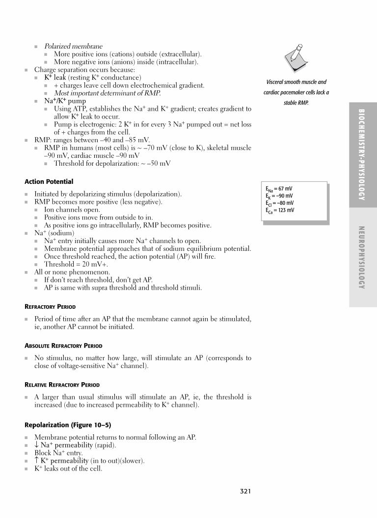
321
■ Polarized membrane■ More positive ions (cations) outside (extracellular).■ More negative ions (anions) inside (intracellular).
■ Charge separation occurs because:■ K+ leak (resting K+ conductance)
■ + charges leave cell down electrochemical gradient.■ Most important determinant of RMP.
■ Na+/K+ pump■ Using ATP, establishes the Na+ and K+ gradient; creates gradient to
allow K+ leak to occur.■ Pump is electrogenic: 2 K+ in for every 3 Na+ pumped out = net loss
of + charges from the cell.■ RMP: ranges between –40 and –85 mV.
■ RMP in humans (most cells) is ~ –70 mV (close to K), skeletal muscle–90 mV, cardiac muscle –90 mV■ Threshold for depolarization: ~ –50 mV
Action Potential
■ Initiated by depolarizing stimulus (depolarization).■ RMP becomes more positive (less negative).
■ Ion channels open. ■ Positive ions move from outside to in.■ As positive ions go intracellularly, RMP becomes positive.
■ Na+ (sodium)■ Na+ entry initially causes more Na+ channels to open.■ Membrane potential approaches that of sodium equilibrium potential.■ Once threshold reached, the action potential (AP) will fire.■ Threshold = 20 mV+.
■ All or none phenomenon.■ If don’t reach threshold, don’t get AP.■ AP is same with supra threshold and threshold stimuli.
REFRACTORY PERIOD
■ Period of time after an AP that the membrane cannot again be stimulated,ie, another AP cannot be initiated.
ABSOLUTE REFRACTORY PERIOD
■ No stimulus, no matter how large, will stimulate an AP (corresponds toclose of voltage-sensitive Na+ channel).
RELATIVE REFRACTORY PERIOD
■ A larger than usual stimulus will stimulate an AP, ie, the threshold isincreased (due to increased permeability to K+ channel).
Repolarization (Figure 10–5)
■ Membrane potential returns to normal following an AP.■ ↓ Na+ permeability (rapid).■ Block Na+ entry.■ ↑ K+ permeability (in to out)(slower).■ K+ leaks out of the cell.
BIOCHEM
ISTRY-PHYSIOLOGY
NEURO
PHYSIOLOGY
Visceral smooth muscle and
cardiac pacemaker cells lack a
stable RMP.
ENa = 67 mVEK = –90 mVECl = –80 mVECa = 123 mV

322
Hyperpolarization
■ During repolarization there is an overshoot in the more negative direction.■ Membrane potential briefly becomes more negative than RMP before
returning to RMP.■ This is because of ↑ K+ conductance.
■ K+ channels stay open.■ K+ efflux is greater than in resting.
Note: Hyperpolarization is responsible for the relative refractory period (cellremains hypoexcitable). Influx of Cl– will also hyperpolarize and make APmore difficult to generate.
� LOCAL AN ESTH ETICS
■ Block sodium channels (↓ Na+ permeability).■ Bind to inactivation gates of fast, voltage-gated Na+ channels,
■ keeping them closed and■ prolonging absolute refractory period.
■ ↓ Membrane excitability → cannot generate AP → no nerve impulseconduction.
■ Reversible.■ K, Cl, Ca conductances are unchanged.
Local Anesthetics
■ Affect small myelinated fibers first (size rule).Unmyelinated C-fibers (slow, dull, long lasting) (smallest)
↓Small myelinated nerve fibers (pain, temp)
↓Larger A-fibers (touch proprioception, Golgi tendon)
BIOC
HEM
ISTR
Y-PH
YSIO
LOGY
NEU
ROPH
YSIO
LOGY
ms
mmho/cm2
membrane
0 1 2 3 4
30
Action potential
Sodium conductance
Potassiumconductance
80
60
40
20
20
10
mV
Con
duct
ance
F I G U R E 1 0 – 5 . Repolarization.
Reproduced, with permission, from Ganong WF. Review of Medical Physiology, 22nd ed.New York: McGraw-Hill, 2005:59. As modified from Hodgkin AL. Ionic movements and
electrical activity in giant nerve fibers. Pro R Soc Lond Ser B 1958;143:1.
Remember:Excitable cells
■ Neurons■ Muscle cells■ Cardiac pacemaker

323
� N E U ROTRANSM ITTE RS
Excitatory
■ Depolarize (more positive) the postsynaptic membrane potential.■ Brings it closer to threshold.■ ↑ probability of AP in postsynaptic neuron.
■ Creates an excitatory postsynaptic potential (EPSP).■ Glutamate is the major excitatory neurotransmitter.
Inhibitory
■ Hyperpolarize (more negative) the postsynaptic membrane potential. ■ Moves it away from threshold. ■ ↓ probability of AP in postsynaptic neuron.
■ Creates an inhibitory postsynaptic potential (IPSP).■ Result of ↑ membrane permeability to either Cl– or K+.■ Examples of inhibitory NTs:
■ Glycine.■ GABA.
■ Both bind receptors and open Cl– channels (↑ Cl– permeability).
� SU M MATION
Spatial Summation
■ Two excitatory inputs arrive at a postsynaptic neuron simultaneously.■ Converging circuit.■ Arrival of impulses from multiple presynaptic fibers at same time.
Temporal Summation
■ Two excitatory inputs arrive at a postsynaptic neuron in rapid succession.■ ↑ frequency of nerve impulses from a single presynaptic fiber.
� N E RVE CON DUCTION
■ Nerve impulse = action potential spreads along plasma membrane.
Saltatory Conduction
■ Occurs in myelinated fibers (remember: Schwann cells (periphery) andoligodendrocytes (CNS)).
■ ↑ velocity of nerve transmission along myelinated fibers.■ Conserves energy because:
■ Only the Ranvier node depolarizes. ■ Less energy for Na+/K+ ATPase to reestablish resting ion gradients.■ Na+/K+ pumps reestablish concentration gradient only at Ranvier
nodes.■ Allow repolarization to occur with less transfer of ions.
■ Electrochemical basis behind saltatory conduction is ↓ membranecapacitance (increase distance between charges; less charges necessary).
BIOCHEM
ISTRY-PHYSIOLOGY
NEURO
PHYSIOLOGY
EPSPs can combine, using
summation (2 forms) to reach
threshold and initiate an AP.
The inhibitory or excitatory
effects of NTs depend on their
binding characteristics to
receptors.
NeurotransmitterReview:
MAO—catalyze oxidativedeamination of NE,serotonin, epiACh—voluntarymovement, skeletalmuscles/movement ofvisceraNE—wakefulness, arousalDA—voluntary movementand motivation, pleasure,addiction, loveSerotonin—memory,emotion, sleep/wake,temperatureGlycine—spinal reflexesand motor behaviorHistamine—sleep/wake,inflammation

324
MYELIN
■ Prevents movement of Na+ and K+ through the membrane.■ Na+, K+ conductance only at Ranvier nodes.
■ ↓ membrane capacitance. ■ ↑ membrane resistance.
NODES OF RANVIER
■ Exposed nerve membrane where depolarization occurs.■ Continue fueling spread of AP during nerve transmission.
■ Located every 0.2–2 mm along the myelin sheath.■ APs could not be produced if the myelin sheath were continuous.■ APs travel down axon and “jump” from node to node.
CONTINUOUS CONDUCTION
■ Occurs in unmyelinated fibers.■ Nerve transmission (AP) travels along entire membrane surface.■ Relatively slow conduction (1.0 m/sec).
Problems with Nerve Conduction
WALLERIAN DEGENERATION
■ Axon is cut.■ The axon remnant distal to the cut (away from the cell body) degenerates
because axonal transport is interrupted.■ Regeneration of axons possible if endoneurial sheath is intact (neurolemma).
■ Occurs at a rate of 2–4 mm/day.■ If cell body is irreversibly injured, the entire neuron degenerates.
NEUROPRAXIA
■ Transient block (bruise).■ Incomplete paralysis or loss of sensation.■ Rapid recovery.
AXONOTMESIS
■ Axon damaged, but connective sheath remains intact.■ Wallerian degeneration occurs distally but then regeneration can occur.
NEUROTMESIS
■ Complete transaction of nerve trunk.■ Results in:■ Motor
■ Flaccid paralysis.■ Atrophy of end-organ.
BIOC
HEM
ISTR
Y-PH
YSIO
LOGY
NEU
ROPH
YSIO
LOGY
Saltatory conduction is a
faster way to conduct an
impulse down the axon (up to
100 m/sec).
Conduction velocitydepends on:■ Diameter of nerve fiber.
■ ↑ diameter → ↑resistance to flow→↑ velocity.
■ Presence of myelinsheath.
Fibers of CNS (brain andspinal column) are notenclosed by neurilemma—regenerated severedaxons are rare.
Neurilemma: Thinmembrane spirallywrapping the myelinlayers of fibers, especiallyperipheral nerves andaxons of unmyelinatednerve fibers. All axons ofthe PNS have it.
In CNS, neurons aremyelinated byoligodendrocytes and lacka neurilemma.

325
■ Sensory■ Total loss of cutaneous sensation.
� SYNAPSE
Functional connection, anatomical junction between
■ Nerve axon (presynaptic axon) and ■ Target cell
■ Nerve (postsynaptic neuron)■ Muscle (NMJ)■ Gland
Synapse controls direction of nerve impulse
PRESYNAPTIC NEURONS
■ Transmit information toward a synapse.
SYNAPTIC CLEFT
■ Space between presynaptic terminal and postsynaptic cell.
POSTSYNAPTIC NEURONS
■ Transmit away from a synapse (dendrite → axon).
NERVE IMPULSES
■ Travel in only one direction because synapses are polarized.
Neuronal Excitability
■ Nerves are excited (APs generated) electrically or chemically.■ Ligand-gated (NT binding) (most common).■ Voltage-gated channels.■ Mechanically gated (stretching).
Types of Synapse
■ Chemical synapse (most common).■ Ligand-gated: Use NTs.
■ Electrical synapse.
BIOCHEM
ISTRY-PHYSIOLOGY
NEURO
PHYSIOLOGY
Depolarization of the
presynaptic cell initiates a
response in the postsynaptic
cell either by release of NTs
(most common) or by direct
passage of electrical current.
Site of interneuronal
communication can involve
one axon to one dendrite, one
axon to many dendrites, or
many axons to one dendrite.

326
CHEMICAL SYNAPSE
■ Most common type.■ Consists of:
■ Presynaptic membrane.■ Synaptic vesicles within this terminal contain a NT.
■ Synaptic cleft.■ Space between the presynaptic and postsynaptic membranes.
■ Postsynaptic membrane.■ Membrane of postsynaptic neuron that contains specific receptors
for the NT.
SYNAPTIC TRANSMISSION
■ Release of NTs.■ NTs are stored in synaptic vesicles within the presynaptic axon terminal.■ AP depolarizes the presynaptic membrane, causing:
■ Voltage-gated Ca2+ channels opened (on the presynaptic membrane).■ ↑ Ca2+ influx.
■ Ca2+ causes the synaptic vesicles to fuse with membrane. ■ NTs are released by exocytosis into synaptic cleft.■ NTs diffuse across cleft. ■ Bind to specific receptors on postsynaptic cell.
■ The time required for this process to occur is called synaptic delay.
CHEMICAL NEUROTRANSMITTERS
■ Mediate most connections.■ Small molecule NTs (contained within vesicles):
■ Glutamate, GABA, glycine, ACh, 5HT, NE, Epi, etc.■ Neuropeptides (large dense vesicles):
■ Somatostatin, endorphins, enkephalins, opioids, etc.
ELECTRICAL SYNAPSE
■ Gap junctions; minority.■ Cytoplasm of adjacent cells is connected by gap junctions. ■ Allows passage of local electrical currents (ions and small molecules)
(from APs in presynaptic neuron) to pass directly to postsynaptic neuron.■ Rare in the CNS. ■ Common in cardiac and smooth muscle.■ Ensure a group of neurons act together; Synchronize groups of
neurons.■ Important in embryonic development (morphogenic gradients).
� N E U ROM USC U LAR J U NCTION (N M J)
Synapse between lower motor neuron (efferent nerve) and muscle.
■ Presynaptic terminal (lower motor neuron axon)■ Releases ACh.
■ Postsynaptic membrane (skeletal muscle membrane).■ Displays nicotinic receptor (NM).
BIOC
HEM
ISTR
Y-PH
YSIO
LOGY
NEU
ROPH
YSIO
LOGY

327
■ Threshold is –65 mV → all or nothing!■ ~35 ACh required.
SEQUENCE
ACh binds NM↓
Na+ channels open on the motor end-plate (ligand-gated ion channel)↓
Muscle fiber depolarized (Na+ influx)↓
Voltage-gated Na+ channels on the sarcolemma open↓
AP stimulated in skeletal muscle fiber↓
AP travels down the transverse tubules (dihydropyridine receptors (voltage-gated Ca2+ channel))
↓Ca2+ release from the sarcoplasmic reticulum (ryanodine receptor in SR
(mechanically activated))↓
Muscle contraction (Ca2+ binds troponin, troponin moves off actin, myosincan bind, etc)
Acetylcholine Metabolism
■ Synthesized in the presynaptic terminal of the motor neuron from whichit is released.■ Acetyl CoA + choline – (choline acetyltransferase)→ ACh (acetyl-
choline).■ Stored in synaptic vesicles.■ Released into synaptic cleft (generates effect).■ Breakdown:
■ ACh – (acetylcholinesterase [AChE])→ acetate + choline.
� SPEC IAL SE NSES
Vision
General eye anatomy (Chapter 2).
EYEBALL
■ Grossly divided into two segments:■ Anterior segment
■ Consists of two chambers (anterior and posterior). ■ Filled with aqueous humor (watery fluid).
■ Posterior segment■ Filled with vitreous humor (thick, gelatinous material).
BIOCHEM
ISTRY-PHYSIOLOGY
NEURO
PHYSIOLOGY
Two enzymes involved inACh metabolism:
■ Cholineacetyltransferase: AChgeneration.
■ Acetylcholinesterase(AChE): AChbreakdown.
In the NMJ, AChE is located on
the muscle end-plate.
If AChEs are inhibited, get
prolongation of end-plate
potential (EPP).
AP in LMN → Ca2+
voltage-gated channelopens → Ca2+ in → AChvesicles fuse and releasedinto synaptic cleft → AChbinds to Nm.

328
STRUCTURAL COMPONENTS
■ Sclera■ Tough, white outer layer.■ Maintains size and form of the eyeball.
■ Cornea■ Transparent dome on the anterior eye surface.■ Protective function.■ Helps focus light on retina at back of eye.
■ Choroid■ Lining of the inner aspect of the eyeball beneath the retina; very vascular.
COMPONENTS THAT CONTROL LIGHT ENTERING THE EYE AND FOCUS LIGHT ON THE RETINA
■ Pupil■ Circular opening (black area) in the middle of the iris.■ Light enters the eye to reach retina through this opening.■ Lens is located behind this aperture.■ Size of the pupil is controlled by muscles in the iris.
■ Iris■ Circular colored area of the eye (amount of pigment in the iris deter-
mines the color of the eye).■ Miosis: Constriction of pupil:
■ Sphincter pupillae (iris sphincter) closes iris.■ Response to:
■ Increased light.■ Drugs (eg, narcotics).■ Pathologic conditions.■ Parasympathetic stimulation.
■ Mydriasis: Dilation of the pupil (term often used for prolonged papil-lary dilation):■ Dilator pupillae (iris dilator) opens iris.■ Response to:
■ Decreased light.■ Sympathetic stimulation (fight or flight).■ Drug.■ Disease.
■ Lens■ Directly behind the iris and pupillary opening.■ Focuses light on the retina.■ Controlled by ciliary muscle (within the ciliary body).
■ Ciliary body■ Functions:
■ Accommodation.■ Ciliary muscle alters the lens refractory power (to focus light on the
retina).■ Produces aqueous humor.■ Holds lens in place.
■ Retina■ Innermost layer of the eye on the posterior surface.■ Receives visual stimuli.■ Communicates via CN II with the brain (visual cortex).■ Photoreceptors (visual receptors) of retina (on deepest surface to
incoming light).
BIOC
HEM
ISTR
Y-PH
YSIO
LOGY
NEU
ROPH
YSIO
LOGY

329
■ Rods (higher sensitivity, lower acuity)■ Contain rhodopsin (photopigment).■ Perceive different degrees of brightness: Responsible for night
vision (dark adaptation).■ Relative lack of color discrimination.■ Located mostly at the periphery of the retina.
■ Cones (higher acuity (fovea); color)■ Each contains one of three photopigments.■ Each being sensitive to a particular wavelength of light.
■ Three types: Red, green, blue.■ Primarily responsible for color vision.
■ Principal photoreceptors during daylight or in brightly lit areas.■ Located in the center of the retina, especially in the fovea.
■ Photopigments■ Four photopigments:
■ Rhodopsin (rods).■ Red, green, and blue (cones).
■ Each photopigment contains:■ Opsin (protein) bound to ■ Retinal (a chromophore molecule).■ Together they compose rhodopsin.
■ The difference among the opsin molecules allows a photopigment tohave specificity for a particular type or color of light.
VISUAL PATHWAY
See Figure 10–6.Light
↓(passes through cornea, lens, aqueous humor, vitreous humor, and onto retina)
↓Retina (rods & cones)
↓Bipolar neurons
↓Ganglion cells
↓Optic disc
↓Optic nerve (exits through optic foramen)
↓Optic chiasm (temporal visual fields decussation; crossing of images on nasal
side of each retina)↓
Lateral geniculate (of thalamus)↓
Optic radiations↓
Visual cortex (area 17) and visual association cortex (18, 19)
BIOCHEM
ISTRY-PHYSIOLOGY
NEURO
PHYSIOLOGY
Rods are more abundant,
have higher sensitivity, and
lower acuity compared with
cones.
Retinal (a vitamin A
aldehyde) is constant among
all photopigments and is
produced from vitamin A.
Oxidation of vitamin A
(retinol) leads to retinal.
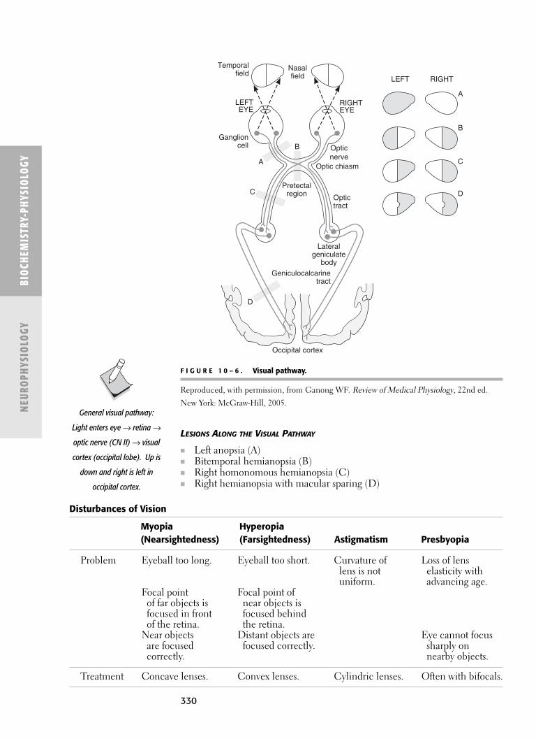
330
BIOC
HEM
ISTR
Y-PH
YSIO
LOGY
NEU
ROPH
YSIO
LOGY
LESIONS ALONG THE VISUAL PATHWAY
■ Left anopsia (A)■ Bitemporal hemianopsia (B)■ Right homonomous hemianopsia (C)■ Right hemianopsia with macular sparing (D)
General visual pathway:
Light enters eye → retina →
optic nerve (CN II) → visual
cortex (occipital lobe). Up is
down and right is left in
occipital cortex.
Temporalfield
Ganglioncell
Geniculocalcarinetract
LEFTEYE
LEFT RIGHT
A
B
C
D
Opticnerve
Optic chiasm
Pretectalregion
Nasalfield
Lateralgeniculate
body
A
B
C
D
Optictract
Occipital cortex
RIGHTEYE
F I G U R E 1 0 – 6 . Visual pathway.
Reproduced, with permission, from Ganong WF. Review of Medical Physiology, 22nd ed.New York: McGraw-Hill, 2005.
Disturbances of Vision
Myopia Hyperopia (Nearsightedness) (Farsightedness) Astigmatism Presbyopia
Problem Eyeball too long. Eyeball too short. Curvature of Loss of lens lens is not elasticity with uniform. advancing age.
Focal point Focal point of of far objects is near objects is focused in front focused behind of the retina. the retina.
Near objects Distant objects are Eye cannot focus are focused focused correctly. sharply on correctly. nearby objects.
Treatment Concave lenses. Convex lenses. Cylindric lenses. Often with bifocals.

� H EARI NG
Anatomy of the Ear
EXTERNAL EAR
■ Auricle (Pinna) ■ Directs sound waves.
■ External auditory canal (meatus) ■ Contains hair and cerumen (wax).■ Serves as resonator and conduit.
MIDDLE EAR
■ Tympanic cavity.■ Air-filled cavity in temporal bone.
■ Auditory tube: Equalizes pressure.■ Ossicles (malleus, incus, stapes): Transmit sounds from tympanic
membrane to oval window → to inner ear → converting them tohigher pressure sounds that are able to travel through fluid.
INNER EAR
See Figure 10–7.
■ Formed by bony labyrinth and membranous labyrinth■ Vestibule (saccule and utricle)
■ Associated with sense of balance and linear acceleration (static position)■ Semicircular canals
■ Concerned with equilibrium and angular momentum.■ Cochlea (two membranes: vestibular and basilar)
■ Responsible for hearing.■ Spiral organ (organ of Corti).
■ Receptors (hair cells) for hearing.■ Basic functional unit of hearing. ■ Transforms fluid vibrations from sound waves (mechanical energy)
into a nerve impulse (electrical energy).
BIOCHEM
ISTRY-PHYSIOLOGY
NEURO
PHYSIOLOGY
Nasopharynx
Lat Post
Semicircular canals:
Sup
Incus
Malleus Vestibular nerve
Vestibule
Facial nerve
Cochlear nerve
Cochlea
Auditory tube
Round window
Internal carotid artery
StapesEardrum
Externalauditory canal
F I G U R E 1 0 – 7 . Inner ear.
Reproduced, with permission, from Ganong WF. Review of Medical Physiology, 22nd ed. New York: McGraw-Hill, 2005.
331

332
Hearing
■ Ability to detect sound.■ Human hearing range = 20–20,000 Hz.
CHARACTERISTICS OF A SOUND WAVE
■ Pitch■ Related to the frequency of the sound wave.■ ↑ frequency = ↑ pitch.
■ Frequency is measures in hertz (Hz) or cycles per second.■ Loudness (amplitude)
■ Related to the intensity and the amplitude of the wave.■ ↑ amplitude = ↑ intensity = ↑ loudness.
■ Intensity is measured in decibels (dB).■ Timbre (quality)
■ Related to the presence of additional sound-wave frequencies superim-posed on the principal frequency.
HEARING PATHWAY
Sound vibration (external ear (pinna to canal))↓
Tympanic membrane (middle ear)↓
Ossicles (malleus → incus → stapes)↓
Oval window↓
Waves in perilymph (bony labyrinth)↓
Endolymph (membranous labyrinth)↓
Bending of hairs (stereocilia)↓
Hair cell depolarization within tectorial membrane or organ of Corti in cochlea(between basilar and vestibular membanes)
↓Short axons to spiral ganglion (located within bony modiolus)
↓Long axons form the cochlear nerve
↓Synapse w/cochlear nuclei (dorsal and ventral)
↓Superior olivary nucleus (ipsi- and contralaterally)
↓Lateral lemniscus
↓Inferior colliculus
↓Medial geniculate of thalamus
↓Transverse temporal gyrus (Heschl’s gyrus)
BIOC
HEM
ISTR
Y-PH
YSIO
LOGY
NEU
ROPH
YSIO
LOGY
Unlike other sensory systems,
hearing has bilateral central
representation (ie, sound from
one ear reaches auditory
cortex in both hemispheres).
Superior olivary nuclei are
important in sound
localization. Parallel nuclei
involved in vision: inferior
olivary, superior colliculus,
and lateral geniculate.
Pitch = Frequency.
Loudness = Amplitude.
Sound amplification:
■ Lever action of ossicles■ Concentration of
sound waves fromtympanic membraneto oval window
Presbycusis = hearing loss that
gradually occurs because of
changes in the inner or middle
ear in individuals as they grow
older.
Organ of Corti lies on basilar
membrane and is bathed in
endolymph.
Organ of Corti is the basic
functional unit of hearing—
transforms the sound waves
(mechanical energy) into fluid
vibrations into a nerve impuse
(electrical energy).
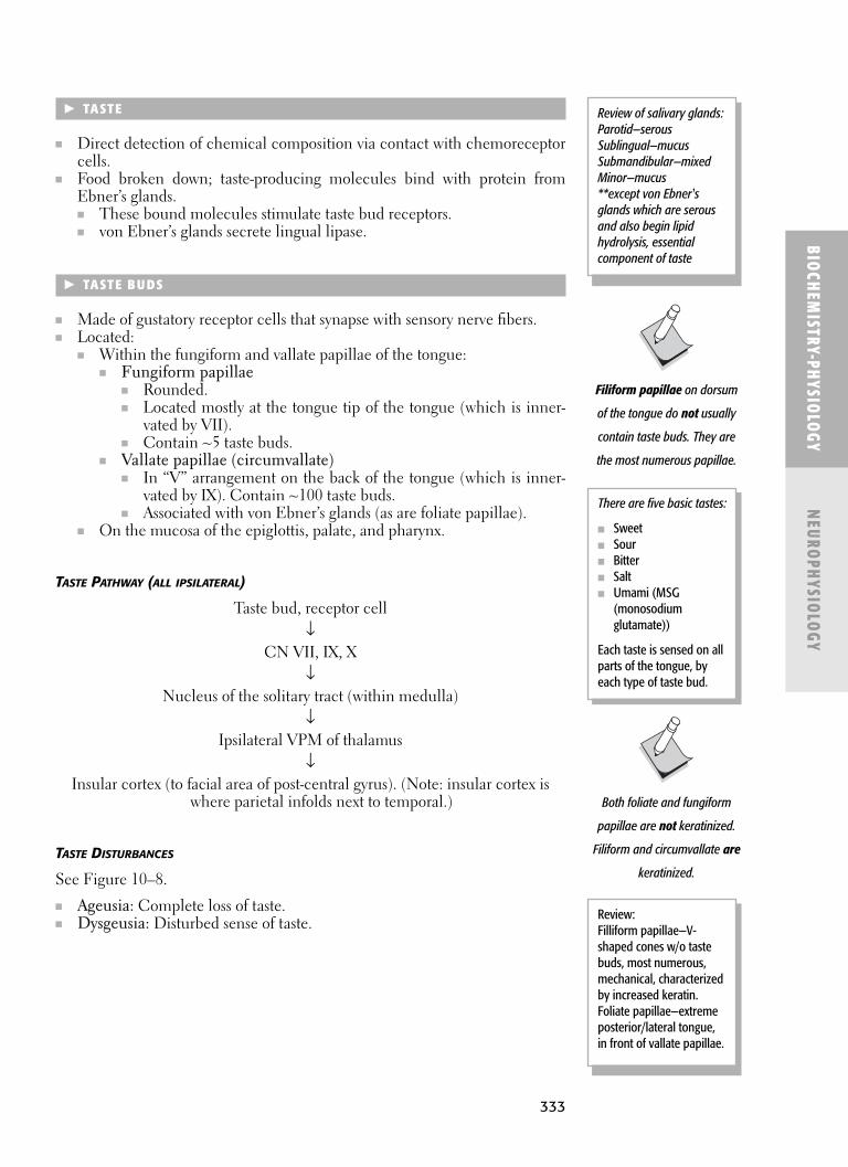
333
� TASTE
■ Direct detection of chemical composition via contact with chemoreceptorcells.
■ Food broken down; taste-producing molecules bind with protein fromEbner’s glands.■ These bound molecules stimulate taste bud receptors.■ von Ebner’s glands secrete lingual lipase.
� TASTE BU DS
■ Made of gustatory receptor cells that synapse with sensory nerve fibers.■ Located:
■ Within the fungiform and vallate papillae of the tongue: ■ Fungiform papillae
■ Rounded.■ Located mostly at the tongue tip of the tongue (which is inner-
vated by VII). ■ Contain ~5 taste buds.
■ Vallate papillae (circumvallate)■ In “V” arrangement on the back of the tongue (which is inner-
vated by IX). Contain ~100 taste buds.■ Associated with von Ebner’s glands (as are foliate papillae).
■ On the mucosa of the epiglottis, palate, and pharynx.
TASTE PATHWAY (ALL IPSILATERAL)
Taste bud, receptor cell↓
CN VII, IX, X↓
Nucleus of the solitary tract (within medulla) ↓
Ipsilateral VPM of thalamus↓
Insular cortex (to facial area of post-central gyrus). (Note: insular cortex iswhere parietal infolds next to temporal.)
TASTE DISTURBANCES
See Figure 10–8.
■ Ageusia: Complete loss of taste.■ Dysgeusia: Disturbed sense of taste.
BIOCHEM
ISTRY-PHYSIOLOGY
NEURO
PHYSIOLOGY
Filiform papillae on dorsum
of the tongue do not usually
contain taste buds. They are
the most numerous papillae.
Both foliate and fungiform
papillae are not keratinized.
Filiform and circumvallate are
keratinized.
Review of salivary glands: Parotid—serousSublingual—mucusSubmandibular—mixedMinor—mucus**except von Ebner'sglands which are serousand also begin lipidhydrolysis, essentialcomponent of taste
There are five basic tastes:
■ Sweet■ Sour■ Bitter■ Salt■ Umami (MSG
(monosodiumglutamate))
Each taste is sensed on allparts of the tongue, byeach type of taste bud.
Review: Filliform papillae—V-shaped cones w/o tastebuds, most numerous,mechanical, characterizedby increased keratin.Foliate papillae—extremeposterior/lateral tongue,in front of vallate papillae.

334
� SM E LL
■ Detection of inhaled odors.■ Chemoreceptor cells associated with the olfactory nerve (CN I). Note that
the neuron is a chemoreceptor not separate like taste.
Smell Pathway
See Figure 10–9.
Odorant particles (dissolve in mucous from Bowman’s glands)↓
Bipolar olfactory cells (within nasal mucosa)↓
Olfactory nerve (passes through cribriform plate)↓
Olfactory bulb on cribiform plate (synapses with mitral and tufted cells)↓
Olfactory tract (outgrowth of brain)↓
Primary olfactory cortex and amygdala
BIOC
HEM
ISTR
Y-PH
YSIO
LOGY
NEU
ROPH
YSIO
LOGY
Foot of post-central gyrus
MIDLINE
Nucleus of thetractus solitarius
Taste bud
Axons of solitarynucleus
Thalamicradiation
Taste fibers to brain viafacial, glossopharyngeal,and vagus nerves
Ventral posteriormedial nucleus ofthe thalamus
Insula
F I G U R E 1 0 – 8 . Taste disturbances.
Reproduced, with permission, from Ganong WF. Review of Medical Physiology, 22nd ed.New York: McGraw-Hill, 2005.

335
BIOCHEM
ISTRY-PHYSIOLOGY
NEURO
PHYSIOLOGY
DISTURBANCES OF SMELL
■ Anosmia: Absence of smell.■ Disease of olfactory mucous membranes (common cold, allergic
rhinitis).■ Kallman syndrome (hypogonadism, GnRH deficiency).
■ Hyposmia: Diminished smell.■ Dysosmia: Distorted smell.
Olfactorymucousmembrane
Superior, middle,and inferiorturbinates Hard palate
Olfactorybulb
Olfactorytract
Cribriform plateof ethmoid
F I G U R E 1 0 – 9 . Smell pathway.
Reproduced, with permission, from Waxman, SG. Clinical Neuroanatomy, 25th ed. New York:
McGraw-Hill, 2003.
Kallman syndrome—
hypogonadism caused by
deficiency of GnRH created by
hypothalamus, association
with asnosmia.

This page intentionally left blank

C H A P T E R 1 1
Muscle Physiology
Muscle 338
MAJOR CELLULAR COMPONENTS 338
INNERVATION 338
Skeletal Muscle 339
HISTOLOGY 339
CONNECTIVE TISSUE 340
MOTOR INNERVATION 340
CONTRACTION 340
CONTRACTION (TWITCH) SPEED 341
FIBER TYPES 341
SENSORY INNERVATION 342
SPINAL REFLEXES 342
Cardiac Muscle 343
HISTOLOGY 343
INNERVATION 343
MUSCLE CONTRACTION 343
Smooth Muscle 343
HISTOLOGY 343
INNERVATION 343
MUSCLE CONTRACTION 344
337

338
BIOC
HEM
ISTR
Y–PH
YSIO
LOGY
MUS
CLE
PHYS
IOLO
GY
� M USC LE
■ The largest tissue type in the human body. ■ Converts chemical energy to mechanical energy. ■ Three types:
■ Skeletal■ Cardiac■ Smooth
Major Cellular Components
■ Sarcolemma: The plasma membrane of muscle cells.■ Sarcoplasm: The cytoplasm of muscle cells.■ Sarcoplasmic reticulum (SR): A network of channels extending through-
out the sarcoplasm that stores Ca2+.■ Myofilaments: Mediate muscle contraction. Located in the sarcoplasm.
■ Thin filaments: ~6–8 nm in diameter. ■ Actin: Globular (G) actin is arranged in double helical chains called
fibrous (F) actin.■ Troponin: Attached to each tropomyosin molecule.■ Tropomyosin: Blocks actin-binding sites during rest.
■ Thick filaments: ~15 nm in diameter.■ Myosin
■ Light meromyosin (LMM): Makes up the rod-like backbone ofmyosin filaments.
■ Heavy meromyosin (HMM): Forms the shorter cross-bridgeswhich bind to actin during contraction.
Comparison of Skeletal, Cardiac, and Smooth Muscle
Characteristic Skeletal Cardiac Smooth
Striations Yes Yes NoNucleation Multinucleated Single SingleNucleus location Peripheral Central CentralInnervation Motor Autonomic AutonomicMovement Voluntary Involuntary InvoluntaryContraction by A.P. Intrinsic A.P./hormonesSyncytium No Yes YesCa2+ source SR SR/extracellular SR/extracellularT-tubules Yes Yes NoRegulation Actin Actin MyosinTroponin Yes Yes No
Innervation
■ Afferent nerves: Sensory receptor → CNS.■ Efferent nerves: CNS → muscle cell.

BIOCHEM
ISTRY–PHYSIOLOGY
MUSCLE PHYSIO
LOGY
339
� SKE LETAL M USC LE
Histology
See Figure 11–1.
■ Cells are long and multinucleated.■ Nuclei are generally elongated and peripherally located. ■ The major structural unit is the myofibril:
■ Thick filaments (contain myosin).■ Thin filaments (contain actin, troponin, and tropomyosin). ■ Myosin cross-bridges link the two filaments.
■ The sarcomere is the functional (contractile) unit of the myofibril. Definedas the area between two Z lines.
■ Cross-striations are apparent due to alternating light and dark banding ofthe myofibrils.■ A band: Dark band contains myosin. Never changes length.■ H band: Light band that bisects the A band. Shortens during contraction.■ I band: Light band containing actin. Shortens during contraction.■ Z line: Dark band that bisects the I band. Anchor for actin.■ M line: Dark band that bisects the H band. Anchor for myosin.
The dArk band is the Aband.
The lIght band is the Iband.
HAZI (hazy):
The H band bisects theA band.
The Z line bisects the Iband.
Two t-tubules lie within a
single sarcomere.
F I G U R E 1 1 – 1 . Skeletal muscle histology.
Reproduced, with permission, from Bhushan V, et al. First Aid for the USMLE Step 1: 2006.
New York: McGraw Hill, 2006.
Satellite cells are responsible
for skeletal muscle
regeneration.
As thin and thick filaments
overlap, the H and I bands are
shortened, thereby contracting
each sarcomere.

340
Connective Tissue
■ Contains a rich supply of blood vessels and nerves required for nerveconduction.
■ Divided into three layers:■ Epimysium: Surrounds the entire muscle.■ Perimysium: Surrounds muscle bundles (fascicles).■ Endomysium: Surrounds each muscle fiber.
Motor Innervation
■ One α-motor neuron innervates several skeletal muscle fibers (axon ishighly branched).
■ When a motor neuron transmits an action potential, ALL of the fibers itinnervates contract simultaneously.
■ All motor neurons are arranged in various positions within the ventralhorn of the spinal cord.
■ Fractionation: Not necessary to activate all motor units in a muscle.■ Size principle: Motor units are recruited in order of size of motor unit.
With greater muscle force, larger motor units are recruited.
Contraction
■ When an action potential reaches the neuromuscular junction, acetylcholineis released from vesicles within the axon terminus and binds to postsynapticnicotinic receptors on the sarcolemma. This, in turn, increases the membranepermeability of Na+ and K+ and depolarizes the muscle cell.
■ See Figure 11–2.
BIOC
HEM
ISTR
Y–PH
YSIO
LOGY
MUS
CLE
PHYS
IOLO
GY
F I G U R E 1 1 – 2 . Skeletal muscle contraction.
Reproduced, with permission, from Bhushan V, et al. First Aid for the USMLE Step 1: 2006. New York: McGraw-Hill, 2006.
Occurs via actin-linked
regulation.
Weight lifting causes musclehypertrophy, increasing
skeletal muscle cell size, notnumber. Contrarily, muscledisuse causes a decrease in
muscle size.
Each motor unit consistsof the following:
■ a -Motor neuron■ Synaptic cleft■ Associated muscle
fibers

341
■ The action potential travels along the sarcolemma and through a system oft-tubules, which extend from the outer surface of the muscle fiber to theSR of two adjacent sarcomeres.
■ Ca2+ released from the terminal cisternae of each SR bind to troponin C,which is attached to the tropomyosin molecule of thin filaments. Thiscauses a conformational change in the shape of tropomyosin, allowingthe actin filament to interact with the myosin cross-bridge.
■ An ATP molecule bound to myosin is hydrolyzed to ADP + Pi. Whenthe ADP + Pi is released from myosin, the actin filament is pulled closertoward the center of the sarcomere, shortening its length (power stroke).
■ As long as Ca2+ and ATP are available, this cycle continues, further con-tracting the muscle. If more muscle force is needed, more motor units areactivated.
■ During relaxation, Ca2+ is taken up by the SR, causing the release of actinfrom the myosin cross-bridges. Tropomyosin returns to its normal configu-ration, blocking this interaction.
Contraction (Twitch) Speed
■ Oxidative capacity of muscle fibers is related to■ Number of capillaries■ Myoglobin content■ Number of mitochondria
Comparison of Skeletal Muscle by Twitch Speed
BIOCHEM
ISTRY–PHYSIOLOGY
MUSCLE PHYSIO
LOGY
Fiber Types
■ Extrafusal fibers■ Make up the majority of skeletal muscle. ■ Innervated by α-motor neurons.
■ Intrafusal fibers■ Located within the bulk of the muscle. ■ Encapsulated.■ Innervated by γ-motor neurons.■ Includes muscle spindle and Golgi tendon organs.
Both extrafusal and intrafusal
fibers run parallel with each
other and attach to tendons at
either end of the muscle.
The sources of ATP formuscle contraction arisefrom:
■ Glycogen stores →glycolysis.
■ TCA cycle → oxidativephosphorylation.
■ Creatine phosphate
Characteristic Slow (Type I) Fast (Type II)
Function Postural/endurance Rapid/powerfulmovement
Rate of fatigue Low HighContraction speed Slow FastFiber diameter Small LargeColor Red WhiteMyoglobin content High LowMitochondria content High LowCapillary content High LowATP source Oxidative phosphorylation Anaerobic glycolysis
of stored glycogenATPase activity Low HighGlycogen content Low HighSR content Low High
One Slow Fat RedOx:
Type ISlow twitchLipid accumulationRed fibersOxidative
phosphorylation
Two Fast SkinnyWhite Chickens:
Type IIFast twitchLow lipid accumulationWhite fibers
(like chicken breasts)

342
Sensory Innervation
■ Encapsulated intrafusal nerve fibers.■ Stretch receptors.■ Fine-tunes muscle tone.■ Run parallel with extrafusal muscle fibers.
BIOC
HEM
ISTR
Y–PH
YSIO
LOGY
MUS
CLE
PHYS
IOLO
GY
Type Ia fibers (annulospiral
endings): Largest and fastest.
Rapid adaptation to change in
muscle length.
Type II fibers (flower-spray
endings): Non-adaptive.
Efferent Afferent SensoryReceptor Location Terminals Terminals Function Characteristic
Muscle spindle Muscle γ-motor Ia Detect Activate α-motor■ Nuclear bag belly dynamic Δ in neurons
fibers muscle length■ Nuclear Ia, II Detect static Δ
chain fibers in musclelength
Golgi tendon Muscle None Ib Detect Δ in Inhibit α-motororgan tendon muscle tension neurons
Spinal Reflexes
■ Reflex arcs:1. Receptor2. Sensory (afferent) neuron3. Integration center (CNS)4. Interneuron5. Motor (efferent) neuron6. Effector
Reflex Type Receptor Sensitivity Action Example
Stretch Muscle spindle Δ in length Contraction Knee-jerk reflex(myotactic)
Tendon (inverse Golgi tendon Δ in tension Relaxation Pick up somethingmyotatic) organ too heavy and
drop it
Flexor-withdrawal Nociceptor Δ in temperature Flexion Touch something(nociceptive) hot and withdraw
hand, causing pain
The stretch reflex maintains
muscle tone.

343
� CARDIAC M USC LE
Histology
■ Cells have a similar contractile structure (myofilaments) and striated(actin, myosin) appearance as skeletal muscle cells.
■ Fibers are firmly linked by desmosomes.■ Nuclei are centrally located. ■ More mitochondria between myofibrils.■ Richer in myoglobin.■ Cells have many branches, which communicate to adjacent cardiac mus-
cle cells via gap junctions.■ Intercalated discs coordinate the action of cardiac muscle cells.■ Cells do not undergo mitosis: Injury results in fibrosis with loss of function
at that site.
Innervation
■ Mediated by its own intrinsic contractile activity.■ There are no motor units.■ The autonomic nervous system (both sympathetic and parasympathetic
fibers) controls the rate and strength of myocardial depolarization.
Muscle Contraction
■ Ca2+ enters the myocytes via specific calcium channels, which are regu-lated by cAMP protein kinases.
■ The influx of Ca2+ enables the release of more Ca2+ from the SR, initiat-ing troponin-C binding and eventual muscle contraction.
■ Relaxation occurs when Ca2+ exits the myocytes through a regulatedCa2+-Na+ exchange system.
� SMOOTH M USC LE
Histology
■ Commonly found in tubular organs such as blood vessels, the GI tract,and the respiratory tract, but also in ciliary bodies of the eye and hairfollicles.
■ Cells are small in diameter but very long. ■ Nuclei are single and centrally located.■ Myofibrils are not striated.■ No t-tubules are present.■ SR system is poorly developed.
Innervation
■ Mediated by the autonomic nervous system (both sympathetic andparasympathetic).
■ However, there is a considerable synaptic distance from the nerve terminalto the sarcolemma because the autonomic axons terminate in the sur-rounding connective tissue.
BIOCHEM
ISTRY–PHYSIOLOGY
MUSCLE PHYSIO
LOGY
Cardiac muscle behaves as a
functional syncytium.
An increase in cardiac demand
causes a compensatory
hypertrophy, increasing
cardiac cell size, not number.
The sinuatrial (SA) node is
known as the pacemaker of
the heart.
Occurs via actin-linked
regulation, but relies more on
extracellular Ca2+ than
skeletal muscle because its SR
is less extensive.
The plateau in the cardiac
muscle action potential is due
to the influx of Ca2+.
Smooth muscle behaves as a
functional syncytium.
Smooth muscle contraction
can also be stimulated by
hormones such as
epinephrine and oxytocin.

344
■ Because not all smooth muscle cells are directly innervated, they rely oncell-cell gap junctions to propagate the action potential. ■ Single unit: Numerous gap junctions between adjacent cells. Fibers
contract spontaneously without nerve impulses. Examples: GI, uterus,ureters, arterioles.
■ Multi-unit: Cells lack gap junctions. Fibers are directly innervated.Examples: Iris, vas deferens, arteries.
Muscle Contraction
See Figure 11–3.
■ Ca2+ (from the SR or extracellular sources) enters the smooth muscle cellcytoplasm and binds to calmodulin.
■ Calmodulin activates myosin light-chain kinase, which transfers a Pi froman ATP molecule to the myosin light-chain.
■ The phosphorylation of myosin enables it to interact with actin in thesame manner as skeletal and cardiac muscle.
■ Relaxation occurs when Ca2+ is taken up by the SR or plasma membraneand the myosin light-chain kinase becomes inactivated.
■ Contraction is slow and prolonged. Each contraction cycle requires oneATP.
BIOC
HEM
ISTR
Y–PH
YSIO
LOGY
MUS
CLE
PHYS
IOLO
GY
Voltage-gatedCa2+ channelsopen
Activatesmyosinlightchainkinase
Myosin+ actin
Ca2+ bindsto calmodulin
[Ca2+]in cytoplasm
Myosinlightchainphosphatase
Myosin~P+ actin
Cross bridge formationand contraction
Action potential
Smooth musclemembranedepolarization
Relaxation
F I G U R E 1 1 – 3 . Smooth muscle contraction.
Reproduced, with permission, from Bhushan V, et al. First Aid for the USMLE Step 1: 2006. New York: McGraw-Hill, 2006.
Occurs via myosin-linked
regulation; thin filaments
lack troponin, so they are
always ready to interact with
myosin.
Maintains contraction for
extended periods of time
because the cross-bridges
detach very slowly, allowing
them to stay attached longer.

C H A P T E R 1 2
Circulatory and CardiacPhysiology
Blood Flow and Cardiodynamics 347
BLOOD FLOW 347
RESISTANCE 347
PROPERTIES OF FLOW 348
VELOCITY OF BLOOD FLOW 348
OXYGEN EXCHANGE 350
Blood Volume 350
Capacitance 350
Total Peripheral Resistance 351
Blood Pressure 352
Determinants of Cardiac Function 353
CARDIAC OUTPUT 353
HEART RATE AND CONTRACTILITY 354
VENOUS RETURN 355
PRESSURE–VOLUME LOOP 356
Electrophysiology 358
ELECTRICAL CONDUCTION OF THE HEART 358
Electrocardiogram (ECG) 360
Regulatory Mechanisms 362
BAINBRIDGE REFLEX 362
BARORECEPTORS 362
CHEMORECEPTORS 363
HORMONAL REGULATION SYSTEMS 364
EXERCISE 364
Hematocrit (Hct) 364
Hemoglobin Concentration 365
Anemia 365
345

346
CONSEQUENCES OF ANEMIA 365
CARBON MONOXIDE POISONING 365
CYANOSIS 366
Erythropoietin 366
NEGATIVE FEEDBACK 366
Coagulation and Hemostasis 366
VIRCHOW’S TRIAD 366
HEMOSTASIS 366
COAGULATION 367
347
BIOCHEM
ISTRY–PHYSIOLOGY
CIRCULATORY AN
D CARDIACPHYSIO
LOGY
� BLOOD FLOW AN D CARDIODYNAM ICS
Blood Flow
■ F = P/R (flow = pressure/resistance). I = V/R.■ Q = Δ P/R.■ Q = flow.■ Flow is proportional to pressure difference at two ends of vessel.■ Flows from high pressure to low pressure.■ Inversely proportional to resistance along the vessel.■ Greatest in
■ Large straight vessels. ■ Low turbulence.■ Low viscosity.■ Low resistance.
PERFUSION PRESSURE
■ Pressure at the arterial end minus pressure at venous end.
Resistance
■ Poiseuille’s law (F = P�r4/8nL) describes the flow rate of liquid through atube.
■ The resistance to f low is determined by the equation (R = 8nL/�r4).■ n = viscosity.
■ Hematocrit (HCT)■ Affect is greater in larger vessels than smaller.■ Overall vascular resistance is not affected unless severe (eg,
polycythemia).■ Decrease in deformation of cells.■ Plasma concentration (eg, increase in protein, multiple myeloma).
■ L = length of tube.■ r = radius.
■ Type and size of vessel.■ Regulation of tone (sympathetic nervous system, medications, local
factors).■ Pathologic narrowing of vessel.
■ R�1/r4.■ If radius (r) increases by 2, the resistance (R) drops 16 ×. ** most powerful
relationship with resistance.
SERIES RESISTANCE
■ Rtotal = R1 + R2 + R3 + R4 . . .
PARALLEL RESISTANCE
■ Recruitment of capillaries serves to lower the resistance and thereforeincreases the flow.1/Rtotal = 1/R1 + 1/R2 + 1/R3 + 1/R4 . . .
Resistance and conductance
are inversely related.
The RADIUS has the most
powerful relationship with
resistance.
Remember:artery Æ arteriole Æcapillary Æ venule Ævein

348
BIOC
HEM
ISTR
Y–PH
YSIO
LOGY
CIRC
ULAT
ORY
AN
D CA
RDIA
CPH
YSIO
LOGY
Resistance to flow of blood
offered by the entire systemic
circulation is called total
peripheral resistance (TPR).
When the vena cava is
somewhat collapsed, as it
often is, it has a lower cross-
sectional area and a higher
velocity of blood flow when
compared to the aorta.
Properties of Flow
See Figure 12–1.
■ Laminar flow (streamline)■ Low Reynolds number
■ In straight vessels.■ Layer closest to vessel surface does not move.■ Layer in center moves at maximum velocity. ■ Laminar flow occurs up to a certain critical velocity.
■ Turbulent flow occurs above critical velocity.
■ Turbulent flow■ High Reynolds number
■ Occurs above critical velocity.■ Reynolds number.■ Represents probability for turbulent flow.■ Related to:
■ Velocity.■ Diameter of vessel.■ Blood viscosity.
■ Examples of turbulence:■ Constricted, atherosclerotic vessel.■ Ascending aorta.■ Anemia.
Velocity of Blood Flow
■ Fastest to slowest:■ Vena cavae. ■ Aorta.■ Large veins.■ Small arteries.■ Arterioles.■ Capillaries.
See Figure 12–2 for systemic blood flow and changes in pressure and velocity.
Vessel wall
Flow
F I G U R E 1 2 – 1 . Properties of flow.
Reproduced, with permission, from Ganong WF. Review of Medical Physiology, 22nd ed.
New York: McGraw-Hill, 2005.
Capillaries have the largest
cross-sectional area and the
lowest blood flow velocity
Inverse relationship between
area and velocity of
blood flow.
V = Q/A
where Q = flow and A = cross-
sectional area

349
BIOCHEM
ISTRY–PHYSIOLOGY
CIRCULATORY AN
D CARDIACPHYSIO
LOGY
LAPLACE’S LAW
■ Wall stress = Pr/t.■ P = pressure.■ r = radius.■ t = wall thickness.
■ In vessels: A thin-walled, distended vessel, under large amounts of pres-sure has more wall tension/stress and is at greater risk for rupture.
■ In the heart: A dilated, thin-walled myocardium, under increasing pres-sure and volume has higher wall tension (and the myocardial oxygendemand is elevated).■ P within the ventricle depends on afterload (eg, TPR, aortic stenosis). ■ r within the ventricle depends on preload (amount of venous return).■ t thickness of the ventricle: Increased thickness decreases wall stress to
a point, then ■ Oxygen demand increases because of greater muscle mass.■ Oxygen supply decreases by narrowing coronary vessels.
0
Velocity
RR
TA
0
40
Pre
ssur
e (m
m H
g)M
ean
velo
city
(cm
/s)
80
120Systolic
Diastolic
Aor
ta
Art
erie
s
Art
erio
les
Cap
illar
ies
Ven
ules
Vei
ns
Ven
a ca
va
F I G U R E 1 2 – 2 . Systemic blood flow—changes in pressure and velocity.
Reproduced, with permission, from Ganong WF. Review of Medical Physiology, 22nd ed.
New York: McGraw-Hill, 2005.

350
BIOC
HEM
ISTR
Y–PH
YSIO
LOGY
CIRC
ULAT
ORY
AN
D CA
RDIA
CPH
YSIO
LOGY
Oxygen Exchange
CAPILLARIES
■ Greatest total area.■ Large surface area.■ Slowest velocity of an individual blood cell.
■ Allows time for oxygen, nutrient exchange/diffusion.
ARTERIOLES
■ Account for:■ Largest drop in BP (~50% drop from arteries to arterioles).■ Highest proportion of peripheral vascular resistance.
■ Pressure decreases as blood moves through systemic circulation. ■ This pressure gradient is required for blood flow.
� BLOOD VOLU M E
Most is held within the systemic venous circulation:
■ >60% in systemic veins.■ >10% in systemic arteries.■ <10% in arterioles and capillaries.■ 9% in pulmonary vessels.■ 7% in heart.
� CAPAC ITANC E
■ Ability to hold blood volume.■ Act as a reservoir.
Blood Vessel Types and Characteristics
Arteries* Arterioles Capillaries Veins
Oxygenated blood Regulate blood Nutrient, oxygen, Carry deoxygenated carried under flow into capillaries. and waste blood back to heart.high pressure Primary resistance exchange. Large lumens.from heart to body. vessels. Thin walls. High compliance
Muscular walls. Tissue metabolites, (volume reservoirs).High pressure. humoral factors May have valves.Low compliance. affect vasoconstriction
and vasodilation.
*Pulmonary and umbilical arteries carry deoxygenated blood. Pulmonary vein carries oxygenated blood.
At rest most capillaries are
closed. In active tissue they
dilate and perfuse. In
inflammation capillary
leakiness is mediated by
substance P, bradykinin, etc.
Venules are small veins that
collect blood from capillaries
(coalesce into larger veins).

351
BIOCHEM
ISTRY–PHYSIOLOGY
CIRCULATORY AN
D CARDIACPHYSIO
LOGY
■ Veins.■ Capacitance vessels.■ Dilate to accommodate blood volume.■ Hold 50–60% of blood volume.
In hypovolemia, veins/venules constrict.
■ Sympathetic mediated.■ Compensatory.
■ No clinical manifestations with 15–20% blood loss.■ Helps maintain mean systemic filling pressure in the face of blood loss.■ Preload is maintained with venous constriction.
Arterial constriction system has much less effect on mean systemic fillingpressure.
■ Arterial system contains relatively small amount of blood.■ Arterial constriction increases afterload.
Capillaries do not constrict because they lack smooth muscle in their walls.
Comparison of Pulmonary and Systemic Circulations
Pulmonary Circulationa Systemic Circulationb
BP ↓ ↑
Resistance ↓ ↑
Compliance ↑ (store blood without ↓changing BP).
Circuitc Right heart → pulmonary Left heart → aorta →artery → lungs → systemic pulmonary vein → arteries/arterioles/capillaries →left heart (oxygenated). venules/veins → SVC, IVC →
right heart (deoxygenated).
aPulmonary arteries supply only the lung gas exchange areas (alveoli).bSystemic circulation supplies the body with oxygenated blood. Note that the bronchial arteries are part ofthe systemic circulation that supplies the lung tissue and bronchi for cellular metabolism (not gas exchange).cVolume of blood flow is 5 L/min in both circuits (systemic and pulmonary).
� TOTAL PE RI PH E RAL RES ISTANC E
■ TPR (peripheral vascular resistance).■ Vascular resistance of the systemic circulation.■ Mean arterial pressure minus central venous pressure divided by the cardiac
output (MAP – CVP)/CO.■ Increases with sympathetic activation; arteriolar constriction.
Because MAP = CO × TPR,the sympathetic/
adrenergic system ↑ MAPby both:
■ ↑ CO (via ↑ HR andSV).
■ ↑ TPR (byvasoconstriction, a1).

352
BIOC
HEM
ISTR
Y–PH
YSIO
LOGY
CIRC
ULAT
ORY
AN
D CA
RDIA
CPH
YSIO
LOGY
� BLOOD PRESSU RE
■ BP = CO × TPR.■ Systolic pressure/diastolic pressure (120/80).■ Pulse pressure = SBP – DBP.
■ Normal is (120 – 80) = 40. ■ Increases with age because of stiffened arteries (atherosclerosis, arte-
riosclerosis).■ Mean arterial pressure (MAP) = ~DBP + pulse pressure/3.
■ MAP = CO × TPR.■ Vascular compliance – increase in volume/increase in pressure.■ Average pressure throughout the course of the cycle. ■ MAP is slightly less than halfway between SBP and DBP because dias-
tole is longer than systole.
↑TPR ↓TPR
Example Cold ExerciseVasoconstriction Vasodilation ■ a1 (sympathetic nervous ■ Especially in skeletal muscle
system) ■ b2 (sympathetic nervous system)
■ Local metabolites■ Lactate■ K+
■ Adenosine
Factor Regulates
TPR Blood flow from systemic circulation into venous circulation.
CO Blood flow from veins back into arterial system.Compliance Amount of blood in systemic veins.
Pulse Pressure
Narrow Wide
Decreased arterial compliance AV malformations(stiff, less distensible wall) Aortic regurgitation
■ HTN/atherosclerosis Anemia■ Aortic stenosis Atherosclerosis■ Cardiac tamponade Fever■ Heart failureDecreased SV(less blood ejected with each beat)
Sympathetic activation ↓
venous compliance and ↑
venous return (returns more
blood to heart). This ↑ CO, via
the Starling mechanism, and
more blood is pumped back
into arterial circulation.
Hypertension: Wall:lumen
ratio ↑, arteriolar and
capillary density ↓. See
pathology for further
discussion of pathogenesis.
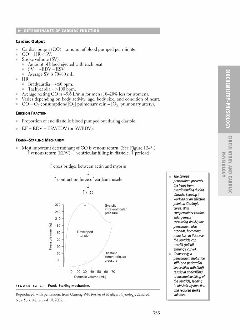
353
BIOCHEM
ISTRY–PHYSIOLOGY
CIRCULATORY AN
D CARDIACPHYSIO
LOGY
� DETE RM I NANTS OF CARDIAC FU NCTION
Cardiac Output
■ Cardiac output (CO) = amount of blood pumped per minute.■ CO = HR × SV.■ Stroke volume (SV).
■ Amount of blood ejected with each beat. ■ SV = ~EDV – ESV.■ Average SV is 70–80 mL.
■ HR■ Bradycardia = <60 bpm.■ Tachycardia = >100 bpm.
■ Average resting CO is ~5.6 L/min for men (10–20% less for women).■ Varies depending on body activity, age, body size, and condition of heart.■ CO = O2 consumption/([O2] pulmonary vein – [O2] pulmonary artery).
EJECTION FRACTION
■ Proportion of end diastolic blood pumped out during diastole.
■ EF = EDV - ESV/EDV (or SV/EDV).
FRANK–STARLING MECHANISM
■ Most important determinant of CO is venous return. (See Figure 12–3.)↑ venous return (EDV): ↑ ventricular filling in diastole: ↑ preload
↓↑ cross bridges between actin and myosin
↓↑ contraction force of cardiac muscle
↓↑ CO
Systolicintraventricularpressure
Diastolicintraventricularpressure
Diastolic volume (mL)
Developedtension
Pre
ssur
e (m
m H
g)
270
240
210
180
150
120
90
60
30
010 20 30 40 50 60 70
F I G U R E 1 2 – 3 . Frank–Starling mechanism.
Reproduced, with permission, from Ganong WF. Review of Medical Physiology, 22nd ed.
New York: McGraw-Hill, 2005.
■ The fibrouspericardium preventsthe heart fromoverdistending duringdiastole, keeping itworking at an effectivepoint on Starling’scurve. Withcompensatory cardiacenlargement(occurring slowly) thepericardium alsoexpands, becomingmore lax. In this casethe ventricle canoverfill (fall offStarling’s curve).
■ Conversely, apericardium that is toostiff (or a pericardialspace filled with fluid)results in underfillingor incomplete filling ofthe ventricle, leadingto diastolic dysfunctionand reduced strokevolumes.
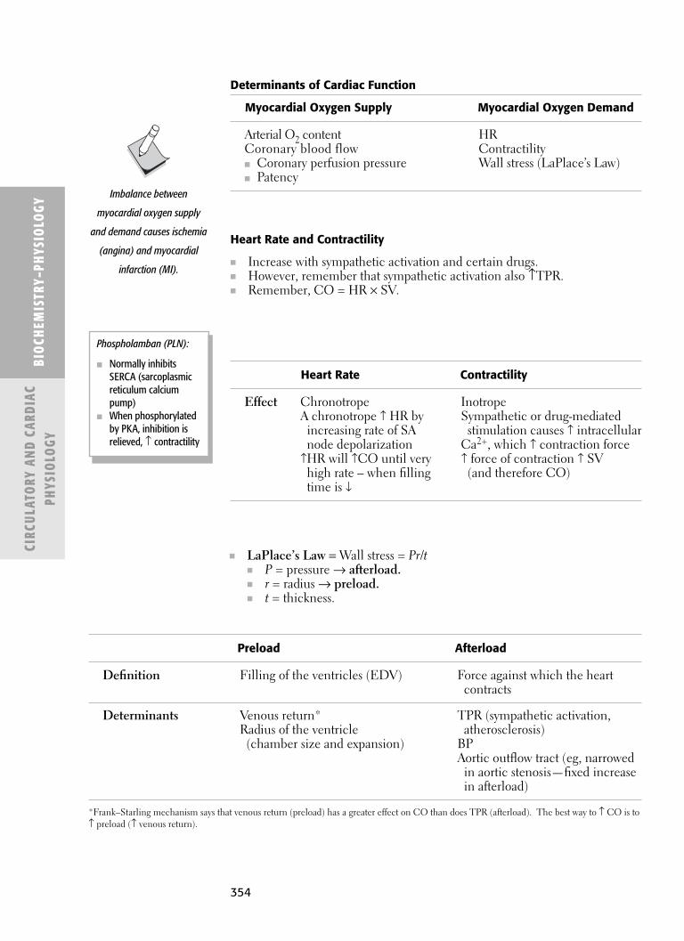
354
BIOC
HEM
ISTR
Y–PH
YSIO
LOGY
CIRC
ULAT
ORY
AN
D CA
RDIA
CPH
YSIO
LOGY
Heart Rate and Contractility
■ Increase with sympathetic activation and certain drugs.■ However, remember that sympathetic activation also ↑TPR.■ Remember, CO = HR × SV.
Determinants of Cardiac Function
Myocardial Oxygen Supply Myocardial Oxygen Demand
Arterial O2 content HRCoronary blood flow Contractility■ Coronary perfusion pressure Wall stress (LaPlace’s Law)■ Patency
Heart Rate Contractility
Effect Chronotrope InotropeA chronotrope ↑ HR by Sympathetic or drug-mediated
increasing rate of SA stimulation causes ↑ intracellularnode depolarization Ca2+, which ↑ contraction force
↑HR will ↑CO until very ↑ force of contraction ↑ SVhigh rate – when filling (and therefore CO)time is ↓
Preload Afterload
Definition Filling of the ventricles (EDV) Force against which the heartcontracts
Determinants Venous return* TPR (sympathetic activation, Radius of the ventricle atherosclerosis)
(chamber size and expansion) BPAortic outflow tract (eg, narrowed
in aortic stenosis—fixed increase in afterload)
*Frank–Starling mechanism says that venous return (preload) has a greater effect on CO than does TPR (afterload). The best way to ↑ CO is to↑ preload (↑ venous return).
Imbalance between
myocardial oxygen supply
and demand causes ischemia
(angina) and myocardial
infarction (MI).
■ LaPlace’s Law = Wall stress = Pr/t■ P = pressure → afterload.■ r = radius → preload.■ t = thickness.
Phospholamban (PLN):
■ Normally inhibitsSERCA (sarcoplasmicreticulum calciumpump)
■ When phosphorylatedby PKA, inhibition isrelieved, ↑ contractility
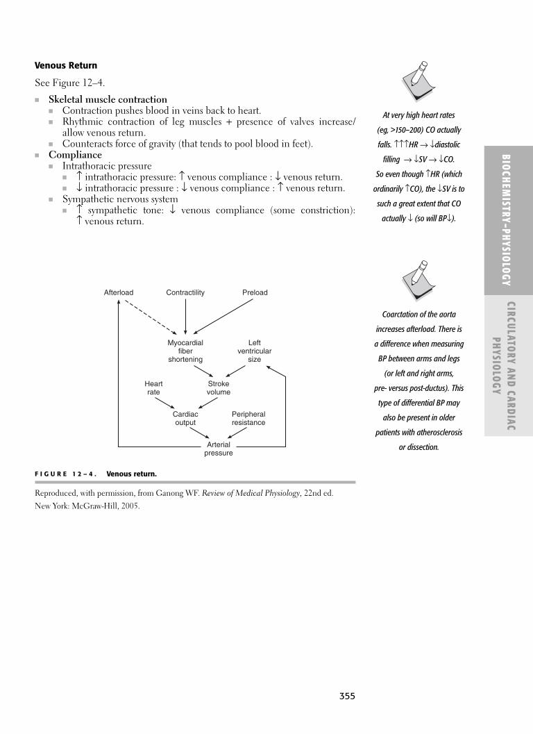
355
BIOCHEM
ISTRY–PHYSIOLOGY
CIRCULATORY AN
D CARDIACPHYSIO
LOGY
Venous Return
See Figure 12–4.
■ Skeletal muscle contraction■ Contraction pushes blood in veins back to heart.■ Rhythmic contraction of leg muscles + presence of valves increase/
allow venous return.■ Counteracts force of gravity (that tends to pool blood in feet).
■ Compliance■ Intrathoracic pressure
■ ↑ intrathoracic pressure: ↑ venous compliance : ↓ venous return.■ ↓ intrathoracic pressure : ↓ venous compliance : ↑ venous return.
■ Sympathetic nervous system ■ ↑ sympathetic tone: ↓ venous compliance (some constriction):
↑ venous return.
Arterialpressure
Peripheralresistance
Cardiacoutput
Strokevolume
Heartrate
Myocardialfiber
shortening
Leftventricular
size
Afterload PreloadContractility
F I G U R E 1 2 – 4 . Venous return.
Reproduced, with permission, from Ganong WF. Review of Medical Physiology, 22nd ed.
New York: McGraw-Hill, 2005.
Coarctation of the aorta
increases afterload. There is
a difference when measuring
BP between arms and legs
(or left and right arms,
pre- versus post-ductus). This
type of differential BP may
also be present in older
patients with atherosclerosis
or dissection.
At very high heart rates
(eg, >150–200) CO actually
falls. ↑↑↑HR → ↓diastolic
filling → ↓SV → ↓CO.
So even though ↑HR (which
ordinarily ↑CO), the ↓SV is to
such a great extent that CO
actually ↓ (so will BP↓).

356
BIOC
HEM
ISTR
Y–PH
YSIO
LOGY
CIRC
ULAT
ORY
AN
D CA
RDIA
CPH
YSIO
LOGY
Cardiac Cycle
Diastole Systole (Filling Isovolumetric (Ejection Isovolumetric Phase)a Contraction Phase) Relaxation
AV valves Open. Closed. Closed. Closed.
Pulmonic, Closed. Closed. Open. Closed.aorticvalves b
Heart S1 (“lub”)c S2 (“dub”)sound Sound of the AV Sound of the
valves (mitral semilunar valves and tricuspid) (aortic and closing. This pulmonic) closing.sound begins This sound beginssystole (ventricular diastole (ventriclecontraction). filling).d
Blood enters Blood is ventricles ejected out through the the open open AV valves: aortic and
pulmonic valves.■ 70–80%
passive.■ 20–30% b/c
atrial “kick” (contraction).
Diastole is the long phase of the cycle.
a The coronary arteries are perfused during diastole.b The aortic valve closes before the pulmonic, causing a splitting of S2 (this is accentuated on inspiration).c S1 is louder and longer than S2. d The pericardium helps regulate ventricular filling. (See the earlier section on the “Frank–Starling Mechanism.”)
Pressure–Volume Loop
See Figure 12–5.See Figure 12–6 for the cardiac cycle.
Splitting:↑blood to R heartbecause of intrathoracicpressure. At same time ↓blood to L heart becauseretained in lungvasculature. Pulmonaryvalve closes after aortic.

357
BIOCHEM
ISTRY–PHYSIOLOGY
CIRCULATORY AN
D CARDIACPHYSIO
LOGY
F I G U R E 1 2 – 5 . Pressure-volume loop.
Reproduced, with permission, from Ganong WF. Review of Medical Physiology, 22nd ed.
New York: McGraw-Hill, 2005.
c ="dub"
d a ="lub"
b
Diastolic pressure-volume relationship
Isovolumic pressure-volume curve
Pre
ssur
e (m
m H
g)
200
100
50 130
Volume (mL)
0
F I G U R E 1 2 – 6 . Cardiac cycle.
Reproduced, with permission, from Ganong WF. Review of Medical Physiology, 22nd ed.
New York: McGraw-Hill, 2005.
0 0.2 0.4 0.6 0.8
DiastoleAtr.syst.
Ventric.syst.
PR T
Q S
U
2 314
Time (second)
Electrocardiogram
Heart sounds(phonocardiogram)
Aortic pressure(at o, the aortic valve opens;at c, it closes)
Left ventricular pressure ( )
Left atrial pressure ( )(right is similar)
Left ventricular volume(at c', the mitral valve closes;at o', it opens)
Jugular venous pressure,showing a, c, and v waves
Carotid pressure (n = dicrotic notch)Radial pressure
Pulmonary arterial pressureRight ventricular pressure
1 2 3 4 5Phases of cardiac cycle
120
130
25
15
65
80
40
0
0
0
30150
n
vca
c'
o'
o c
Pressure(mm Hg)
Ventricularvolume (mL)
Aorticblood flow
(L/min)
Pressure(mm Hg)
Letter “a” is from atrial“kick”.

358
BIOC
HEM
ISTR
Y–PH
YSIO
LOGY
CIRC
ULAT
ORY
AN
D CA
RDIA
CPH
YSIO
LOGY
Cardiac Conductive Tissues
Nodal/Pacemaker Tissue Cardiac Myocytes
Gap junctions relay electrical signals, causing contraction.
Phases 0–4. Phases 0–4.Phase 0 dependent on Ca2+. Phase 0 dependent on Na+.
� E LECTROPHYSIOLOGY
Electrical Conduction of the Heart
See Figure 12–7.■ Automaticity
■ The spontaneous phase 4 depolarization that generates Aps. ■ These electrical signals conduct to atrial tissue, causing it to contract.■ SA node → AV node → ventricular bundles (His/Purkinje) → ventric-
ular myocytes → ventricular contraction.■ Refractory period
■ Long refractory period of heart allows relaxation (diastolic filling) andprevents the heart from going into reentry (arrhythmia).
■ Takes 0.22 seconds for AP to spread through the heart.■ Ventricular muscle’s refractory period is 0.25–0.30 second. ■ Atrial muscle’s refractory period is 0.15 second.
Absolute Refractory Period Relative Refractory Period
During this, another AP Immediately follows cannot be elicited, regardless the absolute of magnitude of stimulus. refractory period.
Determined by Na+ channel Continues until the inactivation/gate closure. membrane potential
returns to resting level.During this, it is possible
to generate another AP but need larger stimulus.
Prevents reentry of ventricles.
As opposed to cardiacmuscle, skeletal musclehas a short refractoryperiod.
■ This allows successivestimulation, a shorttime after initialcontraction.
■ Tetany serves toincrease strength ofcontraction.
Heart Murmurs with Valvular Disease
Diastolic Systolic
Aortic insufficiency Aortic stenosisMitral stenosis Mitral regurgitation

359
BIOCHEM
ISTRY–PHYSIOLOGY
CIRCULATORY AN
D CARDIACPHYSIO
LOGY
Superior vena cava
Sinoatrial node
Internodal pathways
Atrioventricular node
Bundle of His
Right bundle branch
Purkinje system
Left posterior fascicle
Aorta
LAF
SA node
Atrial muscle
AV node
Common bundle
Bundle branches
Purkinje fibers
Ventricular muscle
Action potential
ECG P TU
QRS0.2 0.4 0.6
Time (s)
+40
+20
0
−20
−40
−60
−80
−100
Mem
bran
e po
tent
ial (
mV
)
Ca2+ in
3 K+ out
Na+ in,Ca2+ leak in
Na+ in
K+ slow leak
1 K+ and Cl– out, Na+ inactive
2 Ca2+ in K+ out slow (=)
3 K+ out
30 ms
Sinoatrial node fiber Ventricular muscle fiber
0
4
0
F I G U R E 1 2 – 7 . Electrical conduction of the heart.
Reproduced, with permission, from Ganong WF. Review of Medical Physiology, 22nd ed. New York: McGraw-Hill, 2005.

360
BIOC
HEM
ISTR
Y–PH
YSIO
LOGY
CIRC
ULAT
ORY
AN
D CA
RDIA
CPH
YSIO
LOGY
� E LECTROCARDIOG RAM (ECG)
■ Records the flow of electrical impulses through the heart.
See Figure 12–8 for ECG waves.
■ To read ECG:■ Remember that a single lead corresponds to an anatomic territory.■ Look at multiple leads together for voltage abnormalities and axis
deviations.■ Look at a single lead for arrhythmias.
ECG Leads
Standard Bipolar Standard Unipolar Chest leads
I—right arm (−), left arm (+) AVR—right arm (+) V1, V2, V3, V4, V5, V6 represent II—right arm (−) and left leg (+) AVL—left arm (+) 6 places along chest wall.III—left arm (−) and left leg (+) AVF—left leg (+)
Nodal/Pacemaker Conduction System
Internodal His-Purkinje SA Node Pathways AV Node System
Located in posterior Transmits Located in the Fiber network arisingwall of RA near depolarization lower right in AV node.opening of SVC. wave from the interatrial septum. Purkinje fibers
Pacemaker of the SA node to the The impulse is originate from R andheart (starts the LA and AV node.a delayed in AV L bundle branches.bdepolarization). node (~0.13 s) Extend to papillary
Depolarizes at (slow conduction muscles and lateralintrinsic rate that rate). walls of ventricles.drives depolarization Atria to contract Depolarization wave of rest of heart. before ventricles. travels extremely
Transmits signals fast through bundle faster than AV. branches and
Atria contract before Purkinje fibers. ventricles, preventing Total elapsed timefast, arrythmogenic is 0.03 sec.beats from reaching the ventricle.
a Aside from AV node, the atria and ventricles are electrically isolated.b His-Purkinje fibers fan across subendocardial surface of ventricles from endocardium outward through the myocardium. As cardiac impulsespreads, these fibers cause ventricles to depolarize and contract.
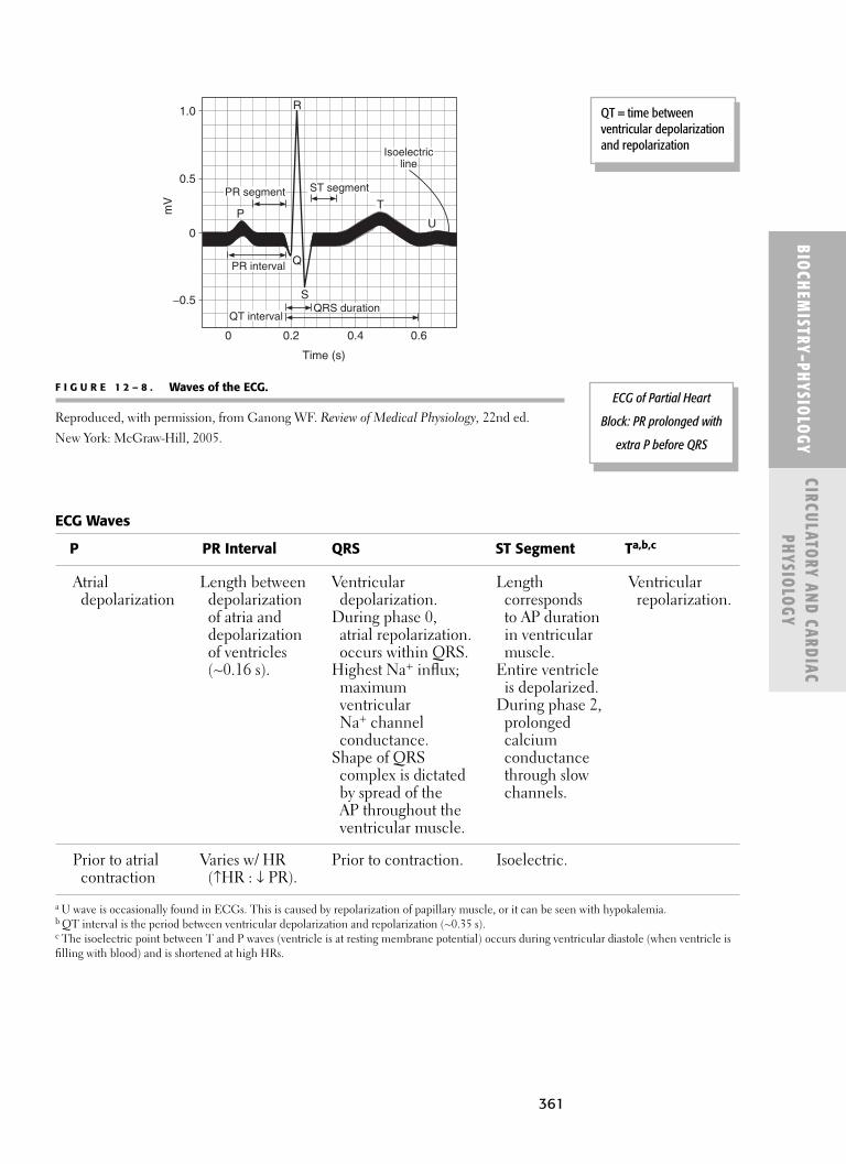
361
BIOCHEM
ISTRY–PHYSIOLOGY
CIRCULATORY AN
D CARDIACPHYSIO
LOGY
1.0
0.5
−0.5
0
0 0.2 0.4 0.6
P
R
S
Q
T
U
PR segment
PR interval
QT interval
ST segment
QRS duration
mV
Isoelectricline
Time (s)
F I G U R E 1 2 – 8 . Waves of the ECG.
Reproduced, with permission, from Ganong WF. Review of Medical Physiology, 22nd ed.
New York: McGraw-Hill, 2005.
ECG Waves
P PR Interval QRS ST Segment Ta,b,c
Atrial Length between Ventricular Length Ventriculardepolarization depolarization depolarization. corresponds repolarization.
of atria and During phase 0, to AP durationdepolarization atrial repolarization. in ventricularof ventricles occurs within QRS. muscle.(~0.16 s). Highest Na+ influx; Entire ventricle
maximum is depolarized.ventricular During phase 2,Na+ channel prolonged conductance. calcium
Shape of QRS conductancecomplex is dictated through slow by spread of the channels.AP throughout the ventricular muscle.
Prior to atrial Varies w/ HR Prior to contraction. Isoelectric.contraction (↑HR : ↓ PR).
a U wave is occasionally found in ECGs. This is caused by repolarization of papillary muscle, or it can be seen with hypokalemia.b QT interval is the period between ventricular depolarization and repolarization (~0.35 s).c The isoelectric point between T and P waves (ventricle is at resting membrane potential) occurs during ventricular diastole (when ventricle isfilling with blood) and is shortened at high HRs.
QT = time betweenventricular depolarizationand repolarization
ECG of Partial Heart
Block: PR prolonged with
extra P before QRS

362
BIOC
HEM
ISTR
Y–PH
YSIO
LOGY
CIRC
ULAT
ORY
AN
D CA
RDIA
CPH
YSIO
LOGY
� REG U LATORY M EC HAN ISMS
Autonomic Control of the Heart
Sympathetic Parasympathetic
Control center Medulla Medulla
Nerve T1–4 (sympathetic chain) Vagus nerve (CN X)
NT NE ACh
HR ↑ ↓↑ SA nodal discharge ↓ SA nodal discharge↑ Rate of depolarization ↓ Rate of depolarization
through heart through heart↑ Intracellular Ca2+
↑ Ventricular contraction force
Bainbridge Reflex
■ Stretch of atria (with ↑ blood volume) causes ↑ HR and, therefore, ↑ CO.■ Mediated by stretch receptors in atria.■ Receptor cells are sensitive to pressure and stretch.
■ Mediated by vagal (CN X) afferents to the medulla.■ Efferent loop is slowing of vagal output.
■ Pumps more blood out of pulmonary system to the systemic system.■ This helps prevent pulmonary edema.
Receptors
Receptor type Baroreceptor ChemoreceptorEffect exerted HR, BP Respiration > vasomotor
Baroreceptors
■ Modulate intravascular pressure and HR over a short time period. ■ Activated by ↑ BP. ■ Causes ↓ HR, vasodilation, ↓ BP.
BARORECEPTOR REFLEX (VALSALVA)
■ ↑ BP/↑ stretch of baroreceptors → ↑ parasympathetic afferent output (CNIX, X) → medulla → ↑ vagal efferent tone (CN X) (↓ sympathetic tone)→ ↓ HR, ↓ BP (vasodilation, venodilation) → ↓ CO.
■ Antagonist to Bainbridge reflex.
The right vagus nerve supplies
the SA node; the left vagus
nerve innervates the AV node.

363
BIOCHEM
ISTRY–PHYSIOLOGY
CIRCULATORY AN
D CARDIACPHYSIO
LOGY
CAROTID SINUS SYNDROME
■ Excessive stimulation of both carotid sinuses (eg, convulsive seizures) canlead to momentary loss of consciousness because of vagal discharge, ven-odilation, vasodilation.
RESPONSE TO SUDDEN STANDING
■ ↓ BP in brain, upper body sensed by baroreceptors →■ ↓ parasympathetic firing (CN IX, X); (sympathetic discharge → ↑HR,
↑ conduction velocity, ↑ cardiac contractility, ↑ peripheral resistance ↑vasoconstriction), ↓ renal blood flow ↑a1 vasocontriction of afferent artery;b1 on JGA → ↑renin → ↑ angiotensin-II → aldosterone → ↑ blood back toheart (↑preload because venoconstriction of large veins) → ↑ CO →return/maintain BP.
HEMORRHAGE OR HYPOVOLEMIA
COMPENSATIONS:
■ Baroreceptor reflex (↑ sympathetic, ↓ parasympathetic tone).■ ↑ epinephrine from adrenal medulla.■ ↓ vagal output from medulla (↓ carotid sinus, ↓ aortic baroreceptor firing).
■ RAAS (Renin-Angiotensin-Aldosterone system). See renal also.■ AT-II, aldosterone and increase in TPR
■ AT-II: On efferent arteriole to preserve GFR.■ Aldosterone: On cortical collecting duct, increase Na+ reabsorp-
tion, K+ excretion, H+ secretion by intercalated cells.■ ADH
■ Capillary fluid shift■ Increases water reabsorption in CCD.
Chemoreceptors
■ Detect changes in blood oxygen, carbon dioxide, and hydrogen ionconcentrations.
■ Modulate respiratory center in brain (regulate respiratory activity).
Carotid Sinusa Aortic Arch Baroreceptora
Spindle-shaped dilation of receptors at Receptors in the aortic arch.the common carotid artery bifurcation(superior border of thyroid cartilage).
Afferent = CN IX. Afferent = CN X.
aThese receptors are located in the adventitia of the vessels. They are extensively branched, knobby, coiled,and intertwined ends of myelinated nerve fibers (resemble Golgi tendon organs).
Carotid Body Aortic Body
Afferent CN IX Afferent CN X
Stretch receptors in the atria
and pulmonary circulations
are stimulated by expansion
of blood volume; they do not
directly respond to changes in
systemic arterial BP.
Bainbridge reflexstimulated by stretch ofatria versus Hering Breuer reflexwhich
■ Prevents overinflationof lungs
■ Pulmonary stretchreceptors of smoothmuscle in airwaysrespond to excessivestretching
■ Action potentialsthrough vagusafferents to medullaand pons
■ Inspiration inhibitedand expiration occurs

364
BIOC
HEM
ISTR
Y–PH
YSIO
LOGY
CIRC
ULAT
ORY
AN
D CA
RDIA
CPH
YSIO
LOGY
CHEMORECEPTOR PATHWAY
■ See also Chapter 13, “Respiratory Physiology.”■ ↑ CO2, ↑ H+, and/or ↓ O2 → stimulates chemoreceptors (carotid, aortic
bodies) → ↑ parasympathetic afferent output (CN IX, X) → medulla (res-piratory center) → ↑ ventilation (to breathe down CO2) → (↑ BP, ↑ HRsecondary to simultaneous secretion of catecholamines from the adrenalmedulla).■ Increase in sensitivity to CO2 and pH when <60 mm Hg.
Hormonal Regulation Systems
See Chapter 14, “Renal, Fluid, Acid–Base Physiology.”
Exercise
■ Mechanisms to meet ↑ demand to muscles during exercise (↑ supply).■ ↑ CO because ↑ HR and ↑SV.■ Sympathetic nervous system.
■ b2 adrenergic receptors in muscle/pulmonary tree■ Vasodilate■ ↑ Blood flow to muscles
■ b1 receptors in heart ■ ↑ HR■ ↑ Contraction force (inotropic) (phospholumbar “releasing brake”
on SERCA pump)■ ↑ CO
■ a1 in other parts of body■ Vasoconstriction■ ↑ Arterial pressure
■ Enhanced venous return (↑ preload).■ ↓ Venous compliance■ Pumping effect of the skeletal muscle ■ Vasoconstriction
■ Local metabolites (released as O2 ↓ 0).■ Adenosine, CO2, lactic acid
■ Vasodilate: ↓vascular resistance, ↑ blood flow
AUTOREGULATION OF BLOOD FLOW
■ As blood flow ↑ to muscles during exercise, the adenosine is washed out.■ ↓ adenosine → arterioles and small arteries vasoconstrict → keeping blood
flow at a normal rate (in face of ↑arterial pressure).
� H E MATOC RIT (Hct)
■ Percentage of RBCs in blood sample.■ Normal:
■ Male: 44–46. ■ Female: 40–42.
■ Venous Hct is typically higher than arterial Hct because of “chloride shift.”■ More RBC mass in comparison to plasma. CO2 from tissues gets con-
verted by carbonic anhydrase → HCO3 in RBC increases osmotic pressure→ water rushes in → RBC greater volume
Note: During exercise ↑ CO is
slightly > ↓ TPR, so MAP ↑.
During exercise initially ↑ SV,
then later ↑ HR (which is
important, >50% of maximal
work capacity is reached).
SVR ↓ during exercise
(vasodilation, b2, local
metabolites). Blood flow to
skeletal muscle can increase
20-fold during strenuous
exercise.

365
BIOCHEM
ISTRY–PHYSIOLOGY
CIRCULATORY AN
D CARDIACPHYSIO
LOGY
� H E MOG LOBI N CONC E NTRATION
■ See also Chapter 6, “Biological Compounds” and Chapter 13, “RespiratoryPhysiology,” for discussion of hemoglobin (Hb).
■ Normal:■ Male: 15–16 g/dL.■ Female: 13–14 g/dL.
■ Severe anemia:■ <7.5 g/dL.■ Hct = Hb × 3.
� AN E M IA
■ ↓ Hct.■ ↓ RBC and/or ↓ Hb concentration.
Consequences of Anemia
■ ↓ oxygen transport in blood.■ Fatigue, respiratory compensation, cardiac compensation.■ Hypoxia in the tissues.
■ Causes small arteries and arterioles to dilate (so ↑ blood return to theheart [preload]).
■ Hypoxia in the pulmonary circulation results in vasoconstriction of thosevessels (opposite to effect on other body tissues).
Compensations with Anemia
Cardiac Compensation Respiratory Compensation
↑ CO Bohr effectChemoreceptors sense ↓O2 O2–Hb curve shifts to the right (→)■ ↑SV (↑ pulse pressure) Unloads more O2 to the tissues■ ↑HR (try to deliver more O2 ■ ↑2, 3-DPG
to tissues) ■ ↑CO2↓Hct → ↓blood viscosity ■ ↑RBC H+
■ ↓ resistance to flow → ↓ PVR■ ↑ blood returns to the heart
(↑ preload) (helps to increase SV)
Types of Anemia and Associated Cell Size
↓ Production ↑ Destruction
Fe deficiency (microcytic) Blood loss (normocytic)Folate deficiency (megaloblastic) Hemolysis (normocytic)
or macrocyticB12 deficiency (megaloblastic) Hemoglobinopathies (microcytic)
or macrocytic
Carbon Monoxide Poisoning
■ CO competes with oxygen for Hb-binding sites and changes allosterics;CO has greater affinity (250×)
■ Normal Hb level■ O2 content ↓
■ Cherry red cheeks

366
BIOC
HEM
ISTR
Y–PH
YSIO
LOGY
CIRC
ULAT
ORY
AN
D CA
RDIA
CPH
YSIO
LOGY
Cyanosis
■ Deoxygenated hemoglobin in tissues gives the skin/mucous membranes ablue tint.
■ Does not occur in severe anemia because you need >5 g of deoxygenatedHb per 100 mL of blood to appreciate.
� E RYTH ROPOI ETI N
■ Glycoprotein hormone produced in kidneys■ ↑ RBC production by bone marrow■ Acts at the hemocytoblast (pluripotent stem cell)■ ↓ Erythropoiesis → anemia■ ↑ Erythropoiesis → polycythemia
■ ↑ Blood viscosity, sluggish blood flow, ie, polycythemia vera (JAK2mutation downstream of Epo Receptor)
Negative Feedback
■ ↓ O2 tension (anoxia/hypoxia) → ↑erythropoietin (HIF)■ ↑ O2 → ↓ erythropoietin
� COAG U LATION AN D H E MOSTASIS
Virchow’s Triad
■ Endothelial injury■ Stasis■ Hypercoaguability
Hemostasis
■ Three parts:■ Vasoconstriction■ Platelet aggregation/plug (primary)—platelets bind wWF on damaged
endothelium■ Coagulation (secondary)—crosslinking with fibrin meshwork
Tissue factor (tissue
thromboplastin) is part of the
extrinsic pathway (it is not
present in blood).

367
BIOCHEM
ISTRY–PHYSIOLOGY
CIRCULATORY AN
D CARDIACPHYSIO
LOGY
(PTT) INTRINSIC SYSTEM(PTT: XII, XI, IX, VIII, and common to both: X, V, II)
HMW kininogenKallikrein
XII XIIa
HMW kininogen
XI XIa(PT) EXTRINSIC SYSTEM
(PT: VII and common to both: X, V, II)
TFI
VIIVIIAIX IXaPLCa2+
VIIIa
Ca2+
PLTPL
VIII
X XaPL (note: PL is common endpoint in pathway)Ca2+
VaV
Prothrombin Thrombin
Fibrinogen Fibrin
Stabilization
XIIIaXIII
TPL
F I G U R E 1 2 – 9 . Intrinsic and extrinsic pathways of coagulation.
Reproduced, with permission, from Ganong WF. Review of Medical Physiology, 22nd ed.
New York: McGraw-Hill, 2005.
Both intrinsic and extrinsic
pathways are activated when
blood vessels are damaged.
In cirrhosis, proteins, including
prothrombin and fibrinogen,
are deficient.
Coagulation
See Figure 12–9.
■ Intrinsic and extrinsic pathways■ Prothrombin is cleaved to thrombin.
■ Converted by prothrombin activator (Factor Va).■ Fibrinogen cleaved to fibrin.
■ Converted by thrombin.■ Fibrin forms the clot and cross-links with the platelets.

This page intentionally left blank

C H A P T E R 1 3
Respiratory Physiology
Lung Volumes 370
Lung Mechanics 371
INSPIRATION 371
EXPIRATION 371
Gas Exchange in the Lungs 373
Hemoglobin 373
O2-HB DISSOCIATION CURVE 373
BOHR EFFECT 374
HALDANE EFFECT 374
AMOUNT OF O2 IN BLOOD 375
CARBON DIOXIDE 376
HYPOXEMIA 377
HYPERCARBIA 378
HYPERVENTILATION 378
Respiratory Regulation 379
RESPIRATORY DRIVE 379
ACID–BASE BALANCE AND RESPIRATORY CHANGES 380
HERING–BREUER REFLEX (REFLEX TO PREVENT OVERINFLATION) 381
PULMONARY CHEMOREFLEX 381
HIGH ALTITUDE 382
369

370
BIOC
HEM
ISTR
Y–PH
YSIO
LOGY
RESP
IRAT
ORY
PHY
SIO
LOGY
� LU NG VOLU M ES
See Figure 13–1.
■ Total lung volume (TLV) = IRV + TV + ERV + RV.■ Vital capacity (VC) = TLV − RV.
Maximal inspiratorylevel
Volume(L)
Resting expiratorylevel
Maximal expiratorylevel
6.0
2.7
2.2
1.2
IRV = Inspiratory reserve volumeERV = Expiratory reserve volume
TV = Tidal volumeRV = Residual volume
0
IRV
IRV
TV
TV
ERVERV
RV
RV
Dead space
F I G U R E 1 3 – 1 . Lung volumes.
Reproduced, with permission, from Ganong WF. Review of Medical Physiology, 22nd ed. New York: McGraw-Hill, 2005.
RV is increased in older
individuals and in chronic
obstructive pulmonary disease
(COPD) or asthma because of
air trapping.
Lung Volumes
■ Inspiratory Air inspired with Inspiratory Maximum air reserve maximal inspiratory capacity (IC) inspired up to volume (IRV) effort (after TLC (limited by
inspiring at TV). elastic recoilof lung)(TV + IRV).
■ Tidal Air inspired or expiredvolume (TV) with a normal breath.
■ Expiratory Air pushed out after Functional Air left inreserve passive expiration residual lungs after avolume (ERV) (FRC − RV). capacity normal passive
(FRC) expiration(ERV + RV).
■ Residual Air remaining involume (RV) lungs after maximal
exhalation; cannot be measured by spirometry.

BIOCHEM
ISTRY–PHYSIOLOGY
RESPIRATORY PHYSIO
LOGY
371
� LU NG M EC HAN ICS
Inspiration
■ Active process ■ Requires muscular effort.
■ Mostly diaphragm at rest.■ Intercostals used on exertion (accessory muscles).
■ Inspiratory effort causes: ■ ↓ intrapleural pressure. ■ ↓ alveolar pressure. ■ Pressure gradient from mouth to alveoli. ■ Gas flow down pressure gradient.
Expiration
■ Passive process (usually).■ Due to lung recoil.
■ Relaxation of inspiratory muscles causes: ■ ↑ intrapleural pressure (intrapleural pressure becomes less negative).■ ↑ alveolar pressure.■ Pressure gradient from alveoli to mouth. ■ Gas flow down pressure gradient.
FUNCTIONAL RESIDUAL CAPACITY
■ FRC = At rest.■ Balance between inspiratory and expiratory forces.
■ Collapsing forces = Expanding forces.■ Muscle contraction is needed to ↑ or ↓ lung volume from FRC.
Collapsing Forces Expanding Forces
Favors ↓ lung volume. Favors ↑ lung volume.■ Elastic connective ■ Elastic connective tissue
tissue of the lungs of chest wall↑ Surface tension helps counteract collapse.■ Surfactant coating the alveoli
ALVEOLAR PRESSURE
■ Atmospheric pressure in resting position.■ 760 mm Hg (at FRC). Palv = 0 mm Hg
INTRAPLEURAL PRESSURE
■ Pressure within pleural cavity between outer surface lung and inner sur-face chest cavity.
■ 756 mm Hg (at FRC) (< atomospheric pressure). Ppl = −34 mm Hg
VC = greatest amount of air
that one can exchange in a
forced respiration (inhalation +
expiration).
Accessory muscles ofinspiration:
■ Scalene■ SCM■ Trapezium■ External intercostals
Accessory muscles ofexpiration:
■ Intercostals (internal)■ Abdominal
372
ALVEOLAR VENTILATION
■ (VA) = RR × (TV − dead space air volume).■ Amount of gas that reaches the functional respiratory units (ie, alveoli) per
minute.■ Amount of atmospheric air that can undergo gas exchange.■ Good gauge for breathing effectiveness.
RESPIRATORY RATE
■ Breaths per minute.
TIDAL VOLUME
■ TV = amount of air brought into/out of lungs with a normal breath.■ 500 mL.
■ 350 mL used for alveolar ventilation.■ 150 mL dead space (fixed due to conducting airways).
DEAD SPACE
■ VD = Volume of air not participating in gas exchange.■ Anatomic dead space.
■ Typically 150 mL.■ Volume of nonventilated gas in airways.■ No gas exchange occurs within the nasal passages, pharynx, trachea,
bronchi.■ Physiologic dead space.
■ Due to alveoli that are ventilated but not perfused. ■ Usually insignificant, unless there is disease.
MINUTE VENTILATION
■ TV × RR = VT.
BIOC
HEM
ISTR
Y–PH
YSIO
LOGY
RESP
IRAT
ORY
PHY
SIO
LOGY
ZONES OF THE LUNG
Conducting zone Trachea(no gas exchange) Main bronchi 2 − left, 3 − right
lobar, segmentalBronchioles
(first place w/o cartilage)Terminal bronchioles
Respiratory zone Respiratory bronchioles(gas exchange occurs) Alveoli
■ Conducting zone airways contain mucous-secreting cells:■ Goblet cells■ Mucous cells■ The epithelium is pseudostratified ciliated columnar.

373
BIOCHEM
ISTRY–PHYSIOLOGY
RESPIRATORY PHYSIO
LOGY
■ Respiratory zone, alveolar wall has: ■ Type I epithelial cells■ Type II epithelial cells ↓ pneumocytes
■ Produce surfactant■ See also Chapter 2
� GAS EXC HANG E I N TH E LU NGS
■ O2 uptake, CO2 elimination by the blood■ O2 diffusion (alveolus → blood)■ CO2 diffusion (alveolus ← blood)
■ Depends on:■ Partial pressure gradient
■ Pressure difference between two sides of the membrane.■ Diffusion occurs from high to low pressure (down the gradient).
■ PACO2 > PaO2 (alveolar > pulmonary arterial); O2 diffuses fromalveoli → blood.
■ PaCO2 blood > PACO2 in alveolus; CO2 diffuses from blood → alve-oli.
■ Gas solubility■ Number of molecules dissolved in the liquid ↑ partial pressure of gas ↑.■ Solubility is an intrinsic property of the gas.■ Solubility ↑ as partial pressure ↑ (Henry’s law). CO2 more soluble
than O2.■ Thickness of membrane (alveolus)
■ Rate of diffusion is inversely proportional to the diffusion distance.■ ↑ diffusion as ↓ alveolar thickness.
■ Alveolar surface area■ Rate of diffusion is directly proportional to surface area.
■ ↓ surface area (eg, emphysema), ↓ diffusion, ↓ gas exchange.
� H E MOG LOBI N
■ See also Biological Compounds, Chapter 6.■ Carries O2 from lungs to tissues. ■ Carries CO2 from tissues to lungs.■ Normally:
■ 98% saturated with O2 in lungs (arterial).■ 75% saturated in tissues (venous).
■ PaO2 = 40 mm Hg■ 300 million Hb molecules in each erythrocyte.
■ Synthesis begins in erythroblasts.
O2-Hb Dissociation Curve
See Figure 13–2.
The PO2 determines the
affinity of Hb for O2 binding
by causing a conformational
change of Hb. Each O2
binding increases the affinity
for O2 stepwise.
30 mm Hg → 50%
60 mm Hg → 90%
90 mm Hg → 99%
More CO2 (HCO3) invenous RBC→↑osmoticpressure →↑ watercontent →↑ volume ofRBC, increased HC+??
CO has 240 times the affinity
for Hb as O2 does.
It, therefore, interferes with
unloading of O2 from Hb.
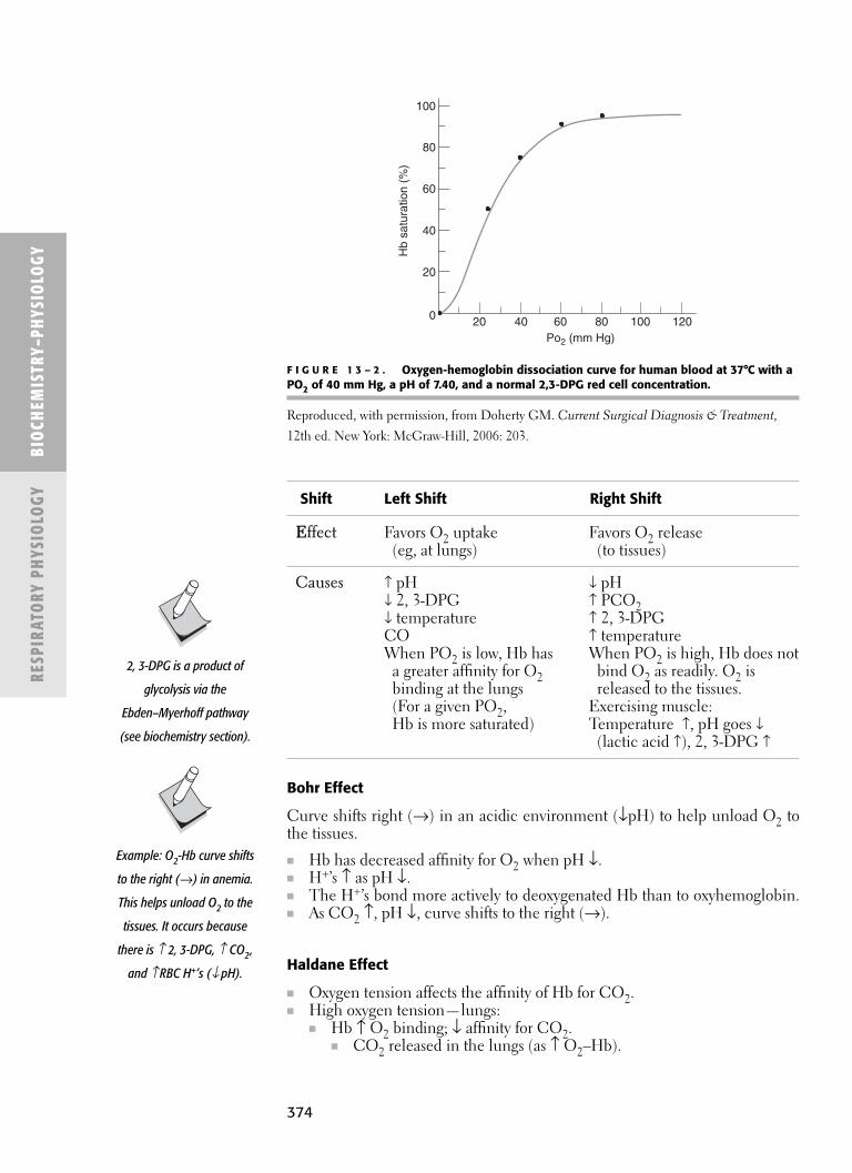
374
BIOC
HEM
ISTR
Y–PH
YSIO
LOGY
RESP
IRAT
ORY
PHY
SIO
LOGY
Bohr Effect
Curve shifts right (→) in an acidic environment (↓pH) to help unload O2 tothe tissues.
■ Hb has decreased affinity for O2 when pH ↓.■ H+’s ↑ as pH ↓.■ The H+’s bond more actively to deoxygenated Hb than to oxyhemoglobin.■ As CO2 ↑, pH ↓, curve shifts to the right (→).
Haldane Effect
■ Oxygen tension affects the affinity of Hb for CO2.■ High oxygen tension—lungs:
■ Hb ↑ O2 binding; ↓ affinity for CO2.■ CO2 released in the lungs (as ↑ O2–Hb).
Example: O2-Hb curve shifts
to the right (→) in anemia.
This helps unload O2 to the
tissues. It occurs because
there is ≠ 2, 3-DPG, ≠ CO2,
and ≠ RBC H+’s (Ø pH).
2, 3-DPG is a product of
glycolysis via the
Ebden–Myerhoff pathway
(see biochemistry section).
100
80
60
40
20
0 20 40 60 80 100 120Po2 (mm Hg)
Hb
satu
ratio
n (%
)
F I G U R E 1 3 – 2 . Oxygen-hemoglobin dissociation curve for human blood at 37°C with aPO2 of 40 mm Hg, a pH of 7.40, and a normal 2,3-DPG red cell concentration.
Reproduced, with permission, from Doherty GM. Current Surgical Diagnosis & Treatment,
12th ed. New York: McGraw-Hill, 2006: 203.
Shift Left Shift Right Shift
EEffect Favors O2 uptake Favors O2 release(eg, at lungs) (to tissues)
Causes ↑ pH ↓ pH↓ 2, 3-DPG ↑ PCO2↓ temperature ↑ 2, 3-DPGCO ↑ temperatureWhen PO2 is low, Hb has When PO2 is high, Hb does not
a greater affinity for O2 bind O2 as readily. O2 isbinding at the lungs released to the tissues.(For a given PO2, Exercising muscle: Hb is more saturated) Temperature ↑, pH goes ↓
(lactic acid ↑), 2, 3-DPG ↑

375
OXYGEN CONTENT
■ Total amount of oxygen carried in blood (PO2 + O2–Hb).■ Determined mostly by the amount of hemoglobin and its saturation. ■ Amount of hemoglobin is affected by anemia (production, loss, or
destruction).■ The more hemoglobin in blood, the more O2 that can be carried.
OXYGEN SATURATION
■ The amount of Hb saturated with O2.■ Corresponds to O2–Hb curve.■ Determined by:
■ PO2 (important; see table corresponding SaO2 : PO2).■ O2 affinity of Hb altered by:
■ Changes in Hb molecule.■ Intrinsic (hemoglobinopathies).■ Extrinsic (eg, changes in pH, PCO2, temperature, etc.).■ Competition for Hb binding (eg, CO poisoning).
NORMAL VALUES
■ Oxygen content (per 1 g Hb) = 1.34 mL of O2.■ Hemoglobin concentration = ~15 g/dL.
■ Women: 12–16 g/dL.■ Women have ↓ Hb concentrations than men
■ Men: 14–18 g/dL.■ Infants: 14–20 g/dL.
■ Oxygen concentration = ~20 g-mL/dL (or 15 g/dL × 1.34 mL)—just 20.1 mL.
BIOCHEM
ISTRY–PHYSIOLOGY
RESPIRATORY PHYSIO
LOGY
Dissolved O2 O2–Hb
Partial pressure of O2 Hb + O2↔HbO2in blood (PO2). Hb4(O2)4
Small fraction of the total O2 Hb-O2 depends on:carried in the blood. ■ PO2.
■ 0.003 mL/dL O2 content (pulmonary function, anemia).blood/mm Hg PO2. Hb concentration.
■ Hb affinity for O2.%Hb bound with O2 = O2 saturation.
Venous blood carries more
CO2 than arterial blood, and
CO2 uptake is facilitated in the
tissues and CO2 release is
facilitated in the lungs.
AV fistula: Venous PCO2 is
lower than normal (because
it bypassed the tissues);
likewise venous PO2 is higher
(some O2 is not dropped off at
the tissues).
Blood Hb concentration does
not affect O2 saturation of Hb
or PaO2 (both PaO2 and SaO2
are independent of the Hb
concentration).
With anemia, ↑ CO as
compensation to maintain
adequate oxygenation to the
tissues.
■ Low oxygen tension—tissues:■ Hb ↓ O2 binding; ↑ affinity for CO2 (binds H+, forms carbamino com-
pounds).■ CO2 uptake in the tissues (as ↓ O2–Hb).
Amount of O2 in Blood
■ Dissolved O2 + O2 bound to Hb.

376
OXYGEN-CARRYING CAPACITY OF BLOOD
Depends on:
■ Oxygenation (from lungs).■ FiO2.■ PaO2 (gradient).■ Effective gas exchange (no dead space or shunt).
■ Hb concentration.■ Hb avidity for oxygen.
■ CO.■ Left shift of curve.
■ Perfusion.■ Cardiac function.■ Patency of vessels.■ Adequacy of forward flow.
Carbon Dioxide
See Figure 13–3.
■ Carbon dioxide (CO2) is carried in blood as:■ Bicarbonate in serum (most).■ Bicarbonate in RBC.■ Carbaminohemoglobin.
■ CO2+ NH2 group of Heme (not Fe2+ of Heme like O2 or CO).
■ Dissolved in blood (PCO2).
CHLORIDE SHIFT
■ Bicarbonate carried in serum is generated within the RBC.■ It is transported to the serum in exchange for Cl−.■ Cycle:
■ CO2 in blood diffuses passively into RBC.■ Carbonic anhydrase (within RBC) combines intracellular CO2 with
H2O to form bicarbonate and H+.■ Bicarbonate passes across the RBC membrane into serum in exchange
for Cl−.
BIOC
HEM
ISTR
Y–PH
YSIO
LOGY
RESP
IRAT
ORY
PHY
SIO
LOGY
Carbonic anhydrase is not
present in the serum.
Bicarbonate can be produced
in serum by nonenzymatic
means, but the process is slow.
Key Points:
■ Most O2 in blood iscarried by Hb.
■ Only a small amount ofoxygen is dissolved inblood under normalconditions.
■ PO2 (difference/gradient betweenalveolus and capillary)is what drives oxygentransfer to the blood.
CO2CI−
CO2 + H2O H2CO3Carbonic
anhydrase HHb H+ + Hb−
H+ + HCO3−
F I G U R E 1 3 – 3 . Carbon dioxide (CO2) as carried in the blood.
Reproduced, with permission, from Ganong WF. Review of Medical Physiology, 22nd ed.
New York: McGraw-Hill, 2005.

377
Hypoxemia
■ Low oxygen level in blood (PO2 <80).■ Causes of hypoxemia:
■ ↓ FiO2■ Hypoventilation■ V/Q mismatch■ Shunt■ Diffusion limitation
HYPOXIC VASOCONSTRICTION
■ Mechanism to minimize V/Q mismatch. ■ Example:
■ Shunt (air cannot get into alveolus).■ Peanut occluding bronchiole (child).■ Atelectasis.
■ Blood perfuses past the alveolus. ■ No/minimal gas exchange occurs.■ Response is vasoconstriction of the pulmonary vasculature in that region.
■ ↓ amount of blood going to nonventilated segment of lung. ■ If this vasoconstriction secondary to hypoxia exists for long enough,
■ Get permanent secondary changes to the pulmonary vasculature.■ Pulmonary hypertension.■ Only place in body to constrict, not dilate.
BIOCHEM
ISTRY–PHYSIOLOGY
RESPIRATORY PHYSIO
LOGY
Causes of Hypoxemia
DiffusionLimitation
↓ FiO2 Hypoventilation V/Q Mismatch Shunt (↓↓ DLCO)
↓ fraction ↓ respiratory Unequal ventilation Segment of lung ↓ surface area of of inspired drive (central) and perfusion is perfused but blood gas barrier.O2 (usually 21%). ■ Narcotics Segment of lung is not aerated. ■ Pneumonectomy
■ High altitude ■ Medullary ventilated but Intraalveolar ■ Emphysemainjury filling
■ Incorrect ↓ ability for chest not perfused ■ Atelectasis Thick blood–gasventilator excursion (= dead space) ■ Pneumonia barrier settings (peripheral) Asthma ■ Pulmonary ■ Diffuse interstitial
■ Polio ■ COPD edema fibrosis■ Chest trauma ■ Interstitial lung ■ Intracardiac ■ Sarcoidosis
(rib fracture) disease shunt ■ Asbestosis■ Diaphragmatic ■ Alveolar disease ■ Vascular shunt ■ ARDS
injury ■ Pulmonary ↓ Hb to carry oxygen■ Phrenic nerve vascular disease ■ Anemia
paralysis ■ PE
Hypoxemia in COPD is
secondary to both
hypoventilation (↑ PCO) and
V/Q mismatch (A-a gradient).

378
Hypercarbia
■ ↑ CO2 in blood.■ Occurs because of either or both of the following:
■ ↑ CO2 production.■ ↓ VA (alveolar ventilation)—hypoventilation.
■ Compensation: hyperventilation.■ Headache.■ Confusion.■ Coma.
Hyperventilation
■ ↑ rate and depth of breathing exceeding requirement for O2 delivery andCO2 removal
■ Stimulated by:■ ↓ PO2 in normal circumstances (non-COPD).■ Chemoreceptor stimulation (↑CO2, ↑H+, ↓PO2).■ Effect on brain—emotional situations, anxiety.
■ Results in:■ ↓ CO2: hypocapnia (hypocarbia).■ Respiratory alkalosis (pH ↑).■ ↑cerebrovascular resistance.■ ↓ cerebral blood flow.■ ↑PO2 (and arterial oxygen concentration).
BIOC
HEM
ISTR
Y–PH
YSIO
LOGY
RESP
IRAT
ORY
PHY
SIO
LOGY
Effect of CO2 on Cerebral Circulation
↑ PCO2 ↓ PCO2
Vasodilation → ↑ cerebral Vasoconstriction → ↓ cerebralblood flow. blood flow.
SYMPTOMS OF HYPERVENTILATION
■ Related to ↓ cerebral blood flow.■ Example: anxiety → ↑ventilation → ↓ CO2 → ↓ cerebral blood flow →
neurologic symptoms:■ Faintness/dizziness.■ Blurred vision.■ Also experience sensation of:
■ Suffocation.■ Chest tightness.
■ Terminate hyperventilation attack must:■ ↑ PCO2.
■ Breathing in and out of a plastic bag.■ Inhale 5% CO2 mixture.
In shock, hyperventilation is
caused by chemoreceptor
stimulation (secondary to
↓ PO2 (hypoxia) and ↑ H+
(acidosis)) because of local
stagnation of blood flow.
Extreme hypercarbia (CO2 ↑↑)
depresses the CNS (including
respiratory center) whereby
respiratory compensation
does not occur. Result is CO2
narcosis.

379
� RESPI RATORY REG U LATION
Respiratory Drive
■ Based on arterial PCO2, specifically H+.■ The H+ (derived from CO2) that acts at central chemoreceptors (medulla).
PATHWAY
■ As ↑ PCO2 → CO2 diffuses from cerebral blood vessels into CSF → car-bonic acid (H2CO3) is formed → dissociates into bicarbonate (HCO3
−)and protons (H+s) → these protons (H+s) stimulate the central chemore-ceptors → ↑ventilation.■ CO2 can diffuse from the blood vessels into CSF across the BBB because
it is nonpolar.
↑RESPIRATORY DRIVE
■ Central chemoreceptors (medulla)■ ↑ PCO2 (as its byproduct, H+, in CSF or brain interstitial fluid sensed
in medulla).■ Peripheral chemoreceptors (carotid or aortic bodies)
■ ↑ H+ (in blood or brain interstitial fluid).■ ↓ PO2 (in blood)(<60 mm Hg).
FUNCTION OF RESPIRATORY REGULATION
■ Keep alveolar PCO2 stable (prevent hypercarbia or hypocarbia).■ Buffer acid–base changes.■ Prevent hypoxemia (↑ PO2 when it falls).
BIOCHEM
ISTRY–PHYSIOLOGY
RESPIRATORY PHYSIO
LOGYRespiratory Chemoreceptors
Receptors Central PeripheralMedullary Carotid and aortic bodies chemoreceptors (see Figure 13–4).
Affected by ↑ H+ CSF, brain ↑ PCO2.interstitial fluid. ↑ H+.
↓ PO2 (<60 mm Hg).Major regulators of ventilation. Less important than central Not respond to PO2. chemoreceptors (medulla).Not respond to arterial PCO2; However, they respond more it is PCO2 that diffuses quickly and regulate abrupt across into CSF then is changes in PCO2.converted to H+.
Carotid Body Aortic Body
Afferent CN IX CN XEfferent Phrenic, intercostals nerves Phrenic, intercostals nerves
↑ PCO2 (↑ H+ sensed in
medulla) → ↑ respiration
(blow off more CO2) →
↓ PCO2 → ↓ respiration.
↓ inspired O2 has no effect on
respiratory drive unless O2 is
<60 mm Hg; then it stimulates
carotid and aortic
chemoreceptors, resulting
in ↑ respiration.
Distinguish carotid and aortic
bodies from carotid sinus and
aortic baroreceptors (the
latter two control changes in
blood pressure and HR). CN IX
(glossopharyngeal) provides
afferent innervation for both
carotid body and carotid
sinus; CN X (vagus) is afferent
for both aortic body and
aortic baroreceptors.
Body = chemoreceptors
Sinus = baroreceptors

380
Acid–Base Balance and Respiratory Changes
■ See chart in Chapter 14.■ Primary respiratory processes: respiratory acidosis and alkalosis → meta-
bolic compensation.■ Primary metabolic processes: metabolic acidosis and alkalosis → respira-
tory compensation.
BIOC
HEM
ISTR
Y–PH
YSIO
LOGY
RESP
IRAT
ORY
PHY
SIO
LOGY
Carotid body
Carotid sinus
Common carotidarteries
Aortic bodies
Aortic arch
Heart
F I G U R E 1 3 – 4 . Carotid and aortic bodies.
Reproduced, with permission, from Ganong WF. Review of Medical Physiology, 22nd ed. New
York: McGraw-Hill, 2005.
Primary Metabolic Respiratory Process Compensation Mechanism
Metabolic acidosis. ↑ respiration (hyperventilate). ↑ H+ stimulates central and■ Diabetic ketoacidosis. ■ Blow off CO2. peripheral chemoreceptors.
■ Kussmaul ■ ↓ alveolar CO2.breathing Respiratory alkalosis.(deep, labored).
Metabolic alkalosis. ↓ respiration (hypoventilate). ↓ H+ ↓ stimulation to the ■ ↓ HCL. ■ ↑ alveolar CO2. central and peripheral
■ Vomiting. ■ ↑ H+. chemoreceptors.NG suctioning. Respiratory acidosis.
■ pCO2 elevated,not enough blown off.

381
Hering–Breuer Reflex (Reflex to Prevent Overinflation)
■ Inflate lungs → expiration.■ Deflate lungs → inspiration.■ Mediated by myelinated slow responding receptors (stretch receptors).■ Vagus nerve (afferent).
Pulmonary Chemoreflex
■ Lung hyperinflation causes:■ First apnea.■ Then:
■ Rapid breathing (tachypnea).■ Bradycardia.■ Hypotension.
■ Mediated by: ■ J (juxtacapillary) (vagus nerve) receptors—in alveolar insterstitium.
■ C fiber endings (unmyelinated) close to pulmonary vessels ↓ O2,hyperinflation, chemical administration.
BIOCHEM
ISTRY–PHYSIOLOGY
RESPIRATORY PHYSIO
LOGY
Stretch Receptors J Receptors Irritant Receptors
Located Airway smooth Alveolar walls Between airway epithelial cellsmuscle
Stimulated by Lung distention Engorgement of Noxious substances capillary or alveolar (eg, dust, pollen)walls with fluid Histaminepulmonary edema,pneumonia (eg, CHF)
Causes Expiration (prevents Rapid shallow Coughing, overinflation; breathing bronchoconstriction
Hering–Breuer reflex) Pulmonary chemoreflex
Pulmonary capillaries
(endothelial cells and alveolar
capillary bed) contain ACE
(angiotensin-converting
enzyme). ACE converts
angiontensin-I to angiotensin-II
(RAAS system).
The pulmonary chemoreflex is
analogous to the Bezold–Jarisch
reflex in the heart.

382
High Altitude
■ ↓ FiO2■ Results in:
■ Alveolar hypoxia (↓ PAO2).■ Arterial hypoxemia (↓ PaO2).■ Secondary to ↓ barometric pressure.
■ Compensation:■ Pulmonary vasoconstriction (because of alveolar hypoxia).■ ↑ erythropoietin (↑ Hct, ↑ O2-carrying capacity).■ ↑ mitochondrial density. ■ ↑ 2, 3-DPG (shifting O2–Hb curve to the right, release O2 to tissues).■ ↑ respiratory rate (because of arterial hypoxia, secondary to ↓ baromet-
ric pressure; sensed by peripheral chemoreceptors; feeding back tomedulla to ↑ respiratory rate).
■ Respiratory alkalosis (secondary to ↑ RR).■ ↑ renal bicarbonate excretion, ↓ in H+ excretion (compensation for
respiratory alkalosis).
BIOC
HEM
ISTR
Y–PH
YSIO
LOGY
RESP
IRAT
ORY
PHY
SIO
LOGY
Respiratory Conditions
Condition Description
Dyspnea Difficulty breathing; unpleasant sensation.
Apnea Cessation of breathing, usually transient (central versus peripheral).
Hyperapnea Abnormally deep and rapid breathing.
Hypercapnia ↑ CO2 in arterial blood, secondary to underbreathing (↓ ventilation).
Hypocapnia ↓ CO2 in arterial blood, secondary to overbreathing (↑ ventilation).
Respiratory arrest Cessation of breathing.
Hyperventilation ↑ alveolar ventilation (greater than metabolic requirements); leads to ↓ PCO2 (with↓ cerebral perfusion, fainting/syncope).
Hypoventilation ↓ alveolar ventilation (less than needed for metabolic requirements); leads to ↑ PCO2.
Kussmaul breathing is rapid
deep labored breathing in
people with acidosis, in
particular diabetic
ketoacidosis.

C H A P T E R 1 4
Renal, Fluid, and Acid–Base Physiology
Body Fluid Distribution 384
TOTAL BODY WATER 384
Fluid Movement 384
STARLING FORCES 384
BALANCE OF FLUIDS 385
OSMOSIS 385
Edema 386
VOLUME EXPANSION AND CONTRACTION 386
Acid–Base Physiology 387
HENDERSON–HASSELBALCH EQUATION 387
ISOELECTRIC POINT 388
ZWITTERIONS (DIPOLAR IONS) 388
BUFFER 388
CONTROL OF ACID–BASE BALANCE 388
Urinary System 389
AMMONIA 389
NEPHRON 389
RENAL BLOOD SUPPLY 390
FILTRATION 391
Countercurrent Mechanim 392
COUNTERCURRENT EXCHANGE 392
Effective Renal Plasma Flow (ERPF) 393
Renal Blood Flow 393
Glomerular Filtration Rate 393
Renal Clearance 394
Hormonal Response to Volume Changes 394
383

384
BIOC
HEM
ISTR
Y–PH
YSIO
LOGY
RENA
L, F
LUID
, AN
DAC
ID–B
ASE
PHYS
IOLO
GY
� BODY FLU I D D ISTRI BUTION
Total Body Water
Intra- and Extracellular Fluids
Extracellular Fluid Intracellular Fluid
[Na+] 142 10[K+] 4 140%Body weight 15–20 35–40Components (%BW) Blood plasma (4–5) NA
Interstitial fluid (11–15)Transcellular fluid ■ CSF■ Intraocular■ Synovial■ Pericardial■ Pleural■ Peritoneal
Total body water (TBW)(60% total body weight)Measurement: tritium,deuterium, antipyrine
Intracellular fluid (ICF)(2/3 TBW)
Contains K+, Mg+, PO43−, protein
Extracellular fluid (ECF)(1/3 TBW)
Contains more Na+, Cl−, HCO−3
Measurement: inulin, mannitol, sulfate
Interstitial fluid(75% of ECF)
Plasma(25% of ECF)
Contains albumin, Ig, and otherplasma proteins; affects BP
Skin epidermis is nourished by
way of diffusion of interstitial
fluid (tissue fluid) from
capillary beds in the dermis;
this fluid bathes the cells.
� FLU I D MOVE M E NT
Starling Forces
■ Forces that move fluid■ Hydrostatic pressure■ Oncotic pressure
■ Fluid equilibrates between the intravascular and interstitial spaces. ■ Fluid in the interstitial space
■ Is taken up by cells (becomes intracellular fluid)■ Returns to the capillaries or■ Returns to circulation via lymphatics.
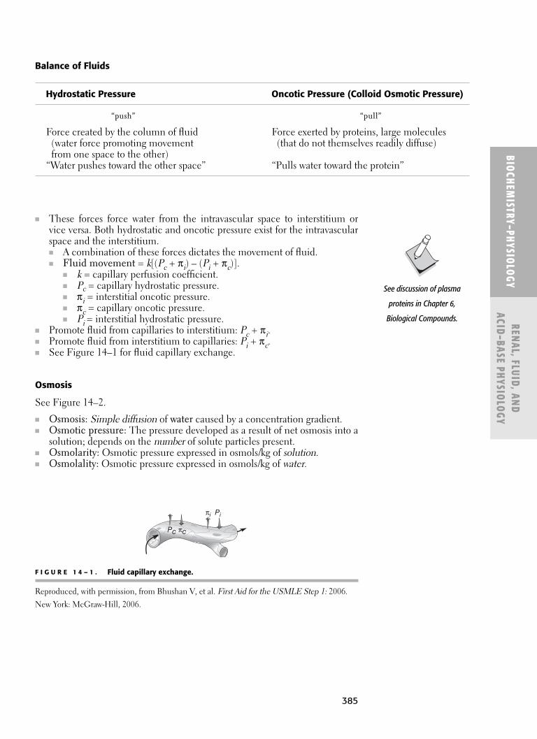
BIOCHEM
ISTRY–PHYSIOLOGY
RENAL, FLUID, AND
ACID–BASEPHYSIO
LOGY
385
Balance of Fluids
πi Pi
Pc πc
■ These forces force water from the intravascular space to interstitium orvice versa. Both hydrostatic and oncotic pressure exist for the intravascularspace and the interstitium.■ A combination of these forces dictates the movement of fluid.■ Fluid movement = k[(Pc + πi) – (Pi + πc)].
■ k = capillary perfusion coefficient.■ Pc = capillary hydrostatic pressure.■ πi = interstitial oncotic pressure.■ πc = capillary oncotic pressure.■ Pi = interstitial hydrostatic pressure.
■ Promote fluid from capillaries to interstitium: Pc + πi.■ Promote fluid from interstitium to capillaries: Pi + πc.■ See Figure 14–1 for fluid capillary exchange.
Osmosis
See Figure 14–2.
■ Osmosis: Simple diffusion of water caused by a concentration gradient.■ Osmotic pressure: The pressure developed as a result of net osmosis into a
solution; depends on the number of solute particles present.■ Osmolarity: Osmotic pressure expressed in osmols/kg of solution.■ Osmolality: Osmotic pressure expressed in osmols/kg of water.
F I G U R E 1 4 – 1 . Fluid capillary exchange.
Reproduced, with permission, from Bhushan V, et al. First Aid for the USMLE Step 1: 2006.
New York: McGraw-Hill, 2006.
See discussion of plasma
proteins in Chapter 6,
Biological Compounds.
Hydrostatic Pressure Oncotic Pressure (Colloid Osmotic Pressure)
“push” “pull”
Force created by the column of fluid Force exerted by proteins, large molecules (water force promoting movement (that do not themselves readily diffuse)from one space to the other)
“Water pushes toward the other space” “Pulls water toward the protein”

386
� E DE MA
■ Excess f luid in the interstitial space
BIOC
HEM
ISTR
Y–PH
YSIO
LOGY
RENA
L, F
LUID
, AN
DAC
ID–B
ASE
PHYS
IOLO
GY
Semipermeablemembrane Pressure
F I G U R E 1 4 – 2 . Osmosis.
Reproduced, with permission, from Ganong WF. Review of Medical Physiology, 22nd ed.
New York: McGraw-Hill, 2005.
Cause ↑ Pc ↓ πc ↑ πi ↓ Pi
Examples CHF Nephrotic Lymphatic blockageCirrhosis syndrome ■ Elephantiasis
Protein-losing ■ Lymph nodeenteropathy resection
CirrhosisMalnutrition
OTHER CAUSES OF EDEMA
■ Increased capillary permeability (burns, sepsis, anaphylaxis/angioedema,ARDS).
■ Inappropriate renal sodium and water retention.
Volume Expansion and Contraction
EXTRACELLULAR FLUID (ECF) EXPANSION
■ Excess fluid in the intervascular or interstitial spaces.■ Evidence in physical examination.
■ Rales■ Edema■ Hypertension■ Bulging fontanelle
INTRACELLULAR FLUID (ICF) EXPANSION
■ Excess fluid intracellularly.■ Gauged by serum Na+.
■ Low serum Na+ = intracellular expansion. ■ If chronic, formation of intracellular osmolytes prevents drastic influx
of H2O and cell burst.
Nephrotic syndrome isdefined by > 3.5 g proteinin urine per day.
SIADH: Syndrome ofInappropriate ADHSecretion. Excessiverelease of ADH fromposterior pituitary oranother source andusually caused by cancer.

387
BIOCHEM
ISTRY–PHYSIOLOGY
RENAL, FLUID, AND
ACID–BASEPHYSIO
LOGY
UNa+
(UrinaryExpansion ECF ICF ECF Sodium(Overall Gain) Examples Volume Volume Osmolarity Hct [Na+] [Albumin] Concentration)
(Hypo-) H2O > Na+ SIADH ↑ ↑ ↓ ↓ ↓ ↓ ↑(Iso-) H2O = Na+ Isotonic NS ↑ − − ↓ − ↓ ↑(Hyper-) H2O Hypertonic ↑ ↓ ↑ ↓ ↑ ↓ ↑< Na+ NS
Contraction ECF ICF ECF (Overall Loss) Examples Volume Volume Osmolarity Hct [Na+] [Albumin] UNa+
(Hyper-) Sweating, ↓ ↓ ↑ ↑ ↑ ↑ ↓loss H2O > Na+ fever, DI(Iso-) H2O = Na+ Diarrhea ↓ − − ↑ − ↑ ↓(Hypo-) Adrenal ↓ ↑ ↓ ↑ ↓ ↑ ↓H2O < Na+ insufficiency
Solutions
Solution Characteristics Effect on Cells Example
Hypertonic Solute > solvent Shrink >0.9% NaClIsotonic Solute = solvent No change 0.9% NaClHypotonic Solute < solvent Swell <0.9% NaCl
� AC I D–BASE PHYSIOLOGY
Process pH HCO3/PaCO2 Compensation pH HCO3/PaCO2
Metabolic acidosis ↓ HCO3− ↓ Respiratory (hyperventilate) ↑ PaCO2 ↓
Metabolic alkalosis ↑ HCO3− ↑ Respiratory (hypoventilate) ↓ PaCO2 ↑
Respiratory acidosis ↓ PaCO2 ↑ Metabolic (conserve HCO3) ↑ HCO3 ↑Respiratory alkalosis ↑ PaCO2 ↓ Metabolic (excrete HCO3) ↓ HCO3 ↓
Isotonic solutions separated
by a semipermeable
membrane do not cause net
movement of water or solutes.
The osmotic potential is the
same on either side of the
membrane.
Henderson–Hasselbalch Equation
■ pH = pKa + log [A-]/[HA].■ pH = pK when an acid is half neutralized.

388
Isoelectric Point
■ pH where the number of positive charges equals the number of negativecharges.
■ Solute has no electric charge at this pH.■ Does not move an electric field (pI for that solute).
■ Allows separation of proteins or amino acids based on charge.
Zwitterions (Dipolar Ions)
■ Physiologic pH: Amino acids have both ■ Negatively charged carboxyl group (-COO−) and■ Positively charged amino group (-NH3
+)
Buffer
■ System to minimize pH changes■ Consists of
■ Weak acid (proton donor) ■ Conjugate base (a salt)(proton acceptor)
■ Releases H+ ions when the pH rises.■ Accepts H+ when the pH drops.
BUFFER SYSTEMS
■ Sodium bicarbonate: carbonic acid buffer.■ The major buffer in extracellular fluid, blood. ■ Breaks down to CO2 and H2O with respiratory elimination.
■ Blood pH is ~ 7.4.■ Range 7.35–7.45. ■ Usually >7.4: slightly alkaline/basic.
■ Primary determinant is the bicarbonate and CO2 ratio.■ pH = pKa + log [HCO3
−]/0.03 × pCO2.■ pH = 6.1 + log[HCO3
−]/(0.03 × pCO2).■ 6.1 is the pKa of the bicarb-CO2 buffer system.
Control of Acid–Base Balance
■ Renal■ In tubules: H2CO3 (carbonic anhydrase) → H2O + CO2 → H+ +
HCO3−.
■ H+ secreted, then excreted.■ HCO3
− reabsorbed.■ NH4 (ammonium) phosphates are excreted to compensate for acidity.
■ Buffers■ Bicarbonate■ Hemoglobin■ Albumin
■ Respiratory■ Blow off CO2 (elevated CO2 stimulates respiration).
BIOC
HEM
ISTR
Y–PH
YSIO
LOGY
RENA
L, F
LUID
, AN
DAC
ID–B
ASE
PHYS
IOLO
GY
Glycine has a net negative
charge at any pH above its
isoelectric point, so it moves
toward the positive electrode
(anode). At any pH below its
isoelectric point (pI), glycine
exhibits a net positive charge
and moves toward the
negative electrode (cathode).
In the oral cavity, the carbonic
acid system helps neutralize
dietary and bacteria-
generated acids.
389
BIOCHEM
ISTRY–PHYSIOLOGY
RENAL, FLUID, AND
ACID–BASEPHYSIO
LOGY
� U RI NARY SYSTE M
■ Regulates water and solute balance.■ Removes nitrogenous wastes (as urea) from blood.
■ Urea is a byproduct of protein breakdown.■ Urea cycle in liver.
Kidneys ordinarily excrete
1–2 L per day in urine.
180 L of glomerular filtrate
are produced each day
(99% reabsorbed).
Kidney Function
Filtration Hormonal
Water and solute balance Erythropoietin (stimulates RBCs)Excretion of wastes Renin (stimulates angiotensin,
aldosterone system)
Urine
Characteristics Contents
Clear, yellow Na+
Slightly acidic pH Cl–Specific gravity 1.005–1.030 K+
Ca2+
Mg2+
SulfatesPhosphatesHCO3
–
Uric acidAmmoniaCreatinineUrobilinogen
Ammonia
■ Waste product■ Sources:
■ Amino acids■ Liver■ Kidneys■ Amines■ Purines and pyrimidines
Nephron
See Chapter 2, Histology.
■ Functional and structural unit of the kidney■ Glomerulus■ Bowman’s capsule/space
The urea cycle in the liver is
important for disposal of
ammonia.

390
■ Tubules■ Proximal convoluted■ Loop of Henle■ Distal convoluted
■ Collecting duct■ Renal pelvis■ Ureter
Renal Blood Supply
See Figure 14–3.
■ Renal arteries branch from the aorta.■ Left renal artery is longer than the right.
■ Renal artery■ Interlobar arteries■ Arcuate arteries■ Interlobular arteries■ Afferent arteriole■ Glomerulus■ Efferent arterioleBI
OCH
EMIS
TRY–
PHYS
IOLO
GYRE
NAL,
FLU
ID, A
ND
ACID
–BAS
EPH
YSIO
LOGY
Glomerulus is a tuft of
capillaries (endothelial cells)
that interface with Bowman’s
capsule (epithelial cells).
Filtration occurs at this
interface.
Majority of glomerular filtrate
occur at the proximal
convoluted tubule (just distal
to Bowman’s space).
Proximalconvoluted tubule (ciliated)
Distalconvoluted tubule
Collecting duct
Cortex
Outermedulla
Innermedulla
Loop of Henle,thin descending limb
Loop of Henle,thick ascending
limb
Glomerulus
F I G U R E 1 4 – 3 . Renal blood supply.
Reproduced, with permission, from Ganong WF. Review of Medical Physiology, 22nd ed.
New York: McGraw-Hill, 2005.

391
BIOCHEM
ISTRY–PHYSIOLOGY
RENAL, FLUID, AND
ACID–BASEPHYSIO
LOGY
Reabsorption Secretion Excretion
Tubule back From tubular cell Tubular fluid (and to circulation. or circulation solute) that ends
into tubular fluid. up as urine.
Substance Reabsorption Comments
H2O Proximal tubule Ascending loop is impermeable Loop of Henle (descending) to H2ODistal tubule, collecting duct ADH
Na+ Proximal tubule (70%) Cl and H2O follow passivelyNa/K/2Cl, impermeable to H2O
Loop of Henle Na/K/2Cl cotransportDistal tubule Aldosterone Na+ channelCollecting duct
Glucose Proximal tubule
Reabsorption occurs by active
transport, facilitated diffusion,
and solvent drag. It can be
paracellular or transcellular.
Function of Nephron Segments
Segment Reabsorption Secretion
Proximal convoluted H2O, Na+, K+, Cl–, H+, ammoniatubule. HCO3
–, amino acids, glucose, proteins
Descending loop H2O (impermeable of Henle. to Na+)
Ascending loop of Na+, K+, Cl–Henle. (impermeable to H2O)
Distal convoluted Na+, Cl–tubule. Ca2+ (regulated
by PTH)Collecting duct. H2O (regulated by ADH)
Na+ (regulated K+, H+, ammonia, by aldosterone) drugs
Reabsorption of NaCl (and
impermeability to water) by
the thick ascending loop is the
basis of the countercurrent
multiplier system.
Collecting duct drains multiple
(> 1) nephron.
Filtration
See Figure 14–4.
■ Renal blood flow delivered to glomerulus.■ Filtrate passes:
■ Endothelial cell barrier (glomerulus).■ Epithelial cell barrier (Bowman’s capsule).
■ Filtrate enters Bowman’s space, the first part of the tubule.
Furosemide is a loopdiuretic and thiazideswork on the distalconvoluted tubule.

392
� COU NTE RC U RRE NT M EC HAN ISM
■ Countercurrent multiplier.■ Creation of a gradient to allow for concentrated urine.■ Occurs in:
■ Loop of Henle. ■ Collecting tubules (cortical and medullary).■ Because these are anatomically located in both the medulla and cortex.■ Mechanism:■ Ascending loop:
■ Active Na reabsorption (pumping)■ Impermeability to water
■ This preferential egress of Na but not water creates:■ Dilute tubular fluid.■ Hyperosmotic interstitium.■ Urine is dilute at cortical collecting duct (water > Na).■ Urine concentrates as it descends medullary collecting duct.■ Because of the gradient created by hyperosmotic medulla■ Gradient (“pulls” water from the tubule).■ In presence of ADH
■ Aquaporins promote egress of water along this gradient.
Countercurrent Exchange
■ Allows the hyperosmotic medullary gradient to be maintained.■ Hairpin (or loop) configuration of vasa recta capillaries. ■ Minimizes removal of excess medullary interstitial solute.■ Maintains gradient so that urine can be concentrated.
BIOC
HEM
ISTR
Y–PH
YSIO
LOGY
RENA
L, F
LUID
, AN
DAC
ID–B
ASE
PHYS
IOLO
GY
In the absence of ADH
aquaporins are not expressed
on the collecting duct
membrane, therefore water
reabsorption at the medullary
collecting duct is much less
pronounced and dilute urine
is excreted.
If the vasa recta coursed
straight through medulla, then
water would move from the
blood in the capillary into the
interstitium (and interstitial
solute would move into the
capillary). This would
dissipate the countercurrent
gradient and decrease the
concentrating ability.
H2O
Ca2+
H2O
H2OHCO3
−
H+(2° to ADH)
H+K+ Na+
Glucose, aas
Phosphatecalcium
NaCl
Collecting duct
Loop of Henle
Proximal tubule
Distal tubule
(2° to aldosterone)
NaCl
Secrete
Reabsorb
(Impermeable to H2O.Urine becomes
more dilute)
F I G U R E 1 4 – 4 . Filtration.

393
� E FFECTIVE RE NAL PLAS MA FLOW (E RPF)
■ Volume of plasma flowing through the peritubular capillaries. ■ Determined by calculation of paraaminohippuric acid (PAH).
■ PAH is completely secreted into the proximal tubule and excreted intothe urine.
■ Therefore the volume of plasma cleared of PAH is ~ = ERPF.■ PAH is both filtered and secreted and used to estimate renal plasma flow.
� RE NAL BLOOD FLOW
Response to decreased renal blood flow.■ Decreased renal blood flow (eg, renal artery stenosis).■ Decrease in glomerular hydrostatic pressure (initially).■ Decrease in GFR.
■ Decreases NaCl delivery to the macula densa (Cl- more important).■ Causes JG cells to secrete renin.■ AT-II is formed (aldosterone released).■ AT-II constricts efferent arteriole.■ Increases glomerular hydrostatic pressure. ■ GFR back to normal.
� G LOM E RU LAR F I LTRATION RATE
■ GFR = rate of filtrate formation at the glomerulus.■ Surrogate of renal function■ Determined by:
■ Hydrostatic pressures (Bowman’s space and glomerulus).■ Oncotic (colloid) pressures (Bowman’s space and glomerulus).■ Capillary filtration coefficient.
■ Calculate using Cr or inulin clearance.■ Freely filtered.■ Not reabsorbed.■ Minimally secreted into the urine.
BIOCHEM
ISTRY–PHYSIOLOGY
RENAL, FLUID, AND
ACID–BASEPHYSIO
LOGY
GFR Increased ↑↑ GFR Decreased ↓↓
Increased renal perfusion (blood flow) Decreased renal perfusion ■ Eg, dopamine, fenoldopam ■ Eg, renal artery stenosis, ■ Afferent arteriole vasodilation low BP
■ Increases glomerular capillary Afferent arteriole vasoconstrictionhydrostatic pressure. Efferent arteriole vasodilation
■ Eg, ACE-IEfferent arteriole vasoconstriction Increased hydrostatic pressure in
■ Increases glomerular capillary Bowman’s space hydrostatic pressure. ■ Eg, blocked ureter,
■ However, excessive outflow obstructionconstriction here willdecrease RBF and GFR.
Decreased plasma oncotic pressure ■ Eg, decrease plasma proteins
ERPF = UPAH × V/PPAH = CPAH
GFR = UInulin × V/PInulin =
CPinulin
Filtration fraction
(FF) = GFR/RPF
(RPF = renal plasma flow)
AT II functions:
■ Constricts efferentarterioles, ↑GFR
■ Systemicvasoconstriction
■ Retention of Na/H2Ovia proximal tubuleNa+/H+ transporter
■ Aldosterone
ACE-i used for renalfailure to ↓ glomerularpressure.

394
� RE NAL C LEARANC E
■ Rate of substance is cleared from plasma.■ Clearance = (urine concentration × urine flow rate)/plasma concentration.
■ Use Cr or inulin clearance to estimate GFR.■ Cr and inulin are freely filtered by the glomerulus. ■ Not secreted or reabsorbed.■ Filtration rate and excretion rate are equal in steady state.
Excretion rate = GFR – reabsorption + secretion.
BIOC
HEM
ISTR
Y–PH
YSIO
LOGY
RENA
L, F
LUID
, AN
DAC
ID–B
ASE
PHYS
IOLO
GY
Hormones and the Kidney
Released Release Site of from Stimulated by Action Effect
ADH Posterior Thirst Distal ↑ H2O resorptionpituitary tubule ↓ serum Na
↑ Na+ collectingduct
(↑ osmolarity) Aquaporins ↑ blood volume↓ extracellular
f luid. Renin Juxtaglomerular ↓ renal BP Cleaves AT AT-II
apparatus (or PaO2) (made in liver) Vasoconstrictor ↓ GFR into AT-I ↑ PVR↓ Cl- delivery to ACE (in efferent arteriole
macula densa pulmonary vasoconstrictor ↑Sympathetic capillaries) GFR
stimulation AT-I* → AT-II Na+, H2OB1 receptors retention
stimulates releaseof aldosterone
Aldosterone Adrenal Renin via AT-II Collecting duct, NaCl, H2Ocortex (zona ↑ K+ ENaC reabsorptionglomerulosa). channels K+ excretion
HCO3reabsorption
*AT-I and AT-II = angiotensin I and II
� HORMONAL RESPONSE TO VOLU M E C HANG ES
See Figure 14–5.
■ See the following chart for hormonal response to volume changes undernormal circumstances.
Inulin clearance = rate insulin
cleared from plasma/unit
time = UInulin × V/PInulin =
CPInulin = GFR.
Note: Inulin is a starch.
Net reabsorption:
Solute clearance is less than
that of inulin.
Net secretion:
Solute clearance is greater
than that of inulin.
When GFR decreases by 50%,
Cr clearance is decreased by
about 50%. Cr excretion is
decreased and plasma Cr
level is increased.
Cr clearance is used to assess
renal function in the clinical
setting.

395
BIOCHEM
ISTRY–PHYSIOLOGY
RENAL, FLUID, AND
ACID–BASEPHYSIO
LOGY
Volume Expansion Volume Contraction
↓ ADH ↑ ADH↓ renin-aldosterone ↑ renin-aldosterone↑ ANP, BNP (natriuretic peptides) ↑renal ↓ ANP, BNP (natriuretic peptides) − ↓ renal
Na+ excretion Na+ excretion↑ baroreceptor response (↓ HR, ↓ BP) ↓ baroreceptor response (↑ HR, ↑ BP)
Thirst
Angiotensin II
Angiotensin I
Angiotensinogen
Renin
ACE
Sodiumretention
Waterretention
Vasopressin
Brain
Adrenalcortex
Aldosterone
Kidney
Vasoconstriction
F I G U R E 1 4 – 5 . Hormonal response to volume changes.
Reproduced, with permission, from Ganong WF. Review of Medical Physiology, 22nd ed. New
York: McGraw-Hill, 2005.

This page intentionally left blank

C H A P T E R 1 5
Gastrointestinal Physiology
Gastrointestinal System 398
NERVOUS CONTROL 398
Gastric Secretions 399
STAGES OF GASTRIC SECRETION 400
CHYME 400
GI Contractions 400
PERISTALSIS 400
SEGMENTATION (MIXING) 400
TONIC CONTRACTIONS 401
DIGESTION AND ABSORPTION 401
EXOCRINE PANCREAS 402
BILE 403
Liver 403
LIVER FUNCTIONS 403
CHOLESTEROL SYNTHESIS 403
UREA CYCLE 404
GLUCOSE METABOLISM 404
GLUCOSE 404
PROTEIN METABOLISM 405
JAUNDICE 405
397

398
BIOC
HEM
ISTR
Y–PH
YSIO
LOGY
GAST
ROIN
TEST
INAL
PHYS
IOLO
GY
� GASTROI NTESTI NAL SYSTE M
■ Functions to digest and absorb nutritive substances, vitamins, nutrient,and fluids.
Nervous Control
■ Extrinsic■ Mostly vagus (CN X, parasympathetic).
■ Intrinsic■ Enteric nervous system
■ Myenteric plexus (Auerbach’s plexus): Between outer longitudinaland middle circular muscle layers.
■ Submucous plexus (Meissner’s plexus): Between the middle circularlayer and the mucosa.
ENTERO GASTRONE
■ Includes:■ Secretin■ Cholecystokinin■ Gastric inhibitory peptide (GIP)
Hormonal Control
Hormone Stimulus Action
Gastrina Peptides, amino acids in Stimulates HCl secretion and gastric lumen; stomach gastric motility.distention.
Cholecystokininb Fats, fatty acids, amino ↑ pancreatic digestive enzyme acids in duodenum. secretion.
↑ gallbladder contraction(bile secretion).
Secretinb ↓ pH in duodenum. ↑ HCO3–from pancreas
HCL into duodenum. (neutralizes H+ in duodenum).↓ gastric motility.↓ gastric acid secretion.
GIPb,c Fats and glucose in ↓ gastric motility.duodenum. ↓ gastric acid secretion.
↑ insulin release (b cells) in setting of ↑ BGL.
Somatostatind ↑acid and vagal tone. ↓ HCl and pepsinogen secretion,↓ pancreatic and gallbladder
secretion, ↓ insulin and glucagon secretion.
Ghreline ↑ before meals. ↑ GH, ATCH, cortisol, PRL.
aGastrin is produced/secreted from G cells (enteroendocrine) in the stomach in response to a meal. bCholecystokinin, secretin, and GIP are all produced/secreted from duodenum. (CCK by I cells, secretin by S cells, GIP by K cells).cGIP = gastric inhibitory peptide.dSomatostatin released by D cells of pancreas, GI.eGhrelin relased by P/D cells of stomach.
Enterogastric reflex
■ Duodenum distendswith chyme.
■ Pyloric pump inhibited.■ ↓ gastric motility and
emptying.

BIOCHEM
ISTRY–PHYSIOLOGY
GASTROINTESTINAL
PHYSIOLOGY
399
■ All are released by small intestine in response to chime entering theduodenum:■ Acidity (HCL)■ Fats (FFAs)■ Amino acids
■ These hormones then enter the bloodstream. ■ Target organ is stomach.
■ ↓ gastric motility (↓ pyloric pump).■ ↓ gastric emptying.
� GASTRIC SEC RETIONS
■ Gastric glands make 2–3 L secretions per day. ■ pH of gastric secretion is 1.0–3.5.■ Secretions include:
■ Mucous■ Adheres to stomach wall.■ Lubricates the walls. ■ Protects the gastric mucosa from acidic secretions.
■ Mucous is alkaline.■ HCL
■ Assists in digestion.■ ↑ by Ach, gastrin, and histamine.
■ Secreted by parietal cells.■ Pepsinogen
■ Converted to pepsin (when in contact with HCl).■ Pepsin is a proteolytic enzyme.
■ Released by chief cells.■ Intrinsic factor
■ Binds vitamin B12 to allow for its absorption in terminal ileum.■ Released by parietal cells.
■ Gastrin■ Hormone, bloodstream to parietal (oxyntic) cells in gastric glands.■ Stimulates HCl secretion.
Gastric Secretions by Region
Stomach Region Gland(s) Secretion(s)
Cardiac Cardiac glands Mucous
Fundus (and body)* Gastric or fundic glands■ Mucous neck cells Mucous ■ Parietal (oxyntic) cells HCl and IF■ Chief cells Pepsinogen■ Enteroendocrine cells Gastrin
Pylorus Pyloric glands Mucous
*In the fundus there are 3 to 7 glands in the base of each gastric pit.
Peptic ulcers can resultfrom:■ ↑ HCl (acid
oversecretion).■ ↓ mucous (↓ ability of
mucosal barrier toprotect against acid).
■ Also contributionby H. pylori.
Remember: Enterochromaffin-
like cells synthesize and
secreted HCL and D cells
produce somatostatin.

400
Stages of Gastric Secretion
■ Cephalic phase■ Smell, sight, taste, thought of food triggers reflexes to stimulate gastric
secretion (parasympathetic activation).■ Gastric phase
■ Begins when food enters the stomach.■ Stomach wall distends and ↑ pH triggers gastrin release.■ Accounts for ~70% of gastric secretion.
■ Intestinal phase■ Begins when acidic, high-osmolarity food enters the intestine.■ Gastric secretion is inhibited via
■ Enterogastric reflex.■ Hormonal mechanisms (eg, secretin).
Chyme
■ Semiliquid contents of the stomach. ■ Consist of partially digested foods and gastric secretions.■ Passes through pyloric valve into duodenum.
■ Volume and composition of chyme affect gastric motility and gastric emptying.■ Chyme is broken down by digestive enzymes in the small intestine.
BIOC
HEM
ISTR
Y–PH
YSIO
LOGY
GAST
ROIN
TEST
INAL
PHYS
IOLO
GY
Gastric Emptying
Increased by Decreased by
Eating CCKGastric distention SecretinGastrin GIPVagal input (parasympathetic) Duodenal distention (enterogastric reflex)
� G I CONTRACTIONS
Three major types.
1. Peristalsis
■ Coordinated contractions with relaxation ahead. ■ Propels chyme forward (from proximal to distal).■ Controlled by the enteric nervous system.
■ Local reflexes (stimulated by distention).
2. Segmentation (Mixing)
■ Most common in the small intestine.■ Rhythmic contractions in a piece of gut.■ Chops chyme and mixes it with digestive enzyme juices.■ Ensures chyme comes into maximum contact with the gut wall.
■ ↑ surface area for digestion and absorption. ■ Stimulated by distention.■ Occurs at rate of 11–12 cycles per minute in duodenum.
Contractions in the GI tract
are influenced by mechanical,
neural, and hormonal inputs.
Myenteric (intrinsic) and
parasympathetic (extrinsic)
stimulation both increase
contractile force.
Remember: CCK, secretin,
and GIP are all produced in
duodenum.
401
■ Progressively slower rate as you progress further down distally in the gut.■ About 6–7 cycles per minute in terminal ileum.
3. Tonic Contractions
■ Long duration—minutes to hours.■ Examples are sphincters located along GI tract.
Digestion and Absorption
BIOCHEM
ISTRY–PHYSIOLOGY
GASTROINTESTINAL
PHYSIOLOGY
Digestion
Component Enzymea Location Absorption
Carbohydrates Amylase (saliva)b Upper GI Na+ cotransportAmylase (pancreas) (mouth, stomach) ■ GlucoseDisaccharidases Duodenum, ■ Galactose■ Sucrase small intestine Facilitated diffusion■ Lactase Small intestine ■ Fructose■ Maltase ■ Brush border
Proteins Pepsin Stomach Na+ cotransportorFacilitated diffusion
Trypsin PancreasChymotrypsinCarboxypeptidasePeptidases Small intestine
■ Brush border
Fats Lingual lipase Upper GI (mouth, Diffusion into stomach) enterocyte
Pancreatic lipase Small intestine Exocytosis intolymph
aNo human enzymes can hydrolyze cellulose.bPtyalin (a-amylase) is secreted by parotid glands. It hydrolyzes starch (into the disaccharide maltose).
DISACCHARIDE ABSORPTION
■ Disaccharides and small glucose polymers are hydrolyzed at brush borderby disaccharidases:■ Lactase■ Sucrase■ Maltase■ a-dextrinase
■ Hydrolization forms:■ Monosaccharides■ Glucose■ Galactose
■ Disaccharides are absorbed by Na+ cotransport (secondary active trans-porters are driven by the sodium gradient).
■ Fructose absorption is mediated by facilitated diffusion.
Duodenum: Fe2+
Jejunum- folate (celiac)Ileum- B12

402
PROTEIN ABSORPTION
■ Proteins are broken down by peptidases on brush border.■ Protein breakdown gives rise to
■ Dipeptides■ Amino acids
■ Absorption immediately follows breakdown■ Via secondary active transporters using sodium or hydrogen gradients.
TRIGLYCERIDES DIGESTION AND ABSORPTION
■ Triglycerides make up majority of dietary lipid.■ Digestion starts with lingual lipase.
Bile salts (amphipathic)↓
Emulsify triglycerides (in duodenum)↓
Pancreatic lipase, which breaks down emulsified triglycerides into: ↓
Monoglyceride + free fatty acids (FFAs)↓
Monoglycerides + FFAs stay associated with bile salts as:↓
Micelles (smaller fat droplets)↓
Contact brush border and are absorbed into enterocyte by simple diffusion↓
Endoplasmic reticulum were reconstituted as triglycerides↓
Packaged as chylomicrons (triglycerides + cholesterol, lipoproteins, and other lipids)↓
Chylomicrons into lymphatics by exocytosis.
Exocrine Pancreas
■ Pancreatic secretion stimulated by:■ Ach■ Cholecystokinin■ Secretin
■ Enzymatic secretions from pancreatic acinar cells.■ Protein breakdown:
■ Trypsin■ Chymotrypsin■ Carboxypepsidase
■ Carbohydrate breakdown: ■ Amylase
■ Fat breakdown: ■ Lipase■ Cholesterol esterase■ Phospholipase
■ Enzymes are secreted in inactive form. ■ Activated in small intestine (contact acidic chyme).
■ Pancreatic ductal cells secrete fluid high in bicarbonate.■ pH of pancreatic secretions is 8.0 to 8.3.
BIOC
HEM
ISTR
Y–PH
YSIO
LOGY
GAST
ROIN
TEST
INAL
PHYS
IOLO
GY
Triglycerides need to be
broken into monoglyceride
and free fatty acids.
403
BIOCHEM
ISTRY–PHYSIOLOGY
GASTROINTESTINAL
PHYSIOLOGY
� LIVE R
Liver Functions
■ Stores minerals and vitamins.■ Stores iron (Fe) as ferritin.■ Synthesizes cholesterol. ■ Conjugates steroids.■ Metabolizes carbohydrates:
■ Regulates blood glucose level:■ Glucose metabolism.■ Glucose/carbohydrate storage.■ Gluconeogenesis.
■ Metabolizes lipids:■ Synthesizes lipoproteins.■ Regulates lipid metabolism.
■ Metabolizes protein:■ Deaminates AAs → urea (urea cycle).■ Synthesizes AAs.■ Synthesizes plasma proteins (eg, fibrinogen, prothrombin, clotting
proteins).■ Detoxifies; reduces or eliminates toxic elements (eg, alcohol detoxification).■ Destroys damaged red blood cells.■ Phagocytosis of foreign antigens.■ Serves as bile reservoir.
■ Secretes bile.
Cholesterol Synthesis
■ See also Chapter 7.■ Cholesterol is important for:
■ Bile salts (formed from cholesterol in liver)
pHs of Secretions
Secretiona pH
Gastric 1.0–3.5Pancreatic 8.0–8.3Bile 7.8Intestinal 7.5–8.0
aIntestinal secretions are mainly mucous. They are secreted bygoblet cells and enterocytes.
Bile
■ Produced by the liver.■ Stored in the gallbladder.■ pH ∼7.8.■ Helps with lipid digestion and absorption.
■ Emulsification■ Release (gallbladder contraction) stimulated by:■ Cholecystokinin (hormone produced by wall of upper intestine).

404
■ Steroid hormones■ Vitamin D
Made from acetyl CoA ↓
HMG CoA reductase ←↓
Mevalonic acid↓
Cholesterol (negative feedback to HMG CoA reductase)
Urea Cycle
■ See also Chapter 7.■ Urea is the fate of most of the ammonia channeled to the liver.■ Deamination of amino acids results in ammonia (NH3).■ Ammonia is toxic, fatal if it accumulates.■ Urea cycle converts NH3 into urea:
■ 2 NH3 + CO2 → urea (3 ATP are required).■ Hydrolysis of arginine gives rise to urea.
■ Ornithine regulates turn of the cycle (see biochemistry).■ Urea then passes to bloodstream to kidneys and is excreted in urine.
Glucose Metabolism
■ See also Chapter 7.■ Glucose is required by all tissues, especially brain and RBCs.■ Liver maintains blood glucose level:
■ Liver releases glucose during exercise and between meals.■ Released glucose is from one of two sources:
■ Glycogenolysis (breakdown of stored glycogen).■ Gluconeogenesis (formation of new glucose).
Glucose
■ Glucose is the major fuel source derived from carbohydrate ingestion.■ The presence of glucose in urine occurs only when a person has exceeded
the renal threshold (KM) for glucose (eg, uncontrolled DM).■ During fasting liver glycogen ↓ because glycogenolysis occurs (to deliver
glucose to the bloodstream).■ Skeletal muscle lacks glucose-6-phosphatase, so cannot deliver glucose to
the bloodstream.■ The brain has no significant stores of glycogen and is completely dependent
on blood glucose. The brain oxidizes nearly 140 g/day of glucose to CO2and H2O, making ATP.
GLUCONEOGENESIS
■ Synthesis of glucose from noncarbohydrate compounds.■ Mostly occurs in liver.■ ~10% occurs in kidneys.■ During prolonged starvation, the kidneys become major glucose-producing
organs.
BIOC
HEM
ISTR
Y–PH
YSIO
LOGY
GAST
ROIN
TEST
INAL
PHYS
IOLO
GY
Nonprotein nitrogen in the
blood is made up primarily
of urea.
Glucose transporterreview:
■ glut1 - erythrocytes■ glut2 - renal, small
intestine, liver,pancreatic B cell
■ glut 3 - expressed inneuron, placenta
■ glut 4 - insulinregulated, adipose,striated muscle tissue
Cholesterol synthesis in the
liver is regulated by negative
feedback. Statin allosterically
inhibits HMG CoA reductase
(Ø cholesterol formation from
acetyl CoA).

405
GLYCOGENESIS
■ Glucose uptake to liver (via GLUT proteins).■ Glucokinase
■ Enzyme present only in the liver (expressed significantly only after ameal).
■ Converts glucose to glucose-6-phosphate, using ATP to catalyze thephosphorylation.
Protein Metabolism
■ Deamination of amino acids.■ Convert to carbohydrates or fats for energy source.
■ Amino acid synthesis. ■ Nonessential amino acids can be synthesized in the liver.
Jaundice
■ Yellowish discoloration of skin, sclera, and tissues.■ Secondary to hyperbilirubinemia (high levels of bile pigment bilirubin in
the blood).■ Jaundice is common manifestation of liver disease.■ Occurs at any age and in either sex.
BIOCHEM
ISTRY–PHYSIOLOGY
GASTROINTESTINAL
PHYSIOLOGY
Under the tongue is the firstlocation to spot jaundice.
BILIRUBIN
■ Derived from broken-down hemoglobin.■ Carried to liver and is conjugated (converted into diglucuronide derivative)
(conjugated with glucuronic acid by glucuronyltransferase).■ Excreted into the intestine as a component of bile.■ Normal plasma bilirubin concentration = 0.5 mg/100 mL.■ In jaundice, can rise to 40 mg/100 mL.
EtOH METABOLISM
Causes of Jaundice
Prehepatic Hepatic Posthepatic (Hemolytic) (Hepatocyte Damage) (Obstructive)
Hemolytic anemia Hepatitis Choledocholithiasis (presence of gallstonesin common bile duct)
Malaria Alcoholic liver disease Pancreatic head cancerPrimary biliary cirrhosis
See the section on plasmaconstituents. Liver plasma
protein synthesis accounts for90% of plasma proteins.
Other tissues use hexokinaseto do the same thing as
glucokinase.
NAD+ → NADH
Alcohol dehydrogenase Acetaldehyde dehydrogenase
NAD+ → NADHEtOH
↑ NADH production with alcoholism → ↑ gluconeogenesis and FA synthesis → steatosis
Acetaldehyde Acetate
■

This page intentionally left blank
C H A P T E R 1 6
Nutrition
Vitamins 408
FAT-SOLUBLE VITAMINS 408
WATER-SOLUBLE VITAMINS 409
Minerals 410
MAJOR MINERALS 411
MINOR MINERALS 412
407

408
BIOC
HEM
ISTR
Y-PH
YSIO
LOGY
NUT
RITI
ON
Excess fat-soluble vitamins
accumulate in body fat.
Excess water-soluble vitamins
are eliminated in urine.
Sunlight
1,25-dihydroxycholecalciferol
Kidney
Skin
Liver
(Active form)
7-dehydrocholesterol
Cholecalciferol (vitamin D3)
25-hydroxycholecalciferol
� VITAM I NS
■ Essential organic compounds required for growth and metabolism.■ Most act as coenzymes.
Fat-Soluble Vitamins
See Table 16–1.Synthesis of biologically active vitamin D:
T A B L E 1 6 – 1 . The Fat-Soluble Vitamins
VITAMIN FUNCTION DEFICIENCY OTHER FACTS
Vitamin A
(Retinol)
Vitamin D
(Calciferol)
Vitamin E
(Tocopherol)
Vitamin K
(Phylloquinone)
(Menaquinones)
(Menadione)
Epithelial development,
differentiation, and
maintenance
Growth and remodeling
of bone
Growth and mineralization
of bone and teeth
Ca2+ and (PO4)3−
metabolism
Antioxidant (prevents free
radical formation)
Activation of prothrombin
and vitamin K–dependent
clotting factors
(II, VII, IX, X)
Synthesis of γ-carboxyglutamate
(chelates Ca2+)
Night blindness
Xerophthalmia (ocular
tissue keratinization)
Dry skin
Enamel irregularities
Rickets (children)
Osteomalacia (adults)
Neurologic dysfunction
(premature infants)
Diminished blood
clotting
↑ PT and INR (extrinsic
clotting pathway)
Constituent of the visual
pigments rhodopsin (rods)
and iodopsin (cones)
Most toxic of fat-soluble
vitamins
Least toxic of fat-soluble
vitamins
Warfarin (Coumadin) blocks
hepatic synthesis of
vitamin K–dependent
clotting factors
Synthesized by intestinal
bacteria
Synthesized by intestinalbacteria:■ Vitamin K■ B2■ B12■ Biotin■ Folic acid

BIOCHEM
ISTRY-PHYSIOLOGY
NUTRITIO
N
Water-Soluble Vitamins
See Table 16–2.
409
T A B L E 1 6 – 2 . The Water-Soluble Vitamins
VITAMIN FUNCTION DEFICIENCY OTHER FACTS
Vitamin B1 Metabolism of carbohydrates Beriberi: Peripheral Deficiency associated with
(thiamine) and amino acids nerve damage alcoholism and malnutrition
Decarboxylation of Wernicke–Korsakoff
a-ketoacids syndrome: CNS damage
Vitamin B2 Component of FAD Cheilosis Synthesized by intestinal bacteria
(riboflavin) and FMN Dermatitis
Antioxidant Photosensitivity
Glossitis
Vitamin B3 Component of NAD Pellagra: 3 D’s Formed from tryptophan
(niacin) and NADP ■ Dementia
■ Dermatitis
■ Diarrhea
Vitamin B5 Component of coenzyme A Dermatitis
(pantothenic Fatty acid synthesis Fatigue
acid) Sleep impairment
Diarrhea
Vitamin B6 Precursor of pyridoxal Fatigue Formed from pyridine
(pyridoxine) phosphate (a coenzyme Depression, mood swings Deficiency associated with oral
in transamination reactions) Impaired growth contraceptive use
Convulsions
Vitamin B12 Formation of methionine Pernicious anemia: Not found in plant foods
(cobalamin) Converts methylmalonyl ■ Megaloblastic anemia + (only animal sources)
CoA → succinyl CoA demyelinating Synthesized by intestinal bacteria
Intrinsic factor (IF) required for neurologic disorder Excreted solely in feces
GI absorption ■ Caused by ↓ IF Contains cobalt
Glossitis
Malabsorption associated
with Crohn’s disease
Folic acid (B9) Synthesis of purines Megaloblastic anemia Most common vitamin deficiency
(adenosine and guanine) Glossitis in U.S.
and thymidine (required Inhibited by antimetabolites
for DNA formation) (eg, methotrexate)
Essential for neural tube formation
Synthesized by intestinal bacteria
Stored in liver
(Continued )

410
BIOC
HEM
ISTR
Y-PH
YSIO
LOGY
NUT
RITI
ON
� M I N E RALS
■ Essential inorganic elements required for structural and metabolicfunction.
■ Functions of minerals:■ Maintenance of acid–base balance■ Coenzymes/catalysts for biochemical reactions■ Components of essential body compounds■ Transmission of nerve impulses■ Regulation of muscle contractions■ Maintenance of water balance■ Growth of oral and other body tissues
T A B L E 1 6 – 2 . The Water-Soluble Vitamins (Continued)
VITAMIN FUNCTION DEFICIENCY OTHER FACTS
Biotin (B7) Protein and amino acid Fatigue Inactivated by avidin (a protein
(vitamin H) synthesis Depression in egg whites)
Converts acetyl CoA → malonyl Muscle pain Synthesized by intestinal bacteria
CoA in fatty acid synthesis Hair loss
Dermatitis
Vitamin C Coenzyme for the hydroxylation Scurvy: Delayed wound Deficiency associated with
(ascorbic acid) of proline and lysine healing, poor bone matrix gingival disease and
(in collagen synthesis) formation, ↑ permeability dentin irregularities
Antioxidant of oral mucosa, capillary
fragility

BIOCHEM
ISTRY-PHYSIOLOGY
NUTRITIO
N
T A B L E 1 6 – 3 . The Major Minerals
ELEMENT FUNCTION
Calcium Bone and tooth formation
Muscle contraction
Nerve function
Blood clotting
Chlorine Membrane function (major negative ion in extracellular fluid)
Water balance
Digestion
Magnesium Protein synthesis
Phosphorous Bone and tooth formation
Component of ATP
Acid–base balance
Potassium Membrane function (major positive ion in intracellular fluid)
Acid–base balance
Nerve function
Water balance
Sodium Membrane function (major positive ion in extracellular fluid)
Acid–base balance
Nerve function
Water balance
Sulfur Cartilage and tendon formation
A deficiency in calcium results
in osteopenia and
osteoporosis. Their diagnosis is
based on bone density scans.
411
Calcium and iron are the
minerals most lacking in the
average American diet.
Major Minerals
See Table 16–3 for the major minerals.
■ Greater than 0.005% of body weight.

412
BIOC
HEM
ISTR
Y-PH
YSIO
LOGY
NUT
RITI
ON
T A B L E 1 6 – 4 . The Minor Minerals
ELEMENT FUNCTION DEFICIENCY
Chromium Glucose metabolism
Cobalt Constituent of vitamin B12 (cobalamin) Pernicious anemia
Copper Iron metabolism Microcytic anemia
Maturation of collagen and elastin
(cofactor for lysyl oxidase)
Component of cytochrome oxidase
Fluoride Converts hydroxyapatite → fluorapatite ↑ caries risk
(reduces solubility of enamel)
Excreted by the kidney
Passes the placental barrier
Deposited in other calcified tissues
Iodine Constituent of thyroid hormones Cretinism (children)
Energy metabolism Myxedema (adults)
Iron Constituent of hemoglobin and myoglobin Microcytic anemia
Constituent of cytochromes and
cytochrome oxidase
Stored as ferritin and hemosiderin
intracellularly
Manganese Cofactor of several enzymes
Molybdenum Constituent of some enzymes
Selenium Fat metabolism Cardiomyopathy
Skeletal muscle function
Zinc Collagen metabolism Delayed wound healing
(constituent of MMPs) Hypogonadism
Taste/smell acuity
Stabilizes cell membranes
Cell-mediated immunity
Component of carbonic anhydrase
DNA synthesis
Transferrin transports copper
and iron in blood plasma.
Fluoride toxicity causes:
■ Enamel mottling &
discoloration
■ ↑ bone density
■ Calcification
Fe2+ (ferrous): absorbed form
via proximal duodenum.
Fe3+ (ferric): stored form
bound to ferritin and
hemosiderin.
Minor Minerals
See Table 16–4 for the minor minerals.
■ Less than 0.005% of body weight.

C H A P T E R 1 7
Endocrine Physiology
Hormone Types and Classification 415
STEROID HORMONES 415
AMINE HORMONES 415
PEPTIDE HORMONES 415
Hormone Mechanisms 416
SECOND MESSENGER 416
Pituitary and Hypothalamus 417
HYPOTHALAMUS 417
PITUITARY GLAND 418
HYPOTHALAMIC–PITUITARY ENDOCRINE ORGAN AXIS 418
DIABETES INSIPIDUS 420
SYNDROME OF INAPPROPRIATE ADH SECRETION (SIADH) 421
SOMATOSTATIN 422
SEX HORMONES 422
PUBERTY 422
Reproduction 424
MENSTRUATION 424
OOGONIA 425
CORPUS LUTEUM 426
Pancreas 426
DIABETES MELLITUS 427
DIABETES INSIPIDUS 427
Adrenal Gland 427
ADRENAL CORTEX 427
ADRENAL MEDULLA 430
Thyroid Gland 432
HYPOTHALAMIC–PITUITARY–THYROID AXIS 432
NEGATIVE FEEDBACK LOOP 432
THYROID GLAND 433
HYPO- AND HYPERTHYROIDISM 434
413

414
Calcium and Phosphorus Metabolism 435
HORMONES AND CALCIUM REGULATION 436
Hormones and the Kidney 436
ERYTHROPOIETIN 436
ALDOSTERONE 437

BIOCHEM
ISTRY–PHYSIOLOGY
ENDOCRIN
E PHYSIOLOGY
415
� HORMON E TYPES AN D C LASS I F ICATION
Steroid Hormones
■ Hormone response elements that bind to intracellular receptors to affectgene transcription
■ Derivatives of cholesterol■ Cyclopentanoperhydrophenanthrene core
■ Sex hormones ■ Estradiol■ Progesterone■ Testosterone
■ Adrenal hormones■ Cortisol■ Aldosterone
■ Not water soluble■ Action:
■ Bind to intracellular receptors.■ Form hormone response elements (HREs), complexes that activate or
inactivate genes.
Amine Hormones
■ Derived from amino acids■ Tyrosine
■ Thyroid hormones ■ Thyroxine (T4)■ Triiodothyronine (T3)
■ Catecholamines ■ Norepinephrine■ Epinephrine■ Dopamine
■ Tryptophan■ Melatonin
Peptide Hormones
■ Made in precursor form (pre-hormone).■ Transported in blood unbound.■ Secreted in secretory vesicles.■ Action:
■ Bind to plasma membrane receptor.■ Generate second messenger.
■ Peptide hormones include:■ Anterior pituitary hormones: growth hormone (GH), thyroid-stimulating
hormone (TSH), follicle-stimulating hormone (FSH), luteinizing hormone(LH), prolactin
■ Posterior pituitary hormones: antidiuretic hormone (ADH), oxytocin■ Pancreatic hormones: insulin and glucagons■ Parathyroid hormone (PTH)
Remember:Phenylalanine↓Tyrosine↓Dopa↓Dopamine↓Norepinephrine↓Epinephrine
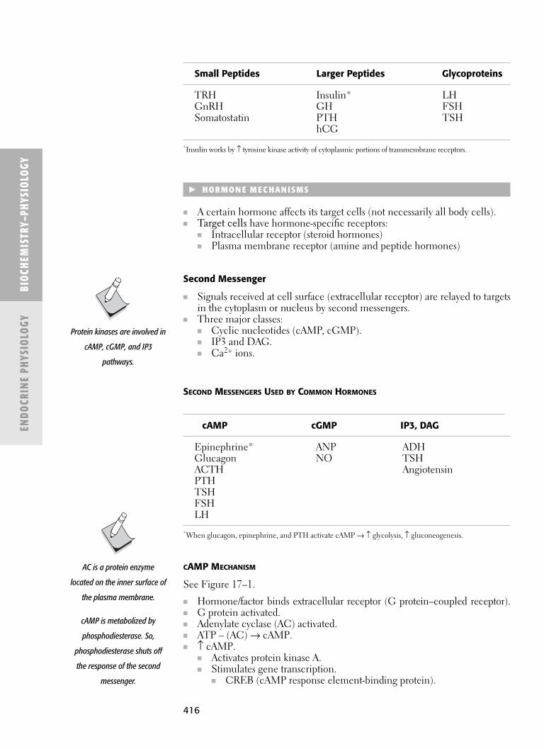
416
� HORMON E M EC HAN ISMS
■ A certain hormone affects its target cells (not necessarily all body cells).■ Target cells have hormone-specific receptors:
■ Intracellular receptor (steroid hormones)■ Plasma membrane receptor (amine and peptide hormones)
Second Messenger
■ Signals received at cell surface (extracellular receptor) are relayed to targetsin the cytoplasm or nucleus by second messengers.
■ Three major classes:■ Cyclic nucleotides (cAMP, cGMP).■ IP3 and DAG.■ Ca2+ ions.
BIOC
HEM
ISTR
Y–PH
YSIO
LOGY
ENDO
CRIN
E PH
YSIO
LOGY
Small Peptides Larger Peptides Glycoproteins
TRH Insulin* LHGnRH GH FSHSomatostatin PTH TSH
hCG
*Insulin works by ↑ tyrosine kinase activity of cytoplasmic portions of transmembrane receptors.
SECOND MESSENGERS USED BY COMMON HORMONES
cAMP cGMP IP3, DAG
Epinephrine* ANP ADHGlucagon NO TSHACTH AngiotensinPTHTSHFSHLH
*When glucagon, epinephrine, and PTH activate cAMP → ↑ glycolysis, ↑ gluconeogenesis.
CAMP MECHANISM
See Figure 17–1.
■ Hormone/factor binds extracellular receptor (G protein–coupled receptor).■ G protein activated.■ Adenylate cyclase (AC) activated.■ ATP – (AC) → cAMP.■ ↑ cAMP.
■ Activates protein kinase A.■ Stimulates gene transcription.
■ CREB (cAMP response element-binding protein).
Protein kinases are involved in
cAMP, cGMP, and IP3
pathways.
AC is a protein enzyme
located on the inner surface of
the plasma membrane.
cAMP is metabolized by
phosphodiesterase. So,
phosphodiesterase shuts off
the response of the second
messenger.
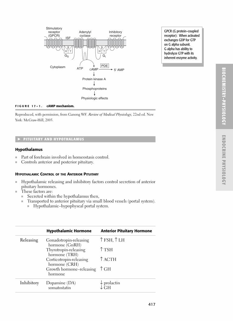
417
� PITU ITARY AN D HYPOTHALAM US
Hypothalamus
■ Part of forebrain involved in homeostasis control.■ Controls anterior and posterior pituitary.
HYPOTHALAMIC CONTROL OF THE ANTERIOR PITUITARY
■ Hypothalamic releasing and inhibitory factors control secretion of anteriorpituitary hormones.
■ These factors are:■ Secreted within the hypothalamus then,■ Transported to anterior pituitary via small blood vessels (portal system).
■ Hypothalamic–hypophyseal portal system.
BIOCHEM
ISTRY–PHYSIOLOGY
ENDOCRIN
E PHYSIOLOGY
Stimulatoryreceptor(GPCR)
Adenylylcyclase
Inhibitoryreceptor
Protein kinase A
ISF
Phosphoproteins
Cytoplasm
Physiologic effects
GS Gi
βα γ
βα γ
ATP CAMPPDE
5' AMP
F I G U R E 1 7 – 1 . cAMP mechanism.
Reproduced, with permission, from Ganong WF. Review of Medical Physiology, 22nd ed. New
York: McGraw-Hill, 2005.
Hypothalamic Hormone Anterior Pituitary Hormone
Releasing Gonadotropin-releasing ↑ FSH, ↑ LHhormone (GnRH)
Thyrotropin-releasing ↑ TSHhormone (TRH)
Corticotropin-releasing ↑ ACTHhormone (CRH)
Growth hormone–releasing ↑ GHhormone
Inhibitory Dopamine (DA) ↓ prolactinsomatostatin ↓ GH
GPCR (G protein–coupledreceptor): When activatedexchanges GDP for GTPon G alpha subunit.G alpha has ability tohydrolyze GTP with itsinherent enzyme activity.

418
HYPOTHALAMIC CONTROL OF THE POSTERIOR PITUITARY
■ Posterior pituitary hormones are synthesized in cell bodies of neurosecre-tory cells (located in hypothalamus).
■ Axons project to and deliver hormones to pars nervosa of posteriorpituitary.
■ Brain neural inputs influence their release.
BIOC
HEM
ISTR
Y–PH
YSIO
LOGY
ENDO
CRIN
E PH
YSIO
LOGY
Hypothalamic Neurosecretory Cells Hormones Produced
Supraoptic (ADH) and paraventricular ADH(oxytocin) nuclei Oxytocin
Pituitary Gland
See Figure 17–2.
■ Formed from:■ Diencephalon (posterior pituitary).■ Rathke’s pouch (anterior pituitary).
■ In week 5 of development, Rathke’s pouch and diencephalon comein contact.
Hypothalamic–Pituitary Endocrine Organ Axis
See Figures 17–3 and 17–4.
■ Hypothalamus: Releasing factors ■ Pituitary: Trophic hormones■ Endocrine organ: Hormones
■ Hormones exert metabolic affect.■ Hormones give feedback on the system.
Parstuberalis
Anteriorlobe
Posterior lobe
Intermediate lobeRathke’s pouch
Third ventricleThird ventricle
F I G U R E 1 7 – 2 . Pituitary gland.
Reproduced, with permission, from Ganong WF. Review of Medical Physiology, 22nd ed.
New York: McGraw-Hill, 2005.
GH is the only anterior
pituitary hormone that does
not act directly on a target
gland.
Neither ADH nor oxytocin is
produced in the posterior
pituitary (made in
hypothalamus).

419
BIOCHEM
ISTRY–PHYSIOLOGY
ENDOCRIN
E PHYSIOLOGY
Hypothalamus
Pituitary
CNS
Releasingfactors
Target hormone feedback inhibition
Gonads
Thyroid
Adrenal
Trophic hormones
+
+
−
−
F I G U R E 1 7 – 3 . Hypothalamic-pituitary endocrine organ axis.
Reproduced, with permission, from Kasper DL, et al. eds. Harrison’s Principles of InternalMedicine, 16th ed. New York: McGraw-Hill, 2004.
F I G U R E 1 7 – 4 . Hypothalamic-pituitary endocrine organ axis.
Reproduced, with permission, from Ganong WF. Review of Medical Physiology, 22nd ed. New York: McGraw-Hill, 2005.
Anterior pituitary
ACTH TSH FSH LH ProlactinGrowth
hormone
Breast
17-Hydroxy-corticoids
Aldosterone,sex hormones
ie, testosteroneand estradiol
Somato-medins
Thyroxine Estrogen Progesterone

420
Diabetes Insipidus
■ Distinguish from diabetes mellitus.■ Shares in common with diabetes mellitus:
■ Polydipsia. ■ Polyuria (very dilute).
■ Different mechanism■ ↓ ADH (or ADH doesn’t work).■ ↓ tubular reabsorption of H2O in the kidney.
■ Pass large amount of dilute urine.■ Hypertonic serum.
■ ↑ thirst.
BIOC
HEM
ISTR
Y–PH
YSIO
LOGY
ENDO
CRIN
E PH
YSIO
LOGY
Ø ADH (or ADH doesn’t
work) = DI (polydipsia,
polyuria).
Causes of Diabetes Insipidus
Central Diabetes Insipidus Nephrogenic Diabetes Insipidus(↓ ADH) (ADH Normal OR ↑)
↓ ADH to stimulate renal tubule Renal tubule does not ■ ↓ activity of posterior respond to ADH
pituitary gland ■ Congenital and familial■ Destruction of supraoptic nuclei ■ Drug induced (eg, lithium)
(hypothalamus)
Posterior Pituitary Hormones
Hormone ADH Oxytocin
Produced Hypothalamus (mainly, Hypothalamus (supraoptic supraoptic nuclei) and paraventricular nuclei)
Stored Posterior pituitary Posterior pituitary
↑ release ↑ plasma Na+ Suckling↑plasma osmolarity Cervical dilation↓ blood volume Estrogen (↑ uterine↓ BP oxytocin receptors)NicotineSweatingSIADH
↓ release Ethanol Stress—catecholaminesCaffeine Drinking (↑ H2O)Diabetes insipidus (DI)
Action H2O reabsorption of Milk letdown* (ejection water from collecting duct: of milk with breast feeding)↑ aquaporin expression Uterine contraction with
Produce small volume of birthingconcentrated urine Maternal behavior
Conserve intravascular H2OHelps control BP
*Oxytocin causes contraction myoepithelial cells surrounding sac-like alveoli of the mammary glands toinduce milk letdown.
Body fluid volume stays closeto normal in DI providedthe patient drinks enough
water (↑ thirst) to make upfor the ↑ passage of water
in the urine.
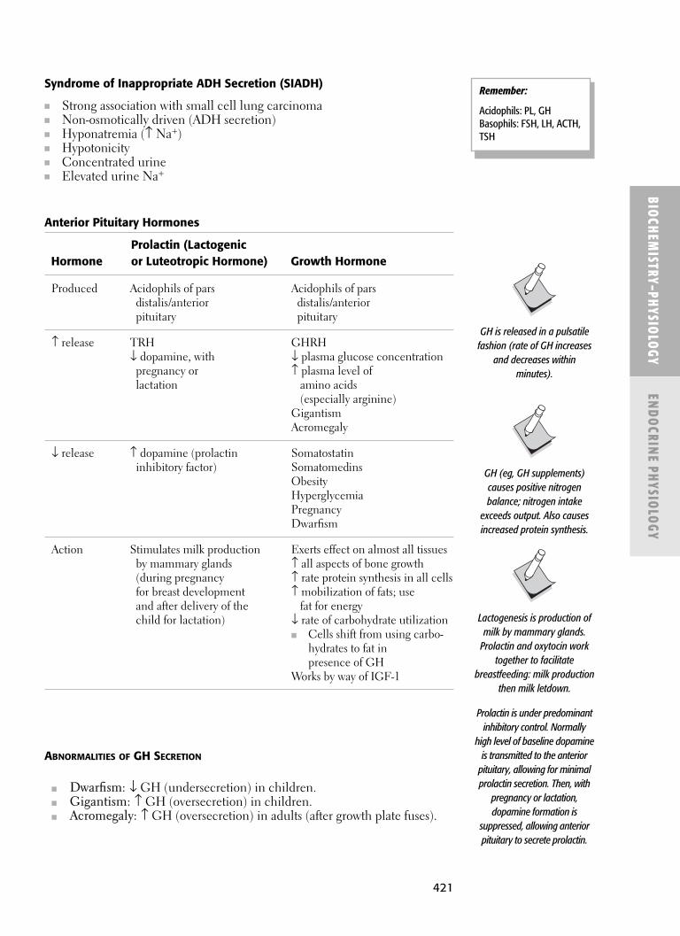
421
Syndrome of Inappropriate ADH Secretion (SIADH)
■ Strong association with small cell lung carcinoma■ Non-osmotically driven (ADH secretion)■ Hyponatremia (↑ Na+)■ Hypotonicity■ Concentrated urine■ Elevated urine Na+
BIOCHEM
ISTRY–PHYSIOLOGY
ENDOCRIN
E PHYSIOLOGY
Anterior Pituitary Hormones
Prolactin (Lactogenic Hormone or Luteotropic Hormone) Growth Hormone
Produced Acidophils of pars Acidophils of pars distalis/anterior distalis/anterior pituitary pituitary
↑ release TRH GHRH↓ dopamine, with ↓ plasma glucose concentration
pregnancy or ↑ plasma level oflactation amino acids
(especially arginine)GigantismAcromegaly
↓ release ↑ dopamine (prolactin Somatostatininhibitory factor) Somatomedins
ObesityHyperglycemiaPregnancyDwarfism
Action Stimulates milk production Exerts effect on almost all tissuesby mammary glands ↑ all aspects of bone growth(during pregnancy ↑ rate protein synthesis in all cellsfor breast development ↑ mobilization of fats; useand after delivery of the fat for energychild for lactation) ↓ rate of carbohydrate utilization
■ Cells shift from using carbo-hydrates to fat in presence of GH
Works by way of IGF-1
GH is released in a pulsatilefashion (rate of GH increases
and decreases withinminutes).
GH (eg, GH supplements)causes positive nitrogenbalance; nitrogen intake
exceeds output. Also causesincreased protein synthesis.
Lactogenesis is production ofmilk by mammary glands.
Prolactin and oxytocin worktogether to facilitate
breastfeeding: milk productionthen milk letdown.
Prolactin is under predominantinhibitory control. Normally
high level of baseline dopamineis transmitted to the anterior
pituitary, allowing for minimalprolactin secretion. Then, with
pregnancy or lactation,dopamine formation is
suppressed, allowing anteriorpituitary to secrete prolactin.
ABNORMALITIES OF GH SECRETION
■ Dwarfism: ↓ GH (undersecretion) in children.■ Gigantism: ↑ GH (oversecretion) in children.■ Acromegaly: ↑ GH (oversecretion) in adults (after growth plate fuses).
Remember:
Acidophils: PL, GHBasophils: FSH, LH, ACTH,TSH

422
Somatostatin
■ Peptide hormone ■ Secreted by:
■ Median eminence of hypothalamus.■ Delta cells of pancreatic islets.
■ Functions to inhibit release/secretion of:■ GH (somatotropin) and thyrotropin (TSH) by anterior pituitary.■ Insulin (from beta cells of pancreas).■ Glucagon (from alpha cells of pancreas).■ Gastrin (by gastric mucosa).
Sex Hormones
■ FSH and LH.■ GnRH from hypothalamus stimulates release of both FSH and LH.
BIOC
HEM
ISTR
Y–PH
YSIO
LOGY
ENDO
CRIN
E PH
YSIO
LOGY
COMPARISON OF MAJOR SEX HORMONES
FSH LH
Males Sertoli (sustentacular) cells Leydig (interstitial) cells■ Sperm production, ■ Produce testosterone
maturation
Females Ovary Ovary ■ Graafian follicle development ■ Steroid production—
theca cells■ Steroid production ■ Estrogen-induced LH
■ Estradiol (during surge stimulates ovulation follicular phase) ■ Forms corpus luteum(granulosa cells)
■ Progesterone (during luteal phase)
Puberty
See Figures 17–5 and 17–6.
■ Age 10–15.■ ↑ GnRH (from the hypothalamus).
■ Stimulates anterior pituitary:■ ↑ FSH■ ↑ LH
■ Stimulates gonadal tissue (growth and function)■ Ovaries■ Testes
■ Secondary sex characteristics appear (see following chart).■ During early childhood, a boy does not secrete gonadotropins and so has
little circulating testosterone.■ On completion of puberty a male has full adult sexual capabilities and is
capable of reproduction.■ Females reach puberty 1 to 2 years earlier than males. Puberty in females
is heralded by menarche (first menses).
Progesterone is made by the
ovaries.
All estrogens have an
aromatic A ring (unlike other
steroid hormones).

423
BIOCHEM
ISTRY–PHYSIOLOGY
ENDOCRIN
E PHYSIOLOGY
GnRH
LH
Androgenicand anabolic
effects
Testosterone
FSH
Leydigcells
Sertolicells
Inhibin B
Hypothalamus
Anteriorpituitary
Testis
F I G U R E 1 7 – 5 . Puberty in the male.
Reproduced, with permission, from Ganong WF. Review of Medical Physiology, 22nd ed.
New York: McGraw-Hill, 2005.
Androgens
GnRH
LH
Estrogeniceffects
Estrogen
FSH
Thecainterna
Granu-losa
Inhibin B
Hypothalamus
Anteriorpituitary
Ovary
F I G U R E 1 7 – 6 . Puberty in the female.
Reproduced, with permission, from Ganong WF. Review of Medical Physiology, 22nd ed.
New York: McGraw-Hill, 2005.
Secondary Sex Characteristics
Males (↑↑ Testosterone) Females (↑ Estrogens)∗
Enlargement of penis, Enlargement of vagina, uterus, scrotum, testis, hair uterine tubes; deposition growth, voice changes of fat in breasts and hips
*Estrogen is effective at very low concentrations and generates a slowly developing long-term response intarget tissues by binding to an intracellular receptor.
A decrease in estrogen levels
feeds back to hypothalamus
to start cycle over again.
Granulosa cells convert
androgens from theca cells to
estradiol by aromatase.

424
PRECOCIOUS PUBERTY
■ Puberty occurs at an abnormally early age.■ Adrenal cortex: ↑ hormones similar to male and female sex hormones.
� RE PRODUCTION
Menstruation
See Figure 17–7.
■ Average menstrual cycle is 28 days (range 22–34).■ Without estrogen and progesterone, the endometrial lining is shed as men-
strual fluid during menstruation/menses.■ Usually lasts about 3–5 days and involves loss of about 50 mL of blood.
BIOC
HEM
ISTR
Y–PH
YSIO
LOGY
ENDO
CRIN
E PH
YSIO
LOGY
Follicular Phase Ovulation* Luteal Phase
Days 1–14 15 16–28Events FSH, LH: LH surge: ↓ FSH, LH levels.
Stimulate ovarian Caused by estrogenfollicle. ~day 14 or 15.↑ estrogen secretion. Stimulates ovulation.
Estrogen: Ovulation occurs Corpus luteum forms.Endometrium 14 days before ↑ estrogen and proliferation. menses (regardless progesterone.
of cycle length).
Late in this phase: If no fertilization:Estrogen levels ↑ peak. Corpus luteum FSH secretion ↓. degenerates. LH secretion ↑. ↓ estrogen and
progesterone levels.Feedback to hypothalamus: ↑ GnRH (cycle begins again).
*Oral contraceptive pills (OCPs) contain synthetic estrogen-like and progesterone-like substances thatinhibit ovulation. OCPs suppress LHRH (GnRH) (by hypothalamus) → prevent the LH surge (by pituitary)→ prevent ovulation.
Ovulation
■ Stimulated byestrogen-inducedLH surge.
■ Ovulation is thedischarge of an ovum(oocyte) from themature follicle(Graafian follicle) of the ovary.

425
Oogonia
■ See the embryology section, Chapter 4.■ Source of oocytes■ Primordial follicles
■ Stimulated by FSH (from ant pituitary) to form:■ Primary oocytes (in first meiotic division) and become:
↓■ Primary follicles
■ With primary oocytes■ Form the antrum (cavity) to become:
↓■ Secondary follicles
■ Primary oocytes complete first meiotic division.↓
■ Mature Graafian follicles ■ With secondary oocytes (in second meiotic division). ■ LH surge causes release of secondary oocyte (egg) into abdominal cavity
(ovulation).
BIOCHEM
ISTRY–PHYSIOLOGY
ENDOCRIN
E PHYSIOLOGY
1 2 3 4 5 6 7 8 9 10 11 12 13 14 15 16 17 18 19 20 21 22 23 24 25 26 27 28
Day of menstrual cycle
End
omet
rial
hist
olog
y
Menstruation
Follicular phase Luteal phase
Ovu
latio
nFollicle-stimulating hormone
EstradiolLuteinizing hormone
Progesterone
Body temperature
37°C
36°C
Hor
mon
esO
varia
nhi
stol
ogy
Follicle OvulationMaturingfollicle
DegenerateC. Luteum
CorpusLuteum
F I G U R E 1 7 – 7 . Female reproductive cycle.
Courtesy of WikiMedia user Lyrl, licensed and distributed under the Creative Commons
Attribution ShareAlike 2.5 license (http://creativecommons.org/licenses/by-sa/2.5). Image is
available in digital format at: http://commons.wikimedia.org/wiki/Image:MestrualCycle2.png.
Key points of the femalesexual cycle:
■ Only a single matureovum is released(normally) from theovaries each month.
■ So only a single fetuscan begin to grow at atime.
■ Uterine endometriumis prepared forimplantation of thefertilized ovum at thetime of ovulation.
The LH surge leads to final
maturation of the follicle,
rupture of follicle, and
ovulation. In the absence
of LH, even in the presence
off large amounts of FSH,
the follicle will not progress
to ovulation.
Human chorionic
gonadotropin (hCG),
produced by the placenta,
stimulates corpus luteum to
persist and to secrete/produce
progesterone and estradiol.
Oogenesis:Formation of primaryoocytes before birth andsecondary oocytes atovulation.

426
■ Egg swept by fimbraie into infundibulum/ostium of Fallopian tube(uterine tube, oviduct) to be fertilized.
Fertilization No fertilization
Zygote implants Egg discardedon uterus
Corpus Luteum
■ Ruptured mature ovarian follicle forms a yellow mass of cells after ovula-tion (egg release).
■ No fertilization:■ Corpus luteum retrogresses into corpus albicans.
■ Becomes a mass of scar tissue.■ Disappears.
■ Yes fertilization and pregnancy:■ Corpus luteum persists (for several months).
■ Granulosa cells secrete hormones.■ Progesterone > estrogen.
� PANC REAS
BIOC
HEM
ISTR
Y–PH
YSIO
LOGY
ENDO
CRIN
E PH
YSIO
LOGY
Endocrine Pancreas
Hormone Insulin Glucagon
Secreted Pancreas PancreasBeta cells of islets Alpha cells in islets of Langerhans of Langerhans
↑ release ↑ BGL (hyperglycemia) ↓ BGL (hypoglycemia).major stimuli. ↓ amino acids (especially
↑ amino acids, especially arginine).arginine, lysine, leucine. Cholecystokinin secretion.
Glucagon. Sympathetic stimulation.GH. ■ Epinephrine, NE Cortisol. secretion.Parasympathetic stimulation.
↓ release ↓ BGL (hypoglycemia). ↑ BGL.Sympathetic Insulin.
stimulation. Somatostatin.Free fatty acids.Ketoacids.
Action ↓ BGL. ↑ BGL.■ ↑ glucose uptake and utilization. ↑ glycogenolysis in liver.■ ↑↑ GLUT transporters. ■ Glycogen → glucose.
↑ glycogenesis in liver. ↑ free fatty acids.■ ↑ glucose → glycogen. ↑ amino acids.
↑ synthesis of triglycerides, ↑ urea production.proteins. It does not stimulate
glycogen breakdown in the muscle.
Insulin conserves proteins,
carbohydrates, and fats in the
body: ≠ protein synthesis
(inhibits protein breakdown),
≠ glycogenesis (inhibits
glucose breakdown), and
≠ triglyceride synthesis
(inhibits lipolysis).
Removal of anterior lobe of
pituitary gland results in ≠
sensitivity to insulin.
FSH and LH work together to
cause ovulation and
formation of corpus luteum.

427
Diabetes Mellitus
■ Metabolic syndrome characterized by hyperglycemia. ■ Secondary to either or both
■ Insulin deficiency.■ Reduced effectiveness of insulin.
■ Most common endocrine disorder involving the pancreas.■ Pancreas itself may be entirely normal in cases of insulin resistance.
Diabetes Insipidus
■ Distinguish from diabetes mellitus.■ Not a problem with the pancreas and insulin, but rather with ADH secre-
tion or action.
� ADRE NAL G LAN D
Adrenal Cortex
■ Steroid hormones■ Cholesterol core
CRH–ACTH–CORTISOL AXIS
■ Hypothalamus
CRH acts on (travels via portal system)↓
Anterior pituitary (basophils of pars distalis)ACTH acts on
↓Adrenal cortex (zona fasciculata)
Cortisol (travels via bloodstream → systemic effects)
NEGATIVE FEEDBACK LOOP
■ CRH → ACTH → cortisol.■ Cortisol feeds back and inhibits both the hypothalamus and anterior
pituitary.■ ACTH
■ Controls production and secretion of cortisol.■ Secretion is controlled by hypothalamus.■ Secreted in bursts, causing cortisol levels to rise and fall.■ Bursts are most frequent in the early morning; ~ 75% of the daily pro-
duction of cortisol occurs between 4 am and 10 am.
BIOCHEM
ISTRY–PHYSIOLOGY
ENDOCRIN
E PHYSIOLOGY
Polyuria in diabetes mellitus(DM) is a result of osmotic
diuresis, secondary to sustainedhyperglycemia. (Distinguish
from polyuria in DI).
Normal blood glucose level(BGL) = 80–100 mg/dL.
Histologic layers of theadrenal cortex, fromoutermost to innermost:
■ Salt (aldosterone) =Glomerulosa.
■ Sugar (cortisol) =Fasciculata.
■ Sex (androgens) =Reticularis.
Corticosteroids = aldosterone(mineralocorticoid) + cortisol
(glucocorticoid).
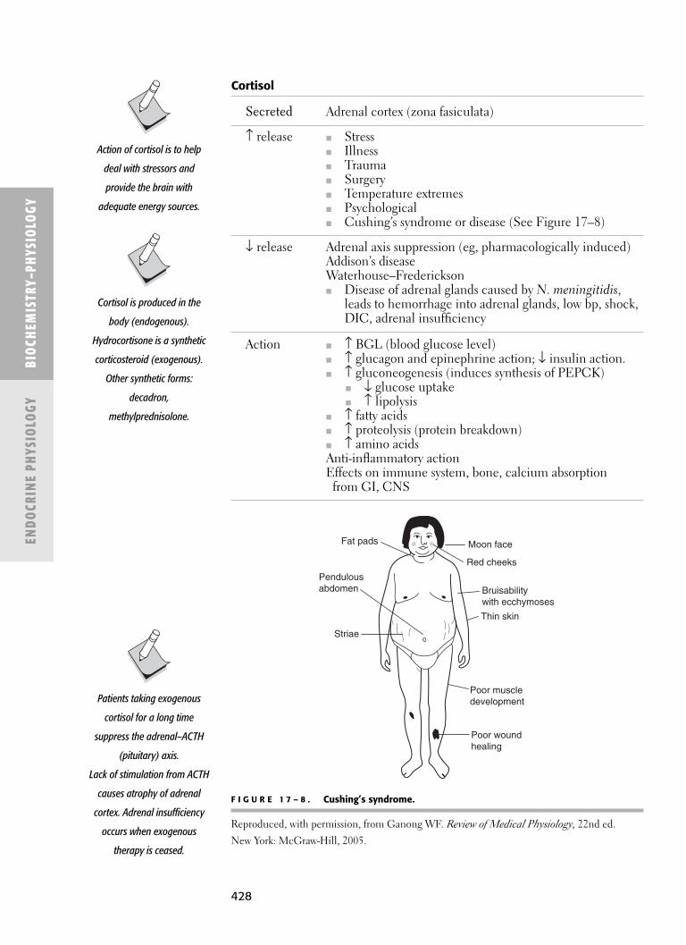
428
BIOC
HEM
ISTR
Y–PH
YSIO
LOGY
ENDO
CRIN
E PH
YSIO
LOGY
Cortisol
Secreted Adrenal cortex (zona fasiculata)
↑ release ■ Stress■ Illness■ Trauma■ Surgery■ Temperature extremes■ Psychological■ Cushing’s syndrome or disease (See Figure 17–8)
↓ release Adrenal axis suppression (eg, pharmacologically induced)Addison’s diseaseWaterhouse–Frederickson■ Disease of adrenal glands caused by N. meningitidis,
leads to hemorrhage into adrenal glands, low bp, shock,DIC, adrenal insufficiency
Action ■ ↑ BGL (blood glucose level)■ ↑ glucagon and epinephrine action; ↓ insulin action.■ ↑ gluconeogenesis (induces synthesis of PEPCK)
■ ↓ glucose uptake■ ↑ lipolysis
■ ↑ fatty acids■ ↑ proteolysis (protein breakdown)■ ↑ amino acidsAnti-inflammatory actionEffects on immune system, bone, calcium absorption
from GI, CNS
Poor muscledevelopment
Thin skin
Bruisabilitywith ecchymoses
Red cheeks
Moon face
Poor woundhealing
Striae
Pendulousabdomen
Fat pads
F I G U R E 1 7 – 8 . Cushing’s syndrome.
Reproduced, with permission, from Ganong WF. Review of Medical Physiology, 22nd ed.
New York: McGraw-Hill, 2005.
Action of cortisol is to help
deal with stressors and
provide the brain with
adequate energy sources.
Cortisol is produced in the
body (endogenous).
Hydrocortisone is a synthetic
corticosteroid (exogenous).
Other synthetic forms:
decadron,
methylprednisolone.
Patients taking exogenous
cortisol for a long time
suppress the adrenal–ACTH
(pituitary) axis.
Lack of stimulation from ACTH
causes atrophy of adrenal
cortex. Adrenal insufficiency
occurs when exogenous
therapy is ceased.
429
ADDISON’S DISEASE
■ Adrenocortical insufficiency.■ ↓ corticosteroids (glucocorticoids and mineralocorticoids).■ Can occur with any age and M = F.■ In primary Addison’s, cutaneous pigmentation tends to disappear following
therapy, but oral pigmentation usually persists.
BIOCHEM
ISTRY–PHYSIOLOGY
ENDOCRIN
E PHYSIOLOGY
Cushing’s Syndrome vs. Cushing’s Disease
Cushing’s Syndrome Cushing’s Disease
Cause ↑ cortisol ↑ cortisolExogenously administered Secondary to ACTH
glucocorticoids (cortisol-like (ACTH-secreting medications) pituitary tumor)
Endogenous ↑ cortisol(cortisol-secretingadrenalcortical tumor)
Ectopic ACTH production(eg, oat cell carcinoma,small cell carcinoma)
Symptoms Usually no ↑ pigmentation HyperpigmentationCentral obesityMoon faciesBuffalo humpExophthalmos (retroorbital fat)Skin atrophyStriae(Hyperpigmentation if ↑ ACTH)Proximal muscle wasting/weaknessBone lossGlucose intolerance/DM-2 pictureHTN■ ↑ sensitivity to cathecholamines,
↑ angiotensinogen, ↑ aldosteronereceptors
■ Cortisol can exhibit mineral corticoid activity in increasedconcentrations
■ Cortisol enhances epi effects (both of these effects canlead to HTN)ThromboembolismIrritability, anxiety, panic attacksDepression, insomnia↑ Infection

430
Adrenal Medulla
See Figure 17–9.
■ Specialized ganglion of sympathetic nervous system■ Preganglionic sympathetic fibers (cholinergic)■ Synapse directly on chromaffin cells in the adrenal medulla■ Chromaffin cells then secrete into the circulation:
■ Epinephrine (80%)■ Norepinephrine (NE) (20%)
BIOC
HEM
ISTR
Y–PH
YSIO
LOGY
ENDO
CRIN
E PH
YSIO
LOGY
Primary Addison’s Disease Secondary Addison’s Disease
Cause ↓ cortisol ↓ cortisolDestruction of adrenal cortex ↓ or lack of ACTH■ >90% cortex must be ■ Abruptly stopping chronic
destroyed before exogenous corticoids symptoms (eg, prednisone); usually
■ 70% unknown temporary■ Likely autoimmune ■ ↓ pituitary function: surgical ■ 30% identifiable cause removal, apoplexy, tumor,■ Infection (eg, TB) infection
Cancer/neoplasmAmyloidosisHemorrhage in
the gland
Symptoms ↑ skin pigmentation No ↑ skin pigmentation(bronzing of skin)
↓ BP (hypotension)↓ Na+, Cl−, HCO3
−
↑ K+
↓ BGLMalaiseWeight lossDepression
Oral signs Diffuse intraoral No oral pigmentationpigmentation
■ Gingiva■ Tongue■ Hard palate■ Buccal mucosa
Treatment Cortisol (exogenous, as hydrocortisone or prednisone)
The adrenal medulla is a
neural crest derivative.
NE can be released in twoways:
■ By adrenal medullainto bloodstream.
■ Directly within anorgan:■ Via sympathetic
innervation.■ Postganglionic
sympathetic(adrenergic) neuronthat stores NE.
Note: Effects are morewidespread when NE isreleased into bloodstreamby adrenal medulla thanwithin an organ from apostganglionicsympathetic nerve ending.

431
BIOCHEM
ISTRY–PHYSIOLOGY
ENDOCRIN
E PHYSIOLOGY
CRH
ACTH
Systemiceffects
Cortisol
Hypothalamus
Drive forcircadianrhythm
Emotion vialimbic system
Trauma vianociceptivepathways
Afferentsfrom NTS
Anteriorpituitary
Adrenalcortex
F I G U R E 1 7 – 9 . Adrenal medulla.
Reproduced, with permission, from Ganong WF. Review of Medical Physiology, 22nd ed.
New York: McGraw-Hill, 2005.
EPINEPHRINE AND NOREPINEPHRINE
■ Direct-acting adrenergic agonists■ Biosynthesized from tyrosine (amino acid)■ Released from adrenal medulla from storage vesicles in response to:
■ Sympathetic stimulation (fright, startle)■ Exercise■ Cold■ ↓ BGL
■ Function: (regulators of carbohydrate and lipid metabolism, cardiovascu-lar effects)■ ↑ Fatty acids
■ ↑ Triacylglycerol breakdown (lipolysis)■ NE on α2 inhibits lipolysis
■ ↑ BGL■ ↑ Glycogen breakdown (glycogenolysis)
■ Activates muscle glycogen phosphorylase. (Remember McArdle’ssyndrome)
■ ↑ Gluconeogenesis■ ↑ CO (especially epinephrine)
■ ↑ ↑ rate, force, and amplitude of heart beat■ ↑ BP
■ ↑ Vasoconstriction in skin, mucous membranes, kidneys■ Bronchodilation
■ ↑ Relaxes bronchiolar smooth muscle
Catecholamines (epinephrine,
norepinephrine, and
dopamine) are products of
tyrosine metabolism.
Pathway:
Tyrosine → DOPA →
dopamine → norepinephrine
→ epinephrine.

432
� THYROI D G LAN D
See Figure 17–10.
Hypothalamic–Pituitary–Thyroid Axis
■ TRH-TSH-thyroid hormone axis
Hypothalamus: TRH released (travels via portal system).↓
Anterior pituitary (basophils of pars distalis): TSH acts on↓
Thyroid gland (TSH receptor): Thyroid hormones (T3, T4) (travel viabloodstream → systemic effects).
Negative Feedback Loop
See Figure 17–10.
■ TRH → TSH → thyroid hormones. ■ Thyroid hormones feedback and inhibit both the hypothalamus and ante-
rior pituitary.■ ↑ thyroid hormones: ↓ both TRH and TSH secretion.■ ↑ TSH: ↓ TRH secretion.
BIOC
HEM
ISTR
Y–PH
YSIO
LOGY
ENDO
CRIN
E PH
YSIO
LOGY
TRH
TSH
Metaboliceffects
Free T3and T4
Hypothalamus
Anteriorpituitary
Thyroid
F I G U R E 1 7 – 1 0 . Thyroid gland.
Reproduced, with permission, from Ganong WF. Review of Medical Physiology, 22nd ed.
New York: McGraw-Hill, 2005.
Stress can inhibit TSH secretion
secondary to neural influences
that inhibit secretion of TRH
from hypothalamus.

433
Thyroid Gland
■ Well vascularized gland.■ One of highest rates of blood flow per gram of tissue of any organ.
■ Composed of follicles, filled with colloid. ■ Colloid contains thyroglobulin (tyrosine-containing glycoprotein).
■ Thyroglobulin contains thyroid hormones within its molecule.
THYROID CELLS (FOLLICULAR CELLS)
Three functions:
■ Collect and transport iodine.■ Synthesize and secrete (into colloid) thyroglobulin.■ Remove thyroid hormones from thyroglobulin and secrete them into the
circulation.
THYROGLOBULIN
■ Glycoprotein hormone (10% carbohydrate).■ Contains many tyrosine residues (incorporated into thyroid hormones).■ Synthesized by thyroid follicular cell. ■ Secreted/stored in colloid region of follicles.
THYROID HORMONE PRODUCTION
■ Thyroglobulin is iodinated (within the colloid). ■ Iodine attaches to tyrosine molecules.■ Thyroid hormones (T3, T4) remain part of the thyroglobulin molecules
until secreted.
THYROID HORMONE SECRETION
Colloid is taken up into the cytoplasm of the thyroid follicular cells (smallglobules of colloid are endocytosed).
↓Peptide bonds between iodinated residues and thyroglobulin are hydrolyzed.
↓Thyroglobulin releases thyroid hormones as free T4 and T3.
↓T4 (mostly) and T3 are discharged into the bloodstream/capillaries
(systemic effects).
THYROID HORMONES
■ Necessary for normal growth and development. ■ ↑ cellular metabolism, growth. ■ ↑ differentiation of tissues, especially brain, neural tissues.
■ Affect many metabolic processes and metabolic rate ↑ HR, ↑ cardiaccontractility.
■ ↑ O2 consumption and heat production.■ ↑ glycogenolysis.■ ↑ gluconeogenesis.■ ↑ GI carbohydrate absorption.■ ↑ lipolysis.■ ↑ protein breakdown.
BIOCHEM
ISTRY–PHYSIOLOGY
ENDOCRIN
E PHYSIOLOGY
Thyroid hormones are
lipophillic hormones. They exert
effect via gene transcription.
T4 acts as pro-hormone to T3
(enzymatic removal of one
iodine atom) in the peripheral
tissues. T3 is a more potent
hormone.

434
Hypo- and Hyperthyroidism
BIOC
HEM
ISTR
Y–PH
YSIO
LOGY
ENDO
CRIN
E PH
YSIO
LOGY
HYPOTHYROID HYPERTHYROIDISM
Diseases Cretinism (children) Grave’s disease*Myxedema (adults) Plummer’s diseaseHashimoto’s thyroiditis Solitary toxic adenoma
Toxic multinodular goiterTSH-secreting pituitary tumor
Symptoms Weight gain Restlessness, irritability Cold intolerance FatigueLow-pitched voice Heat intolerance (sweating)Mental and physical slowness ↑ temperatureConstipation TachycardiaDry skin Fine hairCoarse hair DiarrheaPuffiness of face, eyelids, hands Tremor (shakiness)
↑ Basal metabolic rateWeight lossGeneralized osteoporosisOral manifestations:If hyperthyroid in childhood, can get:■ Premature eruption of teeth■ Loss of deciduous dentition
*Grave’s disease is the most common form of hyperthyroidism; 50% get exophthalmos (secondary to TSH-receptor antibodies in circulation).
GRAVE’S DISEASE
■ Most common form of hyperthyroidism.■ Caused by binding of immunoglobulin antibodies to TSH receptors in the
thyroid.■ Stimulates production of thyroxin.
■ Typical signs ■ Goiter (enlarged thyroid) ■ Exophthalmos (bulging eyes) (hypertrophy of eye muscles in response
to IgG)
PLUMMER’S DISEASE
■ Toxic multinodular goiter.■ Multiple secreting thyroid nodules (adenomas) within gland.■ Uncommon in adolescents and young adults (↑ with age).■ Exophthalmos is rare.

435
� CALC I U M AN D PHOSPHORUS M ETABOLISM
BIOCHEM
ISTRY–PHYSIOLOGY
ENDOCRIN
E PHYSIOLOGY
Normal Serum Value
Regulation Calcium Phosphorus
8.5–10.5 mg/dL 3.0–4.5 mg/dLPTH ↑ Ca2+ ↓ P
↑ bone resorption ↓ renal tubular reabsorption ↑ renal tubular reabsorption ↑ renal P excretion
Vitamin D ↑ Ca2+
↑ GI absorption of Ca2+
Hypocalcemia Hypercalcemia
Effect Irritability/excitability Cardiac and CNS depressionnerves and muscles
Tetany
■ PTH is the most important hormone in Ca2+ metabolism (principal con-troller of Ca2+ and phosphate metabolism) and is involved in remodeling ofbone. Plasma Ca2+ is major controller of PTH secretion.
■ PTH stimulates 1-alpha hydroxylase in the kidneys.■ Compared to PTH, calcitonin has only a minor role in regulating blood
calcium (in fact, calcitonin is not required in adult humans). Calcitonin isimportant during bone development.
Hormones and Calcium Regulation
Hormone Parathyroid Hormone Calcitonin
Secreted Parathyroid gland (chief cells) Thyroid gland (parafollicular [C-cells])
↑ release ■ ↓ plasma Ca2+■ ↑ plasma Ca2+
↓ release ■ ↑ plasma Ca2+■ ↓ plasma Ca2+
Action ↑ plasma Ca2+ Plasma Ca2+
■ ↑ bone resorption ■ ↑ Ca2+ deposition in bone■ Directly ■ ↓ bone resorption■ By osteoclasts ↓ GI absorption of Ca2+
↑ Ca2+ reabsorption in kidney ↑ Ca2+ renal excretion↑ GI absorption of Ca2+ ↓ plasma phosphate(via vitamin D) ■ ↑ phosphate renal excretion↓ plasma phosphate
■ ↑ phosphate excretion in urine (via vitamin D)
↓ calcium in diet = ↑ PTH
secretion, ↑ bone resorption
(to compensate).
436
Hormones and Calcium Regulation
See Chapter 15, “Gastrointestinal Physiology.”
� HORMON ES AN D TH E K I DN EY
See Chapter 14.
Erythropoietin
■ Released by kidneys. ■ Stimulates RBC production.
BIOC
HEM
ISTR
Y–PH
YSIO
LOGY
ENDO
CRIN
E PH
YSIO
LOGY
Hypoparathyroidism Hyperparathyroidism
Example(s) Congenital (DiGeorge syndrome). Parathyroid adenomaIatrogenic (surgery) Parathyroid carcinoma (rare)
Infiltration/destruction (sarcoidosis) Multiple endocrine neoplasm (MEN)Idiopathic von Recklinghausen’s disease■ ↓ bone resorption ■ ↑ bone resorption■ ↓ renal Ca2+ reabsorption ■ ↑ renal Ca2+ reabsorption■ ↑ renal phosphate reabsorption ■ ↓ renal phosphate ■ ↓ 1, 25-dihydroxycholecalciferol* reabsorption
■ ↓/↓ 1, 25-dihydroxycholecalciferol
Calcium ↓ plasma Ca2+ (↑ Ca2+ in bone) ↑ plasma Ca2+
(↓ Ca2+ in bone)
Phosphate ↑ plasma phosphate ↓ plasma phosphatesymptom(s) Tetany Bone pain
Mental status changes (MS Δ’s) Pathologic fracturesseizures Osteitis fibrosa cystica
↓ Cardiac contractility Brown tumors of boneVomiting Nephrolithiasis
Muscular weakness
*1, 25-dihydroxycholecalciferol = active form of vitamin D.
Erythropoietin
Secreted Kidney (peritubular capillary interstitial cells)
↑ release ↓ PO2 (hypoxemia)■ Anemia■ Lung disease■ Cardiac disease■ High altitudes
↓ release Normal PO2↑ RBC volume
Action ↑ RBCs■ EPO receptors on erythroid progenitors↑ replication■ ↑ maturation

437
BIOCHEM
ISTRY–PHYSIOLOGY
ENDOCRIN
E PHYSIOLOGY
Aldosterone
See Figure 17–11.
■ Released by adrenal cortex zona glomerulosa.
Angiotensin-
converting enzyme
ReninAngiotensinogen
Angiotensin I
Angiotensin II
Aldosterone Decreased Na+
(and water) excretion
Increased extracellularfluid volume
Increased renal arterialmean pressure, decreaseddischarge of renal nerves
Juxtaglomerularapparatus
Adrenalcortex
F I G U R E 1 7 – 1 1 . Adrenal cortex.
Reproduced, with permission, from Ganong WF. Review of Medical Physiology, 22nd ed.
New York: McGraw-Hill, 2005.
↓ Na+ → juxtaglomerular (JG)
cells secrete renin → renin
cleaves angiotensinogen into
AT-I → AT-I (converted by ACE)
→ AT-II. AT-II then stimulates
adrenal cortex to release
aldosterone.
In Addison’s disease there is
hyposecretion of both
aldosterone and cortisol.
Hormone Aldosterone (Mineralocorticoid)
Secreted Adrenal cortex (zona glomerulosa)
↑ release ↑ K+
↑ AT-II (via ↑ renin)↓ release ↓ K+
↓ AT-II (via ↓ renin)
Action Site of action is distal nephron—DCT, collecting ducts■ ↑ plasma volume (↑ H2O reabsorption)■ ↑ plasma Na+ (↑ Na+ reabsorption)■ ↓ plasma K+ (↓ K+ reabsorption; ↑ K+ excretion)

This page intentionally left blank

� Microbiology
� Oral Microbiology andPathology
� Reactions to TissueInjury
� Immunology andImmunopathology
� Systemic Pathology
� Neoplasia
S E C T I O N 3
Microbiology-Pathology
439

This page intentionally left blank

C H A P T E R 1 8
Microbiology
General Microbiology 443
CELL TYPES 443
MICROORGANISMS 443
INFECTIONS 443
Sterilization and Disinfection 444
STERILIZATION 444
DISINFECTION 446
PASTEURIZATION 446
SANITIZATION 447
INFECTION CONTROL 447
Bacteriology 449
BACTERIA 449
PATHOGENESIS 449
BACTERIAL VIRULENCE FACTORS 450
CLASSIFICATION 452
MEDICALLY RELEVANT BACTERIA 455
GRAM-NEGATIVE BACTERIA 459
BACTERIAL VACCINES 464
ANTIBIOTIC DRUGS 464
PATIENT SELECTION 469
Virology 470
VIRUSES 470
PATHOGENESIS 471
CLASSIFICATION 473
HEPATITIS VIRUSES 478
VIRAL VACCINES 480
ANTIVIRAL DRUGS 480
BACTERIOPHAGES (PHAGES) 480
LYSOGENIC CONVERSION 481
PRIONS 481
441

442
Mycology 482
FUNGI 482
PATHOGENESIS 482
CLASSIFICATION 482
ANTIFUNGAL DRUGS 484
Parasitology 485
PARASITES 485
MICRO
BIOLOGY–PATHO
LOGYM
ICROBIO
LOGY
443
� G E N E RAL M IC ROBIOLOGY
Cell Types
See Table 18–1.
■ Prokaryotes■ Eukaryotes
Microorganisms
See Table 18–2.
■ Bacteria■ Viruses■ Fungi■ Parasites
T A B L E 1 8 – 1 . Comparison of Prokaryotic and Eukaryotic Cells
MEMBRANE- MEMBRANE-
ENVELOPED BOUND RIBOSOME NUCLEIC ACID
CELL TYPE NUCLEUS ORGANELLES SIZE LOCATION DNA TYPE REPLICATION
Prokaryote No No 70S Nucleoid Circular Binary
(cytoplasm) fission
Eukaryote Yes Yes 80S Nucleus Linear Mitosis
T A B L E 1 8 – 2 . Comparison of Major Microorganisms Implicated in Infectious Diseases
MICRO- OUTER NUCLEIC
ORGANISM CELL TYPE SURFACE ACID RIBOSOMES MITOCHONDRIA REPLICATION
Bacteria Prokaryotic Cell wall of RNA and DNA 70S No Binary
peptidoglycan fission
Viruses Noncellular Protein capsid RNA or DNA None No Assembly
and lipoprotein within host
envelope cells
Fungi Eukaryotic Cell wall of RNA and DNA 80S Yes Budding
chitin (yeasts)
Mitosis
(molds)
Parasites Eukaryotic Cell membrane RNA and DNA 80S Yes Mitosis
Infections
EPIDEMIOLOGY
■ An endemic infection occurs at minimal levels within a population.■ An epidemic infection occurs more frequently than normal within a population.■ A pandemic infection occurs worldwide.
444
BACTERIAL AND VIRAL INFECTIONS
■ Bacteremia: The presence of bacteria in the bloodstream. Leads to sepsis.■ Viremia: The presence of viruses in the bloodstream. Leads to disseminated
infections.
INFECTIOUS STATES
■ Acute: Short-term active infection with symptoms.■ Chronic: Long-term active infection with symptoms.■ Subclinical: Infection is detectable only by serological tests.■ Latent: No active growth of microorganisms but potential for reactivation.■ Carrier: Active growth of microorganisms with or without symptoms.
SYMBIOTIC ASSOCIATIONS
■ Symbiosis: The essential association between two different organisms thatlive close to each other with or without mutual benefit.■ Mutualism: Both organisms derive benefit from each other.■ Commensalism: One organism benefits, whereas the other is neither
harmed nor helped.■ Parasitism: One organism benefits, whereas the other is harmed.
INFECTIOUS SWELLINGS
■ Abscess: An acute inflammatory lesion consisting of a localized collectionof pus surrounded by a cellular wall.
■ Granuloma: A chronic inflammatory lesion consisting of granulation tissue:fibrosis (fibroblasts), angiogenesis (new capillaries), and inflammatory cells(macrophages, lymphocytes, plasma cells, epithelioid cells, and multinu-cleated giant cells).
■ Cyst: An epithelial-lined sac filled with fluid or air.■ Cellulitis: An acute, diffuse swelling along fascial planes that separate
muscle bundles.
SEPSIS
■ Infection of bloodstream by toxin-producing bacteria.■ Common signs and symptoms of sepsis:
■ Fever■ Fatigue/weakness■ Nausea/vomiting■ Chills■ Diarrhea
� STE RI LI ZATION AN D DIS I N FECTION
Sterilization
See Table 18–3.
■ The killing of all microorganisms, including bacterial spores.■ Heat sterilization (moist heat and dry heat) is the most reliable mode of
sterilization because it can be easily biologically tested.■ Ultrasonic cleaning of instruments before sterilization helps to minimize
microbial concentrations and maximize sterilization efficacy.
MIC
ROBI
OLO
GY–P
ATHO
LOGY
MIC
ROBI
OLO
GY
Pus is a creamy substance
that contains dead
neutrophils, necrotic cells,
and exudate.
Ludwig’s angina is a rapidly
occurring cellulitis involving
the submandibular,
sublingual, and submental
fascial spaces, bilaterally.
Because it can cause airway
obstruction, emergency
treatment is critical.
Sepsis is most commonly
caused by Staph. aureus,
Klebsiella sp., and E. coli.
Spore tests (with Bacillus
stearothermophilus) are
recommended on a weekly
basis.

445
MICRO
BIOLOGY–PATHO
LOGYM
ICROBIO
LOGY
T A B L E 1 8 – 3 . Common Sterilization Techniques
STERILIZATION TECHNIQUE MECHANISM OF ACTION COMMON USE CHARACTERISTICS
Moist heat (autoclaving) Coagulates and Normal cycle heats to Most common form of
denatures proteins 121ºC (250ºF) for sterilization
15–20 minutes, yielding Can corrode or dull
15 lb/in2 of vapor pressure carbon-steel instruments
Fast cycle heats to 134ºC
(270ºF) for 3 minutes, yielding
30 lb/in2 of vapor pressure
Dry heat Denatures proteins Heats to 160ºC (320ºF) Does not corrode or dull
for 2 hours, or instruments
170ºC (340ºF) for
1 hour
Chemical vapor Denatures and Heats to 132ºC (270ºF) Uses a combination
(chemiclave) alkylates nucleic acids for 20–30 minutes, of alcohol and
and proteins yielding 25 lb/in2 of formaldehyde
vapor pressure Does not corrode or dull
instruments
Ethylene oxide gas Alkylates nucleic acids Sterilization is slow, Requires an appropriate
and proteins taking 8–10 hours chamber and ventilation
system (toxic to humans)
Used mostly in hospitals
for heat/moisture-sensitive
materials
Formaldehyde Alkylates nucleic acids Commonly used as a 37% Toxic fumes
and proteins solution in water (formalin) Less efficacious
Cross-links proteins compared to other
methods of sterilization
Glutaraldehyde (2%) Alkylates nucleic acids Sterilization is slow, Most potent chemical
■ Broadest antimicrobial and proteins taking 10 hours germicide
spectrum of activity Cross-links proteins Used mostly for heat-sensitive
materials
Associated with
hypersensitivity
Filtration Physically and Commonly uses a The preferred method
electrostatically traps nitrocellulose filter of sterilizing liquid
microorganisms larger with a 0.22 μm pore size solutions
than the pore size

446
Disinfection
■ The killing of many, but not all, microorganisms.
DISINFECTANTS
See Table 18–4.
■ Disinfectants are used only on inanimate objects such as countertops andchairs.
ANTISEPTICS
See Table 18–5.
■ Chemicals that kill microorganisms on the surface of skin and mucousmembranes.
■ Many antiseptics can also be used as disinfectants.
Pasteurization
■ A method of heat-killing milk-borne pathogens such as Mycobacteriumtuberculosis, Salmonella, Streptococcus, Listeria, and Brucella.
■ Heats milk to 62°C for 30 minutes, then cools rapidly.
MIC
ROBI
OLO
GY–P
ATHO
LOGY
MIC
ROBI
OLO
GY
T A B L E 1 8 – 4 . Common Disinfectants
MECHANISM
DISINFECTANT FAMILY OF ACTION CHARACTERISTICS
Phenols Phenol Disrupts cell Rarely used today
membranes and because they are
denatures proteins extremely caustic
Quarternary Cationic Disrupts cell Also used as antiseptics
ammonium detergent membranes and
compounds denatures proteins
Chlorine Chlorine Oxidation of Most common form is
sulfhydryl enzymes sodium hypochlorite
and amino acids (household bleach)
Disinfectant effectiveness is
determined by killing activity
against Mycobacterium
tuberculosis.
HIV is relatively easy
to kill on most environmental
surfaces.

447
Sanitization
■ A method of treating public water supplies to reduce microbial loads.
Infection Control
REGULATIONS VS RECOMMENDATIONS
■ The Centers for Disease Control and Prevention (CDC) is the majoragency responsible for infectious disease epidemiology, surveillance, andprevention. It provides recommendations and guidelines for infection con-trol procedures used by healthcare workers.
■ The Occupational Safety and Health Administration (OSHA), a divisionof the U.S. Department of Labor, is responsible for composing and enforc-ing infection control laws and regulations that must be followed by health-care workers. OSHA may use CDC recommendations and guidelines indrafting its mandates.
MICRO
BIOLOGY–PATHO
LOGYM
ICROBIO
LOGY
T A B L E 1 8 – 5 . Common Antiseptics
ANTISEPTIC FAMILY MECHANISM OF ACTION CHARACTERISTICS
Iodophors Iodine Oxidation of sulfhydryl Most effective skin
enzymes and amino antiseptics
acids Associated with
hypersensitivity
Ethanol and Alcohol Disrupts cell Most widely used skin
isopropyl alcohol membranes and antiseptic
(70–90%) denatures proteins Evaporates quickly
Poor activity against Used on skin prior to
viruses in dried blood venipuncture or
and saliva immunizations
Isopropyl alcohol can be
used as a waterless
handwash
Hydrogen Peroxide Oxidation of sulfhydryl Effective only against
peroxide enzymes and amino catalase-negative
acids organisms
Chlorhexidine Bis-biguanide Disrupts cell Highly substantive
gluconate membranes (due to its Used as a handwash
cationic properties) and a mouthrinse
Triclosan Bis-phenol Disrupts cell membranes Used as a handwash
and as an ingredient
in some toothpastes

448
UNIVERSAL PRECAUTIONS
■ Universal precautions: All human blood (and other body fluids that con-tain visible blood) are treated as infectious. Emphasizes prevention ofbloodborne diseases (HIV, HBV, etc).
■ Standard precautions: Any body fluid (excretion or secretion) exceptsweat, regardless of the presence of blood, is treated as infectious. Empha-sizes prevention of bloodborne, as well as airborne, droplet, and contacttransmitted diseases.
■ Engineering controls (sharps disposal containers, etc) and work practicecontrols (prohibiting food storage near potentially contaminated material,etc) must be used to eliminate or minimize exposure.
■ All reusable equipment and instruments that contact a patient’s blood,saliva, or mucous membranes must be sterilized.
■ All environmental and working surfaces must be cleaned and disinfectedafter contact with a patient’s blood or other potentially infectiousmaterials.
■ All disposable sharps (needles, blades, etc.) must be discarded in closable,puncture-resistant containers.
■ Extracted teeth are considered to be potentially hazardous materials, andshould be disposed of in medical waste containers.
■ All other regulated wastes (disposable equipment, gauze, etc.) must be dis-carded in closable containers.
■ All containers of sharps, regulated wastes, and potentially infectious materialmust be labeled “biohazard.”
■ Dental unit waterlines should not exceed a bacterial concentration of500 CFU/mL.
■ Appropriate personal protective equipment (PPE), including gloves, faceshields or masks, eye protection, long-sleeve gowns/coats, surgical caps,and shoe covers must be worn (depending on the degree of exposure risk).
■ Gloves are required during the handling and cleaning of blood-soiledequipment.
■ Latex-free gloves (vinyl or nitrile) must be provided for patients or employeeswith rubber latex hypersensitivity.
PREEXPOSURE PROTOCOL
■ Dentists must offer the HBV vaccine to their employees at no cost.■ If the employee declines, a declination form must be signed.■ The employee must be given the vaccine at no cost if he or she changes
his mind.
POSTEXPOSURE PROTOCOL
■ If someone is exposed to or comes into contact with infectious material,immediate aggressive washing and first aid of the site is required.
■ The dentist must provide confidential medical evaluation and follow-up atno cost to the employee.
■ The medical evaluation includes blood collection and testing from thesource individual and the employee.
■ The follow-up includes postexposure prophylaxis, counseling, and evaluation.■ The employee has the right to decline blood collection and testing.■ The dentist does not have the right to know the test results of the
employee or the source individual.
MIC
ROBI
OLO
GY–P
ATHO
LOGY
MIC
ROBI
OLO
GY
Handwashing is the most
important infection control
practice for reducing
nosocomial infections and
must be performed after
removal of PPE.
The greatest risk for
bloodborne infection among
healthcare workers is HBV.

449
� BACTE RIOLOGY
Bacteria
■ Prokaryotic cells of various shapes and sizes. ■ All bacteria (except Mycoplasma sp.) contain a selectively permeable
plasma membrane surrounded by a peptidoglycan cell wall of differingthicknesses.
■ A gelatinous polysaccharide capsule surrounds the cell wall, which func-tions in virulence (prevents opsonization and phagocytosis), antigenicity,and bacterial adhesion.
Pathogenesis
BACTERIAL GROWTH CURVE
See Figure 18–1.
■ Lag phase: Increased metabolic activity in preparation for division.■ Log phase: Exponential growth and division.■ Cidal antibiotics work best in this phase.■ Stationary phase: Cell growth plateaus as the number of new cells bal-
ances the number of dying cells due to the depletion of required nutrients.■ Death phase: Exponential increase in bacterial cell death.
BACTERIAL GENETIC EXCHANGE
■ Genetic information is exchanged between bacteria in three ways (conju-gation, transduction, and transformation), creating genetic variability andantibiotic resistance (Table 18–6).
■ Plasmids: Extrachromosomal DNA that replicates independently withinbacteria. They determine traits not essential to their viability, but allowthem to adapt (eg, antibiotic resistance).
■ Transposition: Transfer of DNA within a bacterial cell occurs via trans-posons, which are portions of DNA that “jump” from plasmid → chromosomeand vice versa.
■ Regardless of the mode of exchange, the DNA becomes integrated in thehost cell chromosome by recombination.
MICRO
BIOLOGY–PATHO
LOGYM
ICROBIO
LOGY
Time
a
b
c
d
Log
num
ber
of c
ells
F I G U R E 1 8 – 1 . Bacterial growth curve.
Reproduced, with permission, from Levinson W. Medical Microbiology and Immunology, 8th ed.
New York: McGraw-Hill, 2004.
Peptidoglycan is a cross-linked
polysaccharide consisting of
alternating N-acetylmuramic
acid and N-acetylglucosamine
residues.
Salivary lysozyme cleaves the
glycosidic bonds of the
peptidoglycan molecule.
Bacteria reproduce by binary
fission, in which one parent
cell divides into two progeny
cells. Bacterial growth is thus
exponential.
All bacterial capsules are
polysaccharides except that of
Bacillus anthracis, which is a
protein containing
D-glutamate.

450
Bacterial Virulence Factors
MEDIATORS OF BACTERIAL ADHESION/ATTACHMENT
■ Capsule: Polysaccharide (except in Bacillus anthracis)■ Glycocalyx: Polysaccharide (allows adhesion to teeth, heart valves,
catheters)■ Fimbriae/pili: Glycoproteins (shorter appendages)■ Adhesins: Surface proteins
MEDIATORS OF EVASION OF HOST DEFENSES
■ Capsule■ Prevents opsonization and phagocytosis.
■ Surface proteins■ M protein: Prevents phagocytosis (from group A streptococci).■ Protein A: Prevents opsonization and phagocytosis (from Staph. aureus).
■ Enzymes■ Coagulase: Promotes fibrin clot formation.■ IgA protease: Degrades IgA (from Strep. pneumoniae, H. influenzae,
Neisseria sp.).■ Leukocidins: Destroy polymorphonuclear neutrophils (PMNs) and
macrophages.
MEDIATORS OF HOST TISSUE DESTRUCTION
■ Enzymes■ Collagenases (metalloproteinases): Degrade collagens.■ Hyaluronidase: Degrades hyaluronic acid.■ Lecithinase: Hydrolyzes lecithin to destroy plasma membranes. Causes
gas gangrene.■ Streptodornase (DNase): Depolymerizes DNA.■ Streptolysin O: Causes b-hemolysis (oxygen-labile).■ Streptolysin S: Causes b-hemolysis (oxygen-stable).
MIC
ROBI
OLO
GY–P
ATHO
LOGY
MIC
ROBI
OLO
GY
T A B L E 1 8 – 6 . Bacterial Genetic Exchange
GENETIC EXCHANGE DNA TRANSFER BY CHARACTERISTICS
Transformation Uptake DNA transfer from environment
Only a few natural transformers:
■ Streptococcus sp.
■ Haemophilus sp.
■ Neisseria gonorrhoeae
Conjugation Conjugation tube (sex pilus) Bacterial DNA is transferred as a separate
■ Transfers largest amount F plasmid (fertility factor):
of genetic information ■ F+ cell → F– cell (plasmid only)
Or, the F plasmid can be incorporated into
the bacterial chromosome:
■ Hfr cell → F– cell (plasmid and chromosomal
genes)
Transduction Virus (bacteriophage) Can occur via lytic or lysogenic bacteriophage
replication pathways

451
■ Pneumolysin: Causes b-hemolysis.■ Streptokinase: Activates plasminogen to dissolve clots.■ Staphylokinase: Activates plasminogen to dissolve clots.■ Exfoliatin: Epidermolytic protease that cleaves desmoglein. Causes
scalded skin syndrome.■ Toxins (Tables 18–7 and 18–8)
■ Exotoxin■ Endotoxin (See Figures 18–2 and 18–3.)
MICRO
BIOLOGY–PATHO
LOGYM
ICROBIO
LOGY
T A B L E 1 8 – 7 . Exotoxin vs Endotoxin
TOXIN LOCATION BACTERIA STRUCTURE TOXICITY HEAT STABLE CHARACTERISTICS
Exotoxin Outside cell wall G (+) Polypeptides High No Include enterotoxins
G (–) Detected by ELISA
Endotoxin Within cell wall G (–) LPS Not secreted, but
Listeria ■ Lipid A: most Low Yes released when
toxic portion bacteria die
T A B L E 1 8 – 8 . Bacterial Exotoxins
CLASSIFICATION EXOTOXIN MODE OF ACTION BACTERIA
ADP-ribosylation Diphtheria toxin Inhibits protein synthesis Corynebacterium diphtheriae
Cholera toxin ↑ adenylate cyclase activity Vibrio cholerae
Pertussis toxin ↑ adenylate cyclase activity Bordetella pertussis
Anthrax toxin (edema factor) ↑ adenylate cyclase activity Bacillus anthracis
Heat-labile toxin ↑ adenylate cyclase activity E. coli
Superantigens TSST Binds to both class II MHC and Staph. aureus
T-cell receptors, stimulating
release of cytokines from
TH-cells
Erythrogenic toxin Binds to both class II MHC and Strep. pyogenes
T-cell receptors, stimulating
release of cytokines from
TH-cells
Proteases Tetanus toxin Neurotoxin: inhibits glycine NT Clostridium tetani
Botulinum toxin Neurotoxin: inhibits Ach at synapse Clostridium botulinum
Anthrax toxin (lethal factor) Cleaves phosphokinase Bacillus anthracis
Exfoliatin Cleaves desmoglein Staph. aureus
Lecithinase α-toxin Cleaves lecithin in cell membrane Clostridium perfringens
Enterotoxins: Exotoxins that
affect intestinal endothelial
cells.
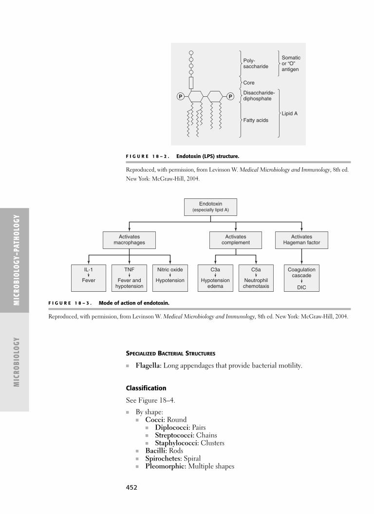
452
MIC
ROBI
OLO
GY–P
ATHO
LOGY
MIC
ROBI
OLO
GY
Activatesmacrophages
Activatescomplement
Endotoxin(especially lipid A)
ActivatesHageman factor
IL-1 TNF Nitric oxide
Fever Fever andhypotension
Hypotension
C3a C5a
Hypotensionedema
Neutrophilchemotaxis
Coagulationcascade
DIC
F I G U R E 1 8 – 3 . Mode of action of endotoxin.
Reproduced, with permission, from Levinson W. Medical Microbiology and Immunology, 8th ed. New York: McGraw-Hill, 2004.
SPECIALIZED BACTERIAL STRUCTURES
■ Flagella: Long appendages that provide bacterial motility.
Classification
See Figure 18–4.
■ By shape:■ Cocci: Round
■ Diplococci: Pairs■ Streptococci: Chains■ Staphylococci: Clusters
■ Bacilli: Rods■ Spirochetes: Spiral■ Pleomorphic: Multiple shapes
PDisaccharide-diphosphate
Core
Fatty acidsLipid A
Somaticor “O”antigen
Poly-saccharide
P
F I G U R E 1 8 – 2 . Endotoxin (LPS) structure.
Reproduced, with permission, from Levinson W. Medical Microbiology and Immunology, 8th ed.
New York: McGraw-Hill, 2004.

453
■ By diagnostic stain (based on cell wall type) (see Table 18–9).■ Gram stain: Separates almost all bacteria into two categories (see
Figure 18–5):■ Gram positive.■ Gram negative.
■ Acid-fast stain: Stains Mycobacteria sp.
MICRO
BIOLOGY–PATHO
LOGYM
ICROBIO
LOGY
Mycobacteria cell walls
contain mostly mycolic acid,
and very little peptidoglycan.
Bacteria
Rigid, thick-walledFlexible, thin-walled
(Spirochetes)No cell wall
(Mycoplasma sp.)
Obligate intracellular bacteria(Rickettsia and Chlamydia sp.)
Extracellular bacteria
Acid-fast(Mycobacteria sp.)
Gram positive Gram negative
Non-spore-formingbacilli
(Actinomyces, Listeria,Corynebacterium sp.)
Spore-forming bacilli(Bacillus and
Clostridium sp.)
Cocci(Neisseria sp.)
Bacilli
Aerobic(Pseudomonas sp.)
Facultative(Escherichia,Salmonella,
Helicobacter sp.)
Anaerobic(Bacteroides sp.)
Cocci(Streptococcus,
Staphylococcus sp.)
F I G U R E 1 8 – 4 . Classification of bacteria.
T A B L E 1 8 – 9 . Comparison of Gram-Positive and Gram-Negative Bacteria
GRAM STAIN PEPTIDOGLYCAN MAJOR WALL PERIPLASMIC
STAIN COLOR WALL CONSTITUENT SPACE ENDOTOXIN
Gram (+) Purple Thick Lipoteichoic No No
acid (LTA)
Gram (–) Pink Thin Lipopolysac- Yes Yes (LPS)
charide (LPS)

454
MIC
ROBI
OLO
GY–P
ATHO
LOGY
MIC
ROBI
OLO
GY
■ By oxygen requirements:■ Obligate aerobic: Require oxygen for growth.■ Obligate anaerobic: Cannot grow in oxygenated environments because
they lack superoxide dismutase (rids O2 radicals) and/or catalase (ridsH2O2).
■ Facultative anaerobic: Grow aerobically when oxygen is present, butuse fermentation pathways in its absence.
Bacteria Superoxide Dismutase Catalase Peroxidase
Obligate aerobes + + –Most facultative anaerobes
Most aerotolerant anaerobes + – +
Obligate anaserobes – – –
■ By spore production:■ Spores are thick-walled cells produced only by certain gram-positive
rods: Bacillus (aerobic) and Clostridium (anaerobic) species. ■ They are formed in low-nutrient states (during the stationary phase of
bacterial cell growth) and remain dormant, often in soil. ■ When nutrients are restored, they germinate to form new bacteria. ■ Spores are extremely heat resistant.
■ By pH Range■ Acidophiles: Thrive in acidic environment (pH < 2).■ Neutrophiles: Thrive in neutral pH environment.■ Alkaliphiles: Thrive in basic environment (pH > 9).
Only sterilization procedures
(autoclaving, ethylene oxide
gas, etc) are sporicidal.
Gram positive
Cellwall
Cytoplasmicmembrane
Teichoicacid
Unique to
gram-positive
organisms
Unique to
gram-negative
organisms
Peptidoglycan
CapsuleEndotoxin/LPS lipid(outer membrane)
Periplasmic space(location of manyβ-lactamases)
Pilus
Flagellum
Common to both
F I G U R E 1 8 – 5 . Comparison of Gram-positive and Gram-negative cell walls.
Adapted, with permission, from Levinson W, Jawetz E. Medical Microbiology and Immunology: Examination and BoardReview. 9th ed. New York: McGraw-Hill, 2006.

455
MICRO
BIOLOGY–PATHO
LOGYM
ICROBIO
LOGY
Gram-positive lab algorithm
Gram stain
Gram + (purple/blue)
Clostridium (anaerobe)CorynebacteriumListeriaBacillus
S. pneumoniaeCapsule ( + quellung)Optochin sensitive, bile soluble
Cocci Rods (bacilli)
Catalase +(clusters)Staphylococcus
Catalase –(chains)Streptococcus
Coagulase +S. aureus
Important pathogens are in bold type.
Note: Enterococcus is either α- or γ-hemolytic.
Coagulase –Novobiocin sensitiveS. epidermidisNovobiocin resistantS. saprophyticus
Hemolysis
No hemolysis
Partia
l hem
olysis
(gre
en)
Completehemolysis
(clear)
αViridans streptococci(eg, S. mutans)No capsuleOptochin resistant, not bile soluble
Group AS. pyogenesBacitracin sensitive
Group B(S. agalactiae)Bacitracin resistant
Enterococcus(E. faecatis)
andPeptostreptococcus
(anaerobe)Group D strep
β
γ
F I G U R E 1 8 – 6 . Gram-positive bacteria.
Reproduced, with permission, from Le T, Bhushan V, Vasan N. First Aid for the USMLE Step 1 2011. 21st ed. New York:McGraw-Hill, 2011.
GRAM-POSITIVE COCCI
■ Streptococci■ Grow in pairs or chains.■ Many are part of the normal human flora.■ Catalase (–).■ Classified by lysis of erythrocytes (hemolysis) when plated on blood
agar. (See Tables 18–10 and 18–11.)■ Beta-hemolytic Strep are further classified by Lancefield groups
(Group A, B, C, F, G, etc.). Many group D Strep have since beenreclassified as enterococci.
■ Staphylococci■ Grow in grapelike clusters.■ Produce pyogenic (suppurative) infections.■ Catalase (+): rids H2O2.
See Table 18–12.
Lancefield group is
determined by the
C carbohydrate composition
of the cell wall.
Streptococcihemolysis:
Alpha: Almost(incomplete)
Beta: Best (complete)Gamma: Garbage
(none)
Medically Relevant Bacteria
Gram-Positive Bacteria (Figure 18–6)

456
MIC
ROBI
OLO
GY–P
ATHO
LOGY
MIC
ROBI
OLO
GY
T A B L E 1 8 – 1 1 . Comparison of the Major Streptococci
HEMOLYSIS SPECIES MAJOR VIRULENCE FACTORS DISEASE
Alpha S. pneumoniae Capsule Meningitis
■ Lancet-shaped Pneumolysin Otitis media (children)
diplococcus IgA protease Pneumonia
Sinusitis
Alpha Viridans group: Normal flora of oropharynx Caries
S. mutans Endocarditis
S. sanguis
Beta (Group A) S. pyogenes M protein Pyogenic infections
Hyaluronidase Pharyngitis
Streptokinase Cellulitis
Erythrogenic toxin Impetigo
Streptolysin O and S Scarlet fever
Exotoxin A and B Rheumatic fever
Glomerulonephritis
Beta (Group B) S. agalactiae Capsule Neonatal pneumonia
Neonatal meningitis
Neonatal sepsis
Gamma (Group D) S. bovis Normal colon flora Subacute endocarditis
(assoc. with colon cancer)
Gamma (Group D) Enterococci: Normal colon flora UTI
E. faecalis Subacute endocardidits
E. faecium
T A B L E 1 8 – 1 0 . Hemolytic Classification
CLASSIFICATION HEMOLYSIS BLOOD AGAR APPEARANCE
Alpha Incomplete Green ring around colonies
Beta Complete Clear area of hemolysis
Gamma None No hemolysis
GRAM-POSITIVE BACILLI
See Table 18–13.
■ Spore-forming.■ Non-spore-forming.
Spores contain dipicolinic acid
in their core. Killed by
autoclave sterilization.

457
MICRO
BIOLOGY–PATHO
LOGYM
ICROBIO
LOGY
T A B L E 1 8 – 1 3 . Comparison of Major Gram-Positive Bacilli
MAJOR
OXYGEN VIRULENCE
BACTERIUM REQUIREMENT FACTORS DISEASE
Spore-forming
Bacillus anthracis Aerobic Anthrax toxin Anthrax
Polypeptide capsule ■ Cutaneous: black eschar
(has D-glutamate) (painless ulcer)
■ Pulmonary: inhalation
of spores
Bacillus Facultative Enterotoxins Food poisoning
cereus ■ Ingestion of reheated
grains and rice (fried rice)
Clostridium Anaerobic Botulinum toxin Botulism
botulinum (neurotoxin) ■ CN/muscle paralysis
■ Most potent ■ Respiratory failure
bacterial toxin ■ Ingestion of undercooked
■ Botox canned foods, fish,
derives from ham, pork
exotoxin A
T A B L E 1 8 – 1 2 . Comparison of Major Staphylococci
MAJOR
VIRULENCE
SPECIES COAGULASE HEMOLYSIS FACTORS DISEASE
S. aureus + Beta Protein A Abscess
a-lactamase Pneumonia
Enterotoxin Toxic shock
TSST syndrome
Exfoliatin Scalded skin
Hyaluronidase syndrome
Staphylokinase Food poisoning
MRSA
Endocarditis
Osteomyelitis
S. epidermidis – Gamma Normal skin flora Infection of IV
catheters and
prosthetic devices
S. saprophyticus – Gamma Normal vaginal flora UTI
(Continued)

458
MIC
ROBI
OLO
GY–P
ATHO
LOGY
MIC
ROBI
OLO
GY
T A B L E 1 8 – 1 3 . Comparison of Major Gram-Positive Bacilli (Continued)
MAJOR
OXYGEN VIRULENCE
BACTERIUM REQUIREMENT FACTORS DISEASE
Clostridium Anaerobic Tetanus toxin Tetanus
tetani (neurotoxin) ■ Associated with puncture
wounds
■ Spastic paralysis
■ Trismus (lockjaw)
Clostridium Anaerobic Alpha toxin Gas gangrene
perfringens (lecithinase) ■ Necrotizing faciitis
■ Myonecrosis
Food poisoning
■ Ingestion of reheated
meats
Clostridium Anaerobic Exotoxin A and B Pseudomembranous colitis
difficile ■ Often secondary to
antibiotic use (clindamycin)
Non-spore-forming
Corynebacterium Aerobic Diphtheria toxin Diphtheria
diphtheriae ■ Pseudomembranous
■ Club-shaped pharyngitis (grayish-white
membrane on tonsils)
Listeria Facultative Listeriolysin O Neonatal meningitis
monocytogenes Endotoxin ■ Vaginal transmission
■ Motile via actin during birth
rockets Gastroenteritis
■ Ingestion of unpasteurized
milk/cheese and deli meats
Actinomyces Anaerobic Normal oral flora Actinomycosis
israelii ■ Slow-growing, lumpy
orofacial abscesses
■ Characteristic sulfur
granules in colonies

459
MICRO
BIOLOGY–PATHO
LOGYM
ICROBIO
LOGY
T A B L E 1 8 – 1 4 . Comparison of Major Gram-Negative Cocci
OXYGEN MAJOR VIRULENCE
BACTERIUM REQUIREMENT FACTORS DISEASE
Neisseria Aerobic Capsule Meningitis (adolescents)
meningitidis Endotoxin (LPS) Waterhouse–Friderichsen syndrome
■ Vaccine IgA protease
■ Low prevalence
■ High mortality
Neisseria Aerobic Endotoxin (LOS) Gonorrhea (STD):
gonorrhoeae Fimbriae ■ Urethritis: burning sensation during
■ No vaccine IgA protease urination
■ High prevalence ■ �: Vaginal discharge, PID
■ Low mortality ■ �: Epididymitis
■ Symptoms start 2–10 days after intercourse
Pelvic inflammatory disease (PID)
Neonatal conjunctivitis
Septic arthritis
Gram-negative lab algorithm
Gram stain
Gram – (pink)
CocciNeisseria meningitidis,
N. gonorrhoeae
“Coccoid” rodsHaemophilus influenzae (X + V factors)Pasteurella—animal bitesBrucella—brucellosisBordetella pertussisdairy product, animals
Rods
Important pathogens are in bold type.
MaltosefermenterN. meningitidis
Oxidase +glucose fermenters Oxidase +
PseudomonasOxidase –ShigellaSalmonellaProteus
MaltosenonfermenterN. gonorrhoeae
Lactosefermenter
Lactosenonfermenter
FastfermenterKlebsiellaE. coliEnterobacter
SlowfermenterCitrobacterSerratiaOthers
Lactose
Oxidase
F I G U R E 1 8 – 7 . Gram-negative bacteria.
Reproduced, with permission, from Le T, Bhushan V, Tolles J. First Aid for the USMLE Step 1 2011. 20th ed.New York: McGraw-Hill, 2010.
Gram-Negative Bacteria (Figure 18–7)
GRAM-NEGATIVE COCCI
See Table 18–14.

460
MIC
ROBI
OLO
GY–P
ATHO
LOGY
MIC
ROBI
OLO
GY
GRAM-NEGATIVE BACILLI
See Table 18–15.
■ Enteric■ Respiratory■ Zoonotic
T A B L E 1 8 – 1 5 . Comparison of Major Gram-Negative Bacilli
BACTERIUM OXYGEN REQUIREMENT MAJOR VIRULENCE FACTORS DISEASE
Enteric bacilli (associated with the enteric tract)
Escherichia coli Facultative Heat-labile toxin UTI
Enterotoxin Dysentery
Endotoxin Traveler’s diarrhea
Neonatal meningitis
Septic shock
Salmonella sp. Facultative Endotoxin Enterocolitis
■ Flagella ■ Transmission: animals
(eggs, poultry, pets)
Typhoid fever
■ Septicemia → osteomyelitis
■ Often in patients with
sickle cell anemia
Shigella sp. Facultative Enterotoxin Enterocolitis
Endotoxin Dysentery
■ Bloody diarrhea
Vibrio cholerae Facultative Cholera toxin Cholera
■ Comma-shaped Enterotoxin ■ Watery diarrhea
Endotoxin
Campylobacter jejuni Facultative Enterotoxin Enterocolitis (children)
■ Comma-shaped Endotoxin
Helicobacter pylori Facultative Endotoxin Gastritis
Peptic ulcers
Gastric carcinoma (associated)
Klebsiella pneumoniae Facultative Capsule Pneumonia
Endotoxin ■ Assoc. with chronic
respiratory disease,
alcoholism, or diabetes
UTI (nosocomial)
(Continued)

461
MICRO
BIOLOGY–PATHO
LOGYM
ICROBIO
LOGY
T A B L E 1 8 – 1 5 . Comparison of Major Gram-Negative Bacilli (Continued)
BACTERIUM OXYGEN REQUIREMENT MAJOR VIRULENCE FACTORS DISEASE
Pseudomonas Aerobic Exotoxin A Pneumonia (cystic fibrosis)
aeruginosa Endotoxin Sepsis (burn infections)
■ Produces a blue-green External otitis (swimmer’s ear)
pigment in culture UTI
Diabetic osteomyelitis
Bacteroides sp. Anaerobic Endotoxin Abscess
Fimbriae Periodontitis (associated)
Respiratory bacilli (associated with respiratory tract)
Haemophilus influenzae Facultative Capsule Epiglottitis
Endotoxin Meningitis (children)
IgA protease Otitis media
Pneumonia
Haemophilus aegyptius Facultative Endotoxin (LOS) Acute conjunctivitis (pink eye)
■ Koch-Weeks bacillus ■ Transmission: hand-to-hand contact
Brazilian purpuric fever
Legionella pneumophila Facultative Endotoxin Legionnaires’ disease
■ Silver stain > Gram stain ■ Pneumonia and fever
■ Transmission: environmental
water sources (air conditioners)
■ Patients tend to be older,
smokers, and alcoholics
Bordetella pertussis Aerobic Pertussis toxin Pertussis (whooping cough)
■ Fimbriae Tracheal cytotoxin
Endotoxin
Zoonotic bacilli (transmitted by animals)
Brucella sp. Facultative Endotoxin Brucellosis (undulant fever)
■ Transmission: dairy products;
contact with animals (goats,
sheep, pigs, cattle)
Francisella tularensis Facultative Endotoxin Tularemia
■ Transmission: ticks; contact
with wild animals (rabbits, deer)
Yersinia pestis Facultative Exotoxin Plague
Endotoxin ■ Transmission: fleas; rodents
F-1, V, and (prairie dogs, rats)
W antigens
Pasteurella multocida Facultative Endotoxin Cellulitis
■ Transmission: animal bites
(cats, dogs)

462
MYCOBACTERIA
■ Aerobic, nonmotile bacilli.■ Cell wall constituents:
■ Peptidoglycan: Prevents osmotic lysis.■ Mycolic acid: Impedes chemical entry; resists phagocytosis. Waxy coat-
ing.■ Surface proteins: Adhesins.■ Periplasm: Contains enzymes for nutrient breakdown.
■ Stain with acid-fast stain (carbolfuchsin): Red against blue background.
See Table 18–16.
OTHER BACTERIAL TYPES
(See Table 18–17.)
■ Mycoplasma sp.: Lack a cell wall.■ Chlamydia sp.: Obligate intracellular bacteria (cannot make own ATP).■ Rickettsia sp.: Obligate intracellular bacteria (need host CoA and NAD+).■ Spirochetes: Spiral-shaped.
MIC
ROBI
OLO
GY–P
ATHO
LOGY
MIC
ROBI
OLO
GY
T A B L E 1 8 – 1 6 . Comparison of Major Mycobacteria Species
MAJOR VIRULENCE
FACTORS CHARACTERISTICS DISEASE
Mycobacterium Cord factor Inhalation of airborne Tuberculosis:
tuberculosis Tuberculoproteins droplets ■ Fever, night sweats, weight
Treatment: rifampin, loss, hemoptysis
isoniazid, and 1° TB: granulomatous lesions and hilar
pyrazinamide lymphadenopathy (Ghon complex)
PPD skin test elicits type IV in lungs
(delayed) hypersensitivity 2° TB: caseous granulomas which may
reaction lead to miliary or disseminated infection
Mycobacterium Lepromin Reservoir in US: armadillos Leprosy:
leprae proteins Treatment: dapsone, rifampin Tuberculoid type: Cell-mediated immune
response and granulomas in nerves
Lepromatous type: Foam cells containing
bacteria in skin
If PPD(+): Indicates current TB
infection, past exposure, or
BCG vaccinated.
If PPD(–): Indicates no
infection, or anergic.

463
MICRO
BIOLOGY–PATHO
LOGYM
ICROBIO
LOGY
T A B L E 1 8 – 1 7 . Comparison of Wall-Less, Obligate Intracellular, and Spirochete Bacteria
BACTERIUM CHARACTERISTICS DISEASE
Wall-less bacteria
Mycoplasma Smallest bacterium Atypical “walking”
pneumoniae No cell wall; cell membrane contains pneumonia
cholesterol
Obligate intracellular bacteria
Chlamydia Cannot make its own ATP Trachoma:
trachomatis Cell wall lacks muramic acid ■ Chronic infection
Forms cytoplasmic inclusions ■ Inclusion conjunctivitis (IC)
Most common cause of preventable ■ Blindness
blindness (can be contracted in Chlamydia (STD):
a swimming pool) ■ Urethritis/PID
Most common cause of STDs ■ Neonatal conjunctivitis
Rickettsia rickettsii Need host CoA and NAD+ Rocky Mountain spotted
Causes vasculitis, headache, fever fever
Positive Weil-Felix reaction ■ Rash: palms and soles
Transmission: ticks
Rickettsia Need host CoA and NAD+ Epidemic typhus
prowazekii Causes vasculitis, headache, fever ■ Rash: central →Positive Weil-Felix reaction peripheral (no
Transmission: lice palms/soles)
Coxiella burnetii An atypical rickettsia Q fever
Need host CoA and NAD+ ■ Pneumonia
Negative Weil-Felix reaction
Transmission: inhaled aerosols
Spirochetes
Treponema Visualized by dark-field microscopy Syphilis:
pallidum Congenital syphilis: CN VII deafness, 1°: Painless chancre (ulcer)
Hutchinson’s incisors, mulberry molars at site of local contact
Treatment: penicillin 2°: Highly infectious
maculopapular rash,
condylomata lata, mucous
patch
3°: Gummas (granulomas)
often on tongue or palate;
neurosyphilis; Argyll-
Robertson pupil
Borrelia Visualized using aniline dyes (Wright’s Lyme disease:
burgdorferi or Giemsa stain) with light microscopy Stage 1: Erythema migrans
Transmission: ticks (require deer) (“bull’s eye” rash)
Most often occurs in CT, NY, PA, NJ Stage 2: Neuropathies
Treatment: doxycycline (Bell's palsy)
Stage 3: Arthritis and
CNS disease

464
Bacterial Vaccines
Bacterial vaccinations can confer two types of acquired immunity. (See alsoChapter 21, “Immunology and Immunopathology.”)
MIC
ROBI
OLO
GY–P
ATHO
LOGY
MIC
ROBI
OLO
GY
Antibiotic Drugs
See Table 8–18.
CLASSIFICATION
■ By spectrum:■ Broad: Effective against several types of bacteria.■ Narrow: Effective against only one or a few types of bacteria.
■ By activity:■ Bacteriocidal: Kills bacteria.■ Bacteriostatic: Inhibits bacterial growth (host immune cells kill bacteria).
PENICILLINS
■ Inhibit peptidoglycan cross-linking by blocking transpeptidase during laststage of cell wall synthesis
■ Bacteriocidal■ Contain b-lactam rings, which are cleaved by bacterial b-lactamase (peni-
cillinase), inactivating the drug
Concomitant administration of
bacteriocidal and
bacteriostatic antibiotics has
an antagonistic effect because
each interferes with the
other’s mechanism of action.
The DPT vaccine is given to all
children in the US. It is a
toxoid vaccine.
ConferredVaccine Type Immunity Treated Diseases
Live attenuated Active TBTyphoid feverTularemia
Killed bacteria Active Cholera■ Whole bacteria Typhus
PlagueQ fever
Toxoid Active Diphtheria■ Inactivated endotoxin Pertussis
Tetanus
Capsular polysaccharide Active Pneumonia (Strep. pneumoniae)Meningitis (N. meningitidis and
H. influenzae)
Purified protein Active PertussisLyme diseaseAnthrax
Antitoxin Passive Diphtheria■ Preformed antibody to Tetanus
endotoxin Botulism
Toxoid: inactivated bacterial
exotoxin.
Antitoxin: antibody to
bacterial exotoxin.
Most antibacterial activity
occurs during the log phase
of bacterial growth.

465
MICRO
BIOLOGY–PATHO
LOGYM
ICROBIO
LOGY
T A B L E 1 8 – 1 8 . Comparison of Common Antibiotics
PREGNANCY
DRUG CLASS MECHANISM OF ACTION ACTIVITY SPECTRUM SAFE
Vancomycin Inhibits peptidoglycan Cidal Narrow Yes
cross-linking by binding to
D-alanyl-D-alanine during
cell wall synthesis
Penicillins Inhibit peptidoglycan cross-linking Cidal Narrow Yes
by blocking transpeptidase during ↓
last stage of cell wall synthesis Broader
Cephalosporins Inhibit peptidoglycan cross-linking Cidal Narrow Yes
by blocking transpeptidase during ↓
last stage of cell wall synthesis Broader
Metronidazole Inhibits DNA synthesis Cidal Narrow No
Fluoroquinolones Inhibit DNA gyrase (topoisomerase) Cidal Broader No
Aminoglycosides Inhibit protein synthesis by binding Cidal Broader No
to 30S ribosomal subunits (block
the formation of the initiation
complex)
Macrolides Inhibit protein synthesis by binding Static Narrow Yes
to 50S ribosomal subunits (block
the release of tRNA)
Clindamycin Inhibit protein synthesis by binding Static Narrow Yes
to 50S ribosomal subunits (blocks
the release of tRNA)
Chloramphenicol Inhibits protein synthesis by Static Broad No
binding to 50S ribosomal
subunits (blocks peptidyl
transferase)
Tetracyclines Inhibit protein synthesis by binding Static Broad No
to 30S ribosomal subunits (block
aminoacyl-tRNA binding)
Sulfonamides Inhibit folic acid synthesis by Static Broad No
competing with p-aminobenzoic
acid (PABA)
Buy AT 30, CCELL at 50:
30S: Aminoglycocides,
Tetracyclines
50S: Clindamycin,
Chloramphenicol,
Erythromycin, Lincomycin,
Linezolid

466
■ 10% risk of hypersensitivity■ First generation
■ Narrow spectrum: G(+) cocci/bacilli and some G(–) aerobic cocci■ Penicillin G (IV)■ Penicillin V (PO)
■ Second generation (b-lactamase resistant)■ Narrow spectrum: Target G (+) b-lactamase-producing staphylococci
(especially S. aureus)■ Methicillin■ Nafcillin■ Oxacillin■ Cloxacillin■ Dicloxacillin
■ Third generation (extended spectrum)■ Broader spectrum: Also include some G(–) bacilli.
■ Ampicillin (IV)■ Amoxicillin (PO)
■ Combinations■ An extended spectrum penicillin with a b-lactamase inhibitor.■ Broader spectrum: Target more G(+) bacilli including b-lactamase
staphylococci (especially S. aureus)■ Amoxicillin and clavulanic acid■ Ampicillin and sublactam
CEPHALOSPORINS
■ Inhibit peptidoglycan cross-linking by blocking transpeptidase during laststage of cell wall synthesis
■ Bacteriocidal■ Contain b-lactam rings■ 10% cross-hypersensitivity with penicillins
■ First generation■ Narrow spectrum: G(+) cocci (but not enterococci) and some G(–)
bacilli■ Cefazolin■ Cephalexin■ Cefadroxil
■ Second generation■ Broader spectrum: Fewer G(+) cocci, but more G(–) bacilli.
■ Cefaclor■ Cefprozil■ Cefoxitin
■ Third generation■ Broader spectrum: Fewer G(+) cocci (except some enterococci),
but more G(–) bacilli, and anaerobes■ Cefpodoxime■ Cefixime
■ Fourth generation■ Broader spectrum: Fewer G(+) cocci (except more enterococci),
but more G(–) bacilli, and anaerobes■ Cefepime
VANCOMYCIN
■ Inhibits peptidoglycan cross-linking by binding to D-alanyl-D-alanine duringcell wall synthesis
MIC
ROBI
OLO
GY–P
ATHO
LOGY
MIC
ROBI
OLO
GY
467
MICRO
BIOLOGY–PATHO
LOGYM
ICROBIO
LOGY
■ Bacteriocidal■ Treatment of choice for MRSA■ Narrow spectrum: Mostly G(+) cocci and bacilli (especially penicillinase-
resistant S. aureus)
MACROLIDES
■ Inhibit protein synthesis by binding to 50S ribosomal subunits (block therelease of tRNA)
■ Bacteriostatic■ Associated with GI upset
■ Erythromycin■ Narrow spectrum: G(+) cocci/bacilli, some G(–) anaerobes, and
Mycoplasma sp.■ Inhibits cytochrome P450
■ Clarithromycin■ Broader spectrum: Targets more anaerobes
■ Azithromycin■ Broader spectrum: Targets more anaerobes
CLINDAMYCIN
■ Inhibit protein synthesis by binding to 50S ribosomal subunits (blocks therelease of tRNA)
■ Bacteriostatic (in low doses)■ Narrow spectrum: G(+) and some G(–) anaerobes (Bacteroides sp.)■ Associated with diarrhea and pseudomembranous colitis (caused by an
overgrowth of Clostridium difficile)
TETRACYCLINES
■ Inhibit protein synthesis by binding to 30S ribosomal subunits (block aminoacyl-tRNA binding)
■ Bacteriostatic■ Broad spectrum: G(+) and G(–) aerobes/anaerobes, spirochetes, Mycoplasma
sp., Chlamydia, and Rickettsia■ Divalent and trivalent cations inhibit absorption■ Associated with staining of teeth during their calcification
■ Tetracycline■ Doxycycline■ Minocycline
METRONIDAZOLE
■ Inhibits DNA synthesis■ Bacteriocidal■ Narrow spectrum: Targets anaerobes and some protozoa■ Disulfuram-like reaction with alcohol■ Often used to treat pseudomembranous colitis
FLUOROQUINOLONES
■ Inhibit DNA gyrase (topoisomerase)■ Bacteriocidal■ Disulfuram-like reaction with alcohol■ Broader spectrum: G(+) and G(–) aerobes/facultatives and mycobacteria,
but not anaerobes

468
■ Divalent and trivalent cations inhibit absorption■ Ciprofloxacin■ Ofloxacin
AMINOGLYCOSIDES
■ Inhibit protein synthesis by binding to 30S ribosomal subunits (block theformation of the initiation complex)
■ Bacteriocidal (in clinical doses)■ Broader spectrum: G(+) and G(−) aerobes; ineffective against anaerobes■ Associated with ototoxicity and nephrotoxicity; teratogens
■ Streptomycin■ Gentamycin
SULFONAMIDES
■ Inhibit folic acid synthesis by competing with p-aminobenzoic acid (PABA)■ Bacteriostatic■ Broad spectrum: Targets G(+), many G(−), Actinomyces sp., and Chlamydia sp.■ Associated with hypersensitivity, renal toxicity, and hematopoietic toxicity
■ Sulfadiazine■ Sulfamethoxazole■ Trimethoprim
CHLORAMPHENICOL
■ Inhibits protein synthesis by binding to 50S ribosomal subunits (blockspeptidyl transferase)
■ Bacteriostatic■ Broad spectrum: Some G(+) cocci, G(–) aerobes/anaerobes, spirochetes,
Rickettsia sp., Chlamydia sp., Mycoplasma sp., and Salmonella sp.■ Associated with bone marrow toxicity, gray baby syndrome
TOPICAL ANTIBIOTICS
■ Neomycin: An aminoglycoside■ Polymyxin B: Alters cell membrane permeability■ Bacitracin: Inhibits cell wall synthesis
ANTIBIOTIC PROPHYLAXIS PRIOR TO DENTAL TREATMENT
See Table 18–19.
■ Prophylaxis is recommended for all dental procedures that involve manip-ulation of gingival tissues or periapical regions of the teeth, or perforationof the oral mucosa.
■ Prophylaxis recommendations are for patients with:■ Heart conditions that may predispose them for infective endocarditis (IE).■ Total joint replacement that may be at risk for infection at the site of
the prosthesis.■ The American Dental Association (ADA) has standardized the current
guidelines for IE prevention in conjunction with the American HeartAssociation (AHA).
■ The ADA and the American Association of Orthopedic Surgeons (AAOS)do not have a joint recommendation at this time.
MIC
ROBI
OLO
GY–P
ATHO
LOGY
MIC
ROBI
OLO
GY

469
MICRO
BIOLOGY–PATHO
LOGYM
ICROBIO
LOGY
T A B L E 1 8 – 1 9 . Guidelines for Antibiotic Prophylaxis
SITUATION DRUG DOSAGE
Standard prophylaxis Amoxicillin Adults: 2 g PO 1 h before procedure.
Children: 50 mg/kg PO 1 h before procedure.
Unable to take oral Ampicillin Adults: 2 g IM or IV within 30 min before
medications procedure.
Children: 50 mg/kg IM or IV within 30 min before procedure.
Allergic to penicillins Clindamycin Adults: 600 mg PO 1 h before procedure.
Children: 20 mg/kg PO 1 h before procedure.
Azithromycin or Adults: 500 mg PO 1 h before procedure.
Clarithromycin Children: 15 mg/kg PO 1 h before procedure.
Cephalexin or Adults: 2 g PO 1 h before procedure.
Cefadroxil Children: 50 mg/kg PO 1 h before procedure.
Allergic to penicillins Clindamycin Adults: 600 mg IM or IV within 30 min before
and unable to take procedure.
oral medications. Children: 20 mg/kg IM or IV within 30 min before procedure.
Cefazolin Adults: 1 g IM or IV within 30 min before
procedure.
Children: 50 mg/kg IM or IV within 30 min
before procedure.
Patient Selection
See Table 18–20.
T A B L E 1 8 – 2 0 . Antibiotic Prophylaxis for Patients at Risk for IE
PROPHYLAXIS NECESSARY PROPHYLAXIS NOT NECESSARY
Artificial heart valves Mitral valve prolapse
History of previous IE Rheumatic heart disease
Heart transplant that develops a valve problem Calcified aortic stenosis
Unrepaired or incompletely repaired cyanotic congenital heart Congenital ventricular septal defect
disease, including those with palliative shunts and conduits
Repaired congenital heart defect with a prosthetic device Congenital atrial septal defect
during the first 6 months after the procedure
Any repaired congenital heart defect with residual defect at Congenital hypertrophic cardiomyopathy
the site of or adjacent to the site of a prosthetic device

470
MIC
ROBI
OLO
GY–P
ATHO
LOGY
MIC
ROBI
OLO
GY
■ Nucleocapsid: nucleic acid core + protein capsid. ■ Some viruses have an outer membrane called an envelope, which is com-
posed of plasma membrane lipoproteins and glycoproteins obtained asthe virus leaves its host cell (budding).
■ Do not contain mitochondria or ribosomes; must replicate within livinghost cells.
■ Not visible by light microscopy.
Matrix protein
Nucleocapsid
Lipid envelope
Glycoprotein spikes
CapsidNucleocapsidCore
Capsomer
Nucleic acidcore
A
B
F I G U R E 1 8 – 8 . Comparison of nonenveloped and enveloped viruses.
Reproduced, with permission, from Levinson W. Medical Microbiology and Immunology, 8th ed.
New York: McGraw-Hill, 2004.
Viroid: Single molecule of
circular RNA without a protein
envelope. Causes plant
diseases.
� VI ROLOGY
Viruses
See Figure 18–8.
■ Virion: Infectious; complete virus particle (DNA/RNA + proteins).■ Characterized by single- or double-stranded DNA or RNA (never both)
surrounded by a protein capsid.■ Repeating polypeptide subunits (capsomeres).■ Protects viral genome from extracellular nucleases.■ Essential for infectivity of virus.■ Antigenic; provokes host immune response.■ Serves as attachment protein in nonenveloped viruses.
All viruses are haploid (1 copy
of RNA/DNA) except
retroviruses, which are diploid.
All RNA viruses (except
retrovirus and
orthomyxovirus) replicate in
the cytoplasm using their own
RNA polymerase.
All DNA viruses (except
poxvirus) replicate in the
nucleus using host RNA
polymerase.

471
MICRO
BIOLOGY–PATHO
LOGYM
ICROBIO
LOGY
Pathogenesis
VIRAL GROWTH CURVE
See Figure 18–9.
■ Latent phase: Viral penetration → viral release 10–12 hours.■ Eclipse phase: Viral penetration → viral assembly within host cell.
■ No virus can be detected during this phase.■ Rise phase: Viral assembly → viral release.
VIRAL REPLICATION
See Figure 18–10.
■ Attachment: Determined by the specificity of viral proteins to host cells.■ Penetration: Via receptor-mediated endocytosis (eg, pinocytosis).■ Uncoating: Viral nucleic acid is spilled into the cytoplasm.■ Transcription and Translation:
■ All DNA viruses (except poxviruses) replicate in the nucleus using hostcell RNA polymerase.
■ All RNA viruses (except retroviruses and orthomyxoviruses) replicatein the cytoplasm using their own RNA polymerase. Transcription isonly necessary for viruses whose RNA has negative polarity (a virus’sRNA that has positive polarity serves as the mRNA itself).
■ All retroviruses use their own reverse transcriptase for transcription.■ Assembly: The new viral nucleic acid and capsid proteins are packaged.■ Release: Either by budding through the host plasma membrane (creating
a viral envelope) or by host plasma membrane rupture.
Rise periodEclipse period
Hours
Nucleic acid
00.01
0.1
1
10
100
1000
1 2 3 4 5 6 7 8 9 10 11 12
Virus
Yie
ld
Viri
ons/
Cel
l (
)
Vira
l nuc
leic
aci
d (
)F I G U R E 1 8 – 9 . Viral growth curve.
Reproduced, with permission, from Levinson W. Medical Microbiology and Immunology, 8th ed.
New York: McGraw-Hill, 2004.
One virion can replicate to
form hundreds of progeny
viruses.
The host cellular morphologic
and functional changes
associated with viral
replication and release are
known as the cytopathic
effect (CPE). CPE is often
specific for a particular virus.
Not all viruses cause CPE.
Roboviruses are rodent-
borne viruses (rodent
excrement): hantavirus
(Sin Nombe virus).
Arboviruses are arthropod-
borne viruses (mosquitoes,
ticks): West Nile virus, yellow
fever virus, Dengue virus,
Colorado tick fever virus, and
Eastern/Western encephalitis
virus.
Poliovirus, coxsackieviruses,
and hepatitis A virus are
enteroviruses.

472
MIC
ROBI
OLO
GY–P
ATHO
LOGY
MIC
ROBI
OLO
GY
F I G U R E 1 8 – 1 0 . Viral growth cycle.
Reproduced, with permission, from Levinson W. Medical Microbiology and Immunology, 8th ed. New York: McGraw-Hill, 2004.
F I G U R E 1 8 – 1 1 . HIV.
Reproduced, with permission, from Bhushan V, et al. First Aid for the USMLE Step 1: 2006.
New York: McGraw-Hill, 2006.
HIV contains two strands of
RNA (diploid) and reverse
transcriptase. The two
important envelope proteins are
gp120 (mediates attachment to
CD4) and gp41 (mediates
fusion to the host cell). The
nucleocapsid is composed of
p24 and p7. (See Figure 18–11.)

473
MICRO
BIOLOGY–PATHO
LOGYM
ICROBIO
LOGY
VIRAL ANTIGENIC CHANGES
■ Antigenic changes contribute to the cause of epidemics and pandemics. ■ Commonly associated with influenza viruses (orthomyxoviruses).■ There are two modes of antigenic change:
■ Antigenic drifts: Minor changes caused by genomic mutations.■ Antigenic shifts: Major changes caused by genomic re-assortment.
Classification
RNA NONENVELOPED VIRUSES
See Table 18–21.
T A B L E 1 8 – 2 1 . RNA Nonenveloped Viruses in Order of Increasing Size
ANTIVIRAL
FAMILY NUCLEIC ACID VIRUS DISEASE TREATMENT VACCINE
Picornavirus Single, Linear Poliovirus Polio None Yes
■ Smallest ■ Salk
RNA virus ■ Sabin
Echovirus Aseptic meningitis None No
Rhinovirus Common cold None No
Coxsackie A virus Herpangina: None No
■ Soft palate, posterior
pharynx
Hand-foot-and-mouth
disease:
■ Palms, soles, anterior
oral mucosa
Acute lymphonodular
pharyngitis
Aseptic meningitis
Coxsackie B virus Pleurodynia None No
Myocarditis
Pericarditis
Aseptic meningitis
HAV Hepatitis A None Yes
Calicivirus Single, Linear Norwalk virus Gastroenteritis (adults) None No
Hepevirus Single, Linear HEV Hepatitis E None No
Reovirus Double, Linear Rotavirus Gastroenteritis (infants) None No
Coltivirus Colorado tick fever None No
■ Only virus
transmitted
by ticks
Influenza viruses have two
envelope glycoprotein spikes,
hemagglutinin and
neuraminidase, which exhibit
the majority of antigenic
changes.

474
MIC
ROBI
OLO
GY–P
ATHO
LOGY
MIC
ROBI
OLO
GY
■ Picornavirus■ Calicivirus■ Hepevirus■ Reovirus
RNA-ENVELOPED VIRUSES
See Table 18–22.
■ Deltavirus■ Flavivirus■ Togavirus■ Orthomyxovirus■ Retrovirus■ Paramyxovirus■ Rhabdovirus■ Filovirus■ Coronavirus■ Bunyavirus
T A B L E 1 8 – 2 2 . RNA-Enveloped Viruses in Order of Increasing Size
ANTIVIRAL
FAMILY NUCLEIC ACID VIRUS DISEASE TREATMENT VACCINE
Deltavirus Single, circular HDV Hepatitis D α-interferon Yes
Flavivirus Single, linear Japanese Encephalitis None Yes
encephalitis
virus
Yellow fever Yellow fever None Yes
virus
West Nile virus Encephalitis None No
Dengue virus Dengue fever None No
HCV Hepatitis C α-interferon No
Hepatocellular Ribavirin
carcinoma (associated)
Togavirus Single, linear Rubella virus Rubella: (German, None Yes
“3-day” measles)
Truncal rash
Teratogen
Eastern/Western Encephalitis None No
encephalitis virus
(Continued)
Poliovirus, coxsackieviruses,
echoviruses, and HAV are
enteroviruses: they primarily
infect the enteric tract.

475
MICRO
BIOLOGY–PATHO
LOGYM
ICROBIO
LOGY
T A B L E 1 8 – 2 2 . RNA-Enveloped Viruses in Order of Increasing Size (Continued)
ANTIVIRAL
FAMILY NUCLEIC ACID VIRUS DISEASE TREATMENT VACCINE
Orthomyxovirus Single, linear Influenza viruses Influenza Amantidine Yes
■ Exhibit various Reye’s syndrome Rimantidine
antigenic (association) Zanamivir
changes Oseltamivir
■ Replicate in
nucleus
Retrovirus Single, linear HTLV Adult T-cell leukemia/ None No
■ Uses reverse lymphoma
transcriptase Chronic progressive
■ Replicate in myelopathy
nucleus
■ Diploid
HIV AIDS Zidovudine (AZT) No
■ Infects CD4 Lamivudine
T-helper cells Stavudine
■ Diagnosis Indinavir
confirmed by Ritonavir
Western blot
Paramyxovirus Single, linear Measles virus Measles (rubeola) None Yes
■ Nonpruritic
maculopapular
brick-red rash
■ Multiple white lesions
(Koplik’s spots) are
often seen on the
buccal mucosa
Mumps virus Mumps None Yes
■ Parotitis, orchitis,
deafness
RSV Bronchiolitis Ribavirin No
■ Infants only Pneumonia
Parainfluenza Croup None No
viruses Bronchiolitis
Common cold
Rhabdovirus Single, linear Rabies virus Rabies None Yes
Filovirus Single, linear Ebola virus Ebola None No
Hemorrhagic fever
Coronavirus Single, linear Coronavirus Common cold None No
SARS
Bunyavirus Single, circular Hantavirus (Sin Hantavirus None No
Nombre virus) Hemorrhagic fever

476
DNA NONENVELOPED VIRUSES
See Table 18–23.
■ Parvovirus■ Papillomavirus■ Polyomavirus■ Adenovirus
DNA-ENVELOPED VIRUSES
See Table 18–24.
■ Hepadnavirus■ Herpesvirus■ Poxvirus
MIC
ROBI
OLO
GY–P
ATHO
LOGY
MIC
ROBI
OLO
GY
T A B L E 1 8 – 2 3 . Major DNA Nonenveloped Viruses in Order of Increasing Size
NUCLEIC ANTIVIRAL
FAMILY ACID VIRUS DISEASE TREATMENT VACCINE
Parvovirus Single, B19 virus Aplastic anemia None No
■ Smallest linear ■ Sickle cell disease
DNA associated
virus Erythema
infectiosum
(fifth disease)
Fetal infections
Papillomavirus* Double, HPV Papillomas (warts) Podophyllin Yes
circular Condyloma a-interferon
acuminatum Cidofovir
(genital warts)
Verruca vulgaris
Cervical cancer
Polyomavirus* Double, JC virus Progressive multifocal None No
Circular leukoencephalopathy
(PML)
■ HIV associated
Adenovirus Double, Adenovirus Pharyngitis None Yes
linear Conjunctivitis
Pneumonia
Common cold
* Formerly classified as papovaviruses.
Reye’s syndrome is apotentially deadly disease that
typically occurs in childrenaged 4–12 years. It is
associated with the use ofaspirin to treat influenza or
varicella (chickenpox).

477
MICRO
BIOLOGY–PATHO
LOGYM
ICROBIO
LOGY
T A B L E 1 8 – 2 4 . Major DNA-Enveloped Viruses in Order of Increasing Size
NUCLEIC ANTIVIRAL
FAMILY ACID VIRUS DISEASE TREATMENT VACCINE
Hepadnavirus Double, circular HBV Hepatitis B α-interferon Yes
■ Has reverse ■ Hepatocellular Lamivudine
transcriptase carcinoma
(associated)
Herpesvirus Double, linear HSV-1 Herpes labialis Acyclovir No
■ Lies dormant in Keratoconjunctivitis Penciclovir
sensory nerve ganglia, Gingivostomatitis Valacyclovir
esp. trigeminal Recurrent encephalitis Famciclovir
ganglion
■ Obtains envelope HSV-2 Herpes genitalis Acyclovir
from host nuclear Neonatal encephalitis Penciclovir
membrane (not Aseptic meningitis Valacyclovir
plasma membrane) Famciclovir
VZV Varicella—primary Acyclovir Yes
■ Chickenpox Famciclovir
■ Pruritic, macular Valacyclovir
lesions that become
pustular and crusted
■ Reye’s syndrome
(associated)
Zoster—recurrent
■ Shingles
■ Usually localized to
a single dermatome
CMV Congenital abnormalities Ganciclovir No
■ Primary viral cause of Valganciclovir
mental retardation Foscarnet
Cytomegalic inclusion
disease
EBV Infectious None No
mononucleosis (self-limiting in
■ Transmission via saliva 2–3 weeks)
■ Splenomegaly, necrotizing
pharyngitis ↑ abnormal
lymphocytes
■ Heterophile test
used for screening
Burkitt’s lymphoma
(association)
Nasopharyngeal
carcinoma (association)
B-cell lymphoma
(association)
Hairy leukoplakia
(association)
HHV-8 Kaposi’s sarcoma None No
(Continued)
■ Tzanck test assay
for HSV-1, HSV-2,
and VZV

478
MIC
ROBI
OLO
GY–P
ATHO
LOGY
MIC
ROBI
OLO
GY
T A B L E 1 8 – 2 4 . Major DNA-Enveloped Viruses in Order of Increasing Size (Continued)
NUCLEIC ANTIVIRAL
FAMILY ACID VIRUS DISEASE TREATMENT VACCINE
Poxvirus Double, linear Variola virus Smallpox None Yes
■ Largest DNA virus ■ Eradicated
■ Replicates in cytoplasm
MCV Molluscum None No
contagiosum
Hepatitis Viruses
CLASSIFICATION
See Table 18–25.
SIGNS AND SYMPTOMS OF VIRAL HEPATITIS
■ Fatigue■ Myalgia■ Loss of appetite■ Nausea■ Diarrhea■ Constipation■ Fever■ Jaundice
TESTS OF LIVER FUNCTION
■ Bilirubin■ ALT (alanine aminotransferase)■ AST (aspartate aminotransferase)
SEROLOGY
See Figure 18–12 and Table 18–26.
T A B L E 1 8 – 2 5 . Comparison of Major Hepatitis Viruses
NUCLEIC VIRAL
VIRUS ACID CLASS ENVELOPE TRANSMISSION CHARACTERISTICS VACCINE
HAV ss RNA Picornavirus No Fecal-oral Usually self-limiting; recovery within 4 months. Yes
HBV ds DNA Hepadnavirus Yes Bloodborne Increased incidence of chronic liver disease, cirrhosis, HCC. Yes
HCV ss RNA Flavivirus Yes Bloodborne Increased incidence of chronic liver disease, cirrhosis, HCC. No
HDV ss RNA Deltavirus Yes Bloodborne Requires presence of HBsAg for replication. Yes
HEV ss RNA Calicivirus No Fecal-oral Causes occasional epidemics in underdeveloped countries. No
HCC: hepatocellular carcinoma.
Viral hepatitis: ALT > AST
Alcoholic hepatitis: AST > ALT
Hepatitis viruses are extremely
heat resistant (more so than
HIV). Proper autoclaving kills
all hepatitis viruses.

479
MICRO
BIOLOGY–PATHO
LOGYM
ICROBIO
LOGY
F I G U R E 1 8 – 1 2 . Serologic findings associated with acute HBV.
Reproduced, with permission, from Bhushan V, et al. First Aid for the USMLE Step 1: 2006.
New York: McGraw-Hill, 2006.
HCV is the most common
reason for liver
transplantation in the
United States.
An individual vaccinated for
HBV will show serology
positive only for anti-HBs.
T A B L E 1 8 – 2 6 . Serological Profiles of Hepatitis Infections
ACUTE CHRONIC PERCENT
VIRUS INFECTION DISEASE IMMUNITY CHRONICITY
HAV IgM Anti-HAV None Anti-HAV None
HBV IgM Anti-HBc IgG Anti-HBc Anti-HBs 10% (adults)
HBsAg HBsAg 30–90% (infants)
HBeAg
HCV Anti-HCV Anti-HCV Anti-HCV 80%
HDV IgM Anti-HDV Anti-HDV Anti-HDV 6%
HEV IgM Anti-HEV None Anti-HEV None

480
MIC
ROBI
OLO
GY–P
ATHO
LOGY
MIC
ROBI
OLO
GY
Viral Vaccines
Vaccinations can confer two types of acquired immunity. (See also Chapter 21,“Immunology and Immunopathology.”)■ Viral vaccinations can confer two types of acquired immunity.■ Live attenuated vaccines provide longer-lasting and broader range of
immunity than killed vaccines, but they have the potential to revert tovirulence, or can be excreted and transmitted to nonimmune individuals.
■ Live attenuated vaccines should not be given to immunocompromisedindividuals or pregnant women.
■ Preformed antibody vaccines provide immediate protection, but only for afew weeks-months.
■ Most viral vaccines are given pre-exposure; however, vaccines for rabiesand hepatitis B may be given post-exposure since their incubation periodsare so long.
Vaccine Type Conferred Immunity Protection Treated Diseases
Live attenuated Active Slow onset Measles■ IgA Longest lasting Mumps■ IgG Rubella■ TC-cell Polio (Sabin): oral
ChickenpoxSmallpox
Killed virus Active Slow onset Rabies■ IgG Long lasting Influenza
Polio (Salk): injectionHepatitis A
Purified viral Active Slow onset Hepatitis Bprotein subunits Long lasting ■ via HBsAg
Preformed antibody Passive Fast onset RabiesShort life span Hepatitis B
Shingles
Antiviral Drugs
INTERFERONS
■ Glycoproteins that originate from infected host cells to protect other non-infected host cells.
■ Do not directly affect viruses, but instead nonspecifically prevent theirreplication within host cells.
■ Block various stages of viral RNA/DNA synthesis.■ INF-α: Treats chronic hepatitis B and C.■ Toxicity: neutropenia.
Bacteriophages (Phages)
■ Viruses that infect bacterial cells. ■ Replication can occur by two pathways (see Figure 18–13).
■ Lytic cycle: The process by which some phages replicate within thehost cell, producing hundreds of new progeny phage. The host cell isultimately destroyed.
Adjuvants are often added to
vaccines to slow their
absorption and increase their
effectiveness.
Enterobacteria phage T4:
phage that infects E. coli.

481
MICRO
BIOLOGY–PATHO
LOGYM
ICROBIO
LOGY
■ Lysogenic cycle: The process by which some phages incorporate theirDNA in the host cell chromosome. The integrated viral DNA is calleda prophage. Replication occurs only when the host DNA is damaged,excising the viral DNA. The host cell is usually not destroyed.
Lysogenic Conversion
■ Alteration of the host bacterium to a pathogenic strain via expression ofthe integrated prophage genes (Figure 18--14). Eg, Corynebacteriumdiphtheriae producing diphtheria toxin.
Prions
■ Infectious agents composed entirely of protein (no nucleic acid). ■ Do not elicit inflammatory or antibody responses.■ Cause transmissible spongiform encephalopathy:
■ Creutzfeldt–Jakob disease (in humans). ■ Mad cow disease (in cows).
λ phage DNAexcised fromintegrated stateand λ phagesynthesized
λ phage DNAintegrates intoE. coli DNAand no progenyphage are madeat this time
Release ofλ phageand lysisof E. coli
Release ofλ phageand lysisof E. coli
Synthesisof manyprogenyλ phage
UV irradiation
Choice Lysogenic cycleLytic cycle
Prophage
Bacterial chromosome
λ bacteriophageinfects E. coli
F I G U R E 1 8 – 1 3 . Bacteriophage replication.
Reproduced, with permission, from Levinson W. Medical Microbiology and Immunology, 8th ed.
New York: McGraw-Hill, 2004.

482
MIC
ROBI
OLO
GY–P
ATHO
LOGY
MIC
ROBI
OLO
GY
� MYCOLOGY
Fungi
■ Gram (+), eukaryotic microorganisms. ■ All are either obligate (the majority) or facultative aerobes.■ Cell membranes: lipid bilayer contains ergosterol.■ Cell walls: carbohydrate and protein (chitin).■ Capsule (if present): polysaccharide coating.■ Laboratory diagnosis by KOH preparation.
FUNGAL REPRODUCTION
■ Sexual: Mating and formation of spores.■ Zygospores: Single, large spores with thick walls.■ Ascospores: Formed in a sac (ascus).■ Basidiospores: Formed on the tip of a pedestal (basidium).
■ Asexual: Budding and formation of conidia (asexual spores).■ Arthrospores: Formed by fragmentation of the ends of hyphae.■ Chlamydospores: Rounded, thick-walled, and highly resistant.■ Blastospores: Formed by budding process.■ Sporangiospores: Formed on a stalk within a sac (sporangium).
Pathogenesis
■ Fungal infection: Leads to a largely cell-mediated immune response(type IV hypersensitivity reaction) and granuloma formation.
■ Mycotoxicosis: Induced by ingestion of fungal toxins. ■ Allergic response: Type I hypersensitivity reactions to inhalation of fungal
spores.
Classification
See Table 18–27.
■ Yeasts■ Molds
Some fungi are dimorphic—
they exist as molds at ambient
temperatures but as yeasts at
warmer (body) temperatures.
Most fungal spores and
conidia are killed at
temperatures > 80°C
for 30 minutes.
Most antifungal drugs target
the ergosterol component of
fungal cell membranes,
altering their permeability.
Aflatoxins are hepato-
carcinogenic toxins produced
by Aspergillus flavus,
generally found in
contaminated grains and
peanuts.
Bacterialchromosome
Diphtheria toxin genes integrated into chromosome of C. diphtheriae;bacterium becomespathogenic.
β phage genes
β phage carryingdiphtheria toxin geneinfects C. diphtheriaenot lysogenized byβ phage; bacterium is nonpathogenic priorto infection by β phage.
β phage genes
Diphtheriatoxin gene
Diphtheriatoxin
Lysogenic conversion
F I G U R E 1 8 – 1 4 . Lysogenic conversion.
Reproduced, with permission, from Levinson W. Medical Microbiology and Immunology,8th ed. New York: McGraw-Hill, 2004.

483
MICRO
BIOLOGY–PATHO
LOGYM
ICROBIO
LOGY
T A B L E 1 8 – 2 7 . Comparison of Yeasts and Molds
FUNGUS MORPHOLOGY REPRODUCTION
Yeasts Single cells Asexual budding
Molds Hyphae (long filaments), which form a Cell division
mat-like structure (mycelium)
MEDICALLY RELEVANT FUNGI
■ Systemic fungal infections. (See Table 18–28.)■ Cutaneous fungal infections (See Table 18–29).■ Opportunistic fungal infections. (See Table 18–30.)
T A B L E 1 8 – 2 8 . Major Systemic Fungal Infections
FUNGUS DISEASE TYPE CHARACTERISTICS TREATMENT
Blastomyces Blastomycosis Dimorphic Endemic in eastern US and Itraconazole
dermatitidis Central America. Amphotericin B
Inhalation of microconidia
produces granulomatous
nodules in lungs and
respiratory infection.
Coccidioides immitis Coccidioidomycosis Dimorphic Endemic in southwest Amphotericin B
(desert valley fever) US and Latin America. Itraconazole
(San Joaquin fever) Inhalation of arthrospores Ketoconazole
produces respiratory Fluconazole
infection.
Histoplasma Histoplasmosis Dimorphic Endemic in Ohio and Itraconazole
capsulatum Mississippi River valleys. Amphotericin B
Found in soil often Fluconazole
contaminated by bird/bat
droppings.
Inhalation of microconidia
produces respiratory infection.
Yeast cells located within
host macrophages.
Most systemic mycoses can
mimic TB infection – they form
granulomas.
T A B L E 1 8 – 2 9 . Major Cutaneous Fungal Infections
FUNGUS DISEASE TYPE CHARACTERISTICS TREATMENT
Trichophyton sp. Dermatophytosis Molds Tinea corporus (ringworm) Miconazole
Epidermophyton sp. Tinea capitis (scalp itch) Clotrimazole
Microsporum sp. Tinea cruris (jock itch) Tolnaftate
Tinea pedis (athlete’s foot) Griseofulvin
Tinea unguium (nail fungus)

484
MIC
ROBI
OLO
GY–P
ATHO
LOGY
MIC
ROBI
OLO
GY
T A B L E 1 8 – 3 0 . Major Opportunistic Fungal Infections
FUNGUS DISEASE TYPE CHARACTERISTICS SUSCEPTIBILITY
Aspergillus fumigatus Aspergillosis Mold Inhalation of conidia causes AIDS
respiratory infection and Organ
aspergilloma (fungus ball) transplantation
formation in lungs.
Candida albicans Candidiasis Yeast Part of normal human flora AIDS
Vaginitis of mouth, vagina, GI tract, Prolonged use of
Angular cheilitis and skin. antibiotics
Median rhomboid Appears as budding yeasts
glossitis or pseudohyphae.
Cryptococcus Cryptococcosis Yeast Heavily encapsulated. AIDS
neoformans Inhalation of spores causes
respiratory infection,
meningitis, and pneumonia.
Mucor sp. Mucormycosis Molds Inhalation of conidia causes Diabetes
Rhizopus sp. respiratory, skin, paranasal (ketoacidosis)
Absidia sp. sinus, and brain infections. Leukemia
Burns
Antifungal Drugs
See Table 18–31.
■ The vast majority of the antifungal drugs prescribed by dentists are used totreat candidiasis, the major fungal infection that affects the oral cavity.
T A B L E 1 8 – 3 1 . Common Antifungal Drugs to Treat Candidiasis
DRUG MECHANISM OF ACTION COMMON FORMS
Nystatin Binds to ergosterol. Topical
Oral suspension
Amphotericin B Binds to ergosterol. Topical
Oral suspension
Intravenous
Clotrimazole Inhibits ergosterol synthesis. Troche
Ketoconazole Inhibits ergosterol synthesis. Tablet
Blocks fungal cytochrome P450.
Fluconazole Inhibits ergosterol synthesis. Tablet
Blocks fungal cytochrome P450.
Opportunistic infections
induce disease in
immunocompromised patients,
such as those with AIDS or
who take immunosuppressive
medications.

485
MICRO
BIOLOGY–PATHO
LOGYM
ICROBIO
LOGY
Parasites
Sarcodina(Amoebas)
Sporozoa(Sporozoans)
Mastigophora(Flagellates)
Ciliata(Ciliates)
ProtozoaMetazoa
(Helminths)
Nemathelminthes(Roundworms)
Platyhelminthes(Flatworms)
Trematoda(Flukes)
Cestoda(Tapeworms)
F I G U R E 1 8 – 1 5 . Phylogeny of parasites.
Reproduced, with permission, from Levinson W. Medical Microbiology and Immunology, 8th ed.
New York: McGraw-Hill, 2004.
� PARASITOLOGY
Parasites
CLASSIFICATION
■ Protozoa■ Unicellular, eukaryotic microorganisms that lack a cell wall and largely
infect blood cells, intestinal and urogenital tissue, and meninges.■ Metazoa (helminths)
■ Multicellular worms that often infect the intestines, brain, liver, andother tissues.
PHYLOGENY
See Figure 18–15.
Parasitic infections generally
elicit an IgE-mediated host
immune response
accompanied by marked
eosinophilia.

486
MIC
ROBI
OLO
GY–P
ATHO
LOGY
MIC
ROBI
OLO
GY
T A B L E 1 8 – 3 2 . Common Protozoa Associated with Human Infection
PROTOZOAN DISEASE TRANSMISSION MORPHOLOGY TREATMENT
Entamoeba Amebiasis: Fecal-oral Motile trophozoite (intestinal). Metronidazole
histolytica Dysentery Nonmotile cyst (contaminated
Liver abscess food/water).
Giardia lamblia Giardiasis: Fecal-oral Motile trophozoite (intestinal). Metronidazole
Diarrhea Nonmotile cyst (contaminated
Flatulence food/water).
Common in US.
Cryptosporidium Cryptosporidiosis: Fecal-oral Associated with immuno- Preventive
parvum Diarrhea suppression (AIDS)
Trichomonas Trichomoniasis: STD Motile trophozoite. Metronidazole
vaginalis Vaginitis (�)
Urethritis (�)
Plasmodium vivax Malaria Female Sporozoite. Chloroquine
Plasmodium ovale (influenza-like mosquitoes Merozoite (liver). Mefloquine
Plasmodium onset): (Anopheles) Hypnozoite (latent). Primaquine
malariae Fever Infected blood Causes lysis of erythrocytes.
Plasmodium Headache products
falciparum Anemia
(most fatal) Splenomegaly
Toxoplasma Toxoplasmosis: Fecal-oral Motile trophozoite (intestinal). Sulfadiazine
gondii CNS infection Transplacental Nonmotile cyst (uncooked Pyrimethamine
Encephalitis meat and cat feces).
Seizures
Associated with AIDS
Pneumocystis Pneumonia Inhalation Fungus (originally classified Trimethoprim
jiroveci as protozoan). Sulfamethoxazole
(formerly Associated with Pentamidine
P. carinii ) immuno-suppression (AIDS).
Found in oral cavity, but not
pathologic.
MEDICALLY RELEVANT PARASITES
■ Protozoa. (See Table 18–32.)■ Metazoa (helminths). (See Table 18–33.)

487
MICRO
BIOLOGY–PATHO
LOGYM
ICROBIO
LOGY
T A B L E 1 8 – 3 3 . Common Metazoa (Helminths) Associated with Human Infection
METAZOAN DISEASE TRANSMISSION CHARACTERISTICS TREATMENT
Taenia solium Taeniasis Ingestion of Cysticercosis Praziquantel
(tapeworm) undercooked (muscle cysts). Albendazole
pork. Neurocysticercosis
(brain cysts).
Taenia Taeniasis Ingestion of Does not cause Praziquantel
saginata (tapeworm) undercooked cysticercosis.
beef.
Enterobius Enterobiasis Ingestion of Most common Mebendazole
vermicularis (pinworm) worm eggs. worm in US.
Associated
with perianal
pruritis.
Trichinella Trichinosis Ingestion of Larvae only Mebendazole
spiralis (roundworm) undercooked grow in striated
meat (pork, wild muscle.
game). Associated
with muscle
pain, periorbital
edema, fever.

This page intentionally left blank

C H A P T E R 1 9
Oral Microbiology and Pathology
Oral Microbiology 490
PLAQUE 490
CALCULUS 491
MATERIA ALBA 491
Oral Pathology 491
CARIES 491
PLAQUE-INDUCED GINGIVITIS 492
CHRONIC PERIODONTITIS 492
AGGRESSIVE PERIODONTITIS 493
NECROTIZING PERIODONTAL DISEASES 494
CANDIDIASIS 494
Plaque Control 495
MECHANICAL 495
CHEMICAL 495
489

490
MIC
ROBI
OLO
GY–P
ATHO
LOGY
ORA
L M
ICRO
BIO
LOGY
AN
DPA
THO
LOGY
� ORAL M IC ROBIOLOGY
■ Even though the oral cavity harbors more than 300 species of bacteria,most are part of the normal oral flora. A few, however, are heavily associatedwith both dental and periodontal diseases.
Plaque
■ An organized biofilm consisting of 80% water and 20% solids: 95%microorganisms; and 5% organic components (polysaccharides, proteins,glycoproteins), inorganic components (calcium, phosphorus), desqua-mated cells (epithelial cells, leukocytes), and food debris.
■ It adheres to teeth, dental prostheses, and oral mucosal surfaces and is alsofound in the gingival sulcus and periodontal pockets.
■ Dental plaque is classified as supragingival and subgingival based on itsposition along the tooth surface. (See Table 19–1.)
■ As plaque matures, there is a transition from the early aerobic environ-ment characterized by gram-positive facultative species to an exceedinglyoxygen-deprived milieu in which gram-negative anaerobes predominate.
■ It is the key etiologic agent in the initiation of both caries and periodontaldiseases.
PLAQUE FORMATION
■ Pellicle formation: Salivary and GCF glycoproteins bind to oral mucosal,tooth, and dental prosthesis surfaces almost immediately via electrostaticand van der Waals forces. It prevents tissue desiccation and provides surfacelubrication, but also promotes bacterial adherence.
■ Bacterial colonization: Occurs within a few hours of pellicle formation.Gram-positive facultative species (Streptococcus sp., Actinomyces sp.,Lactobacillus sp.) are the first to colonize through the binding of theiradhesins and fimbriae to the pellicle.
T A B L E 1 9 – 1 . Comparison of Supragingival and Subgingival Plaque
DOMINANT AFFECTED BY
BACTERIAL DIET AND
PLAQUE LOCATION FORM SPECIES SALIVA
Supragingival At or coronal Attached to Gram-positive Yes
to the gingival gingival facultative
margin epithelial cocci
and tooth
surfaces
Subgingival Apical to the Attached to Gram-negative No
gingival gingival anaerobic
margin epithelial and bacilli and
tooth surfaces, spirochetes
but also loosely
adherent within
the sulcus
Periodontal health is
characterized by the
predominant inhabitants
of the oral cavity, mostly
gram-positive facultative
species such as Streptococcus
sanguis, Streptococcus mitis,
Streptococcus salivarius,
Actinomyces viscosus,
Actinomyces naeslundii,
and a few beneficial
gram-negative species
such as Veillonella parvula
and Capnocytophaga
ochracea.
As a plaque biofilm extends
from a supragingival to a
subgingival environment,
there is a shift in the microbial
flora from mostly gram-
positive, facultative cocci to
predominantly gram-negative,
anaerobic bacilli and
spirochetes.
Streptococcus salivarius is the
microorganism most
commonly found on the
surface of the tongue.
MICRO
BIOLOGY–PATHO
LOGYO
RAL MICRO
BIOLOGY AN
DPATHO
LOGY
491
■ Maturation: Multiplication and coaggregation of bacterial species that donot initially colonize tooth and gingival epithelial surfaces.
■ Mineralization: Calculus formation. Plaque becomes 50% mineralized inabout 2 days, and 90% mineralized in about 12 days.
Calculus
■ Calcified bacterial plaque that forms on teeth and dental prostheses.■ 70–90% of calculus is composed of inorganic components (calcium, phos-
phorus), the majority of which are crystalline (hydroxyapatite).■ The remaining 10–30% of calculus is organic, consisting of protein—
carbohydrate complexes, desquamated cells (epithelial cells, leukocytes),and microorganisms.
■ Saliva is the main mineral source for supragingival calculus, but GCFprovides most of the mineral for subgingival calculus.
■ The most common locations for supragingival calculus are the lingual ofmandibular anterior teeth and the buccal of maxillary molars (due to theirproximity to salivary ducts).
CALCULUS FORMATION
■ Epitactic concept: The predominant theory of calculus formation, whichsuggests that seeding agents (protein–carbohydrate complexes or bacteria)induce small foci of mineralization, which ultimately enlarge and coalesceto form a calcified mass.
Materia Alba
■ Loosely adherent matter largely composed of desquamated cells, food debris,and other components of dental plaque that is easily washed away.
� ORAL PATHOLOGY
Caries
CHARACTERISTICS
■ Cariogenic bacteria synthesize glucans (dextrans) and fructans (levans)from their metabolism of dietary sucrose (via glucosyltransferase), whichcontribute to their adherence to tooth surfaces.
■ As a consequence, lactic acid is formed, reducing salivary pH and creatingsites of enamel demineralization and cavitation.
STEPHAN CURVE
See Figure 19–1.
■ Rapid drop in salivary pH within a few minutes after fermentable carbohy-drate (eg, sucrose) intake.
■ Enamel demineralization occurs once the pH falls below 5.5.■ Recovery to a normal salivary pH can take 15–40 minutes.■ The frequency of carbohydrate intake is more detrimental than the quantity
because it maintains a prolonged decrease in pH.
Calculus does not directly
cause gingival inflammation,
but provides a rough surface
for the continued
accumulation of
perio-pathogenic bacterial
plaque.
Caries formation needs:
■ Cariogenic bacteria
■ A susceptible surface
■ A fermentable
carbohydrate source

492
CLASSIFICATION
■ Pit and fissure■ Smooth-surface■ Root■ Recurrent
PREDOMINANT MICROBIAL FLORA
■ Generally gram-positive, facultative cocci and bacilli.■ They are acidogenic (make acid) and aciduric (tolerate living in acid).■ Streptococcus mutans is the primary etiologic agent of caries.
Plaque-Induced Gingivitis
CHARACTERISTICS
■ Presence of plaque.■ Absence of attachment loss.■ Gingival inflammation, starting at the gingival margin.■ Can be modified by systemic factors, medications, or malnutrition.■ Reversible with removal of plaque (and modifying factor).
PREDOMINANT MICROBIAL FLORA
■ Variable microbial pattern containing predominantly gram-positive andgram-negative facultative and anaerobic cocci, bacilli, and spirochetessuch as Streptococcus sanguis, Streptococcus mitis, Actinomyces viscosus,Actinomyces naeslundii, Peptostreptococcus micros, Fusobacterium nucleatum,Prevotella intermedia, and Campylobacter rectus.
Chronic Periodontitis
■ Formerly known as adult periodontitis.
MIC
ROBI
OLO
GY–P
ATHO
LOGY
ORA
L M
ICRO
BIO
LOGY
AN
DPA
THO
LOGY
7
6.5
6
5.5
5
pH
0 5 10 15 20 40
Minutes
F I G U R E 1 9 – 1 . The Stephan curve.
■ Streptococcus sp.(S. mutans, S. sanguis,S. salivarius) generallycause pit and fissure,smooth-surface, androot caries.
■ Lactobacillus sp.(L. casei, L. acidophilus)generally cause pit andfissure caries.
■ Actinomyces sp. (A. viscosus, A.naeslundii) generallycause root caries.
Gingivitis associated with
sex steroid fluctuations
(pregnancy, puberty,
menstrual cycle, oral
contraceptive use)
is associated with elevated
proportions of Prevotella
intermedia, which uses these
steroids as growth factors.
Recurrent caries occurs
around an existing
restoration. It may occur on
the crown or the root.

493
CHARACTERISTICS
■ Presence of plaque.■ Presence of attachment loss.■ Amount of periodontal destruction is consistent with presence of microbial
deposits (subgingival plaque and calculus).■ Most prevalent in adults but can occur in children and adolescents.■ Generally progresses at a slow to moderate rate but may have periods of
rapid progression.■ Can be modified by other local factors, systemic factors, medications,
smoking, or emotional stress.
PREDOMINANT MICROBIAL FLORA
■ Variable microbial pattern containing predominantly gram-negative,anaerobic bacilli and spirochetes such as Porphyromonas gingivalis,Tanerella forsythensus (formerly Bacteroides forsythus), Treponema denti-cola, Prevotella intermedia, Fusobacterium nucleatum, Eikenella corrodens,and Campylobacter rectus.
Aggressive Periodontitis
■ Formerly known as juvenile periodontitis or early-onset periodontitis.
CHARACTERISTICS
■ Presence of plaque.■ Presence of attachment loss.■ Amount of periodontal destruction is generally inconsistent with presence
of microbial deposits.■ Familial aggregation.■ Usually affects individuals younger than 30 years but can occur in older
patients.■ Generally progresses rapidly but may be self-arresting.■ Phagocyte abnormalities are common.■ Hyper-responsive monocyte/macrophage phenotype is common.■ Can be modified by other local factors, systemic factors, medications,
smoking, or emotional stress.
CLASSIFICATION
■ Localized: Localized to first molars and/or incisors. Typically a circumpubertalonset. Often self-limiting (“burns out”) in 20s.
■ Generalized: Affects at least three permanent teeth other than first molarsand incisors. Usually affects people <30 years old, but patients may be older.Often associated with systemic diseases (neutropenias, leukemias, etc).
PREDOMINANT MICROBIAL FLORA
■ Similar to chronic periodontitis with often elevated proportions of Aggregatibacteractinmomycetemcomitans (formerly Actinobacillus actinomycetemcomitans) and/orPorphyromonas gingivalis.
MICRO
BIOLOGY–PATHO
LOGYO
RAL MICRO
BIOLOGY AN
DPATHO
LOGY
Recurrent periodontitis
describes a recurrence of
periodontitis after successful
treatment.
Refractory periodontitis
describes periodontitis that
does not respond to
treatment.

494
Necrotizing Periodontal Diseases
■ Formerly known as trench mouth or Vincent’s disease.
CHARACTERISTICS
■ Presence of plaque.■ Interproximal gingival necrosis (“punched-out” papillae).■ Marginal gingival pseudomembrane formation.■ Attachment loss may or may not be present.■ Gingiva bleeds easily.■ Pain when brushing or eating.■ Bad breath (fetor oris).■ Commonly associated with emotional stress, malnutrition, smoking, or
immunosuppression (HIV).
CLASSIFICATION
■ Necrotizing ulcerative gingivitis (NUG): No attachment loss.■ Necrotizing ulcerative periodontitis (NUP): Attachment loss present.
PREDOMINANT MICROBIAL FLORA
■ Variable microbial flora with elevated proportions of spirochetes (Treponemasp.), Prevotella intermedia, Fusobacterium sp., and Selenomonas sp.
Candidiasis
CHARACTERISTICS
■ An opportunistic fungal infection.■ Commonly associated with immunosuppression (HIV), ill-fitting dentures,
chronic xerostomia (Sjögren’s syndrome), or prolonged use of antibiotics.
CLASSIFICATION
■ Pseudomembranous candidiasis (thrush): Whitish patches of desquama-tive epithelium, which can be easily wiped off, leaving a slightly bleedingsurface. Most common form.
■ Atrophic (erythematous) candidiasis: Painful bright-red, smooth, “beefy”lesions on the tongue, palate, or other mucosal surfaces, usually associatedwith ill-fitting dentures.
■ Chronic hyperplastic candidiasis: Asymptomatic whitish plaques com-monly found on the buccal mucosa near the commissures that cannot beremoved, resembling oral leukoplakia.
PREDOMINANT MICROBIAL FLORA
■ Candida albicans.
MIC
ROBI
OLO
GY–P
ATHO
LOGY
ORA
L M
ICRO
BIO
LOGY
AN
DPA
THO
LOGY
Spirochetes have been shown
to invade the sulcular
connective tissue in cases of
NUG.

495
MICRO
BIOLOGY–PATHO
LOGYO
RAL MICRO
BIOLOGY AN
DPATHO
LOGY
T A B L E 1 9 – 2 . Commercially Available Products Used for Chemical Plaque Control
CHEMICAL FORMULATION INHIBITS CHARACTERISTICS
Chlorhexidine Rinse Plaque Side effects: extrinsic staining,
gluconate Gingivitis altered taste perception,
(most potent and supragingival
inhibitor) calculus formation
Phenolic/essential Rinse Plaque Contains highest alcohol
oil compound Gingivitis content (up to 26.9%)
Triclosan Paste Plaque
Gel Gingivitis
Stannous fluoride Rinse Plaque Side effects: extrinsic
Gel Gingivitis staining
Anti-caries properties
Cetylpyridinium Rinse Plaque
chloride
Two mouthrinses are ADA
approved for the treatment of
gingivitis: Rx-only
chlorhexidine gluconate and
an OTC phenolic/essential oil
compound.
� PLAQU E CONTROL
Mechanical
■ Toothbrushing and flossing are proven methods to remove plaque.■ However, they cannot remove subgingival plaque in pockets >3 mm deep
(as in cases of periodontal disease). ■ Dental professionals must scale teeth and root plane in order to remove
the subgingival plaque and calculus.
Chemical
■ Chemical means of plaque control is often used as an adjunct to mechanicalcleaning.
■ See Table 19–2 for commercially available products for chemical plaquecontrol.
Chlorhexidine gluconate is
the most effective antiplaque
mouthrinse due to its
substantivity.

This page intentionally left blank

C H A P T E R 2 0
Reactions to TissueInjury
Inflammation and Repair 498
CELL INJURY 498
CELL DEATH 502
INFLAMMATION 503
INFLAMMATORY MEDIATORS 505
WOUND REPAIR 507
497

498
MIC
ROBI
OLO
GY–P
ATHO
LOGY
REAC
TIO
NS
TO T
ISSU
EIN
JURY
� I N FLAM MATION AN D RE PAI R
Cell Injury
MODES OF CELL INJURY
■ Hypoxia and ischemia.■ Physical trauma: Burns, frostbite, radiation, electric shock, etc.■ Microorganisms: Bacteria, viruses, fungi, parasites, etc.■ Immunologic reactions: Autoimmunity and anaphylaxis.■ Chemical / pharmacologic insult: Poisons, drugs, alcohol, etc.■ Nutritional imbalances: Vitamin deficiency, obesity, etc.■ Genetic defects: Hemoglobinopathies, storage diseases, etc.■ Aging: ↑ telomerase activity, inaccurate repair of DNA, etc.
CHEMICAL INJURY
See Table 20–1.
Heart, brain, and lungsare very vulnerable tohypoxia. Results from:
■ Vascular ischemia.■ ↓ blood oxygen (eg,
anemia, pulmonarydisease).
■ ↓ tissue perfusion (eg,shock, cardiac failure).
■ CO poisoning.
FREE RADICAL INJURY
■ Induced by activated oxygen species.■ Initiate autocatalytic reactions.■ Cellular damage:
■ Membrane lipid peroxidation■ Nucleic acid denaturation■ Cross-linking of proteins
■ Generated from:■ Redox reactions■ Radiation (UV light)■ Drugs and chemicals■ Reperfusion injury
■ Antioxidants:■ Superoxide dismutase (2O2 + 2H+ → H2O2 + O2)■ Catalase (2H2O2 → O2 + 2H2O)
T A B L E 2 0 – 1 . Chemical-Induced Cell Injury
CHEMICAL FINDINGS
Carbon monoxide (CO) Systemic hypoxia
Carbon tetrachloride (CCl4) Hepatocellular damage (”fatty liver”)
Mercury Renal tubular necrosis
Pneumonitis
GI ulceration
Gingival lesions
Cyanide Prevents cellular oxidation
Odor of bitter almonds
Methanol Blindness
Lead Basophilic stippling of RBCs

499
MICRO
BIOLOGY—PATHO
LOGYREACTIO
NS TO TISSUE
INJURY
■ Vitamin E■ Ceruloplasmin: Carries copper in blood
TYPES OF CELL INJURY
■ Reversible cell injury■ Cellular and organelle swelling (due to Ca2+ influx).■ Bleb formation.■ Ribosomal detachment from ER.■ Clumping of chromatin (due to ↓ pH).■ Increased lipid deposition (due to ↓ protein synthesis).
■ Irreversible cell injury ■ Extensive plasma membrane damage.■ Massive Ca2+ influx.■ Diminished oxidative phosphorylation within mitochondria (due to
accumulation of Ca2+-rich densities).■ Release of lysosomal enzymes into the cytoplasm (due to lysosomal
rupture).■ Nuclear fragmentation (karyorrhexis).■ Cell death (necrosis).
CELLULAR REACTIONS TO INJURY
See Table 20–2.
T A B L E 2 0 – 2 . Cellular Reactions to Injury
CELLULAR REACTION DEFINITION RESULTS
Atrophy Decrease in cell size Decrease in tissue or organ size
(usually secondary to decreased
workload, neurovascular supply,
nutrition, endocrine stimulation,
and increase in age)
Hypertrophy Increase in cell size Increase in tissue or organ size
(usually secondary to increased
tissue workload or endocrine
stimulation)
Aplasia Complete lack of cells Agenesis (tissue or organ absence)
Hypoplasia Decrease in cell numbers Decrease in tissue or organ size
Hyperplasia Increase in cell numbers Increase in tissue or organ size
(eg, glandular breast proliferation
associated with pregnancy)
Metaplasia Reversible, morphological Usually occurs in response to stress
change from one (eg, conversion of columnar
cell type to another epithelium to stratified squamous)
The outcome of cell injury
depends largely on the
severity and duration of the
insult, but also on the cell type
and its adaptive mechanisms.
Hypertrophy and hyperplasia can
occur simultaneously (eg, uterine
enlargement during pregnancy).
Gingival overgrowth isoften drug related:
■ Phenytoin (Dilantin)■ Ca2+ channel blockers
(eg, nifedipine)■ Cyclosporin

500
MIC
ROBI
OLO
GY–P
ATHO
LOGY
REAC
TIO
NS
TO T
ISSU
EIN
JURY
ENZYMATIC CELLULAR DEGRADATION
■ Autolysis: Cellular degradation caused by intracellular enzymes indige-nous to the cell itself.
■ Heterolysis: Cellular degradation caused by enzymes extrinsic to the cell.
ENDOGENOUS PIGMENTATION
See Table 20–3.
T A B L E 2 0 – 3 . Comparison of Endogenous Pigments
PIGMENT CHARACTERISTICS ASSOCIATED PATHOLOGY
Lipofuscin Yellow-brown color Brown atrophy
“Wear-and-tear” pigment Age
Derived from lipid
peroxidation
Has no effect on cell
function
Accumulates in heart,
liver, brain
Bilirubin Yellowish color Biliary tree obstruction
Major bile pigment Hepatocellular injury
Derived from heme Hemolytic anemias
Jaundice: Bilirubin
accumulation
Hemosiderin Golden brown color Hemosiderosis
(see Table 20–4) Derived from heme Hemochromatosis
Aggregates of ferritin
micelles (iron storage
sites)
Identified with Prussian
blue stain
Accumulates in
phagocytes of:
Bone marrow
Liver
Spleen
Melanin Brown-black color Increased melanin:
Derived from tyrosine Physiologic
Synthesized in melanocytes pigmentation
Accumulates in skin, eyes, Suntan
hair Addison’s disease
Decreased melanin:
Albinism
Vitiligo
Ceroid Derived from lipofuscin Hepatocellular injury
(via auto-oxidation)
Accumulates in hepatic
Kupffer cells
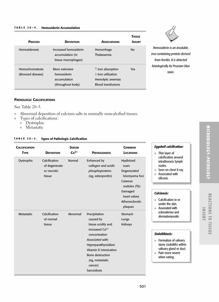
501
MICRO
BIOLOGY—PATHO
LOGYREACTIO
NS TO TISSUE
INJURY
PATHOLOGIC CALCIFICATIONS
See Table 20–5.
■ Abnormal deposition of calcium salts in normally noncalcified tissues.■ Types of calcifications:
■ Dystrophic■ Metastatic
T A B L E 2 0 – 5 . Types of Pathologic Calcification
CALCIFICATION SERUM COMMON
TYPE DEFINITION CA2+ PATHOGENESIS LOCATIONS
Dystrophic Calcification Normal Enhanced by Hyalinized
of degenerate collagen and acidic scars
or necrotic phosphoproteins Degenerated
tissue (eg, osteopontin) leiomyoma foci
Caseous
nodules (Tb)
Damaged
heart valves
Atherosclerotic
plaques
Metastatic Calcification Abnormal Precipitation Stomach
of normal caused by Lungs
tissue tissue acidity and Kidneys
increased Ca2+
concentration
Associated with:
Hyperparathyroidism
Vitamin D intoxication
Bone destruction
(eg, metastatic
cancer)
Sarcoidosis
Eggshell calcification:
■ Thin layer ofcalcification aroundintrathoracic lymphnodes.
■ Seen on chest X-ray.■ Associated with
silicosis.
Calcinosis:
■ Calcification in orunder the skin.
■ Associated withscleroderma anddermatomyositis.
T A B L E 2 0 – 4 . Hemosiderin Accumulation
TISSUE
PROCESS DEFINITION ASSOCIATIONS INJURY
Hemosiderosis Increased hemosiderin Hemorrhage No
accumulation (in Thalassemia
tissue macrophages)
Hemochromatosis More extensive ↑ Iron absorption Yes
(Bronzed disease) hemosiderin ↓ Iron utilization
accumulation Hemolytic anemias
(throughout body) Blood transfusions
Hemosiderin is an insoluble,
iron-containing protein derived
from ferritin. It is detected
histologically by Prussian blue
stain.
Sialolithiasis:
■ Formation of salivarystone (sialolith) withinsalivary gland or duct.
■ Pain more severewhen eating.
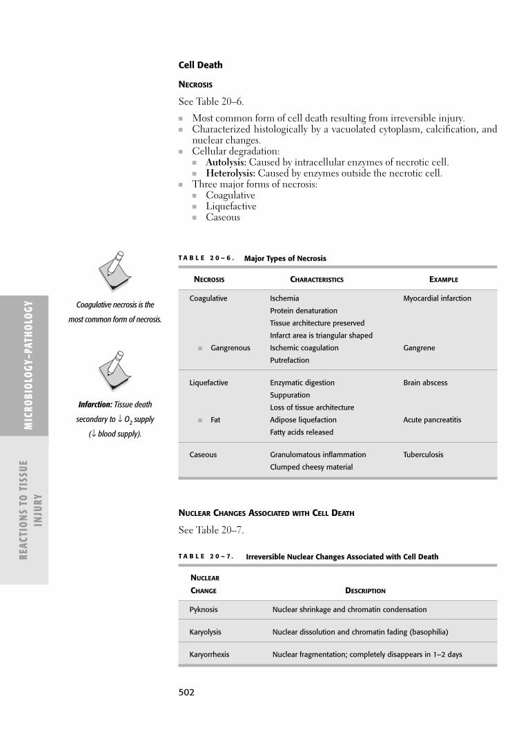
502
MIC
ROBI
OLO
GY–P
ATHO
LOGY
REAC
TIO
NS
TO T
ISSU
EIN
JURY
Cell Death
NECROSIS
See Table 20–6.
■ Most common form of cell death resulting from irreversible injury. ■ Characterized histologically by a vacuolated cytoplasm, calcification, and
nuclear changes.■ Cellular degradation:
■ Autolysis: Caused by intracellular enzymes of necrotic cell.■ Heterolysis: Caused by enzymes outside the necrotic cell.
■ Three major forms of necrosis:■ Coagulative■ Liquefactive■ Caseous
T A B L E 2 0 – 6 . Major Types of Necrosis
NECROSIS CHARACTERISTICS EXAMPLE
Coagulative Ischemia Myocardial infarction
Protein denaturation
Tissue architecture preserved
Infarct area is triangular shaped
■ Gangrenous Ischemic coagulation Gangrene
Putrefaction
Liquefactive Enzymatic digestion Brain abscess
Suppuration
Loss of tissue architecture
■ Fat Adipose liquefaction Acute pancreatitis
Fatty acids released
Caseous Granulomatous inflammation Tuberculosis
Clumped cheesy material
Coagulative necrosis is the
most common form of necrosis.
Infarction: Tissue death
secondary to ↓ O2 supply
(↓ blood supply).
T A B L E 2 0 – 7 . Irreversible Nuclear Changes Associated with Cell Death
NUCLEAR
CHANGE DESCRIPTION
Pyknosis Nuclear shrinkage and chromatin condensation
Karyolysis Nuclear dissolution and chromatin fading (basophilia)
Karyorrhexis Nuclear fragmentation; completely disappears in 1–2 days
NUCLEAR CHANGES ASSOCIATED WITH CELL DEATH
See Table 20–7.

503
MICRO
BIOLOGY—PATHO
LOGYREACTIO
NS TO TISSUE
INJURY
APOPTOSIS
■ Programmed cell death that occurs in both physiologic and pathologicstates.
■ Does not result in an inflammatory response. ■ Induced by several cytosolic proteases.■ There is no breakdown in the mechanisms that supply cellular energy.■ Leads to nuclear pyknosis and karyorrhexis, cell shrinkage, and ultimately
phagocytosis by macrophages or neighboring parenchymal cells.■ There is no rupture of the cell membrane.■ Normal cell volume is maintained.
Inflammation
■ A vascular and cellular response to tissue injury, resulting in the isolation of thecausative agent, elimination of necrotic cells and tissues, and host tissue repair.
■ There are two basic types of inflammatory response:■ Acute■ Chronic
ACUTE INFLAMMATION
See Table 20–8 for two stages of acute inflammation.
■ The initial response to tissue injury, largely consisting of leukocyte infiltra-tion, which rids the affected area of infectious agents (mostly bacteria) anddegrades necrotic tissues resulting from the damage.
■ PMNs are the first leukocytes to respond.■ Will elicit increased antibody titer.■ The presence of monocytes and macrophages marks the transition from
acute to chronic inflammation.■ Its outcome may include the following (see also the section “Wound Repair”
later in this chapter):■ Regeneration: Complete resolution of affected tissues.■ Repair: Fibrosis (scarring) of affected tissues.■ Abscess: Formation of pus (neutrophils, necrotic cells, and exudate).■ Chronic inflammation: See the section “Chronic Inflammation.”
T A B L E 2 0 – 8 . The Two Stages of Acute Inflammation
STAGE EVENTS CHARACTERISTICS
Vascular Vasodilation and increased Formation of an exudative edema
vascular permeability, (a straw-colored protein-rich
predominantly mediated by exudate of extravascular fluid)
histamine-producing cells (mast Increased blood flow (hyperemia)
cells, basophils, and platelets) may lead to vascular congestion
Cellular Margination, adhesion, Phagocytosis and leukocyte
diapedesis, and chemotaxis degranulation leads to microbial
of leukocytes (predominantly cell lysis, but also host tissue
polymorphonuclear neutrophils damage
[PMNs]) toward the site of injury
Acute inflammation generally
precedes chronic
inflammation, although some
types of injury can directly
induce a chronic
inflammatory response.
The five classic signs ofacute inflammation:
■ Redness (rubor):From vasodilation and↑ vascularpermeability.
■ Heat (calor): Fromvasodilation and ↑vascular permeability.
■ Swelling (tumor):From edema.
■ Pain (dolor): Frominflammatorymediators andpressure due toedema.
■ Loss of function(functio laesa): Fromswelling and pain.

504
MIC
ROBI
OLO
GY–P
ATHO
LOGY
REAC
TIO
NS
TO T
ISSU
EIN
JURY
EDEMA
The Three Stages of Chronic Inflammation
Stage Events
Mononuclear cell infiltration Migration of macrophages, lymphocytes, plasma cells, and eosinophils
Granulation tissue formation Healing tissue consisting largely of fibrosis (fibroblasts), angiogenesis (new capillaries), and inflammatory cells
Host tissue destruction Mediated by cytokines of host leukocytes
COMPARISON OF ACUTE AND CHRONIC INFLAMMATION
MaximumPredominant Healing
Inflammation Duration Cells Response Potential Example
Acute Days Neutrophils Exudative Regeneration Pulpal Mast cells abscess
Chronic Weeks to Macrophages Proliferative Repair Chronicyears Lymphocytes (fibrosis) periodontitis
Plasma cells
CHRONIC INFLAMMATION
■ A prolonged inflammatory response consisting of continuous inflammatorycell infiltrates, tissue injury, and wound healing.
■ Can be caused by persistent infections, foreign bodies, immune reactions,or unknown reasons.
Edema Type Cause Characteristics
Exudate Inflammatory increase in Cell-rich (WBCs,vascular permeability cellular debris)
High plasma proteinHigh specific gravityFlushes away foreign
material
Transudate Noninflammatory Few cellsalteration of vascular Low plasma proteinhydrostatic or Low specific gravityosmotic pressures
GRANULOMATOUS INFLAMMATION
■ A specific type of chronic inflammation characterized by macrophages thathave been transformed into epithelioid cells and are surrounded by lym-phocytes, fibroblasts, and local parenchymal cells.

505
MICRO
BIOLOGY—PATHO
LOGYREACTIO
NS TO TISSUE
INJURY
■ Associated with tuberculosis, leprosy, sarcoidosis, syphilis, blastomycosis,histoplasmosis, coccidioidomycosis, Crohn’s disease, and foreign bodycontainment (eg, sutures).
SYSTEMIC EFFECTS OF INFLAMMATION
■ Fever■ Leukocytosis
■ Neutrophilia: Largely associated with bacterial infections.■ Eosinophilia: Largely associated with parasitic infections.■ Lymphocytosis: Associated with some viral infections (mumps, rubella).
Inflammatory Mediators
CYTOKINES
See Table 20–9 for inflammatory cytokines.
■ Small peptides secreted by many cell types.■ Most are involved in host defense and immunity.■ There are four major categories of cytokines:
■ Interleukins (IL): Largest group of cytokines. Regulate leukocyte activity.■ Interferons (INF): Interfere with viral replication. Anti-proliferative
effects (protect cells that haven’t been infected yet). Associated withnonspecific immune system.
■ Tumor necrosis factors (TNF): Regulate tumor suppression.■ Colony stimulating factors (CSF): Regulate differentiation and growth
of bone marrow elements.
T A B L E 2 0 – 9 . Important Inflammatory Cytokines
CYTOKINE SECRETED BY FUNCTION
IL-1 Monocytes Produces fever
Macrophages Stimulates TH cells
PMNs Stimulates osteoclasts
Fibroblasts
Epithelial cells
Endothelial cells
IL-2 TH-1 cells Stimulates other TH cells and TC cells
IL-3 (CSF)* TH cells Stimulates hematopoietic stem cells
NK cells
IL-4 TH-2 cells Stimulates B cells and IgE
IL-5 TH-2 cells Stimulates B cells and IgA
Stimulates eosinophils
IL-6 Monocytes Produces fever
Macrophages Stimulates B cells
Fibroblasts Stimulates TH cells
TH cells Stimulates osteoclasts
Neurons (CNS), alveolar(lung), cardiac (heart), and
skeletal and smooth (muscle)cells do not have potential to
regenerate.
A granuloma consists ofgranulation tissue (fibrosis,
angiogenesis, andinflammatory cells) frequently
containing epithelioid cellsand multinucleated giant cells.
Hot T-Bone stEAk:
IL-1: Fever (“hot”)IL-2: T cell activationIL-3: Bone marrowstimulationIL-4: IgE stimulationIL-5: IgA stimulation
Activated macrophages
secrete:
■ IL-1
■ IL-6
■ IL-8
■ IL-12
■ TNF-a
Lymphokines : Cytokines
secreted by T cells.
(Continued)

MIC
ROBI
OLO
GY–P
ATHO
LOGY
REAC
TIO
NS
TO T
ISSU
EIN
JURY
506
INFLAMMATORY MEDIATORS
See Table 20–10 and “Eicosanoids” section (pg. 273).
T A B L E 2 0 – 9 . Important Inflammatory Cytokines (Continued)
CYTOKINE SECRETED BY FUNCTION
IL-8 Monocytes Chemotaxis of PMNs
Macrophages
IL-10 TH-2 cells Inhibits TH-1 cells
IL-12 Monocytes Stimulates TH-1 cells
Macrophages
INF-γ TH-1 cells Stimulates monocytes, macrophages,
NK cells, and PMNs
TNF-α Monocytes (α) Stimulates adhesion molecules
TNF-β Macrophages (α) Stimulates TH cells
T cells (β) Stimulates osteoclasts
Cachexia (wasting syndrome)
TGF-β Monocytes The “anti-cytokine”
Macrophages Inhibits T cells, B cells, PMNs, monocytes,
T cells macrophages, NK cells
B cells Stimulates collagen formation and
wound healing
* IL-3 is also known as colony stimulating factor (CSF)
T A B L E 2 0 – 1 0 . Major Inflammatory Mediators
SECRETED VASCULAR BRONCHIAL DERIVE MAJOR
MEDIATOR BY RESPONSE RESPONSE FROM FUNCTION
Histamine Mast cells Dilation Constriction Histidine Vasodilation
Basophils Vascular
permeability
Primary
mediator of
anaphylaxis
Prostaglandins Many tissues Dilation Variable Arachidonic Vasodilation
acid Pain
Fever
Leukotrienes Leukocytes Dilation Constriction Arachidonic Vascular
(SRS-A) Lung acid permeability
Endothelium Chemotaxis
Mast cells (LT-B4)
Primary
mediator of
asthma

507
MICRO
BIOLOGY—PATHO
LOGYREACTIO
NS TO TISSUE
INJURY
T A B L E 2 0 – 1 0 . Major Inflammatory Mediators
SECRETED VASCULAR BRONCHIAL DERIVE MAJOR
MEDIATOR BY RESPONSE RESPONSE FROM FUNCTION
Cytokines Leukocytes None None Various Fever (IL-1,
Endothelium directly directly IL-6, TNF-α)
Chemotaxis
(IL-8)
Serotonin Platelets Constriction No effect Tryptophan Vascular
Gastric permeability
mucosa
Bradykinin In plasma Dilation Dilation Kininogen Pain
Vascular
permeability
Nitric oxide Endothelium Dilation Variable Arginine Vasodilation
Macrophages Tissue
damage
Complement In plasma Dilation Constriction Hepatocytes Vascular
proteins permeability
(C3a, C5a)
Chemotaxis
(C5a)
Lysosomal Neutrophils No effect No effect Various Bacterial
enzymes Macrophages killing
Tissue
damage
f-met-leu-phe Bacterial No effect No effect Various Chemotaxis
(FMLP) cells amino
acids
Hageman factor (factor XII) is
necessary in the production of
bradykinin.
Wound Repair
STAGES OF WOUND REPAIR
■ Inflammatory stage■ Starts immediately after tissue injury and lasts 3–5 days. ■ The vascular and cellular phases of acute inflammation are activated.
(See the section “Inflammation” earlier in this chapter.)■ Fibrin clot formation.■ Epithelial migration starts from opposing wound margins.■ Local mesenchymal cells differentiate into fibroblasts.
■ Fibroplastic stage■ Starts 3–4 days after tissue injury and lasts 2–3 weeks.■ Epithelial migration is completed and its thickness increases.■ Collagen formation occurs as fibroblasts migrate across the fibrin
network.
Although there are three
sequential stages, they are not
mutually exclusive; their
events commonly overlap.

508
MIC
ROBI
OLO
GY–P
ATHO
LOGY
REAC
TIO
NS
TO T
ISSU
EIN
JURY
■ Angiogenesis and new capillary formation starts from the woundmargins.
■ Fibrinolysis occurs as more connective tissue is formed.■ Remodeling stage
■ Starts 2–3 weeks after tissue injury and continues indefinitely.■ Epithelial stratification is restored.■ Collagen remodeling, re-orienting the fibers to provide better tensile
strength.■ Wound contraction and scar formation.
METHODS OF WOUND HEALING
Method Definition Characteristics Examples
Primary Occurs when Faster healing Well-approximated intention wound margins with minimal surgical incisions or
are closely scarring and risk lacerationsre-approximated of infection Well-reduced bone
fracturesReplaced
periodontal flaps
Secondary Occurs when there Slower healing Extraction socketsintention is a gap between with granulation Large burns and
the wound margins tissue formation ulcersbecause close and scarring Poorly reduced bone re-approximation More prone to fracturescannot occur infection External-bevel
gingivectomies
During epithelialization, the
free edges of the wound
epithelium migrate toward
each other until they meet,
signaling a stop in lateral
growth. This is known as
contact inhibition.
Liver is a very uncommon site
for infarction because of its
regenerative capacity; can
remove as much as 70% of
hepatic tissue. Hepatocyte
mitosis peaks at 33 hours.
REGENERATION
■ Adaptive mechanism for restoring a tissue or organ.■ Occurs in several tissue types:
■ Liver■ Bone■ Cartilage■ Intestinal mucosa■ Surface epithelium
■ Does not occur in several tissue types:■ Skeletal muscle■ Cardiac muscle■ Neurons
The tensile strength of a
healing wound is dependent
on collagen fiber formation.

C H A P T E R 2 1
Immunology andImmunopathology
Immunology 510
THE IMMUNE SYSTEM 510
CELL-MEDIATED VS ANTIBODY-MEDIATED IMMUNITY 511
CELLULAR COMPONENTS OF THE IMMUNE SYSTEM 511
OPSONIZATION AND PHAGOCYTOSIS 514
IMMUNOGLOBULINS (ANTIBODIES) 515
COMPLEMENT 516
TRANSPLANTATION 518
HYPERSENSITIVITY 518
LABORATORY TESTS 521
Immunopathology 522
IMMUNODEFICIENCY DISEASES 522
AUTOIMMUNE DISEASES 524
509

510
MIC
ROBI
OLO
GY–P
ATHO
LOGY
IMM
UNO
LOGY
AN
DIM
MUN
OPA
THO
LOGY
� I M M U NOLOGY
The Immune System
■ Its primary purpose is to prevent microbial infection. ■ Once infection occurs, the combined effects of the immune system elicit
an inflammatory response, targeting the microbial antigens.
LINES OF DEFENSE
■ Skin and mucous membranes■ Innate (natural) immunity
■ Functions immediately after microbial infiltration.■ Nonspecific targeting of antigens.■ No memory: Does not arise from previous infection or vaccination.
■ Natural killer (NK) cells■ Polymorphonuclear neutrophils (PMNs)■ Macrophages■ Complement system■ Nonspecific enzymes (cytokines, lysozyme, etc)
■ Acquired (adaptive) immunity■ Functions days after microbial infiltration.■ Specific targeting of antigens.■ Exhibits diversity: Responds to millions of unique antigens.■ Memory: Improves on multiple exposure to microorganisms.■ Two types of acquired immunity (see table below):
■ Cell-mediated: T cells■ Antibody-mediated (humoral): B cells, antibodies
CLASSIFICATION OF ACQUIRED IMMUNITY
ANTIGENS
■ Most are proteins, but many are also polysaccharides, lipoproteins, andnucleoproteins.
■ Immunogens: Molecules that react with antibodies to induce an immuneresponse. All immunogens are antigens, but not all antigens are immunogens.
■ Hapten: An antigen that cannot elicit an immune response on its own(can’t activate TH cells); it must be bound to a carrier protein.
■ Superantigen: Activates a large number of TH cells at one time. Eg, TSST.■ Epitope: The specific antibody-binding site on an antigen.
Type Mediators Occurs Onset Duration Example
Active Antibodies After exposure to Slow Long Previous T cells foreign antigens (days) (years) microbial
infectionVaccination with
live attenuated orkilled antigens
Passive Antibodies After exposure to Immediate Short Pregnancy (IgG)preformed (months) Breast feedingantibodies from (IgA)another host Vaccination with
antibodies
Many drugs, such as penicillin,
are haptens.

511
MICRO
BIOLOGY–PATHO
LOGYIM
MUN
OLOGY AN
DIM
MUN
OPATHO
LOGY
■ Adjuvant: A molecule that enhances the immune response to an antigen.■ Added to vaccine to ↓ absorption and ↑ effectiveness.■ Elicits stronger T- and B-cell response.■ Eliminates need for repeated boosters.
Cell-Mediated vs Antibody-Mediated Immunity Freund’s adjuvant:
■ Inactivated M. tuberculosis
suspended in lanolin and
mineral oil
■ Functions as an
immunopotentiator
(booster)
■ Used for research as it is
toxic in humans
Bonemarrow Thymus
T-cellprecursor
T-cell receptor(binds MHC Ior MHC II)
CD8
CD4+ CD8+
T cell
Cortex(positive
selection)
Medulla(negativeselection)
CD4
CD4+ T cell Helper T cell
IL - 12
IL - 4
Th1 cell (cell-mediated response)
Make IL-2, IFN-γ,and activatemacrophages & CD8+ T-cell;inhibited by IL-10
Make IL-4, IL-5, IL-10,and help B cellsmake antibody(IgE > IgG): inhibitedby IFN-γ
CD8+ T cell Cytotoxic T cell (kills virus-infected, neoplastic, and donor graft cells)
Lymph node
Th2 cell (humoral response)
F I G U R E 2 1 – 1 . T-cell differentiation.
Reproduced, with permission, from Le T, Bhushan V, Tolles J. First Aid for the USMLE Step 1 2011. 20th ed. New York:McGraw-Hill, 2010.
Immunity Host Defense Mediators Example
Cell mediated Viruses T cells Intracellular infectionsBacteria NK cells Granulomatous
(intracellular) Macrophages infectionsFungi Tumor suppressionProtozoa Organ transplant
rejectionGraft versus host
reactionsType IV (delayed)
hypersensitivity
Antibody Bacteria B cells Bacterial toxin-inducedmediated Some viruses Antibodies infections(humoral) Helminths Autoimmune reactions
Type I, II, III hypersensitivity
Cellular Components of the Immune System
T CELLS
See Table 21–1.
■ Differentiate in the thymus. See Figure 21–1. ■ Long life span, ranging from months to years. ■ Have a CD3-associated T-cell receptor (TCR), which recognizes a
unique antigen only in conjunction with MHC proteins.

512
MIC
ROBI
OLO
GY–P
ATHO
LOGY
IMM
UNO
LOGY
AN
DIM
MUN
OPA
THO
LOGY
T A B L E 2 1 – 1 . T cells
T CELL FUNCTION CHARACTERISTICS
CD4 lymphocytes, Respond to antigen
helper T cells (TH cells) associated with class II
MHC proteins
■ TH-1 cells Signal CD8 cells to Secrete IL-2 and INF-g
differentiate into
cytotoxic T cells
Signal macrophages in
type IV (delayed)
hypersensitivity
reactions
■ TH-2 cells Signal B cells to Secrete IL-4 and IL-5
differentiate into
plasma cells, producing
antibodies
CD8 lymphocytes, Kill virus-infected, tumor, Respond to antigen
cytotoxic T cells (TC cells) and allograft cells associated with class I
MHC proteins
Memory T cells Activated in response to Exist for years after initial
re-exposure to antigen exposure
T A B L E 2 1 – 2 . Major Types of B cells
B CELL FUNCTION CHARACTERISTICS
Plasma cells Synthesize immunoglobulins Only monomeric IgM and
(antibodies) IgD are expressed on their
surfaces as antigen receptors
Mature B cells Antigen presentation Express class II MHC proteins
Memory B cells Activated in response to Exist for years after initial
re-exposure to antigen exposure
The process by which an
antigen binds to a specific TCR
(T cell) or Ig (B cell),
activating that immune cell to
clonally expand into cells of
the same specificity is called
clonal selection.
B CELLS
See Table 21–2.
■ Differentiate in the bone marrow.■ Short life span, ranging from days to weeks.
CD8 lymphocytes functionin two ways:
■ Release perforins(disrupt cellmembranes).
■ Induce apoptosis(programmed celldeath).
Class I MHC surface proteins:
On all nucleated cells.
Recognition of self vs non-self.
Class II MHC surface
proteins: Only on antigen-
presenting cells (APCs).
Present antigen to TH cells.

513
NATURAL KILLER (NK) CELLS
■ Lack a CD3-associated TCR and surface IgM or IgD. ■ Are not specific to any antigen and do not need to recognize MHC proteins.■ No memory: Do not require previous exposure to antigen.■ Activated by IL-12 and INF-γ.■ Functions:
■ Kill virus-infected cells and tumor cells (induce apoptosis via perforinsand granzymes).
MONOCYTES AND MACROPHAGES
■ Agranular leukocytes.■ Derived from bone marrow histiocytes. ■ Exist in plasma (monocytes) and in tissues (macrophages). ■ Activated by bacterial LPS, peptidoglycan, and DNA, as well as TH–1 cell-
mediated INF-γ.■ Functions:
■ Phagocytosis: Via Fc and C3b receptors.■ Antigen presentation: Express class II MHC proteins.■ Cytokine production: IL-1, IL-6, IL-8, INF, and TNF.
DENDRITIC CELLS
■ Agranular leukocytes.■ Located primarily in the skin and mucous membranes. ■ Functions:
■ Antigen presentation: Express class II MHC proteins.
POLYMORPHONUCLEAR NEUTROPHILS
See Table 21–3.
■ Granular leukocytes. Cytoplasmic granules (lysosomes) contain severalbacteriocidal enzymes.
■ Functions:■ Phagocytosis■ Cytokine production.
MICRO
BIOLOGY–PATHO
LOGYIM
MUN
OLOGY AN
DIM
MUN
OPATHO
LOGY
T A B L E 2 1 – 3 . Major Contents of PMN Cytoplasmic Granules
GRANULE TYPE ENZYMES
Primary (azurophilic) Hydrolase
Myeloperoxidase
Neuraminidase
Secondary Collagenase
Lysozyme
Lactoferrin
IgG antibodies enhance NK
cell effectiveness via antibody-
dependent cellular cytotoxicity
(ADCC).
Monocytes and macrophages
are major components of the
reticuloendothelial system,
which includes all phagocytic
cells except for granulocytes
(PMNs).
Langerhans cells are the
major dendritic cells of the
gingival epithelium.
Other phagocytes:
■ Histiocytes: CT
■ Microglia: CNS
■ Dust cells: lung
■ Kupffer cells: liver

514
MIC
ROBI
OLO
GY–P
ATHO
LOGY
IMM
UNO
LOGY
AN
DIM
MUN
OPA
THO
LOGY
EOSINOPHILS
■ Granular leukocytes.■ Bind antigen-bound IgG or IgE, subsequently releasing cytoplasmic granules. ■ Do not present antigen to T cells. ■ Functions:
■ Defense against parasitic infections (especially nematodes).■ Mediate hypersensitivity diseases: Release histaminase, leukotrienes, and
peroxidase.■ Phagocytosis.
BASOPHILS AND MAST CELLS
■ Granular leukocytes.■ Exist in plasma (basophils) and in tissues (mast cells). ■ Bind antigen-bound IgE, subsequently releasing cytoplasmic granules (his-
tamine, heparin, peroxidase, and hydrolase) and inflammatory cytokines. ■ Function:
■ Mediate immediate hypersensitivity reactions such as anaphylaxis.
Opsonization and Phagocytosis
OPSONIZATION
■ Enhances phagocytosis of encapsulated microorganisms. ■ Antibody (IgG) or complement protein (C3b) coat the outer surface of mi-
croorganisms, allowing phagocytes to bind and engulf them more efficiently.
PHAGOCYTOSIS
See Table 21–4.
■ The process by which microorganisms, cell debris, dead or damaged hostcells, and other insoluble particles are taken up and broken down byphagocytes.
T A B L E 2 1 – 4 . Stages of Phagocytosis
STAGE EVENTS CHARACTERISTICS
Adhesion Plasma phagocytes (PMNs, monocytes) Mediated by selectins and
bind to vascular endothelium cellular adhesion molecules (CAMs)
Migration Phagocytes migrate toward the Diapedesis is the movement of the phagocyte
microorganisms through the vascular endothelium
Mediated by chemokines (IL-8, C5a, LT-B4, FMLP)
Ingestion The phagocyte cell membrane forms Phagosome formation occurs when the
pseudopods, which surround and internalized endosome fuses with lysosomes
engulf the microorganism Mediated by opsonization (C3b, IgG)
Lysosomal The lysosome empties its hydrolytic Mediated by lysosomal enzymes
degranulation enzymes into the phagosome,
killing the microorganism
Antigen-presenting cells
(APCs) express class II MHC
proteins and present antigen
to CD4 T cells. The
predominant APCs of the
immune system are
monocytes and macrophages,
dendritic cells (Langerhans
cells), and B cells.
Chemokines (IL-8, C5a, LT-B4,
FMLP) are chemotactic
cytokines for PMNs and
macrophages.
The two major opsonins are
IgG and C3b.

515
LYSOSOMAL CONTENTS
■ Superoxide radicals (O2−)
■ O2 + e− → O2–
■ Superoxide dismutase■ Produces hydrogen peroxide (H2O2).■ 2O2
− + 2H+ → H2O2 + O2.■ Myeloperoxidase
■ Produces hypochlorite ions (ClO−), which damage cell walls.■ Cl− + H2O2 → ClO− + H2O.
■ Lactoferrin■ Chelates iron from bacteria.
■ Lysozyme■ Degrades bacterial cell wall peptidoglycan.
■ Proteases■ Nucleases■ Lipases
Immunoglobulins (Antibodies)
See Table 21–5.
■ Y-shaped glycoproteins secreted by plasma cells. (See Figure 21–2.)■ Contain two identical light polypeptide chains (� or �), and two identical
heavy polypeptide chains (�, �, �, �, or ) linked by disulfide bonds.
MICRO
BIOLOGY–PATHO
LOGYIM
MUN
OLOGY AN
DIM
MUN
OPATHO
LOGY
T A B L E 2 1 – 5 . Immunoglobulin Isotypes
ISOTYPE SUBUNITS LOCATION OF ACTION FUNCTION CHARACTERISTICS
IgA 1 or 2 Blood plasma Prevents microbial attachment 2nd most abundant antibody
(monomer) to mucous membranes
Exocrine secretions
(dimer)
IgD 1 B cells Uncertain Least abundant antibody
IgE 1 Mast cells Mediates type I Main host defense against
Basophils hypersensitivity parasites (especially
Eosinophils reactions (anaphylaxis) helminths)
IgM 1 or 5 B cells (monomer) Main antimicrobial defense Largest antibody
Plasma (pentamer) of primary response Most potent activator
Activates complement of complement
Opsonizes B cells Has highest avidity
of all antibodies
IgG 1 Plasma Main antimicrobial defense Most abundant antibody
of secondary response Crosses the placenta
Opsonizes bacteria Has four subclasses
Activates complement (IgG1, 2, 3, 4)
Neutralizes bacterial
toxins and viruses
Lysosomes are membrane-
bound vesicles that contain
hydrolytic enzymes necessary
for intracellular digestion.
Catalase and other
peroxidases (enzymes that
break down H2O2) are located
in membrane-bound
organelles called
peroxisomes (microbodies).
Bacteria that contain catalase
(Staphylococci sp.) are able to
resist the cidal effects of H2O2.

516
■ The constant regions of the two heavy chains form the Fc site, which bindsto APCs or C3b. They define the immunoglobulin class (isotype).
■ The two variable regions of the heavy and light chains form the Fab sites,which are specific for binding antigen and determine the idiotype.
■ Can be bound to plasma membrane of B cells, or free in extracellular fluid.■ Functions:
■ Neutralize bacterial toxins and viruses.■ Opsonization (enhances phagocytosis).■ Activate complement via the classical pathway.■ Inhibit microbial attachment to mucosal surfaces.
Complement
See Tables 21–6 and 21–7 and Figure 21–3.
■ Consists of about 20 plasma proteins.■ Mostly synthesized in the liver.■ Augment the humoral immune system and inflammation.■ All modes of activation lead to the production of C3.
MIC
ROBI
OLO
GY–P
ATHO
LOGY
IMM
UNO
LOGY
AN
DIM
MUN
OPA
THO
LOGY
Consta
nt
(C L)
Variab
le
(V L)
Consta
nt
(C H1)
Variab
le
(V H)
Hea
vy c
hain
Light c
hain
Variable(V
L )
Constant(C
L )
Variable(V
H )
Constant(C
H 1)
Heavy chain
Light chain
Constant(C
H 2)C
onstant(C
H 3)
Con
stan
t(C
H3)
Con
stan
t(C
H2)
He
av
y c
ha
in
Fc
frag
men
tF
ab fr
agm
ents
Carboxy terminal end
Hinge region
LEGEND:
S-S bonds
Amino terminal end
He
av
y c
ha
in
F I G U R E 2 1 – 2 . Immunoglubulin structure.
Reproduced, with permission, from Levinson W. Medical Microbiology and Immunology, 8th ed.
New York: McGraw-Hill, 2004.
T A B L E 2 1 – 6 . Pathways of Complement Activation
PATHWAY CHARACTERISTICS
Classic Primarily activated by antigen–antibody complexes with IgG1, 2, 3 or IgM
Alternative Primarily activated by bacterial LPS (endotoxin)
Lectin Primarily activated by microorganisms containing cell-surface mannan (a polymer of mannose)
IgA and IgM are the only
antibodies that can exist as
polymers, as a dimer, and a
pentamer, respectively. Only
the polymeric forms contain a
J chain, which initiates the
polymerization process.
Secretory IgA (sIgA) differs
from serum IgA in that it is
more resistant to proteolytic
degradation. It always exists
as a dimer.
IgM and IgG are the only
antibodies that can activate
complement.
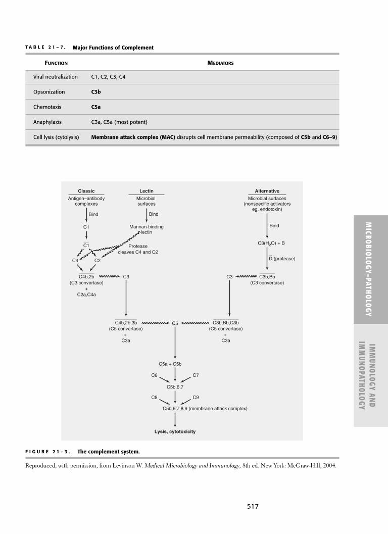
517
MICRO
BIOLOGY–PATHO
LOGYIM
MUN
OLOGY AN
DIM
MUN
OPATHO
LOGY
T A B L E 2 1 – 7 . Major Functions of Complement
FUNCTION MEDIATORS
Viral neutralization C1, C2, C3, C4
Opsonization C3b
Chemotaxis C5a
Anaphylaxis C3a, C5a (most potent)
Cell lysis (cytolysis) Membrane attack complex (MAC) disrupts cell membrane permeability (composed of C5b and C6–9)
F I G U R E 2 1 – 3 . The complement system.
Reproduced, with permission, from Levinson W. Medical Microbiology and Immunology, 8th ed. New York: McGraw-Hill, 2004.
C3b,Bb,C3b(C5 convertase)
+C3a
Antigen–antibodycomplexes
Microbial surfaces(nonspecific activators
eg, endotoxin)
Alternative
C1
Bind
Bind
C1
C4b,2b(C3 convertase)
+C2a,C4a
C3 C3
C4b,2b,3b(C5 convertase)
+C3a
C5
C5a + C5b
C7
C9
C6
C8
C5b,6,7
Lysis, cytotoxicity
C5b,6,7,8,9 (membrane attack complex)
C4 C2
C3b,Bb(C3 convertase)
C3(H2O) + B
D (protease)
Classic
Microbialsurfaces
Mannan-bindinglectin
Bind
Proteasecleaves C4 and C2
Lectin

518
Transplantation
TYPES OF GRAFT
■ Autograft: Transplantation of tissue from one site to another within thesame individual.
■ Isograft: Transplantation of tissue between two genetically identical indi-viduals in the same species.
■ Allograft: Transplantation of tissue between two genetically different indi-viduals in the same species.
■ Xenograft: Transplantation of tissue between two different species.
GRAFT REJECTION
■ T-cell-mediated (mostly CD8 TC cells) immune response against donoralloantigens.
■ The severity and rapidity of graft rejection is determined by the degree ofdifferences between donor and recipient class I and II MHC proteins.
■ If a second graft from the same donor is given to a sensitized recipient, an accel-erated rejection response occurs due to the presence of presensitized TC cells.
■ Allografts are the most common grafts used for organ transplantation, bloodtransfusions, and other tissue grafts.
■ Graft rejection can occur at different time intervals.
MIC
ROBI
OLO
GY–P
ATHO
LOGY
IMM
UNO
LOGY
AN
DIM
MUN
OPA
THO
LOGY
Types of Graft Rejection
Rejection Time after Common Reason Type Transplantation for Rejection
Hyperacute Minutes Preformed antibody-mediated immune response to graft antigens
Acute Weeks T-cell-mediated immune response to foreign class I and II MHC proteins
Chronic Months to years Antibody-mediated necrosis of graft vasculature
CD8 TC cells elicit most of the
destruction in graft rejection.
The most common types of
hyperacute rejection are ABO
blood mismatches.
GRAFT-VERSUS-HOST (GVH) REACTION
■ Immunocompetent T cells from the graft recognize the recipient’s cells asforeign, eliciting their destruction.
■ Host cells are targeted because the recipient generally undergoes radiationtherapy, inducing severe immunocompromise.
■ Occurs most commonly after bone marrow transplants and can be fatal.
Hypersensitivity
See Table 21–8.
■ Hypersensitivity reactions elicit exaggerated immune responses, which aredamaging and destructive to the host.
■ See Chapter 20 for a list of the major inflammatory mediators.

519
HYPERSENSITIVITY REACTIONS
See Figures 21–4 to 21–7.
■ Type I: Immediate (anaphylactic).■ Type II: Cytotoxic.■ Type III: Immune-complex.■ Type IV: Delayed (cell-mediated).
MICRO
BIOLOGY–PATHO
LOGYIM
MUN
OLOGY AN
DIM
MUN
OPATHO
LOGY
T A B L E 2 1 – 8 . Hypersensitivity Reactions
HYPERSENSITIVITY MAJOR
TYPE MEDIATOR REACTION EXAMPLE
Type I: IgE Antigen-bound IgE Atopic allergy
Immediate activates the release Angioedema
(anaphylactic) histamine and other Anaphylaxis
mediators from mast Prausnitz-Küstner
cells and basophils reaction
Type II: Cytotoxic IgM IgM or IgG bind to host Hemolytic anemia
IgG cell surface antigens, ADCC
activating complement Goodpasture’s
and producing syndrome
MAC-mediated cell Erythroblastosis
destruction fetalis
Type III: Antigen– Antigen–antibody Arthus reaction
Immune-complex antibody complexes (IgG, IgM, Serum sickness
complexes and IgA) are deposited Glomerulonephritis
in various tissues, RA
activating complement SLE
and eliciting PMN/
macrophage-mediated
tissue destruction
Type IV: T cell Macrophages present Contact dermatitis
Delayed antigen, activating Tuberculin (PPD) test
(cell-mediated) T cells and producing Tuberculosis
lymphokine-mediated Sarcoidosis
tissue destruction Leprosy
Starts hours-days GVHD
after contact with
antigen
Atopic allergies are common
type I hypersensitivity
reactions that have a strong
genetic predisposition for
excessive IgE production.
Clinical manifestations include
asthma, edema, and
erythema (“wheal and flare”),
and urticaria (hives).
Common allergens include
pollens, animal danders,
foods (shellfish and peanuts),
drugs (penicillin), bee venom,
and latex.
ACID: Anaphylactic + Atopic
Cytotoxic
Immune complex
Delayed
Angioedema is a moregeneralized version oftype I hypersensitivity. Itinvolves larger areas anddeeper tissues beneaththe skin and underlyingtissues, causing a morediffuse swelling. Mayinvolve the hands, feet,lips, eyelids, genitals, oralmucosa, and airway.Rapid (immediate) onset.
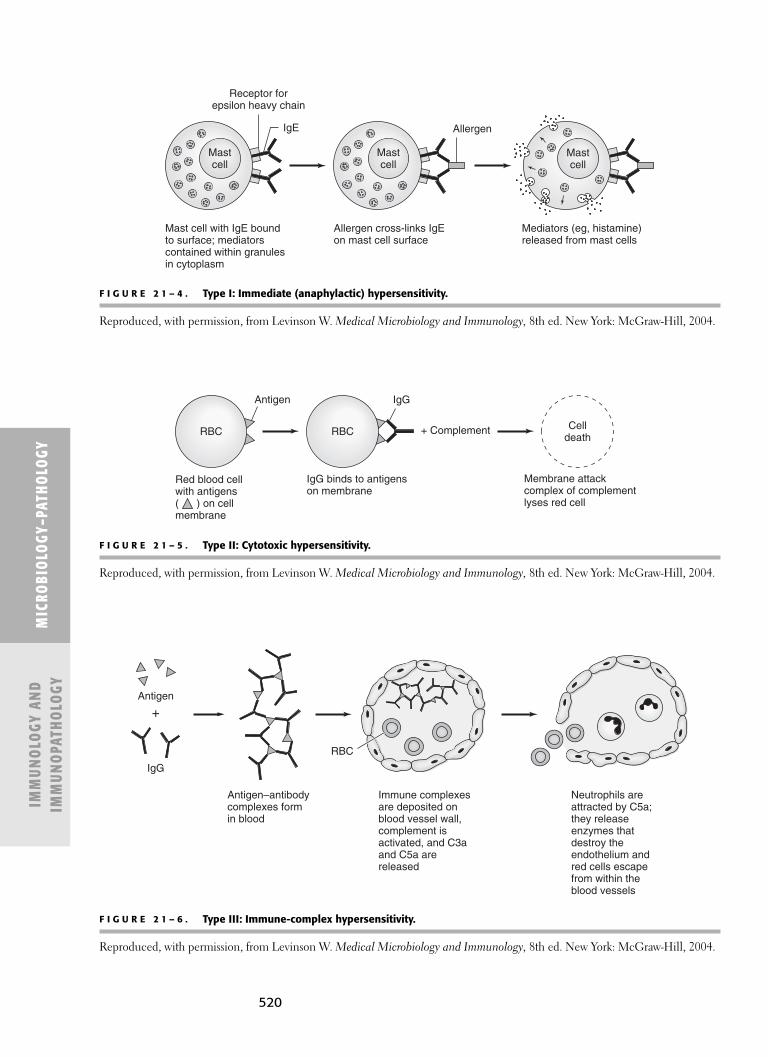
520
MIC
ROBI
OLO
GY–P
ATHO
LOGY
IMM
UNO
LOGY
AN
DIM
MUN
OPA
THO
LOGY
Receptor forepsilon heavy chain
IgE
Mast cell with IgE boundto surface; mediators contained within granules in cytoplasm
Allergen cross-links IgEon mast cell surface
Mediators (eg, histamine)released from mast cells
Allergen
Mastcell
Mastcell
Mastcell
F I G U R E 2 1 – 4 . Type I: Immediate (anaphylactic) hypersensitivity.
Reproduced, with permission, from Levinson W. Medical Microbiology and Immunology, 8th ed. New York: McGraw-Hill, 2004.
F I G U R E 2 1 – 5 . Type II: Cytotoxic hypersensitivity.
Reproduced, with permission, from Levinson W. Medical Microbiology and Immunology, 8th ed. New York: McGraw-Hill, 2004.
F I G U R E 2 1 – 6 . Type III: Immune-complex hypersensitivity.
Reproduced, with permission, from Levinson W. Medical Microbiology and Immunology, 8th ed. New York: McGraw-Hill, 2004.
RBC
Red blood cellwith antigens ( ) on cellmembrane
IgG binds to antigens on membrane
Membrane attackcomplex of complementlyses red cell
RBC + Complement Celldeath
Antigen IgG
IgG
Antigen
RBC
Antigen–antibodycomplexes formin blood
Immune complexesare deposited onblood vessel wall,complement isactivated, and C3aand C5a are released
Neutrophils areattracted by C5a;they releaseenzymes thatdestroy theendothelium andred cells escapefrom within theblood vessels
+

521
MICRO
BIOLOGY–PATHO
LOGYIM
MUN
OLOGY AN
DIM
MUN
OPATHO
LOGY
Gamma-interferon
TCR
IL-1, IL-12
Class IIMHC protein
Antigen+
HelperT cell(TH-1)
MacrophageMacrophage
F I G U R E 2 1 – 7 . Type IV: Delayed (cell-mediated) hypersensitivity.
Reproduced, with permission, from Levinson W. Medical Microbiology and Immunology, 8th ed.
New York: McGraw-Hill, 2004.
T A B L E 2 1 – 9 . Common Antigen–Antibody Laboratory Tests
TEST FUNCTION COMMON USE
Agglutination Antibody cross-links with a ABO blood typing.
particulate antigen, creating
visible clumping if positive.
Precipitation Antibody cross-links with a Detection of serum
soluble antigen, creating antigen or antibody.
visible precipitates if positive.
Radioimmunoassay Radio-labeled antibodies cross-link Detection of serum
(RIA) with unlabeled (unknown) antigen, antigen or hapten.
creating measurable radioactive
complexes if positive.
Enzyme-linked Enzyme-labeled antibody binds Detection of antigen or
immunosorbent to serum antibody–antigen antibody in patient
assay (ELISA) complexes. A substrate is then specimens.
added, activating the enzyme,
and eliciting a color reaction
(determined by
spectrophotometry) if positive.
Immunofluorescence Fluorescent-labeled antibodies Detection of antigen in
(IF) bind to unlabeled (unknown) histologic sections or
antigen, creating visible tissue specimens.
fluorescence in UV light
if positive.
Anaphylaxis is the most
severe form of type I
hypersensitivity, leading to
bronchoconstriction and
hypotension (shock); can be
life-threatening without
treatment. Its treatment is
epinephrine 0.3 mg (1:1,000)
IM or 0.1 mg (1:10,000) IV.
Once vital signs are stabilized,
antihistamines or
corticosteroids can be
administered.
Laboratory Tests
See Table 21–9.
■ There are several methods for the detection and diagnosis of infectiousdiseases, autoimmune diseases, and blood typing.

T A B L E 2 1 – 1 0 . Immunodeficiency Diseases
CELLULAR IMMUNOLOGICAL
DISEASE PATHOLOGY CAUSE FINDINGS CLINICAL FINDINGS
Bruton’s B cells Pre-B-cells do not Absence of B cells, Increased bacterial
X-linked differentiate into plasma cells, and infections
agammaglobulinemia mature B cells Ig Affects young boys
(tyrosine kinase T-cells are normal after 6 months of age
mutation) (maternal IgG initially
protects)
Lymphoma, leukemia,
myeloma
Isolated IgA B cells Mature B cells do Absence of IgA Most common inherited
deficiency not differentiate Other Ig isotypes are B-cell defect
into IgA-secreting normal Increased infection at
plasma cells mucosal surfaces
Common B cells Mature B cells do not Absence of various Hemolytic anemias
variable differentiate into Ig isotypes Lymphoid tumors
immunodeficiency (CVID) plasma cells
522
ABO BLOOD TYPING
■ All erythrocytes have alloantigens of the ABO type, which are importantfor blood typing and transfusions.
■ Although there are only two genes (A and B) that encode for these antigens,there are four possible antigenic combinations: A, B, AB, or O.
■ Hemagglutination occurs when blood types are mismatched.
MIC
ROBI
OLO
GY–P
ATHO
LOGY
IMM
UNO
LOGY
AN
DIM
MUN
OPA
THO
LOGY
Blood Type Antigen (on RBC) Antibody (in Plasma)
A A Anti-B
B B Anti-A
AB (universal recipient) Both A and B Neither anti-A nor anti-B
O (universal donor) Neither A nor B Both anti-A and anti-B
Type O blood is theuniversal donorbecause it containsno A or B antigens.
� I M M U NOPATHOLOGY
Immunodeficiency Diseases
See Table 21–10.

523
MICRO
BIOLOGY–PATHO
LOGYIM
MUN
OLOGY AN
DIM
MUN
OPATHO
LOGY
T A B L E 2 1 – 1 0 . Immunodeficiency Diseases (Continued)
CELLULAR IMMUNOLOGICAL
DISEASE PATHOLOGY CAUSE FINDINGS CLINICAL FINDINGS
DiGeorge’s T cells Thymic aplasia Absence of T cells Defective development of 3rd
syndrome (undeveloped thymus B cells are normal and 4th pharyngeal arches
and parathyroids) Increased viral and
fungal infections
CATCH 22: Cardiac defects,
Abnormal facies, Thymic
aplasia, Cleft palate,
Hypocalcemia (tetany),
microdeletion of
chromosome 22
Job’s syndrome T cells TH cells do not Elevated IgE levels FATED: Abnormal Facies,
produce INF-γ Staph aureus Abscesses,
(poor PMN retained primary Teeth, ↑ IgE,
chemotaxis) Dermatologic problems
(eczema)
Severe combined B cells and X-linked (defective Absent or Most severe inherited
immunodeficiency T cells IL-2 receptor) diminished immunodeficiency
disease (SCID) Autosomal recessive lymphoid tissue Increased viral, bacterial,
(adenosine deaminase fungal infections
deficiency) Death generally
before 2 y old
Wiskott–Aldrich B cells and Defective IgM Decreased IgM levels X-linked
syndrome T cells response to Elevated IgA levels (male infants)
bacterial LPS Normal IgE levels Clinical triad:
■ Infections
■ Eczema
■ Thrombocytopenia
Ataxia-telangiectasia B cells and Defective DNA Decreased IgA levels Ataxia: Cerebellar problems
T cells repair (often by (uncoordinated gait)
ionizing radiation) Telangiectasias: Dilation of
capillaries (sclera, ear, nose)
Graying of hair
Irregular skin pigmentation
to sun-exposed areas
Increased infections
and malignancies
524
Autoimmune Diseases (Table 21–11)
■ Caused by immune reactions versus self (host tissues).■ Usually involves auto-antibodies.■ HLA antigen association.■ Mechanisms:
■ Hypersensitivity reactions■ Disordered immunoregulation
■ ↑ TH-cell function■ ↓ TS-cell function■ Nonspecific B-cell activation
■ Examples:■ Graves’ disease■ Hashimoto’s thyroiditis■ Pernicious anemia■ Sjögren’s syndrome■ Systemic lupus erythematosus■ Scleroderma■ Polyarteritis nodosa■ Rheumatoid arthritis■ Reiter’s syndrome■ Ankylosing spondylitis■ Myasthenia gravis■ Multiple sclerosis
MIC
ROBI
OLO
GY–P
ATHO
LOGY
IMM
UNO
LOGY
AN
DIM
MUN
OPA
THO
LOGY
Thymus gland:
■ Important fordevelopment of theimmune system,beginning prenatally.
■ Involved in T celldevelopment anddifferentiation.
■ Located behind thesternum.
■ Functions in childhoodthen graduallyatrophies with age.
T A B L E 2 1 – 1 1 . Autoimmune Diseases Affecting Oral Mucosa
DISEASE AUTOANTIBODY HISTOPATHOLOGY IF CLINICAL
Lichen planus Unknown Epithelial acanthosis Fibrinogen in a Oral lesions on buccal
with “sawtooth” “shaggy” linear mucosa, tongue, and
rete pegs pattern along BM gingiva
Dense accumulation Types: Reticular (with
of T cells in Wickham’s striae),
underlying erosive, plaque
CT and into Certain medications
epithelium can cause lichenoid
reactions
Skin lesions appear
as clusters of pruritic
purplish papules with
a white keratotic “cap”
Mucous membrane Anti-BP-1 (of Separation of IgG and C3 in Erythematous and erosive
pemphigoid hemidesmosomes) epithelial basal linear pattern lesions on gingiva
cells from BM along BM precede other oral areas:
buccal mucosa,
palate, FOM
Can extend to other
mucosa: nasopharynx,
esophagus, vaginal, eye
conjunctiva (symblepharon
formation), and skin
(Continued)

525
MICRO
BIOLOGY–PATHO
LOGYIM
MUN
OLOGY AN
DIM
MUN
OPATHO
LOGY
T A B L E 2 1 – 1 1 . Autoimmune Diseases Affecting Oral Mucosa (Continued)
DISEASE AUTOANTIBODY HISTOPATHOLOGY IF CLINICAL
Pemphigus vulgaris Anti-desmoglein Suprabasilar IgG antibody in Oral lesions often
(of desmosomes) acantholysis with “fishnet” pattern precede skin lesions
Tzanck cells within spinous Erosive lesions on soft
layer palate, buccal mucosa,
gingiva, lateral tongue
High mortality rate
Erythema multiforme Immune complexes Intraepithelial IgM and C3 in Causative agents
pooling of perivascular pattern include infections
eosinophilic within lamina (HSV), medications, GI
amorphous propria diseases (Crohn’s, UC)
coagulum Bullous “target” lesions
Perivascular on skin and oral mucosa
infiltrate of (25% prevalence), which
mononuclear cells may collapse and crust
within lamina over (Stevens-Johnson
propria syndrome)
Severe form is toxic
epidermal necrolysis (TEN)

This page intentionally left blank

C H A P T E R 2 2
Systemic Pathology
Developmental Pathology 530
TERATOGENESIS 530
CHROMOSOMAL ABNORMALITIES 531
LYSOSOMAL STORAGE DISEASES 532
OTHER CHILDHOOD GENETIC DISORDERS 532
Dermatopathology 533
DISORDERS OF SKIN PIGMENTATION 533
SKIN INFECTIONS 534
IMMUNOLOGIC SKIN LESIONS 534
BENIGN (NONNEOPLASTIC) SKIN LESIONS 536
Cardiovascular Pathology 536
EDEMA 536
SHOCK 537
CONGESTION (HYPEREMIA) 538
THROMBOSIS 539
EMBOLUS 539
PHLEBITIS 540
ARTERIOSCLEROSIS 540
HYPERTENSION 541
AORTIC ANEURYSM 543
AORTIC DISSECTION 543
RHEUMATIC FEVER 545
CORONARY ARTERY DISEASE (CAD) 546
HEART FAILURE 547
CV PULMONARY CROSS-CORRELATION 548
Respiratory Pathology 549
ASTHMA 549
CHRONIC OBSTRUCTIVE PULMONARY DISEASE (COPD) 550
BRONCHIECTASIS 552
ATELECTASIS 552
PNEUMOCONIOSES 552
527
528
SARCOIDOSIS 553
IDIOPATHIC PULMONARY FIBROSIS 554
PNEUMONIA 554
LUNG ABSCESS 555
TUBERCULOSIS (TB) 556
Gastrointestinal Pathology 556
ESOPHAGUS 556
STOMACH 558
SMALL AND LARGE INTESTINES 559
PANCREAS 560
LIVER DISEASE 562
Genitourinary Pathology 564
NEPHROLITHIASIS 564
PYELONEPHRITIS 565
DIABETES INSIPIDUS 566
POLYCYSTIC KIDNEY DISEASE (PCKD) 566
MEDULLARY CYSTIC DISEASE 566
MEDULLARY SPONGE KIDNEY 566
NEPHROSCLEROSIS 566
ARTERIOSCLEROSIS 567
NEPHROTIC SYNDROME 567
GLOMERULONEPHROPATHIES 568
Blood–Lymphatic Pathology 568
ANEMIA 568
ANEMIA FROM ↑ RBC DESTRUCTION 569
ANEMIA FROM ↓ RBC PRODUCTION 571
OTHER CAUSES OF ↓ RBC PRODUCTION 573
ERYTHROCYTES AND POLYCYTHEMIA (↑ RBCS) 573
BLEEDING PROBLEMS 574
DISORDERS IN PRIMARY HEMOSTASIS 575
QUANTITATIVE PLATELET DEFICIENCIES 575
QUALITATIVE PLATELET PROBLEMS 577
DISORDERS IN SECONDARY HEMOSTASIS 577
Endocrine Pathology 578
ANTIDIURETIC HORMONE (VASOPRESSIN) DEFICIENCY 578
INSULIN DEFICIENCY 578
INCREASED GROWTH HORMONE 579
THYROID HORMONE 580
PARATHYROID HORMONE 582
CORTISOL 584
Muscoloskeletal Pathology 585
COLLAGEN VASCULAR DISEASES 585
OSSEOUS PATHOLOGY 587
ARTHRITIS 593
529
Neuropathology 595
NEUROLOGIC TRAUMA 595
NEURODEGENERATIVE DISEASES 596
MYASTHENIA GRAVIS 597

530
� DEVE LOPM E NTAL PATHOLOGY
Teratogenesis
■ Induction of nonhereditary congenital malformations (birth defects) in adeveloping fetus by exogenous factors:■ Physical■ Chemical■ Biologic agents
■ Teratogens = Teratogenic agents■ Physical agents
■ Radiation■ Hypoxia■ CO2■ Mechanical trauma
■ Maternal infection■ TORCH complex (Toxoplasmosis, Other agents, Rubella, CMV, and
HSV)■ Hormones
■ Sex hormones■ Corticosteroids
■ Vitamin deficiencies■ Riboflavin■ Niacin■ Folic acid■ Vitamin E
■ Drugs■ Mitomycin■ Dactinomycin■ Puromycin
MIC
ROBI
OLO
GY–P
ATHO
LOGY
SYST
EMIC
PAT
HOLO
GY
■ Effects of teratogens:■ Death■ Growth retardation■ Malformation■ Functional
impairment■ Mechanism of
teratogens:■ Specific for each
teratogen: inhibit,interfere, or blockmetabolic stepscritical for normalmorphogenesis.
■ Most are site ortissue specific.
■ Susceptibility toteratogens is■ Variable.■ Specific for each
developmental stage.■ Dose dependent.

531
MICRO
BIOLOGY–PATHO
LOGYSYSTEM
IC PATHOLOGY
AUTOSOMAL ABNORMALITIES
ChromosomalSyndrome Abnormality Findings
Down syndrome Trisomy 21 Mental retardationEpicanthal foldsLarge protruding tongueSmall head, low-set earsBroad flat faceSimian creaseComplications↑ leukemia↑ infectionAlzheimer-like brain Δ
Edward syndrome Trisomy 18 Mental retardationSmall headMicrognathia (small lower jaw)Pinched facial appearanceLow-set, malformed earsRocker bottom feetHeart defectsPrognosis: months
Patau syndrome Trisomy 13 Mental retardationMicrocephalyMicrophthalmiaBrain abnormalitiesCleft lip and palatePolydactylyHeart defectsPrognosis: < 1 y
SEX CHROMOSOME ABNORMALITIES
ChromosomalSyndrome Abnormality Findings
Klinefelter syndrome XXY 1:500 menManifests at pubertyHypogonadism, atrophic testesTall statureGynecomastiaFemale pubic hair distribution. Low IQAssociated with ↑ maternal and ↑ paternal age
Turner syndrome XO 1:3000 live female birthsDiagnose at birth or pubertyFemale hypogonadismPrimary amenorrheaShort statureWebbed neckWide-spaced nipplesCoarctation of aorta
Chromosomal Abnormalities
Down syndrome is the most
frequent chromosomal
disorder (1:700 births). People
with Down syndrome can live
into their 30s and 40s.
Chromosomalabnormalities indecreasing order ofincidence:
■ Down’s = 1:700.■ Edward’s = 1:3000.■ Patau’s = 1:5000.

532
Lysosomal Storage Diseases
LYSOSOMAL STORAGE DISEASES
■ Hunter’s ■ Tay–Sachs■ Gaucher■ Neimann–Pick■ Fabry■ See also Chapter 6, Biological Compounds.
MIC
ROBI
OLO
GY–P
ATHO
LOGY
SYST
EMIC
PAT
HOLO
GY
Autosomal Recessive X-linked
Tay–Sachs FabryGaucher Hunter’sNiemann–Pick
Other Childhood Genetic Disorders
CYSTIC FIBROSIS
■ Most common fatal genetic disease in white children.■ Occurs in both males and females (M = F).■ Life expectancy = 28 years.■ Generalized exocrine gland dysfunction. Problem with Cl- transporter.■ Multiple organ systems.
■ Characterized by respiratory and digestive problems.■ Pathogenesis: Chromosome 7q.
■ Gene encodes CFTR (cystic fibrosis transmembrane regulator).■ Regulates Cl− and Na+ transport across epithelial membranes.
■ Affects Na+ channels, especially mucous and sweat glands. ■ Test: Sweat chloride test.■ Findings
■ Chronic pulmonary disease.■ From thick mucous in airways:
■ Lung infections.■ Bronchiectasis.
■ Pancreatic exocrine insufficiency.■ Meconium ileus.
■ Intestinal obstruction in infants/newborns.
VON HIPPEL–LINDAU DISEASE
■ Autosomal dominant■ Chromosome 3■ VHL gene
■ Findings■ Hemangiomas
■ Retina■ Cerebellum
Lysosomal storage diseases
are most common in people
of Eastern European ancestry.
Cystic fibrosis■ Problem with Cl−
transporter■ Findings
■ Bronchiectasis■ Meconium ileus
The most common cause of
cystic fibrosis is a deletion
causing loss of phenylalanine
at position 508 in the
CFTR gene.

533
■ Cysts and adenomas■ Liver■ Kidney■ Adrenal glands■ Pancreas
MARFAN’S SYNDROME
■ Uncommon hereditary connective-tissue disorder:■ Fibrillin gene mutation.
■ Findings■ Skeletal
■ Tall and thin patients.■ Abnormally long legs and arms.■ Spiderlike fingers.
■ Cardiovascular■ Cystic medial necrosis of aorta.
■ Risk aortic incompetence, dissecting aortic aneurysms.■ Distensible mitral valve.
■ Ocular: Lens dislocation.
� DE RMATOPATHOLOGY
Disorders of Skin Pigmentation
HYPOPIGMENTATION
■ Albinism■ Failure in pigment production from otherwise intact melanocytes.■ Usually tyrosinase problem; can’t convert tyrosine to DOPA (in the
pathway to form melanin).■ Vitiligo
■ Acquired loss of melanocytes.■ Discrete areas of skin with depigmented white patches.■ May be autoimmune.
HYPERPIGMENTATION
■ Freckle (ephelis): Increased melanin pigment within basal keratinocytes.■ Lentigo: Pigmented macule caused by melanocytic hyperplasia in epidermis.■ Pigmented nevi: (See the section “Benign (Nonneoplastic) Skin Lesions”
later in this chapter.)■ Lentigo maligna (See the section “Benign (Nonneoplastic) Skin Lesions”
later in this chapter.)■ Café au lait spots
■ Increase in melanin content with giant melanosomes.■ Conditions with café au lait spots include:
■ Neurofibromatosis type 1 (most frequent neurocutaneous syndrome).■ McCune–Albright syndrome (See the section “Fibrous Dysplasia”
later in this chapter).■ Tuberous sclerosis (rare disease that causes nonmalignant tumors in
the brain and organs).■ Fanconi anemia (rare disease resulting in loss of DNA repair with
increased risk of cancer and endocrine problems).■ Diffuse hyperpigmentation with Addison’s disease.
■ Secondary to ↑ melanocyte-stimulating hormone.
MICRO
BIOLOGY–PATHO
LOGYSYSTEM
IC PATHOLOGY

534
Skin Infections
VIRAL SKIN ERUPTIONS
■ Molluscum contagiosum (Poxvirus)■ Verruca vulgaris (common wart) (human papilloma virus [HPV])■ Herpes simplex ■ Roseola (exanthema subitum) (herpes virus 6 and 7)■ Rubella■ Measles (rubeola) (paramyxovirus)
BACTERIAL SKIN ERUPTIONS
IMPETIGO
■ Common skin infection.■ Common in preschool age children (2–5 years old).■ Especially during warm weather.
■ Etiology■ Invasion of epidermis by Staphylococcus aureus or Streptococcus pyogenes.■ Similar to cellulitis, but more superficial. ■ Highly infectious.
■ Signs/symptoms■ Starts as itchy, red sore. ■ Blisters → breaks → oozes.■ Ooze dries; lesion becomes covered with a tightly adherent crust.■ Grows and spreads circumferentially (not deep); rarely impetigo forms
deeper skin ulcers■ Contagious; carried in the oozing fluid.
■ Treatment■ Topical antimicrobial (eg, bactroban).■ Oral antibiotic (eg, erythromycin or dicloxacillin); rapid clearing of
lesions.■ Course/prognosis
■ Impetigo sores heal slowly and seldom scar.■ Cure rate is extremely high.■ Recurrence is common in young children.
Note: Acute glomerulonephritis (renal disease) is an occasional complication(poststreptococcal glomerulonephritis [GMN]).
Immunologic Skin Lesions
HIVES
■ Urticaria = wheals.■ Type I hypersensitivity.■ (See section on hypersensitivity in Immunology, Chapter 21.)
PEMPHIGUS VULGARIS
■ Ages 30–60.■ Clinical
■ Oral mucosal lesions (often first sign).■ Skin lesions follow.■ Bullae rupture, leaving raw surface susceptible to infection.
MIC
ROBI
OLO
GY–P
ATHO
LOGY
SYST
EMIC
PAT
HOLO
GY

535
■ Etiology■ Autoimmune: IgG antibodies against desmosome proteins.
■ Histology■ Formation of intradermal bullae
■ Acantholysis: Tzanck cells.■ Basal layer intact.
■ Immunofluorescence shows encircling of epidermal cells.
BULLOUS PEMPHIGOID
■ Resembles pemphigus vulgaris.■ Clinically less severe.■ Etiology
■ Autoimmune: IgG antibodies against hemidesmosome proteins.■ Histology
■ Subepidermal bullae.■ Characteristic inflammatory infiltrate of eosinophils in surrounding
dermis.■ Immunofluorescence shows linear band.
ERYTHEMA MULTIFORME
■ Peak incidence second and third decades.■ Etiology
■ Type III hypersensitivity ■ Response to:
■ Medications:■ Sulfa drugs■ Penicillins■ Barbiturates
■ Infections■ HSV■ Mycoplasma■ Other illnesses
■ Damage to blood vessels of skin (because of immune complexes).■ Clinical
■ Classic “target,” “bull’s-eye,” or “iris” skin lesion.■ Central lesion surrounded by concentric rings of pallor and redness.■ Dorsal hands. ■ Forearms.
■ No systemic symptoms.
STEVENS–JOHNSON SYNDROME
■ Variant, more severe form of erythema multiforme.■ Severe systemic symptoms. ■ Extensive skin target lesions
■ Involve multiple body areas, especially mucous membranes.
TOXIC EPIDERMAL NECROLYSIS
■ Also called TEN syndrome and Lyell’s syndrome. ■ Multiple large blisters (bullae) that coalesce, sloughing of all or most of
skin and mucous membranes.
MICRO
BIOLOGY–PATHO
LOGYSYSTEM
IC PATHOLOGY
Pemphigus vulgaris =
Intraepidermal bullae.
Bullous pemphigoid =
Subepidermal bullae.
Tzanck cells are multinucleate
giant cells caused by a variety
of skin pathologies.
536
MIC
ROBI
OLO
GY–P
ATHO
LOGY
SYST
EMIC
PAT
HOLO
GY
Benign (Nonneoplastic) Skin Lesions
ACANTHOSIS NIGRICANS
■ Cutaneous finding of velvety hyperkeratosis and pigmentation. ■ Flexural areas, most often.
■ Axilla.■ Nape of neck.■ Other flexures.■ Anogenital region.
■ Often a marker of visceral malignancy. ■ > 50% have cancer.
■ Gastric carcinoma.■ Breast, lung, uterine cancer.
■ Seen in diabetes. ■ Histology
■ Acanthosis.■ Hyperkeratosis.■ Hyperpigmentation.
HEMANGIOMA
■ Hamartoma (not true neoplasm).
XANTHOMA
■ Associated with hypercholesterolemia.■ Clinical
■ Most common sites:■ Eyelids (xanthelasma).■ Nodules over tendons or joints.
■ Histology■ Yellowish papules or nodules composed of
■ Focal dermal collections of lipid-laden histiocytes.
� CARDIOVASC U LAR PATHOLOGY
Edema
■ Abnormal accumulation of fluid in interstitial spaces or body cavities.■ Fluid moves out of intravascular space.■ Results from some combination of:■ ↑ capillary permeability (as with histamine).■ ↑ capillary hydrostatic pressure.■ ↑ interstitial fluid colloid osmotic pressure.■ ↓ plasma colloid osmotic pressure.
(See also Chapter 20, Reactions to Tissue Injury.)
Types of Edema
Transudate Exudate
More watery (serous) edema fluid More protein-rich edema fluid Usually noninflammatory Usually inflammatoryFrom altered intravascular From ↑ vascular permeability
hydrostatic or osmotic pressure (with inflammation)
A hamartoma is disorganized
growth composed of tissue
normally found in a given
location.
A choristoma is a growth
composted of histologically
normal tissue found at a site
where it is not normally found
in the body.
537
EXAMPLES OF TRANSUDATE
■ Anasarca: Generalized edema.■ Hydrothorax: Excess serous fluid in pleural cavity.■ Hydropericardium: Excess watery fluid in pericardial cavity.■ Ascites (hydroperitoneum): Excess serous fluid in peritoneal cavity.
CLINICAL EXAM OF EDEMA
■ Pitting: Press finger for 5 s and quickly remove; an indentation is left thatfills slowly.
■ Nonpitting: No indentation left when finger is removed.
MICRO
BIOLOGY–PATHO
LOGYSYSTEM
IC PATHOLOGY
Edemas and Causes
Process Cause
Congestive heart failure ↑ plasma/capillary hydrostatic pressureNephrotic syndrome ↓ plasma oncotic pressureCirrhosis ↑ capillary hydrostatic pressure
↓ plasma oncotic pressureElephantiasis ↑ plasma/capillary hydrostatic pressure
Secondary to ↓ lymphatic return
Shock
■ ↓ tissue perfusion.■ Hemodynamic changes result in:
■ ↓ blood flow, thereby.■ ↓ oxygen and metabolic supply to tissues.
■ Can result in multiple organ damage or failure.■ Symptoms
■ Fatigue■ Confusion
■ Signs■ Cool pale skin (pallor)■ Weak rapid pulse (tachycardia = ↑ HR)■ ↓ BP (hypotension)■ ↓ urine output
■ Shock requires immediate medical treatment and can worsen rapidly.
MAJOR CATEGORIES OF SHOCK
■ Hypovolemic■ Cardiogenic■ Distributive
■ Septic■ Neurogenic■ Anaphylactic
Remember: Right-sided CHF
results in peripheral edema;
left-sided CHF results in
pulmonary edema.
↓ cardiac output is the major
factor in all types of shock
(because either ↓ HR, ↓ SV,
or both).

538
Congestion (Hyperemia)
■ ↑ volume of blood in local capillaries and small vessels.■ Active congestion (active hyperemia): ↑ arteriolar dilation (inflammation,
blushing).■ Passive congestion (passive hyperemia): ↓ venous return (obstruction,
increased back pressure).■ Two forms:
■ Acute■ Shock or right-sided heart failure
■ Chronic ■ In lung (usually secondary to left-sided heart failure)■ In liver (usually secondary to right-sided heart failure)
MIC
ROBI
OLO
GY–P
ATHO
LOGY
SYST
EMIC
PAT
HOLO
GY
TYPES OF SHOCK
Type Cause Examples
Hypovolemic ↓ blood volume HemorrhageDehydrationVomitingDiarrheaFluid loss from burns
Cardiogenic Pump failure Massive MIUsually LV failure ArrhythmiaSudden ↓ CO
Septic Infection (endotoxin release) Severe infectionGram-negative bacteriaCauses vasodilation
Neurogenic CNS injury CNS injuryCauses vasodilation
Anaphylactic Type I hypersensitivity Anaphylactic allergic Histamine release reaction (eg, insect sting)Vasodilation
STAGES OF SHOCK
Nonprogressive (Early) Progressive Irreversible
= compensated ↓ cardiac perfusion Organ damage↑ sympathetic nervous Cardiac depression ↓ high energy
system phosphate↑ CO CO ↓ reserves↑ TPR Death (even if Try to maintain Metabolic acidosis. restore blood flow)
perfusion to (Compensatory vital organs mechanisms are no
longer adequate)

539
Thrombosis
■ Blood clot attached to endothelial surface (blood vessel or heart [endo-cardium])■ Usually a vein.■ Virchow’s triad.
■ Arterial thrombi■ Lines of Zahn (morphologically).■ Alternating red and white laminations.
■ Venous thrombi■ Propagate: Enlarge while remaining attached to vessel wall.■ Embolize.■ Detach as large embolus. ■ Fragment off as many small emboli; shower emboli.
MICRO
BIOLOGY–PATHO
LOGYSYSTEM
IC PATHOLOGY
TYPES OF THROMBUS
Agonal Intracardiac thrombi■ After prolonged heart failure
Mural Thrombus from endocardial surface (or endothelium of large vessel) protrudes into lumen of heart or large vessel
Forms after ■ MI; damage to ventricular endocardium
(LV most often)■ Atrial fibrillation■ Aortic atherosclerosis
Can cause cerebral embolism
White Thrombus composed mostly of blood platelets
Red Thrombus composed of RBCs (rather than platelets)Occurs rapidly by coagulation with blood stagnation
Fibrin Thrombus composed of fibrin depositsDoes not completely occlude the vessel
Embolus
■ Intravascular mass.■ Solid■ Liquid■ Gas
■ Travels within a blood vessel. ■ Lodges at distant site.■ Occludes blood flow to vital organs. ■ Possibly leads to infarction.
■ Thromboemboli (most common): Blood clot. ■ Breaks off existing thrombus.■ Forms and is released downstream in the circulation (eg, from heart
chambers in atrial fibrillation).■ Fat embolism: Especially in long bone fractures.■ Gas embolism: Air into circulation (eg, Caisson disease).
Thrombolysis = Breakdown
of a clot.
Virchow’s triad:■ Endothelial injury.■ Alteration in blood
flow.■ Hypercoagulability of
blood.
Predisposing factors tothrombosis■ Arterial thrombosis
■ Atherosclerosis(major cause)
■ Venous thrombosis■ Heart failure■ Tissue damage■ Bed rest
(immobilization)■ Pregnancy■ Oral contraceptive
pills■ Age■ Obesity■ Smoking
Lines of Zahn1. White (fibrin and
platelet) layersalternating with dark(RBC) layers.
2. Indicated thrombosis inaorta or heart beforedeath.
540
■ Amniotic fluid embolism: With delivery; can activate diffuse/disseminatedintravascular coagulation (DIC).
■ Tumor embolism.
Phlebitis
■ Inflammation of the vein.■ Common sites
■ Legs.■ Varicose veins.
■ Common causes■ Local irritation (eg, IV). ■ Infection in or near vein.■ Blood clots.
THROMBOPHLEBITIS
■ Inflammation of the vein related to blood clot.■ Associated with
■ Superficial thrombophlebitis: Veins near skin surface.■ Deep venous thrombosis: Deeper, larger veins.■ Pelvic vein thrombosis.
■ Clinical symptoms■ Tenderness over vein.■ Pain in body part affected.■ Skin redness or inflammation.
Arteriosclerosis
■ Hardening of the arteries■ General term for several diseases causing changes to artery wall:
■ Thicker■ Less elastic
ATHEROSCLEROSIS
■ Degenerative changes in artery walls■ Most common cause of arteriosclerosis■ Atherosclerotic plaques: Fatty material accumulating under the arterial wall’s
inner lining■ Occurs in arteries (not veins)■ Risks
■ Men and postmenopausal women (estrogen may be protective)■ Smoking■ Hypertension■ Heredity (familial hypercholesterolemia)■ Nephrosclerosis■ Diabetes■ Hyperlipidemia
■ Sites■ Carotid■ Coronary
MIC
ROBI
OLO
GY–P
ATHO
LOGY
SYST
EMIC
PAT
HOLO
GY
Saddle embolus: Large
pulmonary embolus that
obstructs the bifurcation of the
pulmonary artery.
Paradoxical emboli
Begin in the venous system.
End up in the systemic arterial
system rather than the
pulmonary artery.
Most often allowed by an
atrial septal defect.
The only time a DVT can
cause a stroke.
Pulmonary embolism
■ Embolism causingpulmonary arteryobstruction.
■ Usually arises from ■ Deep vein
thrombosis (DVT);usually from lowerextremities (abovepopliteal fossa).
■ Course■ Systemic vein.■ Right heart.■ Pulmonary artery.
■ Causes■ Right heart strain.■ Possibly infarction
in the affectedsegment.
■ Possibly pleurisy(pleuritic chest pain).
■ Predisposing factors■ Virchow’s triad.
■ Especiallyimmobilization(leading tostagnation).
■ Thrombophlebitis.■ Hypercoagulable
states.

541
■ Circle of Willis■ Renal and mesenteric arteries
■ Pathogenesis■ Fatty streak
■ Foam cells in intima (lipid laden macrophages)■ Atheromas
■ Cholesterol■ Fibrous tissue■ Necrotic debris■ Smooth muscle cells
■ Complications■ ↓ elasticity of vessel.■ Ulceration of plaque; predisposing to thrombus formation.■ Hemorrhage into the plaque; narrowing lumen, possibly occluding
blood flow.■ Thrombus formation.■ Embolization; overlying thrombus or plaque material itself.
■ Symptoms■ Depend on site.
■ Visual changes, dizziness: Carotid or intracerebral arteries.■ Angina: Coronary arteries.■ Leg pain (claudication): Lower extremity arteries.
Hypertension
■ Primary (essential).■ Secondary (related to another disease).
PRIMARY (ESSENTIAL) HYPERTENSION
■ Accounts for 90–95% of hypertension.■ No identifiable cause; related to ↑ CO, ↑ TPR.■ Risks
■ Genetic■ Family history■ African Americans
■ Environmental■ ↑ dietary salt intake■ Stress■ Obesity■ Cigarette smoking■ Physical inactivity
■ Pathologic findings■ Hypertrophy of arteries and arterioles not capillaries (because no
smooth muscle).■ ↑ wall-to-lumen ratio.
■ ↑ smooth muscle cell growth (because of ↑ pressure, stretch). ■ ↓ arteriolar and capillary density. ■ ↓ total cross-sectional area of capillaries and arterioles.
■ Three organs most often damaged.■ Heart: 60% die of cardiac complications.■ Kidneys: 25% die of renal failure.■ Brain: 15% die of stroke or neurologic complications.
MICRO
BIOLOGY–PATHO
LOGYSYSTEM
IC PATHOLOGY
Atherosclerosis
■ Most importantcontributor to arterialthrombosis.
■ Most susceptiblearteries—aorta andcoronary arteries.
Atherosclerosis can leadto:
■ Ischemic heart disease(CAD).
■ Heart attack(myocardial infarction[MI]).
■ Stroke or aneurysmformation.
Familialhypercholesterolemia ■ Autosomal dominant
disease.■ Anomalies of LDL
(low-densitylipoprotein) receptors.
■ Atherosclerosis and itscomplications.■ Xanthomas.■ MI by age 20.
■ Diet, exercise, andantihypertensivemedications are usedto treat hypertension.
■ Untreated(or undertreated)hypertension canresult in■ “wear out”
(cardiac failure).■ “blow out” (CVA).■ “run out”
(renal failure).
Hypertension is the “silent
killer”; usually has no
symptoms.

542
SECONDARY HYPERTENSION
■ Hypertension (HTN) from known causes.■ 5–10% of HTN cases.■ Identifiable, often correctable cause.
■ Renal disease■ Most common cause of secondary hypertension.■ Renin–angiotensin–aldosterone system.■ Two categories:
■ Renal parenchymal diseases.■ Renal artery stenosis.
■ Endocrine disorders■ Hyperaldosteronism (Conn syndrome).■ Cushing syndrome.■ Hyperthyroidism.■ Diabetes.■ Pheochromocytoma.
■ Other causes■ Coarctation of the aorta.■ Preeclampsia/eclampsia/toxemia of pregnancy.
MALIGNANT HYPERTENSION
■ Severe form of high blood pressure. ■ Medical emergency.■ Can lead to death in 3–6 months if untreated.
■ Complication of either primary or secondary hypertension.■ Most often with secondary hypertension from kidney disease.
MIC
ROBI
OLO
GY–P
ATHO
LOGY
SYST
EMIC
PAT
HOLO
GY
CLINICAL FINDINGS AND COMPLICATIONS
Neurologic Ophthalmologic Cardiovascular Renal
Headaches Retinal hemorrhage BP >140/90 PolyuriaNausea/vomiting and exudates Arteriolar NocturiaDrowsiness ■ Damage to constriction ↓ urineAnxiety retinal ↑ resistance concentrationMental arterioles Claudication Nephrosclerosis
impairment ↓ blood supply Hardening of renal■ Intracerebral to legs arterioles
vessel ↑ cardiac workload Proteinuria possibledamage Angina Hematuria possible
↓ coronaryblood flow
Heart failure ■ Dyspnea on
exertion (LHF) ■ Peripheral
edema (RHF)Renal disease is the most
common cause of secondary
hypertension.
Preeclampsia occurs inpregnant patients:
■ Hypertension: >140systolic, or > 90diastolic after 20 weeks’gestation.
■ Proteinuria.■ Edema.
Eclampsia = Preeclampsia +
seizures.
Predisposing conditionsfor preeclampsia andeclampsia:
■ Hypertension■ Diabetes■ Autoimmune diseases
(eg, SLE)■ Laboratory findings■ Hyperuricemia■ Thrombocytopenia

543
■ Occurs in■ African American young adults■ Women with toxemia of pregnancy (preeclampsia/eclampsia)■ Patients with renal or collagen vascular disorders
■ Findings■ Sudden rapid ↑ BP, usually without a precipitating event
■ Life-threatening consequences to multiple organ systems:■ Cerebrovascular accident■ Retinal hemorrhage and papilledema■ Cardiac failure■ Renal failure
Aortic Aneurysm
■ Abnormal, localized dilation of the aorta■ True aneurysm = dilation of all three layers (intima, media, adventitia)■ Causes
■ Atherosclerosis■ Cystic medial necrosis
■ Marfan■ Ehlers–Danlos
■ Infectious aortitis■ Syphilitic aortitis
■ Vasculitis■ Risk
■ Rupture
Aortic Dissection
■ Life-threatening■ Blood into medial layers of aorta
■ “Dissecting” the intima from the adventitia■ Symptoms
■ Ripping/tearing chest pain■ Different BP measurement in each arm
■ Complications■ Rupture
■ Pericardial tamponade■ Hemomediastinum or hemothorax
■ Occlusion of aortic branches■ Carotid (stroke)■ Coronary (MI)■ Splanchnic (organ infarction)■ Renal (acute renal failure)
■ Distortion of aortic valve■ Aortic regurgitation
MICRO
BIOLOGY–PATHO
LOGYSYSTEM
IC PATHOLOGY
Aortic dissection most often
ruptures into the pericardial
sac (hemopericardium),
causing fatal tamponade.
Hemopericardium:Can occur after MI.
■ Ventricular rupture(necroticmyocardium).
■ Blood into pericardialspace.
■ Can cause cardiactamponade and death.
544
PATHOLOGY INVOLVING THE PERICARDIUM
MIC
ROBI
OLO
GY–P
ATHO
LOGY
SYST
EMIC
PAT
HOLO
GY
Chronic (Constrictive) Pericardial Effusion Acute Pericarditis Pericarditis
Accumulation of fluid in Inflammation of the Thickening and scarring the pericardial space pericardium: of the pericardium
■ Hydropericardium = ■ Serous Tb.serous fluid ■ Fibrinous ↓ elasticity
■ Hemopericardium = ■ Purulent Can cause blood ■ Hemorrhagic tamponade-like picture
Can cause cardiac ■ Often painfultamponade ■ Can cause cardiac
tamponade
CARDIAC TAMPONADE
■ Extrinsic compression of the heart ■ Fluid (accumulating quickly into the pericardial space)
■ Pericardial effusion■ Acute pericarditis
■ Cardiac compression■ ↓ venous return (↓ filling)■ ↓ CO■ Death
■ Signs■ Distended neck veins■ Hypotension■ ↓ heart sounds■ Tachypnea■ Weak/absent peripheral pulses
ENDOCARDITIS
■ Inflammation of endocardium and/or heart valves■ Symptoms: Develop quickly (acute) or slowly (subacute):
■ Fever (hallmark)■ Nonspecific constitutional signs■ Fatigue■ Malaise■ Headache■ Night sweats
■ Findings■ Murmur (secondary to vegetations); may change with time.■ Splenomegaly■ Splinter hemorrhages (small dark lines) under fingernails
Infections of the heart
■ Pericardium(pericarditis)
■ Myocardium(myocarditis, oftenviral)
■ Endocardium andheart valves(endocarditis, oftenbacterial orinflammatory)
Remember that the tricuspid
valve is most often affected in
IV drug users.
Beck's triad
1. Distension of jugularveins
2. Decreased bloodpressure
3. Muffled heart sounds
545
Rheumatic Fever
■ Acute inflammatory disease.■ Develops after streptococcal infection, usually a URI with group A
β-hemolytic strep.■ Most common in children 5–15 years old. ■ Onset
■ Usually sudden, often after 1–5 weeks (asymptomatic) after recoveryfrom sore throat or scarlet fever.
■ Course■ Mild cases = 3–4 weeks.■ Severe cases = 2–3 months.
■ Etiology■ Due to cross-reactivity, not direct effect of the bacteria.■ Type III hypersensitivity.
■ Sites■ Heart■ Joints■ Skin■ Brain
MICRO
BIOLOGY–PATHO
LOGYSYSTEM
IC PATHOLOGY
TYPES OF ENDOCARDITIS
Infective Rheumatic Libmann–Sacks
Usually bacterial Complication of Occurs in SLEIntrinsic bacteremia rheumatic fever■ Dental Occurs in areas with Nonbacterial■ Upper respiratory greatest hemodynamic endocarditis■ Urologic stress■ Lower GI tract■ Introduced bacteremia■ IV drug usersValvular involvement Mitral valve most often Mitral valve most oftenVegetations Calcification Small vegetations ■ Mitral ■ Stenosis on either or both ■ Tricuspid (in IVDU) ■ Insufficiency surfaces of valve
■ Both leaflets
INFECTIVE ENDOCARDITIS
Acute Endocarditis Subacute (Bacterial) Endocarditis
Staphylococcus aureus (50%) Streptococcus viridans (> 50%)Usually secondary to infection Patients with preexisting valve disease
elsewhere in bodyIVDUs
Mnemonics forrheumatic fever
PECCS
PolyarthritisErythema marginatumChoreaCarditisSubcutaneous nodules
FEVERSS
FeverErythema marginatumValve damageESRRed-hot (polyarthritis)Subcutaneous nodulesSt. Vitus dance (chorea)
Antibiotic prophylaxisbefore dentalprocedures
■ Children withcongenital heartdisease
■ To help preventsubacute bacterialendocarditis (SBE),Streptococcus viridans,from a dental/oralsource.
Valvular vegetations canbe dislodged and sendseptic emboli to organs:
■ Brain■ Lungs■ Kidneys■ Spleen

546
■ Pathology■ Aschoff bodies
■ Focal interstitial myocardial inflammation: Fragmented collagen,and fibrinoid material
■ Anitschkow cells: Large unusual cells■ Aschoff myocytes: Multinucleated giant cells
■ Laboratory■ ↑ ASO titers■ ↑ ESR
■ Findings/criteria for diagnosis = Jones criteria■ Diagnose when two major or one major and one minor criteria are
met. (See chart.)■ Treatment
■ Penicillin■ Rest
MIC
ROBI
OLO
GY–P
ATHO
LOGY
SYST
EMIC
PAT
HOLO
GY
JONES CRITERIA FOR DIAGNOSIS OF RHEUMATIC FEVER
Major Criteria Minor Criteria
Carditis FeverArthritis ArthralgiasChorea History of rheumatic feverErythema marginatum ECG changes Subcutaneous nodules Leukocytosis
Coronary Artery Disease (CAD)
■ CAD = Narrowing of coronary arteries■ Atherosclerosis plaques■ ↓ blood supply to myocardium
■ Consequences■ Ischemia■ Infarction
■ Symptoms■ Classic symptom of CAD = angina
■ Risks■ Same as for atherosclerosis■ Hypertension■ Hyperlipidemia■ Smoking■ Obesity■ Inactivity■ Diabetes■ Male gender
ANGINA
■ Squeezing (tight) substernal chest discomfort; may radiate to■ Left arm■ Neck■ Jaw■ Shoulder blade
Though rheumatic fever may
follow a strep infection, it is
not an infection. Rather, it is
an inflammatory reaction to
an infection.
Rheumatic carditis usually
disappears within 5 months.
However, permanent damage
to the heart/heart valves
usually occurs, leading to
rheumatic heart disease.
Valves involved are the mitral,
aortic, and tricuspid.
Pulmonic valve is rarely
involved.
CAD causes
■ ↓ blood flow tomyocardium.
■ ↓ O2 and nutrients tomyocardium.
In turn, this can lead to
■ Ischemia (angina).■ Infarction (MI = heart
attack with ECGchanges and enzymesreleased from infarctedmyocardial tissue).
Angina (stable angina) can be
relieved by rest or nitrates.

547
■ Caused by ↓ myocardial oxygenation■ Atherosclerotic narrowing■ Vasospasm
MICRO
BIOLOGY–PATHO
LOGYSYSTEM
IC PATHOLOGY
Stable Angina Unstable Angina Prinzmetal Angina
Most common type Occurs even at rest Intermittent chestCAD/atherosclerotic More severe CAD pain at rest
narrowing Often imminent MI VasospasmPrecipitated by exertion
MYOCARDIAL INFARCTION (MI)
■ Most important cause of morbidity from CAD.■ Prolonged interruption of coronary blood flow to myocardium; coagulative
necrosis.■ Cause: Usually thrombus formation in the setting of a ruptured, unstable
plaque.
Complete Occlusion Partial Occlusion
Transmural infarction Subendocardial infarctionST elevation MI Non-ST elevation MI
■ Symptoms■ Angina (that does not remit)■ Sweating■ Nausea, stomach upset
■ Signs■ ECG changes (ST elevation, ST depression, T waves, Q waves).■ Enzyme leak.
■ Prognosis: Good (if patient reaches hospital).■ Complications
■ Arrhythmia.■ Myocardial (pump) failure.■ Cardiac rupture.■ Papillary muscle rupture.■ Ventricular aneurysm.
Heart Failure
■ Heart’s ability to pump does not meet needs of the body.■ Usually a chronic progressive condition.■ Can occur suddenly.■ Causes: Secondary to heart muscle damage
■ After MI■ Cardiomyopathy■ Valvular diseases
Most deaths from heart attack
occur outside hospital; are
due to arrhythmias causing
ventricular fibrillation.
Cardiac enzymes elevatedafter an MI:
■ CK–MB (creatinekinase, MB fraction)
■ TnT (troponin T)■ Myoglobin
Creatine phosphokinase(CPK): Enzyme found in(↑ when damage to):
■ Heart■ Brain■ Skeletal muscle■ Not found in liver
Pulmonary edema: Life-
threatening complication of
left heart failure.
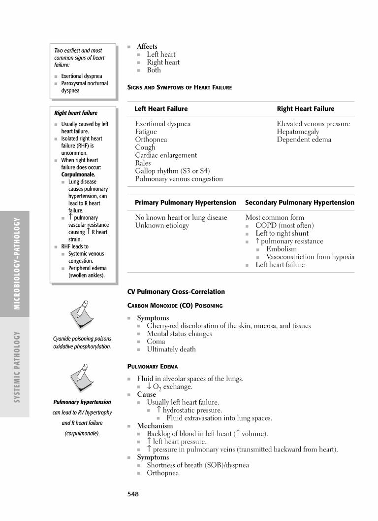
548
■ Affects■ Left heart■ Right heart■ Both
MIC
ROBI
OLO
GY–P
ATHO
LOGY
SYST
EMIC
PAT
HOLO
GY
SIGNS AND SYMPTOMS OF HEART FAILURE
Left Heart Failure Right Heart Failure
Exertional dyspnea Elevated venous pressureFatigue HepatomegalyOrthopnea Dependent edemaCoughCardiac enlargementRalesGallop rhythm (S3 or S4)Pulmonary venous congestion
CV Pulmonary Cross-Correlation
CARBON MONOXIDE (CO) POISONING
■ Symptoms■ Cherry-red discoloration of the skin, mucosa, and tissues■ Mental status changes■ Coma■ Ultimately death
PULMONARY EDEMA
■ Fluid in alveolar spaces of the lungs.■ ↓ O2 exchange.
■ Cause■ Usually left heart failure.
■ ↑ hydrostatic pressure.■ Fluid extravasation into lung spaces.
■ Mechanism■ Backlog of blood in left heart (↑ volume).■ ↑ left heart pressure.■ ↑ pressure in pulmonary veins (transmitted backward from heart).
■ Symptoms■ Shortness of breath (SOB)/dyspnea■ Orthopnea
Primary Pulmonary Hypertension Secondary Pulmonary Hypertension
No known heart or lung disease Most common formUnknown etiology ■ COPD (most often)
■ Left to right shunt■ ↑ pulmonary resistance
■ Embolism■ Vasoconstriction from hypoxia
■ Left heart failure
Two earliest and mostcommon signs of heartfailure:
■ Exertional dyspnea■ Paroxysmal nocturnal
dyspnea
Right heart failure
■ Usually caused by leftheart failure.
■ Isolated right heartfailure (RHF) isuncommon.
■ When right heartfailure does occur:Corpulmonale.■ Lung disease
causes pulmonaryhypertension, canlead to R heartfailure.
■ ↑ pulmonaryvascular resistancecausing ↑ R heartstrain.
■ RHF leads to■ Systemic venous
congestion.■ Peripheral edema
(swollen ankles).
Pulmonary hypertension
can lead to RV hypertrophy
and R heart failure
(corpulmonale).
Cyanide poisoning poisonsoxidative phosphorylation.

549
■ Cough■ Tachypnea■ Dependent crackles■ Tachycardia ■ Neck vein distention
■ Treatment■ ↓ Vascular fluid
■ Diuretics■ ↑ gas exchange and heart function
■ O2■ Antihypertensives■ Positive inotropic agents■ Antiarrhythmics
� RESPI RATORY PATHOLOGY
Asthma
■ Chronic reactive airway disorder caused by episodic airway obstruction■ Any age
■ 3–5% of adults (usually < 30 years old)■ 7–10% of children■ 50% of asthma cases occur in children (<10 years old)■ Boys > Girls (2:1)
■ Cause■ Bronchospasm (primary cause of airway obstruction)■ Other contributors (↑ airflow resistance)
■ ↑ mucus secretion■ Mucosal edema
■ Symptoms■ Dyspnea/SOB■ Expiratory wheezes■ Chest tightness■ Cough
■ Pathology■ Bronchial smooth muscle hypertrophy■ ↑ bronchial submucosal glands■ Eosinophils■ Charcot–Leyden crystals
MICRO
BIOLOGY–PATHO
LOGYSYSTEM
IC PATHOLOGY
TYPES OF ASTHMA
Extrinsic (Allergic, Intrinsic (Nonimmune, Atopic, Immune) Idiosyncratic)
Type I hypersensitivity ExerciseInhaled allergens (allergy triggers) Cold air■ Pet dander Tobacco smoke■ Dust mites Respiratory infections■ Cockroach allergens Stress/Anxiety■ Molds Other pollutants■ Pollens Drugs
Mild CO poisoning canpresent with exhaustion and
symptoms similar to acommon cold or flu,
delaying the diagnosis.
CO = colorless, odorlessgas.Sources: Automobileexhaust; home heatingfumes.Mechanism: Competitiveinhibition
■ Much higher affinityfor Hb than O2.
■ Attaches to Hb andprevents O2 carrying.
Pulmonary edema fromheart failure
■ Transudate into lungs■ Heart failure cells
(macrophages thathave phagocytized redblood cells in thealveoli)
Pulmonary edema canbe caused by
■ ↑ intracapillaryhydrostatic pressure■ Heart failure
■ ↑ capillarypermeability■ Acute respiratory
distress syndrome(ARDS)

550
MIC
ROBI
OLO
GY–P
ATHO
LOGY
SYST
EMIC
PAT
HOLO
GY
■ Treatment■ β2 agonist inhalers.
■ Preferred treatment for acute asthma attack.■ Terbutaline.■ Albuterol.
■ Steroids.■ Mast cell stabilizers.■ Epinephrine (sympathetic stimulation for status asthmaticus).
Chronic Obstructive Pulmonary Disease (COPD)
■ Group of lung diseases characterized by ↑ airflow resistance.■ Emphysema.■ Chronic bronchitis.
Obstructive Lung Diseases Restrictive Lung Diseases
↑ TLV ↓ TLV
Asthma Intrinsic lung diseases:COPD ■ Pneumoconioses■ Emphysema ■ Sarcoidosis■ Chronic bronchitis ■ Idiopathic pulmonary fibrosis
Extrinsic lung diseases:■ Kyphosis■ Obesity■ Neuromuscular weakness
Distinguish between
■ Intraalveolar fluid:Pulmonary edema.
■ Intrapleural (betweenvisceral and parietalpleura): Pleuraleffusion.
Status asthmaticus
■ Severe asthma attack■ Does not respond
to normalmeasures.
■ Usually requireshospitalization.
■ Can lead to death fromrespiratory acidosis(obstruction).
■ Treat with epinephrine.
Nitrous oxide is safe to
administer to people with
asthma, especially if their
asthma is triggered by anxiety.
Asthmatics taking chronic
steroids may need
corticosteroid augmentation.
In children, asthma symptoms
can decrease with time
(children can outgrow it).
COPD can overlap with
asthma and bronchiectasis.
Cigarette smoking is the
greatest cause of COPD.
Secondary pulmonary
hypertension is most
commonly caused by COPD.
MECHANISMS OF ASTHMA PRECIPITATION
Allergic (Immune) Intrinsic (Nonimmune)
DirectType I Bronchoconstrictor ↑ Vagal Cox Inhibitors Hypersensitivity Release Stimulation (NSAIDs, ASA)
Atopic asthma Caused by Respiratory Cyclooxygenase■ IgE vs allergen chemical threshold to pathway blocked■ Fc binds mast cell inhalation vagal stimulation ■ Arachidonic acid■ Reexposure to or medications: is lowered by metabolism
allergen ■ Chemicals ■ Viral infections Cause■ IgE cross-linked ■ Resins ■ URIs (cold ■ ↑ Leukotrienes■ Mast cells ■ Plastics or flu) ■ Bronchocon-
degranulate: ■ Cotton fibers Parasympathetic strictors ■ Histamine ■ Toluene activity causes (as opposed to■ Other Formaldehyde bronchoconstriction prostaglandins =
substances ■ Penicillin bronchodilators)Cause■ Bronchospasm■ More
inflammatorycell recruitment

551
EMPHYSEMA
■ “Pink puffer.”■ Adults, usually smokers.
MICRO
BIOLOGY–PATHO
LOGYSYSTEM
IC PATHOLOGY
TYPES OF EMPHYSEMA
Centrilobular Panlobular
Cause Cigarette smoking Familial antiproteinase deficiency ↓ alpha-1 antitrypsin
Region Upper lobes of lungs Upper and lower lobes
■ Pathophysiology■ Destruction of elastic fibers in alveolar walls (distal to respiratory
bronchioles).■ ↓ elastic recoil.
■ Distal airspaces enlarge (dilated alveoli) with inhalation.■ Lungs overexpand (↑ total lung capacity [TLC]).
■ ↓ radial traction.■ Airways collapse with exhalation (leaving air behind, air trapping).
■ ↓ functioning parenchyma.■ ↓ surface area for gas exchange.
■ Microscopic■ Enlarged air spaces.■ Broken septae projecting into alveoli.
■ No fibrosis.■ Clinical
■ “Pink puffer.”■ Dyspnea.■ Labored breathing.■ No productive cough.
■ Scant clear mucoid sputum production.■ ↑ infection susceptibility.
■ Findings■ PO2 near normal.
■ No cyanosis (they are “pink”).■ Barrel chest on chest x-ray (CXR) (overexpansion).■ Quiet chest to auscultation.■ Adventitious sounds.
■ Pulmonary function tests (PFTs)■ ↑ TLC.■ ↑ residual volume (RV).■ ↓ FEV1/FVC (forced expiratory volume in 1 second/forced vital capacity).
■ Course■ Damage worsens with time.
■ ↑ as continue smoking.
Smoking related diseases:
■ COPD■ Carcinoma
■ Oral cavity■ Larynx■ Lung■ Esophagus■ Pancreas■ Kidney■ Bladder■ Peptic ulcer disease
(PUD)■ Low birth weight
infants
Mechanisms of AirwayObstruction
■ Airway hyperreactivity(bronchoconstriction):Asthma.
■ ↓ elastic recoil (airwayscollapse): Emphysema.
■ ↑ mucous: Chronicbronchitis.
Restrictive lungdiseases:
■ ↓ lung compliance.■ ↓ all lung volumes.■ ↑ FEV1/FVC.

552
CHRONIC BRONCHITIS
■ “Blue bloaters”■ Adults with history of cigarette smoking■ Definition: Chronic productive cough for at least 3 months of the year for
2 years.■ Microscopic
■ Mucous hypersecretion.■ Bronchi: Hypertrophy of mucous glands and smooth muscle.■ Smaller airways: Goblet cell hyperplasia.
■ ↑ Reid index ■ ↑ ratio mucous gland thickness: bronchial wall thickness.
■ Clinical■ Productive cough.
■ ↑ sputum production.■ Wheezing.■ Auscultation.
■ Noisy chest.■ Rhonchi.
■ Findings■ ↓ PO2.
■ Cyanosis (look blue).■ Complications
■ Pulmonary hypertension.■ RV overload; Cor pulmonale (R heart failure).■ Peripheral edema (bloaters).
■ ↑ lung cancer risk (bronchogenic carcinoma).■ Squamous metaplasia from chronic inflammation.
Bronchiectasis
■ Permanent abnormal bronchial dilation.■ Cause
■ Bronchial obstruction.■ Cystic fibrosis.■ Lung tumor.■ Kartagener syndrome (loss of function of the cilia).■ Chronic sinusitis.
■ Clinical■ Chronic, productive cough.
■ Foul-smelling purulent sputum.■ Hemoptysis.■ Recurrent pulmonary infection.
Atelectasis
■ Lung collapse (alveolar collapse)■ Causes
■ Failure of expansion■ Bronchial obstruction■ External compression
Pneumoconioses
■ Caused by prolonged inhalation of foreign material.■ Named for the particle inhaled.
MIC
ROBI
OLO
GY–P
ATHO
LOGY
SYST
EMIC
PAT
HOLO
GY
Emphysema has twobasic problems:
■ Lungs are “fixed” ininspiration: Difficult toexhale (airwayscollapse, trapping air).
■ ↓ gas exchange: ↓ respiratory surfacearea.
In emphysema, destruction of
lymphatic structural support
can lead to ↑ pigment
deposition in the lungs.
Productive cough
■ Cough containssputum. Sputum cancontain■ Mucus■ Cellular debris■ Bacteria■ Blood■ Pus
■ Common causes ofproductive cough:■ Chronic lung
abscess■ Tb■ Lobar PNA■ Bronchogenic
carcinoma■ Pulmonary
embolism
Atelectasis neonatorum:
Alveolar collapse in newborn,
usually due to ↓ surfactant
(in premature infants).

553
■ Symptoms■ Chronic dry cough■ Shortness of breath
■ Leads to pulmonary fibrosis.
ANTHRACOSIS
■ Coal workers pneumoconiosis = black lung disease.■ Inhalation of carbon dust.
■ Two forms■ Simple: Small lung opacities.■ Complicated (= progressive massive fibrosis): Masses of fibrous tissue.
■ Findings■ Carbon-carrying macrophages.■ Progressive nodular pulmonary disease (pulmonary fibrosis).
SILICOSIS
■ Most common and most serious pneumoconiosis.■ Inhalation of silica (eg, by miners, glass makers, stone cutters).■ Findings
■ Silica dust in alveolar macrophages.■ Silicotic nodules (dense, made of collagen, can calcify).
■ Visualized silica within the nodules using polarized light.■ Thick pleural scars. ■ ↑ susceptibility to Tb.
■ Silicotuberculosis.
ASBESTOSIS
■ Inhalation of asbestos fibers.■ Can develop 15–20 years after cessation of regular asbestos exposure.■ Results in: Diffuse interstitial fibrosis.■ Findings
■ Asbestos in alveolar macrophages.■ Ferruginous bodies (yellow-brown, rod-shaped).
■ Stain with Prussian blue.■ Hyalinized fibrocalcific plaques on parietal pleura.
■ Course■ ↑ predisposition to
■ Bronchogenic carcinoma. ■ Malignant mesothelioma of pleura.
Sarcoidosis
■ Unknown etiology■ Diagnosis/biopsy; noncaseating granulomas■ Black females
■ Manifests in teen or young adult years■ Findings
■ Interstitial lung disease
MICRO
BIOLOGY–PATHO
LOGYSYSTEM
IC PATHOLOGY
Mesothelioma
■ Rare tumor.■ Involves parietal or
visceral pleura.■ Associated with
asbestos exposure; 25- to 45-year latency.
■ Diffuse lesion; spreadsover lung surface.
Byssinosis: Pneumoconiosiscaused by inhalation of cotton
particles.
Beryliosis
■ Inhalation of beryliumparticle.
■ Causes■ Systemic
granulomatousdisorder.■ Noncaseating
granulomas.■ Primary
pulmonaryinvolvement.
■ Mimics sarcoidosis.

554
■ Enlarged hilar lymph nodes■ Uveitis■ Erythema nodosum■ Polyarthritis■ Hypercalcemia
■ Pathology■ Noncaseating granulomas
■ Schaumann and asteroid bodies■ Clinical
■ Bilateral hilar lymphadenopathy on CXR■ Interstitial lung disease (restrictive lung disease)
■ Cough■ Dyspnea
■ Skin findings
Idiopathic Pulmonary Fibrosis
■ Chronic inflammation and fibrosis of alveolar wall■ Progression
■ Alveolitis■ Fibrosis■ Fibrotic lung (honeycomb lung)
■ Prognosis■ Death within 5 years
Pneumonia
■ Lung infection ■ Bacteria■ Viruses■ Fungi
■ Clinical■ Fever■ Chills■ Productive cough■ Blood-tinged sputum■ Dyspnea■ Chest pain
■ Findings■ Hypoxia■ Infiltrate on CXR■ Crackles, other noises on auscultation
MIC
ROBI
OLO
GY–P
ATHO
LOGY
SYST
EMIC
PAT
HOLO
GY
Interstitial fibrosis
■ Usually the result offibrosing alveolitis.
■ Characterized by ■ Thickening/fibrosis
of the alveolarinterstitium.■ Wall/space
between thealveolus and thecapillary.
■ Results in restrictivelung disease.
■ Causes■ Idiopathic.■ Secondary to
■ Connectivetissue disorders:SLE, PAN, RA.
■ Allergic extrinsicalveolitis.

555
Lung Abscess
■ Localized collection of pus in lung■ Causes
■ Aspiration■ Altered mental status
■ Bronchial obstruction ■ Cancer■ Pneumonia■ Bronchiestasis
■ Septic emboli■ Organisms
■ Staphylococcus (most common)■ Other organisms
■ Pseudomonas■ Klebsiella (in alcoholics)■ Proteus■ Anaerobes
■ Clinical■ Productive cough; large amounts of foul-smelling sputum■ Fever■ Dyspnea■ Chest pain■ Cyanosis■ Chest X-ray with fluid-filled cavity
MICRO
BIOLOGY–PATHO
LOGYSYSTEM
IC PATHOLOGY
TYPES OF PNEUMONIA
Lobar Interstitial Pneumonia Bronchopneumonia Pneumonia
Causes Pneumococcus Staph. aureus Viruses; RSV, H. flu adenovirusesKlebsiella MycoplasmaStrep. pyogenes Legionella
Findings Exudate within alveolus Bronchiole and Diffuse patchy Consolidation alveolar infiltrates infiltrates (withinLobe or entire Patchy interstitium)
lung 1+ lobe 1+ lobe
Age Middle age Infants Young Elderly children
Viral Pneumonia Bacterial Pneumonia
Most common cause of pneumonia Most serious pneumonias (typically)in young children Pneumococcus (Strep. pneumo)
Peaks between age 2 and 3 y is most common causeMost common fatal infection
in the hospital
Lung abscess
■ Most commonpredisposing factor =alcoholism.■ In particular,
aspiration. ■ Aspiration predisposed
by■ Altered
consciousness:■ Alcoholic stupor.■ Drug overdose.■ Seizures.■ Debilitated,
comatose.■ Neurologic
dysfunction:■ Impaired gag
reflex.■ Impaired
swallowing.■ Contents aspirated.
■ Bacteria (includinganaerobes) from■ Oral secretions.■ Decayed teeth.■ Vomitus.■ Foreign material.

556
Tuberculosis (TB)
■ Worldwide condition■ ↑ in conditions of
■ Poor sanitation■ Poverty■ Overcrowding
■ Mycobacterium TB■ Acid-fast bacilli■ Strict aerobe
■ Transmitted by aerosolized “droplets”■ Pathology
■ Granulomas■ Giant cells■ Caseous necrosis
■ Clinical■ Hemoptysis■ Weight loss■ Night sweats■ Malaise■ Weakness
MIC
ROBI
OLO
GY–P
ATHO
LOGY
SYST
EMIC
PAT
HOLO
GY
TYPES OF TUBERCULOSIS
Primary TB Secondary TB Miliary TB
Site Between upper and Lung apices Widely middle lobes High O2 disseminated
Lower part of tensionupper lobe or
Upper part of lower lobe
Characteristic Ghon’s complex: Reactivation Lesions like Parenchymal of Ghon’s “millet seed”
lesion complex Multiple Hilar LNs extrapulmonary
sites
� GASTROI NTESTI NAL PATHOLOGY
Esophagus
MALLORY–WEISS SYNDROME
■ Mild to major bleeding (usually painless) at the distal esophagus, proximalstomach.■ Near or involving the LES (lower esophageal sphincter).■ Cause: Longitudinal mucosal lacerations (tear in mucous membrane). ■ Occur because of severe retching or vomiting, especially following
alcohol intake.■ Most common in:
■ Men > age 40.■ Alcoholics.■ Hiatal hernia.
Hemoptysis = Coughingup blood (or blood-streaked sputum).
■ Respiratory infections(minor URIs)
■ Bronchitis■ TB■ Pneumonia■ Bronchogenic
carcinoma■ Idiopathic pulmonary
hemosiderosis (iron inlungs)
Remember: Ghon’s complex =
Calcified primary lesion +
lymph node (LN) involvement.
Clinical presentation of TB
■ Usually secondary TB;only 5% patients withprimary TB havesymptoms.
■ Secondary TB =reactivation of theprimary Ghon’scomplex, which hasremained quiescent(subclinical) and/oroccurred years earlier.
Pott’s disease = TB involving
the vertebral body.

557
■ Clinical■ Vomiting of blood (hematemsis).
■ Treatment■ Depends on amount/severity of bleeding.■ Often stops spontaneously.■ Tear usually heals in about 10 days without special treatment.■ Local control.
■ Cautery.■ Banding.
ACHALSIA
■ ↓ propulsion of food down the esophagus (↓ peristalsis).■ Failure of lower esophageal sphincter (LES) to relax.
■ High pressure on manometry.■ Difficulty opening.■ Characteristic “bird’s-beak appearance” on barium swallow.
■ Cause■ Nerve related.
■ Clinical■ Dysphagia to both solids and liquids.■ Regurgitation of food.
■ Treatment■ Pneumatic dilation.■ Botox.■ Heller myotomy.■ Calcium channel blockers.
GASTROESOPHAGEAL REFLUX DISEASE (GERD)
■ Acid reflux■ Backflow of acidic stomach contents up into esophagus
■ Lower esophageal sphincter leaky■ Risks
■ Hiatal hernia■ Scleroderma
■ Symptoms■ Heartburn■ Regurgitation of food■ Hoarse voice■ Wheeze■ Cough
■ Treatment■ Proton pump inhibitors■ Antacids
■ Complications■ Can lead to Barrett’s esophagus
■ Premalignant condition
HIATAL HERNIA
■ Protrusion of part of the stomach through diaphragm into the thoracic cavity. ■ Symptoms
■ Heartburn■ Dysphagia■ Belching
MICRO
BIOLOGY–PATHO
LOGYSYSTEM
IC PATHOLOGY

558
ESOPHAGEAL ULCERS
■ Erosion on the esophageal lining mucosa.■ Usually caused by repeated regurgitation of stomach acid (HCl) to lower
part of esophagus.■ Can also get esophageal infections causing erosion (eg, candidal or viral).
BARRETT’S ESOPHAGUS
■ Type of metaplasia.■ Change from squamous cell epithelium to columnar cell epithelium.■ Precancerous condition.■ Complication of chronic heartburn.
Stomach
PEPTIC ULCER DISEASE (PUD)
■ Erosion in the lining of the stomach or duodenum.■ Circumscribed lesions in the mucous membrane. ■ Occur mostly in men age 20–50 years.■ ~80% are duodenal ulcers.■ Causes
■ Imbalance between acid and mucosal protection.■ NSAIDs (↓ mucosal protection: ↓ prostaglandins).■ Acid hypersecretion (eg, Zollinger–Ellison).
■ Infection■ Helicobacter pylori
■ Risks■ Aspirin, NSAIDs (as above)■ Cigarette smoking■ Older age
■ Symptom■ Pain
■ Complications■ Bleeding
■ When erode deep into blood vessels■ Bleeding ulcer
■ Perforation■ Ulcerate transmurally (through the entire wall).■ Causes acute peritonitis; can lead to death.■ Most often occurs with duodenal ulcers.
■ Malignant change is uncommon.■ Treatment
■ Antibiotics■ Antacid medications■ Proton pump inhibitors
MIC
ROBI
OLO
GY–P
ATHO
LOGY
SYST
EMIC
PAT
HOLO
GY
Gastric Ulcer Duodenal Ulcer
20% of PUD 80% of PUDPeptic ulcer located in the stomach Peptic ulcer located in duodenumMore common in middle-aged More associated with H. pylori
and elderly men
Hemorrhage is the most
common complication of PUD.
It is most likely with duodenal
ulcers.
Zollinger-Ellison triad
1. Increased gastric acid
2. Peptic ulcers
3. Pancreatic gastrinoma

559
HEMATEMESIS
■ Vomiting bright red blood■ Usually indicates upper GI bleeding
■ Esophageal varices ■ Peptic ulcers
Small and Large Intestines
MECKEL DIVERTICULUM
■ Most common congenital anomaly of the small intestine.■ Remnant of the embryonic vitelline duct.■ Located in distal small bowel.■ May contain ectopic gastric and duodenal, colonic, or pancreatic tissue.
INTESTINAL LYMPHANGIECTASIA
■ In children, young adults in which lymph vessels supplying lining of smallintestine become enlarged; fluid retention is massive
INFLAMMATORY BOWEL DISEASE
■ Crohn’s and ulcerative colitis (UC)■ Both can present with:
■ Abdominal pain■ Obstruction■ Bloody diarrhea (occult or gross)
MICRO
BIOLOGY–PATHO
LOGYSYSTEM
IC PATHOLOGY
COMPARISON OF CROHN’S DISEASE AND ULCERATIVE COLITIS
Crohn’s Disease Ulcerative Colitis
Can affect entire GI from mouth to anus Only the colonCobblestone appearance Inflammation limited to Transmural inflammation (giant cells) mucosa and submucosa
↑ risk of secondary malignancy
MALABSORPTION SYNDROMES
■ Nutrients from food are not absorbed properly.■ Not absorbed across small intestine into the bloodstream.■ Clinical
■ Children■ Growth retardation■ Failure to thrive
■ Adults■ Weight loss
Steatorrhea = Soft, bulky,foul-smelling light-coloredstool.
■ Occurs when fat is notabsorbed.■ Pancreatic disease
(↓ digestiveenzymes).
■ Bile obstruction(↓ bile foremulsification).

560
Pancreas
PANCREATITIS
■ Inflammation or infection of the pancreas. ■ Cause
■ Injury to pancreatic cells.■ Obstruction of normal pancreatic outflow.
■ Autodigestion/autolysis by pancreatic enzymes. ■ Zymogens prematurely convert into their catalytically active forms.■ These enzymes then attack the pancreatic tissue itself.
■ In chronic cases, inflammation and fibrosis cause destruction of func-tioning glandular tissue.
■ Symptoms■ Pain, destruction of pancreas.
■ Laboratory■ ↑ lipase (more important).■ ↑ amylase.
MIC
ROBI
OLO
GY–P
ATHO
LOGY
SYST
EMIC
PAT
HOLO
GY
MalabsorptionSyndrome* Cause Comments
Celiac disease Autoimmune Child or adultdisease triggered ↑ risk of GI lymphoma by gluten protein MALToma
Tropical sprue Unknown Travelers to the tropicsProbably infection Steatorrhea
DiarrheaWeight lossSore tongue (↓ vitamin B)
Whipple’s disease Tropheryma whippelii Middle-aged menSlow onset of symptoms:■ Skin darkening■ Inflamed painful joints■ DiarrheaCan be fatal without treatment
*For vitamin and mineral deficiencies, see Chapter 16, “Nutrition.”
Note: In celiac disease, a
longer duration of
breastfeeding correlates to
more atypical symptoms and
later presentation of the
disease.

561
CHOLELITHIASIS
■ Stones in the gallbladder = gallstones.■ Almost all gallstones are formed in the gallbladder, where bile is stored
after it is produced in liver.■ Bile is composed of
■ Water■ Bile salts (glycine and taurine)■ Lecithin■ Cholesterol■ Small solutes
■ Stone formation■ Changes in relative concentration of bile components.■ Precipitation from solution. ■ Nidus, or nest, around which gallstones are formed.
■ Stone size■ Ranges from small as a grain of sand to large as 1-in diameter. ■ Depends on time elapsed since initial formation.
■ Stone color■ Depends on primary precipitated substance:
■ Yellow-white—cholesterol.■ Red-brown—bilirubin and calcium salts.
CHOLESTEROLOSIS
■ Called strawberry gallbladder (GB).■ Small yellow cholesterol flecks against a red background in the lining of
the gallbladder.■ Polyps may form inside the GB. ■ May necessitate GB removal if outflow is blocked.
MICRO
BIOLOGY–PATHO
LOGYSYSTEM
IC PATHOLOGY
Acute Pancreatitis Chronic Pancreatitis
Causes Gallstones (#1) Alcoholism (most often)Other biliary disease HyperlipidemiaTrauma HyperparathyroidismCystic fibrosis in children
Symptoms Abdominal pain Abdominal painKnifelike Nausea and vomitingRadiating to back Fatty stoolsNausea and vomitingJaundice (if gallstone)Pale or clay-colored stools
(if bile blockage too)
Complications Enzymatic hemorrhagic Pseudocyst*fat necrosis Pancreatic abscesswith calcium Ascitessoap formation with resultant hypocalcemia
*Pancreatic abscesses occur in pancreatic pseudocysts that become infected.
Choledocholithiasis =
Gallstones in the common
bile duct (CBD).
Gallstones that block the CBD
result in obstructive jaundice,
with yellow skin color caused
by bile pigments being
deposited in skin.

562
GALLBLADDER DIVERTICULOSIS
■ Small fingerlike outpouchings of the GB lining may develop as a personages; may cause inflammation and require GB removal.
Liver Disease
CIRRHOSIS (OF LIVER)
■ Most common chronic liver disease.■ Occurs twice as often in males as in females.■ Third most common cause of death among people aged 45–65 (behind
heart disease and cancer).■ Characteristics
■ Scarring/fibrosis■ Loss of hepatic architecture■ Formation of regenerative nodules
■ Causes■ Alcoholism (75%)■ Viral hepatitis (hepatitis B, C)■ Hemochromatosis■ Wilson disease■ Drugs/toxic injury■ Biliary obstruction■ Other inborn errors of metabolism:
■ Galactosemia■ Glycogen storage diseases■ Alpha-1 antitrypsin deficiency
■ Clinical findings/complications■ Ascites■ Splenomegaly■ Jaundice■ Coagulopathy/bleeding disorders■ Confusion/hepatic encephalopathy■ Portal hypertension (and its complications)■ Esophageal varices (with hematemesis)
PORTAL HYPERTENSION
■ Abnormally high blood pressure in the portal vein/system ■ Factors ↑ BP in portal vessels
■ ↓ volume of blood flowing through the portal system■ ↑ resistance to blood flow through the liver
■ Causes■ Cirrhosis of liver—most common cause.■ Splenic or portal vein thrombosis (prehepatic).■ Schistosomiasis (intrahepatic).■ Congestion distal hepatic venous circulation (Budd–Chiari syndrome)
(posthepatic).■ Results/complications
■ Development of venous collaterals■ Esophageal varices (common source of massive hematemesis in
alcoholics).
MIC
ROBI
OLO
GY–P
ATHO
LOGY
SYST
EMIC
PAT
HOLO
GY
Cirrhosis is associated with
increase in hepatocellular
carcinoma.
Wilson’s disease
(hepatolenticular
degeneration) is the
hereditary accumulation of
copper in liver, kidney, brain,
and cornea. It is characterized
by cirrhosis of the liver,
degeneration of the basal
ganglia in the brain, and
deposition of green pigment
in periphera of cornea.
Splenomegaly is the most
important sign of portal
hypertension.

563
■ Hemorrhoids■ Enlarged veins on the anterior abdominal wall (caput Medusae)■ Spider angiomas
■ Ascites (fluid within abdominal cavity)■ Splenomegaly (congestive)
■ See Chapter 1 for the anatomy of the portal blood supply.
ASCITES
■ Excess fluid in the space between the membranes lining the abdomen andabdominal organs. ■ Peritoneal cavity
■ Visceral peritoneum.■ Parietal peritoneum.
■ Ascites can be free serous fluid or protein-laden (like almost pure plasma).■ Disorders associated with ascites include:
■ Cirrhosis.■ Hepatitis.■ Portal vein thrombosis. ■ Constrictive pericarditis.■ CHF.■ Liver cancer.■ Nephrotic syndrome.■ Pancreatitis.
JAUNDICE
■ Yellow discoloration of skin, mucous membranes, eyes.■ Mechanism
■ ↑ bilirubin in blood.■ Dissolves in subcutaneous tissues/fat.
■ Causes■ ↑ RBC destruction.
■ Release of bilirubin into blood (unconjugated).■ Biliary obstruction.
■ Inability of bilirubin to be excreted into GI tract (conjugated).■ Liver damage.
■ Bilirubin release into blood.■ Any age, either sex.
BILIRUBIN
■ Waste product from hemoglobin breakdown in RBCs. ■ Excreted from body as chief component of bile.■ Conjugated bilirubin.
■ Bilirubin conjugates with glucuronic acid in the liver. ■ Free bilirubin (unconjugated) (converted to conjugated bilirubin in the
liver).■ Travels in blood bound to albumin. ■ Toxic.
MICRO
BIOLOGY–PATHO
LOGYSYSTEM
IC PATHOLOGY
Hematemesis and bleeding
from esophageal varices
(dilated tortuous veins in
submucosa of the lower
esophagus) is often the first
sign of cirrhosis and portal
hypertension; it requires
emergency treatment to
control hemorrhage and
prevent hypovolemic shock.
This is an important cause of
death in this group.
Liver disease is the most
common cause of ascites.
Jaundice is a leading
manifestation of liver disease.

564
■ Kernicterus■ Newborn infants.■ High levels of bilirubin accumulate in the brain.■ Characteristic form of crippling.
■ Athetoid cerebral palsy.
HEPATITIS
■ Inflammation of the liver.
TRANSAMINITIS
■ Damage to liver cells → release enzymes into blood → ↑ serum levels ofenzymes (transaminases = AST, ALT).
■ ↑ transaminases used to diagnose liver disease.
VIRAL HEPATITIS
■ Liver inflammation caused by a virus.■ See Chapter 16 for virology.
� G E N ITOU RI NARY PATHOLOGY
Nephrolithiasis
■ Renal calculi (kidney stones).■ Form within renal pelvis, calyces.■ Pass into urinary system.
■ Occurs more often in males than in females (M > F); rare in children.■ Predisposing factors
■ Dehydration■ Infection■ Changes in urine pH■ Obstruction of urine flow■ Immobilization with bone reabsorption■ Metabolic factors (eg, hyperparathyroidism with hypercalcemia)■ Renal acidosis■ ↑ uric acid■ Defective oxalate metabolism
■ Stone composition■ Calcium oxalate or calcium phosphate (most common)■ Struvite (from infection)■ Uric acid (from metabolic disorder, obesity)■ Cystine (inborn errors of metabolism)
■ Clinical symptoms■ Usually asymptomatic (until stones pass into the ureter)■ Renal colic (severe pain)
■ Once stone is in ureter, increased pressure and peristalsis from theureter
■ Complications■ Obstruction of the ureter (with pressure and pain, renal colic)■ Pyelonephritis (acute or chronic)■ Hydronephrosis
MIC
ROBI
OLO
GY–P
ATHO
LOGY
SYST
EMIC
PAT
HOLO
GY

565
MICRO
BIOLOGY–PATHO
LOGYSYSTEM
IC PATHOLOGY
STONE FORMATION
■ Stones result from different processes or disease.■ But pathogenesis is the same: Supersaturation of the urine with a poorly
soluble material.■ Renal stones grow on the surfaces of the papillae.■ Stones detach and flow downstream in the urine.■ Large stones will not fit through narrow conduits.
Usually >1 cm to as large as staghorn stone.■ Obstruction → renal colic, hydronephrosis.
Bilateral renal calculi are more apt to cause infection.
HYDRONEPHROSIS
■ Abnormal dilation of renal pelvis and calyces of one or both kidneys.■ Caused by urinary tract obstruction.
■ ↓ urine outflow.■ Pressure ↑ behind the obstruction.■ Renal pelvis and calyces dilate.
■ Physical manifestation.■ Not a disease process itself.■ Disease = Stone, stricture, benign prostatic hyperplasia, etc.
Pyelonephritis
■ Infection of the renal pelvis (kidney and ureters), usually E. coli.■ Cause
■ Most often from a urinary tract infection (UTI).■ Retrograde/backflow bacteria-laden urine from bladder into ureters up
to kidney pelvis.■ Vesicoureteral reflux
■ Forms■ Acute
■ Active infection of the renal pelvis.■ Abscess can develop; renal pelvis filled with pus (neutrophil rich).
■ Chronic ■ Scarring fibrosis; renal failure is possible.
Hematuria Glucosuria Ketonuria Proteinuria
Blood in urine Glucose in urine Ketones in urine; Protein in urineWomen acetonelike odorBlood may come from vagina.MenBloody ejaculation may be due
to a prostate problem.ChildrenBleeding disorders.Recent strep infection may imply
post-strep GMN.Kidney or urinary tract Diabetes mellitus Starvation Kidney disease
disease Uncontrolled DMAlcohol intoxication
Calcium stones account for
80–90% of urinary stones.
They are composed of calcium
oxalate, calcium phosphate,
or both.
Kidney infections are usually
caused by microorganisms
ascending from the lower
urinary tract.
Poststreptococcal GMN is the
classic cause of blood in urine
in children.

566
Diabetes Insipidus
See Chapter 17, “Endocrine Physiology.”
Polycystic Kidney Disease (PCKD)
■ Adult form (APCKD)■ Inherited disorder with multiple cysts on the kidneys.■ Caused by mutation in the PKD gene.■ Course
■ Early stages■ Kidney enlargement (as cysts form and grow) ■ Kidney function altered, resulting in:
■ Chronic high blood pressure; hypertension caused by polycystickidneys is difficult to control.
■ Anemia.■ Erythrocytosis; if cysts cause ↑ erythropoietin (cause ↑ RBCs).■ Kidney infections.■ Flank pain, if bleeding into a cyst occurs.
■ Later■ Slowly progressive.■ Ultimately results in end-stage kidney failure.
■ APCKD also associated with liver disease and infection of liver cysts.■ Childhood form
■ Autosomal recessive form of polycystic kidney disease.■ More serious form appears in infancy or childhood.
■ Course■ Progresses rapidly.■ Resulting in ESRD.
■ Kidney failure leads to death in infancy or childhood.
Note: Kidney stones are less common in PCKD.
Medullary Cystic Disease
■ Cysts in the kidney medulla (deep)■ Results in kidney failure
■ Uncommon■ Affects older children
Medullary Sponge Kidney
■ Congenital disorder.■ Kidney tubules are dilated, causing the kidney to appear spongy.
Nephrosclerosis
■ Renal impairment secondary to arteriosclerosis or hypertension.■ Arterial nephrosclerosis.
■ Atrophy and scarring of the kidney. ■ Due to arteriosclerotic thickenings of the walls of large branches of
renal arteries.
MIC
ROBI
OLO
GY–P
ATHO
LOGY
SYST
EMIC
PAT
HOLO
GY
Kidney disease is often
associated with malignant
hypertension. Malignant
hypertension is defined as BP
over 200/140 and can cause
papilledema and CNS
dysfunction.
Bilirubinuria = Yellow-brown
urine.
Frothy urine = Albumin or
protein (> 2 g/24 h).

567
■ Arteriolar nephrosclerosis■ Arterioles thicken.■ Areas they supply undergo ischemic atrophy and interstitial fibrosis.■ Associated with HTN.
■ Malignant nephrosclerosis■ Inflammation of renal arterioles.■ Results in rapid deterioration of renal function.■ Accompanies malignant hypertension.
Arteriosclerosis
■ Associated with chronic HTN.■ Reactive changes in the smaller arteries and arterioles throughout the
body.
BENIGN NEPHROSCLEROSIS
■ Arteriosclerosis affecting renal vessels.■ Result is a loss of renal parenchyma.
■ ↓ kidney function.
Nephrotic Syndrome
■ Not a disease itself.■ Glomerular defect underlies the process, indicating renal damage.
■ Demographics■ Any age
■ Children; usually age 18 mo–4 years; boys > girls.■ Adults; males and females alike (M = F).
■ Characterized by■ Proteinuria; ↑↑ loss of protein in the urine (> 3.5 g/d).■ Hypoalbuminemia.■ Hyperlipidemia.■ Edema; ↑ salt and water retention.
■ Cause; ↑ glomerular capillary permeability.■ Leads to ↓ blood protein (albumin).■ Leads to ↑ protein in urine.
■ Associated diseases■ Malignancy such as leukemia, lymphoma, and multiple myeloma■ Autoimmune disease such as lupus, Goodpasture’s syndrome, and Sjogren’s
syndrome■ Infections such as bacterial and HIV■ Diabetes mellitus■ Drugs such as NSAIDS
■ Clinical symptoms■ Loss of appetite■ General sick feeling (malaise)■ Puffy eyelids■ Abdominal pain■ Muscle wasting ■ Tissue swelling/edema■ Frothy urine (protein-laden)
MICRO
BIOLOGY–PATHO
LOGYSYSTEM
IC PATHOLOGY
Primary NS means that the
disease is limited to the
kidneys. Secondary NS means
that the disease affects the
kidneys and other organs.
Hyperlipidemia innephrotic syndrome issecondary to
■ ↑ hepatic fat synthesis.■ ↓ fat catabolism.
Lipiduria: Cholesterol,
triglycerides, lipoproteins leak
into the urine.
568
Glomerulonephropathies
■ Kidney disorders where inflammation affects mainly the glomeruli.■ Varied causes, but glomeruli respond to injury similarly.■ Acute nephritic syndrome
■ Example: Acute post-streptococcal glomerulonephritis (acute glomeru-lonephritis).■ Most common in boys 3–7; can occur at any age.■ Starts suddenly and usually resolves quickly.
■ Acute glomerular inflammation.■ Sudden hematuria.
■ Clumps of RBCs (casts).■ Protein in urine.
■ Rapidly progressive nephritic syndrome■ Example: Rapidly progressive glomerulonephritis (RPGN).
■ Uncommon disorder; usually occurs at age 50–60.■ Starts suddenly and worsens rapidly.■ Most of the glomeruli are partly destroyed.■ Results in kidney failure.■ Idiopathic or associated with a proliferative glomerular disease (eg,
acute GN).■ Nephrotic syndrome
■ See earlier discussion. ■ Many disease processes can affect the kidney to cause nephrotic
syndrome■ Findings
■ Proteinuria (loss of large amounts of protein in the urine)■ Hypoalbuminemia■ Generalized edema■ Hyperlipidemia■ Hypercholesterolemia
■ Chronic nephritic syndrome■ Also called chronic glomerulonephritis■ Examples of diseases causing:
■ SLE■ Goodpasture’s syndrome■ Acute GN■ Slowly progressive disease ■ Inflammation of the glomeruli
■ Sclerosis■ Scarring■ Eventual renal failure
� BLOOD–LYM PHATIC PATHOLOGY
Anemia
■ ↓ RBCs and/or hemoglobin (absolute or qualitative).■ ↓ oxygen-carrying capacity.■ ↓ energy.
■ Fatigue■ Weakness■ Inability to exercise■ Lightheadedness
MIC
ROBI
OLO
GY–P
ATHO
LOGY
SYST
EMIC
PAT
HOLO
GY

569
Anemia from ↑ RBC Destruction
BLOOD LOSS ANEMIA
■ ↓RBCs because of external loss.■ Hemorrhage.■ Symptoms are related to hypovolemia.
Note: Iron-deficiency anemia can be caused by both blood loss (↑ RBCdestruction) and dietary deficiency (↓ RBC production).
HEMOLYTIC ANEMIA
■ ↓ RBC life span (↑ RBC destruction).
AUTOIMMUNE HEMOLYTIC ANEMIA
■ IgG antibodies combine with RBC surface antigens.■ Fc site of the bound antibody reacts with Fc receptor of phagocytic cells
(and reticuloendothelial system).■ Antibody coated RBCs are sequestered in the spleen (where hemolysis occurs).
■ Splenomegaly.■ Diagnosis: Positive direct Coombs’ test.■ Features (as with other hemolytic anemias):
■ Unconjugated hyperbilirubinemia.■ Jaundice.
ERYTHROBLASTOSIS FETALIS
■ Mother produces antibodies against fetal RBCs.■ Destruction of fetal RBCs.■ Mechanism
■ Occurs in cases where Mom = Rh− and fetus = Rh+.■ Rh+ is dominant trait (passed from father in this case).■ Rh− mom forms Abs against Rh+ fetal blood. ■ Antibodies cross placenta into fetus circulation where they attach and
lead to destruction of fetal RBCs. ■ Anemia in the fetus.
MICRO
BIOLOGY–PATHO
LOGYSYSTEM
IC PATHOLOGY
CLASSIFICATION OF ANEMIA
↑ RBC Destruction ↓ RBC Production
■ Blood loss ■ Hematopoietic cell damage■ Hemolytic ( = RBC destruction) ■ Aplastic anemia
■ Autoimmune hemolytic ■ Deficiency of factorsanemia ■ B12 deficiency (pernicious
■ Erythroblastosis fetalis anemia)■ Hereditary spherocytosis ■ Folate deficiency■ G6PD deficiency ■ Fe deficiency■ Sickle cell anemia ■ Bone marrow replacement■ Thalassemia ■ Leukemia
■ Myelophthisis■ Myelodyplasia
Other causes of anemia:
■ Splenic sequestration ■ Massive
splenomegaly leadsto ↑ repository ofRBCs. ■ RBCs are
sequestered inspleen so thereare fewer in thebloodstream.
■ ITP and purpuraoccur withsplenicsequestrationtoo.■ ↓ usable
platelets(as they aresequesteredin the spleen).
■ Renal failure ■ ↓ synthesis of
erythropoietin.■ Normocytic,
normochromicanemia.
■ Treat with Epo.
Hemolytic anemias
■ Hyperbilirubinemiaand hemoglobinuria.
■ Broken down RBC:■ Hb liberated.■ Hb in urine.■ Hb in blood.
■ Bilirubin(breakdownproduct of Hb).
■ ↑unconjugatedbilirubin (waterinsoluble).■ Jaundice
(yellow undertongue = firstsign; yellowsclera).
The direct Coombs’ test looksfor autoimmune antibodies toone’s own RBCs. The indirectCoombs’ test looks for cross-
reactivity during pregnancy orbefore a blood transfusion.

570
Note: Erythroblastosis fetalis can also occur because of ABO incompatibility(eg, Mom = O, fetus = type A or B). However, Rh incompatibility causes themost severe form.
HEREDITARY SPHREROCYTOSIS
■ Sphere-shaped RBCs are selectively trapped in the spleen.■ Sequestration splenomegaly.■ Unconjugated hyperbilirubinemia.
■ Autosomal dominant. ■ Abnormality of RBC membrane protein (spectrin protein).■ ↑ erythrocyte fragility to hypotonic saline.
G6PD DEFICIENCY
■ X-linked inheritance. ■ ↓ activity of RBC G6PD.■ Failure of RBC hexose monophosphate shunt under oxidative stress.■ Leads to hemolysis (with hemoglobinemia, hemoglobinuria).
SICKLE CELL ANEMIA
■ Primarily in African Americans.■ Inherited autosomal recessive (inherited HbS from both parents).■ Abnormal type of hemoglobin HbS.
■ Globin portion: Valine substituted for glutamic acid in the sixth position.■ Heterozygote (AS) (less severe) = Sickle cell trait.
■ One normal Hb (HbA).■ One abnormal Hb (HbS).
■ Homozygote (SS) (more severe) = Sickle cell disease.■ Two abnormal Hb (HbS).■ HbS causes Hb instability when exposed to stress (eg, ↓ O2 [hypoxic
conditions]). ■ Lead to formation of fibrous precipitates. ■ Distort erythrocytes into sickle shape (crescent shape). ■ ↓ function.
■ Sickle cell pain crisis■ Sickled cells form small blood clots.■ Clots occlude blood vessels and give rise to painful episodes; usually
affect bones of back, long bones, and chest.■ Episodes can last hours to days.
■ Hemolytic crisis■ Life-threatening breakdown of damaged RBCs.
■ Splenic sequestration crisis■ The spleen enlarges because sickled RBCs are trapped.
■ Aplastic crisis■ Infection causes bone marrow to stop producing RBCs.
THALASSEMIAS (MAJOR AND MINOR)
■ Group of inherited Hb synthesis disorders (autosomal recessive).■ ↓ globin chain synthesis (one of the four amino acid chains that make up
Hb).
MIC
ROBI
OLO
GY–P
ATHO
LOGY
SYST
EMIC
PAT
HOLO
GY
■ Conjugatedbilirubin (water-soluble) formswhenunconjugatedbilirubincombines withglucuronic acid.■ Secreted with
bile into thesmallintestine.
■ Reduction ofbilirubin inintestine tourobilinogen.
■ ↑ uro-bilinogen.
Toxic accumulation ofunconjugated bilirubin in thebrain and spinal cord is called
kernicterus.
Patients with sickle cellanemia can become
functionally asplenic. As aresult, they are prone to
infections caused byencapsulated organisms
(including Strep pneumoniaand Hemophilus influenza).Salmonella bone infections/
osteomyelitis can occur.
Healing leg ulcers and recurrentbouts of abdominal and chestpain are characteristic of sickle
cell anemia.
Repeated sickle cell crises candamage kidneys, lungs,bones, eyes, and CNS.

571
■ Abnormal Hb.■ ↓ RBCs.
■ Chronic anemia.
Anemia from ↓ RBC Production
HEMATOPOIETIC CELL DAMAGE
■ Aplastic anemia■ ↓ production of RBCs.
■ Inhibition or destruction of bone marrow.■ Pancytopenia (↓ levels of all blood elements—cells and platelets).■ BM biopsy.
■ Normal architecture, but■ ↓ cellularity (< 25% normal).
■ Absolute neutrophil counts are extremely low.■ Causes
■ Hereditary■ Acquired:
■ Radiation■ Toxins:
■ Benzene■ Insecticides
■ Viral infections ■ CMV■ Parvovirus■ Hepatitis
■ Medications:■ Chloramphenicol■ Anticonvulsants■ Phenylbutazone
↓↓ FACTORS FOR RBC PRODUCTION
B12 DEFICIENCY
■ Pernicious anemia■ Autoimmune disorder
■ Caused by autoimmune gastritis (failure in production of intrinsicfactor).■ Anti-intrinsic factor and antiparietal cell antibodies.
■ Lack of intrinsic factor (IF needed to absorb B12).■ Achlorhydria.
■ Features■ Megaloblastic (macrocytic) anemia.■ Abnormal Schilling test.
■ Impaired absorption of B12; corrected by adding intrinsic factor.■ Hypersegmented neutrophils on peripheral blood smear. ■ Lemon-yellow skin.■ Stomatitis and glossitis.■ Subacute combined degeneration of the spinal cord.
■ Symptoms■ Fatigue, SOB, tingling sensations, difficulty walking, diarrhea.
MICRO
BIOLOGY–PATHO
LOGYSYSTEM
IC PATHOLOGY
In drug-induced aplastic
anemias, the RBCs appear
normochromic (normal
concentration of Hb) and
normocytic (normal size).
Intrinsic factor is a protein
produced in gastric parietal
cells. B12 must be bound by IF
to be absorbed in the ileum.
B12 is necessary for RBC
formation and is needed by
nerves.

572
■ Other causes of vitamin B12 megaloblastic anemia:■ Gastric resection (where IF is produced).■ Strict vegetarian diet.■ Distal ileum resection (where B12 is absorbed).
Note: Patients with atrophic gastritis are prone to gastric carcinomas.
FOLATE DEFICIENCY
■ Megaloblastic (macrocytic) anemia.■ Hypersegmented neutrophils.■ Lack of folate results in delayed DNA replication.
■ Causes■ Dietary deficiency■ Malabsorption syndromes (sprue, Giardia lamblia)■ Pregnancy■ Drugs such as methotrexate and Dilantin
Note: Folate deficiency has no neurologic abnormalities (in distinction toB12 deficiency)
IRON DEFICIENCY
■ Hypochromic microcytic anemia (pale small RBCs)■ Causes
■ Chronic blood loss (major cause of Fe deficiency anemia)■ Excessive menstrual bleeding■ GI bleeding■ Results in:
■ ↓ or absent bone marrow Fe stores■ ↓ serum ferritin
■ Dietary deficiency■ Rare, except infants, elderly■ ↓ Fe requirement■ Pregnancy■ Infants, fast-growing adolescents.
■ Findings■ Pallor■ Fatigue■ Shortness of breath■ Glossitis, koilonychias
MIC
ROBI
OLO
GY–P
ATHO
LOGY
SYST
EMIC
PAT
HOLO
GY
MICROCYTIC VERSUS MACROCYTIC ANEMIA
Microcytic Macrocytic
Average size of RBC < normal Average size of RBC > normalFe deficiency B12 deficiency
Folate deficiency
Major causes of B12deficiency with theresultant megaloblasticanemia:■ Pernicious anemia (↓
intrinsic factor becauseof autoimmunegastritis).
■ Dietary deficiency ofB12 (strict vegetarian).
■ Gastric resection(remove cells thatproduce IF—parietalcells).
■ Ileal resection (removeintestinal cells thatabsorb B12).
Plummer—Vinson syndrome:
Fe deficiency anemia
associated with upper
esophageal web.

573
Other Causes of ↓↓ RBC Production
ANEMIA OF CHRONIC DISEASE
■ Isolated ↓ RBC proliferation; may resemble Fe deficiency.
MYELODYSPLASTIC SYNDROMES
■ Example: Myelodysplasia with myelofibrosis.■ Bone marrow is replaced by abnormal (dysplastic) stem cells or by fibrous
tissue.■ Ineffective hematopoiesis■ ↑ immature RBCs and WBCs■ Abnormally shaped RBCs (teardrop shaped)■ Anemia■ Splenomegaly
■ Myeloproliferative disorders ■ Myeloid stem cells develop and reproduce abnormally in bone marrow■ Peak incidence in middle age■ ↓ blood basophils■ ↑ serum uric acid■ Splenomegaly
PURE RED CELL APLASIA
■ Severe ↓ of RBC lineage only.
Erythrocytes and Polycythemia (↑ RBCs)
RELATIVE POLYCYTHEMIA
■ ↓ Plasma volume.■ RBCs are more concentrated.■ Spurious polycythemia = Gaisbock syndrome.
TRUE POLYCYTHEMIA
■ ↑ total blood volume■ ↑ RBC mass■ Opposite of anemia
PRIMARY POLYCYTHEMIA
■ Polycythemia vera (a myeloproliferative disease).■ Genetic predisposition.■ ↓ sensitivity of myeloid precursors to erythropoietin.
MICRO
BIOLOGY–PATHO
LOGYSYSTEM
IC PATHOLOGY
Erythropoeitin is used to treat
anemia caused by chronic
kidney disease.
574
■ Characteristics■ Erythrocytosis.■ Leukocytosis.■ Thrombocytosis.■ Splenomegaly.■ ↓ erythropoietin.
SECONDARY POLYCYTHEMIA
■ ↑ RBCs by conditions other than polycythemia vera.■ Secondary to ↑ erythropoietin. ■ Causes
■ Renal disease ■ Polycystic kidney disease■ Renal cell carcinoma
■ Chronic hypoxia■ Pulmonary disease■ Heavy smoking■ High altitude (Oskar’s disease)■ CHF
■ Tumors ■ Renal cell cancer (see above)■ Hepatocellular carcinoma■ Meningioma■ Pheochromocytoma■ Cerebellar hemangioma■ Adrenal adenoma
■ Androgen therapy■ Bartter syndrome
■ Features■ Plethora
■ Redness of skin and mucous membranes■ ↓ blood viscosity
■ ↓ tissue perfusion■ Propensity for thrombosis
■ No splenomegaly (this is a feature of polycythemia vera)
Bleeding Problems
■ Bleeding occurs when there are abnormalities in:■ Primary hemostasis (platelet plug); quantitative or qualitative platelet
disorders.■ Secondary hemostasis; disorder in the coagulation cascade.■ Disorders affecting the structural integrity of blood vessels.
MIC
ROBI
OLO
GY–P
ATHO
LOGY
SYST
EMIC
PAT
HOLO
GY
Remember: In polycythemia
vera (primary polycythemia)
erythropoietin levels are
normal or low. In secondary
polycythemia, erythropoietin
levels are elevated.
Polycythemia is a
myeloproliferative disorder, as
is myelofibrosis (see above
with anemia).
Remember: Erythropoietin is
made by the kidney in the
juxtaglomerular apparatus.
Aspirin inactivates
cyclooxygenase (COX) by
acetylation; causes ↓
production thromboxane A2
(a platelet aggregant). More
prone to bleeding secondary
to ↓ platelet aggregation.

575
Disorders in Primary Hemostasis
Quantitative Platelet Deficiencies
THROMBOCYTOPENIA
■ ↓ platelet count (quantitative ↓ platelets).■ ↑ bleeding time.■ Features
■ Mucosal oozing■ Petechial cutaneous bleeding■ Bruising■ Hemorrhage into tissues
■ Causes■ Marrow damage
■ Aplastic anemia (all blood cell elements, including platelets ↓)■ Marrow replacement
■ Myelophthisis (marrow replaced by tumor cells)■ Splenic sequestration■ Disseminated intravascular coagulation (DIC)■ Idiopathic thrombocytopenic purpura (ITP)■ Thrombotic thrombocytopenia purpura (TTP)
MICRO
BIOLOGY–PATHO
LOGYSYSTEM
IC PATHOLOGY
BLEEDING DISORDERS
Primary Secondary Combined Hemostasis Hemostasis Primary and (Platelet Plug) (Coagulation) Secondary Defect Vessel Damage
Quantitative Hemophilias von Willebrand’s Scurvy↓ platelet Vitamin K disease Henoch–
Thrombocytopenia deficiency DIC Schonlein■ Marrow damage Anticoagulants Coagulopathy purpura
■ Aplastic anemia ■ Heparin of liver HHT■ Marrow replacement ■ Warfarin disease Connective
■ Leukemia tissue■ Myelophthisis disorders
■ Splenic sequestrationDICITPTTPQualitative ↓ plateletAspirin usevon Willebrand’s
diseaseBernard–Soulier
syndromeGlanzman
thrombasthenia
Note: DIC and von
Willebrand’s disease exhibit
problems with both platelets
and coagulation cascade.
Platelets
■ Primary hemostasis(platelet plug, prior toclotting).
■ Help maintain theintegrity of thecapillary lining.
Thrombocytopenia is the most
common cause of bleeding
disorders.

576
MIC
ROBI
OLO
GY–P
ATHO
LOGY
SYST
EMIC
PAT
HOLO
GY
Petechiae Ecchymoses
Small purpura spots Large purpura spots
DISSEMINATED INTRAVASCULAR COAGULATION (DIC)
■ Life-threatening.■ Coagulation system widespread activation. ■ Uncontrolled cycle of bleeding and clotting.■ Consumptive coagulopathy (platelets are depleted).■ Clotting: Extensive microclot formation. ■ Bleeding: Accompanying fibrinolysis (see increased D-dimer and other
fibrin split products).■ ↑ fibrinolysis = ↑ fibrin split products (eg, D-dimer).■ Important indicators of DIC.■ Laboratory
■ ↓ platelet count.■ ↑ PT.■ ↑ PTT.■ Hypofibrinogenemia (↓ fibrinogen).■ ↓ levels of all clotting factors.■ ↓ levels of fibrinolytic proteins.
■ Causes■ Amniotic fluid embolism.■ Infection (gram-negative sepsis).■ Malignancy.■ Major trauma.
IDIOPATHIC THROMBOCYTOPENIC PURPURA
■ ITP; immune TP. ■ Antiplatelet antibodies coat and lead to destruction of platelets.
■ Platelets are opsonized in the spleen.■ Usually ensues after a viral URI.■ Acute form (usually in children).
■ Explosive but self-limited.■ Chronic form (in adults).
■ May respond to steroid therapy.
THROMBOTIC THROMBOCYTOPENIC PURPURA (TTP)
■ Thrombocytopenia.■ Hyaline microthrombi in small vessels.■ Microangioathic hemolytic anemia.
■ Lesions in the microcirculation damage platelets and RBCs (becomeschistocytes) passing through.
Qualitative platelet disorders = von Willebrand’s disease.
TTP = ↓ platelet count due to
platelet consumption by
thrombosis in the terminal
arterioles and capillaries.
Purpura
■ Purplish spotsproduced by smallbleeding vessels.■ Skin (cutaneous).■ Mucous
membranes (mouthlining and ininternal organs).
■ Sign of underlyingcauses of bleeding.
■ Nonthrombocytopenicpurpura■ Normal platelet
counts.■ Thrombocytopenic
purpura ■ ↓ platelet counts.
von Willebrand’s disease andBernard–Soulier disease arecauses of qualitative platelet
dysfunction.
von Willebrand’s disease =qualitative platelet defect
resulting in impaired plateletadhesion. It can also affectcoagulation by decreasing
function of factor VIII.
Remember, clotting occurswhen in the presence of
thromboplastin and calciumions, prothrombin is converted
to thrombin, which in turnconverts fibrinogen into fibrin.
Fibrin threads then entrapblood cells, platelets, and
plasma to form a blood clot.

577
Qualitative Platelet Problems
VON WILLEBRAND’S DISEASE
■ Autosomal dominant.■ No gender predilection.■ Platelet dysfunction secondary to ↓ von Willebrand factor (vWF).■ vWF
■ Large glycoprotein. ■ Allows adhesion of platelets to collagen.
■ Important in formation of platelet plug.■ Binding sites for factor VIII.
Disorders in Secondary Hemostasis
■ Bleeding caused by problems with the coagulation cascade
HEMOPHILIA
■ X-linked bleeding disorder.■ Strong male predilection.
■ ↓ plasma clotting factors.■ Clinically
■ Bleeding from larger vessels (unlike small vessel bleeding in plateletdisorders).
■ Laboratory■ ↑ PTT (PT and INR are normal).
MICRO
BIOLOGY–PATHO
LOGYSYSTEM
IC PATHOLOGY
Hemophilia A Hemophila B Hemophilia C (Classic (Christmas (Rosenthal’s Hemophilia) Disease) Syndrome)
↓ factor VIII IX XI (Antihemophilic (Plasma (Plasma factor) thromboplastin thromboplastin
component) antecedent)
Features X-linked (occurs in X-linked Not sex linkedmales) Clinically identical Less severe
< 25 y to hemophilia A bleedingExcessive bleeding 1/5 – 1/10 as
from minor cuts, common as epistaxis, hematomas, hemophilia Ahemarthroses.
VITAMIN K DEFICIENCY
■ ↓ activity of vitamin K dependent factors (II, VII, IX, X)■ ↑ PT■ ↑ PTT■ Normal platelet count■ Causes
■ Fat malabsorption (pancreatic or GI disease)■ Warfarin
Hemophilia ischaracterized by:
■ ↑ PTT.■ Normal PT.■ Normal bleeding time.
All clotting factors are made inthe liver. Factors II, VII, IX, Xare vitamin K dependent.
Severe liver disease(eg, cirrhosis), bleedingbecause:
■ ↓ clotting factorproduction.
■ ↓ vitamin K (↓production of factors II,VII, IX, X even more).
Note: ↓ prothrombin(factor II) =hypoprothrombinemia.Prothrombin is formedand stored in parenchymalcells of liver; in cirrhosisthere is profuse damage tothese cells.
Warfarin (Coumadin) is an
anticoagulant that interferes
with vitamin K. It inhibits
formation of prothrombin in
liver. The effect of delayed
blood clotting is useful to
prevent and treat
thromboembolic disease.

578
� E N DOC RI N E PATHOLOGY
Antidiuretic Hormone (Vasopressin) Deficiency
DIABETES INSIPIDUS
■ See also Chapter 17, “Endocrine Physiology.”■ Characterized by large volume of dilute urine.■ Central or nephrogenic.■ Central DI
■ Damage to hypothalamus; supraoptic nuclei or pituitary (post).■ Lack or ↓ ADH secretion
■ Surgery■ Infection■ Inflammation■ Tumor■ Head injury■ Rarely, idiopathic or genetic
■ Body fluid tonicity remains close to normal as long as patient drinksenough water to make up for ↑ water clearance in the urine.
■ Nephrogenic DI ■ Kidney tubules are not sensitive to ADH.■ ADH production and secretion is normal.
■ Resistance of ADH receptors.■ Same result.■ Large volumes of dilute urine.■ Cause
■ Genetic; sex-linked congenital DI ■ Affects men. ■ Women can pass it on to their children.
■ Lithium (or other medications).
Insulin Deficiency
DIABETES MELLITUS (TYPE 1)
■ Most common pancreatic endocrine disorder.■ Metabolic disease involving mostly carbohydrates (glucose) and lipids.■ Causes
■ Absolute deficiency of insulin (type 1) or ■ Resistance to insulin action (type 2)
■ Classic symptom triad: ■ Polydipsia (increased thirst)■ Polyuria (increased urine output)■ Polyphagia (increased hunger)
MIC
ROBI
OLO
GY–P
ATHO
LOGY
SYST
EMIC
PAT
HOLO
GY
Review physiology:
Hypothalamus makes ADH
(vasopressin); it is then stored
and secreted from the
posterior pituitary. ADH is a
hormone that causes kidneys
to conserve water, creating
concentrated urine. Absence
of ADH results in urinary loss
of water, lots of highly dilute
urine.
Recall that insulin is produced
by the pancreatic beta cells in
the islets of Langerhans.
The HbA1c test measures
long-term elevated blood
glucose. An HbA1c of over
6.5% defined diabetes.

579
Increased Growth Hormone
ACROMEGALY
■ ↑ secretion of GH by anterior pituitary gland. ■ Usually secondary to a benign secreting pituitary tumor.■ Ages 35–55.■ After end plates/growth plates of bone have fused/closed.■ After completion of skeletal growth.
■ Bones become deformed.■ Long bones do not elongate.
■ Common findings■ Gradual marked enlargement of
■ Head/skull■ Face
MICRO
BIOLOGY–PATHO
LOGYSYSTEM
IC PATHOLOGY
SUMMARY OF DIABETES TYPE 1 VS. TYPE 2
DM-1 DM-2
Symptoms Polyuria PolyuriaPolydipsia PolydipsiaPolyphagia PolyphagiaBlurred vision Blurred visionParesthesias ParesthesiasWeakness/fatigue Weakness/fatigueWeight loss Generally overweight
Incidence 15% 85%
Age of onset Childhood Adulthood
Obese? No Usually
Insulin production Minimal or none May be normal or supranormal
Cause Viral or immune ↑ insulin resistance destruction (reduced sensitivity
B cells at target cells)
Genetic predisposition Weak Strong
Ketoacidosis Common Rare
Classic symptoms of Common Sometimespolyuria, polydipsia, thirst, weight loss
Rapidity of sexual Rapid Slowdevelopment
Treatment Insulin injection DietDiet Weight loss
Oral hypoglycemic drugs
12 hour fasting blood
glucose level
<70 mg/dL (4 mmol/L) =
hypoglycemia
80–100 mg/dL
(4–6 mmol/L) = normal
>125 mg/dL (7 mmol/L) =
hyperglycemia.
Glycosuria occurs when BGL >
160–180 mg/dL.
Blood insulin
■ Absent in DM-1.■ Normal or ↑ in DM-2
(eg, insulin resistance).
Ketoacidosis
■ Accumulation ofketone bodies.
■ Synthesized from freefatty acids■ In cases of
starvation or severeinsulin deficiency.
■ Usually in DM–1. ■ (Rare in DM-2
unless advanced orimproperlytreated).
Blood ketones
■ Acetoacetic acid ■ Beta-hydroxybutyric
acid

580
■ Jaw■ Hands■ Feet■ Chest
■ Excessive perspiration and an offensive body odor.■ Prognathism.■ Enlarged tongue.■ Deep voice.
MIC
ROBI
OLO
GY–P
ATHO
LOGY
SYST
EMIC
PAT
HOLO
GY
Hypothyroidism Hyperthyroidism
Causes Primary-Hashimoto’s Iodine deficiency Graves’ diseasethyroiditis (late) Thyroid adenoma
Secondary-pituitary, Pituitary adenoma (secondary decreased TSH hyperthyroidism)
Tertiary-hypothalamus, Hashimoto’s thyroiditis (early)decreased TRH
Findings Mental slowing, mental Restlessness, irritabilityretardation Heat intolerance
Cold intolerance Weight loss, muscle wastingWeight gain TremorLow-pitched voice DiarrheaConstipation Sweaty, warm moist skinFace, eyelid, hand edema Fine hairDry skinHair loss, brittle hair
Thyroid Hormone
MYXEDEMIA
■ Extreme hypothyroidism.■ Affects females more than males.■ Risks
■ Age > 50 y■ Female■ Obesity■ Past thyroid surgery or neck exposure to XRT
■ Causes■ Thyroid surgery, XRT.■ Hashimoto’s disease.
Hashimoto’s thyroiditis
(autoimmune) is the most
common cause of primary
hypothyroidism. But
remember early in the disease
Hashimoto’s causes
hyperthyroidism.
HbA1c (glycosylated Hb)
■ Used to assess long-term diabetes control.
■ Reflects glucose levelsover past 2–3 months.
■ Well controlled is4–6%.
3 steps in hemoglobinglycosylationReversible1. Schiff base: C, N
double bond2. Amadori product:
KetoneIrreversible3. Advanced glycosylated
end product:ketoamine or HbA1c
Causes ↑↑ GH ↑↑ GH ↓↓ GH
Before end plates After end plates Before end closed closed plates
closed
Disease Gigantism Acromegaly Dwarfismprocess
Findings Abnormally large Enlargement of skin, Arrested growthheight, limbs, soft tissue, viscera, Small limbs and features bones of face features

581
■ Idiopathic■ Iodine deficiency.
■ Symptoms■ Slow basal metabolic rate.
■ Mental and physical sluggishness.■ Fatigue
■ Puffiness of face and eyelids.■ Swelling of tongue and larynx.■ Dry, rough skin.■ Sparse hair.■ Poor muscle tone.
■ Treatment■ Exogenous thyroid hormone.
CRETINISM
■ Severe hypothyroidism in a child due to lack of thyroid hormone.■ Findings
■ Growth retardation. ■ Abnormal development of bones. ■ Mental retardation because of improper development of CNS.
■ Treatment■ Thyroid hormone; recognized and treated early, cretinism can be
markedly improved.
See physiology section in Chapter 17 for discussion of TSH, TRH, T3(triiodothyronine), and T4 (thyroxine).
HASHIMOTO’S DISEASE
■ Autoimmune disease.■ Form of chronic thyroiditis.
■ Slow disease onset (months–years).■ Most common cause of hypothyroidism.
■ Affects 0.1–5% of adults in Western countries.■ Can occur at any age.
■ Often middle-aged women.■ Risks
■ Female■ Family history of Hashimoto’s disease
■ Clinical■ Typical symptoms of hypothyroidism:
■ Fatigue■ Slowed speech■ Cold intolerance■ Dry skin■ Coarse hair■ Edema, etc
HYPERTHYROIDISM
■ Thyrotoxicosis.■ ↑ thyroid hormone production■ Role of thyroxine:
MICRO
BIOLOGY–PATHO
LOGYSYSTEM
IC PATHOLOGY
Dental findings in childwith hypothyroidism:
■ Underdevelopedmandible.
■ Delayed tootheruption.
■ Retained deciduousteeth.
Dental findings (few, unless
presents in childhood) are
premature eruption of teeth
and loss of deciduous dentition.
Myxedema = extreme
hypothyroidism in adults;
Cretinism = extreme
hypothyroidism in child.
(during development)
582
■ ↑ cellular metabolism, growth, and differentiation of all tissues ■ ↑ basal metabolism
■ Fatigue■ Weight loss■ Excitability■ ↑ temperature■ Generalized osteoporosis
GRAVES’ DISEASE
■ Most common form of hyperthyroidism; 85% of cases.■ Women aged 20–40 most often affected.■ Autoimmune disease.■ Onset often after infection or physical or emotional stress.■ Clinical
■ Symptoms of hyperthyroidism +■ Exophthalmos
■ Eyeballs protrude; retrobulbar tissue buildup■ Irritation■ Tearing
PLUMMER’S DISEASE
■ Toxic nodular goiter. ■ Usually arises from long-standing simple goiter. ■ Most often in elderly.■ Risks
■ Female■ Age > 60 y■ Never seen in children
■ Symptoms■ Typical symptoms of hyperthyroidism■ No exophthalmos
Parathyroid Hormone
HYPERPARATHYROIDISM
■ ↑ PTH
PRIMARY HYPERPARATHYROIDISM
■ Common■ Usually caused by secreting adenoma
■ Benign productive tumor of parathyroid glandular epithelium■ Clinical
■ Cystic bone lesions ■ Osteitis fibrosa cystica (with giant cells)■ von Recklinghausen’s disease of bone
■ Nephrocalcinosis■ Kidney stones (renal calculi)
■ Metastatic calcifications
MIC
ROBI
OLO
GY–P
ATHO
LOGY
SYST
EMIC
PAT
HOLO
GY

583
■ Laboratory■ ↑ PTH.
■ ↑ Ca2+ (hypercalcemia) ■ ↓ phosphorus■ ↑ alkaline phoshatase
SECONDARY HYPERPARATHYROIDISM
■ Feedback for ↓ Ca2+ (hypocalcemia)■ Chronic renal disease
■ ↑ loss of calcium in urine ■ ↓ serum Ca2+
■ ↑ parathyroid gland function (chief cells)■ Hyperplasia
■ Clinical■ ↑ Same as primary hyperparathyroidism■ Cystic bone lesions■ Metastatic calcifications
■ Laboratory■ ↑↑ PTH■ ↓ Ca2+ or normal■ ↓ phosphorus
HYPOTHYROIDISM
■ ↓ PTH■ ↓ Ca2+ (hypocalcemia)■ Causes
■ Excision of parathyroid glands; usually accidental during thyroidectomy■ DiGeorge’s syndrome
■ Clinical■ Neuromuscular excitability■ Tetany
TETANY
■ Irritability of central and peripheral nervous system.■ Occurs when blood Ca2+ falls from 10 mg% (normal) to 6 mg% (4 mg% is
lethal).■ Causes
■ ↓ Ca2+ = usual cause■ Hypoparathyroidism■ ↓ vitamin D
■ Alkalosis■ Characterized by
■ Muscle twitches■ Cramps■ Carpopedal spasm■ Laryngospasm and seizures (if severe)
■ Tests for acute hypocalcemia and tetany ■ Chvostek’s sign
■ Tap facial nerve above mandibular angle (next to earlobe). ■ Upper lip twitches; facial nerve causes muscle spasm. ■ Confirms tetany.
MICRO
BIOLOGY–PATHO
LOGYSYSTEM
IC PATHOLOGY
Symptoms of
hyperparathyroidism: bones,
stones, moans, abdominal
groans (mostly the result of
PTH acting at the bone to
liberate calcium [↑ Ca2+], by
activating osteoclasts: bone
lesions, kidney stones, pain,
peptic ulcers).
Hyperparathyroidism can be
associated with MEN I and
MEN IIa.
Remember: The parathyroid
glands are located on the
sides and posterior surface of
thyroid gland.
Hypoparathyroidism is
associated with congenital
thymic hypoplasia
(DiGeorge’s syndrome).

584
■ Trousseau’s sign■ BP cuff applied to arm and inflated for 3 minutes.■ Carpopedal spasm (thumb adduction and phalangeal extension).■ Confirms tetany.
Cortisol
■ Adrenal glands
MIC
ROBI
OLO
GY–P
ATHO
LOGY
SYST
EMIC
PAT
HOLO
GY
Adrenal Cortex
↑ Cortisol ↓ Cortisol
Cushing’s disease∗ Addison’s disease∗Cushing’s syndrome Waterhouse–Friderichsen syndrome
*See Chapter 17, “Endocrine Physiology,” for discussion of Cushing’s and further discussion ofAddison’s.
ADDISON’S DISEASE
■ Primary adrenal hypofunction or adrenal insufficiency■ Partial or complete failure of adrenocortical function
■ ↓ Cortisol from adrenal cortex■ Primary: Caused by damage to adrenal cortex (outer layer of the gland,
zona fasiculata especially)■ Autoimmune ■ Infection■ Neoplasm■ Hemorrhage within the gland
■ Secondary: Caused by ↓ ACTH from pituitary■ Clinical findings■ ↓ adrenocortical function by > 90% before obvious symptoms occur
■ Symptoms, insidious onset of ■ Weakness ■ Fatigue■ Depression■ Hypotension■ Skin bronzing (with primary adrenocortical insufficiency)■ Feedback to pituitary causing ↑ ACTH causes ↑ MSH to be
released also■ Laboratory
■ ↓ cortisol■ ↓ serum Na+
■ ↑ serum K+
PRIMARY VS. SECONDARY ADDISON’S DISEASE
■ Primary Addison’s■ ↑ ACTH : ↓ cortisol.
Remember, the adrenalcortex also producesaldosterone and weakandrogens (sexhormones).
■ ↑ Aldosterone =Conn’s syndrome. ■ HTN.■ ↑ Na+.■ ↑ H2O.■ ↓ K+.
■ ↑ androgens =adrenal virilism(adrenogenitalsyndrome).
Oral signs of Addison’sdisease (secondaryAddison’s): Diffusemucosal pigmentation:
■ Gingiva■ Tongue■ Hard palate■ Buccal mucosa
Note: Cutaneouspigmentation usuallydisappears followingtherapy, but the oralmucosal pigmentationtends to persist.
Waterhouse–Friderichsen
syndrome: Hemorrhagic
necrosis of the adrenal cortex;
usually associated with
meningococcal infection;
catastrophic adrenal
insufficiency and vascular
collapse.
585
■ Secondary Addison’s■ ↓ cortisol : ↓ ACTH.
■ ACTH stimulation test (helps distinguish primary from secondary). ■ Administer exogenous ACTH.■ Cortisol level ↑ (if adrenal cortex works).
■ Indicates primary Addison’s.■ Cortisol level does not change (adrenal cortex is damaged).
■ Indicates secondary Addison’s.■ Treatment
■ Administer cortisol (as hydrocortisone).
� M USCOLOSKE LETAL PATHOLOGY
Collagen Vascular Diseases
■ Autoimmune in origin■ ANA association (antinuclear antibodies)■ Collagen vascular diseases include:
■ Rheumatoid arthritis, Still’s disease■ Systemic lupus erythematosus (SLE)■ Scleroderma■ Dermatomyositis■ Polyarteritis nodosa
SYSTEMIC LUPUS ERYTHEMATOSUS (SLE)
■ Prototypical autoimmune connective tissue disease.■ Women = > 90% SLE patients.■ Age of onset = late teens to 30s.■ Cause
■ Autoimmune■ Mechanism poorly understood.■ Hypersensitivity reactions (type III mostly).
■ Clinical■ Affects many organs (skin, tendons, joints, kidneys, heart, blood cells,
and CNS).■ Butterfly rash
■ Characteristic rash in 50% SLE patients.■ Erythematous rash over cheeks and bridge of nose; worsened by
sunlight■ Diffuse skin rash
■ Other sun-exposed areas■ Joint pain and arthritis■ Raynaud’s phenomenon (vasospasm in small vessels in finger)
■ Acrocyanosis■ Pulmonary fibrosis■ Immune complex vasculitis■ Glomerulonephritis (from immune complexes)
■ Renal failure occurs and = usual cause of death■ CNS involvement
MICRO
BIOLOGY–PATHO
LOGYSYSTEM
IC PATHOLOGY
Collagen vasculardiseases arecharacterized by
■ Inflammatory damageto connective tissuesand blood vessels.
■ Deposition of fibrinoidmaterial.
See section on bone diseases
and arthritis for discussion of
rheumatoid arthritis (RA).
Drug-induced lupus can be
caused by certain drugs
phenytoin and quinidine. It
usually reverses when the
medication is stopped.
586
■ Course■ Often begins with one organ system.
■ Later, other organs become involved.■ Periods of remission and exacerbation. ■ Symptom severity ranges from mild and episodic to severe and fatal.
■ Laboratory■ Positive antinuclear antibody test (ANA).
■ Positive anti-double-stranded DNA; rim pattern on immunofluo-rescence.
■ Positive anti-Sm.■ Treatment
■ Immunosuppressive therapy. ■ Corticosteroids
SCLERODERMA
■ Progressive systemic sclerosis■ Common in young women■ Widespread connective tissue fibrosis■ Clinical
■ Subcutaneous collagen hypertrophy■ Fixed facial expression
■ Sclerodactyly■ Clawed hands
■ Raynaud’s phenomenon■ Visceral fibrosis
■ Esophageal dysmotility■ GERD; ↑ Barrett’s esophagus
POLYARTERITIS NODOSA
■ Most common in men.■ Blood vessel disease.
■ Small and medium-sized arteries become inflamed and damaged.■ Immune complex vasculitis mechanism.■ ↓ blood supply to organs.
■ Antigen implicated■ Hepatitis B (30% of cases).■ Drugs: Sulfa, PCN (penicillin).
POLYMYALGIA RHEUMATICA AND TEMPORAL ARTERITIS
■ Other inflammatory diseases of unknown cause.■ Closely related and occur together in 25% of cases.
MIC
ROBI
OLO
GY–P
ATHO
LOGY
SYST
EMIC
PAT
HOLO
GY
Polymyalgia Rheumatica Temporal Arteritis
Pain and stiffness around Inflammation of large arteries (giant cells), muscles of the neck, especially temporal artery.shoulders, and hips. Causes headache, visual changes.
Fatigue and anemia
Anti-DNA and anti-Sm
antibodies are specific for SLE.
CREST syndrome
■ Variant of scleroderma.■ Positive anti-Scl-70.
Characterized by
■ Calcinosis.■ Raynaud’s
phenomenon.■ Esophageal
dysfunction.■ Sclerodactyly.■ Telangiectasia.
Polyarteritis nodosa is the
only autoimmune disease and
only connective tissue disease
to occur more often in men.

587
ANKYLOSING SPONDYLITIS
■ Type of spondyloarthropathy.■ Inflammation of the spine and large joints.
■ Stiffness■ Pain
■ Associated with HLA-B27.■ More common in men.
REITER’S SYNDROME (REACTIVE ARTHRITIS)
■ Arthritis triggered by an infection in another part of the body■ Associated with HLA-B27■ Triad
■ Arthritis: Inflammation of joints, tendon attachments■ Eye inflammation: Conjunctivitis and uveitis■ Urethritis
SJÖGREN’S SYNDROME
■ Second most common autoimmune rheumatic disorder (RA is first.)■ 90% = women. ■ Mean age = 50 y.■ Clinical triad
■ Xerostomia (dry mouth)■ Keratoconjunctivitis sicca (dry eyes)■ Presence of other autoimmune disorder (eg, SLE or RA)
■ Glands ultimately become fibrotic and atrophy■ Decrease salivation can cause rampant caries because of shift to more
acidogenic microflora
BEHÇET’S SYNDROME
■ Most common in Turkey and Japan.■ Immune mediated vasculitis.■ Clinical
■ Aphthous ulcers■ Skin blisters■ Genital sores■ Swollen joints■ Hypopyon: Puslike fluid in the anterior chamber of the eye. ■ Pyodermas: Pus-producing disease of skin.■ CNS involvement.
Osseous Pathology
OSTEOPOROSIS
■ ↓ bone mass.■ Bone thinning (↓ bone density over time) from
■ ↑ bone resorption (osteoclastic)■ ↓ bone formation (osteoblastic)■ Both
■ Occurs in■ Females > males (F > M)■ Age approximate 50 y
MICRO
BIOLOGY–PATHO
LOGYSYSTEM
IC PATHOLOGY
Clinical triad with Reiter’s =
“Can’t pee, can’t see, can’t
climb a tree.”
Uveitis = Inflammation of
uveal tract of the eye
(including iris, ciliary body,
and choroids).
Sicca complex =
■ Dry eyes (↓ lacrimalsecretion).
■ Dry mouth (↓ salivarysecretion).
■ Lymphocytic exocrinegland infiltration.
■ Diagnose with two ofthese three symptoms.

588
■ Clinical■ Weak, brittle, fragile bones; subject to pathologic fractures (fxs), even
in absence of trauma.■ Cause
■ ↓ estrogen in postmenopausal women.■ ↓ testosterone in men.
■ Risks■ Postmenopausal.■ Bed rest, immobilization.■ Hypercoticism.■ Hyperthyroidism.■ ↓ calcium.
OSTEOMALACIA
■ F > M.■ Bone softening secondary to ↓ vitamin D.
■ Osteoid matrix does not calcify without vitamin D.■ Causes of ↓ vitamin D:
■ ↓ dietary intake. ■ Hyperparathyroidism■ ↓ absorption (malabsorption).■ ↓ sunlight exposure.■ Hereditary or acquired disorders of vitamin D metabolism.■ Kidney failure and acidosis.■ Medication side effects.■ ↓ phosphate intake.
■ Radiologic findings■ Diffuse radiolucency; mimics osteoporosis.
■ Bone biopsy■ Difficult to distinguish osteoporosis from osteomalacia.
■ Clinical■ Diffuse bone pain, especially hips.■ Muscle weakness.■ Fractures with minimal trauma.
RICKETS
■ Osteomalacia in children (before growth plates fuse).■ More widespread effects.
MIC
ROBI
OLO
GY–P
ATHO
LOGY
SYST
EMIC
PAT
HOLO
GY
Ca2+ Phosphorus Alkaline Phosphate
Osteoporosis – – –/↓Brown tumor ↑ ↓ ↑Rickets/osteomalacia ↓/– ↓/–/↑ ↑/–Paget’s disease – – ↑↑
Dental findings in childrenwith Rickets:
■ Delayed eruption.■ Malocclusion.■ Dentin and enamel
defects; ↑ caries.
Bone softening fromvitamin D deficiency =
■ Rickets (children).■ Osteomalacia (adults,
growth plates fused).
Renal osteodystrophy =
Osteomalacia secondary to
renal disease.
Bisphosphonates are used to
treat osteoporosis by
decreasing osteoclasts.
Long-term IV bisphosphonate
use can increase risk of a
bone infection with invasive
dental surgery.

589
OSTEITIS FIBROSA CYSTICA
■ Brown tumor = von Recklinghausen disease of bone.■ Bone lesion in hyperparathyroidism.■ Osteolytic lesions.■ Cystic spaces with multinucleated osteoclasts, fibrous stroma, brown dis-
coloration from hemorrhage.■ Laboratory
■ ↑ PTH.■ ↑ Ca2+
■ ↓ Phosphorus.■ ↑ Alkaline phosphatase.
PAGET’S DISEASE
■ Osteitis deformans■ Metabolic bone disease■ Occurs mostly in the elderly■ Etiology unknown
■ Viral infection ■ Possibly with mumps, measles, paramyxovirus■ Intranuclear osteoclast inclusions
■ Genetic■ Findings
■ Cycle of bone destruction and regrowth of abnormal bone.■ New bone is structurally enlarged, but weakened, filled with new vessels.
■ Sites■ Widespread.■ Localize to one or two areas of skeleton.
■ Pelvis■ Femur■ Tibia■ Vertebrae■ Clavicle■ Humerus■ Skull
■ ↑ head size■ Foraminal constriction (with CN compression, eg, hearing loss).■ Teeth displacement intraorally
■ Laboratory■ Anemia.■ ↑↑ alkaline phosphatase.■ ↑ urinary hydroxyproline.
■ Risks■ ↑ osteosarcomas.
FIBROUS DYSPLASIA
■ Normal bone being replaced by fibrous tissue. ■ Unknown etiology.■ Clinical
■ Bone enlargement (can be deforming)■ Pain■ Fractures
MICRO
BIOLOGY–PATHO
LOGYSYSTEM
IC PATHOLOGY
Alkaline phosphatase is a
marker of ↑ osteoblastic
activity and bone formation.
Hydroxyproline is a marker of
↑ osteoclastic hyperactivity.
Note: ↑ serum acid
phosphatase level with
prostate cancer.
Café au lait spots areassociated with
■ Neurofibromatosis.■ McCune–Albright
syndrome.■ Fanconi’s anemia.■ Tuberous sclerosis.

590
■ Three classifications (depending on involvement):■ Monostotic: One bone.■ Polyostotic: Multiple sites (> one bone).■ McCune–Albright: Polyostotic fibrous dysplasia + endocrine abnor-
mality.■ Precocious puberty.■ Café au lait spots.■ Short stature.
OSTEOGENESIS IMPERFECTA
■ “Brittle bones.”■ Hereditary: Autosomal dominant is most common type.■ Defective Type I collagen synthesis.
■ ↓ osteoid production.■ General connective tissue abnormalities.
■ Clinical■ Skeletal fragility.■ Thin skin.■ Weak teeth: Malformation of dentin = dentinogenesis imperfecta.■ Blue sclera. ■ Macular bleeding tendency.■ Joint hypermobility.
OSTEOCHONDROSES
■ Groups of diseases in children that cause areas of bone necrosis.■ Cause
■ Rapid bone growth/turnover during childhood.■ ↓ Blood supply to bone (usually epiphyses, growing ends of bones).
■ Avascular necrosis of bone, usually near joints.■ Necrotic areas self-repair over a period of weeks to months because
bone is turning over rapidly (continually rebuilding).■ Cycle of
■ Necrosis.■ Regeneration.■ Reossification.
■ Three common variants and locations: ■ Articular
■ Legg–Calvé-Perthes disease: At joints or articulations (eg, hip).■ Kohler’s disease: Foot.■ Freiberg’s disease: Second toe.■ Panner’s disease: Elbow.
■ Nonarticular■ Osgood–Schlatter disease: Tibia.
■ Physeal■ Scheuermann’s disease: Spine of intervertebral joints (physes)
(especially chest/thoracic region)
OSTEOPETROSIS
■ Albers–Schonberg disease.■ Autosomal dominant or recessive inheritance.
MIC
ROBI
OLO
GY–P
ATHO
LOGY
SYST
EMIC
PAT
HOLO
GY
Prominent clinical findingsof osteogenesisimperfecta:
■ Blue sclera.■ Multiple childhood
fractures.
All osteochondroses occur
M = F, except Freiberg’s
disease (F > M).
Remember: Achondroplasia
(dwarfism) is an autosomal
dominant disorder
manifesting with short limbs
and normal-sized head and
trunk.

591
■ Types■ Adult type (mild).■ Intermediate type.■ Infantile (severe, often fatal).
■ Findings■ ↑ bone density.
■ Overgrowth and sclerosis of bone.■ Thickening of the cortex.■ Narrowing or even obliteration of the medullary cavity.
■ Brittle bones.■ Trivial injuries may cause fractures due to abnormalities of the
bone.■ Skeletal abnormalities, sometimes.
■ Other symptoms■ Hepatosplenomegaly■ Blindness■ Progressive deafness
■ Cause■ Failure of osteoclastic activity
OSTEOMYELITIS
■ Bone infection.■ Acute or chronic■ Usually caused by Staph. aureus.
ACUTE OSTEOMYELITIS
■ Pyogenic bone infection■ ↓ blood supply to bone■ Abscess can develop
■ Pus■ Pyogenic organisms (usually Staph. aureus)
■ Sequestrum (necrotic bone)■ Involucrum (new bone formation surrounding sequestrum)
MICRO
BIOLOGY–PATHO
LOGYSYSTEM
IC PATHOLOGY
Etiology of Osteomyelitis
Hematogenous Direct Spread
Infection in other parts of body Direct inoculation after trauma, spreads via blood to bone. surgery, or nearby soft-tissue infection.
Sites Affected
Children Adults
Long bones Vertebra, pelvis

592
■ Symptoms■ Pain■ Redness■ Swelling of affected area■ Fever■ Malaise
■ Risks■ Trauma■ Diabetes■ Hemodialysis■ Intravenous drug users (IVDU)■ Splenectomy■ Sickle cell disease
CHRONIC OSTEOMYELITIS
■ Longstanding osteomyelitis results in ■ Loss of blood supply.■ Necrosis of bone tissue.
■ Chronic infection can persist for years
FRACTURES
■ Break in bone.■ Most common bone lesion.■ Force exerted overcomes bone strength.
■ Often with some surrounding tissue injury■ Fracture classifications
■ Complete: Bone broken into two pieces.■ Greenstick: Bone cracks one side only (not all the way through).■ Single: Bone broken in one place.■ Comminuted: Bone is crushed into two or more pieces.■ Bending: In children; bone bends but doesn’t break.■ Open: Bone pierces the skin.
■ Fracture healing ■ Three phases:
■ Inflammatory phase: Formation of blood clot.■ Reparative phase: Characterized by formation of cartilage callus;
replaced by bony callus (compact bone).■ Remodeling phase: Cortex is revitalized.
■ Nonunion: Fracture fails to heal.■ Associated with
■ Ischemia: ↓ bone vascularity in some site subject to coagulationnecrosis after a fracture.■ Navicular bone of the wrist.■ Femoral neck.■ Lower third of tibia.
■ Excessive mobility: Pseudoarthrosis or pseudojoint may occur.■ Interposition of soft tissue between the fractured ends.■ Infection; more likely with compound fractures.
■ Malunion■ Bone heals/unites, but in abnormal position.
MIC
ROBI
OLO
GY–P
ATHO
LOGY
SYST
EMIC
PAT
HOLO
GY
Salmonella osteomyelitis is
seen commonly in sickle cell
patients.
Osteomyelitis of the vertebra
with TB is called Pott’s
disease.
Condensing osteitis
(sclerosing osteitis) =
Periapical bone
reaction/inflammatory
response from a low-grade
pulpal infection.
Fat embolism is a
complication of bone fractures
(usually long bones). Fat in
the marrow is mechanically
disrupted and enters
bloodstream. Can be fatal.
Bisphosphonate medications
have been associated with
osteonecrosis of the jaws.

593
Arthritis
OSTEOARTHRITIS (OA)
■ Degenerative joint disease.■ Most common form of arthritis.■ F > M.■ Age > 50 y.■ Continual wear and tear (overuse).
■ Chronic inflammation■ Articular cartilage gradually degenerates
■ Overused joints affected:■ Intervertebral joints■ Phalangeal joints■ Knees■ Hips
■ Symptoms■ Those of inflammation:
■ Pain■ Swelling■ Stiffness
■ Findings■ Eburnation: Bone wearing on bone.
■ Polished, ivory-like appearance■ Osteophytes: Bone spurs.
■ Can break off and float into synovial fluid along with fragments ofseparated cartilage = joint mice.
■ Heberden’s nodes: Nodules/bony swellings around DIP joints.■ Produced by osteophytes at the base of the terminal phalanges. ■ Second or third fingers most often affected.
■ Bouchard’s nodes: Nodules affecting the PIP joints.
ACUTE GOUTY ARTHRITIS
■ Uric acid deposits in the joints.■ Causes painful arthritis.
■ Joints of feet, especially big toe.■ Legs.
■ Overlying skin erythema.
GOUT
■ M > F.■ ↑ uric acid.■ ↓ uric acid elimination by kidney.■ Inherited form: Disorder of purine metabolism.
■ Uric acid is product of purine metabolism (specifically xanthinemetabolism).
■ Other forms of gout associated with■ DM■ Obesity■ Sickle cell anemia■ Kidney disease■ Drugs that block uric acid excretion
MICRO
BIOLOGY–PATHO
LOGYSYSTEM
IC PATHOLOGY
Remember: Cardinalsigns of inflammation:
■ Rubor (red)■ Calor (hot)■ Tumor (swelling)■ Dolor (pain)

594
■ Four stages:■ Asymptomatic■ Acute
■ Symptoms develop suddenly.■ Usually involve one or a few joints.■ Pain begins at night and is throbbing, crushing, excruciating.■ Joint appears with signs of inflammation (can be confused with
infection).■ Warmth.■ Tenderness.■ Redness (erythema).■ Painful attack may subside in several days.■ Recurs at irregular intervals.■ Subsequent attacks have longer duration.■ Some progress to gouty arthritis (others may have no further
attacks).■ Intercritical■ Chronic
■ Findings■ Gouty arthritis.■ Uric acid kidney stones (in ~25%).
PSEUDOGOUT
■ Mimics acute gouty arthritis.■ Disorder of intermittent painful arthritis.
■ Deposition of calcium pyrophosphate crystals.■ M = F.■ Older people.
RHEUMATOID ARTHRITIS (RA)
■ Chronic inflammatory disease (autoimmune)■ Affects joints and surrounding tissues
■ Synovium■ Muscles■ Tendons■ Ligaments■ Blood vessels
■ Proliferative inflammation of synovial membranes■ Can affect organs systems■ Demographics
■ ~1–2% of population is affected ■ F 2.5 > M■ Age = 25–55 y (can occur any age)
■ More often in older people■ Cause
■ Likely autoimmune ■ Genetic predisposition (association with HLA-DR4)
■ Course■ Gradual onset
■ Fatigue■ Morning stiffness (lasting >1 h)■ Diffuse muscle aches■ Loss of appetite■ Weakness
MIC
ROBI
OLO
GY–P
ATHO
LOGY
SYST
EMIC
PAT
HOLO
GY
Gout = Uric acid deposits in
joints.
Pseudogout =
Calciumpyrophosphate
deposits in joints.
Still’s disease is type of RA
that occurs in young people.
Note: Osteophytes (bone
spur) occur in OA not RA.
Remember: Collagenvascular diseases =
■ RA■ SLE■ Polyarteritis nodosa■ Dermatomyositis■ Scleroderma

595
■ Progress to severe joint pain. ■ Warmth■ Swelling■ Tenderness■ Stiffness after inactivity■ Ultimate condition
■ Permanent deformity■ Ankylosis■ Possible invalidism
� N E U ROPATHOLOGY
Neurologic Trauma
MICRO
BIOLOGY–PATHO
LOGYSYSTEM
IC PATHOLOGY
Epidural Hematoma Subdural Hematoma
Site Overlying dura Between dura and arachnoid
Vascular structure Middle meningeal artery Bridging veins
Time until symptoms Immediate or hours Hours or daysdevelop (lucid interval)
CT Lenticular, biconvex Crescent-shaped lucency lucency
CONCUSSION
■ Diffuse reversible brain injury. ■ Occurs secondary to trauma.
■ Immediate and temporary disturbance of brain function.■ Cause
■ Shear strain from inertial force.■ ↑ brain energy demand.■ ↓ cerebral function (transient); involves reticular formation.
■ Characteristics■ Mental status change.
■ Loss of consciousness; resolution ranges from near immediate toseveral hours.
■ Signs■ Confusion■ Amnesia■ Headache■ Visual disturbances■ Nausea, vomiting■ Dizziness■ Lack of awareness of surroundings■ Cold perspiration
CEREBRAL INFARCTION
■ Stroke = Infarction of cerebrum or other brain part. ■ Causes
Symptoms with headtrauma include:
■ Headaches■ Disorientation■ Fluctuating levels of
consciousness■ Coma
Epidural hematomas occur in
~1% of head traumas.
Subdural hematomas occur in
~15% head traumas.
Subarachnoidhemorrhage
■ Blood in subarachnoidspace.
■ Usually not traumatic(unlike epidural andsubdural hematomas).■ Most are caused by
rupture of asaccular aneurysm.
■ Prodromal/warningheadache.
■ Postconcussionsyndrome: Can be acomplication followingconcussion.
■ Persistence of three ofthe followingsymptoms:■ Headache■ Dizziness■ Fatigue■ Irritability■ Impaired memory
and concentration■ Insomnia■ Lowered tolerance
for noise and light

596
■ Arterial occlusion. ■ Thrombus or embolism (brain or carotid).■ Bleed; hemorrhagic stroke from rupture of vessel.
■ Symptoms■ Depend on area affected.
■ Often include sudden paralysis (hemiparesis) and numbness onside opposite the stroke.
SEIZURES
■ Temporary abnormal electrical activity of a group of brain cells.■ Usually manifesting as
■ Changed mental state.■ Tonic or clonic movements.
MIC
ROBI
OLO
GY–P
ATHO
LOGY
SYST
EMIC
PAT
HOLO
GY
Partial Seizures Generalized (Diffuse) Seizures
Simple partial Absence (vacant stare)
Complex partial Myocolonic (muscle twitching)(impaired consciousness) Tonic–clonic (grand mal)
Tonic (increased muscle tone)Atonic (decreased muscle tone)
Degenerative Diseases
Disease Signs and Symptoms
Alzheimer’s Dementia in elderlyPlaques (amyloid) and
neurofibrillary tangles
Pick’s Pick bodies
Huntington’s Autosomal dominantChorea + dementia
Parkinson’s Lewy bodiesDepigmentation of substantia nigra
ALS (amyotrophic lateral sclerosis Motor neuron diseaseor Lou Gehrig’s disease)
Werdnig–Hoffman Floppy babyTongue fasciculations
Poliomyelitis Lower motor neuron
Shy–Drager syndrome (multiple Symptoms similar to Parkinson’ssystem atrophy)
Neurodegenerative Diseases
Epilepsy = Recurrent seizures.
Parkinson’s ischaracterized clinically by
■ Resting tremor.■ Cogwheel rigidity.■ Akinesia.■ Shuffling gait
Multiple sclerosis
■ Autoimmune,demyelinating diseasecharacterized by■ Disparate lesions in
time and space.■ ↓ or blocked nerve
transmission.■ Affects 1:1000.■ F > M.■ Usually begins age
20–40.■ ↑ IgG in CSF.■ IV interferon ↓ the
frequency ofrelapses.
■ Triad■ Scanning speech.■ Intention tremor.■ Nystagmus.

597
Demyelinating Diseases
MICRO
BIOLOGY–PATHO
LOGYSYSTEM
IC PATHOLOGY
Myasthenia Gravis
■ MG = Neuromuscular disease.■ Affects 3:10000.■ Any age.■ Common in young women and older men.■ Characterized by variable weakness of voluntary muscles.
■ Improves with rest.■ Worsens with activity.
■ Cause: Autoimmune.■ Auto-abs attack Ach receptors on postsynaptic (muscle side) of NMJ.■ Decreases muscle fiber responsiveness.
■ Note: Patients with MG have a higher risk of developing other autoim-mune disorders.■ Thyrotoxicosis■ RA■ SLE
Disease Signs and Symptoms
Multiple sclerosis Damage to white matterRelapsing, remittingSymptoms may include visual
changes, hemisensory findings, loss of bladder control
Progressive multifocal JC virusleukoencephalopathy (PML) Seen in 2–4% of AIDS patients.
Guillain-Barré syndrome Peripheral nerve demyelination(acute idiopathic polyneuritis) Motor > sensory
Myasthenia crisis = Life-
threatening weakness in the
respiratory muscles; ~10%
MG patients.
Eaton–Lambertsyndrome
■ Similar to MG. ■ Autoimmune disease.■ Causes weakness.■ Caused by ↓ release of
Ach (not auto-antibodies versus Achreceptor, like MG).
Thymoma
■ Tumor of thymusgland.
■ Associated withmyasthenia gravis.
■ Clinical■ Dyspnea (difficulty
breathing).■ Secondary to
pressure ontrachea.
■ Engorgement ofdeep andsuperficial neckveins.■ Secondary to
pressure on theSVC.

This page intentionally left blank

C H A P T E R 2 3
Neoplasia
Dysplasia 601
Metaplasia 601
Neoplasms 601
INVASION AND METASTASIS 602
Malignant Tumors 603
CARCINOMA 603
SARCOMA 603
Benign Tumors 604
PAPILLOMA 604
ADENOMA 604
BENIGN TUMORS OF MESENCHYMAL ORIGIN 604
CHORISTOMA 604
HAMARTOMA 604
MYXOMA 604
APUDOMA 604
TERATOMA 605
Mutations and Carcinogenesis 605
RADIOSENSITIVITY 606
Neoplasia in Children 606
NEUROBLASTOMA 606
RHABDOMYOSARCOMA 606
Neoplasia in Women 606
LEIOMYOMA 606
BREAST CANCER 607
FIBROCYSTIC DISEASE (OF THE BREAST) 608
Neoplasia in Men 608
PROSTATE CANCER 608
BENIGN PROSTATIC HYPERPLASIA 608
Neoplasia of the Nervous System 609
Neoplasia of the Skin 609
BENIGN SKIN NEOPLASMS 609
599

Malignant Skin Neoplasms 611
SQUAMOUS CELL CARCINOMA (OF THE SKIN) 611
KERATOACANTHOMA 612
MALIGNANT MELANOMA 613
Lung Cancer 614
BRONCHOGENIC CARCINOMA 614
Gastrointestinal Cancer 615
ESOPHAGEAL CANCER 615
GASTRIC CANCER 615
COLORECTAL CANCER 616
MULTIPLE POLYPOSIS SYNDROMES 617
Neoplasia of the Genitourinary System 617
RENAL CELL CARCINOMA 617
BLADDER CANCER 618
Neoplasia of the Blood and Lymphatic Systems 618
LEUKEMIA 618
LYMPHOMAS 621
MULTIPLE MYELOMA 623
AMYLOIDOSIS 624
HISTIOCYTOSIS X 625
Neoplasia of the Adrenal Gland 625
ADRENAL MEDULLA 625
PHEOCHROMOCYTOMA 625
NEUROBLASTOMA 626
Neoplasia of Bone 626
OSTEOSARCOMA 626
CHONDROSARCOMA 627
MALIGNANT GIANT CELL TUMOR 627
Bone Tumors of Nonosseous Origin 627
EWING’S SARCOMA 627
FIBROSARCOMA 628
CHORDOMA 628
600

601
MICRO
BIOLOGY–PATHO
LOGYN
EOPLASIA
� DYSPLASIA
■ Nonmalignant cellular growth.■ Can be precursor to malignancy (eg, cervical dysplasia).■ Reversible.■ Causes
■ Chronic irritation.■ Chemical agent.■ Cigarette smoke.■ Chronic inflammatory irritation.■ Chronic cervicitis.
■ Characteristics■ Disorganized, structureless maturation and spatial arrangement of
cells.■ Atypical cells without invasion.
■ Pleomorphism (variability in size and shape).■ ↑ mitoses.
■ Acanthosis in epithelium.■ Abnormal thickening of prickle cell layer.
� M ETAPLASIA
■ Replacement of one tissue cell type with another.■ Example: Squamous metaplasia.
■ Respiratory epithelium bronchi with long-term smoking.■ Respiratory epithelium is replaced by squamous epithelium.
■ Barrett’s esophagus (squamous to glandular columnar).■ Associated with chronic irritation (helps host adapt to the stress/irritant).■ Can be associated with vitamin A deficiency.■ Often reversible.
� N EOPLASMS
■ Abnormal growth, independent of host control mechanisms.■ Classification
■ Malignant vs benign■ Well- or poorly differentiated■ Based on appearance■ Based on tissue of origin
Malignant vs Benign Tumors
Benign Malignant
Well-differentiated Less well-differentiated (anaplastic)Slow growth Rapid growthEncapsulated/well-circumscribed InvasionLocalized MetastasisMovable Immovable
Metastasis is most important
characteristic distinguishing
malignant from benign.
Benign tumors do not invade
or metastasize.
Neoplasia = Uncontrolled,
disorderly proliferation of cells,
resulting in a benign or
malignant tumor or neoplasm.
Benign tumors causeharm by:
■ Pressure■ Hormone
overproduction■ Hemorrhage following
ulcerations of anoverlying mucosalsurface
They usually do not recurafter surgical excision.
602
MIC
ROBI
OLO
GY–P
ATHO
LOGY
NEO
PLAS
IA
Invasion and Metastasis
■ Invasion■ Aggressive infiltration of adjacent tissues by a malignant tumor.
■ Extension into lymphatics and blood vessels.■ Can lead to metastasis.
■ However, tumor emboli within blood or lymph does not = metastasis.■ Indicates only the penetration of basement membrane.
■ Metastasis■ Spread of tumor to secondary site distant and separate from primary
site.■ Implantation of tumor in distal site.
■ Liver■ Lung■ Brain■ Bone marrow■ Spleen■ Soft tissue
■ Multiple steps■ Growth and vascularization.■ Invasion (penetrate basement membrane to reach vessels, lymph).■ Transport of tumor emboli in circulation (and survival).■ Collection of tumor emboli in target tissue (again pierce basement
membrane).■ Avoid target tissue defense mechanism.
■ Routes of metastasis■ Bloodstream (usually venous at first) (hematogenous).
■ Sarcomas.■ Lymph.
■ Carcinomas.
HISTOLOGIC CHARACTERISTICS OF MALIGNANCY
■ Anaplasia: Absence of differentiation.■ Differentiation is a measure of a tumor’s resemblance to normal
tissue.■ Hyperchromatism.■ Pleomorphism.■ Abnormal mitosis.
PROGNOSIS OF MALIGNANT TUMORS
■ Depends on:■ Degree of localization
■ Most reliable indicator. ■ Staging
■ Helps determine treatment.■ Helps determine chance for cure.
■ Factors for staging:■ Location of tumor■ Size■ Growth into nearby structures■ Metastasis
Malignant tumors spread by
local invasion and metastasis.
Metastasis is an absolute
indicator of malignancy.
A malignancy will continue to
grow even after removal of
the irritating agent, as
opposed to a benign lesion of
inflammatory origin like an
irritation fibroma (it may
recede when the irritation is
removed).
Immunologic response by the
host to malignancy is reflected
by lymphocytic infiltration at
the tumor edge.
Cathepsin D (protease cleaves
fibronectin and laminin, high
levels associated with greater
invasiveness) is involved in
invasion and metastasis.

MICRO
BIOLOGY–PATHO
LOGYN
EOPLASIA
603
� MALIG NANT TU MORS
Carcinoma
■ Malignant tumor of epithelial cell origin.
SQUAMOUS CELL CARCINOMA
■ Originates from stratified squamous epithelium.■ Examples: Skin, mouth, esophagus, vagina, bronchial epithelium (squamous
metaplasia).■ Histologically marked by keratin production (keratin pearls).
TRANSITIONAL CELL CARCINOMA
■ Arises from transitional cell epithelium of urinary tract.
ADENOCARCINOMA
■ Carcinoma of glandular epithelium.■ Examples: GI tract glandular mucosa, endometrium, pancreas.■ Desmoplasia (secondary to an insult)
■ Tumor-induced proliferation of nonneoplastic fibrous connectivetissue.
■ Especially in breast, pancreatic, prostate adenocarcinomas.
Sarcoma
■ Malignant tumor of mesenchymal origin.■ Prefix usually denotes tissue of origin.■ Examples:
■ Osteosarcoma (bone) (most common primary malignant tumor of bone).■ Leiomyosarcoma (smooth muscle).■ Rhabdomyosarcoma (skeletal muscle).■ Liposarcoma (adipose).
Tumor Markers
Marker Associated Cancer
AFP Hepatoma, hepatocellular carcinomaYolk sac tumor
CEA (carginoembryonic Adenocarcinoma (colon, gastric)antigen involved in celladhesion)
hCG Choriocarcinoma (usually of the placenta)Desmin RhabdomyosarcomaLSA LymphomasEnolase Neuroblastoma
Remember: Carcinomas
usually spread by lymph.
Sarcomas spread
hematogenously.

604
MIC
ROBI
OLO
GY–P
ATHO
LOGY
NEO
PLAS
IA
� BE N IG N TU MORS
Papilloma
■ From surface epithelium (eg, skin, larynx, tongue).■ Fingerlike epithelial processes overlying core of connective tissue stroma
with blood vessels.■ May also form from transitional epithelium of urinary tract.
Adenoma
■ Benign neoplasm of glandular epithelium; several variants.■ Papillary cystadenoma
■ Adenomatous papillary processes that extend into cystic spaces (eg,cystadenoma of ovary).
■ Fibroadenoma■ Proliferation of connective tissue surrounding neoplastic glandular
epithelium (eg, fibroadenoma of the breast).
Benign Tumors of Mesenchymal Origin
■ Leiomyoma (smooth muscle, eg, uterine fibroid) ■ Most common neoplasm in women■ Rhabdomyoma■ Lipoma■ Fibroma■ Chondroma
Choristoma
■ Small benign nonneoplastic mass of normal tissue misplaced withinanother organ.
■ Example: Pancreatic tissue in stomach wall.
Hamartoma
■ Benign tumorlike overgrowth (nonneoplastic) of cell types regularly foundin the affected organ
■ Example: Hemangioma (accumulation of blood vessels).
Myxoma
■ Benign tumor derived from connective tissue.
APUDoma
■ Tumor characterized by amine precursor uptake and decarboxylation(converts precursors to amines) (APUD).
■ Resultant production of hormonelike substances.
APUD cells: unrelated
endocrine cells that share a
common function of secreting
a low molecular weight
polypeptide hormone
APUDoma: endocrine tumor
derived from neural crest cells.

605
MICRO
BIOLOGY–PATHO
LOGYN
EOPLASIA
Teratoma
■ Can be malignant or benign.■ Neoplasm comprised of all three embryonic germ cell layers.
■ Including tissues not normally found in the organ in which they arise.■ May contain skin, bone, cartilage, teeth, hair, intestinal epithelium.
■ Occurs:■ Ovary (most frequently)
■ Mature teratoma = Dermoid cyst in ovary (benign)—benign becauseit contains mature tissue.
■ Testes (can be malignant).
Tumors of Mesenchymal Origin
Benign Malignant
Leiomyoma LeiomyosarcomaRhabdomyoma RhabdomyosarcomaLipoma LiposarcomaFibroma FibrosarcomaChondroma ChondrosarcomaOsteoma Osteosarcoma
� M UTATIONS AN D CARC I NOG E N ES IS
See also Chapter 8, “Molecular Biology.”
■ Results in alteration in the protein product coded for by the gene. ■ Three types of mutation/molecular change:■ Base substitutions
■ One base inserted in place of another.■ Results in either missense mutation or a nonsense mutation.
■ Frameshift mutations ■ One or more base pairs are added or deleted.
■ Transposons■ Insertion sequences or deletions are integrated into the DNA.
Causes of Mutagenesis
Cause Example Mechanism
Chemicals Nitrous oxide Alters existing baseAlkylating agents Alter existing baseBenzpyrene (found in Binds to existing DNA bases and causes
tobacco smoke) frameshift mutations
Ionizing radiation Gamma and X-rays Produce free radicals that can attackDNA bases
UV light Causes cross-linking of adjacent pyrimidine bases to form
Dimers Thymine dimers Interfere with DNA replication
Viruses Bacterial virus Mu Frameshift mutations (mutator bacteriophage) or deletions
Mutation = Stable, heritable
change in the DNA nucleotide
sequence.

606
MIC
ROBI
OLO
GY–P
ATHO
LOGY
NEO
PLAS
IA
Radiosensitivity
■ Cells with high proliferation are more sensitive to radiation.■ ↑ mutagenesis.■ ↑ response to therapy (with XRT).
■ High radiosensitive cells: Cells with high turnover; more radiosensitive.■ Lymphocytes.■ Bone marrow blood-forming cells.■ Reproductive cells (gonads).■ Epithelial cells of GI tract.
■ Low radiosensitivty cells: Cells with low turnover; more radioresistant.■ Nerve cells.■ Mature bone cells.■ Muscle cells.
� N EOPLASIA I N C H I LDRE N
Neuroblastoma
See also Chapter 17, “Endocrine Pathology.”
■ Most common malignancy in children. ■ Usually affects adrenal medulla.
Rhabdomyosarcoma
■ Malignant tumor of muscle; striated muscle/skeletal. ■ Infants/children
■ Throat■ Bladder■ Prostate■ Vagina
■ Elderly■ Large muscle groups of the arm or leg. ■ Prognosis is poor.
� N EOPLASIA I N WOM E N
Leiomyoma
■ Uterine fibroid.■ Most common tumor in women.
■ 25% of women older than 30 y during reproductive years.■ Most common pelvic tumor. ■ 15–20% reproductive-aged women and 30–40% of women > 30 years old.■ Benign tumor. ■ Derived from smooth muscle.■ Can occur anywhere in body. ■ Most frequently in uterus (= uterine fibroid).■ Less often in:
■ Stomach■ Esophagus■ Small intestine
Sarcoma botryoides =Embryonal type ofrhabdomyosarcoma.
■ Resembles bunches ofgrapes.
■ Protrudes into lumen.
All neoplasms of muscle are
rare; usually malignant when
they occur.
Rhabdomyoma
■ Benign tumor ofskeletal muscle.
■ Presents as mass inaffected muscle.
Leukemia in children is usually
acute lymphoblastic leukemia
(ALL).
Remember leiomyoma is a
benign process.

607
MICRO
BIOLOGY–PATHO
LOGYN
EOPLASIA
■ Cause■ Unknown.■ Growth depends on regular estrogen stimulation (reproductive age).■ Menstruation.
■ Continues to grow as long as patient menstruates (though can growquite slowly).
■ Fibroids are rare before age 20.■ Shrink after menopause.
■ ↑ size with estrogen therapy. ■ OCPs (oral contraceptive pills).■ Pregnancy.
■ Size■ Microscopic to macroscopic (eg, weigh several pounds and fill uterine
cavity).■ Prognosis is good.
Malignant Neoplasia in Women
Cancer Incidence Cancer Death (Highest to Lowest) (Highest to Lowest)
Breast LungLung BreastColorectal ColorectalUterine Leukemia and lymphoma
Breast Cancer
■ Most common cancer in women.■ Rare before age 25.■ ↑ age until menopause (incidence ↓ postmenopausal).
■ Cause/history■ Family history.
■ Strongest association.■ Specifically in first-degree relatives (mother, sister, daughter).
■ BRCA-1, BRCA-2 genes.■ Clinical
■ Painless mass (in breast) (initially).■ Breast skin or nipple retraction.■ Peau d’orange (swollen, pitted surface). ■ Enlargement of the axillary lymph nodes (LNs).■ Left breast > right.■ Upper outer quadrant.
■ Spread/metastasis■ Chest wall.■ Axillary LNs.■ Distant metastasis (via lymphatic system and bloodstream) to:
■ Other breast.■ Liver.■ Bone.■ Brain.
■ Histologic type■ Usually adenocarcinoma.
Breast cancer Can present as
■ Axillary mass.■ Paget’s disease of the
nipple: Eczematoidlesion.
Distinguish breast cancer from
fibrocystic disease. The latter
is a benign process (but may
increase breast cancer risk).
BRCA-1 is associated with
both breast and ovarian
cancer.
Prostate cancer spreads to:
bones, lungs
Breast cancer spreads to: liver,
bones, brain.

608
MIC
ROBI
OLO
GY–P
ATHO
LOGY
NEO
PLAS
IA
■ Prognosis■ LN involvement (most valuable prognostic predictor).■ 10-year survival rate (with adjuvant therapy):
■ Node negative = 70–75% will survive 10 years or more.■ Node positive = 20–25% will survive 10 years.
Fibrocystic Disease (of the Breast)
■ Not malignant, but may ↑ risk of carcinoma.■ Most common cause of a clinically palpable breast mass in women 28–44 y.■ Signs/symptoms
■ Lumpiness throughout both breasts.■ Pain (especially prior to menstrual period).
� N EOPLASIA I N M E N
Cancer Incidence Cancer Death (Highest to Lowest) (Highest to Lowest)
Prostate LungLung ProstateColorectal ColorectalUrinary tract Leukemia and lymphoma
Prostate Cancer
■ Common cause of death from cancer in men of all ages.■ Most common cause of cancer death in men over age 75.
■ Rare in men younger than 40.■ Cause
■ Unknown.■ Possibly ↑ testosterone (hormonally dependent).
■ ↑ dietary fat leads to ↑ testosterone.■ Laboratory
■ ↑ PSA (prostate-specific antigen). ■ PSA testing helps detect prostate cancers prior to development of
symptoms.■ ↑ acid phosphatase.
■ Histology■ Adenocarcinoma
■ Usually arises in gland periphery.■ May invade entire prostate.
■ Metastasis■ Common sites:
■ Bone■ Lungs
Benign Prostatic Hyperplasia
■ Nodular hyperplasia of the prostate.■ Benign enlargement of the prostate.
■ Hyperplastic nodules of stroma and glands (distorting the prostate).
Lung cancer is the leading
cause of cancer death in both
men and women.
Distinguish prostate cancer
from benign prostatic
hyperplasia (BPH). The latter
is a benign process, not
thought to be premalignant.

609
MICRO
BIOLOGY–PATHO
LOGYN
EOPLASIA
■ This hyperplasia usually occurs in the center of the gland.■ Compresses the urethra.■ Causes urinary difficulty or obstruction.
■ Complications■ Pyelonephritis.■ Hydronephrosis.■ Painful or difficult urination (dysuria).
■ Not considered premalignant.
� N EOPLASIA OF TH E N E RVOUS SYSTE M
Brain Tumors
Tumor Signs and Symptoms
Adults Glioblastoma multiforme Most common primary brain tumor (most aggressive)
Meningioma 2nd most common primary brain tumor
Arises from arachnoidPituitary adenoma Usually secrete prolactin
Can present as bitemporal hemianopia
Children Craniopharyngioma BenignCan cause bitemporal hemianopiaDerived from Rathke’s pouch
Low-grade astrocytoma Posterior fossaMedulloblastoma Highly malignant cerebellar tumor
Form of PNET (primitive neuroectodermal tumor)
Ependymoma Often in the 4th ventricleCan cause hydrocephalus
� N EOPLASIA OF TH E SKI N
Benign Skin Neoplasms
PIGMENTED NEVI
■ Nevocellular nevus (common mole) = hamartoma (benign tumor); clusterof nevus cells (derived from melanocytes).
■ Junctional nevus: Confined to epidermal–dermal junction (usually flat).■ Compound nevus: Occurs at epidermal–dermal junction and in the
dermis (“beauty mark”).■ Intradermal nevi: Within the dermis (often not pigmented).
BLUE NEVUS
■ Present at birth.■ Nodular foci of dendritic, pigmented melanocytes in dermis.■ Blue appearance (because dermal location) (melanocytes are deep in skin).
Astrocytes: support cell of
endothelial cells that make
the BBB.

610
MIC
ROBI
OLO
GY–P
ATHO
LOGY
NEO
PLAS
IA
SPITZ NEVUS (VARIANT OF THE INTRADERMAL NEVUS) (RAISED AND REDDISH)
■ Children■ Benign■ Spindle-shaped cells
DYSPLASTIC NEVUS
■ Atypical, irregularly pigmented lesion.■ Disordered proliferation of melanocytes.■ May transform into malignant melanoma, especially in dysplastic nevus
syndrome (autosomal dominant).
LENTIGO MALIGNA (MELANOMA IN SITU)
■ Precursor to lentigo maligna melanoma.■ Irregular macular pigmented lesion on sun-exposed skin.■ Atypical melanocytes in epidermal–dermal junction.
BASAL CELL CARCINOMA
■ Most common skin cancer■ 75% of all skin cancers ■ Most common form of cancer in U.S.
■ Derived from basal cells of epidermis■ Clinical
■ > 90% occur on sun-exposed areas of head and neck, including upperface
■ Pearly papule■ Overlying telangiectatic vessels (often)■ Locally aggressive■ Locally invasive and destructive■ Ulcerate■ Bleed■ Nonhealing skin growth
■ Histology■ Clusters of darkly staining basaloid cells with a typical palisade arrangement
of the nuclei at the periphery of the tumor cell clusters.■ Prognosis/course
■ Good when found and treated early.■ Almost never metastasizes (benign).■ Cured by surgical excision.■ Radiosensitive if necessary.
■ When diagnosis or treatment is delayed:■ Disability■ Disfigurement■ Destruction■ Death (rarely)
DERMATOFIBROMA (DISORDERED COLLAGEN LAID DOWN BY FIBROBLASTS—MAY BE A
REACTIVE LESION)
■ Benign neoplasms. ■ Clinical.
Incidence of skin cancer is up,
partly due to increased sun
exposure (increased UV
radiation).
Basal cell carcinoma is
considered benign, even
though “carcinoma” denotes
malignancy.

611
MICRO
BIOLOGY–PATHO
LOGYN
EOPLASIA
■ Firm, red-brown nodules.■ Armpit or groin (usually).
■ Histology■ Acanthosis (sometimes).■ Intertwining of collagen bundles and fibroblasts.
ACROCHORDON (CAUSED BY FRICTION)
■ Skin tag = fibroepithelial polyp.■ Very common lesion.■ Most common sites:
■ Face (near eyelids).■ Neck.■ Armpit (axilla).■ Groin.
SEBORRHEIC KERATOSIS
■ Seborrheic warts.■ Very common benign neoplasm. ■ Premalignant epidermal lesion caused by chronic excessive sunlight.■ Older people.■ Clinical
■ Rough, scaling, poorly demarcated flesh-colored, brown, or blackplaques.■ “Stuck-on” appearance.■ Anywhere on the skin, especially, face, neck, upper trunk, or
extremities.
� MALIG NANT SKI N N EOPLASMS
Squamous Cell Carcinoma (of the Skin)
■ Epithelial malignant tumor.■ More aggressive than basal cell carcinoma.■ Mean age = 50.
■ Causes■ Sun damage: Can arise within actinic keratosis.■ Radiation■ X-ray exposure.■ Chemical carcinogens (eg, arsenic).
■ Clinical: Can occur in:■ Normal skin■ Damaged skin:
■ Burn■ Scar■ Site of chronic inflammation (as with skin disorders)
■ Sun-damaged skin areas (eg, preexisting areas of actinic keratosis)■ Face (lower face unlike basal cell carcinoma)■ Dorsal hands
■ Lesion■ Scaling, indurated, ulcerative nodule.■ Painless initially.
■ Pain may occur as it enlarges or ulcerates.
Skin cancer is the most
common malignancy in the
U.S.
Most to least common: Basal
cell carcinoma > squamous
cell carcinoma > malignant
melanoma.
Malignant melanoma is the
leading cause of death from
skin cancer.

612
MIC
ROBI
OLO
GY–P
ATHO
LOGY
NEO
PLAS
IA
■ Course■ Usually is locally invasive.
■ May be relatively slow growing.■ Metastasis (unlike basal cell carcinoma).
■ < 5% metastasize (lymph).■ Lymph nodes, internal organs.
■ Histology■ Sheets and islands of neoplastic epidermal cells (keratinocytes) in the
middle portion of epidermis.■ Keratin pearls.
■ Molecular properties■ Malignant epithelial cells have:
■ ↑ laminin receptors■ Laminin (a glycoprotein) = Major component of basement membranes.■ Biological activities:
■ Cell adhesion■ Migration■ Growth and differentiation
■ Treatment■ Excision usually curative.■ XRT may be used.
Keratoacanthoma
■ Common low-grade malignancy. ■ Originates in pilosebaceous glands and pathologically resembles SCC.■ Thought to be variant of invasive SCC.■ Was classified as benign, now as malignant.■ Causes
■ Sunlight.■ Chemical carcinogens.■ Trauma.■ Human papilloma virus (HPV).■ Genetic factors.■ Immunocompromised status.
■ Clinical■ Sun-exposed areas:
■ Face.■ Neck.■ Dorsum of upper extremities.
■ Lesion■ Usually solitary. ■ Firm, roundish, skin-colored, or reddish papules. ■ Rapidly progress to dome-shaped nodules.
■ Smooth shiny surface. ■ Central crateriform ulceration or keratin plug (that may project like
a horn).■ Course
■ Rapid growth (over weeks to months), then ■ Spontaneous resolution over 4–6 months in most cases.■ Can progress (rarely) to invasive or metastatic carcinoma.
■ Treatment■ Aggressive excision advocated due to possible progression to squamous
cell carcinoma.
Squamous cell carcinoma
(SCC) of the skin resembles
cervical cancer in histologic
appearance and biologic
behavior.

613
MICRO
BIOLOGY–PATHO
LOGYN
EOPLASIA
Malignant Melanoma
■ Malignant tumor of melanocytes or nevus cells■ ↑ in incidence■ Median age 53■ Most common in:
■ Fair-skinned persons■ Those with blue or green eyes■ Those with red or blonde hair
■ Causes■ Sunlight exposure.■ Particularly related to sunburns during childhood.
■ Clinical■ May appear on what was
■ Normal skin.■ Mole (nevus). Some moles present at birth may develop into
melanomas.■ Other skin area that has changed appearance.
■ Four major types:■ Superficial spreading
■ Most common type. ■ Any age or site.■ Most common in Caucasians.■ Usually flat and irregular in shape and color; varying shades of black
and brown.■ Nodular melanoma
■ Raised/nodular area■ Blackish-blue or bluish-red (usually), although some lack color■ Worst prognosis
■ Lentigo maligna melanoma■ Elderly (usually).■ Develops from preexisting lentigo maligna (Hutchinson freckle).■ Most common in sun-damaged skin (face, neck, arms).■ Lesion: Large, flat, and tan with intermixed areas of brown.
■ Acral lentiginous melanoma■ Least common form. ■ Occurs on palms, soles, or under the nails (usually).■ Most common in African Americans.
■ Histology:■ Course
■ Can spread very rapidly. ■ Most deadly form of skin cancer.■ Growth phases■ Radial (initial) phase:
■ Growth in all directions but predominantly lateral (not invading deep).■ Does not metastasize in this growth phase.■ Radial growth is characteristic of spreading types.
■ Vertical (later) phase:■ Growth into reticular dermis or beyond (deep).■ Metastasis may occur. ■ Prognosis varies with depth of lesion.■ Vertical growth is characteristic of nodular melanoma.

614
MIC
ROBI
OLO
GY–P
ATHO
LOGY
NEO
PLAS
IA
� LU NG CANC E R
■ Metastatic■ Most common.■ From primary tumors that occur elsewhere, then metastasize to the
lung.■ Primary
■ Bronchogenic carcinoma (usually).
Bronchogenic Carcinoma
■ Primary malignant neoplasm of the lung.■ Develops in the wall or epithelium of the bronchial tree.
■ M > F (4:1).■ 50% of cases are inoperable at diagnosis.■ Most common cause of cancer in men and women.
■ Causes■ Cigarette smoking.
■ Most common etiologic agent.■ 90% of lung cancers are due to smoking.■ Especially squamous cell and small cell carcinomas.
■ Benzpyrene (frameshift mutations) is the carcinogen in cigarette smoke.■ Carcinogenic exposure.
■ Industrial or air pollutants.■ Familial susceptibility.
■ Clinical■ Persistent cough (smoker’s cough).■ Hemoptysis■ Hoarseness■ Wheezing■ Dyspnea■ Chest pain■ Weight loss■ Digital clubbing (fingers and toes)
■ Associated with chronic low oxygen tension■ Proliferation of distal tissues; nail beds broaden and curve
■ Symptoms related to metastatic spread, especially to brain■ Prognosis: 14% survive 5 years + after diagnosis
Types
Small Cell Carcinoma Non-Small Cell Carcinoma (NSC)
Oat cell carcinoma (25%) Squamous cell carcinoma (epidermoid) (35%)
Adenocarcinoma (25%)Large cell carcinoma (anaplastic) (15%)
Sites
Peripheral Central (Smoking-Associated)
Adenocarcinoma Small cell carcinomaLarge cell carcinoma Squamous cell carcinoma
Adenocarcinoma
■ Less associated withsmoking.
■ Arises in periphery,usually in upper lobes.
■ Develops in sites ofprior pulmonaryinflammation or injury: ■ Calcified TB
granuloma.■ Scars.■ Healed infarcts.
Metastatic cancer to thelungs
■ Via lymphatics■ Most often from:
■ Breast■ Colon■ Prostate■ Kidney■ Thyroid■ Stomach■ Cervix■ Rectum■ Testis■ Bone■ Skin■ Brain
Pancoast tumor
■ Tumor in apicalsegment of upper lunglobe.
■ Hoarseness fromcompression ofrecurrent laryngealnerve.
■ Horner syndrome:■ Ptosis■ Miosis■ Anhidrosis■ From compression
of cervicalsympathetic plexus

615
MICRO
BIOLOGY–PATHO
LOGYN
EOPLASIA
� GASTROI NTESTI NAL CANC E R
Esophageal Cancer
■ Type (in U.S.)■ 50% squamous cell carcinoma.■ 50% adenocarcinoma (related to Barrett’s esophagus).■ Clinical findings
■ Dysphagia■ Weight loss■ Anorexia■ Pain■ Hematemesis
■ Course■ Spreads by local extension.
Gastric Cancer
■ Men > age 50.■ Japan■ Risks
■ H. pylori■ Nitrosamines■ Salt intake.
Lung Cancers Associated with Smoking; Both Arise Centrally
Squamous Cell Carcinoma Small (Oat) Cell Carcinoma
Appears as hilar mass. Most aggressive type Often undergoes central cavitation. bronchogenic carcinomaParaneoplastic; PTH-like hormone Highly malignant
with ↑Ca2+ 80% are men90% are smokersPathologyOat cell:■ Short, bluntly shaped,
anaplastic cell■ Large, hyperchromatic nucleus■ Little or no cytoplasm■ Paraneoplastic syndromes;
production of ACTH, ADH
Causes of SVC Syndrome
Malignancy Infection Other
Bronchogenic carcinomas Tuberculosis GoiterLymphomas Syphilis Thrombus Leiomyosarcoma Histoplasmosis Indwelling IV linesPlasmacytoma Pacemaker wires
Hypertrophic pulmonaryosteoarthropathy
■ Unknown etiology.■ Clinical syndrome of
■ Clubbing of thefingers and toes.
■ Enlargement of theextremities.
■ Painful swollenjoints.
■ X-ray shows newperiosteal bone.
■ Associated with■ Bronchogenic
carcinoma (notsquamous cell).
■ Benignmesothelioma.
■ Diaphragmaticneurilemmoma.
Superior vena cava(SVC) syndrome
■ ↓ Venous return fromthe head, neck, andupper extremities.
■ Facial swelling.■ Cyanosis.■ Dilation of head and
neck veins.■ Cause: Compression of
SVC■ > 80% related to
malignancy.■ > 80% =
bronchogeniccarcinomas. ■ Those that are
locatedcentrally:■ Small cell
carcinoma.■ Squamous cell
carcinoma.

616
MIC
ROBI
OLO
GY–P
ATHO
LOGY
NEO
PLAS
IA
■ Achlorhydria (low HCL in stomach).■ Chronic gastritis.
■ Histology■ Adenocarcinoma (most often).■ Signet-cell rings (mucin pushes nucleus to periphery).■ Intestinal type.■ Infiltrating or diffuse carcinoma (linitus plastica).
■ Location■ Distal stomach.
■ Antrum, lesser curve, prepylorus.■ Course
■ Local spread.■ LN metastases.
■ Virchow’s node (supraclavicular node with metastatic gastric carci-noma).
■ Distant metastasis■ Krukenberg tumor; metastatic gastric carcinoma to bilateral ovaries.
GASTRIC LYMPHOMA
■ 4% malignant gastric tumors.■ Associated with H. pylori.■ MALToma■ Better prognosis than adenocarcinoma.
Colorectal Cancer
■ Second most common cancer causing death in men.■ Third most common cancer causing death in women.
■ Lung cancer ranks first for both■ Rapidly increasing incidence after age 40.■ Histology
■ Adenocarcinoma■ Cause
■ No single cause for colon cancer.■ Tumor progression: Most begin as benign polyps → develop into cancer
over many years.■ Risk factors
■ Adenomatous polyps.■ Inherited multiple polyposis syndromes.■ Long-standing ulcerative colitis.■ Family history.■ Low-fiber, high-fat diet.
■ Clinical findings■ Rectal bleeding, possibly with diarrhea.■ Abdominal pain.■ Weight loss.
■ Site■ Sigmoid colon is most common site.
■ Screening and diagnosis■ CEA (carcinoembryonic antigen).■ Colonoscopy (treatable if caught early by colonoscopy).■ Left (descending) colon cancers are diagnosed earlier (obstructing)
than tumors of right colon (ascending) because they present as obstruction(constipation/obstipation).
Colon and rectum are most
likely parts of GI to develop
cancer.

617
MICRO
BIOLOGY–PATHO
LOGYN
EOPLASIA
COLON POLYPS
■ Benign polyps.■ Inflammatory polyps.■ Hamartomatous polyps.
PEUTZ–JEGHERS SYNDROME
■ Hereditary■ Hamartomatous polyps.
■ Not true neoplasms.■ Many lumps along GI.
■ Colon and small intestine (especially the jejunum).■ Melanin pigmentation of oral mucosa, especially lips and gingiva.■ Course
■ Presence of hamartomatous polyps.■ Does not increase risk of GI cancer.
■ However, increased risk cancer of pancreas, breast, lung, ovary,uterus.
Multiple Polyposis Syndromes
■ Associated with ↑ risk malignant transformation
Familial Polyposis Gardner’s Syndrome Turcot’s Syndrome
Autosomal dominant True hereditary polyposis; Adenomatous polyps Multiple adenomatous autosomal dominant + CNS tumors
polyps Numerous adenomatous − recessiveRisk of malignant polyps along intestine − mismatch repair
transformation is Osteomas (MLH1, MSH 2)near 100% Soft tissue tumors
(desmoid tumors)High risk of colon cancer
� N EOPLASIA OF TH E G E N ITOU RI NARY SYSTE M
Renal Cell Carcinoma
■ Most common renal malignancy.■ Originates from renal tubules.■ Men 50–70.■ Associated with
■ Cigarette smoking■ von Hippel–Lindau (cavernous hemangiomas)
■ Clinical.■ Flank pain■ Palpable mass■ Hematuria■ Erythrocytosis■ Secondary polycythemia
VHL:
Multiple hemangiomas
Spinal cord/cerebellar
hemangioblastomas
Pheochromocytomas
Retinal angiomata
Pancreatic cysts

618
MIC
ROBI
OLO
GY–P
ATHO
LOGY
NEO
PLAS
IA
■ Tumor secretes erythropoietin.■ Erythroid line stimulated in bone marrow.
Bladder Cancer
■ Transitional cell carcinoma (usually).■ Hematuria.
� N EOPLASIA OF TH E BLOOD AN D LYM PHATIC SYSTE MS
Leukemia
■ Leukemia = Group of malignancies that begin in the blood-forming cellsof the bone marrow (lymphoid or hematopoietic cell origin).
LEUKOCYTES IN LEUKEMIA
■ Normal leukocyte function■ Immune cells■ Fight against infection (viruses and bacteria)
■ Leukemic proliferation of leukocytes■ ↑ immature leukemic cells.■ Effects
■ ↓ function of leukocytes.■ ↑ infection susceptibility.
■ Marrow replacement (↑ leukemic cells = ↓ space for other cell linesin marrow).
■ ↓ RBC (so ↓ O2-carrying ability).■ ↓ Platelets (↓ clotting = ↑ bleeding).■ When ↑↑ leukemic proliferation, overwhelms bone marrow. ■ Leukemic cells into bloodstream.■ Invade other parts of the body (LNs, spleen, liver, CNS).
LEUKEMIA ETIOLOGY
■ Exact cause of leukemias remains unknown■ Evidence points to a combination of factors:
■ Familial tendency.■ Congenital disorders/genetic predisposition.
■ Down’s syndrome (↑ acute leukemias).■ Philadelphia chromosome (in chronic myelogenous leukemia
[CML]).■ Viral infection
■ Herpes-like viral particles have been cultured.■ High EBV antibody titers have been seen in leukemic patients.■ HYLV-1 (adult T-cell leukemia) (human T-cell lymphotropic virus-1).
■ CD4 T cells
Bone marrow
Hematopoietic cells
Erythrocytes Leukocytes
Leukemia can modify the
inflammatory reaction.
Leukemia differs from other
cancers by beginning in bone
marrow and spreading to
major organs (as opposed to
beginning in major organs
then spreading to bone
marrow).
The principal organs involved
in leukemia = bone marrow,
then the spleen and liver.

619
MICRO
BIOLOGY–PATHO
LOGYN
EOPLASIA
■ Ionizing radiation (usually causes myelogenous).■ Toxin exposures
■ Benzene.■ Alkylating agents.
LEUKEMIA CLASSIFICATION
■ Dominant type cell:■ Myeloid.■ Lymphocytic.
■ Duration from onset to death.■ Acute■ Chronic
LEUKEMIA TYPES
ALL AML CLL CML
Progression Rapid Rapid Slow Slow
WBC Lymphocytes Myelocytes Lymphocytes Myelocytesaffected
Age 3–5 y (children to > 60 Any ageadults)
% diagnosed 20 27 31 22per year
Features Peak age = 4 y Responds 2–3 × M > F Young and Form of poorly to Abnormal small middle-aged
acute leukemia treatment lymphocytes adultsmost (vs. ALL) Lymphoid Rare in childrenresponsive tissue blood M > F (slightly)to therapy Bone marrow Philadelphia
■ variable course, chromosomesmudge cells
■ most common
ACUTE LEUKEMIAS
■ Rapid onset (few months).■ Fatal unless treated quickly.
■ No treatment = Die within 6 months.■ Death is usually due to hemorrhage (brain) or superimposed bacter-
ial infection.■ Intensive treatment can get remissions up to 5 years.
■ Chemotherapy■ Radiation■ Bone marrow transplants.
■ Immature abnormal cells. ■ ↓ immune system functions.■ ↓ bone marrow production of erythrocytes, platelets.■ ↑ leukemic cells spill from bone marrow into peripheral blood and
invade:■ Liver■ Spleen
Of all cases of leukemia, 50%are acute, 50% are chronic
(evenly split).However, in children, ALL
(acute lymphocyticleukemia) = two-thirds of
cases.
AML (acute myelogenousleukemia) and CLL (chronic
lymphocytic leukemia) are themost common types of
leukemia in adults.

620
MIC
ROBI
OLO
GY–P
ATHO
LOGY
NEO
PLAS
IA
■ Lymph nodes■ Other parenchymal organs.
■ Clinical■ Anemia (usually severe).
■ Fatigue■ Malaise
■ ↑ infection.■ No functioning granulocytes. ■ ↑ bacterial infections are common.■ ↑ bleeding (thrombocytopenia)■ Petechiae and ecchymosis (in skin and mucous membranes).■ Hemorrhage from various sites.
■ Leukemic infiltration of organs.■ Lymphadenopathy (enlarged LNs)■ Splenomegaly (enlarged spleen)■ Hepatomegaly (enlarged liver)
■ Fever■ Bone and joint pain (common in children).
■ Laboratory■ Leukocytosis.
■ Leukocyte counts vary greatly in acute leukemias.■ 30,000–1 million/mm3.■ Immature forms (myeloblasts and lymphoblasts) predominating.
■ ↑ ESR.
CHRONIC LEUKEMIAS
■ Slower onset of progression. ■ Longer, less devastating clinical course (vs acute leukemias).
■ CML median survival time is 4 years. ■ Death due to hemorrhage or infection.
■ CLL has a variable course.■ Older patients may survive even without treatment.
■ Affected cells are more mature. ■ Perform some of their duties, but not well. ■ These abnormal cells proliferate at a slower rate.
■ Accounts for the slower disease progression.■ Clinical
■ Insidious onset (often diagnosed during examination for some othercondition)
■ Weight loss ■ Anemia
■ Weakness/fatigue■ Malaise■ Note: Lymphocytic anemia may be complicated by autoimmune
hemolytic anemia.■ ↑ infection
■ ↑ bacterial infections are common.■ ↑ bleeding (thrombocytopenia)
■ Petechiae and ecchymosis (in skin and mucous membranes)■ Hemorrhage from various sites (unexplained)
■ Leukemic infiltration of organs■ Lymphadenopathy (enlarged LNs) (primary finding in CLL)■ Massive splenomegaly (enlarged spleen) (characteristic of CML)■ Hepatomegaly (enlarged liver)
In 75% of cases of ALL, the
lymphocytes are neither B-
nor T cells and are called
“null cells.”

621
MICRO
BIOLOGY–PATHO
LOGYN
EOPLASIA
■ Laboratory■ Leukocytosis > 10,000/mm3.■ Mature forms on blood smear (granulocytes and lymphocytes predomi-
nating).■ CML
■ Philadelphia chromosome. ■ Low levels of leukocyte alkaline phosphatase.
CHRONIC MYELOGENOUS LEUKEMIA (CML)
■ 90% of patients with CML have the Philadelphia chromosome. ■ Philadelphia chromosome = Remnant of chromosome 22 + small seg-
ment of 9.■ Translocation:
■ Long arm of chromosome 22 (bcr [oncogene]).■ Chromosome 9 (c-abl [proto-oncogene]).
■ Caused by:■ Radiation■ Carcinogenic chemicals
■ Characterized by:■ ↑ granulocytic precursors (myeloblasts and promyelocytes).
■ Bone marrow.■ Peripheral blood.■ Body tissues.
■ Most common in young and middle-aged adults.■ Men > women (slightly).■ Rare in children.
■ Course■ Two distinct phases:
■ Insidious chronic phase: Anemia and bleeding disorders.■ Blastic crisis or acute phase: Rapid proliferation of myeloblasts (the
most primitive granulocyte precursors).■ CML is invariably fatal.
Lymphomas
■ Lymphoid neoplasms.■ Post germinal centers
■ Hodgkin’s lymphoma (Hodgkin’s disease)■ Non-Hodgkin lymphoma
■ B-cell neoplasms■ T-cell neoplasms
HODGKIN’S LYMPHOMA
■ Hodgkin’s disease (HD).■ Cause unknown.■ Theories
■ Inflammatory or infectious process that becomes a neoplasm.■ Immune disorder primarily.■ M > F (2:1).■ Usually between ages 15 and 35.
■ Signs/symptoms■ Painless lymph node enlargement (usually first sign) (contiguous spread).
■ Rubbery on exam.■ Often without obvious/known cause.
Hodgkin’s lymphoma is
characterized by painless,
progressive enlargement of
lymphoid tissue.
Reed–Sternberg cells are the
pathognomonic histological
finding and are the actual
malignant cells.
Hodgkin’s lymphoma isclassified as follows:
■ Lymphocyte-rich(classical) Hodgkin’slymphoma.
■ Mixed cellularityHodgkin’s lymphoma.
■ Lymphocyte-depletedHodgkin’s lymphoma.
■ Nodular sclerosisHodgkin’s lymphoma.

622
MIC
ROBI
OLO
GY–P
ATHO
LOGY
NEO
PLAS
IA
■ Constitutional signs and symptoms may be present:■ Fever■ Diaphoresis■ Anorexia■ Weight loss■ Pruritis■ Low-grade fever■ Night sweats■ Anemia■ Leukocytosis
■ Can spread (continguous spread)■ Adjacent LNs ■ Extranodal organs
■ Lungs■ Liver■ Bones or bone marrow
■ Splenomegaly is common■ Histology
■ Reed–Sternberg cells (pathognomonic for HD).■ Binucleated or multinucleated giant cells.■ = the actual neoplastic cells.
■ Prognosis is favorable with early diagnosis and limited involvement.■ Lymphocyte predominance is linked with favorable prognosis.
NON-HODGKIN’S LYMPHOMA (NHL)
■ Malignant lymphoma.■ Heterogeneous group of malignant disease originating in lymph nodes or
other lymphoid tissue.■ Cause is unknown; some suggest a viral source.■ M > F (2–3:1).■ All ages (greatest risk >50--60).■ Signs/symptoms
■ Lymph node or other lymph tissue (eg, tonsils or adenoids) (first indi-cation).
■ Painless rubbery nodes in cervical or supraclavicular areas.■ Symptoms specific to the area involved (as the lymphoma progresses).
■ Systemic signs/symptoms■ Fatigue■ Malaise■ Weight loss■ Fever■ Night sweats■ Circumscribed solid tumor masses.
■ Composed of primitive cells or cells resembling lymphocytes,plasma cells, or histiocytes
■ Classification: By cell type.■ B cell (85%).
■ Hairy cell leukemia■ MALT lymphoma■ Follicular lymphoma■ Mantle cell lymphoma■ Diffuse large cell lymphoma■ Burkitt’s lymphoma
■ T cell (15%)■ Adult T-cell leukemia (HTLV1+)
NHLs can also be classified by
morphology and clinical
behavior as low-,
intermediate-, or high-grade.
NHL is similar clinically to HD
but Reed–Sternberg cells are
not present (also the specific
mechanism of LN destruction
is different). So, biopsy
differentiates NHL (malignant
lymphoma) from HD.

623
MICRO
BIOLOGY–PATHO
LOGYN
EOPLASIA
■ Mycosis fungoides (Sezary syndrome)■ Cutaneous lymphoma
BURKITT’S LYMPHOMA
■ High-grade (aggressive) B-cell lymphoma (classified as a NHL).■ Defective B lymphocytes.■ LNs tend to be spared.
■ Etiology■ Closely linked to EBV infection (especially the African variety).■ Closely related to B-ALL (acute lymphoblastic leukemia).■ Cytogenic change = t(8; 14).
■ African type■ Middle African regions.■ Large jaw mass. ■ Closely associated with EBV (95% of cases).
■ American type■ Abdominal mass.■ Less closely associated with EBV.
MYCOSIS FUNGOIDES
■ Rare persistent slow-growing type of NHL that originates from a mature T lymphocyte and affects the skin.
■ Clinical■ Skin lesions
■ Erythematous, eczematoid, or psoriasiform lesion.■ Progress to raised plaques then to a tumor stage.
■ Histology■ Atypical CD4+ T cells with cerebriform nuclei.■ Pautrier microabscesses: Small pockets of tumor cells within epidermis.■ May progress to LNs and internal organs.
Multiple Myeloma
■ ~1% of all cancers.■ Mostly in men > 40.■ Cancer of plasma cells.
■ Arises in the bone marrow.■ ↑ Plasma cells
■ Interferes with other bone marrow cell lineages.■ ↓ RBC: Anemia.■ ↓ platelets: Bleeding.■ ↓ WBC: ↑ infection.
■ Abnormally functioning plasma cells: ↑ infection.■ Osteolytic lesions
■ As plasma cell proliferation ↑↑; expand in the bone marrow.■ X-rays = “Punched-out” appearance.
■ Flat bones■ Vertebrae■ Skull■ Mandible■ Pelvis■ Ribs
■ Severe, constant back and rib pain (early diagnostic sign).■ ↑ with exercise.
There is a relationship
between diffuse lymphocytic
lymphoma and chronic
lymphocytic lymphoma (CLL).
Presence of Bence–Jones
protein in the urine usually
confirms the diagnosis of
multiple myeloma. However,
absence of Bence–Jones
protein does not rule out
multiple myeloma (sensitive
but not specific).
Death in patients with multiple
myeloma is most commonly
related to the increased
susceptibility to infection.
624
MIC
ROBI
OLO
GY–P
ATHO
LOGY
NEO
PLAS
IA
■ ↑ at night.■ Pain is caused when malignant plasma cells create pressure on the
nerves in the periosteum. ■ Pathologic bone fractures can occur.
■ Renal failure ■ Hypercalcemia (from bone destruction) leading to hypercalciuria. ■ Bence–Jones proteins (in the urine; is a diagnostic finding).
■ Caused by light chain dimers.■ Amyloidosis
■ May be seen in multiple myeloma (overproduction of Ig light chains).■ Can also contribute to renal failure.
Amyloidosis
■ Rare, chronic disease.■ Affects adults (middle-aged and older).■ Accumulation of abnormal fibrillar scleroprotein (amyloid).
■ Deposited in various organs and tissues of the body.■ Organ function is eventually compromised (eg, renal disease is a frequent
manifestation.)■ Disease states associated with amyloidosis may be
■ Inflammatory■ Hereditary■ Neoplastic
■ Deposition of amyloid may be■ Local■ Generalized■ Systemic
Secondary Amyloidosis(Overproduction of Acute-
Primary Phase Proteins in Chronic HereditaryAmyloidosis Inflammation) Amyloidosis
Cause Unknown cause Secondary to Autosomal dominantAssociated w/plasma another disease: Mutation of transthyretin
cell abnormalities ■ TB proteinAbnormal Ig ■ RA Portugal, Sweden, and
production ■ Familial JapanMultiple myeloma Mediterranean fever
■ Alzheimer’s disease■ DM-2
Sites of Heart Spleen Nervesdeposition Lung Liver GI tract
Skin KidneysTongue Adrenal glandsThyroid gland Lymph nodesGI tract Heart rarely involved LiverKidneyBlood vessels
Congo red stains amyloid.
Deposits of amyloid (amylin)
in islet cells can cause DM-2.
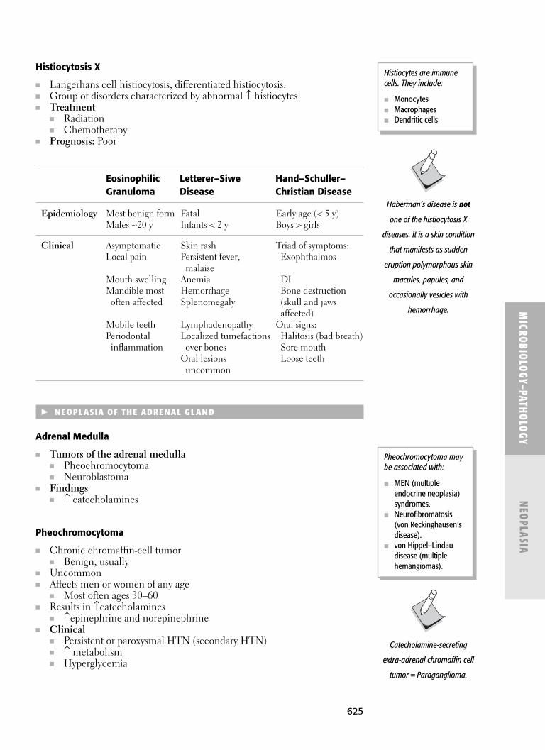
625
MICRO
BIOLOGY–PATHO
LOGYN
EOPLASIA
Histiocytosis X
■ Langerhans cell histiocytosis, differentiated histiocytosis.■ Group of disorders characterized by abnormal ↑ histiocytes.■ Treatment
■ Radiation■ Chemotherapy
■ Prognosis: Poor
Eosinophilic Letterer–Siwe Hand–Schuller– Granuloma Disease Christian Disease
Epidemiology Most benign form Fatal Early age (< 5 y)Males ~20 y Infants < 2 y Boys > girls
Clinical Asymptomatic Skin rash Triad of symptoms: Local pain Persistent fever, Exophthalmos
malaiseMouth swelling Anemia DIMandible most Hemorrhage Bone destruction
often affected Splenomegaly (skull and jaws affected)
Mobile teeth Lymphadenopathy Oral signs: Periodontal Localized tumefactions Halitosis (bad breath)
inflammation over bones Sore mouthOral lesions Loose teeth
uncommon
� N EOPLASIA OF TH E ADRE NAL G LAN D
Adrenal Medulla
■ Tumors of the adrenal medulla■ Pheochromocytoma■ Neuroblastoma
■ Findings■ ↑ catecholamines
Pheochromocytoma
■ Chronic chromaffin-cell tumor■ Benign, usually
■ Uncommon■ Affects men or women of any age
■ Most often ages 30–60■ Results in ↑catecholamines
■ ↑epinephrine and norepinephrine■ Clinical
■ Persistent or paroxysmal HTN (secondary HTN)■ ↑ metabolism■ Hyperglycemia
Histiocytes are immunecells. They include:
■ Monocytes■ Macrophages■ Dendritic cells
Haberman’s disease is not
one of the histiocytosis X
diseases. It is a skin condition
that manifests as sudden
eruption polymorphous skin
macules, papules, and
occasionally vesicles with
hemorrhage.
Pheochromocytoma maybe associated with:
■ MEN (multipleendocrine neoplasia)syndromes.
■ Neurofibromatosis(von Reckinghausen’sdisease).
■ von Hippel–Lindaudisease (multiplehemangiomas).
Catecholamine-secreting
extra-adrenal chromaffin cell
tumor = Paraganglioma.
626
MIC
ROBI
OLO
GY–P
ATHO
LOGY
NEO
PLAS
IA
Neuroblastoma
■ Most common malignant tumor of childhood and infancy■ Adrenal medulla (most common site)■ Etiology
■ N-myc oncogene amplification.■ Clinical presentation
■ Abdominal mass.■ Abdominal distention.■ Sensation of fullness.■ Abdominal pain.
■ 80% of neuroblastomas are productive of catecholamines.■ ↑ epinephrine■ ↑ HR■ ↑ anxiety
■ Complications■ Invasion of abdominal viscera.■ Direct spread. ■ Metastasis to liver, lung, or bone.
� N EOPLASIA OF BON E
Bone Tumors
Primary Bone Tumors Metastatic Bone Tumors
Rare (<1% all malignant tumors) Majority of bone tumorsM > F, young men Secondary; seeds from primary site■ Osteosarcoma ■ Breast
■ Lung■ Prostate■ Kidney■ Thyroid
Osseous Nonosseous
Osteosarcoma Ewing’s (primitive neuroectodermal tumors)
Parosteal osteosarcoma Chordoma (remnants ofnotocord, clivus, andcoccyx)
Chondrosarcoma (actually from cartilage) Fibrosarcoma (fibroblasts)Malignant giant cell tumor
(multinucleated giant cells[osteoclast like])
Osteosarcoma
■ Most common primary tumor of bone■ Malignant tumor ■ Mesenchymal origin (bone)
■ Arise from osteoblast and osteoclasts
Osseous bone tumors arise
from bony structure itself.
Nonosseous tumors arise
most often from hematopoietic,
vascular, or neural tissues.
Most common symptomof bone tumor = Bonepain.
■ Dull.■ Usually localized.■ ↑ at night.■ ↑ with movement .
Most common type ofbone tumors in childrenare
■ Osteosarcoma(osteogenic sarcoma).
■ Ewing’s sarcoma.

627
MICRO
BIOLOGY–PATHO
LOGYN
EOPLASIA
■ Males 10–30■ Common sites:
■ Femur■ Tibia■ Humerus
■ Less frequent sites:■ Fibula■ Ileum■ Vertebra■ Mandible
PAROSTEAL OSTEOSARCOMA
■ Women aged 30–40.■ Most often affects the distal femur.
■ May occur in the humerus, tibia, ulna.■ Develops on bone surface.
■ Not endosseous (interior).■ Progresses slowly.
Chondrosarcoma
■ Males aged 30–50.■ Arises from cartilage.■ Common sites
■ Pelvis■ Proximal femur■ Ribs■ Shoulder girdle
■ Clinical■ Usually painless
■ Course■ Grows slowly■ Locally recurrent and invasive
Malignant Giant Cell Tumor
■ Females aged 18–50.■ Arises from benign giant cell tumor.■ Most common site: Long bones, especially knee area.
� BON E TU MORS OF NONOSSEOUS ORIG I N
Ewing’s Sarcoma
■ Nonosseous malignant bone tumor. ■ Children
■ Usually males aged 10–20 y.■ Usually develops during puberty when bones are growing rapidly.
■ Sites■ Originates in bone marrow; invades long and flat bones.■ Long bones upper and lower extremities
■ Femur

628
MIC
ROBI
OLO
GY–P
ATHO
LOGY
NEO
PLAS
IA
■ Tibia■ Fibula■ Humerus
■ Pelvis/innominate bones■ Ribs■ Vertebra■ Skull■ Facial bones
■ Histology■ Innominate; difficult to distinguish from neuroblastoma and reticulum
cell sarcoma.■ Symptoms
■ Few symptoms■ Pain: ↑ severity and persistence with time■ Swelling at site■ Fever ■ Pathologic fractures
■ Course■ Metastasis■ One-third of children have metastases at time of diagnosis.■ To lungs and other bones.
Fibrosarcoma
■ Males 30–40 y■ Sites: Originates in fibrous tissue of bone
■ Invades long or f lat bones: ■ Femur■ Tibia■ Mandible
■ Invades periosteum and overlying muscle.
Chordoma
■ Males 50–60 years old.■ Derived from embryonic remnants of notochord.■ Slowly progressive.■ Sites
■ End of spinal column ■ Vertebra■ Sacrococcygeal region■ Spheno-occipital region
■ Symptoms (depending on site)■ Constipation■ Visual disturbances
Ewing’s is very radiosensitive.
Metastasize to bone:
■ Prostate■ Breast■ Kidney■ Thryoid■ Lung
“Everything”metastasizes to lung,most commonly:
■ Breast■ Colon■ Prostate

� Tooth Morphology
� Pulp Morphology
� Calcification andEruption
� Occlusion andFunction
� Tooth Anomalies
S E C T I O N 4
Dental Anatomy and Occlusion
629

This page intentionally left blank

C H A P T E R 2 4
Tooth Morphology
Dental Anatomy 632
CLASSIFICATION OF DENTITIONS 632
DENTAL ANATOMIC TERMINOLOGY 632
INNERVATION 635
BLOOD SUPPLY 635
Permanent Tooth Form 635
GENERAL CONCEPTS 637
MAXILLARY PERMANENT TEETH 641
MANDIBULAR PERMANENT TEETH 645
Primary (Deciduous) Tooth Form 648
GENERAL CONCEPTS 648
MAXILLARY PRIMARY TEETH 650
MANDIBULAR PRIMARY TEETH 651
631

632
DENT
AL A
NATO
MY
AND
OCCL
USIO
NTO
OTH
MO
RPHO
LOGY
� DE NTAL ANATOMY
Classification of Dentitions
BY MORPHOLOGY
■ Homodont dentition: All teeth have the same morphology.■ Heterodont dentition: Teeth have different morphology (eg, humans).
BY SETS OF TEETH
■ Monophyodont dentition: One set of teeth.■ Diphyodont dentition: Two sets of teeth (eg, humans).■ Polyphyodont dentition: Multiple sets of teeth.
Dental Anatomic Terminology
TOOTH LOCATION
■ Anterior teeth: Incisors and canines. 12 total (6 per arch).■ Posterior teeth: Premolars and molars. 20 total (10 per arch).
CROWN TYPES
■ Anatomic crown: The portion of the tooth that extends from the cemen-toenamel junction (CEJ) to the incisal edge or occlusal surface (enamel-covered portion of the tooth).
■ Clinical crown: The portion of the tooth that extends incisally or occlusallyfrom the gingival margin (clinically visible portion of the tooth).
CHEWING SURFACES
■ Incisal edge: The chewing surface of anterior teeth.■ Occlusal surface: The chewing surface of posterior teeth consisting of
cusps, ridges, and grooves.■ Occlusal table: The occlusal surface within the cusp and marginal ridges.
ENAMEL SURFACE ELEVATIONS
■ Lobe: The primary center of enamel formation in a tooth. In fully formedteeth, lobes are represented by cusps, mamelons, and cingula, and are sep-arated by developmental depressions (anterior teeth) or developmentalgrooves (posterior teeth).
■ Mamelon: A round extension of enamel on the incisal edge of all incisors.(See Figure 24–1.) There are usually three mamelons per incisor (one foreach facial lobe). They are often translucent because of a lack of underly-ing dentin. Mamelons are typically worn down by attrition and mastica-tion; thus, their presence in adults is an indication of malocclusion.
■ Cingulum: A bulbous convexity of enamel located on the cervical third ofthe lingual surface of all anterior teeth.
■ Cusp: A large elevation of enamel located on the occlusal surface of allposterior teeth and the incisal edge of canines.
■ Tubercle: An extra formation of enamel on the crown of a tooth. Oftenmanifests as a supernumerary cusp, such as the cusp of Carabelli.
F I G U R E 2 4 – 1 .Mamelons.

633
RIDGES
See Figures 24–2 and 24–3.■ Ridge: A linear elevation on the enamel surface.■ Marginal ridge: A ridge on all teeth that forms the mesial and distal mar-
gins of posterior occlusal surfaces and anterior lingual surfaces.
DENTAL ANATOM
Y AN
D OCCLUSION
TOOTH MO
RPHOLOGY
Cementoenameljunction
Cementoenameljunction
Cingulum
Distalmarginal
ridge
Cingulum
Lingual ridge
Mesial marginalridge
A. B.
Lingual fossa Mesialmarginal ridge
Mesial fossa
Distal fossaDistalmarginal ridge
F I G U R E 2 4 – 2 . Occlusal landmarks of two anterior teeth.
A. Maxillary right central incisor; B. Maxillary right canine.
Oblique ridge
Distal marginal
ridge
Distobuccalcusp ridge
Mesiobuccalcusp ridge
Mesialmarginal
ridge
Distolingualcusp ridge
Mesiolingualcusp ridge
Cusp ofCarabelli
Transversegroove
Buccalgroove Central pit
Central groove
Cusp of Carabelligroove
Lingualgroove
Distal obliquegroove
A. B.
F I G U R E 2 4 – 3 . Occlusal landmarks of a posterior tooth.
A. Maxillary right 1st molar––ridges; B. Maxillary right 1st molar––grooves.

634
DENT
AL A
NATO
MY
AND
OCCL
USIO
NTO
OTH
MO
RPHO
LOGY
■ Labial ridge: A ridge only on canines that runs incisocervically in the cen-ter of the facial crown surface. More prominent in maxillary canines.
■ Buccal (cusp) ridge: A ridge only on premolars that runs occlusocervicallyin the center of the buccal crown surface. More prominent in firstpremolars.
■ Cervical ridge: A ridge on all primary teeth and permanent molars thatruns mesiodistally in the cervical third of the buccal surface of the crown.
■ Oblique ridge: A ridge on all maxillary molars that extends from the MLto DB cusps (it separates the MB and DL cusps).
■ Triangular ridge: A ridge on all posterior teeth that extends from the cusptip to the central groove. The ML cusp of all maxillary molars has two tri-angular ridges.
■ Transverse ridge: A ridge on most posterior teeth that runs buccolinguallyand connects opposing buccal and lingual triangular ridges. Most com-mon on maxillary premolars and mandibular molars.
ENAMEL SURFACE DEPRESSIONS
■ Sulcus: A V-shaped depression on the occlusal surface of posterior teethbetween ridges and cusps.
■ Fossa: An irregularly shaped depression in the enamel surface.■ Developmental groove: A well-defined, shallow, linear depression in
enamel that separates the cusps, lobes, and marginal ridges of a tooth.■ Fissure: A narrow crevice at the deepest portion of the developmental
groove in enamel.■ Pit: A small pinpoint concavity at the termination or junction of develop-
mental grooves.■ Supplemental groove: An irregularly defined, short groove auxiliary to a
developmental groove that does not separate major tooth parts.
ENAMEL SURFACE JUNCTIONS
■ Line angle: An angle formed by the junction of two surfaces.■ Point angle: An angle formed by the junction of three surfaces.
EMBRASURES
■ Contact area: The location at which the proximal surfaces of two adjacentteeth make contact.
■ Embrasure: A triangular-shaped space between the proximal surfaces ofadjacent teeth which diverges in four directions from the contact area.■ Buccal■ Lingual■ Occlusal/incisal■ Cervical/gingival (interproximal space): In health, this space is com-
pletely filled with the gingival papilla.■ Largest occlusal embrasure: between max canine and PM1.■ Largest incisal embrasure: between max lateral and canine.■ Smallest incisal embrasure: between mand centrals.■ In general, lingual embrasures > buccal embrasures, except Max M1 (buccal
embrasures > lingual)
INTERDENTAL SPACES
■ Diastema: A space between two adjacent teeth in an arch.
Caries is most likely to occur in
pits, fissures, and grooves. The
teeth least likely to be lost to
caries are mandibular incisors.
Anterior teeth have six line
angles. Posterior teeth have
eight line angles. All teeth
have four point angles.

635
INTRADENTAL SPACES
■ Furcation: The area of a multirooted tooth where the roots diverge.
Innervation
Figure 24–4 illustrates the nerve supply to the teeth and gingiva.
■ All dental and periodontal innervation arises from the trigeminal nerve(CN V).
■ The maxillary nerve (V-2) supplies the maxillary teeth.■ The mandibular nerve (V-3) supplies the mandibular teeth.
Blood Supply
ARTERIAL SUPPLY
■ All dental and periodontal arterial supply arises from the maxillary artery.■ The arterial supply generally parallels the corresponding nerves.
VENOUS RETURN
■ All dental and periodontal venous return drains to the pterygoid plexus ofveins, which eventually forms as the maxillary vein.
DENTAL ANATOM
Y AN
D OCCLUSION
TOOTH MO
RPHOLOGY
Maxillary molars are
generally trifurcated;
mandibular molars are
generally bifurcated.
G. P.
1 2
4 3
88
77
66
55
4433
22 11
L.
8
7
6
5
3 32 11 2
445
6
8
7
N.P.
Anterosuperior alveolar n.
Middle superior alveolar n.
Posterosuperior alveolar n.
Buccal n.
Mental n.
Anterosuperior alveolar n.
Middle superior alveolar n.
Posterosuperior alveolar n.
Inferior alveolar n.
Gingiva Teeth
F I G U R E 2 4 – 4 . Nerve supply to maxillary and mandibular dental arches.
Reprinted, with permission, from Liebgott B. The Anatomical Basis of Dentistry. Toronto: BC
Decker, 1986.
The DB root of the maxillary
first molar is innervated by the
posterosuperior alveolar
nerve; the MB root receives its
nerve supply from the middle
superior alveolar nerve.

T A B L E 2 4 – 1 . Comparison of Permanent Tooth Forms
INCISO-CERVICAL
DIRECTION ROOT CROWN # OF
MESIAL DISTAL FACIAL LINGUAL LENGTH LENGTH # OF # OF # OF ROOT
CONTACT CONTACT HOC HOC (mm) (mm) LOBES CUSPS ROOTS CANALS
Maxilla
Central Incisal Junction* Cervical Cervical 1/3 13 10.5 4 1 1 1
incisor 1/3 1/3 (cingulum)
Lateral Junction* Middle Cervical Cervical 1/3 13 9 4 1 1 1
incisor 1/3 1/3 (cingulum)
Canine Junction* Middle Cervical Cervical 1/3 17 10 4 1 1 1
1/3 1/3 (cingulum)
First Middle Middle Cervical Middle 14 8.5 4 2 2 2
premolar 1/3 1/3 1/3 1/3
Second Middle Middle Cervical Middle 14 8.5 4 2 1 1
premolar 1/3 1/3 1/3 1/3
First Junction* Middle Cervical Middle 12–13 7 5 4 or 5 3 3 or 4
molar 1/3 1/3 1/3
Second Junction* Middle Cervical Middle 11–12 6.5 4 4 3 3 or 4
molar 1/3 1/3 1/3
Third Junction* N/A Cervical Middle 11 6 4 or 5 3 3 Variable
molar 1/3 1/3
Mandible
Central Incisal Incisal Cervical Cervical 1/3 12.5 9 4 1 1 1
incisor 1/3 1/3 1/3 (cingulum)
Lateral Incisal Incisal Cervical Cervical 1/3 14 9.5 4 1 1 1 or 2
incisor 1/3 1/3 1/3 (cingulum)
Canine Incisal Middle Cervical Cervical 1/3 16 11 4 1 1 or 2 1
1/3 1/3 1/3 (cingulum)
First Middle Middle Cervical Middle 14 8.5 4 2 1 1
premolar 1/3 1/3 1/3 1/3
636
DENT
AL A
NATO
MY
AND
OCCL
USIO
NTO
OTH
MO
RPHO
LOGY
� PE RMAN E NT TOOTH FORM
Table 24–1 compares the important characteristics of each permanent tooth.
(Continued)

637
DENTAL ANATOM
Y AN
D OCCLUSION
TOOTH MO
RPHOLOGY
T A B L E 2 4 – 1 . Comparison of Permanent Tooth Forms (Continued)
INCISO-CERVICAL
DIRECTION ROOT CROWN # OF
MESIAL DISTAL FACIAL LINGUAL LENGTH LENGTH # OF # OF # OF ROOT
CONTACT CONTACT HOC HOC (mm) (mm) LOBES CUSPS ROOTS CANALS
Mandible
Second Middle Middle Cervical Occlusal 14.5 8 5 2 or 3 1 1
premolar 1/3 1/3 1/3 1/3
First Junction* Middle Cervical Middle 14 7.5 5 5 2 3 or 4
molar 1/3 1/3 1/3
Second Junction* Middle Cervical Middle 13 7 4 4 2 3 or 4
molar 1/3 1/3 1/3
Third Junction* N/A Cervical Middle 11 7 4 or 5 4 or 5 2 Variable
molar 1/3 1/3
*Junction of the incisal/occlusal and middle thirds.
General Concepts
TOOTH NUMBERING
See Figures 24–5 through 24–7.
■ There are 32 total permanent teeth (16 per arch).
F I G U R E 2 4 – 5 . The maxillary arch.

638
DENT
AL A
NATO
MY
AND
OCCL
USIO
NTO
OTH
MO
RPHO
LOGY
F I G U R E 2 4 – 6 . The mandibular arch.
F I G U R E 2 4 – 7 . Tooth numbering systems.
A. Universal Numbering System of the permanent dentition; B. Federation Dentaire Interna-
tionale (FDI) System for the permanent dentition.
1 2 3 4 5 6 7 8 9 10 11 12 13 14 15 16
32 31 30 29 28 27 26 25 24 23 22 21 20 19 18 17
Max
MandA
Right Left
■ Universal Numbering System: Teeth are numbered 1 to 32, starting fromthe maxillary right 3rd molar and ending with the mandibular right 3rdmolar (Figure 24–7A).
■ Federation Dentaire Internationale (FDI) System: Each tooth isgiven a two-digit number. The first digit represents the quadrant inwhich the tooth is located (1, 2, 3, or 4). The second digit indicatesthe tooth position relative to the midline, from closest to farthest away(1 to 8) (Figure 24–7B).
18 17 16 15 14 13 12 11 21 22 23 24 25 26 27 28
48 47 46 45 44 43 42 41 31 32 33 34 35 36 37 38
Max
Mand
Right Left
B
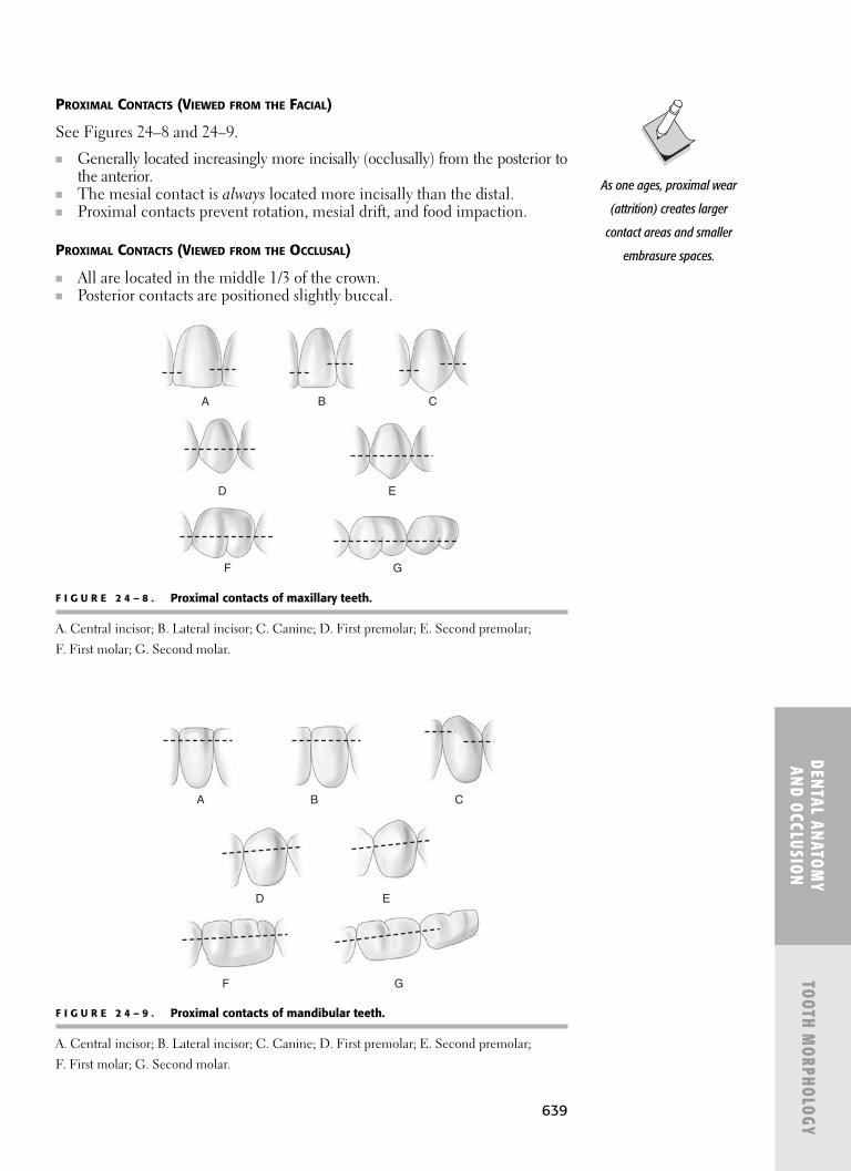
639
DENTAL ANATOM
Y AN
D OCCLUSION
TOOTH MO
RPHOLOGY
A
D E
F G
B C
F I G U R E 2 4 – 8 . Proximal contacts of maxillary teeth.
A. Central incisor; B. Lateral incisor; C. Canine; D. First premolar; E. Second premolar;
F. First molar; G. Second molar.
F I G U R E 2 4 – 9 . Proximal contacts of mandibular teeth.
A. Central incisor; B. Lateral incisor; C. Canine; D. First premolar; E. Second premolar;
F. First molar; G. Second molar.
A
D E
F G
B C
PROXIMAL CONTACTS (VIEWED FROM THE FACIAL)
See Figures 24–8 and 24–9.
■ Generally located increasingly more incisally (occlusally) from the posterior tothe anterior.
■ The mesial contact is always located more incisally than the distal.■ Proximal contacts prevent rotation, mesial drift, and food impaction.
PROXIMAL CONTACTS (VIEWED FROM THE OCCLUSAL)
■ All are located in the middle 1/3 of the crown.■ Posterior contacts are positioned slightly buccal.
As one ages, proximal wear
(attrition) creates larger
contact areas and smaller
embrasure spaces.
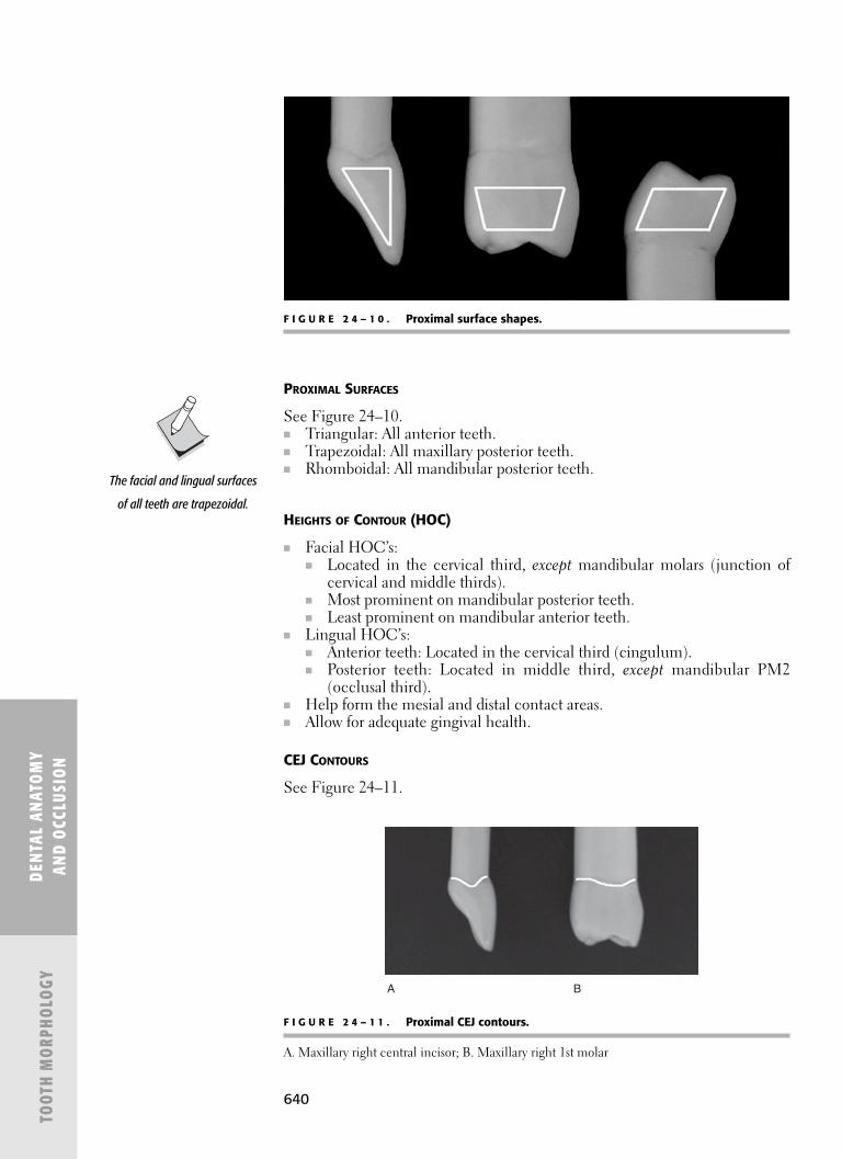
640
PROXIMAL SURFACES
See Figure 24–10.■ Triangular: All anterior teeth.■ Trapezoidal: All maxillary posterior teeth.■ Rhomboidal: All mandibular posterior teeth.
HEIGHTS OF CONTOUR (HOC)
■ Facial HOC’s:■ Located in the cervical third, except mandibular molars (junction of
cervical and middle thirds).■ Most prominent on mandibular posterior teeth.■ Least prominent on mandibular anterior teeth.
■ Lingual HOC’s:■ Anterior teeth: Located in the cervical third (cingulum).■ Posterior teeth: Located in middle third, except mandibular PM2
(occlusal third).■ Help form the mesial and distal contact areas.■ Allow for adequate gingival health.
CEJ CONTOURS
See Figure 24–11.
DENT
AL A
NATO
MY
AND
OCCL
USIO
NTO
OTH
MO
RPHO
LOGY
F I G U R E 2 4 – 1 0 . Proximal surface shapes.
F I G U R E 2 4 – 1 1 . Proximal CEJ contours.
A. Maxillary right central incisor; B. Maxillary right 1st molar
A B
The facial and lingual surfaces
of all teeth are trapezoidal.

641
■ The maximum height of the proximal CEJ contour increases anteriorly.■ The mesial CEJ contour is always greater than the distal contour.■ The greatest CEJ contour is on the maxillary central incisor (mesial surface).■ Facial and lingual CEJs curve apically.■ Mesial and distal CEJs curve coronally.
LOBES
■ Incisors and canines: 4 lobes (3 labial [mamelons], 1 lingual [cingulum]).■ Premolars: 4 lobes (3 buccal, 1 lingual) except mand PM2, which has 5
lobes (3 buccal, 2 lingual).■ First molars: 5 lobes (one for each cusp).■ Second molars: 4 lobes (one for each cusp).■ Third molars: 4 or 5 lobes (one for each cusp, depending on variation).
ARCH LENGTHS
■ Maxillary: 128 mm (slightly longer).■ Mandibular: 126 mm.
Maxillary Permanent Teeth
CENTRAL INCISOR
See Figure 24–12.
■ Unique characteristics■ Most prominent tooth.■ Greatest mesial CEJ contour of all teeth.■ Second longest crown (next to mandibular canine).■ Widest (M-D) of all anterior teeth.
■ Crown morphology■ Occlusal shape is triangular; proximal shape is triangular.■ Has three mamelons and four developmental grooves.■ MMR > DMR.■ M-I corner sharper than D-I.■ Narrowest incisal embrasures of all maxillary teeth.
■ Occlusal morphology■ M-D dimension > F-L (from occlusal).■ Cingulum: slightly distal.
■ Root morphology■ Least likely to have a divided root canal.■ Roundest root form; can be rotated during extraction.■ Blunted apex.■ Has greatest axial inclination toward the facial.■ Sits almost vertically (M-D) in alveolar bone.
■ Occlusal contacts■ Occludes with mandibular central and lateral incisors.
LATERAL INCISOR
See Figure 24–13.
■ Unique characteristics■ Third most common congenitally missing tooth (next to third molars).
DENTAL ANATOM
Y AN
D OCCLUSION
TOOTH MO
RPHOLOGY
F I G U R E 2 4 – 1 2 .Maxillary central incisor.
F I G U R E 2 4 – 1 3 .Maxillary lateralincisor.
For all anterior teeth when
viewed from the facial,
faciolingual dimension >
mesiolingual dimension.

642
■ Third most variable tooth form (next to third molars).■ Peg lateral (microdont)■ Dens-in-dente
■ Most common tooth to have a palato-radicular groove (2–5% preva-lence), which can make scaling difficult.
■ Crown morphology■ Occlusal shape is oval; proximal shape is triangular.■ MMR (straight) > DMR.■ M-I corner sharper than D-I.
■ Occlusal morphology■ M-D dimension > F-L (from occlusal).■ Most developed lingual anatomy of all anterior teeth.■ Lingual surface is the most concave of all incisors.■ Cingulum: central. Often prominent (talon cusp), creating a lingual
pit.■ Root morphology
■ Root just as long as maxillary central.■ Typically curves to the distal.
■ Occlusal contacts■ Occludes with mandibular lateral incisor and canine.
CANINE
See Figure 24–14.
■ Unique characteristics■ Longest tooth.■ Longest cusp.■ Third longest crown (next to mand canine and max central).■ Widest (B-L) anterior tooth.■ Maxillary tooth least likely to be extracted.
■ Crown morphology■ Has prominent facial ridge.■ Cusp tip centered over root (from facial).■ Mesial cusp ridge < distal.■ Mesial surface is straighter (less convex) than the distal.
■ Occlusal morphology■ F-L dimension > M-D (from occlusal).■ Has a prominent lingual ridge, which splits the lingual fossa into
mesial and distal fossae.■ Cusp tip located slightly M-F (from occlusal).■ Cingulum: central and prominent.
■ Root morphology■ Longest root.■ Root is oval-shaped and flattened M-D (deeper on distal).
■ Occlusal contacts■ Occludes with mandibular canine and first premolar.
FIRST PREMOLAR
See Figure 24–15.
■ Unique characteristics■ Largest of all premolars.
■ Crown morphology■ Has a prominent buccal ridge.
DENT
AL A
NATO
MY
AND
OCCL
USIO
NTO
OTH
MO
RPHO
LOGY
F I G U R E 2 4 – 1 4 .Maxillary canine.
F I G U R E 2 4 – 1 5 .Maxillary first pre-molar.

643
■ Only permanent tooth with mesial cusp ridge > distal cusp ridge.■ Has a prominent mesial marginal ridge groove, which can make scal-
ing difficult.■ Occlusal morphology
■ Occlusal shape is hexagonal; proximal shape is trapezoidal.■ Occlusal table has long central groove with fewer supplemental
grooves. No central pit, but has mesial/distal pits.■ 2 cusps: buccal (slightly distal), palatal (slightly mesial).■ Buccal cusp height > palatal.
■ Root morphology■ Only premolar with two roots (B > P).■ Has a prominent mesial root concavity.■ Sits most vertically (B-L) in alveolar bone.
■ Occlusal contacts■ Occludes with mandibular first and second premolars.
SECOND PREMOLAR
See Figure 24–16.■ Unique Characteristics
■ Shorter and smaller than maxillary first premolar.■ Crown morphology
■ Distal cusp ridge > mesial cusp ridge.■ Has a buccal ridge, but not as prominent as first premolar.
■ Occlusal morphology■ Occlusal shape is hexagonal; proximal shape is trapezoidal.■ Occlusal table is more ovoid and symmetrical than maxillary first
premolar.■ Occlusal table has a short central groove with more supplemental
grooves. Has “wrinkled” appearance.■ 2 cusps: buccal, palatal (slightly mesial).■ Buccal cusp height = palatal.
■ Root morphology■ Only premolar without a mesial root depression.
■ Occlusal contacts■ Occludes with mandibular second premolar and first molar.
FIRST MOLAR
See Figure 24–17.
■ Unique characteristics■ Largest permanent tooth.■ Only tooth that is broader lingually than buccally.■ Widest tooth (B-L).■ First permanent maxillary tooth to erupt.
■ Crown morphology■ Only tooth with a pronounced distal concavity at the CEJ, which can
make scaling difficult.■ Has a long buccal groove with a central pit.■ Has a distolingual groove with a pit (on all maxillary molars).
■ Occlusal morphology■ Occlusal shape is rhomboidal; proximal shape is trapezoidal.■ 4 cusps: MB, ML (largest), DB, DL (smallest).■ Sometimes has fifth cusp of Carabelli lingual to ML cusp.
DENTAL ANATOM
Y AN
D OCCLUSION
TOOTH MO
RPHOLOGY
F I G U R E 2 4 – 1 6 .Maxillary secondpremolar.
F I G U R E 2 4 – 1 7 .Maxillary first molar.

644
■ Primary cusp triangle: formed by ML, MB, and DB cusps (same forall maxillary molars).
■ Secondary cusp triangle: formed by DL cusp.■ Cusp heights: ML > MB > DB (primary cusp triangle) > DL (sec-
ondary cusp triangle) > Carabelli.■ Only tooth with two triangular ridges on one cusp (ML cusp): form the
transverse and oblique ridges.■ Has most prominent oblique ridge of all maxillary molars.■ The transverse groove of the oblique ridge connects the central and
distal fossae (same for all maxillary molars).■ Root morphology
■ 3 roots: MB, DB (shortest), palatal (longest).■ Distance from furcation entrance to CEJ: Mesial (3.6 mm) < buccal
(4.2 mm) < distal (4.8 mm).■ Apices are closest to maxillary sinus.■ MB and DB roots are often shaped like “plier handles.”■ MB root has more common (94%) and deeper (0.3 mm) concavities
than other roots.■ Occlusal contacts
■ Occludes with mandibular first and second molars.
SECOND MOLAR
See Figure 24–18.
■ Unique characteristics■ Similar to maxillary first molar but smaller and more angular.■ Second most common tooth to have cervical enamel projections (next
to mandibular second molar).■ Closest tooth to the opening of Stenson’s (parotid) duct.
■ Crown morphology■ Has a short buccal groove without a pit.
■ Occlusal morphology■ Occlusal table is usually rhomboidal, but can be heart-shaped if the
DL cusp is absent; proximal shape is trapezoidal.■ 4 cusps: MB, ML (largest), DB, DL (smallest).■ Has smaller oblique ridge with a transverse groove.
■ Root morphology■ Roots are typically longer, closer together, and more distally inclined
than the maxillary first molar. Occasionally fused.■ Longer root trunk than maxillary first molar.
■ Occlusal contacts■ Occludes with mandibular second and third molars.
THIRD MOLAR
See Figure 24–19.
■ Unique characteristics■ Most variable shape of any other tooth (with mandibular third molar).■ Second most congenitally missing tooth (next to mandibular third
molar).■ Shortest permanent tooth.■ Most common tooth to have enamel pearls (with mandibular third molars).
■ Crown morphology■ Shortest maxillary crown.
DENT
AL A
NATO
MY
AND
OCCL
USIO
NTO
OTH
MO
RPHO
LOGY
F I G U R E 2 4 – 1 8 .Maxillary secondmolar.
F I G U R E 2 4 – 1 9 .Maxillary thirdmolar.

645
■ Occlusal morphology■ Occlusal table is usually heart-shaped since DL cusp has little or no
development; proximal shape is trapezoidal.■ Crown tapers lingually.■ 3 cusps: MB, DB, lingual.■ Oblique ridge is poorly developed and often absent.
■ Root morphology■ Roots are often fused and distally inclined.
■ Occlusal contacts■ Occludes only with mandibular third molar.
Mandibular Permanent Teeth
CENTRAL INCISOR
See Figure 24–20.
■ Unique characteristics■ Smallest tooth.■ Narrowest (M-D) tooth.■ Most symmetrical tooth.■ Greatest mesial CEJ contour of mandibular teeth.■ First succedaneous tooth to erupt.
■ Crown morphology■ MMR = DMR.■ D-I corner is equally as sharp as M-I.■ Incisal edge lingual to long axis (from proximal).
■ Occlusal morphology■ F-L dimension > M-D (from occlusal).■ Incisal edge perpendicular to B-L bisector (from occlusal).■ Cingulum: central and indistinct.
■ Root morphology■ Root has mesial and distal (deeper) concavities (hourglass shaped).■ Longer than maxillary central.
■ Occlusal contacts■ Occludes with maxillary central incisor.■ Only anterior tooth to occlude with one tooth.
LATERAL INCISOR
See Figure 24–21.
■ Unique characteristics■ Slightly larger in all dimensions than the mandibular central.■ Second smallest tooth (next to mandibular central).■ Not as symmetrical as the mandibular central.
■ Crown morphology■ Crown is tilted distally on the root (from facial).■ MMR > DMR.■ Incisal edge lingual to long axis (from proximal).
■ Occlusal morphology■ F-L dimension > M-D (from occlusal).■ Incisal edge is twisted disto-lingually (from occlusal).■ Cingulum: slightly distal and indistinct.
■ Root morphology■ Longest root of all incisors.■ Root has mesial (deeper) and distal concavities (hourglass shaped).
DENTAL ANATOM
Y AN
D OCCLUSION
TOOTH MO
RPHOLOGY
F I G U R E 2 4 – 2 0 .Mandibular centralincisor.
F I G U R E 2 4 – 2 1 .Mandibular lateralincisor.

646
■ Occlusal contacts■ Occludes with maxillary central and lateral incisors.
CANINE
See Figure 24–22.
■ Unique characteristics■ Longest crown.■ Second longest tooth (next to max canine). Longest tooth in mandible.■ Second longest root (next to max canine).■ Mandibular tooth least likely to be extracted.
■ Crown morphology■ Has a facial ridge, but less prominent than max canine.■ Mesial cusp ridge < distal.■ Mesial surface is nearly parallel with the long axis.■ Cusp tip slightly lingual to long axis (from proximal).
■ Occlusal morphology■ F-L dimension > M-D (from occlusal).■ Has a lingual ridge, but less prominent than max canine.■ Cusp tip located slightly distal (from occlusal).■ Cingulum: slightly distal and less prominent than max canine.
■ Root morphology■ Root is oval-shaped and flattened M-D (deeper on mesial).■ Anterior tooth most likely to have a bifurcated root.■ Only root that may be mesially inclined.
■ Occlusal contacts■ Occludes with maxillary lateral incisor and canine.
FIRST PREMOLAR
See Figure 24–23.
■ Unique characteristics■ Smallest of all premolars.
■ Crown morphology■ Has a prominent mesio-lingual groove.■ MMR < DMR.
■ Occlusal morphology■ Occlusal shape is diamond-shaped; proximal shape is rhomboidal
(tilted lingually).■ Only posterior tooth with an occlusal plane tilted lingually.■ Has most prominent transverse ridge of all premolars, which splits
mesial and distal fossae. No central groove.■ 2 cusps: buccal (functional), lingual (nonfunctional).■ Buccal cusp is larger (2/3 of occlusal surface) than the lingual.
■ Root morphology■ Root is broader (B-L), and may have proximal concavities.
■ Occlusal contacts■ Occludes with maxillary canine and first premolar.
SECOND PREMOLAR
See Figure 24–24.
■ Unique characteristics■ Most congenitally missing premolar.■ The gingival papilla between the first and second premolar is the shortest.
DENT
AL A
NATO
MY
AND
OCCL
USIO
NTO
OTH
MO
RPHO
LOGY
F I G U R E 2 4 – 2 2 .Mandibular canine.
F I G U R E 2 4 – 2 3 .Mandibular first premolar.
F I G U R E 2 4 – 2 4 .Mandibular secondpremolar.

647
DENTAL ANATOM
Y AN
D OCCLUSION
TOOTH MO
RPHOLOGY
■ Crown morphology■ Lingual surface is wider (M-D) than that of the mandibular first premolar.■ Shorter and wider than the mandibular first premolar (from buccal).■ Mesial marginal ridge has slight concavity.■ Only premolar with five lobes: three buccal and two lingual.
■ Occlusal morphology■ Occlusal shape is square; proximal shape is rhomboidal (tilted lin-
gually).■ Occlusal surface can have three configurations:
■ Y (most common configuration): 3 cusps—buccal > ML > DL,with a single central pit.
■ H: 2 cusps—buccal and lingual with short central groove.■ U: 2 cusps—buccal and lingual with crescent-shaped central groove.
■ Buccal cusp is shorter and blunter than lingual cusps.■ ML cusp is larger than the DL cusp.■ Mesial marginal ridge has slight concavity.
■ Root morphology■ Root is longer than the mandibular first premolar.■ Apex closest to mental foramen.
■ Occlusal contacts■ Occludes with maxillary first and second premolars.
FIRST MOLAR
See Figure 24–25.
■ Unique characteristics■ Largest mandibular tooth.■ Widest tooth (M-D).■ First permanent tooth to erupt, and most often restored/extracted.
■ Crown morphology■ Can see all 5 cusps from the buccal.■ Buccal cusps are shorter and blunter then the lingual cusps.
■ Occlusal morphology■ Occlusal shape is pentagonal; proximal shape is rhomboidal (tilted
lingually 15–20°).■ 5 cusps: MB (largest), DB, distal (smallest), ML (tallest), DL.■ Occlusal pattern resembles a +< with a twisted central groove.■ Has two transverse ridges, three fossae with pits, two buccal grooves
(MB and DB), and a short lingual groove.■ Root morphology
■ Roots are usually widely separated.■ The mesial root is longer and wider (B-L).■ Both roots have concavities (mesial slightly more prominent).■ The root trunk is typically shorter than the mandibular second
molar.■ Occlusal contacts
■ Occludes with the maxillary second premolar and first molar.
SECOND MOLAR
See Figure 24–26.
■ Unique characteristics■ Most symmetrical molar.■ Most common tooth to have cervical enamel projections.
F I G U R E 2 4 – 2 5 .Mandibular first molar.
F I G U R E 2 4 – 2 6 .Mandibular secondmolar.
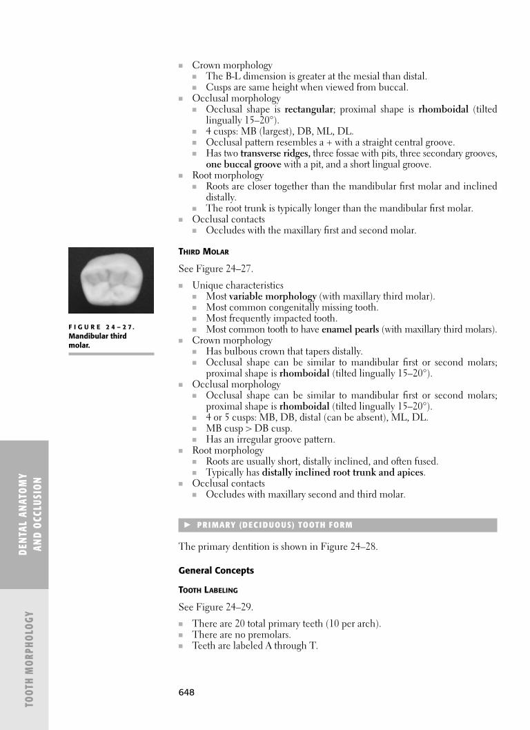
648
■ Crown morphology■ The B-L dimension is greater at the mesial than distal.■ Cusps are same height when viewed from buccal.
■ Occlusal morphology■ Occlusal shape is rectangular; proximal shape is rhomboidal (tilted
lingually 15–20°).■ 4 cusps: MB (largest), DB, ML, DL.■ Occlusal pattern resembles a + with a straight central groove.■ Has two transverse ridges, three fossae with pits, three secondary grooves,
one buccal groove with a pit, and a short lingual groove.■ Root morphology
■ Roots are closer together than the mandibular first molar and inclineddistally.
■ The root trunk is typically longer than the mandibular first molar.■ Occlusal contacts
■ Occludes with the maxillary first and second molar.
THIRD MOLAR
See Figure 24–27.
■ Unique characteristics■ Most variable morphology (with maxillary third molar).■ Most common congenitally missing tooth.■ Most frequently impacted tooth.■ Most common tooth to have enamel pearls (with maxillary third molars).
■ Crown morphology■ Has bulbous crown that tapers distally.■ Occlusal shape can be similar to mandibular first or second molars;
proximal shape is rhomboidal (tilted lingually 15–20°).■ Occlusal morphology
■ Occlusal shape can be similar to mandibular first or second molars;proximal shape is rhomboidal (tilted lingually 15–20°).
■ 4 or 5 cusps: MB, DB, distal (can be absent), ML, DL.■ MB cusp > DB cusp.■ Has an irregular groove pattern.
■ Root morphology■ Roots are usually short, distally inclined, and often fused.■ Typically has distally inclined root trunk and apices.
■ Occlusal contacts■ Occludes with maxillary second and third molar.
� PRI MARY (DEC I DUOUS) TOOTH FORM
The primary dentition is shown in Figure 24–28.
General Concepts
TOOTH LABELING
See Figure 24–29.
■ There are 20 total primary teeth (10 per arch).■ There are no premolars.■ Teeth are labeled A through T.
DENT
AL A
NATO
MY
AND
OCCL
USIO
NTO
OTH
MO
RPHO
LOGY
F I G U R E 2 4 – 2 7 .Mandibular thirdmolar.

649
DENTAL ANATOM
Y AN
D OCCLUSION
TOOTH MO
RPHOLOGY
F I G U R E 2 4 – 2 8 . The primary dentition, occlusal view.
A B C D E F G H I J
O N M L K
Max
Mand
Right LeftT S R Q P
F I G U R E 2 4 – 2 9 . Tooth labeling of the primary (deciduous) dentition.
PRIMARY VS PERMANENT TEETH
See Table 24–2.
TOOTH STRUCTURE PRIMARY TEETH ARE/HAVE…COMPARED TO PERMANENT TEETH
Enamel Lighter (whiter) in colorThinner (only about 1 mm thick)Less calcified (more caries prone)No mamelons (but still develop from lobes)Stops abruptly at the CEJ
Dentin Thinner (easier to have pulp exposure)
Crowns More bulbous (larger cervical bulges)Buccal and lingual surfaces are flatter above the HOCSmaller occlusal tablesFewer grooves and pits (shallower and smoother)Anterior crowns: wider M-D and shorter inciso-cervicallyPosterior crowns: narrower M-D and shorter occluso-cervially
CEJs More constricted (narrower cervix)
Root trunks ShorterNarrower
Roots Longer and more slender relative to crown sizeSmaller crown:root ratioMore tapered (anterior teeth)More divergent (posterior teeth)
Apple Jacks Kill Teeth
The mesiodistal width of the
primary molars is 2–5 mm >
the width of the permanent
premolars.

650
PRIMATE SPACE
■ Allows for proper alignment of the permanent incisors.■ Occurs in about 50% of primary dentitions.■ Maxillary: Between lateral incisor and canine (mesial to canine).■ Mandibular: Between canine and first molar (distal to canine).
ARCH LENGTHS
■ Maxillary: 68.2 mm (longer).■ Mandibular: 61.8 mm.
Maxillary Primary Teeth
See Figure 24–30.
CENTRAL INCISOR
■ Straighter incisal edge than permanent maxillary central.■ No mamelons.■ Prominent facial and lingual cervical ridges.■ Occludes with mandibular central and lateral incisors.
LATERAL INCISOR
■ Straighter incisal edge than permanent maxillary lateral.■ No mamelons.■ Prominent facial and lingual cervical ridges.■ Occludes with mandibular lateral incisor and canine.
CANINE
■ Only primary tooth in which facial shape is pentagonal (not trapezoidal).■ Longer and sharper cusp than permanent maxillary canine.■ Only primary tooth with mesial cusp ridge > distal cusp ridge.■ Occludes with mandibular canine and first molar.
FIRST MOLAR
■ Smallest primary molar.■ Generally resembles a permanent maxillary premolar.■ Occlusal shape is rectangular.■ Occlusal surface has H-shaped configuration.■ Has a prominent buccal cervical ridge.■ 4 cusps: MB (largest), ML (sharpest), DB, DL (smallest).■ Occludes with mandibular first and second molars.
SECOND MOLAR
■ Generally resembles permanent maxillary first molar but smaller.■ Occlusal shape is rhomboidal.■ Widest (B-L) primary tooth.■ Has a prominent buccal cervical ridge.■ Has an oblique ridge.
DENT
AL A
NATO
MY
AND
OCCL
USIO
NTO
OTH
MO
RPHO
LOGY
G H
I J
A
D EF
B
C
F I G U R E 2 4 – 3 0 . Theprimary teeth, facial view.
A. Maxillary central incisor;
B. Maxillary lateral incisor;
C. Maxillary canine;
D. Mandibular central
incisor; E. Mandibular
lateral incisor; F. Mandibular
canine; G. Maxillary first
molar; H. Maxillary second
molar; I. Mandibular first
molar; J. Mandibular
second molar.

651
■ 4 cusps: MB (largest), ML (almost as large as MB), DB, DL (smallest).■ May have a fifth cusp of Carabelli.■ Occludes only with mandibular second molar.
Mandibular Primary Teeth
CENTRAL INCISOR
■ Straighter incisal edge than permanent mandibular central.■ No mamelons.■ Prominent facial and lingual cervical ridges.■ Occludes only with maxillary central incisor.
LATERAL INCISOR
■ Straighter incisal edge than permanent mandibular lateral.■ No mamelons.■ Prominent facial and lingual cervical ridges.■ More exaggerated incisal edge slope than permanent mandibular lateral.■ Occludes with maxillary central and lateral incisors.
CANINE
■ Distal cusp ridge > mesial cusp ridge.■ Occludes with maxillary lateral incisor and canine.
DENTAL ANATOM
Y AN
D OCCLUSION
TOOTH MO
RPHOLOGY
T A B L E 2 4 – 2 . Comparison of Primary (Deciduous) Tooth Forms
ROOT CROWN NUMBER OF NUMBER OF
LENGTH (mm) LENGTH (mm) CUSPS ROOTS
Maxilla
Central incisor 10 6 1 1
Lateral incisor 11.4 5.6 1 1
Canine 13.5 6.5 1 1
First molar 10 5.1 4 3
Second molar 11.7 5.7 5 3
Mandible
Incisor 9 5 1 1
Lateral incisor 10 5.2 1 1
Canine 11.5 6 1 1
First molar 9.8 6 4 2
Second molar 11.3 5.5 5 2

652
FIRST MOLAR
■ Most unique primary or permanent tooth.■ Most difficult primary tooth to restore.■ Occlusal shape is rhomboidal.■ Has no central fossa (but has mesial and distal triangular fossae).■ Has a prominent buccal cervical ridge.■ Has a well-developed mesial marginal ridge.■ Has a prominent transverse ridge (connects MB and ML cusps).■ CEJ curves apically on the mesial (from buccal view).■ 4 cusps: MB (largest), ML (tallest, sharpest), DB, DL (smallest).■ Occludes with maxillary canine and first molar.
SECOND MOLAR
■ Generally resembles permanent mandibular first molar.■ Occlusal shape is rectangular.■ Widest (M-D) primary tooth.■ Has a prominent buccal cervical ridge.■ 5 cusps: MB, ML, D (almost as large as MB and DB cusps), DB, DL.■ Occludes with maxillary first and second molars.
DENT
AL A
NATO
MY
AND
OCCL
USIO
NTO
OTH
MO
RPHO
LOGY

C H A P T E R 2 5
Pulp Morphology
Classification 654
PULP CANAL SYSTEM CONFIGURATION 654
Permanent Dentition 654
ACCESSORY PULP CANALS 654
Primary (Deciduous) Dentition 656
COMPARISON TO PERMANENT TEETH 656
653

654
DENT
AL A
NATO
MY
AND
OCCL
USIO
NPU
LP M
ORP
HOLO
GY
� C LASS I F ICATION
Pulp Canal System Configuration
See Figure 25–1.
I. Type I: Contains a single canal from the pulp chamber to the apical fora-men.
II. Type II: Contains two separate canals leaving the pulp chamber, but latermerge together just short of the apical foramen.
III. Type III: Contains two separate canals leaving the pulp chamber whichexit the root at two separate apical foramina.
IV. Type IV: Contains a single canal leaving the pulp chamber, but divid-ing into two separate canals which exit the root at two separate apicalforamina.
� PE RMAN E NT DE NTITION
■ See Table 25–1 for a comparison of pulpal morphology of the permanentdentition.
Accessory Pulp Canals
■ Found in cervical 1/3 of root and furcations.■ Allow pulp to communicate with PDL space.■ Contains pulpal nervous and vascular tissue.
Size of pulp cavity is dictated
by:
■ Age
■ Parafunctional activity
■ Trauma history (caries,
abrasion, erosion, etc)
There are as many pulp horns
as there are cusps. In
permanent teeth, the size of a
pulp horn is directly
proportional to the size of its
associated cusp.
Type I Type II Type III Type IV
F I G U R E 2 5 – 1 . Pulp canal system classification (Weine).

T A B L E 2 5 – 1 . Comparison of Permanent Teeth
SHAPE OF PULP CROSS SECTION AT
TOOTH # OF ROOTS # OF CANALS CHAMBER AT CEJ LEVEL OF CEJ
Maxilla
Central incisor 1 1 Triangular
Lateral incisor 1 1 Egg
Canine 1 1 Oval
First premolar 2 2 Kidney*
Second premolar 1 1 Oval
First molar 3 3† or 4 (55% 2nd in MB) Rhomboidal
Second molar 3 3† or 4 (35% 2nd in MB) Rhomboidal
Third molar 3 Variable Rhomboidal
Mandible
Central incisor 1 1 Oval
Lateral incisor 1 1 or 2 (40% 2nd is lingual) Oval**
Canine 1 1 Oval
First premolar 1 1 Oval§
Second premolar 1 1 Oval
First molar 2 3‡ or 4 (25% 2nd in D) Rectangular
Second molar 2 3‡ or 4 (8% 2nd in D) Trapezoidal (tapers distally)
Third molar 2 Variable Trapezoidal (tapers distally)
* Often pinched off creating 2 separate canals: B and P.** Sometimes pinched off creating 2 separate canals: B and L.§ Oval, but the roundest of all premolars.† In general, maxillary molars have at least 3 canals, one in each root: MB, DB, and P.‡ In general, mandibular molars have 3 canals, two in the M root (MB and ML) and one in the D root.
655
DENTAL ANATOM
YAN
D OCCLUSION
PULP MO
RPHOLOGY

656
� PRI MARY (DEC I DUOUS) DE NTITION
■ See Table 25–2 for a comparison of pulpal morphology of the primary dentition.
DENT
AL A
NATO
MY
AND
OCCL
USIO
NPU
LP M
ORP
HOLO
GY
T A B L E 2 5 – 2 . Comparison of Primary Teeth
AGE AT ROOT
TOOTH # OF ROOTS # OF CANALS COMPLETION (y)
Maxilla
Central incisor 1 1 1.5
Lateral incisor 1 1 1.5–2
Canine 1 1 2–2.5
First molar 3 3 or 4 (MB root may have 2 canals) 3–3.5
Second molar 3 3 or 4 (MB root may have 2 canals) 3
Mandible
Central incisor 1 1 1.5
Lateral incisor 1 1 1.5–2
Canine 1 1 2–2.5
First molar 2 4 (2 in each root) 3–3.5
Second molar 2 4 (2 in each root) 3
Comparison to Permanent Teeth (Table 25–3)
T A B L E 2 5 – 3 . Comparison to Permanent Teeth
PRIMARY TEETH ARE/HAVE…COMPARED TO
TOOTH STRUCTURE PERMANENT TEETH
Pulp horns More pointed
Extend closer to cusp tips (especially on mesial surfaces)
Pulp chambers Larger
More closely approximate shape of crowns

C H A P T E R 2 6
Calcificationand Eruption
Definitions and General Concepts 658
DEFINITIONS 658
GENERAL CONCEPTS 658
Primary (Deciduous) Dentition 658
Mixed Dentition 659
Permanent Dentition 659
657

658
DENT
AL A
NATO
MY
AND
OCCL
USIO
NCA
LCIF
ICAT
ION
AND
ERUP
TIO
N
� DE F I N ITIONS AN D G E N E RAL CONC E PTS
Definitions
■ Succedaneous teeth: Permanent teeth that occupy positions held by pri-mary teeth.
■ Exfoliation: Shedding of the primary teeth. This is accomplished partly bythe resorption of the deciduous roots by odontoclasts.
■ Mixed dentition: A dentition having both primary and permanent teeth.■ Active eruption: Tooth movement from its germinative position until con-
tact is made with opposing and/or adjacent teeth.■ Passive eruption: Tooth exposure secondary to the apical migration of the
junctional epithelium.
General Concepts
■ Teeth erupt in pairs.■ Girls’ teeth erupt before boys’ teeth.■ Mandibular teeth generally erupt before maxillary teeth.■ Eruption starts once 50% of root formation is complete.
� PRI MARY (DEC I DUOUS) DE NTITION
NUMBER OF TEETH
■ 20 total primary teeth (10 per arch).■ There are no premolars.
DENTAL FORMULA
■ I 2/2 + C 1/1 + M 2/2
ERUPTION SEQUENCE AND TIMING
■ Same sequence for both arches (months):
CALCIFICATION (TABLE 26–1)
I1 I2 M1 C M2
6 9 12 18 24
Only the permanent incisors,
canines, and premolars are
succedaneous.
T A B L E 2 6 – 1 . Calcification of the Primary Dentition
TOOTH CALCIFICATION STARTS (MO) CALCIFICATION ENDS
Central incisor 4–6 in utero 6–10 wk
Lateral incisor 4–6 in utero 10–12 wk
First molar 4–6 in utero 6 mo
Canine 4–6 in utero 9 mo
Second molar 4–6 in utero 10–12 mo

659
DENTAL ANATOM
YAN
D OCCLUSION
CALCIFICATION
AND ERUPTIO
N
DEVELOPMENT
See Table 26–2.
■ Begin to develop at 6 weeks in utero.■ Calcification starts at 4–6 months (18 weeks) in utero (2nd trimester).■ Apices are complete 1–11/2 years after eruption (by age 3).
Permanent mandibular teeth
generally erupt before
maxillary teeth, except the
premolars.
M1 I1 I2 C PM1 PM2 M2 M3
6 6–7 7–8 9–10 10–12 11–12 12 17–21
M1 I1 I2 PM1 PM2 C M2 M3
6 7–8 8–9 10–11 10–12 11–12 12 17–21
T A B L E 2 6 – 2 . Tooth Development of the Primary (Deciduous) Dentition
AGE AT ERUPTION AGE AT
ROOT AGE AT
TOOTH MANDIBULAR MAXILLARY COMPLETION EXFOLIATION
(mo) (mo) (y) (y)
Central 6–10 8–12 11/2 6–7
incisor
Lateral 10–16 9–13 11/2–2 7–8
incisor
First molar 14–18 13–19 2–2/2 9–11
Canine 17–23 16–22 3–31/2 9–12
Second 23–31 25–33 3 10–12
molar
� M IXE D DE NTITION
■ Occurs from ages 6 through 12 years.■ Starts: Eruption of the permanent 1st molar.■ Ends: Exfoliation of the primary maxillary canine.■ Permanent incisors often erupt lingual to their primary counterparts.■ 24 teeth (12 per arch) once the permanent 1st molars erupt.
� PE RMAN E NT DE NTITION
NUMBER OF TEETH
■ 32 total permanent teeth (16 per arch).
DENTAL FORMULA
■ I 2/2 + C 1/1 + PM 2/2 + M 3/3ERUPTION SEQUENCE AND TIMING
■ Mandibular arch (years):
■ Maxillary arch (years):

660
DENT
AL A
NATO
MY
AND
OCCL
USIO
NCA
LCIF
ICAT
ION
AND
ERUP
TIO
N
T A B L E 2 6 – 4 . Tooth Development of the Permanent Dentition
AGE AT ERUPTION (y) AGE AT
ROOT
TOOTH MANDIBULAR MAXILLARY COMPLETION (y)
First molar 6–7 6–7 9–10
Central incisor 6–7 7–8 9–101/2
Lateral incisor 7–8 8–9 10–11
Canine 9–10 11–12* 12–14
First premolar 10–12 10–11 12–131/2
Second premolar 11–12 10–12 121/2–14
Second molar 11–13 12–13 14–15
Third molar 17–21 17–21 18–25
*The maxillary canine is the last succedaneous tooth to erupt.
DEVELOPMENT
See Table 26–4.
■ Begin to develop at 4 months in utero.■ Calcification starts at birth (mandibular 1st molar).■ Apices are complete about 21/2 years after eruption.
T A B L E 2 6 – 3 Calcification of the Permanent Dentition
TOOTH CALCIFICATION STARTS (YR) CALCIFICATION ENDS (YR)
First molar Birth 21/2–3
Central incisor <1 31/2
Lateral incisor <1 4–5
Canine <1 4–7
First premolar 2 5–6
Second premolar 2 51/2–7
Second molar 3 6–8
Third molar 7–9 13–16
CALCIFICATION (TABLE 26–3)

C H A P T E R 2 7
Occlusion and Function
Occlusal Terminology 662
OCCLUSAL RELATIONSHIPS 663
Mandibular Movements 667
FACTORS OF MANDIBULAR MOVEMENT 667
MANDIBULAR POSITIONS 667
POSSELT’S ENVELOPE OF MOTION 667
MANDIBULAR MOVEMENT AND GUIDANCE 667
OCCLUSAL INTERFERENCES 668
CONDYLAR MOVEMENTS 669
OCCLUSAL MUSCULATURE AND LIGAMENTS 670
661

662
DENT
AL A
NATO
MY
AND
OCCL
USIO
NOC
CLUS
ION
AND
FUN
CTIO
N
� OCC LUSAL TE RM I NOLOGY
GENERAL TERMS
■ Occlusion: The contact relationships of the teeth. The contacts are generallyedges or points touching other edges, points, or areas.
■ Functional occlusion: Occlusion during mandibular movement (mastica-tion, swallowing, etc.).
■ Plane of occlusion: An imaginary plane anatomically related to the cra-nium that theoretically touches the incisal edges of incisors and the cusptips of posterior teeth.
■ Occlusal adjustment (equilibration): Reshaping the occlusal surfaces ofteeth to create harmonious contacts between the maxillary and mandibularteeth.
OCCLUSAL DIMENSIONS
■ Vertical dimension: A vertical measurement of the face between any twoarbitrary points (usually in the midline), one above and one below themouth.
■ Vertical dimension of occlusion (VDO): The vertical dimension of theface when the teeth are in centric occlusion.
■ Vertical dimension of rest (VDR): The vertical dimension of the facewhen the mandible is in the rest position.
OCCLUSAL CURVATURES
■ Curve of Spee: The anteroposterior curvature of the maxillary andmandibular occlusal surfaces. Concave above the curve; convex below thecurve. (See Figure 27–1.)
■ Curve of Wilson: The mediolateral curvature of the maxillary andmandibular occlusal surfaces. Concave above the curve; convex below thecurve. (See Figure 27–2.)
■ Compensating curve: The anteroposterior curvature (in the medianplane) and the mediolateral curvature (in the frontal plane) in the align-ment of the occluding surfaces and incisal edges of artificial teeth that areused to develop balanced occlusion.
OCCLUSAL CONTACTS
■ Functional contacts: Contacts made during functional occlusion.■ Parafunctional contacts: Abnormal contacts; typically result from habits
such as bruxism and include area-to-area contacts.
F I G U R E 2 7 – 1 . Curve of Spee.

663
■ Protrusive contacts: Contacts made when the mandible has moved anteri-orly from centric occlusion (protrusive movement). These are usuallyedge-to-edge contacts for anterior teeth.
■ Working side contacts (laterotrusive contacts): Contacts on the side towardwhich the mandible has moved from centric occlusion (working side move-ment).
■ Nonworking side contacts (mediotrusive contacts): Contacts on the sideaway from which the mandible has moved from centric occlusion (non-working side movement).
OCCLUSAL RELATIONSHIPS
■ Overbite: The vertical overlapping of the mandibular incisors by the max-illary incisors when the jaws are in centric occlusion. (See Figure 27–3.)
■ Overjet: The horizontal overlapping of the mandibular incisors by themaxillary incisors when the jaws are in centric occlusion.
■ Open bite: A condition in which opposing teeth do not occlude.■ Cross-bite: An abnormal relation of one or more teeth in one arch to its
antagonist in the other arch due to a deviation of tooth or jaw position.■ Anterior cross-bite: One or more maxillary incisors are positioned lin-
gually to the mandibular incisors when in centric occlusion.■ Posterior cross-bite: One or more maxillary posterior teeth are positioned
palatally to the mandibular posterior teeth when in centric occlusion.
Occlusal Relationships
FOUR DETERMINANTS OF OCCLUSION
1. The teeth and their occlusal surfaces.2. The right TMJ.3. The left TMJ.4. The neuromusculature of the jaws and TMJs.
DENTAL ANATOM
YAN
D OCCLUSION
OCCLUSION AN
DFUN
CTION
F I G U R E 2 7 – 2 . Curve of Wilson.
F I G U R E 2 7 – 3 .Overbite (A) and overjet (B).
B.
A.
In cross-bite occlusions, the
working and nonworking
cusps are reversed for the
affected teeth.

664
ANGLE’S CLASSIFICATION
DENT
AL A
NATO
MY
AND
OCCL
USIO
NOC
CLUS
ION
AND
FUN
CTIO
N
F I G U R E 2 7 – 4 . Class I occlusion.
Angle’s classification is basedon molar and canine
relationships.
Chronic thumb sucking cancause:
■ Anterior open bite
■ Flaring/rotation of
maxillary incisors
■ Crowding of mandibular
incisors
■ Constriction of maxillary
arch
■ High palatal vault
■ Class II, div I malocclusion
Angle’s Class Molars Canines Characteristics
I (See Figure 27–4) MB cusp of max Max canine 70% of M1 occludes occludes populationwith MB betweengroove of mand caninemand M1 and PM1
II MB cusp Max canine 25% of■ Division I: of max M1 occludes population
All max incisors occludes mesially to Retrognathicproclined mesially to the distal(flared) MB groove surface of (See Figure 27–5) of mand M1 mand canine
■ Division II:Max centralsretroclined;lateralsproclined(See Figure 27–6)
III (See Figure 27–7) MB cusp of Max canine 5% ofmax M1 occludes populationoccludes distally to Prognathicdistally to the distalMB groove surface ofof mand M1 mand canine

DENTAL ANATOM
YAN
D OCCLUSION
OCCLUSION AN
DFUN
CTION
F I G U R E 2 7 – 5 . Class II, division I occlusion.
F I G U R E 2 7 – 6 . Class II, division II occlusion.
F I G U R E 2 7 – 7 . Class III occlusion.
665

666
INTERCUSPAL RELATIONSHIPS
DENT
AL A
NATO
MY
AND
OCCL
USIO
NOC
CLUS
ION
AND
FUN
CTIO
N
An ideal occlusion is a class I
occlusion with a smooth plane
of occlusion.
In ideal CO, every tooth in one
arch contacts two teeth in the
opposite arch, EXCEPT:
■ Max 3rd molars
■ Mand central incisors
Cen
Cen Can 1PM 2PM 1M 2M 3MLat
Lat Can 1PM 2PM 1M 2M 3M
F I G U R E 2 7 – 8 . Ideal occlusion.
IDEAL OCCLUSION
See Figure 27–8.
■ Supporting (lingual) cusps of maxillary posterior teeth occlude with the distalmarginal ridge of their mandibular counterpart + the mesial marginal ridgeof tooth distal, except:■ ML cusp of molars, which occlude in the central fossa of their counterpart.
■ Supporting (buccal) cusps of mandibular posterior teeth occlude with themesial marginal ridge of their maxillary counterpart + the distal marginalridge of the tooth mesial, except:■ DB cusp of molars, which occlude in the central fossa of their counterpart,■ D cusp of M1, which occludes in the triangular fossa of max M1.
■ Nonworking (buccal) cusps of maxillary posterior teeth oppose the buccalembrasures of their mandibular counterpart + the tooth distal, except:■ MB cusp of molars, which oppose the buccal groove of their counterpart,■ DB cusp + oblique ridge of M1, which oppose the DB groove of mand
M1.■ Nonworking (lingual) cusps of mandibular posterior teeth oppose the lingual
embrasures of the maxillary counterpart + the tooth mesial, except:■ DL cusp of molars, which oppose the lingual groove of their counterpart,■ L cusp of PM1, which opposes nothing.
Cusp Type Location Oppose Shape Function
Working, Max lingual Central fossae Broader Support supporting, Mand buccal Marginal ridges Rounder verticalholding dimension of
the face
Nonworking, Max buccal Embrasure Sharper Guide balancing, Mand lingual spaces movementsguiding Grooves during
function

667
� MAN DI BU LAR MOVE M E NTS
Factors of Mandibular Movement
1. Initiating position:■ CR: Most stable and reproducible position
2. Two types of motion:■ Rotation■ Translation
3. Three directions of movement:■ Frontal■ Sagittal■ Horizontal
4. Degree of movement5. Clinical significance of movements
Mandibular Positions
■ Centric occlusion (CO): The maximum intercuspation of the opposingarches. It is a purely tooth-guided position. This creates the vertical dimen-sion of occlusion (VDO).
■ Centric relation (CR): The most anterior and superior position of themandibular condyles within the glenoid fossae (terminal hinge position).By maintaining this position, only rotational movements around a hori-zontal (hinge) axis can occur. It is a purely ligament-guided position.
■ Rest (postural) position (RP): The position of the mandible when it is ina physiologic rest position. There are no tooth contacts in this position; itis a purely muscle-guided position. This creates the vertical dimension ofrest (VDR). The resulting space between the teeth in this position (about1–3 mm) is called the freeway space (FS).
■ Maximum opening (MO): The maximum separation of the opposingarches. Creates the greatest amount of space between the teeth (about40–50 mm).
Posselt’s Envelope of Motion
See Figure 27–9.
■ Sagittal plane ■ Horizontal plane ■ Frontal (coronal) plane
Mandibular Movement and Guidance
■ Protrusive movement: The anterior movement of the mandible. As themandible moves anteriorly, the posterior teeth disarticulate. This is knownas anterior guidance. The maximum posterior disarticulation occurswhen the anterior teeth are edge-to-edge. Anterior guidance dependslargely on the horizontal and vertical relationship of the maxillary andmandibular incisors and canines. Cusp length varies on the extent of thisrelationship.
■ Lateral movement: The lateral movement of the mandible. The side towardwhich the mandible moves is the working side; the side away from whichthe mandible moves is the nonworking side. Lateral movements occur viatwo types of guidance:
DENTAL ANATOM
YAN
D OCCLUSION
OCCLUSION AN
DFUN
CTION
During the act of swallowing,
the mandible meets the
maxilla in centric occlusion
and the tongue touches the
palate.
VDR = VDO + FS
Posselt’s envelope of motion
illustrates the range of motion
of the mandible in three
planes.
The mandible acts as a
class III lever.

668
■ Canine guidance: The contact between the opposing canines on theworking side disarticulates all posterior teeth. Any premature contactson the working or nonworking sides are working and nonworking inter-ferences, respectively.
■ Group function: The contact between the opposing canines and pos-terior teeth on the working side disarticulates the posterior teeth on thenonworking side. Any contacts on the nonworking side are nonworkinginterferences.
DENT
AL A
NATO
MY
AND
OCCL
USIO
NOC
CLUS
ION
AND
FUN
CTIO
N
Effects of Occlusal Relationships on Cusp Height
Occlusal Relationship If… Then…
Articular eminence Steeper eminence Taller cusps
Anterior guidance Increased overbite Taller cuspsIncreased overjet Shorter cusps
Curve of Spee More concave curve Shorter cusps
Lateral movement Greater movement Shorter cuspsMore superior
movement ofrotating condyle
Greater Bennett shift
Occlusal Interferences
■ Occlusal interference: Any contact that inhibits the remaining occlusalsurfaces from achieving harmonious contacts.
■ Protrusive interference: Premature contact between the mesial aspects ofmandibular posterior teeth and the distal aspects of maxillary posterior teeth.
■ Working interference: Premature contact on the working side during lat-eral mandibular movement.
■ Nonworking interference: Premature contact on the nonworking sideduring lateral mandibular movement.
Mastication involves:
■ Incision: incisors
■ Prehension: canines
■ Trituration: premolars +
molars
Eliminating nonworking
contacts is important because:
■ They are damaging
■ They are difficult to control
due to mandibular flexure
■ They deliver more force
than other contacts
Maximumprotrusion
Edge-to-edge position
Edge-to-edgeposition
Centric occlusion
Centricocclusion
Retruded contact
Rest position
Rotation
Translation
MaximumopeningA B C
Maximum rightlateral contact
position
Retrudedcontact
Maximumprotrusion
Maximum leftlateral contact
position
Maximum rightlateral contact
position
Centricocclusion
Maximum leftlateral contact
position
Maximumopening
x
F I G U R E 2 7 – 9 . Three planes of Posselt’s envelope of motion.
A. Sagittal; B. Horizontal; C. Frontal.

669
Condylar Movements
See Figure 27–10.
■ Protrusive movement: Both right and left condyles rotate and translateanteriorly simultaneously (downward along the articular eminence).
■ Right working movement: The left condyle rotates and translates anteriorly(downward along the articular eminence) and medially (to the right). Theright condyle rotates forward and translates slightly laterally (to the right).
■ Left working movement: The right condyle rotates and translates anteri-orly (downward along the articular eminence) and medially (to the left).The left condyle rotates forward and translates slightly laterally (to the left).
DENTAL ANATOM
YAN
D OCCLUSION
OCCLUSION AN
DFUN
CTION
(Nonworking)Mediotrusive
condyle
(Working)Laterotrusive
condyle
Laterotrusion(Working movement)
Protrusion
F I G U R E 2 7 – 1 0 . Condylar movements.
Reprinted, with permission, from Carranza FA. Clinical Periodontology, 8th ed., page 176,
copyright 1996 by Elsevier.
F I G U R E 2 7 – 1 1 . Lateral condylar movements.
A. Pure left lateral rotation. B. Left lateral movement incorporating a Bennett shift. Reprinted,
with permission, from Carranza FA. Clinical Periodontology, 8th ed., page 177, copyright 1996
by Elsevier.
A B
A Bennett movement occursduring lateral excursivemovements in which the
working side condyle bodilyshifts laterally (toward theworking side). It has beenreported to occur in about86% of lateral movements,
and its average length ranges from 0.5 to 3 mm.
(See Figure 27–11.)
Protrusive movements are
caused by contraction of both
lateral pterygoids. Lateral
excursive movements to the
working side are caused by
the contraction of the lateral
pterygoid on the contralateral
(nonworking) side.

670
Occlusal Musculature and Ligaments
See Chapter 1 for details of oral musculature.
MUSCLES OF MASTICATION
DENT
AL A
NATO
MY
AND
OCCL
USIO
NOC
CLUS
ION
AND
FUN
CTIO
N
Action on ExcursiveMuscle Mandible Movement Innervation
Temporalis Elevation Ipsilateral CN V3Retrusion
Masseter Elevation Ipsilateral CN V3Retrusion
Medial Elevation Contralateral CN V3pterygoid Protrusion
Lateral Depression Contralateral CN V3pterygoid Protrusion
(when bothcontract)
Muscle Function Innervation
Digastric (anterior) Elevates hyoid CN V3
Digastric (posterior) Elevates hyoid CN VII
Mylohyoid Elevates hyoid, FOM CN V3
Geniohyoid Elevates hyoid C1 fibers via CN XII
Stylohyoid Elevates hyoid CN VII
SUPRAHYOID MUSCLES
■ Elevate the hyoid, especially during swallowing■ Assist in depression of mandible
INFRAHYOID MUSCLES
■ Depress the hyoid and larynx, especially after swallowing■ Assist in depression of mandible
Muscle Function Innervation
Omohyoid Depresses hyoid + larynx Ansa cervicalis
Sternohyoid Depresses hyoid + larynx Ansa cervicalis
Sternothyroid Depresses larynx Ansa cervicalis
Thyrohyoid Depresses hyoid C1 fibers via CN XII
The suprahyoid muscles
generally act to elevate the
hyoid, especially during
swallowing.
The buccinator is considered
the accessory muscle of
mastication. It compresses the
cheeks, holding food in.
Empty mouth swallowing
occurs throughout the day:
■ Mandible braces in
intercuspal position
■ Masseters contract
■ Tip of tongue touches
palate
Tooth-tooth contacts are of
longer duration during
swallowing than during
mastication.

671
EXTRINSIC MUSCLES OF THE TONGUE
■ Move the tongue bodily
DENTAL ANATOM
YAN
D OCCLUSION
OCCLUSION AN
DFUN
CTION
Muscle Action on Tongue Innervation
Genioglossus Protrusion CN XII
Hyoglossus Depression CN XII
Styloglossus Elevation CN XIIRetrusion
Palatoglossus Elevation (posterior CN X (pharyngealtongue) plexus)
Closes oropharyngealisthmus
INTRINSIC MUSCLES OF THE TONGUE
■ Change shape of tongue
Muscle Action on Tongue Innervation
Longitudinal Shortens, curls tip CN XII
Transverse Narrows CN XII
Vertical Flattens, broadens CN XII
LIGAMENTS
Ligament Location Function
Temporomandibular Articular eminence Prevents posterior(Lateral) (lateral aspect) of and inferior
zygomatic process → displacement ofcondyle of mandible condyle(posterior aspect) Provides direct
support toTMJ capsule
Sphenomandibular Spine of sphenoid Accessory ligamentbone → lingulaof mandible
Stylomandibular Styloid process of Accessory ligamenttemporal bone →angle of mandible
If the hypoglossal nerve
becomes paralyzed or injured,
the tongue will always deviate
to the same side as the muscle
injury.

This page intentionally left blank

C H A P T E R 2 8
Tooth Anomalies
Developmental Anomalies 674
SIZE 674
NUMBER 674
MORPHOLOGY 674
Acquired Anomalies 675
DENTAL INJURIES 675
673

674
DENT
AL A
NATO
MY
AND
OCCL
USIO
NTO
OTH
ANO
MAL
IES
� DEVE LOPM E NTAL ANOMALI ES
Size
■ Microdontia: Having one or more teeth that are smaller than normal.■ Macrodontia: Having one or more teeth that are larger than normal.
Number
■ Complete anodontia: Congenital absence of teeth; generally due to devel-opmental abnormalities such as ectodermal dysplasia.
■ Partial anodontia: Congenital absence of one or more teeth.■ Hypodontia: Congenital absence of a few teeth.■ Oligodontia: Congenital absence of a large number of teeth.
■ Supernumerary teeth: Teeth in excess of the normal number. Most com-mon in maxilla.
■ Mesiodens: A supernumerary tooth located between the maxillary centralincisors.
Morphology
■ Ankylosis: Fusion of the tooth and alveolar bone.■ Dilaceration: A bend in the root of a tooth.■ Taurodontism: A molar with an elongated root trunk. Generally occurs in
patients with amelogenesis imperfecta, Klinefelter’s syndrome, or Down’ssyndrome.
■ Dens invaginatus (dens in dente): Developmental abnormality of maxil-lary lateral incisors in which the focal crown is invaginated for variousdistances.
■ Dens evaginatus: Developmental abnormality in which a focal portion ofthe crown projects outward, creating an extra cusp. A prominent densevaginatus often seen on maxillary lateral incisors is called a talon cusp.(See Figure 28–1.)
■ Hypercementosis: Excessive deposition of cementum.
The most common
congenitally missing teeth:
■ 3rd molars
■ Maxillary lateral incisors
■ Second premolars
F I G U R E 2 8 – 1 . Talon cusp.
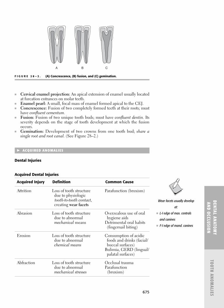
675
DENTAL ANATOM
YAN
D OCCLUSION
TOOTH ANO
MALIES
■ Cervical enamel projection: An apical extension of enamel usually locatedat furcation entrances on molar teeth.
■ Enamel pearl: A small, focal mass of enamel formed apical to the CEJ.■ Concrescence: Fusion of two completely formed teeth at their roots; must
have confluent cementum.■ Fusion: Fusion of two unique tooth buds; must have confluent dentin. Its
severity depends on the stage of tooth development at which the fusionoccurs.
■ Gemination: Development of two crowns from one tooth bud; share asingle root and root canal. (See Figure 28–2.)
� ACQU I RE D ANOMALI ES
Dental Injuries
Acquired Dental Injuries
Acquired Injury Definition Common Cause
Attrition Loss of tooth structure Parafunction (bruxism)due to physiologic tooth-to-tooth contact,creating wear facets
Abrasion Loss of tooth structure Overzealous use of oral due to abnormal hygiene aidsmechanical means Detrimental oral habits
(fingernail biting)
Erosion Loss of tooth structure Consumption of acidic due to abnormal foods and drinks (facial/ chemical means buccal surfaces)
Bulimia, GERD (lingual/ palatal surfaces)
Abfraction Loss of tooth structure Occlusal traumadue to abnormal Parafunction mechanical stresses (bruxism)
A B C
F I G U R E 2 8 – 2 . (A) Concrescence, (B) fusion, and (C) gemination.
Wear facets usually develop
at:
■ L-I edge of max. centrals
and canines
■ F-I edge of mand. canines

This page intentionally left blank

677
AA band, skeletal muscle and, 339, 339bA fibers, 171α-Amylase, 259α-globulins, function of, 269α-ketoacids, oxidative deamination and, 287abdomen, 81–86
regions of, 83abdominal aorta, 92, 94abdominal muscles, 84abdominal viscera, 86–110, 143
pelvic splanchnic nerves and, 143abducens nerve (CN VI), 24, 121–124, 122f
pituitary tumor and, 25abfraction, 675ABO blood typing, 522abrasion, 675abscess, 444
acute inflammation and, 503absolute refractory period, 321
heart and, 358, 359fabsorption, and digestion, 401–402acanthosis nigricans, 536accessory canals, 207accessory hemiazygos vein (superior
hemiazygos vein), 95accessory muscles, 371accessory nerve (CN XI-accessory),
schematic illustration, 143faccessory pulp canals, 654accommodation, pupillary light reflex and,
124acetone, ketone bodies and, 286acetylcholine metabolism, 327acetyl-CoA fate, metabolic fates of, 286ACh metabolism, enzymes in, 327bachalasia, 557achlorhydria, pernicious anemia and, 571achondroplasia (dwarfism), 471, 590acid–base balance, 387–388
control of, 388respiratory changes and, 380
acidophiles, 454acquired dental injuries, 675acquired immunity, 510
classification of, 510acral lentiginous melanoma, 613acrochordon, 611acromegaly, 421, 579–580
GH and, 580acrosome, egg fertilization and, 220ACTH, negative feedback loop and,
427
actin, 157, 338skeletal muscle contraction and, 340,
340fActinomyces species
root caries and, 492baction potential, 321
of cardiac muscle, 343activation energy, 253
reaction rate, 253, 254tactive cells, 156active eruption, 658active immunity, 464, 510
viruses and, 480acute gouty arthritis, 593acute hypocalcemia, Chvostek’s sign and,
583acute infection, 444acute inflammation
chronic inflammation v., 504signs of, 503bstages of, 503, 503t
acute leukemias, 619–620acute lymphoblastic leukemia (ALL), 606acute myelogenous leukemia (AML), 619acute nephritic syndrome, 568acute osteomyelitis, 591–592acute pancreatitis, 561acute pericarditis, 544Addison’s disease, 429–430, 437, 584–585
cortisol and, 584hyperpigmentation, 533oral signs of, 584b
adenine, 292badenocarcinoma, 603, 614, 615
prostate cancer and, 608smoking and, 614b
adenohypophysis, 177adenoma, 604adenomatous polyps, colorectal cancer, 616adenovirus, 476tadenylate cyclase, 416ADH. See antidiuretic hormoneadipose tissue, lipids and, 275adjuvants
immunity and, 511vaccines and, 480
ADP-ribosylation exotoxins, host tissuedestruction and, 451t
adrenal cortex, 107, 431f, 437f, 584–585,584b
aldosterone and, 437atrophy of, ACTH and, 428histologic layers, 427b
precocious puberty, 424zones of, 179b
adrenal glands, 107, 179, 584–585neoplasia of, 625–626
adrenal hormones, 415adrenal insufficiency, 428adrenal medulla, 107, 430, 431f
tumors of, 625adrenal–ACTH axis, exogenous cortisol
and, 428adrenergic agonists, 431adrenergic neurons, 311adrenergic receptors, 313adult periodontitis, 492adult polycystic kidney disease (APCKD),
566adults, brain tumors, 609aerotolerant anaerobic, bacteria, 454afferent nerves, 338aflatoxins, 482afterload, 354ageusia, 333aggressive periodontitis
characteristics of, 493classification, 493microbial flora in, 493
agingcementum and, 210dentin and, 204enamel matrix and, 207PDL and, 214presbycusis and, 138–139pulp and, 209
agranular leukocytes, 513agranulocytes, 172air embolism, 539airway, CN XII paralysis and, 143airway obstruction, mechanisms of, 551bAlbers–Schonberg disease, 590albinism, 533albumin, function of, 269albuterol, asthma and, 550alcoholism, lung abscess and, 555, 555baldoses, 258faldosterone (mineralocorticoid), 437
kidney function and, 394alkaline phosphatase
bone formation and, 589bone mineralization and, 166
alkaliphiles, 454ALL (acute lymphoblastic leukemia), 606allantois, formation, 224allergic response, fungi and, 482
INDEX
Note: Page numbers followed by f, t, and b indicate figures, tables, and boxes, respectively.

678
allografts, 518allosteric enzyme, 256allosteric inhibitor, 255allosteric regulation, 256, 256falpha-adrenergic receptors, 313ALS (amyotrophic lateral sclerosis),
596alveolar bone
clinical implications and, 211components of, 211proper, 211
alveolar epithelial cells, 181alveolar pressure, 371alveolar septum, 180alveolar surface area, gas exchange and,
373alveolar ventilation, 372alveolar wall, epithelial cells of, 373alveoli, 180alveologingival fibers, 201Alzheimer’s disease, 596amelogenesis imperfecta, 207amelogenin, enamel matrix protein and,
205amine hormones, 415amino acids, 264, 264t–265t
carbon skeletons of, metabolicintermediates formed, 287f, 289f
classification of, 265derivatives, 266
amine hormones, 415by dietary necessity, classification of,
265tmetabolic end product, classification of,
265tmetabolism, disorders of, 266by R-Group, classification of, 264tstructure, 264transamination, 286
amino sugar, 260aminoglycosides, 465t, 468aminotransferases, transamination, 286AML (acute myelogenous leukemia), 619ammonia
kidney function and, 389oxidative deamination and, 287
amniotic fluid embolism, 540DIC and, 576
amoxicillin, 469tamphiarthrosis, 167amphipathic lipid bilayer, membranes and,
302ampulla of Vater, 104, 185amygdala, limbic system and, 309amylases
starch catabolism and, 259starch hydrolysis and, 259
amyloidosismultiple myeloma and, 624types of, 624
amyotrophic lateral sclerosis (ALS),596
ANA (antinuclear antibody test), 586anal canal, blood supply and description,
106anaphase, 158anaphylactic shock, 538
anastomosing vessels of gingiva, PDL and,213
anatomic crown, 632anemia(s), 365–366, 568–573
of chronic disease, 573classification of, 569compensations with, 365consequences, 365from decreased red blood cell
production, 569, 571–573erythropoietin and, 366, 573from red blood cell destruction, 569–571types of, 365
aneurysm, atherosclerosis and, 541bangina, CAD and, 546–547angioedema, 519Angle’s classification, occlusal relationships,
class I-III, 664, 664f–665fangular artery, 66ankylosing spondylitis, 587ankylosis, teeth and, 674anosmia, 335ANS. See autonomic nervous systemansa cervicalis, 64antacid mediations, peptic ulcer disease,
558anterior cranial fossa, contents of, 8anterior cross-bite, 663anterior ethmoid artery, 18anterior facial vein, 69
connections, 25anterior guidance
mandible and, 667occlusal relationship and, 668
anterior hard palate, nasopalatine arteryand, 66
anterior pituitaryhormones, 417, 421hypothalamic control of, 417
anterior pituitary. See Rathke’s pouchanterior rectus sheath, 84anterior teeth, 632, 634anterior triangle, of neck, subsets of, 59, 60f,
61anterior/ventral horn, 145anterolateral system (spinothalamic tract),
pathway, 317anthracosis, 553anthrax toxin, host tissue destruction and,
451tantibiotic drugs, 464–469, 465t
classification of, 464comparison of, 465toral, impetigo and, 534peptic ulcer disease, 558topical, 468
antibiotic prophylaxis, 468, 469t, 545bantibody-mediated immune response v.
cell-mediated, 511anticonvulsants, aplastic anemia and, 571antidiuretic hormone (ADH; vasopressin),
415, 578diabetes insipidus and, 420, 578kidney function and, 394pituitary gland and, 177
antifungal drugs, for candidiasis, 484, 484tantigenic drifts, viruses and, 473
antigenic shifts, viruses and, 473antigen-presenting cells (APCs), 511, 514antigens, immunity and, 510antinuclear antibody test (ANA), 586antiseptics, 446, 447tantitoxin vaccine, 464aorta, 92–93aortic aneurysm, 543aortic arch derivatives, embryology and,
231, 232faortic body, respiratory chemoreceptors and,
379, 380faortic dissection, 543–545aortic valve, 88APCKD (adult polycystic kidney disease), 566APCs (antigen-presenting cells), 511, 514apical foramen, pulp and, 207apical vessels, PDL and, 213aplasia, cell injury, 499taplastic anemia, 571aplastic crisis, sickle-cell anemia and, 570apnea, 382apoenzyme, 251aponeurosis, 162apoprotein, 274apoptosis, 503appositional growth, 165, 166APUDoma, 604aqueous humor, 192arachnoid barrier, 27arachnoid mater, 22arboviruses, 471arch lengths of teeth, 641, 650area postrema, 27arginine, histones and, 295Argyll Robertson pupil, 124tarm movement, at shoulder, 79–80arterial constriction system, mean systemic
filling pressure and, 351arterial nephrosclerosis, 566arterial supply
dental, 635liver and, 103
arterial thrombi, 539arterial thrombosis, predisposing factors for,
539barteries of brainstem, 28farteriolar nephrosclerosis, 567arterioles, 174
oxygen exchange and, 350arteriosclerosis, 540–541, 567arthritis, 593–595articular capsule (bursa), 51, 168articular cartilage, 168articular disc, 50–51articular eminence, 50
occlusal relationship and, 668articular osteochondroses, 590artificial teeth, compensating curve and,
662arytenoids, 74asbestosis, 553ascending palatine artery, 66ascending pharyngeal artery, 65, 65fascites, 563
disorders associated with, 563liver disease and, 563

679
asexual reproduction, spores, 482aspiration, right lung and, 87aspirin
bleeding problems and, 574cyclooxygenase and, 273irreversible inhibition, 255Reye’s syndrome and, 476
assembly, viral replication and, 471asthma, 549–550
mechanisms of precipitation, 550treatment, 550types of, 549
astigmatism, 330astrocytes, 170tAtaxia-telangiectasia, 523tatelectasis, 552atelectasis neonatorum, 552atherosclerosis, 355, 540–541, 541batlanto-axial joint, 54atlanto-occipital joint, 54atopic allergies, 519α-Toxin, host tissue destruction and, 451t3 ATP, urea cycle, 289ATP
molecule, skeletal muscle contractionand, 340, 340f, 341, 341b
production, metabolic pathway and,279–280
atria, 89atrial septal defect, embryology and, 231batrioventricular valves, 89atrophic candidiasis, 494atrophy, cell injury, 499tattached gingiva, 197attachment, viral replication and, 471attrition, 675Auerbach’s plexus (myenteric plexus), 182,
398auricular branch of vagus, distribution, 131,
131fauriculotemporal nerve, 51
distribution, 126, 131, 131fautoclaving, hepatitis viruses and, 479autograft, 518autoimmune diseases, 524–525
polyarthritis nodosa, 586RA as, 594–595systemic lupus erythematosus, 585–586
autoimmune gastritis, pernicious anemiaand, 571
autoimmune hemolytic anemia, 569autolysis, enzymatic cellular degradation
and, 500automaticity, heart and, electrical
conduction of, 358, 359fautonomic ganglia, ANS and, 311autonomic nervous system (ANS), 149f,
307–309, 311autonomic ganglia, 311basic pathway, 311divisions, 150fpathway, 151smooth muscle and, 343summary of, 313
autoregulation of blood flow, exercise and,364
autosomal abnormalities, 531
AV node, nodal/pacemaker conduction system and, 360
avascular connective tissue, 164axilla, 76–77axillary artery, 76, 78axillary nerve, 78, 80axillary vein, 76axon, 168axoneme, 157axonotmesis, 324azithromycin, 469tazygous system, 94, 94f
Bβ-globulins, function of, 269β2 agonist inhalers, asthma and, 550Bacillus, 454bacitracin, 468bacteremia, 444bacteria, 449
classification of, 452–454, 453tgram-positive v. gram-negative, 453t,
454fgram-positive, 455–458, 455fmedically relevant, 455–458
bacterial adhesion/attachment, mediatorsof, 450
bacterial colonization, 490bacterial genetic exchange, 449bacterial growth curve, 449, 449fbacterial infection, neutrophilia and, 505bacterial metabolism, 289bacterial pneumonia, 555bacterial polysaccharides, 262bacterial structures, specialized, 452bacterial virulence factors, 450–452bacteriocidal drugs, 464bacteriology, 449–469bacteriophages, 480–481
replication, 481fbacteriostatic drugs, 464bactroban, impetigo and, 534Bainbridge reflex, 362, 363bbalance, hair cells and, 316balance of fluids, 385baroreceptor, 139
reflex, 362sudden standing and, 363
Barr body, 157Barrett’s esophagus, 558
GERD and, 557scleroderma, 586
basal bodies, 157basal cell carcinoma, 610basal ganglia, motor behavior and, 309basal nuclei, 112base pairing
DNA and, 292–293organization of, 292
base substitutions, mutation and, 605basement membrane, 161, 191, 196
components, 161, 161ffunctions of, 161
basilar artery, 27basilic vein, 77basophils, 514BBB (blood-brain barrier), 27
B-cell, 512timmune system and, 512maturation, bone marrow and, 175NHL and, 622white pulp, 176
B-complex vitamins, nonprotein enzymecomponents, 251
Beck’s triad, 544bBehcet’s syndrome, 587Bell’s palsy, 135, 136Bence–Jones protein, multiple myeloma,
623benign nephrosclerosis, 567benign prostatic hyperplasia, 608–609benign tumors
harm of, 601bmalignant tumors v., 601of mesenchymal origin, 604types of, 604–605
Bennet movement, 669Bernard–Soulier disease, 576berylliosis, causes, 553bbeta-adrenergic receptors, 313betaine, 273beta-oxidation, lipid metabolism and,
285–286, 285fBezold–Jarisch reflex, 381BGL (blood glucose level), 427bifid tongue, 244bilaminar disc, 223bile, 104
composition, 185, 561duodenum and, 107pHs of, 403pigment, 172salts, 275secretion pHs, 403
biliary flow, 185, 185fbiliary tree, 185, 185fbilirubin, 405, 563–564
jaundice and, 405levels, 185
bilirubinuria, 566binary fission, bacteria and, 449biofilm, 490biologic width, 199bisphosphonates
osteonecrosis of jaws and, 592osteoporosis and, 588
black lung disease, 553bladder, 109bladder cancer, 618bleeding
disorders, 575problems, 574–575
blood, 171–173components of, 171fformation, primitive embryology, 224formed elements of, 172functions of, 171neoplasia of, 618–625oxygen in, 375–376oxygen-carrying capacity in, 375b,
376PO2 and, oxygen transfer in, 375btype O, 522bin urine, children and, 565

680
blood flow, 347autoregulation, exercise and, 364cardiodynamics and, 347–350properties of, 348, 348fvelocity of, 348–349
changes in, 349fblood glucose level (BGL), 427blood ketones, 579bblood pressure, 352
arterioles, 350changes in, 349fpulmonary circulation, systemic
circulation v., 351blood supply
buccinator, 136dental arterial, 635to face, 65meninges and, 22bto nasal cavity, 17stomach, 102, 103ftongue, 65
blood typing. See ABO blood typingblood vessel(s)
bleeding problems and, 574damage, intrinsic/extrinsic pathways and,
367oxytalan fibers and, 213types
and characteristics, 350histologic differences, 174
walls, layers of, 173–174blood volume, systemic venous circulation
and, 350blood-brain barrier (BBB), 27blood-CSF barrier, 27blood loss anemia, 569blood-lymphatic pathology, 568–577bloodstream, endocrine glands and, 163blotting technique, clinical considerations
in, 300blue bloaters, chronic bronchitis and, 552blue nevus, 609body cavities, posterior and anterior, 86body fluid, distribution, 384Bohr effect, 271, 374
anemia and, 365bone, 165–167
formation, 166formation, alkaline phosphatase and, 589fracture, fat embolism and, 592functions of, 166growth, 166matrix, 166remodeling, 167resorption, 435surfaces, 167types of, 166
bone marrow, 167, 175B-cells and, 512leukemia and, 618multiple myeloma, 623
bone softening from vitamin D deficiency,588b
bone spurs, osteoarthritis and, 593bone tumors, 626–628, 626b
of nonosseous origin, 627–628types of, 626
botulinum toxin, host tissue destructionand, 451t, 457t
Bouchar’s nodes, osteoarthritis and, 593Bowman’s capsule, filtration and, 390, 391brachial arches
derivatives of structures for, 238fembryology, 237
brachial artery, 76brachial plexus, 78–80, 148
actions, by roots, 79branches of, 78–79, 78fnerves, 79terminal nerves of, 78and upper extremities, 76–81
brachial vein, 77brachiocephalic artery, 92brachiocephalic veins, 92bradycardia, 174brain, 110–114, 111f
drainage of, IJV and, 24embryology of, 230hypoxia and, 498binjury, 595neurocranium and, 6tumors, 609
brain stem, 113–114, 113f, 230components, 310
branched amylopectin, starch, 259branchiomeric nerves, 237breast, 84–85breast cancer, 607–608, 607b
BRCA-1 gene, 607fibrocystic disease v., 607
breastfeeding, celiac disease and, 560broad-spectrum drugs, 464bronchial arteries, 87bronchiectasis, 552bronchoconstrictor release, asthma and, 550bronchodilation
epinephrine and, 266norepinephrine and, 266
bronchogenic carcinoma, 614bronchopneumonia, 555bronchus, pulmonary artery v., 87bronzed disease, 501brown tumor, blood chemistry values, 588brownish-gray banding, tetracycline and,
207brush border enzymes, 258Bruton’s X-linked agammaglobulinemia,
522tbuccal nerve of V3, 125buccal ridge, 634buccal space, boundaries and contents, 47fbuccinator muscle, 135buccinator nerve, 670
blood supply to, 136budding, viruses and, 470buffer, 388buffer systems, 388bullous pemphigoid, 535bunyavirus, 475tBurkitt’s lymphoma, 623bursa (articular capsule), 51, 168butterfly rash, systemic lupus erythematosus,
585byssinosis, 553
CC fibers, 171CA2+ -Na+ exchange system, cardiac muscle
and, 343CAD (coronary artery disease), 546–547Café au lait spots, 533
disease association with, 589btuberous sclerosis, 589b
calcification, 165calcinosis, 501bcalcitonin, 167
calcium regulation and, 435in cellular metabolism, 71parafollicular cells and, 72
calciumdeficiency, 408hyperparathyroidism and, 436hypoparathyroidism and, 436metabolism, 435–436regulation, 167skeletal muscle contraction and, 341stones, 565
calcium channel blockers, gingival overgrowth and, 499b
calculus, 491calicivirus, 473tcalmodulin, smooth muscle contraction
and, 344caloric test, vestibuloocular reflex, 139bcAMP protein kinases
cardiac muscle and, 343hormones and, 416mechanism, 416–417, 417f
canaliculi, 165, 185, 209cancellous bone, 166, 211cancer. See specific types, e.g., prostate
cancerCandida albicans, candidiasis, 494candidiasis
antifungal drugs and, 484, 484tcharacteristics and classification, 494
canine guidance, mandible and, 668canines
lobes of, 641mandibular permanent teeth, 646, 646f,
655tmandibular primary teeth, 651, 656tmaxillary permanent teeth, 642, 642f,
655tmaxillary primary teeth, 650, 656t
capacitance, 350–351capillaries, 174
oxygen exchange and, 350capillary permeability, edema and, 386capsid, viruses and, 470capsular polysaccharide vaccine, 464capsulated joint receptors, nonencapsulated
joint receptors v., 315carbohydrate(s), 258–264
cell membranes and, 303classification, 258complexes, calculus and, 491digestion and absorption, 401metabolism, 280–285structure, 258
carbon dioxide (CO2)blood and, 376, 376f

681
hypercarbia, 378narcosis, 378
carbon monoxide poisoning, 365, 548carcinogenesis, mutations causing,
605–606carcinogenic bacteria, dietary sucrose and,
262carcinomas, 603cardiac compensation, anemia and, 365cardiac conduction, 174
tissues, 358cardiac cycle, 356, 357fcardiac demand, compensatory hypertrophy
and, 343cardiac enzymes, after MI, 547bcardiac glands, 101
gastric secretion and, 399cardiac muscle. See also heart
action potential, 343contraction, 343histology, 343innervation, 343skeletal muscle v., refractory period and,
358bcardiac myocytes, cardiac conductive tissues
and, 358cardiac output (CO), 353
high HR and, 355shock and, 537
cardiac system, embryology of, 231–232,233f
cardiac tamponade, 544cardiac veins, 91cardiodynamics, blood flow and, 347–350cardiogenic shock, 538cardiovascular pathology, 536–549cardiovascular pulmonary cross-correlation,
548–549cardiovascular tissue, 173–174caries, 634
characteristics of, 491classification, 492formation, 491predominant microbial flora in, 492Stephan curve and, 491, 492f
carnitine-mediated enzyme system, fattyacids, 285
carotid artery, 93carotid body, respiratory chemoreceptors
and, 379, 380fcarotid body O2, pH levels and, 316carotid sheath, 56, 57fcarotid sinus, 363carotid sinus syndrome, 363carotid triangle, of neck, 61carrier infection, 444cartilage, 164–165
ECM of, 164–165growth of, 165surface of, 165types of, 165
cartilaginous joints, 167catabolism, glycogen synthesis and,
284–285, 284fcatalase, 515catecholamines, 415
tyrosine metabolism and, 431
cathepsin D, malignant tumors and, 602cauda equina, 145cavernous sinus, 24–25, 24f, 25f
infection and, 69CD3-associated T-cell receptor (TCR),
T-cells and, 511CD8 lymphocytes, 512bCDC (Centers for Disease Control and
Prevention), 447cecum, blood supply and description, 106cefadroxil, 469tcefazolin, 469tCEJ contours, 640–641, 640fceliac artery, 96celiac disease, 560celiac trunk, 93cell(s)
cardiac muscle and, 343of small intestine, 183tturnover, tissue types and, 158types, comparison, 443, 443t
cell cycle, 158, 158fcell death, 498–501
chemical-induced, 498tnecrosis, types of, 502, 502tnuclear changes with, 502, 502treactions to, 499
cell injurychemical-induced, 498free-radical, 498–499modes of, 498reactions to, 499ttypes of, 499
cell surface appendages, 157cell-mediated immune response
fungal infection, 482v. antibody-mediated, 511
cell-to-cell contacts, 158–159, 159f, 159tcellulitis, 444cellulose, 261cementicles, 212cementoblasts, 209cementocytes, 209cementogenesis, 209cementum, 209–210
aging effects on, 210classifications of, 210functions of, 209morphology, 210f
Centers for Disease Control and Prevention(CDC), 447
central diabetes insipidus, 420, 578central hearing connections, deafness and,
139central incisor
mandibular permanent teeth, 645, 645f,655t
mandibular primary teeth, 651, 656tmaxillary permanent teeth, 641, 641f, 655tmaxillary primary teeth, 650, 656t
central nerves (CNs), taste sensation,tongue and, 30
central nervous system (CNS), 307embryology of, 229–231, 229fhypercarbia, 378nervous tissue, 168neurons, 324b
centric occlusion (CO), mandible and, 667centric relation (CR), mandible and, 667centrioles, 157centroacinar cells, 186cephalexin, 469tcephalic vein, 76cephalosporins, 465t, 466cerebellum
function and components, 112motor control, 310
cerebral arteries, major, 28fcerebral circulation, 378cerebral circulation, CO2 effect on, 378cerebral cortex, 110, 111f, 307
functions of, 110, 111fhemispheres, lobes of, 307
cerebral infarction, 595–596cerebral medulla, 110cerebrospinal fluid (CSF)
circulation pathway, 26ependymal cells and, 26spinal cord and, 145
cerebrum, 110–112cervical enamel projection, 675cervical loop, root formation and, 218, 218fcervical plexus, 63–64, 64f, 148cervical ridge, 634cervical vertebrae, 54cetylpyridinium chloride, 495tcGMP, hormones and, 416chemical neurotransmitters, 326chemical plaque control, 495, 495tchemical synapse, 170, 325–326chemokines, 514chemoreception, 139chemoreceptor, 316, 362, 363–364
pathway, 364chewing surfaces, of teeth, 632CHF (congestive heart failure), edema and,
537children
acute osteomyelitis, 591bone tumors, 626b, 627brain tumors in, 609cretinism and, 581dental findings
hypothyroidism and, 581brickets, 588
genetic disorders of, 532–533histiocytosis X and, 625intestinal lymphangiectasia, 559leukemia and, 606osteomalacia and, 588PCKD and, 566Reye’s syndrome and, 476skin eruptions, impetigo, 534
chitin, 262fungi cells and, 482
chloramphenicol, 465t, 468aplastic anemia and, 571
chlorhexidine gluconate, 495tchloride shift, 376cholecystokinin, stimulus and
control, 398cholelithiasis, 561cholera toxin, host tissue destruction and,
451t

682
cholesterolderivatives of steroid hormones, 415membranes and, 303as steroid, 273synthesis, liver and, 403–404
cholesterolosis, 561choline, 273choline deficiency, 273cholinergic neurons, 311chondroblasts, 164chondrocytes, 164chondrosarcoma, 627chorda tympani nerve
components of, 31course of, 31lingual nerve and, 128
chordoma, 628choristoma, 536, 604choroid, 192choroid plexus, intracranial pressure and,
25Christmas disease, 577chromatin, 157, 296
DNA, 295chromosomal disorders, 531, 531bchronic bronchitis, 552chronic hyperplastic candidiasis, 494chronic infection, 444chronic inflammation
acute inflammation and, 503, 504stages of, 504
chronic leukemias, 620–621chronic lymphocytic lymphoma (CLL),
619, 623chronic myelogenous leukemia (CML),
621chronic nephrotic syndrome, 568chronic obstructive pulmonary disease
(COPD), 550–552hypoxemia in, 377
chronic osteomyelitis, 592chronic pancreatitis, 561chronic pericarditis, 544chronic periodontitis
characteristics, 493predominant microbial flora in, 493
Chvostek’s sign, tetany, 583chylomicrons, 402chyme, 400
segmentation, 400–401cigarette smoking
adenocarcinoma, 614bCOPD and, 550emphysema and, 551lung cancers and, 615related diseases, 551b
cilia, 157ciliary body, 192
of eyeball, 328ciliary zone, 193fcingulum, 632circle of Willis, 27, 28f
arteries of brainstem and, 28fof brainstem, 28fmiddle cerebral artery v., 29
circuit, pulmonary circulation, systemiccirculation v., 351
circulation, blood vessels in, arrangementof, 173
circumferential fibers, 201circumpulpal dentin, 203
formation, 203circumvallate papillae, 32, 333cirrhosis
edema and, 537proteins and, 367
cirrhosis (of liver), 562cisterna chyli, 98citrate–malate shuttle, fatty acid synthesis,
285citric acid cycle, 282, 283f, 287fclarithromycin, 469tclass I MHC surface proteins, nucleated
cells and, 511class II MHC proteins, immune system and,
514classic hemophilia, 577clavicle, 81cleavage stages, morula and blastula
embryology and, 223fcleft lip, 241f–242f, 244cleft palate, 243–244climbing fibers, 112clindamycin, 465t, 467, 469tclinical crown, 632CLL (chronic lymphocytic lymphoma),
619, 623clonal selection, 512Clostridium, 454clotting factors, liver and, 577CML (chronic myelogenous leukemia), 621CN I (olfactory nerve), 17CN II (optic nerve), 120–121, 121fCN III injury, lesions and, 124CN IX. See glossopharyngeal nerveCN V. See trigeminal nerveCN VI. See abducens nerveCN VII (orbicularis oculi), 131bCN VIII. See vestibulococholear nerveCN X. See vagus nerveCN XI-accessory, 143fCN XII. See hypoglossus nerveCNs (central nerves), taste sensation,
tongue and, 30CNS. See central nervous systemCO. See centric occlusion; See cardiac
outputCO2. See carbon dioxidecoagulase, 450coagulation, intrinsic and extrinsic
pathways, 367, 367fcoagulation factor XII (Hageman factor),
507coagulation factors, 367coagulative necrosis, 502coal workers pneumoconiosis, 553codons, 298coenzyme, 251coenzyme A, in fatty acid synthesis and
catabolism, 285coenzyme Q, ubiquinone as, 284cold caloric test, 139, 139bcollagen, 165
structure, 267f
synthesis, 267f, 268type I, dentogingival connective tissue
and, 200type II, 164types of, 268
collagen remodeling, wound repair and,508
collagen vascular diseases, 585–587, 585btypes, 594b
collagenases (metalloproteinases), host tissue destruction and, 450
collapsing forces, lung and, 371colloid osmotic pressure (oncotic pressure),
385colon
blood supply and description, 106GI cancer and, 616
colon polyps, 617colony stimulating factors (CSFs), 505,
505t–506tcolorectal cancer, 616–617columnar epithelial cells, 161commensalism, 444common bile duct, 104
choledocholithiasis and, 561common carotid, 93common central vein, 103common facial vein, 69common iliac artery
pelvic kidney and, 96ureter and, 108
common variable immunodeficiency(CVID), 522t
communicating–obstruction, in subarachnoid space, 26b
compensating curve, 662compensatory hypertrophy, cardiac demand
and, 343competitive inhibition, 254, 254f, 304complement protein system, 517fcomplement proteins, 515–518
activation, pathways of, 516tfunction of, 269, 517t
complete anodontia, 674complete bilateral cleft, 244complex carbohydrates, 263compliance
pulmonary circulation, systemic circulation v., 351
venus return and, 355compound nevus, 609concrescence, 675, 675fconcussion, 595condensing osteitis, 592conducting zone, lung and, 372conduction velocity, 324bconductive loss, 139condylar movements, 669, 669fcondyle, 20
of mandible, 50cones, 194, 329confluent cementum, concrescence and,
675, 675fcongenital syphilis, enamel and, 207congestion (hyperemia), 538congestive heart failure (CHF), edema and,
537

683
congo red, amyloid stain and, 624conidia, 482conjugated bilirubin, 563conjugation, bacterial cell and, 450tconnective tissue (CT)
attachments, 162cells of, 162classification of, 162consistency of, 162proper, 162skeletal muscle and, 340
contact area, dental, 634, 639contact inhibition, 508continuous conduction, 324contour lines of Owen, 204contractility, HR and, 354contraction, of skeletal muscle, 340, 340fcontraction (twitch) speed
pneumonic for, 341bskeletal muscle, comparison by, 341
contralateral tracts, 314bconus medullaris, 145convergence, pupillary light reflex and, 124Coombs’ test, 569COPD. See chronic obstructive pulmonary
diseasecopper, transferrin and, 412cor pulmonale, right heart failure and, 548bCori cycle, 281, 282fcornea, 192corneal reflex, 131b, 132corneoscleral coat, 192coronary arteries, 90, 90fcoronary artery disease (CAD), 546–547coronary ligaments, 103coronary sinus, 91coronavirus, 475tcoronoid process, 20corpora cavernosa, 190corpus callosum, 110, 307corpus luteum, 426corpus spongiosum, 190cortex, medullary rays and, 187cortical bone, 166, 211cortical sinuses, lymph and, 175corticospinal tract (pyramidal system), 319,
320fcorticosteroids, 427
phospholipase A2 and, 273cortisol, 428, 584–585Coumadin (warfarin), 577countercurrent exchange, 392countercurrent multiplier, 392coupled, transport proteins and, 303covalent modification, enzyme regulation
and, 255COX (cyclooxygenase)
aspirin and, 273inhibitors, asthma and, 550
coxsackieviruses, 474CPE (cytopathic effect), 471CPK (creatine phosphokinase), 547bCR (centric relation), 667cranial base, of internal skull, 11, 11fcranial foramina, 20
important, 9cranial fossae, 8–12. See also internal skull
cranial nerve, 114–120brainstem and, 113f, 114chart, 116t–120tcomponents, 114–115motor nuclei, unilateral corticonuclear
connections and, 114bsensory nuclei, hearing and, 114
craniopharyngioma, 609cranium, 6–7
bones of, 6creatine phosphate, skeletal muscle
contraction and, 341bcreatine phosphokinase (CPK), 547bcremaster muscle, 84CREST syndrome, 586bcretinism, 434, 581CRH–ACTH–cortisol axis, 427cricothyroid membrane, 73cricothyroid muscle, 74crista terminalis, 89Crohn’s disease, 559
UC v., comparison of, 559cross-bite, 663cross-striations, skeletal muscle and, 339crown, types, 632crypts of Lieberkühn, 182CSF. See cerebrospinal fluidCSFs (colony stimulating factors), 505,
505t–506tCT. See connective tissuecuboidal epithelial cells, 161curve of Spee, 662, 662f
occlusal relationship and, 668curve of Wilson, 662, 663fCushing’s disease
cortisol and, 584Cushing’s syndrome v., 429
Cushing’s syndrome, 428, 428fcortisol and, 584Cushing’s disease v., 429
cusp, 632height, occlusal relationship and, 668
cusp of Carabelli, 632cutaneous innervation, face/scalp/auricle, 131fCVID (common variable
immunodeficiency), 522tcyanide poisoning, oxidative
phosphorylation and, 548cyanosis, 366cyclooxygenase. See COXcyclosporin, gingival overgrowth and, 499bcyst, 444cystic fibrosis, 532, 532bcysticercosis, 487tcytochromes, 283–284cytokeratin, 157cytokines, 505–506, 505t–506tcytopathic effect (CPE), 471cytoplasm, 156
viral replication and, 471cytosine, guanine and, 292bcytoskeleton, 168cytotoxic hypersensitivity, 520f
DDAG, hormones and, 416daily imbrication line of von Ebner, 204
D-amino acids, 264dead space, 372dead tracts, 203deafness, central hearing connections and,
139death phase, bacteria and, 449decidual reaction, week 2, 224deciduous (primary) tooth form, 648–652,
649f, 651tdeep cervical fascia, investing layer of, 55deep facial vein, connections, 25deep venous thrombosis, 540defecation, 110degenerative diseases, 596DEJ (dentino–enamel junction), 206–207delayed hypersensitivity, 521fdelayed passive eruption, 219deltavirus, 474tdemyelinating diseases, 597dendrites, 168dendritic cells, 513dens evaginatus, 674dens invaginatus (dens in dente), teeth and,
674dense connective tissue
irregular, 162regular, 162
dental anatomy, 632–635dental findings
in child, hypothyroidism and, 581, 581brickets, 588, 588b
dental follicle, 217dental formula
permanent dentition, 659primary dentition, 658
dental injuries, 675dental papilla, 217
differentiated ectomesenchymal cells,202
dental treatment, antibiotic prophylaxis and,468, 469t
denticles, 209dentin, 202–205
aging and, 204classifications of, 204deformities, 205formation, chronological order of, 217b
dentin dysplasia, 205dentinal hypersensitivity, 204dentino–enamel junction (DEJ), 206–207dentinogenesis, 202–203, 203fdentinogenesis imperfecta, 205dentitions. See also primary dentition
classification of, 632dentogingival connective tissue, type I
collagen and, 200dentogingival epithelium, 199, 200fdentogingival fibers, 201dentogingival junction, 199
tooth eruption, 218, 218fdentoperiosteal fibers, 201depolarization, 325depressor anguli muscle, 135dermatofibroma, 610–611dermatomes, 85, 85f
derivatives, 227fdermatomyositis, calcinosis and, 501b

684
dermatopathology, 533–536dermis, 191descending colon, 105descending motor tracts
(efferent/descending pathways), 319desmin, 157desmoplasia, 603desmosome (macula adherens), 158
cardiac muscle and, 343detoxification, 103developmental groove, 634developmental pathology, 530–533dextran, 262dextrins, 259D-form isomerism, monosaccharides,
258ΔG. See Gibbs free energy changediabetes insipidus, 427, 578
causes of, 420diabetes mellitus v., 420
diabetes mellitus, 427pancreas and, 427type 1, 578–579
v. diabetes type 2, 579diabetic coma, 286diapedesis, 514tdiaphragm muscles, 82, 98diaphragma sella, 22diarrhea, clindamycin and, 467diarthrosis, 167diastema, 634diastole
cardiac cycle and, 356fibrous pericardium and, 353b
diastolic pressure, 352DIC. See disseminated intravascular
coagulationdicloxacillin, 466
impetigo and, 534diencephalon, 112, 307. See also posterior
pituitarydiffuse lymphocytic lymphoma, 623DiGeorge’s syndrome, 523t
hypoparathyroidism and, 583digestion, absorption and, components,
401–402dilaceration, teeth and, 674Dilantin (phenytoin), gingival overgrowth
and, 499bdimorphism, molds and, 482diphtheria toxin, host tissue destruction and,
451tdiphyodont dentition, 632diploic veins, 24dipolar ions (Zwitterions), 388disaccharide absorption, 401disaccharides, 258disease. See specific type disease i.e.
degenerative diseasesdisinfectants, 446, 446tdisinfection, 446disseminated intravascular coagulation (DIC)
causes, 576laboratory indications, 576platelet disorders and, 575
disulfide bonds, 265immunoglobulins and, 515
DNAbackbone structure of, 294fbase pairing and, 292–293, 292fgyrase, DNA synthesis and, 297ligases, 299
DNA synthesis and, 297, 297fmetronidazole and, 467organization, 295–296, 296fpolymerase, 299
DNA synthesis and, 297, 297fsynthesis, 296–297, 297ftransduction and, 450ttransfer, bacteria and, 449transformation and, 450tviruses, 470
viral replication and, 471domains, protein structures and, 267dorsal column system, pathway, 317dorsal root ganglion, afferent/sensory nerves
and, 146dorsal scapular artery, 77fDown syndrome, 531
chromosomal disorders and, 531bdrug-induced
aplastic anemia, 571lupus erythematosus, 585
drugsantiviral, 480teratogenesis and, 530
duct of Wirsung, 107ductus deferens, 110duodenal ulcer, 558duodenum, 93, 182
blood supply and description of, 105dura mater, dural folds of, 22, 23fdural folds, of dura mater, 22, 23fdural sinuses, 23f
list of, 23tributaries of, 24
dust cells, 181, 513dwarfism, 421, 590dysarthria, hypoglossus nerve and, 143dysgeusia, 333dysosmia, 335dysplasia, 601dysplastic nevus, 610dyspnea, 382dystrophic calcifications, 209
Eear, anatomy, 137–138, 138f, 331early-onset periodontitis, 493Eaton–Lambert syndrome, 597beburnation, osteoarthritis and, 593ecchymoses, petechiae v., 576eclampsia, secondary hypertension and,
542, 542bECM (extracellular matrix), 164–165ectomesenchyme, tooth type and, 216edemas, 386–387, 504, 536–537
cause of, 386, 537clinical exam of, 537nephrotic syndrome and, 537types of, 536–537
Edward syndrome, 531chromosomal disorders and, 531b
EFC. See extracellular fluids
effective renal plasma flow (ERPF), 393efferent nerves, 338efferent pathways (descending motor tracts),
319egg, stages of meiosis in, 219feggshell calcification, 501beicosanoids, 273–274ejaculatory duct, 110ejection fraction, 353elastic arteries, 174elastic cartilage, 165elastin, hydroxylysine and, 268electrical synapse, 170, 325, 326electrocardiogram (ECG), 360–361
leads, 360waves, 361, 361f
electron transport chain, 282–283, 283felephantiasis, edema and, 537Embden–Meyerhof pathway, 280–281embolus, 539–540embrasures, 634, 639embryo development
pituitary gland, 418, 418fweek 1, 222fweek 4, 229b
embryology, 219–246brain, 230of face, 236–246forebrain, 230gametogenesis, 219–228, 219f–221fheart, 233fkidney, stages of, 235fpancreas, 236fpatent foramen ovale, 231bof pharyngeal arches, 236–246of renal system, 234–236, 235f, 236ftongue and, 30
embryonic body axis, 225–228, 226femissary veins, 24emphysema
problems with, 552btypes of, 551
empty mouth swallowing, 670enamel
aging effects on, 207amelogenesis of, 205–206cross-striations and incremental lines, 206deformities of, 207formation, 206f, 217b
enamel hypoplasia, 207enamel knot, 217enamel matrix formation, defect, 207enamel maturation defect, 207enamel organ, 216enamel pearl, 675enamel surface depression, 634enamel surface elevations, 632enamel surface junctions, 634encapsulated joint receptors, types
of, 315bendemic infection, 443endocarditis, 544–545
types of, 545endocardium, 174endochondral ossification, 166endocrine disorders, secondary hypertension
and, 542

685
endocrine glands, 177–179, 186function and excretions of, 163
endocrine pancreas, 186endocrine pathology, 578–585endogenous pigments, 500
comparison of, 500tendomysium, CT and, 340endopeptidases, 268endosomes, 157endosteum, 167endothelium, 161endotoxin
host tissue destruction and, 451, 451tmode of action, 452tstructure, 452fv. exotoxin, 451t
end-stage kidney disease, APCKD, 566enolase, fluoride and, 281enteric nervous system, 398entero gastrone, 398–399enterogastric reflex, 398benterohepatic circulation, lipoproteins and,
275enteroviruses, 471Entner–Doudoroff pathway, 289envelope, viruses and, 470enzymatic cellular degradation, 500enzyme(s), 250–256
in ACh metabolism, 327bbiomechanics, induced-fit model,
251–252, 252fclassification of, 251concentration, reaction rate influencing
factors, 254tdefinitions, 251digestion and absorption, 401host tissue destruction and, 450–451inhibition, 254–255in intestinal villi, 105bkinetics, 252–254
ΔG and, 252–253, 253treaction equilibrium, 253reaction rate, 253
rate-limiting, 279tregulation of, 186
covalent modification and, 255triglycerides and, digestion and, 402
enzyme inhibition, 254–255competitive, 254, 254firreversible, 255noncompetitive, 255, 255funcompetitive, 255
enzyme kinetics, 252–254. See alsoenzymes
Gibbs free energy change (ΔG),252–253, 253t
substrate concentration, 252, 253fenzyme mechanics, 251–252, 252fenzyme regulation, 255–256
allosteric regulation, 256covalent modification, 255protease activity, 256, 256f
eosinophilia, parasitic infections and,505
eosinophilic granuloma, clinical symptoms,625
eosinophils, 514
ependymal cellscerebrospinal fluid and, 26ventricular system and, 25
ependymoma, 609epidemic infection, 443epidermal cells, specialized, 191epidermal layers, of skin, mnemonic for,
191epidermis, 191epididymis, 189
sperm and, 222epidural hematoma, 22b, 595epiglottis, 73epilepsy, 596epimysium, CT and, 340epinephrine, 313, 431
adrenal medulla and, 266asthma and, 550gluconeogenesis and, 285smooth muscle and, 343status asthmaticus and, 550b
epiploic foramen of Winslow, 99bepistaxis (nosebleed), 66
Kiesselbach’s plexus and, 18epitactic concept, in calculus formation, 491epithalamus, 112epithelial cells
of alveolar wall, 373lining microvilli, 182specialized, 196
epithelial intercellular junctions, 159fepithelial malignant tumor, 611epithelial migration, wound repair and, 507epithelial rests of Malassez, 212
root formation and, 218, 218fepithelial rests of Serres, 217epithelial types, 160tepithelium, 160–161epitope, immunity and, 510ergosterol
fungal cell membranes and, 482fungi cells and, 482
erosion, 675ERPF (effective renal plasma flow), 393ERV (expiratory reserve volume), 370, 370ferythema multiforme, 525t, 535erythematous candidiasis, 494erythroblastosis fetalis, 569–570erythrocytes, 172
and polycythemia, 573–574red pulp and, 176
erythrogenic toxin, host tissue destructionand, 451t
erythromycin, 467impetigo and, 534
erythropoietin, 366, 436, 573kidney and, 574polycythemia, 574
Escherichia coli, sepsis and, 444esophageal cancer, 615esophageal plexus, vagus nerves and, 141esophageal ulcers, 558esophageal varices, hematemesis and, 563esophagus, 101, 181
disorders of, 556–558estradiol, hCG and, 425estrogens, 422, 423
ethanol metabolism, 405ethmoid bones, 6
components and functions of, 12ethmoid sinuses, 18
location of drainage, 19euchromatin, 157eukaryotic cells, 80s ribosomes and, 297eustachian tube, 138EV (residual volume), 370, 370fEwing’s sarcoma, 627–628excitable cells, 322bexcitatory neurotransmitters, 323excretion rate, 394exercise, physiologic response to, 364exfoliatin, host tissue destruction and, 451,
451texfoliation, 658exocrine glands
classification of, 163, 164ffunction and excretions of, 163
exocrine pancreas, 186, 402exogenous cortisol, adrenal-ACTH axis and,
428exonuclease, 299
DNA synthesis and, 297exotoxins
classification of, 451tmode of action, 451tv. endotoxin, 451t
expanding forces, lung and, 371expiration, lung mechanics and, 371expiratory reserve volume (ERV), lung
volumes, 370, 370fexternal basal lamina, 200external capsule, of spleen, 176external carotid artery, 65–69, 65f
branches of, mnemonic for, 66course of, 65supply, 66
external ear, 137, 138fanatomy, 331innervation of, 63, 131fsensation of, nerves for, 131
external jugular vein, 68–69external nose, 20, 20fexternal oblique fibers, 84external thorax, 81–86exteroreceptors, CNS and, 315extracellular fluids (EFC), 384
expansion, edema and, 386extracellular matrix (ECM), cartilage,
164–165extracellular spaces, paracrine glands, 163extrafusal fibers, 341extraocular movements, muscles and nerves
in, 121extrapyramidal system, 319extra-testicular duct system, 189extrinsic asthma, 549extrinsic muscles of tongue, 671exudate, 504exudate edema, 536eye, 192–194
layers, 192ophthalmic artery and, 12structures, 193f
eye chambers, 192
686
eye elevators, eye muscle action and,122–123, 123f
eye muscle action, eye elevators and,122–123, 123f
eyeball, 327–330components, 328–329segments, 327
FFabry’s disease, 276, 532face, 20–21
blood supply, 65development of, 241f–242fembryology of, 236–246formation, 240lymph nodes in, 69–70, 70flymphatic drainage of, 70forbital bones of, 12, 13fvenous drainage from, 68–69, 68f, 69fviscerocranium and, 6
facial artery, 65f, 66, 67ECA and, 42facial expression and, 66portions of, 67supply, mnemonic for, 66
facial expression, facial musculature and,134f, 135
facial musculature, facial expression, 134f,135
facial nerve (CN VII)chorda tympani nerve and, 31components of, 133corticonuclear connections and, 114bcourse of, 132, 133ffacial expression, muscles of, 135lesions, 135nuclei, 134
facial paralysis, total and partial, 136ffacial reflexes, CN V and, 132facial sensation, CN V distribution, 130ffacial vein, 69factor 1. See fibrinogenfacultative anaerobic, bacteria, 454falciform ligament, 99, 103fallopian tubes (oviducts), 190falx cerebelli, 22falx cerebri, 22familial hypercholesterolemia, 274, 541bfamilial polyposis, 617Fanconi’s anemia, café au lait spots and,
589bfascia, 84bfascial spaces, 46–47, 47ffasting, glucose and, 404fat(s)
digestion and absorption, 401excess vitamins in, 408
fat catabolism, hyperlipidemia, 567bfat embolism, 539
bone fracture and, 592fatal tamponade, 543bfatty acids
classification, 272structure, 271synthesis, lipid metabolism and, 285,
285ftypes, 272
fauces, 37Fc site, immunoglobulins and, 516FDI (federation dentaire internationale)
system, 638Fe2+ (ferrous), 412Fe3+ (ferric), 412federation dentaire internationale (FDI)
system, 638feedback regulation, 256, 256ffemales, urinary infections and, 109femoral triangle, 86, 86fferric (Fe3+), 412ferrous (Fe2+), 412fertilization, 220fetal circulation, 224–225, 225ffetus
erythroblastosis fetalis and, 569–570stages of, 228f
Feulgen reaction, 293fever, 505FFAs. See free fatty acidsfibers, of CNS, 324bfibrin clot, wound repair and, 507fibrin, coagulation, 367, 367ffibrinogen (factor 1)
coagulation, 367, 367ffunction of, 269
fibrinolysis, wound repair and, 507–508fibroadenoma, 604fibroblasts, PDL and, 212fibrocartilage, 165fibrocystic disease, 608
breast cancer v., 607fibroplastic stage of wound repair, 507–508fibrosarcoma, 628fibrous dysplasia, 589–590fibrous joints, 167fibrous pericardium, diastole, 353bfilaments, 157filiform papillae, 32, 333filovirus, 475tfiltration, 391, 392f
glomerulus and, 390kidney function and, 389
filum terminale, 145first arch, embryology and, 238bfirst molar
mandibular permanent teeth, 647, 647f,655t
mandibular primary teeth, 652, 656tmaxillary permanent teeth, 643–644,
643f, 655tmaxillary primary teeth, 650, 656t
first premolarmandibular permanent teeth, 646, 646f,
655tmaxillary permanent teeth, 642–643,
642f, 655tfissure, 634flagella, 157, 452flavivirus, 474tflexor-withdrawal reflex, 342fluid capillary exchange, 385, 385ffluid mosaic model, plasma membrane and,
302, 302ffluoride, enolase and, 281fluoride toxicity, 412
fluoroquinolones, 465t, 467–468fluorosis, 207folate deficiency, 572foliate papillae, 32, 333follicle-stimulating hormone (FSH), 415
corpus luteum and, 426follicular cells, 71, 177follicular phase, 424food
mastication and, 47pharynx and, 41
foramen cecum, 9foramen lacerum, 9foramen magnum, 9foramen ovale, 9, 140f, 225bforamen rotundum, 9, 140fforamen spinosum, 9foramina, 20–21forebrain, embryology of, 230foregut, 100formen ovale, 140ffossa, 634fossa ovalis, 89fovea, 194fractures, 592
classifications, 592healing phases, 592repair, 167
frameshift mutation, 299, 605Frank–Starling mechanism, 353, 353fFRC (functional residual capacity), 370,
370f, 371freckle, 533free bilirubin, 563free fatty acids (FFAs)
ketoacidosis and, 579btriglycerides and, digestion and, 402
free gingiva, 199free gingival groove, 197free nerve endings, 315freeway space (FS)
mandible and, 667Freiberg’s disease, 590Freund’s adjuvant, 511frontal bone, 6frontal lobe, 111, 111f, 307frontal nasal processes, in face formation,
240frontal sinuses, 18
location of drainage, 19fructose, inulin, 260FS. See freeway spaceFSH. See follicle-stimulating hormonefunctional contacts, 662functional occlusion, 662functional residual capacity (FRC), 370,
370f, 371functional syncytium, smooth muscle and,
343fundus, gastric secretion and, 399fungal cell membranes, ergosterol and,
482fungal infection
cutaneous, 483topportunistic, 484tpathogenesis of, 482systemic, 483t

687
fungi, 482classification, 482, 483tmedically relevant, 483, 483t, 484treproduction, 482
fungiform papillae, 32, 333furcation, 635furosemide, 391bfusion, 675, 675f
Gγ globulins, function of, 269G6PD. See glucose-6-phosphate
dehydrogenasegag reflex, 139GAGs (glycosaminoglycans), 260–261, 261tgallbladder, 104, 186
lipoproteins and, 275gallbladder diverticulosis, 562gallstones, 561GALT (gut-associated lymphatic tissue), 184gametogenesis, 219–228, 219f–221fganglia, 148gap junction, 159Gardner’s syndrome, 617gas embolism, 539gas poisoning, colorless/odorless, 549, 549bgas solubility, 373gastric acid secretion
H2 receptors and, 266pHs of, 403
gastric cancer, 615–616gastric emptying, 400gastric gland secretion
by region, 399stages of, 400
gastric lymphoma, 616gastric pits, 181gastric resection, B12 megaloblastic anemia,
572, 572bgastric secretion, 399–400gastric ulcer, 558gastrin, 398
as gastric secretion, 399stimulus and control, 398
gastrocolic ligament, 99gastroduodenal ligament, 99gastroesophageal reflux disease (GERD), 557
scleroderma and, 586gastrohepatic ligament, 99gastrointestinal cancer, 615–617gastrointestinal contractions, 400–403gastrointestinal pathology, 556–564gastrointestinal system
ectodermal derivatives to, 232bembryology of, 232hormonal control, 398nervous control, 398–399
gastrointestinal tract, 100–101, 100fgastrosplenic ligament, 99Gaucher’s disease, 276, 532GCF (gingival crevicular fluid), 201gemination, 675, 675fgene transcription, thyroid hormones and,
433general visceral efferent motor system
(GVE), 149generalized seizures, 596
genetic disorders, childhood, 532–533genial tubercles, 34genioglossus muscle, 34, 35geniohyoid muscle, 34genitourinary pathology, 564–568genitourinary system, neoplasia of,
617–618GERD. See gastroesophageal reflux
diseasegerm layer derivatives, 226tGFR. See glomerular filtration rateGH. See growth hormoneGhon’s complex, 556ghrelin, stimulus and control, 398Gibbs free energy change (ΔG), 252–253
enzyme kinetics and, 252–253, 253tgigantism, 421gingiva, 196–199
anatomy, 198f–199fnerve supply to, 635, 635fzones of, 197–199
gingival col, 199gingival crevicular fluid (GCF), 201gingival fiber groups, 201, 201fgingival overgrowth, 499bgingivitis
calculus, 491plaque-induced, characteristics, 492sex steroids and, 492
GIP, stimulus and control, 398glands
of stomach, 182ttypes of, 163
glandular tissue, 163–164glenoid/mandibular fossa, 50glioblastoma multiforme, 609glomerular filtrate, 389glomerular filtration rate (GFR), 393
kidney function and, 394glomerulonephritis, systemic lupus
erythematosus, 585glomerulonephropathies, 568glomerulus, filtration and, 390, 391glossopharyngeal nerve (CN IX)
components of, 139, 140fparotid gland and, innervation of, 44f
glottis, 73glucagon, 285
pancreas and, 426gluconeogenesis, 404
stoichiometry of, 284glucose, 258
carbohydrate metabolism, 280liver and, 404–405metabolism, liver and, 404–405molecule, ATP production, 280tpolymers, absorption and, 401
glucose transporters, 304, 404bglucose-6-phosphate dehydrogenase
(G6PD), 284deficiency, 570
glucosuria, 565glutamate, oxidative deamination of, 287,
288glutamate dehydrogenase, oxidative
deamination and, 288, 288tglycine, 264, 388
glycine-x-y, proelastin polypeptide chainand, 268
glycocalyx, 450glycocholic acid, lipoproteins and, 275glycogen, 259–260
metabolism, enzymatic regulation of,284–285, 284f
glycogen storage diseases, 260glycogen synthase, 285glycogen synthesis, catabolism and,
284–285glycogenesis, 405glycogenin, 285glycogenolysis
important enzymes of, 259glycolipids, 263glycolysis, 280–281
major reactions of, 281fstoichiometry of, 281f
glycoproteins, 263viral replication and, 480
glycosaminoglycans (GAGs), 260–261, 261tglycosphingolipids, membranes and, 303glycosuria, 579glycosylated Hb (HbA1c), 580bgoblet cells, conducting zone airways and,
372Golgi apparatus, 157Golgi tendon organs, 342gomphosis, 167gout, 593–594
stages, 594graft
rejection, 518types of, 518
graft-versus-host reaction, 518gram stain, 453, 453tgram-negative bacilli, major, comparison of,
460t–461tgram-negative bacteria, 459–463gram-negative cocci, major, comparison of,
459tgram-positive bacilli, comparison of,
457t–458tgram-positive bacteria, 455–458gram-positive cocci, 455–457, 456t, 457tgranulocytes, 172granuloma, 444, 505granulomatous inflammation, 504–505Grave’s disease, 434, 582gray rami, spinal nerve and, 151Gray ramus communicans, 313bgreat auricular nerve, 64great cardiac vein, 91great extensor nerve, 79greater auricular nerve, distribution, 131,
131fgreater omentum, 99greater palatine artery, 18, 36, 67greater palatine foramen, 9greater petrosal nerve, 42, 136–137
pterygopalatine ganglion and, 137gross anatomy, 3–152group function, mandible and, 668growth hormone (GH), 415, 579–580
actions of, 421pituitary gland and, 177

688
guanine, 292bcytosine and, 292b
Guillain-Barré syndrome, 597gustatory nucleus, 134gut-associated lymphatic tissue (GALT), 184GVE (general visceral efferent) motor
system, 149
HH band, skeletal muscle and, 339, 339bH1 receptors, type I hypersensitivity and,
266H2 receptors, pepsin secretion, 266Haberman’s disease, 625Hageman factor (factor XII), 507hair, 192hair cells, 316hair follicle receptors, 315Haldane effect, 374–375haloenzyme, 251hamartoma, 536, 604Hand–Schuller–Christian disease, clinical
symptoms, 625handwashing, infection control and, 448hapten, immunity and, 510hard palate, 16
composition of, 37lateral, 17f
Hashimoto’s thyroiditis, 434, 580, 581haustra, 106, 183Haversian canal, 166Haversian systems, 166HbA1c (glycosylated Hb), 578, 580bHBV. See hepatitis B virushCG (human chorionic gonadotropin), 425HCL, as gastric secretion, 399HCT (hematocrit), 171, 347, 364HCV. See hepatitis C virushead and neck
arteries of, 65fsympathetic ganglia, 313vagus nerve branches in, 116t–120t, 141,
142fveins of, 68f
head, cranial anatomy and osteology of,6–29
head injurydiabetes insipidus and, 578symptoms, 595b
hearing, 331–332cranial nerve sensory nuclei and, 114hair cells and, 316
hearing loss, 138–139hearing pathway, 332heart
autonomic control of, 362electrical conduction of, 358, 359fgreat vessels and, 88–89, 88fhypoxia and, 498bLaplace’s law in, 349layers of, 174receptors, 362
heart attack, atherosclerosis and, 541bheart embryology, 233fheart failure, 547–548
pulmonary edema from, 549bsigns and symptoms of, 548, 548b
heart murmurs, with valvular disease, 358heart rate (HR)
CO and, 353and contractility, 354
heart valves, 89heat loss, ANS and, 308heat regulation, ANS and, 308–309heat transfer, ANS and, 309heat-labile toxin, host tissue destruction
and, 451theavy polypeptide chains, 515Heberden’s nodes, osteoarthritis and, 593heights of contour (HOC), teeth and, 640helical amylose, starch, 259helicase, DNA synthesis and, 296helminths. See metazoahemagglutination, ABO blood typing and,
522hemagglutinin, influenza virus and, 473hemangioma, 536hematemesis, 559
esophageal varices and, 563hematocrit (HCT), 171, 347, 364hematopoiesis, 169f, 172, 173fhematopoietic cell damage, 571hematuria, 565heme, structure, 270, 270fhemiazygos vein (inferior hemiazygos vein),
95hemidesmosome, 159hemochromatosis, 501themoglobin, 269, 373–378
concentration, 365, 375myoglobin v., 270, 270toxygen-binding curves of, 271types of, 270t
hemolytic anemias, 569, 569b–570bhemolytic crisis, sickle-cell anemia and,
570hemopericardium, 543bhemophilia, 577
characterization, 577bhemoptysis, 556bhemorrhage
baroreceptor reflex and, 363PUD and, 558
hemosiderin accumulation, 501, 501themosiderosis, 501themostasis, 366Henderson–Hasselbalch equation, 387hepadnavirus, 477thepatic artery, 103hepatic fat synthesis, hyperlipidemia, 567bhepatic sinusoids, 103, 184–185hepatic veins, IVC and, 103hepatitis, 564
infections, serological profiles of, 479f,479t
viral, 564viruses, 478–479
comparison of, 478thepatitis B virus (HBV)
serological findings, 479f, 479tvaccine, 479
preexposure protocol and, 448hepatitis C virus (HCV), liver
transplantation and, 479
hepatocellular carcinoma, cirrhosis, 562hepatocytes, 184hepatoduodenal ligament, 99hepevirus, 473thereditary amyloidosis, cause and sites of,
624hereditary spherocytosis, 570heredity, aplastic anemia and, 571Hering–Breuer reflex, 363b, 381herpesvirus, 477tHertwig’s epithelial root sheath (HERS),
root formation and, 218, 218fheterochromatin, 157heterodont dentition, 632heterolysis, enzymatic cellular degradation
and, 500heterophile test, Burkitt’s lymphoma and,
477theteropolymer chains, 260heterozygote, sickle-cell anemia and, 570hiatal hernia, 557high altitude, respiration and, 382high radiosensitive cells, 606hindbrain, embryology of, 230hindgut, 100, 105hipbone (os coxa), 168hippocampus, limbic system and, 309His-Purkinje system, nodal/pacemaker
conduction system and, 360histamine, 266histidine, 266histiocytes, 513, 625bhistiocytosis X, 625histones, 295HIV
environmental surfaces and, 446RNA strands and, 472, 472f
hives, 534HLA-B27
ankylosing spondylitis, 587Reiter’s syndrome and, 587
HOC (heights of contour), teeth and, 640Hodgkin’s lymphoma, 621–622, 621bhomodont dentition, 632homozygote, sickle-cell anemia and, 570horizontal fold, 22hormone(s). See also parathyroid hormone
calcium regulation and, 435endocrine glands and, 163kidney and, 389, 394–395, 436–437mechanisms, 416–417pancreas and, 426–427regulation, triacylglycerol lipase and,
triglyceride lipolysis and, 285second messengers and, 416smooth muscle and, 343teratogenesis and, 530types and classification, 415–416
Horner’s syndrome, 124thost defenses, evasion of, mediators of, 450host tissue destruction, mediators of,
450–451Hot T-Bone stEAk, 505bHowship’s lacunae, 167HR. See heart rateHTN. See hypertensionhuman choriogonadotropin (hCG), 425
689
Hunter’s syndrome, 260Hunter–Schreger bands, 206Huntington’s disease, 596Hurler’s syndrome, 260Hutchinson incisors, enamel and,
207hyaline, 165
external nose and, 20hyaline cartilage, 164, 166hyaluronic acid, 261hyaluronidase, 261
host tissue destruction and, 450hydrocephalus, CSF and, 26bhydrocortisone, 428
primary Addison’s disease and, secondaryAddison’s disease v., 585
hydrogen bonds, 292bhydrolases, 251hydronephrosis, 565hydrostatic pressure, fluids and, 385hydroxyapatite, 165, 491hydroxylysine, elastin and, 268hydroxyproline, 268
osteoblastic hyperactivity and, 589hyoglossus muscle, relationships to, 35hyoid bone, 61–63
attachments, 62hyperaldosteronism, respiratory
compensation for, 380hyperapnea, 382hypercalcemia, 435hypercapnia, 382hypercarbia, 378hypercementosis, 209, 674hyperemia (congestion), 538hyperglycemia
diabetes mellitus, 427values for, 579
hyperlipidemia, 567bnephrotic syndrome and, 567
hyperopia, 330hyperparathyroidism, 582
symptoms of, 436, 583hyperpigmentation, 533hyperplasia, cell injury and, 499thyperpolarization, 322hypersensitivity, 518–521
reactions, types I-IV, 519hypertension (HTN), 352, 541–543
treatment, 541bhyperthyroidism, 581–582
causes and findings, 580hypothyroidism and, diseases and
symptoms, 434hypertrophic pulmonary osteoarthropathy,
615bhypertrophy, cell injury, 499thyperventilation, 378, 382
symptoms of, 378hypoalbuminemia, nephrotic syndrome
and, 567hypocalcemia, 435hypocapnia, 382hypochromic microcytic anemia, 572hypodermis, 191hypodontia, 674hypoglossal nerve, tongue, 671
hypoglossus nerve (CN XII)corticonuclear connections and, 114bcourse of, 143, 144fdamage, 30lesions, 144paralysis, 143relationships to, 35
hypoparathyroidism, symptoms of, 436hypophysis, 177hypopigmentation, 533hypoplasia, cell injury, 499thypoglycemia, values for, 579hyposmia, 335hypothalamic hormones, 417hypothalamic neurosecretory cells,
hormones by, 418hypothalamic–pituitary endocrine organ
axis, 418–419, 419fhypothalamic–pituitary–thyroid axis (TRH-
TSH-Thyroid hormone axis), 432hypothalamus, 112, 308
BBB and, 27pituitary gland and, 417–424posterior pituitary hormones and, 420
hypothenar region, ulnar nerve and, 79hypothyroidism, 580, 581, 583
causes and findings, 580hyperthyroidism and, diseases and
symptoms, 434hypoventilation, 382hypovolemia
baroreceptor reflex and, 363veins/venules in, 351
hypovolemic shock, 538hypoxemia, 377hypoxia, 498bhypoxic vasoconstriction, 377
II band, skeletal muscle and, 339, 339bIC (inspiratory capacity), 370, 370fICA (internal carotid artery), 27ICF. See intracellular fluidsideal occlusion, 666, 666fidiopathic pulmonary fibrosis, 554idiopathic thrombocytopenic purpura
(ITP), 576idiotype, immunoglobulins and, 516IEE (inner enamel epithelium), 216IgA protease, 450IgG antibodies, NK cell and, 513IJV. See internal jugular veinIL (interleukins), 505, 505t–506tileum, 182
blood supply and description of, 105IMA (inferior mesenteric artery), 94immediate anaphylactic hypersensitivity, 520fimmune response, hypersensitivity and, 518,
519timmune system, 510
cellular components of, 511–514lines of defense in, 510
immune-complex hypersensitivity, 520fimmunity
live attenuated vaccine, 480zinc and, 91
immunodefiency diseases, 522t–523t
immunogens, 510immunoglobulin, 515–516, 515t, 516fimmunologic response, malignant tumors
and, 602immunology, 510–522immunopathology, 522–525impetigo, 534implantation, 222, 222fincisal edge, 632incisive canal, 9incisors
lobes, 641mandibular permanent teeth, 645–646,
645fmandibular primary teeth, 651maxillary permanent teeth, 641–642, 641fmaxillary primary teeth, 650
INF (interferons), 480, 505, 505t–506tinfarction, 502infection control
regulations v. recommendations, 447universal precautions, 448
infection. See also viral infections; fungalinfections
antibiotic prophylaxis and, 468, 469tcutaneous, 483tDIC and, 576enamel and, 207epidemiology and, 443of heart, 544blatent, 444maternal, teratogenesis and, 530metazoa and, 487topportunistic, 484tprotozoa and, 486tretrograde flow and, 69rheumatic fever v., 546skin, 534states of, 444subclinical, 444symbiotic associations of, 444systemic, 483t
infectious diseases, microorganisms in, 443,443t
infectious swellings, 444infective endocarditis
acute v. subacute, 545antibiotic prophylaxis and, 468, 469t
inferior alveolar nerve, distribution, 126inferior alveolar nerve block, needle course,
127, 127finferior hemiazygos vein (hemiazygos vein),
95inferior meatus, 41, 42finferior mediastinum, 91inferior mesenteric artery (IMA), 94inferior mesenteric vein, 97inferior nasal conchae, 12inferior oblique muscle, 122inferior orbital fissure, 9inferior vena cava (IVC), 91
paired branches of, 96inflammation, 503–505
cardinal signs, 593brepair and, 498–508systemic effects of, 505tissue injury and, responses to, 503

690
inflammatory bowel disease, 559inflammatory cytokines, 505–506,
505t–506tinflammatory infiltrate, 201inflammatory mediators, 505–507,
506t–507tinflammatory reaction, leukemia and, 618inflammatory stage of wound repair, 507influenza virus, 473infrahyoid muscles, 61, 62, 63f, 73, 670infraorbital foramen, 9infraspinatus, 81infratemporal fossae, 15f
boundaries of, 14tinguinal canal, males v. female, 109–110inhibitor concentration, reaction rate
influencing factors, 254tinhibitory neurotransmitters, 323innate immunity, 510inner ear, 138, 138f
anatomy, 331, 331finner enamel epithelium (IEE), 216inner medulla (of ovary), 190innervation, dental, 635inspiration, lung mechanics and, 371inspiratory capacity (IC), lung volumes,
370, 370finspiratory reserve volume (IRV), lung vol-
umes, 370, 370finsulin, 426
diabetes mellitus and, 579binsulin clearance, 394insulin deficiency, 578–579integral proteins, 303integument, 190–192interalveolar septum, 210intercalated disc, cardiac muscle and, 343intercalated duct, 164intercostal artery, 81intercostal muscles
orientation of, 82respiration and, 81
intercostal nerves, 81intercostal muscles and, 82
intercostal space, 81intercostal vein, 81, 94intercuspal relationship, 666interdental papilla, 199interdental spaces, 634interferons (INF), 480, 505, 505t–506tinterglobular dentin, 203interleukins (IL), 505, 505t–506tintermediate filaments, 157intermediate junction (zonula adherens),
158intermediolateral horn, gray matter, 145internal acoustic meatus, 9internal basal lamina, 200internal carotid artery (ICA), 27internal cranial skull base, 10, 10finternal jugular vein (IJV), 69
brain, 24internal oblique fibers, 84internal skull, 10, 10f
cranial base of, 11, 11finternal thoracic artery, 93interneurons, 169
internodal pathways, nodal/pacemakerconduction system and, 360
internuclear ophthalmoplegia, 124tinteroreceptors, CNS and, 315interosseous membrane, 80interphase, cell cycle and, 158interproximal space, 634interradicular septum, 210interstitial fibrosis, 554binterstitial growth, 165, 166interstitial lamellae, 166interstitial lung disease, sarcoidosis and, 553interstitial pneumonia, 555interstitial space, edema and, 386–387intertubular dentin, 203intestinal bacteria, vitamins synthesized by,
408bintestinal glands, 182intestinal lymphangiectasia, 559intestinal secretions, pHs of, 403intestinal villi, enzymes in, 105bintraalveolar fluid, intrapleural fluid v.,
550bintracellular fluids (ICF), 384
expansion, edema and, 386–387intracranial circulation, 27–29intracranial pressure, choroid plexus and, 25intradental spaces, 635intradermal nevi, 609intrafusal fibers, 341intramembranous growth, 6intramembranous ossification, 166intraperitoneal colon, 106bintrapleural fluid, intraalveolar fluid v.,
550bintrapleural pressure, 371intra-testicular duct system, 189intrathoracic lymph nodes, eggshell
calcification and, 501bintrathoracic pressure, compliance, 355intrinsic asthma, 549–550intrinsic factor
as gastric secretion, 399pernicious anemia and, 571
intrinsic muscles of tongue, 671inulin, 260invasion, metastasis and, 602inverse myotatic (tendon reflex), 342iodopsin, 194IP3, hormones and, 416ipsilateral motor loss, 320ipsilateral tracts, 314biris, 192, 328iron
deficiency, 572transferrin and, 412
iron atom, heme, 270irreversible cell injury, 499irreversible inhibition, 255irritant receptors, 381IRV (inspiratory reserve volume), 370, 370fischemic heart disease, atherosclerosis and,
541bischemic injury, middle cerebral artery and,
29Islets of Langerhans, 186isoelectric point, 388
isograft, 518isolated IgA deficiency, 522tisomaltase, 259isomerases, 251isomerism, 258isotonic solutions, membrane and, 387isovolumetric contraction, cardiac cycle
and, 356isovolumetric relaxation, cardiac cycle and,
356isozymes, 251Ito cells, 185ITP (idiopathic thrombocytopenic
purpura), 576IV drug users, tricuspid valve and, 544IVC. See inferior vena cava
JJ (juxtacapillary) receptors, 381jaundice, 563
bilirubin levels and, 185causes, 405liver disease and, 563
jaw jerk reflex, 132jejunum, 182
blood supply and description of, 105JG cells (juxtaglomerular cells), 187JGA (juxtaglomerular apparatus), 187Job’s syndrome, 523tjoint receptors, types of, 315bjoints, 167–168. See also rheumatoid
arthritisclassification of, 167
jugular foramen, 9jugular lymph trunk, 69
venous emptying of, 70bjunctional complex, 158b, 159fjunctional epithelium, 199junctional nevus, 609juvenile periodontitis, 493juxtacapillary receptors, 381juxtaglomerular apparatus (JGA), 187juxtaglomerular cells (JG cells), 187
KK+ conductance, 322K leak, 321K+ permeability, repolarization and, 321Kallmann syndrome, 335karyokinesis, 158karyolysis, cell death and, 502tkaryorrhexis, cell death and, 502tkeratinocytes, 196keratoacanthoma, 612keratohyalin granules, 196kernicterus, 564, 570ketoacidosis, 285, 579bketone bodies
fatty acids and, 286ketoacidosis and, 579b
ketonuria, 565ketoses, 259fketosis, 286kidney(s), 96, 108–109
components of, 187, 188fdiseases, nephrotic syndrome and, 568embryology, stages of, 235f

691
erythropoietin and, 574functions of, 187, 389–391hormones and, 394–395, 436–437infections, lower urinary tract and, 565structure of, 108f
Kiesselbach’s plexus, arteries and, 18killed vaccine, 464
viruses and, 480Klinefelter syndrome, 531Km (Michaelis constant), 252Kohler’s disease, 590Korff’s fibers, 217Krabbe’s disease, 276Kupffer cells, 103, 185, 513Kussmaul breathing, 382
Llabial ridge, 634laboratory tests, antigen-antibody, 521, 521tlacrimal canals, 42, 42flacrimal gland, 42, 42flacrimal nerve, 42lacrimal puncta, 42, 42flacrimal sacs, 42, 42flactate, glycolysis, 281lactic acid
caries and, 491Cori cycle and, 281
lactic acid cycle, 282, 282fLactobacillus species, root caries and, 492blactogenesis, 421lacunae, 165lag phase, bacteria and, 449lamina densa, 161lamina lucida, 161laminar flow, 348flaminin, oral mucosa and, 196Lancefield classification, bacteria and, 455Langerhans cells, 196, 513Laplace’s law, 349, 354large cell carcinoma, 614large intestine, 105–106, 183
disorders, 559–560larger arteries, 173laryngeal muscles, extrinsic v. intrinsic,
73–74laryngeal nerves, 74laryngeal skeleton, cartilages of, 72–73laryngopharynx, 41tlarynx, voice production and, 72latent infection, 444lateral cleft lip, 241lateral incisor
mandibular permanent teeth, 645–646,645f
mandibular primary teeth, 651, 656tmaxillary permanent teeth, 641–642,
641f, 655tmaxillary primary teeth, 650, 655t, 656t
lateral movementmandible and, 667–668occlusal relationship and, 668
lateral nasal branches of facial artery, 18lateral pterygoid muscle, 48, 49f
damage, 50laterotrusive contacts. See working side
contacts
lecithinase, host tissue destruction and, 450,451t
left atrium, 89left gastric artery, 102left gonadal vein, 96left heart failure, 548. See also heart failureleft lateral pterygoid muscle, 47left vagus nerve, 141
AV node innervation and, 362left ventricle, 89–90left working movement, condylar
movements as, 669, 669fLegg–Calvé–Perthes disease, 590leiomyoma, 606–607lens, 328lentigo maligna, 533, 610lentigo maligna melanoma, 613lesions
CN VII, 135cranial nerve, 124lower motor neuron, 144squamous cell carcinoma, 611
lesser occipital nerve, 64distribution, 131, 131f
lesser palatine artery, soft palate and, 67lesser palatine foramen, 9Letterer–Siwe disease, clinical symptoms,
625leukemia
childhood and, 606classification, 619etiology, 618–619types, 619
leukocidins, 450leukocytes, 172
comparison of, 173tin leukemia, 618
leukocytosis, 505levan, 262levator palpebrae superioris (CN III),
eyelids and, 131blevator veli palatini, tensor veli palatini v.,
38L-form isomerism, monosaccharides, 258LH. See luteinizing hormoneLibmann–Sacks endocarditis, 545Lichen planus, 524tligament, 162ligamentum arteriosum, 92ligases, 251light polypeptide chains, 515limb muscles. See muscle(s)limbic system, ANS and, 309limbus, 192line angle, 634linea alba, 84lines of defense, immune system and, 510lines of Zahn, 539blingual artery, 65f, 66
branches of, 36fECA and, 42lingual nerve v., 127relationships to, 35
lingual frenulum, 31lingual nerve
course of, 127, 128cutting of, 127
damage, third molar extractionand, 128
distribution, 126lingual artery v, 127relationships to, 35
lingual tongue, branches of, 36flingual tonsils, 37lingula, 21lip formation, 240lipid(s), 271–276
membranes and, 172, 302–303,303f
metabolism, 285–286, 286fβ oxidation and, 285–286, 285ffatty acid synthesis and, 285, 285ftriglyceride lipolysis and, 285, 285f
structure, 271transport, 274–275types, 272
lipid storage diseases, 275, 276tlipiduria, urine and, 567lipoproteins, 274–275
major, 275structure, 274fviruses and, 470
lithium, nephrogenic diabetes insipidus,578
live attenuated vaccine, 464viruses and, 480
liver, 103, 184, 403–405cholesterol synthesis in, 403–404clotting factors and, 577complement proteins, 516endocrine, 184exocrine, 184functions, 403
tests, 478glucose metabolism and, 404glycogen, 259leukemia and, 618lipoproteins and, 274–275lobules, 184plasma proteins, 268–269
synthesis, 403transplantation, hepatitis C virus and,
479urea cycle in, 389, 404
liver disease, 562–564jaundice and, 563
lobar pneumonia, 555lobes, dental, 632, 641local anesthetics, neurophysiology of, spinal
tract and, 322log phase
antibacterial activity during, 464bacteria and, 449
loose connective tissue, 162low radiosensitive cells, 606lower digestive system, 184–186lower jaw, components of, 20lower motor neuron lesion, 144lower urinary tract, kidney infections and,
565low-grade astrocytoma, 609Ludwig’s angina, 444lumbar plexus, 149lumbar puncture, 23b

692
lung(s)abscess, 555, 555bgas exchange in, 373hilum, 87hyperinflation, 381hypoxia and, 498bmechanics, 371–373metastatic cancer to, 614bright and left, 87, 87fvolumes, 370, 370fzones, 372–373
lung cancer, 608, 614–615chronic bronchitis and, 552
luteal phase, 424luteinizing hormone (LH), 415
corpus luteum and, 426follicle and, 425
luxated TMJ, 51lyases, 251Lyell’s syndrome, 535lymph, 175
drainage, 175gallbladder and, 104
lymph nodes, 175, 176fexternal capsule of, 175facial, 69–70, 70finner medulla of, 175outer cortex of, 175paracortex of, 175thymus and, 91
lymphatic drainage, 42lymphatic system, 98, 175–176
components of, 175functions of, 175neoplasia of, 618–625
lymphatic vessels, 175lymphocytes
lymph and, 175red pulp and, 176
lymphocytosis, viral infections, 505lymphokines, 505lymphomas, 621–623lysine
collagen synthesis and, 268histones and, 295–296
lysogenic conversion, 481, 482flysogenic cycle, 481lysosomal storage diseases, 532lysosomes, 157, 515lytic cycle, 480
MM line, skeletal muscle and, 339M protein, 450macrocytic anemia, microcytic anemia v.,
572macrodontia, 674macrolides, 465t, 467macrophages, 172, 513
lymph and, 175red pulp and, 176secretion of, 505
macula adherens. See desmosomemacula densa, 187macula lutea, 194magnetic resonance imaging (MRI), TMJ
and, 51
malabsorption syndrome, 559–560malignancy. See also multiple polyposis
syndromesDIC and, 576histologic characteristics of, 602malignant tumors and, 602
malignant epithelial cells, 612malignant giant cell tumor, 627malignant hypertension, 542–543
kidney disease and, 566malignant melanoma, 611, 613malignant neoplasia, in women, cancer
incidence and death, 607malignant nephrosclerosis, 567malignant skin neoplasms, 611–613malignant tumors
benign tumors v., 601prognosis, 602spread of, 602types of, 603
Mallory–Weiss syndrome, 556–557malunion, fractures, 592mamelons, 632, 632fmammary glands, 190mandible, 20. See also lower jaw
formation, 240guidance, 667–668left lateral pterygoid muscle and, 47movements, 667–671positions, 667Posselt’s envelope of motion, 667, 668fTMJ ligaments and, 47
mandibular arch, permanent dentition,638f, 659
mandibular artery, 67mandibular canal, 20mandibular capsule, 54f
meniscus and, 53fpalpation of, 50
mandibular foramen, 9, 20mandibular nerve (V3), 125, 126f
branches of, 126, 126fmandibular permanent teeth
canine, 646, 646f, 655tcentral incisor, 645, 645f, 655tfirst molar, 647, 647f, 655tfirst premolar, 646, 646f, 655tlateral incisor, 645–646, 645f, 655tsecond molar, 647–648, 647f, 655tsecond premolar, 646–647, 646f, 655tthird molar, 648, 648f, 655t
mandibular primary teeth, 651–652canine, 651, 656tcentral incisor, 651, 656tfirst molar, 652, 656tlateral incisor, 651, 656tsecond molar, 652, 656t
mandibular teethprimate space and, 650proximal contacts of, 639, 639f
mantle dentin, 203formation, 202–203
MAO (monoamine oxidase), monaminesand, 313
MAP (mean arterial pressure), 351, 351b,352
Marcus–Gunn pupil, 124t
Marfan’s syndrome, 533marginal ridge, 633, 633fmasseter muscle, 48, 48f, 50mast cells, 514master endocrine gland, 177mastication, 668
mandibular functions for, 50muscles of, 48, 48f–49fTMJ and, 47–54
masticator space, boundaries and contents,46, 47f
materia alba, 491maternal infection, teratogenesis and, 530matrix vesicles, 203maturation, plaque formation and, 490mature bone, 165maxilla, 14
formation, 240processes of, 14
maxillary arch, permanent dentition, 637f,659
maxillary artery, 65f, 67, 635maxillary nerve (V2), 125, 126fmaxillary permanent teeth, 641–645
canine, 642, 642f, 655tcentral incisor, 641, 641f, 655tfirst molar, 643–644, 643f, 655tfirst premolar, 642–643, 642f, 655tlateral incisor, 641–642, 641f, 655tsecond molar, 644, 644f, 655tsecond premolar, 643, 643f, 655tthird molar, 644–645, 644f, 655t
maxillary primary teeth, 650–651, 651tcanine, 650, 656tcentral incisor, 650, 656tfirst molar, 650, 656tlateral incisor, 650, 656tsecond molar, 650–651, 656t
maxillary sinus, 18maxillary teeth
primate space and, 650proximal contacts of, 639, 639f
maxillary vein, 68, 635maximum opening (MO), mandible and,
667maximum reaction velocity (Vmax), 252McArdle syndrome (type V glycogenosis),
260McCune–Albright syndrome, café au lait
spots and, 589bmean arterial pressure (MAP), TPR and,
351, 351b, 352mean systemic filling pressure, arterial
constriction system and, 351mechanical plaque control, 495mechanoreceptor, 316Meckel diverticulum, 559medial lemniscus system, pathway, 317medial pterygoid muscle, 48, 49f
mandible and, 50median nerve, 78mediastinum, 91mediotrusive contacts. See nonworking side
contactsmedulla
PCO2 and, 379pyramids and, 187

693
medulla oblongata, 113, 310medullary cystic disease, 566medullary rays, cortex and, 187medullary sponge kidney, 566medulloblastoma, 609meiosis, 158meiosis II, fertilization and, 220Meissner’s corpuscles, 315Meissner’s plexus (submucous plexus), 398melanocytes, 196melanoma in situ, 610membrane(s)
capacitance, 323carbohydrates and, 303components, 302–303isotonic solutions and, 387phospholipids, 303ftransport, 303type of substance movement, 304f
membrane-bound organelles, 156–157membranous urethra, 189meningeal veins, 24meninges, 22–25, 145
space and description of, 22meningioma, 609meningitis, 22meniscus, 168
articular disk of TMJ, 50–51damage, 50
mandibular condyle and, 53fmenstruation, 424–425, 425fmental foramen, 9, 21mental nerve, 21
distribution, 126Merkel cells, 196Merkel’s disc, 315mesencephalic nucleus, 128mesencephalon. See midbrainmesenchyme, thymus and, 91mesentery, 98–99mesiodens, 674mesoderm, cardiovascular system from, 231mesothelioma, 553bmesothelium, 161messenger RNA (mRNA), 294metabolic acidosis, respiratory
compensation for, 380metabolic alkalosis, respiratory
compensation for, 380metabolic intermediates, 278, 279fmetabolic pathways, 277–290, 278fmetallic coenzymes, 251tmetalloproteinases (collagenases), host
tissue destruction and, 450metals, nonprotein enzyme components,
251metaphase, 158metaplasia, 601
cell injury, 499tmetastasis, 601
bone, 628bbreast cancer and, 607and invasion, 602routes of, 602squamous cell carcinoma and, 612
metastatic bone tumors, 626metastatic cancer, to lungs, 614b
metazoa (helminths), 485human infection and, 487t
methemoglobinemia, 270metronidazole, 465t, 467MG (myasthenia gravis), 597MI (myocardial infarction), 547micelles, 402Michaelis constant (Km), 252Michaelis–Menten equation, enzyme
kinetics and, 252, 252fmicrobodies, 515microcytic anemia, macrocytic anemia v.,
572microdontia, 674microfilaments, 157microglia, 513microorganisms, infectious diseases and,
443, 443tmicrotubules, 157microvilli, 157, 182midbrain (mesencephalon), 113, 310
embryology of, 230middle cerebral artery, 29middle cranial fossa
contents of, 8middle meningeal artery and, 7
middle ear, 137, 138fanatomy, 331muscles in, 138
middle meatus, sinus openings and, 19middle meningeal artery, 67
middle cranial fossa and, 7midgut, 100miliary tuberculosis, 556milk, pasteurization of, 446mineralization, plaque formation and, 491mineralocorticoid. See aldosteroneminerals
major, 411, 411tminor, 412, 412t
minor salivary glands, 44minute ventilation, 372miosis, eyeball and, 328missense mutation, 299mitochondria, 157mitosis, 158mitral valve, 89mixed dentition, 658, 659MO. See maximum openingmolar(s)
extraction, lingual nerve damage and,128
lobes, 641mandibular permanent teeth, 647–648,
647f, 648fmandibular primary teeth, 652maxillary permanent teeth, 643–645,
643f, 644fmaxillary primary teeth, 650–651
molds, yeasts v., 483tmonoamine oxidase (MAO), monamines
and, 313monocytes, 172, 513monoglycerides, triglycerides and, digestion
and, 402monophyodont dentition, 632monosaccharides, 258, 258f–259f
mossy fibers, 112motor branches, distribution, 126motor control, and coordination, 309–310motor innervation, skeletal muscle and,
340motor nerve, 125motor nucleus, 128, 134motor pathway, 310motor units, 340bmouth, formation, 240–246, 244fmouth breathing, chronic, 196mouthrinses, plaque control, 495movement disorders, motor pathway, 310MRI (magnetic resonance imaging), TMJ
and, 51mRNA (messenger RNA), 294mucopolysaccharide storage diseases,
260mucous cells, conducting zone airways and,
372mucous membrane pemphigoid, 524tmulberry molars, enamel and, 207multiple myeloma, 623–624multiple polyposis syndromes, 617multiple sclerosis, 596b, 597muscle(s), 338
cellular components, 338controlling tongue, 33, 34f, 35exercise, 364innervation, 338of limbs
arm movement, at elbow, 80functions by joint, 79–80hand movement, at elbow, 80
of mastication, 48, 48f–49f, 670middle ear, 138receptors, 342spindles, 342tone, stretch reflex and, 342Trichinella spiralis and, 487ttypes of, comparison of, 338
muscle fiber, 341muscle hypertrophy, 340muscular arteries, 174muscular triangle, of neck, 61musculocutaneous nerve, 78, 80musculoskeletal pathology, 585–595mutagenesis, 605mutagenic chemicals, mutations by,
298–299mutations, 298–299
and carcinogenesis, 605–606change, 605
mutualism, 444myasthenia crisis, 597myasthenia gravis (MG), 597mycobacteria, 462
cell walls, mycolic acid and, 453comparison of, 462t
mycolic acid, mycobacteria cell walls and,453
mycosis fungoides, 623mycotoxicosis, fungi and, 482mydriasis, eyeball and, 328myelin, 324myelin sheath, 171myelination, 170–171, 170t

694
myelodysplastic syndromes, 573myeloproliferative disorders, 573myenteric plexus (Auerbach’s plexus), 182,
398Mycobacterium tuberculosis, 446myocardial infarction (MI), 547myocardial oxygen demand, cardiac
function and, 354myocardial oxygen supply, cardiac function
and, 354myocardium, 174myofibril, skeletal muscle and, 339myofilaments, 338myoglobin, 270
hemoglobin v., 270, 270toxygen-binding curves of, 271
myopia, 330myosin, 157, 338myosin light-chain kinase, smooth muscle
contraction and, 344myotome, derivatives, 227fmyxedema, 434, 580–581myxoma, 604
NNa+ permeability, repolarization and, 321Na/K pump, 321narrow-spectrum drugs, 464nasal cavity
blood supply of, 17boundary and contributing structures to,
16formation, 243fsensory innervation of, 17
nasal conchae, function, 17nasal meatuses, function, 17nasal placodes, formation, 240nasal septum, cartilaginous and bone
components of, 18, 18fnasolacrimal apparatus, 42–45, 42f
location of drainage, 19nasolacrimal duct, 42, 42fnasopharynx, 41tnatural killer (NK) cells, 513NE. See norepinephrineneck
anatomy, 54–75ICA, 28layers and fascia of, 55topographic anatomy, 58ftriangles of, 58–61
necrotizing periodontal diseasescharacteristics of, 494classification of, 494microbial flora in, 494
necrotizing ulcerative gingivitis (NUG), 494necrotizing ulcerative periodontitis (NUP),
494negative feedback, erythropoietin and, 366negative feedback loop
adrenal gland and, 427hypothalamic–pituitary–thyroid axis, 432
neomycin, 468neonatal line, 204neoplasia
adrenal gland, 625–626blood, 618–625
bone, 626–627in children, 606genitourinary system, 617–618lymphatic systems, 618–625in men, 608–609nervous system, 609skin, 609–611in women, 606–608
neoplasms, 601–603nephrogenic diabetes insipidus, 420, 578nephrolithiasis, 564–565nephron
functions and components, 187kidney function and, 389–390types, 188
nephron segments, 391nephrosclerosis, 566–567nephrotic syndrome
associated diseases, 567causes, 567defined by, 386bedema and, 537kidney diseases and, 568symptoms, 567
nerve(s)dermatomes and, 85, 85fto masseter, 51
nerve conduction, 323–325problems with, 324–325
nerve fiber, 148nerve impulse, 323, 325nerve of Bell (thoracic nerve), 80nervous system, 110
myelin-producing cells of, 170tnervous tissue, 168–171
supporting cells of, 170tnet reabsorption, 394net secretion, 394neural crest
adrenal medulla, 430, 431fneuroectoderm forms, 230f
neural plate, embryonic life and, 229neural retina, 192neural tube cells, 231neural tube, neuroectoderm forms, 230fneuraminidase, influenza virus and, 473neurapraxia, 324neurilemma, 171, 324bneuroanatomy, 110–151neuroblastoma, 606, 626neurocranium
brain and, 6formation, 236, 236f
neurocysticercosis, 487tneurodegenerative diseases, 596–597neuroectoderm, 229
neural crest and, 230fneural tube and, 230f
neurofibromatosis, Café au lait spots and,589b
neurogenic shock, 538neurohypophysis, 177neurologic trauma, 595–596neuromuscular junction (NMI), sequence,
326–327neuromuscular spindles, 315bneuronal excitability, 325
neurons, 168classification of, 168–169
neuropathology, 595–597neurotendons, 315bneurotmesis, 324–325neutrophiles, 454neutrophilia, bacterial infection
and, 505nevocellular nevus, 609NHL (non-Hodgkin’s lymphoma),
622–623Niemann–Pick disease, 276, 532nitrates, angina and, 546nitrogenous base, nucleotides, 292nitrous oxide, asthma and, 550NK (natural killer) cells, 513NMI (neuromuscular junction), sequence,
326–327nociceptor, 316, 342nodal/pacemaker
conduction system, 360tissue, 358
nodes of Ranvier, 171, 324nodular melanoma, 613nonarticular osteochondroses, 590noncommunicating obstruction, in
ventricular system, 26bnoncompetitive inhibition, 255, 255fnonencapsulated joint receptors
capsulated joint receptors v., 315types of, 315b
non-Hodgkin’s lymphoma (NHL),622–623
non membrane-bound organelles, 157nonosseous bone tumor, 626, 627–628nonprotein nitrogen, urea and, 404nonsense mutation, 299non-small cell carcinoma (NSC), 614nonsteroidal antiinflammatory drugs
(NSAIDs), 273nonunion, fractures, 592nonworking cusps, 666nonworking interference, 668nonworking side contacts (mediotrusive
contacts), 663mandible and, 667–668
norepinephrine (NE), 431adrenal medulla and, 266alpha-adrenergic receptors, 313release, 430b
normal valueshemoglobin concentration, 375oxygen content, 375
nosebleed. See epistaxisNSAIDs (nonsteroidal antiinflammatory
drugs), 273NSC (non-small cell carcinoma),
614nuclear bag fibers, 342nuclear chain fibers, 342nuclear membrane, 157nucleic acids, 293–295
DNA v. RNA, 293nucleocapsid, 470nucleolus, 157nucleoplasm, 157nucleosomes, DNA and, 295f

695
nucleotides, 292biosynthesis of, 293
nucleus, 157nucleus ambiguous, swallowing and, 41NUG (necrotizing ulcerative gingivitis), 494null cells, 620NUP (necrotizing ulcerative periodontitis),
494nutritional deficiency, enamel and, 207nystagmus, 139
Ooat cell carcinoma, 614, 615obligate aerobic, bacteria, 453obligate anaerobic, bacteria, 454obligate intracellular bacteria, 463toblique ridge, 634obstructive jaundice, gallstones and, 561obstructive lung diseases, 550occipital artery, 65f, 66occipital bone, 6occipital lobe, 111, 111f, 307occlusal adjustment, 662occlusal contacts, 662–663occlusal dimensions, 662occlusal interferences, 668occlusal ligaments, 671occlusal musculature, 670–671occlusal relationships, 663–664
Angle’s classification, 664, 664f–665fdeterminants of, 663
occlusal surface, 632occlusal table, 632occlusion, 662–666Occupational Safety and Health
Administration (OSHA), 447ocular muscles, 122foculomotor nerve, 121–124, 122fodontogenesis
appositional stage, 217bell stage, 216f, 217bud stage, 216–219, 216fcap stage, 216–217, 216finitiation, 216, 216f
OEE (outer enamel epithelium), 216Okazaki fragments, DNA synthesis and,
297, 297folfactory nerve (CN1), primary olfactory
cortex and, 17oligodendrocyte, 170toligodontia, 674oligosaccharides, 258olivospinal tract, 319oncotic pressure (colloid osmotic pressure),
385oocyte, maturation of, 221foogenesis, 425boogonia, 425–426open bite, 663, 663fophthalmic artery
ICA and, 28orbit and, 12
ophthalmic nerve (V1), 125, 126fophthalmic veins, retrograde flow and, 24bophthalmoplegia, 124topsonins, 514opsonization, 514
optic canal, 9optic disc, 194optic nerve (CN II), 120–121, 121foral cavity, 29–41, 29f
antifungal drugs and, 484origin, 240–246, 241f–242f
oral cavity proper, 29oral microbiology, 490–491oral mucosa
layers, 196types of, 196, 198t
oral pathology, 491–494oral vestibule, 29orbicularis oculi (CN VII)
eyelids and, 131bmuscle, 135
orbicularis oris muscle, 135orbit, 12, 13forbital septum, 13organ of Corti, 332organelles, 156, 156forganogenesis, 229oropharynx, 41torthomyxovirus, 475tOsgood–Schlatter disease, 590OSHA (occupational safety and health
administration), 447osmolality, 385osmolarity, 385osmoreceptors, pH levels and, 316osmosis, 385, 386fosmotic pressure, 385osseous bone tumor, 626osseous pathology, 587–592osteitis fibrosa cystica, 589osteoarthritis, 593osteoblasts, 165
alkaline phosphatase and, 589osteochondroses, 590osteoclasts, 167osteocytes, 165osteogenesis imperfecta, clinical findings,
590, 590bosteoid bone matrix, 165osteolytic lesions, multiple myeloma, 623osteomalacia, 588
blood chemistry values, 588osteomyelitis, 591–592
sickle-cell anemia and, 570osteonecrosis of jaws, bisphosphonate drugs
and, 592osteopetrosis, 590–591osteophytes, RA and, 594osteoporosis, 587–588
blood chemistry values, 588osteosarcoma, 626–627otitis externa, 138otitis media, 138outer cortex, of ovary, 190outer enamel epithelium (OEE), 216ovale foramina, of splenoid bone, 12ovarian cancer, BRCA-1 and, 607ovarian follicle development, 190ovarian follicle, maturation of, 221fovary, 190overbite, 663overjet, 663, 663f
oviducts (fallopian tubes), 190ovulation, 424, 424boxidative deamination, glutamate and, 288,
288foxidative phosphorylation
ATP and, 279cyanide poisoning and, 548
oxidoreductases, 251oxygen
in blood, 375–376Laplace’s law, 349
oxygen content, 375normal values for, 375
oxygen exchange, 350oxygen saturation, 375oxygen-hemoglobin dissociation curve,
373–374, 374foxyntic glands, 101oxytalan fibers, blood vessels and, 213oxytocin, 415
breastfeeding and, 421smooth muscle and, 343
PPacinian corpuscles, 315Paget’s disease, 589
blood chemistry, 588pain
CN V sensory distribution for, 129spinal lesions and, 320
palatal aponeurosis, tongue muscles and, 37palatal foramen, 16palatal salivary glands, location, 36palate, 36–37. See also specific types
blood supply, 36formation, 240–244, 244fformation defects, 244finnervation, 36
palate close, swallowing and, 41palatine tonsils, 37palatoglossus muscle, 34, 35palmar digital nerves, 79PALS (periarterial lymphatic sheath), of
spleen, 176pampiniform veins, 110Pancoast tumor, 614bpancreas, 107, 426–427
amylases and, 259disorders, 560–562embryology, 236f
pancreatic acinar cells, triglycerides and,digestion and, 402
pancreatic acini, 186pancreatic digestive enzymes, 186
duodenum and, 107trypsin, 186
pancreatic ducts, 107pancreatic hormones, 415pancreatic islets, cells of, 186pancreatic secretion, pHs of, 403pancreatitis, 560–561pandemic infection, 443Panner’s disease, 590papillary cystadenoma, 604papillary layer, of dermis, 191papillary muscles, valve closure and, 89papilloma, 604

696
Papillomavirus, 476tparacrine glands, function and excretions
of, 163paradoxical emboli, 540parafollicular cells, 177
calcitonin and, 72parafunctional contacts, 662paraganglioma, 625parallel resistance, 347paramyxovirus, 475tparanasal sinuses, 18, 19fparapharyngeal space, 46parasites
classification of, 485common metazoa, 487tcommon protozoa, 486tmedially relevant, 486t–487tphylogeny of, 485f
parasitic infections, eosinophilia and, 505parasitism, 444parasitology, 485–487parasympathetic cranial nerves, 115parasympathetic ganglia, 115parasympathetic nervous system,
composition of, 312parasympathetic preganglionics, 136parathyroid cells, major, 179tparathyroid glands, 72, 177, 179t, 583parathyroid hormone (PTH), 415
bone resorption, 167calcium and phosphorus, normal values,
435calcium regulation and, 435disorders, 582–585effects of, 177b
parietal lobe, 111, 111f, 307parietal pericardium, 90parietal peritoneum, 98parietal pleura, 87Parkinson’s disease, 596, 596bparosteal osteosarcoma, 627parotid gland, 42, 43, 44f, 164
amylases and, 259innervation schematic of, 44fsalivary fluid and, 44serous-only saliva and, 44
parotid space, boundaries and contents, 46,47f
partial anodontia, 674partial pressure gradient, 373partial seizures, 596parvovirus, 476tpassive eruption, 658passive immunity, 464, 513pasteurization, 446Patau syndrome, 531
chromosomal disorders and, 531bpatent foramen ovale, embryology and,
231bpathologic calcifications, 501, 501tpatient selection, for antibiotic prophylaxis,
469tPautrier microabscesses, 623PCKD (polycystic kidney disease), 566PCR (polymerase chain reaction), clinical
considerations in, 299PDL. See periodontal ligament
pectinate muscles, 89pectoral girdle, muscles of, 83pectoralis muscle, 83pellicle formation, 490pelvic cavity, female v. male, 109pelvic kidney, common iliac artery and, 96pelvic splanchnic nerves, 105
abdominal viscera and, 143pelvic vein thrombosis, 540pemphigoid, 159pemphigus, 159pemphigus vulgaris, 525t, 534–535penetration, viral replication and, 471penicillin(s), 464–466
combinations, 466mechanism of action, 465tpolyarthritis nodosa, 586rheumatic fever, 546
penile urethra, 189penis, 190pentose phosphate pathway, 284pentose sugar, nucleotides, 292pepsin secretion, H2 receptors and, 266pepsinogen, as gastric secretion, 399peptic ulcer disease (PUD), 558peptic ulcers, 399peptide bonds, 264
by dietary necessity, 265, 265tmetabolic end product, 265by R-Group, 264, 264t
peptide hormones, 415–416peptidoglycan cell wall, bacteria and, 449perfusion pressure, 347perianal pruritis, metazoa and, 487tperiarterial lymphatic sheath (PALS), of
spleen, 176pericardial cavity, 90pericardial effusion, 544pericardioperitoneal canal, 98pericardium, 90, 174
diastole and, 353bpathology involving, 544
perichondrium, cartilage and, 165Perikaryon cell body, 168perimysium, CT and, 340periodontal health, 490periodontal ligament (PDL), 211–214, 211f
collagen fibers, 212–213, 212fcontents of, 212functions of, 212nerve fibers, 213
periodontitis, 492–494. See also specifictypes
periodontium, 211bperiorbital edema, Trichinella spiralis and,
487tperiosteal vessels, PDL and, 213periosteum, 167peripheral edema, right-side CHF and, 537peripheral nerve, 147peripheral nervous system (PNS), 147–151,
307nervous tissue, 168subdivisions of, 311
peripheral proteins, 303perisinusoidal space, 185peristalsis, 181, 400
peritoneal fluid, 98peritoneal ligaments, 99peritoneum, 98peritubular dentin, 203permanent dentition, 659–660
calcification of, 660tdental formula, 659eruption sequence and timing, 659mandibular arch, 638f, 659maxillary arch, 637f, 659number of teeth, 659primary teeth v., 649, 649t, 656ttooth development in, 660, 660ttooth numbering, 637–638, 638f
permanent tooth forms, 636–648,636t–637t
pernicious anemia, 571schilling test, 571
peroxisomes, 157, 184, 515personal protective equipment (PPE),
448pertussis toxin, host tissue destruction and,
451tpetechiae, ecchymoses v., 576petrosal nerve, course and components of,
136–137petrotympanic fissure, 9petrous temporal bone, 7Peutz–Jeghers syndrome, 617Peyer’s patches, 106PFK (phosphofructokinase), glycolysis and,
280, 281fPFTs (pulmonary function tests), 551PGs (proteoglycans), 261pH levels, 254t, 316phagocytes, 514, 514tphagocytosis, 103
stages of, 514tphagosome formation, 514tpharyngeal arches
embryology of, 236–246tongue and, 244, 245f
pharyngeal derivatives, 239fpharyngeal muscles, 39pharyngeal plexus, 41pharyngeal pouches, 239pharyngeal region, derivatives, 237fpharyngeal tonsils, 37pharynx, 39–41, 40f
external view, 40ffood, 41location and description, 41tparasagittal view, 38fposterior view, 40f
phenolic/essential oil compound, 495tphenylbutazone, aplastic anemia and, 571phenytoin (Dilantin), gingival overgrowth
and, 499bpheochromocytoma, 625, 625bphlebitis, 540phosphate
hyperparathyroidism and, 436hypoparathyroidism and, 436
phosphate groups, nucleotides, 292phosphatidic acid, structure, 272phosphodiesterase, second messenger and,
416

697
phosphofructokinase (PFK), glycolysis and,280, 281f
phospholamban (PLN), 354bphospholipase A2, corticosteroids and, 273phospholipids
membrane lipids and, 302, 303ftypes and structure, 272
phosphorus metabolism, 435–436photophosphorylation, 280photopigments, of eyeball, 329photoreceptor, 316phrenic nerve, 63, 64physeal osteochondroses, 590pia mater, 22Pick’s disease, 596picornavirus, 473tpigment epithelium, 192pigmented nevi, 609pineal gland, 177
BBB and, 27pitch
sound wave and, 332pituitary adenoma, 609pituitary gland, 177, 418, 418f
functional components of, 178thormones, 177hypothalamus and, 417–424insulin and, 426surgical approach to, sphenoid sinus and,
19tumor, abducens nerve and, 25
placenta, immunoglobulins and, 515tplacentation, 224fplane of occlusion, 662plaque, 490–491, 490t
chemical control of, 495, 495tformation, 490–491gingivitis induced by, 492mechanical control of, 495
plasma, body fluid and, 384plasma cell membrane, 156, 302
fluid mosaic model of, 302, 302fprotein types, 303
plasma cells, multiple myeloma, 623plasma proteins
functions of, 268–269liver and, 268–269protein types, 303
plasmids, 449platelets, 172, 575bplatysma muscle, 55, 55fpleura, 98pleural cavity, 87plexuses, major, 148–149plica fimbriata, 31plicae circulares, 182PLN (phospholamban), 354bPlummer’s disease, 434, 582Plummer–Vinson syndrome, 572PML (progressive multifocal
leukoencephalopathy), 597pneumoconioses, 552–553pneumolysin, host tissue destruction and,
451pneumonia, 554–555PNS. See peripheral nervous systempoint angle, 634
point mutations, 299Poiseuille’s law, resistance and, 347poliomyelitis, 596polyarthritis nodosa, 586polycystic kidney disease (PCKD), 566polycythemia, 573–574
erythropoietin and, 366as myeloproliferative disorder, 574
polycythemia vera, 573–574polydipsia, 420polymerase chain reaction (PCR), clinical
considerations in, 299polymorphonuclear neutrophils, 513, 513tpolymyalgia, temporal arteritis v., 586polymyxin B, 468polyomavirus, 476tpolyphyodont dentition, 632polysaccharide capsule, cell wall and, 449polysaccharides, 258
storage of, 259–260polyuria, 420
diabetes mellitus and, 427Pompe disease (type II glycogenosis), 260pons, 113, 310portal blood, 97portal circulation, 275portal hypertension, 562–563
splenomegaly and, 562portal system, capillary beds of, 177portal systemic anastomoses, 97fportal triad, 97, 103, 184portal vein, and branches, 96–97, 96fportal venous blood, liver, 103portocaval shunt, 97Posselt’s envelope of motion
mandible and, 667, 668fplanes of, 668f
postcentral gyrus (somatosensory cortex),318, 318f
postconcussion syndrome, 595bposterior abdominal muscles, innervation
of, 107posterior auricular artery, 65f, 66posterior cord, nerves and, 79posterior cranial fossa, contents of, 8posterior cross-bite, 663posterior deep temporal nerve, 51posterior pituitary (diencephalon), 418
hormones, 415, 420hypothalamic control of, 418
posterior rectus sheath, 84posterior teeth, 632, 634posterior triangle of neck, 59posterior/dorsal horn, 145postexposure protocol, 448postganglionic autonomic fibers, 151postganglionic autonomic systems,
cholinergic effects, 312postganglionic nerves, lacrimal gland and,
42postganglionic neurons
parasympathetic nervous system, 312sympathetic nervous system, 313
postganglionic receptors, nicotinic and muscarinic, 312
postsynaptic neurons, 325Pott’s disease, 556, 592
poxvirus, 478tPPE (personal protective equipment), 448preameloblasts, 217precocious puberty, 424predontoblasts, 217preeclampsia, secondary hypertension and,
542, 542bpreexposure protocol, 448preformed antibody vaccines, 480preganglionic autonomic systems,
cholinergic effects, 313preganglionic neuron
parasympathetic nervous system, 312sympathetic nervous system, 312–313
preganglionic parasympathetic nerve, 31cranial nerves and, 115
preganglionics, brainstem and, 115pregnancy, preeclampsia and, 542bpreload, 354premolars
lobes, 641mandibular permanent teeth, 646–647,
646fmaxillary permanent teeth, 642–643,
642f, 643fpresbycusis, 138–139, 332presbyopia, 330pressure, CN V sensory distribution for,
129, 130fpressure-volume loop, cardiac cycle and,
356, 357fpresynaptic neurons, 325pretracheal layer, of deep cervical fascia, 56,
56fprevertebral layer, of deep cervical fascia,
56–57Prevotella intermedia, plaque induced
gingivitis and, 492primary Addison’s disease, 430
secondary Addison’s disease v.,584–585
primary amyloidosis, 624primary bone tumors, 626primary cusp triangle, first molar, 644primary dentition, 658–659
calcification of, 658tdental formula, 658development, 659, 659teruption sequence and timing, 658number of teeth, 658permanent v., 656tprimate space and, 650tooth labeling, 648–649, 649f
primary hemostasisbleeding problems and, 574disorders in, 575–577
primary hyperparathyroidism, 582–583primary hypertension, 541–542
clinical findings and complications, 542primary nephrotic syndrome, 567primary olfactory cortex, olfactory nerve
and, 17primary palate, formation, 242primary polycythemia, 573–574primary protein structure, 266primary pulmonary hypertension, heart
failure and, 548
698
primary response, immunoglobulins and,515t
primary teeth, permanent teeth v., 649tprimary (deciduous) tooth form, 648–652,
649f, 651tprimary tuberculosis, 556primate space, primary dentitions and, 650prinzmetal angina, 547prions, 481productive cough, 552bproelastin polypeptide chain, glycine-x-y
and, 268proenzyme, 256profunda brachii artery, 76progesterone
hCG and, 425ovaries and, 422
progressive multifocal leukoencephalopathy(PML), 597
prokaryotic cells, 70s ribosomes and, 297prolactin, breastfeeding and, 421proline, collagen synthesis and, 268prophage, 481prophase, 158proprioception, CN V sensory distribution
for, 130fproprioreceptors, CNS and, 315prostaglandins, 273prostate cancer, 608
serum acid phosphatase level and, 589prostate gland, 189prostatic urethra, 189protease, 256, 256f
host tissue destruction and, 451tprotein A, 450proteins, 264–271
absorption, 402cirrhosis and, 367digestion and absorption, 401immunity and, 510kinases, 416metabolism, 286–289, 405peripheral, 303physiologically relevant, 267–271plasma membranes and, 303structure, 266–267
proteinuria, 565nephrotic syndrome, 567
proteoglycan aggrecan, 262fproteoglycans (PGs), 261prothrombin, coagulation and, 367,
367fproton pump inhibitors, peptic ulcer
disease, 558protozoa, 485
human infection and, 486tprotrusive contacts, 663protrusive interference, 668protrusive movement
condylar movements as, 669mandible and, 667
proximal contacts of teeth, 639, 639fproximal convoluted tubule, glomerular
filtrate and, 390proximal surfaces, shapes, 640, 640fPSA testing, prostate cancer, 608pseudogout, 594
pseudomembranous candidiasis (thrush), 494pseudomembranous colitis, clindamycin
and, 467pseudostratified epithelial cell layer, 160pterion, 7pterygoid plexus of veins, 25, 67, 635
connections, 25pterygomandibular space, boundaries and
contents, 47fpterygopalatine artery, 67pterygopalatine fossae
boundaries of, 14tlateral scheme of, 16fmajor communications, 15
pterygopalatine ganglion, greater petrosalnerve and, 137
PTH. See parathyroid hormoneptosis, lesions and, 124puberty. See also precocious puberty
in female, 422, 423fhormones and, 422–424in male, 422, 423f
PUD (peptic ulcer disease), 558pulmonary artery, bronchus v., 87pulmonary capillaries, ACE, 381pulmonary chemoreflex, 381pulmonary circulation, systemic circulation
v., 351pulmonary edema, 547, 548–549
causes, 549bheart failure and, 549bleft-side CHF and, 537
pulmonary embolism, 540bpulmonary fibrosis, coal workers
pneumoconiosis, 553pulmonary function tests (PFTs), 551pulmonary hypertension, 548pulmonic valve, 88pulp, 207–209
aging and, 209classifications of, 208functions of, 208
pulp calcifications, 209pulp chamber, 207, 656tpulp horns, 656tpulp morphology
classification, 654permanent dentition, 654–655primary dentition, 656
pulp zones, 208–209, 208fpulse pressure, narrow v. wide, 352pupil, 192, 328pupillary light reflex, 122, 123fpure red cell aplasia, 573purines, pyrimidines v., 293Purkinje cells, 112purpura, 576bpus, 444pyelonephritis, 565pyknosis, cell death and, 502tpyloric glands, 101pyloric stenosis, 102pylorus, gastric secretion and, 399pyramidal system (corticospinal tract), 319,
320fpyramids, medulla and, 187pyriform recesses, food and, 41
pyrimidines, purines v., 293pyruvate
to alanine, transamination of, 286, 287metabolic fates, 281
pyruvate dehydrogenase, 282
Qquantitative platelet deficiencies, 575–577quaternary protein structure, 267
RRA (rheumatoid arthritis), 594–595radial nerve, 78, 79, 80radiation, aplastic anemia and, 571radiosensitivity, 606rami, 20rapidly progressive glomerulonephritis
(RPGN), 568rapidly progressive nephritic syndrome, 568rate-limiting steps, metabolic pathway, 279tRathke’s pouch (anterior pituitary), 418
diencephalon and, 112Raynaud’s phenomenon, scleroderma and,
586reabsorption, 391reaction equilibrium, enzyme kinetics and,
253reaction rate
enzyme kinetics and, 253factors influencing, 254t
receptorsCNS, 315of heart, 362by stimulus type, 316
recombinant DNA technology, clinical considerations and, 299
recombination, bacterial cell and, 449rectum
blood supply and description, 106GI cancer and, 616
rectus sheath, 84recurrent caries, 491recurrent laryngeal nerve, 74–75, 74frecurrent periodontitis, 492red marrow, 167red pulp, of spleen, 176reduced enamel epithelium (REE),
appositional stage, 217reducing sugars, 258REE (reduced enamel epithelium), 217Reed–Sternberg cells, HD and, 622reflex arc, pathway, 123, 123freflexes, 86refractory period, 321. See also relative
refractory periodheart and, electrical conduction of, 358,
359fskeletal muscle and, cardiac muscle v.,
358brefractory periodontitis, 492regeneration
acute inflammation and, 503wound healing and, 508
Reiter’s syndrome, 587clinical triad with, 587
relative afferent pupil, 124trelative polycythemia, 573

699
relative refractory period, 321heart and, 358, 359f
release, viral replication and, 471remodeling stage of wound repair, 508renal artery, 93, 96renal blood
flow, 393supply, 390, 390f
renal cell carcinoma, 617–618renal clearance, 394renal disease, secondary hypertension and,
542renal failure
anemia and, 569bmultiple myeloma and, 624
renal osteodystrophy, 588renal sodium, edema and, 386renal system, embryology of, 234–236, 235f,
236frenal vein, 96renin
JG cells and, 187kidney function and, 394
reovirus, 473trepair, acute inflammation and, 503reparative dentin, formation, 203repeat mutation, 299repolarization, 321, 322freproduction, 424–426reproductive system, 189–190rER (rough endoplasmic reticulum), 156residual volume (EV), lung volumes, 370, 370fresistance to blood flow, 347
heart and, 89pulmonary circulation, systemic
circulation v., 351respiration
accessory muscles of, 83intercostal muscles and, 81muscles of, 82–83
respiratory acidosis, status asthmaticus and,550b
respiratory arrest, 382respiratory chemoreceptors, 379respiratory compensation, anemia and, 365respiratory conditions, 381respiratory drive pathway, 379respiratory rate, 372respiratory regulation, 379–382
function of, 379respiratory system, 179–181
components, 75divisions, 180functions, 179pathology, 549–556segments, 180t
respiratory zone, 373rest position of mandible, 667resting membrane potential,
neurophysiology and, 320–321restriction endonucleases, 299restrictive lung diseases, 550, 551breticular lamina, 161reticular layer, of dermis, 191reticuloendothelial system, components of,
513reticulospinal tract, 319
retina, 192, 328–329layers of, 194vitamin A and, 194
retrograde flowinfection and, 69ophthalmic veins and, 24b
retromandibular vein, 68retroparapharyngeal space, 46retroperitoneal structures, 106, 106b, 106fretropharyngeal space, 57, 57fretrovirus, 475treverse transcriptase
clinical considerations and, 299retrovirus and, 471
reversible cell injury, 499Reye’s syndrome, aspirin and, 476rhabdomyoma, 606brhabdomyosarcoma, 606rhabdovirus, 475trheumatic carditis, rheumatic heart disease
and, 546rheumatic endocarditis, 545rheumatic fever, 545–546
diagnosis criteria for, 546mnemonics for, 545b
rheumatic heart disease, rheumatic carditisand, 546
rheumatoid arthritis (RA), 594–595rhodopsin, 194ribosomal RNA (rRNA), 294ribosomes, 157ribs, 81
spleen injury and, 81rickets, 588
blood chemistry values, 588ridges, 633–634, 633fright atrium, 89right auricle, 89right gastric artery, 102right gastroepiploic, 102right heart failure, 548, 548b. See also heart
failureright lymphatic duct, 98right vagus nerve, 141
SA node and, 362right ventricle, 89
injury and, 81right working movement, condylar
movements as, 669, 669fright-side CHF, peripheral edema and, 537risorius muscle, 135Rivian ducts, 31RNA
protein translation, 297–298, 298ftranscription, 297, 298ftypes of, 294viruses and, 470, 474, 474t
RNA viruses, viral replication and, 471roboviruses, 471rods, 194, 329Rokitansky–Aschoff sinuses, 186root formation, 217–218, 218frootless teeth, 205Rosenthal’s syndrome, 577rotator cuff, 81rotundum foramina, of splenoid bone, 12rough endoplasmic reticulum (rER), 156
RPGN (rapidly progressive glomerulonephritis), 568
rRNA (ribosomal RNA), 294rubrospinal tract, 319Ruffini’s corpuscles, 315rugae, 181
SSA node. See sinuatrial nodesacral plexus, 149saddle embolus, 540saliva
components of, 263secretions, 263
salivary control, 264salivary fluid
parotid gland and, 44submandibular gland and, 44
salivary glands, 43primary secretions of, 263structure of, 164
salmonella bone infections, sickle-cellanemia and, 570
saltatory conduction, 323–324sanitization, 447Santorini’s duct, 107sarcoidosis, 553–554sarcolemma, 338sarcoma, 603sarcoma botryoides, 606bsarcomere, skeletal muscle and, 339sarcoplasm, 338sarcoplasmic reticulum (SR), 338satellite cells, 339scalene muscles, 77scalp, 21
components of, mnemonic for, 21fvenous drainage and, 68
scar, wound repair and, 508Scheuermann’s disease, 590Schneiderian membrane, maxillary sinus
and, 18Schwann cell, 170tSCID (severe combined immunodeficiency
disease), 523tsclera, 192scleroderma, 586
calcinosis and, 501bsclerotic dentin, 203sclerotome, derivatives, 227fSCM (sternocleidomastoid muscle), 61sebaceous glands, 192seborrheic keratosis, 611sebum, 192second messenger, 416–417second molar
mandibular permanent teeth, 647–648,647f, 655t
mandibular primary teeth, 652, 656tmaxillary permanent teeth, 644, 644f, 655tmaxillary primary teeth, 650–651, 656t
second premolarmandibular permanent teeth, 646–647,
646f, 655tmaxillary permanent teeth, 643, 643f, 655t
secondary Addison’s disease, 430primary Addison’s disease v., 584–585

700
secondary amyloidosis, cause and sites of,624
secondary cusp triangle, first molar, 644secondary hemostasis, 575
bleeding problems and, 574disorders in, 577
secondary hyperparathyroidism, 583secondary hypertension, 542secondary palate, 242secondary polycythemia, 574secondary protein structure, 266secondary pulmonary hypertension
COPD and, 550heart failure and, 548
secondary response, immunoglobulins and,515t
secondary sex characteristics, male v.female, 423, 423f
secondary tuberculosis, 556, 556bsecretin, stimulus and control, 398secretory IgA, 516segmentation, 400–401seizures, 596semilunar valves, 88seminal fluid, 189seminiferous tubules, 189senses, 327–330sensorineural loss, 139sensory distribution, 129–130, 130fsensory distribution, CN V and, 130fsensory nerve, 125
branches, 64fibers, taste buds and, 333
sensory nucleus, 128sensory spinal tract pathway, 314sensory tracts, ascending, 316–318sepsis, signs and symptoms, 444septic shock, 538sER (smooth endoplasmic reticulum), 157series resistance, 347serous, 263serous glands, 164. See also parotid glandsserum, 171
bicarbonate, 376serum sodium (Na+), edema and, 386severe combined immunodeficiency disease
(SCID), 523tsevere liver disease, 577bsex chromosome abnormalities, 531sex hormones, 415, 422sex steroids, gingivitis and, 492sexual cycle, female, 425bsexual reproduction, spores, 482Sharpey’s fiber, 162, 213sheath of Schwann, 171shivering, ANS and, 308shock, 537–538
categories of, 537stages of, 538types of, 538
Shy–Drager syndrome, 596SIADH (syndrome of inappropriate ADH
secretion), 386b, 421sicca complex, 587bsickle-cell anemia, 570sickle-cell crisis, organ damage and, 570sickle-cell pain crisis, sickle-cell anemia
and, 570
silicosiseggshell calcification and, 501bsilica dust and, 553
simple epithelial cell layer, 160sinoatrial (SA) node, 174
heart and, 343nodal/pacemaker conduction system and,
360sinusoids, 174Sjögren’s syndrome, 587skeletal muscle, 339–342
cardiac muscle v., refractory period and,358b
contraction, venus return and, 355contraction of, 340, 340ffiber types, 341histology, 339, 339fmotor innervation of, 340sensory innervation of, 342twitch speed, comparison by, 341
skinantiseptics and, 447tepidermal layers of, mnemonic for, 191epidermis, intracellular fluids, 384eruptions
bacterial, 534viral, 534
functions of, 190–191infections, 534layers of, 191lesions
benign, 536immunologic, 534–535
neoplasia of, 609–611neuronal endings of, 191pigmentation, disorders of, 533
skin cancer, sun exposure, 610, 611skull
anterior aspect of, 6, 6flateral aspect of, 7, 7fposterior aspect of, 7, 7f
SMA (superior mesenteric artery), 94small cell carcinoma, 614small intestine, 105, 182
cells of, 183tdisaccharides, 258disorders, 559–560hormones and, 398–399pancreatic digestive enzymes and,
186smaller arteries, 173smell, 334–335
disturbances of, 335pathway, 334, 335f
smoking. See cigarette smokingsmooth endoplasmic reticulum (sER),
157smooth muscle
contraction, 344, 344fhistology, 343innervation, 343–344
sodium reabsorption, countercurrent multiplier and, 392
soft oral tissues, 196–201soft palate
composition of, 37lesser palatine artery and, 67parasagittal view, 38f
solitary toxic adenoma, 434solitary tract nucleus, 134somatic nervous system, 311somatosensory cortex (postcentral gyrus),
318, 318fsomatosensory pathways, 316–320, 316fsomatostatin, 186, 422somatostatin, stimulus and control, 398sound amplification, 332bsound wave, characteristics, 332space of Disse, 185spatial summation, 323specialized epithelial cells, 196sperm, 189
development, 220fmature, 220fstages of meiosis in, 219f
spermatic cord, 189layers and contents of, 109
spermatogenesis, 189sphenoid bones, 6
components and functions of, 12spinosum foramina, 12
sphenoid sinuses, 18location of drainage, 19
sphenomandibular ligament, 54f, 671sphenopalatine artery, 18sphenopalatine vessels, 36sphincter of Boyden, 185sphincter of Oddi, 185
gallbladder and, 104sphingomyelin, 273spinal accessory nerve, damage to, trapezius
and, 61spinal cord, 145–146
anatomy, 314bcross section, 145–146, 145f, 146flesions, 320tracts, 146
spinal nerves, 147spinal reflexes, stretch, 342spinal tracts, 314–316spinal trigeminal nucleus, 128spinosum foramina, of sphenoid bone, 12spinothalamic tract (anterolateral system),
pathway, 317spirochete bacteria, comparison, 463tspirochetes, NUG and, 494spitz nevus, 610splanchnic nerves, 95, 148spleen, 176
embryology of, 104histology of, 104leukemia and, 618
spleen injury, ribs and, 81splenic vein, 96splenic artery, 102splenic sequestration, anemia and, 569bsplenic sequestration crisis, sickle-cell
anemia and, 570splenic vein, 97splenomegaly, portal hypertension and, 562splenorenal ligament, 99spore, 454, 456
sterilization and, 454, 456tests, 444
squamous cell carcinoma, 603, 614, 615of skin, 611–612

701
squamous epithelial cells, 161SR (sarcoplasmic reticulum), 338stab wound, intercostal space and, 81stable angina, 546–547staging, malignant tumors and, 602standing, sudden, 363stannous fluoride, 495tstapedius muscle, 135, 137Staphylococci, 455, 457t
comparison of, 457tStaphylococcus aureus
osteomyelitis and, 591sepsis and, 444
staphylokinase, host tissue destruction and,451
starch, 259starch hydrolysis, amylases in, 259Starling forces, 384Starling mechanism, 352Starling’s curve, 353bstatin and HMG-CoA reductase, 273stationary phase, bacteria and, 449status asthmaticus, 550bsteatorrhea, 559bstellate reticulum, 217Stephan curve, caries and, 491, 492fstereocilia, 157sterilization
disinfection and, 444–448spores and, 454techniques, 445t
sternocleidomastoid muscle (SCM), 61sternum, 81steroids, 273
asthma and, 550hormones, 415
Stevens–Johnson syndrome, 535Still’s disease, 594stomach, 101–103, 101f, 181–182
blood supply to, 102–103, 103fdisorders of, 558–559gastric secretion and, 399glands of, 182tsphincters of, 102
stomodeum, 240stone formation
gallstones and, 561nephrolithiasis and, 565
stratified epithelial cell layer, 160stratified squamous epithelium, 196stratum basale, 191stratum corneum, 191stratum granulosum, 191stratum intermedium, 217stratum lucidum, 191stratum spinosum, 191strawberry gallbladder, 561Streptococci, 455, 456t
classification of, by hemolysis, 455, 455b,456t
comparison of, 456tStreptococcus salivarius, 490Streptococcus species, root caries and, 492bstreptodornase, host tissue destruction and,
450streptokinase, host tissue destruction and, 451streptolysin O, host tissue destruction and,
450
streptolysin S, host tissue destruction and,450
stress, TSH secretion and, 432stretch receptors, 363, 381stretch reflexes, spine and, 342striae of Retzius, 206striated duct, 164stroke, 29, 136
atherosclerosis and, 541blenticulostriate arteries and, 29
stroke volume (SV), 353, 353bstructural polysaccharides, 260–261styloglossus muscle, 35styloid process (of temporal bone),
connections to, 62bstylomandibular ligament, 671stylomastoid foramen, 9stylopharyngeus muscle, 41, 73subarachnoid hemorrhage, 22b, 595bsubarachnoid space, communicating–
obstruction in, 26bsubclavian arteries, 93
branches of, 93upper extremities and, 76vertebral arteries and, 28
subclavius muscle, 83subclinical infection, 444subcondylar fracture, 47subdural hematoma, 22b, 595subepithelial connective tissue, oral mucosa
and, 196subgingival plaque, supragingival plaque v.,
490tsublingual gland, 43, 45fsublingual space, boundaries and contents,
46, 47fsubmandibular duct, 127
relationships to, 35submandibular gland, 60f
innervation of, 43, 45fsalivary fluid and, 44
submandibular space, boundaries and contents, 46, 47f
submandibular triangle, 59, 60fsubmental space, boundaries and contents,
47fsubmental triangle, of neck, 61submucosa
gallbladder and, 104oral mucosa and, 196
submucosal glands of Brunner, 182submucous plexus (Meissner’s plexus), 398subscapularis, 81substrate concentration, effect of, enzyme
kinetics and, 252, 253f, 254tsubstrate-level phosphorylation, 279subunit vaccine, viruses and, 480subunits, protein structures and, 266succedaneous dental lamina, 217succedaneous teeth, 658sucrose, 258
cariogenic bacteria, 262sulcular epithelium, 199sulcus, 634sulcus terminalis, 89sulfa drugs, polyarthritis nodosa, 586sulfonamides, 465tsun exposure, skin cancer and, 610
superantigen, 510host tissue destruction and, 451t
superficial spreading melanoma, 613superficial temporal artery, 65f, 66superficial temporal vein, 68superficial thrombophlebitis, 540superior epigastric artery, 93superior hemiazygos vein (accesory
hemiazygos vein), 95superior labial artery, 18superior laryngeal nerve, 75superior mediastinum, 91superior mesenteric artery (SMA), 94superior mesenteric vein, 97superior orbital fissure, 9superior petrosal sinus, connections, 24superior rectus, 122, 123fsuperior salivatory nucleus, 42, 134superior thyroid artery, 65f, 66superior vena cava (SVC), 91, 95superior vena cava (SVC) syndrome, 615,
615bsupernumerary teeth, 674supplemental groove, 634supporting alveolar bone, 211supporting cells, 170
of nervous tissue, 170tsupraclavicular nerves, 63supragingival plaque, v. subgingival plaque,
490tsuprahyoid muscles, 62, 73, 670supraorbital foramen, 9supraspinatus, 81suture, 167SV (stroke volume), 353, 353bSVC (superior vena cava), 91, 95SVC (superior vena cava) syndrome, 615,
615bswallowing, suprahyoid muscles, 670swallowing center, sensory information and,
41sweat chloride test, cystic fibrosis
and, 532sweat glands, 192swinging flashlight test, 124tsymbiosis, 444sympathetic ganglia of head and neck, 115,
313sympathetic nervous system, 312–313
compliance, 355exercise, 364
sympathetics, 137symphysis, 167synapses, 169–170, 325–326synaptic cleft, 325synaptic transmission, 326synarthrosis, 167synchondrosis, 167syndesmosis, 167syndrome of inappropriate ADH secretion
(SIADH), 386b, 421synovial cavity, 168synovial fluid, 168synovial joints, 167
components of, 168types of, 168
synovial membrane, 168synovium, 51

702
systemic circulation, pulmonary circulationv., 351
systemic lupus erythematosus, 585–586systemic venous circulation, blood volume
in, 350systole, cardiac cycle and, 356systolic pressure, 352
TT3 (triiodothyronine), 71T4 (thyroxine), 71tachycardia, 174talon cusp, dens evaginatus, 674, 674ftarget cells, 416taste pathways, 333
chorda tympani nerve and, 31, 32ftaste sensation(s), 333–334
basic, 333bdisturbances, 333, 334fnerves for, 137tongue and, CNs and, 30
tastebudsreceptors, 333type and description of, 32
taurocholic acid, lipoproteins and, 275taurodontism, teeth and, 674Tay–Sachs disease, 276, 532TB. See tuberculosisTBW (total body water), 384TCA cycle, 280
stoichiometry of, 283fT-cell-mediated immune response, graft
rejection and, 518T-cells
immune system and, 511, 512tmaturation, 176NHL and, 622–623
TCR (CD3-associated T-cell receptor), 511tectospinal tract, 319teeth
formation, 243fnerve supply to, 635, 635fstaining, tetracyclines, 467
telophase, 158temperature, 129, 254ttemperature, CN V sensory distribution for,
130ftemperature loss, spinal lesions and, 320temporal arteritis, polymyalgia v., 586temporal lobe, 111, 111f, 307temporal space, boundaries and contents,
47ftemporal summation, 323temporalis muscle, 48, 49ftemporomandibular joint (TMJ), 167
bony components of, 50–51disc placement, 52dislocation, 52imaging, 51ligaments, 52mastication and, 47–54movements, 53, 53f–54fnerves of, 51noises, 52
temporomandibular ligament, 54f, 671TEN syndrome, 535tendon, 162tendon reflex (inverse myotatic), 342
teniae coli, 183tensor veli palatini, levator veli palatini v.,
38tentorium cerebelli, 22teratogenesis, 530teratogens, 530bteratoma, 605terbutaline, asthma and, 550teres minor, 81terminal excretory duct, 164terminal secretory unit, 164tertiary protein structure, 267testis, 189testosterone, 189, 423tetanus toxin, host tissue destruction and,
451ttetany, 583–584tetracyclines, 465t, 467
brownish-gray banding and, 207tetrahydrofolic acid (TFA), 293thalamus, 112, 308thalassemias, 570–571thermoreceptor, 316thick filaments, skeletal muscle and, 339thickness of membrane, gas exchange and,
373thin filaments, skeletal muscle and, 339third arch, embryology and, 237bthird molar
mandibular permanent teeth, 648, 648f,655t
maxillary permanent teeth, 644–645,644f, 655t
third ventricle, BBB and, 27thoracic aorta, and branches, 92f, 93thoracic duct, 98thoracic nerve (nerve of bell), 80thoracic outlet syndrome, 77thoracic viscera, 86–110thorax, muscles of, 83thrombocytopenia, 575–576thromboemboli, 539thrombolysis, 539thrombophlebitis, 540thrombosis, 539
predisposing factors to, 539bthrombotic thrombocytopenic purpura
(TTP), 576thrombus, 539thrush (pseudomembranous candidiasis),
494thymic hormones, 92thymine/uracil, hydrogen bonds, 292bthymoma, 597bthymus gland, 176
anatomy, 92function, 92immune system and, 524blocation of, 91lymph nodes and, 92bT-cells and, 511
thyroglobulin, 71, 433thyroglossal duct, 72thyroid cells (follicular cells), 71, 433thyroid follicles, cellular components of,
177thyroid gland, 71–72, 71f, 177, 432–435
blood supply and drainage, 72
thyroid hormone, 415, 433, 580–582production, 433secretion, 433
thyroid-stimulating hormone (TSH), 415,432
thyroxine (T4), 71hypothyroidism and, 581
tidal volumes (TV), 372lung volumes, 370, 370f
tight junction (zonula occludens), 158tissue factor, extrinsic pathway and, 366,
367ftissue types, cell turnover rate, 158TLV (total lung volume), 370TMJ. See temporomandibular jointTNF (tumor necrosis factors), 505,
505t–506ttogavirus, 474ttongue, 30–36
blood supply, 34, 65development, 245–246, 245fdorsal view, 33fextrinsic muscles of tongue, 671function and innervation of, 30hard palate v., 37inferior surface of, 31jaundice and, 405lymphatic drainage of, 33, 34fmuscles controlling
bony attachments of, 33, 34fextrinsic v. intrinsic, 35
sensory innervation of, 30, 31fshape, intrinsic muscles of tongue, 671soft palate v., 37speaking sounds and, 35surface components of, 33taste pathway of, 30, 32f
tonic contractions, GI system and, 401tonsillar space, boundaries and contents, 46,
47ftonsils
lesser palatine artery and, 67location and description, 37
toothacquired anomalies, 675developmental anomalies, 674–675eruption, 218f, 658location, 632and periodontium, 211fsupporting tissues and, 202–214tissues, comparison of, 202t
tooth germ, 216btooth labeling, primary dentition, 648–649,
649ftooth numbering, 637–638, 638f
of permanent dentition, 637–638tooth-tooth contacts, 670topical antimicrobial, impetigo and, 534topoisomerase, DNA synthesis and, 296TORCH complex, 530torticollis, 61total body water (TBW), 384total lung volume (TLV), 370total peripheral resistance (TPR), 348,
351–352touch, 129touch, CN V sensory distribution for, 130ftoxic epidermal necrolysis, 535

703
toxic shock syndrome toxin (TSST), hosttissue destruction and, 451t
toxinsaplastic anemia and, 571host tissue destruction and, 451, 451t
toxoid vaccine, 464TPR (total peripheral resistance), 348, 351–352trabeculae, 166trabecular sinuses, lymph and, 175trachea, branching pattern of, 75transalveolar vessels, PDL and, 213transaminases, medically relevant, 288ttransamination, protein metabolism and, 287transcription, viral replication and, 471transduction, DNA and, 450ttransfer RNA (tRNA), 294transferases, 251transferrin, blood plasma and, 412transformation, DNA and, 450ttransition mutation, 299transitional cell carcinoma, 603transitional epithelial cells, 161translation, viral replication and, 471transplantation, 518transport proteins, 303transposons, 605
bacterial cell and, 449, 450ttransudate, 504transudate edema, 536–537transverse cervical nerve, 63, 64transverse groove, first molar, 644transverse mutation, 299transverse ridge, 634trapezius muscles, 61trauma. See also neurologic trauma
DIC and, 576trench mouth, 494TRH-TSH-thyroid hormone axis
(hypothalamic–pituitary–thyroidaxis), 432
triacylglycerol lipase, hormone regulationand, triglyceride lipolysis and, 285
triacylglycerols (triglycerides), 272digestion and absorption, 402structure, 272
triangle of face, danger area, 69triangles of neck, 58–61triangular ridge, 634triclosan, 495ttricuspid valve, 89
IV drug users and, 544trigeminal nerve (CN V), 125–132, 635
branches of, 126fdivisions of, 125facial reflexes and, 132facial sensation from, 129, 130f, 131flesions, type and findings, 131
trigeminal nuclei, 128triglyceride lipolysis, 285, 285ftriglycerides. See triacylglycerolstriiodothyronine (T3), 71tRNA (transfer RNA), 294trochlear nerve, 121–124, 122ftropical sprue, 560tropocollagen, 268tropoelastin, 268tropomyosin, 338
skeletal muscle contraction and, 341
troponin, 338Trousseau’s sign, tetany and, 584true polycythemia, 573trypsin, pancreatic digestive enzymes and,
186tryptophan, 415TSH (thyroid-stimulating hormone), 415,
432TSH-secreting pituitary tumor, 434TSST (toxic shock syndrome toxin), 451tTTP (thrombotic thrombocytopenic pur-
pura), 576t-tubules in skeletal muscle contraction, 341tubercle, 632tuberculosis (TB), 556
clinical presentation, 556btypes of, 556
tuberculum impar, tongue and, 245, 245ftuberous sclerosis, café au lait spots and,
589btubuloalveolar glands, 190tumor markers, 603tumor necrosis factors (TNF), 505,
505t–506ttumors, 602. See also metastasis; neoplasia
of adrenal medulla, 625of mesenchymal origin, benign v.
malignant, 605tunica adventitia, 174tunica albuginea, 189tunica intima, 173–174tunica media, 174turbulent flow, 348fTurcot’s syndrome, 617Turner syndrome, 531TV. See tidal volumestwinning, 224ftwitch speed. See contraction speedtympanum plexus, 140ftype 1 sensitivity, asthma and, 550tyrosine, 415tyrosine metabolism, pathway, 431Tzanck cells, 535
Uubiquinone, coenzyme Q as, 284ulcerative colitis (UC), 559
Crohn’s disease v., comparison of, 559ulnar nerve, 78
hypothenar region and, 79uncoating, viral replication and, 471uncompetitive inhibition, 255uniport, transport proteins and, 303universal numbering system, dental, 638universal precautions, 448unstable angina, 547upper digestive system, 181–184
esophagus, 181functions, 181layers, 181stomach, 181–182
upper extremities, subclavian arteries and, 76upper motor neurons, 319
lesion of, 144urea
formation of, reactions and intermediatesin, 286f
nonprotein nitrogen and, 404
urea cycle, 289liver and, 404stoichiometry of, 289f
ureter, 109urethra, 108, 109, 189uric acid synthesis, 293urinary bladder, 109urinary infections, females and, 109urinary system, 108, 187–189, 389–392urine
characteristics and contents, 389lipiduria and, 567
uronic acid, 260uterine tube, fertilization and, 220uterus, 190UV light, mutations by, 298–299uvea, 192uveitis, 587uvula, 37
VV1 (ophthalmic) nerve, 125, 126fV2 (maxillary) nerve, 125, 126fV3 nerve. See mandibular nervevaccination, hepatitis B virus and, 479vaccines. See also specific types
bacterial, 464preexposure protocol and, 448viral, 480
vagal stimulation, asthma and, 550vagina, 190vagus nerve (CN X), 88
cardiac branches of, 143course of, 141lesions, 143
vallate papillae, 333vallecula recesses, food and, 41valves, head and neck, 69valves of Kerckring, 182valvular disease, heart murmurs with,
358valvular vegetations, 545bvancomycin, 465t, 466–467vascular compliance, 352vascular-endothelial barrier, 27vasoconstriction
epinephrine and, 266serotonin and, 266
vasodilationhistamine and, 266nitric oxide and, 266
vasopressin. See antidiuretic hormoneVC (vital capacity), 370VDO. See vertical dimension of occlusionVDR. See vertical dimension of restveins, 174, 351veins of heart, 91velopharyngeal incompetence, prevention
of, 41venous anastomoses, 97, 97fvenous drainage, from face, 68–69,
68f, 69fvenous return, 355, 355f
CO and, 353dental, 635exercise, 364
venous sinuses, 23, 23fvenous thrombi, 539

704
venous thrombosis, predisposing factors for,539b
ventricles, 89–90nearby anatomical structure, 25
ventricular fibrillation, 547ventricular system, 25–26, 26f
parts of, 25venules, 174, 350vertebral arteries, 27
subclavian artery and, 28vertebral canal, spinal cord and, 145vertical dimension, 662vertical dimension of occlusion (VDO), 662
mandible and, 667vertical dimension of rest (VDR), 662
mandible and, 667vertical fold, 22vessels, Laplace’s law in, 349vestibular folds, 73vestibular lamina, embryo and, 240vestibulocochlear nerve (CN VIII),
137–139, 137fcourse of, 137, 137flesions, 138–139
vestibulo-ocular reflex, caloric test and,139b
vestibulospinal tract, 319villi, 182vimentin, 157Vincent’s disease, 494viral hepatitis, 564
signs and symptoms of, 478viral infections
aplastic anemia and, 571lymphocytosis and, 505
viral pneumonia, 555Virchow’s triad, 366, 539bviremia, 444viroid, 470virology, 470–482viruses, 470–471
antigenic changes, 473DNA nonenveloped, 476DNA-enveloped, 476–478, 477t–478tenveloped v. nonenveloped, 470growth curve, 471fpathogenesis of, 471–473, 472freplication and, 471, 472f
glycoproteins, 480RNA nonenveloped, 473–474, 473tRNA-enveloped, 474t–475tskin eruptions and, 534
visceral pericardium, 90visceral peritoneum, 98visceral pleura, 87viscerocranial region of head, 236fviscerocranium, 20–21
bones of, 6face and, 6formation, 236, 236forbital bones of, 12, 13f
vision, 327–330disturbances, 330
vision pathway, 329–330, 330flesions along, 330
vital capacity (VC), 370vitamin(s), 408–410
deficiencies, teratogenesis and, 530fat-soluble, 408water-soluble, 408, 409t–410t
vitamin A, 194, 329vitamin B6, transamination, 287vitamin B12
deficiency, 571–572megaloblastic anemia, causes, 572, 572b
vitamin C, collagen synthesis and, 267vitamin D, 588
biologically active, synthesis of, 408fcalcium and phosphorus, normal values,
435deficiency, 588b
vitamin Kdeficiency, 577warfarin and, 577
vitiligo, 533vitreous humor, 192Vmax (maximum reaction velocity), 252vocal cords, 72vocal folds, 73vocalis, 74voice production, larynx and, 72Volkmann’s canals, 166volume changes, hormonal response to,
394–395, 395fvon Ebner glands, serous only saliva and, 44von Gierke disease (type I glycogenosis), 260von Willebrand’s disease, platelet disorders
and, 576–577Von Hippel–Lindau disease, 532–533vullate papillae, 333
WWaldeyer’s ring, 38Wallerian degeneration, 324
wall-less bacteria, comparison of, 463twarfarin (Coumadin), vitamin K and,
577water retention, edema and, 386Waterhouse–Friderichsen syndrome,
cortisol and, 584Weine’s pulp canal system configuration,
654, 654fWharton’s ducts, 31Whipple’s disease, 560white pulp, of spleen, 176white rami, spinal nerve and, 151white ramus communicans, 313bWilson’s disease, 562Wiskott–Aldrich syndrome, 523twobble effect, 299Wolffian duct, 234working cusps, 666working interference, 668working side contacts (laterotrusive
contacts), 663mandible and, 667
wound healing, methods, primary v.secondary, 508
wound repair, stages of, 507–508woven bone, 166
Xxanthoma, 536xenograft, 518x-ray diffraction analysis, protein structures
and, 266x-ray, eggshell calcification and, 501b
Yyeasts, molds v., 483yellow marrow, 167
ZZ line, skeletal muscle and, 339,
339bzinc, immunity and, 91Zollinger–Ellison triad, 558zonula adherens (intermediate junction),
158zonula occludens (tight junction),
158zwitterions (dipolar ions), 388zygoma bone, 13zygomatic arch, 14zygomaticus muscle, 135zymogens, 256