Embed Size (px)
Citation preview
Proc. Nadl. Acad. Sci. USAVol. 88, pp. 4845-4849, June 1991Biochemistry
Facile formation of a crosslinked adduct between DNA and thedaunorubicin derivative MAR70 mediated by formaldehyde:Molecular structure of the MAR70-d(CGTnACG) covalent adduct
(antitumor drug/rational drug design/DNA crosslink/x-ray diffraction/DNA conformation)
Yi-Gui GAO*, YEN-CHYWAN LIAW*, YU-KUN Li*, Gus A. VAN DER MARELt, JACQUES H. VAN BOOMt,AND ANDREW H.-J. WANG*:*Department of Physiology and Biophysics, University of Illinois at Urbana-Champaign, Urbana, IL 61801; and tGorlaeus Laboratories, Leiden StateUniversity, Leiden, 2300 RA, The Netherlands
Communicated by I. C. Gunsalus, March 4, 1991 (receivedfor review October 23, 1990)
ABSTRACT MAR70 is a synthetic derivative of the anti-cancer drug daunorubicin that contains an additional sugar,attached to the O4' of daunosamine. When MAR70 was crys-tallized with the DNA hexamer d(CGT'ACG), where "A is2-aminoadenine, a covalent methylene bridge was formedbetween the N3' ofdaunosamine and the N2 of 2-aminoadenine.This spontaneous reaction occurred through the crosslinkingaction offormaldehyde. The crosslink was demonstrated by thethree-dimensional structure of the 2:1 adduct between MAR70and d(CGT'ACG) solved at 1.3-A resolution by x-ray diffrac-tion analysis. The perfectjuxtaposition ofthe two amino groupsin the complex provides a template for efficient addition offormaldehyde. This adduct structure is compared with theanalogous structure at 1.5-A resolution of the complex ofMAR70-d(CGTACG), in which no formaldehyde addition wasobserved. In both complexes, twoMAR70 molecules bind to theDNA hexamer double helix; the elongated aglycon chro-mophore is intercalated between the CpG steps and spans theGC Watson-Crick base pairs. The disaccharides occupynearly the entire minor groove of the distorted B-DNA hexamerdouble helix. The second sugar is in contact with the sugar-phosphate backbone and does not affect the binding interac-tions of the daunorubicin portion to DNA. The structure allowsus to model the binding toDNA of drugs having more extensiveoligosaccharides. In addition, it suggests that placing a reactive(e.g., alkylating) functional group at the N3' amino position ofdaunorubicin might be a fruitful route for designing anticancerdrugs.
Daunorubicin (Dau) and doxorubicin (Dox) (Fig. 1) areimportant anticancer drugs currently in widespread clinicaluse (1, 2). Despite the success ofthese drugs, there have beenproblems associated with the undesirable side effects ofcardiotoxicity and drug resistance. For these reasons, intenseefforts have been made to improve the pharmacologicalproperties of anthracycline compounds by modifying eitherthe aglycone or the amino sugar. This approach has resultedin the preparation of literally hundreds of synthetic or semi-synthetic compounds, some of which seem to have betteranticancer activities (3). Another approach is to search fornew antibiotics, including anthracycline antibiotics, fromdifferent microbial sources; many of those drugs, whilemaintaining the similar aglycone chromophore, have morecomplicated chemical structures. For example, nogalamycincontains two sugar moieties attached to rings A and D,respectively. The interactions of nogalamycin with DNAhave recently been elucidated by x-ray crystallography (4, 5)and NMR studies (6-8). Another related class of anthracy-
cline antibiotics contains longer sugar moieties, exemplifiedby aclacinomycin A (9), viriplanin (10), or chromomycin A3(11). Some drugs, such as esperamicin (12) and calicheamicin(13), cut DNA very efficiently. How these sugar moietiesinteract with the DNA double helix remains mostly unan-swered.A very different class of antitumor antibiotics acts by
forming covalent adducts between the drug and DNA. Forexample, mitomycin C crosslinks two adjacent guanines ofthe sequence CpG of B DNA via the N2 amino groups (14,15). Similarly, CC-1065 binds to the narrow minor groove ofA+T-rich sequences in a way not unlike distamycin andforms a covalent adduct at N3 of adenine in a sequence-specific manner (ref. 16; for review, see ref. 17). Thesecrosslinking reactions require critical spatial positioning be-tween the nucleophile and the electrophile of the two mole-cules.Because the biological activities of these drugs are prob-
ably closely related to their DNA-binding affinity and se-quence specificity, knowing how these drug molecules inter-act with their target DNA would help to better understand thestructure-function relationships and to improve the design ofagents based on these correlations. Toward this goal, wehave determined the three-dimensional structure of molecu-lar complexes between several antitumor drugs and DNAoligomers by high-resolution x-ray diffraction analysis (4,18-20). These studies have provided valuable informationregarding the role of various functional groups of the drugmolecules.
In this paper we present the high-resolution (1.3 A) mo-lecular structure of a covalent adduct formed between theDau derivative MAR70 (Fig. 1) and the DNA hexamerd(CGTnACG) (MAR70-TnA) (where "A is 2-aminoadenine),mediated through a formaldehyde (HCHO) molecule. Thisadduct forms readily with high yield during the crystallizationsteps, likely due to the trace amounts of HCHO in the2-methyl-2,4-pentandiol (2-MPD) solvent. We compare thisunusual adduct structure to the analogous structure of com-plex MAR70-d(CGTACG) (MAR70-TA), in which no HCHOaddition was observed.§
Abbreviations: 'A, 2-aminoadenine; Dau, daunorubicin (daunomy-cin); Dox, doxorubicin (adriamycin); MAR70, synthetic compoundwith 4'-epi-2'-deoxyfucose attached to the 04 of daunosamine;MAR70-T nA, MAR70-d(CGT nACG) complex; MAR70-TA, MAR70-d(CGTACG) complex; 2-MPD, 2-methyl-2,4-pentandiol; A, T, G, C,nAindicate respective bases or corresponding nucleotides.tTo whom reprint requests should be addressed.§The atomic coordinates and structure factors have been depositedin the Protein Data Bank, Chemistry Department, BrookhavenNational Laboratory, Upton, NY 11973 [reference: (CGTDCG)/MAR70, 1D35; and (CGTACG)/MAR70, 1D36].
4845
The publication costs of this article were defrayed in part by page chargepayment. This article must therefore be hereby marked "advertisement"in accordance with 18 U.S.C. §1734 solely to indicate this fact.
Proc. Natl. Acad. Sci. USA 88 (1991)
R= R'= HR= H; R'= OH
R=H3OCMAR70 R ;
HO
(4'-Epi-2'-deoxyfucose)
RI= H
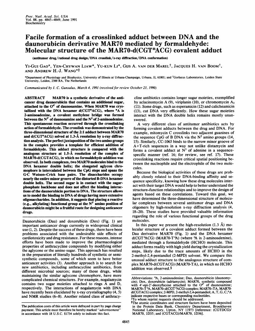
FIG. 1. Molecular formulas of anthracycline antibiotics, Dau,Dox, and MAR70. These compounds contain an aglycon chro-mophore with four fused rings (A-D); rings B-D are unsaturated,whereas ring A is semisaturated. MAR70 is a synthetic compoundwith an additional sugar, 4'-epi-2'-deoxyfucose, attached to the 04'of the daunosamine; it has similar activity as Dau.
EXPERIMENTAL
Oligonucleotides d(CGTACG) and d(CGT 'ACG) were syn-thesized according to a published procedure (21). MAR70hydrochloride was dissolved in water as stock solutions forcrystallization. The MAR70-T'A complex was crystallizedfrom a mixture containing 1.2 mM d(CGT"ACG) (singlestrand), 4mM BaC12, 30 mM sodium cacodylate (pH 6.0), 2.5mM spermine, 1.2 mM MAR70 plus 5% (vol/vol) 2-MPD.The solution was equilibrated with 40o (vol/vol) 2-MPD atroom temperature (=25°C) by vapor diffusion. TheMAR70-TA complex was crystallized under similar condi-tions, except that BaCl2 was replaced with 20mM MgCl2. Thecrystals were in space group P41212 with respective unit celldimensions a = b = 28.12 (1) and c = 52.98 (4) A for TnAcomplex and a = b = 28.01 (1) and c = 53.11 (3) A for TAcomplex. The crystal was mounted in a sealed glass capillarywith a droplet of mother liquor. Data were collected on aRigaku (Japan) AFC-5R rotating anode x-ray diffractometer at25°C using the w-scan mode with CuKa radiation to a resolutionof 1.3 A (MAR7OT'A) and 1.5 A (MAR7O-TA). There were2599 and 2168 independent reflections seen at the 2.0 o'(F) levelabove background after Lorentz-polarization, empirical ab-sorption, and decay corrections for the two data sets. Coordi-nates from the Dau-d(CGTACG) structure (19) were used as thestarting model and refined by the Konnert-Hendrickson con-strained-refinement procedure (22). A series of Fourier mapswas calculated to locate the second sugar and solvent watermolecules in the crystal lattice. During refinement of theMAR70-PA structure, we noticed a well-defined electrondensity bridged between N2 of nA and N3 of MAR70. Thiselectron density could not be interpreted as solvent or ion, as itwas too close to both nitrogen atoms. We concluded that thisdensity was best assigned as a methylene bridge resulting fromthe crosslinking reaction ofthe HCHO and refined as such. TheMAR70-T'A structure was refined to a final R factor of 17.4%oat 1.3- resolution with the root-mean-square differences in
bond distances of 0.013 A from the ideal values. The final Rfactor is 16.4% at 1.5-A resolution with a root-mean-squarevalue of 0.025 A for the MAR70-TA structure. One hydratedsodium ion coordinated to N7 of the G6 residue was located inboth complexes, as in the Dau-d(CGTACG) complex (19). Noother ions (Mg+2, Ba+2, or spermine) could be unambiguouslyidentified.We used the color-test method of chromotropic acid to
determine HCHO in 2-MPD solvent (Aldrich, 11210-0, 99%6)(23). Ten microliters of 99% 2-MPD was added to 2 ml of 12M sulfuric acid containing some solid chromotropic acid.After 30 min of heating at 600C, the solution became yellow.Adding 100 1.l or more of 2-MPD resulted in a deep orangecolor. For comparison, a low concentration of HCHO gavea yellowish-brown color, and a high concentration gave adeep-purple color. This experiment suggests that certainimpurities in 2-MPD can cause a crosslinking reaction toproduce coloration of chromotropic acid. We note that evenifHCHO constitutes only 0.001% of the 2-MPD stock used inthe crystallization reservoir (30 ml of 40%o 2-MPD), it repre-sents a 1000-fold molar excess over MAR70 in the dip (2.5 ILIat 7 mM concentration). We have also demonstrated thecrosslinking between Dau and DNA oligonucleotides medi-ated by HCHO by HPLC.
RESULTS AND DISCUSSIONMolecular Structure. Both structures were determined and
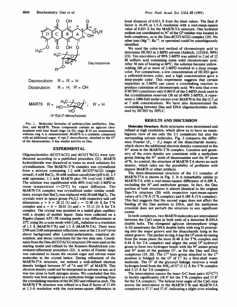
refined at high resolution, which allow us to have an unam-biguous view of not only the 2:1 complexes but also thesurrounding solvent molecules. Fig. 2 compares the differ-ence Fourier (F0 - Fc) maps of the disaccharide moiety,which shows the additional electron density connected to theN3' atom in the MAR70-T 'A complex. Location and geom-etry of the extra density are consistent with a methylenegroup linking the N3' atom of daunosamine and the N2 atomof 'A. As control, the structure ofMAR70-TA shows no suchdensity, which rules out the possibility of problems withimpure MAR70 or other artifacts.The three-dimensional structure of the 2:1 complex of
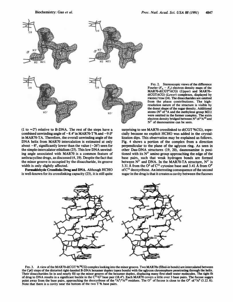
MAR70-TPA is shown in Fig. 3. It is remarkably similar toMAR70-TA with a root-mean-square difference of 0.209 A,excluding the N2 and methylene groups. In fact, the Dauportion of both structures is almost identical to the originalDau-TA structure (20) with respective root-mean-squarevalues of 0.170 A (T'A complex) and 0.189 A (TA complex).This fact suggests that the second sugar does not affect thebinding of the Dau portion to DNA, and the methylenecrosslink does not perturb the structure to any significantextent.
In both complexes, two MAR70 molecules are intercalatedbetween the CpG steps at both ends of a distorted B-DNAdouble helix. The elongated aglycon chromophore (ringsA-D) penetrates the DNA double helix with ring D protrud-ing into the major groove and the disaccharide lying in theminor groove. The pucker in ringA has the 09 atom deviatingfrom the mean plane the most (0.48 A for T 'A complex and0.44 A for TA complex) and aligns the axial 09 hydroxylgroup to form two hydrogen bonds with the N2 amino groupand N3 atom of the guanine G2, as for the Dau and Doxcomplexes (19, 20). The C13 keto group attached to the C9position is bridged to the o2 of C' by a first-shell watermolecule. The 07 of the glycosyl linkage receives a weakhydrogen bond from the NH2 of G2 (3.12 A for T 'A complexand 3.13 A for TA complex).The intercalation causes the inner G-C base pairs (G(2<11)
to buckle significantly (18.40 for the T 'A complex and 17.80for the TA complex). The helical twist angle of the CpG stepacross the intercalator in the MAR70-T 'A and MAR70-TAcomplexes is 37.10 and 37.60, indicating a slight over-winding
0
H3C ..- UHuH3C 1'
Daunosamine
DaunorubicinDoxorubicin
4846 Biochemistry: Gao et A
Proc. Natl. Acad. Sci. USA 88 (1991) 4847
FIG. 2. Stereoscopic views of the differenceFourier (F. - Fj) electron density maps of theMAR70-d(CGT"ACG) (Upper) and MAR70-d(CGTACG) (Lower) complexes, displayed byFRODO/TOM (24). The disaccharides are omittedfrom the phase contributions. The high-resolution nature of the structure is visible bythe donut shape of the sugar density. Additionalatoms (N2 of 'A and the methylene group M21)were omitted in the former complex. The extraelectron density bridged between N2 of"A'0 andN3 of daunosamine can be seen.
(1 to =2°) relative to B-DNA. The rest of the steps have acombined unwinding angle of -8.4° in MAR70-T 'A and -9.0°in MAR70-TA. Therefore, the overall unwinding angle of theDNA helix from MAR70 intercalation is estimated at onlyabout -8°, significantly lower than the value (-26°) seen forthe simple-intercalator ethidium (25). This low DNA unwind-ing angle associated with MAR70 is a common feature ofanthracycline drugs, as discussed (4, 19). Despite the fact thatthe minor groove is occupied by the disaccharide, its groovewidth is only slightly affected.Formaldehyde Crosslinks Drug and DNA. Although HCHO
is well-known for its crosslinking capacity (23), it is still quite
surprising to see MAR70 crosslinked to d(CGT 9ACG), espe-cially because no explicit HCHO was added in the crystal-lization dips. This observation may be explained as follows.Fig. 4 shows a portion of the complex from a directionperpendicular to the plane of the aglycon ring. As seen inother Dau-DNA structures (19, 20), daunosamine is posi-tioned with its N3 amino group approaching the edge of thebase pairs, such that weak hydrogen bonds are formedbetween N3' and DNA. In the MAR70-TA structure, N3' is3.31 A from the o2 of C1' cytosine base and 3.41 A from 04'ofC" deoxyribose. An interesting consequence ofthe secondsugar in the drug is that it creates a cavity between the fucoses
FIG. 3. A view ofthe MAR70-d(CGT nA'OCG) complex looking into the minorgroove. Two MAR70s (filled-in bonds) are intercalated betweenthe CpG steps of the distorted right-handed B-DNA hexamer duplex (open bonds) with the aglycon chromophore penetrating through the helix.Their disaccharides lie in and nearly fill up the minor groove of the hexamer duplex, displacing many first-shell water molecules. The tight fitof drug to DNA results in a significant buckle in the C"'-G2 base pair (18.4°). Each MAR70 covers a little over 3 base pairs. The fucose sugarspoint away from the base pairs, approaching the deoxyribose of the nA4/nA'0 residues. The 05' of fucose is close to the 04' of nA4 (3.12 A).Note that there is a cavity near the bottom of the two T 9A base pairs.
N3' N3'
Biochemistry: Gao et al.
Proc. Natl. Acad. Sci. USA 88 (1991)
M14
G2 j.,
..c
-,'.r-I.
T3 T3
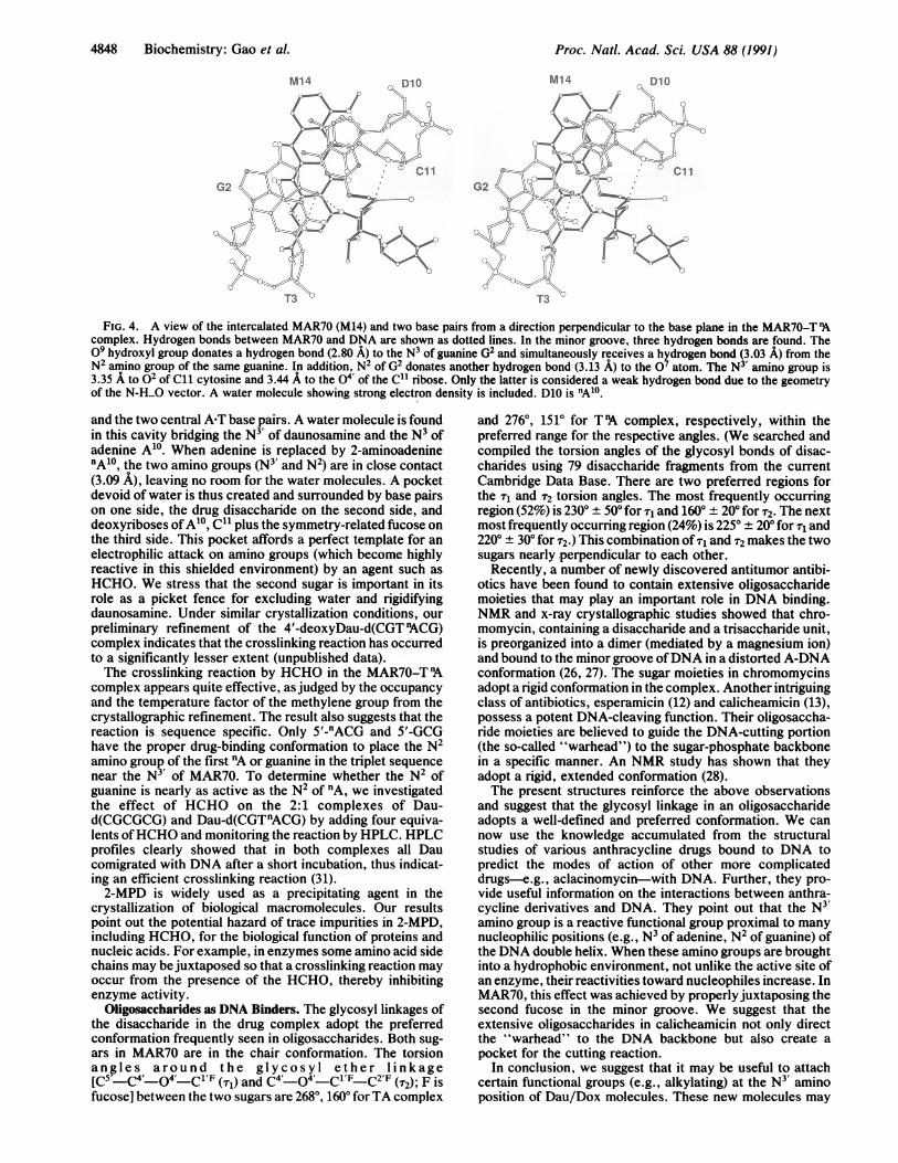
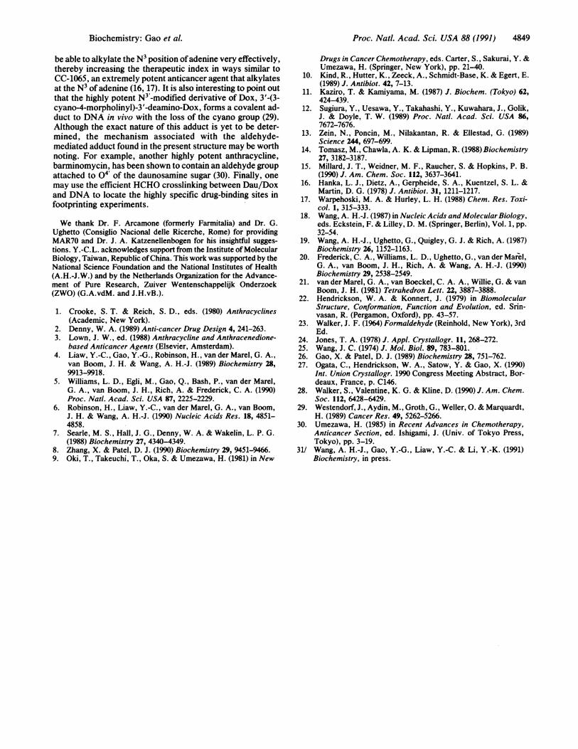
FIG. 4. A view of the intercalated MAR70 (M14) and two base pairs from a direction perpendicular to the base plane in the MAR70-Tcomplex. Hydrogen bonds between MAR70 and DNA are shown as dotted lines. In the minor groove, three hydrogen bonds are found. The09 hydroxyl group donates a hydrogen bond (2.80 A) to the N3 of guanine G2 and simultaneously receives a hydrogen bond (3.03 A) from theN2 amino group of the same guanine. In addition, N2 of G2 donates another hydrogen bond (3.13 A) to the O7 atom. The N3 amino group is3.35 A to 02 of C11 cytosine and 3.44 A to the 04' of the C" ribose. Only the latter is considered a weak hydrogen bond due to the geometryof the N-H...O vector. A water molecule showing strong electron density is included. D10 is nA10.
and the two central APT base pairs. A water molecule is foundin this cavity bridging the N3 of daunosamine and the N3 ofadenine A'0. When adenine is replaced by 2-aminoadeninenA10, the two amino groups (N3' and N2) are in close contact(3.09 A), leaving no room for the water molecules. A pocketdevoid of water is thus created and surrounded by base pairson one side, the drug disaccharide on the second side, anddeoxyriboses ofA'0, C" plus the symmetry-related fucose onthe third side. This pocket affords a perfect template for anelectrophilic attack on amino groups (which become highlyreactive in this shielded environment) by an agent such asHCHO. We stress that the second sugar is important in itsrole as a picket fence for excluding water and rigidifyingdaunosamine. Under similar crystallization conditions, ourpreliminary refinement of the 4'-deoxyDau-d(CGTnACG)complex indicates that the crosslinking reaction has occurredto a significantly lesser extent (unpublished data).The crosslinking reaction by HCHO in the MAR70-T 'A
complex appears quite effective, as judged by the occupancyand the temperature factor of the methylene group from thecrystallographic refinement. The result also suggests that thereaction is sequence specific. Only 5'-nACG and 5'-GCGhave the proper drug-binding conformation to place the N2amino group of the first nA or guanine in the triplet sequencenear the N3' of MAR70. To determine whether the N2 ofguanine is nearly as active as the N2 of nA, we investigatedthe effect of HCHO on the 2:1 complexes of Dau-d(CGCGCG) and Dau-d(CGTnACG) by adding four equiva-lents ofHCHO and monitoring the reaction by HPLC. HPLCprofiles clearly showed that in both complexes all Daucomigrated with DNA after a short incubation, thus indicat-ing an efficient crosslinking reaction (31).2-MPD is widely used as a precipitating agent in the
crystallization of biological macromolecules. Our resultspoint out the potential hazard of trace impurities in 2-MPD,including HCHO, for the biological function of proteins andnucleic acids. For example, in enzymes some amino acid sidechains may be juxtaposed so that a crosslinking reaction mayoccur from the presence of the HCHO, thereby inhibitingenzyme activity.
Oligosaccharides as DNA Binders. The glycosyl linkages ofthe disaccharide in the drug complex adopt the preferredconformation frequently seen in oligosaccharides. Both sug-ars in MAR70 are in the chair conformation. The torsionangles around the glycosyl ether linkage[C5'_C4'_04'_Cl'F (Tr) and C4'_04'_Cl'F-C2'F (T2); F is
fucose] between the two sugars are 2680, 160° for TA complex
and 2760, 151° for T 'A complex, respectively, within thepreferred range for the respective angles. (We searched andcompiled the torsion angles of the glycosyl bonds of disac-charides using 79 disaccharide fragments from the currentCambridge Data Base. There are two preferred regions forthe T1 and 2 torsion angles. The most frequently occurringregion (52%) is 2300 + 500 for r1 and 1600 ± 200 for T2. The nextmost frequently occurring region (24%) is 2250 200for x1 and2200 ± 300 for T2.) This combination of , and r2 makes the twosugars nearly perpendicular to each other.
Recently, a number of newly discovered antitumor antibi-otics have been found to contain extensive oligosaccharidemoieties that may play an important role in DNA binding.NMR and x-ray crystallographic studies showed that chro-momycin, containing a disaccharide and a trisaccharide unit,is preorganized into a dimer (mediated by a magnesium ion)and bound to the minor groove ofDNA in a distorted A-DNAconformation (26, 27). The sugar moieties in chromomycinsadopt a rigid conformation in the complex. Another intriguingclass of antibiotics, esperamicin (12) and calicheamicin (13),possess a potent DNA-cleaving function. Their oligosaccha-ride moieties are believed to guide the DNA-cutting portion(the so-called "warhead") to the sugar-phosphate backbonein a specific manner. An NMR study has shown that theyadopt a rigid, extended conformation (28).The present structures reinforce the above observations
and suggest that the glycosyl linkage in an oligosaccharideadopts a well-defined and preferred conformation. We cannow use the knowledge accumulated from the structuralstudies of various anthracycline drugs bound to DNA topredict the modes of action of other more complicateddrugs-e.g., aclacinomycin-with DNA. Further, they pro-vide useful information on the interactions between anthra-cycline derivatives and DNA. They point out that the N3'amino group is a reactive functional group proximal to manynucleophilic positions (e.g., N3 of adenine, N2 of guanine) ofthe DNA double helix. When these amino groups are broughtinto a hydrophobic environment, not unlike the active site ofan enzyme, their reactivities toward nucleophiles increase. InMAR70, this effect was achieved by properlyjuxtaposing thesecond fucose in the minor groove. We suggest that theextensive oligosaccharides in calicheamicin not only directthe "warhead" to the DNA backbone but also create a
pocket for the cutting reaction.In conclusion, we suggest that it may be useful to attach
certain functional groups (e.g., alkylating) at the N3 aminoposition of Dau/Dox molecules. These new molecules may
D10
{d E
M14 D10I:
4848 Biochemistry: Gao et al.
Proc. Natl. Acad. Sci. USA 88 (1991) 4849
be able to alkylate the N3 position ofadenine very effectively,thereby increasing the therapeutic index in ways similar toCC-1065, an extremely potent anticancer agent that alkylatesat the N3 of adenine (16, 17). It is also interesting to point outthat the highly potent N3'-modified derivative of Dox, 3'-(3-cyano-4-morpholinyl)-3'-deamino-Dox, forms a covalent ad-duct to DNA in vivo with the loss of the cyano group (29).Although the exact nature of this adduct is yet to be deter-mined, the mechanism associated with the aldehyde-mediated adduct found in the present structure may be worthnoting. For example, another highly potent anthracycline,barminomycin, has been shown to contain an aldehyde groupattached to 04 of the daunosamine sugar (30). Finally, onemay use the efficient HCHO crosslinking between Dau/Doxand DNA to locate the highly specific drug-binding sites infootprinting experiments.
We thank Dr. F. Arcamone (formerly Farmitalia) and Dr. G.Ughetto (Consiglio Nacional delle Ricerche, Rome) for providingMAR70 and Dr. J. A. Katzenellenbogen for his insightful sugges-tions. Y.-C.L. acknowledges support from the Institute of MolecularBiology, Taiwan, Republic ofChina. This work was supported by theNational Science Foundation and the National Institutes of Health(A.H.-J.W.) and by the Netherlands Organization for the Advance-ment of Pure Research, Zuiver Wentenschappelijk Onderzoek(ZWO) (G.A.vdM. and J.H.vB.).
1. Crooke, S. T. & Reich, S. D., eds. (1980) Anthracyclines(Academic, New York).
2. Denny, W. A. (1989) Anti-cancer Drug Design 4, 241-263.3. Lown, J. W., ed. (1988) Anthracycline and Anthracenedione-
based Anticancer Agents (Elsevier, Amsterdam).4. Liaw, Y.-C., Gao, Y.-G., Robinson, H., van der Marel, G. A.,
van Boom, J. H. & Wang, A. H.-J. (1989) Biochemistry 28,9913-9918.
5. Williams, L. D., Egli, M., Gao, Q., Bash, P., van der Marel,G. A., van Boom, J. H., Rich, A. & Frederick, C. A. (1990)Proc. Nati. Acad. Sci. USA 87, 2225-2229.
6. Robinson, H., Liaw, Y.-C., van der Marel, G. A., van Boom,J. H. & Wang, A. H.-J. (1990) Nucleic Acids Res. 18, 4851-4858.
7. Searle, M. S., Hall, J. G., Denny, W. A. & Wakelin, L. P. G.(1988) Biochemistry 27, 4340-4349.
8. Zhang, X. & Patel, D. J. (1990) Biochemistry 29, 9451-9466.9. Oki, T., Takeuchi, T., Oka, S. & Umezawa,-H. (1981) in New
Drugs in Cancer Chemotherapy, eds. Carter, S., Sakurai, Y. &Umezawa, H. (Springer, New York), pp. 21-40.
10. Kind, R., Hutter, K., Zeeck, A., Schmidt-Base, K. & Egert, E.(1989) J. Antibiot. 42, 7-13.
11. Kaziro, T. & Kamiyama, M. (1987) J. Biochem. (Tokyo) 62,424-439.
12. Sugiura, Y., Uesawa, Y., Takahashi, Y., Kuwahara, J., Golik,J. & Doyle, T. W. (1989) Proc. Natl. Acad. Sci. USA 86,7672-7676.
13. Zein, N., Poncin, M., Nilakantan, R. & Ellestad, G. (1989)Science 244, 697-699.
14. Tomasz, M., Chawla, A. K. & Lipman, R. (1988) Biochemistry27, 3182-3187.
15. Millard, J. T., Weidner, M. F., Raucher, S. & Hopkins, P. B.(1990) J. Am. Chem. Soc. 112, 3637-3641.
16. Hanka, L. J., Dietz, A., Gerpheide, S. A., Kuentzel, S. L. &Martin, D. G. (1978) J. Antibiot. 31, 1211-1217.
17. Warpehoski, M. A. & Hurley, L. H. (1988) Chem. Res. Toxi-col. 1, 315-333.
18. Wang, A. H.-J. (1987) in Nucleic Acids and Molecular Biology,eds. Eckstein, F. & Lilley, D. M. (Springer, Berlin), Vol. 1, pp.32-54.
19. Wang, A. H.-J., Ughetto, G., Quigley, G. J. & Rich, A. (1987)Biochemistry 26, 1152-1163.
20. Frederick, C. A., Williams, L. D., Ughetto, G., van der Marel,G. A., van Boom, J. H., Rich, A. & Wang, A. H.-J. (1990)Biochemistry 29, 2538-2549.
21. van der Marel, G. A., van Boeckel, C. A. A., Willie, G. & vanBoom, J. H. (1981) Tetrahedron Lett. 22, 3887-3888.
22. Hendrickson, W. A. & Konnert, J. (1979) in BiomolecularStructure, Conformation, Function and Evolution, ed. Srin-vasan, R. (Pergamon, Oxford), pp. 43-57.
23. Walker, J. F. (1964) Formaldehyde (Reinhold, New York), 3rdEd.
24. Jones, T. A. (1978) J. Appl. Crystallogr. 11, 268-272.25. Wang, J. C. (1974) J. Mol. Biol. 89, 783-801.26. Gao, X. & Patel, D. J. (1989) Biochemistry 28, 751-762.27. Ogata, C., Hendrickson, W. A., Satow, Y. & Gao, X. (1990)
Int. Union Crystallogr. 1990 Congress Meeting Abstract, Bor-deaux, France, p. C146.
28. Walker, S., Valentine, K. G. & Kline, D. (1990) J. Am. Chem.Soc. 112, 6428-6429.
29. Westendorf, J., Aydin, M., Groth, G., Weller, 0. & Marquardt,H. (1989) Cancer Res. 49, 5262-5266.
30. Umezawa, H. (1985) in Recent Advances in Chemotherapy,Anticancer Section, ed. Ishigami, J. (Univ. of Tokyo Press,Tokyo), pp. 3-19.
31/ Wang, A. H.-J., Gao, Y.-G., Liaw, Y.-C. & Li, Y.-K. (1991)Biochemistry, in press.
Biochemistry: Gao et al.

![cis-Diamminedichloroplatinum(II)-DNA Adduct Formation in ... · [CANCER RESEARCH 47, 718-722, February 1, 1987] cis-Diamminedichloroplatinum(II)-DNA Adduct Formation in Renal, Gonadal,](https://img.pdfslide.us/doc/110x75/60934a1bfda1347d92293bf5/cis-diamminedichloroplatinumii-dna-adduct-formation-in-cancer-research-47.jpg)