Embed Size (px)
Citation preview
Covalent Adduct Formation between the Antihypertensive DrugHydralazine and Abasic Sites in Double- and Single-Stranded DNADouglas Melton,† Calvin D. Lewis,† Nathan E. Price,† and Kent S. Gates*,†,‡
†Department of Chemistry, ‡Department of Biochemistry, University of Missouri, 125 Chemistry Building, Columbia, Missouri65211, United States
*S Supporting Information
ABSTRACT: Hydralazine (4) is an antihypertensive agentthat displays both mutagenic and epigenetic properties. Here,gel electrophoretic, mass spectroscopic, and chemical kineticsmethods were used to provide evidence that medicinallyrelevant concentrations of 4 rapidly form covalent adductswith abasic sites in double- and single-stranded DNA underphysiological conditions. These findings raise the intriguingpossibility that the genotoxic properties of this clinically useddrug arise via reactions with an endogenous DNA lesion rather than with the canonical structure of DNA.
■ INTRODUCTIONHydralazine (1-hydrazinophthalazine, 4, Scheme 1) is anantihypertensive agent that was introduced into the clinic in
the early 1950s,1,2 and this drug remains in use, primarily forthe treatment of gestational hypertension.3,4 In addition, 4induces demethylation of cellular DNA,5 a property that hasgiven the compound a second life as a possible epigeneticdrug.6−8
Interestingly, a number of reports indicate that 4 ismutagenic in Ames assays.9 Chemically induced mutagenesistypically involves covalent modification of the canonicalnucleobases of the DNA in target cells.10−12 Subsequenterror-prone replication of the damaged DNA introducesmutations into the genetic code. Accordingly, a variety of
DNA-damage mechanisms have been proposed to explain themutagenic action of 4, including oxidation of the drug to aDNA-damaging diazonium ion, diazene radical, or aryl radical,nucleophilic addition to pyrimidine residues in DNA, andoxidative conversion of a formaldehyde-derived hydrazoneadduct into a DNA-alkylating species.13−17 However, noconsensus has emerged regarding a chemical mechanism forthe damage of cellular DNA by 4.In the work described here, we explored the novel possibility
that the mutagenic properties of the clinically used drug 4 arisevia the drug’s ability to covalently capture endogenous abasic(Ap) lesions in genomic DNA rather than by modification ofcanonical DNA bases. Ap sites are generated by spontaneousand enzymatic hydrolysis of the glycosidic bonds that hold thecoding nucleobases to the 2-deoxyribose-phosphate backboneof DNA.18−21 As a result, the DNA of normal mammaliantissue harbors between 50 000 and 200 000 Ap sites percell.22,23 Ap sites exist as an equilibrium mixture of the ring-closed hemiacetal 2 and ring-opened aldehyde 3 (Scheme 1).24
The aryl hydrazine group of 4 has the potential to react withthe Ap aldehyde residue to generate a hydrazone adduct (7 or8, Scheme 1). Hydrazone formation is a well-known reactionthat has found use in biochemistry and chemical biology forchemoselective ligations;25−30 however, at the outset of ourstudies, it was by no means clear that medicinally relevantconcentrations of 4 would be capable of forming adducts withAp sites in DNA under physiological conditions. This isbecause hydrazone formation in neutral aqueous solutiontypically is rather slow.26 As a result, hydrazone-formingreactions involving biomolecules usually employ high concen-trations of at least one reaction partner, low pH (4−5), or anadded organocatalyst.25−27 To the best of our knowledge, the
Received: September 8, 2014Published: November 6, 2014
Scheme 1
Article
pubs.acs.org/crt
© 2014 American Chemical Society 2113 dx.doi.org/10.1021/tx5003657 | Chem. Res. Toxicol. 2014, 27, 2113−2118
reaction of aryl hydrazines with Ap sites in DNA has notpreviously been examined under physiologically relevantconditions. In the work described here, we employed gelelectrophoretic, mass spectroscopic, and chemical kineticsmethods to provide evidence that medicinally relevantconcentrations of 4 rapidly form covalent adducts with abasicsites in double- and single-stranded DNA under physiologicalconditions.
■ EXPERIMENTAL PROCEDURESMaterials. Oligonucleotides were purchased from Integrated DNA
Technologies (Coralville, IA). Hydralazine hydrochloride, 2-deoxy-D-ribose, sodium hydroxide, and other chemicals were purchased fromSigma-Aldrich (St. Louis, Mo) and used without further purification.The enzyme uracil DNA glycosylase (UDG) was purchased from NewEngland Biolabs (Ipswich, MA). [γ-32P]-ATP (6000 Ci/mmol) waspurchased from PerkinElmer (Waltham, MA). C18 Sep-Pak cartridgeswere purchased from Waters (Milford, MA), and BS Polyprepcolumns were obtained from BioRad (Hercules, CA). Measurement ofradioactivity in polyacrylamide gels was carried out using a PersonalMolecular Imager (Bio-Rad) with Quantity One software (v.4.6.5).Reaction of 4 with Double- and Single-Stranded DNA
Oligonucleotides. The 2′-deoxyuridine-containing oligonucleotidesused here were 5′-32P-labeled, annealed with their complementarystrand (in the case of duplex B), and treated with uracil DNAglycosylase (UDG) to form the Ap site at a defined location usingstandard procedures.31−33 Subsequent reactions were carried out inHEPES buffer (50 mM, pH 7) containing NaCl (0.1 M) at 37 °C in afinal volume of 10 μL unless otherwise specified. Efficient formation ofthe Ap site resulting from UDG treatment was confirmed by workupof the DNA with NaOH (3.3 μL of a 500 mM stock solution in water),followed by incubation for 2 h at 37 °C. Typically, the Ap site wasgenerated in >90% yield. The same NaOH workup was used to deducethe amount of base-labile lesions (e.g., Ap site) remaining in thelabeled oligonucleotide at end of incubation either with or withoutcompound 4. As described above, the samples were mixed with NaOH(3.3 μL of a 500 mM stock solution in water), followed by incubationfor 2 h at 37 °C. Control experiments showed that the pH of samplesafter mixing with NaOH in the base workup step was the sameregardless of whether compound 4 was present. The samples (13.3μL), without further purification, were mixed with formamide loadingbuffer (75−115 μL)33 containing bromophenol blue to achieveapproximately 800 cpm/1.5 μL. Samples were loaded onto a 20%denaturing polyacrylamide gel, the gel was electrophoresed at 1000−1500 V for 2−4 h until the bromophenol blue marker dye hadmigrated approximately 10−15 cm from the wells, and the resolvedlabeled DNA fragments were visualized by phosphorimager analysis. Inthe gel shift experiments (Figures 2 and S4), duplex B was incubatedwith 4 (100 μM) for 2 h under the standard reaction conditionsdescribed above and then diluted with loading dye without any furthertreatment or purification. Duplex A was incubated with 4 under thesame conditions to ensure that reaction of the compound with nativenucleobases (as opposed to the Ap site in duplex B) does not induce agel shift. These samples were analyzed on a 20% denaturingpolyacrylamide gel as described above except that the bands wererun at least 30−35 cm from the wells to allow a clear separation of theAp-oligonucleotide from the adducted oligonucleotide. In the case ofcontrol reactions involving the incubation of phthalazine with duplexB, the reaction conditions were the same as those describe above, withthe exception that stock solutions of phthalazine were prepared bydissolution of the compound in DMSO at a concentration 100 mM,followed by dilution with water to a final concentration of 1 mM. Thefinal concentration of phthalazine in the assay was 100 μM, andDMSO concentration was 0.1% (v/v).The amount of full-length labeled oligonucleotide product
remaining in reactions of duplex B and oligonucleotide C with 4following NaOH workup were corrected for the small amounts of full-length oligonucleotide remaining after NaOH workup in a controlsample that was not treated with 4. Generally, the amount of
remaining full-length product was less than 5% of total radioactivity inthe lane. The small amounts of full-length product in these controlsamples presumably correspond to 2′-deoxyuridine-containing oligo-nucleotide (in duplex A) that was not converted to the Ap-containingoligonucleotide (in duplex B) by UDG.
Kinetics Analysis of the Reaction between 4 and DNAOligonucleotides. The data for the reaction of duplex B with 4 (10μM) or single-stranded oligonucleotide D with 4 (50 μM) shown inFigure 4 was fit to the equation for appearance of product via a first-order process: Yt = Y∞ + (Y0 − Y∞) e
(−kt), where Yt is the reading forproduct at time t, Y0 is the reading at time 0, and Y∞ is the final readingwhen reaction is complete (see pp. 22−23 of ref 34). Both Y∞ and Y0were floated in the fitting process. Fitting provided an observedpseudo-first-order rate constant for each reaction. The r2 values for theresulting fits were 0.98 ± 0.01. Average values and standard deviationswere obtained by fitting and averaging the resulting values from at leastthree separate experiments. The apparent second-order rate constantswere obtained by dividing the observed first-order rate constants bythe concentration of 4 in the reaction (10 μM 4 in the reaction withduplex B and 50 μM 4 in the reaction with oligo D). Alternatively,exploiting the equation ln[(Yt − Y∞)/[(Y0 − Y∞)] = −kt, a plot ofln[(Yt − Y∞)/[(Y0 − Y∞)], or ln|Yt − Y∞| versus time was generatedfor each reaction, and the data was fit to a line (Figure S7).34 Theslope of the resulting lines in these plots corresponds to −k. Again, theapparent second-order rate constant for each reaction was obtained bydividing the observed first-order rate constant by the pseudo-first-order concentration of 4 employed in the reaction. The valuescalculated for the second-order rate constants by this graphical methodmatched well with those obtained by the nonlinear curve-fittingmethod (Figure S7).
Static Nanospray QTOF-MS of Adduct-Containing DNA. Theoligonucleotide sample was analyzed in a 40 mM dimethylbutylam-monium acetate (pH 7.1) buffer. Negative ion MS spectra was takenfor mass range of 280−3200 Da on an Agilent 6520A QTOF MS withChip Cube source (G4240A). Monoisotopic neutral masses werecalculated from the multiply charged ion spectra of signals present inthe 500−2000 Da mass range. Sample introduction was done withNew Objective Econo12-N uncoated borosilicate glass emitters.Negative ion spectrum was acquired at a capillary potential sufficientto initiate spray of the sample. The nitrogen gas was heated at 290 °Cand introduced at a flow rate of 4 L/min. The fragmentor, skimmer,and octapole1 RF Vpp potentials were set to 200, 65, and 750 V,respectively. External calibration was done with the Agilent ESI-lowcalibration tuning mixture (cat. no. G1969-85000), and data analysiswas performed with Agilent Mass Hunter Workstation qualitativeanalysis software v B.02.00, build 2.0.197.0, with Bioconfirm Software(2008). Peptide isotope model was assumed, and peak set heightthreshold for extraction was set to ≥500 counts. Deconvolution wascarried out with a 0.1 Da step size with a result of 20 iterations of thealgorithm calculation
■ RESULTSGel Electrophoretic Evidence for a Reaction between
4 and an Abasic Site in Duplex DNA. Here, we examinedthe reaction of 4 with Ap sites in synthetic DNAoligonucleotides. Toward this end, the Ap-containing DNAduplex B was generated by treatment of the corresponding5′-32P-labeled, 2′-deoxyuridine-containing duplex A with uracilDNA glycosylase (UDG).31,32 Efficient formation of the Ap sitewas confirmed by treatment of the DNA with mild alkali togenerate a mixture of the expected 3′-4-hydroxy-2-pentenal-5-phosphate (5) and 3′-phosphate (6) cleavage products (Figure1, lane 4).18,35,36 Our initial approach for detecting the reactionof 4 with the DNA abasic site capitalized on the expectationthat formation of a hydrazone adduct 7/8 would render the Ap-containing oligonucleotide resistant to cleavage under mildalkaline conditions, analogous to the properties of the oximeadduct derived from reaction of methoxyamine with an Ap site
Chemical Research in Toxicology Article
dx.doi.org/10.1021/tx5003657 | Chem. Res. Toxicol. 2014, 27, 2113−21182114
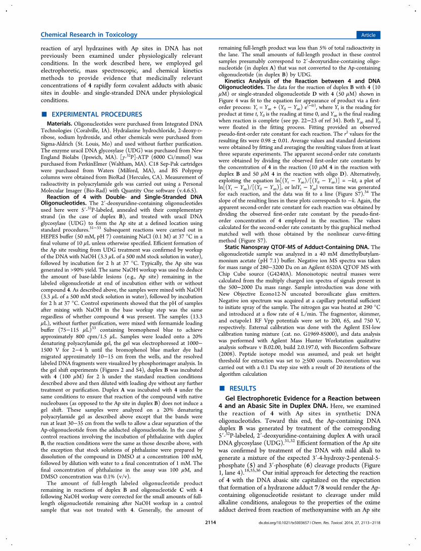
in DNA.37 We found that incubation of duplex B with 4 (100μM) in HEPES buffer (50 mM, pH 7, containing 100 mMNaCl) for 2 h at 37 °C rendered the 32P-labeled, Ap-containingstrand almost completely refractory to strand cleavage inducedby NaOH workup (Figure 1, lane 5). This result was strikingbecause an early study showed that, in unbuffered water, theinteraction of phenylhydrazine hydrochloride with Ap-contain-ing DNA fragments induced strand cleavage at the Ap siterather than formation of a phenylhydrazone adduct on the full-length strand.38,39 Under our reaction conditions, incubation of4 with duplex B generated little or no strand cleavage abovebackground (Figure 1, lane 5). Identical results were obtainedwhen 4 was incubated with a longer, 35 base pair, duplexcontaining a single Ap site (Figure S1). Medicinally relevantplasma concentrations of 4 are in the low micromolar range,40
so we examined the reaction of duplex B with a 1 μMconcentration of 4 in HEPES buffer (50 mM, pH 7) at 37 °Cfor 1 h. This resulted in 67 ± 5% inhibition of NaOH-mediated
strand cleavage (Figure S2). Compound 4 is reported toundergo slow decomposition to phthalazine in aqueoussolutions near neutral pH (t1/2 ∼ 7 h).41 A control experimentshowed that phthalazine (10 μM) did not significantly inhibitNaOH-mediated strand cleavage of duplex B (Figure S3) and,thus, does not contribute to the action of 4 described here.Seeking direct evidence of a covalent adduct between 4 and
the Ap site in duplex B, we conducted experiments designed todetect altered gel mobility of the 32P-labeled oligonucleotide induplex B following treatment with 4. When the DNA fragmentswere run at least 30 cm from the origin of a 20% denaturingpolyacrylamide gel, we observed a clear shift in the gelelectrophoretic mobility of the Ap-containing oligonucleotideupon treatment with 4 (Figure 2). The retardation in gel
mobility observed here was consistent with formation of acovalent drug−DNA adduct. Control experiments showed thatincubation of 4 with the labeled dU-containing duplex A didnot generate a gel-shifted product (Figure S4). This providedevidence that the gel shift shown in Figure 2 was due toreaction of 4 with the Ap site in duplex B rather than withnative nucleobases in the labeled oligonucleotide.
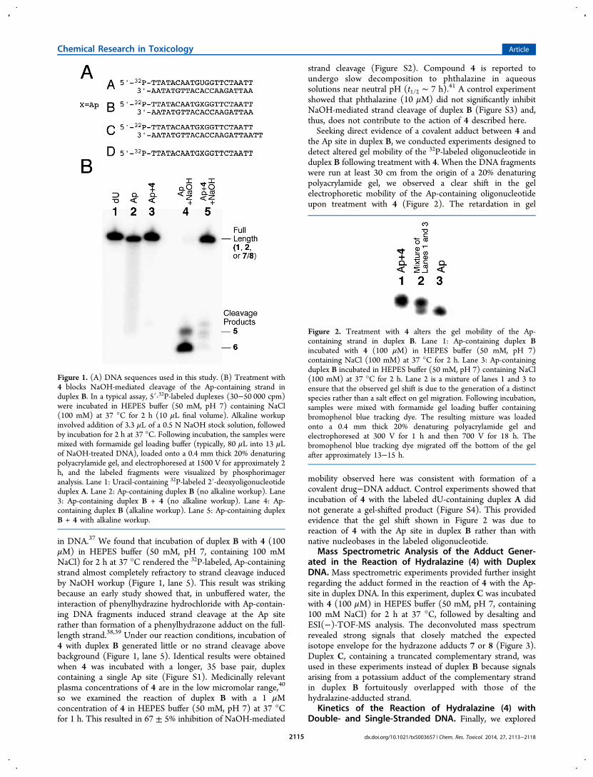
Mass Spectrometric Analysis of the Adduct Gener-ated in the Reaction of Hydralazine (4) with DuplexDNA. Mass spectrometric experiments provided further insightregarding the adduct formed in the reaction of 4 with the Ap-site in duplex DNA. In this experiment, duplex C was incubatedwith 4 (100 μM) in HEPES buffer (50 mM, pH 7, containing100 mM NaCl) for 2 h at 37 °C, followed by desalting andESI(−)-TOF-MS analysis. The deconvoluted mass spectrumrevealed strong signals that closely matched the expectedisotope envelope for the hydrazone adducts 7 or 8 (Figure 3).Duplex C, containing a truncated complementary strand, wasused in these experiments instead of duplex B because signalsarising from a potassium adduct of the complementary strandin duplex B fortuitously overlapped with those of thehydralazine-adducted strand.
Kinetics of the Reaction of Hydralazine (4) withDouble- and Single-Stranded DNA. Finally, we explored
Figure 1. (A) DNA sequences used in this study. (B) Treatment with4 blocks NaOH-mediated cleavage of the Ap-containing strand induplex B. In a typical assay, 5′-32P-labeled duplexes (30−50 000 cpm)were incubated in HEPES buffer (50 mM, pH 7) containing NaCl(100 mM) at 37 °C for 2 h (10 μL final volume). Alkaline workupinvolved addition of 3.3 μL of a 0.5 N NaOH stock solution, followedby incubation for 2 h at 37 °C. Following incubation, the samples weremixed with formamide gel loading buffer (typically, 80 μL into 13 μLof NaOH-treated DNA), loaded onto a 0.4 mm thick 20% denaturingpolyacrylamide gel, and electrophoresed at 1500 V for approximately 2h, and the labeled fragments were visualized by phosphorimageranalysis. Lane 1: Uracil-containing 32P-labeled 2′-deoxyoligonucleotideduplex A. Lane 2: Ap-containing duplex B (no alkaline workup). Lane3: Ap-containing duplex B + 4 (no alkaline workup). Lane 4: Ap-containing duplex B (alkaline workup). Lane 5: Ap-containing duplexB + 4 with alkaline workup.
Figure 2. Treatment with 4 alters the gel mobility of the Ap-containing strand in duplex B. Lane 1: Ap-containing duplex Bincubated with 4 (100 μM) in HEPES buffer (50 mM, pH 7)containing NaCl (100 mM) at 37 °C for 2 h. Lane 3: Ap-containingduplex B incubated in HEPES buffer (50 mM, pH 7) containing NaCl(100 mM) at 37 °C for 2 h. Lane 2 is a mixture of lanes 1 and 3 toensure that the observed gel shift is due to the generation of a distinctspecies rather than a salt effect on gel migration. Following incubation,samples were mixed with formamide gel loading buffer containingbromophenol blue tracking dye. The resulting mixture was loadedonto a 0.4 mm thick 20% denaturing polyacrylamide gel andelectrophoresed at 300 V for 1 h and then 700 V for 18 h. Thebromophenol blue tracking dye migrated off the bottom of the gelafter approximately 13−15 h.
Chemical Research in Toxicology Article
dx.doi.org/10.1021/tx5003657 | Chem. Res. Toxicol. 2014, 27, 2113−21182115
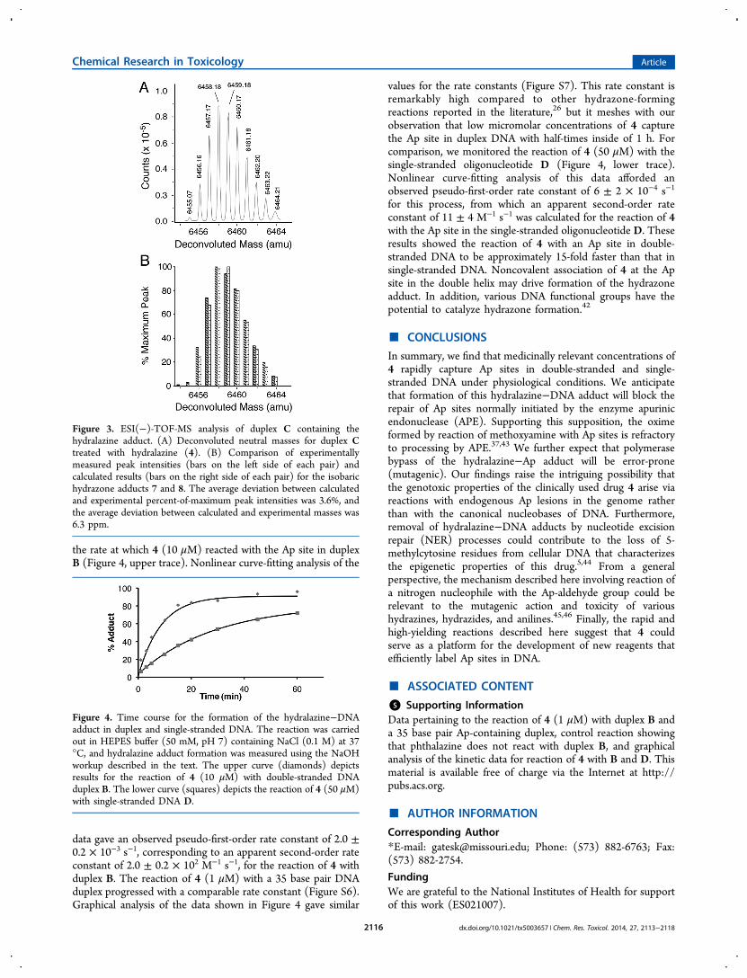
the rate at which 4 (10 μM) reacted with the Ap site in duplexB (Figure 4, upper trace). Nonlinear curve-fitting analysis of the
data gave an observed pseudo-first-order rate constant of 2.0 ±0.2 × 10⁻³ s−1, corresponding to an apparent second-order rateconstant of 2.0 ± 0.2 × 10² M−1 s−1, for the reaction of 4 withduplex B. The reaction of 4 (1 μM) with a 35 base pair DNAduplex progressed with a comparable rate constant (Figure S6).Graphical analysis of the data shown in Figure 4 gave similar
values for the rate constants (Figure S7). This rate constant isremarkably high compared to other hydrazone-formingreactions reported in the literature,26 but it meshes with ourobservation that low micromolar concentrations of 4 capturethe Ap site in duplex DNA with half-times inside of 1 h. Forcomparison, we monitored the reaction of 4 (50 μM) with thesingle-stranded oligonucleotide D (Figure 4, lower trace).Nonlinear curve-fitting analysis of this data afforded anobserved pseudo-first-order rate constant of 6 ± 2 × 10⁻⁴ s−1
for this process, from which an apparent second-order rateconstant of 11 ± 4 M−1 s−1 was calculated for the reaction of 4with the Ap site in the single-stranded oligonucleotide D. Theseresults showed the reaction of 4 with an Ap site in double-stranded DNA to be approximately 15-fold faster than that insingle-stranded DNA. Noncovalent association of 4 at the Apsite in the double helix may drive formation of the hydrazoneadduct. In addition, various DNA functional groups have thepotential to catalyze hydrazone formation.42
■ CONCLUSIONS
In summary, we find that medicinally relevant concentrations of4 rapidly capture Ap sites in double-stranded and single-stranded DNA under physiological conditions. We anticipatethat formation of this hydralazine−DNA adduct will block therepair of Ap sites normally initiated by the enzyme apurinicendonuclease (APE). Supporting this supposition, the oximeformed by reaction of methoxyamine with Ap sites is refractoryto processing by APE.37,43 We further expect that polymerasebypass of the hydralazine−Ap adduct will be error-prone(mutagenic). Our findings raise the intriguing possibility thatthe genotoxic properties of the clinically used drug 4 arise viareactions with endogenous Ap lesions in the genome ratherthan with the canonical nucleobases of DNA. Furthermore,removal of hydralazine−DNA adducts by nucleotide excisionrepair (NER) processes could contribute to the loss of 5-methylcytosine residues from cellular DNA that characterizesthe epigenetic properties of this drug.5,44 From a generalperspective, the mechanism described here involving reaction ofa nitrogen nucleophile with the Ap-aldehyde group could berelevant to the mutagenic action and toxicity of varioushydrazines, hydrazides, and anilines.45,46 Finally, the rapid andhigh-yielding reactions described here suggest that 4 couldserve as a platform for the development of new reagents thatefficiently label Ap sites in DNA.
■ ASSOCIATED CONTENT
*S Supporting InformationData pertaining to the reaction of 4 (1 μM) with duplex B anda 35 base pair Ap-containing duplex, control reaction showingthat phthalazine does not react with duplex B, and graphicalanalysis of the kinetic data for reaction of 4 with B and D. Thismaterial is available free of charge via the Internet at http://pubs.acs.org.
■ AUTHOR INFORMATION
Corresponding Author*E-mail: [email protected]; Phone: (573) 882-6763; Fax:(573) 882-2754.
FundingWe are grateful to the National Institutes of Health for supportof this work (ES021007).
Figure 3. ESI(−)-TOF-MS analysis of duplex C containing thehydralazine adduct. (A) Deconvoluted neutral masses for duplex Ctreated with hydralazine (4). (B) Comparison of experimentallymeasured peak intensities (bars on the left side of each pair) andcalculated results (bars on the right side of each pair) for the isobarichydrazone adducts 7 and 8. The average deviation between calculatedand experimental percent-of-maximum peak intensities was 3.6%, andthe average deviation between calculated and experimental masses was6.3 ppm.
Figure 4. Time course for the formation of the hydralazine−DNAadduct in duplex and single-stranded DNA. The reaction was carriedout in HEPES buffer (50 mM, pH 7) containing NaCl (0.1 M) at 37°C, and hydralazine adduct formation was measured using the NaOHworkup described in the text. The upper curve (diamonds) depictsresults for the reaction of 4 (10 μM) with double-stranded DNAduplex B. The lower curve (squares) depicts the reaction of 4 (50 μM)with single-stranded DNA D.
Chemical Research in Toxicology Article
dx.doi.org/10.1021/tx5003657 | Chem. Res. Toxicol. 2014, 27, 2113−21182116
NotesThe authors declare no competing financial interest.
■ ACKNOWLEDGMENTS
MALDI mass spectrometry was conducted at the Charles W.Gehrke Proteomics Center at the University of Missouri, andwe thank Beverly DaGue for assistance with these experiments.We thank Professor Phillip Wilmarth (Oregon State Uni-versity) for assistance fitting the observed data to the expectedresults in the mass spectrometric experiments.
■ ABBREVIATIONS
Ap site, abasic site; UDG, uracil DNA glycosylase; APE, Apendonuclease; NER, nucleotide excision repair
■ REFERENCES(1) Hilker, R. R., Rhoads, P. S., and Billings, C. E. (1953) Clinical useof hydralazine and hexamethonium in treatment of hypertension. J.Am. Med. Assoc. 153, 5−9.(2) Gross, F., Druey, J., and Meier, R. (1950) Eine neue gruppeblutdrucksenkender substanzen von besonderem wirkungscharakter.Experientia 15, 19−21.(3) Magee, L. A., Abalos, E., von Dadelszen, P., Sibai, B., Easterling,T., and Walkinshaw, S. (2011) How to manage hypertension inpregnancy effectively. Br. J. Clin. Pharmacol. 72, 394−401.(4) Slim, H. B., Black, H. R., and Thompson, P. D. (2011) Olderblood pressure medicationsdo they still have a place? Am. J. Cardiol.108, 308−316.(5) Cornacchia, E., Golbus, J., Maybaum, J., Strahler, J., Hanash, S.,and Richardson, B. (1988) Hydralazine and procainamide inhibit Tcell DNA methylation and induce autoreactivity. J. Immunol. 140,2197−2200.(6) Arce, C., Segura-Pacheco, B., Perez-Cardenas, E., Taja-Chayeb, L.,Candelaria, M., and Duennas-Gonzalez, A. (2006) Hydralazine target:from blood vessels to the epigenome. J. Transl. Med. 4, 1−16.(7) Ren, J., Singh, B. N., Huang, Q., Li, Z., Gao, Y., Mishra, P., Hwa,Y. L., Li, J., Dowdy, S. C., and Jiang, S.-W. (2011) DNAhypermethylation as a chemotherapy target. Cell. Signalling 23,1082−1093.(8) Burridge, S. (2013) Drugging the epigenome. Nat. Rev. Drug.Discovery 12, 92−93.(9) Blanco, M., Martínez, A., Urios, A., Herrera, G., and O’Connor, J.E. (1998) Detection of oxidative mutagenesis by isoniazid and otherhydrazine derivatives in Escherichia coli WP2 tester strain IC203,deficient in OxyR: strong protective effects of rat liver S9. Mutat. Res.417, 39−46.(10) Dipple, A. (1995) DNA adducts of chemical carcinogens.Carcinogenesis 16, 437−441.(11) Shrivastav, N., Li, D., and Essigmann, J. M. (2010) Chemicalbiology of mutagenesis and DNA repair: cellular responses to DNAalkylation. Carcinogenesis 31, 59−70.(12) Hemminki, K. (1993) DNA adducts, mutations and cancer.Carcinogenesis 14, 2007−2012.(13) Sinha, B. K. (1983) Enzymatic activation of hydrazinederivatives. A spin-trapping study. J. Biol. Chem. 258, 796−801.(14) Yamamoto, K., and Kawanishi, S. (1991) Free radicalproduction and site-specific DNA damage induced by hydralazine inthe presence of metal ions or peroxidase/hydrogen peroxide. Biochem.Pharmacol. 41, 905−914.(15) Reilly, C. A., and Aust, S. D. (1997) Peroxidase substratesstimulate the oxidation of hydralazine to metabolites which causesingle-strand breaks in DNA. Chem. Res. Toxicol. 10, 328−334.(16) Dubroff, L. M., and Reid, R. J. J. (1980) Hydralazine−pyrimidine interactions may explain hydralazine-induced lupuserythematosus. Science 208, 404−406.
(17) Mathison, B. H., Murphy, S. E., and Shank, R. C. (1994)Hydralazine and other hydrazine derivatives and the formation ofDNA adducts. Toxicol. Appl. Pharmacol. 127, 91−98.(18) Gates, K. S. (2009) An overview of chemical processes thatdamage cellular DNA: spontaneous hydrolysis, alkylation, andreactions with radicals. Chem. Res. Toxicol. 22, 1747−1760.(19) Guillet, M., and Bioteux, S. (2003) Origin of endogenous DNAabasic sites in Saccaromyces cerevisiae. Mol. Cell. Biol. 23, 8386−8394.(20) Gates, K. S., Nooner, T., and Dutta, S. (2004) Biologicallyrelevant chemical reactions of N7-alkyl-2′-deoxyguanosine adducts inDNA. Chem. Res. Toxicol. 17, 839−856.(21) David, S. S., and Williams, S. D. (1998) Chemistry ofglycosylases and endonucleases involved in base-excision repair.Chem. Rev. 98, 1221−1261.(22) De Bont, R., and van Larebeke, N. (2004) Endogenous DNAdamage in humans: a review of quantitative data. Mutagenesis 19, 169−185.(23) Nakamura, J., and Swenberg, J. A. (1999) Endogenous apurinic/apyrimidinic sites in genomic DNA of mammalian tissues. Cancer Res.59, 2522−2526.(24) Wilde, J. A., Bolton, P. H., Mazumdar, A., Manoharan, M., andGerlt, J. A. (1989) Characterization of the equilibrating forms of theabasic site in duplex DNA using 17O-NMR. J. Am. Chem. Soc. 111,1894−1896.(25) Mahal, L. K., Yarema, K. J., and Bertozzi, C. R. (1997)Engineering chemical reactivity on cell surfaces through oligosacchar-ide biosynthesis. Science 276, 1125−1128.(26) Kool, E. T., Park, D.-H., and Crisalli, P. (2013) Fast hydrazonereactants: electronic and acid/base effects strongly influence rate atbiological pH. J. Am. Chem. Soc. 135, 17663−17666.(27) Dirksen, A., and Dawson, P. E. (2008) Rapid oxime andhydrazone ligations with aromatic aldehydes for biomolecular labeling.Bioconjugate Chem. 19, 2543−2548.(28) Pfander, S., Fiammengo, R., Kirin, S. I., Metlzer-Nolte, N., andJas̈chke, A. (2007) Reversible site-specific tagging of enzymaticallysynthethesized RNAs using aldehyde−hydrazine chemistry andprotease-cleavable linkers. Nucleic Acids Res. 35, e25.(29) Raindlova,́ V., Pohl, R., Sanda, M., and Hocek, M. (2010) Directpolymerase synthesis of reactive aldehyde-functionalized DNA and itsconjugation and staining with hydrazines. Angew. Chem., Int. Ed. 49,1064−1066.(30) Wang, X., and Canary, J. W. (2012) Rapid catalyst-freehydrazone ligation: protein−pyridoxal phosphoramides. BioconjugateChem. 23, 2329−2334.(31) Lindahl, T., Ljunquist, S., Siegert, W., Nyberg, B., and Sperens,B. (1977) DNA N-glycosidases: properties of uracil-DNA glycosidasefrom Escherichia coli. J. Biol. Chem. 252, 3286−3294.(32) Varshney, U., and van de Sande, J. H. (1991) Specificities andkinetics of uracil excision from uracil-containing DNA oligomers byEscherichia coli uracil DNA glycosylase. Biochemistry 30, 4055−4061.(33) Sambrook, J., Fritsch, E. F., and Maniatis, T. (1989) MolecularCloning: A Lab Manual, Cold Spring Harbor Press, Cold SpringHarbor, NY.(34) Espenson, J. H. (1995) Chemical Kinetics and ReactionMechanisms, 2nd ed., pp 22−23, McGraw-Hill, Inc., New York.(35) Bailly, V., and Verly, W. G. (1988) Possible roles of beta-elimination and gamma-elimination reactions in the repair of DNAcontaining AP (apurinic/apyrimidinic) sites in mammalian cells.Biochem. J. 253, 553−559.(36) Bayley, C. R., Brammer, K. W., and Jones, A. S. (1961) Thenucleotide sequence in deoxyribonucleic acids. Part V. The alkalinedegradation of apurinic sites. J. Chem. Soc., 1903−1907.(37) Fortini, P., Bignami, M., and Dogliotti, E. (1990) Evidence forAP site formation related to DNA-oxygen alkylation in CHO cellstreated with ethylating agents. Mutat. Res. 236, 129−137.(38) Coombs, M. M., and Livingston, D. C. (1969) Reaction ofapurinic acid with aldehyde reagents. Biochim. Biophys. Acta 174, 161−173.
Chemical Research in Toxicology Article
dx.doi.org/10.1021/tx5003657 | Chem. Res. Toxicol. 2014, 27, 2113−21182117
(39) Vasseur, J.-J., Rayner, B., and Imbach, J.-L. (1988) ApurinicDNA reactivity: modelisation of apurinic DNA breakage by phenyl-hydrazine and formation of pyrazole adduct. J. Heterocycl. Chem. 25,389−392.(40) Shepherd, A. M., McNay, J. L., Ludden, T. M., Lin, M. S., andMusgrave, G. E. (1981) Plasma concentration and acetylatorphenotype determine response to oral hydralazine. Hypertension 3,580−585.(41) Halasi, S., and Nairn, J. G. (1990) Stability studies of hydralazinehydrochloride in aqueous solutions. J. Parenter. Sci. Technol. 44, 30−34.(42) Crisalli, P., and Kool, E. T. (2013) Important of ortho protondonors in the catalysis of hydrazone formation. Org. Lett. 15, 1646−1649.(43) Liu, L., Yan, L., Donze, J. R., and Gerson, S. L. (2003) Blockageof abasic site repair enhances antitumor efficacy of 1,3-bis-(2-chloroethyl)-1-nitrosourea in colon tumor xenografts. Mol. Cancer.Ther. 2, 1061−1066.(44) Wu, S. C., and Zhang, Y. (2010) Active DNA demethylation:many roads lead to Rome. Nat. Rev. Mol. Cell Biol. 11, 607−620.(45) Stepan, A. F., Walker, D. P., Bauman, J., Price, D. A., Baille, T.A., Kalgutkar, A. S., and Aleo, M. D. (2011) Structural alert/reactivemetabolite concept as applied in medicinal chemistry to mitigate therisk of idiosyncratic drug toxicity: a perspective based on the criticalexamination of trends in the top 200 drugs marketed in the UnitedStates. Chem. Res. Toxicol. 24, 1345−1410.(46) Parodi, S., De Flora, S., Cavanna, M., Pino, A., Robbiano, L.,Bennicelli, C., and Brambilla, G. (1981) DNA-damaging activity invivo and bacterial mutagenicity of sixteen hydrazine derivatives asrelated quantitatively to their carcinogenicity. Cancer Res. 41, 1469−1482.
Chemical Research in Toxicology Article
dx.doi.org/10.1021/tx5003657 | Chem. Res. Toxicol. 2014, 27, 2113−21182118