Embed Size (px)
Citation preview

The Regensburg Experience
Extracorporeal Life Support Group
Dept. of Anaesthesiology
Dept. of Internal Medicine
Dept. of Cardiothoracic Surgery – Perfusion
Alois Philipp
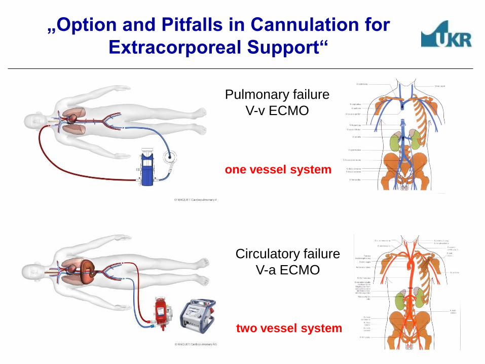
„Option and Pitfalls in Cannulation
for Extracorporeal Support“

„Option and Pitfalls in Cannulation for
Extracorporeal Support“
Fundamentals
for Extracorporeal Support
„Option and Pitfalls in Cannulation for
Extracorporeal Support“
Pulmonary failure
V-v ECMO
Circulatory failure
V-a ECMO
one vessel system
two vessel system

Peripheral vs Central Cannulation
central peripherial

Cannulation: Percutanous vs surgical
Surgical subclavia artery
Surgical femoral artery and vein
Percutaneously
using seldinger‘s technique
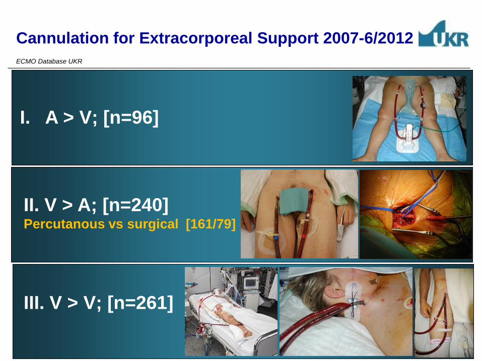
Cannulation for Extracorporeal Support 2007-6/2012
Legionellosis
I. A > V; [n=96]
II. V > A; [n=240] Percutanous vs surgical [161/79]
III. V > V; [n=261]
ECMO Database UKR

Pros and Cons of Percutaneus Cannulation
Pro: - Few staffing and logistic requirements
- Preferred out of cardiothoracic surgery
- Smaller wound surface area
- Reduced risk of bleeding / infection
Con: - Cannulation of wrong vessel possible (arterial vs. venous)
- Complications entail delay of vital therapy
- Limited choice of accessible vessels

Extracorporeal Assist Application
Possible locations
of extracorporeal
assist application
In-House
Out-of-Hospital
Out-of-Center

Percutanous ECMO Implantation
V-a
In-House
V-v
In-Hous
V-a / V-v
Out of Center
Team
Composition
Cardiac surg. (if available)
Intennsivist or Anästhesist
Perfusionist
Intensivist
Anästhesist
Perfusionist
Anästhesist
Perfusionist
US control
of vessels Yes Yes Yes
Heparin 5000 IE 5000 IE 5000 IE
Cannulaes
Inflow
15 or 17 Fr
and 15 cm long
17 or 19 Fr
and 23 cm long
(or Doppellumen)
Doppellumen not
preferred (V-V)
Cannulaes
Outflow
21 or 23 Fr
55 cm long
21 or 23 Fr
38 cm long
(Doppellumen)
Doppellumen not
preferred (V-V) ECMO Database UKR

Divices for percutanous cannulation
have the right tools

ECMO – Cannulaes (V-V)

V-v ECMO: Positioning of Cannulaes
both cannulaes from
the femoral side
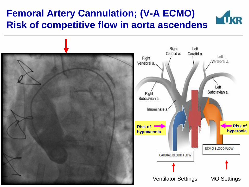
Femoral Artery Cannulation; (V-A ECMO)
Risk of competitive flow in aorta ascendens
Ventilator Settings MO Settings
Risk of
hypoxaemia
Risk of
hyperoxia

Femoral Artery Cannulation; (V-A ECMO)
Risk of competitive flow in aorta ascendens

„Option Cannulation for Extracorporeal Support“
Options in V- v ECMO

V-v ECMO; Bi-Caval Dual Lumen
Novalung Avalon

Implantation:
V-v ECMO; Bi-Caval Dual Lumen (Avalon)

V-v ECMO - Bi-Caval Dual Lumen

Case history:
50 yrs, male
Diagnosis:
Pulmonary embolism, DVB
Special circumstances:
PFO
Ventilator settings and BGA
Out-of-Center Interhospital Transport V-v ECMO
Pre ECMO 2 hrs on ECMO
FiO2 1.0 0.7
PaO2 59 126
PCO2 49 29
TV [ml/min] 695 300
MV [l/min] 17.2 4.8
Duration of ECMO support 10 days
Discharged from hospital

V-v ECMO both cannulaes in femoral veins

„Cannulation for Extracorporeal Support“
Pitfalls in V- v ECMO

Extreme obesity
Particularly risky in out of center ECMO implantation
220 kg 250 kg

Advantage vs Risk

V-v ECMO; Bi-Caval Dual Lumen (AVALON)
day 30
weaning
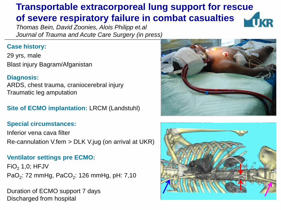
Case history:
29 yrs, male
Blast injury Bagram/Afganistan
Diagnosis:
ARDS, chest trauma, craniocerebral injury
Traumatic leg amputation
Site of ECMO implantation: LRCM (Landstuhl)
Special circumstances:
Inferior vena cava filter
Re-cannulation V.fem > DLK V.jug (on arrival at UKR)
Ventilator settings pre ECMO:
FiO2 1,0; HFJV
PaO2: 72 mmHg, PaCO2: 126 mmHg, pH: 7,10
Duration of ECMO support 7 days
Discharged from hospital
Transportable extracorporeal lung support for rescue
of severe respiratory failure in combat casualties Thomas Bein, David Zoonies, Alois Philipp et.al
Journal of Trauma and Acute Care Surgery (in press)

Pitfalls in Cannulation (V-v ECMO)
Pre
ECMO
Day 1 on
ECMO
PaO2/FiO2 (mm Hg) 39 160
PaCO2 (mm Hg) 43 44
pH 7.41 7.39
MV (mL/min) 9.5 2.5
PIP (cm H2O) 40 29
Norepinephrine (mg/h) 2.0 0.4
MAP (mmHg) 49 65
HR 150 90
Case history: 27 yrs, female
Sectio cesarean, Intestinal perforation, Aspiration
Diagnosis: ARDS
Transport: Following call–150 mins to start ECMO
Transport by helicopter 120 km

V-v ECMO: Pitfalls in Cannulation - fatal injury
Doppellumen
in coronary sinus
Ruptur of the
subclavian vein
Ruptur of the
jugular vein
Ruptur of the
femoral vein Guidwire stucks
„Option Cannulation for Extracorporeal Support“
Options in V- a ECMO

V-a ECMO with different access
ECMO; Femoral arterie and Femoral vein ECMO; Subclavia arterie and Subclavia vein
Ciculatory failure with normal Gasexchange Ciculatory failure with impaired Gasexchange
Fixation

Modification in cannulation V-a ECMO
Ouflow vena jugularis Inflow arteria femoralis
Pat. with ICM
Resuscitation 35 min
Femoral vene no guidewire placable
Extubation day 3 on ECMO
Day 10 LVAD on ECMO
Venous line

Modification in cannulation (V-V-A ECMO)
AoAsc in
V. jugularis in
V. femoralis out

Modification in cannulation
Surgical management for Stanford type A aortic dissection: direct cannulation of real lumen at
the level of the Botallo’s ligament by Seldinger chnique
Laszlo Göbölös, Alois Philipp, Maik Foltan, Karsten Wiebe
Interactive CardioVascular and Thoracic Surgery 7 (2008)
Apex cannulation

ECMO in Cath Lab - High risk TAVP and PCI
ECMO prevents circulatory failure
A simple method of vascular access to perform
emergency coronary angiography in patients with veno-
arterial extracorporeal membrane oxygenation
Endemann DH, Philipp A, Hengstenberg C, et. al.,
Intensive Care Med. 2011 Dec;37(12):2046-9

PCI subclavia artery calcification femoral vessel
ECMO in Cath Lab - High risk TAVP and PCI

„Cannulation for Extracorporeal Support“
Pitfalls in V- a ECMO

ECMO supported CPR
Cannulation of wrong vessel
Art. cannulae
in femoral vene

Percutanous Cannulation for Extracorporeal Life Support P. Ganselmeier, A. Philipp, L. Rupprecht et. al.
Thorac Cardiovasc Surg. 2011;59: 103-107
Ischaemia Malperfusion
Dissection of femoral arterie

Cannulaes:

V. fem. A. fem.
Percutaneous Cannulation - De-cannulation
A. fem.
24 hrs with 30 ml
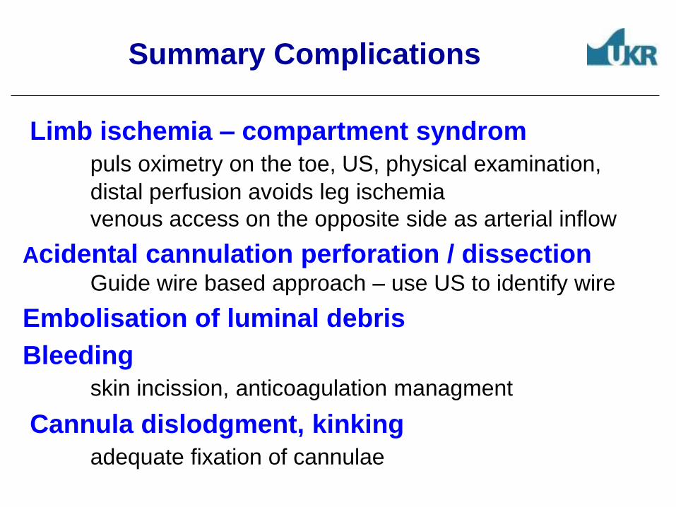
Summary Complications
Limb ischemia – compartment syndrom
puls oximetry on the toe, US, physical examination,
distal perfusion avoids leg ischemia
venous access on the opposite side as arterial inflow
Acidental cannulation perforation / dissection Guide wire based approach – use US to identify wire
Embolisation of luminal debris
Bleeding
skin incission, anticoagulation managment
Cannula dislodgment, kinking
adequate fixation of cannulae

Cannulation for Extracorporeal Support
Resume
don‘t force it