Embed Size (px)
Citation preview

Extensions of MADM (Mosaic Analysis with DoubleMarkers) in MiceBosiljka Tasic1.¤, Kazunari Miyamichi1., Simon Hippenmeyer1., Vardhan S. Dani1, Hong Zeng2,
William Joo1,4, Hui Zong3, Yanru Chen-Tsai2, Liqun Luo1*
1 Department of Biology, Howard Hughes Medical Insitute, Stanford University, California, United States of America, 2 Transgenic Facility, Stanford Cancer Center, Stanford
University School of Medicine, California, United States of America, 3 Institute of Molecular Biology, University of Oregon, Eugene, Oregon, United States of America,
4 Neurosciences Program, Stanford University, California, United States of America
Abstract
Mosaic Analysis with Double Markers (MADM) is a method for generating genetically mosaic mice, in which sibling mutantand wild-type cells are labeled with different fluorescent markers. It is a powerful tool that enables analysis of gene functionat the single cell level in vivo. It requires transgenic cassettes to be located between the centromere and the mutation in thegene of interest on the same chromosome. Here we compare procedures for introduction of MADM cassettes into new lociin the mouse genome, and describe new approaches for expanding the utility of MADM. We show that: 1) Targetedhomologous recombination outperforms random transgenesis in generation of reliably expressed MADM cassettes, 2)MADM cassettes in new genomic loci need to be validated for biallelic and ubiquitous expression, 3) Recombinationbetween MADM cassettes on different chromosomes can be used to study reciprocal chromosomal deletions/duplications,and 4) MADM can be modified to permit transgene expression by combining it with a binary expression system. Theadvances described in this study expand current, and enable new and more versatile applications of MADM.
Citation: Tasic B, Miyamichi K, Hippenmeyer S, Dani VS, Zeng H, et al. (2012) Extensions of MADM (Mosaic Analysis with Double Markers) in Mice. PLoS ONE 7(3):e33332. doi:10.1371/journal.pone.0033332
Editor: Brian D. McCabe, Columbia University, United States of America
Received November 6, 2011; Accepted February 7, 2012; Published March 27, 2012
Copyright: � 2012 Tasic et al. This is an open-access article distributed under the terms of the Creative Commons Attribution License, which permitsunrestricted use, distribution, and reproduction in any medium, provided the original author and source are credited.
Funding: This work was supported by a National Institutes of Health grant to LL (R01-NS050835). BT was a Damon Runyon Fellow and was supported by theDamon Runyon Cancer Research Foundation Grant DRG-1819-04. KM was supported by the Japan Society for the Promotion of Science program for ResearchAbroad and Human Frontier Science Program Organization (LT00300/2007-L). SH was supported by postdoctoral fellowships from the European MolecularBiology Organization (ALTF 851-2005), Human Frontier Science Program Organization (LT00805/2006-L), and Swiss National Science Foundation (PA00P3_124160and PA00P3_136482). HZ is a Pew Scholar in Biomedical Sciences, supported by The Pew Charitable Trusts. LL is an investigator of the Howard Hughes MedicalInstitute. The funders had no role in study design, data collection and analysis, decision to publish, or preparation of the manuscript.
Competing Interests: The authors have declared that no competing interests exist.
* E-mail: [email protected]
. These authors contributed equally to this work.
¤ Current address: Allen Institute for Brain Science, Seattle, Washington, United States of America
Introduction
Genetically mosaic animals (genetic mosaics) contain cells with
different genotypes. Phenotypic analysis of genetic mosaics has
become an indispensible tool in modern genetics. Genetic mosaics
are usually created by using site-specific recombinases from
heterologous biological systems, most prominently including Cre
recombinase from the E. coli phage P1 [1] and Flp recombinase
from the S. cerevisiae 2 m plasmid [2]. DNA recombination can
occur either in cis (on the same chromosome) or in trans (between
chromosomes). Intrachromosomal recombination techniques usu-
ally rely on the presence of two recombination sites flanking a
particular DNA sequence that will be excised upon recombination
[3]. In contrast, interchromosomal recombination techniques
depend on recombination between chromatids after DNA
replication in the G2 phase of the cell cycle to generate sibling
cells of different genotypes. Interchromosomal recombination has
been used to develop various versions of mosaic analysis in fruit
flies [4,5,6,7,8,9]. The common and key feature of these
approaches is that they create cells with different genotypes in
vivo and at the same time label those cells with unique markers that
strictly correlate with the genotype. To enable such concomitant in
vivo genetic manipulation and labeling in mammals, we have
established Mosaic Analysis with Double Markers (MADM) in
mice (Figure 1A) [10]. We have used MADM since its inception
to perform lineage studies [11] and analyze gene function in a
number of biological processes including cell proliferation [12],
dendritic patterning [13], neuronal migration [14] and tumor
initiation and progression [15]. To expand the utility and
versatility of MADM, we present here modifications and new
applications of the technique, and compare different procedures
for establishment of MADM-ready chromosomes.
Results
Design of new MADM cassettesThe original version of MADM relied on the DsRed2
fluorescent protein as one of the two markers [10]. Due to the
low DsRed2 fluorescence signal in tests in vitro, six Myc epitope
tags were added to its C-terminus. The addition of these epitope
tags proved to be essential, because the detection of DsRed2
expression from knocked-in MADM cassettes in vivo required anti-
Myc immunostaining [10]. For the new MADM cassettes, we
chose tdTomato (tdT) over DsRed2, due to its improved
PLoS ONE | www.plosone.org 1 March 2012 | Volume 7 | Issue 3 | e33332

brightness [16]. We also added three Myc epitope tags to its C-
terminus, and this addition did not appear to affect the tdT
fluorescence (data not shown).
The original MADM cassettes were designed to split two
fluorescent protein genes approximately in the middle of each
gene [10] (Figure 1B, left). To replace one fluorescent protein
gene with another (e.g., DsRed2 with tdT), an entirely new set of
cassettes needs to be constructed, as neither of the existing cassettes
would be compatible with any new cassette. We therefore aimed to
create a more flexible design for new cassettes, such that one of
them would be compatible with any new cassette and could be
subsequently reused. In our new design for splitting the red
fluorescent protein tdT, the first exon contains only the start
codon (Figure 1B, right). Therefore the two new cassettes are:
GFPN-terminus-intron-tdT3MycATG-less (for simplicity, GT) and ATG-
intron-GFPC-terminus (for simplicity, TG). The new TG cassette is now
compatible with any GFPN-terminus-intron-XATG-less (for simplicity, GX)
cassette, where XATG-less (for simplicity, X) is any gene without the
start codon (Figure 1B, bottom right). This design has been
especially useful for combining MADM with a binary expression
system to create MADM-Tet, where X is the tetracycline
transactivator tTA2 [17] without the start codon (for details see
below).
Expansion of MADM to additional chromosomes viarandom transgenesis in ES cells
MADM requires two reciprocally chimeric marker genes
(‘MADM cassettes’, e.g., GT and TG), which are targeted into
Figure 1. The MADM principle and design of new MADM cassettes. A) MADM relies on two reciprocally chimeric marker genes (for example,GR and RG, see part B below for cassette description) that have been knocked into the same locus on homologous chromosomes. Recombination inthe G2 phase of the cell cycle regenerates the functional marker genes on a pair of chromatids. X-segregation of chromatids (the recombinantchromatids segregate to different cells) generates a red and a green cell. Z-segregation of chromatids (the recombinant chromatids congregate tothe same cell) generates a double-labeled (yellow) cell and an unlabeled cell. If a mutation (asterisk) is present distally to the GR cassette, the greencells will be homozygous for the mutation. This orientation of the cassettes corresponds to MADM in the Rosa26 locus. If the cassettes are in theopposite orientation with respect to the centromere, the genotypes for green and red cells will be inverted (for example in MADM-11). If mitoticrecombination occurs in G0 or G1, a double-labeled cell is produced without altering the genotype of the cell. B) The ‘‘old’’ MADM cassettescontained two genes encoding fluorescent proteins (dsRed2 and GFP) split roughly in the middle. The ‘‘new’’ cassettes use the same GFP split, butsplit the second gene (for example, tdTomato) into ATG and GeneATG-less. That way, the ATG-GC-terminus (for simplicity, TG) becomes a universal cassettethat can be paired with any G-GeneATG-less cassette. The single white triangle represents a single loxP site, a combination of loxP sites or the loxP-flanked (floxed) neomycin resistance gene (see Figure S1 for detailed description of MADM cassettes).doi:10.1371/journal.pone.0033332.g001
Extensions of MADM
PLoS ONE | www.plosone.org 2 March 2012 | Volume 7 | Issue 3 | e33332

identical loci on homologous chromosomes. When MADM is used
to study gene function, the MADM cassettes must be located
between the gene of interest and the centromere. This is because
only chromosomal segments distal to the recombination sites
within the MADM cassettes can undergo exchange and produce
homozygosity after X-segregation in the G2 phase of the cell cycle
(Figure 1A, top right). At present, only genes located on mouse
chromosome (Chr.) 6 distal to the Rosa26 locus and on Chr. 11
distal to the Hipp11 locus can be subjected to MADM [10,14]. To
extend the MADM technology to other genes in the mouse
genome, MADM cassettes need to be inserted into additional
chromosomes. One possibility is to employ random transgenesis to
obtain integrations throughout the mouse genome. However,
random transgene integration of one MADM cassette is in
principle not suited for subsequent repeated targeting of the
complementary cassette to the same locus. To overcome this
problem, we performed random transgenesis using convertible
precursor transgenes (pMADMa and pMADMb, Figure 2A, 2B)
that can be subsequently transformed into GT and TG MADM
cassettes.
pMADMa contains the ubiquitously active CA promoter and GT
and TG MADM cassettes flanked by FRT sites. After individual
integrants are isolated, they can be converted into GT or TG
cassettes by partial recombination catalyzed by the Flp recombi-
nase (Figure 2A). We screened ES cell clones to identify single-
copy, intact pMADMa transgenes integrated into intergenic
regions of the genome (for details see Methods). 25 out of ,190
ES clones had intact 59 and 39 ends of the transgene; 12 of them
were estimated to be single-copy based on Southern hybridization;
6 insertion sites were identified by using inverse PCR. Among
them, the location of one clone was confirmed to be within an
intergenic region, in a new locus we call Miya10 (M10), ,20 Mb
distal to the centromere of Chr. 10 (Figure 2C, blue triangle). To
obtain GT and TG transgenes, we introduced the Flp recombinase
into this ES cell clone. Among ,200 ES cell subclones, ten
subclones had partial recombination between the second and third
FRTs to convert pMADMa to GT, while only one subclone had the
reciprocal partial recombination between the first and the second
FRTs to generate TG. We established transgenic mouse lines from
these ES cells (hereafter called M10TG and M10GT) via standard
blastocyst injection to generate chimeras and subsequent germline
transmission of the transgenes.
As expected, mice transheterozygous for GT and TG in the Miya10
locus (M10GT/TG) do not have colored cells in the absence of Cre-
mediated recombination (Figure 2D). When Nestin-Cre [18] or HprtCre
[19] transgenes were separately introduced to create M10GT/TG;Nestin-
Cre+/2 or M10GT/TG;HprtCre/+ or M10GT/TG;HprtCre/Y, we observed
cells labeled with GFP (green), tdTomato (red), or both (yellow), in
patterns predicted by Cre expression. For example, Nestin-Cre generates
MADM-labeled cells throughout the central nervous system, including
cortical pyramidal cells, interneurons, glia, hippocampal granule and
pyramidal cells (Figure 2D), and cerebellar Purkinje cells (data not
shown). HprtCre allows the labeling of cells in the liver, heart, and small
intestine (Figure 2D) and all other tissues examined (data not shown).
When using the same Cre driver, labeling was qualitatively less dense in
MADM-10 than in MADM-6 or MADM-11 (data not shown; ‘‘MADM-
number’’ refers to a genotype, where ‘‘MADM’’ signifies two reciprocal
cassettes in the same MADM locus combined with a Cre line, while the
number refers to a chromosome number). These data demonstrate that
random insertion-based transgenesis in ES cells can be used to establish
a functional MADM system in a new genomic locus.
However, the pMADMa-based approach had two limitations: 1)
The frequency of insertion of single copy transgenes was low
(,6%); 2) Partial recombination events to generate GT and TG
cassettes were highly biased in favor of GT (10:1), and therefore
obtaining the TG cassette became laborious. These limitations led
us to develop a second convertible precursor transgene, pMADMb.
pMADMb contains the CA promoter driving the bGeo marker (a
fusion of b-galactosidase and neomycin resistance gene), flanked
by non-compatible variants of FRT: wild-type FRT and FRT5
[20]. This transgene could be subsequently converted into any
other transgene, including a GT or TG cassette via Flp
recombinase-mediated cassette exchange [20] (Figure 2B). To
increase the chance of intact pMADMb integration, ‘protecting’
arms containing bacterial DNA were placed at the 59 and 39 ends
of the transgene (500 bp and 3.0 kbp, respectively). We electro-
porated pMADMb into mouse ES cells and isolated ,1000
subclones; 484 showed strong lacZ expression and 325 (,32%)
had intact 59 and 39 ends of the transgene. The intact transgene
frequency was ,2–3 fold higher for pMADMb than for pMADMa,
presumably due to the longer protection arms. We were able to
determine insertion sites for 161 clones using ‘‘splinkerette’’ PCR
[21]; 65 insertion sites were located in coding or intronic
sequences, and 96 were located in the intergenic areas (triangles
in Figure 2B). We confirmed the insertion sites by independent
genomic PCR for a subset of pMADMbtransgene insertions located
relatively close to corresponding centromeres (red triangles in
Figure 2B).
To test the Flp-mediated cassette exchange reaction in ES cells,
we selected one single-copy integrant located ,39 Mb from the
centromere of Chr. 1 in the Miya1 (M1) locus (Figure 2B) and
transfected it with a Flp recombinase plasmid and a plasmid
containing either the GT or TG cassette flanked with FRT5 and
FRT (see Methods). The cassette exchange efficiency was ,9% (5
out of 54 sub-clones) or 25% (12 out of 48 sub-clones) for GT or
TG cassettes, respectively. We established mice from these
converted ES cells (hereafter called M1GT and M1TG) via regular
blastocyst injection and chimeragenesis followed by the germline
transmission. Similarly to MADM-10, we observed Cre-dependent
labeling in conjunction with Nestin-Cre and HprtCre (data not
shown). However, it is important to note that although the cellular
labeling obtained by MADM-1 and MADM-10 appears as
expected (red, green and yellow cells are all evident), this labeling
may not accurately report the cellular genotypes unless the loci are
biallelically and ubiquitously expressed (see below).
MADM expansion via targeted knock-inTargeted knock-in [22,23] is a standard method for introducing
a transgene into a precise location in the mouse genome [24]. The
vast majority of ubiquitously expressed transgenes, including some
made in our lab [10,25], have been made via knock-in into the
Rosa26 locus [26]. To establish new MADM cassettes (GT and TG)
in a locus that has been already proven to support ubiquitous
expression, we inserted them into the Rosa26 locus. The knock-in
procedure into Rosa26 generated the GT and TG alleles that
allowed marker expression as described previously [10] (Figure 3,
S1). Single-labeled, green and red, cells and double-labeled,
yellow, cells were observed only when Cre was present in this
version of MADM-6 containing the new MADM cassettes
described above. As expected from in vitro cell culture tests, both
tdT and GFP fluorescence were visible without immunostaining
(Figure 3). Thus, this ‘new MADM-6’ is superior to the original
version of MADM-6 [10], which required immunostaining to
detect the red fluorescent protein.
To expand MADM to other chromosomes via targeted knock-
in, we aimed to select loci that should enable the majority of genes
on a particular chromosome to be subjected to mosaic analysis,
and that are likely to support ubiquitous and biallelic expression of
Extensions of MADM
PLoS ONE | www.plosone.org 3 March 2012 | Volume 7 | Issue 3 | e33332

Figure 2. Random integration-based approach to expand MADM to other mouse chromosomes. A and B) Schematic representations ofMADM precursor (pMADM) constructs. A) pMADMa contains the CA promoter, FRT-flanked MADM GT and TG cassettes and a single polyadenylationsignal (pA). The cassette containing the floxed neomycin phosphotransferase gene (loxP-pPGK-Neo-pA-loxP) is placed in the introns of both cassettes.pMADMa can be converted into either GT or TG via partial Flp-mediated recombination in ES cells. B) pMADMb construct contains the CA promoterdriving the bgeo gene (a lacZ and neomycin-phosphotransferase fusion) flanked by FRT5 and FRT. pMADMb can be converted into any transgene,including a GT or TG cassette via Flp- and FRT5/FRT-mediated cassette exchange in ES cells. These MADM cassettes contained a hygromicin resistancegene (H) that was removed by QC31 integrase-mediated recombination (see Figure S1) before performing the experiments shown in D. C)Distribution of pMADM transgene intergenic integration sites in the mouse genome. Each centromere is represented by a blue circle, and mappedinsertion sites are indicated by triangles (Mb, mega base pair). The pMADMa insertion site used to establish MADM-10 (Figure 2D) is represented bythe blue triangle located close to the centromere of Chr. 10. All the other triangles represent the insertion sites of pMADMb transgenes based on the59 genomic sequence amplified by Splinkerette PCR. Insertion sites that were mapped close to centromeres, and were independently confirmed by
Extensions of MADM
PLoS ONE | www.plosone.org 4 March 2012 | Volume 7 | Issue 3 | e33332

transgenes. Therefore, we focused on chromosomal regions close
to the centromere that are located between highly expressed genes
as judged by EST abundance [14]. As a specific example, we used
the above strategy to knock-in MADM cassettes into the Hipp11
locus on Chr. 11, and observed Cre-dependent labeling as
described [14] (Figure S1). We are in the process of generating
several other mouse chromosomes with inserted MADM cassettes
via the targeted knock-in approach described above.
Test for biallelic and ubiquitous marker expressionIn order for MADM cassettes to reliably report cellular
genotypes, the locus containing the cassettes must support biallelic
expression in a cellular population of interest. Ideally, the locus
should also promote ubiquitous expression. To examine biallelic
expression of the new MADM cassettes in all loci examined in this
study, we generated the following alleles: GG (GFPN-terminus-intron-
GFPC-terminus) and TT (tdT3MycN-terminus-intron-tdT3MycC-terminus). Us-
ing the genetic scheme described in Figure 4A, we stimulated
interchromosomal recombination during meiosis independently in
the Hipp11, Miya1, Miya10 and Rosa26 loci to generate GG or TT
alleles in sperm or oocytes. In the process of generation of these
alleles, the floxed Neo was removed from the introns as confirmed
by PCR (data not shown). The newly generated alleles were then
transmitted to progeny to generate GG/+ and TT/+ animals for all
the loci. The frequency of progeny with GG or TT cassettes was
5.1% (4/78) for Rosa26, 15% (4/26) for Miya1, 17% (5/29) for
Miya-10, and 23% (8/35) for Hipp11, respectively. In these new
transgenes, the expression of GFP or tdT is driven by the CA
promoter, which should be ubiquitously active.
To test if a particular locus can indeed support biallelic
expression in various organs and cell types, we crossed GG and TT
mice from the same locus and examined labeling in various tissues.
If a locus can support biallelic and ubiquitous marker expression,
all cells should be double-labeled in an animal of the GG/TT
genotype. R26GG/TT and H11GG/TT appeared to support biallelic
and ubiquitous expression in all organs examined (Figure 4B).
Interpretation of the data for the brain is more complicated due to
the structure of neuronal tissue where many different cell types and
their processes are intermingled. We therefore focused on the
Purkinje cell layer of the cerebellum, where large Purkinje cell
bodies can be unambiguously defined. Indeed, all examined
Purkinje cells in R26GG/TT and H11GG/TT were double-labeled.
The same was observed for M10GG/TT in the cerebellum Purkinje
cells and the heart (Figure 4B). However, M10GG/TT in the liver
and M1GG/TT in all examined organs showed highly mosaic
expression, displaying many unlabeled or single-labeled cells
(Figure 4B). Unlabeled cells likely represent biallelic silencing,
while single-labeled cells likely represent monoallelic expression/
silencing. These data indicate that the M1 locus does not provide
reliable marker expression, and that MADM-1 cannot be used for
phenotypic analysis since it does not guarantee 100% correlation
between genotype and marker expression. MADM-10 can be used
in heart and Purkinje cells, but biallelic expression must be tested
before application of this system to other cell populations. In
summary, the data observed for MADM-1 and MADM-10
underscore the importance of testing biallelic marker expression
from any new locus harboring MADM cassettes and for any
specific cell type before pursuing functional gene analysis.
Translocations and aneuploidy generated by MADM invivo
In addition to the interchromosomal recombination between
homologous chromosomes described above, MADM could, in
principle, mediate interchromosomal recombination between non-
homologous chromosomes. These recombination events could
generate uniquely labeled cells with reciprocal translocations, a
combination of partial trisomy and monosomy, or acentric and
dicentric chromosomes (Figure S2, 5A, 5C). Although Cre/LoxP-
mediated interchromosomal recombination between non-homol-
ogous chromosomes was previously demonstrated in mouse ES
cells [27,28] and in vivo [29,30,31,32], MADM could permit
unambiguous detection and distinction of cells with spatially and/
or temporally controlled translocation events by fluorescent
markers in vivo (Figure 5A, 5C).
To test the possibility of recombination between non-homolo-
gous chromosomes in vivo, we generated transgenic animals
containing MADM cassettes on non-homologous chromosomes
and a Cre recombinase transgene, and analyzed various tissues for
GFP and tdT expression. The genotype and labeling for a
particular combination of chromosomes depends on the orienta-
tion of MADM cassettes (Figure 5A, 5C). It is important to note
that in order for these strategies to accurately report cellular
genotypes, the loci carrying MADM cassettes need to support their
ubiquitous expression (at least at the tissue-wide level). As all our
analyses below include the M10 locus, we performed our tests in
the brain, where M10 appears biallelically and ubiquitously
expressed (see Figure 4B for the cerebellar Purkinje cells and data
not shown).
In animals in which the MADM cassettes are in the same
orientation on the centromere-to-telomere axis (e.g., R26GT/+;
M10TG/+;HprtCre/+), double-labeled cells should contain a simple
reciprocal translocation. Single-labeled (green and red) cells should
exhibit abnormal copy numbers for parts of the chromosomes distal to
the loxP sites. For the particular combination of the R26 and M10
transgenes above, the red cells should be monosomic for the Chr. 6
portion and trisomic for the Chr. 10 portion, while the green cells
should be monosomic for the Chr. 10 portion and trisomic for the
Chr. 6 portion (Figure 5A). We observed both single- and double-
labeled cells in all tissues examined (nervous system, liver and small
intestine, Figure 5B). The majority of cells were double-labeled
(yellow) suggesting that most labeled cells were generated by post-
mitotic recombination (Figure S2A) [10]. Another contributing
factor to relative abundance of double-labeled cells could be the
differential survival of cells with different genotypes. At present, we
cannot distinguish between these possibilities.
Interestingly, in the olfactory epithelium, in which extensive
post-mitotic cell migration does not occur, we detected ‘‘twin-
spots’’ of adjacent green and red cells (Figure 5B). Moreover,
these single-colored cells were able to further divide several times
to form neighboring single-labeled clusters (Figure 5B and data
not shown). Because the overall labeling frequency was very low,
these red and green clusters most likely originated from a single
mitotic recombination event. These observations demonstrate that
MADM cassettes can be used to create uniquely labeled cells with
site-specific reciprocal translocations or aneuploidy in vivo.
Moreover, we show that cells with this type of aneuploidy are
viable and can even divide in vivo.
both 59 and 39 genomic PCR, are represented by red triangles. The insertion located ,39 Mb from the centromere of Chr. 1 (indicated by an asterisk)was used to establish MADM-1. D) Representative epifluorescence images of tissue sections with genotypes indicated on top and tissue identity onthe bottom. The sections were unstained or stained only with DAPI to label nuclei (blue). The creation of fluorescent cells was Cre-dependent. Scalebars, upper row of images: 100 mm, lower row: 50 mm.doi:10.1371/journal.pone.0033332.g002
Extensions of MADM
PLoS ONE | www.plosone.org 5 March 2012 | Volume 7 | Issue 3 | e33332

Extensions of MADM
PLoS ONE | www.plosone.org 6 March 2012 | Volume 7 | Issue 3 | e33332

In animals in which the MADM cassettes are in the opposite
orientation (e.g., M10TG/+;H11GT/+;HprtCre/+), double-labeled cells
should contain an acentric and a dicentric chromosome. Single-
labeled cells should contain a dicentric or an acentric chromosome,
and also exhibit abnormal copy numbers: red cells should contain an
acentric chromosome and should be trisomic for the portion of Chr.
11 distal to loxP and monosomic for the portion of Chr. 10 proximal
to loxP; green cells should contain a dicentric chromosome and be
monosomic for the portion of Chr. 11 distal to loxP and trisomic for
the portion of Chr. 10 proximal to loxP (Figure 5C). We observed
both single- and double-labeled cells in various parts of the nervous
system (Figure 5D). Again, most of the cells were double-labeled,
and may have arisen postmitotically. It is important to note that in
this case, the recombinant chromosomes that produce labeling are
either dicentric or acentric. Unreliable transmission and loss of
dicentric and acentric chromosomes in mitosis may therefore result in
‘‘conversion’’ of double-labeled cells to single- or unlabeled cells.
Equivalent loss of acentric or dicentric chomosomes could convert
single-labeled cells into unlabeled cells. As double-labeled cells can be
generated both mitotically and postmitotically (Figure S2B), while
the single-labeled cells can be generated only mitotically, the numbers
of single-labeled cells may be disproportionately decreased compared
to double-labeled cells due to the loss of dicentric and acentric
chromosomes during mitosis.
Figure 3. Targeted knock-in approach to create new Rosa26 MADM with GT and TG cassettes. A) Schematic representation of new alleles:R26GT and R26TG. B), C) and D) Representative confocal images from tissues indicated on the bottom and genotypes indicated on top. Expectedlabeling was observed only when Cre was present (compare B with C and D). Bright cellular labeling observed in C and D originates from native tdTand GFP fluorescence (no additional immunostaining was performed). Some sections were stained with DAPI to label nuclei (blue). Scale bars, 50 mm.doi:10.1371/journal.pone.0033332.g003
Figure 4. Test for global, biallelic expression from the newly modified MADM loci by creation of GG/TT transheterozygotes. A)Mating scheme outlines the creation of GG and TT alleles via Cre-mediated meiotic recombination. The two new lines for each locus were crossed toeach other to generate the transheterozygous GG/TT animals. B) Representative confocal images of unstained tissue sections obtained from animalswith genotypes represented above. Cells or groups of cells, in which the expression of one marker is markedly higher than the expression of theother, are indicated by asterisks. Scale bars, 100 mm.doi:10.1371/journal.pone.0033332.g004
Extensions of MADM
PLoS ONE | www.plosone.org 7 March 2012 | Volume 7 | Issue 3 | e33332

MADM-Tet: combining MADM with a binary expressionsystem
Introduction of a binary expression system into MADM could
expand the scope and utility of the technique by permitting
expression of any transgene in one of the two mitotically generated
and uniquely labeled sibling cells. In addition, if a binary system can
be regulated, it would enable new types of analyses and enhance
their spatial and temporal resolution. These additional capabilities
could be used to: 1) rescue mutations with transgenes and test the
critical periods of gene function (mutation in gene X combined with
temporally-regulated expression of gene X); 2) test genetic
interactions (mutation in gene X and temporally-regulated expres-
sion of gene Y, or dominant negative gene Y); 3) test the effect of
gene overexpression; or 4) enable versatile subcellular labeling (e.g.,
synapse-specific labeling by expressing Synaptophysin-GFP fusion
protein to assess synaptic phenotypes) [33]. For example, the
inclusion of one or more binary expression systems in the similar
mosaic system in flies (Mosaic Analysis with a Repressible Cell
Marker, MARCM) [6] has greatly extended its utility [9,34].
The new design of MADM cassettes allowed us to reuse the TG
cassette for this purpose. In addition, we generated another new
cassette containing a split transcription factor, the tetracycline
transactivator, tTA2 [17]. Therefore, the two new cassettes for
MADM-Tet are: ATG-intron-GFPC-terminus (TG) and GFPN-terminus-
intron-tTA2ATG-less (G-tTA2). The plasmids containing these cassettes
were tested in tissue culture to show that they express functional
GFP and tTA2 only in the presence of Cre (data not shown). We
knocked-in the new G-tTA2 cassette into the Rosa26 locus, and tested
it by creating a quadruple-transgenic mouse: R26TG/G-tTA2;Nestin-
Cre+/2;TRE-KZ+/2 (Figure 6A). TRE-KZ, (originally called teto-
Kir2.1-IRES-tau-LacZ), is a random transgene encoding the
potassium channel Kir2.1 and a tau-LacZ fusion under the control
Figure 5. Cells with translocations and aneuploidy generated and labeled by MADM in vivo. A) Schematic representation of cellulargenotypes generated by interchromosomal recombination between non-homologous Chr. 6 and Chr. 10. In both chromosomes, the MADM cassettesare oriented in the telomere-to-centromere fashion. Each double-labeled cell contains the same reciprocal translocation, resulting in no net loss or gainof DNA. Single-labeled (green and red) cells exhibit abnormal copy numbers for parts of the chromosomes distal to the loxP sites: red cells aremonosomic for the Chr. 6 portion and trisomic for the Chr. 10 portion; green cells have the reciprocal trisomy/monosomy. B) Representative confocalimages of tissue sections obtained from R26GT/+;M10TG/+;HprtCre/Y mice. The sections were unstained or stained only with DAPI to label nuclei (blue, in theolfactory epithelium panel). The insets within the olfactory epithelium panel show examples of twin-spot labeling where red and green cells are locatedin close proximity. Due to the overall low frequency of labeling, each twin-spot labeling most likely originated from a single mitotic recombination event.Scale bars, panels: 100 mm, insets: 25 mm. C) Schematic representation of cellular genotypes generated by interchromosomal recombination betweennon-homologous Chr. 10 and Chr. 11. The MADM cassettes are oriented differently in the two chromosomes with respect to the correspondingcentromeres. Each double-labeled cell contains the reciprocal translocation, resulting in one acentric and one dicentric chromosome. Single-labeled cellscontain a dicentric or an acentric chromosome, and also exhibit abnormal copy numbers; the red cells are trisomic for Chr. 11 portion distal to loxP andmonosomic for Chr. 10 portion proximal to loxP; the green cells are monosomic for Chr. 11 portion distal to loxP and trisomic for Chr. 10 portion proximalto loxP. D) Representative confocal images of unstained tissue sections obtained from M10TG/+;H11GT/+;HprtCre/Y mice. Scale bars, 100 mm.doi:10.1371/journal.pone.0033332.g005
Extensions of MADM
PLoS ONE | www.plosone.org 8 March 2012 | Volume 7 | Issue 3 | e33332
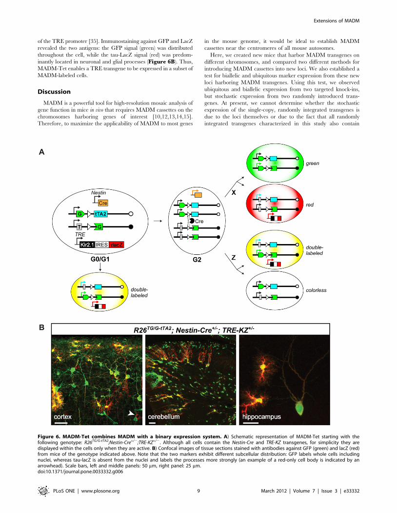
of the TRE promoter [35]. Immunostaining against GFP and LacZ
revealed the two antigens: the GFP signal (green) was distributed
throughout the cell, while the tau-LacZ signal (red) was predom-
inantly located in neuronal and glial processes (Figure 6B). Thus,
MADM-Tet enables a TRE transgene to be expressed in a subset of
MADM-labeled cells.
Discussion
MADM is a powerful tool for high-resolution mosaic analysis of
gene function in mice in vivo that requires MADM cassettes on the
chromosomes harboring genes of interest [10,12,13,14,15].
Therefore, to maximize the applicability of MADM to most genes
in the mouse genome, it would be ideal to establish MADM
cassettes near the centromeres of all mouse autosomes.
Here, we created new mice that harbor MADM transgenes on
different chromosomes, and compared two different methods for
introducing MADM cassettes into new loci. We also established a
test for biallelic and ubiquitous marker expression from these new
loci harboring MADM transgenes. Using this test, we observed
ubiquitous and biallelic expression from two targeted knock-ins,
but stochastic expression from two randomly introduced trans-
genes. At present, we cannot determine whether the stochastic
expression of the single-copy, randomly integrated transgenes is
due to the loci themselves or due to the fact that all randomly
integrated transgenes characterized in this study also contain
Figure 6. MADM-Tet combines MADM with a binary expression system. A) Schematic representation of MADM-Tet starting with thefollowing genotype: R26TG/G-tTA2;Nestin-Cre+/2;TRE-KZ+/2. Although all cells contain the Nestin-Cre and TRE-KZ transgenes, for simplicity they aredisplayed within the cells only when they are active. B) Confocal images of tissue sections stained with antibodies against GFP (green) and lacZ (red)from mice of the genotype indicated above. Note that the two markers exhibit different subcellular distribution: GFP labels whole cells includingnuclei, whereas tau-lacZ is absent from the nuclei and labels the processes more strongly (an example of a red-only cell body is indicated by anarrowhead). Scale bars, left and middle panels: 50 mm, right panel: 25 mm.doi:10.1371/journal.pone.0033332.g006
Extensions of MADM
PLoS ONE | www.plosone.org 9 March 2012 | Volume 7 | Issue 3 | e33332

plasmid bacterial DNA, which was used as a ‘‘buffer’’ to protect
transgene ends. Our recent observations during the development
of a site-specific transgenesis technique show that bacterial DNA
can have a severe silencing effect, which is most prominent in the
liver [36]. The silencing effect of bacterial elements on
mammalian transgenes has been observed before in randomly
integrated transgenes [37] and episomal transgenes [38]. This
correlation suggests that the bacterial sequences flanking these
transgenes could contribute to their variable expression. There-
fore, targeted knock-ins or new random transgenesis screens,
where bacterial protection arms are avoided, should be the
methods of choice for expanding MADM cassettes onto other
chromosomes.
In the future, we recommend that new genomic loci harboring
MADM cassettes should be tested for biallelic expression by
creation of GG and TT alleles from GT and TG alleles (Figure 4A).
To expedite this key validation experiment, we recommend
creation of the GG allele before or in parallel to the GT and TG
alleles. The ubiquity of the expression in the new locus can then be
tested by examining the GG allele alone [10] or by crossing it to
R26TT and assessing the extent of double-labeled cells for any cell
type of interest. However, the most rigorous test for biallelic
expression should finally be performed by crossing the GG and TT
alleles in the same locus as described in Figure 4.
Visual inspection of the efficiencies of MADM labeling
(including both single- and double-labeled cells) revealed that
H11.R26,M1.M10. For M10 and particularly M1, these
estimates are not completely reliable as these loci are not reliably
expressed. Nevertheless, these data suggest that homologous
recombination efficiencies differ quite widely for different
chromosomal loci in somatic cells in vivo, and they are consistent
with similar findings previously reported in embryonic stem cells
[39]. The expression levels of the marker genes driven by the same
pCA promoter in M10, H11 and R26 loci do not appear
dramatically different, at least in tissues in which the markers
are reliably expressed.
We also show that complementary MADM cassettes on
different chromosomes can be used to produce and label cells
that undergo various translocation events. This new application of
MADM now permits the analysis of single-cell phenotypes
produced by precisely defined translocation events in vivo.
Interestingly, in the M10/H11 translocation case, we observed
double-labeled Purkinje cells with elaborate dendritic trees,
suggesting that the presence of dicentric and acentric chromo-
somes does not perturb the development or maintenance of a
complex dendritic arbor. Systematic studies in the future can
determine the consequences of chromosomal aneuploidy on the
differentiation and function of different cell types.
Finally, we demonstrate that by replacing one of the fluorescent
markers with the tTA2 transcription factor, MADM can also
express a TRE-controlled transgene of interest in a small
population of cells. This capability can be used in the future to
combine transgene expression with loss-of-function mutations in
the same, uniquely labeled cells. Further modifications of the
technique would extend MADM-Tet capabilities. For example, a
new G-tTA2 cassette that would include labeling of tTA2-
expressing cells independently of the tTA2 activity would allow
their visualization before or after the expression of tTA-dependent
transgenes. Efficient generation of reliable TRE transgenes would
further facilitate the use of MADM-Tet. As a built-in TRE
transgene was mostly silent in the Rosa26 locus (as part of the G-
TET allele; Figure S1B and S3) and because TRE transgenes are
prone to silencing [40,41], we have been modifying the TIGRE T1
[42] locus to enable integrase-mediated site-specific transgenesis
[36] for efficient creation of reliable TRE transgenes. Together,
these advances enable new applications of MADM and will
facilitate additional extensions of MADM in the future.
Methods
Ethics StatementAll animal procedures were in compliance with the institutional
animal care guidelines and were approved by Stanford Uni-
versity’s Administrative Panel on Laboratory Animal Care (A-
PLAC, protocol number 14007).
Plasmid constructionRecombinant DNA was constructed using standard techniques.
When fragments were amplified by PCR, we used Phusion Taq
polymerase (Finnzymes), and confirmed the sequences fully by
DNA sequencing. All synthetic DNA fragments were also fully
confirmed by DNA sequencing.
pMADMa (pCA-FRT-G-Neo-T-FRT-T-Neo-G-FRT-pA).pCA promoter (containing the chicken b-actin promoter and a
CMV enhancer) and the SV40 polyadenylation signal (pA) from pCA
(HZ2) [10], and synthetic DNA fragments containing FRT sites
were sequentially introduced into pBluescript to create a plasmid
intermediate, pKM3 (pCA-FRT-XmaI-EcoRI-FRT-SpeI-HindIII-FRT-
pA). The XmaI/EcoRI fragment of MADM-TG cassette [14] (see
construction details below) was introduced into a pBluescript vector
to flank this cassette with SpeI and HindIII. Then, the SpeI/HindIII
restriction fragment of MADM-TG cassette, and XmaI/EcoRI
fragment of MADM-GT cassette [14] were sequentially introduced
into pKM3 to generate pMADMa. The construct was digested with
restriction enzymes PvuI and AflIII, and the insert was gel-purified
using Qiagen gel extraction kit and eluted into 10 mM Tris-HCl,
pH 7.4, 0.1 mM EDTA. The purified and linearized DNA
contained ,50 bp and ,300 bp of vector sequence at its 59 and
39 ends, respectively. This vector sequence was deliberately retained
to minimize the transgene damage with exonucleases after
electroporation of the DNA into mouse ES cells.
pMADMb (pFRT5-pCA-bGeo-pA-pPGK-TK-pA-FRT). The
following fragments were assembled together in this order to make
pMADMb:
i) 59 protection arm: ,500 bp PCR fragment of b-lactamase
gene from pBluescript amplified by the following primers:
G G T A C C A T T T A A A T A G A T T A T C A A A A A G -
GATCTTCACC and GGTACCTAACTCGCCTT-
GATCGTTGG.
ii) 319 bp PCR fragment of wheat germ agglutinin (WGA) gene
amplified by PCR primers: GGTACCGACGTGTCC-
CAACAACCACT and AAGCTTCATGCCACAG-
GATCCCCACT. This arm was placed immediately
downstream of the protection arm to provide unique
sequence in the mouse genome for the Splinkerette PCR.
Single NlaIII restriction site was artificially introduced in the
39 end of this arm for Splinkerette PCR.
iii) FRT5 (GAAGTTCCTATTCCGAAGTTCCTATTCTT-
CAAAAGGTATAGGAACTTC) from a synthetic DNA
was introduced after the unique 59 arm.
iv) pCA promoter from pCA (HZ2) [10].
v) SalI/KpnI fragment containing the bGeo gene from the
plasmid Z/EG [43].
vi) KpnI/HindIII fragment containing the pPGK-TK-pA cassette
from the plasmid pLOXPNT [24]. (Note that we eventually
decided not to use thymidine kinase (TK)-based selection.)
Extensions of MADM
PLoS ONE | www.plosone.org 10 March 2012 | Volume 7 | Issue 3 | e33332
vii) FRT (GAAGTTCCTATTCCGAAGTTCCTATTCTC-
TAGAAAGTATAGGAACTTC) from a synthetic DNA
was introduced after the TK cassette.
viii) 218 bp PCR fragment of yeast His3 gene amplified by PCR
primers GCGGCCGCTCGAAGTAGCCGCCGTTG-
TTGTTAT and GAGCTCGGTGATAGGTGG-
CAAGTGGT. (Note: This sequence was originally intro-
duced as a 39 unique area for screening purposes, but was
not used in this study.)
The construct was digested with restriction enzymes PvuI and
AflIII, and the insert was gel-purified using Qiagen gel extraction
kit and eluted into 10 mM Tris-HCl, pH 7.4, 0.1 mM EDTA.
The purified and linearized DNA contained ,500 bp and
,3000 bp of vector sequence at its 59 and 39 ends, respectively.
The vector sequences served as protection arms against endonu-
cleases after electroporation into the mouse ES cells.
pExG and pExT (pFRT5-pCA-GT-pA-FRT-attPx3-pPGK-Hyg-
pA-attB and pFRT5-pCA-TG-pA-FRT-attPx3-pPGK-Hyg-pA-attB):
Synthetic DNA fragments containing FRT5 [20,44] and FRT
were sequentially introduced 59 and 39 to the MADM cassettes in
the MADM-GT and TG constructs to generate intermediates
pFRT5-GT-FRT and pFRT5-TG-FRT. Independently, pattB-Hyg-attPx3 was generated by flanking the hygromycin resistance
gene (Hyg) driven by the phosphoglyceratekinase promoter (pPGK)
with QC31 integrase recognition sites: three 70-bp long attP sites
from pBT298 [36] and a ‘‘full length’’ attB [45]. To create the final
constructs (pExG and pExT), the XmaI/BamHI fragment from
pattB-Hyg-attPx3 was introduced into the SwaI site of pFRT5-GT-
FRT and pFRT5-TG-FRT. The constructs were prepared by using
Endotoxin Free-Maxi prep (Qiagen) for the electroporation into
the mouse ES cells.
MADM cassettesTo construct and test final MADM targeting constructs we
created a set of constructs in the pCA (HZ2) plasmid, which
contains a polylinker between the pCA promoter (chicken b-actin
promoter and CMV enhancer) and an SV40 polyadenylation site
[10].
pCA-G-intron-T. The previously used first GFP exon from the
GR cassette [10] and tdT3MycATG-less were assembled in pHZ2
separated by the previously described modified b-globin intron
containing BglII and loxP sites [10].
pCA-T-intron-G. The previously used second GFP exon from
the RG cassette [10] was assembled with a fragment containing a
Kozak sequence, ATG start codon and the same b-globin intron
desribed above in pHZ2.
pCA-G-intronNeo-T and pCA-T-intronNeo-G. We inserted a
BglII/BamHI fragment containing the neomycin resistance gene
(Neo) driven by an SV40 promoter and followed by the HSV TK
polyadenylation site into the BglII site of pCA-G-intron-T or pCA-T-
intron-G, respectively. We created three different versions of the Neo
cassette to contain different numbers or identities of recombination
sites: version 1: pLN: loxP-pSV40-Neo-pA; version 2:pFLN: FRT-loxP-
pSV40-Neo-pA; version 3: pFLLFLN: FRT-Lox5171-Lox2272-FRT-
loxP-pSV40-Neo-pA. The loxP versions, Lox5171 and Lox2272 are
incompatible with each other and with loxP, but each one is
compatible with itself [46]. They were introduced in attempts to
increase recombination efficiency. Comparisons of these intron
versions and their effect on recombination efficiency will be
described elsewhere (A. Henner and H. Zong, in preparation).
The intron versions that were used for creating targeting
constructs for particular loci described in this study are
schematically represented in Figure S1.
pBT234 (pCA-G-intron-tTA2ATG-less-pA). Used for testing
the cassettes before the construction of final targeting constructs.
pBT250 (pCA-G-intronNeo-tTA2ATG-less-pA). pGLLFNL was
cloned into BglII site of pBT234.
pBT270 (pCA-G-intronNeo-tTA2ATG-less-pA-ii-TRE-tdT3Myc-pA-ii). ii-TRE-tdT3Myc-pA-ii from pBT264 (pii-TRE-
tdT3Myc-pA-ii) [47] was inserted into pBT250.
Targeting constructsAll targeting constructs for the Rosa26 locus were created by
inserting a PmeI/AscI-digested fragment from a precursor plasmid
into the pROSA26-PA [48]. The pRosa26-GT precursor is pCA-G-
intronNeo-T; pRosa26-TG precursor is pCA-T-intronNeo-G;
pRosa26-G-tTA2 (pBT259) precursor is pBT250; pRosa26-GTET (pBT272) prescursor is pBT270.
Control constructspBT255 (pCA-GFP4m). Used as a positive control for GFP
expression. GFP4m (or mut4EGFP) is a thermotolerant GFP
variant [49,50]. All other GFP-containing constructs in this study
contain this variant of GFP.
pCA-G-intron-G. Described originally in [10]. Used as a
positive control for split GFP expression.
pBT225 (pCA-tdT3Myc): Used as a positive control for tdT
expression and Myc staining.
pCA-ATG-intron-tdT. Constructed initially as a test for
splitting the tdT gene into an exon containing a start codon
(ATG) and and an exon containing the rest of tdT by the b-globin
intron containing the BglII site and loxP [10].
pBT224 (pCA-tTA2-pA). Used as a positive control for tTA2
activity in conjunction with pBT239 (TRE-tdT3Myc-pA) [47]. tTA2
gene was subcloned from pUHT61-1 [17].
pBT241 (pCA-ATG-intron-tTA2ATG-less). Constructed ini-
tially as a test for splitting the tTA2 gene into ATG and the rest of
tTA2 by the b-globin intron. PCR was used to construct two DNA
fragments: XmaI-ATG-intron and tTA2ATGless-EcoRI. The b-globin
intron was modified at the same position as described previously
[10] to contain single BglII, loxP and FRT sites. The two fragments
were ligated to each other (via blunt ends) and to BglII/EcoRI-
digested pCA (HZ2) [10] in a 3-way ligation reaction. This
construct was used to test the functionality of tTA2 after insertion
of the intron in conjunction with pBT239 (see above).
pBT267 (pCA-ATG-intron-tTA2ATG-less-TRE-tdT3Myc-pA) and pBT268 (pCA-ATG-intron-tTA2ATG-less-iiTRE-tdT3Myc-pAii). Constructed to compare the effect of insulators
on decreasing tTA2-independent activation of TRE. The con-
structs were also tested in the presence of doxycycline to assess
which construct has higher background expression of tdT3Myc.
Screening of ES cell clones obtained by randomtransgenesis
We used standard techniques [24] to modify R1 mouse ES cells,
which originated from a 129 mouse strain [51].
pMADMa construct was introduced into ES cells via electropo-
ration, and individual G418-resistant clones were evaluated for
intact transgene integration by genomic PCR using primers KM1
(GTGCTGCAAGGCGATTAAGT) and KM2 (TTATG-
TAACGCGGAACTCCA) to detect the 59 end of each transgene
(PCR product, 211 bp), and CCCCCTGAACCTGAAACATA
and TGTGGAATTGTGAGCGGATA to detect the 39 end of
each transgene (PCR product, 275 bp). We further analysed the
genomic DNA from the ES cells containing intact transgenes by
Southern blotting. We used a probe for Neo, which is located in the
Extensions of MADM
PLoS ONE | www.plosone.org 11 March 2012 | Volume 7 | Issue 3 | e33332
intron of MADM cassette, and the genomic DNA obtained from
the R26TG/+ mouse line as a reference for a single-copy transgene.
For ES cells that contained single-copy transgenes based on the
Southern blot, we performed inverse PCR to identify the 59-
flanking genome sequence. Genomic DNA was digested with
restriction enzyme NlaIII and subjected to the ligase-mediated self-
ligation. The resultant circular DNA was then used as a template
for a two-step nested PCR to amplify the transgene flanking
region. For the first round of PCR, we used primers: TAATC-
GAAACCCTGGCGTTA and GTTTTCCCAGTCACGA-
CGTT. For the second round of PCR, we used 0.3 ml of the
first round PCR product and primers: ACTTAATCGCCTTG-
CAGCAC and TATAGGGCGAATTGGGGAAT. The final
PCR products were analyzed by electrophoresis, gel-extracted by
the Qiagen gel extraction kit, and analyzed by DNA sequencing.
For the intact single-copy transgenes integrated in intergenic
regions close to any centromere, we performed additional
Splikerette PCR [21] to confirm integration sites.
pMADMb was also introduced into R1 mouse ES cells by
electroporation and individual G418-resistant clones in 96-well
plates were evaluated for the expression of bgeo by lacZ staining.
96-well plates were washed with PBS, fixed by 0.2% glutaralde-
hyde in PBS for 5 min. at room temperature (RT), and washed 3
times at RT with the staining buffer (2 mM MgCl2, 0.01%
Deoxycholate, 0.02% NP40, 100 mM phosphate buffer, pH 7.5).
Cells were then treated with the solution containing: 5 mM
potassium ferricyanide (Sigma), 5 mM potassium ferrocyanide
(Sigma), 2.5 mM 5-bromo-4-chloro-3-indolyl-beta-D-galactopyra-
noside (X-gal, Invitrogen) at 37uC for 1-2 hours. We recorded the
activity of lacZ in individual clones and then extracted genomic
DNA from 96-well plates. We tested the intactness of 59 and 39
ends of the transgenes by PCR. We used the following primers:
CTATGCCCGAACAACCTCTG and ATCATATGCCAAG-
TACGCCC (for pMADMb 59 end); CCCCCTGAACCTGAAA-
CATA and ACCACTTGCCACCTATCACC (for pMADMb 39
end). The clones with high lacZ expression and intact transgenes,
were further analyzed by Splinkerette PCR [21] to identify the
flanking genomic sequence at the 59 end of the transgene.
Splinkerette PCR was reported to be more efficient than inverse
PCR method we used for pMADMa screening. The genomic DNA
was digested with NlaIII and a specifically designed Splinker was
ligated overnight at 16uC. To make the Splinker adaptor, two
synthetic DNA oligonucleotides: TAACCGTTGCTAGGAGA-
GACCGTGGCTGAATGAGACTGGTGTCGACACTAGTG-
GCATG and CCACTAGTGTCGACACCAGTCTCTAATT-
TTTTTTTTCAAAAAAA were annealed. The ligation products
were purified by the Qiagen PCR purification kit and eluted into
30 ml of TE. 1 ml of the sample was used for the first round of
PCR with primers: AACCGTTGCTAGGAGAGACC and
CCGCAGAACTCGGAACCTA. 0.3 ml of the first PCR product
was subjected to the second round of PCR with primers:
GCTGAATGAGACTGGTGTCG and CAGAGGTTGTT-
CGGGCATAG. The final PCR products were analyzed by
electrophoresis, gel-extracted by the Qiagen gel extraction kit, and
analyzed by DNA sequencing.
Converting pMADM transgenes into GT or TG cassettesTo convert pMADMa to MADM-GT or TG, 5 mg of a plasmid
(pPGK-Flpo-Puro) containing a codon-optimized Flp recombi-
nase gene (Flpo) [52] and the puromycine resistance gene was
introduced into 5 million cells of selected clones via electropora-
tion. We cultured ES cells with 200 mg/ml G418 and 2 mg/ml
puromycine for 48 hours, and ES cell were subsequently cultured
only in the presence of G418 until colonies formed. Individual
G418-resistant clones were analyzed by two genomic PCRs to
specifically detect 59 and 39 parts of pMADMa and identify non-
recombined clones and partially recombined clones (GT or TG
cassette). One PCR used primers GCAACGTGCTGGT-
TATTGTG and CGCCTCAGGACTCTTCCTTT to amplify
,600 bp product for non-recombined pMADMa cassette and
MADM-GT cassette or ,300 bp product for MADM-TG cassette.
Another PCR used primers GAAACTGGGCATGTGGAGAC
and TGAGTTTGGACAAACCACAAC to amplify ,800 bp
product for non-recombined pMADMa cassette and MADM-TG
cassette or ,2.0 kb PCR product for MADM-GT cassette. The
ES cell clones that contained correctly recombined cassettes were
used to generate chimeric mice by injection into C57BL/6
blastocysts.
To convert pMADMb’1 into MADM-GT or TG, 25 mg of one
of the exchange cassette plasmids (ExG or ExT) and 25 mg of
pPGKFLPobpA plasmid (Addgene plasmid 13793) [52] were
introduced into the selected ES cells (,56106 cells). We cultured
the ES cells without selection for 72 hours and then applied
hygromycin (120 mg/ml) for one week. Individual hygromycin-
resistant clones in 96-well plates were divided into five replicas:
two for stock, one for lacZ staining, and two for genomic DNA
preparation. To detect the FRT5/FRT mediated site-specific
recombination events, we used PCR with primers: CTATGCCC-
GAACAACCTCTG and GGGCGTACTTGGCATATGAT to
amplify the junction containing FRT5. This PCR not only
confirmed that the ES clone still contained the MADM transgene,
but also generated different sizes of PCR products for non-
recombined (510 bp) and recombined (550 bp) clones. The PCR
products were analyzed by electrophoresis on a 2% agarose gel.
Site-specific recombination was confirmed by additional PCR
primer sets that specifically amplify the newly formed junctions at
FRT5 (59 of the transgene): CTATGCCCGAACAACCTCTG
and TCCCAGTCCTTGCCATTTAG (create a 286 bp product)
and FRT (39 of the transgene): AAGCATCAACGACAACAACG
and 59- CGGAATACCACTGAAATTGG (create a 200 bp
product). The ES cell clones that contained correctly recombined
cassettes were used to generate chimeric mice by injection into
C57BL/6 blastocysts. The chimeras were directly crossed to a
QC31o integrase mouse line (Jackson laboratory, stock# 007670)
[52] to remove the pPGK-Hyg-pA cassette from the genome in the
next generation.
Tissue processing, immunohistochemistry and imagingTissues were processed according to previously described
procedures [10,11]. Neither tdT nor GFP required immunostaining
for visualization. Although the majority of the data presented in the
paper were obtained from unstained tissue sections, sections can be
immunostained for better signal preservation according to previously
published methods [11] using the following primary antibodies:
chicken anti-GFP (1:500; Aves Labs), goat anti-MYC (1:200; Novus;
the best results are obtained if antibody is pre-absorbed with fixed,
finely minced, wild-type brain according to the previously described
procedure [33]), rabbit anti-DsRed (1:1000; Clontech), or rabbit anti-
LacZ (1:500; MP Biomedicals (previously Cappel) Cat.
No. 0855976). Secondary antibodies (donkey anti-chicken FITC,
donkey anti-rabbit Cy3 and donkey anti-goat Cy3 from Jackson
ImmunoResearch), were used at 1:200 dilution. In some cases,
sections were also stained with DAPI. Sections were imaged with a
Nikon CCD camera or a confocal microscope (Zeiss 510).
GenotypingMouse DNA was extracted and genotyping PCR performed as
described previously [36].
Extensions of MADM
PLoS ONE | www.plosone.org 12 March 2012 | Volume 7 | Issue 3 | e33332

For genotyping M1 MADM transgenes, we used primers: KM5
(CTATGCCCGAACAACCTCTG), KM6 (ATCATATGCCA-
AGTACGCCC), KM7 (GGGGTCGATCTTGTCAGTCT) and
KM8 (TTGCGTTGCAATTTTCTGAG). These primers ampli-
fy a 512 bp transgene fragment and a 700 bp wt M1 locus
fragment.
For genotyping M10 MADM transgenes, we used primers:
KM1 (GTGCTGCAAGGCGATTAAGT) and KM2 (TTATG-
TAACGCGGAACTCCA). These primers amplify a 211 bp
fragment for either MADM cassette. To distinguish heterozygous
vs. homozygous transgene, we used additional primers KM3
(CATATTCCAAAGCTACCACACACT) and KM4 (ATCATG-
GAGGAGCAGTGGAG), which amplify a 300 bp fragment from
the wt M10 locus.
H11 MADM transgenes (available at The Jackson Laboratory:
MADM-11-GT, stock# 013749 and MADM-11-TG, stock#013751) were genotyped as described [14], using primers: SH176
(TGGAGGAGGACAAACTGGTCAC), SH177 (TCAATGGG-
CGGGGGTCGTT), SH178 (TTCCCTTTCTGCTTCAT-
CTTGC) according to the genotyping protocol deposited to The
Jackson Laboratory.
For genotyping Rosa26 knock-ins we used primers Rosa4
(TCAATGGGCGGGGGTCGTT), Rosa10 (CTCTGCTGC-
CTCCTGGCTTCT) and Rosa11 (CGAGGCGGATCACAAG-
CAATA). These primers amplify a 250 bp knock-in fragment and
a 330 bp wt Rosa26 locus fragment.
For genotyping TRE-KZ, we used primers Tau1
(GGTGGCAAGGTGCAGATAAT) and Tau2 (CAGCTTGTG-
GGTTTCGATCT) to amplify a 315 bp tau fragment. We
combined them with primers IMR0015 (CAAATGTTGCTT-
GTCTGGTG) and IMR0016 (GTCAGTCGAGTGCACAG-
TTT) to amplify a 200 bp internal control fragment.
For genotyping Foxg1tTA, we used primers Ling40 (TCTGC-
ACCTTGGTGATCAAA) and Ling57 (ATCGCGATGGAG-
CAAAAGTA) to amplify a 270-bp fragment of tTA. We
combined them with primers Globin1 (CCAATCTGCTCACA-
CAGGATAGAGAGGGCAGG) and Globin2 (CCTTGAGGC-
TGTCCAAGTGATTCAGGCCATCG) to amplify a 500 bp
internal control fragment.
For genotyping Cre transgenes, we used Foxg1-Cre-A
(CACCCTGTTACGTATAGCCG) and Foxg1-Cre-B (GAGT-
CATCCTTAGCGCCGTA) to amplify a 300 bp transgene
fragment and internal Globin primers as described for tTA
genotyping to amplify the internal control.
Genotyping for the presence of the neomycin resistance gene
(Neo) was performed using primers IMR3742 (GTGAGCTG-
CACTTCCAGAAG), IMR3743 (GACTTTCGGCATGTGA-
AATG), IMR013 (CTTGGGTGGAGAGGCTATTC) and
IMR014 (AGGTGAGATGACAGGAGATC). These primers
produce a 280 bp Neo band and a 180 bp wt band.
Mouse maintenance and crossesAll mice were kept in a mixed background. All mouse lines
contained some 129 and CD1 strain backgrounds, and some
additionally contained C57Bl/6 and FVB. We preferred to keep
mice with as much CD1 background as possible to increase
fecundity.
We kept GT and TG stocks separately from each other. This
approach prevents mixing up the stocks and allowed us to use the same
PCR for genotyping either stock using the common pairs of primers.
Other transgenes were crossed into one of the MADM cassette alleles.
Once a Nestin-Cre or HprtCre line is crossed into one of the MADM
cassette strains, the loxP-flanked (floxed) Neo is removed in the germline.
Therefore any double positive animal of this type will transmit to its
progeny the MADM cassette with removed Neo. The MADM cassette
alleles were then usually homozygosed during maintenance to obviate
the need for genotyping for that allele. For example, to generate the
experimental animals R26TG/GT;Nestin-Cre+/2;TRE-KZ+/2, we would
create two lines: R26GT/GT and R26TG/TG;Nestin-Cre+/2;TRE-KZ+/2.
The first line, as well as other MADM-cassette lines, were usually kept
homozygous (no genotyping required). In the case of R26GT/GT only,
some homozygous males show decreased fertility, so from time to time
a homozygous female was crossed to a CD1 wt male, and after that the
homozygous stock was reestablished by crossing heterozygous mice to
each other. The second line was created by sequentially introducing
Nestin-Cre and TRE-KZ transgenes into R26TG mice. After the
triple-transgenic mice R26TG/+;Nestin-Cre+/2;TRE-KZ+/2 were
created, they were crossed to R26TG/TG to create R26TG/TG;
Nestin-Cre+/2;TRE-KZ+/2. These mice were maintained by crossing
to R26TG/TG homozygous stock and genotyping only for the presence
of Cre and tau.
All GG and TT alleles were generated by Cre-mediated
interchromosomal recombination and were detected by screening
tail samples for expression of GFP or tdT under the fluorescence
microscope. In addition, under UV light, these animals appeared
uniformly green and red, respectively. All GG and TT alleles had
lost the floxed Neo from the intron as confirmed by Neo PCR (see
Genotyping).
HprtCre is located on the X chromosome [19]. If maximal level of
recombination is desired, it is recommended to use males for
phenotypic analysis (HprtCre/Y as opposed HprtCre/+). In females,
due to the random X inactivation, only roughly half of the cells
have the active HprtCre allele.
Reagent availabilityThe DNA constructs described in this paper will be deposited to
Addgene. We will also deposit the following lines to The Jackson
Laboratory; R26GT (stock# 017912), R26TG (stock# 017921),
R26TT (stock# 017922), R26G-tTA2 (stock# 017909), Miya10GT
(stock# 017923), and Miya10TG (stock# 017932). Note that we
have already deposited the following lines to The Jackson
Laboratory: Rosa26GG (also called MADM-GG; stock# 006053)
[10], Hipp11GT (also called MADM-11GT; stock# 013749), and
Hipp11TG (also called, MADM-11TG; stock# 013751) [14].
Whereas the GT and TG mice can be used for MADM analysis
of genes located on those specific chromosomes, GG and TT mice
express high-level green or red fluorescence proteins globally and
can be used, for example, as tissue donors in transplantation or
chimeragenesis experiments.
Supporting Information
Figure S1 Loci and alleles used in this study. A) Miya1
(M1) on Chr. 1; B) Rosa26 (R26) on Chr. 6; C) Miya10 (M10) on
Chr. 10 and D) Hipp11 (H11) on Chr. 11. Panel D is modified
after [14], where H11GT and H11TG were referred to as MADM-
11GT and MADM-11TG, respectively. The loxP-flanked (floxed) Neo
in any of the alleles above is converted into a single wild-type loxP
site after the allele is crossed to a germline-expressed Cre transgene
(Nestin-Cre or HprtCre). All GG and TT alleles described here were
created by Cre-mediated interchromosomal recombination in
meiosis and have lost the floxed Neo. The previously described
R26GG allele (also referred to as MADM-GG), which was created by
targeted knock-in, contains the floxed Neo in the intron [10].
(TIF)
Figure S2 A scheme for generation of translocations andaneuploidy using MADM. A) A cell containing Cre and two
non-homologous chromosomes with reciprocal cassettes in the
Extensions of MADM
PLoS ONE | www.plosone.org 13 March 2012 | Volume 7 | Issue 3 | e33332

same orientation, e.g., Chr. 6 and Chr. 10, can generate cells
containing the reciprocal translocation or aneuploidy. B) A cell
containing Cre and two non-homologous chromosomes with
reciprocal cassettes in the opposite orientation, e.g., Chr. 10 and
Chr. 11, can generate cells with acentric and dicentric chromo-
somes and aneuploidy. In this case, change in labeling and
genotype could result from the loss of acentric or dicentric
chromosomes during cell division.
(TIF)
Figure S3 A built-in TRE reporter within the G-TETallele is mostly silent. A) With the aim of simplifying the use of
MADM-Tet by minimizing the number of transgenes that need to
be combined in a single animal, we generated another version of
G-tTA2 that had a built-in TRE reporter (TRE-tdT-3Myc), which
we call G-TET (Figure S1B). In the G-TET construct, we flanked
the TRE expression unit with pairs of insulators to decrease the
tTA-independent leakiness of TRE. This leakiness was initially
observed in transient transfection experiments with a plasmid
containing a pCA-containing unit preceding the TRE unit
(pBT267). This leakiness was significantly decreased when
insulators were inserted to flank the TRE (pBT268, data not
shown). We tested the G-TET construct in vivo by creating a knock-
in mouse in Rosa26 and then by creating a triple-transgenic mouse:
R26TG/G-TET;Nestin-Cre+/2. We observed only GFP expression.
The panel shows an epifluorescence image of a cortical tissue
section from the genotype indicated on top, stained with anti-GFP
and anti-Myc antibodies, and DAPI. B) To test for TRE activation
in the brain, we crossed R26G-TET to Foxg1tTA knock-in allele,
which expresses tTA strongly in the mouse forebrain [53] and is
capable of activating a TRE line previously generated in our lab by
random transgenesis (TRE-SG-T; [33]). However, when G-TET
was crossed to Foxg1tTA, the activation was observed only in a
subset of vomeronasal receptor neurons in a tTA-dependent
manner. The panel shows native tdT fluorescence in forebrain
tissue sections with genotypes indicated on top. Thus, we conclude
that our TRE-tdT-3Myc, which is part of G-TET, cannot be
activated by tTA in most cells of the forebrain.
(TIF)
Acknowledgments
Funding sources have been acknowledged on the front page per journal
style. We thank Roger Tsien for the tdT plasmid, Ron Yu for the TRE-KZ
mouse line, Gord Fishell for the Foxg1tTA mouse line, Carlota Manalac for
technical support, Ling Li for help in generating tdT-3Myc, and Lindsay
Schwarz and Nadja Makki for helpful comments on the manuscript.
Author Contributions
Conceived and designed the experiments: BT KM SH LL. Performed the
experiments: BT KM SH VSD H. Zeng WJ. Analyzed the data: BT KM
SH VD LL. Contributed reagents/materials/analysis tools: H. Zong YC.
Wrote the paper: BT KM SH LL. Created and characterized the R26
knock-ins and MADM-Tet: BT KM VSD H. Zong. Performed ES cell
random transgenesis and characterized M1 and M10: KM H. Zeng.
Performed tests for biallelic expression: BT KM SH. Performed the
translocation analysis: BT KM SH WJ.
References
1. Sternberg N, Hamilton D (1981) Bacteriophage P1 site-specific recombination.
I. Recombination between loxP sites. J Mol Biol 150: 467–486.
2. Broach JR, Guarascio VR, Jayaram M (1982) Recombination within the yeastplasmid 2mu circle is site-specific. Cell 29: 227–234.
3. Branda CS, Dymecki SM (2004) Talking about a revolution: The impact of site-specific recombinases on genetic analyses in mice. Dev Cell 6: 7–28.
4. Golic KG (1991) Site-specific recombination between homologous chromosomes
in Drosophila. Science 252: 958–961.
5. Xu T, Rubin GM (1993) Analysis of genetic mosaics in developing and adultDrosophila tissues. Development 117: 1223–1237.
6. Lee T, Luo L (1999) Mosaic analysis with a repressible cell marker for studies ofgene function in neuronal morphogenesis. Neuron 22: 451–461.
7. Lai SL, Lee T (2006) Genetic mosaic with dual binary transcriptional systems in
Drosophila. Nat Neurosci 9: 703–709.
8. Griffin R, Sustar A, Bonvin M, Binari R, del Valle Rodriguez A, et al. (2009)The twin spot generator for differential Drosophila lineage analysis. Nat
Methods 6: 600–602.
9. Potter CJ, Tasic B, Russler EV, Liang L, Luo L (2010) The Q system: a
repressible binary system for transgene expression, lineage tracing, and mosaic
analysis. Cell 141: 536–548.
10. Zong H, Espinosa JS, Su HH, Muzumdar MD, Luo L (2005) Mosaic analysis
with double markers in mice. Cell 121: 479–492.
11. Espinosa JS, Luo L (2008) Timing neurogenesis and differentiation: insightsfrom quantitative clonal analyses of cerebellar granule cells. J Neurosci 28:
2301–2312.
12. Muzumdar MD, Luo L, Zong H (2007) Modeling sporadic loss of heterozygosityin mice by using mosaic analysis with double markers (MADM). Proc Natl Acad
Sci U S A 104: 4495–4500.
13. Espinosa JS, Wheeler DG, Tsien RW, Luo L (2009) Uncoupling dendritegrowth and patterning: single-cell knockout analysis of NMDA receptor 2B.
Neuron 62: 205–217.
14. Hippenmeyer S, Youn YH, Moon HM, Miyamichi K, Zong H, et al. (2010)Genetic mosaic dissection of Lis1 and Ndel1 in neuronal migration. Neuron 68:
695–709.
15. Liu C, Sage JC, Miller MR, Verhaak RG, Hippenmeyer S, et al. (2011) Mosaic
Analysis with Double Markers Reveals Tumor Cell of Origin in Glioma. Cell.
16. Shaner NC, Campbell RE, Steinbach PA, Giepmans BN, Palmer AE, et al.(2004) Improved monomeric red, orange and yellow fluorescent proteins derived
from Discosoma sp. red fluorescent protein. Nat Biotechnol 22: 1567–1572.
17. Urlinger S, Baron U, Thellmann M, Hasan MT, Bujard H, et al. (2000)Exploring the sequence space for tetracycline-dependent transcriptional
activators: novel mutations yield expanded range and sensitivity. Proc NatlAcad Sci U S A 97: 7963–7968.
18. Petersen PH, Zou K, Hwang JK, Jan YN, Zhong W (2002) Progenitor cell
maintenance requires numb and numblike during mouse neurogenesis. Nature
419: 929–934.
19. Tang SH, Silva FJ, Tsark WM, Mann JR (2002) A Cre/loxP-deleter transgenicline in mouse strain 129S1/SvImJ. Genesis 32: 199–202.
20. Seibler J, Schubeler D, Fiering S, Groudine M, Bode J (1998) DNA cassette exchange
in ES cells mediated by Flp recombinase: an efficient strategy for repeated
modification of tagged loci by marker-free constructs. Biochemistry 37: 6229–6234.
21. Horn C, Hansen J, Schnutgen F, Seisenberger C, Floss T, et al. (2007)Splinkerette PCR for more efficient characterization of gene trap events. Nat
Genet 39: 933–934.
22. Doetschman T, Gregg RG, Maeda N, Hooper ML, Melton DW, et al. (1987)
Targetted correction of a mutant HPRT gene in mouse embryonic stem cells.Nature 330: 576–578.
23. Thomas KR, Capecchi MR (1987) Site-directed mutagenesis by gene targeting
in mouse embryo-derived stem cells. Cell 51: 503–512.
24. Joyner AL (2000) Gene Targeting, A Practical Approach. New York: Oxford
University Press.
25. Muzumdar MD, Tasic B, Miyamichi K, Li L, Luo L (2007) A global double-fluorescent Cre reporter mouse. Genesis 45: 593–605.
26. Soriano P (1999) Generalized lacZ expression with the ROSA26 Cre reporterstrain. Nat Genet 21: 70–71.
27. Van Deursen J, Fornerod M, Van Rees B, Grosveld G (1995) Cre-mediated site-
specific translocation between nonhomologous mouse chromosomes. Proc Natl
Acad Sci U S A 92: 7376–7380.
28. Smith AJ, De Sousa MA, Kwabi-Addo B, Heppell-Parton A, Impey H, et al.(1995) A site-directed chromosomal translocation induced in embryonic stem
cells by Cre-loxP recombination. Nat Genet 9: 376–385.
29. Collins EC, Pannell R, Simpson EM, Forster A, Rabbitts TH (2000) Inter-
chromosomal recombination of Mll and Af9 genes mediated by cre-loxP inmouse development. EMBO Rep 1: 127–132.
30. Buchholz F, Refaeli Y, Trumpp A, Bishop JM (2000) Inducible chromosomal
translocation of AML1 and ETO genes through Cre/loxP-mediated recombi-
nation in the mouse. EMBO Rep 1: 133–139.
31. Forster A, Pannell R, Drynan LF, McCormack M, Collins EC, et al. (2003)Engineering de novo reciprocal chromosomal translocations associated with Mll
to replicate primary events of human cancer. Cancer Cell 3: 449–458.
32. Drynan LF, Pannell R, Forster A, Chan NM, Cano F, et al. (2005) Mll fusions
generated by Cre-loxP-mediated de novo translocations can induce lineagereassignment in tumorigenesis. EMBO J 24: 3136–3146.
33. Li L, Tasic B, Micheva KD, Ivanov VM, Spletter ML, et al. (2010) Visualizing
the distribution of synapses from individual neurons in the mouse brain. PLoSOne 5: e11503.
Extensions of MADM
PLoS ONE | www.plosone.org 14 March 2012 | Volume 7 | Issue 3 | e33332

34. Luo L (2007) Fly MARCM and mouse MADM: genetic methods of labeling and
manipulating single neurons. Brain Res Rev 55: 220–227.35. Yu CR, Power J, Barnea G, O’Donnell S, Brown HE, et al. (2004) Spontaneous
neural activity is required for the establishment and maintenance of the olfactory
sensory map. Neuron 42: 553–566.36. Tasic B, Hippenmeyer S, Wang C, Gamboa M, Zong H, et al. (2011) Site-
specific integrase-mediated transgenesis in mice via pronuclear injection. ProcNatl Acad Sci U S A 108: 7902–7907.
37. Townes TM, Lingrel JB, Chen HY, Brinster RL, Palmiter RD (1985) Erythroid-
specific expression of human beta-globin genes in transgenic mice. EMBO J 4:1715–1723.
38. Chen ZY, He CY, Ehrhardt A, Kay MA (2003) Minicircle DNA vectors devoidof bacterial DNA result in persistent and high-level transgene expression in vivo.
Mol Ther 8: 495–500.39. Liu P, Jenkins NA, Copeland NG (2002) Efficient Cre-loxP-induced mitotic
recombination in mouse embryonic stem cells. Nat Genet 30: 66–72.
40. Pankiewicz R, Karlen Y, Imhof MO, Mermod N (2005) Reversal of the silencingof tetracycline-controlled genes requires the coordinate action of distinctly acting
transcription factors. J Gene Med 7: 117–132.41. Zhu P, Aller MI, Baron U, Cambridge S, Bausen M, et al. (2007) Silencing and
un-silencing of tetracycline-controlled genes in neurons. PLoS One 2: e533.
42. Zeng H, Horie K, Madisen L, Pavlova MN, Gragerova G, et al. (2008) Aninducible and reversible mouse genetic rescue system. PLoS Genet 4: e1000069.
43. Novak A, Guo C, Yang W, Nagy A, Lobe CG (2000) Z/EG, a double reportermouse line that expresses enhanced green fluorescent protein upon Cre-
mediated excision. Genesis 28: 147–155.
44. Seibler J, Bode J (1997) Double-reciprocal crossover mediated by FLP-
recombinase: a concept and an assay. Biochemistry 36: 1740–1747.
45. Groth AC, Olivares EC, Thyagarajan B, Calos MP (2000) A phage integrase
directs efficient site-specific integration in human cells. Proc Natl Acad Sci U S A
97: 5995–6000.
46. Lee G, Saito I (1998) Role of nucleotide sequences of loxP spacer region in Cre-
mediated recombination. Gene 216: 55–65.
47. Miyamichi K, Amat F, Moussavi F, Wang C, Wickersham I, et al. (2011)
Cortical representations of olfactory input by trans-synaptic tracing. Nature 472:
191–196.
48. Srinivas S, Watanabe T, Lin CS, William CM, Tanabe Y, et al. (2001) Cre
reporter strains produced by targeted insertion of EYFP and ECFP into the
ROSA26 locus. BMC Dev Biol 1: 4.
49. Okada A, Lansford R, Weimann JM, Fraser SE, McConnell SK (1999) Imaging
cells in the developing nervous system with retrovirus expressing modified green
fluorescent protein. Exp Neurol 156: 394–406.
50. Siemering KR, Golbik R, Sever R, Haseloff J (1996) Mutations that suppress the
thermosensitivity of green fluorescent protein. Curr Biol 6: 1653–1663.
51. Nagy A, Rossant J, Nagy R, Abramow-Newerly W, Roder JC (1993) Derivation
of completely cell culture-derived mice from early-passage embryonic stem cells.
Proc Natl Acad Sci U S A 90: 8424–8428.
52. Raymond CS, Soriano P (2007) High-efficiency FLP and PhiC31 site-specific
recombination in mammalian cells. PLoS One 2: e162.
53. Hanashima C, Li SC, Shen L, Lai E, Fishell G (2004) Foxg1 suppresses early
cortical cell fate. Science 303: 56–59.
Extensions of MADM
PLoS ONE | www.plosone.org 15 March 2012 | Volume 7 | Issue 3 | e33332