Embed Size (px)
Citation preview
EXPLORATIONS OF AUDITORY THETA RHYTHMS TO
IMPROVE HUMAN COGNITION
by
NOA COHEN
A THESIS
Presented to the Department of Psychology
and the Robert D. Clark Honors College in partial fulfillment of the requirements for the degree of
Bachelor of Arts
May 2021
ii
An Abstract of the Thesis of
Noa Cohen for the degree of Bachelor of Arts in the Department of Psychology to be taken May 2021
Title: Explorations of Auditory Theta Rhythms to Improve Human Cognition
Approved: _____Professor Michael Posner ______ Primary Thesis Advisor
Recent research on improving human cognition has emphasized stimulation of
the brain with sensory, electrical, or magnetic stimulation. This is done to improve
disorders or enhance normal cognition. Auditory or electrical stimulation in the theta
range (4-8 Hz) has been found in some studies to enhance memory and attention in
typically developing young adults. The mechanism of this effect is unknown but may
include changes in mood or motivation or more specific changes in underlying neural
functions such as synaptic plasticity. My thesis tries to replicate these findings by
comparing brief auditory stimulation with a control group of pink noise and examining
the effects of theta on mood, attention, and memory. While my findings did not
successfully replicate previous studies using auditory theta stimulation, I found
significant correlations between mood and attention and mood and memory, supporting
the body of evidence that mood impacts both working memory and attention. Further
studies determining the effects of brief auditory theta entrainment on cognition are
necessary to continue building the body of research about improving attention networks
exogenously.
iii
Acknowledgements
I would like to thank my primary thesis advisor and first reader Professor
Michael Posner, second reader Pascale Voelker, and CHC advisor Dr. Elizabeth
Raisanen for helping me to fully examine the relationship between auditory theta
rhythms and human cognition and consider the various perspectives and contexts
related to this subject matter. Additionally, I would like to express my sincerest
gratitude to the Clark Honors College for the close guidance and mentorship I received
during this strenuous and rewarding process. I would also like to thank the Institute of
Neuroscience for providing me with the resources to successfully accomplish this
project. Finally, I express my most humble thanks to my family and friends who
encouraged me to excel and created a safe environment for me to satiate my intellectual
curiosity throughout my college career.
iv
Table of Contents
Introduction 1 Methods 8
Participants 8 Materials 8 Experimental Procedure 10 Analysis 12
Results 13 ANT 13 POMS 15 K 17 Correlations 18
Change in K Performance and Orienting in ANT 18 POMS and K 19
Discussion 20 Mood 20 Attention 21 Memory 22 Study Limitations 22
Use of Pink Noise Control 22 Brief Stimulation 23 Remote Experiment Structure 24
Future Issues 24 Theta Stimulation to Enhance Cognition and Treat Disorders 24
Conclusions 27 Bibliography 28
Introduction
Recent studies have found that neuromodulation improves several aspects of
human cognition. Neuromodulation is generally defined as brain stimulation that results
in the manipulation of brain activity in one or more target regions in the brain. Sensory,
electrical, and magnetic stimulation are the most highly researched and most clinically
performed forms of neuromodulation. Electrical stimulation uses alternating or direct
current to entrain brain activity in brain areas, often at particular frequencies designed to
enhance brain plasticity.
Deep Brain Stimulation (DBS), a more invasive form of electrical stimulation,
involves surgically implanted electrodes connected to a pulse generator. The most
popular clinical use of electrical stimulation is to treat Parkinson’s disease or migraine
headaches by either stimulating the vagus nerve on the neck or the trigeminal nerve on
the forehead (Benabid, 2003; Little et al., 2013; Schoenen et al., 2013; Volkmann,
2004; Yarnitsky, et al., 2017).
Non-invasive methods can be used with volunteer participants. Research with
normal volunteers has found that electrical stimulation of areas such as the anterior
cingulate, medial prefrontal cortex and/or ventro-lateral prefrontal cortex may improve
attentional control and memory (Piscopo et al., 2018; Reinhart, 2017; Reinhart &
Nguyen, 2019; Weible, Rothbart, Posner & Niell, 2017)
Magnetic neuromodulation involves a pulsed magnetic field that causes a
current flow in the brain. Placed above the scalp is a magnetic coil that produces a brief,
high-current pulse, thus producing a magnetic field (Hallet, 2000). Unlike electrical
stimulation, magnetic stimulation does not require direct contact with the scalp.
2
Researchers have used magnetic stimulation as an investigatory tool to research motor
function, language, vision, and brain disorders (Hallet, 2000). Researchers have also
found that magnetic neuromodulation can potentially be used therapeutically to alleviate
depression and migraine headaches (Conforto et al., 2014; Connolly et al., 2012).
Sensory neuromodulation has been found to be an effective tool for modulating
behavior. Forms of sensory neuromodulation that have been examined include visual,
somatosensory, and auditory stimulation. Clinicians and researchers performing visual
stimulation sometimes modulate visual perception by using an optokinetic drum (a
rotating cylinder with alternating black and white stripes) or creating images on a
computer monitor of linearly moving white dots or stripes presented on a black
background (Karnath, 1996; Vicario et al., 2007). Visual stimulation is often used to
treat individuals with nystagmus, disturbed perception of body orientation, and
unilateral spatial neglect (Iacarino et al., 2016; Karnath, 1996; Vallar et al., 1993).
Somatosensory stimulation involves placing contact electrodes on the tongue and has
been found to help those with balance disorders (Wildenberg et al., 2010, 2011).
Auditory stimulation involves the presentation of a sound with a particular
amplitude that reflects the brain oscillation the researcher or clinician wishes to
manipulate in the brain. Intensity, frequency, and entrainment are key parameters to
ascertain that the acoustic stimulus frequency will interact with internal brain oscillator.
A researcher or clinician chooses the frequency of their auditory stimulus based on the
desired behavioral result of the subject. For example, researchers may use acoustic
stimulation between 0.5-3Hz to enhance slow-wave sleep (Bellesi et al., 2014). This
frequency mirrors the natural delta brainwave which is generated during non-REM
3
sleep. Recent studies have also found that auditory stimulation using theta frequencies
of 4-8Hz improves attentional control and memory (Roberts, Clarke, Addante, and
Ranganath, 2018).
Brain oscillations are defined as the rhythmic firing of a local neuronal
population. This neuronal coordination allows for network communication within a
region or separates regions in which the oscillations are correlated. Different oscillation
frequencies are associated with different cognitive states. For example, the gamma
frequency (32-100 Hz) is associated with heightened perception and cognitive
processing while the delta frequency (0.5-4 Hz) is associated with deep sleep. The theta
frequency (4-8 Hz) has been implicated with states of restful awareness and is
frequently found during cognitive processing (Cavanaugh, et al., 2014).
Theta oscillations are associated with several aspects of cognition including
successful learning and retention, memory encoding and retrieval, attentional and self-
control, and improvement in overall mood. A study in the rat hippocampus found that
intrinsic theta oscillations function by temporally structuring neural ensemble activity
during both memory retrieval and spatial navigation (Hasselmo et al., 2002). Studies
looking at human EEG recordings in the 4-8 Hz frequency range found theta
oscillations present in the hippocampus, medial prefrontal cortex, parietal lobes, and
anterior cingulate cortex. Depending on the brain region stimulated, stimulating theta
may impact different aspects of cognition. For example, Reinhart (2017) found that in-
phase electrical theta stimulation (6 Hz) of the medial prefrontal cortex and ventral-
lateral prefrontal cortex improved attentional control of behavioral timing. These brain
4
regions are important nodes of the executive attention network and therefore theta
stimulation of this region improved attention-related tasks.
Piscopo et al. (2018) found that targeted electrical stimulation of the anterior
cingulate (ACC) (4-8) in mice led to white matter increase in the region, demonstrating
that theta stimulation of the ACC can cause changes in white matter in mice. They also
found that frontal theta is implicated in the activation of previously dormant
oligodendrocytes responsible for myelination, therefore increasing white matter in the
region following theta stimulation.
Other studies have found that auditory theta stimulation improves cognition
related to memory in humans (Reinhart & Nguyen, 2019; Roberts, Clarke, Addante, &
Ranganath, 2018). In one memory study (Roberts, Clarke, Addante & Ranganath,
2018), participants were presented with two sets of words respectively classified as
“alive” or “manmade” and then asked to identify whether the word is classified as
animate versus inanimate or manufactured versus natural. Following this task, the
experimental group was presented with auditory theta stimulation (5.5 Hz) (or sham
stimulation) for 36 minutes. They were then presented with a memory task in which
they were asked to recognize words exposed to them previously and the context in
which the words were presented (“alive” vs “manmade”). The group exposed to theta
stimulation performed better at determining the classification of each word but mirrored
the control group in recognition of whether the word was from the previous task. One
potential hypothesis to explain this improvement in memory is that the increase in
intrinsic theta following auditory stimulation may enhance synchronization of the
5
information recalled prior to the enhanced theta activity and would thus improve
participants’ retrieval scores.
Research has also found that theta oscillations following meditation training
amplified theta around the ACC and prefrontal cortex (PFC) (i.e., frontal theta), leading
to improved attention, self-regulation, stress-levels, and mood (Tang et al., 2007; Lutz
et al., 2008; Tang & Posner, 2009; Holzel et al., 2011; Tang, 2011; Tang et al., 2012).
For example, Tang et al (2010) found that one month of integrative body-mind training
(IBMT) led to an increase in white matter around the ACC and PFC. Although these
results were found using meditation practice alone in comparison with an active control
group given relaxation training, they demonstrate that amplified frontal theta improves
participants’ mood and other cognitions in addition to memory.
Because these theta studies with human participants used stimulation too brief to
produce significant white matter change as a mechanism to improve cognitive functions
(unlike with rodents, as demonstrated by Piscopo et al, 2018), other synaptic
mechanisms are likely responsible for these cognitive improvements. For example,
Larson & Mukascy (2015) demonstrated that a fast burst of theta stimulation more
successfully induced hippocampal long-term potentiation when synchronized with
intrinsic theta rhythms. Long term potentiation is the process of strengthening synapses
so that long-term signal transmission increases between neurons. This finding indicates
that brief theta stimulation, while not necessarily inducing white matter change, may
improve cognition by enhancing endogenous theta and increasing synaptic plasticity.
A recent study compared the ability of auditory and electrical theta stimulation
to enhance endogenous theta within the ACC (Voelker et al., 2020). It was found that
6
combining electrical or auditory stimulation and an attention task that activated the
ACC produced a significant increase in intrinsic theta following stimulation in
comparison to either stimulation or the task alone. Because the Covid-19 pandemic
required us to operate remotely, we decided to examine the ability of auditory theta to
improve memory, attention, and mood. By examining both mood and performance we
might be able to determine whether any enhancement of memory and attention
correlates with mood change.
In this study we aim to determine whether brief auditory theta stimulation
enhances memory, attention, and mood in comparison to pink noise, which serves as
our control condition. To determine improvements in memory, we used a visual
working memory test (Luck & Vogel, 2013) where participants were presented with 5
colored squares distributed across the screen. After a brief delay the participants were
then asked to report the colors presented by selecting from a set of options at each
location. To determine improvements in attention we used the Attention Network Test
(ANT), which measures the efficiency of the alerting, orienting, and executive network.
Finally, we measured mood using the Profile of Mood States (POMS) questionnaire
where the participant responds to a list of 40 moods using a 5-point Likert scale.
Each participant will undergo two hour-long sessions; in one session they will
be presented with theta stimulation, and in another they will be presented with pink
noise (a random noise having equal energy per octave) a week apart. Before and after
stimulation, participants underwent the working memory task followed by the POMS
questionnaire. During stimulation each participant performed the Attention Network
Task (ANT). I hypothesize that positive mood will be enhanced, and negative mood
7
reduced in the theta condition compared to the control condition. Theta stimulation
should also improve working memory performance, orienting attention, and mood in
comparison to pink noise stimulation. I will also examine possible correlations between
mood, memory, and attention performance to determine if mood is a likely reason for
improved performance.
8
Methods
Participants
96 volunteer participants between the ages of 18-30 were recruited by personal
contact, flyers, or through the pool maintained by the psychology department at the
University of Oregon. The research was approved through the University of Oregon
Institute Review Board. Participants were randomly assigned to one of two groups,
varying only in the order in which they received theta and control stimulation.
Two 1-hour sessions were scheduled, consisting of one experimental treatment
and one control treatment. 71 of the recruited participants successfully completed both
sessions, which were separated by a one-week interval. All sessions operated remotely
(using the website Gorilla: https://app.gorilla.sc) and consisted of the following: a pre-
and post-stimulation visual working memory test and POMS questionnaire, and an
experiment or control auditory stimulation while performing the ANT.
Materials
The working memory test presented to participants is a change detection task presented
on the computer. This task is based on the Visual Working Memory (VWM) capacity
test developed by Luck & Vogel (2013), which is a reliable measure of working
memory. Four blocks of 40 trials are presented, where the participant is shown 2-6
randomly chosen colored squares at 2-6 different locations on the screen and, after a
brief delay, is required to report the color at each location. We measured the capacity of
visual working memory performance using the K value; the formula is written below
(Pashler, 1988):
9
𝐾𝐾 = 𝑠𝑠𝑠𝑠𝑠𝑠𝑠𝑠𝑠𝑠𝑠𝑠𝑠𝑠 ∗ (𝑃𝑃(ℎ𝑠𝑠𝑠𝑠) − 𝑃𝑃(𝐹𝐹𝐹𝐹))(1− 𝑃𝑃(𝐹𝐹𝐹𝐹))
Where P(hit)=hits/(hits + misses) and P(FA)=false alarms/(false alarms + correct
rejections)
In order to induce ACC activity through an executive control-requiring task, all
subjects performed the Attention Network Task (ANT) (Fan et al., 2002) during theta or
control stimulation. Using computer graphics, the ANT presents five arrows in a
horizontal row that appear above or below the fixation point. Subjects press a key
indicating the direction the central arrow is pointing, which may require them to ignore
the flanker arrows. Two sets of 64 trials were presented, with an equal but random
assortment of the cueing and congruence conditions. Completion of the task allows
calculation of three scores related to the efficiency of attentional networks, measuring
how response times are influenced by alerting cues, spatial cues, and flankers. The
alerting scores (median RTs for trials where the cue does not appear minus median RT
in trials where the double cue appeared) provide a measure of the benefit in
performance provided by a warning signal. The orienting measure (median RTs for
trials with a central cue minus median RT in trials with orienting valid-cues) includes
both the benefit obtained from a valid cue and the cost of the spatially uninformative
cue. The executive attention score (median RTs for incongruent minus median RT in
congruent) indicates the amount of interference experienced in performing the task
when stimulation conflicting with the target was presented in the display. Larger
interference scores indicated less efficiency in resolving conflict.
10
We also used an abbreviated, revised POMS (McNair, Lorr, & Droppleman,
1971) to measure mood state before and after theta or sham stimulation. The participant
responded to a list of 40 moods using a 5-point Likert scale.
Participants will either receive pulses of auditory theta stimulation at two tones
that are separated by a 6 Hz that converge every 6 Hz to produce an audible pulse, or
pink noise, which is a random noise that has equal energy per octave (thus having more
low-frequency components than white noise). All participants were requested to use
over-the-ear headphones or earbuds for auditory stimulation. The participant was asked
to indicate their available listening device before the onset of auditory stimulation.
Experimental Procedure
The entire experimental procedure took place on the online experiment builder,
Gorilla. Participants had access to the website through a secure link sent to their email
or SONA account. Firstly, the participants completed a virtual consent form online and
provided an electronic signature. This consent form was approved by the University of
Oregon IRB and listed all risks of participation, a general outline of what will take place
during the study, and a reminder that they may decide to not take part in the study at
any time for any reason. During this phase, participants were also asked to silence and
set aside cellphones and to avoid noise distractions during the experiment. Participants
were then required to fill out a demographic questionnaire detailing sex, age, ethnicity,
years of post-secondary education, and smoking habits. Following completion of the
demographic questionnaire, participants filled out the POMS questionnaire.
Participants were then introduced to the memory task. They began with a set of
practice trials where they are notified whether their answers were correct. Upon
11
completion of the memory task, participants were asked to listen to the theta frequency,
(experimental group) or pink noise (control group) while completing a session of the
ANT. The ANT was the only session performed with sound, while the mood and
memory measures were taken both before and after the sound. I attempted to have the
participant adjust the sound level to approximately 55 decibels, the volume of sound
typically experienced during a verbal conversation. The theta stimulation was provided
as two tones (100 and 106 Hz) producing sound with an amplitude varying at 6 Hz. The
pink noise consisted of all audible tone frequencies, with each random noise having an
equal energy per octave. Both auditory stimuli were provided continuously as the
participant performed the ANT. Upon completion of the task, participants took the
visual working memory test and completed the POMS an additional time.
There was an error in which some participants (6 theta, 28 control) only received
50% to 99% of sound during the ANT trials rather than the full 100% sound duration
(41 theta, 10 control) during their first session. To address this issue, we ran 20
participants who we were certain received 100% of the appropriate sound during all
ANT trials. We then utilized a within subject comparison to determine whether there
was a significant difference in ANT, K, or POMS performance between participants
who experienced 50%-99% sound versus participants who experienced 100% sound.
We found no significant difference and therefore included participants who received
50%-100% sound. Thus, we used all participants who had more than 50% of the ANT
trials with the appropriate sound.
12
Analysis
We looked at Within Subject comparison for the tasks repeated in a session,
namely the POMS and K. We also looked at between subject comparisons to investigate
differences in ANT performance. We are interested in determining whether the
condition type (theta or control) influenced performance. Additionally, we looked for
learning effects over repeated tasks. Based on previous work, we expected most of the
effects of theta on K would be seen in the first session.
13
Results
ANT
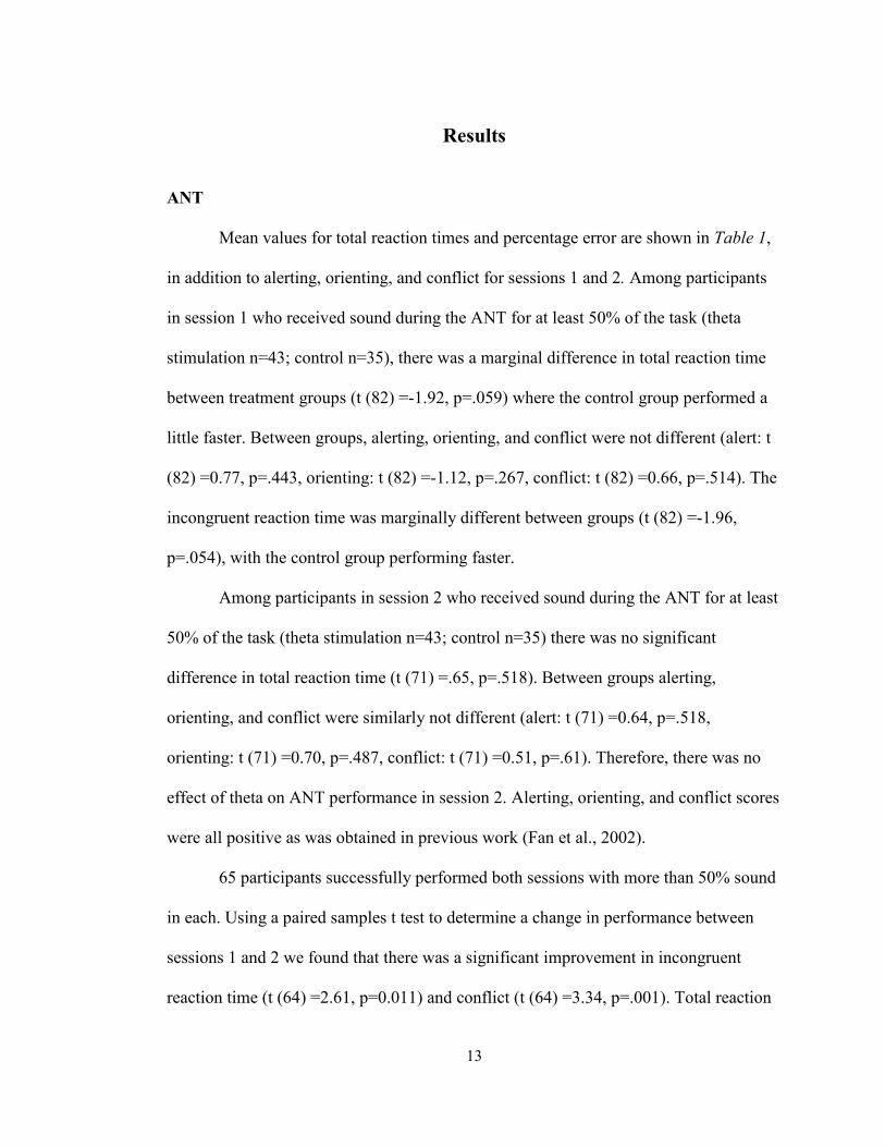
Mean values for total reaction times and percentage error are shown in Table 1,
in addition to alerting, orienting, and conflict for sessions 1 and 2. Among participants
in session 1 who received sound during the ANT for at least 50% of the task (theta
stimulation n=43; control n=35), there was a marginal difference in total reaction time
between treatment groups (t (82) =-1.92, p=.059) where the control group performed a
little faster. Between groups, alerting, orienting, and conflict were not different (alert: t
(82) =0.77, p=.443, orienting: t (82) =-1.12, p=.267, conflict: t (82) =0.66, p=.514). The
incongruent reaction time was marginally different between groups (t (82) =-1.96,
p=.054), with the control group performing faster.
Among participants in session 2 who received sound during the ANT for at least
50% of the task (theta stimulation n=43; control n=35) there was no significant
difference in total reaction time (t (71) =.65, p=.518). Between groups alerting,
orienting, and conflict were similarly not different (alert: t (71) =0.64, p=.518,
orienting: t (71) =0.70, p=.487, conflict: t (71) =0.51, p=.61). Therefore, there was no
effect of theta on ANT performance in session 2. Alerting, orienting, and conflict scores
were all positive as was obtained in previous work (Fan et al., 2002).
65 participants successfully performed both sessions with more than 50% sound
in each. Using a paired samples t test to determine a change in performance between
sessions 1 and 2 we found that there was a significant improvement in incongruent
reaction time (t (64) =2.61, p=0.011) and conflict (t (64) =3.34, p=.001). Total reaction
14
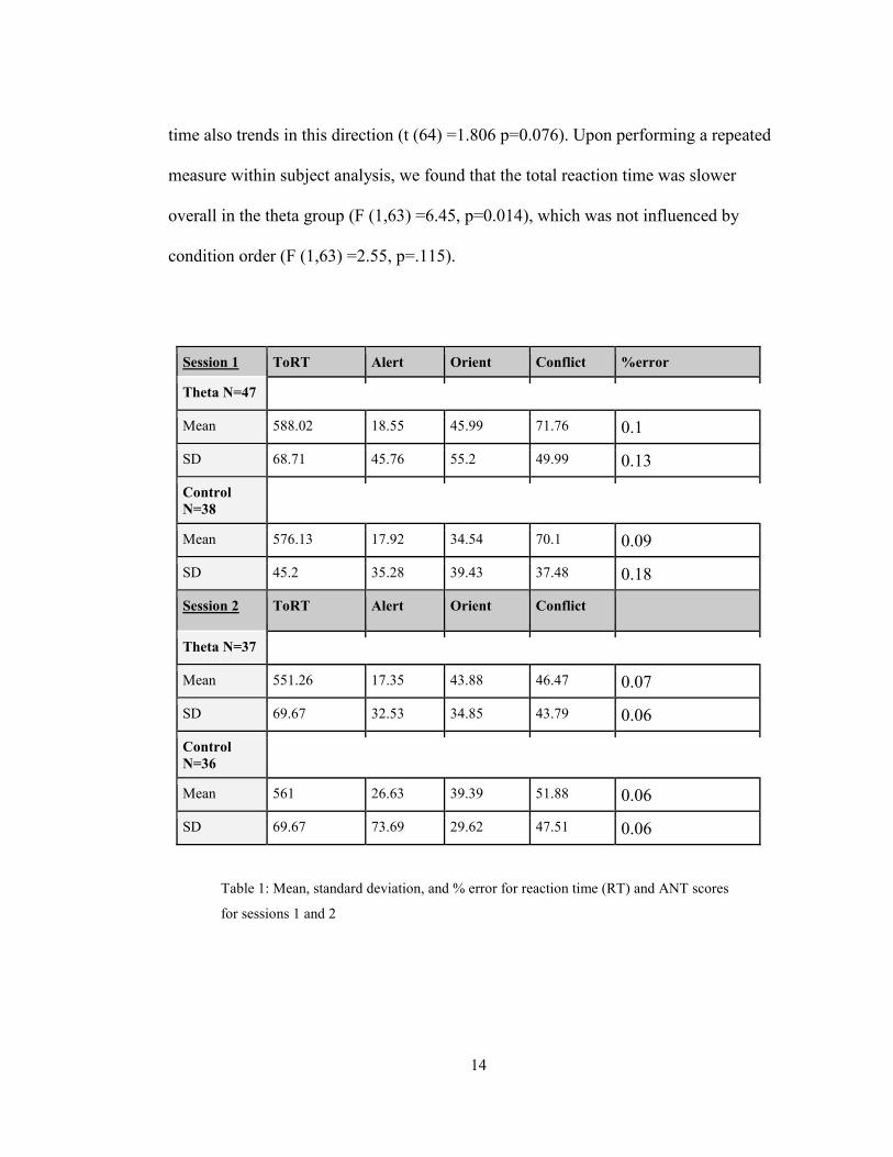
time also trends in this direction (t (64) =1.806 p=0.076). Upon performing a repeated
measure within subject analysis, we found that the total reaction time was slower
overall in the theta group (F (1,63) =6.45, p=0.014), which was not influenced by
condition order (F (1,63) =2.55, p=.115).
Session 1 ToRT Alert Orient Conflict %error
Theta N=47
Mean 588.02 18.55 45.99 71.76 0.1
SD 68.71 45.76 55.2 49.99 0.13
Control N=38
Mean 576.13 17.92 34.54 70.1 0.09
SD 45.2 35.28 39.43 37.48 0.18
Session 2 ToRT Alert Orient Conflict
Theta N=37
Mean 551.26 17.35 43.88 46.47 0.07
SD 69.67 32.53 34.85 43.79 0.06
Control N=36
Mean 561 26.63 39.39 51.88 0.06
SD 69.67 73.69 29.62 47.51 0.06
Table 1: Mean, standard deviation, and % error for reaction time (RT) and ANT scores
for sessions 1 and 2
15
POMS
Mean values for average positive and negative mood scales before and after
stimulation for session 1 and session 2 are listed in Table 3. Between subjects, there was
no significant difference in mood between the theta and control groups for session 1.
Using a repeated measure analysis, we did identify a within-subject change in positive
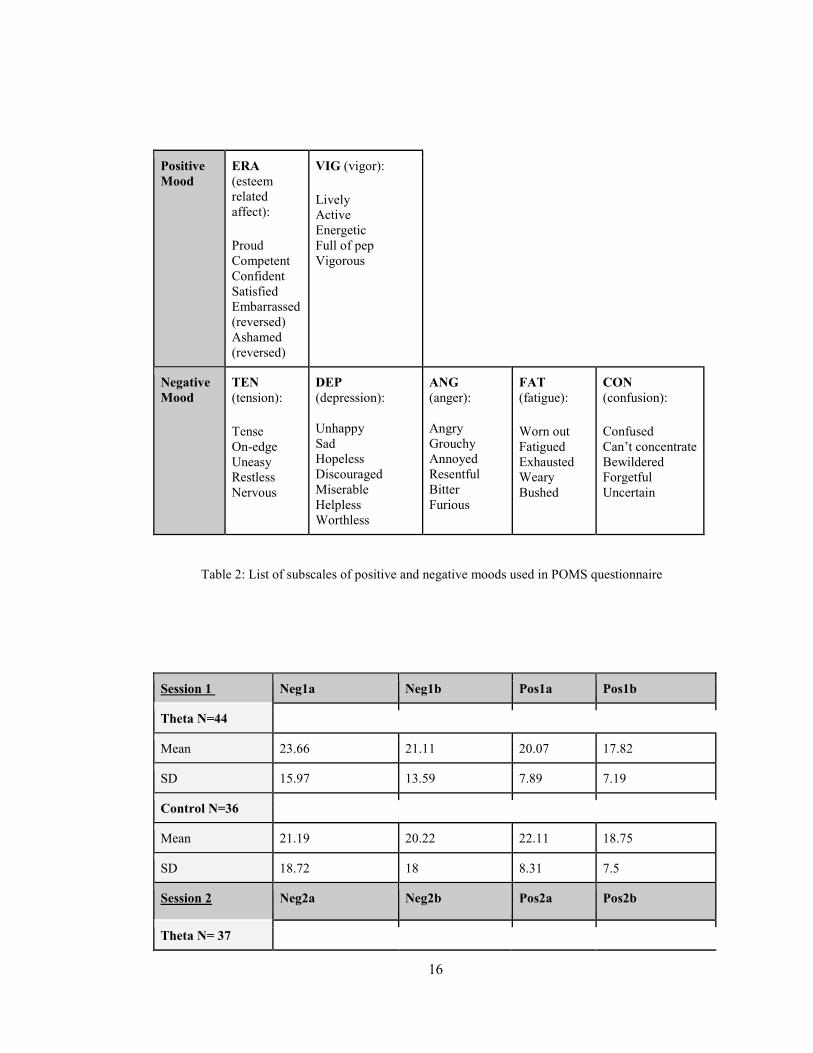
mood during the first session. Positive and negative moods are listed in Table 2. Both
components of the positive mood, esteem-related affect (ERA) and vigor significantly
declined from the before after treatment (ERA: F (1,83) =19.39, p<.001, vigor: F (1,83)
=33.77, p<.001). The vigor measure showed a marginal difference between conditions
(F (1,83) =3.60, p=.061), with the control group showing a sharper decline with testing.
There was a significant within-subject decline in depression with stimulation (F (1,83)
=13.96, p<.001). The other mood measures did not differ significantly.
Vigor and esteem-related effect significantly declined between test 1 and test 2
in the second session (VIG: t (72) =2.93, p=0.005, ERA: t (72) =2.71. p=0.008) and
TMD (a negative measure subtracting negative measures of mood from positive
measures of mood) marginally increased (t (72) =-1.91, p=0.60). There is no evidence
that theta influenced any other significant difference in POMS performance.
The control-first group had a significantly larger decline in VIG during the
control session, and a relatively smaller decline during the theta session. The decline in
VIG is consistent between sessions among the theta-first group. Therefore, VIG is
significantly different by condition (F (1,63=4.07, p=0.048) and by order of condition
(F (1,63) =4.33, p=0.042).
16
Positive Mood
ERA (esteem related affect): Proud Competent Confident Satisfied Embarrassed (reversed) Ashamed (reversed)
VIG (vigor): Lively Active Energetic Full of pep Vigorous
Negative Mood
TEN (tension): Tense On-edge Uneasy Restless Nervous
DEP (depression): Unhappy Sad Hopeless Discouraged Miserable Helpless Worthless
ANG (anger): Angry Grouchy Annoyed Resentful Bitter Furious
FAT (fatigue): Worn out Fatigued Exhausted Weary Bushed
CON (confusion): Confused Can’t concentrate Bewildered Forgetful Uncertain
Table 2: List of subscales of positive and negative moods used in POMS questionnaire
Session 1 Neg1a Neg1b Pos1a Pos1b
Theta N=44
Mean 23.66 21.11 20.07 17.82
SD 15.97 13.59 7.89 7.19
Control N=36
Mean 21.19 20.22 22.11 18.75
SD 18.72 18 8.31 7.5
Session 2 Neg2a Neg2b Pos2a Pos2b
Theta N= 37
17
Mean 16.76 17.84 21 19.14
SD 11.43 13.87 8.25 9.18
Control N= 36
Mean 18 17.5 21.78 20
SD 15.41 13.03 6.99 7.29
Table 3: Mean and standard deviation for negative and positive moods before and after
stimulation for sessions 1 and 2
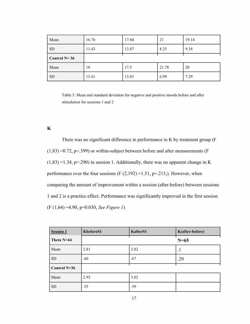
K
There was no significant difference in performance in K by treatment group (F
(1,83) =0.72, p=.399) or within-subject between before and after measurements (F
(1,83) =1.34, p=.290) in session 1. Additionally, there was no apparent change in K
performance over the four sessions (F (2,192) =1.51, p=.213,). However, when
comparing the amount of improvement within a session (after-before) between sessions
1 and 2 is a practice effect. Performance was significantly improved in the first session
(F (1,64) =4.90, p=0.030, See Figure 1)
Session 1 KbeforeS1 KafterS1 K(after-before)
Theta N=44 N=65
Mean 2.81 2.82 .1
SD .60 .67 .29
Control N=36
Mean 2.93 3.02
SD .55 .59
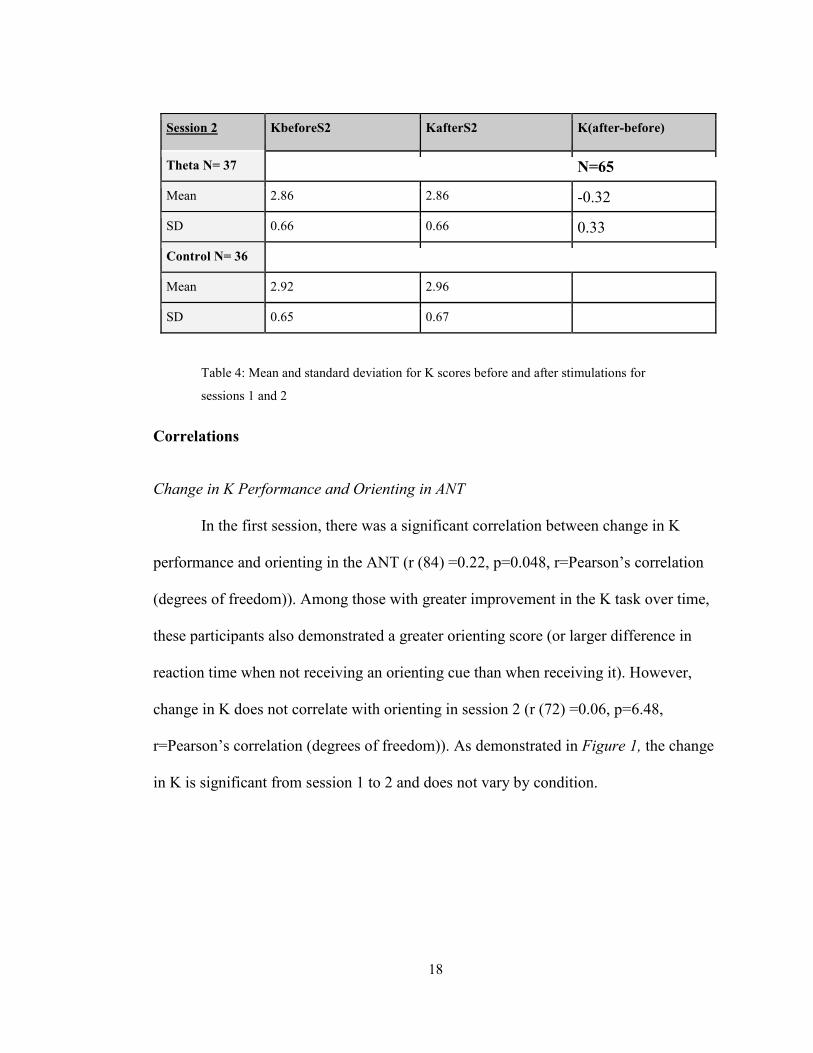
18
Session 2 KbeforeS2 KafterS2 K(after-before)
Theta N= 37 N=65
Mean 2.86 2.86 -0.32
SD 0.66 0.66 0.33
Control N= 36
Mean 2.92 2.96
SD 0.65 0.67
Table 4: Mean and standard deviation for K scores before and after stimulations for
sessions 1 and 2
Correlations
Change in K Performance and Orienting in ANT
In the first session, there was a significant correlation between change in K
performance and orienting in the ANT (r (84) =0.22, p=0.048, r=Pearson’s correlation
(degrees of freedom)). Among those with greater improvement in the K task over time,
these participants also demonstrated a greater orienting score (or larger difference in
reaction time when not receiving an orienting cue than when receiving it). However,
change in K does not correlate with orienting in session 2 (r (72) =0.06, p=6.48,
r=Pearson’s correlation (degrees of freedom)). As demonstrated in Figure 1, the change
in K is significant from session 1 to 2 and does not vary by condition.
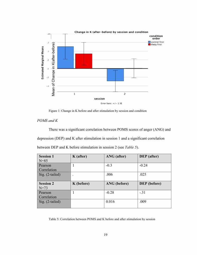
19
Figure 1: Change in K before and after stimulation by session and condition
POMS and K
There was a significant correlation between POMS scores of anger (ANG) and
depression (DEP) and K after stimulation in session 1 and a significant correlation
between DEP and K before stimulation in session 2 (see Table 5).
Session 1 N=85
K (after) ANG (after) DEP (after)
Pearson Correlation
1 -0.3 -0.24
Sig. (2-tailed) . .006 .025
Session 2 N=73
K (before) ANG (before) DEP (before)
Pearson Correlation
1 -0.28 -.31
Sig. (2-tailed) 0.016 .009
Table 5: Correlation between POMS and K before and after stimulation by session
Mea
n of
Cha
nge
in K
(afte
r-bef
ore)
20
Discussion
I hypothesized that theta stimulation would improve working memory
performance, orienting attention, and mood in comparison to pink noise stimulation. In
addition, I hypothesized that positive mood would be enhanced, and negative mood
reduced in the theta condition compared to the control condition. I also aimed to
determine whether mood, memory, and attention performance were correlated. Overall,
there was little difference between the theta and control groups in mood, memory, and
attention. While depression was reduced in both groups, positive mood also declined
across both groups. Although there were some significant correlations between mood
and attention and mood and memory, we did examine many correlations, making it
likely that these could be due to chance. If further research shows reliable correlations
of this type, it will be necessary for researchers to consider more carefully the possible
role that mood might play in changes observed in memory or attention.
Mood
While there wasn’t a significant difference in mood between theta and control
groups, we identified a within-subject decline in positive mood (ERA and VIG) before
and after auditory stimulation in both the first and second session. The control-first
group demonstrated a sharper decline in vigor than the theta group and demonstrated a
relatively smaller decline during their theta session. This potentially demonstrates a
marginal effect of theta on positive mood decline. Despite the decrease in positive
mood, individuals demonstrated a significant decline in depression following
21
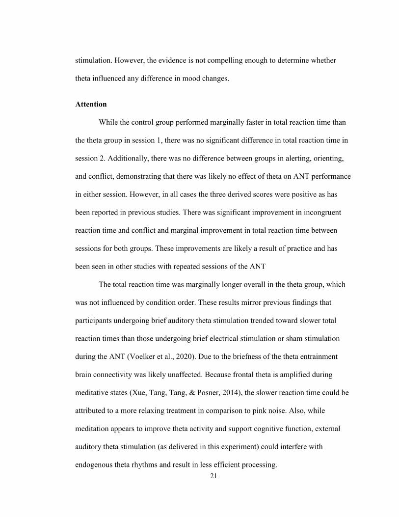
stimulation. However, the evidence is not compelling enough to determine whether
theta influenced any difference in mood changes.
Attention
While the control group performed marginally faster in total reaction time than
the theta group in session 1, there was no significant difference in total reaction time in
session 2. Additionally, there was no difference between groups in alerting, orienting,
and conflict, demonstrating that there was likely no effect of theta on ANT performance
in either session. However, in all cases the three derived scores were positive as has
been reported in previous studies. There was significant improvement in incongruent
reaction time and conflict and marginal improvement in total reaction time between
sessions for both groups. These improvements are likely a result of practice and has
been seen in other studies with repeated sessions of the ANT
The total reaction time was marginally longer overall in the theta group, which
was not influenced by condition order. These results mirror previous findings that
participants undergoing brief auditory theta stimulation trended toward slower total
reaction times than those undergoing brief electrical stimulation or sham stimulation
during the ANT (Voelker et al., 2020). Due to the briefness of the theta entrainment
brain connectivity was likely unaffected. Because frontal theta is amplified during
meditative states (Xue, Tang, Tang, & Posner, 2014), the slower reaction time could be
attributed to a more relaxing treatment in comparison to pink noise. Also, while
meditation appears to improve theta activity and support cognitive function, external
auditory theta stimulation (as delivered in this experiment) could interfere with
endogenous theta rhythms and result in less efficient processing.
22
Memory
There was no significant difference in performance in K between the control and
theta group and before and after stimulation for either sessions 1 and 2. There were
significant improvements by participants in the first session, however, indicating that
short term practice improved memory scores. There was no evidence of improved
performance in session 2, suggesting that practice effects on K occur only at the start of
training.
However, we did identify a significant correlation between change in K
performance and orienting solely in session 1. Those who improved more in K over
time also demonstrated a greater orienting score. In other words, those who performed
better in K with practice were better able to use the positioning cue to undergo the ANT
more quickly. This demonstrates a relationship between orienting in attention and
memory improvement. Because K assesses spatial working memory, it is likely those
who show better orienting efficiency would also show better improvements in spatial
memory. This finding supports the existing literature that neural systems for visual
orienting relate to spatial working memory (Corbetta, Kincade & Schulman, 2002).
Study Limitations
Use of Pink Noise Control
We chose pink noise as the control because it is a random noise that has equal
energy per octave, which contrasts auditory theta at two tones 6Hz apart. However, pink
noise (in relation to silence or white noise) has steady low-frequency components which
may have replicated the effects of theta on the listener. Additionally, studies have
23
shown that pink noise may reduce the brain wave complexity resulting in a restful state
(appropriate for stable sleep) (Zhou et al., 2012), thus inducing a similar state as theta.
A benefit of using pink noise is that it offers a steady sound across a spectrum of
frequencies, as opposed to no noise, which may lead the listener to become distracted
by uncontrolled noises in the environment (including their own breathing). However, it
is possible we saw little difference in mood, attention, and memory between the control
and theta group because the low-frequency nature of pink noise mirrors auditory theta.
Brief Stimulation
Additionally, it is possible that the theta entrainment session was too brief to
affect any significant changes to mood, memory, or attention. Although brief theta
stimulation has been shown to successfully induce hippocampal long-term potentiation
when synchronized with intrinsic theta rhythms (Larson & Mukascy, 2015), it is
possible that auditory theta stimulation may take longer to take effect than electrical
theta-burst stimulation. In Voelker et al.’s (2020) study, they found that combining
electrical stimulation and ANT produced a larger increase in intrinsic theta than
auditory stimulation and ANT. While there was a significant increase in intrinsic theta
with combined auditory stimulation and ANT compared to ANT or stimulation alone,
the brief auditory theta entertainment in this experiment may have not increased
intrinsic theta enough to improve mood, attention, or memory in comparison to control
participants.
24
Remote Experiment Structure
Because this experiment was operated entirely remotely, it was impossible to
fully monitor participant performance. There was no way to enforce an optimally
controlled environment for participants or to enforce compliance and attention. Thus, it
was difficult to determine the level of discipline and compliance of each participant or
whether they understood the instructions for each task. Additionally, we encountered an
error in which some participants only received 50%-99% of the sound during ANT
trials rather than the 100% sound duration during their first session. Furthermore, the
participant was responsible for their own computer, internet, and headphones.
Therefore, each participant underwent the tasks with their own personal devices.
Overall, the remote format of this experiment allowed for variation in participants’
environments, level of compliance, and allowed for technological errors. It is strongly
recommended that this study be replicated post-COVID-19 so that researchers may
enforce a controlled environment for participants.
Future Issues
Theta Stimulation to Enhance Cognition and Treat Disorders
Although I found no evidence of cognitive improvement in the theta group in
relation to the control group, there have been successful reports of improvement in
memory (Reinhart & Nguyen, 2019; Roberts et al., 2018) using brief auditory theta
stimulation. There is no evidence that such brevity of auditory stimulation could lead to
white matter increase; therefore the mechanism leading to improved cognition
following brief theta must involve increasing the amplitude of intrinsic theta (Larson &
25
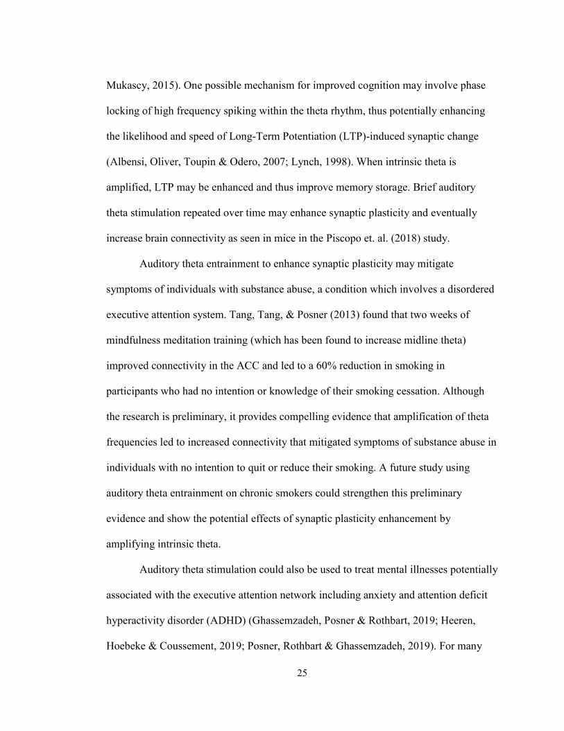
Mukascy, 2015). One possible mechanism for improved cognition may involve phase
locking of high frequency spiking within the theta rhythm, thus potentially enhancing
the likelihood and speed of Long-Term Potentiation (LTP)-induced synaptic change
(Albensi, Oliver, Toupin & Odero, 2007; Lynch, 1998). When intrinsic theta is
amplified, LTP may be enhanced and thus improve memory storage. Brief auditory
theta stimulation repeated over time may enhance synaptic plasticity and eventually
increase brain connectivity as seen in mice in the Piscopo et. al. (2018) study.
Auditory theta entrainment to enhance synaptic plasticity may mitigate
symptoms of individuals with substance abuse, a condition which involves a disordered
executive attention system. Tang, Tang, & Posner (2013) found that two weeks of
mindfulness meditation training (which has been found to increase midline theta)
improved connectivity in the ACC and led to a 60% reduction in smoking in
participants who had no intention or knowledge of their smoking cessation. Although
the research is preliminary, it provides compelling evidence that amplification of theta
frequencies led to increased connectivity that mitigated symptoms of substance abuse in
individuals with no intention to quit or reduce their smoking. A future study using
auditory theta entrainment on chronic smokers could strengthen this preliminary
evidence and show the potential effects of synaptic plasticity enhancement by
amplifying intrinsic theta.
Auditory theta stimulation could also be used to treat mental illnesses potentially
associated with the executive attention network including anxiety and attention deficit
hyperactivity disorder (ADHD) (Ghassemzadeh, Posner & Rothbart, 2019; Heeren,
Hoebeke & Coussement, 2019; Posner, Rothbart & Ghassemzadeh, 2019). For many

26
mental health disorders, the underlying physiology remains unknown. Therefore, further
studies are necessary to determine whether theta stimulation (via auditory or electrical
entrainment) may mitigate symptoms of the disorder.
27
Conclusions
My hypothesis, based on prior human studies, was that brief auditory theta
entrainment while performing a task known to amplify intrinsic theta would enhance
cognition. Specifically, I hypothesized that working memory performance, attention,
and mood in comparison to pink noise stimulation would improve following auditory
theta stimulation. I also examined possible correlations between mood, memory, and
attention performance. My effort to test this hypothesis showed significant correlations
between mood and memory and mood and attention but did not show improved mood,
memory, and attention relative to condition/theta.
I cited several studies showing cognitive improvements in memory and attention
following brief auditory and electrical theta entrainment. I also described the connection
between brief theta stimulation, amplification in intrinsic theta, increased LTP, and
enhanced synaptic plasticity. Further studies should be conducted to determine whether
brief auditory theta stimulation over time leads to structural changes in the brain. In
addition to pursuing further studies with healthy participants, clinical trials to determine
whether auditory theta entrainment mitigates symptoms in patients with disordered
mental states could lead to breakthroughs in noninvasive treatments for individuals with
conditions such as ADHD and substance abuse disorders.
28
Bibliography
Albensi, B.C., Oliver, D.R., Toupin, J., & Odero, G. (2007) Electrical stimulation protocols for hippocampal synaptic plasticity and neuronal hyper-excitability: Are they effective or relevant? Experimental Neurology 204, 1–13
Bellesi, M., Riedner, B. A., Garcia-Molina, G. N., Cirelli, C., and Tononi, G. (2014). Enhancement of sleep slow waves: underlying mechanisms and practical consequences. Front. Syst. Neurosci. 8:208. doi: 10.3389/fnsys.2014.00208
Benabid, A.L. (2003). Deep Brain Stimulation for Parkinson’s Disease. Current Opinion in Neurobiology, 13(6), 696-706.
Cavanagh, J. F., & Frank, M. J. (2014). Frontal theta as a mechanism for cognitive control. Trends in cognitive sciences, 18(8), 414-421.
Conforto, A. B., Amaro, E. Jr., Gonçalves, A. L., Mercante, J. P., Guendler, V. Z., Ferreira, J. R., et al. (2014). Randomized, proof-of-principle clinical trial of active transcranial magnetic stimulation in chronic migraine. Cephalalgia 34, 464–472. doi: 10.1177/0333102413515340
Connolly, K. R., Helmer, A., Cristancho, M. A., Cristancho, P., and O’Reardon, J. P. (2012). Effectiveness of transcranial magnetic stimulation in clinical practice post-FDA approval in the United States: results observed with the first 100 consecutive cases of depression at an academic medical center. J. Clin. Psychiatry 73, e567–e573. doi: 10.4088/jcp.11m07413
Corbetta, M., Kincade, J. M., & Shulman, G. L. (2002). Neural systems for visual orienting and their relationships to spatial working memory. Journal of cognitive neuroscience, 14(3), 508-523.
Fan, J., McCandliss, B.D., Sommer, T., Raz, M. & Posner, M.I. (2002). Testing the efficiency and independence of attentional networks. Journal of Cognitive Neuroscience, 3(14):340-347.
Ghassemzadeh, H. Rothbart, M.K. & Posner, M.I. (2019) Anxiety and brain networks of attentional control. Cognitive and Behavioral Neurology 32/1, 54-62
Hallett, M. Transcranial magnetic stimulation and the human brain. Nature, 406, 147–150 (2000). https://doi.org/10.1038/35018000.
Hasselmo, M. E., Bodelón, C., & Wyble, B. P. (2002). A proposed function for hippocampal theta rhythm: separate phases of encoding and retrieval enhance reversal of prior learning. Neural computation, 14(4), 793-817.
29
Heeren, A., Hoebeke, Y., & Coussement, C. (2019). Unfolding the complex dynamic interplay between attentional processes and anxiety: A commentary on Ghassemzadeh, Rothbart, and Posner. Cognitive and behavioral neurology, 32(1), 63-66.
Holzel, B.K, et al. (2011) How does mindfulness meditation work? Proposing mechanisms of action from a conceptual and neural perspective. Perspect Psychol Sci 6:537–559.
Iaccarino, H. F., Singer, A. C., Martorell, A. J., Rudenko, A., Gao, F., Gillingham, T. Z., et al. (2016). γ frequency entrainment attenuates amyloid load and modifies microglia. Nature 540, 230–235. doi: 10.1038/nature20587
Karnath, Hans-Otto. "Optokinetic stimulation influences the disturbed perception of body orientation in spatial neglect." Journal of Neurology, Neurosurgery & Psychiatry 60, no. 2 (1996): 217-220.
Larson, J., & Munkácsy, E. (2015). Theta-burst LTP. Brain research, 1621, 38-50.
Little, S., Pogosoyan, A., Neal, S., Zavala, B., Zrinzo, L., Haritz, M., … & Brown, P. (2013). Adaptive Deep Brain Stimulation in Advanced Parkinson Disease. Annals of neurology, 74(3), 449-457.
Luck, SJ & Vogel, EK (2013) Visual working memory capacity: from psychophysics and neurobiology to individual differences. Trends in Cognitive Science 17/8 391-400
Lutz, A.; Slagter H.A.; Dunne, J.D.; & Davidson, R.J. (2008) Attention regulation and monitoring in meditation. Trends Cogn Sci 12(4):163–169.
Lynch, G. (1998) Memory and the brain unexpected chemistry and new pharmacology Neurobiology of Learning and Memory 70, 82-100
McNair, D. M., Lorr, M., & Droppleman, L. F. (1971). Manual for the profile of mood states (POMS). San Diego: Educational and Industrial Testing Service.
Pashler, H. (1988). Familiarity and visual change detection. Perception & psychophysics, 44(4), 369-378.
Piscopo, D., Weible, A., Rothbart, M.K., Posner, M.I. & Niell,C.M. (2018) Changes in white matter in mice resulting from low frequency brain stimulation. Proceedings of the National Academy of Sciences, 115/27 6639-6646. https://doi.org/10.1073/pnas.1802160115
30
Posner, M.I., Rothbart, M.K. & Ghassemzadeh, H (2019) Restoring Attention Networks. Yale Journal of Biology and Medicine 92/1, 139-143.
Reinhart R.M.G and Nguyen J.A (2019). Working memory revived in older adults by synchronizing rhythmic brain circuits. Nature Neuroscience, 22(5):820-827
Reinhart R.M.G. (2017). Disruption and rescue of interareal theta phase coupling and adaptive behavior. Proceedings of the National Academy of Sciences, 114(43):11542-11547.
Roberts, B.M., Clarke, A., Addante, R.J., & Ranganath, C. (2018). Entrainment enhances theta oscillations and improves episodic memory. Cognitive Neuroscience, 9(3-4), 181-193.
Schoenen, J., Vandersmissen, B., Jeangette, S., Herroelen, L., Vandenheede, M., Gerard, P., et al. (2013). Migraine prevention with a supraorbital transcutaneous stimulator: a randomized controlled trial. Neurology 80, 697–704. doi: 10.1212/WNL.0b013e3182825055
Tang, Y.Y. (2011) Mechanism of integrative body-mind training. Neurosci Bull 27(6):383–388.
Tang, Y.Y, Lu, Q., Geng, X., Stein, E.A., Yang, Y., & Posner, M.I. (2010) Short term mental training induces white-matter changes in the anterior cingulate PNAS 107 16649-16652
Tang, Y.Y., Lu, Q., Fan, M., Yang, Y., & Posner,M.I. (2012) Mechanisms of White Matter Changes Induced by Meditation Proceedings of the National Academy of Sciences USA 109 (26) 10570-10574 doi10/.1073pnas.1207817109
Tang, Y. Y., Ma, Y., Wang, J., Fan, Y., Feng, S., Lu, Q., ... & Posner, M. I. (2007). Short-term meditation training improves attention and self-regulation. Proceedings of the National Academy of Sciences, 104(43), 17152-17156.
Tang, Y.Y. & Posner, MI. (2009) Attention training and attention state training. Trends Cogn Sci 13(5):222–227.
Tang, Y.Y., Tang, R., & Posner, M. I. (2013). Brief meditation training induces smoking reduction. Proceedings of the National Academy of Sciences, 110(34), 13971-13975.
Vallar, Giuseppe, Gabriella Antonucci, Cecilia Guariglia, and Luigi Pizzamiglio. "Deficits of position sense, unilateral neglect and optokinetic stimulation." Neuropsychologia 31, no. 11 (1993): 1191-1200.
31
Vicario, Carmelo Mario, Carlo Caltagirone, and Massimiliano Oliveri. "Optokinetic stimulation affects temporal estimation in healthy humans." Brain and cognition 64, no. 1 (2007): 68-73.
Voelker, P., Parker, A. N., Luu, P., Davey, C., Rothbart, M. K., & Posner, M. I. (2020). Increasing the amplitude of intrinsic theta in the human brain. AIMS neuroscience, 7(4), 418.
Volkmann, J. (2004). Deep brain stimulation for the treatment of Parkinson’s disease. Journal of Clinical Neurophysiology, 21(1), 6-17.
Weible, A.P., Piscopo, D.M., Rothbart, M.K., Posner, M.I.,& Niell, C.M. (2017) Rhythmic Brain Stimulation Reduces Anxiety-Relate Behavior in a Mouse Model Based on Meditation Training. Proceedings of the US National Academy of Sciences,114/no10 2532-2537.
Wildenberg, J. C., Tyler, M. E., Danilov, Y. P., Kaczmarek, K. A., and Meyerand, M. E. (2011). High-resolution fMRI detects neuromodulation of individual brainstem nuclei by electrical tongue stimulation in balance-impaired individuals. NeuroImage 56, 2129–2137. doi: 10.1016/j.neuroimage.2011.03.074
Wildenberg, J. C., Tyler, M. E., Danilov, Y. P., Kaczmarek, K. A., and Meyerand, M. E. (2010). Sustained cortical and subcortical neuromodulation induced by electrical tongue stimulation. Brain Imaging Behav. 4, 199–211. doi: 10.1007/s11682-010-9099-7
Xue, S., Tang, Y., Tang, R., & Posner, M. (2014). Short-term meditation induces changes in brain resting EEG theta networks. Brain And Cognition, 87, 1-6. doi: 10.1016/j.bandc.2014.02.008
Yarnitsky, D., Volokh, L., Ironi, A., Weller, B., Shor, M., Shifrin, A., & Granovsky, Y. (2017). Nonpainful remote electrical stimulation alleviates episodic migraine pain. Neurology, 88(13), 1250-1255.
Zhou, J., Liu, D., Li, X., Ma, J., Zhang, J., & Fang, J. (2012). Pink noise: effect on complexity synchronization of brain activity and sleep consolidation. Journal of theoretical biology, 306, 68-72.